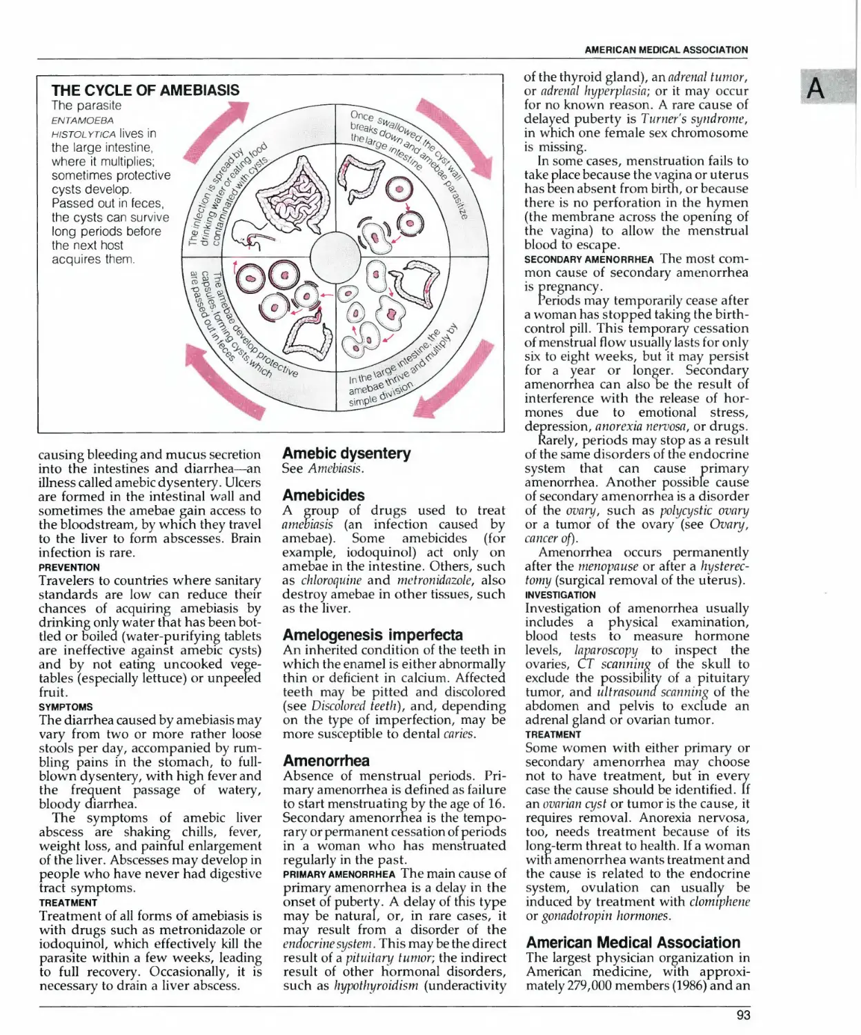
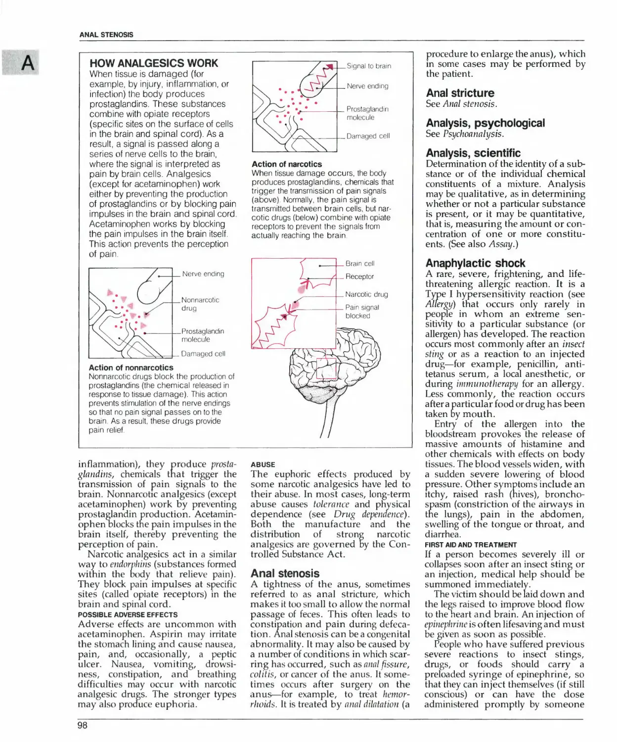
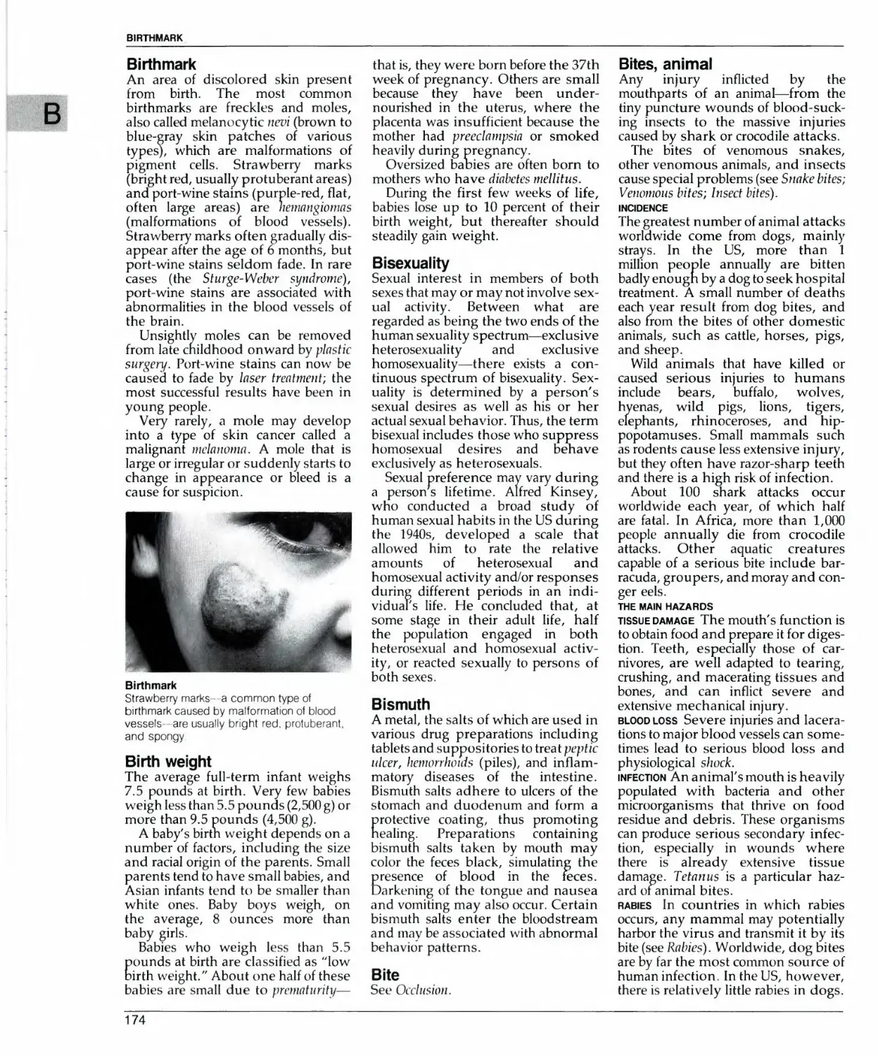
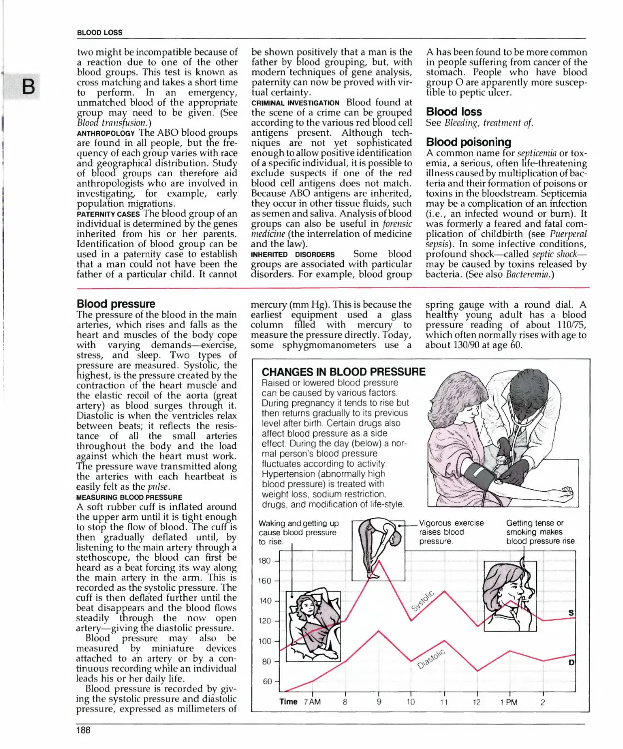
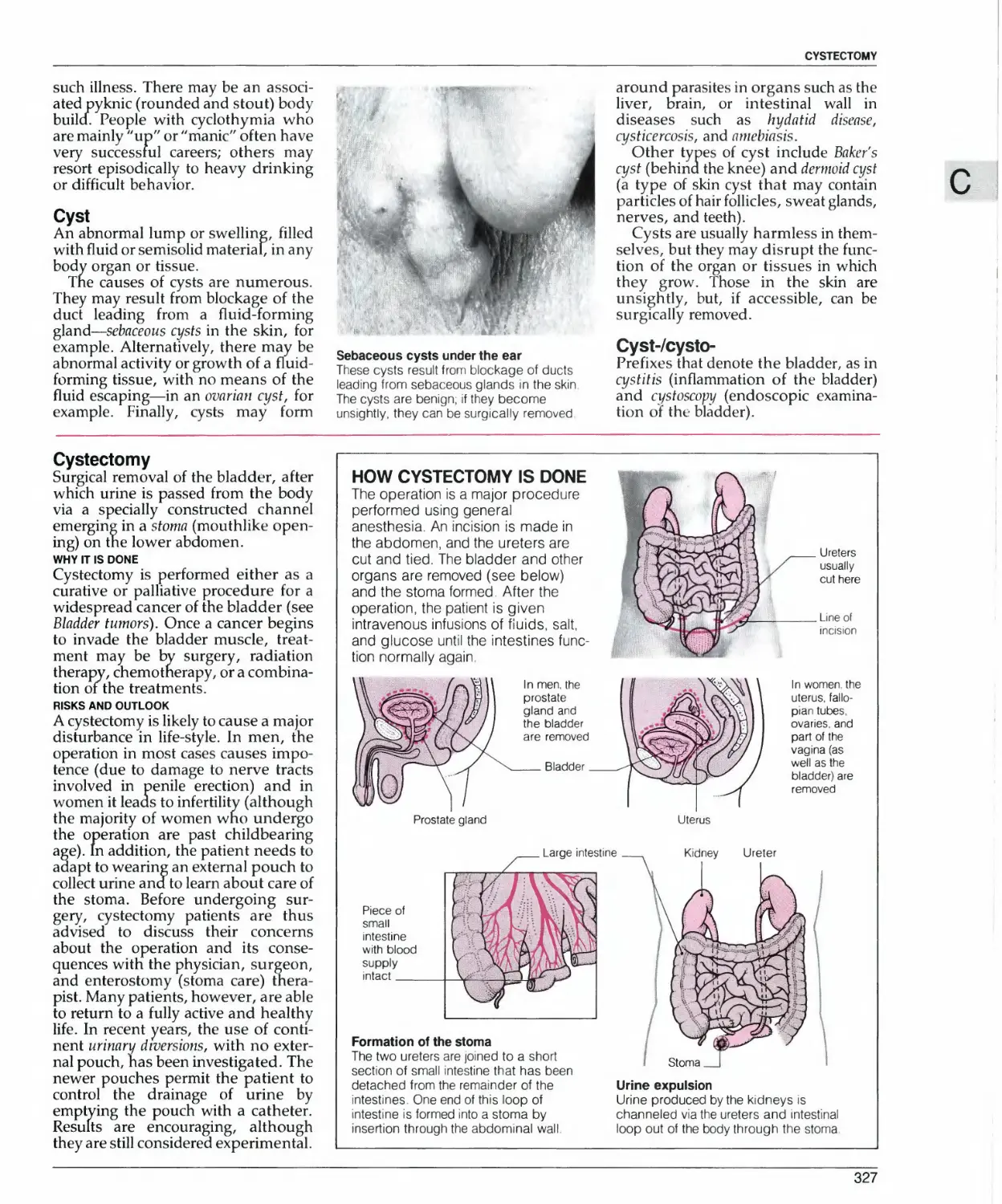
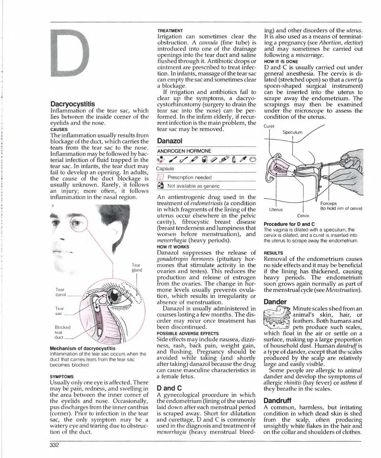
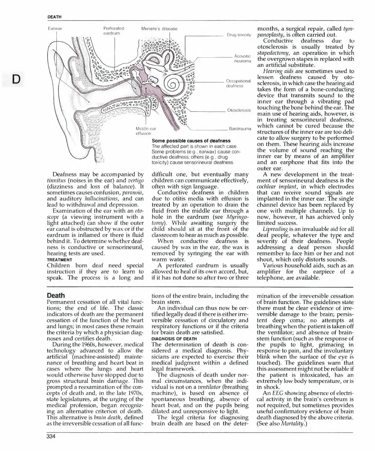
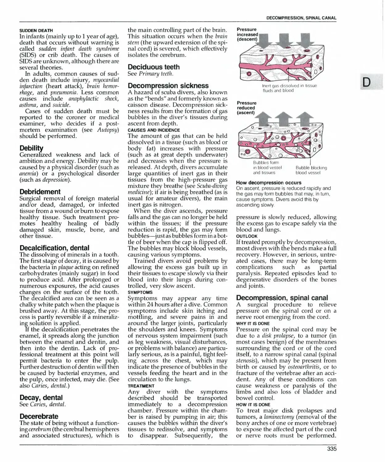
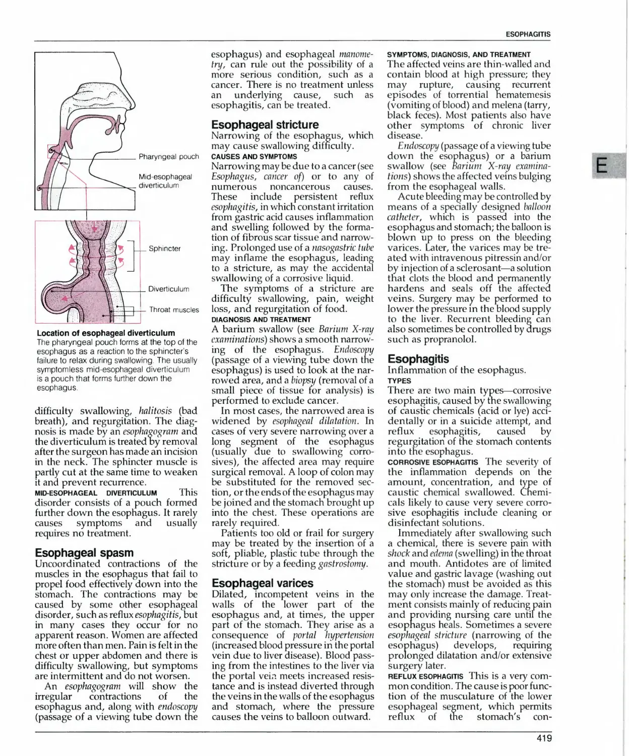
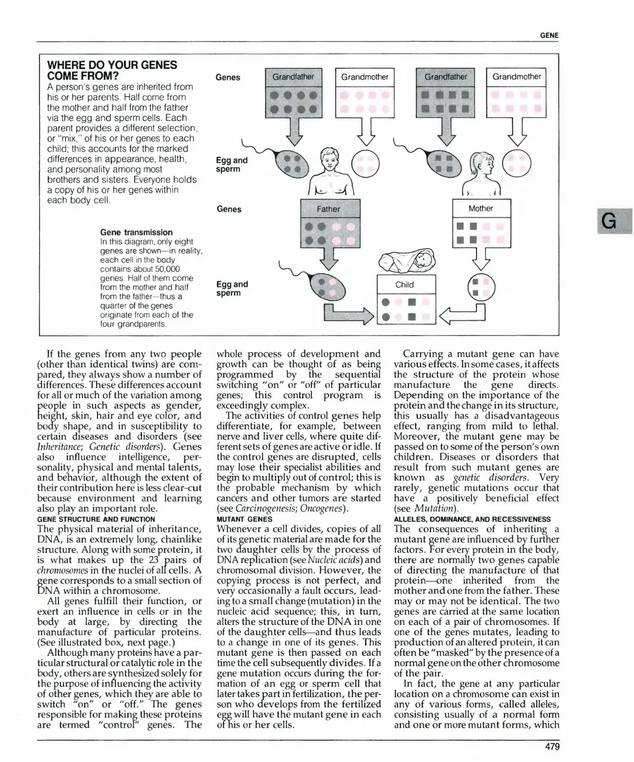
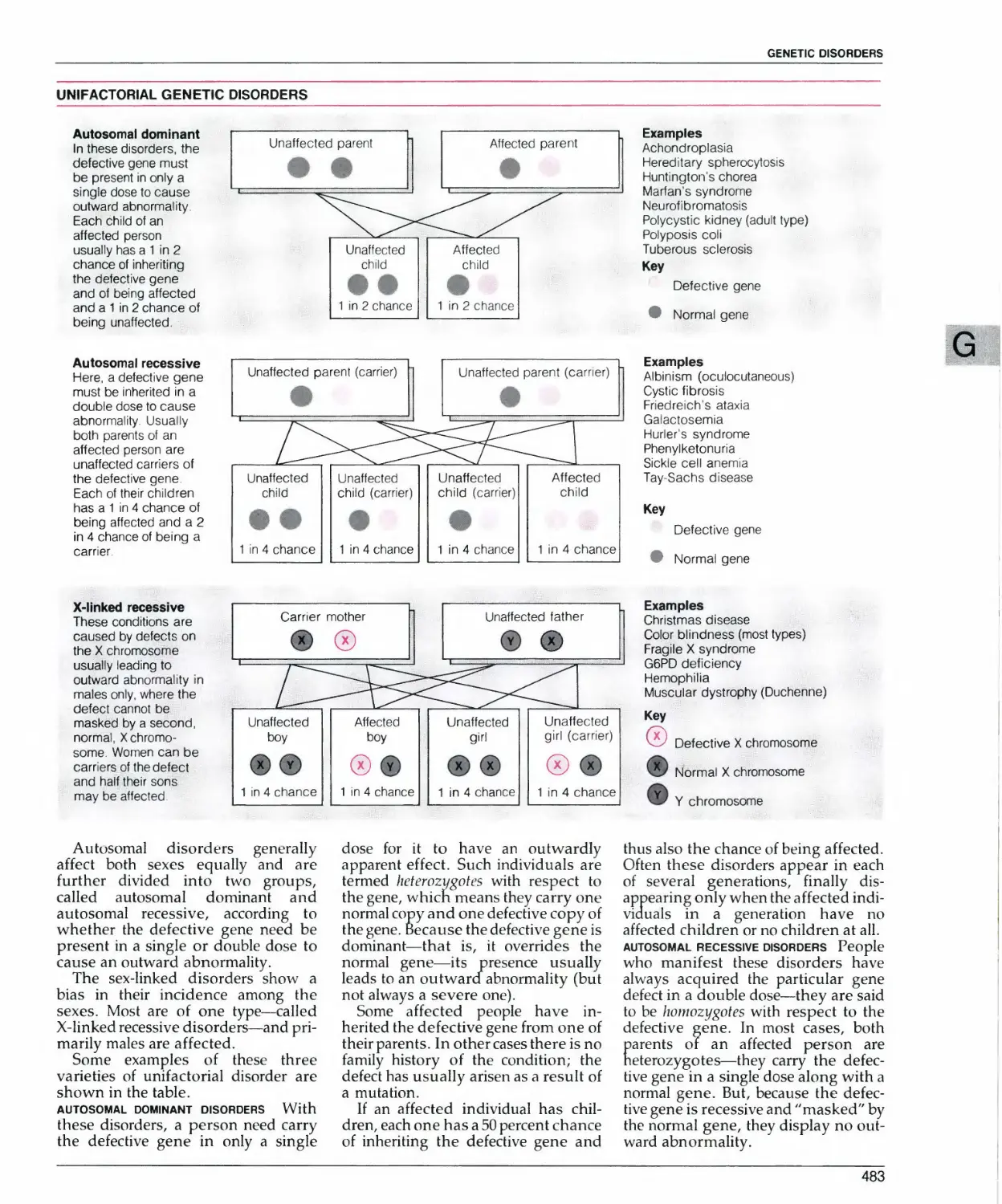
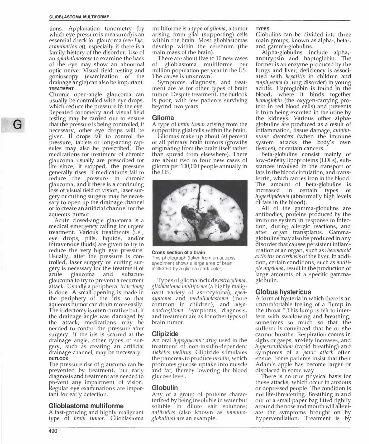
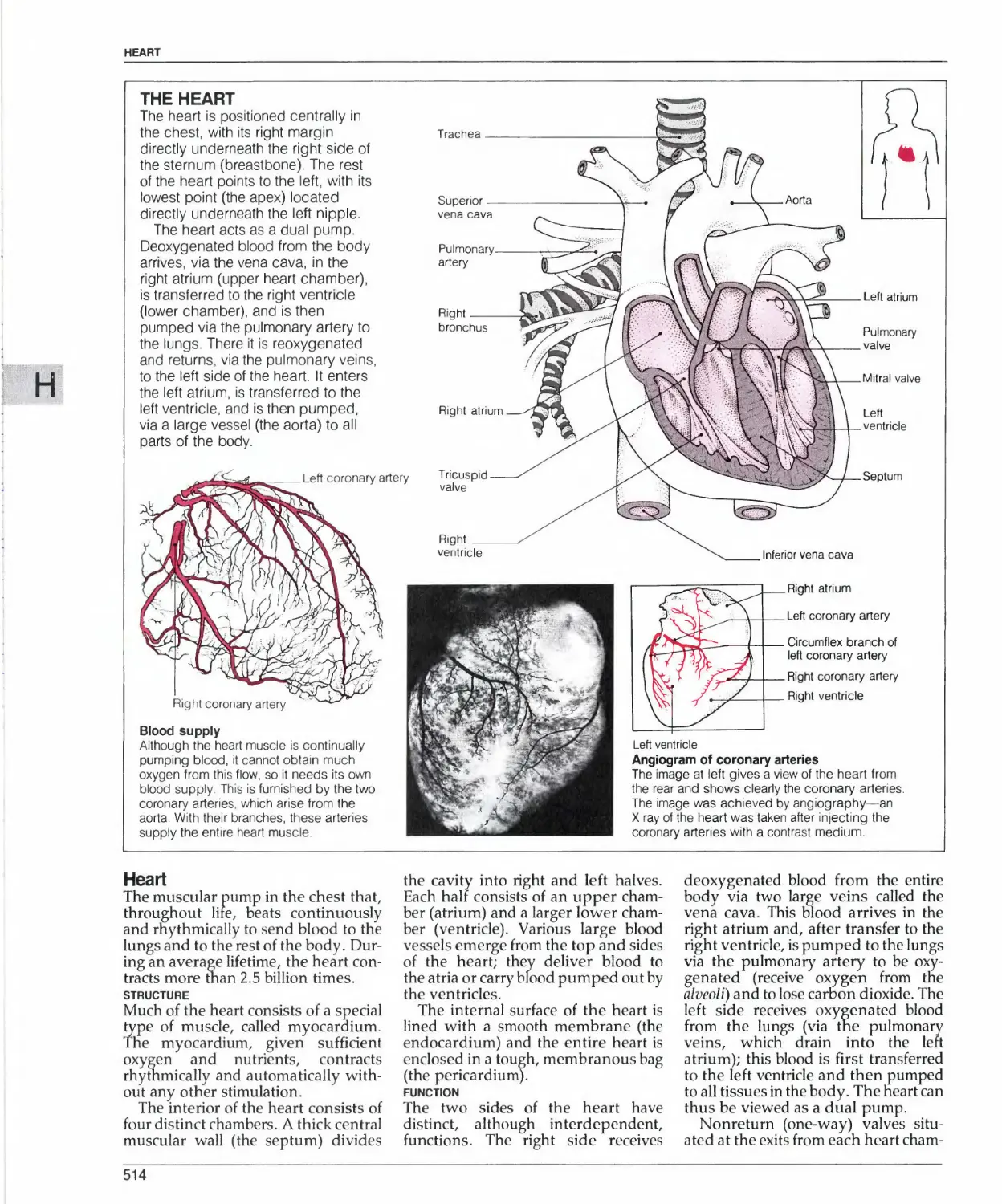
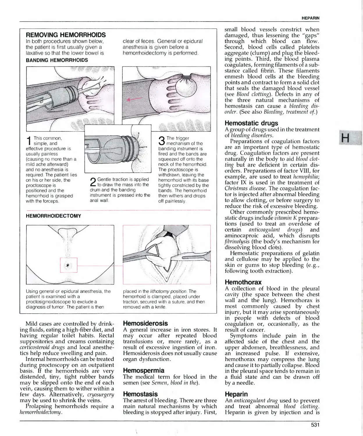
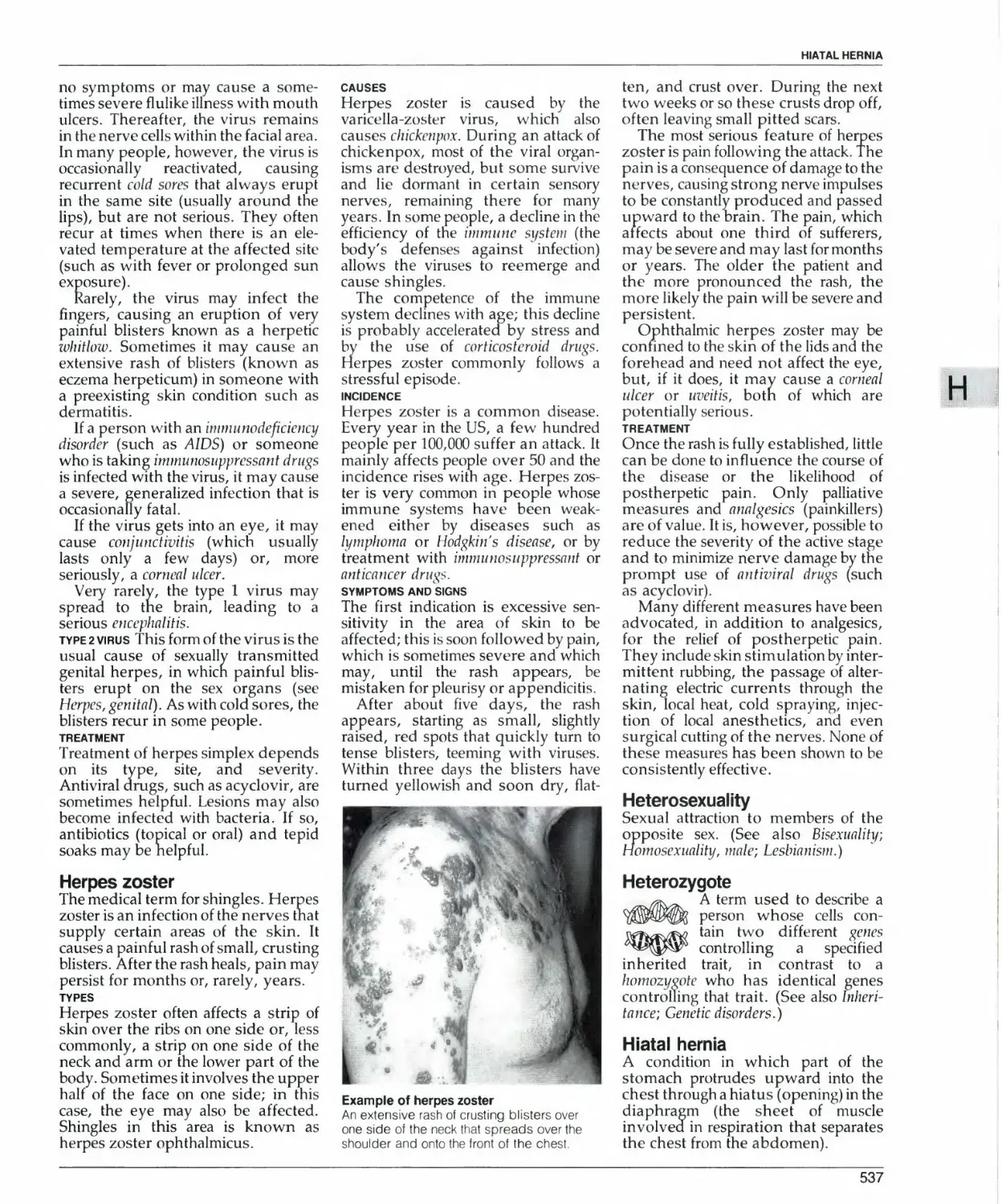
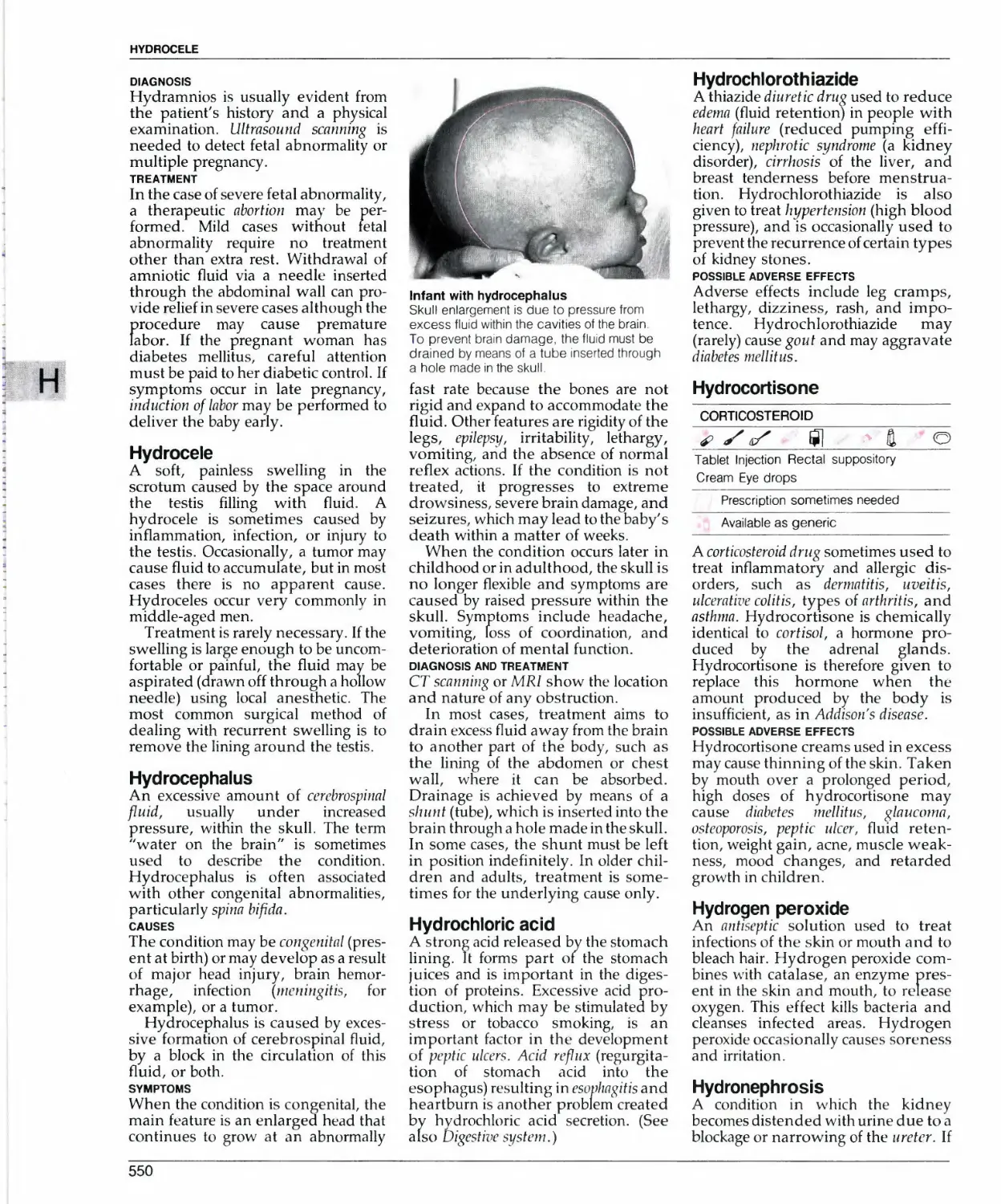
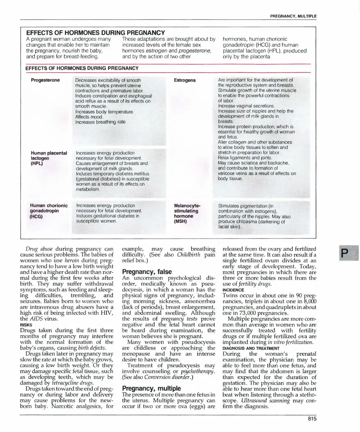
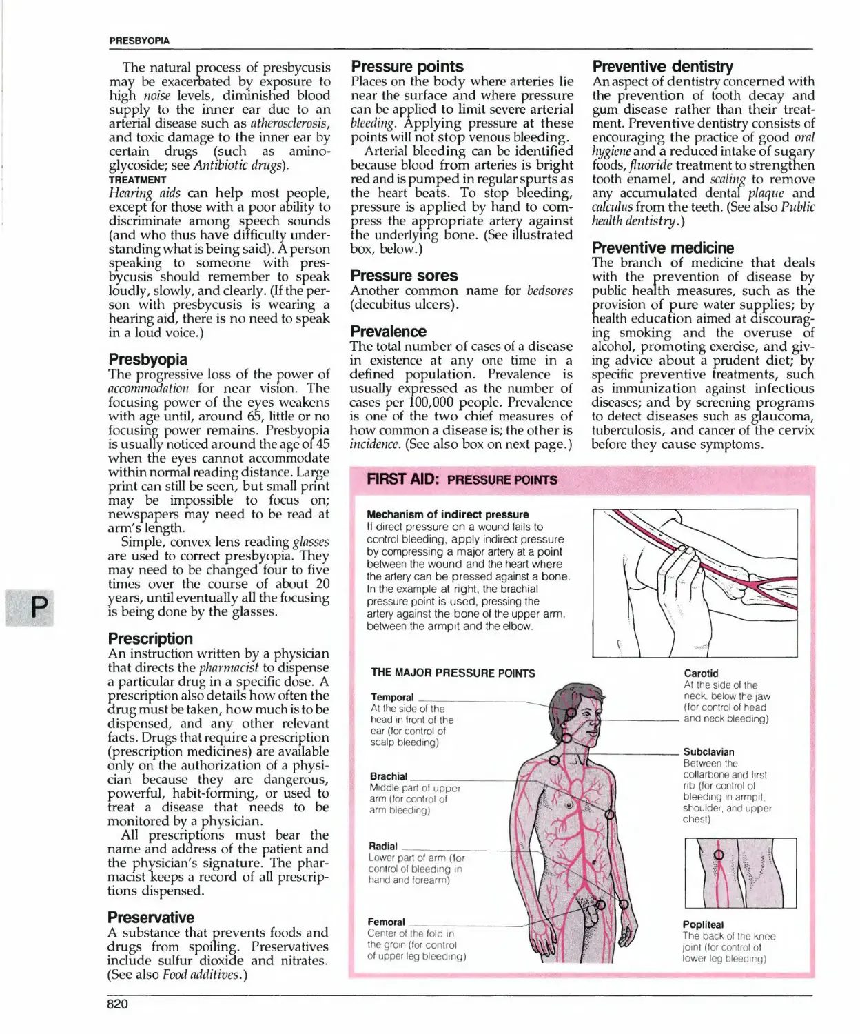
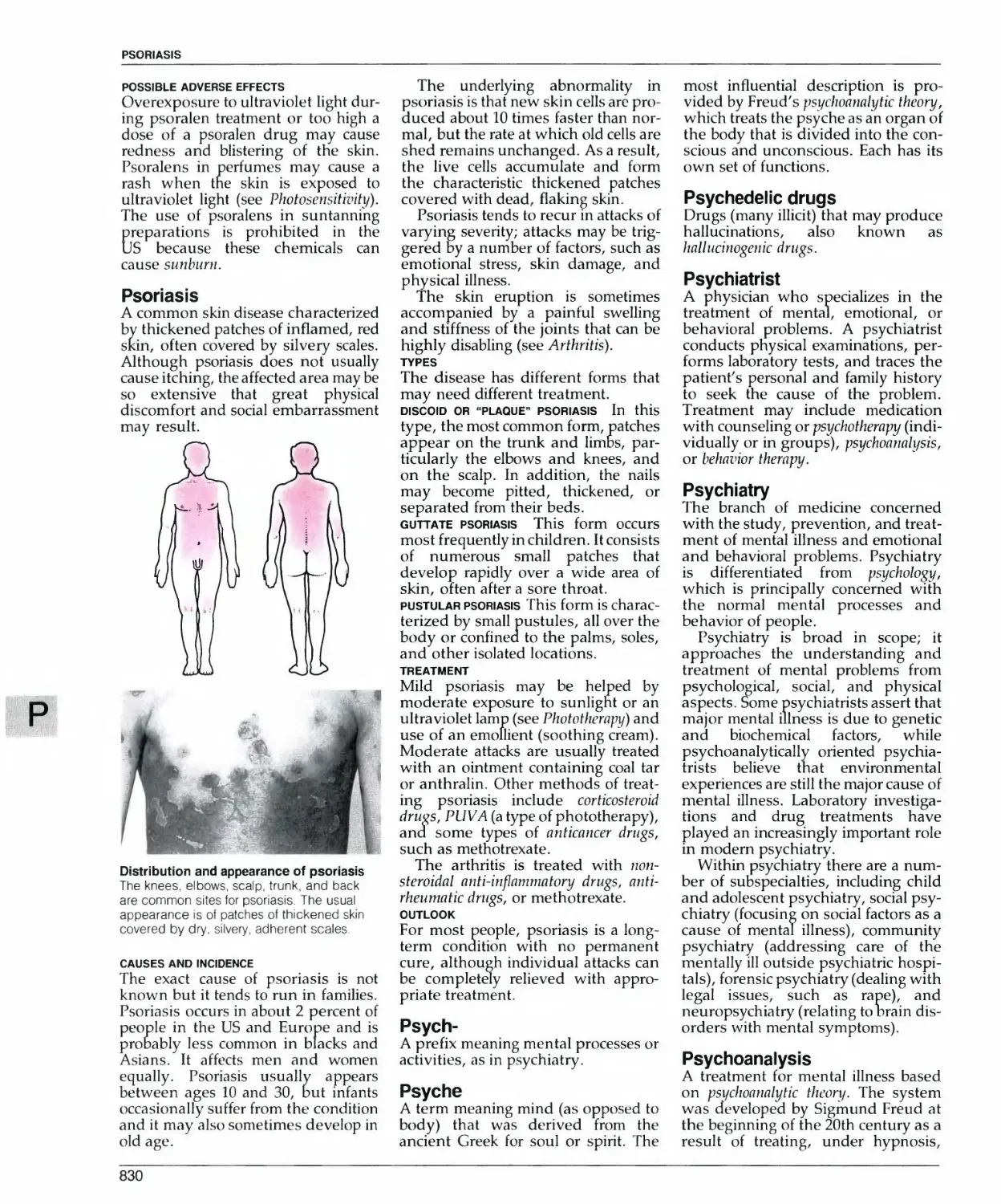
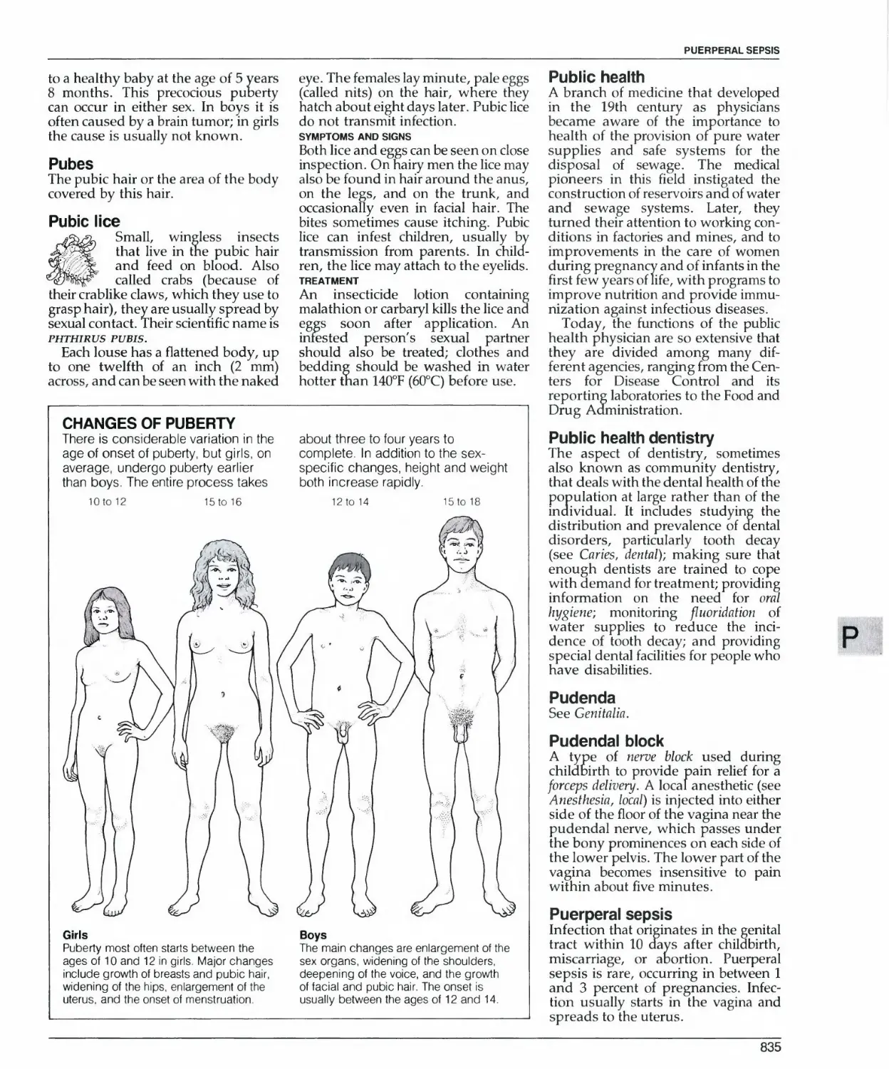
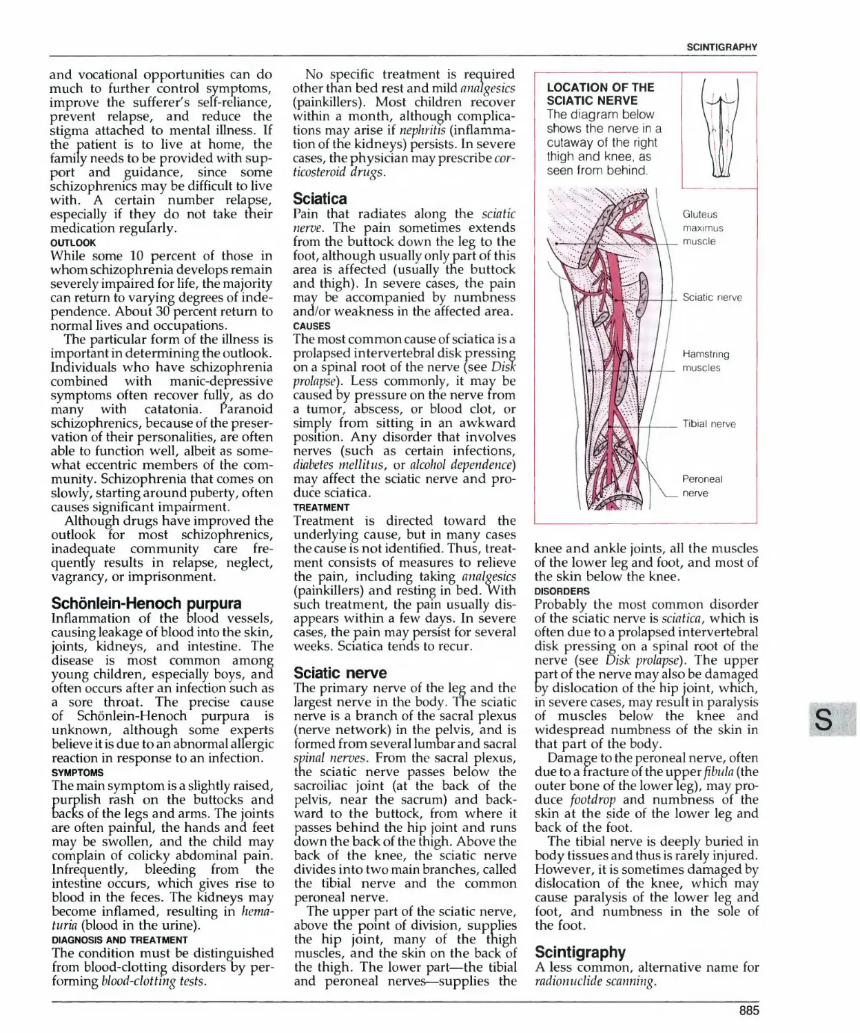
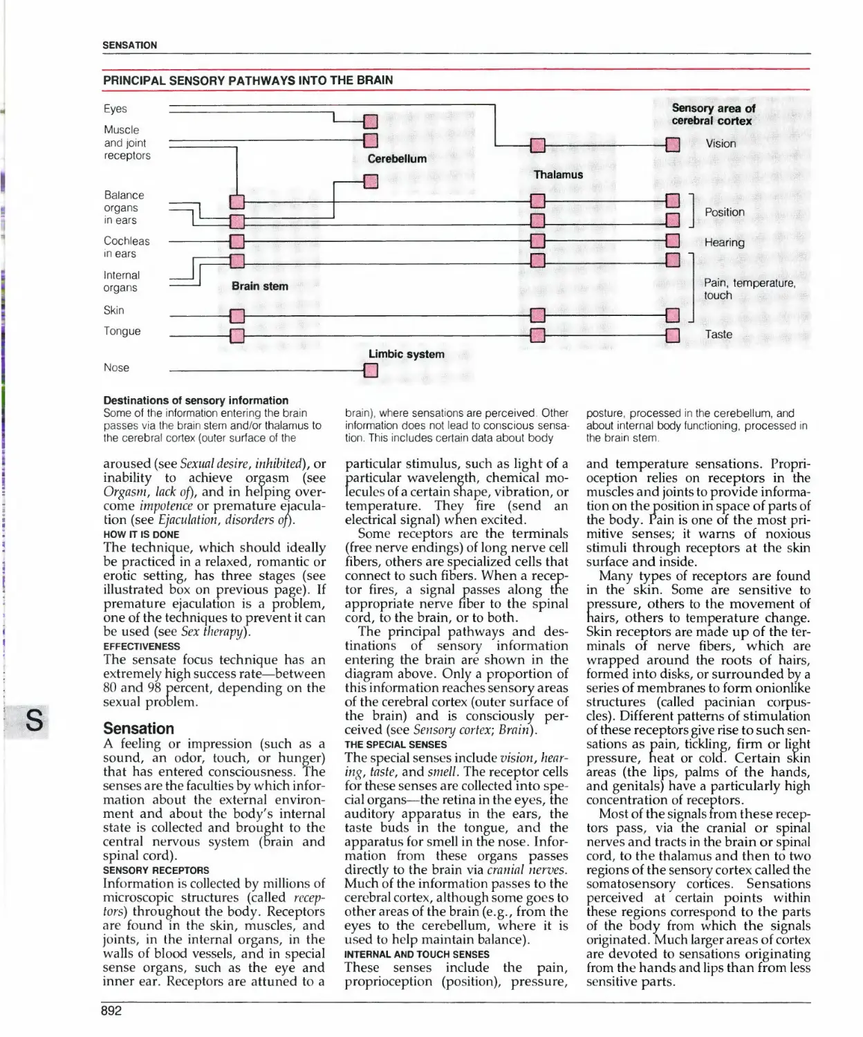
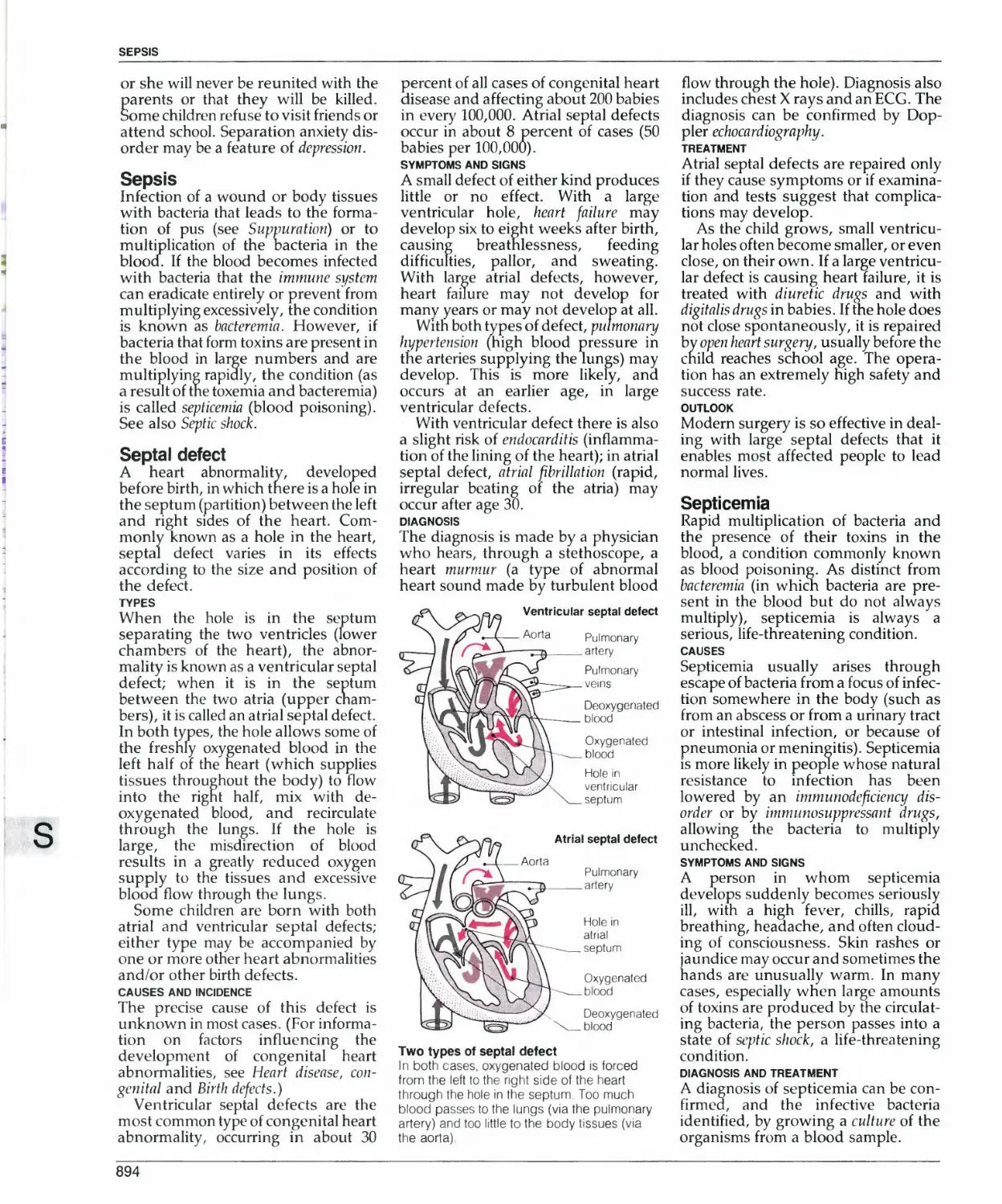
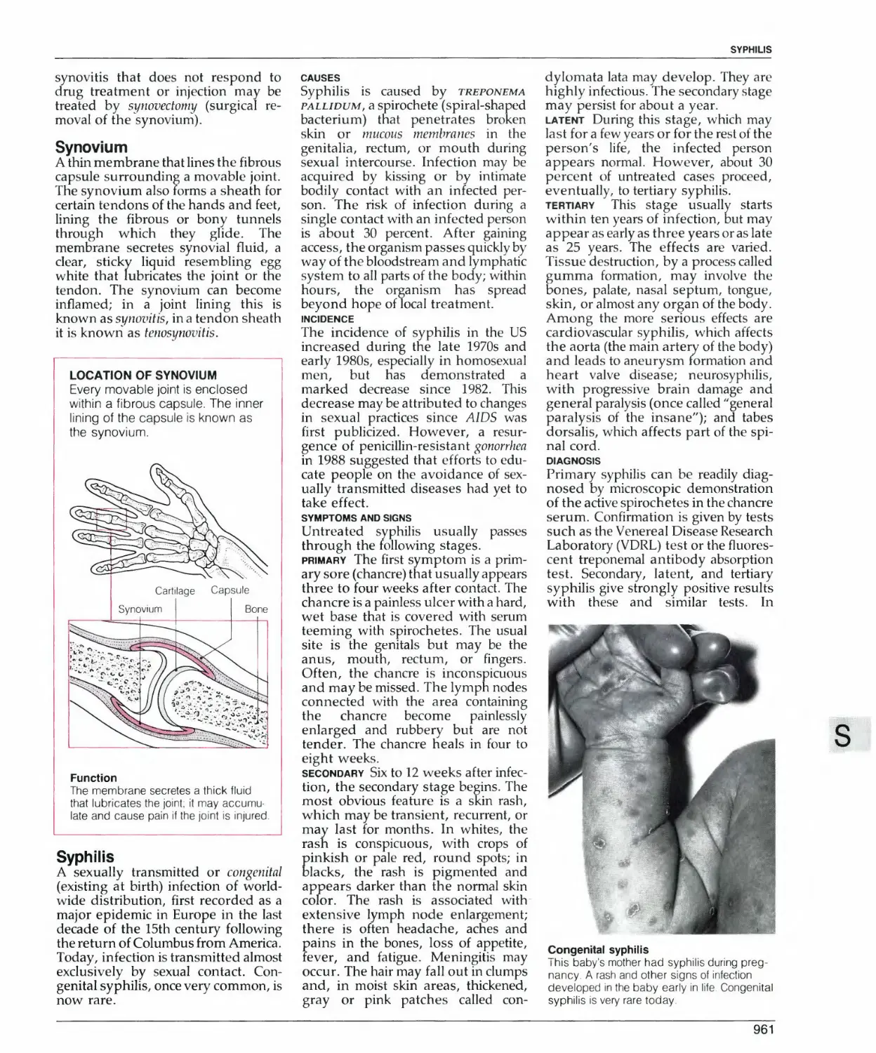
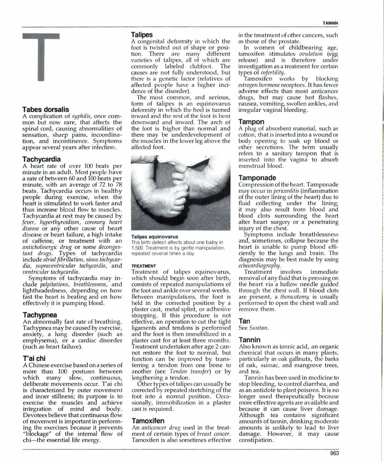
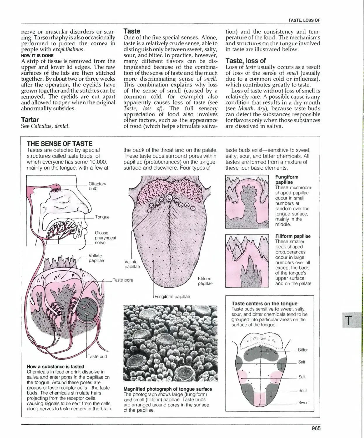
/


Текст
A _
I i
a*
V*
k
&
THE
AMERICAN
MEDICAL
ASSOCIATION
ENCYCLOPEDIA
OF MEDICINE
TU p
AMERICAN
MEDICAL
ASSOCIATION
ENCYCLOPEDIA
OF MEDICINE
MEDICAL EDITOR
Charles B. Clayman, MD
Random House
New York
Copyright © 1989 by Dorling Kindersley Limited
and the American Medical Association
All rights reserved under International and Pan-
American Copyright Conventions. Published in the
United States bv Random House, Inc., New York.
The information in this encyclopedia reflects current
medical knowledge. The recommendations and
information are appropriate in most cases; however,
they are not a substitute for medical diagnosis. For
specific information concerning your personal
medical condition, the AMA suggests that you
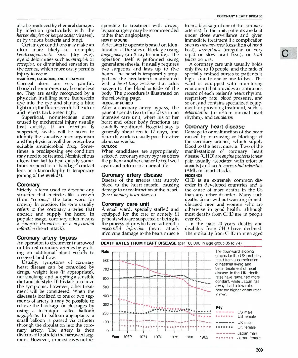
consult a physician.
The names of organizations, products, ur alternative
therapies appearing in this encyclopedia are given for
informational purposes only. Their inclusion does not
imply AMA endorsement, nor does the omission of any
organization, product, or alternative therapy indicate
AMA disapproval.
Library of Congress Cataloging-in-Publication Data
Main entry under title:
The American Medical Association
encyclopedia of medicine.
Includes index.
1. Medicine, Popular—Dictionaries.
I. American Medical Association.
RC81.A2A52 1989 610'.3'21—dcl9 88-29693
ISBN 0-394-56528-2
Manufactured in the United States of America
Computerset by M. F. Graphics Limited, Lngland
Reproduction by Mandarin Offset Limited, Hong Kong
9
PREFACE
Medicine has changed a great deal in recent years. First,
technological advances such as magnetic resonance imaging, PET
scanners, and endoscopes have transformed methods of
diagnosis. Second, the development of new treatments—including
transplants, implants, laser surgery, lithotripsy, and
angioplasty—has widened the range of disorders that can be treated
safely and effectively. New drugs have also extended medicine's
capabilities, or made possible safer and more effective treatments
than their predecessors.
However, probably the most important change has been the
recognition by the medical profession that today's patients are not
prepared to be merely passive recipients of medical care. Today,
people want to be involved in decisions that affect their health;
they want to know not only what is wrong with them, but also
what the choices of treatment are, and what risks are involved.
Recognizing this, most physicians now try to spend more time
with patients and their families explaining the problems and
options available.
This medical encyclopedia has been produced by the American
Medical Association to help you understand the language of
medicine. It provides clear, illustrated explanations of how the
body works and also gives detailed information on all of the
common diseases (and many of the less common ones).
Furthermore, in contrast to many popular medical books, this
encyclopedia does not expound an individual author's theory or market any
"breakthrough" treatment or diet. What this book provides is a
clear systematic account of state-of-the-art, validated, medical
knowledge—reviewed and endorsed by expert physicians
selected by the American Medical Association.
We hope you will use this volume in the best of health.
James H. Sammons, MD
Executive Vice President
American Medical Association
THE AMERICAN MEDICAL ASSOCIATION
James H. Sammons, MD • Executive Vice President
John T. Baker • Vice President, Publishing
Heidi Hough • Director, Office of Consumer Books
Kathy Kaye • Managing Editor
Brenda A. Clark • Staff Assistant
EDITORIAL STAFF
Charles B. Clayman, MD • Medical Editor
Heidi Hough • Editor-in-Chief
Robin Fitzpatrick Husayko • Editor
James Ferris • Assistant Editor
Julie S. Ferris • Assistant Editor
Brenda A. Clark • Editorial Assistant
Frank D. Campion • Consulting Editor
ACKNOWLEDGMENTS
Mary Banas Jacqueline Martin
Joaquin Chang Mary Ann McCann
Gary Hubler, RPh William Smith
Lynne Lamberg Michaela Sullivan-Fowler
Kenneth F. Lampe, PhD Bonnie B. Wilford
Alliance for the Mentally 111 of Greater Chicago
American Dental Association
American Dietetic Association
American Osteopathic Association
American Podiatric Association
American Society of Plastic and Reconstructive Surgeons, Inc.
Mallinckrodt Institute of Radiology
National Organization for Women, Chicago Chapter
Public Health Service, US Department of Health and Human Services
University of Washington School of Medicine
Editorial Director Amy Carroll; Managing Editor Ruth Midgley; Editors Andrea Bagg, Stephen Carroll,
Robert Dinwiddie, Mary Lindsay, Ricki Ostro\, Martyrt Page, Jillian Somerscales, Frances Williams,
Robert M. Youngson; Editorial Secretary Pat White; Managing Art Editors Chez Picthall, Denise Brown;
Art Editor Caroline Murray; Designers Tina Hill, Tracy Timson, Lydia Umney, Peter Cross, Gail Jones,
Melissa Gray, Sarah Ponder, Sandra Archer; Picture research Sandra Schneider; Production Rupert Wheeler
MEDICAL CONSULTANTS
Bruce Berkson, MD
Urology
Rowland W. Chang, MD
Rheumatology
Joseph L. Clay man, MD
Dermatology
Arthur W. Curtis, MD
Otolaryngology
Theodore Doege, MD
Environmental Medicine
Roger K. Ferguson, MD
Clinical Pharmacology
Asher J. Finkel, MD
Internal Medicine
Edward J. Fitzsimons, PhD
Biochemistry
Richard M. Gore, MD
Radiology
Jourdan Gottlieb, MD
Plastic and Reconstructive Surgery
Alan B. Halle, MD
Geriatrics
James A. Hill, MD
Orthopedic Surgery
Linda Hughey Holt, MD
Obstetrics and Gynecology
Allen Horwitz, MD
Genetics
Howard N. Jacobson, MD
Nutrition
Gary S. Lissner, MD
Ophthalmology
Gary J. Martin, MD
Cardiology
Helen Gartner Martin, MD
Pulmonary Medicine
Ronald M. Meyer, MD
Anesthesiology
Lawrence Michaelis, MD
Cardiovascular Surgery
Vincent T. Miller, MD
Neurology
Robert Murphy, MD
Infectious Diseases
Robert V. Rege, MD
General Surgery
Domeena C. Renshaw, MD
Psychiatry/Sexual Dysfunction
Mary Beth Shwayder, MD
Pathology
Michael W. T. Shwayder, MD
Nephrology
Tor Shwayder, MD
Pediatrics
Mark Stolar, MD
Endocrinology
E. Fuller Torrey, MD
Clinical and Research Psychiatry
Edward S. Traisman, MD
Pediatrics
Merrill S. Kies, MD Ronald J. Vasu MD
Hematology-Oncology Psychiatry
Margaret M. Yungbluth, MD
Microbiology
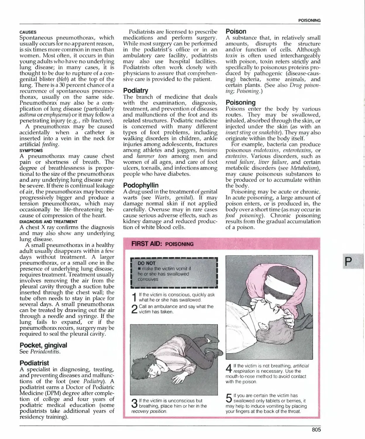
EMERGENCY FIRST-AID TECHNIQUES
Use this quick-reference list to find illustrated first-aid boxes containing
step-by-step instructions for performing emergency techniques.
Artificial respiration
Bleeding, treating
Burns, treating
Cardiopulmonary resuscitation
Childbirth, emergency
Choking (adult)
Choking (infant and child)
Electrical injury
Frostbite
Heat stroke
134
179
220
237
266
271
272
395
469
526
Hypothermia
Poisoning
Pressure points
Recovery position
Shock
Snakebite
Suffocation
Unconsciousness
Wounds
563
805
820
855
902
921
953
1022
1079
SYMPTOM CHARTS
Use this quick-reference list to find question-and-answer flow charts that
indicate the possible causes and significance of many common symptoms.
Abdomen, swollen
Abdominal pain
Abdominal pain, recurrent
Backache
Breathing difficulty
Chest pain
Constipation
Cough
Diarrhea
Dizziness
Feeling faint and fainting
Fever
56
51
53
151
211
260
299
315
355
368
436
450
Headache
Hoarseness or loss of voice
Intercourse, painful (men)
Intercourse, painful (women)
Menstruation, irregular
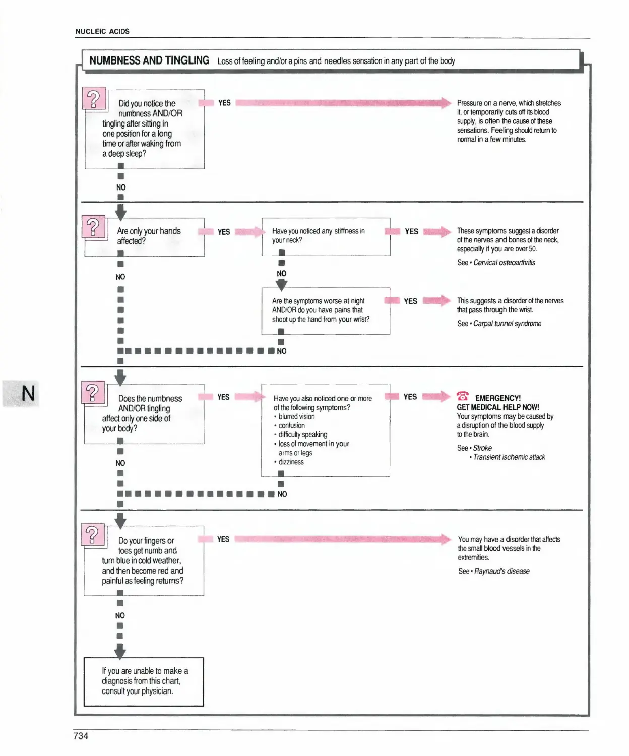
Numbness and tingling
Rash with fever
Rash with itching
Tiredness
Vomiting
Weight loss
508
542
596
597
679
734
851
852
990
1063
1074
8
CONTENTS
HOW TO USE THE ENCYCLOPEDIA 10
MEDICINE TODAY 15
THE A to Z OF MEDICINE 49
SELF-HELP ORGANIZATIONS 1090
DRUG GLOSSARY 1096
INDEX 1118
HOW TO USE THE ENCYCLOPEDIA
This highly illustrated encyclopedia is an
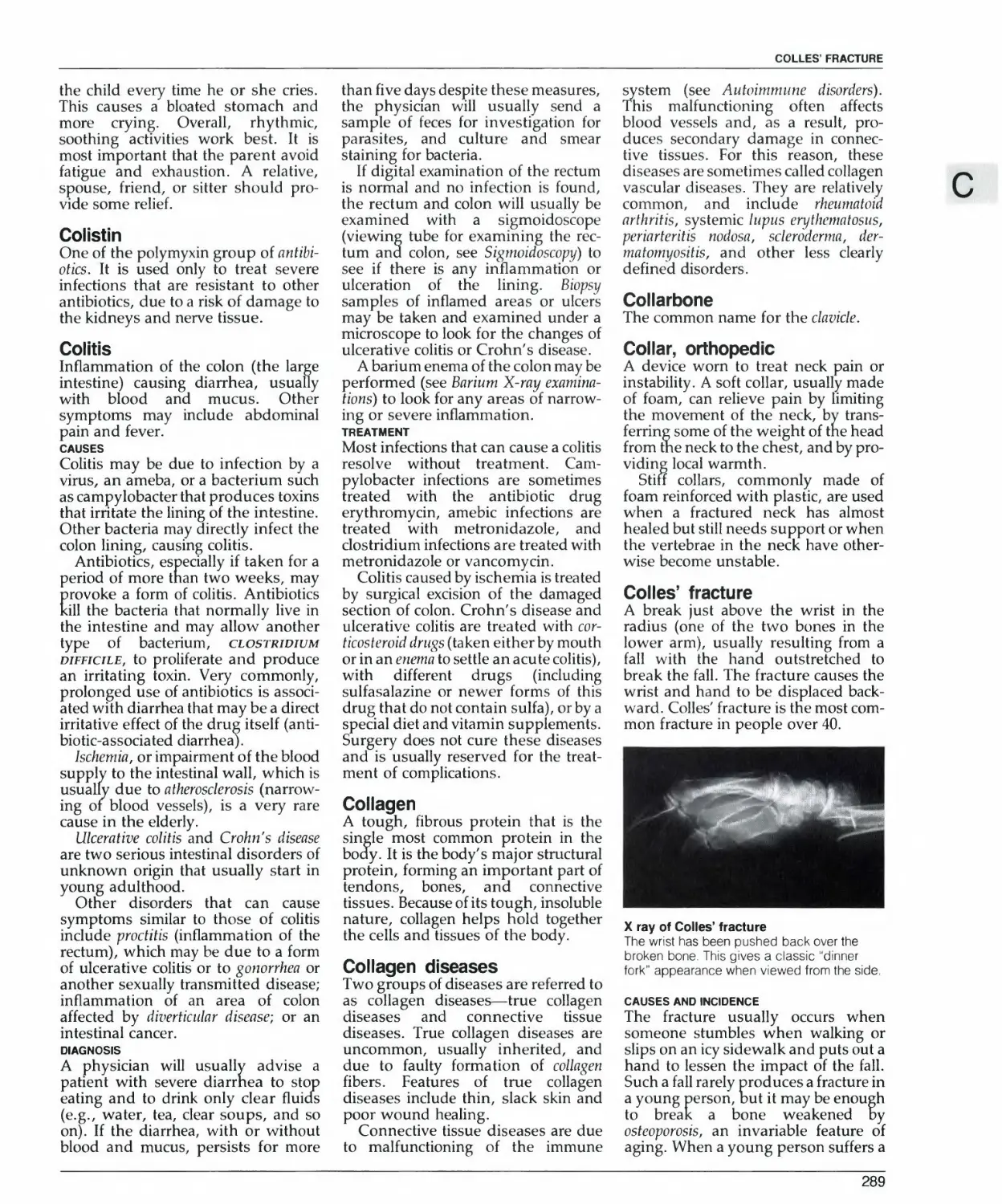
authoritative guide to all aspects of medicine. For swift and
easy reference, encyclopedia entries are arranged
alphabetically and longer entries are subdivided
into sections, each with a descriptive subheading.
Information within entries is presented in clear,
concise language. Technical or unfamiliar medical
terms are generally explained as they appear.
The main body of the encyclopedia, the A to Z of
Medicine, contains more than 5,000 entries
covering a vast range of medical and medically related
topics. It is obviously impossible in a one-volume
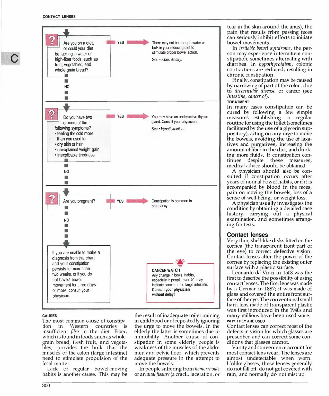
compendium to provide separate entries for every
medical term, but many additional terms and topics
are discussed within relevant entries. The Index
refers you to all such items as well as to the major
entries themselves.
The encyclopedia also contains a full-color
introductory section, Medicine Today, which gives
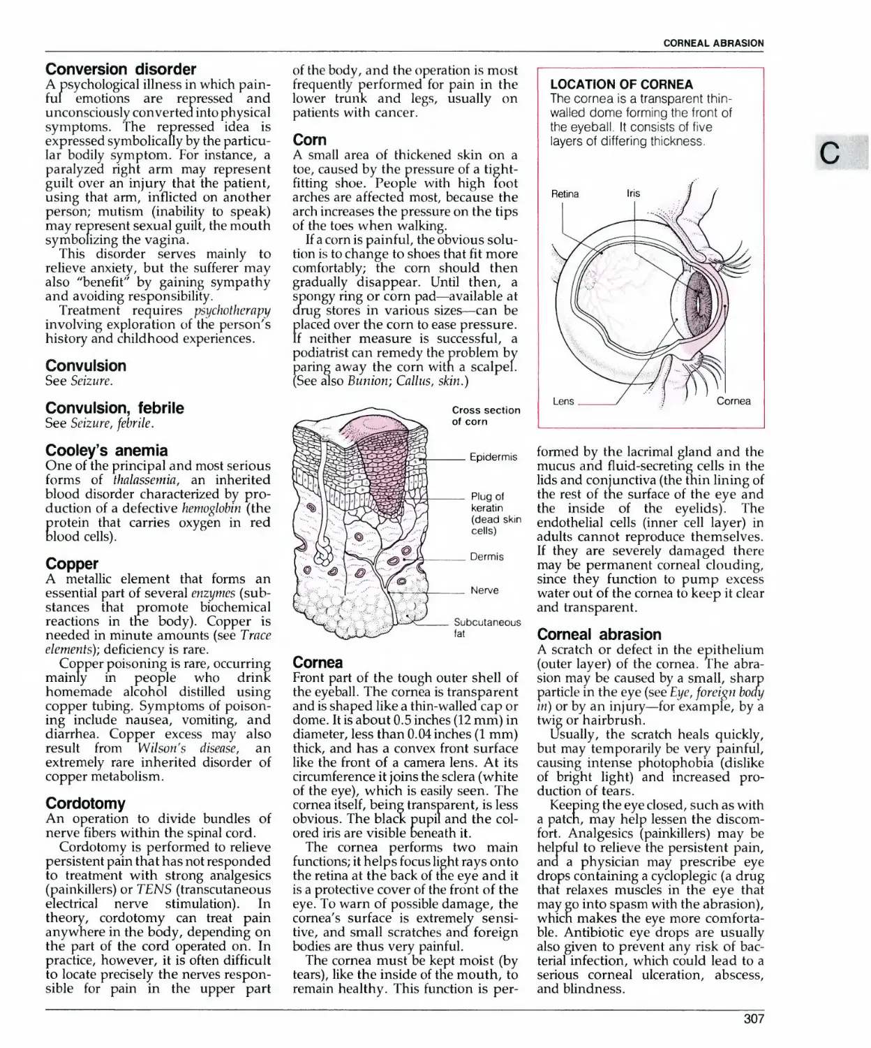
useful information on staying healthy and describes
the latest advances in diagnosis and treatment. At
the back of the book, additional information on
generic and brand-name drugs is contained in the
Drug Glossary. There is also a list of telephone
numbers, many of them toll free, for a wide range of
Self-help Organizations.
HOW TO FIND THE INFORMATION YOU WANT
All entries in the A to Z of Medicine
and in the Index are arranged
alphabetically using the "letter-by-letter"
system. In this system, any spaces or
punctuation in the entry titles are
ignored. Sick building syndrome is
thus followed by Sickle cell anemia
and then by Sick sinus syndrome - the
fifth letter gives the order.
When the name of a topic consists
of more than one word, begin by
looking up what seems to be the key
word. Thus, for information on
general anesthesia you will find
what you want under the heading
Anesthesia, general. If the key word is
not obvious, you may find it easier to
turn first to the Index, although
many alternative topic names and
common abbreviations are included
as short cross-reference entries
within the main part of the book.
You will also find cross-references
within the entries themselves. These
are indicated by italics and take
several forms, which are explained
in the annotated illustration below.
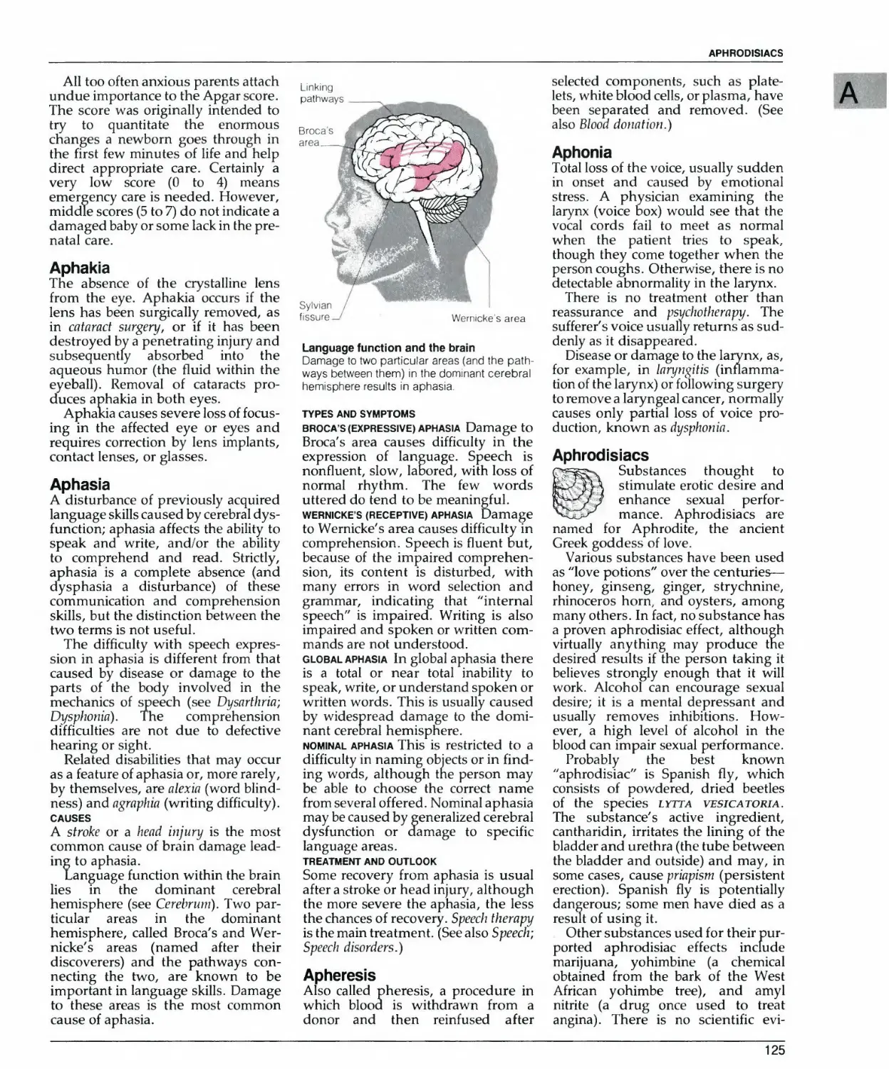
bee Breast.
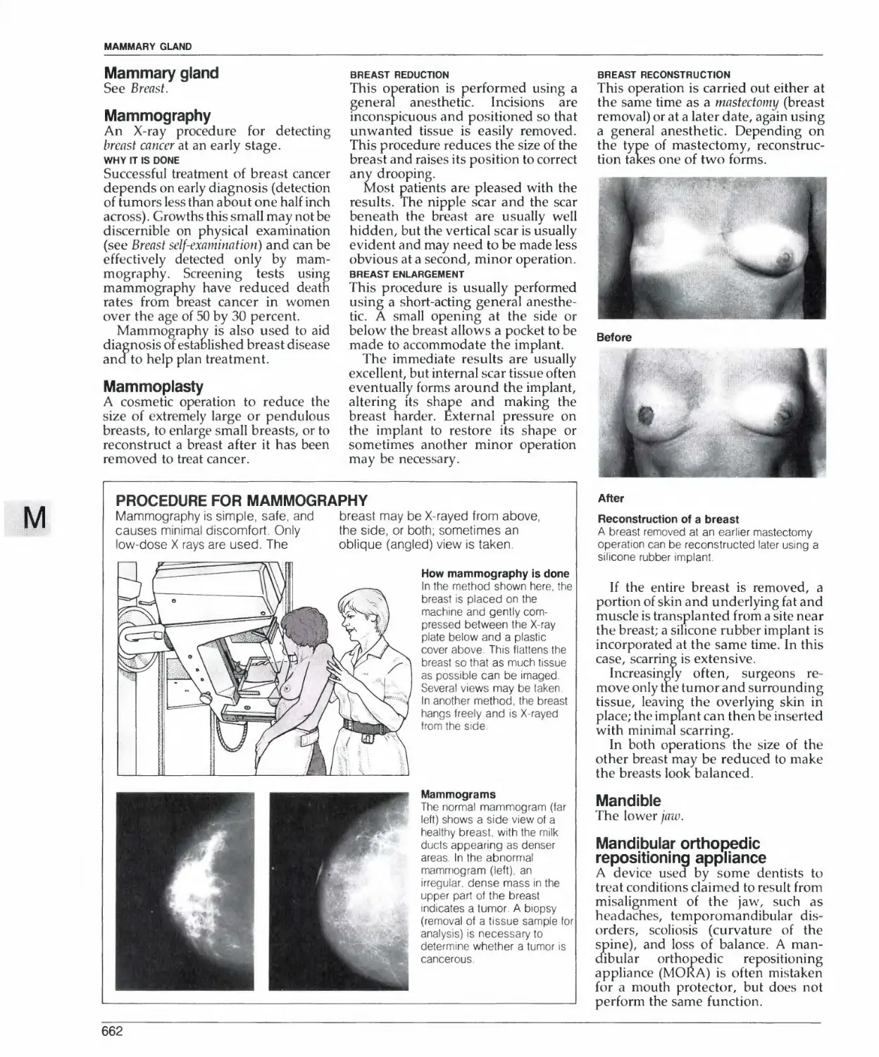
Mammography
An X-ray procedui*^1cir de
breast cancer dt^Hrearly stage.
WHY IT IS D<
Successful treatment of breast
depends on early diagnosis (dt
of tumors less than about one h
across). Growths this small ma)
discernible on physical exam
(see Breast self-examn
effectively detected
mography. Screeni
Subordinate
subheadings
A second level of
subheading is
sometimes used I
this case, the text
continues on the
same line
"See also" cross-
references
At the end of some
entries, "see also"
cross-references
direct you to related
entries that may be of
interest to you.
Standard
subheadings
Standard
subheadings are
used to tell you what
each part of the entry
is about.
Callosity
ee Callus, skin.
ballus, bony
\ diffuse growth of new soft bont
brane that lines the nose,
manifested bv some combina
nasal obstruction, nasal dis
sneezing, and facial pressure o
TYPES
_vjsal^hinitis A feature of the c<
cold (seeCaW, common), rhiniti:
viral infection may lead to sim
allergic rhinitis Also known
fever, this type may be seasor
to pollens, or year-round,
house dust, molds, or pe ■
Rhinitis, allergic). It most con
occurs with vasomotor rhiniti?
"See" cross-
references
Italicized "see" cross-
references within
parentheses refer
you to other entries
for more detailed
information
brms around a fracture as il lii'dls
■alius is eventually replaced
IrcTng-erJxme with a more orgar
Itructure (se~p-£oH(').
| A callus can be seen on an X ra\
•rovides evidence that healing
'tarted. A callus can sometimes b
round a fracture site as a luj
_ Cross-references
Italicized cross-
references within
sentences refer you
to other entries for
more detailed
information
l *-' • » V > <^ I , HIV. L'l\. »U1\.1IV.V, Vt ^.1111
itution and of child sexual abus
in families seems much highe
previously thought. Nearly 1
nt ol women in some studie
"t some form of sexual intei
ce in childhood or early adolej
;. (See <i I so Child abuse; (ncest.)
*£iri£ t*iw ii rruki il
•uIVi*1h-> ■ timr
trur-iinn* Ih< !•»■ -h3
Ifi riitirJJuiii^lhrMi
hfu.*>n£ (4 I hi I.B.
ITKI ^'ri '
»J Ih. Un
Lt,
£
t,
T
'^i
rxji
I**'
ilili Fix-i 4ft rrm>
j||*r tht >»f.irrali<>f li rrn-M J«- 'h>
<.ii< Hhkr< i*J« -■Ihir * irn *n
hiililcr hi rutuul ,imm in»- "hi
IhrlYln rh.-rll^t.-l jl*. <illtn.ii.i1U
1**1* J rum I livi <rji*
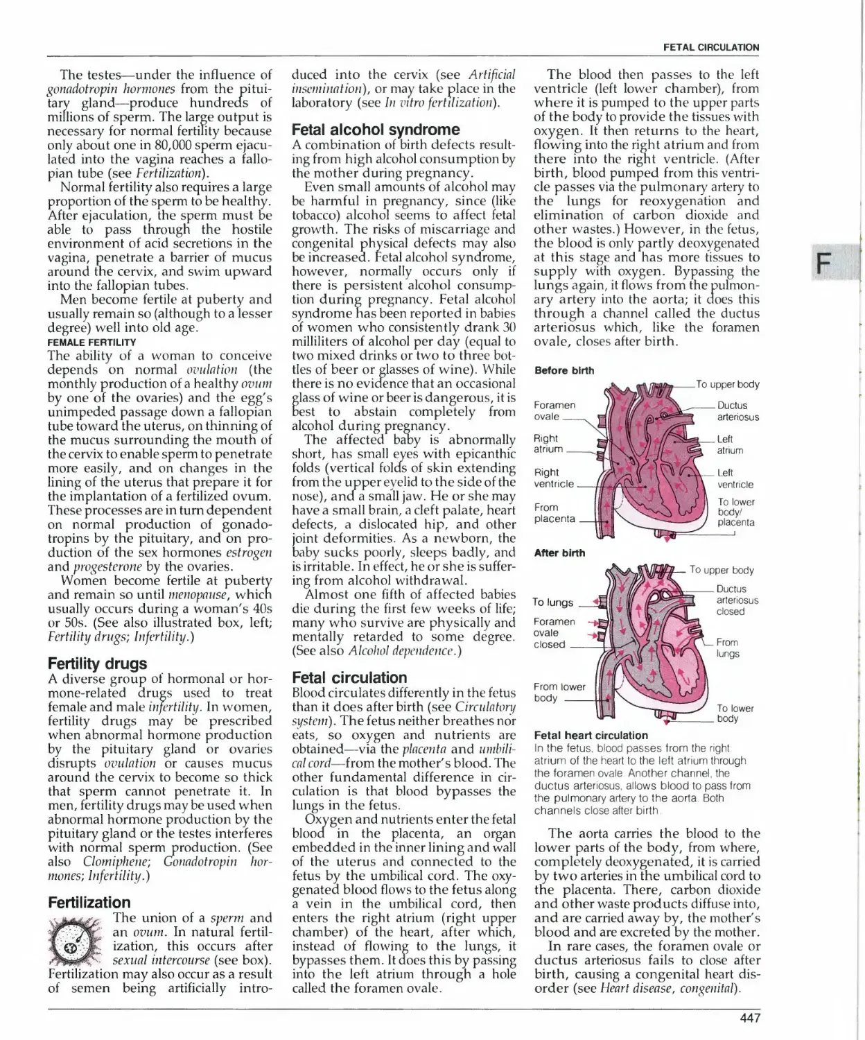
t Hi j*iL<n*lit *jii»i*.t.w\ Fta-j.iniL
Jia-% riiit ■.»■ uf *% * fr-.ul! ,.t *r>itft.--w
IhU i-illn^ unitrf Ow iIiiiiih iiifnfHf
k-4ilirlji li'ir\nt m j«nn£ ihlru wim^i
Facial narv*
lit. vtn<lh .•
r riM-i-hr*:
T",
uncle
Iklike connecting structure. Th *'""'■
usually refers to bands of nerv
i that connect different parts c
rain, or to the ropelike connec
nnli
-tlii
"'"•^^ '
iOCATlOHOf TMt I*CIAL NlflVE
£v
y—
I*, u r*r» ■ ,*rli*m- r*»lh
*nj **nvf\ lutVlKTii |l %aai
ihr miixlp ,ft ih,- nnk *nd .<*
■ nviuJinn rau-,lr n
jllv'^lifnulJIirs vi fr
« utrrun J iHj Lai j r*]
>uhlintfu4l »JlHjrv ^Lir*]* lr JjJi
LhuI mnt ii<nini l«»Ir
il nmliilLi utifciiidjl^ %rn»jliia/i% litvrnhi' lri«nl l*vi< ihiitli ■
in *h*>nl jnJ »rfidN ,h* »'-njt"«- -"iJ un«f» Mrnwli.i
U.r iiiih viliun twftfitylriwi
„, l>im«)tt- Ik iht Uyu: n*rM nux
wi-dkrv^i t>1 I hi- tiiMl urn-war* iv
frill ' fWl'VUnJ U' SiHIM-l JStN l|H%l
M*lr 'Suih Jjini(j;i i^ihiim iiKiim.ir
tiut d'Jiiiiu-irntiln>n limit jlwl
*lr*luTfi'lJ-/'!!*■■ niutriMr\\ ilnu
miur j* * rouh m -uf^i-fA |tv|Hi.*l
im j tumoil m Iht- i^n.tid filjnj him
■■I the wliwrv gUiiiKl
Facial pain
I'jin in Ihr l*n' a\4\ hi- i^lim-iI h«
injurs miniiimi m j nrm iliviiili I
mit hr rWrrii-d Invnr1<« mhrri in ihi
hiut m ^r'-ii^imi iiinut mui
lur ■>■ kn^mri rt-js.ni
fUII>*
In *JJrti.m in pai» m Irn- !*-• invn t
Jirr* ■ iriwrt p*tts nut !>' iIiimsI
>rvj:rritl* t1* r>i*>i-i, |Vi44rm« ■■> *
Nh m^l>«ri um*t\ p/.Mrm^ ■■> jn
JlUixjf»m ,at Ihr (In
■Micron
- "_-•■ i,»lUlnAul. i ,-t IK. jit
*,>*n J'owriJ Ih. lentri »!* .4un
C"
>ur\J Ihri-
1S, lVlM.|
■» »r»J m iiw >'
i«u«ir y**\r ir irar ihiri* IVIiiri
»»rl ilk *\>yvi<- ir litinl i<* »r
r«-k«a ifw rj>r% I'jin Inin-* r»nl lr
n.« ft rat nvii t«Ou*if ii.itv Ijk
nut pjir- lri<rn j ■••4l<i ihNir^i
lljm
r ifiji iup[>lii
twig or hairbrush.
Usually, the scratch heals c
but may temporarily be very j
causing intense photophobia >
of bright light) and increase
duction of tears.
Keeping the eye closed, such
a paten, may help lessen the c
fort. Analgesics (painkillers) i
| helpful to relieve the persisted
and a physician may prescri
drops containing a cvcloplegic i
j that relaxes\muscles in the ej
j may go into sV»asm with the ab
I umflrN iitiluiV ttv fcrnlVtiki
Ihji |>rr<r<]r% irtr ,« tnlrO ii
hirjv ,i'r' i*hiii|(k-«i jn J rrn-
nullcnl ihimliinii [ijir irl r-i^r»
■i^brd\w |lu JiiifaKirrui i ■■
u>iu«lk *tl»'i1%iinli urit' Mih' ill Ihr
jiii) » ,.ll*r hrtw|(l>i ifT r-v Itiuil
Ihr \*\V i>i i hrmnjt,
MIUMO f **
In tl'itrind ftr. itim tp^ininlhril
!•> lihtV ■•! mtUi'ii lo Ihr I'ijr1| ]
OLm U- fi-l* ■»■ tin- pam rVilh niifi
rwjJ^irH-% jwiit m*\ itiui w
%i.lr ni ihi- r*< VSruii ImmI |hju
ht-juUihi «nui>inriKiir*vin. hi
l« * it,m|'Nim ill Jrfi *»h''i
IMIMH
AndlKi-Wii. tpjirikilkrni ■jp pti*-
lrm|i>«Jn rrlirl but ■> ih*' p*l
v\rrr •■» |*rr%»irrl « phttnuf
JrrliM sKujIJ U ■••r^ullrj
^
the heart valves), usually fol
rheumatic fever. However, mala
is not always present in
stenosis, and many people wi
coloring do not have heart dise
A serious parasitic d
spread by the bi
ANOPttTTES HIUM|l
The disease pn
severe fever and, in some cases
lications atfecting the kidneys
FacW pater
*!*.. krwti »t h-ll » Mix ItlU Ihi
%.4li.h .ulcn^. •>■■ / h^rlr. Brill
plications aitecting tne kidneys
brain, and blood that can be fat
Technical terms
Medical and other
technical terms are
explained briefly in
parentheses
Species names
Latin species names
are printed in itanc
small capital letters
kUlrnuwhi w«tulh
10
HOW TO FIND INFORMATION ON SYMPTOMS
The A to Z of Medicine contains
individual entries on a wide range of
physical and psychological
symptoms. In general, you will find the
main description of a particular
symptom under its common name,
if it has one. For example, Vomiting is
the main entry and Emesis, the
medical term for vomiting, is a short
cross-reference entry Alternative
names for symptoms also appear in
the Index.
Symptom charts
The question-and-answer symptom
charts will help you understand the
significance or common symptoms
that have no obvious cause. Each
chart singles out possible causes of
the symptom, refers you to relevant
entries in the encyclopedia, and
suggests appropriate action, if any. Start
at the top of the chart and answer
"yes" or "no" to the series of
carefully selected questions until you
reach the box mat advises you on
your particular problem. For the
page numbers of all symptom
charts, consult page 8.
Title and
description
The title of each
symptom chart is
followed by a brief
description of the
symptom in
nontechnical
language.
The questions
Each question is
phrased so that it
requires either a
"yes" or "no" reply.
Follow your "yes" or
"no" answer to the
appropriate next
question and
continue until you
reach the box with
advice on your
problem
Warning
Warning boxes draw
your attention to
potentially serious
symptoms and give
practical advice
Cancer warning
"Cancer watch"
boxes alert you to the
possibility of cancer if
you have certain
symptoms.
CHEST I RAT
iY tslhtpan
^^~ cnjshiig AND/OR
floes it radiate
fronvii^ ceol
o™ie chest
s nee*, or
arms'
tjEST PAIN Pair anywhere between the neck and the bottom ol the rib cage that may be due and
persistent stabbing burning or crushing
Has pain perKBtrjd despite' 5
minutes ol rest'
NO
t
Does (tie p*f come on during
cieftor and navt you hafl
previous aflicKs'
S EMERGENCY1
GET MEDICAL HEI
See • JUYocjrdur 'itonon
s
Consult your phyweujn without delay'
Angina Demons is the most Nutty
eKpttftaHOn lor your Darfl
See*Anoropeoy>s
• Oorwry ."war: disease
Arayouihorlof
breach''
Have you recently had in
operator- ireyry or itlnna that
nil kept you m Deo'
Do you have a cough OR ii
your temperature too F iJJ Ci
K EMERGENCY!
GET MEJXCAI. HELP NOW!
Ttvs suggests that you may have a
blood cut* tie k/ig
See ■ ftenonjry emooWm
s
I
NO I
WARNING
n whorr severe chest p»n develops tor me
^ rhether or not he or she leets so
• should call an ambulance or go to the
tospiai ■mmeftaieiy Ttw could be a heart attack
•no is a me*cei emergency
I
Call rowf phyttcan
Th#M lymptonu r—
pOttWtfyOtpn*
SN'fivfhM
Cea your physician wMhoul dMay'
Vou «nay have a coeapserj kjng
• Wfleumornorai
260
CANCER WATCH
Recurrent abdominal pain
(especially in people over 40) may
indicate cancer, especially if the
symptoms are newly developed
and are accompanied by a
change in eating or bowel habits.
Consult your physician without
delay!
Fever in children
sually caused by infection
or bacterium. However, a
Iso become feverish if
Other information _
Some symptom
charts have
additional boxes
containing self-help
advice or information
directed at particular
groups of people
•^m*m*r^*^*^m
otherwise well.
pecome overheated. Do
)irin; use an aspirin
\ raised temperature
lild's forehead to feel hot
increased sweating and a
ling of being sick. Normal
3 may vary from 97 to
37.5°C). Minor
within this range are no
Dncern if your child seems
If your baby's temperature rises
above 102°F (39°C), whatever the
suspected cause, call your physician
at once. High temperatures can lead
to seizures in some babies.
Emergency! Get
medical help now!
This instruction
indicates that the
problem may be
life-threatening and
needs immediate
medical attention
Call for an
ambulance or, if you
are certain that the
person can be
moved safely, take
him or her to the
nearest hospital
emergency room.
Consult/call your
physician without
delay!
You should telephone
your physician at
once, either to
discuss the problem
over the telephone or
to arrange for an
early appointment at
the physician's office.
Cross-references
When appropriate, end boxes
cross-refer you to encyclopedia
entries for further information.
Consult your
physician
This instruction
means that you
should make an
appointment to see
your physician about
the problem. Haste is
not, however,
essential.
Irritation of the stomach is the most
likely cause of the pain. However,
there is also a possibility of an ulcer.
Consult your physician.
• See • Gastritis
• Peptic ulcer
Dull aches or cramps are often
associated with menstruation.
Discuss with your physician.
Discuss with your
physician
You should mention
the problem to your
physician on your
next visit, but there is
no need for a special
appointment.
11
HOW TO USE THE ENCYCLOPEDIA
HOW TO FIND INFORMATION ON DISORDERS
The A to Z of Medicine contains
individual entries on all major and
many minor disorders. There are
also general entries covering groups
of disorders, such as Genetic
disorders, or disorders that affect
different parts of the body in different
ways, such as Caticer. These group
entries provide an overview and
explain the basic disease processes.
Specific forms, such as Hemophilia or
Breast cancer, are covered in separate
entries. Group entries contain cross-
references to more specific entries. If
you look up certain group entries in
the Index, you will find references to
disorders in those groups.
Consult the Index, too, if you fail
to find an entry on a specific disorder
within the A to Z of Medicine. You
may find that the disorder has
another name. For example, if you
look up decubitus ulcer in the Index
you will be directed to the
encyclopedia entry on Bedsores—decubitus
ulcer and bedsore are different
names for the same condition. In
other cases, the Index will show you
that a specific disorder is discussed
within a more general entry. For
example, conductive deafness is
included in the entry on Deafness.
Disorder boxes
Entries on the main organs and body
parts are accompanied by boxed
summaries of the various disorders
that may affect them. These disorder
boxes help you see at a glance the
types of problems most often
associated with a particular organ or
body part. They also cross-refer you
to entries on specific disorders and
investigation techniques.
Cross-references
Italicized cross-
references direct you
to entries on specific
disorders for more
information.
Subheadings
Different disorders
are grouped under
standard
subheadings,
allowing you to see at
a glance the
problems most likely
to affect a particular
body part.
DISORDERS OF THE BREAST
Problems involving, the breasts are
usually minor and respond readily to
treatment. The most important.
causes of problems are infection,
disturbance
INFECTION
This is uncommon except during
breast-feeding Nursing mothers
may suffer from mastitis
(inflammation of the breast), usually due to a
blocked milk duct. An abscess may
follow if mastitis is not treated
TUMORS
A breast lump may be a cyst (a fluid-
filled sac), a fibroadenoma (a
thickening of the milk-producing
glandular tissue) or other benign
tumor, or, rarely, breast cancer
HORMONAL DISORDERS
It is common for women to notice
that before menstruation their
breasts become bigger and lumpy.
Such lumps are swollen milk glands that
shrink when menstruation is over. More
common are breast pain and
tenderness, which often occur just before
or from taking hormones
In men, gynecomastia (unusual
breast development) may result from
hormonal disturbance or treatment with
certain drugs. Hormones may also
cause the rare disorder galactorrhea
(abnormal milk production)
Fibroadenoma
• a common
benign tumor
Cancer
• a malignant
growth
Cyst
• a collection
of fluid
Abscess
• a collecbon of pus
INVESTIGATION .
Disorders of the breast may be
discovered during breast self-
examination or by your
physician during a physical
examination Special investigations
for the breast are
biopsy and
mammography
Nipple
• discharge
Galactorrhea
• abnormal
production
of milk
Mastitis
• inflammation
of tissue
Illustrations
Some disorder boxes
have an annotated
illustration showing
areas most likely to
be affected by
different disorders
Investigation box
A summary of
investigation
techniques is given
here, including
italicized cross-
references to entries
on diagnostic
procedures.
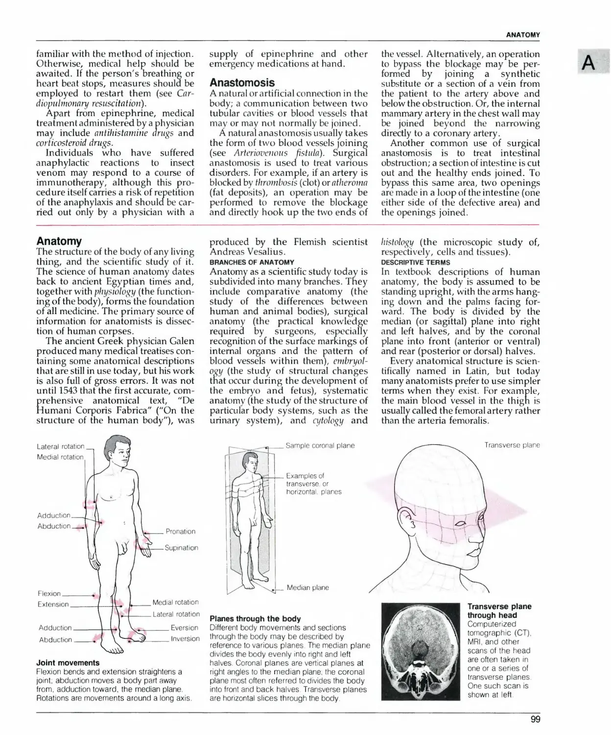
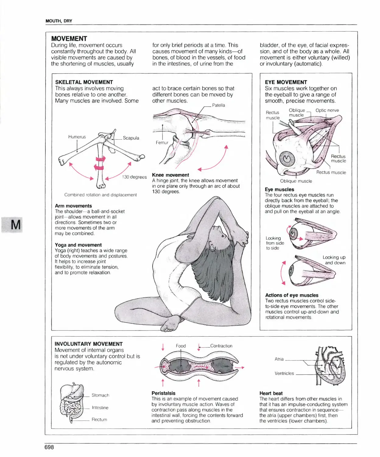
HOW TO FIND INFORMATION ON ANATOMY AND PHYSIOLOGY
All body systems (e.g., Biliary
system) and major organs and body
parts (e.g., Brain and Coccyx) have
individual entries in the A to Z of
Medicine. There are also entries on
the senses (e.g., Vision) and on other
body processes (e.g., Breathing and
Blood clotting). Anatomy and
physiology entries explain how the
healthy body works and provide
a background for understanding
medical disorders.
Most anatomy and physiology
entries are accompanied by
illustrated boxes. Annotated illustrations
show the main structural features of
body parts, the location of different
body parts in relation to each other,
and, for physiology entries, the
main stages in important body
processes.
Anatomical drawings
Detailed drawings,
often with cutaway
sections, show the
structure of body
parts.
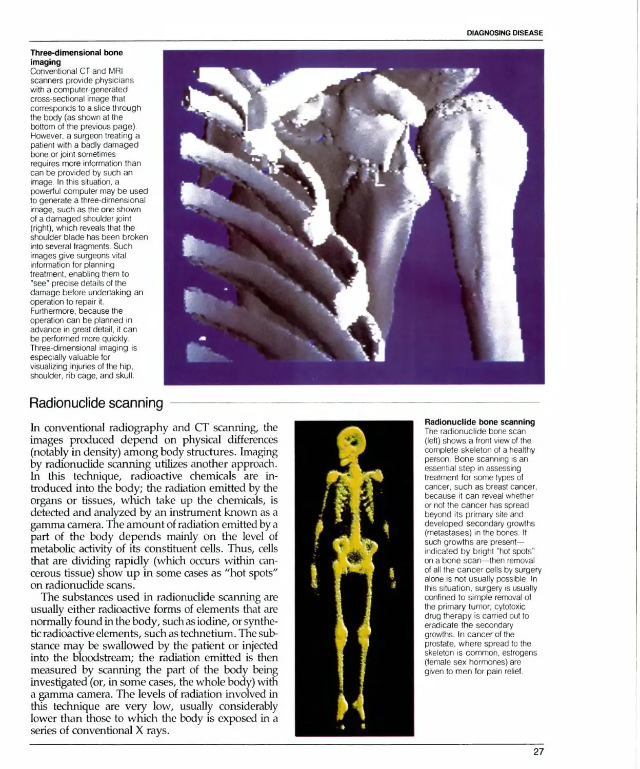
Medical images
Pictures obtained by specialized
imaging techniques, such as X rays
or scans, are used in many cases
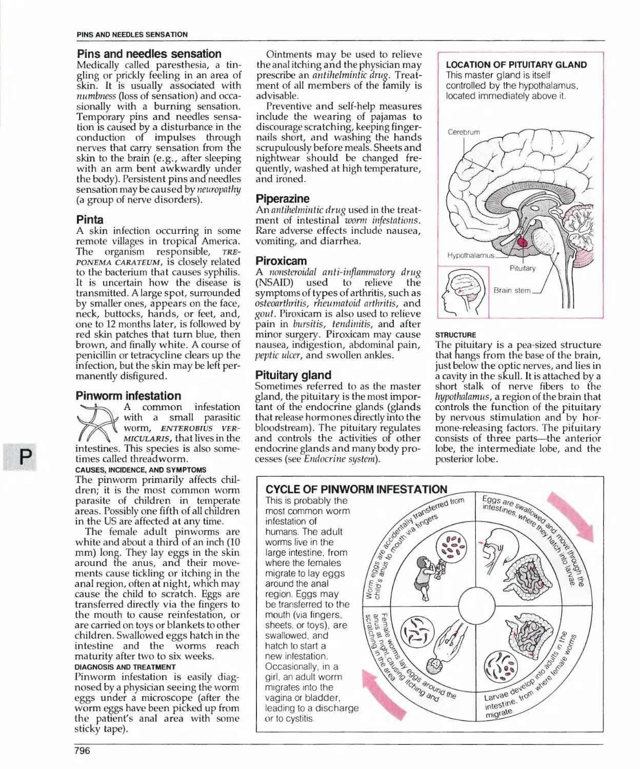
ANATOMY OF THE
ALVEOLI
These tiny sacs con:air
capillaries >n Iheir lh r
wans that allow oxygen
to be absorbed nto
the blood
Brorcrio<p
FUNCTION OF
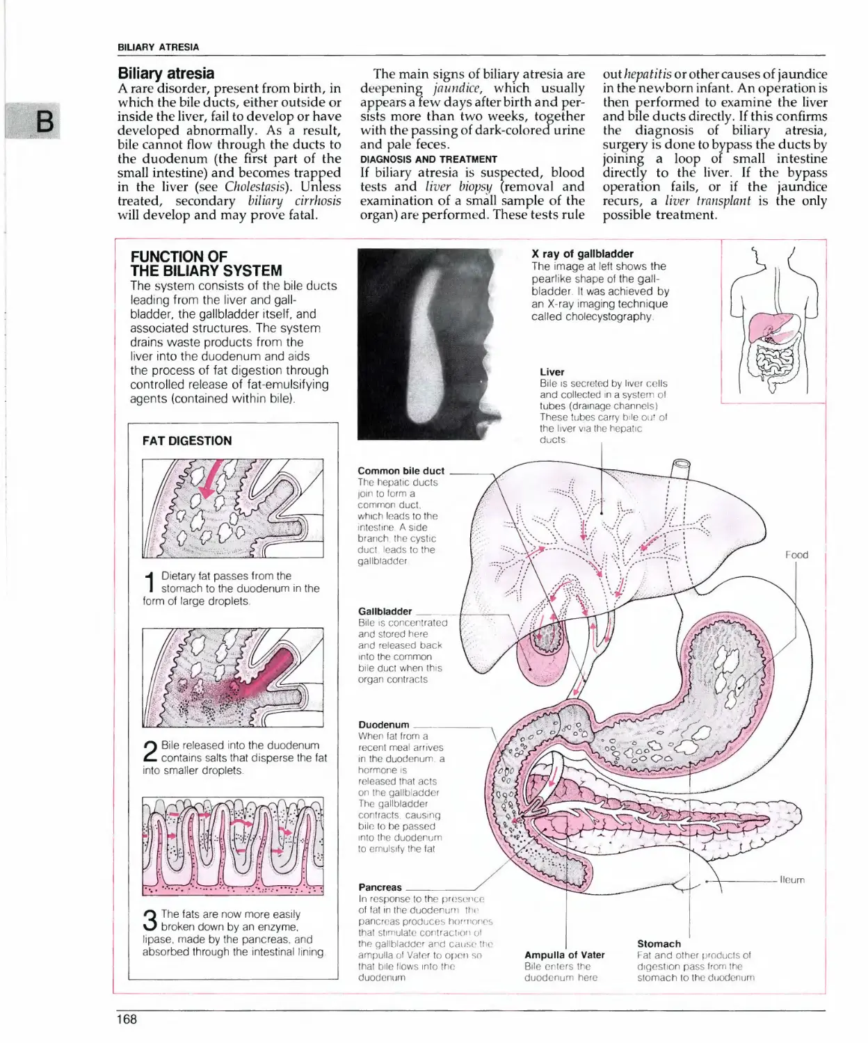
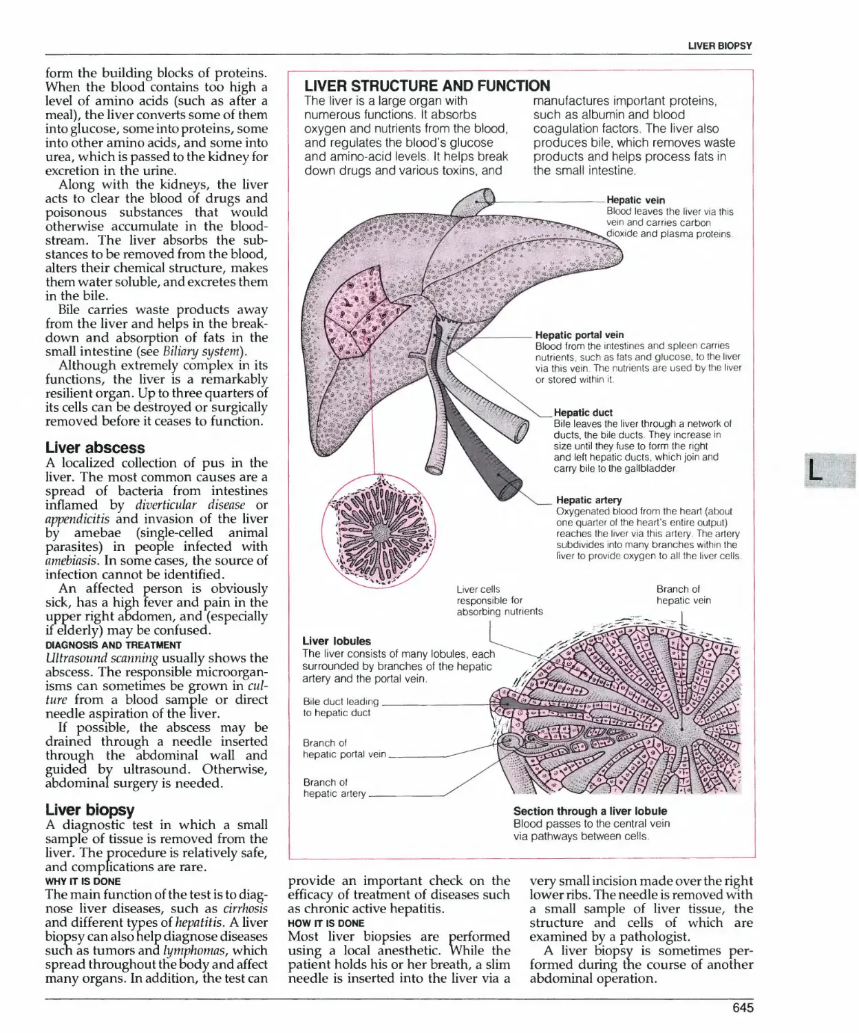
THE BILIARY SYSTEM
The sysiem consiSIS o' Ihe bilp duels
leading Irgm Ihe liver and (jail
bladder, ihp gallblddder iself. and
associated siruciures The sYMen
drams waste prodjcis from ine
liver mio Ihe duodunjrr and aids
ihe process ol lat digesl-on ihrojgn
conirolled release of 'ai-e'i'jlsfyng
ayenls Ironiained w thin btei
X ny o( galibrMfer
L<?5 **■ a' l[^ ' "i ja
V t a. "dc;'-;■?:—Gue
Location diagrams
Most anatomy boxes
include a diagram
showing the part's
position within the
body
J
Entire body systems
Large illustrations
show the structure
and relative position
of organs within
different body
systems.
How body systems
work
Clearly written captions
give detailed
descriptions of the
different stages in
physiological processes
12
HOW TO USE THE ENCYCLOPEDIA
HOW TO FIND INFORMATION ON MEDICAL TESTS
Many different tests for diagnosing
or monitoring medical conditions
have individual entries within the A
to Z of Medicine. Many of these
entries are illustrated by step-by-
step diagrams.
More general information on tests
is given in the entry headed Tests,
medical. This entry is accompanied
by a table that shows the tests used
to investigate different parts of the
body and directs you to individual
entries within the encyclopedia. You
can also find out which tests are used
to investigate a particular part of the
body by consulting the investigation
section of the disorder box for that
body part.
Entries on specific disorders in the
A to Z of Medicine cross-refer you to
entries that describe the tests used to
diagnose and monitor them.
Illustrations showing techniques
Clear illustrations show a test's
most important stages, including,
for example, where a needle is
inserted in the body or how
samples are prepared for testing
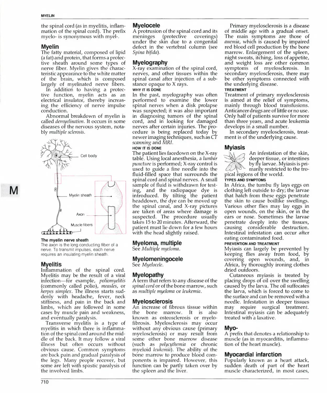
PROCEDURE FOR CHROMOSOME ANALYSIS
K^/
v\
\
II vUt t f .in*
nljl.tnftr Iiy i'nnif)
( mlPMS O' by t *KH OIHL
V Ulft SaiTl|JhlKJ O' WVlli'P
hlfXKMtil S AH! (rti'H <*Mf
Ikmh Hie bhxxi oi Nw*
henq ipstol
3 The ce s d'e Phm* bp*eaa or
liCroscopu S1 Op s'aineii
and d selected lew i n whic'1 trie
chromosomes a'p oedr,y vsib'e
and we' separdieuj "ave me"
oucc ohoKxj'dphcd o* are
C'C-sey examined "^'ouqfi d Ivyli
priWf m-c-oscopr
Step-by-step text
Concise captions describe the
various stages in each test,
including what happens to the
patient and how test samples are
analyzed in the laboratory.
KX xx n
4
Mil s£
■ ■■ ■"e1
L.' JO" " """t
anQoj '"j "-C 22
i na "s j" jj"osct''>s
XX XX XX XX XX XX XX
""je,re a "f "r>p <;«?«
^aots S"l.'J. o
•hese re.eas m .
it , -- ■_;
u n u
13
u
19
XX XX U
16 17 18
n
20
XX XX
21 2?
X*
X« XX
Female
Test results
Diagrams,
photographs, and
captions explain the
. results of the test.
HOW TO FIND INFORMATION ON SURGICAL PROCEDURES
Surgical procedures are described
either in individual entries or in
the treatment section of disorder
entries. In many cases, entries are
accompanied by illustrated boxes.
Individual entries on surgical
procedures are included under their
generally accepted medical names.
In many cases, these names are self-
explanatory—Heart-lung transplant or
Hernia repair, for example.
If you do not know the name of a
particular procedure, look up the
encyclopedia entry on the disorder
for which it is a treatment, where
you will find a description of, or a
cross-reference to, the appropriate
procedure. Alternatively, consult
the Index, which lists some popular
names. For example, if you look up
Stomach, removal of, you will be
referred to the encyclopedia entry on
Gastrectomy.
Reasons for surgery
Different medical images - such
as photographs, X rays, or scans -
of diseased tissue show why
operations are necessary
Surgery in progress
True-to-life drawings show surgical
procedures in progress and
provide an accurate representation
of current surgical practice.
Incision sites
A red line superimposed on a
photograph of the relevant part of
the body shows the position and
shape of the surgeon's incision
HEART-LUNG TRANSPLANT
in (his procedure both ihe < aM and
Ungs ol a patieni are remo\ ed and
■epiaced with organs taken Itom a
Dram dead donor The remi ived
heari can somelimes be giv 3n to
another patieni
HOW IT IS DONE
Heat! and lungs must be re
Irom both donor and paden
organs then are inserted int
'he palienl
4 The donor hear: ana
I lungs must be healthy
and trie lungs must match
■he sue ol the pa'ieni s
chesi as measured by
chest x rays
2'n both donor and
oaNeni ihe hea't and
lungs are -eached v a dn
incsion rnade m the
breastbone and the chest
'S opened jp
Sttv of Incision
WHY IT IS DONE
Subject (o availability ol
a donor a heart fung
transplant can oflet hope
to someone who is dying
ol an end stage chronic
lung disease whether or
nol he or she >s also
suliering irom heart
disease Diseases
'reated include
emphysema cystic
fibrosis, sarcoidosis or
interstitial pulmonary
fibrosis The heart-lung
iransplani operaiion has
a betier success record
ihan thai ot lung
Iranspiant alone
These seciions were taken
Irom d healthy lung (ai
leit) and dn emphysematous
lung, transplanting ol
heart rfiid lungs may give dn
emphyserna palienl new hope
3 The parent is connected lo a
hear; 'ung machine >t lakes
ove* (he lunct'On ot hea^
ana .ungs oxygenating
blood taken from Ihe venae
cavae and pumping i back
lo ihe body via the aorta
Trachea
Aorta
Righi alnumVena cava
in both panen: ana
donor h^,
are removed through cuts m
the irachea aorta and
where ihe heat connecis to
the venae cavae The b-ood
vessels i«* ng donor heart
and ungs are leli >niac:
Tracheal reconneclion
AorliC reconneclion
Right alnumVena cava
reconnect-on
5 Insertion ol the donor
organs mio ihe palienl
is m some respecis easier
than m ihe hean transplant
smce lewer reconnections
have lo be made The main
reconnecnons are beiween
ihe paheni s and donor s
racheas and aortas and
beiween the righi ainum ot
the donor heart and Ihe
paheni s venae cavae
Equipment
Detailed illustrations
show the workings of
special equipment,
such as a heart-lung
machine.
Step-by-step text
and illustrations
Explanatory captions
and anatomically
correct illustrations
take you through the
most important
stages of surgical
procedures. Large
numbers show the
sequence of events
and help you follow
the different stages.
13
HOW TO USE THE ENCYCLOPEDIA
HOW TO FIND INFORMATION ON DRUGS
The A to Z of Medicine contains
individual entries on all major drug
groups (from ACE inhibitor drugs to
Vasodilator drugs) and on the most
important generic drugs (from Ace-
butolol to Zidovudine). Other
information on drugs can be found in
general entries, such as Drug, Drug
dependence, and Drug poisoning.
The Drug Glossary gives concise
information on almost 3,000 generic
and brand-name drugs, showing the
generic equivalents of brand-name
drugs, and identifying the drug
eroups to which individual drugs
elong. Each glossary entry cross-
refers you for more information to
the A to Z of Medicine
Drug charts
Selected entries on individual
drugs are accompanied by an
illustrated chart that summarizes
important information about the
drug.
Group to which the
drug belongs
Generic name
Forms in which the
drug is available
Digdxin
Injection Intravenous
infusion
Suppository Inhaler
Availability as a
generic or under a
brand name only
^.-DIGITALIS
j-
Sj^tcs^ _
prescription or over
the-counter
T^leKCa^>€^eiJc|uid
^£J \Presci^tior>xQeecTBd
ailable^s gentle
Drops
Applicable symbols are
printed in red
Transdermal
patch
\
Tablet Capsule Liquid Powder Cream
or ointment
Drug group charts
Most drug group entries are
accompanied by a chart showing
examples of common drugs within
the group
Group name
Division within the
main drug group
Generic examples
ntirheumatic drugs
COMMON DRUGS
orticosteroid drugs
Dexamethasone Prednisolone
Immunosuppressant drugs
zathioprine Chlorambucil
Others
Sola" Penicillamine
r
I
I
I
I
I
l.
WARNING
Sudden withdrawal o< corticosteroid
drugs may cause serious illness or
death Always inform a physician you
are taking or have recently taken
corticosteroids
'1
I
I
I
I
J
Drug warning
Warning boxes draw your attention
to the possible dangers associated
with taking particular types of
drugs
HOW TO USE THE ENCYCLOPEDIA IN AN EMERGENCY
EMERGENCY
FIRST-AID
TECHNIQUES
Turn to page 8
for the page numbers
of emergency first-aid boxes.
Lifesaving and other, less urgent,
first-aid techniques are explained in
easy-to-follow first-aid boxes. These
boxes accompany relevant entries
within the A to Z of Medicine. All
first-aid boxes have a distinctive red
border and a special heading. A list
of first-aid boxes describing
emergency techniques, with their page
numbers, is given on page 8.
WARNING
Warning boxes contain
essential advice and indicate
when professional help should
be sought.
DO NOT
■ rub the affected parts
■ attempt to burst blisters
■ warm the affected ar^ea with
direct heat
I
I
WARNING
Frostbite is often accompanied by
hypothermia, which must be
treated first. Proper medical
attention should be sought promptly,
but first aid should be given
immediately
Step-by-step
First-aid techniques
are clearly described
in numbered
sequences of text
and illustrations
i
+
■ allow the victim to walk on a
frostbitten foot
I
I
.J
DO NOT
These boxes tell you what not
to do when treating an injured
person
Close-ups
More detailed
illustrations show you
exactly what to do.
Distinctive appearance
Bold red borders and
special headings make
first-aid boxes easy to find
FIRST AID: CHOKING
CONSCIOUS VICTIM
UNCONSCIOUS VICTIM
- , V
1IJ <k v II**- 'i(jc ul. ru- r* iinr ,i(i.iu i\:
'Tlf- rtMllMl1 lil 'N' Y'C'M.'. .llHl'MTH'M
tjN" ■» ilKJvr II ip ri.iv*- |j .ii i yuu' on**'
r>,inii on lop i'tpss n *-'*i .1 Mil1 l> up
A.J' I 'MfUS'
VUrf*.
if-r r' .ifci- 1
1 ii.-i* m»r* r,A.ViI
2t* "»■ •. I.I ■. Is ki'f»r?t(iti*'in* iiiisF'Uf
l.( fi It 'tn- v-irMrn ,*, «,fin ,jnt oms( iuus
summon Hnir'(|pnc y ht"p •' l>'isiHiin<|
'•.is stopped M«v* .if'ttn .!■ *!•■.(» \*t*rt'i
14
MEDICINE
TODAY
PROGRESS IN MEDICINE
People living in developed countries today have the
opportunity to be healthier than ever before. Our
understanding of how the body works has broadened
rapidly in the last few years, paralleled by equally
dramatic improvements in medical technology. As a
result of this progress, a person's chances of staying
healthy into old age depend increasingly on following
expert advice on a healthy life-style, making full
use of preventive techniques such as vaccination and
screening tests, and seeking medical advice from your
physician at the first sign of illness.
This section of the encyclopedia presents an
overview of recent developments in preventive health
care, diagnosis, treatment, prenatal technology, and
new diseases. It also discusses human potential,
including the ways in which we can maximize it.
One important area of advancement in medical
science is computer imaging. Computers can now
provide images of the interior of the body that are
based on various types of scanning techniques, such
as X rays, radioactive isotopes, ultrasound, or
magnetic resonance. These images—which may be two-
or three-dimensional, and even colored to highlight
selected features (e.g., specific tissues)—can give
Left atrium
Right atrium
N
Left ventricle
Right venrncle
Three-dimensional magnetic
resonance imaging (3-D MRI)
The image of a section through
the heart showing its four
chambers (above) was obtained
by the recently developed
technique of 3-D MRI. This
technique, which is noninvasive
and without known risk to the
patient, gives more accurate
and detailed images than could
be obtained by other means As
a result, any structural
abnormalities are often immediately
apparent when the images are
examined
Surgery
Despite trends toward less
invasive methods of treatment,
conventional surgery (right) is
still as important as ever
Significant growth areas include
transplants, implants, and
repairing damaged or
obstructed blood vessels. These
operations, along with the
removal of cancers, will
probably be performed in the
future much as they are today,
although there will be
refinements in anesthesia and
postoperative care
physicians almost as much information as they would
be able to obtain through an exploratory operation on
the chest or abdomen. In some cases, such as the
functional "maps'7 of tissue metabolism provided by
PET scanning, the images provide information that
cannot be obtained even by direct examination.
A second major area of advancement has been in
medical treatment. As recently as the 1970s, major
surgery usually required an incision large enough for
the surgeon to get his or her hands into the body.
Today, the emphasis in surgery is on less invasive
techniques, such as extracorporeal lithotripsy, a
noninvasive procedure that uses ultrasound to shatter
stones in the body, and endoscopic surgery, in which
a viewing tube is passed into the body through a
natural orifice or through a tiny incision, directed to
the target structure, and then used as a guide through
which instruments are passed to enable the physician
to perform the surgery. Electrocautery and the
use of lasers permit bloodless surgery. As a result
of these and other advances in anesthesia, the use of
antibiotic drugs, and the monitoring of various
body functions, medical treatment today is safer
and more effective.
> i
I
■\
*'■ •'
\.
t 1
u
X
\
/ /
M,
I
t
D
\.
k.
16
STAYING HEALTHY
Today, most children in developed countries are
healthier than ever before; a majority can expect to live
well beyond the age of 70. These achievements are
partly due to improvements in public health, such as
the provision of safe water supplies and sewage
disposal systems, adequate housing, and good
nutrition, and partly to improvements in medical care.
The health of women today can be carefully
monitored before and during pregnancy, and during
the birth of the baby. After birth, the newborn
infant is closely examined and, if necessary, provided
with specialized care.
Throughout infancy and childhood, children are
immunized against infections and given vision and
hearing tests, as well as tests for physical and mental
development. As a result, any problems can be
detected and treated promptly.
In the US and Europe the principal causes of death
or disability in youth and middle age are preventable.
World health
Not all countries have high
standards of health. In many
developing countries, infant
mortality is high, principally
because many people do not
have access to safe water,
adequate food, or basic health
care (see diagram, right).
GLOBAL HEALTH INDICATORS
Percentage of population
with access to local
health care
Percentage of population
with access to safe water
1,000
Key
□
Infant survival rate
(per 1,000 live births)
Least developed
countries
Other developing
countries
Developed
countries
. ^Hr
\
.. V.
• *>
» *
sc
Childhood infectious diseases
The photomicrograph (above)
shows the bacterium bordetella
pertussis, the cause of pertussis
(whooping cough). In
developed countries, this and
other potentially serious
childhood diseases (such as
measles, poliomyelitis, and
diphtheria) have been brought
under control by immunization.
In developing countries, such
diseases are still common; the
World Health Organization is
working to combat this problem
by making immunization
available to all children
In early adult life, most deaths are due to accidents or
violence; other important causes include suicide, and
complications of drug abuse and sexual habits
(notably AIDS). In addition, the risk of developing the
principal serious disorders of middle age—coronary
heart disease and cancer—is reduced by following a
healthy life-style and avoiding known health
hazards, especially tobacco and alcohol.
Even though following a healthy life-style
minimizes an individual's chance of developing life-
threatening disease, people living healthy lives may
still become ill, either because they have an inborn
susceptibility to a disorder or simply by chance. As
understanding of the genetic basis of disease
improves, screening tests for familial inherited
disorders will become wider ranging.
Physicians strongly advise people to become aware
of the regular manner in which their bodies function.
This includes monitoring bowel habits, appetite, and
sleep patterns. When a change in body function
is noted, advice should be sought promptly from
your physician. Early attention to any illness aids
treatment and promotes cure.
Avoiding premature death is not the only benefit to
be gained from following a healthy life-style. The
quality of life can also improve: the body's natural
aging processes are slowed, and physical and mental
vigor are retained for much longer.
17
STAYING HEALTHY
PERSONAL HEALTH CARE
An individual's health is determined partly by
inheritance and partly by external factors. Health and
longevity tend to run in families, so a person whose
grandparents lived beyond the age of 80 is likely to do
the same. However, this is not always the case. Even
the intrinsically healthiest body can be damaged by
neglect or external factors, especially by unwise use
of drugs, including tobacco and alcohol. With the
AIDS epidemic still spreading, avoidance of illicit
intravenous drugs and sexual promiscuity and
adherence to "safe" sex practices are essential for the
maintenance of good health.
For many people, however, these "do nots" are less
important than three positive keys to a healthy
lifestyle: a sound, balanced diet, a positive attitude, and
regular, vigorous exercise.
Diet and exercise
For most of history, diseases associated with diet were
due to vitamin or mineral deficiencies, or simply to
prolonged semi-starvation. In developed countries
today, however, the main dietary threat to health is
caused by excess: many people overeat, and almost
everybody consumes too much saturated fat.
Research has shown that variations around the world
in the rates of heart disease and some cancers are
primarily due to differences in diet. Most Americans
consume too much dairy fat, animal fat, and sugar
but consume insufficient vegetables and grains.
Asian people tend to eat more fish, grains,
vegetables, and fruit and so have a healthier diet.
01
Health and exercise
Physical exercise, such as
running (above), can reduce the
risk of coronary heart disease,
but only if it is vigorous enough
to stimulate the heart and is
performed three or more times a
week for at least 20 minutes
each session
\
\.
Types of exercise
The table (right) compares the
value of various activities for
improving three main aspects of
health and fitness heart and
lung efficiency, joint
suppleness, and muscle power
The average calorie
consumption rate of each
activity is also given
However, over the past few years, a dramatic
improvement in our understanding of food and diet
has taken place and the dietary habits of many
people in the US and Europe have begun to improve.
A growing number of people now eat larger amounts
of vegetables, fruit, and cereals and have substituted
unsaturated fats and oils for dairy fats. In addition, the
consumption of fatty meats has declined. According
to researchers, these changes have contributed
to a significant decrease in the incidence of heart
disease in people in many—but not all—developed
countries. Nevertheless, many people in the US
and other developed countries continue to eat
THE FITNESS VALUES OF VARIOUS ACTIVITIES
Easy walking
Light housework
Light gardening
(weeding, etc.)
Golf (flat course)
Brisk walking
Badminton
Gymnastics
Heavy gardening
(digging, etc.)
Dancing
Easy jogging
Tennis
Skiing (downhill)
Skiing
(cross-country)
Football
Racquetball or
handball
Brisk jogging
Bicycling
Swimming
60
90
90
90
100
115
140
140
160
160
160
160
180
180
200
210
220
240
D
D
D
D
DD
DD
DD
DD
DD
DDD
DDD
DDD
DDDD
DDD
DDD
DDDD
DDDD
DDDD
D
DD
DD
DO
D
DDD
DDDD
DDD
DDD
D
DDD
DDD
DDD
DD
DDD
DD
DD
DDDD
D
DD
DD
D
DD
DD
DD
DDDa
D
DD
DD
DD
aa
DDD
DD
DD
DDD
DDDD
Key
I I Calories consumed in
1—' 20 minutes of activity
I I Value in improving heart I II II 1
'—' and lung fitness
| | Value in improving joint | | | |
suppleness
DDDD Excellent
Good
Value in improving
muscle power
□
Fair
Minimal
18
STAYING HEALTHY
RECOMMENDED DAILY DIETARY ALLOWANCES (RDAS) OF SELECTED VITAMINS
Folic acid
(meg)
Niacin
(mg)
Pyridoxine
(mg)
Riboflavin
(mg)
Thiamine
(mg)
Vitamin A
(meg) 1
Vitamin B12
(meg)
Vitamin C
(mg)
Vitamin D
(meg) 2
Vitamin E
(mg)3
Birth to 6
months
30
60
0.3
0.4
0.3
420
0.5
35
10
3.0
6 months
to 1 year
45
8.0
0.6
0.6
0.5
400
1.5
35
10
3.0
1 to 3
years
100
9.0
0.9
0.8
0.7
400
2.0
45
10
5.0
4 to 6
years
200
11
1.3
1.0
0.9
500
2.5
45
10
6.0
7 to 10
years
300
16
1.6
1.4
1.2
700
3.0
45
10
70
11 to 14
years
400
M 18
F 15
1.8
M 1.6
F 1.3
M 1.4
F 1.1
M 1.000
F 800
3.0
M 50
F 60
10
80
15 to 18
years
400
M 18
F 14
20
M 1 7
F 1.3
M 1.4
F 1.1
M 1.000
F 800
3.0
60
10
M10
F 8.0
19 to 22
years
400
M 19
F 14
M 2.2
F 2.0
M 1.7
F 1.3
M 1.5
F 1.1
M 1.000
F 800
3.0
60
7.5
M10
F 8.0
23 to 50
years
400
M 18
F 13
M 2.2
F 2.0
M 1 6
F 1.2
M 1.4
F 1.0
M 1.000
F 800
3.0
60
5.0
M10
F 80
51 +
years
400
M 16
F 13
M2.2
F 2.0
M 1.4
F 1.2
M 1.2
F 1.0
M 1.000
F 800
3.0
60
50
M10
F 8.0
Extra needed
preg- breast-
nancy feeding
400
2.0
0.6
0.3
0.4
200
10
20
50
20
100
50
0.5
0.5
0.5
400
1.0
40
50
3.0
1 RDA expressed in meg of retinol
(a form of vitamin A). 1 meg of
retinol (a unit called a retinol
equivalent, or RE) equals 6 meg of beta-
carotene (another form of vitamin A).
2 RDA expressed in meg of
cholecalciferol (one of the forms of
vitamin D). 10 meg of
cholecalciferol equals 400
international units (IU) of vitamin D.
3 RDA expressed in mg of alpha-
tocopherol (one of the forms of
vitamin E) 1 mg of alpha-
tocopherol equals 1 alpha-
tocopherol equivalent (1 alpha-TE)
RECOMMENDED DAILY DIETARY ALLOWANCES (RDAS) OF SELECTED MINERALS
Calcium
(mg)
Iodine
(meg)
Iron
(mg)
Magnesium
(mg)
Phosphorus
(mg)
Zinc
(mg)
Birth to 6
months
360
40
10
50
240
30
6 months
to 1 year
540
50
15
70
360
5.0
1 to 3
years
800
70
15
150
800
10
4 to 6
years
800
90
10
200
800
10
7 to 10
years
800
120
10
250
800
10
11 to 14
years
1.200
150
18
M350
F 300
1,200
15
15 to 18
years
1.200
150
18
M400
F 300
1.200
15
19 to 22
years
800
150
M 10
F 18
M 350
F 300
800
15
23 to 50
years
800
150
M 10
F 18
M 350
F 300
800
15
51 +
years
800
150
10
M350
F 300
800
15
Extra needed
preg- breast-
nancy feeding
400
25
30-60
150
400
50
400
50
A
150
400
10
A Iron requirements while breast-feeding are
approximately the same as those for nonpregnant
women, but additional iron may be recommended for
two to three months after the birth to replenish iron
stores depleted by pregnancy.
UNITS
mg = milligrams
(thousandths of a gram)
meg = micrograms
(millionths of a gram)
Vitamins and minerals
The tables (above) give
recommended daily allowances
(RDAs) of vitamins and minerals
for which amounts have been
established; when different, the
RDAs for males and females are
denoted by M and F.
19
STAYING HEALTHY
an unhealthy diet, so diet-related diseases continue to
be a major problem.
While food is the body's energy source, people who
engage in little exercise can easily become overweight
by eating more than they "burn off" in physical
activity. Regular exercise (at least three times a week)
keeps the heart, lungs, muscles, and bones in good
health and slows down the aging process. A person
who is in good physical condition at the age of 60
can achieve up to 80 percent of the level of physical
/~
Left mam coronary artery
Narrowed section
of artery
\
t.
J. 4-* "th*' ■'■■ * Vv
Smoking and drinking
The full extent of the damage to health caused by
smoking tobacco and drinking alcohol is gradually
being revealed with continuing research. It has
become apparent that many people drink regularly
and heavily without being recognized as the
alcoholics they have become.
Alcohol is a major contributing factor in traffic
accidents and drownings. It plays an important role in
exertion that he or she could achieve in the mid-20s.
Regular exercise improves the circulation in the heart
and muscles and provides increased stamina.
Exercise as simple as regular, brisk walking also maintains
the density of bones, thus reducing the risk of
developing osteoporosis ("thinning" and weakening
of the bones) and fractures. A person who exercises
regularly is less likely than a person who does not
exercise sufficiently to have a heart attack or to die
from a heart attack if one occurs.
Diet and atherosclerosis
The colored angiogram of the
heart (left) shows narrowing of a
coronary artery, a cause of
coronary heart disease. Such
narrowing can critically reduce
the heart's blood supply during
exertion; the narrowed section
may even become blocked by a
blood clot, causing a heart
attack. The narrowing of
coronary, and other, arteries is
usually due to atherosclerosis,
the deposition of fatty plaques in
the arteries (see diagram
below). The risk of
atherosclerosis is linked closely to the
amount of cholesterol in the
blood, which, in turn, is linked to
the dietary intake of fats.
ATHEROSCLEROTIC ARTERY
Adventitia
Media
Intima
Plaque forms
in intima
Arterial
wall
TOBACCO USE AND RESPIRATORY TRACT CANCER (1980)
40 30 20 10 0 20
1 1 1
i
32.6
13 4
39 0
18.0
l l i i
20 15 10
Key
I I Percentaqe of population |
over 15 who smoke
regularly
5 C
| Average daily
of cigarettes p
smoker
i
40 60 80
i I i
46 3
US
72 3
UK
)
consumption |
er regular
| Death rate from respiratory tract
cancer (per 100.000 population)
domestic violence, sexual assaults, and other violent
crimes. Furthermore, there is growing evidence that
young people who drink heavily are more likely to
experiment with other addictive drugs. Alcohol is a
significant cause of ill health. Regular heavy drinking
can damage the liver (eventually leading to cirrhosis),
heart, stomach, and esophagus. It also leads to brain
damage, resulting in impairment of motor and
Tobacco smoking and
mortality
It is now well established that
smoking can cause cancer; it is
also known that, the more a
person smokes, the greater the
risk. The relationship between
smoking and deaths from
respiratory tract cancer is
shown in the chart (left) In the
US in 1980, 32.6 percent of
adults smoked, averaging 13 4
cigarettes per person per day;
the death rate from respiratory
cancer was 46.3 per 100,000
people. In the UK in 1980, 39
percent of adults smoked,
averaging 18 cigarettes per
person per day; the death rate
from respiratory cancer was
72.3 per 100.000 people
20
STAYING HEALTHY
intellectual capabilities. Drinking during pregnancy
can damage the unborn child, causing the mother
to give birth to a physically and intellectually
maldeveloped baby.
The adverse effects of tobacco are almost as
extensive and destructive as those of alcohol. The link
between smoking and lung cancer is well known.
Moreover, smokers face a substantially greater risk of
premature death from coronary heart disease.
It has been demonstrated that people who smoke
suffer more angina (chest pain due to inadequate
blood supply to the heart muscle) as well as a more
recently described phenomenon in which the heart
muscle is deprived of oxygen without any angina.
This latter, "silent" form of coronary heart disease
could be a major factor in the sudden death that can
occur with this disease.
Smoking has also become the primary cause of
chronic bronchitis and emphysema. It has been
linked with ulcers of the stomach and duodenum,
and cancers of the cervix and bladder. Smoking
during pregnancy, like drinking during pregnancy,
damages the developing fetus.
Although smoking is a hazard to the health of
smokers themselves, it is also a threat to the health of
nonsmokers, particularly children. Recent research
has demonstrated that nonsmokers who are
exposed to tobacco smoke—as may occur in a
household with both smokers and nonsmokers—face an
increased risk of respiratory disorders such as
bronchitis and lung cancer.
Smoking and emphysema
Although not all smokers get
lung cancer, most do develop
chronic bronchitis and
emphysema. In emphysema,
continual irritation by smoke
causes progressive damage to
the lung tissue. The alveoli (the
tiny air sacs across which
oxygen and carbon dioxide
interchange occurs) first burst
and then merge to form fewer,
larger sacs with less surface
area (see diagrams below). As a
result, the working volume of the
lungs is progressively reduced,
causing breathlessness.
THE EFFECTS OF EMPHYSEMA ON THE LUNGS
Normal lung tissue
o
a
Bronchiole
0
g?°o
Blood capillaries
Normal alveoli
e
Emphysematous
lung tissue
.Alveolar damage
due to emphysema
HEALTHY TISSUE
*",v#
9%
V * ' '*
■ is « J.
•» »
Effects of alcohol on the liver
The photomicrographs above
show healthy liver tissue (left)
and liver tissue damaged by
alcoholic cirrhosis (right). In the
healthy liver, the cells are
arranged regularly; in the
cirrhotic liver, the regular cell
arrangement is disrupted by
fibrous scar tissue (the blue
areas in the photomicrograph).
The scar tissue develops as a
result of destruction of liver cells
by alcohol. The liver attempts to
compensate by growing new
CIRRHOTIC TISSUE
.; $ '"<■:.* v '**
w> , zr&
' • "Iv J-
f '
v
cells, but in some areas the
damage is so great that
nonfunctioning scar tissue
develops instead. With
continued alcohol use,
increasing amounts of scar
tissue are formed; eventually it
affects so much of the liver that
normal liver function cannot be
maintained.
Alcohol and mortality
There is a close link between
alcohol and death from
cirrhosis. In 1980, average
intake in the US was about 660
glasses of beer (or equivalent)
per person; cirrhosis deaths
were 13.6 per 100.000 people.
This compares with 1,025
glasses of beer (or equivalent)
per person and 28.5 cirrhosis-
caused deaths per 100,000
people in France.
ALCOHOL USE AND LIVER CIRRHOSIS (1980)
500 600 700 800 900
1.000 1.100
US
France
I
660
13 6
1,025
28 5
I I I
10
20
Key
30
Estimated average annual alcohol consumption per person
(number of 12-oz glasses of beer, or equivalent*)
J Death rate from liver cirrhosis (per 100,000 population)
*ln alcohol content one 12-oz glass of beer is roughly
equivalent to one 3-oz glass of wine, or a 1-oz shot of liquor
STAYING HEALTHY
PREVENTIVE MEDICINE
Many common and serious diseases cause no
symptoms in their early stages. This is particularly
true of cancer, which often does not produce
symptoms until it is beginning to or has already
spread to other areas of the body. However, many of
these tumors can be detected by simple, reliable
tests. Treatment can then arrest or minimize the
progress of the disease. Periodic screening tests for
cancer become important after age 40, particularly
among people in high-risk groups.
Regular check-ups
The most important medical check-ups are for weight
and blood pressure. Both tend to increase with age,
but a sustained rise above a normal range for a
particular age increases the risk of disorders such as heart and
kidney disease and stroke, and may warrant
treatment. A physical examination should also include
checking the eyes, ears, throat, and skin, and an
assessment of the condition of all major organ systems
of the body (cardiovascular-respiratory,
gastrointestinal, genitourinary, musculoskeletal, and
neurological). For women who have been sexually active, a
cervical smear (Pap test) is included. In addition,
middle-aged women should have a mammogram,
every two years for those over 40 and annually after
the age of 50. A dental examination should also be
performed at least once a year.
During a routine check-up, the physician may ask
questions designed to screen for a variety of disorders.
He or she may also inquire about your life-style,
including your diet, sleeping patterns, sexual activity,
exercise program, alcohol and tobacco consumption,
and whether you use any other addictive drugs.
Screening high-risk groups
MEASURING BLOOD PRESSURE
Stethoscope
Blood pressure
gauge
Inflatable cuff
Blood pressure
Blood pressure is measured by
using an inflatable cuff attached
to a pressure gauge, and a
stethoscope to listen for sounds
of blood flow that indicate
maximum (systolic) and
minimum (diastolic) pressure A
healthy young adult has a blood
pressure of about 110/75, which
rises to about 130/90 by age 60.
The medical tests outlined above apply to all adults,
irrespective of their medical history. Other specialized
tests are recommended for people from families in
which a particular disease is prevalent. For example, if
a blood relative has had a heart attack before 50, you
should tell your physician, who may want to measure
the levels of lipids (LDL, HDL, and total cholesterol
and triglycerides) in your blood. Coronary heart
disease in early middle age almost always indicates an
inherited disorder associated with raised lipid levels in
the blood, but is a condition that can be treated by a
combination of diet and drugs. Similarly, if your
mother, father, sister, or brother has had bowel
cancer, you should consult your physician about
Procedure for
mammography
— Abnormal growth in breast
(cancer, tumor, or cyst)
Normal breast tissue
Mammography
This procedure uses low-dose X
rays to image the breasts in
screening for cancer (see
diagram, left). The mammogram
(far left) shows a white area
toward the center of the image;
such "shadows" may indicate a
cancer, but in most cases the
cause is a noncancerous tumor
or cyst. When a mammogram
reveals an abnormality, a
sample of the abnormal tissue
may be taken for microscopic
examination
22
STAYING HEALTHY
when to have regular tests for the presence of blood in
your feces and a proctosigmoidoscopic examination.
Detection of bowel cancer in its early stages usually
enables surgery to be performed with an excellent
prospect of a cure.
People who have worked in certain industries have
an increased risk of developing some forms of cancer.
Workers who have been in regular contact with
amine dyes should make sure they have regular tests
for bladder cancer.
Other high-risk groups, such as middle-aged men
with vague chest discomfort or those about to
embark on an exercise program, may be advised to
have an exercise stress test to assess the condition of
the heart. Liver function tests may be recommended
to detect any early evidence of liver damage
associated with alcohol consumption. In addition, people
who have spent a long period in tropical countries
may be advised to undergo special screening tests for
tropical diseases.
Electrocardiography (ECG)
The ECG treadmill stress test
(right) is a widely used test for
evaluating chest pain.
Electrodes are taped to the
chest and connected to the
ECG machine, which makes a
continuous recording of the
heart's electrical activity while
the patient walks on a treadmill
The walking speed is then
increased to the maximum the
patient can tolerate. If the ECG
trace remains normal during
maximum exertion, it is unlikely
that the person has a serious
heart disorder.
ECG TREADMILL STRESS TEST
Immunization and preventive drug treatment
Until as recently as the 1940s, deaths in infancy from
infectious diseases, such as diphtheria, measles, and
pertussis (whooping cough), were relatively
common, even in some developed countries. Today,
however, vaccines are available against many potentially
fatal diseases, and most children in developed
countries are routinely immunized against not only
diphtheria, measles, and pertussis, but also against
typhoid, poliomyelitis, tetanus, rubella, and mumps.
Vaccines are also available against tuberculosis and
some types of meningitis.
People planning to travel to a foreign country
should ensure that their immunizations are current
and should also be immunized against any other
diseases (such as cholera, yellow fever, and hepatitis)
that may exist in the region to be visited. A vaccine has
yet to be developed against malaria, one of the most
common tropical diseases. However, prophylactic
treatment with an antimalarial drug such as chloro-
quine can help protect against the disease.
Development of drug-resistant forms of malaria in some parts
of the world requires the traveler to discuss plans
with his or her physician. It is also advisable to take
other preventive measures, including using insect
repellents, wearing protective clothing, and sleeping
under a mosquito net.
Electrodes
\
Preventing malaria
Malaria is caused by infection
with protozoal parasites called
Plasmodia. The parasites enter
the body by passing down
through the proboscis (circled in
the photograph above) of
female anopheles mosquitoes
while they suck blood. Travelers
to malarial regions can protect
themselves against the disease
by taking preventive antimalarial
drugs (beginning a few days
before entering a malarious
area), which affect the parasites
in the liver before they can
cause symptoms. In recent
years, control of malaria has
become more difficult, partly
because the parasites have
developed resistance to certain
antimalarial drugs, and partly
because the mosquitoes have
become resistant to many
insecticides.
23
DIAGNOSING DISEASE
Accurate diagnosis of disease is one of the most
important aspects of medicine. Without knowing the
identity of a disorder, a physician can only relieve
symptoms, such as pain or fever. Indeed, medicine
consisted largely of a collection of remedies for specific
symptoms and injuries until the fifth century BC,
when the Greek physician Hippocrates (c.460-c.377
BC) attempted to identify and describe the course of a
disease along with its symptoms.
A diagnosis enables the physician to make a
prognosis—an estimate of the outcome of a certain
disorder. The concepts of diagnosis and prognosis led
physicians to identify diseases from the medical
history, the patient's account of the illness. The history
remains an essential element in diagnosis. It should be
followed by a systematic physical examination in
which the physician looks for further signs that can
help confirm the underlying cause of the disorder.
Despite these developments, diagnosis remained
mostly guesswork until the 17th century, when
anatomists and pathologists began to study the body's
structure and the changes in organs and tissues that
were caused by specific diseases. The invention and
development of the microscope led to a greater
understanding of the body's structure and function, and
to understanding how organs and tissues are affected
by disease. The 19th century brought another major
advance in the history of diagnosis with the discovery
that microorganisms (e.g., bacteria and fungi) can
cause disease. This discovery led to the development
of the germ theory of disease by, among others, the
French microbiologist and chemist Louis Pasteur
(1822-1895) and the German bacteriologist Robert
\
Koch (1843-1910). The germ theory of disease is
generally regarded as one of the greatest
achievements of medical and biological science.
Today, physicians still rely heavily on the patient's
medical history to arrive at a diagnosis. Success in
making a diagnosis is augmented by the physical
examination and by myriad biochemical
immunological, and microbiological tests. Physicians
also have access to the interior of the patient's body in
ways previously unimagined. Modern techniques
such as ultrasound, endoscopy, body scanning (such
as CT scanning and magnetic resonance imaging),
and biopsy provide detailed, accurate information
V. '
V
C
Computers and diagnosis
Many diagnostic scanning
techniques, such as CT
scanning and MRI. use
computers to image the body
(above). Computers can
enhance scanning images to
highlight specific features.
Diagnosing communicable
diseases
Because of rapid air transport, a
person with a rare infectious
disease may develop the first
symptoms anywhere in the
world. When physicians suspect
such an illness, the patient is
treated in strict isolation and
samples are cultured in a high-
security environment (such as
the one shown at left) to
determine the causative
microorganism.
24
DIAGNOSING DISEASE
about internal organs with only minimal risk and
discomfort to the patient.
Many advances in diagnosis would not have been
possible without developments in other disciplines.
For example, most modern scanning techniques
depended on the development of the computer to
process the information. These and other major
landmarks in the history of diagnosis are listed in
the chart below.
LANDMARKS IN DIAGNOSIS
Medical thermometer
The Italian physician Sanctorius
(1561-1636) devised the first
medical thermometer in 1612
(right). An adaptation of
Galileo's air thermometer, it
consisted of a globe-shaped top
that was placed in the mouth, a
coil graduated with glass
beads, and a bulbous bottom
immersed in a bowl of water.
Date
c.400 BC
1612
c.1660
1810
1850-1900
1895
1905
1906
C1932
1938
1957
1973
1975
1976
1981
1985
Development
Disease concept. Introduced by Greek physician Hippocrates.
Medical thermometer. Devised by Italian physician Sanctorius.
Light microscope. Single-lens microscope developed by Dutch naturalist
Antonj van Leeuwenhoek, who discovered microorganisms with it. A
practicable compound microscope was not developed until the 19th
century.
Stethoscope. Invented by French physician Rene Laennec.
Germ theory of disease. Proposed by French scientist Louis Pasteur and
developed by German bacteriologist Robert Koch.
X rays. Discovered by German physicist Wilhelm Roentgen. He also
produced the first X-ray picture of the body.
X-ray contrast medium. First demonstrated (in retrograde pyelography)
by Jean Athanese Sicard in Paris.
Electrocardiograph (ECG). Invented by Dutch physiologist Willem
Einthoven.
Transmission electron microscope (TEM). Constructed by German
scientists Max Knoll and Ernst Ruska.
Cardiac catheterization. First performed by George Peter Robb and Israel
Steinberg in New York.
Fiberoptic endoscopy. Pioneered by South African-born physician Basil
Hirschowitz at the University of Michigan.
CT scanner. Invented by British engineer Godfrey Hounsfield of EMI
Laboratories, England, and South African-born physicist Allan Cormack of
Tufts University, Massachusetts.
Monoclonal antibodies. Large-scale production method developed by
Argentinean-born scientist Cesar Milstein at the Medical Research
Council Laboratories, England.
Chorionic villus sampling. Developed by Chinese gynecologists as an aid
to the early diagnosis of genetic disorders.
MRI scanner. Developed by scientists atThom-EMI Laboratories,
England, and Nottingham University, England.
PET scanner. Developed by scientists at the University of California
Xrays
Shortly after his discovery of X
rays in 1895, the German
physicist Wilhelm Roentgen
(1845-1923) made the first X-ray
picture of the human body
(above). The picture shows his
wife's hand, with the ring she
was wearing.
=~
"3=B=~ ~~
sas r __
=
=
~z.^= —
=5
-
~ =^P
8
—
^3=
~
PET scanning
This technique, developed in
1985, enables the functioning of
organs to be studied. For
example, the image of a normal
heart (above) shows the left
ventricle pumping blood into the
aorta; the image has been
computer-colored so that the
areas containing the greatest
amounts of blood are shown in
purple and gray.
25
DIAGNOSING DISEASE
MODERN DIAGNOSTIC TECHNIQUES
In the 19th century, physicians usually had to wait
until an autopsy had been performed on a patient
before they could determine whether or not their
diagnosis had been correct. Today, the body's internal
organs can be studied during life with minimal risk or
no risk to the patient. Physicians use imaging
techniques, including X rays, ultrasound, CT scanning,
Xrays
A type of electromagnetic radiation with extremely
short wavelengths, X rays are invisible and cause no
sensation when passed through tissue (although they
may damage the skin and internal organs and may
cause cancer many years later). When X rays pass
through the body, they are absorbed more by dense
structures, such as bone, than by softer tissues. Thus,
when an X-ray beam is focused onto photographic
film behind the patient, shadows of variable intensity
are cast on the film, producing the X-ray image.
Digital subtraction angiography —
\ V
\
m
\
\
T.
.V
CT scanning
Conventional X rays and angiograms are essentially
shadow photographs, but CT scanning uses X rays in
a completely different way. Multiple beams of X rays
are passed through the part of the body being
examined, and their degree of absorption is recorded
by sensors. The scanner moves around the patient,
emitting and recording X-ray beams from every point
on the circle. The resulting data are then analyzed by a
computer, which uses the variations in absorption of
the X rays to construct an image.
CT scan through the trunk
The CT scan (right) shows a
cross section through the trunk
of the body at the level of the
kidneys. The image has been
colored by computer to make
the internal structures (the most
important of which are labeled)
more clearly visible.
radioisotope scanning, and MR1 (magnetic resonance
imaging); endoscopy (using a fiberoptic tube to view
directly the interior of hollow organs such as the
lungs, stomach, bladder, or some joints); and biopsy
(examination under a microscope of a small sample of
tissue that has been removed from an organ by
surgery, by a needle, or via an endoscope).
Chest X rays
Because the lungs are full of air,
they offer little resistance to X
rays. A radiograph of the chest
(left) gives a clear picture of the
ribs, heart, and major blood
vessels. A "shadow on the
lungs" may be caused by an
area of infection (such as from
tuberculosis), a tumor, or an
inhaled foreign body.
Because many organs have a relatively uniform
consistency, their structure does not show up on
conventional X rays. One solution is angiography—
injecting a radiopaque dye into blood vessels to make
them visible on X rays. In digital subtraction
angiography, images before and after injection of the dye are
computer-processed to remove confusing detail and
produce a clear image.
Digital cardiac imaging
Digital subtraction angiography
can be used to visualize the
coronary arteries. The digital
cardiac imaging unit (left) has
four screens to monitor the
introduction of radiopaque dye
and to display the arteries (seen
here on three of the screens).
Intestine (gas-filled)
Rib
Fat around
kidney 1
I
Left
kidney
\
Rib
Liver
Right
.kidney
Vertebral canal Vertebra
(containing spinal cord) (thoracic)
Paravertebral
muscles
26
DIAGNOSING DISEASE
Three-dimensional bone
imaging
Conventional CT and MRI
scanners provide physicians
with a computer-generated
cross-sectional image that
corresponds to a slice through
the body (as shown at the
bottom of the previous page).
However, a surgeon treating a
patient with a badly damaged
bone or joint sometimes
requires more information than
can be provided by such an
image. In this situation, a
powerful computer may be used
to generate a three-dimensional
image, such as the one shown
of a damaged shoulder joint
(right), which reveals that the
shoulder blade has been broken
into several fragments. Such
images give surgeons vital
information for planning
treatment, enabling them to
"see" precise details of the
damage before undertaking an
operation to repair it.
Furthermore, because the
operation can be planned in
advance in great detail, it can
be performed more quickly.
Three-dimensional imaging is
especially valuable for
visualizing injuries of the hip,
shoulder, rib cage, and skull.
•to
Radionuclide scanning
In conventional radiography and CT scanning, the
images produced depend on physical differences
(notably in density) among body structures. Imaging
by radionuclide scanning utilizes another approach.
In this technique, radioactive chemicals are
introduced into the body; the radiation emitted by the
organs or tissues, which take up the chemicals, is
detected and analyzed by an instrument known as a
gamma camera. The amount of radiation emitted by a
part of the body depends mainly on the level of
metabolic activity of its constituent cells. Thus, cells
that are dividing rapidly (which occurs within
cancerous tissue) show up in some cases as "hot spots"
on radionuclide scans.
The substances used in radionuclide scanning are
usually either radioactive forms of elements that are
normally found in the body, such as iodine, or
synthetic radioactive elements, such as technetium. The
substance may be swallowed by the patient or injected
into the bloodstream; the radiation emitted is then
measured by scanning the part of the body being
investigated (or, in some cases, the whole body) with
a gamma camera. The levels of radiation involved in
this technique are very low, usually considerably
lower than those to which the body is exposed in a
series of conventional X rays.
•\
* -,
Radionuclide bone scanning
The radionuclide bone scan
(left) shows a front view of the
complete skeleton of a healthy
person. Bone scanning is an
essential step in assessing
treatment for some types of
cancer, such as breast cancer
because it can reveal whether
or not the cancer has spread
beyond its primary site and
developed secondary growths
(metastases) in the bones. If
such growths are present—
indicated by bright "hot spots"
on a bone scan—then removal
of all the cancer cells by surgery
alone is not usually possible. In
this situation, surgery is usually
confined to simple removal of
the primary tumor; cytotoxic
drug therapy is carried out to
eradicate the secondary
growths. In cancer of the
prostate, where spread to the
skeleton is common, estrogens
(female sex hormones) are
given to men for pain relief.
27
DIAGNOSING DISEASE
MRI
Unlike X-ray radiography, CT scanning, and
radionuclide imaging, MRI (magnetic resonance
imaging) does not employ potentially harmful
ionizing radiation. Instead, it exploits the natural behavior
of the protons (nuclei) of hydrogen atoms when they
are subjected to a very strong magnetic field and radio
waves. As a result of this stimulation, the protons
emit radio signals, which are detected and computer-
processed to generate an image. The most abundant
sources of protons in the body are the hydrogen
atoms in water molecules; an MRI scan therefore
reflects differences in the water content of tissues.
Superficially, MRI scans look like CT scans.
However, CT scans usually show little differentiation in
soft tissues; MRI scans show more of the detailed
structure because of differences in water content
within these tissues. For example, white and gray
matter in the brain are relatively poorly differentiated
in CT scans, while they are distinct and well-defined
in MRI scans.
NORMAL CHEST SCAN
ABNORMAL CHEST SCAN
Diagnostic MRI scanning
The two MRI scans (right) show
cross sections through the
chest. The upper scan, which
reveals a healthy chest, shows
the lungs (the large black areas
in the center) and ribs (the
crescent-shaped red-yellow
areas toward the edge). In
comparison, the lower scan
shows a tumor in the left lung
The tumor is indicated by the
increased amount of blue in the
image of that lung and by the
lung's small size.
Ventricle
Pituitary gland.
i _ -
- -^
**L ^
1 -- "
4- ~ -*
~~ ~~ ^~
_ =i ^ r=^"~"
- -m-j?
'■%
_ * ■AMr.
i/:*.- b^M^
"^ 1 -m- "'
*
•
**<
* »
mm.
'4
• r—*
*• • r
. « —5. - - rV: -
- - = -- - —** =
~ j^ jcm, __
rmKlT^^-^ r4_=J =?:
--_ -ZT^-^^r — ~" "=^
--t, "Si" £i- _=r _-=- l
- .-nm ^r^-r c-
^~ ^- *,-^=- ■=- =r-
r ====^ *
'•* * '^W" -
-5 ■=.■ ■ "«
•
7.
1
»•
r
r
M V
1
•
1 *
Skull
Cerebrospinal fluid
between meninges
Cerebrum
Corpus callosum
Midbrain 1
Pons
Medulla oblongata _
-Brain stem
Cprphplhim
Spinal card
Vprtehrae in neck
MRI head scan
The MRI scan (left) shows a
longitudinal section through the
middle of the head and upper
neck. The image has been
colored to differentiate the
internal structures.
28
DIAGNOSING DISEASE
HOW MRI WORKS
Radio-wave source
Magnet
Radio-wave detector
The MRI scanner
An MRI scanner consists of a
massive electromagnet, a
radio-wave emitter, and a
radio-wave detector. These
instruments are arranged
around a central tunnel in
which the patient lies, and
are connected to a computer
and display screen. The
electromagnet is extremely
powerful. It is able to create a
magnetic field up to about
60,000 times that of the
Earth's magnetic field.
&
Scanning plane
Scanning plane
When the patient is inside the
machine, it is set up to scan a
plane ("slice") of the body
The scanning process
Scanning begins when a plane
of the body has been selected
for imaging. First, the
electromagnet is turned on.
Normally, the protons (nuclei) of
the body's hydrogen atoms
point randomly in different
directions, but under the
influence of the scanner's
powerful magnetic field they
align themselves in the same
direction (diagram, top right).
Next, the radio-wave source
emits a powerful pulse of radio
waves, the effect of which is to
knock the protons out of
alignment (diagram, middle
right). However, milliseconds
later, the protons realign
themselves, emitting faint radio
signals as they do so; these
signals are picked up by the
scanner's radio-wave detector
(diagram, bottom right). A
computer then processes the
radio signals to produce an
image of the plane of the body
being scanned, such as the
"slice" through the chest shown
in the photograph (bottom right).
Computer software enables
three-dimensional images to be
derived from MRI.
¥J=m
Magnet
Proton
Radio-wave
source
Radio-wave
stimulation
vf
Radio-wave
detector
Radio-wave
emissions
PET scanning
PET (positron emission tomography) scanning is a
development of radionuclide scanning and resembles
it in many ways. Both techniques use a radioactive
substance introduced into the body to produce an
image that reflects the level of activity of tissues.
However, radionuclide scanning usually produces an
image analogous to a conventional X-ray picture; PET
scanning gives a cross-sectional image that is
analogous to a CT scan.
In PET scanning, a substance that takes part in
metabolic biochemical processes is labeled with a
radioisotope to make it radioactive; it is then injected
into the bloodstream. The substance is taken up by the
most metabolically active areas of tissue. In the tissue,
the substance emits positrons. The positrons, in turn,
release photons, which are then detected by an array
of sensors around the patient. The sensors are linked
to a computer, which calculates the origins of the
photons to construct an image of the distribution of
the substance within the tissues.
PET scanning is currently being utilized for
investigating brain tumors, locating the origin of
epileptic activity, and studying the brain function in
various mental illnesses. It is anticipated that PET
scanning will be used for other organs.
PET brain scanning
The PET scan (above) of a
section through a normal brain
has been colored to show levels
of protein synthesis (high levels
are indicated by pink, red, and
orange, intermediate levels by
yellow and green, and low levels
by blue). In this scan,
methionine (an essential amino
acid) was used to show protein
synthesis. Other brain functions
can be imaged by using
different substances—such as
radioactively labeled glucose to
show carbohydrate metabolism.
DIAGNOSING DISEASE
Biochemical analysis
Body fluids contain thousands of different chemicals.
A healthy body maintains the concentration of each
substance within clearly defined normal limits.
However, in disease, the level of a body chemical may be
abnormally high or low; detecting this abnormality by
biochemical analysis can help in diagnosis.
A physician may order biochemical tests on
samples of blood, urine, and other body fluids, such
as spinal fluid, saliva, or sweat, when confirming a
diagnosis. These tests may be part of a diagnostic
investigation in which the chemical constituents of
body fluids are checked primarily to confirm an illness
that the physician suspects may exist based on the
patient's description of his or her symptoms. The tests
may also be used to assess the function of a particular
organ, such as the liver or kidneys.
In recent years, the automation of equipment used
for analysis has made biochemical testing faster and
more accurate. New analytical techniques have been
developed that make it possible to measure
infinitesimal amounts of substances, such as hormones, in a
person's body fluids.
Diagnostic ultrasound
i
^
' "1 1
r
"N
" 1
1
if
Protein analysis
Fast protein liquid
chromatography (above) is one of the
many laboratory techniques that
provides physicians with rapid,
accurate measurement of
substances in the blood and
other body fluids. This
technique is used to separate
the individual protein
constituents of a complex
mixture so that they can be
identified and quantified by, for
example, measuring differences
in their molecular sizes,
electrical charges, and motility.
Screen for displaying blood-flow data
Ultrasound transducer
Computer
i
--• \
C\
>*^^
^
Angiodynography
This noninvasive ultrasound
technique utilizes the Doppler
effect to investigate blood flow.
The photograph (above) shows
the technique being used to
examine blood flow through the
neck. The physician places a
transducer (an ultrasound
emitter and detector) against
the patient's neck; a computer
processes ultrasound echo data
to produce an image of the
blood flow on a display unit
The diagnostic use of ultrasound is based on the
principle of sonar, in which sound waves are used to
locate underwater objects by their echoes. In
diagnostic ultrasound, a device called a transducer is placed
on the skin and transmits inaudible high-frequency
sound waves into the body, where they are reflected
by the internal structures. The transducer detects
these echoes, which are converted to numerical data
and then displayed directly on a screen or analyzed by
computer to produce an image of the structures.
In contrast to X rays, which are potentially harmful,
ultrasound is thought to be completely safe—a belief
based on 30 years' use on hundreds of millions of
patients with no evidence of any ill effects. Ultrasound
is especially useful in obstetrics because it enables the
physician to examine the fetus at no known risk to
either the woman or her developing child.
A modification of the basic ultrasound technique
makes use of the Doppler effect (the change in pitch
that occurs when a sound source is moving relative to
the detector) to give information about the rate of
blood flow through blood vessels. This procedure,
known as angiodynography, enables the physician to
detect narrowing of blood vessels or turbulence in the
flow of blood.
Another ultrasound procedure, known as
echocardiography, provides information about the heart,
including the structure and flexibility of heart valves,
the condition of the heart muscle and the flow of
blood within the heart.
30
DIAGNOSING DISEASE
Biopsy
Biopsy, the examination of a sample of tissue removed
from a living patient (in contrast to necropsy, the
examination of tissue after death), is a valuable aid in
establishing a precise diagnosis. Samples of muscle,
liver, kidney, lung, and other tissues may be removed
safely and simply by passing a needle through the
skin and into the target organ or tissue, sometimes
using ultrasound, CT scanning, or fluoroscopy to help
guide the needle. Tumors and cysts in organs such as
the breast, ovary, testis, or thyroid can be sampled in
the same way. Once obtained, the tissue sample may
be examined under a microscope and subjected to a
variety of biochemical tests, thereby yielding a
diagnosis that is accurate.
Endoscopic investigation
Early endoscopes were simply rigid or partially rigid
tubes, sometimes with complex lens systems and
interior lighting, and were of limited use. In the late
1950s, the introduction of fiberoptics enabled
endoscopes to be completely flexible, thereby greatly
increasing their versatility. Today, many specialized
endoscopes are available, enabling physicians to view
directly virtually any structure in the body, including
the digestive tract, nasal sinuses, lungs, bladder,
abdominal cavity, and joints. In addition, many
endoscopes can be fitted with attachments that enable
samples of tissue to be taken for biopsy or surgical
procedures to be carried out.
Vi.
*A
V
y~ -u /
;*.
1 v^r
a
t
^
\ - <l.
n
> t?
1 '
Normal ovarian tissue
Malignant teratoma
Ovarian biopsy sample
Tissue samples removed by
biopsy may be examined under
a light microscope or,
occasionally, under a scanning
electron microscope. The
scanning electron micrograph
(left) of a sample of ovarian
tissue shows a malignant
teratoma (a type of ovarian
cancer) invading the tissue of
the ovary.
(■:
K
Bronchoscopy
Using a bronchoscope, a type
of endoscope designed for
looking at the lungs, physicians
examine an infant who has
Genetic analysis
breathing difficulty (right).
During this procedure, fluid and
debris may also be removed
from the air passages.
Recent advances in genetics have allowed the genetic
defects responsible for certain inherited disorders to
be identified and detected. When there is a family
history of an inherited disorder, such as muscular
dystrophy or Down's syndrome, or when a couple
has had a child with such a disorder, tests may be
performed early in pregnancy to discover whether or
not the fetus has the genetic defect. The tests involve
removing cells from the membranes or fluid around
the fetus, culturing the cells, and then analyzing their
gene content by examining the chromosomes under a
microscope. As a result of genetic analysis and elective
termination of pregnancy, the frequency of some
inherited disorders has been reduced. Parents at risk
of having a baby with an inherited defect may (with
genetic counseling and prenatal testing) be able to
reassure themselves about the fetus's health.
GENE MAP
Sickle-cell anemia;
beta-thalassemia,
methemoglobinemia;
polycythemia vera
Acute intermittent
porphyria
Gene mapping
Every normal cell in the body
contains 46 chromosomes
(except the sex cells— eggs and
sperm - which have only 23
each). Each chromosome
carries several thousand genes;
geneticists are now able to
pinpoint the locations of many of
the individual genes that, when
defective, cause inherited
disorders. For example, the
gene map (left) of chromosome
11 shows the locations of gene
defects responsible for the
inherited blood disorders sickle
cell anemia, beta-thalassemia,
methemoglobinemia, and
polycythemia vera, and the
metabolic disorder acute
intermittent porphyria.
31
TREATING DISEASE
Until well into this century, most of the advances in
medical science focused on understanding the nature
of diseases rather than their treatment. The exception
was surgery, which had made considerable progress
since the introduction of general anesthesia in the
1840s, antiseptic procedures in the 1870s, and blood
transfusions in the early 1900s. Physicians, however,
had at their disposal few effective drugs with which to
work, and they could do little to cure, or even slow the
progress of, most diseases.
The situation changed dramatically in the 1930s
with the introduction of antibacterial drugs, advances
in the production of synthetic drugs, and the
development of drugs that act on the body's metabolic
processes. Thereafter, progress was (and still is) rapid, as
scientists developed an ever wider range of effective
drugs and improved methods of drug production.
Important landmarks in the development of
treatment methods are listed in the accompanying charts
on this page and the next.
#>>
W
^
f
•y.
Drugs from plants
Traditionally, plants were the
source of many useful drugs,
including the antimalarial drug
quinine, from the bark of the
cinchona tree (called Jesuits'
bark, shown above), the
painkiller morphine, from
poppies, and the heart drug
digitalis, from the foxglove plant
J
Paul Ehrlich (1854-1915)
A German bacteriologist and
pioneer of chemotherapy.
Ehrlich introduced (in 1911)
salvarsan, the first drug to be
prepared for treating the cause
of a specific disease in this
case, syphilis
LANDMARKS IN DRUG DEVELOPMENT
Date Development
1666
1785
1796
1805
1899
1911
1928
1935
1951
1959
1962
1984
1986
Quinine. British physician Thomas Sydenham popularized use of Jesuits' bark
(containing quinine) for treating malaria.
Digitalis. Use of digitalis to treat heart failure described by British physician
William Withering.
Smallpox vaccination. The first vaccination to be performed, by British
physician Edward Jenner. The first true vaccine (consisting of weakened
microorganisms)—against chicken cholera -was developed in 1880 by
French scientist Louis Pasteur.
Morphine. Extracted from opium and used to relieve pain by German
pharmacist Friedrich Sertumer
Aspirin. Developed as a drug by German scientist Felix Hoffmann.
Salvarsan. Introduced by Paul Ehrlich to treat syphilis.
Penicillin. Antibacterial action first recognized by British bacteriologist
Alexander Fleming. It was produced as a drug in 1940, by Australian-born
British pathologist Howard Florey, and German-born British biochemist Ernst
Chain.
Sulfonamides. Antibacterial action discovered by German pharmacologist
Gerhard Domagk.
Oral contraceptive. Developed by American physicians Gregory Pincus and
John Rock, and the Austrian-born American chemist Carl Djerassi.
Librium (chlordiazepoxide). The first benzodiazepine minor tranquilizer,
introduced by Swiss pharmaceutical company Hoffmann-LaRoche.
Nethalide (pronethalol). The first beta-blocking heart drug, developed by
scientists at Imperial Chemical Industries, England.
Genetically engineered human insulin. Developed by scientists at Genentech
California.
Zidovudine (originally called AZT). Introduced for treating AIDS after
development by scientists at Burroughs Wellcome Research Laboratories,
North Carolina.
32
TREATING DISEASE
LANDMARKS IN SURGERY
Date Development
1545
1842
1870
1901
1951
1955
1967
1976
1987
Basic surgical principles. Established by French surgeon Ambroise Par6
General anesthesia. First operation using general anesthesia performed by
American surgeon Crawford Long, who used ether. In 1845, American
dentist Horace Wells used nitrous oxide ("laughing gas") as an anesthetic.
In 1847, British obstetrician James Simpson introduced chloroform
anesthesia
Antiseptic surgery. Pioneered by British surgeon Joseph Lister, who used a
carbolic acid (phenol) spray during surgery to help prevent infection.
Blood groups. ABO blood groups discovered by Austrian pathologist Karl
Landsteiner, so establishing the basis for safe transfusions.
Coronary artery bypass graft. First attempted by Canadian surgeon Arthur
Vineberg at the Royal Victoria Hospital, Montreal, Canada.
Kidney transplant. First successful kidney transplant (between identical
twins) performed by team of American surgeons—led by Joseph Murray—of
the Harvard Medical School, Massachusetts.
Heart transplant. First human heart transplant performed by South African
surgeon Christiaan Barnard at the Groote Schuur Hospital, Capetown, South
Africa.
Coronary angioplasty. Introduced by Swiss surgeon Andreas Gruntzig at the
University Hospital, Zurich, Switzerland.
Fetal tissue transplant. First transplant of fetal brain tissue into brains of
patients with Parkinson's disease performed by research groups in Mexico,
the US, and Europe.
LANDMARKS IN OTHER FORMS OF TREATMENT
Date
c.1270
1817
1891
1901
1945
1978
1979
Development
Eyeglasses. Thought to have been invented in Italy. Contact lenses were
invented in 1887, by Swiss optician Eugen Frick.
Dental plate. Introduced by American dentist Anthony Plantson.
Baby incubator. Introduced by French physician Alexandre Lion
Hearing aid (electric). Developed by American inventor Miller Reese
Hutchinson. The first truly miniature hearing aid introduced in 1952 by the
Sonotone Corporation.
Kidney dialysis machine. Developed by Dutch surgeon Willem Kolff to treat
patients with renal failure.
"Test-tube baby." The first (Louise Brown) born in England as a result of in
vitro fertilization (IVF) techniques developed by British gynecologist Patrick
Steptoe and embryologist Robert Edwards.
Shock-wave lithotripsy. Pioneered by researchers at the University Hospital,
Munich, West Germany.
Early surgery
Boring a hole in the skull to
relieve pressure on the brain
(above) dates from prehistoric
times. Moreover, archaeological
evidence suggests that some
patients survived this early form
of surgery.
- i n ^
V<
V
\. '•
Anesthesia and surgery
A successful surgical operation
(above) relies to a great degree
on anesthesia. Introduced in the
1840s, anesthetics not only free
the patient from pain and
restless agitation, but also give
the surgeon more time to
perform the operation.
Endoscopic surgery
A view through an endoscope of
a damaged knee joint is shown
in the photograph (above).
Endoscopes enable surgeons to
directly examine, and perform
operations on, internal body
structures.
TREATING DISEASE
MODERN SURGICAL TREATMENT
Until about the 1940s, most surgery consisted of
excision—cutting out abnormal tissue or structures that
were diseased or damaged. The primary exception
was orthopedics, in which surgeons were attempting
to encourage the healing of bones by employing rods,
Transplant surgery
pins, and immobilization. Since then, surgery has
been transformed by technological advances such as
the operating microscope, the laser scalpel, and the
endoscope. Today, many operations emphasize
repair or replacement rather than excision.
-v
x
Bone marrow transplants
These transplants replace
malignant or defective marrow.
A syringe is used to suck out
healthy marrow from the donor's
hipbone (shown above). This
marrow is then injected into the
recipient's bloodstream, which
carries it to the bones. Drugs
Surgical implants
that suppress the immune
system are given to prevent
rejection of the transplanted
marrow, but they make the
recipient vulnerable to infection.
Thus the patient must remain in
an isolation unit until the new
marrow "takes."
There are four main problems in replacing a part of the
body with an artificial implant. First, the implant must
be sufficiently inert so as not to provoke the immune
system to reject it. Second, it must also be tough
enough to last for many years. Third, a means must be
found to secure the implant in position. Fourth,
scrupulous operative techniques are necessary to pre-
/
N
*J
J I
\
*,. ».
Experience with skin grafting in World War II
demonstrated that the body's immune system attacks tissue
from another person (causing rejection) in the same
way that it attacks microorganisms. Therefore, when
transplant surgery began in the 1950s, there were two
challenges—the technical task of replacing a diseased
organ with a healthy one, and preventing the
rejection of the transplanted organ. Because drugs that
prevent rejection also suppress the immune system,
they lower the body's resistance to infection.
Consequently, many early transplant recipients survived
the operation but died from infection. Gradually, the
technical problems were overcome, first with kidney
transplants and later with heart and liver transplants.
Improved immunosuppressant drugs were then
developed, notably cyclosporine. As a result, the
success rate for transplants is generally high. The primary
problem today is the lack of available organ donors.
Another technical and ethical problem that is
currently the subject of much discussion is the use of fetal
tissue and organs for transplantation.
vent infection. Today, these problems have largely
been solved, and most implants are successful; hip
joint replacements last for 10 years or more in 70 to 80
percent of cases. Furthermore, a wide range of
implants is now available—joints, heart valves, lenses
for the eye, sections of blood vessels, and
replacements for parts of the skull.
Heart valve implants
Damaged heart valves may be
replaced by synthetic
substitutes (or by valves from a
pig or a cadaver). The
photograph (left) shows a
synthetic heart valve being
sewn in place.
Hip joint replacement
The colored X ray (right) shows
an artificial hip joint in place.
Replacing the hip joint—which
is usually done when it has been
damaged by arthritis is one of
the most successful types of
implant surgery
34
TREATING DISEASE
Microsurgery
In microsurgery, surgeons use a specially designed
microscope, tiny instruments, and delicate sutures
(stitches) to perform operations on minute structures,
such as small nerves or blood vessels. One of the
major advantages of microsurgery is that more of the
tiny blood vessels and nerves can be preserved,
minimizing damage to the tissues being repaired and
thus reducing the amount of scar tissue formed.
Today, microsurgical techniques are used in many
operations on the eye or ear. These techniques are also
used in operations that involve the joining of blood
vessels, such as in reconstructive surgery when the
tiny arteries and veins of skin and muscle flaps are
sewn together. Microsurgery has also enabled
surgeons to reattach severed fingers, toes, complete
limbs, and even the two sections of a vas deferens
separated in a vasectomy.
Microsurgical operations
In the photograph (right), two
surgeons perform a delicate
microsurgical operation. Each
is looking into a binocular
eyepiece of an operating
microscope, which is used to
magnify the structures being
Laser surgery
operated on (such as tiny blood
vessels). The surgical
instruments are also magnified
so that the surgeons can
achieve the extreme precision
necessary for the operation to
be successful.
A laser produces a narrow beam of intense energy
that surgeons can use as a "light knife" to cut through
tissue (while simultaneously cauterizing blood
vessels), usually without causing damage beyond the
target area. Lasers are used in many operations on the
eye, especially those on the retina, and are also used to
seal bleeding arteries in peptic ulcers, to destroy
tumors, and to remove some skin blemishes.
vessels in the skin In such
treatment (shown right), the
physician and patient wear
goggles to protect their eyes.
Removing skin blemishes
Lasers are effective at removing
certain skin blemishes, such as
birthmarks that are due to
excessive growth of blood
Endoscopic surgery
Endoscopes are used not only for diagnosis, but also
for performing certain surgical procedures. Special
attachments such as scissors or a wire loop snare may
be fitted to remove tumors, and sharp-toothed
forceps may be used to grasp and remove foreign bodies.
Other operations for which endoscopes are used
include shattering stones in the urinary tract and
repairing torn cartilages in joints.
Endoscopic surgery is generally safer and easier to
perform than conventional surgery that requires an
incision. It also causes less tissue damage, producing
less discomfort and a quicker recovery for the patient.
\
N^
\
a
LASER TREATMENT
Protective goggles
Fiberoptic cable
from laser source
Laser beam
Object lodged in trachea
Endoscopic forceps
Removing foreign objects
The photograph (left), taken
through an endoscope (a
fiberoptic viewing tube), shows
the removal of an inhaled object
(the top of a martini stirrer) from
the windpipe of a young child
The forceps that are being used
to grasp the object have been
passed through a special
channel in the endoscope. The
forceps are operated by remote
manipulation.
TREATING DISEASE
DRUG TREATMENT
The medicinal herbs and plants that provide
physicians with drugs have been identified over thousands
of years. More recently, advances in drug production
have helped pharmacologists and biochemists
develop an immense range of new synthetic drugs.
Drugs from natural sources
Before any substance is approved for use as a drug, it
is tested for effectiveness and safety. Only after
satisfactory evidence has been made available to the
Food and Drug Administration can a drug be
approved for marketing.
PLANT SOURCES
Poppy seed pod
Opium poppy
(Papaver somniferum)
Deadly nightshade
(Atropa belladonna)
In medicine, most drugs that were originally derived
from plants have now been replaced by pure
preparations of the active ingredients. For example, some of
the refined derivatives of opium include morphine
sulfate, codeine, and papaverine. Plants continue to
be a valuable source of new substances that might
prove to be effective drugs.
Drugs from plants
Morphine and atropine were
once obtained directly from
seed pods of the opium poppy
and from deadly nightshade,
respectively. Today, purified
forms are available.
Antibacterials and antibiotics
The first drugs effective against bacterial infections
were the chemical-based antibacterials salvarsan
(introduced in 1911) and the sulfonamides (developed
in the 1930s). Antibiotics (originally derived from
molds or fungi, rather than chemicals) were
introduced in the 1940s, when penicillin became
available as a drug (although its antibacterial activity was
recognized in 1928). Since then, numerous other
antibacterials and antibiotics have been discovered;
physicians today have a wide range of effective drugs
from which to choose.
■** •■>
■/,.
-1 t-;
r t
t * (
V r
Penicillin
The photograph (right) shows a
culture of the mold penicillium
notatum. This species was an
Vaccines
early source of the antibiotic
penicillin; today, synthetic forms
of the drug are usually used
<
£1
*£
v i
, -\
s
v.. il
r
**Vj I
Vaccination dates from 1796, when Edward Jenner
inoculated against smallpox, but vaccines against
other potentially lethal diseases were not developed
until this century. Today, vaccines are available to
give protection against measles, mumps, diphtheria,
poliomyelitis, tetanus, pertussis, rubella, and other,
less common, diseases such as hepatitis, typhoid,
typhus, rabies, cholera, and yellow fever.
Vaccination in developing countries
Many infectious diseases (such
as measles) that have been
brought under control in
developed countries remain a
major cause of death in
developing countries. To reduce
such deaths, a variety of
international agencies run mass
vaccination programs (left)
36
TREATING DISEASE
Synthetic drugs
Many drugs that were originally derived from natural
sources are now produced synthetically in the
laboratory. Such production ensures that the drugs are of
consistent potency (drug extracts from natural sources
tend to vary in strength) and that they are available
when required.
Furthermore, recent advances in our
understanding of the functioning of the body have made it
possible to "tailor-make" drugs for specific purposes,
rather than relying solely on synthetic analogues of
natural substances. For example, propranolol was
developed specifically to block the action of the
natural hormones epinephrine and norepinephrine
on the heart and blood vessels. This blocking effect is
useful in treating some cases of high blood pressure,
angina, or abnormal heart rhythms. Cimetidine was
tailor-made to treat ulcers by reducing the stomach's
acid-secreting response to histamine.
Drugs may also be synthesized to supplement
hormones or other body chemicals that are produced in
insufficient amounts due to disease. One example is
the synthetic drug levodopa, which is given to
counteract the deficiency of the natural chemical dopamine
that occurs in Parkinson's disease. Drugs developed
In this purposeful way to meet particular needs may
be further modified chemically to increase their
potency and duration of action, or to reduce possible
adverse effects.
Vw
V -
v\
i
V, v
*>,'
»»
\ -»
v. •
■>'
1"N
\
Synthetic hormones
Many hormones that are used
therapeutically are produced
synthetically. Epinephrine
(shown in a polarized light
&V
« *
<
1
■J
V
t
\
photomicrograph, above) is
used to counteract cardiac
arrest and control bleeding in
surgery.
Genetically engineered drugs
This recently developed technique for modifying the
genetic structure of living organisms could
revolutionize medicine. However, much more research is
needed to explore its full potential. To date, its
principal medical use has been to produce human forms of
hormones and other natural body chemicals that can
be used to treat certain metabolic disorders. One
example is the use of genetically engineered human
insulin to treat diabetes mellitus (cattle insulin was
once used, but it is not well tolerated by some people
with diabetes). Other substances produced by genetic
engineering include human growth hormone (used to
treat pituitary dwarfism), factor VIII (used to treat
hemophilia), and erythropoietin (used to treat some
forms of anemia).
Although technically difficult to achieve, producing
a human hormone (or other body chemical) by genetic
engineering is simple in theory. The gene that
instructs human cells to produce the hormone is
identified, isolated, and inserted into the genetic material
of a microorganism such as a bacterium or yeast. The
microorganism is then cultured in large vats so that it
multiplies and produces large amounts of the
hormone for commercial purposes.
GENETIC ENGINEERING AND DRUG PRODUCTION
Bacterial
cell
Gene for.
insulin
synthesized
D
A
""""-v^— Synthetic gene
<\^^\ inserted into
jf J bacterial DNA.
>/ Bacterium then
— produces insulin
Bacterium
multiplies
Insulin
extracted
Genetically engineered insulin
Genetic engineering can force
bacteria to produce human
insulin. The insulin gene is
obtained (by removing it from
human DNA, then purifying it)
and spliced into the DNA of a
bacterium, causing it to produce
human insulin. The bacterium is
then cultured for large-scale
insulin extraction.
37
TREATING DISEASE
OTHER METHODS OF TREATMENT
Drug treatment and surgery are still the primary
divisions of medical care, but technological advances have
become so specialized in recent years that some types
of treatment have become separate branches of
Intensive care
medicine. Examples include the various forms of
intensive care (e.g., medical, coronary, surgical, and
neurosurgical). These newer forms of treatment often
require specialized medical and nursing staff.
Some patients require continuous monitoring of their
body functions—heart rate and rhythm, breathing
rate, blood pressure—so that any deterioration in
their condition can be detected and treated
immediately. Many patients who are seriously ill also must be
given fluids, nutrition, and medication intravenously,
and some may require a ventilator to help them
breathe. Monitoring of a patient's condition is best
provided in an intensive-care unit (or, for a patient
who has had or is suspected of having a myocardial
infarction, a coronary-care unit), which has the
necessary equipment and is staffed around the clock by
specially trained hospital personnel.
Intensive-care facilities
An intensive-care unit contains
all the equipment needed to
continuously monitor vital body
functions and to maintain a
stable condition in the patient.
Such units are also equipped to
deal with any medical crises
Radiation therapy
that may arise, such as
ventricular fibrillation (rapid,
random beating of the lower
heart chamber), which may
require a defibrillator to restore
a normal heart beat.
A TYPICAL INTENSIVE-CARE UNIT
ECG monitor
Blood pressure
monitor
Intravenous
equipment
Defibrillator
Ventilator
Urinary catheter
The use of X rays and implants of radium (a naturally
radioactive substance) to treat cancers dates from the
turn of this century. Today, X rays are still used in
radiation therapy (also known as radiation oncology),
but the range of treatment has been greatly expanded
to include other forms or sources of radiation (e.g.,
neutron beams or radioactive varieties of substances
such as iodine or yttrium). It is now also possible to
focus radiation beams more accurately, thereby
concentrating them on the tumor and minimizing
damage to surrounding healthy tissue.
Radiation may be used alone to treat some
tumors—such as basal cell carcinoma and certain
tumors of the pituitary gland—but it is more often
used in combination with surgery, chemotherapy, or
both. The choice between radiation and surgery may
depend on the stage of the tumor. In some cases, the
results of surgery or radiation are the same.
X-ray therapy
The photograph (left) shows a
patient about to receive X-ray
therapy to treat cancer The
illuminated disks are the areas
that will be irradiated; the
pattern is determined by the
arrangement of lead blocks on
the metal platform (which
shields the lungs from excessive
radiation) below the source of
the X rays.
38
TREATING DISEASE
Dialysis
A person whose kidneys fail will die within a few
days unless impurities that accumulate in the blood
are removed, either by a kidney dialysis machine or by
peritoneal dialysis.
In hemodialysis, a dialysis machine removes
impurities by filtering the patient's blood through a
semipermeable membrane immersed in a special
dialysis solution. The procedure takes a few hours
and the patient visits a dialysis unit for each treat-
Lithotripsy
ment. In peritoneal dialysis, the patient's abdominal
cavity lining is used to filter impurities from the blood
into the dialysis solution (which is passed in and out of
the abdomen through a tube).
Dialysis may be necessary for only a few days if
kidney failure is temporary, but it is also effective as a
long-term treatment for permanent kidney failure. In
most of the latter cases, the best remedy for the
patient's condition is a kidney transplant.
Lithotripsy is a noninvasive procedure in which
ultrasound is used to pulverize stones in the body. This
technique seems likely to replace surgery as the
treatment for certain kidney stones and is currently under
investigation in the treatment of gallstones that cause
symptoms. In lithotripsy, repeated pulses of high-
energy ultrasound waves are focused on a stone to
break it down into tiny particles that can be passed out
of the body via the urine (with kidney stones) or via
the feces (with gallstones).
Shock-wave treatment of stones
Kidney stones or gallstones may
be treated by extracorporeal
lithotripsy. This technique
involves focusing ultrasound
shock waves on a stone until it
breaks into tiny fragments that
can be passed out of the body
The patient lies on a couch or in
a water bath. The physician
Incubator
pinpoints the position of the
stone with a fluoroscope or an
ultrasound location transducer,
and the patient and shock-wave
emitter are placed in position for
treatment. The stone is then
subjected to shock waves until it
is pulverized.
EXTRACORPOREAL LITHOTRIPSY
.Arm for positioning shock-wave emitter
Ultrasound shock-wave emitter
Location display
screen
Control
unit
j
?. -y-
J'
.Ultrasound location
transducer
Movable couch
Babies born prematurely are small and have internal
organs that have not fully matured. Additionally,
many basic reflexes and physiological functions, such
as those that regulate body temperature, are not fully
developed. For this reason, a newborn weighing less
than 3.5 pounds is usually placed in an incubator,
where the temperature, humidity, oxygen, and the
baby's vital functions can be monitored.
"» \
babies weighing as little as 1.5
pounds can often be kept alive
until they reach a safe size.
Caring for premature babies
Improved neonatal care has
increased the survival rate of
premature babies. Today,
Psychiatric treatment
In earlier days, people with mental disorders were
incarcerated in remote mental asylums. Today, most
individuals are treated in units within general
hospitals or in community settings. In addition, new drugs
can sometimes transform the outlook for patients with
severe mental disorders such as schizophrenia. Drug
therapy—often combined with psychotherapy—now
enables many people to resume varying degrees of
activity within the community. There have also been
developments in psychotherapy; the types now
available include traditional, Freudian-based
psychoanalysis, group therapy, and counseling.
PRENATAL TECHNOLOGY
Since the turn of the century, improvements in
medical care have resulted in a dramatic decline in infant
and childhood mortality in developed countries.
Currently, infant mortality is more of a socioeconomic
problem. The greatest difficulties are encountered
with teenage pregnancies, which are associated with
little or no prenatal care. Infants born today in
developed countries have a 98 percent chance of
growing up healthily. There has also been much
progress in prenatal medicine as the understanding
of genetics, fertility, pregnancy, and embryonic
development has increased. As a result, prospective
parents can now be counseled on such matters as the
likelihood of their children having a genetic disorder
or, if there is a problem with fertility, how to maximize
the chance of conception.
PROCEDURES BEFORE CONCEPTION
To maximize the chance of having a successful
pregnancy and a healthy baby, a couple should examine
their health habits before pregnancy occurs. Both
partners—but especially the woman—should eat a
balanced diet and should avoid alcohol, tobacco, and
Genetic counseling
all drugs except those taken in consultation with a
physician. Women with metabolic disorders, such as
diabetes mellitus, should ensure that their condition is
controlled. Advice should be sought if either partner
is aware of a disorder that runs in his or her family.
About one child in 50 is born with a physical,
metabolic, or mental defect. Many of these defects are
due to an abnormality in one or more of the genes
inherited from the parents. In some cases, the parents
are unaware that one (or, rarely, both) of them carries
a defective gene. Occasionally, the defect is a new one
that is caused by a genetic mutation occurring during
the formation of the sperm or ovum. Sometimes,
however, a couple planning a family know that one or
Key
□ Cytosine
I I Guanine
Adenine
Thymine
Sugar-phosphate
side chain
more of their relatives have had children with a
genetic disorder. In such cases, the couple should seek
genetic counseling before their first pregnancy. The
counselor can often perform tests to determine if there
is a risk of a genetic disorder being passed on to the
children and, if that is a possibility, may be able to
quantify the risk involved. This allows the couple to
make an informed decision as to whether they wish to
continue with the pregnancy.
INHERITANCE OF CYSTIC FIBROSIS
AA
First generation
I
AA
I
AA
Third generation /aa
Second
generation
Key
AA
Normal individual
©
1
AA
Carrier aa Affected individual
DNA and genetic disorders
DNA is a large, double-helix
molecule (see computer-
generated model, far left)
consisting of two sugar-
phosphate side chains linked by
the bases cytosine, guanine,
adenine, and thymine (diagram,
near left). It carries the 50 000-
plus genes that control cellular
activities. Thus, a defective
gene may disrupt cellular
function and lead to a heritable
disorder, such as cystic fibrosis
(see diagram, above). People
with the faulty gene (a) in a
single dose (Aa) are carriers of
cystic fibrosis but do not suffer
from the disorder; a double
dose (aa) is necessary for the
disease to develop.
40
PRENATAL TECHNOLOGY
Treating infertility
Approximately one in every 10 couples finds it
difficult to have children. In about 40 percent of such
cases, the man is subfertile or infertile; in another 40
percent, the woman has a fertility disorder. In the
remaining 20 percent, both partners have some
degree of infertility. Methods employed to improve
the likelihood of pregnancy include microsurgery to
repair damaged fallopian tubes in women or to
reverse vasectomy in men; drugs or hormones to
stimulate ovulation in women or improve male
fertility; and in vitro fertilization ("test-tube baby")
techniques, although the success rate for this
procedure remains low.
Viability
The photomicrograph (right)
shows a six-day-old fertilized
ovum surrounded by remains of
sperm (in pink). Even though the
ovum has been fertilized, it will
not necessarily develop into a
fetus; about half of all
pregnancies probably terminate
naturally within the first month
due to a genetic or
developmental fault.
r v%* *
*
**
^
* \ \ •
\ >
«4
ft
■\ v
\ A * *.
v?> -V
\*
** * *■
* x
» *
V
*»*
PRENATAL DIAGNOSIS
Major developmental or genetic defects occur in more
than one in 50 pregnancies. Some result in stillbirths;
in others, the baby is born alive. Advances in prenatal
diagnosis have made it possible to identify certain
major defects early in pregnancy. For some cases in
which the fetus has a serious disorder of the brain
and/or spinal cord (e.g., anencephaly or spina bifida),
treatment is not possible. The same is true for
Ultrasound scanning
Ultrasound scanning is part of the care given to many
pregnant women. The scanning technique, which
uses the echoes of high-pitched sound waves to
construct an image of the fetus, is considered harmless
and causes no discomfort. The ultrasound image
enables the obstetrician to identify multiple
pregnancies, to measure the size of the fetus (enabling its age
to be assessed), and to detect certain physical and
developmental defects (such as anencephaly, spina
bifida, or some congenital heart abnormalities) early
in the pregnancy.
chromosomal defects, such as the one that produces
Down's syndrome. In other cases, including certain
defects of the fetal heart or kidney, or blood disorders
such as hemolytic disease of the newborn, early
diagnosis enables treatment to be given even before the
baby is born. Early diagnosis also alerts the
obstetrician and pediatrician to the need for specialized care of
the baby immediately after birth.
"•«»
O
%fy
•^
fr
I '
Uterine wall
* M
■*i
^l -
\
»;.
Fetal ultrasonography
A fetus can be imaged by
ultrasound as early as the 12th
week of pregnancy. The
procedure (shown in the
photograph above) usually
takes about 15 minutes, and the
woman is conscious throughout.
It involves passing a transducer
(a device that emits and
receives ultrasound pulses) over
the woman's abdomen; a
computer then processes the
ultrasound data to produce an
image which is displayed on
one or more screens An
example of a fetal ultrasound
image—of a 12-week-old
fetus—is shown (far left), with a
diagram to help interpret the
image (left).
41
PRENATAL TECHNOLOGY
Fetoscopy
Ultrasound provides relatively clear images of a
developing fetus, but in some cases it is necessary
to examine the fetus directly or to carry out minor
procedures on him or her. These procedures can
be performed by fetoscopy, in which a fetoscope (a
type of endoscope, or fiberoptic viewing tube) is
passed into the woman's uterus through a small
incision in her abdomen.
The direct access that fetoscopy gives allows any
external fetal abnormalities, such as spinal column
defects, facial clefts, or limb defects, to be assessed; it
also enables samples of skin or blood to be taken from
the fetus for tests. By attaching special instruments to
the fetoscope, the physician can perform a variety of
procedures, such as transfusing blood into the fetus to
treat anemia. The fetoscope also allows some types of
disorders (such as obstruction of the urinary tract) to
be surgically corrected.
Viewing the fetus
The view of a fetus (right) was performed only when an
obtained using a fetoscope obstetrician needs more
(fiberoptic viewing tube). The information or access than is
procedure carries a small risk of possible with other methods,
inducing miscarriage, so it is
Amniocentesis and chorionic villus sampling
Amniocentesis and chorionic villus sampling can be
used to diagnose certain fetal abnormalities early
enough in pregnancy for elective abortion to remain a
feasible option. However, because these procedures
carry a small risk of inducing miscarriage, they are
recommended only when there are good medical
reasons for doing so, such as when the woman is
older than 35 or when there is a family history of a
chromosomal abnormality.
Amniocentesis involves removing and testing a
sample of the amniotic fluid that surrounds the fetus.
The procedure cannot be done before about the 16th
to 18th week of pregnancy because there is not
enough amniotic fluid before this time.
The amniotic fluid contains fetal cells, which are
cultured to provide chromosomes for a karyotype
analysis (a preparation of the chromosomes suitable
for identifying chromosomal abnormalities). It may
take two to three weeks to culture a chromosome
sample, which means that results are not usually
available until the 18th to 20th week of pregnancy.
The amniotic fluid also contains various chemicals,
the levels of which may be measured to test for other,
nonchromosomal disorders of the fetus. For example,
raised levels of alpha-fetoprotein may indicate a
neural tube defect, such as spina bifida.
Chorionic villus sampling is another method of
diagnosing fetal abnormalities due to chromosomal
defects. Unlike amniocentesis, it is not suitable for
i
■a
* •» \
A J
" I'
a?
/*■'
fi
.r
8|w
Performing amniocentesis
The photograph (above) shows
a woman undergoing
amniocentesis to remove a
sample of amniotic fluid for
analysis. Before the fluid is
removed, an ultrasound scan is
performed to determine the
positions of the fetus and
placenta A local anesthetic is
then usually applied to a small
area of the abdomen, and a
hollow needle is inserted
through the abdominal wall and
into the uterus, taking care to
avoid the fetus and placenta.
About half an ounce of fluid is
sucked out by a syringe
attached to the needle.
42
PRENATAL TECHNOLOGY
detecting nonchromosomal abnormalities. In
chorionic villus sampling, a small sample of tissue
from the fetal side of the placenta is removed; the cells
in the sample (which are genetically identical to those
of the fetus) then undergo chromosomal analysis,
which can usually be done immediately. Chorionic
sampling can be done as early as eight to nine weeks
into pregnancy (or later, if necessary). It thus gives
results earlier than amniocentesis and permits a safer
elective abortion if this course of action is chosen.
Chromosomal analysis
The set of chromosomes (right)
from fetal cells has three
rather than the normal two
chromosomes in number 21.
This genetic abnormality causes
Down's syndrome.
I !l 1
li
II ft 11 81 )I f I j
X 6 7 8 9 IO II 12
13 14 15
21 22
I 81 *fc xs ii
16 17 18 19 20
PRENATAL TREATMENT
While advances in examining the fetus have been
used to diagnose extremely severe defects, these same
techniques have opened up possibilities for treating
certain fetal disorders. The best-known example is
treatment of hemolytic disease of the newborn due to
Rh incompatibility.
This disorder occurs if the woman's blood is Rh
negative and she is carrying a baby whose blood is Rh
positive and she has previously been sensitized to
Rh-positive blood. The woman can be sensitized
through miscarriage, elective abortion,
amniocentesis, a previous full-term pregnancy, or a blood
transfusion of Rh-positive blood. In each situation
except the last, Rh-positive blood has passed from
the fetus to the Rh-negative woman. During
subsequent pregnancies, the woman produces antibodies
directed against the fetus's Rh-positive blood cells
because her immune system recognizes the Rh-
positive blood cells as "foreign." The fetal blood cells
are destroyed by the maternal antibodies, resulting in
the development of profound anemia.
If the fetus is found to be severely anemic before he
or she is mature enough to be delivered safely (about
30 weeks' gestation), one or more blood transfusions
are given via a fetoscope (fiberoptic viewing tube).
The blood is transfused into one of the blood vessels
in the umbilical cord. Alternatively, blood may be
injected directly into the heart of the fetus via a needle
passed through the mother's abdominal wall and into
the uterus. This technique is called intracardiac
transfusion and relies on accurate visualization of the
fetus with ultrasound scanning. Although these
measures carry some risk of inducing a miscarriage,
the risk is usually justified because the ultimate
outlook is excellent if the fetus can be kept alive long
enough for birth to be feasible.
Sometimes, ultrasound tests show the presence of a
defect in the fetus's urinary system, causing
obstruction and pressure on the kidneys. In these
circumstances, a fetoscope may be used to perform a simple
operation to relieve the pressure and thus limit any
damage to the kidneys.
Hemolytic disease (anemia)
of the newborn
This disease typically occurs
when a woman with Rhesus
(Rh)-negative blood carries a
baby with Rh-positive blood
(see diagram, right). The first
such pregnancy is usually
normal. In subsequent
pregnancies in which the fetus
is Rh positive, maternal
antibodies destroy fetal red
blood cells, causing potentially
fatal anemia. This anemia may
be treated by fetal blood
transfusions or prevented by
injecting a woman who has
Rh-negative blood with anti-D
serum after childbirth. The
serum prevents the woman's
immune system from
developing antibodies against Rh-
positive blood.
RHESUS INCOMPATIBILITY
First pregnancy
Subsequent pregnancies
Rh-negative
mother
At birth, baby's
blood enters
mother's circulation
Antibodies against
Rh-positive blood
formed in mother
Rh-positive
baby
Antibodies cross
placenta and destroy
red blood cells of
subsequent Rh-
positive babies
43
NEW DISEASES
In general, any disease is most dangerous to a human
society upon first exposure; within a few generations,
natural selection encourages the survival of
individuals who have some natural resistance. When
Europeans began colonial expansion in the 15th
century, they brought new diseases (such as smallpox
and measles) to the colonized lands; these diseases
killed millions of indigenous inhabitants. The
colonizers themselves were killed by diseases that they
encountered for the first time.
EMERGENCE OF NEW DISEASES
New diseases in this century have been of three main
types: those caused by microorganisms that have only
recently infected humans, such as AIDS, Marburg
disease, and Lassa fever; diseases that have existed for
a long time but which have been identified only
AIDS
recently, including legionnaires' disease and
Campylobacter food poisoning; and conditions associated
with contemporary industrial society, such as
asbestosis, liver disease in smelters, drug addiction,
and radiation-linked disorders.
First recognized in 1982 as a distinct illness in young
homosexual men, AIDS is now seen as having some
features in common with other viral diseases, such as
hepatitis B. Human immunodeficiency virus (HIV),
the causative agent of AIDS, may be transmitted by
sexual contact (heterosexual or between male
homosexuals), blood-to-blood contact, or from a
pregnant woman to her unborn child. Infection
with HIV follows an unpredictable and, as yet, not
completely mapped course. Some of those infected
develop full-blown AIDS and die within a short
time; others remain in apparent good health (but are
infectious) for many years.
Scientists have learned a great deal about AIDS
since it was first recognized, but they still have not
found a cure or a vaccine, although treatment with
• ;\
■t&-
Y-
.. ■* i # * •
" r j— • .
1 '
\
''■* • * s . 1
f" I, •» 1
AIDS IN THE US
| | Number of cases reported
(thousands)
] Number of known AIDS-related
deaths (thousands)
ZI7\
j£7
/
\A
\A
£7\
\A V7
V7
1981 1982 1983 1984 1985 1986 1987
Incidence of AIDS in the US
The photomicrograph at left
shows the virus (orange)
budding from an infected
lymphocyte (blue). Infection with
HIV may last for years without
causing symptoms, and it is not
yet known whether full-blown
AIDS develops in all of those
infected. Also, recently
developed treatment can slow
the progress of AIDS with the
result that, although the number
of cases increased annually
between 1981 and 1987. the
number of AIDS-related deaths
fell in 1987 (see chart, above)
44
NEW DISEASES
certain antiviral drugs (such as zidovudine) does slow
the progression of HIV infection. Educational
programs have advocated following "safe" sex practices
and, for intravenous drug users, ensuring that
needles and syringes are sterile, in the hope that it may
Other new diseases
control or reduce the risk of infection. However, any
forecast regarding the control of AIDS relies on
assumptions about public awareness and changes in
behavior, and on how soon medical prevention and
treatment of the disease is achieved.
In developed countries, good nutrition, safe drinking
water, proper sanitation, and immunization have
conquered most of the previously fatal diseases, such
as typhoid and diphtheria. However, many people
today live and work in large, air-conditioned
buildings, a modern environment that exposes them to
disorders that were previously unknown, such as
legionnaires' disease. In addition, windowless
rooms, certain food additives, and agricultural
chemicals can cause disorders that may initially puzzle
physicians because their features do not correspond
to any of the classic disease patterns.
Genuinely new infectious diseases are rare—AIDS
is the best known example, but others include
Marburg disease (a potentially fatal viral infection
transmitted to humans from certain species of
monkey) and Lassa fever (a serious viral infection that
is acquired from a type of rat). More commonly, a new
disease is identified and subsequently discovered to
have existed for some time. For example, once the
cause of legionnaires' disease had been identified as a
bacterium research revealed that outbreaks of the
disease had occurred over the previous 40 years—
Radiation and disease
Legionnaires' disease
LEGIONELLA PNEUMOPHILA (right),
one of the bacteria that causes
legionnaires' disease (a form of
pneumonia), was discovered
after the 1976 outbreak of the
disease among members of an
American Legion convention.
The bacterium thrives in warm,
moist conditions; infection
occurs by inhaling
contaminated water droplets,
usually from poorly maintained
air-conditioning systems.
Person-to-person transmission
has not been demonstrated.
much earlier than the 1976 outbreak at an American
Legion convention in Philadelphia that gave the
disease its name. Similarly, Lyme disease (an infection
that causes skin disorders and arthritis) was first
recognized in 1975 in Lyme, Connecticut, but is now
thought to have been in existence before that time. As
time passes, it is believed that other unreported
illnesses will be revealed and proved to have been
present unrecognized for years.
-,% Vi|.c-
•rt v
**. w ■ «
^
<■ ■■ ,
k
F
*■
**■■' v
in . .
.*?■
fc> -
-."
-
'"V
>*;
Vt ,
■te
**
-
*
\
^
y
*
\
n ■»
a
i •'■
V
>■.
i'
,
»,
-t -^- m* v
> -'
A
O
'-.;
'-.
' -,
Y
!".' i
'■• ■ \
%■
.»■ 4.
\ "■ ^
C -
V
n*.
~'l
Chernobyl aftermath
The explosion at Chernobyl in
1986 destroyed a nuclear
reactor (to the right in the
photograph, above) and
released a radioactive cloud
that spread across much of
Europe. Experts have calculated
that, outside the immediate
disaster area, a person exposed
to radioactive fallout received
about as much radiation as that
from a chest X ray.
In recent years, events such as the catastrophes at
Three Mile Island in 1979 and Chernobyl in 1986 have
focused public attention on the hazards of radiation.
However, the principal existing sources of radiation
for the average person in the Western hemisphere are
medical X rays and radioactive chemicals used in
medicine, which together account for more than 90
percent of the radiation to which most people are
exposed. The remaining 10 percent comes from
natural sources of radiation, such as certain rocks and
cosmic rays. Exposure to excessive amounts of
radiation produces an acute illness that is fatal within a few
weeks from the time of exposure. Therapeutic
radiation may be accompanied by an acute form of
radiation sickness that gets better within weeks.
Advances in medical technology have enabled
conventional X rays to be replaced in many circumstances
by nonhazardous imaging methods, such as
ultrasound, MRI, and endoscopy. Moreover, the use of
computers has enabled far lower doses of radiation to
be employed in the different medical imaging
procedures that do rely on radiation—such as CT
scanning and PET scanning.
45
HUMAN POTENTIAL
Humans are an adaptable species, able to survive
under a wide range of environmental conditions and
physical and psychological stresses. One of the
hallmarks of the human species is the ability not only
to adapt to different conditions but also to modify
existing conditions to suit human requirements.
Much of this survival ability is a result of our species'
ability to think, solve problems, and communicate.
This allows us to construct a knowledge base that is
passed from one generation to the next
BIOLOGICAL LIMITS
All creatures have a natural lifespan—that is, the age
at which death occurs due to natural degeneration of
the vital organ system. For humans, the average
lifespan seems to be about 87 years, and the maximum
lifespan is thought to be about 115 years.
Improvements in nutrition and health care have gradually
increased the proportion of people that live to an
advanced age, but it seems unlikely that further such
improvements will increase the maximum lifespan.
This is because maximum lifespan, like maximum
height or maximum intelligence, is determined
genetically. Improving living conditions can therefore
be expected to increase only the proportion of people
that nears the maximum lifespan, rather than
increasing the maximum lifespan itself.
Life expectancy
The increase in life expectancy
over recent decades (see chart,
right) is mainly due to fewer
people dying prematurely from
disease, rather than to an
increase in maximum lifespan.
The gender difference in life
expectancy is probably due to
more men than women smoking
earlier in this century.
LIFE EXPECTANCY IN THE US
80
75
70
65
60
55
/
/ / / /
1930 1940 1950 1960 1970
Key
I | Women's life expectancy at birth (years)
I I Men's life expectancy at birth (years)
I I Average life expectancy at birth (years)
/ /
1980 1985
Environmental constraints
v
« . *
Although humans now inhabit almost every region of
the Earth, the environmental conditions that are
compatible with basic human life (that is, without the
benefits of modern technology) are quite restricted. In
general, vital basic activities such as farming and
hunting require average temperatures of between
about 15°F and 105°F, and average annual rainfall of
some 15 to 20 inches—although some primitive
peoples survive in areas with climatic extremes
outside these limits. The development of modern
technology also means that life in extreme
environments is relatively easily sustainable.
Another environmental constraint is altitude. The
higher a place is above sea level, the "thinner" the air,
■1
<tet
Sf.resf*
Hazards of space
Modern technology has enabled
humans to survive in
environments to which they are
not biologically adapted, such
as deep space. However,
technology has not yet
overcome all of the problems of
space travel. High levels of
radiation, the adverse effects of
weightlessness on bones,
muscles, and the cardiovascular
system, and the psychological
stress of long journeys are still
health hazards for astronauts
46
HUMAN POTENTIAL
and the faster a person must breathe to get enough
oxygen into the blood. People who are born and live at
high altitudes—such as those in the Andes and
Himalayas—become acclimatized to thin air, but
visitors from low altitudes may become acutely or fatally
ill if they ascend quickly to altitudes over about 10,000
feet. A person with a respiratory disorder may have
difficulty breathing even at 5,000 feet. Sustained
human life seems impossible above about 20,000 feet.
Mountaineers who have spent long periods above
this altitude have found that their health
progressively deteriorated—even though they were fully
Physical constraints
acclimatized. Such deterioration is due to a
progressive increase in the red blood cell count and a rise in
the pressure of blood in the lungs.
There has been no evidence that moderate
acceleration or speed is harmful to health. With specially
designed acceleration couches, the human body is
able to withstand the extreme g-forces that occur
during a spacecraft launch. The degree to which health
may deteriorate during prolonged periods in outer
space—as a consequence of high levels of radiation
and the adverse effects of weightlessness—is not yet
fully known.
In this century, athletic records have been broken
repeatedly, an achievement that is due to several
factors. First, the physical size and conditioning of young
people increased until about the 1960s in most
Western countries; this process has now virtually
ceased, although it is continuing in some developing
countries. Second, more young people today have
access to sporting facilities, so the pool of talent has
been enlarged. Finally, training and coaching
methods have improved, as has the intensity of
training undertaken by many athletes. However,
despite these changes and the ever greater efforts
made by athletes, breaking records in many sports is
becoming increasingly difficult. Competitive efforts to
extend physical limits and sports records have
resulted in the unfair and physically dangerous
practice of body building through the use of anabolic
steroid drugs.
\
•s&
n
Physical sporting requirements
In javelin throwing (above) size
and physical strength are
advantageous. Thus, top men
and women throwers tend to be
tall, heavy, and muscular.
However, on average, men are
larger and stronger than
women, which may explain why
they consistently outperform
women in the sport (see chart,
below left).
Sex differences and sporting
achievements
Because of their greater size
and muscle bulk, men have a
natural physical advantage over
women in strength sports. This
advantage is shown by world
records for javelin throwing (see
chart, near right), in which there
has been a continual
improvement in the
performances of men and
women, but no reduction in the
performance difference
between the sexes. Conversely,
in stamina sports, size and
muscle bulk are generally not
advantageous. As a result, the
performance difference
between men and women has
steadily decreased in many
such events, such as the
marathon (see chart, far right).
Whether or not women will ever
match—or even better—men in
these sports remains open to
question. Women do outperform
men in many sports that are
predominantly skill dependent,
such as horse jumping.
JAVELIN WORLD RECORDS
Distance
(feet)
350
300
250
150
Key
I I Men
I I Women
1960 1965 1970 1975 1980 1985
MARATHON WORLD RECORDS
Time
(hours)
4.0
Key
j Men
I I Women
1960 1965 1970 1975 1980 1985
47
HUMAN POTENTIAL
OPTIMIZING HUMAN POTENTIAL
Good nutrition and living conditions, absence of
disease, a healthy, progressive mental attitude on the
part of the parents, and a stable family relationship
all contribute to a child's development. As a result,
children who suffer physical and/or emotional
deprivation tend not to develop to the full potential
allowed by their genes and are thus, in a sense,
victims of their environment.
With continuing economic growth, better nutrition,
and improved prevention of childhood diseases, each
generation, ideally, should include a greater
proportion of children who achieve their full genetic
potential for physical and mental development. However,
measurements of these features in successive
generations have revealed a gradual slowing of the rate of
improvement in most Western countries, which
raises the question of whether the natural limits of
development are being reached.
Genetic modifications
Many diseases are due to genetic defects. In some
cases, such a "defect" has advantages. For example, it
is theorized that the gene responsible for sickle cell
disease gives some protection against malaria because
the malaria parasite does not invade the distorted
sickle cell. Thus the person with sickle cells is not
vulnerable to malaria but is at risk of dying from sickle
cell disease. However, most genetic defects do not
have beneficial effects, so it is desirable to eliminate
them. In communities that have a high incidence of a
particular genetic disorder, screening of young adults
may enable carriers to be identified or affected
pregnancies to be terminated; this approach has reduced
the incidence of Tay-Sachs disease (a brain disorder)
in some Jewish communities.
*v.
\
'«*
>r»
Modifying the body's defenses
The body's defenses against
cancer—T-lymphocyte cells,
which are shown attacking a
cancer in the electron
micrograph (right)—are
relatively inefficient. In the
future, it may be possible to
improve these defenses by
using genetic engineering,
although much research is
still needed.
Environment and development
*
N
<$»
-/
f -x
* ^ i *
* A' -\'
\*
*v
'W
*^&J
%V
'* »
«*-
I
-V
^J-^
* <>
Maximizing development
Children who have plenty of
mental stimulation seem to
develop more rapidly than those
raised in a boring environment
Some scientists also believe that
maximizing mental development
in early life leads to an overall
increase in mental abilities.
By the age of 5 years, a child's brain is 80 percent of its
adult size. The brain's rapid physical growth is
paralleled by equally rapid development of mental
abilities and physical coordination. By the age of 5,
most children can speak fluently; have an
understanding of time; can write several letters of the
alphabet; and can walk, run, and jump.
Just as optimum physical growth requires good
nutrition, so optimum mental development requires
an intellectually stimulating environment. Children
thrive best in a secure setting with plenty of contact
with adults and other children, a wide range of toys
and other objects to play with, and encouragement to
explore, investigate, and learn as part of play.
Children born in the 1970s and 1980s have scored better on
intelligence tests than those born earlier, probably in
part as a result of improvements in both their physical
and mental environments.
48
THE A to Z
OF MEDICINE
Abdomen
The area of the body between the
lower border of the ribs and the upper
border of the thighs. The contents of
the abdominal cavity, separated from
the thorax (chest) by the diaphragm,
include organs of the digestive system
and urinary system. The pelvis (the
bones surrounding the lower part of
the abdomen) contains the organs of
the reproductive system.
STRUCTURE
The abdominal wall is made up of
muscle, and of layers of skin,
connective tissue, and fat of varying
thickness. The peritoneum, a thin, two-
layered membrane, lines the
abdominal cavity and covers the stomach,
intestine, and other organs.
The abdomen is protected from
behind by the spinal column and
lower ribs. It is vulnerable in the front,
where the internal organs are covered
only by the abdominal wall.
Abdomen, acute
The medical term for persistent,
severe abdominal pain of sudden
onset, usually associated with
rigidity, guarding (voluntary
contracting of the abdominal muscles),
vomiting, and fever. Acute abdomen is
purely a working diagnosis and
usually requires urgent medical
investigation. The precise cause of the
abdominal pain is diagnosed by
careful physician assessment and backed
up oy specialized tests and X rays.
Occasionally, a laparotomy (surgical
exploration of the abdomen) or a
laparoscopy (internal examination
of the abdomen using a viewing
tube) is necessary.
CAUSES AND TREATMENT
The most common cause of acute
abdomen is peritonitis (inflammation
of the membrane that lines the
abdomen). Inflammation of any structure
in the abdomen may lead to
peritonitis—for example, salpingitis
(inflammation of the fallopian tubes);
intestinal disorders, such as an
inflamed appendix (see Appendicitis),
Crohn's disease, or diverticular disease;
or a perforated peptic ulcer. Abdominal
injury may also be the cause.
Other possible causes of acute
abdomen include urinary disorders, such
as a stone in the ureter (see Calculus,
urinan/ tract), a stone in the duct that
drains the gallbladder (see Gallstones),
and disorders that stretch the outer
covering of the liver (such as hepatitis)
or of the kidneys.
All of these conditions produce
similar symptoms. The muscular wall
of the abdomen goes into spasm and
becomes rigid, and pain is generally
first felt near the site of inflammation;
later, it tends to become generalized or
may temporarily disappear if an
abscess ruptures.
Treatment depends on the
underlying cause, but, with prompt
diagnosis, the prospects for successful
treatment are excellent.
Abdominal pain
Discomfort in the abdominal cavity,
between the upper border of the
thighs and the lower border of the
ribs. Accompanying symptoms may
include belching, nausea, vomiting,
rumbling and gurgling noises, and
flatulence (gas).
CAUSES
Minor degrees of abdominal pain are
experienced by everybody at some
time, and often the cause is easily
recognized—for example, overeating,
eating too much spicy food, or
drinking too much alcohol. Abdominal
discomfort may also occur with
diarrhea or constipation.
Many women experience pelvic or
lower abdominal pain during part of
their menstrual cycles. The pain may
occur before or during a period, or
around the time of ovulation.
Occasionally, this pain is due to a
gynecological disorder such as
endometriosis (fragments of uterine
lining that have developed in other
regions of the abdomen).
A common cause of pain very low in
the abdomen is a urinary infection,
especially in women, causing cystitis
(inflammation of the bladder).
Abdominal pain can have a
psychological origin. For example, it
may result from anxiety, such as that
felt by a child starting a new school or
an adult changing jobs.
Spasm, or stretching, of internal
organs can result in a wavelike pain
known as colic. Intestinal colic, often
associated with swelling of the
abdomen and flatulence, can occur when
the muscles lining the intestine go into
spasm (as, for example, in irritable
bowel syndrome). Colic may also
develop as the result of blockage of the
gallbladder, bile duct, or urinary tract,
usually by a stone. It can also occur in
an ectopic pregnancy, in which the fetus
develops not in the uterus but in one
of the fallopian tubes.
50
ABDOMINAL PAIN
ABDOMINAL PAIN General or localized pain between the bottom of the rib cage and the groin that has occurred within the last 24 hours.
See also Recurrent abdominal pain chart.
^
Is the pain severe?
YES
NO
Do you have one or more of the
following symptoms?
• vomiting
• swollen or tender abdomen
• severe constipation
• temperature over 100°F (38°C)
YES
NO
S EMERGENCY!
GET MEDICAL HELP NOW!
These symptoms indicate the
possibility of a serious abdominal
condition.
See • Abdomen, acute
• Appendicitis
• Intestine, obstruction of
• Peptic ulcer
<2>
□
Do you have
diarrhea?
YES
You may have food poisoning or an
infection of the digestive tract.
See • Gastroenteritis
NO
<§>
Did the pain start in
the small of the back
and move to the groin7
YES
Is your temperature 100°F (38°C) or
above?
YES
NO
NO
-▼
Is your temperature normal or
elevated?
an
YES
NO
You may have a kidney infection.
Consult your physician.
See • Pyelonephritis
You may have renal colic caused by a
kidney disorder. Consult your physician.
See • Calculus, urinary tract
<§>
Ji
Is the pain in the
lower abdomen?
NO
YES
Have you been unusually constipated
AND/OR have you passed more gas
than usual within the past 24 hours?
YES
NO
Do you have a burning pain when
you urinate AND/OR are you
urinating more frequently than usual?
YES
NO
I
*
Continued on next page
Your intestines are probably disturbed
by a recent change in diet. If the pain
persists for more than three hours
after a bowel movement, consult your
physician.
A urinary tract infection is likely.
Consult your physician.
See • Cystitis
• Urinary tract infection
ABDOMINAL PAIN
Is the pain mainly just
below the ribs on the
right side and have you
been vomiting?
YES
NO
Do you have pain on
one side only AND
does your skin feel
sensitive along the site of
the pain?
YES
NO
Did the pain follow a
bout of heavy
drinking or excessive
eating?
YES
■mh m anna mim ■■ an bbb smb bbhi mh ■
WARNING
Severe abdominal pain
Severe and continuous abdominal
pain requires immediate medical
attention in the following cases:
• if it persists for more than four hours
• if it is accompanied by vomiting
• if the abdomen is swollen and tender
• if it is accompanied by faintness,
drowsiness, or confusion
Waiting for medical attention
While waiting for medical help to
arrive, do not eat or drink anything in
case you need to undergo surgery
immediately. Do not take aspirin to
relieve the pain or drink alcohol; they
can further inflame an irritated
stomach and, in some circumstances,
cause dangerous internal bleeding.
I
You may have a gallbladder disorder.
Consult your physician.
See • Cholecystitis
• Gallstones
You may have a nerve infection.
Consult your physician.
See • Herpes zoster
The lining of your stomach is probably
inflamed. Consult your physician.
See • Gastritis
NO
Is the pain in the
center of the upper
abdomen?
YES
Is it a dull cramping pain accompanied
by rumbling and gurgling AND/OR
nausea?
YES
Overeating or tension is a likely cause
of abdominal pain, especially if it
occurs soon after a meal.
See • Indigestion
NO!
NO
Are you a woman
and do you have
intermittent lower
abdominal cramping
pains?
YES
NO
A
Continued on next page
Are you having a menstrual period?
YES
NO
Could you be in the first three months
of pregnancy AND/OR have you
noticed any vaginal bleeding?
YES
NO
Dull aches or cramps are often
associated with menstruation.
Discuss with your physician.
Consult your physician without delay!
A serious complication, such as a
threatened miscarriage or ectopic
pregnancy, is a possibility.
See • Ectopic pregnancy
• Miscarriage
*
Continued on next page.column 2
52
ABDOMINAL PAIN
Do you have an unpleasant-smelling
vaginal discharge?
YES
*
NO
If you are unable to make a
diagnosis from this chart,
consult your physician.
Consult your physician without delay!
You may have an acute infection of
the genital tract.
See • Pelvic inflammatory disease
• Salpingitis
l
m RECURRENT ABDOMINAL PAIN Abdominal pain that has recurred over a week or more.
Pain mainly above the waist
Do you sometimes
get a burning pain in
the center of your chest,
especially when you are
bending over or lying
down?
YES
NO
Leakage of acid into the esophagus
may be causing the pain This may
occur when there is a relaxation of the
muscle at the junction of the
esophagus and the stomach.
See • Heartburn
• Hiatal hernia
<?
*
Does the pain come
in waves mainly in the
upper right side of the
abdomen?
YES
NO
■Hi
lsyourtemperature100°F(38°C)or WKM YES
above?
NO
Is the pain temporarily relieved by
antacid medicine for indigestion?
YES
NO
*
Is the pain mainly just below the ribs
on the right side9
■MHNHt
■BBBIm
YES
I NO
*
Continued on next page
You may have an inflamed gallbladder
Consult your physician.
See • Cholecystitis
Irritation of the stomach is the most
likely cause of the pain. However,
there is also a possibility of an ulcer.
Consult your physician.
See • Gastritis
'Peptic ulcer
Stones in the duct that connects the
gallbladder to the digestive tract may
be the cause of this type of pain,
especially if you have felt nauseated
or have vomited. Consult your
physician.
See • Gallstones
ABDOMINAL PAIN
o
Have you lost your
appetite AND/OR
lost over 10 lb (4.5 kg)
over the past 10 weeks
without dieting?
■
YES
NO
Pain mainly below the waist
Do you have any
swelling or
discomfort in the groin
that is made worse by
coughing or lifting heavy
objects?
YES
NO
Have you been having
bouts of diarrhea
AND/OR intermittent
constipation with or
without traces of blood or
mucus in your stools?
YES
NO
Z/^N
CANCER WATCH
Recurrent abdominal pain
(especially in people over 40) may
indicate cancer, especially if the
symptoms are newly developed
and are accompanied by a
change in eating or bowel habits.
Consult your physician without
delay!
*-
If you are unable to make a
diagnosis from this chart,
consult your physician.
Consult your physician without delay!
The possibility of a gastrointestinal
disorder should be investigated, but
there is a slight possibility of cancer,
especially if you are over 40.
See • Cancer
• Digestive system
A hernia may be the cause of such
pain.
See • Hernia
If you have traveled abroad recently
you may have picked up an infection.
Irritable bowel syndrome or
diverticular disease (in which pouches
develop on the walls of the large
intestine) may be the cause of your
symptoms. Crohn's disease and
ulcerative colitis may cause lower
abdominal pain and diarrhea.
However, the slight possibility of
cancer must be ruled out. Consult
your physician.
Se&» Amebiasis
• Crohn's disease
• Diverticular disease
• Dysentery
• Intestine, cancer of
' Irritable bowel syndrome
• Ulcerative colitis
An increase in the amount of acid
formed in the stomach may be
associated with the development of a peptic
ulcer, which produces a recurrent
gnawing pain temporarily relieved by
food, milk, or antacids.
Another possible cause of
abdominal pain is infection, such as
pyelonephritis (infection of the kidneys)
or pelvic inflammatory disease (infection
of the uterus and fallopian tubes).
Pain may also be due to ischemia (lack
of blood supply), as occurs, for
instance, when a volvulus (twisting of
the intestine) obstructs blood vessels
or when a clot forms in one of the
intestinal blood vessels.
Tumors affecting any of the
abdominal organs can cause pain by
stretching the lining of the organ, by pressing
on surrounding structures, or by
ulcerating, perforating, or rupturing.
In rare cases, disorders of organs
outside the abdomen can cause
abdominal pain. For example, right
lower lobe pneumonia may produce
pain in the upper right abdomen that
mimics acute appendicitis.
TREATMENT
Abdominal pain can sometimes be
relieved by simple measures, such as
drinking milk, applying a heating pad
to the affected area, or taking an
antacid (to neutralize excess stomach
acid), an antispasmodic, or an
analgesic (painkiller). If none of these
methods alleviates the pain, or if the
pain is severe or recurs, a physician
should be consulted.
Severe pain with sweating,
paleness, or dizziness sometimes indicates
a serious disorder that may require
immediate surgical attention. Urgent
attention is also necessary if the pain is
accompanied by persistent vomiting,
by vomiting of blood (which may
appear brown), by passing of
bloodstained or black feces, or by feeling
weak or faint.
Similarly, prompt medical attention
should be obtained if abdominal pain
lasts for more than about six hours or
is not relieved by vomiting.
Abdominal pain accompanied by
weight loss without dieting or by a
change in bowel habits—sudden
constipation or attacks of diarrhea—may
indicate a serious disorder and should
be investigated by a physician.
INVESTIGATION
The physician makes a diagnosis
based on the patient's detailed
description of the pain and its
relationship to eating, urinating, and bowel
movements, along with a thorough
physical examination of the person.
ABDUCTION
DIAGNOSING ABDOMINAL PAIN
The physician conducts a physical
examination and listens to the
patient's description of the pain.
More investigations such as blood
Gallbladder pain
A crampy or steady pain
under the right ribs and often
accompanied by vomiting and
fever
Kidney pain
An intermittent pain
that may begin in the
flank and radiate to
the groin.
tests or X rays, may be carried out. If
the diagnosis is still in doubt,
gastroscopy, laparoscopy, or
colonoscopy may be performed.
Peptic ulcer pain
This pain often occurs in a
precise place (usually below
the sternum where the ribs
meet) and may be temporarily
relieved by eating.
Gas
Excess in the
digestive system can
cause an
uncomfortable,
distended feeling
Appendicitis pain
This pain starts
around the navel and
settles in the body's
lower right side
Menstrual pain
Cramping pain
occurring at the
beginning of a monthly
period; it usually
begins during the
teenage years
Pelvic organ
inflammation
A diffuse, burning
pain that extends
over the lower
abdomen, usually
accompanied by
vaginal discharge
Ovarian pain
A pain deep within the
pelvis that may be
provoked by sexual
intercourse.
If there is any doubt about the
diagnosis, further investigations may be
carried out. The investigations may
include a urine test, blood tests, X
rays, and ultrasound scanning.
If the cause still cannot be
diagnosed after such tests, endoscopic
examination (looking into a body
cavity with a viewing tube) may be
necessary. This may take the form of
gastroscopy (inspecting the stomach
and duodenum), colonoscopy
(inspecting the large intestine), or laparoscopy
(inspecting the contents of the
abdominal cavity). In some cases, the
diagnosis can be confirmed only by an
exploratory operation on the
abdomen, known as a laparotomy.
Abdominal swelling
Distention of the abdomen due to a
number of causes. Abdominal
swelling is the natural result of obesity and
of enlargement of the uterus during
pregnancy, generally noticeable after
about 12 weeks.
Some causes of abdominal swelling
are harmless. Gas in the stomach or
intestine may cause uncomfortable
bloating distention of the whole
abdomen. Many women experience lower
abdominal distention due to
temporary water retention just before a
menstrual period.
Other causes may be more serious.
For instance, ascites (accumulation of
fluid in the abdomen) may be a
symptom of underlying cancer, heart
disease, kidney disease, or liver
disease; the swelling may also be
caused by intestinal obstruction or an
ovarian cyst.
INVESTIGATION AND TREATMENT
Diagnosis of the underlying cause
may involve abdominal X rays or
ultrasound scanning to look for
abnormalities in the size or shape of the
internal organs or for signs of
intestinal obstruction. If ascites is present,
some of the fluid may be drained for
examination. Occasionally, a
laparotomy (surgical exploration of the
abdomen) or laparoscopy (internal
examination of the abdomen using a
flexible viewing tube) may also be
necessary. (See chart, next page.)
Abdominal X ray
An X-ray examination of the
abdominal contents. An abdominal X ray is
usually one of the first steps in the
investigation of suspected abdominal
disease (after the physician takes a
careful medical history and performs a
physical examination).
Because organs are not completely
opaque to X rays, only the outlines are
visible. The radiologist can see,
however, whether any organ is abnormally
enlarged and is able to spot swallowed
foreign bodies within the digestive
tract. Useful information is also gained
by studying patterns of fluid and gas.
Distended loops of bowel containing
collections of fluid often indicate an
obstruction (see Intestine, obstruction
of); gas found outside the intestine
indicates intestinal perforation.
Calcium, which is opaque to X rays,
is present in most kidney stones (see
Calculus, urinary tract) ana also in some
gallstones; they can sometimes be
detected on an abdominal X ray. Some
aortic aneurysms contain calcium and
therefore are visible.
Abdominal X rays often need to be
followed by other procedures that give
more detail, such as endoscopy, mtra-
sound scanning, CT scanning, barium
X-ray examinations, or intravenous
pyelography.
Abducent nerve
The sixth cranial nerve. The abducent
nerve supplies just one muscle of each
eye, the lateral rectus muscle, which is
responsible for moving the eye
outward. The abducent nerve originates
in the pons (part of the brain stem) and
emerges from the brain immediately
below it. From this point, it extends
through the skull, eventually entering
the back of the eye socket through a
gap between the skull bones.
As a result of its long path inside the
skull, the abducent nerve is often
damaged in fractures of the base of the
skull, or by a disorder such as a tumor
that distorts the brain. Such damage
may give rise to double vision and a
convergent strabismus.
Abduction
Movement of a limb away from the
central line of the body, or of a digit
away from the axis of a limb. Muscles
that carry out this movement are called
abductors. (See also Adduction.)
55
ABDUCTION
ABDOMEN, SWOLLEN Generalized swelling over the whole abdomen between the bottom of the rib cage and the groin
that is not due to simply being overweight.
Of sudden onset-within 24 hours
Have you had severe
abdominal pain for
more than an hour?
NO
Sudden abdominal swelling
that is not accompanied by
severe and continuous pain is
most likely to be the result of
gas in the bowel.
mlm YES ■■M-
Do you have one or more of the
following symptoms?
• vomiting
• severe constipation
• diarrhea
• temperature over 100°F (38°C)
YES
NO I
S EMERGENCY!
GET MEDICAL HELP NOW!
These symptoms may indicate a
dangerous abdominal condition
See'Abdomen, acute
' Intestine, obstruction of
See • Abdominal pain
Of gradual onset/long-standing duration
tf Are your ankles
puffy AND/OR do
they pit when you press
them with your finger?
YES
■Wfef Are you short of breath, particularly
at night?
YES
NO
You may have fluid retention as a
result of a heart, liver, or kidney
disorder. Consult your physician.
See • Heart failure
NO
Are you passing less urine than
usual?
YES
NO
You may have a kidney disorder.
Consult your physician.
See • Glomerulonephritis
• Nephrotic syndrome
<$>
Does the swelling
reduce slightly when
you pass urine?
NO
YES
Consult your physician without delay!
Retention of urine in the bladder may
cause such swelling, which in men may
be the result of enlargement of the
prostate gland.
See'Prostategland
D
Does your skin or do
the whites of your
eyes look yellow?
-
YES
Consult your physician without delay!
This suggests a liver disorder.
See • Cirrhosis
NO
*
Continued on next page
56
ABORTION, ELECTIVE
D
Are you a woman of
child-bearing age?
_■
■ ■YES ■■#>
Might you be more than three
months pregnant?
_
YES
NO
NO
Consult your physician, who will be able
to determine whether you are
pregnant.
See • Pregnancy
Did the swelling develop just before, or
during, your period?
YES
Many women get a swollen abdomen
at this time
See • Premenstrual syndrome
NO
*
Do you have
persistent
constipation?
YES
Constipation sometimes causes a
swollen abdomen.
See • Constipation
NO
If you are unable to make a
diagnosis from this chart,
and your abdomen remains
swollen for more than 24
hours, consult your
physician.
Ablation
The removal of dead or diseased tissue
by excision (cutting away with a sharp
instrument), freezing (see Cryosur-
fery), radiation therapy, burning (see
diathermy), or laser treatment. Ablation
of the thyroid gland is achieved with
radioactive iodine isotopes.
Abnormality
A physical deformity or
malformation, a behavioral or mental problem,
an alteration in function of a tissue or
an organ, or a variation from normal in
cells or tissues.
Abortifacient
An agent that causes abortion. Various
substances have been claimed to cause
abortion, such as large amounts of
castor oil or gin, but none of these is
effective. In medical practice, derivatives of
local hormones called prostaglandins
are used to induce abortion; they
cause softening and widening of the
cervix (neck of the uterus) and
muscular contractions of the uterus. They are
usually administered in the form of
suppositories, placed high in the
vagina, or by intravenous infusion.
Abortion
In medical terminology, the word
abortion denotes either spontaneous
abortion (see Miscarriage) or medically
induced termination of pregnancy
(see Abortion, elective).
Abortion, elective
Medically induced termination of
pregnancy. Up to the 12th week of
pregnancy, an elective abortion in the
US is a private matter between a
woman and her physician. After the
12th week, state laws may regulate
abortion in ways that are related to
maternal health. After the 24th week
of pregnancy, state laws may regulate
and even prohibit abortion except in
cases where the life or health of the
woman is threatened. An abortion is
described as a therapeutic abortion if it
is carried out to save the life or health
of the mother.
MEDICAL REASONS FOR ABORTION
A physician may recommend an
abortion for conditions affecting either the
woman or the fetus. In the woman,
examples of conditions that may
worsen during pregnancy and
possibly become life-threatening include
severe heart disease, chronic kidney
disease, and cancer, especially of the
breast or cervix.
Fetal conditions, revealed by
ultrasound scanning, amniocentesis, or
chorionic villus sampling, include severe
developmental defects incompatible
with normal life (such as anencephaly)
and serious chromosomal abnormalities
(such as Down's syndrome).
Termination may also be recommended if the
woman contracts rubella (German
measles) during the early stages of
pregnancy; this virus can severely
damage trie baby, especially his or her
eyes, ears, and heart. Certain other
infections in the woman may also
damage the fetus. Termination is
recommended if the mother has been
infected by the AIDS virus because it
can be transmitted to the baby.
HOW IT IS DONE
early abortion Vacuum suction
techniques are the simplest and safest
means of terminating an early
pregnancy. Within two weeks of a missed
period, before pregnancy has been
confirmed, menstrual extraction (an
office-based suction procedure) can be
carried out. However, vacuum suc-
57
ABRASION, DENTAL
tion curettage is the most commonly
used method. It is performed between
the seventh and 12th week of
pregnancy at a clinic or in a hospital
outpatient department.
The procedure may be performed
with either a general or a local
anesthetic. If local anesthesia
(paracervical block) is used, it may be
supplemented by an intravenous
narcotic or tranquilizer. The cervix is
dilated with curved metal rods, and a
narrow plastic tube is then inserted
into the uterus. The outer end of the
tube is connected to a suction
machine, which sucks out the fetal
and placental tissue into a vacuum
bottle. After this procedure, which
usually takes less than 10 minutes, the
gynecologist scrapes the lining of the
uterus with a curet (a spoonlike
instrument) to make sure that no placental
tissue has been left behind. The tissue
is analyzed in a laboratory to confirm
that a pregnancy existed and that the
tissue appears complete, since an
ectopic pregnancy or abdominal
pregnancy would require more surgery.
Recovery is fast, although
strenuous activity should be avoided for
several days. There is usually some
bleeding, and sometimes mild
cramps, for up to a week. A normal
period starts four to six weeks after the
termination. Sexual intercourse can be
resumed after two or three weeks.
late abortion Between 12 and 15
weeks, either the suction procedure
used in early abortion or the
evacuation procedure described below may
be recommended, depending on the
facilities available. After the 15th
week, it is normally considered safer
to perform an abortion by causing the
uterus to contract so that the fetus is
expelled, as in natural labor.
Contractions are induced by introducing
saline solution or, more commonly, a
prostaglandin hormone into the uterus.
This may be done either by injection
directly through the woman's
abdomen into the amniotic fluid or by
infusion, via the cervix, into the gap
between the amniotic sac (the
membrane that surrounds the fetus)
and the uterine wall. Alternatively, a
vaginal suppository containing
prostaglandin may be placed high in
the vagina.
It usually takes about 12 hours for
the fetus to be expelled, during which
time the woman is given analgesics
(painkillers). She remains in the
hospital for 24 to 48 hours after
completion of the termination to be
monitored for complications.
COMPLICATIONS
If termination is performed in a well-
equipped clinic or hospital by a
qualified gynecologist, complications
are few. Infection, resulting in a
condition called septic abortion, or serious
bleeding occurs in fewer than 1
percent of cases. Mortality is less than one
per 100,000 when abortion is
performed before the 13th week, rising to
three per 100,000 after the 13th week.
(For comparison, maternal mortality
for full-term pregnancy is nine per
100,000.) Multiple terminations may
increase the risk of miscarriage in
subsequent pregnancies, although there
is little evidence that a single
termination has any effect on future fertility.
Illegal abortions, although rare in
the US, are common worldwide; they
carry a high risk of complications,
including perforation of the uterus,
septic abortion, and severe bleeding.
Infertility or death often result.
Abrasion, dental
The wearing away of enamel, often
accompanied by the wearing away of
dentin (the layer beneath the enamel)
and cementum (the bonelike tissue
that covers the tooth root), usually
through overvigorous brushing. The
areas most commonly affected are the
root surface and the front surfaces of
the canine and premolar teeth where
they emerge from the gum. The
depressions produced by abrasion are
often sensitive to very cold or hot food
and may require use of a desensitizing
dentifrice and/or protection with a
bonding agent or filling.
Abreaction
The process of becoming consciously
aware of repressed thoughts and
feelings. In Freudian theory, abreaction
ideally occurs via catharsis, the open
expression of emotions associated
with the forgotten memories. The
term abreaction is sometimes used
interchangeably with catharsis but, in
its strictest sense, is the result of
catharsis. Abreaction is an important
part of psychotherapy and is more easily
achieved when a recent, specific
traumatic event is the source of the
patient's symptoms.
Abscess
A collection of pus formed as a result
of infection by microorganisms,
usually bacteria. The pus is formed
from destroyed tissue cells, from
leukocytes (white blood cells) that
have been carried to the area to fight
infection, and from dead and live
microorganisms. The abscess may
enlarge or subside, depending on
whetner the microorganisms or
leukocytes gain the upper hand.
Usually, a protective lining
(pyogenic membrane) gradually forms
around the abscess.
TYPES
Abscesses may develop in any organ
and in the soft tissues beneath the SKin
in any area. Common sites include the
breast (see Mastitis) and gums (see
Abscess, dental). Rarer sites include the
liver (see Liver abscess) and the brain
(see Brain abscess).
Common sites for abscesses under
the skin include the axilla (armpit) and
the groin; these two areas have a large
number of lymph glands that are
responsible for fighting infection. A
collar-button abscess is one in which a
small abscess cavity under the skin
connects via a sinus (channel) to a
much larger one in deeper tissues.
Cross
section
of collar-
button
abscess
Epidermis
Small abscess
Sinus
Dermis
Large abscess
_ Subcutaneous
fatty tissue
CAUSES
Common bacteria, such as
staphylococci, are the most common cause,
although the bacillus responsible for
tuberculosis is an important abscess-
forming type. Fungal infections
sometimes cause abscesses, while amebae
(single-celled animal parasites) are an
important cause of liver abscesses (see
Amebiasis). The infection usually
reaches an organ via the bloodstream
or penetrates tissues under the skin
via an infected wound or bite.
SYMPTOMS AND SIGNS
Symptoms of discomfort or pain
depend mainly on the site of the
abscess, though most larger ones,
since they are a source of infection
58
ACCIDENTS
within the body, cause fever
(sometimes with chills), sweating, and
malaise. Abscesses close to the skin
usually cause inflammation with
redness, increased skin temperature, and
tenderness. Tuberculous abscesses
are the exception; hence their
description as cold abscesses.
DIAGNOSIS
The presence of an abscess within
an organ may be apparent from
symptoms and signs. Occasionally the
diagnosis is confirmed by imaging
techniques, such as CT scanning, MRl,
or radionuclide scanning (using radioac-
tively labeled white blood cells or the
element gallium, which becomes
concentrated in regions where pus has
recently formed).
TREATMENT
Antibiotics are usually prescribed to
treat a bacterial infection, antifungal
drugs to treat fungi, and antiamebic
drugs to treat amebiasis. However,
the lining of the abscess cavity tends to
reduce the amount of drug that can
penetrate the source of infection from
the bloodstream. The cavity itself
therefore requires draining by making
a cut in the lining and providing an
escape route for the pus, either
through a drainage tube (see Drain,
surgical) or by leaving the cavity open
to the skin.
OUTLOOK
Many abscesses subside after
drainage alone; others subside after
drainage and drug treatment.
Occasionally, their presence within a vital
organ, such as the liver or brain,
damages enough surrounding tissue
to cause some permanent loss of
normal function.
Abscess, dental
A pus-filled sac in the tissue around
the root of a tooth.
CAUSE
An abscess may occur when bacteria
invade the pulp (the nerves and blood
vessels that fill the central cavity of the
tooth), causing the pulp to die. This
most commonly happens as a result of
dental caries, which destroy the
tooth's enamel and dentin, allowing
bacteria to reach the pulp. Bacteria can
also gain access to the pulp when a
tooth is injured. Bacteria enter either
directly through a fracture or along
damaged blood vessels. The infection
in the pulp spreads into the
surrounding tissue to form an abscess.
Abscesses can also result from
periodontal disease, in which bacteria
accumulate in the deep pockets that
form between the teeth and gums.
SYMPTOMS AND SIGNS
The affected tooth aches or throbs and
biting or chewing is usually extremely
painful. The gum around the tooth is
tender and may be red and swollen.
An untreated abscess eventually
erodes a sinus (small channel)
through the jawbone to the gum
surface, where it forms a gumboil
(swelling). The gumboil may burst,
discharging foul-tasting pus into the
mouth, which usually lessens the
pain. As the abscess spreads through
surrounding tissues and bone, the
glands in the neck and the side of the
face may become swollen. Eventually,
symptoms of infection, such as
headache and fever, may develop.
TREATMENT
It is always best to try first to save the
tooth. To do this, the abscess is
drained by drilling through the crown
of the tooth and into the pulp cavity to
allow the pus to escape. The pulp
cavity is then carefully cleaned and
disinfected. An antibiotic may be
prescribed if the infection has spread
fceyond the tooth. When the infection
has cleared, the cavity is filled with
dental cement (see Root canal
treatment), sealed, and crowned.
When an abscess is caused by
diseased pulp and the infection
cannot be cleared with endodontic
treatment, it is necessary to extract the
tooth. However, this is a last resort.
Extraction removes the source of
infection and drains the abscess.
Antibiotics are usually prescribed to clear
any residual infection.
An abscess in a periodontal pocket
can usually be treated by passing a
probe into the pocket and gently
scraping away infected material;
sometimes it is necessary to make a
small incision in the pocket to reach
the abscess. If there is loss of bony
support and periodontal ligament
attachment due to severe periodontal
disease, a dental extraction may need
to be performed.
Absence
In medical terminology, a temporary
loss or impairment of consciousness
that occurs in some forms of epilepsy,
typically petit mal seizures.
Acanthosis nigricans
A rare, untreatable condition
characterized by thickened dark patches of
skin in the groin, armpits, neck, and
other skin folds. It may occur in young
people as a genetic, inherited
disorder, or as the result of an
endocrine disorder, such as Cushing's
syndrome. It is also seen in people
with carcinomas (malignant tumors)
of the lung and other organs. The
affected skin, which is thickened,
rough, and may itch, is gray to black.
Pseudoacantnosis nigricans is a
much more common condition,
usually seen in dark-complexioned
people who are overweight. In this
form, the skin in fold areas, such as the
groin, armpits, or neck, is both thicker
and darker than the surrounding skin;
there is usually excessive sweating in
these areas. The condition may
improve with dieting.
Access to care
A phrase used to describe a nation's
medical system. Access refers both to
the geographic accessibility of
physicians and facilities, and the financial
accessibility of medical services (i.e.,
the availability of funding provided
individually, governmentally,
charitably, or by insurance that would enable
a person to pay for available needed
medical care).
The degree of access in a medical
system affects its cost; various
attempts have been made to control
costs through the regulation of access.
Cost-sharing devices (making the
patient financially responsible for a
share of the fee or service) are
regarded as a means of controlling
access and, ultimately, overall costs.
Accident-prone
A tendency to have numerous
mishaps as a result of some
personality trait. Many psychologists
doubt that the concept is valid, even
though studies have shown that
accidents are not distributed evenly
among the population according to the
laws of chance. A small group of
people do seem to have more
accidents, but this group changes
constantly from study to study and no
psychological test can isolate it.
Accidents may be slightly more
common in aggressive and
nonconforming men. However, emotional
stress is probably the most important
factor. (Zycles of accidents seem to
occur in the months after stressful "life
events'' regardless of the personality
of the person involved.
Accidents
In people 1 to 37 years old, accidents
are now the leading cause of death in
the US. Accidental injuries, burns,
drownings, and poisonings account
for more than half of all deaths in
youths aged 15 to 24 and for a quarter
59
ACCIDENTS
CAUSES AND INCIDENCE OF ACCIDENTAL DEATH
In young people (up to 37 years), and motor vehicle accidents are the
accidents are the leading cause of most common causes. The very young
death. Burns, drownings, poisonings, and the very old are at greatest
risk in their homes The age groups
in between tend to be victims
of accidents elsewhere
FIVE MOST COMMON CAUSES OF ACCIDENTAL DEATH BY AGE (Rates per 100,000 in each age group)
Age
Most
common
cause
0-1
Choking
6.1
Motor
vehicle
5.7
Suffocation
4.5
Fires,
burns
3.8
Drowning
2.5
Accident mortality by age
Certain accidents are more
common than others among
different age groups. The very
young are most likely to die
from choking on bits of food or
small objects, while the elderly
are more likely to die as a result
of falling in the home.
Incidence of fatal accidents
by age
Among all age groups from
infancy through the early 40s,
accidents are the single most
important cause of death.
1^4
Motor
vehicle
8.0
Drowning
4.8
Fires,
burns
4.7
Choking
0.9
Falls
0.6
5-14
Motor
vehicle
6.7
Drowning
2.1
Fires,
burns
1.4
Firearms
0.7
Falls
03
15-24
Motor
vehicle
37.0
Drowning
4.2
Firearms
1.3
Poison
1.2
Fires,
burns
1 1
25-44
Motor
vehicle
21.4
Drowning
2.6
and
Poison
2.6
Falls
1.4
and
Fires,
burns
1.4
45-64
Motor
vehicle
15.4
Falls
4.0
Fires,
burns
2.3
Drowning
1.9
Poison
1.5
65-74
Motor
vehicle
17.4
Falls
10.1
Fires,
burns
4.0
Medical
complications
3.9
Choking
3.6
75+
Falls
65.7
Motor
vehicle
25.2
Choking
10.6
Medical
complications
9.0
Fires,
burns
8.1
ACCIDENTAL DEATHS AS A PROPORTION OF ALL DEATHS (1982)
Age
75+
65-74
45-64
25-44
15-24
5-14
1^4
0-1
All ages
Non-
accidents
874,454
457,596
394,196
82,480
20,665
5,121
4,826
41,377
All ages
Motor
vehicle
19.7
Falls
5.2
Drowning
2.7
Fires,
burns
2.2
Poison
1.5
Accidents
14,898
8,224
15,907
25,135
21,306
4,504
3,084
1,024
1,880,715 94,082
of all deaths in those aged 25 to 44.
Accidents also cause much long-term
physical and mental handicap; for
every accident resulting in death,
there are almost 10 that cause either
major or minor disability.
Motor vehicle accidents are the
most common cause of accidental
death, accounting for 50 percent of the
total. Nevertheless, the number of
vehicle-related deaths has decreased
in recent years. The introduction of
lower speed limits in 1973 contributed
to a steady decline in death rates
(taking into account miles driven and
number of cars owned). In the last few
years there has been a decline in the
actual number of deaths. This may be
due in part to the raising of the
drinking age in many states. Alcohol is an
important factor in at least half of all
fatal motor vehicle accidents. Another
factor may be the introduction of
mandatory seat belt laws in a growing
number of states.
The home appears to be the most
dangerous place for both the elderly
and the very young. Falls are the
second leading cause of death overall;
in the elderly, they take first place.
Twice as many people 75 years and
older die from falls as from motor
vehicle accidents. Most of these falls
take place in the home while the
victim is going about his or her everyday
activities. Young people usually suffer
only a bruise when they fall, but, in
the elderly, bones are much more
brittle and more than one third of the falls
in those over 74 years old result in
fracture of the spine, hipbone (femur),
or wrist. Death often follows as a
result of complications associated
with the fracture.
Up to the age of 1 year, the most
important cause of accidental death is
choking on a morsel of food or an
object placed in the mouth. Death
from smothering by bedclothes,
plastic bags, or other material is another
major home hazard for infants.
60
ACETAMINOPHEN
THE MECHANISM OF ACCOMMODATION
In a normal, healthy eye, light process called accommodation,
reflected from a near object is Focusing is achieved by an
brought into focus on the retina by a automatic change in lens shape
Ciliary muscle
Focusing on a near object
To bring a near object into sharp focus,
the ciliary muscles contract and the
lens becomes more convex in shape.
Focusing on a distant object
The lens focused on a distant object is
thin, flat, and completely relaxed.
Near
object
Ciliary muscle
>-
Distant
object
Lens
ACE inhibitor drugs
The young and the old are also
particularly vulnerable to death from
burns or asphyxiation as a result of
house fires. One third of these victims
are under 4 or over 75.
The third leading cause of
accidental death is drowning. Eighty percent
of the victims are male and most are
under 45. Drowning most often occurs
when victims are swimming or
playing in water. However, it can also
result from boating accidents or from
falling into water near docks, bridges,
or shores. At home, most drownings
occur in the family's or a frienas
swimming pool. For the very young,
the bathtub is also a hazard.
For those 25 to 44, poisoning is
almost as common a cause of death as
drowning. In many cases, it is due to
drug overdose, and suicide may be
suspected. If it cannot be proved, the
death is classified as accidental.
Accidents at work resulting in death
have steadily decreased since the
beginning of the century; they now
account for less than 2 percent of all
accidental deaths.
Accommodation
Adjustment, especially the process by
which the eye adjusts itself to focus on
near objects. At rest, the eye is focused
for distant vision, with the lens
relatively thin and flat. To make it
possible to focus on a nearer object,
the ciliary muscle of the eye contracts,
allowing the elastic lens to become
thicker and more convex.
With age, the lens loses its elasticity
and, as a result, accommodation
becomes increasingly difficult. This
results in a form of farsightedness
called presbyopia.
Accreditation
A device traditionally used by medical
organizations to maintain and
upgrade the standards of medical care
through the granting or withholding
of approval for teaching institutions,
hospitals, and their programs.
Early in this century, the process
became more effective as the states
began to require graduation from an
"accredited" medical school as a
condition of licensure. In the very early
part of this century, the American
Medical Association led in the
evaluation of medical schools; it was joined
later in the accreditation of medical
schools by the Association of
American Medical Colleges. The
American College of Surgeons began
the early monitoring of the medical
capabilities of hospitals. The
accreditation process continues, but
the various accrediting bodies now
have a broader base, including a wider
variety of organizations.
Acebutolol
A beta-blocker commonly used in the
treatment of hypertension (high blood
pressure), angina pectoris (chest pain
due to impaired blood supply to heart
muscle), and certain types of
arrhythmia (irregular heart beat).
COMMON DRUGS
Captopril Enalapril
A group of vasodilator drugs introduced
in 1981. ACE inhibitors (angiotensin-
converting enzyme inhibitors) are
used to treat hypertension (high blood
pressure) and heart failure (reduced
pumping efficiency), usually when
other drugs have been ineffective.
They are sometimes prescribed with
other drugs such as diuretic drugs or
beta-blocker drugs.
HOW THEY WORK
ACE inhibitors block the action of the
enzyme that converts angiotensin (a
protein present in the blood) from an
inactive form, angiotensin I, to an
active form, angiotensin II, which
constricts (narrows) blood vessels. By
reducing production of angiotensin II,
ACE inhibitors reduce constriction of
blood vessels, making it easier for
blood to flow through them, and thus
reducing blood pressure.
POSSIBLE ADVERSE EFFECTS
These include nausea, loss of taste,
headache, dizziness, and a dry cough.
The first dose may reduce blood
pressure so dramatically that the
patient collapses; treatment is
therefore often started in the hospital.
Acetaminophen
ANALGESIC
3
*> S*S/> 9\<?J^& SO
Tablet Capsule Liquid Rectal suppository
FjF) Available over-the-counter
(■fl Available as generic
A drug widely used since 1955 as an
analgesic (painkiller). Acetaminophen
is used to treat mild pain (for example,
headache or toothache) and to recfuce
fever. Unlike aspirin, it does not cause
stomach irritation or bleeding and so is
particularly useful in the treatment of
people who suffer from peptic ulcer
or who cannot tolerate aspirin.
Acetaminophen may also be usect with
safety to treat children. It was first
introduced in the US in liquid form for
infants and children. Acetaminophen
does not have an anti-inflammatory
effect, however, and so is less effective
than aspirin as a treatment for joint
pain in arthritis.
POSSIBLE ADVERSE EFFECTS
Taken in normal doses, nausea or a
rash rarely occur. An overdose of
acetaminophen may permanently
damage the liver and can be fatal.
61
ACETAZOLAMIDE
Acetazolamide
A type of diuretic drug known as
a carbonic anhydrase inhibitor.
Acetazolamide is used in the
treatment of glaucoma (raised pressure in
the eyeball) and, occasionally, to
prevent or treat symptoms of mountain
sickness (headache or weakness
occurring at high altitudes).
During treatment with
acetazolamide, adverse effects may include
lethargy, nausea, diarrhea, weight
loss, and impotence.
Acetic acid
The colorless, pungent, organic
(carbon-containing) acid that gives
vinegar its characteristic sour taste. In
medicine, acetic acid is an ingredient
of antiseptic jellies that are used to
restore the acidity of the vagina in
some types of vaginal infections.
Acetohexamide
An oral hypoglycemic drug used to
treat non-insulin-dependent diabetes
mellitus. Acetohexamide stimulates
the secretion of insulin, a hormone
that lowers the blood glucose level by
increasing glucose absorption by cells.
Acetylcholine
A type of neurotransmitter (a chemical
that transmits messages between
nerve cells or between nerve and
muscle cells). Acetylcholine
(sometimes abbreviated to ACh) is the
neurotransmitter at all nerve-muscle
junctions as well as at many other sites
in the nervous system. The actions of
acetylcholine are called cholinergic
actions; these actions are blocked by
anticholinergic drugs.
Acetylcysteine
A mucolytic drug used in the treatment
of chronic bronchitis and as an antidote
for acetaminophen overdose.
HOW IT WORKS
Inhaled by nebulizer, acetylcysteine
makes the mucus in sputum less
sticky and therefore easier to cough
up. To be effective as an antidote to
acetaminophen poisoning,
acetylcysteine must be taken by mouth within a
few hours of the overdose; it works by
reducing the amount of toxic
substances produced during the
breakdown of acetaminophen, thus
reducing the risk of liver damage.
POSSIBLE ADVERSE EFFECTS
In rare cases, vomiting, rash, or
increased breathing difficulty may
occur when acetylcysteine is taken by
nebulizer. Vomiting is common when
this drug is taken by mouth.
Achalasia
A condition in which the muscles in
the wall of the esophagus and the
sphincter (valve) between the
esophagus and stomach fail to relax
after swallowing to permit food to
enter the stomach.
Food normally stimulates the
muscles in the wall of the esophagus
to begin a series of contractions that
pushes food toward the stomach. In
achalasia, the sphincter does not
relax to allow food to pass from the
esophagus to the stomach, and the
lowest part of the esophagus becomes
narrowed and blocked with food.
INCIDENCE AND CAUSE
This rare condition can occur at any
age, but is unusual before the age of
15. The underlying cause is unknown.
SYMPTOMS AND SIGNS
Symptoms include difficulty and pain
with swallowing and pain in the lower
chest and upper abdomen.
Regurgitated food that may have been
swallowed a day or two earlier may
cause a foul taste and bad breath. The
ability to swallow gradually
deteriorates until there is difficulty
swallowing liquids as well as solids.
DIAGNOSIS
A barium swallow (a type of barium
X-ray examination) will show
abnormal, ineffective movement of the
esophageal wall and varying degrees
of dilatation of the esophagus, as well
as narrowing at the lowest end of the
esophagus and failure of the sphincter
to open after swallowing. Gastroscopy,
in which a narrow viewing tube is
passed down the esophagus, is used
to check for cancer of the lowest end of
the esophagus.
In achalasia, the pressure in the area
of the lower esophageal sphincter is
markedly elevated; pressure
recordings taken while the patient swallows
reveal that the esophageal sphincter is
incompletely relaxed.
TREATMENT
Drug therapy may rarely help the
symptoms of achalasia by relaxing the
muscles at the lower end of the
esophagus. If drug treatment fails, or
if the symptoms worsen, it is possible
to widen the esophagus for prolonged
periods by passing a slender rubber
bag down it and filling the bag with air
or water to stretch the muscles (see
Esophageal dilatation).
There is a surgical procedure that
cuts some of the muscles at the
stomach entrance to widen the
passageway for food. Swallowing
may return to normal, but the person
may experience acid reflux.
Achilles tendon
The tendon that pulls up the back of
the heel. It is formed from the calf
muscles (the gastrocnemius, soleus,
and plantar muscles) and attached to
the calcaneus (heel bone). The tendon
is named for the legendary Greek hero
Achilles, who was vulnerable only in
the heel.
Minor injuries to the tendon are
common. They are usually due to
overexercise, faulty running
technique, or wearing incorrect footwear
(particularly sport shoes with "tendon
protectors"). All of these can cause
inflammation of the tendon (tendinitis)
and tearing of the tendon fibers. These
conditions usually clear up with rest
and physical therapy.
Violent stretching of the Achilles
tendon can cause it to rupture. In such
cases, surgical repair of the tendon
may be necessary.
LOCATION OF THE ACHILLES
TENDON
The tendon runs from the base of
the calf to the calcaneus.
Achlorhydria
Absence of stomach acid secretions.
This may be due to chronic atrophic
gastritis or to an absence or
malfunction of parietal, acid-producing cells in
the internal lining of the stomach.
About one in every 20 normal
people has achlorhydria without
symptoms. Achlorhydria in itself is no
cause for concern. It is, however, a
feature of pernicious anemia, a blood
disorder caused by defective
absorption of vitamin B12 from the stomach
(see Anemia, megaloblastic) and is
present with some stomach cancers.
62
ACNE
Achondroplasia
A rare disorder of bone growth
present from birth and leading to short
stature (once called dwarfism). The
affected bones are mainly the long
bones of the arms and legs. The
cartilage that links each bone to its
epiphysis (growing area at its tip) is
converted to bone too early, thus
preventing further limb growth. Most
other bones are able to grow normally.
Affected individuals have short,
strong limbs, a well-developed trunk,
and a head of normal size except for a
somewhat protruding forehead.
INCIDENCE AND CAUSES
In the US and Europe, about two to
three persons per 100,000 are
achondroplastic. The condition is
caused by a gene defect of the
dominant variety (see Genetic disorders),
although exactly how the gene defect
leads to the disorder of bone growth is
not known.
The children of achondroplastics
each have a 50 percent chance of
inheriting the defective gene and also
of being achondroplastic. However,
the parents of most achondroplastics
are of normal stature. In these cases,
the abnormality has arisen from a new
gene defect, or mutation.
SYMPTOMS AND OUTLOOK
Achondroplasia is usually obvious at
birth or during the first year of life,
when there is already a noticeable
stunting of the limbs relative to the
size of the head. Growth of the limb
bones slows and stops during
childhood, and no treatment is available to
alter the outlook. Intelligence and
sexual development are not affected. Life
span is close to normal.
Acid
Chemically, an acid is defined as a
donor of hydrogen ions (atoms of
hydrogen with a positive electrical
cnarge). A wide variety of substances
are acids. They include corrosive,
mineral acids, such as sulfuric acid
(used in automobile batteries) and
hydrochloric acid (produced by the
stomach lining), and organic acids,
such as acetic acid (found in vinegar)
and citric acid (found in lemon juice).
When mixed with, or dissolved in,
water, acid molecules dissociate (split
up) to release their constituent ions; all
acids release hydrogen as the positive
ion (positively charged ions are called
cations, negatively charged ones are
called anions). In addition, many
acids react with some metals to release
hydrogen gas. (See also Acid-base
balance; Alkali; Burns.)
Acid-base balance
A combination of mechanisms that
ensures that the body's fluids are
neither too acid nor too alkaline (alkalis
are also called bases). The body
functions healthily only when its fluids are
close to chemical neutrality.
Body metabolism involves the
conversion of sugars and fats into energy,
a process that uses oxygen and
produces carbon dioxide (which forms
carbonic acid when dissolved in
water) and organic acids such as lactic
and pyruvic acids. This produces
fluctuations in the acidity and alkalinity of
the blood and other body fluids.
To maintain normal acid-base
balance, the body has three
mechanisms: buffers, breathing, and the
activities of the kidneys. Buffers are
substances in the blood that tend to
neutralize acid or alkaline wastes.
Rapid breathing increases the rate at
which carbon dioxide is eliminated
from the blood, thereby making it less
acid; conversely, slow breathing
allows the blood to become more acid.
The kidneys help maintain a constant
acidity level in the blood by regulating
the amounts of acid or alkaline wastes
in the urine.
Disturbances of the acid-base
balance result in acidosis (excessive
blood acidity) or alkalosis (excessive
blood alkalinity).
Acidosis
A disturbance of the body's acid-base
balance in which there is an
accumulation of acid or loss of alkali (base).
There are two types of acidosis:
metabolic and respiratory.
CAUSES
In metabolic acidosis, an increased
amount of acid may be produced.
Ketoacidosis, a form of metabolic
acidosis, occurs in uncontrolled
diabetes mellitus and, to a lesser degree,
in starvation. Metabolic acidosis may
also be caused by bicarbonate loss
through severe diarrhea. In kidney
failure there is insufficient excretion of
acid in the urine. An overdose of
aspirin, which is acidic itself and also
causes an increase in acids produced
by the process of cell metabolism, is a
fairly common cause of metabolic
acidosis.
Respiratory acidosis occurs when
breathing fails to remove enough
carbon dioxide from the lungs, which
causes increased blood acidity
because the excess carbon dioxide
remains in the blood, where it
dissolves to form carbonic acid.
Impaired breathing leading to
respiratory acidosis may be due to conditions
such as bronchitis, bronchial asthma, or
airway obstruction.
Acid reflux
Regurgitation of acidic fluid from the
stomach into the esophagus (the tube
connecting the throat with the
stomach). It is associated with
heartburn (a burning pain in the
chest), and often leaas to esophagitis
(inflammation of the esophagus).
Mild acid reflux is common and of
no serious significance. It may occur in
pregnancy and often affects people
who are overweight.
Acid reflux is attributed to
inefficiency of the muscular valve at the
lower end of the esophagus, which
permits regurgitation of the acidic
fluid. Repeatedepisodes of discomfort
may indicate the presence of a hiatal
hernia, a weakness in the diaphragm
that permits part of the stomach to
protrude into the chest.
Acne
A chronic skin disorder caused by
inflammation of the hair follicles and
the sebaceous glands.
TYPES
The most common type of acne is acne
vulgaris, which mainly affects
adolescents. Tropical acne affects young
whites on unaccustomed exposure to
hot, humid environments. Infantile
acne, a rare condition affecting male
infants, is associated with subsequent
severe acne vulgaris in the teen years.
Chemical acne is caused by exposure
to certain chemicals and oils and
results in acne in unusual sites, such as
the legs. Chloracne is a form of acne
caused by exposure to chlorinated
hydrocarbon chemicals; many cases
occurred following a severe explosion
at a factory in Seveso, Italy, in 1976.
V
■r
Severe acne
An example of cystic acne, with widespread
scarring across the back. Few cases are as
extensive as this.
63
ACOUSTIC NERVE
INCIDENCE
Acne almost always begins during
puberty, although it may develop later
in life; most adolescent boys and many
girls have some acne.
CAUSE
Acne spots are caused by the
obstruction of the follicle by sebum (the oily
substance secreted by the sebaceous
glands in the skin). When a plug of
sebum becomes trapped in the follicle,
bacteria multiply and the follicle
becomes inflamed. The cause of the
change in sebum at puberty is
uncertain but it seems to be linked with
increased levels of androgen hormones
(male sex hormones). There may also
be a genetic factor, since acne can run
in families.
Some drugs may bring on or
aggravate existing acne (for example,
corticosteroids and androgens, which
increase oil production By the skin).
Other drugs that can aggravate acne
include barbiturates, isoniazid,
rifampin, bromides, and iodides.
Oil and grease may also cause acne.
The natural oil from the scalp may
cause acne around the hairline.
Regular contact with mineral or cooking oil,
as in restaurant kitchens, can make
the condition worse. Cosmetics with
oily bases are also associated with an
increased tendency to acne.
SYMPTOMS
Acne occurs in areas that have a high
concentration of sebaceous glands,
mainly the face, center of the chest,
upper back, shoulders, and around
the neck. The most common acne
spots are comedones (blackheads),
milia (whiteheads), pustules, nodules
(firm swellings below the skin), and
cysts (larger firm swellings in the
skin). As each spot heals, others tend
to appear. The healing spots often
fade to a pink mark that usually
disappears altogether, although some
spots, particularly the cystic ones,
may leave scars. Acne scars often
appear as small, depressed pits.
Occasionally, as with other scars, keloids
(overgrowths of scar tissue) may form.
PREVENTION
There are many myths about the
prevention of acne, particularly relating
to diet. There is no evidence that diet
plays any part in causing acne. For
example, there is little point in
avoiding sweets such as chocolate.
Although washing affected areas does
not prevent acne it may keep it from
spreading. The skin should be washed
twice daily; more frequent washing is
unnecessary, since washing simply
removes surface oil.
TREATMENT
There is no instant cure for acne,
although many treatments are
available to relieve it. Topical (applied to
the skin) treatments act by unblocking
the pores and removing the sebum.
They also help to promote healing.
Topical applications that are most
often used include benzoyl peroxide,
retinoic acid, antibiotic lotions, and
sulfur-containing creams. Ultraviolet
light is often beneficial in treating
acne. Exposure to natural sunlight is
helpful, and artificial ultraviolet light
may be used to treat more severe
cases. The spots should not be picked
or squeezed; this can worsen the
condition and can lead to scarring.
If topical treatment has failed, long-
term therapy with oral antibiotics
often helps. The antibiotics are
prescribed regularly for up to six
months at a time. They have an effect
not only on the bacteria in the skin but
may also have a direct effect on
inflammatory cells in acne lesions, as well as
on sebum production.
A recent development in the
treatment of acne is the use of retinoid
drugs. These drugs are prescribed
only for severe acne when antibiotics
and other measures have not helped.
Retinoid drugs reduce oil production
and have a drying effect on the skin,
but must be taken cautiously because
they may cause liver damage and
other serious problems.
Acne cysts can often be treated by
intralesional therapy (direct injection
of a drug into the acne spots), which
also helps to reduce scarring. In cases
of severe, extensive scarring,
dermabrasion (removal of the top layer of
affected skin) can help improve the
cosmetic appearance.
OUTLOOK
Acne improves slowly over a period of
time, often clearing up by the end of
the teenage years. With modern
treatment, no one should have severe,
scarring acne.
Acoustic nerve
Also called the auditory nerve, the
acoustic nerve is the part of the
vestibulocochlear nerve (the eighth
cranial nerve) concerned with hearing.
Acoustic neuroma
A rare, benign tumor arising from
supporting cells that surround the
eighth cranial (auditory or acoustic)
nerve, usually within the internal
auditory meatus (the canal in the skull
through which the nerve emerges into
the inner ear).
INCIDENCE AND CAUSES
Acoustic neuromas constitute about 5
to 7 percent of primary brain tumors.
They most commonly occur in people
between the ages of 40 and 60 and are
slightly more common in women than
in men. In the US, about four to five
cases per million population per year
are diagnosed. Usually the cause is
unknown. In some cases, however,
tumors simultaneously affect the
nerves on both sides of the head and
may be part of a widespread
neurofibromatosis, a disease
characterized by changes in the nervous
system, skin, and bones.
SYMPTOMS AND DIAGNOSIS
Acoustic neuroma can cause deafness,
tinnitus (noises in the ear), loss of
balance, and pain in the affected ear.
As the tumor enlarges, it may
compress the brain stem and cerebellum,
causing ataxia (loss of coordination).
As it expands, it presses on the fifth
cranial nerve, causing pain in the face,
or on the sixth cranial nerve, causing
double vision.
Diagnosis is made by hearing tests
and tests of balance, such as the caloric
test or electronystagmography, followed
by X rays or CT scanning to visualize
the internal auditory meatus.
TREATMENT
The tumor is treated by surgical
removal. Before the operation, a CT
scan or MRI is used to show the
location of the tumor and its approximate
size, so that the surgeon can decide on
the best route for removal. The results
of surgery depend on the size of the
tumor; in many cases, hearing can be
preserved with no damage to the
acoustic nerve. Occasionally,
numbness and weakness of part of the face
result from unavoidable damage to
other surrounding nerves.
Acrocyanosis
A condition in which the hands and
feet turn blue, may become cold, and
sweat excessively. Acrocyanosis is
caused by spasm in small blood
vessels and is usually aggravated by
cold weather.
Acrocyanosis is distantly related to
a more serious circulatory disorder,
Raynaud's disease, in which the skin of
the fingers and toes may be damaged
by chronically reduced blood flow.
Acrodermatitis enteropathica
A rare, inherited disease in which the
skin of the fingers and toes, and the
anal, mouth, and scalp area of infants,
is reddened, ulcerated, and covered
with pustules.
64
Acrodermatitis enteropathica is
caused by an inability to absorb
enough zinc from food. The addition
of zinc supplements to the diet leads
to quick improvement.
Acromegaly
A rare disease characterized externally
by abnormal enlargement of the skull,
jaw, hands, and feet.
CAUSE AND SYMPTOMS
Acromegaly is caused by excessive
secretion of growth hormone from the
anterior pituitary gland at the base of
the brain and is the result of a benign
tumor (see Pituitary tumors).
If such a tumor develops in a person
within the first 10 years of life, the
result is gigantism (in which growth is
accelerated) and not acromegaly.
More commonly, the tumor develops
after growth in the long bones of trie
limbs has stopped. This leads to
acromegaly, although it may take
several years before the symptoms
and signs appear.
Appearance of
acromegaly
The woman in
* profile at left
shows many of
the typical
features of
acromegaly
including
lengthening of
/"" •"* the face,
' enlargement of the
jaw and nose.
j. and general
coarsening of the
facial features.
Symptoms and signs of acromegaly
include enlargement of the hands and
feet, coarsening of the facial features,
enlargement of the ears and nose, a
jutting jaw, and a long face. Sufferers
may notice a gradual increase in ring,
shoe, glove, and hat size, and
deepening or huskiness of the voice. Other
possible symptoms are those common
to any tumor in the brain, such as
headache and visual disturbances.
DIAGNOSIS AND TREATMENT
If acromegaly appears to be
developing in a person, the level of growth
hormone in the blood is measured
before and after a quantity of glucose
has been administered. Glucose
usually suppresses growth hormone
secretion; if it has no effect on the
blood level of the hormone, this
confirms uncontrolled secretion of the
hormone by the pituitary gland. CT
scanning or MR/ may reveal a tumor or
overgrowth of the pituitary gland.
In some cases the drug
bromocriptine may cause the pituitary tumor to
regress; alternatively, the tumor may
be treated by raaiation therapy or
removed surgically.
Acromioclavicular joint
The joint between the outer end of the
clavicle (collarbone) and the acromion
(the bony prominence at the top of the
shoulder blade).
INJURIES TO THE JOINT
Injuries to the joint are rare. They are
usually due to a fall on the shoulder
and may result in subluxation (partial
dislocation with the bones still in
contact) or, rarely, dislocation (complete
displacement of the bones).
In subluxation, the synovium (joint
lining) and the ligaments around it are
stretched and bruised, the joint is
swollen, and the bones are felt to be
slightly out of alignment. In
dislocation, the ligaments are torn, the
swelling is greater, and the bone deformity
is pronounced. In both subluxation
and dislocation, the joint is painful
and tender, and movement of the
shoulder is restricted.
Subluxation is treated by resting the
arm and shoulder in a sling. If the pain
and tenderness persist, an injection of
a corticosteroid and a local anesthetic
into the joint often helps.
Dislocation requires a strapping
around the clavicle and elbow to pull
the outer end of the clavicle back into
position. The strapping is usually left
on for three weeks.
I
LOCATION OF THE
ACROMIOCLAVICULAR JOINT
The joint lies at the junction of the
outer end of the clavicle and the
acromion.
Acromio- " C'avicle
clavicular
joint
ACTH
Osteoarthritis (degeneration of the
joint), which makes shoulder
movement painful, can develop if the joint
suffers repeated injuries.
Acroparesthesia
A medical term used to describe
tingling in the fingers or toes (see Pins and
needles sensation).
ACTH
The commonly used abbreviation for
adrenocorticotropic hormone (also
called corticotropin), which is
produced by the anterior part of the
pituitary gland and stimulates the adrenal
cortex (outer layer of the adrenal glands)
to release various corticosteroid
hormones. ACTH is also necessary for the
maintenance and growth of the cells of
the adrenal cortex.
ACTIONS
ACTH stimulates the adrenal cortex to
increase production of the hormones
hydrocortisone (Cortisol), aldosterone,
and androgen hormones. Most
important of these is its stimulation of
hydrocortisone production.
ACTH production is partly
controlled by the hypothalamus (an area in
the center of the brain) and partly by
the level of hydrocortisone in the
blood. When ACTH levels are too
high, hydrocortisone production is
increased; this, in turn, suppresses the
release of ACTH from trie pituitary
gland. If ACTH levels are too low, the
hypothalamus releases its hormones,
stimulating the pituitary gland to
increase ACTH production. This
action stimulates the adrenal cortex to
form hydrocortisone.
ACTH levels increase in response to
stress, emotion, injury, infection,
burns, surgery, and a decrease in
blood pressure.
DISORDERS
A tumor of the pituitary gland can
cause excessive ACTH production
which, in turn, leads to
overproduction of hydrocortisone by the adrenal
cortex, resulting in dishing s
syndrome. Insufficient ACTH production
due, for example, to
hypopituitarism (underactivity of the pituitary
gland), is rare. When it does occur, it
causes adrenal failure.
MEDICAL USES
ACTH is used to treat inflammatory
disorders such as arthritis, ulcerative
colitis, and some types of hepatitis. It
has also been employed to induce
remissions in multiple sclerosis but its
efficacy is uncertain. ACTH is also
used to diagnose disorders of the
adrenal glands.
65
ACTING OUT
Acting out
Impulsive actions that reflect
unconscious wishes. The term is most
often used by psychotherapists to
describe behavior during analysis
when the patient "acts out" rather
than reports fantasies, wishes, or
beliefs. Acting out can also occur
outside of psychoanalysis as a reaction to
frustrations encountered in everyday
life. In this case it usually takes the
form of antisocial, aggressive behavior
that may be directed toward oneself or
others. Typical behavior includes
wrist-cutting, stealing, or impulsively
starting new relationships; these
activities cannot, however, always be
explained in terms of acting out.
Actinic
Pertaining to changes caused by the
ultraviolet rays in sunlight, as in
actinic dermatitis (inflammation of the
skin) and actinic keratosis (roughness
and thickening of the skin), both of
which are caused by overexposure,
especially in the Southwest.
Actinomycosis
An infection caused by
ACTINOMYCES ISRAELII or
related actinomycete
bacteria. These bacteria
resemble fungi and cause diseases of
the mouth and jaw, pelvis, and chest.
TYPES
The most common form of the disease
affects the mouth and jaw. A painful
swelling appears, usually on the jaw.
Small openings later develop on the
skin of the face and discharge pus and
characteristic yellow granules. Poor
oral hygiene may contribute to this
form of the infection.
Another form of actinomycosis,
affecting the pelvis, occurs in women
and may cause lower abdominal pain,
fever, and bleeding between
menstrual periods. This form of the
infection has been associated with the
use of lUDs that do not contain
copper. Rare forms of actinomycosis
affect the appendix or lung.
DIAGNOSIS AND TREATMENT
A diagnosis of actinomycosis is
usually confirmed by the presence of
the granules. Treatment with large
doses of penicillin is usually
successful, although, in severe infections,
medication may need to be continued
for several months.
Acuity, visual
See Visual acuity.
Acupressure
A derivative of acupuncture in which
pressure instead of a needle is applied
to meridians (set points on the body).
Acupuncture
A branch of Chinese medicine in
which needles are inserted into a
patient's skin as therapy for various
disorders or to induce anesthesia.
Traditional Chinese medicine holds
that the Chi (life force) flows through
the body along meridians (channels);
blockage in one or more of these
meridians is believed to cause ill health.
Acupuncturists aim to restore health
by inserting needles at appropriate
sites, known as acupuncture points,
on the affected meridians.
A cautious view is taken of
acupuncture by orthodox medical
practitioners, but some use it to
complement other treatments.
HOW IT WORKS
Research suggests that acupuncture
causes the release within the central
nervous system of morphinelike
substances, endorphins, which act as
natural analgesics (painkillers). It has
also been suggested that acupuncture
may work by inducing a form of
hypnosis or because insertion of the
needles stimulates peripheral nerves,
acting as a distraction from or a coun-
terirritant to the original pain.
WHY IT IS DONE
Acupuncture has been used
successfully as an anesthetic for dental
procedures and surgical operations,
and also during labor and delivery
(including cesarean sections). Some
practitioners use acupuncture to
reduce pain after operations and also
to relieve chronically painful
conditions, such as arthritis, that are not
responding to standard treatments.
It is claimed to be particularly
effective for conditions of the muscles,
bones, joints, eyes, heart, and the
digestive, respiratory, and nervous
systems; it is also claimed to help treat
addiction, depression, and anxiety.
Meridian
affecting
the lung
Meridian
affecting
the heart
Meridian
affecting
the stomach
Meridian
affecting
the kidney
Meridian
affecting
the spleen
Acupuncture meridian lines
There are 14 major acupuncture meridians,
each considered to affect a particular part of
the body. Each meridian contains numerous
acupuncture points (sites at which needles
may be inserted).
HOW IT IS DONE
The traditional Chinese physician
recognizes that accurate diagnosis is
essential so that the acupuncturist
knows which acupuncture points to
use. Diagnosis in traditional Chinese
medicine is made by examination of
the patient's 12 pulses (six in each
wrist). Each pulse is believed to give
information about the health of a
particular body region.
The precise disorder being treated
or the degree of anesthesia required
determines the temperature of the
needle used, the angle of insertion,
whether needles are twisted or
vibrated while inserted, the speed of
insertion and withdrawal, and the
length of time the needles remain in
position. Some practitioners pass a
mild current through the needle to act
as a stimulant in unblocking the
meridian; others inject homeopathic
remedies into the acupuncture point.
RISKS
Currently there are few legal checks
on acupuncturists in the US and no
formal qualification is necessary to
practice. Infection is a possible risk but
good practitioners avoid this with
scrupulous sterilization techniques. It
is rare that a blood vessel, body cavity,
or organ is punctured.
Some people find that they
experience a temporary exacerbation
of their symptoms immediately
following acupuncture treatment.
Others experience lightheadedness,
drowsiness, or exhilaration, although
these feelings disappear soon after the
treatment session nas ended.
66
ADENITIS
Acute
A term used to describe a disorder or
symptom that comes on suddenly.
Acute conditions may or may not be
severe, and they are usually of short
duration. (See also Chronic.)
Acyclovir
ANTIVIRAL
®l>
ssSs g\<?
Capsule Injection Ointment
F)
ffl
Prescription needed
Not available as generic
j?
t ^ o
Eye drops
A drug introduced in 1982 and used in
the treatment of the virus that causes
herpes simplex infections. Acyclovir is
also used in the treatment of herpes
zoster (shingles). Acyclovir also has
some effect against cytomegalovirus
and Epstein-Barr virus infections. To be
effective, acyclovir must be prescribed
soon after infection; it does not
prevent attacks from recurring but does
reduce their severity.
POSSIBLE ADVERSE EFFECTS
Adverse effects are uncommon.
Acyclovir ointment may cause skin
irritation or rash. Taken by mouth,
acyclovir may cause headache,
dizziness, nausea, or vomiting. In rare
cases, acyclovir injections can cause
kidney damage.
Adam's apple
A projection at the front of the neck,
just beneath the skin, that is formed
by a prominence on the thyroid
cartilage, part of the larynx (voice box). It
enlarges in males at puberty.
Addiction
Physiological and psychological
dependence on and craving for a
chemical substance (see Alcohol
dependence; Drug dependence).
Addison's disease
A rare disorder in which symptoms
are caused by a deficiency of the
corticosteroid hormones hydrocortisone
and aldosterone, normally produced by
the adrenal cortex (part of the adrenal
glands). The disease is named for the
English physician Thomas Addison
(1793-1860). It was invariably fatal
before hormone treatment became
available in the 1950s.
CAUSES
Addison's disease can be caused by
any disease process that destroys the
adrenal cortices. The most common
cause is an autoimmune disorder, in
which a person's immune system
produces antibodies that attack the
adrenal glands. Tuberculosis of the adrenal
glands, once the main cause, is now
very rare.
In addition to the deficient
production of aldosterone and
hydrocortisone, excessive amounts of ACTH
and other hormones are poured out by
the pituitary gland. Included among
these hormones is one that increases
the synthesis of melanin pigment in
the sKin.
SYMPTOMS AND DIAGNOSIS
Addison's disease generally has a
slow onset and chronic course, with
symptoms developing gradually over
months or years. However, acute
episodes, called Addisonian crises,
can also occur; they are brought on by
infection, injury, or other stresses.
During these crises, the adrenal
glands cannot increase their
production of aldosterone and
hydrocortisone, which normally help the body
to cope with stress.
The symptoms of Addisonian crises
are mainly due to aldosterone
deficiency (which leads to excessive loss of
sodium and water in the urine),
extreme muscle weakness,
dehydration, hypotension (low blood pressure),
confusion, and coma.
Symptoms of the chronic form of
Addison's disease include tiredness,
weakness, vague abdominal pain, and
weight loss. A more specific symptom
is darkening of the skin in the creases
of the palms and pressure areas of the
LOCATION OF THE ADAM'S APPLE
This projection at the front of
the neck, beneath the skin, is
formed by a prominence on the
thyroid cartilage.
body, and most particularly in the
mouth. The darkening is caused by
excessive production by the pituitary
of the hormone that stimulates
melanin production.
Diagnosis is generally made if the
patient fails to respond to an injection
of ACTH, which normally stimulates
the secretion of hydrocortisone.
TREATMENT
Treatment of acute Addisonian crises
involves monitoring blood pressure
and heart rate during the rapid
infusion of saline, and supplementary
doses of hydrocortisone and
fludrocortisone to correct the sodium
deficiency and dehydration. Long-
term treatment of the disease requires
replacement of the deficient hormones
with corticosteroid drugs.
Because patients with Addison's
disease cannot increase their output of
corticosteroid hormones in response
to stress, they are at risk during
stressful situations such as infection,
surgery, or injury. Their physicians must
instruct them in the use of increased
doses of corticosteroid drugs at such
times so that the body mechanisms
that fight infection and promote
healing are not impaired.
Adduction
Movement of a limb toward the central
line of the body, or of a digit toward
the axis of a limb. Muscles that carry
out this movement are often called
adductors. The opposite movement is
called abduction.
Adenitis
Inflammation of a gland, particularly
of a lymph gland. Cervical adenitis
(swollen, tender lymph glands in the
neck) occurs in certain infections
(especially tonsillitis) caused by
streptococcal bacteria, and in infectious
mononucleosis. In the past, adenitis was
often due to scrofula (tuberculosis
infection of the cervical lymph nodes).
Mesenteric lymphadenitis is
inflammation of the lymph glands in the
peritoneum (the membrane that
encloses the intestines) caused by a
viral infection. The symptoms of
mesenteric lymphadenitis—mainly
abdominal pain and tenderness—may
mimic those of appendicitis and may
require an exploratory operation to
confirm a diagnosis.
Treatment of adenitis may include
the use of analgesics (painkillers), hot
compresses, or a heating pad, and, if a
bacterial infection is suspected,
antibiotic drugs. The inflammation
usually subsides within a few days.
67
ADENOCARCINOMA
Adenocarcinoma
The technical name for a malignant
tumor, or cancer, of a gland or
glandular tissue, or a tumor of which the
gland-derived cells form glandlike
structures. An adenocarcinoma arises
from epithelium, the layer of cells that
lines the inside of an organ.
Cancers of the colon (the main part
of the large intestine), breast,
pancreas, and kidney are usually
adenocarcinomas, as are a proportion
of cancers of the cervix, esophagus,
salivary glands, and many other
organs. (See Intestine, cancer of;
Pancreas, cancer of; Kidney cancer.)
Adenoidectomy
Surgical removal of the adenoids,
usually performed on a child with
abnormally large adenoids that are
causing recurrent infections of the
middle ear or air sinuses. The
operation is often performed in conjunction
with tonsillectomy.
Adenoidectomy is generally an
operation with minimal aftereffects.
Trie patient usually can begin to eat
normally the following day.
Adenoids
Two swellings at the back of the nose,
above the tonsils, made up of lymph
nodes (tissues that contain
lymphocytes, which are white cells that nelp
fight infection). These nodes form part
of the body's defense against upper
respiratory tract infections; they tend
68
to enlarge during early childhood a
time when infections of this type
are common.
DISORDERS
In most children, adenoids shrink
after the age of about 5 years,
disappearing altogether by puberty. In
some children, however, tney become
even larger and obstruct the passage
from the nose to the throat, causing
snoring, breathing through the
mouth, and a characteristically nasal
voice. They can also block the
eustachian tube (which connects the
middle ear to the throat), causing
infection and deafness.
Obstruction to the flow of secretions
behind the nose can result in rhinitis
(infection of the nose), which can
spread to the middle ear (see Otitis
media) and the air sinuses behind the
nose (see Sinusitis).
DIAGNOSIS AND TREATMENT To discover
whether ear, nose, and throat
infections are being caused by abnormally
enlarged adenoids, the physician
usually inspects the back of the throat
using a mirror with a light attached.
Infections usually respond to
antibiotics; if infections recur frequently,
adenoidectomy (surgical removal of the
adenoids) may be advised.
Adenoma
A benign, noncancerous tumor
arising from the epithelium (cell layer
lining the inner surface) of any gland,
forming a benign glandlike tumor
or cyst.
Adenomas of endocrine glands
(those that secrete hormones directly
into the bloodstream), such as the
pituitary, thyroid, and adrenal glands
and pancreas, can cause disease
resulting from excessive hormone
production. Pituitary adenomas, for
example, can result in acromegaly or
Gushing s syndrome.
Adenomatosis
An abnormal condition of glands in
which they are affected either by
hyperplasia (overgrowth) or by the
development of numerous adenomas
(benign tumors).
Adenomatosis may simultaneously
affect two or more different endocrine
glands, such as the adrenal,
parathyroid, and pituitary glands and
pancreas. (See also Adrenal tumors;
Parathyroid tumors.)
ADH
The abbreviation for antidiuretic
hormone (also called vasopressin), which
is released from the posterior part
of the pituitary gland and acts on the
kidneys to increase their reabsorption
of water into the blood.
ACTIONS
ADH reduces the amount of water lost
in the urine and helps control the
body's overall water balance. Water is
continually being taken into the body
in food and drink and is also produced
by the chemical reactions in cells.
Conversely, water is continually being lost
in urine, sweat, feces, and in the
breath as water vapor. ADH helps
maintain the optimum amount of
water in the body.
ADH production is controlled by
the hypothalamus (an area in the center
of the brain), which detects changes in
the concentration and volume of the
blood. If the blood concentration
increases (i.e., contains less water),
the hypothalamus stimulates the
pituitary gland to release more ADH.
If the blood is too dilute, less ADH is
produced and, as a result, more water
is lost from the body in the urine.
DISORDERS
Various factors can affect ADH
production and thus disturb the body's
water balance. For example, alcohol
reduces ADH production by direct
action on the brain, resulting in
temporarily increased production of
urine. This may also occur in diabetes
insipidus, a disorder in which there is
either insufficient production of ADH
in the pituitary gland or, rarely, failure
of the kidneys to respond to ADH.
The reverse effect, water retention,
may result from temporarily increased
ADH production after a major
operation or accident. Water retention may
also be caused by the secretion of
ADH by some tumors, especially of
the lung.
DRUG THERAPY
Synthetic ADH is given via the nose or
by injection to treat diabetes
insipidus. High intravenous doses
cause narrowing of blood vessels and
may stop bleeding from esophageal
varices. Adverse effects include
abdominal cramps, nausea,
headache, drowsiness, and confusion.
Adhesion
Fibrous tissue within the body that
joins normally unconnected parts.
Although sometimes present from
birth, adhesions are usually scar tissue
formed after inflammation.
The most common site of adhesions
is the abdomen, where they often
form after peritonitis (inflammation of
the abdominal lining), or following
surgery as part of the natural healing
ADOLESCENCE
process. Abdominal adhesions
infrequently bind together loops of
intestine, resulting in intestinal
obstruction (see Intestine, obstruction
of). In such cases, an operation is
usually required to cut the fibrous
tissue and free the intestinal loops.
Adipose tissue
A layer of fat just beneath the skin and
around various internal organs. After
puberty, the distribution of superficial
adipose tissue differs in males and
females. Adipose tissue makes up a
larger proportion of total body weight
in women than it does in men.
Distribution of
adipose tissue
In adult males, adipose
tissue accumulates
around the shoulders,
waist, and abdomen; in
women, it accumulates
on the breasts, hips,
and thighs.
_ Epidermis
_ Dermis
Sweat gland
Nerve
Adipose tissue
FUNCTIONS AND DISORDERS
Adipose tissue is built up from fat
deposited as a result of excess food
intake, thus acting as an energy store;
excessive amounts of adipose tissue
produce obesity. The tissue acts as an
insulator against loss of body heat,
particularly in babies. Adipose tissue
helps absorb shock in areas subject to
sudden or frequent pressure, such as
the buttocks and feet. It also
surrounds and cushions the heart,
kidneys, and other internal organs.
Adjuvant
A substance that enhances the action
of another substance in the body. The
term is usually used to describe an
ingredient added to a vaccine to
increase the production of antibodies
by the immune system, therefore
enhancing the vaccine's efficiency
in conferring immunity. Adjuvant
chemotherapy is the addition of
treatment with cytotoxic (anticancer) drugs
to the surgical removal of a tumor.
Adlerian theory
A discipline of psychology set forth by
the Austrian psychiatrist Alfred Adler
(1870-1937).
Adolescence
The period between childhood and
adulthood. Broadly speaking,
adolescence corresponds to the teen years. A
complex stage of personality
development and psychological upheaval,
adolescence overlaps with, but is not
the same as, puberty. Puberty is the
period of hormonally regulated
development when the secondary sex
characteristics appear, marking the
onset of physical and sexual maturity.
FEATURES
Adolescence is a time of much change
for the individual and for the
adolescent's family. The adolescent must
come to terms with the body's
physical changes and with the fact that
family and society no longer expect or
tolerate childish behavior. During
adolescence, the adult's self-identity
is formed. This involves achieving
independence and emotional
separation from parents, understanding
adult sexual roles, and learning to use
increased aggressive drive.
Adolescence is also a time of forming
attitudes and opinions.
Adolescents would like to be
independent but many must still rely on
adults for emotional and financial
support. Parental conflicts often arise
when adolescents demand increased
independence before their parents
consider them ready for it.
During the process of achieving an
adult identity, the adolescent ceases to
define himself or herself only in terms
of the adults who are at home and at
school, which is what a child does. He
or she may seek other figures (such as
pop stars) as role models or leaders
and may rebel against parental
standards and family. Adolescents
experiment with views and opinions, with
allegiances to peer groups and gangs,
and with political movements. They
may adopt outrageous fashions of
dress, appearance, and behavior.
Sexual experimentation occurs in
fantasy and in reality, alone and with
others. Gender identity may be
questioned; this may be the time that a
person first realizes that he or she is
homosexual. Often during
adolescence, homosexual behavior occurs
temporarily. Coming to terms with
sexual drives may be influenced by the
adolescent s perception of his or her
parents' relationship.
PROBLEMS
Common patterns of adolescent
behavior include moodiness, loss of
interest in school, fluctuating school
performance, and, in extreme cases,
truancy. Adolescents worry
tremendously about their physical
appearance, acne, their changing body
shape, and whether they are
physically attractive. They may feel
nervous, depressed, and withdrawn,
and may become painfully shy and
lacking in confidence. Often they feel
very unsure of their personal identity,
suffering a so-called "identity crisis."
There may be a strong sense of
alienation from parents, who may feel they
can no longer talk to their children.
Some adolescents are overassertive
and strive prematurely for
independence. Rebellion against parents is
often exaggerated in adolescents
who were overdependent on, or
overprotected by, their parents during
childhood (such as those with a
chronic physical illness). Childhood
deprivation that has resulted in weak
bonding between child and parents can
also lead to extreme rebelliousness.
On the other hand, a teenager who
remains too dependent may remain
stuck in adolescence and not grow
enough to make his or her own
decisions or to form new relationships
outside the family.
Aggressive drives that are not
controlled and used constructively can
lead to outbursts of bad temper or
other undesirable behavior.
Delinquency is common in adolescents from
all backgrounds, but is usually a
transient phase if dealt with by a mixture of
firmness and understanding.
Adolescents (and children who are
younger) may become involved in
drug abuse for experimentation, for
kicks, or commonly because of peer
group pressure. Those youngsters
who take drugs to relieve anxiety or
depression are more likely to become
dependent. Recently, solvent abuse
(glue sniffing) has become
widespread, particularly among boys.
A high proportion of adolescents
with serious or prolonged
psychological and behavioral problems,
particularly drug abuse and delinquency, are
from disturbed or deprived home
environments where there is poverty,
marital disharmony, alcoholism, or
psychiatric disturbance in the parents.
Unsocial behavior is also more likely
in adolescents who have had
69
ADP
behavioral problems (see Behavioral
problems in children).
Adolescents are often referred for
specialized advice because parents or
teachers are worried about their
behavior. Only a small percentage of
those referred need psychiatric
medicines and/or hospitalization.
Most have temporary problems that
resolve on their own. Psychological
problems underlie the development of
anorexia nervosa. Major psychotic
illnesses—schizophrenia and manic-
depressive illness—are almost never
seen before adolescence, although
"identity crisis" may mimic psychosis
temporarily. Love problems and
family problems can provoke suicidal
attempts in disturbect adolescents.
Maintaining communication with
their offspring is highly important but
not always easy for parents. Parents
should give practical help on
problems such as acne and diet and never
laugh off or underestimate seemingly
minor problems; this may undermine
the adolescent's confidence. The most
valuable support a parent can give the
adolescent is encouraging
self-confidence. Confidence will provide a firm
foundation for coping with the
increasing pressures and
responsibilities of adult life.
ADP
Abbreviation for adenosine
diphosphate, the chemical that takes up
energy released during biochemical
reactions to form ATP (adenosine
triphosphate), the main
energy-carrying chemical in the body. When ATP
releases its energy, ADP is
reconstituted. (See also Metabolism.)
Adrenal failure
Insufficient production of hormones
by the adrenal cortex. It can be acute
(of sudden onset) or chronic (of more
gradual onset). When chronic adrenal
failure is due to a disease or damage to
the adrenal gland itself, it is called
Addison's disease. It can, however, also
result from reduced stimulation of the
adrenal cortex by ACTtI, a hormone
produced by the pituitary gland (see
Adrenal glands disorders box).
Adrenal glands
A pair of endocrine glands (glands
that secrete hormones directly into the
bloodstream). Small and triangular,
they sit on top of the kidneys.
Each adrenal gland can be divided,
anatomically and functionally, into
two distinct organs. The outer region
is the adrenal cortex. It secretes cor-
r
ANATOMY OF THE ADRENAL GLANDS
Also sometimes called the
suprarenal glands, the adrenal
glands are situated on top of
the kidneys Each one is divided
into two regions: the adrenal
cortex (which secretes
hormones that affect the
metabolism) and the adrenal
medulla (which is part of the
sympathetic nervous system)
Inferior vena cava
Adrenal gland
Aorta
Kidney
Renal vein
Renal artery
Cortex
Medulla
ticosteroid hormones that have
important effects on the body's metabolism
(the way in which energy stores and
food are used), on chemicals in the
blood, and on characteristics such as
hairiness and body shape.
The smaller, inner region of the
gland is the adrenal medulla, which is
part of the sympathetic nervous system,
the body's first line of response and
defense against physical and
emotional stresses.
FEEDBACK MECHANISM
The rate at which many glands
produce hormones is influenced by
other hormones, especially those
secreted by the pituitary gland and
the hypothalamus. If too much
hormone is produced, negative
feedback mechanisms act on the
Anterior pituitary gland _
Stimulated by a releasing
factor secreted by the
hypothalamus, it produces
adrenocorticotropic
hormone (ACTH).
ACTH
hypothalamus and pituitary so that
they produce less of their stimulating
hormones; thus reducing the target
gland's activity. If too little hormone
is produced, the feedback weakens,
leading to increased production of
the stimulating hormones.
Hypothalamus
Influences the pituitary by
secreting hormones called
releasing factors. The
hypothalamus itself is
influenced by information
coming from many parts of
the body
Released into the
bloodstream by the
pituitary. ACTH acts
directly on the cortex
of the adrenal
glands.
Adrenal gland.
Stimulated by ACTH,
the adrenal cortex
produces
hydrocortisone and
other
adrenocorticosteroids
Negative feedback
If the level of
hydrocortisone in the
blood rises too high,
it has a negative
feedback effect,
inhibiting ACTH
production by
the pituitary This has
the effect of slowing
hydrocortisone
production by the
adrenal cortex
70
ADRENAL GLANDS
DISORDERS OF THE ADRENAL
Excessive or deficient production of
hormones by the adrenal glands can
occur in a variety of ways. These
disorders are uncommon, but may
be serious. Disturbed hormone
production by the adrenal cortex is
more common than disturbance of
the adrenal medulla.
CONGENITAL DEFECTS
Congenital adrenal hyperplasia
affects about one newborn baby in
10,000. The adrenal cortex is unable
to synthesize sufficient
hydrocortisone and (sometimes)
aldosterone, and the baby is ill or
fails to thrive. As a side effect of the
reduced hydrocortisone production,
the glands are stimulated to produce
androgens (male sex hormones) in
excess; this can cause
masculinization of female babies.
TUMORS
Growths in the adrenal glands are
rare and generally lead to excess
hormone production. Excess
secretion of aldosterone causes
aldosteronism, a condition
characterized by thirst and high
blood pressure. Excess secretion of
hydrocortisone causes Cushing's
syndrome, which is characterized by
muscle-wasting and obesity of the
trunk. Androgens may also be
produced to excess, causing
masculinization in females.
Tumors of the adrenal medulla
include pheochromocytoma and
neuroblastoma. Excess epinephrine
and norepinephrine are secreted,
causing a rise in blood pressure.
AUTOIMMUNE DISORDERS
Deficient production of hormones by
the adrenal cortex is called adrenal
GLANDS
failure; if due to disease of the
adrenal glands themselves, it is called
Addison's disease. The most common
cause of Addison's disease is an
autoimmune process (in which the
body's immune system attacks its own
tissues). Addison's disease can take a
chronic course, characterized by
weakness, weight loss, and skin
darkening, or an acute form (Addisonian
crisis or acute adrenal failure), in which
the patient may become confused
and comatose.
INFECTION
Destruction of the adrenal glands by
tuberculosis was once a major cause of
Addison's disease; it is now uncommon
compared with autoimmune causes.
The onset of infection or other acute
illness can precipitate acute adrenal
failure in someone with Addison's
disease.
IMPAIRED BLOOD SUPPLY
Loss or obstruction of the blood supply
to the adrenals, sometimes as a result
of arterial disease, is another possible
cause of Addison's disease or acute
adrenal failure.
OTHER DISORDERS
In many cases disturbed activity of the
adrenals is caused not by disease of the
glands, but by an increase or decrease
in the blood level of hormones that
influences their activity.
Hydrocortisone production by the
adrenal cortex is controlled by the
secretion of ACTH (adrenocorticotropic
hormone) by the pituitary gland. A
tumor or other disorder of the
pituitary, or tumors elsewhere in the
body, can cause excess ACTH
secretion, leading to too much
hydrocortisone being produced by
the adrenals and, hence, leading to
Cushing's syndrome. Pituitary
disease is, in fact, the most common
cause of Cushing's syndrome.
Destruction or removal of the
pituitary has the opposite effect,
stopping ACTH secretion, preventing
stimulation of the adrenal cortex, and
leading to adrenal failure.
INVESTIGATION
Suspected disturbance of
adrenal function is investigated
by measurement of the levels
of hormones such as
hydrocortisone, aldosterone,
epinephrine, and ACTH in the
blood plasma and/or urine.
Tests may also be carried out
to measure the effects of an
injected substance that would
normally modify the production
of a hormone. Such tests can
help localize the underlying
cause of the disorder—for
example, to distinguish
Cushing's syndrome due to an
adrenal tumor from that due to
pituitary disease.
If disease of the adrenal
glands themselves is
suspected, the glands may be
imaged by such techniques as
CT scanning, arteriography,
radionuclide scanning, or
intravenous pyelography (which
is more likely to show any
downward displacement of the
kidney by an adrenal gland
tumor). If a tumor or
overgrowth of a gland is ~
present, it will usually £.
be detectable by one
of these techniques.
THE ADRENAL CORTEX
The adrenal cortex is made up of three
distinct zones, visible under a
microscope. The outermost zone secretes
the hormone aldosterone, which
inhibits the amount of sodium
excreted in the urine, maintaining
blood volume and blood pressure.
The inner and middle zones
together secrete the hormones
hydrocortisone (also called Cortisol) and
corticosterone, as well as small
amounts of androgen hormones
(hormones that stimulate the
development of male sex characteristics).
Hydrocortisone is the most important
human corticosteroid, controlling the
body's use of fats, proteins, and
carbohydrates. Hydrocortisone and
corticosterone also have the effect of
suppressing inflammatory reactions
in the body and also, to some extent,
the activities of the immune system.
Like the other endocrine glands, the
adrenal cortices secrete hormones
directly into the blood; the amount is
governed by other hormones made in
the hypothalamus and pituitary.
The rate of hydrocortisone
secretion, controlled by the release of
ACTH (adrenocorticotropic hormone)
by the pituitary gland, varies
throughout the 24-hour cycle, being
minimal at midnight, rising to a peak
at around 6:00 AM, and then falling
slowly during the day.
Emotion, stress, and injury are
potent stimulators of ACTH and
hydrocortisone release; without
hydrocortisone, the body is unable to
recover properly from stress.
THE ADRENAL MEDULLA
The adrenal medulla is closely related
to nervous tissue and secretes the
hormones epinephrine and norepinephrine
in response to stimulation by
sympathetic nerves. These nerves are most
active at times of stress.
71
ADRENAL HYPERPLASIA, CONGENITAL
The release of these hormones into
the circulation produces effects similar
to sympathetic nerve stimulation. The
heart rate and force of contraction
increases so that more blood can be
pumped around the body, and the
airways of the lungs are widened to
make breathing easier. The hormones
constrict blood vessels in the
intestines, kidneys, and liver, and
widen blood vessels supplying the
skeletal muscles. Consequently, more
blood is supplied to the active muscles
and less to the internal organs.
Adrenal hyperplasia,
congenital
A rare disorder, present at birth,
caused by the deficient production of
the hormones hydrocortisone and
aldosterone by the adrenal glands and
overproduction of androgen hormones
(male sex hormones).
CAUSES
Most cases result from a gene defect
(see Genetic disorders), causing lack of
an enzyme required by the adrenal
cortex to make hydrocortisone.
Instead, the materials normally used
to make hydrocortisone are channeled
into manufacturing androgens, which
are produced to excess.
The disorder affects about one in
10,000 babies and is the most common
cause of ambiguous genitals at birth.
SYMPTOMS
Symptoms, which vary considerably
in severity, are caused by excess
androgens and the deficiency of
hydrocortisone and aldosterone.
In the most severe forms, a female
infant is born with what appears to be
a penis and scrotum. Within a few
days, vomiting and dehydration
occur. Similar symptoms occur in
male babies except that the genitals
appear normal.
In less severe cases, a considerable
delay may occur before the syndrome
is recognized. In girls, the clitoris may
enlarge to form what appears to be a
small penis, and other male
characteristics develop, such as deepening of
the voice and absence of menstruation
at puberty. In boys, puberty may
occur early. Untreated, the condition
causes infertility in both sexes.
DIAGNOSIS AND TREATMENT
The diagnosis is suggested by the
signs and symptoms at birth or later in
childhood or adolescence and is
confirmed by the measurement of
corticosteroid hormones in the blood and
urine. Ultrasound scanning will show
the adrenals to be enlarged but with
no tumor present.
Treatment consists of replacing the
missing hormones. If the condition is
recognized and treatment started
early, normal sexual development and
fertility usually follow. (See also
Hermaphroditism; Sex determination.)
Adrenaline
See Epinephrine.
Adrenal tumors
Rare malignant or benign tumors
within the adrenal gland that usually
secrete hormones to excess.
Aldosterone-secreting and hydro-
cortisone-secreting tumors of the
adrenal cortex cause, respectively,
primary aldosteronism (also called Conn's
syndrome) and Cushing's syndrome.
Excess secretion of epinephrine and
norepinephrine from the adrenal
medulla may be caused by two types
of adrenal tumors—neuroblastoma or
pheochromocytoma. The tumors cause
hypertension (high blood pressure) and
sweating attacks.
Surgical removal of a tumor, or even
a whole adrenal gland, often results in
cure of the conditions caused by the
excess hormone secretion. (See also
Adrenal glands disorders box.)
Adrenocorticotropic hormone
See ACTH.
Adrenogenital syndrome
See Adrenal hyperplasia, congenital.
Aerobic
Requiring oxygen to live and grow.
Humans and almost all other forms of
life are dependent on oxygen for
"burning" foods to produce energy
(see Metabolism). Because of this
dependence, they are described as
obligate aerobes. In contrast, many
bacteria have fundamentally different
metabolisms and thrive without
oxygen (some of them are even killed
by it); such microorganisms are
described as anaerobic. Tnere are also
some bacteria and yeasts (called
faculative aerobes) that flourish in
oxygen but can also live without it.
Aerobics
Exercise in which the body is able to
meet the muscles7 increased demand
for oxygen continuously during
increased activity. Aerobic means
"requiring air to live."
Oxygen is necessary to release
energy from the body's stores of fat,
glycogen (starchy storage material),
and sugars. In aerobic exercises, a
constant supply reaches the muscles.
Unexercised muscle
Unhealthy muscle
Exercised muscle
Fat deposits
(((
Healthy muscle
Good flow of oxygen
Benefits of aerobic exercise
Regular aerobic exercise improves the
condition of the muscles The supply of blood-
carrying oxygen to the muscles is increased
and the cells' capacity to use oxygen is
consequently enhanced.
During aerobic exercises, such as
swimming, jogging, and cycling, the
rate at which oxygen reaches the
muscles keeps pace with the rate at
which it is used up. Because of this
continual replenishment, the activity
can be sustained for long periods. In
contrast, anaerobic exercises rely on a
different series of biochemical
reactions to obtain energy from the stores
of sugar and fat in muscle. The waste
products of anaerobic exercise are
acidic- and, as they accumulate in
muscles, the muscles become
fatigued. Brief high-intensity
exercises are anaerobic and can be
performed for only short periods.
BENEFITS OF AEROBIC EXERCISE
To benefit your health, aerobic
exercises should involve the large muscles
of the trunk, upper body, and legs and
need to be performed continuously for
20 minutes at least three times a week.
When performed regularly, aerobic
exercises improve stamina and
endurance. They encourage the
growth of capillaries (small blood
vessels), thus improving the supply of
blood to the cells. They also increase
the size and number of mitochondria
(the energy-producing part of muscle
cells), thus improving the cells'
capacity to use oxygen and increasing
the amount of oxygen that the body
can use in a given time.
72
AGE
The condition of the heart is also
improved as the body becomes more
fit. The heart rate becomes slower
during both exercise and rest, the heart
muscle becomes thicker and stronger,
and the stroke volume (the amount of
blood pumped with each beat) is
increased. As a result of these
changes, the heart must do less work
to achieve the same level of efficiency.
For all-around fitness, which
includes strength and suppleness,
other types of exercise should be
performed as well as aerobics. (See also
Exercise; Fitness.)
Aerodontalgia
Sudden pain in a tooth brought on by
a change in surrounding air pressure.
Flying at a high altitude in a lowered
atmospheric pressure can cause a
pocket of air within the dental pulp
chamber to expand and irritate the
nerve in the root. Common sources of
aerodontalgia are improperly fitted
fillings, inadequately filled root
canals, and the presence of pulpitis
(inflammation of the pulp).
Aerophagia
Excessive swallowing of air. This can
occur during rapid eating or drinking
or it can be caused by anxiety. It may
also be a deliberate action to relieve
indigestion by inducing belching.
After laryngectomy (surgical removal of
the larynx), voluntary aerophagia is
used to produce esophageal speech.
Affect
Inner feelings at a particular moment;
mood. This Freudian term is
sometimes used to describe the mood as
perceived by another person. Normal
affect varies according to personality
and setting. A person whose
responses to events seem flat—so that
nothing excites, nothing angers—is
said to have a shallow, flattened, or
reduced affect; this characteristic may
be a sign of a mental disorder (see
Affective disorders).
Affective disorders
Mental illnesses dominated by a
disturbance of affect (mood). Affective
disorders include various degrees of
mania (elation and excitement) and
depression, as well as manic-depressive
illness (swings of mood between the
two). The disorders may be psychotic
(involving loss of contact with reality)
or neurotic (involving no loss of
contact with reality) and are sometimes
the result of another physical or
psychiatric illness.
Affinity
The attraction between chemicals that
causes them to bind together, as, for
example, between an antigen (a
protein that stimulates the body to
produce antibodies against it) and an
antibody (see Immune response). The
term is also used in microbiology to
describe a physical similarity between
organisms (e.g., viruses); it is used in
psychology to describe an attraction
between two people.
Aflatoxin
£j&\ A poisonous substance
(\\f/Jjf$ produced by Aspergillus
\ffi$y flaws molds, which con-
*S£s/ taminates stored foods,
especially grains, peanuts, and
cassava. Aflatoxin has been found to
cause liver cancer in laboratory
animals and is believed to be one of the
factors responsible for the high
incidence of this cancer in tropical Africa
and Asia.
Afterbirth
Common name for the placenta, the
structure that provides the link
between the circulations of the
woman and fetus.
Afterpains
Contractions of the uterus that
continue after childbirth. Afterpains are
normal and indicate that the uterus is
shrinking as it should. They are
common during the first few days after
delivery and are particularly painful
during breast-feeding. Afterpains
disappear after a few days but may
require analgesics (painkillers).
Agar
S!?jSSfe/? ^n extract °f certain
Wy^jfy seaweeds with properties
\{J^S5 similar t° those of gelatin.
• f Its full name is agar-agar. It
is sold to soften and give bulk to feces
in cases of constipation, and to relieve
indigestion and heartburn because of
its bland, soothing properties. It is
also used as a gelling agent in media
for growing bacterial cultures.
Age
Of medical significance in diagnosis
and in determining treatment, age is
usually measured chronologically (in
terms of the period of time since
birth). However, age can also be
measured in terms of physical,
mental, or developmental maturity.
PHYSICAL AGE
Physical age can be measured even
before birth. The age of a fetus is
measured in terms of gestational age.
Estimation of gestational age is
important in neonatal pediatrics for
identifying those babies who are too small for
their gestational age and who may
have problems because of their low
birth weight. Gestational age can be
assessed from the date of the woman's
last menstrual period and by the size
of her pregnant uterus. More accurate
determination is made by measuring
the size of the fetus by ultrasound
scanning. The gestational age of a newborn
baby can be estimated to within about
two weeks by physical examination.
Children vary greatly in the rate at
which they develop and grow, but
their physical age is a measure of
maturity that provides a common scale
of development that can be used
throughout the long period of growth.
In adults, physical age is difficult to
assess other than through physical
appearance. Physical age can be
estimated after death by the state of
certain organs, particularly by the
amount of atheroma (fatty deposits)
lining the arteries.
The most useful measure of physical
development in children is bone age.
Bone age measures how much the
bones of a body area have matured, as
seen on an X ray. Measures such as
height and weight are less useful as
age standards because they vary
greatly among individuals of the same
chronological age. In contrast, all
healthy individuals reach the same adult
level of skeletal maturity and each
bone passes through the same
sequence of changes of shape as it
grows.
Assessment of bone age
is particularly useful in investigating
delayed puberty or short stature in
children. A prediction of the final
adult height can be made by the phvsi-
cian if the chronological age, bone age,
and current height are known.
Dental age is another, though less
useful, measure of physical maturity.
It can be assessed by counting the
number of teeth that have erupted (see
Eruption of teeth), and by comparing
the amount of dental calcification (as
seen on X rays) with standard values
in much the same way as bone age
is measured.
MENTAL AND DEVELOPMENTAL AGE
Mental age is assessed by comparing
scores achieved in intelligence or
achievement tests with standard
scores for different chronological ages
(see Intelligence tests).
A young child's age can be
expressed in terms of developmental
73
AGENESIS
level. Patterns of normal development
have been described for children from
birth to the age of 5 years in the fields
of speech, vision, hearing, and motor
skills (principally walking and delicate
hand-eye coordination). Specific tasks
in these fields are achieved at certain
ages (see Child development).
Agenesis
The complete or partial absence at
birth of an organ or component of the
body, caused by failure of
development in the embryo.
Agent
Any substance or force capable of
bringing about a biological, chemical,
or physical change. An agent can also
be a person acting on behalf of
someone else. (See also Reagent.)
Agent Orange
A preparation of the toxic chemical
dioxin, used as a defoliant in the
Vietnam War (see Defoliant poisoning).
Age spots
Blemishes that appear on the skin
with increasing age. Most common
are seborrheic warts, which are brown
or yellow, slightly raised spots that
can occur at any site and sometimes
get caught on clothing. Also common
in the elderly are freckles, keratoses
(small, wartlike blemishes caused by
overexposure to the sun), and De
Morgan's spots (cherry angiomas),
which are red, pinpoint blemishes
that occur on the trunk.
Treatment is usually unnecessary
for any of these age spots apart from
keratoses, which may progress to skin
cancer. Freezing the keratoses with
liquid nitrogen is the usual treatment;
they may also be removed surgically
using a local anesthetic.
Most spots are harmless but any
unexplained blemish, or one that
grows rapidly or bleeds, should be
seen by your physician because of the
possibility of skin cancer.
Ageusia
Lack or impairment of the sense of
taste (see Taste, loss of).
Aggregation
The clumping of blood cells.
Aggregation of platelets (small, sticky
blood particles) takes place when a
blood vessel is damaged, forming
the first stage of blood clotting and
thus helping to plug the injured
vessel. Aggregation, however, can
also have adverse effects. It
contributes to the formation of atheroma
(the fatty substance that
accumulates inside arteries and causes
atherosclerosis) and thrombi (blood
clots), which can cause thrombosis.
In the treatment of disorders in
which thrombosis plays a
part—coronary heart disease, for example—
drugs such as aspirin, dipyridamole,
or sulfinpyrazone may be prescribed
to reduce aggregation.
Aggression
A general term for a wide variety of
acts of hostility that are outside the
range of normal social behavior.
CAUSES
Among animals, aggression serves to
protect the species by way of self-
defense and in defense of food,
territory, and the young. In humans, there
seem to be other factors, perhaps
based on the needs of prehistoric
survival. Some see it as resulting from
frustration, lack of affection, and the
attitudes of parents; others consider it
part of the creative urge.
EEC studies show changes in
brainwave patterns in people who are
continuously aggressive. Aggression
centers (for example, the amygdaloid
area) have been described. Brain
disease (such as a tumor) or head
injury may sometimes result in
aggressive behavior. Even so, such
outbursts do seem to be related to
events; the person does not start fights
for no reason at all, but rather
overreacts aggressively to problems.
Androgens, the male sex
hormones, seem to promote aggression,
whereas estrogen, the female sex
hormone, actively suppresses it. Age is
another factor, since aggression is
more common in adolescence and the
early 20s, but becomes increasingly
rare the older people get. People truly
do seem to mellow with age.
Psychiatric conditions associated
with aggressive outbursts are
schizophrenia (especially the catatonic
and paranoid types), antisocial
personality disorder, mania, and abuse of
amphetamines or alcohol. Temporal
lobe epilepsy, hypoglycemia, and
confusion due to physical illness are other,
less common, medical causes.
Dementia, whether associated with
Alzheimer's disease or alcohol dependence,
may remove control of aggression.
Aging
The physical and mental changes that
occur in a person over time. The aging
process is sometimes seen as bringing
with it only frailty and increased
vulnerability to disease and injury,
but many societies value their old
people for their wisdom and
experience and recognize other
virtues that often come with age (e.g.,
patience and acceptance).
BIOLOGICAL AGING
All animals have a finite life span, and
the maximum for humans seems to
have changed little since biblical
times. Very few people live beyond
100 years, and the average life span in
the absence of disease seems to be
about 85 years.
Gerontologists have yet to agree on
the biological processes that underlie
aging. Among the many theories are
the "worn template" concept—that
every time cells divide, the copying
mechanism is more likely to introduce
errors; the accumulated toxins theory,
according to which the body is
gradually poisoned by the accumulation of
chemicals it cannot excrete; and the
immune surveillance theory, which
postulates that there is a progressive
decline in the immune system's ability
to detect and destroy microorganisms
and developing tumors.
Aging is associated with
degenerative changes in various organs and
tissues, such as the skin, bones, joints,
blood vessels, and nervous tissue (see
chart). These changes may be
accelerated by factors such as smoking,
excessive alcohol consumption, poor
diet, insufficient exercise, and
excessive exposure of the skin to
strong sunlignt.
Nevertheless, the provocative
evidence of 90 year olds who have
smoked and drunk alcohol all their
lives shows that an important factor
determining life expectancy is genetic.
Just as a person's height is determined
by the interaction of his or her diet
with the genetic potential inherited
from the parents, so life span depends
to a large extent on heredity.
NORMAL CHANGES
As people age, they discover that their
physical performance declines,
although not by as much as is often
believed. A 60 year old who has
always exercised regularly may retain
some 80 percent of the physical
strength and stamina that he or she
had at the age of 25. However, the
natural decline in lung function limits
exertion past the age of about 60.
Wound nealing and resistance to
infection also decline.
Sexual activity past the age of 60 is
variable. Prolonged abstinence tends
to lead to loss of libido and potency,
whereas those who have remained
74
AGITATION
THE PRACTICAL EFFECTS OF
In the body, aging is associated with
loss of elasticity in the skin, blood
vessels, and tendons. There is also
progressive decline in the
functioning of organs such as the
lungs, kidneys, and liver. Mechanical
AGING
wear and tear causes cumulative
damage to certain organ systems.
Brain cells, specialized kidney units,
and many other body structures are
never replaced after they have
reached maturity
EFFECTS OF AGING
Organ or
tissue
Skin
Brain and
nervous
system
Senses
Lungs
Natural effects
Loss of elastic tissue causes skin to
sag and wrinkle. Weakened blood
capillaries cause skin to bruise more
easily.
Loss of nerve cells leads to reduction
in ability to memorize or to learn new
skills. Reaction time of nerves
increases, making responses slower.
Some loss of acuity in all senses,
mainly due to loss of nerve cells.
Loss of elasticity with age, so that
Accelerated by
Exposure to sun;
smoking.
Excessive consumption
of alcohol and other
drugs; repeated head
trauma (for example,
from boxing).
Overexposure to loud
noise; smoking.
Air pollution; smoking;
Heart
Circulation
Joints
Muscles
Liver
breathing is less efficient.
Becomes less efficient at pumping,
causing reduced tolerance to
exercise.
Arteries harden, causing poor blood
circulation and higher blood pressure
Pressure on intervertebral disks
causes height loss; wear on hip and
knee joints reduces mobility.
Loss ot muscle bulk and strength
Becomes less efficient in processing
toxic substances in the blood.
lack of exercise.
Excessive use of alcohol
and cigarettes; a fatty
diet.
Lack of exercise,
smoking; poor diet.
Athletic injuries; being
overweight
Lack of exercise;
starvation.
Damage from alcohol
consumption and virus
infections
Hip joint
in young person
The X ray shows
the rounded head
of the thigh bone
(femur) separated
by cartilage from
the surrounding
hip socket.
Hip joint in
elderly person
This X ray of an
osteoarthritic hip
shows almost
complete
degeneration and
disappearance of
the cartilage in
the joint.
Young skin
The outer skin
layer has an
appreciable
thickness. The
deeper layers
contain numerous
collagen fibers,
which give the
skin elasticity
Elastic fibers
Older skin
The outer layer
has become
thinned, wrinkled,
and prone to
injury. There are
fewer elastic
fibers in the
deeper layers.
sexually active often find little, if any,
decline in their sex life.
As with physical performance,
certain of our mental abilities inevitably
deteriorate with age. Most people
over 60 experience "benign forgetful-
ness," finding greater difficulty in
such things as recalling names or
telephone numbers. Many people
find this change frightening, but, in
fact, about 80 percent of people older
than 80 are not demented.
AGING AND SOCIETY
In the twentieth century there have
been dramatic changes in the age
structure of Western societies because
infant and childhood mortality has
declined markedly. Today, few adults
under the age of 50 die from natural
causes (accidental death is now much
more frequent in young adults than
death from disease). The result has
been a substantial increase in the
proportion of people who live beyond the
age of 65; the proportion living to
beyond 75 continues to increase.
However, today about one third of
people older than 75 have some
disability that restricts their daily lives
in some way; improvements in
preventive medicine are likely to bring
about a decrease in this proportion in
the future.
Agitation
Restless inability to keep still, usually
due to underlying anxiety or tension.
Agitated people may pace up and
down, pluck at clothes or sheets,
wring tneir hands, and start tasks
without completing them. Because
they cannot relax or concentrate,
agitated people constantly repeat such
aimless activities.
Agitation is usually caused by worry
over a particular situation—a father
anxiously awaiting the birth of his
child, for example. Persistent agitation
is also seen in anxiety disorders,
especially when there is an underlying
physical cause such as alcohol
withdrawal. Depressive illness (see
Depression) in older people is usually
accompanied by severe agitation.
Phenothiazines, antidepressants, and
other drugs may also be a cause of
the agitation.
75
AGNOSIA
Agnosia
An inability to recognize objects
despite adequate sensory information
about them reaching the brain via the
eyes or ears or through touch. For an
object to be recognized, the sensory
information about it must be
interpreted, which involves recall of
memorized information about similar
objects. Agnosia is caused by damage
to areas ofthe brain involved in these
interpretative and memory recall
functions. The most common causes
of such brain damage are stroke and
head injury.
TYPES AND SYMPTOMS
Agnosia is usually associated with just
one of the main sensations: vision,
hearing, or touch. For example, an
object may be recognizable by nearing
and sight but not by touch. Some
people, after a stroke that damages the
right cerebral hemisphere, may seem
unaware of any disability in their
affected left limbs. This is called
anosognosia or sensory inattention.
tactile agnosia is an inability to
recognize by touch alone objects that are
placed in the hands, despite adequate
sensation in the fingers.
visual agnosia is an inability to
recognize and name objects despite normal
vision. Affected people may be able to
describe the color, shape, and size of
an object but cannot name what they
see or indicate its use.
auditory agnosia is an inability to
recognize familiar sounds despite
normal nearing.
OUTLOOK
There is no specific treatment for
agnosia but some of the lost
interpretative ability may return over time.
Agoraphobia
Fear of going into open spaces and of
entering shops, restaurants, or other
public places. The condition often
overlaps with claustrophobia (fear of
enclosed spaces), another phobia.
The thought of visiting a public
place or mixing with many other
people fills sufferers with such dread
that they avoid going out as much as
possible. Sufferers fabricate any of a
number of excuses for their friends
and family to remain at home. If they
do venture out, they may have a panic
attack, which leads to further
restriction of their activities. Eventually,
agoraphobics become completely
housebound. People seek psychiatric
treatment for agoraphobia more often
than for any other phobia and are
usually successfully treated by
psychotherapy and antidepressants.
Agraphia
Loss of or reduced ability to write,
despite normal function of hand and
arm muscles, caused by damage to
parts of the cerebrum (the main mass
of the brain) concerned with writing.
CAUSES
Writing depends on a complex
sequence of mental processes,
including the selection of words, recall from
memory of how these words are
spelled, formulation and execution of
the hand movements required, and
visual checking that written words
match their representation in the
brain. These processes probably take
place in a number of connected brain
areas. Agraphia may be caused by
damage to various parts of these areas
(usually within the left cerebral
hemisphere) and can be of different
types and severity. The most common
reasons for such damage are head
injury, stroke, and brain tumors.
Agraphia rarely occurs on its own. It
is often accompanied by alexia (loss of
the ability to read) or may be part of an
expressive aphasia (general
disturbance in the expression of language).
TREATMENT AND OUTLOOK
There is no specific treatment for
agraphia. Over time, following a
stroke or other event that has caused
brain damage, some of the lost writing
skills may return.
Ague
An outdated term for malaria or other
diseases causing fever in which the
sufferer alternately feels excessively
hot and shiveringly cold.
AIDS
A deficiency of the immune system due
to infection with HIV (human
immunodeficiency virus). In its
present form, AIDS (acquired immune
deficiency syndrome) appears to be
new to the human population. As yet,
there is no curative treatment ana no
vaccine for AIDS, but the symptoms
and complications variably respond to
antibiotics, antiviral agents, radiation
therapy, and anticancer drugs.
INCIDENCE
AIDS is not present in all individuals
who are infected with HIV. The
proportion of those infected whose
condition progresses to AIDS has varied
widely in different countries and in
different risk groups. Every year,
AIDS will develop in between 1 and 5
percent of people infected with HIV,
out there is some evidence that in a
few infected people (less than 1
percent) both the virus and the antibodies
eventually disappear from the blood,
suggesting that the body has fought
off the infection.
Once AIDS has been diagnosed, the
condition is considered fatal. By the
end of the 1980s, close to 100,000 men,
women, and children in the US had
been diagnosed as having AIDS and
about half of them had died.
The main risk groups are
homosexual or bisexual men and people
who inject themselves with drugs
using unsterile needles and syringes.
Many hemophiliacs also became
infected in the early 1980s as a result of
receiving infected blood products, but
this route of infection is now closed by
better screening of blood and
treatment of blood products. Other risk
groups include heterosexual contacts
of infected individuals, children of
infected women, and people who
have received infected transfusions.
HISTORY
In 1981, the Centers for Disease
Control (CDC) in Atlanta, Georgia, was
alerted to reports of cases of a rare
lung infection in previously healthy
homosexual men in Los Angeles and
then in New York. Infection was
found to be with Pneumocystis
carinii, a protozoan organism that
had previously caused pneumonia
only in patients with suppressed
immune defenses.
Later, cases of a rare tumor (Kaposi's
sarcoma) were reported in young
homosexual men. Kaposi's sarcoma
was recognized as a slow-growing
skin tumor previously seen in Africa
and also in the US, where it mainly
affected elderly men. In the AIDS
cases, the tumor behaved much more
aggressively and was found in parts of
the body besides the skin.
Soon it appeared that there was a
rapidly increasing epidemic of
conditions associated with depression of
the immune system—most being
opportunistic infections (i.e., infections
tnat do not usually affect people with
efficient immune defenses).
These conditions were observed
not only in male homosexuals, but
also in intravenous drug users
and hemophiliacs, suggesting that
transmission was related to blood as
well as to sexual activity. An infective
cause seemed likely and, in 1984,
French and American researchers
identified the responsible virus. It
was named LAV (lymphadenopathy-
associated virus) by trie French and
HTLV III (human T-cell lymphotropic
virus, strain III) by the Americans. In
1986, the virus was renamed HIV.
76
AIDS
CAUSES AND PREVENTION OF AIDS
AIDS is caused by the human
immunodeficiency virus (HIV) (right),
which consists of some nucleic acid
(genetic material) inside two
protective shells and an outer envelope.
Full-blown AIDS develops in only
some people infected with HIV
Surface proteins
Outer envelope
Outer shell
Inner shell
Nucleic acid
HIV (the AIDS virus)
The electron micrograph
at left shows a human
immunodeficiency virus
(HIV) particle erupting
from an infected human
lymphocyte (cell of the
immune system).
A
HOW HIV (THE AIDS VIRUS) AFFECTS THE IMMUNE SYSTEM
In a person with a healthy immune In a person infected with HIV, the
system, various types of lympho- immune system is weakened; in
cytes combat disease organisms. some cases, this may lead to AIDS
NORMAL IMMUNE SYSTEM
T4-lymphocytes
Disease
organisms
1 Disease organisms entering the
body alert T4-lymphocyt.es and
other immune system components.
0
Other
T4-lymphocytes lymphocytes
o
2 The T4-lymphocytes help regulate
the response of other lymphocytes
(cells of the immune system).
-^
Other
lymphocytes
Disease
organisms
3 These lymphocytes then
counterattack and destroy the disease
organisms by various mechanisms.
IMMUNE SYSTEM IN AIDS VICTIM
V
T4-lymphocytes
1HIV (the AIDS virus) multiplies
within, and ultimately may destroy,
the body's T4-lymphocytes.
Disease organisms
Destroyed
T4-lymphocytes
2 When disease organisms invade,
immune responses may fail, due to
absence of the vital T4-lymphocytes.
Disease organisms
3 The disease organisms may then
overwhelm the immune system and
lead to the features of AIDS.
RECOMMENDATIONS FOR
PREVENTING THE SPREAD
OF AIDS
■ Do not have sexual intercourse
with persons known or
suspected of having AIDS, with
many people, or with people
who have had many partners.
■ Do not use intravenous (IV)
drugs. If you use IV drugs, do
not share needles or syringes
■ Do not have sex with people
who use IV drugs.
■ People with AIDS or who have
had positive HIV antibody test
results may pass the disease on
to others and should not donate
blood, plasma, body organs,
other tissues, or sperm. They
should not exchange body
fluids during sexual activity.
■ There is a risk of infecting (or
being infected by) others
through sexual intercourse,
sharing needles, and, possibly,
exposure of others to saliva
through oral-genital contact or
"wet" kissing. The effectiveness
of condoms in preventing
infection with HIV is not proved,
but their consistent use may
reduce transmission, since
exchange of body fluids is
known to increase risk.
■ Toothbrushes, razors, or other
implements that could become
contaminated with blood should
not be shared.
71
AIDS
HOW AIDS HAS BEEN CONTRACTED IN THE US
NO-RISK ACTIVITIES
Cases (%)
60%
16%
3%
2%
2%
1%
13%
3%
Male homosexual activity
This has accounted for the most cases.
Needle-sharing by drug abusers
The number of cases is increasing in this
second largest group of AIDS victims
Received infected blood or blood
product transfusions
This is no longer a significant
mechanism of transmission in the US.
Male-to-female sexual transmission
In the US, this has accounted for
relatively few AIDS cases, but the
percentage from this cause is rising.
Female-to-male sexual transmission
Again, this has accounted for
relatively few AIDS cases, but the
percentage from this cause is rising.
Mother-to-child via placenta
A baby born to a woman who has AIDS or
who has tested positively for HIV
antibodies has a high risk of being infected
Multiple risk factors
This group consists of victims with more
than one risk factor—mainly homosexual
males who have also shared needles.
Other/unknown
This group, many of them hospital workers,
contracted AIDS by another means
(e.g., needle injury) or by unknown means.
Touching
AIDS virus cannot
be transmitted by
social contact such
as shaking hands.
"Dry" kissing
Kissing without
exchange of saliva
poses no risk of
virus transmission.
Embracing
There is no risk of
transmitting the virus
by embracing or
cuddling.
Sharing utensils
The virus cannot be
transmitted by
sharing a glass or
cutlery.
Giving blood
Only sterile needles
are used for taking
blood from donors in
the US.
Other contact
The virus cannot
be caught from
toilet seats or
other objects
THE EFFECTS OF THE VIRUS
The virus infects a cell known as the T4
or T-helper lymphocyte, which is
crucial for the regulation of the immune
mechanisms. The infected cell may die
or the virus may remain dormant in
the cell, with the possibility of
reactivation later. Many infected
individuals have no sign of disease; they
are "asymptomatic carriers/' Or they
have vague complaints, such as
weight loss, fevers, sweats, or
unexplained diarrhea. People in this group
nave been referred to as having AIDS-
related complex (ARC).
In its most severe form, HIV
infection interferes with the immune
system to make the individual
susceptible to a variety of infections and
cancers, such as Kaposi's sarcoma and
lymphoid tumors (see Lymphoma).
METHODS OF TRANSMISSION
HIV has been isolated from blood,
semen, saliva, tears, nervous system
tissue, breast milk, and female genital
tract secretions. However, only semen
and blood have been proved to
transmit infection.
The major methods of transmission
are sexual contact (penis to anus,
vagina, or mouth), blood to blood
(through transfusions or needle
sharing in addicts), and from woman to
fetus. Other rare methods are through
accidental needle injury, artificial
insemination by donated semen, and
kidney transplantation.
"Casual" or household spread does
not occur. The infection is not spread
by touching or hugging, by breathing
the same air, or by sharing cutlery or
crockery. Heterosexual transmission
does occur. By the late 1980s, this
mode of transmission accounted for
approximately 4 percent of all known
cases in the US. In Africa, AIDS affects
men and women equally;
heterosexual transmission plays a more
important role for unknown reasons.
SYMPTOMS AND SIGNS
Individuals infected with the virus
may have no symptoms; others
experience a short-lived illness,
sometimes resembling infectious
mononucleosis, when they first become
infected. Medical examination of
patients without symptoms may
reveal abnormalities, most commonly
lymph gland enlargement.
Minor features of HIV infection
include skin disorders such as
seborrheic dermatitis (skin inflammation
particularly affecting the face). More
78
AIR CONDITIONING
severe features include marked
weight loss, diarrhea, fever, and oral
candidiasis (thrush).
Other infections that are more
common or more severe in HIV-infected
patients include herpes simplex
infections, shingles, tuberculosis,
salmonellosis, and shigellosis. HIV may also
affect the brain, causing a variety of
neurological disorders including
dementia.
The features of full-blown AIDS
include cancers, such as Kaposi's
sarcoma and lymphoma of the brain, and
infections, such as Pneumocystis
pneumonia, severe cytomegalovirus
infection, toxoplasmosis, diarrhea
caused by Cryptosporidium or
isospora, candidiasis, disseminated
strongyloidiasis, cryptococcosis, and
chronic herpes simplex.
DIAGNOSIS
HIV infection may be suspected in
someone with lymph gland
enlargement or unexplained weight loss if the
person is in a high-risk group.
Confirmation of HIV infection
involves testing a blood sample for the
presence of antibodies to HIV. Testing
for the virus itself is more difficult. A
positive HIV antibody test result
indicates exposure to the virus; most
antibody-positive individuals are
virus carriers. Any positive reaction
is rechecked by other tests for full
confirmation. A negative test result
may occur in someone who has very
recently come into contact with the
virus; a negative result should be
followed by repeated testing after six
months if the individual is at risk.
Diagnosis of full-blown AIDS is
based on positive results for the HIV
antibody test and Western blot (a
confirmatory test), along with
observation of the characteristic infections and
tumors.
PREVENTION OF INFECTION
Until a cure or vaccine is found,
prevention is the most important
measure against AIDS. Risks can be
reduced by practicing "safe" sex.
Anyone who feels that he or she
may nave been exposed to the virus
can request a blood test, although it is
wise to obtain counseling first on the
advisability of the test and
implications of the result. A person who
knows that he or she has been infected
should alter his or her sexual life-style
to avoid transmitting the virus. An
uninfected person should also adopt
safe sex procedures to avoid becoming
infected.
Safe sex techniques involve
reducing the number of sex partners;
ideally sex should be restricted to
partners whose sexual histories are
known. Unprotected anal and vaginal
intercourse should be avoided.
Hugging, mutual masturbation, and "dry"
Kissing are safe. Saliva may contain
the virus but is unlikely to be
important in transmission; the risk of "wet"
kissing with saliva exchange is
unknown. If penile penetration
occurs, a condom should be worn.
Spermicidal jellies seem to inactivate
the virus and should be used as
lubricants. The risk of oral sex is not fully
known, but ejaculation into the mouth
should be avoided.
Intravenous drug users should
avoid sharing needles to prevent
spread of infection; their sexual
partners may also be at risk.
In Africa, prostitutes show a high
rate of infection. Although rates are
much lower in the US, prostitutes are
a potential means of spread. They
should be encouraged to require use
of condoms to protect themselves and
their clients.
There is a small risk to hospital and
other health workers when handling
infected blood products or needles.
The risk of transmission by needles in
hospitals has, however, been lower
than expected; it is minimized if care is
taken with all procedures involving
sharp instruments, including careful
disposal of contaminated materials.
People in risk groups are advised
against donating blood and semen.
All donated blood, organs, and semen
are screened for HIV antibodies.
TREATMENT
There is no cure for AIDS. Supportive
treatment is available only for its
complications. Pneumocystis pneumonia
is treated with antibiotics (such
as pentamidine or trimethoprim-
sulfamethoxazole), but troublesome
side effects often occur and the
infection often recurs. Treatment of
Kaposi's sarcoma by radiation therapy
or anticancer drugs is rarely curative.
Several antiviral drugs, such as
zidovudine (AZT) and acyclovir, are
being used to treat patients with AIDS
and those with HIV infection.
Zidovudine has serious side effects, but it
does slow the progression of HIV
infection. Full assessment will come
only after more patients have been
treated for long periods.
OUTLOOK
Research is continuing on other
methods of attacking the virus or
halting its effects. In laboratory tests,
several drugs have inhibited an
enzyme essential for viral
multiplication, but these drugs have failed in
clinical trials or have been drastically
limited by severe side effects.
While research into drug treatment
for AIDS continues, the failure of
medical science to achieve curative
treatment for other viral infections
makes anything approaching a cure
unlikely in the near future.
Medical prevention is possible if a
vaccine against HIV can be developed.
A successful vaccine has been
developed against feline leukemia
virus, which bears similarities to HIV,
so the technology for vaccine
development is available and work is
progressing with this intent.
AIDS-related complex
A combination of weight loss, fever,
and enlarged lymph nodes in a person
who has Been infected with HiV (the
AIDS virus), but does not actually
have AIDS itself. Many people with
AIDS-related complex (ARC) will
eventually have the features of AIDS.
Air
The colorless, odorless mixture of
gases that forms the Earth's
atmosphere. Air consists of 78 percent
nitrogen, 21 percent oxygen, small
quantities of carbon dioxide and other
gases, and some water vapor.
The balance among the various
atmospheric gases is maintained
largely by the mutual needs of animals
and plants. Animals use oxygen and
produce carbon dioxide as a waste
product; plants use carbon dioxide and
release oxygen in a process called
photosynthesis. The level of carbon
dioxide in the atmosphere is gradually
increasing as a result of extensive
deforestation and the large-scale
burning of fossil fuels. This disturbance of
the atmospheric balance is causing
concern among scientists because of
its potentially disastrous
consequences for all life on this planet. (See
also Pollution.)
Air conditioning
Any system that controls the purity,
humidity, and temperature of the air
in a building. Air conditioning is
essential to the sanitation of hospitals,
but is also important in maintaining
hygienic conditions in hotels, offices,
and other large buildings.
WHY IT IS USED
Air conditioning acts both on the air
already in rooms and on air drawn in
from outside, filtering out dust,
pollen, smoke, bad smells, and excess
moisture, and either cooling or heat-
79
AIR EMBOLISM
ing the air to keep it at a comfortable
temperature. In this way, air
conditioning helps prevent heat disorders,
such as prickly heat, headaches, nasal
congestion, and chest disorders. By
bringing in air from outside, which
has been filtered and exposed to the
antiseptic effect of ultraviolet light, air
conditioning also reduces the risk of
airborne infections and of allergies.
Hospital sterile units, used for
nursing high-risk patients, have air
conditioning units fitted with special
bacterial filters that minimize the risk
of spreading infection.
DISORDERS
Despite its generally beneficial effects,
air conditioning may be responsible
for certain disorders. For example,
some outbreaks of legionnaires' disease
(a type of pneumonia) have been
linked to contaminated air
conditioning systems; and humidifier fever (a
lung disease that causes fever,
coughing, and breathing difficulty) is
thought to be caused by the spread of
infectious microorganisms (usually
fungi) by air conditioning. Air
conditioning has also been blamed for some
cases of sick building syndrome, which is
attributed to the physical working
environment; it produces headache,
irritability, and loss of energy.
Air embolism
Blockage of a small artery by an air
bubble brought to the site of
obstruction by the flow of blood.
An air embolism is a rare event. The
air usually has entered the blood
circulation via a vein in the course of an
injury or an accident during a surgical
procedure. It can also occur as a result
of a pressure accident (as can occur
when scuba diving or flying), causing
rupture of lung tissues and escape of
the air bubbles.
Air pollution
See Pollution.
Air swallowing
See Aerophagia
Airway
Collective term for the passages from
the openings of the mouth and nose to
the alveoli (air sacs in the lungs)
through which air enters and leaves
the lungs. The airway is made up of
the nasal passages, oral cavity, upper
part of the pharynx (throat), larynx
(voice box), trachea (windpipe),
bronchi (main airways in the lungs),
and bronchioles (airways off the
bronchi that end with the alveoli).
The term airway is also applied to an
endotracheal tube, the artificial airway
inserted into the trachea in a person
who is under general anesthesia, and
to the incision through the neck and
into the trachea made in a tracheostomy
operation. (See Respirator]/ system.)
Airway obstruction
Narrowing or blockage of the
respiratory passages. The obstruction may be
due to a foreign body, such as a piece
of food, that becomes lodged in part of
the upper airway and may result in
choking. Certain diseases or disorders,
such as diphtheria, affect the airway
and can cause obstruction.
Additionally, a spasm of the muscular
walls of the airway, as occurs in
bronchospasm (a feature of asthma
and bronchitis), results in breathing
difficulty. (See also Artificial respiration;
Lung disorders box.)
Akathisia
An inability to sit still, occurring
occasionally as a side effect of an
antipsychotic drug used to treat mental
disorders such as schizophrenia and
depression. Less commonly, akathisia
occurs as a complication 01 Parkinson's
disease or as a manifestation oihysteria.
Akinesia
Complete or almost complete loss of
movement. Akinesia may be due to
paralysis (loss of power) in a group of
muscles caused by damage to the
nerves that supply them (for example,
after a stroke). It mav also occur in the
presence of normal power but with
rigidity (stiffness) of muscles (for
example, in Parkinson's disease).
Albinism
A congenital condition (present from
birth) characterized by a lack of the
pigment melanin that gives color to
the skin, hair, and eyes. Although
rare, albinism occurs in all races.
Affected individuals (albinos) suffer
visual problems and a tendency to
have skin cancers.
TYPES
In oculocutaneous albinism (the most
common type), the hair, skin, and
eyes are all affected. There are at least
two main forms. In the more severe
form, the skin and hair are snowv
white throughout life (although the
tips of the hairs may turn slightly
yellow with age). In the less severe
type, the skin and hair are white at
birth but darken slightly with age and
numerous freckles develop on sun-
exposed skin parts. In both forms, the
Appearance of albinism
The condition is caused by lack of melanin
pigment in the skin, hair, and eyes.
Albinism in an African boy
Albinism is found in people of all races,
although it occurs only rarely.
eyes are affected by photophobia
(intolerance to bright light), nystagmus
(abnormal flickering movements),
and, frequently, strabismus (squint)
and myopia (nearsightedness).
Other rare types of albinism affect
only the skin and hair or the eyes.
CAUSES AND INCIDENCE
The gene defects responsible for the
two main forms of oculocutaneous
albinism show an autosomal recessive
pattern of inheritance (see Genetic
disorders). In most cases, the parents
have normal skin coloring; they are
carriers of the gene defect in a hidden
form. If a normally pigmented couple
has an albino child, any subsequent
children generally have a one in four
chance of also being affected.
The overall prevalence of
oculocutaneous albinism is low in the US
and Europe—less than five people per
100,000 are affected. The prevalence is
much higher in certain parts of the
world (for example, about 20 persons
per 100,000 are affected in southern
Nigeria).
COMPLICATIONS AND TREATMENT
skin The most serious complication of
oculocutaneous albinism derives from
the lack of melanin, which normally
protects the skin against harmful
80
ALCOHOL DEPENDENCE
radiation in sunlight. The skin cannot
tan, ages prematurely, and is prone to
cancers on sun-exposed areas (see
Skin cancer).
eyes The visual problems of albinos,
such as photophobia and nystagmus,
can cause great difficulties,
particularly with reading at school. Expert
assessment and treatment should be
sought at an early age; glasses are
usually needed, preferably tinted to
help reduce photophobia.
Albumin
The most abundant protein in the
body. Albumin is made in the liver
from amino acids that have been
absorbed from digested protein in the
small intestine.
Albumin has several important
functions. It helps retain substances
(such as calcium, some hormones,
and certain drugs) in the circulation by
binding to them and thereby
preventing their being filtered out by the
kidneys and excreted in the urine.
Albumin is also important in
regulating the movement of water between
tissues and the bloodstream by osmosis
(the attraction of water to an area with
a higher concentration of salts or
proteins). See also Albuminuria.
Albuminuria
The presence of the protein albumin in
the urine; a type or proteinuria.
Normally, the glomerulus (filtering part of
the kidney) does not allow albumin to
pass through it and into the urine, so
albuminuria usually indicates a failure
of the kidney's filtering mechanism.
Such a failure may be due to a kidney
disorder, such as glomerulonephritis or
nephrotic syndrome, or it may be a sign
that the kidneys have been damaged
by hypertension (high blood pressure).
Albumin in the urine can be
detected by a simple test, providing a
valuable preliminary screening for
kidney disease.
Albuterol
A bronchodilator drug used in the
treatment of asthma, chronic bronchitis, and
emphysema (a lung disorder). Albuterol
relaxes the muscles in the bronchi and
in the wall of the uterus. It is currently
under investigation for use in the
prevention of premature labor.
Alcohol
A colorless liquid produced from the
fermentation of carbohydrates by
yeast. Also known as ethanol or ethyl
alcohol, alcohol is the active
constituent of alcoholic drinks such as beer,
wine, and spirits. In medicine, alcohol
is used as an antiseptic and a solvent.
Methanol, also known as methyl
alcohol or wood alcohol, is a related
substance obtained from distilling
wood and is highly poisonous.
Alcohol is used by most adults in
developed countries, where the
number of women and young drinkers has
greatly increased in recent years.
Recently its use has become common
in developing countries as well.
Any society where alcohol is freely
used and available is invariably
afflicted by the problems of acute
alcohol intoxication (drunkenness),
alcohol dependence (habitual,
compulsive, long-term heavy drinking), and
alcohol-related disorders including liver
disorders, heart disease, hypertension,
neuropathy, and Wernicke-Korsakoff
syndrome. Alcohol is also an important
factor in vehicular and industrial
accidents, domestic violence, marriage
breakdown, child abuse, and other
types of crime.
EFFECTS
The effect of alcohol on the central
nervous system (the brain and spinal cord)
is as a depressant, decreasing its
activity and thus reducing anxiety,
tension, and inhibitions. Taken in
moderate amounts, it gives the
drinker a feeling of relaxation and
confidence that may enable him or her to
socialize more easily. However, any
feeling of heightened mental and
physical efficiency is illusory. Tests
nave shown that even a low level of
alcohol in the blood slows reactions.
The more alcohol drunk, the more
concentration and judgment are
impaired. At the same time, the
drinker's confidence is increased—a
potentially lethal combination while
driving. If excessive amounts are
drunk, poisoning or intoxication
results, with effects ranging from
euphoria to unconsciousness (see
Alcohol intoxication).
In addition to significantly altering
mood and behavior, alcohol has
various other effects on the body. As a
result of peripheral vasodilation
(widening of the small blood vessels),
the face becomes flushed and the
drinker feels warm, although in fact a
greater amount of body heat is lost.
Small amounts of alcohol increase the
flow of gastric juices and therefore
stimulate the appetite and aid
digestion—although large amounts over a
long period can cause erosive gastritis
(inflammation of the stomach lining
with superficial, surface ulcers) and
hematemesis (vomiting blood).
The quantity of urine passed
increases (over and above that
expected from the intake of drink),
because the production of ADH
(antidiuretic hormone) is inhibited.
Heavy drinkers thus often become
dehydrated; great thirst, a dry tongue,
and a hangover may be associated
with the effects of alcohol.
Finally, the effects of alcohol on
sexual behavior are as summarized by
William Shakespeare: "It provokes
the desire but it takes away the
performance."
TOLERANCE
Habitual drinkers acquire a tolerance
to alcohol. This means that, to obtain
the same effects, they gradually must
increase the amount they drink. The
liver breaks down alcohol at a faster
rate, necessitating a greater intake to
achieve the samelevelin the blood. At
the same time, nerve cells in the brain
become less and less responsive to a
given amount of alcohol.
Paradoxically, however, after years of
drinking, many alcoholics experience a
reduced tolerance.
Alcohol dependence
An illness characterized by habitual,
compulsive, long-term, heavy
consumption of alcohol and the
development of withdrawal symptoms when
drinking has stopped suddenly. The
description "alcohol dependence" is
generally preferred medically to
"alcoholism," but the terms are
virtually synonymous.
INCIDENCE
The incidence of alcohol dependence
has been rising throughout the world
for many years. Statistics on the extent
of the problem are difficult to quote
with any certainty. Rough estimates
indicate there are approximately five
million alcohol-dependent persons in
the US (one in 50 of the population)
and another seven million who have
some trouble controlling their
consumption of alcohol.
CAUSES
There is no single cause of alcohol
dependence. Three causative factors
interact in the development of the
illness: personality, environment, and
the addictive nature of the agent (i.e.,
the drug alcohol). Thus, if all other
factors (such as the availability of
alcohol) are equal, then inadequate,
insecure, or immature personalities
are more at risk than more emotionally
mature individuals.
Inherited, genetic factors probably
play a part in causing dependence in
some cases but it is now widely
81
ALCOHOL DEPENDENCE
ALCOHOL AND THE BODY
Alcohol is a drug and, even in small
amounts, its effects on the body are
noticeable. Problems arise when
people fail to take into account the
effects of alcohol on tasks requiring
coordination (such as driving), when
they become intoxicated, or when
they become dependent on the
drug. Alcohol dependence can cause
early death and is a major factor
in crime, marital breakdown, child
abuse, accidents, and absenteeism.
Prolonged heavy drinking that stops
short of dependence still may cause
a wide variety of diseases, such as
cirrhosis of the liver and
cardiomyopathy.
EFFECTS OF INCREASING BLOOD
ALCOHOL LEVELS
Concentration
(milligrams per
100 milliliters)
30-50
150-200
Observable effects
Flushed face, euphoria,
talkativeness, increased
social confidence
50-150 Disturbed thinking and
coordination, irritability,
reduced self control,
irresponsible talk and
behavior
200-350
150-250 Marked confusion,
unsteady gait, slurred
speech, unpredictable
shows of emotion and
aggression
350-500
250-400 Extreme confusion and
disorientation, difficulty
remaining upright,
drowsiness, delayed or
incoherent reaction to
questions progressing to
500-700 coma (a state of deep
unconsciousness from
which the person cannot
be aroused)
400-500 Risk of death due to
arrest of breathing
(although habitual
700+ drinkers may survive
even such high levels)
Occasional social drinker
Alcoholic/problem drinker
Alcohol
in blood
(milligrams
per 100
milliliters)
100"
80
60
40
£
Maximum legal driving limit J
(most states) f
Time 12
2 PM
4 PM
6 PM
8 PM
—I
10 PM
Cumulative effects of alcohol
It takes some time for the body to eliminate
even small amounts of alcohol. For instance
if a person has two drinks at lunchtime and
then has one or two more drinks early in
the evening, his or her cumulative blood
alcohol level could be over the legal limit
for driving even though several hours
have passed.
Alcohol levels in
different drinks
Alcoholic drinks come in
many forms and contain
varying levels of pure
alcohol. It can be very
difficult to estimate
alcohol intake
because the
strengths of drinks
vary. The standard
measures shown here
contain approximately equal
amounts of pure alcohol.
Beer
8 ounces
(5 percent)
Wine
4 ounces
(10 percent)
Sherry Whiskey
2 ounces 1 ounce
(20 percent) (40 percent)
LONG-TERM EFFECTS ON THE BODY
Persistent heavy drinking eventually
damages body tissues; the main
effects are shown below.
Liver
The liver is the main organ
responsible for metabolizing
alcohol from the blood, it
manifests many of the long- %
term effects of heavy drinking.
These effects include fatty
liver, hepatitis, cirrhosis, and
liver cancer.
Cirrhotic liver
In this condition,
commonly caused by
heavy drinking,
bands of scar
tissue form in the
liver, impairing
its function.
Brain and nervous
system
Alcohol depresses the
central nervous system.
Prolonged alcohol abuse
permanently impairs brain
and nerve function.
Skin
Alcohol causes facial
flushing, which becomes
constant in heavy drinkers
.Heart and circulation
Prolonged heavy drinking
can cause coronary heart
disease, hypertension,
heart failure, and stroke.
Digestive system
Irritation from large
amounts of alcohol can
cause gastritis and ulcers
Urinary system
Alcohol acts as a diuretic,
increasing urine output
Prolonged heavy drinking
can cause renal failure
Reproductive system
Alcohol increases sexual
confidence, but high
levels cause impotence
82
ALCOHOL DEPENDENCE
DEATHS FROM ALCOHOL-RELATED CAUSES IN THE US (1980-1983)
Cause
Alcoholic cirrhosis of liver
Alcohol dependence
Alcoholic liver damage
Alcoholic fatty liver
Alcohol abuse
Acute alcoholic hepatitis
Alcoholic cardiomyopathy
Alcohol psychosis
Toxic effects of alcohol
Total
Deaths
1980
9,166
4,350
1,812
1,166
889
794
650
454
385
19,666
1981
8,567
4,207
1,782
951
771
785
647
453
384
18,547
1982
7,893
3,914
1,752
961
768
687
664
389
412
17,440
1983
7,571
4,002
1,753
1,027
835
725
711
346
341
17,311
This table lists deaths that are known to be
wholly attributed to a specific alcohol-related
cause. Many other causes are omitted
because they are only partly related to alcohol
believed that any person, irrespective
of environment, genetic background,
or personality, can become alcoholic if
he or she drinks heavily for a
prolonged period.
Environmental factors are
important, especially the ready availability,
affordability, and widespread social
acceptance of alcohol in the
individual's national culture and among
the people he or she associates with at
work and during leisure hours. Thus
alcoholism is much more common in
certain countries, occupations, and
social groups than in others.
consumption. The most obvious omissions
are fatalities suffered as a result of motor
vehicle accidents. Alcohol also often
contributes to accidents in the home
Stress is another important factor.
Many formerly moderate drinkers
begin to drink excessively at times of
bereavement. Women may turn to
drink when their adolescent children
leave home. Hormonal factors may
also play a role in heavy drinking
among women.
Once social and/or psychological
factors have induced heavy drinking,
the discovery that taking alcohol in the
morning relieves the withdrawal
symptoms induced bv the previous
night's drinking tends to accelerate
the development of dependence.
ALCOHOL AND PREGNANCY
The damage that alcohol can cause
a fetus has been recognized only
recently. Drinking more than two
drinks per day (for example, two
1-ounce glasses of spirits, two
4-ounce glasses of wine, or two
8-ounce glasses of beer) increases
the chance of fetal alcohol
syndrome. This disorder consists of
facial abnormalities such as cleft lip
and palate, heart defects, abnormal
limb development, and lower-than-
average intelligence. This level of
drinking also increases the risk of
miscarriage. Occasional binge
drinking may cause the same effects
even if the mother drinks little
otherwise. Because a proportion of
the alcohol from any drink reaches
the baby, there is a risk that
drinking even small amounts may
disrupt normal development (causing,
for example, low birth weight).
t. * i ,"\ i
/; / v
i i
\ V1 <
>
•*•"!«
/*
Fetal alcohol syndrome
An affected baby is abnormally short, has
small eyes and a small jaw, and may have
heart defects or a cleft lip and palate. He
or she may suck poorly, sleep badly, and
be irritable
DEVELOPMENT OF DEPENDENCE
The development of alcohol
dependence can be divided into four main
stages, which merge imperceptibly.
The time scale of these changes may be
from five to 25 years, although the
average is about 10 years.
In the first phase, tolerance (being
able to drink more alcohol before
experiencing its ill effects) develops in
the heavy social drinker. Entering the
second phase, the drinker experiences
memory lapses relating to events
occurring during the drinking
episodes. The third phase is
characterized by loss, or lack, of control over
alcohol; the drinker can no longer be
certain of discontinuing drinking
whenever he or she wants to. The final
phase starts with prolonged binges of
intoxication, with the drinker
suffering observable mental or physical
complications.
Some people halt their
consumption, temporarily or permanently,
during one of the first three phases.
SYMPTOMS AND EFFECTS
behavioral symptoms are varied and
can include any combination of the
following: furtive behavior (such as
hiding bottles); aggressive or
grandiose behavior; personality changes
(such as irritability, jealousy,
uncontrolled anger, selfishness);
frequent change of jobs; constant
promises to self and others to give up
drinking; changes in drinking pattern
(for example, switching to
early-morning drinking, or changing from beer to
spirits); neglect of food intake and
personal appearance; and lengthy periods
of intoxication.
physical symptoms can also be varied.
The drinker may exhibit any of the
following: nausea, vomiting, or
shaking in the morning; abdominal pain;
cramps; numbness or tingling;
weakness in the legs and hands; irregular
pulse; redness and enlarged capillaries
in the face; unsteadiness; confusion;
poor memory; and incontinence. After
sudden withdrawal of alcohol, the
dependent person may experience
delirium tremens (severe shakes,
hallucinations, and convulsions).
In addition, alcohol-dependent
persons are more susceptible than others
to a wide variety of specific physical
and mental diseases and disorders
(see Alcohol-related disorders).
Combinations of physical, mental,
personality, and often financial
stresses lead to difficulties at work and
at home for the alcoholic. Often, the
person's marriage suffers. Suicide
threats and attempts may occur.
83
ALCOHOLICS ANONYMOUS
PREVENTION
Steps for avoiding the development of
alcohol dependence include the
following: Keep to safe limits of
alcohol intake as recommended by
medical authorities; drink slowly
instead of gulping; and never drink for
relief of anxiety, tension, or
depression, or on an empty stomach. No
person should ever feel embarrassed
about refusing an alcoholic drink.
TREATMENT
Many alcoholics require detoxification
(medical help in getting over their
physical withdrawal symptoms when
they stop drinking). Detoxification is
followed by long-term treatment. No
single form of treatment is best for all
alcoholics. Sometimes psychological,
social, and physical treatments may
be combined.
PSYCHOLOGICAL TREATMENTS involve
psychotherapy and are now commonly
and more reasonably carried out in
groups (see Group therapy). There are
various types of group therapy, using
different techniques.
social treatments include help with
problems at work and, in particular,
the inclusion of family members in the
treatment process.
physical treatment is needed only by
some alcoholics. It generally includes
the use of disulfiram, a drug that
sensitizes the drinker to alcoholso that he
or she is afraid to drink because of
unpleasant symptoms.
Alcoholics are strongly advised to
use the self-help fellowship of
organizations (i.e., Alcoholics Anonymous),
where the alcoholic greatly benefits
from meeting fellow sufferers who
share their experiences.
Alcoholics Anonymous
A worldwide fellowship of people
who readily admit to being alcoholics
and who help each other stay sober.
Alcoholics Anonymous (AA) was
started in 1935 in trie US and now
consists of an estimated 67,000 local
groups and over 1.5 million members
in 92 countries. In the US alone there
are more than 1 million members,
with at least one group in all but the
smallest towns and several groups in
large cities. Contact addresses can be
found in telephone directories.
Membership is open to anyone who
has a drinking problem and has a
desire to become and/or to continue to
stay sober. There is no membership
fee; the organization relies on
voluntary contributions from members.
Members are of all races, nationalities,
and occupations, and range from
people whose health, careers, and
relationships were totally destroyed
by alcoholism to those who sought
help at a much earlier stage of the
illness. AA is not affiliated with any
sect, political party, institution, or
other organization. It has a policy of
cooperation with other organizations
that fight alcoholism.
Local AA meetings are of two types.
At open meetings, which anyone
(including members7 families) can
attend, speakers describe their lives as
alcoholics and the effect AA has had in
helping them refrain from drinking.
At closed meetings, which only
members can attend, new members are
invited to describe their drinking
problems and their difficulties
abstaining. Other members who have
had the same experiences suggest
methods of staying sober and ways in
which other problems can be
surmounted. A program of recovery is
suggested. Many members find that
helping other alcoholics is the best
way to remain sober themselves.
Members of AA do not reveal the
names of other members to people
outside AA. The fellowship does not
keep membership records, monitor or
attempt to control its members,
engage or sponsor research on
alcoholism, dispense drugs or
psychiatric treatment, or provide
detoxification, nursing, social
services, or vocational counseling.
Alcohol intoxication
The common condition, also known
as drunkenness, that results from
drinking an excessive amount of
alcohol over a relatively short period
(usually about 30 minutes to several
hours). Alcohol causes acute
poisoning if taken in sufficiently large
amounts. It depresses the activity of
the central nervous system (the brain
and spinal cord), leading to loss of
normal mental and physical control.
In extreme cases, a person who drinks
a large amount of alcohol over a short
period may lose consciousness and
even die.
SYMPTOMS
The effects of a large alcohol intake
depend on many factors, including
physical and mental state, body size,
social situation, and acquired
tolerance (see Alcohol). Thus, a person
may become "jocose, lachrymose,
bellicose, or comatose" (cheerful,
tearful, argumentative, or unconscious).
There are wide individual variations.
The important factor, however, is the
blood alcohol level (see chart).
TREATMENT
For intoxication that stops short of
coma, no treatment is required.
Recovery takes place naturally as the
alcohol in the person's body is
gradually broken down.
If a drinker lapses into a coma,
medical attention should be sought,
particularly if the person is known or
suspected to be aiabetic or to have
been taking another drug, whether
prescribed by a physician (such as a
sedative) or illicit (such as cocaine) in
addition to alcohol. The person's
clothing should be loosened and the
mouth and back of the throat checked
to make sure there is no obstruction to
breathing. No attempt should be
made to make the person drink water
or make him or her vomit. If breathing
stops, artificial respiration should be
carried out until breathing restarts or
medical help arrives.
For the chronic mental, physical,
and social effects of long-term heavy
drinking, see Alcohol dependence and
Alcohol-related disorders.
Alcoholism
See Alcohol dependence.
Alcohol-related disorders
Apart from the many health and social
problems that may result from alcohol
dependence, and the high accident rate
associated with alcohol intoxication,
people who consume large quantities
of alcohol are susceptible to numerous
physical and mental disorders.
Alcohol consumption can lead to
tissue damage and disease by any, or a
combination, of three main
mechanisms. First, alcohol or its breakdown
products from metabolism can have a
direct toxic or irritant effect on cells
and tissues. Second, many alcoholics
eat little or no nutritious food—
alcohol satisfies their calorie
requirements and at the same time reduces
appetite through an irritant effect on
the stomach. However, it provides no
protein, vitamins, or minerals.
Consequently, chronic alcoholics are prone
to diseases caused by nutritional
deficiency, particularly deficiency of
vitamin Bj (thiamine).
Third, a continual high level of
alcohol in the blood and tissues can
cause wide-ranging disturbances in
body chemistry. These disturbances
can lead to hypoglycemia (reduced
glucose in the blood) and
hyperlipideinia (increased fat), which
may contribute to malfunction and
disease of such organs as the heart,
liver, and blood vessels.
84
ALIENATION
ALCOHOL-RELATED DISORDERS
Cancer
Liver damage
and disease
Nervous system
disorders
Heart and
circulatory
disorders
Other
physical disorders
Psychiatric
illnesses
High alcohol consumption
increases the risk of cancers of
the mouth, tongue, pharynx
(back of the throat), larynx
(voice box), and esophagus,
probably due to irritant action. In
each of these cancers, alcohol
Liver diseases caused by a high
alcohol consumption include
fatty liver, alcoholic hepatitis,
cirrhosis, and liver cancer. They
develop in sequence over a
period of years. It is thought that
a breakdown product of alcohol
(acetaldehyde) has a toxic
effect on liver cells and is the
main cause of these diseases,
although nutritional deficiency
may also play some part.
Thiamine (vitamin B,)
deficiency, also known as
beriberi (which disturbs nerve
functioning), may develop in
alcoholics. The effect of severe
deficiency on the brain
produces Wernicke's
encephalopathy, with symptoms
such as confusion, disturbances
of speech and gait, and
eventual coma. Korsakoff's
Severe thiamine deficiency in
alcoholics can cause heart
failure (reduced pumping
efficiency of the heart), usually
combined with edema (fluid
collection in the tissues). A high
alcohol consumption also
Other physical diseases and
disorders associated with a high
alcohol consumption include
gastritis, pancreatitis, and
peptic ulcer, all probably linked
Alcoholics are more likely than
others to suffer from anxiety and
depression (frequently related to
financial, work, or family
problems) and from paranoia.
They are also more likely to
consumption along with
smoking produces a much
higher total risk of cancer than
the sum of their separate risks.
The risk of liver cancer, along
with most types of liver disease,
is also higher among alcoholics
The risk of alcoholic hepatitis
and cirrhosis developing
increases in proportion to the
amount of alcohol consumed
and the number of years of high
consumption; liver cancer
develops in about one in five
sufferers of cirrhosis. However,
about one third of heavy
drinkers never get liver disease
and in another third, only a fatty
liver develops.
psychosis may also occur. The
effect on the peripheral nervous
system (nerve pathways outside
the brain and spinal cord)
produces polyneuropathy, with
symptoms such as pain,
cramps, numbness, tingling,
and weakness in the legs and
hands. Injections of thiamine
and resumption of a normal diet
can produce a dramatic cure.
increases the risk of coronary
heart disease, of hypertension
(high blood pressure) and of
suffering a stroke. Heavy
drinkers of certain beers risk
cardiomyopathy.
to an irritant action of alcohol
Heavy drinking during
pregnancy carries a risk of the
baby being born with fetal
alcohol syndrome.
have dementia (irreversible
mental deterioration) develop.
The incidence of suicide
attempts and actual suicide is
also higher among alcoholics.
Alcohol, rubbing
A liquid preparation, consisting
mainly of ethyl alcohol, that has a
soothing and hardening effect when
applied to the skin. It is widely used
before injections as an antiseptic and
may also be used to prevent bedsores
and to protect the soles of the feet
before a long walk or run.
Aldosterone
A hormone secreted by the adrenal
cortex (part of the adrenal gland) that
is important in the control of blood
pressure and the regulation of sodium
and potassium concentration.
Aldosterone acts on the kidney to
decrease the amount of sodium lost in
the urine; the sodium is reabsorbed
from urine before it leaves the kidney
and replaced in the urine by
potassium. The sodium carries water
with it, increasing the blood volume
and raising the blood pressure.
The production of aldosterone is
stimulated mainly by the action of
angiotensin II, a chemical produced by
the enzyme renin released by the
kidneys. Its production is stimulated by
the action of ACTH, produced by the
pituitary gland.
Aldosteronism
A disorder caused by the excessive
production of the hormone aldosterone.
It can be caused by a tumor of the
adrenal gland (called Conn's
syndrome) or by a disorder that has
reduced the flow of blood through the
kidney, which leads to
overproduction of renin and angiotensin, and, in
turn, to excessive aldosterone
production. Heart failure and cirrhosis of the
liver can cause reduced blood flow
through the kidney.
SYMPTOMS
Symptoms are directly related to the
actions of aldosterone. Too much
sodium is retained in the body,
leading to elevated blood pressure; at the
same time, excess potassium is lost in
the urine. The low potassium level
causes tiredness and muscle weakness
and impairs kidney function, leading
to overproduction of urine and thirst
due to fluid loss.
DIAGNOSIS AND TREATMENT
The diagnosis is suggested by a
combination of hypertension, a raised level
of sodium in trie blood, and a low level
of potassium.
Treatment in all cases includes
restriction of salt in the diet and use of
the diuretic drug spironolactone. This
drug blocks the action of aldosterone
on the kidneys, leading to increased
loss of sodium from the body, lowered
blood pressure, and reduced loss of
potassium. With Conn's syndrome,
the tumor may be surgically removed.
Alexia
Word blindness; the inability to
recognize and name written words, thus
severely disrupting the ability to read
in a person who was previously
literate. The disability is caused by
damage (e.g., by a stroke) to part of the
cerebrum and is a much more severe
reading disability than dyslexia.
Alienation
Feeling like a stranger, even when
among familiar people or places, and
being unable to identify with a cul-
85
ALIGNMENT, DENTAL
ture, family, or peer group. Alienated
people feel unhappy and uncertain
and may turn to unusual religions or
alternative life-styles in their search
for the meaning of life.
Deprived groups such as
immigrants or the poor may feel distanced
from the society in which they live as a
result of cultural differences, language
difficulties, or financial problems.
Alienation is also a common feeling
among adolescents, made worse by
parental neglect or friction, and is
sometimes an early symptom of
schizophrenia or part of a personality
disorder. Feelings of alienation are
thought to be a contributing cause of
suicide, attempted suicide, and inner-
city violence.
Alignment, dental
The movement of teeth by braces or
fixed orthodontic appliances to correct
malocclusion (incorrect bite).
Alimentary tract
The tubelike structure that extends
from the mouth to the anus (see
Digestive system). It is also known as the
alimentary canal.
Alkali
Also known as a base, an alkali is
chemically defined as a donor of
hydroxyl ions (an atom of hydrogen
linked to an atom of oxygen and
having an overall negative electrical
charge). Alkalis include a variety of
substances, some of which are
corrosive. Examples of alkalis include
caustic soda (sodium hydroxide); lime
(calcium oxide); various bicarbonates,
such as bicarbonate of soda; and
antacids, such as aluminum
hydroxide, used to neutralize stomach acid in
the treatment of peptic ulcers. (See
also Acid; Acid-base balance; Burns.)
Alkaloids
A group of nitrogen-containing
substances isolated from plants for use as
a drug (for example, morphine, codeine,
and nicotine) or as a poison (for
example, strychnine).
Alkalosis
A disturbance of the body's acid-base
balance in which there is an
accumulation of alkali (base) or loss of acid.
CAUSES
There are two types of alkalosis:
metabolic and respiratory. In the
former, the increase in alkalinity may
be caused by taking too much antacid
or by losing a large amount of stomach
acid as a result of severe vomiting.
Respiratory alkalosis is caused by a
reduction in the level of carbon
dioxide in the blood (carbon dioxide
dissolves in the blood to form carbonic
acid). This reduction is caused by
hyperventilation (deep, fast breathing),
which may occur during a panic attack
or at high altitudes as a result of lack of
oxygen. (See also Acidosis.)
Allergist
A physician who diagnoses and treats
any form of allergy. The allergist
conducts tests to determine the agents to
which a person is allergic. Once the
offending agent or agents have been
identified, the allergist can
recommend ways to avoid exposure, may
attempt to build up the patient's
immunity through inoculations of tiny
amounts of the allergen, and may put
the patient on a regimen of medication
to control the reactions.
Allergy
A collection of disease symptoms
caused by exposure of the skin to a
chemical, of the respiratory system to
particles of dust or pollen, or of the
stomach and intestines to a particular
food, which in the majority of people
causes no symptoms.
Allergies are inappropriate or
exaggerated reactions of the immune system
and occur only on second or
subsequent exposures to the offending
agent, after the first contact has
sensitized the body. Many common
illnesses, such as asthma and allergic
rhinitis (hay fever), are caused by
allergic reactions.
TYPES AND CAUSES
The function of the immune system is
to recognize antigens (foreign proteins)
contained on the surfaces of
microorganisms (such as viruses and
bacteria) and to form antibodies (also
called immunoglobulins) and
sensitized white blood cells (called
lymphocytes) that will interact with
these antigens when next
encountered, leading to destruction of the
microorganisms.
In allergies, a similar process
occurs, except that the immune
system forms antibodies or sensitized
lymphocytes against harmless
substances—because these allergens (as
they are called) are misidentified as
potentially harmful antigens.
The inappropriate or exaggerated
reactions seen in allergies are termed
hypersensitivity reactions and can have
any of four different mechanisms
(termed Types I through IV
hypersensitivity reactions). Most well-known
allergies are caused by the Type I
variety (also known as anaphylactic or
immediate hypersensitivity).
TYPE I HYPERSENSITIVITY REACTIONS
Common allergens that can cause Type I
reactions include flowers, grasses,
and tree pollens, animal dander (tiny
particles of skin and hair), house dust,
house-dust mites, yeasts, certain
drugs and foods, and constituents of
bee and wasp venom. Of the food
allergens, the most common are milk,
eggs, shellfish, dried fruits, nuts, and
certain food dyes.
These allergens provoke the
immune system to produce specific
antibodies, belonging to a type called
immunoglobulin E (IgE), which coat
cells (called mast cells or basophils)
present in the skin and the lining of
the stomach, lungs, and upper
respiratory airways. When the
allergen is encountered for the second
time, it binds to the IgE antibodies and
causes granules in the mast cells to
release several different chemicals,
which are responsible for the
symptoms of the allergy.
Among the chemicals released is
histamine, which causes blood vessels
to widen, fluids to leak into tissues,
and muscles to go into spasm.
Symptoms may be restricted to the
skin (itchy swelling or rash), upper
airways (inflammation or mucus
secretion, sneezing in hay fever, and
spasm and narrowing of the airways
in asthma), eyes (inflammation), or
stomach and intestines (vomiting and
diarrhea). Or the symptoms may
affect several organs, especially when
the allergies are to injected drugs,
insect venom, or some foods.
Particular illnesses associated with Type I
reactions include asthma, hay fever,
urticaria (hives), angioedema,
anaphylactic shock (a severe, generalized allergic
reaction), possibly atopic eczema, and
many types of food allergy producing
immediate symptoms.
TYPES II THROUGH IV HYPERSENSITIVITY
REACTIONS These reactions have different
mechanisms from Type I reactions
(see Hypersensitivity) and are less often
implicated in allergies. However,
Type III reactions are responsible for a
type of lung disease called allergic
alveolitis (which includes farmers' lung)
and for the skin swellings that occur
after booster vaccinations. Type IV
reactions are responsible for contact
dermatitis (a rash caused by contact
with substances such as nickel, elastic,
detergents, and cosmetics).
It is not known why certain
individuals and not others get allergies,
86
ALLERGY AND THE BODY
An allergy is an inappropriate
response (causing troublesome
symptoms) to substances that, in
most people, cause no response.
The response is mainly to harmless
substances that come in contact
with the skin, respiratory airways, or
the eye's surface. In diagnosing an
allergy, the individual's medical
history is important. The physician
needs to know whether the
symptoms vary according to the
time of day or the season, and
whether there are any pets or
other likely sources of allergens in
the home.
THE MOST COMMON ALLERGENS
Airborne
Grass pollens
Tree pollens
Spores from molds
House-dust mites
Animal dander
Foods
Pollen
Airborne pollen from plants (especially
from grasses and trees) may trigger an
allergic reaction; symptoms appear during
the warmer months. The most common
reaction is allergic rhinitis (hay fever),
which occurs when pollen is breathed in
and irritates the nasal lining.
Dairy products
Eggs
Strawberries
Fish and shellfish
Cereals
Animal dander
It is often assumed
that allergy results
from contact with
animal hair. In fact,
allergy is caused by
flakes of dead skin
(dander).
Feathers
Bedding containing
feathers may
produce an allergic
reaction. Instead,
use pillows and
quilts containing
synthetic stuffing.
Mites in house dust
Fragments of mites
and their feces in
house dust may
cause an allergic
reaction. Keeping
the house dust-free
helps
DIAGNOSING SKIN ALLERGY
Tests are performed to identify
specific reactions to allergens.
Small amounts of various
substances are applied to the skin.
A wheal indicates sensitivity to
a particular allergen.
<■ r*
•<,'
^#2
t *■
1 ■• * \
Conducting a patch test
Three different allergens are put on a
patch (above left) and taped to the skin
Results are (above right): no reaction; mild
inflammation; severe contact dermatitis.
THE ORIGIN OF AN ALLERGY
The immune system is sensitized
when it is exposed to an allergen
(steps 1 to 3). Symptoms occur
when allergens are met again
(step 4).
Lymphocyte
Allergen
1 Allergens enter the body and are
recognized by lymphocytes (blood
cells that form part of the body's
immune system).
Antibody
V ^
2 A few days to weeks later, the
lymphocytes produce antibodies
specific to the allergens.
Mast cell
3 The antibodies attach to cells in
the tissues called mast cells,
which contain granules of histamine
/ c
V-
, Allergens
■° % V*
)
<zA
Oc-
lo o qO q
Histamine
4 Binding of allergens to antibodies
on the surface of mast cells leads
to the release of histamine and to the
symptoms of allergy.
ALLOPATHY
although about one person in eight
seems to have an inherited
predisposition to have allergies (see Atopy).
Certain mild deficiencies of the
immune system are associated with
allergy. Environmental conditions are
sometimes involved; a child living in a
house with a cat or dog may slowly
become sensitized to the pet and an
allergic reaction such as hives or
asthma may develop. Exposure to
some viruses may precipitate or
increase allergic responses. Emotional
factors are also thought to contribute
to allergic diseases such as asthma.
TREATMENT
Immunotherapy can be valuable for
people who suffer allergic reactions to
insect venom, house-dust mites, and
some pollens. Gradually increasing
doses of the allergen are given to
promote the formation of antibodies that
will then block future adverse
reactions. The therapy is effective in about
two thirds of trie cases, but usuallv
requires two to three years of
treatment. It can produce mild side effects,
such as itching, swelling, or rashes;
occasionally, more serious side effects
occur, such as bronchospasm or
anaphylactic shock. Low-dosage,
long-acting methods of desensitiza-
tion are being researched.
Drug treatment for allergic reactions
includes the use of antihistamine drugs,
which relieve the symptoms (the
itching produced by an insect bite, for
example). Most available
antihistamine drugs are sedative, which is
particularly useful in treating the
itching due to eczema because they permit
the sufferer to sleep more soundly.
Several of the newer antihistamines
do not cause drowsiness.
Other drugs, such as cromolyn
sodium and corticosteroid drugs, can be
taken regularly to prevent symptoms
from developing. Corticosteroid
creams are useful for treating eczema,
but prolonged use on the same area
can damage the skin. In severe allergic
diseases, such as asthma, the use of
oral corticosteroids may be necessary.
Whenever possible, the most
effective treatment for allergy of any kind is
avoidance of the relevant allergen. For
example, anyone with an allergy to
eggs should avoid eating eggs or any
dishes containing eggs as an
ingredient, and shouldcheck the recipes of
dishes consumed in restaurants or at
parties. If pollen is the allergen, it may
be harder to avoid; measures such as
keeping car windows closed while
driving and closing bedroom
windows at night afford some protection.
Allopathy
A term that describes conventional
medicine as practiced by a graduate of
a medical school or college granting
the MD degree. (See also Homeopathy.)
Allopurinol
A treatment for gout that works by
reducmghyperuricemia (raised levels of
uric acid in the blood). Allopurinol
does not relieve pain in acute attacks
but does reduce their frequency.
POSSIBLE ADVERSE EFFECTS
These include itching, rash, and
nausea. Occasionally, during the first
few months of treatment, allopurinol
increases the frequency of gout
attacks. Colchicine may be prescribed
to counteract this effect.
Alopecia
Loss or absence of hair, which is
usually noticeable only on the scalp
but which may occur at any
hair-bearing site on the body.
TYPES
hereditary alopecia This includes male
pattern baldness, the most common
form of alopecia. Normal hair is lost
initially from the temples and crown,
where it is replaced by fine, downy
hair. The affected area gradually
becomes wider as the line of normal
hair recedes. This pattern of hair loss
is inherited; it usually affects men,
although young women and women
who have passed the menopause are
occasionally affected.
Stages in male pattern baldness
In this common form of alopecia, the man
loses hair first from the temples and from the
crown; the bald area then gradually widens
Other hereditary forms of hair loss
are rare. They may be due to an
absence of hair roots or to
abnormalities of the hair shaft, causing it to
snap under the normal effects of sun,
wind, shampooing, and combing.
generalized alopecia In this rare form
of alopecia, the hair falls out in large
amounts, leaving a nearly invisible
covering over the entire scalp. Such
hair loss occurs because all the hairs
simultaneously enter the resting
phase and then fall out about three
months later. Regrowth occurs when
the cause is corrected.
Causes include various forms of
stress, such as an operation, fever,
prolonged illness, or chemotherapy.
localized alopecia This may be due to
permanent damage to the skin—for
example, by burns or radiation therapy.
Another common cause of hair loss is
trauma to the hair roots. Trauma may
be due to excessive pulling of the hair
to produce a particular hairstyle or,
rarely, to a nervous disorder in which
sufferers pull out their hair.
Fungal infection of the scalp (see
Tinea) may cause a localized hair loss
due to breakage of weakened hair
shafts. Other skin diseases, such as
lichen planus, lupus erythematosus, and
skin tumors, may also be responsible.
The bald skin always looks abnormal
in conditions such as these and hair
stubble can usually be seen in the
affected area.
Alopecia areata is a disorder
characterized by localized areas of hair loss in
which the bald skin looks and feels
normal. The cause is unknown.
Alopecia universalis is a rare,
permanent form of alopecia areata that
causes all the hair on the scalp and the
body to be lost, including the
eyelashes and eyebrows.
Wigs and toupees are often used to
disguise alopecia affecting the scalp.
Hair transplants are sometimes
successful as a permanent method of
replacing the lost hair.
Alpha-fetoprotein
A protein produced in the liver and
gastrointestinal tract of the fetus and
by some abnormal tissues in adults.
alpha-fetoprotein in pregnancy
Alpha-fetoprotein (AFP) is excreted in
the fetal urine into the amniotic fluid (in
which the fetus floats in the uterus);
the fluid is then swallowed by the
fetus, which introduces AFP into the
fetal digestive system. Most of the
AFP is broken down in the fetal
intestine, but some passes from the
fetus' circulation. AFP can be
measured in the maternal blood from
the second quarter of pregnancy
onward, peaking between weeks 15
and 20 and then slowly decreasing.
Raised levels of AFP are found in
some cases of fetal abnormality and
occasionally when the fetus is normal.
In neural tube defects (such as spina bifida
or anencephaly), excess AFP may leak
into the amniotic fluid. AFP levels are
also raised in certain kidney
abnormalities and can result from impaired
breakdown in the intestine if the fetus
cannot swallow properly because ot
malformation of the esophagus.
88
ALUMINUM
AFP levels are also raised in
multiple pregnancy and in threatened or
actual miscarriage. They may mis-
leadingly appear to be raised when
there has been an error in the
calculation of gestation dates. AFP levels may
be unusually low in pregnancies in
which a baby has Down's syndrome.
TESTING AFP LEVELS IN PREGNANCY Ideally,
pregnant women should be offered
prenatal screening by measurement of
blood AFP and idtrasound scanning at
about 16 weeks. Scanning accurately
dates the pregnancy, may show
multiple pregnancy, and may show certain
fetal abnormalities. If the blood AFP
level is raised, the test is repeated one
week later. If the second result is also
raised, the woman may be carrying a
baby with a neural tube defect. An
ultrasound scan may strengthen or
confirm the suspicion. Amniocentesis
(removal of a small amount of the fluid
surrounding the fetus) may be
performed, and further measurements of
AFP may be made on the sample. If
the level is significantly raised, the
chances that the woman is carrying an
affected baby are high; a termination
of pregnancy (see Abortion, elective)
may then be considered.
About 10 percent of cases of neural
tube defect are missed during
screening because AFP levels are not
significantly raised in every case.
Conversely, in about five cases per
1,000 in which both amniotic fluid and
blood levels of AFP are raised, the
fetus is normal.
AFP IN ADULTS
In adults, AFP is produced in certain
abnormal tissues. Levels are
commonly raised in patients with
hepatoma (see Liver cancer) and in
those with malignant teratoma of the
testes or ovaries. Some patients with
cancer of the pancreas, stomach, and
lung also have raised levels. Because it
is present in abnormal quantities in
some cancers, AFP is known as a
"tumor marker/7 However, AFP
levels are also raised in some benign
conditions, including viral and
alcoholic hepatitis and in cirrhosis.
AFP levels can be used to monitor
the treatment of hepatomas and
teratomas; increasing levels after
surgery or chemotherapy are a useful
indicator of tumor recurrence.
Alpha-tocopherol
The most potent of the tocopherols, an
alcohol that is the main active
constituent of vitamin E. It is fat-soluble and
occurs in many foods. Deficiency of
alpha-tocopherol is rare.
Alprazolam
A member of the family of
benzodiazepine drugs used to treat
anxiety, panic attacks, and phobias.
Alprostadil
A prostaglandin drug given to newborn
infants who have certain types of
congenital heart disease (such as tetralogy of
Fallot, in which blood flow to the lungs
is impaired). Alprostadil keeps open
the ductus arteriosus—which
normally closes after birth—and thus
provides an alternative route for blood to
reach the lungs. The ductus arteriosus
links the aorta (the main artery
carrying blood to the body) to the
pulmonary artery (which takes blood to the
lungs). Alprostadil, which is given by
intravenous infusion, is used as a
temporary measure until heart surgery
can be performed.
Alprostadil also improves blood
flow in general and is currently under
investigation as a treatment for
Raynaud's disease (a circulatory
disorder affecting the hands and feet).
ALS
Amyotrophic lateral sclerosis (see
Motor neuron disease).
Alternative medicine
Any medical system based on a theory
of disease or its treatment that is
incompatible with the orthodox
science of medicine as it is taught
in medical schools. Complementary
medicine, by contrast, is the use by
orthodox physicians of treatment
techniques, such as acupuncture or
manipulation, that are not generally
included among orthodox therapies.
In complementary medicine tnese
alternative techniques are used
empirically, that is, they are used in
circumstances in which experience
has suggested they may be effective,
although the mechanism of their
action may be unknown or uncertain.
Every society has its folk medicine
and traditional healers who use
methods and beliefs handed down
through the generations. In some
cultures, such as in China (see Chinese
medicine), textbooks of traditional
medicine date back several thousands
of years. Some systems, such as
acupuncture and acupressure, are based on
theories of disease that emphasize
internal balances.
Many cultures contain traditions of
herbal medicine, plant-based remedies
for common complaints. Some plants
have properties recognized by
orthodox medicine.
European folk medicine, based on
the four humors described by the
ancient Greeks and on ordinary herbal
medicine, was transformed by the
scientific discoveries of the eighteenth
and nineteenth centuries.
The early nineteenth century
brought the development and
flourishing of many new alternative
systems, including chiropractic,
homeopathy, and naturopathy. The
popularity of these alternative
practices declined from the 1930s to 1950s
with the successes of orthodox
medicine—notably vaccines,
antibiotics, diuretics, antidepressants, and
advances in anesthesia and surgery. In
the last decade, however, a small but
increasing number of people have
questioned the ability of orthodox
medicine to provide all the answers.
Numerous new and rediscovered
disciplines, such as aroma therapy
and naturopathy, have won a
following without conclusive scientific
confirmation.
Many alternative practitioners are
sympathetic listeners who give
sensible advice backed by treatments that
have, at the very least, a powerful
placebo effect. The opposition by
orthodox physicians to alternative
therapies is based on the principle that
the first step in the treatment of any
disorder is accurate diagnosis, which
itself requires extensive medical
knowledge. Treatment of symptoms
without knowing their cause may be
disastrous if an underlying remediable
but progressive condition has not been
recognized. Some herbal remedies,
although based on natural
ingredients, may cause adverse reactions in
some people and might also be
dangerous if taken at the same time as
conventional medicines.
Altitude sickness
See Mountain sickness.
Aluminum
A light metallic element that is
abundant in various minerals, such as
bauxite. Aluminum is also found in
relatively low concentrations in the
body, though it is not known to have
any useful function and is almost
certainly harmful.
Sources of ingested aluminum
include antacids, cooking utensils and
foil, antiperspirants, some baking
powders, and food additives such as
potassium alum (used to whiten flour)
and aluminum calcium silicate (used
to keep table salt running freely). Most
of the aluminum that is taken into the
89
ALVEOLECTOMY
body is excreted. The remainder is
stored in the lungs, brain, liver, and
thyroid gland.
ADVERSE EFFECTS
Industrial processes in which
aluminum aerosols are used (i.e.,
aluminum processing, pottery
making, and explosives manufacturing)
are associated with various lung
diseases, including fibrosis of the lungs
and emphysema.
Aluminum hydroxide is widely
used in antacids for the treatment of
peptic ulcers and acid indigestion;
prolonged use can cause weakness,
tiredness, and loss of appetite. Some
people get an allergic reaction to the
aluminum chloride contained in
antipcrspirants, resulting in dermatitis
(itchy, inflamed skin).
Aluminum-induced dementia (a
form of mental disorder) has
developed in some patients
undergoing dialysis (artificial purification of the
blood, used to treat kidney failure)
due to the aluminum content of the
water used. However, the condition
can be prevented by adequate
monitoring procedures. There is no
convincing evidence linking
aluminum with Alzheimer's disease.
Alveolectomy
See Alveoloplasty.
Alveolitis
Inflammation of the alveoli (tiny air
sacs) in the lungs, with thickening of
their walls. Alveolitis reduces the
elasticity of the lungs during
breathing and reduces the efficiency
of transfer of gases between the lungs
and the surrounding blood vessels.
CAUSES
There are a number of causes of
alveolitis. The most common is an
allergic reaction to an inhaled dust of
animal or plant origin, often
containing fungal spores. This allergic
alveolitis may be related to
occupation. Examples include farmers' lung
(caused by spores from moldy hay),
bagassosis (spores from moldy bagasse,
a material used in cardboarcl
manufacture), and bird breeders' lung (caused
by particles from bird droppings).
Fibrosing alveolitis may be an
autoimmune disorder, in whicn the body's
defenses produce antibodies against
its own tissues, in this case the
lung tissues. The exact cause of this
disorder is unknown although in
some cases it occurs with other
autoimmune disorders such as
systemic lupus erythematosus or
rheumatoid arthritis.
r\
Bronchiole
Normal alveoli
Signs of alveolitis
The alveoli become inflamed and their walls
thicken, causing the lungs to become less
elastic and less efficient.
Radiation alveolitis is inflammation
caused by exposure to radiation,
usually as a rare complication of
radiation tfierapy treatment for lung or
breast cancer.
SYMPTOMS AND DIAGNOSIS
Alveolitis usually causes a dry cough
and breathing difficulty on exertion.
A chest X ray usually shows mottled
shadowing across the lungs. Blood
tests may be performed to show
specific antibodies to an allergen or as
evidence of an autoimmune disorder.
Pulmonary function tests will
demonstrate reduced lung capacity without
obstruction to airflow through the
bronchi. A lung biopsy may be
performed to examine an area of
damaged lung tissue under the
microscope and may be the only way to
make a conclusive diagnosis.
TREATMENT AND OUTLOOK
The conditions of some people with
alveolitis show improvement on a
course of corticosteroid drugs. In
fibrosing alveolitis, a low dose of
corticosteroids may be continued
indefinitely as a maintenance
treatment; in other types, the benefit of
corticosteroids will apply only to a
short course.
If the cause of allergic alveolitis is
recognized and avoided before any
damage occurs, there is often no
permanent disability. In fibrosing
alveolitis, however, the lung damage
progresses despite treatment, causing
increasing breathing difficulty and,
sometimes, respiratory failure. (See
also Hypersensitivity; Pneumonitis.)
Alveoloplasty
Dental surgery to remove
protuberances and to smooth out other
uneven areas from tooth-bearing bone
in the jaw prior to fitting dentures.
Alveoloplasty is carried out on people
whose alveolar ridge underlying the
gums would not otherwise be even
enough for dentures to be fitted easily
or worn comfortably.
HOW IT IS DONE
Minor alveoloplasty may require only
local anesthesia but in most cases the
patient is given a general anesthetic.
An incision is made in the gum, which
is peeled back to expose the uneven
bone. The bone is either reshaped
with large bone forceps or filed down
to the required shape. The gum is then
replaced and stitched together.
Some bruising and swelling of the
mouth may occur, just as it sometimes
does after a tooth is extracted. The
gum usually heals within two weeks.
Alveolus, dental
The bony cavity or socket that
supports each tooth in the jaw.
Alveolus, pulmonary
One of a group of minute, balloonlike
sacs at the end of a bronchiole (one of
the many small air passages in the
ANATOMY OF THE
ALVEOLI
These tiny sacs contain
\^t_Ak^llllAI IV^O II I LI IV./II CI III I
walls that allow oxygen
to be absorbed into
the blood.
Bronchiole
Capillaries
Alveoli
90
AMA
lungs). Inhaled oxygen is absorbed
into the bloodstream by capillaries in
the thin wall of each alveolus, and
carbon dioxide passes the other way
(from the blood into the lungs) to be
breathed out. There are about 300
million alveoli in each lung.
Alzheimer's disease
A progressive condition in which
nerve cells degenerate in the brain
and the brain substance shrinks.
Alzheimer's disease is the single most
common cause of dementia. Although
originally classified as a "presenile"
dementia, Alzheimer's disease is now
known to be responsible for 75 percent
of dementia cases in those over 65
years old. Because of the increasing
numbers of elderly citizens, interest
and research into the causes and
treatment of Alzheimer's disease have
greatly expanded in recent years. The
progress of the disease (which, in
most cases, represents several years of
intellectual and personal decline until
death) cannot be arrested.
r * • '-.\
' ' .* r» -,
/ £ \\ '/
i +' sH\ :? '. - I
H L <& ■■&
Brain scan in Alzheimer's disease
The volume of the brain substance (gray area)
has shrunk markedly. Its normal outline is
shown by the dotted line.
CAUSES
The cause of Alzheimer's disease is
unknown, although a numDer of
theories have been proposed, ranging
from the effects or a chronic infection
to those of toxic poisoning by a metal
such as aluminum. There is known to
be a reduced level of acetylcholine and
other brain chemicals in people with
Alzheimer's disease. A genetic factor
is also a possibility; the disease is more
common in those with Down's
syndrome, and 15 percent of the victims
of Alzheimer's disease have a family
history of the disease, occasionally
with a dominant pattern of
inheritance (in which children with one
affected parent have a 50 percent
chance or inheriting the disease).
INCIDENCE
Onset is rare before the age of 60, but
thereafter increases steadily with age.
Up to 30 percent of people over the age
of 85 are affected.
SYMPTOMS AND SIGNS
The features of the disease vary
among individuals, but there are three
broad stages. At first the patient
notices his or her increasing forgetful-
ness and may try to compensate by
writing lists or by soliciting the help of
others. Problems with memory often
cause the patient to feel anxious and
depressed, but these symptoms
frequently go unnoticed.
Forgetfulness gradually shades into
a second phase of severe memory loss,
particularly for recent events. Victims
may remember long-ago events, such
as their schooldays and young
adulthood, but they are unable to recall
yesterday's visitors or what they saw
on television. They also become
disoriented as to time or place, losing
their wav even on familiar streets;
their concentration and ability to
calculate numbers declines and dysphasia
(inability to find the right word) is
noticeable. Anxiety increases, mood
changes are sudden and
unpredictable, and personality changes soon
become apparent.
In the third stage, patients become
severely disoriented and confused.
They may also suffer from symptoms
of psychosis, such as hallucinations and
paranoid delusions. Symptoms are
worsened by the patient's
disorientation and memory losses and are
usually most severe at night. Signs of
nervous system disease begin to
emerge, such as primitive reflexes
(involuntarv actions that occur nor-
mally in newborn babies) and
incontinence of urine and feces. Some
patients become demanding,
unpleasant, and sometimes violent, and lose
all awareness of social norms. Some
become docile and somewhat
helpless. They neglect personal hygiene
and may wander purposelessly.
Eventually the burden for caring relatives
becomes impossible, and full-time
hospital care and nursing are often
inevitable. Once the patient is
bedridden the complications of bedsores,
feeding problems, and pneumonia
make life expectancy very short.
DIAGNOSIS
Alzheimer's disease can be definitely
diagnosed only by examination of the
brain, either by brain biopsy or after
death. Not only is the brain shrunken
in size, but under the microscope it is
possible to observe a loss of nerve
cells, specks of brain debris, and
tangles of nerves resembling pieces of
unwound string.
In the absence of any absolute
diagnostic test for the disease during life,
the diagnosis is a clinical one. An EEG
(recording of brain-wave patterns) will
show increasingly slow waves, but
will add nothing by itself to that which
is clinically apparent. CT scanning and
MRI of the brain show evidence of
reduced cerebral size. Mental status
tests indicate a decrease in the
person's intellectual ability.
Some 10 percent of people with
symptoms of dementia have a
reversible disease (such as hypothyroidism,
pernicious anemia or vitamin B12
deficiency, a tumor, or subdural
hematoma). Routine investigations
should be carried out to ensure that
any treatable diseases are noted. It is
also not uncommon for the elderly to
have a depressive pseudodementia, in
which the patient appears to be
demented but is actually suffering
from the effects of depression.
TREATMENT
There is no specific treatment for the
disease itself apart from the provision
of suitable nursing and social care for
both victim and relatives. Keeping the
victim well nourished, exercised, and
occupied helps alleviate anxiety and
personal distress, especially in the
earlier stages when the person is still
sufficiently aware of his or her
condition. Tranquilizing medication can
often improve difficult behavior and
help the patient sleep.
Counseling of victims' families can
help to prevent problems, such as
physical abuse of the afflicted person,
and can minimize disruption of family
life. Provision of suitable day-care
facilities (where patients stay
temporarily to give families a break),
personal care facilities (where patients
live in a structured environment until
inpatient care is necessary), and
inpatient care for advanced cases all can
help ease the burden on families.
OUTLOOK
With the increasing numbers of
elderly people, the pressure on public
health care services is becoming
intense. Ideally, care is best provided
at home, but this may be impossible.
Although efforts are being made to
expand facilities, a shortage of suitable
places exists for elderly people with
dementia. Research into arug therapy
is continuing.
AMA
See American Medical Association.
91
AMALGAM, DENTAL
Amalgam, dental
(^f&(Y~V\ ^ material used to fill
L ' J| J teeth, consisting of an alloy
Id IIA J °^ mercury with one or
Ui/\j(/ more other metals.
Amalgam is soft enough to be easily
workable by the dentist but sets
rapidly (within 24 hours) into a hard,
strong solid; it is thus ideal for use as a
filling, especially for back teeth (see
Filling, dental).
Amantadine
An antiviral drug used in the
prevention and treatment of influenza A.
Amantadine more recently has been
used to help relieve symptoms of
Parkinson's disease.
Amaurosis fugax
Brief loss of vision, lasting for periods
of seconds to minutes, usually caused
by the temporary blockage of small
blood vessels in the eye by tiny emboli
(particles of solid matter such as
cholesterol crystals and particles of
clotted blood). These emboli are
carried in the bloodstream from diseased
carotid arteries in the neck or, rarely,
from the heart. Sufferers typically
experience a loss or dimming of
vision, in one eye only, that is like a
shade being pulled down or up.
Attacks may be infrequent or may
occur many times a day. This
symptom should never be ignored
since it is a clear warning that the
person is at increased risk of stroke or
coronary heart disease. Medical
investigation, with special attention to the state
of the arteries, is urgently needed.
Amaurotic familial idiocy
A term covering a range of rare
inherited disorders, of which the best
known is Tay-Sachs disease. In these
conditions, the nervous system
suffers progressive degeneration and
atrophy, with early involvement of
the optic nerves and retinas, leading
to blindness. Brain involvement
causes mental deterioration. The
conditions are incurable but are
exceedingly uncommon. They may be
diagnosed early in pregnancy and
termination of the pregnancy may
be considered.
Ambidexterity
The ability to perform manual skills,
such as writing or using cutlery,
equally well with either hand because
there is no definite handedness
(preference for using one hand).
Ambidexterity is an uncommon and
often familial trait.
Amblyopia
A permanent visual acuity defect in
which there is usually no structural
abnormality in the eye. In many cases
there is now known to be a failure in
the linkup of nerve connections in the
visual pathway between the retina
and the brain. The term is also
sometimes applied to toxic or nutritional
causes of decreased visual acuity, as in
tobacco-alcohol amblyopia.
If normal vision is to develop during
infancy and childhood, it is essential
that clear, corresponding visual
images are formed on both retinas so
that compatible nerve impulses pass
from the eyes to the brain. If no images
are received, normal vision cannot
develop. If images from the two eyes
are very different, one will be
suppressed to avoid double vision, and
normal vision may not develop in one
of the eyes.
CAUSES
The most common cause of amblyopia
is strabismus (squint) in very young
children, in which only one eye points
at a selected object while the different
image from the other eye is
suppressed. Failure to form normal retinal
images may also result from
congenital cataract (opacity of the internal lens
of the eye at birth), and severe, or
unequal, focusing errors in a young
child, such as when one eye is normal
and the other eye has an uncorrected
large astigmatism, causing a blurred
retinal image. Toxic and nutritional
amblyopia may result from damage to
the retina and/or optic nerve.
TREATMENT AND OUTLOOK
It is important to treat amblyopia at an
early age; after the age of 8, amblyopia
usually cannot be remedied. For
amblyopia due to strabismus,
patching (covering up the good eye to force
the deviating eye to function properly)
is the usual treatment. Glasses and/or
surgery to place the deviating eyes in
the correct position may be necessary.
Glasses may also be necessary to
correct severe focusing errors.
Congenital cataracts may be removed.
Ambulance
A vehicle for conveying sick, injured,
or disabled people. There are two
types of ambulance: nonemergency
ambulances (medi-cars or vans) and
emergency ambulances.
A medi-car or van is designed for
people traveling to outpatient
departments at hospitals and to day-care
clinics. The van usually has eight to 10
seats and an elevator for moving
wheelchairs.
The emergency ambulance
responds to 911 calls, taking accident
victims and severely ill people to the
hospital. In addition to stretchers, it
contains many items useful in
emergency situations, including
oxygen, intravenous fluids, ECG
equipment, a cardioversion unit with
paddles (to treat heart attack victims),
analgesics, splints, and dressings.
Ambulatory care
Medical care given without the patient
being admitted to a hospital.
Ambulatory surgery
Surgery done without the patient
being admitted to a hospital.
Ambulatory surgery may, however, be
performed in the outpatient section of a
hospital. It is also performed in
physicians' offices, or in outpatient surgical
centers specially designed for cases in
which an overnight stay in a hospital
is not necessary.
Amebiasis
#An infection caused by a
tiny animal parasite,
ENTAMOEBA HISTOLYTICA,
which lives in the human
large intestine. The infection is
characterized by persistent moderate to
severe diarrhea and, occasionally, the
development of abscesses in the liver
or, more rarely, in the brain.
INCIDENCE
Amebiasis is prevalent in poor
countries where standards of public
hygiene and sanitation are low.
Although it can be acquired
anywhere, most cases in the US and
other developed countries occur
among travelers who have recently
returned from developing countries in
the tropics or subtropics.
About 5,000 to 10,000 cases are
diagnosed per year in the US, leading to
about 20 deaths annually.
CAUSES
entamoeba histolytica is an ameba,
a type of single-celled animal that
multiplies by simple division and
moves around in the intestine,
scavenging for small morsels of food
and bacteria. The life cycle of this
organism, and method by which it is
spread from one person to another, is
shown in the diagram.
Many people (few in the US) carry
ENTAMOEBA HISTOLYTICA in their
intestines and excrete amebic cysts but
have no symptoms, probably because
the strain of amebae they carry is
harmless. Other strains, for unknown
reasons, invade the intestinal wall,
92
AMERICAN MEDICAL ASSOCIATION
THE CYCLE OF AMEBIASIS
The parasite
ENTAMOEBA
histolytica lives in
the large intestine,
where it multiplies;
sometimes protective
cysts develop.
Passed out in feces,
the cysts can survive
long periods before
the next host
acquires them.
causing bleeding and mucus secretion
into the intestines and diarrhea—an
illness called amebic dysentery. Ulcers
are formed in the intestinal wall and
sometimes the amebae gain access to
the bloodstream, by which they travel
to the liver to form abscesses. Brain
infection is rare.
PREVENTION
Travelers to countries where sanitary
standards are low can reduce their
chances of acquiring amebiasis by
drinking only water that has been
bottled or boiled (water-purifying tablets
are ineffective against amebic cysts)
and by not eating uncooked
vegetables (especially lettuce) or unpeeled
fruit.
SYMPTOMS
The diarrhea caused by amebiasis may
vary from two or more rather loose
stools per day, accompanied by
rumbling pains in the stomach, to
fullblown dysentery, with high fever and
the frequent passage of watery,
bloody diarrhea.
The symptoms of amebic liver
abscess are shaking chills, fever,
weight loss, and painful enlargement
of the liver. Abscesses may develop in
people who have never had digestive
tract symptoms.
TREATMENT
Treatment of all forms of amebiasis is
with drugs such as metronidazole or
iodoquinol, which effectively kill the
parasite within a few weeks, leading
to full recovery. Occasionally, it is
necessary to drain a liver abscess.
Amebic dysentery
See Amebiasis.
Amebicides
A group of drugs used to treat
amebiasis (an infection caused by
amebae). Some amebicides (for
example, iodoquinol) act only on
amebae in the intestine. Others, such
as chloroquine and metronidazole, also
destroy amebae in other tissues, such
as the liver.
Amelogenesis imperfecta
An inherited condition of the teeth in
which the enamel is either abnormally
thin or deficient in calcium. Affected
teeth may be pitted and discolored
(see Discolored teeth), and, depending
on the type of imperfection, may be
more susceptible to dental caries.
Amenorrhea
Absence of menstrual periods.
Primary amenorrhea is defined as failure
to start menstruating by the age of 16.
Secondary amenorrhea is the
temporary or permanent cessation of periods
in a woman who has menstruated
regularly in the past.
primary amenorrhea The main cause of
primary amenorrhea is a delay in the
onset of puberty. A delay of this type
may be natural, or, in rare cases, it
may result from a disorder of the
endocrine system This may be the direct
result of a pituitary tumor; the indirect
result of other hormonal disorders,
such as hypothyroidism (underactivity
of the thyroid gland), an adrenal tumor,
or adrenal hyperplasia; or it may occur
for no known reason. A rare cause of
delayed puberty is Turner's syndrome,
in which one female sex chromosome
is missing.
In some cases, menstruation fails to
take place because the vagina or uterus
has been absent from birth, or because
there is no perforation in the hymen
(the membrane across the opening of
the vagina) to allow the menstrual
blood to escape.
secondary amenorrhea The most
common cause of secondary amenorrhea
is pregnancy.
Periods may temporarily cease after
a woman has stopped taking the birth-
control pill. This temporary cessation
of menstrual flow usually lasts for only
six to eight weeks, but it may persist
for a year or longer. Secondary
amenorrhea can also be the result of
interference with the release of
hormones due to emotional stress,
depression, anorexia nervosa, or drugs.
Rarely, periods may stop as a result
of the same disorders of the endocrine
system that can cause primary
amenorrhea. Another possible cause
of secondary amenorrhea is a disorder
of the ovary, such as polycystic ovary
or a tumor of the ovary (see Ovary,
cancer of).
Amenorrhea occurs permanently
after the menopause or after a
hysterectomy (surgical removal of the uterus).
INVESTIGATION
Investigation of amenorrhea usually
includes a physical examination,
blood tests to measure hormone
levels, laparoscopy to inspect the
ovaries, CT scanning of the skull to
exclude the possibility of a pituitary
tumor, and ultrasound scantling of the
abdomen and pelvis to exclude an
adrenal gland or ovarian tumor.
TREATMENT
Some women with either primary or
secondary amenorrhea may choose
not to have treatment, but in every
case the cause should be identified. If
an ovarian cyst or tumor is the cause, it
requires removal. Anorexia nervosa,
too, needs treatment because of its
long-term threat to health. If a woman
with amenorrhea wants treatment and
the cause is related to the endocrine
system, ovulation can usually be
induced by treatment with clomiphene
or gonadotropin hormones.
American Medical Association
The largest physician organization in
American medicine, with
approximately 279,000 members (1986) and an
93
AMETROPIA
annual budget of approximately $133
million. Its members, nearly all of
whom pay annual dues of $375, tend
to be physicians engaged in patient
care. Although the AMA is a
federation of state and county medical
societies, it provides representation in
its policy-making body for many other
medical groups, such as medical
specialty societies, hospital and staff
pnysicians, the deans of medical
schools, residents, medical students,
and physicians in the armed services,
the Veterans Administration, and the
US Public Health Service.
The house of delegates meets twice
a year to debate issues, make policy,
take positions, and elect officers.
Officers include a president, who
serves a one-year term, and a board of
trustees, which makes policy
decisions between the meetings of the
house. An executive vice-president
serves as a chief executive, overseeing
a staff of more than 1,000 employees,
most of them based at AMA
headquarters in Chicago.
Ametropia
Optical defect of the eye that alters
refraction. (See Hyperopia; Myopia)
Astigmatism; Presbyopia.)
Amiloride
A potassium-sparing diuretic drug.
Combined with loop or thiazide
diuretic drugs, amiloride is used in the
treatment of hypertension and edema
due to heart failure or to cirrhosis of
the liver.
Amino acids
/>v€k^ ^ 8rouP °f chemical com-
FfV^S pounds that forms the
r^T^Si basic structural units of all
(^Q—kj proteins. Each amino acid
molecule consists of nitrogenous
amino and acidic carboxyl groups of
atoms linked to a variable chain or ring
of carbon atoms.
Individual amino acid molecules are
linked together—by chemical bonds,
called peptide bonds, between the
amino and carboxyl groups—to form
short chains of molecules called
polypeptides. Hundreds of
polypeptides are, in turn, linked together—
also by peptide bonds—to form a
protein molecule. What differentiates one
protein from another is the
arrangement of the amino acids.
There are 20 different amino acids
that make up all the proteins in
humans. Of these, 12 can oe made by
the body; they are known as
nonessential amino acids, because they do
not need to be obtained from the diet.
The other eight, the essential amino
acids, cannot be made by the body and
must be obtained from the diet.
The 20 amino acids that make up
proteins also occur free within cells
and in body fluids. In addition, there
are more than 200 other amino acids
that are not found in proteins but
which play an important part in
chemical reactions within cells.
Aminoglutethimide
An anticancer drug that, in 1987, was
under investigation as a treatment for
certain types of breast cancer and for
endocrine gland tumors.
Aminophylline
BRONCHODILATOR VASODILATOR
Tablet Liquid Injection Rectal suppository
FFI Prescription needed
(■Q Available as generic
A drug used in the treatment of
asthma, chronic bronchitis, and other
respiratory disorders.
HOW IT WORKS
Aminophylline relieves breathing
difficulty by widening the bronchi in
the lungs. It also increases the
production of urine and dilates (widens)
blood vessels, which improves blood
flow from the heart.
POSSIBLE ADVERSE EFFECTS
Unless the dose is carefully controlled,
adverse effects, such as nausea,
vomiting, headache, dizziness, and
palpitations, may occur. During long-
term treatment, blood tests may be
carried out to monitor the level of
aminophylline in the body.
Aminosalicylic acid
A drug existing in three distinct
chemical forms. Para-aminosalicylic
acid is an antibacterial drug used with
other drugs to treat tuberculosis. Two
forms (4-aminosalicylic acid and
5-aminosalicylic acid) are under
investigation for use in the treatment
of inflammatory bowel diseases such
as ulcerative colitis.
Amitriptyline
An antidepressant drug, amitriptyline
also has a sedative effect; it is useful in
the treatment of depression
accompanied bv anxiety or insomnia.
Possibfe adverse effects include dry
mouth, blurred vision, dizziness,
drowsiness, and hot flashes, all of
which may disappear if the dosage is
reduced. Amitriptyline usually takes
between two and six weeks to become
fully effective.
Ammonia
A colorless, pungent gas that
dissolves in water to form an alkaline
solution (see Alkali). Ammonia
consists of one nitrogen atom linked to
three hydrogen atoms. Although it is
poisonous, ammonia is produced by
the body; it plays a valuable role in
maintaining tne acid-base balance.
In severe liver damage, the capacity
of the liver to convert ammonia to urea
is diminished. This leads to a high
level of ammonia in the blood, which
is thought to be a cause of the
impairment of consciousness that occurs
with liver failure.
Amnesia
Loss of the ability to memorize
information and/or to recall information
stored in memory. Memory can be
divided into short-term and long-
term: in the former, material is
retained for seconds or minutes only,
in the latter, it is retained indefinitelv.
In most amnesic conditions, the
storage of information in long-term
memory and/or the recall or such
information is impaired.
CAUSES
Amnesia is caused by damage or
disease of brain regions concerned
with memory functions. Possible
causes of such damage include head
injuries (including concussion),
degenerative disorders such as
Alzheimer's disease and other forms of
dementia, infections such as
encephalitis, thiamine deficiency in
alcoholics leading to
Wernicke-Korsakoff syndrome, and also brain tumors,
strokes, and subarachnoid hemorrhage.
Amnesia can also occur in some forms
of psychiatric illness (in which there is
no apparent physical damage).
With many types of amnesia, the
patient has a gap in memory
extending back for some time From the
moment of onset of the brain damage.
This is called retrograde amnesia and
is principally a deficit of recall. Usually
the memory gap gradually shrinks
over time. A patient may additionally
(or alternatively) be unable to store
new information in the period
following damage to the brain. The resultant
gap in memory, called an anterograde
amnesia, extends from the moment of
onset of the amnesia to the time when
long-term memory resumes (if it ever
does). This gap in the memory is
usually permanent.
94
AMPHETAMINE DRUGS
Amniocentesis
A diagnostic procedure in which a
small amount of amniotic fluid is
withdrawn from the amniotic sac, the
membrane that surrounds the fetus in
the uterus.
WHY IT IS DONE
The amniotic fluid contains cells and
chemicals from the fetus that can be
analyzed to detect fetal abnormalities,
such as Down's syndrome, which is a
chromosomal abnormality.
Amniocentesis can also help detect other
chromosomal abnormalities, sex-
linked disorders (such as hemophilia),
metabolic diseases (such as Tay-Sachs
disease), or developmental disorders
(such as spina bifida). It is also used to
assess fetal disorders such as hemolytic
disease of the newborn and respiratory
distress syndrome.
Fetus Uterus
Procedure for amniocentesis
A needle is introduced through the abdomen
and uterine wall into the amniotic sac; a
sample of amniotic fluid is then drawn.
HOW IT IS DONE
Genetic amniocentesis is usually
performed between the sixteenth and
eighteenth week of gestation.
Ultrasound scanning is used to estimate the
age and position of the fetus, the
placental site, and the amount of
amniotic fluid. A needle is then inserted
through the abdomen and uterine
wall into the amniotic sac, avoiding
the fetus and placenta. A syringe is
attached to the needle and about 20 to
30 ml of fluid is removed.
A local anesthetic is occasionally
used. The woman can usually go
home soon after the procedure but is
advised to rest for 24 hours.
RESULTS
The amniotic fluid may be analyzed
biochemically and a chromosome
culture performed to test for fetal
abnormalities such as Down's syndrome
and spina bifida.
Chromosome cultures take up to
four weeks, so any results may not be
available until about 20 weeks7
gestation. The sex of the fetus is also
determined by the chromosome analysis
and the woman should indicate
whether or not she wishes to receive
this information.
COMPLICATIONS
There is a slight increased incidence of
threatened miscarriage and early
rupture of the membranes after
amniocentesis (older studies show a risk of 1
to 2 percent; more recent studies show
a risk of about 0.5 percent). For these
reasons, amniocentesis is usually
recommended only for women over
the age of 35 (who are more likely to
have a child with Down's syndrome)
or when there are other compelling
medical reasons, such as a family
history of a chromosomal abnormality.
(See also Chorionic villus sampling.)
Amnion
The membranous sac that surrounds
the embryo and fills with watery
amniotic fluid as pregnancy advances. The
outside of the amnion is covered by
another membrane called the chorion.
Amnioscopy
Another name for fetoscopy, direct
observation of the fetus and amniotic
fluid by means of an endoscope
(viewing tube) passed through the pregnant
woman's abdomen into the uterus.
Amniotic fluid
The clear fluid or "waters" that
surround the fetus in the uterus
throughout pregnancy. The fluid is
contained within the amniotic sac (a
thin membrane).
The fetus floats in the amniotic fluid
and, in the early months of
pregnancy, can move about freely while
still remaining attached by the
umbilical cord to the placenta (the organ that
nourishes the fetus during
pregnancy). The amniotic fluid cushions
the fetus against pressure from
internal organs and protects it from any
injury from the mother's movements.
The fluid is produced by the cells
that line the amniotic sac and is
constantly circulated. It is swallowed by
the fetus, absorbed into its
bloodstream, then excreted by the
fetal kidneys as urine. Amniotic fluid
is 99 percent water. The remainder
consists of dilute concentrations of the
substances found in blood plasma,
along with cells and lipids (fats) that
have flaked off from the fetus.
Amniotic fluid appears during the
first week after conception and
gradually increases in volume until the
tenth week, when the increase
becomes very rapid. By 35 weeks'
gestation the volume of fluid is about 2
pints. Thereafter it slowly declines
until it is just over 1 pint at term.
In a small number of women,
excessive fluid is formed; this is known as
hydramnios or polyhydramnios. Less
frequently, insufficient amniotic fluid
is formed (a condition called
oligohydramnios).
Amniotomy
Artificial rupture of the amniotic
membranes (breaking of the waters)
performed for induction of labor.
Amoxapine
A tricyclic antidepressant drug.
Amoxapine nas a weaker sedative effect than
many antidepressant drugs and is
used when they have caused daytime
drowsiness. Amoxapine may cause
dry mouth and constipation before the
dosage is adjusted.
Amoxicillin
A penicillin-type antibiotic drug used to
treat a wide variety of infections,
including bronchitis, cystitis, gonorrhea,
and ear and skin infections.
An allergic reaction to amoxicillin
may cause a blotchy rash and, rarely,
fever, swelling of the mouth, itching,
and breathing difficulty.
Amphetamine drugs
COMMON DRUGS
Dextroamphetamine Methamphetamine
A group of stimulant drugs that has an
appetite suppressant effect.
In the past, amphetamine drugs
were commonly prescribed to treat
obesity; this use has been largely
abandoned today because of dependence
problems and abuse.
Amphetamines are now used
mainly in the treatment of narcolepsy
(abnormal daytime sleepiness).
HOW THEY WORK
Amphetamine drugs stimulate the
secretion of neurotransmitters
(chemicals released by nerve endings), such
as norepinephrine, which increase nerve
activity in the brain and make a person
wakeful and alert.
95
AMPHOTERICIN B
POSSIBLE ADVERSE EFFECTS
Taken in high doses, amphetamines
can cause tremor, sweating,
palpitations, anxiety, and sleeping
difficulties. Delusions, hallucinations,
high blood pressure, and, rarely,
seizures may also occur. Prolonged
use may cause tolerance and physical
dependence (see Drug dependence).
ABUSE
Amphetamines are abused for their
stimulant effects; their manufacture
and distribution are governed by the
Controlled Substance Act.
Amphotericin B
A drug used to treat fungal infections.
Minor infections of the eyes, ears, or
skin are treated with drops, lotions, or
creams. Life-threatening infections,
such as cryptococcosis and Histoplasmosis
are treated with injections.
Adverse effects are likely only when
amphotericin B is given by injection;
these include vomiting, fever,
headache, and, rarely, seizures.
Ampicillin
A penicillin-type antibiotic drug used to
treat infections including cystitis,
bronchitis, and ear infections.
Ampicillin is also useful in the
treatment of gonorrhea, typhoid fever, and
biliary system infections.
Some people have an allergic
reaction to ampicillin. This may cause
rash, diarrhea, and, rarely, fever,
swelling of the mouth and tongue,
itching, and breathing difficulty.
Amputation
Surgical removal of part or all of a
limb, usually to prevent gangrene
(death and decay or tissues) or
infection. Until the introduction of
antibiotics in the early 1940s, amputation
was a common operation, especially
in wartime; it was the only means of
preventing gangrene when an open
fracture (one in which the broken
bone is exposed) became infected.
WHY IT IS DONE
Todav, the use of antibiotics to treat
infected wounds means that
amputation is restricted mainly to severe cases
of arterial disease. Eighty-five percent
of the 25,000 amputations performed
in the US each year are carried out
on patients with peripheral vascular
disease, in which a combination of
atherosclerosis and thrombosis may
completely block the blood supply to a
limb, causing gangrene.
Amputation may be performed to
prevent the spread of bone cancer or
malignant melanoma (a skin cancer).
HOW IT IS DONE
Before the operation, the surgeon
decides where on the limb to operate;
the tissue at the amputation site must
be healthy if the wound is to heal
rapidly. Investigative techniques used
at this stage include angiography
(injection into an artery of a solution visible
on X ray) and thermography (recordings
of surface temperatures of the body
with a heat-sensitive camera).
During the operation, skin and
muscle are cut oelow the level at
which the bone is to be severed to
create flaps that will later provide a
fleshy stump. Blood vessels are tied
off, the bone is sawed through the
area is washed with saline, and the
flaps of skin and muscle are stitched
over the sawed end of bone to form a
smooth and rounded stump.
While amputating, the surgeon tries
to ensure that nerves are severed well
above the stump, reducing the risk of
pressure pain when a prosthesis (see
Limb, artificial) is fitted. But, despite
every precaution, a painful neuroma (a
benign tumor of nerve tissue)
sometimes develops in the stump.
In an amputation at the ankle
(Syme's amputation), the tough skin
of the heel pad is retained to cover the
stump. The patient can then place
weight on the stump without
necessarily having to use a prosthesis.
Amputations below the knee are
now more satisfactory than before.
Newer techniques for shaping the
stump make it easier to fit a
prosthesis, and new prostheses,
attached by suction rather than straps,
are easier to put on and take off.
RECOVERY PERIOD
As the wound heals, bandaging and
plaster casts are used to mold the
stump to a shape suitable for
accepting a prosthesis. The stump is usually
swollen for about six weeks after the
operation and a permanent prosthesis
can be fitted only after it has settled
down to a stable size. The patient is
usually helped to move around using
a temporary prosthesis during this
period to avoid becoming
unaccustomed to walking.
For some time after an amputation,
some patients have the unpleasant
sensation that the amputated limb is
still present—a phenomenon known
as "phantom limb."
OUTLOOK
The prospect of a person who has had
a leg amputation remaining mobile
afterward depends on several factors:
age, attitude, general health, the
amount of limb lost, and whether the
pressure of a prosthesis on the stump
causes pain. Some healthy people lead
almost as active lives as they did
before, but many older people become
confined to wheelchairs.
Amputation, congenital
Absence of the lower part of a limb
from birth (see Limb defects).
Amputation, traumatic
Loss of a finger, toe, or limb through
injury. (See also Microsurgery.)
Amyl nitrite
A nitrate drug once prescribed to
relieve angina pectoris (chest pain due
to impaired blood supply to the heart
muscle). Because amy! nitrite
frequently causes adverse effects
(including headache, hot flashes,
palpitations, and restlessness) it has been
largely superseded by other drugs.
Amyl nitrite is sometimes abused
for its effect of intensifying pleasure
during orgasm.
Amyloidosis
An uncommon disease in which a
substance called amyloid, which contains
protein and starch, accumulates in
tissues and organs.
CAUSES
Amyloidosis may occur for no known
reason, when it is called primary;
more commonly, it is a complication of
some other disease, when it is called
secondary. Conditions that may lead
to amyloidosis include rheumatoid
arthritis, multiple myeloma (a cancer of
bone marrow), and tuberculosis or a
pyogenic (pus-generating) process
such as chronic osteomyelitis. Exactly
why amyloid is deposited in any of
these circumstances is not known.
SYMPTOMS AND SIGNS
Amyloid may accumulate in any
organ. The symptoms and signs of
amyloidosis vary, depending on
which part of the body is involved.
The liver, kidney, tongue, spleen, and
heart are often affected. The organs
become enlarged and may have a
rubbery consistency and a waxy, pink or
gray appearance if examined.
Accumulation of amvloid in the
heart may result in arrhythmias
(disturbances of the heart beat) and heart
failure (reduced pumping efficiency).
If the stomach ana intestines are
affected, symptoms such as diarrhea
may develop and the lining of these
organs mav become ulcerated. The
joints may be affected.
Primary amyloidosis is often
characterized by deposits of amyloid in the
96
ANALGESIC DRUGS
skin. Slightly raised, waxy spots
appear, usually clustered around the
armpits, groin, face, and neck.
Some forms of amyloidosis are
inherited. These forms of the disease
tend to involve the nervous system.
Symptoms include peripheral
neuropathy, postural hypotension, urinary or
fecal incontinence, and reduced
sweating. Death may result from renal failure
caused by deposits of amyloid in the
kidneys.
DIAGNOSIS AND TREATMENT
Diagnosis depends on microscopic
examination of a biopsy sample of
tissue from the affected organ.
There is no treatment, but
secondary amyloidosis may be arrested or
even reversed when the underlying
disorder is treated.
Amyotrophic lateral sclerosis
See Motor neuron disease.
Amyotrophy
Shrinkage or wasting away of a
muscle, caused by a reduction in the
size of its constituent contractile fibers
and leading to weakness. It is usually
due to poor nutrition, reduced use of
the muscle (as when a limb is
immobilized for a long period), or
disturbance to the muscle's blood or
nerve supply, as can occur in diabetes
mellitus or poliomyelitis.
Anabolic steroids
See Steroids, anabolic.
Anaerobic
Capable of living and growing
without oxygen. Many important
bacteria are anaerobes and thrive in the
intestinal canal or in tissue with a poor
supply of oxygenated blood. These
species can cause various diseases,
including food poisoning, tetanus,
gangrene, and a form of diarrhea
(pseudomembranous enterocolitis).
Some human body cells are capable
of limited anaerobic activity. For
example, when muscular exertion is
so strenuous that oxygen is used faster
than the blood circulation can supply
it, the muscle cells can temporarily
work anaerobically. When this
happens, lactic acid is produced as waste
(instead of the carbon dioxide from
aerobic activity). Compensation for this
anaerobic activity (converting the
lactic acid to glucose) requires oxygen,
which explains why we need to
continue to breathe rapidly after vigorous
exertion. The deficit of oxygen that
builds up in the muscles during
exercise is known as the oxygen debt.
Anal dilatation
A procedure for enlarging the anus.
Anal dilatation is used to treat
conditions in which the anus becomes too
tight, such as anal stenosis and anal
fissure. It is also used as a treatment for
hemorrhoids. Anal dilatation can be
performed using general anesthetic by
a surgeon, using fingers or an anal
dilator, or by the patient, using a
dilator and lubricating jelly.
Anal discharge
The loss of mucus, blood, or pus from
the anus. Hemorrhoids, anal fissures
(tears in the anal margin), and proctitis
(inflammation of the rectum) all can
cause anal discharge. The production
of mucus from the anus tends to
irritate the surrounding skin and may
cause pruritus ani (itching of the anus).
Anal discharge can be temporarily
relieved by warm, shallow baths.
Analeptic drugs
Drugs that stimulate breathing, used
in the treatment of apnea (absent
breathing) in a newborn infant.
Analeptics, which include doxapram
and nikethamide, are occasionally
used to treat respiratory failure caused
by a drug overdose and to hasten
recovery from a general anesthetic.
Analeptics work By stimulating the
respiratory center, a group of nerve
endings in the brain stem that control
the rate and volume of breathing.
Anal fissure
A fairly common anal disorder caused
by an elongated ulcer that extends
upward into the anal canal from the
anal sphincter. The fissure probably
originates from a tear in the lining of
the anus caused by the passage of
hard, dry feces.
SYMPTOMS
There is usually pain during
defecation and the muscles of the anus may
go into spasm. There may be a small
amount of bright red blood on the
feces or the toilet paper.
TREATMENT
The tear often heals naturally in the
course of a few days, although spasm
of the anal muscles may delay healing.
Treatment of recurrent or persistent
fissures usually includes anal dilatation
(a procedure to enlarge the anus) and
a high-fiber diet, including whole-
grain products, fruits, vegetables, and
plenty of fluids, to soften the feces.
Fissures usually heal within a few
days after such treatment, but, if this
treatment is unsuccessful, surgery to
remove the fissure may be necessary.
Anal fistula
An abnormal channel connecting the
inside of the anal canal with the skin
surrounding the anus.
CAUSES
A fistula is occasionally an indication
of Crohn's disease, colitis, or cancer of the
large intestine. However, in most
cases it results from an abscess (which
develops for unknown reasons in the
anal wall) that discharges pus both
into the anus and out onto the
surrounding skin.
A fistula is treated surgically by
opening the channel, removing the
fistulous lining, and draining the
abscess of thin pus. The operation is
performed using a general anesthetic.
Analgesia
Loss of pain sensation. Analgesia
differs from anesthesia (loss of all
sensation) in that the person's sensitivity to
touch is still preserved. (See also
Analgesic drugs.)
Analgesic drugs
COMMON DRUGS
Nonnarcotic
Acetaminophen Aspirin Sodium salicylate
Narcotic
Codeine Meperidine Morphine Pentazocine
Propoxyphene
| WARNING I
■ Over-the-counter (nonnarcotic) I
J analgesics should be used only for 48 .
I hours before seeking medical advice I
I If pain persists, becomes more |
I severe, recurs, or differs from pain |
previously experienced, consult your J
I physician. I
I J
Drugs that relieve pain. The two main
types of analgesics are nonnarcotic
(most of which contain aspirin,
aspirinlike substances, or
acetaminophen) and narcotic (which are related
to morphine).
WHY THEY ARE USED
Nonnarcotic analgesics are useful in
the treatment of mild or moderate pain
(for example, headache or toothache).
For more severe pain, a preparation
combining one of the weaker narcotic
analgesics (such as codeine) with a
nonnarcotic analgesic (such as aspirin) is
usually prescribed. The most potent
narcotic analgesic drugs are used
only when other preparations would
be ineffective.
HOW THEY WORK
When body tissues are damaged
(for example, by injury, infection, or
97
ANAL STENOSIS
HOW ANALGESICS WORK
When tissue is damaged (for
example, by injury, inflammation, or
infection) the body produces
prostaglandins. These substances
combine with opiate receptors
(specific sites on the surface of cells
in the brain and spinal cord). As a
result, a signal is passed along a
series of nerve cells to the brain,
where the signal is interpreted as
pain by brain cells. Analgesics
(except for acetaminophen) work
either by preventing the production
of prostaglandins or by blocking pain
impulses in the brain and spinal cord
Acetaminophen works by blocking
the pain impulses in the brain itself.
This action prevents the perception
of pain.
Nerve ending
Nonnarcotic
drug
Prostaglandin
molecule
Signal to brain
Nerve ending
Prostaglandin
molecule
Damaged cell
Action of narcotics
When tissue damage occurs, the body
produces prostaglandins, chemicals that
trigger the transmission of pain signals
(above). Normally, the pain signal is
transmitted between brain cells, but
narcotic drugs (below) combine with opiate
receptors to prevent the signals from
actually reaching the brain.
Brain cell
Receptor
Narcotic drug
Pain signal
blocked
Damaged cell
Action of nonnarcotics
Nonnarcotic drugs block the production of
prostaglandins (the chemical released in
response to tissue damage). This action
prevents stimulation of the nerve endings
so that no pain signal passes on to the
brain. As a result, these drugs provide
pain relief.
inflammation), they produce
prostaglandins, chemicals that trigger the
transmission of pain signals to the
brain. Nonnarcotic analgesics (except
acetaminophen) work by preventing
prostaglandin production.
Acetaminophen blocks the pain impulses in the
brain itself, thereby preventing the
perception of pain.
Narcotic analgesics act in a similar
way to endorphins (substances formed
within the tody that relieve pain).
They block pain impulses at specific
sites (called opiate receptors) in the
brain and spinal cord.
POSSIBLE ADVERSE EFFECTS
Adverse effects are uncommon with
acetaminophen. Aspirin may irritate
the stomach lining and cause nausea,
pain, and, occasionally, a peptic
ulcer. Nausea, vomiting,
drowsiness, constipation, and breathing
difficulties may occur with narcotic
analgesic drugs. The stronger types
may also produce euphoria.
ABUSE
The euphoric effects produced by
some narcotic analgesics have led to
their abuse. In most cases, long-term
abuse causes tolerance and physical
dependence (see Drug dependence).
Both the manufacture and the
distribution of strong narcotic
analgesics are governed By the
Controlled Substance Act.
Anal stenosis
A tightness of the anus, sometimes
referred to as anal stricture, which
makes it too small to allow the normal
passage of feces. This often leads to
constipation and pain during
defecation. Anal stenosis can be a congenital
abnormality. It may also be caused by
a number of conditions in which
scarring has occurred, such as anal fissure,
colitis, or cancer of the anus. It
sometimes occurs after surgery on the
anus—for example, to treat
hemorrhoids. It is treated by anal dilatation (a
procedure to enlarge the anus), which
in some cases may be performed by
the patient.
Anal stricture
See Anal stenosis.
Analysis, psychological
See Psychoanalysis.
Analysis, scientific
Determination of the identity of a
substance or of the individual chemical
constituents of a mixture. Analysis
may be qualitative, as in determining
whether or not a particular substance
is present, or it may be quantitative,
that is, measuring the amount or
concentration of one or more
constituents. (See also Assay.)
Anaphylactic shock
A rare, severe, frightening, and life-
threatening allergic reaction. It is a
Type I hypersensitivity reaction (see
Allergy) that occurs only rarely in
people in whom an extreme
sensitivity to a particular substance (or
allergen) has developed. The reaction
occurs most commonly after an insect
sting or as a reaction to an injected
drug—for example, penicillin,
antitetanus serum, a local anesthetic, or
during immunotherapy for an allergy.
Less commonly, the reaction occurs
after a particular food or drug has been
taken by mouth.
Entry of the allergen into the
bloodstream provokes the release of
massive amounts of histamine and
other chemicals with effects on body
tissues. The blood vessels widen, with
a sudden severe lowering of blood
pressure. Other symptoms include an
itchy, raised rash (hives), broncho-
spasm (constriction of the airways in
trie lungs), pain in the abdomen,
swelling of the tongue or throat, and
diarrhea.
FIRST AID AND TREATMENT
If a person becomes severely ill or
collapses soon after an insect sting or
an injection, medical help should be
summoned immediately.
The victim should be laid down and
the legs raised to improve blood flow
to the heart and brain. An injection of
epinephrine is often lifesaving and must
be given as soon as possible.
People who have suffered previous
severe reactions to insect stings,
drugs, or foods should carry a
preloaded syringe of epinephrine, so
that they can inject themselves (if still
conscious) or can have the dose
administered promptly by someone
98
ANATOMY
familiar with the method of injection.
Otherwise, medical help should be
awaited. If the person s breathing or
heart beat stops, measures should be
employed to restart them (see
Cardiopulmonary resuscitation).
Apart from epinephrine, medical
treatment administered by a physician
may include antihistamine ariigs and
corticosteroid drugs.
Individuals who have suffered
anaphylactic reactions to insect
venom may respond to a course of
immunotherapy, although this
procedure itself carries a risk of repetition
of the anaphylaxis and shoula be
carried out only by a physician with a
supply of epinephrine and other
emergency medications at hand.
Anastomosis
A natural or artificial connection in the
body; a communication between two
tubular cavities or blood vessels that
may or may not normally be joined.
A natural anastomosis usually takes
the form of two blood vessels joining
(see Arteriovenous fistula). Surgical
anastomosis is used to treat various
disorders. For example, if an artery is
blocked by thrombosis (clot) or atheroma
(fat deposits), an operation may be
performed to remove the blockage
and directly hook up the two ends of
the vessel. Alternatively, an operation
to bypass the blockage may be
performed by joining a synthetic
substitute or a section of a vein from
the patient to the artery above and
below the obstruction. Or, the internal
mammary artery in the chest wall may
be joined beyond the narrowing
directly to a coronary artery.
Another common use of surgical
anastomosis is to treat intestinal
obstruction; a section of intestine is cut
out and the healthy ends joined. To
bypass this same area, two openings
are made in a loop of the intestine (one
either side of the defective area) and
the openings joined.
Anatomy
The structure of the body of any living
thing, and the scientific study of it.
The science of human anatomy dates
back to ancient Egyptian times and,
together with physiology (the
functioning of the body), forms the foundation
of all medicine. The primary source of
information for anatomists is
dissection of human corpses.
The ancient Greek physician Galen
produced many medical treatises
containing some anatomical descriptions
that are still in use today, but his work
is also full of gross errors. It was not
until 1543 that the first accurate,
comprehensive anatomical text, "De
Humani Corporis Fabrica" ("On the
structure of the human body"), was
Lateral rotation
Medial rotation
Adduction
Abduction
Flexion
Extension
Adduction
Abduction
Pronation
Supination
Medial rotation
Lateral rotation
Eversion
Inversion
Joint movements
Flexion bends and extension straightens a
joint; abduction moves a body part away
from, adduction toward, the median plane.
Rotations are movements around a long axis.
produced by the Flemish scientist
Andreas Vesalius.
BRANCHES OF ANATOMY
Anatomy as a scientific study today is
subdivided into many branches. Tney
include comparative anatomy (the
study of the differences between
human and animal bodies), surgical
anatomy (the practical knowledge
required by surgeons, especially
recognition of the surface markings of
internal organs and the pattern of
blood vessels within them),
embryology (the study of structural changes
that occur during the development of
the embryo and fetus), systematic
anatomy (the study of the structure of
particular body systems, such as the
urinary system), and cytology and
Sample coronal plane
Examples of
transverse, or
horizontal, planes
Median plane
Planes through the body
Different body movements and sections
through the body may be described by
reference to various planes. The median plane
divides the body evenly into right and left
halves. Coronal planes are vertical planes at
right angles to the median plane; the coronal
plane most often referred to divides the body
into front and back halves. Transverse planes
are horizontal slices through the body.
histology (the microscopic study of,
respectively, cells and tissues).
DESCRIPTIVE TERMS
In textbook descriptions of human
anatomy, the body is assumed to be
standing upright, with the arms
hanging down and the palms facing
forward. The body is divided by the
median (or sagittal) plane into right
and left halves, and by the coronal
plane into front (anterior or ventral)
and rear (posterior or dorsal) halves.
Every anatomical structure is
scientifically named in Latin, but today
many anatomists prefer to use simpler
terms when they exist. For example,
the main blood vessel in the thign is
usually called the femoral artery rather
than the arteria femoralis.
Transverse plane
%m>
p.
Transverse plane
through head
Computerized
tomographic (CT),
MRI, and other
scans of the head
are often taken in
one or a series of
transverse planes.
One such scan is
shown at left.
99
ANCYLOSTOMIASIS
Ancylostomiasis
See Hookioorm infestation.
Androgen drugs
A group of drugs that has effects
similar to the androgen hormones; one of the
most important of these hormones
developed as a drug is testosterone.
Androgen drugs are used in the
treatment of male hypogonadism
(underactivity of the testes) to
stimulate the development of sexual
characteristics, such as growth of facial and
pubic hair, enlargement of the
genitals, and deepening of the voice. This
treatment improves libido and
potency but does not increase the
production of sperm.
Androgen drugs are also used to
stimulate production of new blood
cells by the bone marrow in aplastic
anemia. They also may occasionally be
used in the treatment of certain types
of breast cancer.
Androgen drugs are commonly
used by athletes and bodybuilders
wishing to increase muscle bulk and
strength, but this practice is
condemned by physicians due to doubts
concerning the drugs' benefits and the
risk of adverse effects.
POSSIBLE ADVERSE EFFECTS
Adverse effects include fluid
retention, weight gain, increased levels of
cholesterol in the blood, and, rarely,
liver damage. When taken by women
the drugs can cause deepening of the
voice and other male characteristics.
Because androgen drugs affect sexual
development of babies, they are not
prescribed during pregnancy or
breast-feeding. They are prescribed
with caution during adolescence
because they may prematurely halt
the growth of the long bones.
Androgen hormones
A group of hormones that causes
virilization (the development of male
secondary sexual characteristics),
such as the growth of facial hair,
deepening of the voice, and increase
in muscle bulk.
FORMATION
Androgens are produced by
specialized cells in the testes in males
and in the adrenal glands in both
sexes. The ovaries secrete very small
quantities of androgens until the
menopause. The most active
androgen is testosterone, which is produced
in the testes. Androgens produced by
the adrenal glands are less active than
testicular androgens, and have no
significant masculinizing effects unless
produced to excess.
The production of androgens by the
testes is controlled by certain pituitary
hormones, called gonadotropins.
Adrenal androgens are controlled by
ACTH, another pituitary hormone.
EFFECTS
Androgens stimulate the appearance
of male secondarv sexual cnaracteris-
tics at puberty, including enlargement
of the penis and growth of facial and
body hair. Androgens have what is
called an anabolic effect, that is, they
raise the rate of protein synthesis and
lower the rate at which it is broken
down. This increases muscle bulk,
especially in the chest and shoulders,
and accelerates growth, especially
during early puberty. At the end of
puberty, androgens cause the long
bones to stop growing.
Androgens also promote
aggression, a characteristically male trait.
They stimulate sebum secretion,
which, if excessive, causes acne. In
early adult life, androgens promote
male-pattern baldness. Absence of
androgens protects against
male-pattern baldness.
ANDROGEN DEFICIENCY
Adult males may be deficient in
androgens if their testes are diseased or
if the pituitary gland fails to secrete
gonadotropins. Such men are termed
"hypogonadal." Results of androgen
deficiency include decreased body
hair and beard growth, smooth skin, a
high-pitched voice, reduced sexual
drive and performance,
underdeveloped genitalia, and lack of
muscle development.
ANDROGEN EXCESS
Overproduction of androgens may be
the result of adrenal disorders (see
Adrenal tumors; Adrenal hyperplasia,
congenital), of testicular tumors (see
Testis, cancer of), or, rarely, of
androgen-secreting ovarian tumors
(see Ovary, cancer of).
In adult males, excess androgens
accentuate male physical
characteristics. In boys, they cause premature
sexual development. Initially they
increase bone growth but adult height
is reduced because they cause the long
bones to stop growing.
In females, excess androgens cause
virilization, that is, the development
of masculine features such as increase
in body hair, deepening of the voice,
enlargement of the clitoris, and
amenorrhea (absence of menstruation).
Anemia
A condition in which the
concentration of the oxygen-carrying pigment
hemoglobin in the blood is below
normal. Hemoglobin molecules are
carried inside red blood cells and function
to transport oxygen from the lungs to
the tissues. Under normal
circumstances, stable hemoglobin
concentrations in the blood are maintained by a
strict balance between red cell
production in the bone marrow and red cell
destruction in the spleen. Anemia
may result if this balance is lost.
By far the most common form of
anemia worldwide is due to a
deficiency of iron, an essential component
of hemoglobin. However, there are
numerous other causes of anemia,
which is not a disease itself but a
feature of many different diseases and
disorders.
TYPES AND CAUSES
Red blood cells are formed in the bone
marrow over a period of about five
days from less specialized cells called
stem cells. During this time, the cells
change their appearance and
accumulate hemoglobin. The red cells
released from the bone marrow into
the blood are called reticulocytes.
Over a few days reticulocytes mature
into adult red blood cells. The adult
cells circulate in the bloodstream for
about 120 days; they aee and are
eventually trapped in small blood vessels
(mainly in the spleen) and destroyed.
Some cell components, including
iron, are recycled for use in new cells.
The various forms of anemia can be
classified into those caused by
decreased or defective production of
red cells by the bone marrow and
those caused by decreased survival of
the red cells in the blood (see
illustrated box on types and causes).
SYMPTOMS
The symptoms common to all forms of
anemia result from the reduced
oxygen-carrying capacity of the blood.
Their severity depends on how low
the hemoglobin concentration in the
blood is. Normal blood hemoglobin
concentrations are between 14 to 16
g/100 ml for men and 12 to 14 g/100 ml
for women. Concentrations below 10
g/100 ml can cause headaches,
tiredness, and lethargy. Concentrations
below 8 g/100 ml can cause breathing
difficulty on exercise, dizziness due to
reduced oxygen reaching the brain,
angina pectoris due to reduced oxygen
supply to the heart muscle, and
palpitations as the heart works harder
to compensate. General signs include
pallor, although this is not a reliable
indicator of the severity of the anemia.
Symptoms also depend upon the
speed of development of an anemia.
The slowly developing form is toler-
100
ANEMIA, APLASTIC
TYPES AND CAUSES OF ANEMIA
Anemia results either from reduced
or defective production or from an
excessively high rate of destruction
of oxygen-carrying red blood cells.
Bone
Bone marrow
Four of the main types are shown
below, but anemia can have many
other causes (such as various
forms of leukemia).
Red cell production
Red cells are produced
in bone marrow from
stem cells, which divide
to form erythroblasts.
The latter divide to
form red cells, which
enter the blood.
Erythroblasts
Healthy blood
Aplastic anemia
Failed formation and division
of stem cells in the marrow
causes a drop in the number
of red and other blood cells.
Iron-deficiency anemia
Lack of iron prevents the
bone marrow from making
sufficient hemoglobin for
the red cells. The cells
produced are small and
pale-centered and have a
reduced oxygen-carrying
capacity.
Hemolytic anemia
This type includes all
anemias in which the rate of
red cell production is
normal or high but in which
the cells are destroyed
much faster than normal
Megaloblastic anemia
Deficient supply of certain
vitamins causes the bone
marrow to produce red
cells that are larger than
normal and have a reduced
oxygen-carrying capacity.
ated well until it becomes advanced;
the sudden development of anemia
causes immediate symptoms
depending on the degree of blood loss.
Other symptoms may occur with
particular forms of anemia. For
example, some degree of jaundice
occurs in most types of hemolytic
anemia, because the high rate of
destruction of red cells leads to
increased bilirubin in the blood.
DIAGNOSIS
Anemia is diagnosed from the
patient's symptoms and by the
measurement of a low level of
hemoglobin in the blood. To establish
the type and cause of the anemia, a
sample of blood is first examined
under the microscope, the numbers of
different types or blood cells are
counted, and their appearance is
noted (see Blood count; Blood smear). A
low proportion of reticulocytes
suggests that the cause is decreased
production of red cells; a high
proportion of reticulocytes suggests that cells
are being destroyed at a nigh rate. The
size of the red cells—whether small,
normal, or large—provides further
clues. With some specific forms—for
example, sickle-cell anemia (a type of
hemolytic anemia)—some of the red
cells have an abnormal shape.
Other investigations that may help
the diagnosis include examination of
cells in the bone marrow by means of a
bone marroio biopsy and measurement
of the levels of substances such as folic
acid, bilirubin, and vitamin B12 in the
blood. Sometimes, more
investigations must be carried out to establish
the exact cause.
Treatment is aimed at correcting,
modifying, or diminishing the
mechanism or process that is leading to
defective red cell production or
reduced red cell survival. Treatment
tends to be more straightforward for
anemias caused by deficiencies (iron
deficiency and megaloblastic anemias)
than for those caused by inherited
disorders or other disease processes
(thalassemia and many types of
hemolytic anemia).
Anemia, aplastic
A rare but important type of anemia in
which the red cells, white blood cells,
and platelet cells in the blood are
reduced in number. Aplastic anemia is
caused by failure to produce stem
cells, the earliest form or all blood cells,
in the bone marrow.
CAUSES AND INCIDENCE
Treatment of cancer with radiation
therapy or anticancer drugs can interfere
with the marrow's cell-producing
capacity, as can certain viral infections
and other drugs. In these cases, the
bone marrow usually recovers and
resumes normal production of cells
once the cause is removed.
Long-term exposure to the fumes of
benzene (a constituent of gasoline) or
insecticides has been implicated as a
cause of more persistent aplastic
anemia, while a moderate to high dose
of nuclear radiation (from radioactive
fallout or nuclear explosions) is
another recognized cause. In some
people, aplastic anemia develops for
no known reason—a condition that is
known as primary or idiopathic
aplastic anemia.
Aplastic anemia is most common
around the age of 30.
SYMPTOMS
A low level of red blood cells may
result in symptoms common to all
types of anemia. Deficiency of white
cells increases susceptibility to
infections, while deficiency of platelets may
lead to bruising easily, bleeding gums,
or nosebleeds.
DIAGNOSIS
The disorder is usually suspected from
the results of a blood test, particularly
a blood count, and is confirmed by a bone
marrow biopsy, in which a small sample
of marrow is removed and examined
for the presence or absence of blood-
forming cells.
101
ANEMIA, HEMOLYTIC
TREATMENT
When aplastic anemia is due to
infection or treatment for cancer,
transfusions of red cells and platelets
are given until the marrow returns to
its normal state.
In persistent aplastic anemia, bone
marrow transplantation may be carried
out if a suitable donor is available. The
donor must be someone whose tissue
type closely matches that of the
patient genetically (usually a brother
or a sister).
OUTLOOK
Recovery usually occurs in mild forms
of the disease. However, without
bone marrow transplantation, severe
aplastic anemia frequently is fatal.
Anemia, hemolytic
A form of anemia (reduced level of the
oxygen-carrying pigment hemoglobin
in blood) caused by the premature
destruction of red cells in the
bloodstream—a process known as
hemolysis. Bone marrow has the
capacity to increase red cell
production approximately sixfold over
normal rates. Anemia results only if the
shortening of the red cell life span is
sufficiently severe to overcome the
marrow's reserve capacity.
TYPES AND CAUSES
Hemolytic anemias can be classified
according to whether the cause of the
problem is inside the red cell, in which
case it is usuallv an inherited condi-
tion, or outside the cell, in which case
it is usually acquired later in life.
red cell defects Hemolysis due to
disorders within the red cell results from
an abnormal rigidity of the cell
membrane (the envelope that
surrounds the cell). This causes the cell to
become trapped at an early stage of its
life span in the smaller blood vessels
(usually of the spleen) and eventually
be destroyed by macrophages (types
of white blood cell).
The abnormal rigidity may result
from an inherited defect of the
cell membrane (as in hereditary
spherocytosis), from a defect of the
hemoglobin within the cell (as in sickle-
cell anemia), or from a defect of one of
the cell's enzymes. There are only two
chemical processes occurring in red
cells that are essential to their
survival—one that provides energy and
one that helps protect the cell from
chemical damage. The last process is
of great importance; a deficiency of
one of the enzymes that catalyzes the
process, called glucose-6-phosphate
dehydrogenase, is a common cause of
hemolytic anemia. A variety of it is
very common among blacks in the US
and results from exposure to many
types of drugs, including antibiotics
and antimalarials (see G6PD
deficiency). Another variety, in which
hemolysis occurs after eating a certain
type of bean, is most common in
Greece (see Favism).
defects outside the red cell Hemolytic
anemias resulting from defects
outside the red cell fall into three main
groups. First are disorders in which
red cells are destroyed by mechanical
buffeting (for example, when the
lining of the blood vessels is abnormal,
when the blood flows past artificial
surfaces such as replacement heart
valves, or in conditions in which a
blood clot has formed inside the blood
vessel). In all these conditions the
otherwise normal red cell is physically
disrupted by mechanical forces.
A second group of conditions is one
in which red cells are destroyed by
antibodies produced by the immune
system and directed against the red
cells. These immune hemolytic
anemias may occur if foreign blood
cells enter the bloodstream, as during
an incompatible blood transfusion, or
if the immune system becomes
defective and fails to recognize the bodv's
own red cells. This is a type of
autoimmune disorder. Commonly, the
reaction is triggered by a drug such as
methyldopa. In hemolytic disease of the
newborn, the baby's red cells are
destroyed by antibodies produced by
the mother.
In a third group of conditions, the
red cells are destroyed by
microorganisms in the bloocf. By far the most
important cause is malaria.
SYMPTOMS AND DIAGNOSIS
People with hemolytic anemia may
have symptoms common to all types
of anemia (such as pallor, headaches,
fatigue, and shortness of breath on
exertion) or symptoms specifically
due to the hemolysis (such as jaundice,
caused by an excessive concentration
in the blood of bile pigments formed
from red cell destruction).
The diagnosis of hemolytic anemia
depends on microscopic examination
of the blood (see Blood smear), which
often shows abnormally large
numbers of immature red cells and, with
some specific types, red cells that are
abnormally shaped. The patient's
racial background and medical history
may also help establish the diagnosis.
TREATMENT
Some inherited causes of hemolytic
anemia can be controlled by removing
the main site of destruction of the red
cells—the spleen (see Splenectomy).
Others, such as G6PD deficiency
and favism, are largely preventable
through avoidance of the drugs or
foods that precipitate hemolysis.
Treatment of hemolysis caused by
mechanical buffeting of red cells relies
on reducing the disruptive forces.
Those caused by immune or
autoimmune processes can often be
controlled through the use of
immunosuppressant drugs.
More specific treatments may be
required in particular cases—for
example, the use of antimalarial drugs
in hemolysis caused by malaria.
Transfusions of red blood cells, or
exchange transfusions of whole
blood, are sometimes required for
emergency treatment of severe life-
threatening anemia.
Anemia, iron-deficiency
The most common form of anemia (a
reduced level of the oxygen-carrying
pigment hemoglobin in the blood); it is
caused by a deficiency of iron, an
essential constituent of hemoglobin.
Iron-deficiency anemia develops if
insufficient iron is available to the
bone marrow, where hemoglobin is
manufactured and packaged into red
blood cells. Anemia occurs when iron
loss, along with any extra demand for
iron required for growth, exceeds iron
gained from the diet.
Small losses of hemoglobin and iron
occur normally from the body through
occasional minor bleeding; in women
of childbearing age, these losses are
much greater due to menstrual blood
loss. Small amounts of iron are also
shed in skin cells as they peel off from
the body surface and tiny amounts are
lost when red blood cells are
destroyed at the end of their life span
(most of the iron is efficiently
repackaged into new red cells).
Because of their menstrual blood
losses, women of childbearing age
tend to have low, or no, built-up
stores of iron and thus tend to become
anemic more quickly in the event that
iron losses exceed iron intake. The
advent of pregnancy stops the
menstrual losses But is replaced by an
even greater drain on iron stores (from
the baby); hence, pregnant women are
at particular risk.
CAUSES
increased losses The main cause of
iron-deficiency anemia is loss of iron
at a greater rate than normal as a result
of bleeding that is abnormally heavy
or persistent. It may be caused by
disease or by particularly heavy
102
ANEMIA, MEGALOBLASTIC
periods (see Menorrhagia). The
diseases most commonly responsible
for persistent bleeding are those of the
digestive tract, such as erosive
gastritis, peptic ulcer, stomach cancer,
inflammatory bowel disease, and
sometimes hemorrhoids. Prolonged
treatment with aspirin and aspirinlike
nonsteroidal anti-inflammatory drugs
(NSAID) can cause gastrointestinal
bleeding. In some countries, hookworm
infestation of the digestive tract is an
important cause.
blood lost from the lower part of the
intestine and from the rectum is bright
red and usually noticed when feces
are passed. If the bleeding is in the
stomach or upper intestine, it is
invisible; when it is excessive, it usually
makes the feces black.
Bleeding may also take place in
urinary tract disorders (such as kidney
tumors or bladder tumors, cystitis, and
prostatitis), when it colors the urine.
insufficient intake The second most
common cause of iron deficiency is
poor absorption of iron from the diet,
usually as the result of surgical
removal of part or all of the stomach
(see Gastrectomy) but also sometimes
due to celiac sprue, a digestive disorder
of the small intestine.
The third possible, and the least
common, cause is a diet that does not
provide enough iron. Those most
affected are old people who live alone
and who eat a generally poor diet, and
children and pregnant women,
because of their extra needs. Women
should be sure to have an iron-rich
diet and may be prescribed iron tablets
during pregnancy.
Foods containing iron
Foods such as fruit, whole-grain bread, beans,
lean meat, and green vegetables are good
sources of iron that help prevent iron-
deficiency anemia.
SYMPTOMS
The symptoms are those of the
underlying cause (for example,
symptoms accompanying a peptic
ulcer that bled), along with those
common to all forms of anemia—fatigue
and headaches, sometimes a sore
mouth or tongue, brittle nails, and, in
severe cases, oreathlessness and pain
in the center of the chest.
DIAGNOSIS AND TREATMENT
The diagnosis is made from the
measurement of a low level of
hemoglobin in the blood and from a
blood smear test that usually shows the
red blood cells to be smaller than
normal. When the cause is not clear,
investigations such as fecal analysis
(for evidence of blood) and barium
X-ray examination (to find digestive
tract disorders) are carried out.
Treatment is for the underlying
cause, along with a course of iron
tablets or injections (or syrup for
children) to build up the depleted iron
stores and correct the anemia.
Anemia, megaloblastic
An important type of anemia (reduced
level of the oxygen-carrying pigment
hemoglobin in blood), caused by a
deficiency of vitamin B12 or another
vitamin, folic acid. Either of these
deficiencies seriously interferes with the
production of red blood cells in the
bone marrow; the red blood cells that
are formed are enlarged and deformed
and are known as macrocytes.
CAUSES
vitamin b12 deficiency Vitamin B12 is
found only in food of animal origin,
such as meat, fish, and dairy
products. It is absorbed from the small
intestine after first combining with a
chemical called intrinsic factor,
produced by the stomach lining. In most
diets, there is much more vitamin B12
than the body requires; the excess is
stored in the liver, where it can last for
a few years. If a person on a normal
diet acquires a vitamin B12 deficiency,
it is due not to lack of the vitamin but
to an inability to absorb it.
The most common cause of such a
deficiency is called pernicious anemia;
it is caused by failure of the stomach
lining to produce intrinsic factor,
usually because of an autoimmune
disorder, in which antibodies are
produced that block the production of
intrinsic factor. Pernicious anemia has
a tendency to run in families, to start
in middle age, and to affect women
more than men. It is sometimes
associated with other disorders, such as
diabetes mellitus or myxedema. Total
gastrectomy (removal of the stomach)
prevents intrinsic factor production by
removing its source.
Other causes of malabsorption of
the vitamin include removal of part of
the small intestine (where vitamin B12
is absorbed) and Crohn's disease.
Bone marrow in megaloblastic anemia
In this microscopic view, some of the
large cells are abnormal red cell
precursors (megaloblasts).
In a minority of cases, vitamin B,2
deficiency is the result of a vegan diet,
which excludes eggs and dairy
products as well as meat and fish.
folic acid deficiency Present to some
extent in many foods, folic acid is
found mainly in green vegetables and
liver. However, unlike vitamin B12, it is
not stored in the body in large
amounts; therefore, a constant supply
is needed (and pregnant women
require supplements). For this reason,
the usual cause of deficiency is a poor
diet. The disorder is most common in
the poor and in old people living by
themselves. It may also occur in
people with alcohol dependence.
Deficiency can also be caused by
anything that interferes with the
absorption of folic acid from the small
intestine (e.g., disorders such as
Crohn's disease and celiac sprue or
removal of part of the small intestine).
SYMPTOMS
Many people with mild megaloblastic
anemia have no symptoms. In others,
symptoms may include any or all of
the following: tiredness, headaches, a
sore mouth and tongue, weight loss,
and, in pernicious anemia, jaundice,
shown by a slight yellow tint to the
skin. In severe cases there may also be
breathlessness, chest pain, and
sometimes loss of balance and tingling in
the feet due to damage to the nervous
system from lack of the vitamins.
DIAGNOSIS
The anemia is usually first suspected
following blood tests that show a low
level of hemoglobin, a preponderance
of large red blood cells, and low levels
of either vitamin B12 or folic acid or
both. The disease is confirmed if a bone
marrow biopsy (removal of a small
sample of marrow for analysis) reveals
the presence of large numbers of
megaloblasts. Tests may also be
carried out to discover an underlying
cause in cases where it is not clear.
103
ANEMIA, PERNICIOUS
Pernicious anemia is sometimes
diagnosed by a special test, the
Schilling test, in which the absorption of
vitamin B12 into the bloodstream is
measured with the vitamin first
unbound and then bound to intrinsic
factor. If the vitamin is found to be
absorbed only when bound to
intrinsic factor, it confirms the cause as
pernicious anemia.
TREATMENT
When megaloblastic anemia is due to
poor diet, it can be remedied by
adopting a normal diet and taking a short
course of vitamin B]2 injections or folic
acid tablets.
In cases when the deficiencv is due
to inability to absorb the vitamin, it
can sometimes be remedied bv treat-
ing the underlying cause, but often
the power of absorption is lost
permanently. A lifelong course of
replacement injections or tablets of vitamin
B12 or folic acid is then required.
Anemia, pernicious
One of the main types of anemia
caused by vitamin B,2 deficiency. See
Anemia, megaloblastic.
Anencephaly
Absence at birth of the brain, cranial
vault (top of the skull), and spinal
cord. Most affected infants are
stillborn or survive only a few hours.
Anencephaly is detectable early in
pregnancy by measurement of alpha-
fetoprotein, by ultrasound scanning, and
by amniocentesis; if anencephaly is
detected, termination of the
pregnancy may be considered.
Anencephaly is due to a failure in
development of the neural tube, the
nerve tissue in the embryo that
eventually develops into the spinal cord
and brain. Maldevelopment of the
neural tube may also result in spina
bifida and hydrocephalus. These
abnormalities are collectively known as
neural tube defects and seem likely to
have similar causes. Anencephaly
occurs in about five in every 1,000
pregnancies, but only in about one
third of these does the pregnancy
continue to term.
Anesthesia
Literally, the absence of all sensation.
It may occur in an area of the body
after nerve tissues are damaged by
injury or disease. Rarely, anesthesia
may be a result of extreme arousal, as
in the case of boxers or soldiers who
do not notice painful blows or
wounds. Psychological factors may be
responsible for numbness,
particularly in the hand or foot (see Conversion
disorder). Anesthesia is also induced
artificially to abolish pain during
surgical procedures and childbirth.
Two types of anesthesia are used for
medical purposes. Under local
anesthesia (see Anesthesia, local), the
patient remains conscious and
sensation is abolished in only part of the
body. This is usually accomplished by
injection of drugs that temporarily
interrupt the nerve supply from the
region to be anesthetized. Local
anesthetics can also be given in the
form of eye drops, sprays, skin
creams, and suppositories. It may be
possible to produce local pain relief by
using acupuncture; this technique is
widely used in China, but is rarely
used by Western practitioners, who
have less success with it.
Using general anesthesia (see
Anesthesia, general), the patient is
rendered unconscious and maintained in
this state by being given a
combination of drugs that are either injected
intravenously or inhaled. These drugs
affect all parts of the body but have
their main sites of action in the brain
and spinal cord.
Anesthesia, dental
Loss of sensation induced in a patient
to prevent pain during dental
treatment or dental surgery. Most dental
procedures are carried out using local
anesthesia; general anesthesia is
usually reserved for surgical
procedures and special cases.
LOCAL ANESTHESIA
For minor restorative work, such as
fillings, some patients and dentists
choose no anesthesia; otherwise, a
local anesthetic (for example, lido-
caine or procaine) is injected into the
gum at trie site that is being treated.
Sometimes it is not possible to inject
directly into the area to be treated
because the gum is painfully inflamed
or because there is a risk of spreading
an infection in the gum. In these cases,
the anesthetic is injected into or
around the nerve a short distance
away from the site of operation, a
procedure known as a peripheral nerve
block. Topical anesthetics on the gums
are often used in conjunction with
injected anesthetics.
SEDATION
In addition to receiving a local
anesthetic, a patient who is
abnormally anxious, agitated, or
uncooperative may need to be calmed by
sedation. To sedate the patient, an
antianxiety agent is given orally or
intravenously, or through inhalation.
These antianxiety agents include
tranquilizers, nitrous oxide (laughing
gas), and barbiturates.
GENERAL ANESTHESIA
The most common use for general
anesthesia is in surgical procedures
such as periodontal (gum) surgery
and multiple tooth extractions.
General anesthesia is also used for
young children, for people who are
allergic to local anesthetics or who
have extremely sensitive teeth, and
for those who are unable to cooperate
due to, for example, a mental disorder
or physical handicap.
For relatively short dental surgery,
general anesthesia is given by an
injection of a barbiturate, such as
methohexital or thiopental, into a
vein, with a mixture of nitrous oxide
and oxygen being given during
surgery to maintain anesthesia.
For longer or more complicated
procedures, general anesthesia is carried
out as it is for other surgery (see
Anesthesia, general).
Anesthesia, general
Loss of sensation and consciousness
induced in a patient, most often to
prevent pain and discomfort during
surgery. The state of general
anesthesia is produced and
maintained by an anesthesiologist, who gives
combinations of drugs by injection,
inhalation, or both. The
anesthesiologist is also responsible for the
preanesthetic assessment of patients, their
safetv during surgery, and their
recovery cfuring the postanesthetic period.
HISTORY
Until the middle of the nineteenth
century, pain relief during surgery
relied on natural substances such as
alcohol, opium, and cannabis.
However, the relief obtained was often
inadequate and short-lived. It was not
until the 1840s that solutions began to
be found to the problem of inducing
unconsciousness in a manner that was
safe, easily maintained for long
periods, and reversible. Ether was first
demonstrated successfully in Boston
in 1846, where a tooth was extracted
without pain while the patient was
breathing ether. Soon after, the
anesthetic properties of chloroform
and nitrous oxide were discovered,
heralding a new era in surgery.
WHY IT IS DONE
The primary objectives of general
anesthesia are to abolish pain,
awareness, muscle tone, and cardiovascular
reflexes in the patient to make
conditions suitable for surgery or diagnostic
procedures and easier for the patient.
104
ANESTHESIA, GENERAL
TECHNIQUES FOR GENERAL
ANESTHESIA
The main phases in the
administration of a general
anesthetic are induction (bringing
on unconsciousness),
maintenance, and emergence
(returning the patient to
consciousness). Some of the
main stages are shown below.
Often, to allow surgical
manipulation, a muscle relaxant
must be given in addition to
anesthetic gases or injections.
Because the relaxant temporarily
paralyzes the breathing muscles,
the patient's lungs must be
ventilated artificially.
1 Before the
operation, the
anesthesiologist
talks to and
examines the
patient and
assesses his or
her fitness for
anesthesia and
surgery. He or she
also answers the
patient's questions
DRUGS USED IN GENERAL ANESTHESIA
2 Before the operation, premedication
may be given. It may include a drug
that relieves pain or anxiety and one that
prevents excessive salivation.
Type
Drugs given as
premedication
Induction agents
Anesthetic gases
and volatile agents
Analgesics
Muscle relaxants
Reversal agents
Gas mask
Gas cylinder
pressure meters
Action
Relax patient, abolish
pain reduce saliva and
mucus formation
Induce unconsciousness
Induce and/or maintain
unconsciousness
Abolish pain
Relax muscles
Examples
Diazepam, morphine,
atropine
Thiopental sodium
Nitrous oxide, halothane,
enflurane, isoflurane
Morphine, fentanyl
Pancuronium, vecuronium
Reverse muscle relaxation Neostigmine
Anesthesiologist
Assistant
6 While surgery is in progress, the
patient is kept at a level of anesthesia
deep enough to be unaware of the
operation. The composition of the gas mixture,
and the patient's heart rate, breathing,
blood pressure, temperature, blood
oxygenation, and exhaled carbon dioxide
are continuously monitored.
3 The induction agent is usually given
via a cannula inserted into a vein. The
cannula is left in position so that other
drugs can be given rapidly if needed.
^slM
r -•-
\:3
ft- -.;.
v. >"
.*•
'*-
—1
-*>., .X: ^ * * j/A
/ 4r ^^\ '" .)
^^^-"""^ '"'iX. IS vl
(1C /
4 Sometimes, anesthesia is induced or
maintained with gases delivered by mask
If no muscle relaxant is used, the patient may
be able to continue breathing naturally.
5 In other cases, a breathing tube is
inserted for delivery of the anesthetic
gases If a muscle relaxant is used,
artificial ventilation is necessary.
1
ANESTHESIA, LOCAL
COMPLICATIONS
The likelihood of any complications
occurring depends on the
preoperative condition of the patient, the
anesthetic technique used, and the
nature of the operation performed.
General anesthesia is associated
with many possible complications
necessitating the administration of
several powerful drugs.
Complications include hypotension (low blood
pressure), cardiac arrhythmia
(irregular heart beat), myocardial infarction
(heart attack), inhibited respiration,
airway obstruction, life-threatening
febrile (feverish) reactions, allergic
reactions, nausea, vomiting and
aspiration (inhaling vomit into the
lungs), hypoxia, physical injury (such
as chipped teeth, muscle cramps, and
brain damage), and death.
AWARENESS DURING ANESTHESIA It is
possible, though rare with modern
anesthetic techniques, for patients to
remain aware of events auring
surgery but, because of muscle relaxants,
to be unable to signal this distressed
state to the anesthesiologist. The
concentrations of anesthetic agents
required to produce adequate
anesthesia in the majority of patients
are well documented. In addition, the
anesthesiologist can detect an
inadequate level of anesthesia by noting
physical signs, such as increased
sweating, tearing, and salivation, and
irregular breathing, changes in muscle
tone, spontaneous eye movements,
and increases in the patient's heart
rate and blood pressure.
Anesthesia, local
Loss of sensation induced in a limited
region of a person's body to prevent
pain during examinations, diagnostic
procedures, treatments, and surgical
operations. Local anesthesia is
produced by the administration of drugs
that temporarily interrupt the action
of pain-carrying nerve fibers.
HOW IT IS DONE
For minor surgical procedures, such
as stitching of small wounds, local
anesthesia is usually produced by
direct injection into the area to be
treated. When it is necessary to
anesthetize a large area, or when local
injection would not penetrate deeply
enough into body tissues, a nerve block
may be used. For a nerve block, nerves
at a point remote from the area to be
treated are injected (i.e., the palm of
the hand may be anesthetized by
injecting points near the elbow, which
blocks the ulnar and median nerves).
This is known as regional anesthesia.
LOCAL ANESTHETICS
-»
^o
Drug
Benzocaine
Common uses
Bupivacaine
Cocaine
Lidocaine
Procaine
Tetracaine
To treat skin irritation,
toothache, teething pains,
hemorrhoids
As a nerve block (e.g..
epidural anesthesia)
Formerly used for minor
surgical procedures on eye
ear, nose, and throat
To treat skin irritation, relief of
pain during dental treatment,
nerve blocks (e.g.. epidural
anesthesia)
For relief of pain before
surgical and dental treatment
For anal irritation, relief of
pain during dental treatment
and throat examinations,
minor surgical procedures on
the eye
How taken
Cream, ointment,
spray, gel,
liquid
Injection
Liquid, spray
Action
Rapid action,
short duration
Medium action,
long duration
Rapid action,
short duration
Cream, ointment, Rapid action,
spray, injection medium
duration
Injection
Cream, ointment,
spray, liquid, eye
drops
Slow action,
short duration
Rapid action,
medium to
long duration
Nerves can also be blocked as they
branch off from the spinal cord, as in
epidural anesthesia, which is widely
used during childbirth, and spinal
anesthesia (see Spinal block), which is
used mainly for surgery of the lower
limbs and lower abdomen.
Some parts of the body that are
permeable to local anesthetic drugs can be
anesthetized by applying an
anesthetic drug directly to the area. The
throat, larynx, and respiratory
passages can be sprayed before
bronchoscopy, and the urethra can be
numbed with a gel for performing
catheterization or cystoscopy. A cream
has been developed that is rubbed
onto the skin so that injections will be
painless. Other applications include
lozenges for sore throats, and
ointments or rectal suppositories for
relieving hemorrhoids.
For some procedures, particularly if
the patient is anxious, a sedative is
given with the local anesthetic.
COMPLICATIONS
Patients can have adverse reactions to
the anesthetic drug if the dose is too
high or has been absorbed too rapidly.
Such reactions include dizziness, loss
of consciousness, seizures, and
cardiac arrest. In rare cases, people have
an allergic reaction to the drug itself.
During major (epidural and spinal)
blocks, reduced activity of the blocked
sympathetic nervous system may
result in hypotension and reduced
blood flow to the brain and heart.
Infections at the site of injection range
from inconsequential to
life-threatening (e.g., meningitis). Certain local
anesthetics may cause long-term
nerve damage.
Anesthesiologist
A specialist who administers the
drugs that "put you to sleep" during
an operation. Anesthesiologists also
assess the condition of a patient's
heart, lungs, and circulation before he
or she is sent into the operating room.
They decide what type and how much
anesthesia is needed, determine the
patient's position on the operating
table, watch for signs of trouble, and
decide what actions should be taken
if an emergency develops. An
anesthesiologist is also responsible for
monitoring the progress of the waking
patient and watching for any
developing complications in the recovery
room after surgery. In many
institutions the anesthesiologist is
responsible for respiration therapy.
106
ANEURYSM
Aneurysm
Ballooning of an artery due to the
pressure of blood flowing through a
weakened area. The weakening may
be due to disease, injury, or a
congenital defect in the artery wall.
TYPES
Some of the common types, sites, and
shapes of aneurysm are shown in the
illustrated box.
A dissecting aneurysm, usually
associated with atherosclerosis, is a
condition in which a longitudinal,
blood-filled split forms within the
lining of the wall of an artery—usually
the aorta—and spreads so that
extensive areas of the vessel are weakened.
There is usually severe pain and the
vessel may rupture. When the
aneurysm is located in the arch or
ascending aorta and dissects into the
pericardium (membrane surrounding
the heart), the pressure of the blood
around the heart may be fatal because
it prevents the heart from beating. If
an abdominal aorta ruptures, it
hemorrhages behind the peritoneum
(outside the abdominal cavity).
The heart wall may also develop an
aneurysmal swelling after weakening
of the muscle from myocardial infarction
(heart attack). Such aneurysms
seldom rupture but they often
interfere with trie efficient pumping action
of the heart.
A traumatic aneurysm is one caused
by mechanical injury that weakens the
blood vessel wall.
CAUSES
There are several reasons why
aneurysms form. One is congenital
weakness of the muscular middle
layer of an artery; normal blood
pressure causes dilation (ballooning)
of the blood vessel wall at the weak
point. Marfan's syndrome, a condition
in which the middle layer of the wall of
the aorta is defective, often leads to
aneurysm just above the heart. The
arterial wall can also be weakened by
inflammation, as in periarteritis nodosa.
The great majority of aneurysms of
the aorta—usually in the lower part
of the vessel—are caused by
atherosclerotic (see Atherosclerosis)
weakening of a segment of the wall.
Aneurysm of the ascending aorta once
commonly resulted from untreated
syphilis but this is now rare.
Aneurysms of smaller vessels
(mycotic aneurysms) may occur in septicemia
as a result of local infection of the wall
of the artery.
SYMPTOMS AND SIGNS
Symptoms vary according to the type,
size, and location of the swelling.
TYPES OF ANEURYSM
An aneurysm forms when pressure
from the blood flow causes a
weakened artery wall to distend or
forces blood through a fissure.
Outer coat
Aneurysm
Tunica media
Common aneurysm
This type forms when the tunica media,
the artery's middle wall, is weakened; the
strong force of the blood flow distends
the wall of the artery.
\
Aneurysms can form anywhere in the
body, although the most common
sites are the aorta and the arteries
supplying the brain.
Fissure
Aneurysm
Blood
clot
Blood
flow
Dissecting aneurysm
This type occurs when there is a fissure in
the internal wall of the artery; blood
forced through the fissure forms a swelling
Cerebral (or berry)
aneurysm
A swelling where
the arteries branch
at the brain's base,
often caused by
congenital weakness.
Saccular aneurysm
A balloon-shaped
distention of part of
an artery's wall,
often seen in aortic
aneurysms just
above the heart.
Fusiform aneurysm
A distention around
an artery's
circumference, tapered at
both ends, often
seen in lower aortic
aneurysms.
Cerebral aneurysms may persist
for many years without causing
symptoms, but the close proximity of
so many important neurological
structures makes them very dangerous.
Sudden enlargement and bursting of a
berry aneurysm produces obvious
symptoms and signs, such as
paralysis of eye movement, drooping
of the lid, dilation of the pupil, neck
rigidity, severe headache, and
unconsciousness (symptoms similar
to those of a stroke).
Pressure from an aneurysm on
surrounding structures may cause
damage, especially within the
confined space of the skull.
Aneurysms mav rupture,
sometimes causing fatal blood loss, or, in
the case of cerebral aneurysms, severe
damage to the brain structure (see
Subarachnoid hemorrhage).
Aneurysms of the thoracic (chest)
part of the aorta are usually
accompanied by hoarseness (due to pressure
on the esophagus or a nerve
controlling a vocal chord), difficult
swallowing, and chest pain that may be
mistaken for myocardial infarction
(heart attack). Abdominal aortic
aneurysm is sometimes visible as a
throbbing swelling and may also cause
backache.
DIAGNOSIS
X rays will sometimes reveal an
aneurysm in a larger blood vessel, but
angiography provides more detailed
information. In the case of cerebral
aneurysms, valuable information may
be obtained from CT scanning or MRI.
107
ANGEL DUST
TREATMENT AND OUTLOOK
This varies with the site of the
aneurysm and the age and health of
the patient. Nonurgent surgery for
aortic aneurysms of the chest offers an
80 to 90 percent chance of survival-
emergency surgery carries a high risk.
A ruptured aneurysm in the chest
always requires surgery, but the
outlook is poor. Ruptured or enlarged
aneurysms require urgent surgery
(see Arterial reconstructive surgery). If
cerebral aneurysm causes symptoms,
surgery is recommended if possible.
Angel dust
One of the common names for phen-
cyclidine, an illicit drug taken for its
hallucinogenic properties. Adverse
effects of the drug include respiratory
depression, agitation, muscle rigidity,
vomiting, and convulsions; several
deaths have also been reported.
Angiitis
See Vasculitis.
Angina
A term that describes a strangling or
oppressive heaviness or pain. Angina
has become synonymous with the
heart disorder angina pectoris (chest
pain caused by lack of oxygen to the
heart muscle, usually a result of poor
blood supply). Other types of angina
include abdominal angina (abdominal
pain after eating caused by poor blood
supply to the intestines) and Vincent's
angina, pain caused by inflammation
of the mouth (see Vincent's disease).
Angina pectoris
Pain in the chest and arms or jaw due
to a lack of oxygen to the heart muscle,
usually when the demand for oxygen
is increased during exercise and at
times of stress.
CAUSES
Inadequate blood supply to the heart
is usually due to coronary heart disease,
in which the coronary arteries are
narrowed by atherosclerosis (fat deposits
on the walls of the arteries). Other
causes include coronary artery spasm,
in which the blood vessels narrow
suddenly for a short time but return to
normal with no permanent
obstruction, aortic stenosis (narrowing of the
aortic valve), and arrhythmias
(abnormal heart rhythm).
Rare causes of angina pectoris
include severe anemia, which reduces
the oxygen-carrying efficiency of the
blood, and polycythemia (increased
numbers of red blood cells) which
thickens the blood, causing it to slow
its flow through the heart muscle.
Thyrotoxicosis (excessive production of
thyroid hormones) can precipitate
angina pectoris by making the heart
work much harder and faster than its
blood supply will permit.
INCIDENCE
This very common condition may
occur at any time in men. It is common
in men in their fifties, but has been
known to occur after the age of 30; in
women it usually starts later in life.
SYMPTOMS
The chest pain varies from mild to
severe and is often described as a
sensation of pressure on the chest. It
usually starts in the center of the chest
but can spread to the throat, upper
jaw, back, and arms (usually the left
one) or between the shoulder blades.
The pain usually comes on when
the heart is working harder and
requires more oxygen—for example,
during exercise, when under stress, in
extremes of temperature, or during
milder exercise soon after a meaL
Typically the pain develops at the
same point in daily activities—for
example, the third flight of stairs on
the climb to the office, or halfway up
the hill to the stores—and is relieved
by a short rest.
Other symptoms may include
nausea, sweating, dizziness, and
breathing difficulty. These symptoms
are usually characteristic of angina
pectoris, but somewhat similar
symptoms can be caused by esophagitis
(inflammation of the esopnagus),
spasm of the esophagus, arthritis in
the upper spine or rib cage, or a pulled
muscle in trie chest wall.
A prolonged and usually more
severe attack of angina pectoris may
be due to myocardial infarction (heart
attack), in which the heart muscle is
permanently damaged.
DIAGNOSIS
Angina pectoris cannot be diagnosed
with certainty by a physical
examination; more tests are necessary. Tests
usually include an ECG
(measurement of the electrical activity in the
heart) while the patient is at rest and a
cardiac stress test (an ECG performed
while the patient is exercising enough
to cause chest pain—for example,
while walking on a treadmill). The
resting ECG will show no signs of the
angina (unless it is occurring), but
may show former heart damage.
Blood tests may be performed to
look for an underlying cause, such as
anemia or hyperlipidemia (abnormally
high levels of fat in the blood, which
can cause atherosclerosis).
TREATMENT
Initial treatment attempts to control
the symptoms. It is important to stop
smoking; nicotine and carbon
monoxide contribute to the progressive
development of coronary heart
disease and make the symptoms
worse. Overweight people should
lose weight to reduce stress on the
heart during exercise.
Attacks of angina pectoris may be
prevented and treated by nitrates
(nitroglycerin), which increase the
flow of blood through the heart
muscle and improve blood flow
around the body. If nitrates are not
effective, or are causing severe
headaches due to an increased blood
flow through the brain, other drugs
may be used, including beta-blockers
and calcium channel blockers.
If hypertension (high blood pressure)
is found during examination, it is
treated with antihypertensive drugs
to reduce the work of the heart in
pumping out blood. Other specific
causes can also be treated—for
example, arrhythmias with
antiarrhythmic drugs and hyperlipidemia
with a low-fat diet and/or drugs.
Drug treatment can control the
symptoms of angina pectoris for many
years but it cannot cure the disorder. If
attacks become more severe, more
frequent, or more prolonged, despite
drug treatment, and if there is
angiographic evidence of advanced
narrowing of vessels, then coronary
artery bypass surgery or angioplasty may
be performed to reestablish blood flow
to the heart muscle.
OUTLOOK
The effect of angina pectoris on
lifestyle depends on the severity of the
underlying disease and the
effectiveness of drug treatment or surgery.
Some people are able to lead a normal
life apart from some restriction on
strenuous exercise. Others are
severely disabled and can do very little
for themselves.
Angioedema
An allergic reaction, also known as
angioneurotic edema. It is similar to
urticaria (hives) and characterized by
large, well-defined swellings, of
sudden onset, in the skin, larynx (voice
box), and other areas. These swellings
may last several hours or days if they
are left untreated.
INCIDENCE AND CAUSES
Angioedema primarily affects young
people (especially those in their
twenties) and those with a general
tendency toward allergies (see Atopy).
108
ANGIOMA
The most common cause is a
sudden allergic reaction to a food, such as
eggs, strawberries, or seafood. Less
commonly, it results from allergy to a
drug (such as penicillin), a reaction to
an insect sting or bite, or from
infection, emotional stress, or exposure to
animals, molds, pollens, or cold
conditions. There is also a hereditary form
of the disease.
SYMPTOMS
Angioedema may cause very sudden
difficulty breathing, speaking, and
swallowing accompanied by obvious
swelling of the lips, face, and neck.
Angioedema of the throat and the
larynx may be life-threatening
because it can block the airway,
causing asphyxia (suffocation). If the
gastrointestinal tract is involved, colic,
nausea, and vomiting may occur.
TREATMENT
Severe cases are treated with
injections of epinephrine and may require
intubation (passage of a breathing
tube via the mouth into the windpipe)
or tracheostomy (surgical creation of a
breathing hole in the windpipe) to
prevent suffocation. Corticosteroid
drugs may also be prescribed to be
taken intravenously or by mouth. In
less severe cases, antihistamine drugs
often relieve symptoms.
Angiography
A procedure that enables blood
vessels to be seen on film after the
vessels have been filled with a contrast
medium (a substance that is opaque to
X rays).
WHY IT IS DONE
Angiography is used to detect
diseases that alter the appearance of
the blood vessel channel. These
diseases include aneurysms
(weakening of the blood vessel wall and
ballooning of the vessel itself), and
narrowing or blockage from atheroma
(fatty deposits) or from a thrombus or
embolus (clot). Angiography is also
used to detect changes in the pattern
of blood vessels that lead to tumors
and to organs that have been injured.
By noting the abnormal arrangement
of blood vessels, the physician can
evaluate the extent of disease and plan
treatment accordingly.
TYPES
Carotid angiography is sometimes
performed on patients suffering from
transient ischemic attacks (symptoms of
stroke lasting less than 24 hours) to see
whether there is a blockage or
substantial narrowing in one of the
carotid arteries (in the neck), which
supply blood to the brain. Cerebral
Angiogram of a normal kidney
Contrast medium is passed through the
catheter into the kidney's arterial system and
a series of X-ray pictures is taken.
angiography is used to demonstrate
the presence of an aneurysm within
the brain or to help visualize a brain
tumor prior to surgery. Angiography
of the coronary arteries, often
combined with cardiac catheterization, is
carried out to identify the sites of
narrowing or blockage in arteries, which
may be treated by coronary artery bypass
or balloon angioplasty.
HOW IT IS DONE
Contrast medium is usually injected
into the vessel to be examined through
a fine catheter (flexible plastic tube)
inserted into the femoral artery at the
groin, the brachial artery just over the
elbow, or the carotid arteries that run
up the neck.
To insert the catheter, the skin and
tissues around the artery are numbed
with local anesthetic and then a needle
is inserted through the skin into the
artery. A long, thin wire with a soft tip
is inserted through the needle, the
needle is removed, and the catheter is
then threaded over the wire into the
blood vessel. Under X-ray control, the
tip of the catheter is further guided
into the vessel to be examined and
contrast medium is injected. A rapid
sequence (or movie) of X-ray pictures
is taken so that the flow along the
vessels can be studied.
Angiography can take from as little
as a few minutes to as long as two or
three hours.
DIGITAL SUBTRACTION ANGIOGRAPHY
This type of angiography uses
computer techniques to process images
and subtract, or remove, unwanted
background information, leaving only
an image of the blood vessels oeing
studied. This technique makes it
possible to use a much smaller amount
of contrast medium (sometimes
injected intravenously), which makes
the procedure somewhat safer; in
*.
»«
Digital subtraction angiography
Here, the normal kidneys are revealed more
clearly because the computer eliminates
unwanted information.
some cases, it removes the need for
direct injection of contrast medium
into vessels not accessible by catheter.
However, the detail provided is not
always as good as in conventional
angiography.
RISKS
The contrast medium usually
produces a sensation of warmth lasting a
few seconds; it may be felt to a greater
degree in the part of the body that has
been injected. Allergy to the contrast
medium is a more serious side effect,
but with new contrast agents the risk
of a severe reaction is less than one in
80,000 examinations.
Damage to blood vessels can occur
at the site of puncture, anywhere
along the vessel during passage of the
catheter, or at the site of injection.
With digital subtraction
angiography, the risks are considerably
reduced because the catheter does not
need to be passed as far into the blood
vessels, thus lessening the possibility
of damage or blockage.
OUTLOOK
Angiographic techniques have, in the
last few years, been adapted to allow
not only diagnosis, but certain types of
treatment that, in some cases,
eliminate the need for surgery. For
example, small balloons can be
inflated at the tip of a catheter to
expand a narrowed or blocked
segment of artery (balloon angioplasty),
foreign material can be injected to
reduce or shut off blood supply to a
tumor (see Embolism, therapeutic), and
medication to control bleeding or treat
tumors can be infused directly into the
blood supply to individual organs.
Angioma
A benign tumor made up of blood
vessels (see Hemangioma) or lymph
vessels (lymphangioma).
109
ANGIOPLASTY, BALLOON
PROCEDURE FOR BALLOON
ANGIOPLASTY
A blockage or narrowing of a blood
vessel may be treated by introducing
a balloon catheter into the area and
then inflating the balloon to stretch
the constricted part. The balloon is
then deflated and the catheter
withdrawn. The procedure is carried
out using a local anesthetic.
Superficial femoral artery
Common femoral artery
BALLOON CATHETER
Deep femoral artery
How it is done
A hollow needle is inserted
into the femoral artery (left)
A guide wire is pushed
through the needle into the
artery, then along it (using
X-ray imaging) toward the
blood vessel or heart valve
to be treated. The steps
shown below are then
carried out.
^*^*s*^.
Stenosis
Guide wire \ \\)
'' ^X^*<^: ' '^^N^
>v ^\ \. ^
\ \ Catheter \
v\\ \ Inflated balloon
>v "^S^>v>Vx. ^s.
IThe thin guide wire is
maneuvered through
the arteries (using X-ray
control) until it is just past
the stenosis (narrowing) to
be treated.
2 A balloon-tipped
catheter (top right) is
then threaded over the
guide wire and pushed
along it until it reaches the
narrowed area.
3 A sausage-shaped
balloon at the end of
the catheter is inflated
and deflated a few times
to widen the narrowed part,
then withdrawn.
Angioplasty, balloon
A technique for treating stenosis
(narrowing) or occlusion (blockage) of a
blood vessel or heart valve by
introducing a balloon into the
constricted area to widen it.
WHY IT IS DONE
Balloon angioplasty is used in the
treatment of peripheral vascular disease
to increase or restore the flow of blood
through a significantly narrowed
artery in a limb; it is also used in the
treatment of stenosis of the coronary
arteries (see Coronary heart disease).
COMPLICATIONS
There is a slight risk of damaging the
artery or valve during this procedure
and immediate surgery may be
required. Prolonged interruption of
blood supply to an area of the heart,
when the balloon is inflated, can lead
to myocardial infarction (heart attack).
The mortality for balloon angioplasty
is about the same as for a coronary
artery bypass graft.
RESULTS
In the US, coronary balloon
angioplasty is successful in improving the
condition of about two thirds of the
patients treated with it. In three
quarters of these cases the improvement
continues after a year; in the
remainder, stenosis recurs in the
affected vessel, but angioplasty may
be repeated successfully.
Angioplasty of peripheral vessels is
most successful in treating the iliac
and femoral arteries, particularly
when the area of stenosis or occlusion
is small. After five years, 85 to 90
percent of iliac arteries treated by
angioplasty remain free of stenosis.
For femoral arteries, the success rate is
50 to 70 percent after two years.
Angiotensin
The name of two related proteins
involved in regulating blood pressure.
The first of these, angiotensin I, is
itself inactive and is converted to the
second, active form, angiotensin II, by
the action of a converting enzyme.
Angiotensin II causes narrowing of
the small blood vessels in tissues,
resulting in an increase in blood
pressure. It also stimulates the release
from the adrenal cortex (the outer part
of the adrenal glands) of the hormone
aldosterone, which also causes an
increase in blood pressure.
Certain kidney disorders can
increase the production of angiotensin
II, causing hypertension (high blood
pressure). Whatever the cause of the
hypertension, it may be treated
with drugs that are known as
ACE inhibitors (angiotensin-convert-
ing enzyme inhibitors), which reduce
angiotensin II formation.
Animal experimentation
The use of living animals in research
and safety testing to provide
information about biological science and to
increase knowledge about human
physiology or behavior. Animal
experimentation is most prolific in
developed countries, where medical
standards are high and there is intense
concern for public health and safety.
The species most extensively used
in animal experimentation are rats and
mice, which are bred for laboratory
research. Less than 1 percent of
experiments involve cats, dogs, farm
animals, nonhuman primates, frogs,
fish, and birds.
LEGISLATIVE CONTROLS
Animal experimentation is controlled
by law in most countries but the
controls vary in their stringency.
Governments must balance pressures for very
tight controls against the weight of
medical and scientific opinion, which
maintains that animal experiments are
indispensable to health and safety.
TYPES OF EXPERIMENTS
The experiments for which animals
are used fall into several groups. One
group consists of experiments to test
the safety of nonmedical substances,
such as pesticides, cleaning
chemicals, and cosmetics, before they are
released for public use.
Another group includes the many
medical experiments in which a
condition in an animal corresponds to a
human disease. This provides the
110
ANKYLOSING SPONDYLITIS
potential for testing new treatments to
discover cures for the disease in the
animal and therefore in humans.
Other experiments are performed to
discover new drugs and medical
procedures or to develop new substances
for use in the environment.
ETHICAL OBJECTIONS
Campaigners for animal rights point
out that animals suffer in many of
these experiments. Sometimes the
suffering is an implicit part of the
research, as in research to study the
mechanism of pain or to create models
of painful conditions on which to test
analgesics (painkillers). Some of the
conditions or effects being studied
necessarily cause illness, stress, or
pain, such as infecting animals with
disease or studying the irritant or
poisonous effects of chemicals.
Two safety tests, often required by
law, have received considerable
criticism from protest groups and
scientists alike. One test is the LD50 test,
which measures acute toxicity (short-
term poisonous effects) by estimating
the dose that would kill 50 percent of
the animals in a test group. The other
test is the Draize test, which measures
irritant damage of chemicals and
substances to eyes and skin.
BENEFITS
Experiments on animals have
contributed greatly to the advancement of
science and have specifically
produced benefits to humanity by virtue
of new medical and surgical
treatments. In the past 30 years, smallpox
has been eradicated and the incidence
of polio and other infections has been
reduced enormously as a result of
vaccines produced after such testing.
Antibiotics developed using animal
experiments have also prolonged and
saved many lives. It has been
estimated that the reduction in infant
mortality as a result of antibiotics has
alone saved half a million lives. Drugs
for the treatment of noninfectious
disorders, such as arthritis,
diabetes, and hypertension (high blood
pressure), are also the result of
animal-based research. Surgical
treatments may also be developed in
animals; the success of transplant
operations and microsurgery
techniques are notable recent advances.
ALTERNATIVES
Alternatives to animal experiments
have been developed and are used
whenever possible. Laboratory tests
using cultured cells or tissues have
replaced many experiments that once
involved animals. Other alternatives
involve the use of simple organisms,
such as bacteria or yeasts, or the
computerized or mathematical modeling
of an experiment.
New techniques have also led in
some cases to tne replacement of
animals as a source of vaccines and
hormones. Cell culture techniques have
now almost replaced the use of
animals in vaccine production, while
genetically engineered bacteria and
yeasts are increasingly used to
produce substances, such as insulin, that
were once derived only from animals.
However, even with these newer
methods, it is dangerous—and
illegal—not to undertake confirmatory
studies on animals before releasing
the drug or vaccine in question.
Animals, diseases from
See Zoonoses.
Anisometropia
Unequal focusing power in the two
eyes, usually due to a difference in
size and/or shape in the eyes. For
example, one eye may be normal and
the other myopic (nearsighted) or
hyperopic (farsighted), or there may
be astigmatism in one eye.
Significant anisometropia may
cause visual discomfort. Although
glasses may give clear vision in each
eye, if significant anisometropia
exists, the images on the retinas will
be of different sizes, which can be
disturbing. There is no entirely
satisfactory remedy, but contact
lenses minimize the effect. Full
correction may not be prescribed in severe
cases because the resulting perception
of differences in image size could
cause discomfort.
Ankle joint
The hinge joint between the talus
(uppermost bone in the foot) and tibia
(shin). The talus fits between the two
bony protuberances on either side of
the ankle. Strong ligaments on each
side of the joint provide support and
prevent overmovement. The ankle
allows upward and downward
movements of the foot. Other movements,
such as tilting and rotation, take place
around joints in the foot itself.
DISORDERS
An ankle sprain is one of the most
common of all injuries. It usually results
from the foot twisting over onto its
outside edge, which produces
overstretching and bruising of the
ligaments on the outside of the ankle.
Very severe sprains may cause
extensive tearing of the ligaments, which
requires surgical repair.
Excessive or violent twisting of the
ankle can cause a combined fracture
and dislocation (called Pott's fracture),
in which the fibula (outer of the two
bones of the lower leg) breaks above
the ankle, and either the tibia breaks or
the ligaments tear, resulting in
dislocation of the ankle.
Ankylosing spondylitis
An inflammatory disease affecting
joints between the vertebrae of the
spine and the sacroiliac joints (joints
between the spine and the pelvis).
CAUSES
In most cases the cause of this
condition remains unknown. A related
disorder is preceded either by colitis
(inflammation of the intestine) or
psoriasis (a skin disease).
INCIDENCE
Ankylosing spondylitis affects less
than 1 percent of the population, is
more common in men than women,
and has its onset between the ages of
20 and 40 years. It also seems to run in
families. A genetic marker or tissue
type (HLA-B27) is found much more
often in people with this condition
than in the rest of the population.
SYMPTOMS
Ankylosing spondylitis usually starts
with discomfort in the lower back and
hips, with pain and stiffness worse
after resting. Typically, exercise helps
reduce the stiffness.
Other symptoms include pain in the
chest (from the joints between the
spine and the ribs), loss of appetite,
tiredness, and, occasionally, redness
and pain in the eyes due to iritis
111
ANKYLOSIS
(inflammation of the iris). In about one
fifth of patients, other joints may be
inflamed—for example, the hips,
knees, and ankles. Inflammation of
tissues around the heel can cause pain
and tenderness.
In time, the inflammation in the
spine can lead to ankylosis (permanent
stiffness and limitation of motion) and
kyphosis (curvature of the spine).
Movement is restricted and expansion
of the chest is often limited.
DIAGNOSIS
Back pain and stiffness that is worse in
the morning and relieved by exercise
is suggestive of this condition. Other
causes of back pain are usually
mechanical and improve with resting.
Blood tests can be performed to
measure inflammation and to indicate
the presence of the HLA-B27 antigen
on white blood cells. At the onset, X
rays of the back may be normal, but
later in the disease ankylosis is shown
by loss of space between joints and
bony outgrowths.
TREATMENT
There is no curative treatment but
symptoms may be reduced by a
program of heat, massage, and
supervised exercise. Regular daily exercise
(such as swimming) is essential to
keep the back muscles strong. To
prevent curvature of the spine, patients
are taught breathing exercises and
exercises to improve posture; they are
also advised to lie facedown during
the night. Anti-inflammatory drugs
may be prescribed to reduce the pain
and stiffness.
OUTLOOK
The inflammatory process tends to
become less active with age. With
treatment, most people surfer only
minor deformity of the spine and are
able to lead a normal life.
Ankylosis
Complete loss of movement in a joint
caused by degeneration and fusion of
the bony surfaces. This may be due to
injury, infection, or inflammation.
Ankylosis may also occur as a result of
a surgical fusion of a diseased joint to
correct deformity or to alleviate
persistent pain.
Anodontia
Failure of some or all of the teeth to
develop. Anodontia may be due to
congenital absence of tooth buds or
may be the result of damage to
developing tooth buds caused by
infection or systemic disease. Both
primary and permanent teeth, or
permanent teeth only, may be affected.
If only a few teeth are missing, a
bridge can fill the gap; if all the teeth are
missing, a denture is needed.
Anomaly
A deviation from what is accepted as
normal, especially as a result of a birth
defect such as a limb malformation.
Anorexia
The medical term for loss of appetite
(see Appetite, loss of).
Anorexia nervosa
An eating disorder characterized by
intense fear of being fat, by severe
weight loss, and, in time, by
amenorrhea (absence of monthly periods).
Sufferers have a distorted body image
and "see" themselves as fat even
when they are of normal weight or
even emaciated. Anorexia nervosa
primarily affects teenage and young
adult women and occasionally young
men. Popularly known as the
"slimming disease," it is difficult to treat
and sometimes fatal.
CAUSES
There is much debate as to the causes
of anorexia nervosa. Many anorectics
seem to be part of very close,
constricted families, having a special
relationship with one of the parents.
Anorectics are often highly
conforming people and anxious to please, even
obsessional in their habits. As their
bodies change with puberty,
anorectics try to take control over their lives
by dieting stubbornly. It seems as if
they do not wish to grow up and are
trying to keep their childhood shapes.
The exercise of control also helps
anorectics compensate for a general
sense of ineffectiveness.
Others have suggested that
anorexia is a true phobia about putting
on weight, which leads to a special
fear of eating foods containing fats or
carbohydrates. Hormonal changes
related to weight loss and amenorrhea
have led some physicians to regard
anorexia as a physical illness caused
by a disorder of the hypothalamus (part
of the midbrain concerned with
hunger, thirst, and sexual
development). It is likely that these changes
are secondary to the starvation.
Changes in fashion may also be a
contributing factor; the disorder
seems to have increased in the last 30
years, during which slimness has
become closely identified with beauty.
In the US and Europe, most women
diet at some time, especially models,
athletes, and dancers, all of whom are
more prone to the disorder. About one
in 100 young, middle-class women
have anorexic symptoms, although
the figure is five times greater in the
"at-risk" groups.
Some physicians see anorexia
merely as a symptom rather than a
separate disease, citing depression, per-
sonality disorder, or even schizophrenia
as the real cause. However, despite
the numerous cases of anorexia, it is
frequently difficult to identify any
such underlying cause.
SYMPTOMS AND SIGNS
The most obvious sign is emaciation
(extreme thinness), with one third or
more of the body's weight being lost.
Starvation causes certain biochemical
disorders and the balance of sex
hormones is disrupted; amenorrhea
develops as a result of these changes.
A number of other physical changes
and characteristic behavior patterns
occur. They may indicate a person is
suffering from anorexia.
FEATURES OF ANOREXIA NERVOSA
Weight loss
Overactivity and obsessive exercising
Tiredness and weakness
Lanugo (babylike) hair on body
thinning of hair on head
Extreme choosiness over food
Binge eating
Induced vomiting
Use of laxatives to promote weight loss
The anorectic often feels intensely
hungry, although hunger pains are
denied. Food and weight dominate
the anorectic's thinking to an extreme
degree; drug and alcohol abuse
occasionally occur.
TREATMENT
The characteristic stubbornness and
ability of anorectics to cause discord
between health care professionals and
family members has shown that
anorectics are usually best treated in
the hospital by a team experienced in
managing the disorder.
Treatment is usually based on a
closely controlled refeeding program,
combined with individual psycho-
therapy or with family therapy. Unless a
strict watch is kept on feeding,
patients tend to hide or secretly throw
away food. Often, a system of rewards
112
ANTHRALIN
is organized, so that for every pound
of weight gained the anorectic is
allowed more privileges, such as
having visitors or watching a favorite
TV show.
Drugs may be needed if there is a
clear depressive or other illness.
Chlorpromazine is often helpful in the
early stages to calm patients and
promote weight gain. Once they have
achieved a more normal weight,
patients may need to continue
psychotherapy for months or years.
OUTLOOK
Relapses are common whenever there
is the slightest stress and about half of
hospital patients still have symptoms
for many years. Five to 10 percent of
patients treated for anorexia nervosa
in a hospital later die from starvation
or suicide.
Anorgasmia
Inability to achieve orgasm (see
Psychosexual dysfunction).
Anosmia
Loss of smell.
Anoxia
A medical term that means literally a
complete absence of oxygen within a
body tissue—for example, the brain or
a muscle. Anoxia causes a disruption
of cell metabolism (chemical activity)
and cell death unless corrected within
a few minutes.
Anoxia is a very rare occurrence,
whereas hypoxia (the reduction of
oxygen supply to a tissue) is a more
common disorder.
Antacid drugs
COMMON DRUGS
Aluminum hydroxide Magnesium hydroxide
Sodium bicarbonate
j WARNING |
■ Antacid drugs should not be taken ■
| regularly except under medical J
I supervision as they may suppress the |
■ symptoms of a more serious disorder I
I or provoke serious complications. ■
A group of drugs taken to relieve the
symptoms of indigestion, heartburn,
esophagitis (inflammation of the
esophagus), acid reflux (regurgitation
of stomach acids into the esophagus),
and peptic ulcer.
TYPES
Antacid drugs usually contain
compounds of magnesium or aluminum,
which have a long-lasting effect, or
sodium bicarbonate, which produces a
rapid, short-lived effect. Some antacid
drugs also contain dimethicone (to
relieve flatulence), alginate (to help
prevent heartburn), or a local
anesthetic (used in esophagitis to numb pain).
HOW THEY WORK
Antacid drugs neutralize stomach
acids, an action that helps prevent or
relieve inflammation and pain in the
digestive system. Antacid drugs also
give the stomach lining time to heal
when it has been damaged by a peptic
ulcer and is sensitive to normal
amounts of stomach acid.
POSSIBLE ADVERSE EFFECTS
Since aluminum may cause
constipation, and diarrhea may occur with
magnesium, these ingredients are
often given together to avoid these
effects. A person taking sodium
bicarbonate may experience fluid
retention and, because this substance
combines with acid to produce gas,
bloating and belching. Antacid drugs
should not be taken with other
medication without the advice of a
physician because they interfere with
the absorption of other drugs.
Antepartum hemorrhage
Vaginal bleeding after the beginning
of the third trimester of pregnancy.
CAUSES AND INCIDENCE
Antepartum hemorrhage is most
commonly due to a defective placenta, the
organ in the uterus that sustains the
developing fetus. The placental
problem may be placenta previa (a low-lying
placenta), in which the placenta is
close to the birth canal, a placental
abruption (detachment of part of the
placenta from the wall of the uterus),
or bleeding from the edge of a
normally sited placenta. Bleeding can also
be caused by cervical erosion or lesions
of the cervix or vagina.
Antepartum hemorrhage occurs in
about 3 percent of all pregnancies.
SYMPTOMS
The bleeding is often painless but,
when the placenta has become partly
separated from the uterus, there may
be abdominal pain.
INVESTIGATION AND TREATMENT
The physician may be able to ascertain
whether the fetus is alive by listening
for a heart beat and the woman will be
admitted to the hospital. There, the
fetus7 heart beat will be monitored and
the position of the placenta will be
located by ultrasound scanning (not by a
physical examination, which could
damage a low-lying placenta).
If bleeding is not heavy, it is
necessary only to keep a careful watch on
the condition of the woman and baby.
In cases of severe bleeding, the
woman is given an intravenous infusion
and sometimes blood transfusions.
When bleeding is severe the baby may
be starved of oxygen, in which case
immediate delivery is essential,
sometimes by cesarean section.
When delivery is not necessary, the
woman usually stays in the hospital
for at least 48 hours. If the woman is
felt to be at risk of more bleeding,
which could endanger her or the baby,
she sometimes stays in the hospital
until delivery. Otherwise, if the
bleeding has stopped and the conditions of
woman and baby are considered
satisfactory, the woman is allowed to
go home—though her condition is
carefully monitored for the remaining
weeks of the pregnancy.
COMPLICATIONS
Because of good obstetric care,
mortality of women and babies today is
low. Death of the baby is more likely
than maternal death; it is caused by
lack of oxygen or by prematurity if
bleeding triggers labor.
Anterior
Relating to the front of the body.
In human anatomy, the term is
synonymous with ventral.
Anthracosis
A medical term for coal workers'
pneumoconiosis, a lung disease of
workers in the coal industry caused by
inhalation over many years of small
amounts of coal dust. It is likely that all
people living in large industrial cities
nave a mild form of anthracosis.
Anthralin
■ warning I
J Anthralin should not be applied to raw ■
I or blistered areas of skin. |
I I
I J
A drug used to treat psoriasis (a skin
disorder caused by excessive skin cell
production). Anthralin, which is
prescribed as an ointment or a cream,
works by slowing the rate at which
skin cells multiply. This effect is
sometimes boosted by ultraviolet light
treatment (see Phototherapy).
POSSIBLE ADVERSE EFFECTS
Skin inflammation causing redness
and irritation is a common reaction; it
may be relieved by the application of a
corticosteroid drug. The skin around
patches of psoriasis can be protected
from inflammation by applying
petroleum jelly.
113
ANTHRAX
Anthralin may stain clothing and,
temporarily, the skin and hair. It is
therefore advisable to wear gloves and
old clothes when applying the drug.
Anthrax
A serious bacterial infection of
livestock, occasionally spread to
people. The most common type of
human infection—in the skin—is
readily treatable. Anthrax is rare
today, mainly due to widespread
vaccination of livestock, but some serious
epidemics have occurred in animals
and humans in some developing
countries, usually due to lapse of
control programs.
CAUSES AND INCIDENCE
Anthrax is caused by a bacterium,
BACILLUS ANTHRACIS. It produces
spores that can survive dormant for
many years in soil and animal
products but are capable of reactivation.
Animals become infected by grazing
on contaminated land.
People may become infected by
handling materials from animals that
have died of anthrax, by inhaling
spores contained in these materials, or
by eating infected meat.
In the US, human cases of anthrax
are extremely rare. There was one case
in 1980 and six cases in 1978.
SYMPTOMS AND TREATMENT
The most common site of human
anthrax infection is the skin; the
bacterium enters via a scratch or sore
when contaminated animal material
is handled. A raised, itchy, area
develops at the site of entry,
progressing to a large blister and finally to a
black scab, with swelling of the
surrounding tissues.
If infected meat is eaten, the
symptoms are those of severe
gastroenteritis, with fever, diarrhea, and
vomiting. If anthrax spores are
inhaled, the result may be a severe
form of pneumonia. Anthrax is curable
in its early stages with penicillin.
Antianxiety drugs
A group of drugs used to relieve
symptoms of anxiety. Benzodiazepines
and beta-blockers are the two main
types of antianxiety drugs. Other
drugs used to treat anxiety include
antidepressant drugs and, rarely,
barbiturate drugs.
WHY THEY ARE USED
Antianxiety drugs are used to provide
temporary relief from anxiety when it
limits a person's ability to cope with
everyday life. In most cases, the
underlying disorder is best treated by
counseling or psychotherapy.
Antianxiety drugs are sometimes
used to calm a person before surgical
treatment (see Premedication) or before
a public performance.
HOW THEY WORK
Benzodiazepines promote mental and
physical relaxation by reducing nerve
activity in the brain. Beta-blockers
work by reducing the physical
symptoms of anxiety, such as shaking
and palpitations.
POSSIBLE ADVERSE EFFECTS
Long-term drug treatment with
benzodiazepines should be ended
gradually due to a risk of physical
dependence (see Drug dependence).
Antiarrhythmic drugs
A group of drugs used to treat
different types of arrhythmia (irregular
heart beat). Antiarrhythmic drugs
include beta-blockers, calcium channel
blocker drugs, digitalis drugs, and
disopyramide. The choice of drug
depends on the type of arrhythmia.
WHY THEY ARE USED
An arrhythmia can reduce the
pumping efficiency of the heart, causing
breathlessness, dizziness, and chest
pain. Antiarrhythmic drugs relieve
these symptoms and, in some cases,
restore normal heart beat.
HOW THEY WORK
The heart's pumping action is
governed by electrical impulses. Some
antiarrhythmic drugs work by altering
these impulses within or on their way
to the heart. Others affect the
response of the heart muscle to the
impulses received.
Antibacterial drugs
A group of drugs used to treat
infections caused by bacteria. Antibacterial
drugs share the actions of antibiotic
drugs but, unlike antibiotics, have
always been produced synthetically.
The largest group of antibacterial
drugs are the sulfonamide drugs used
mainly for the treatment of urinary
tract infection.
Antibiotic drugs
COMMON DRUGS
Aminoglycosides
Gentamicin Streptomycin
Cephalosporins
Cefaclor Cephalexin
Penicillins
Amoxicillin Penicillin V
Tetracyclines
Doxycycline Oxytetracycline
Others
Erythromycin Neomycin
| WARNING I
I You must inform your physician of any .
previous allergic reaction to an ■
I antibiotic drug. Always complete a |
j course of antibiotics or the infection j
■ may return. .
A group of drugs used to treat
infection caused by bacteria. Originally
derived from molds and fungi,
antibiotic drugs are now made
synthetically.
HOW ANTIBIOTICS WORK
Antibiotic drugs are either
bactericidal (killing bacteria) or bacteriostatic
(halting bacterial growth, allowing
the immune system to cope with the
infection). Penicillin drugs and
cephalosporin drugs are
bactericidal; these drugs work by disrupting
bacterial cell walls.
sC^
V
Antibiotic
Bacterial cell
Bactericidal antibiotics
These cause the bacterial cell wall
to disintegrate; water is taken into
the cell, which expands and then
bursts, killing the bacteria.
Water
»/i!ft
:.i ii
Cell
bursts
Cell expands
114
ANTICHOLINERGIC DRUGS
TYPES
Some antibiotic drugs are effective
against only certain types of bacteria.
Others, known as broad-spectrum
antibiotics, are effective against a wide
range of bacteria. The choice of
antibiotic drug depends on both the type
of bacteria ana the site of the infection.
This choice is most effectively made by
growing a culture of the bacteria and
checking its sensitivity to various
types of antibiotic.
More than one antibiotic drug may
be prescribed to increase the efficiency
of treatment and to reduce the risk of
antibiotic resistance.
WHY THEY ARE USED
Antibiotic drugs help fight infection
either when the body has been
invaded by harmful bacteria or when
bacteria present within the body
multiply uncontrollably. They also
prevent infection in cases when the
immune si/stem is impaired or when
there is a risk of endocarditis
(inflammation of the lining of the heart).
ANTIBIOTIC RESISTANCE
Some bacteria develop resistance to a
previously effective antibiotic drug.
Resistance may occur if a type of
bacteria develops a method of growth
that is not disrupted by the effects of
the drug or if it begins to produce an
enzyme that breaks down or
inactivates the drug.
Resistance is most likely to develop
if a person fails to take an antibiotic
drug as directed or during long-term
treatment. Some powerful antibiotic
drugs are available to treat severe
infections resistant to the commonly
prescribed antibiotic drugs.
POSSIBLE ADVERSE EFFECTS
Apart from side effects typical of each
specific group, most antibiotic drugs
can cause nausea, diarrhea, or a rash.
Antibiotic drugs may kill bacteria
naturally present in the body and
fungi may grow in their place, causing
oral, intestinal, or vaginal candidiasis
(thrush). Some people occasionally
experience a severe allergic reaction,
causing facial swelling, itching, or
breathing difficulty.
Antibody
A protein that is manufactured by
lymphocytes (a type of white blood
cell) to neutralize an antigen (foreign
protein) in the body. Bacteria, viruses,
and other microorganisms commonly
contain many antigens; antibodies
formed against these antigens help
the body neutralize or destroy the
invading microorganisms. Antibodies
may also be formed in response to
vaccines, thereby giving immunity against
some infections. Antibodies are also
known as immunoglobulins.
In some cases, inappropriate or
excessive formation or antibodies
leads to illness. The body's response
to certain substances may lead to
allergy, as in asthma or eczema.
Antibodies against antigens in skin
grafts or organ transplants may result
in rejection. In some disorders,
antibodies are formed against the
body's own tissues, resulting in an
autoimmune disorder, such as some
forms of arthritis and the collagen
diseases. (See also Immune response.)
Antibody, monoclonal
An artificially produced antibody that
neutralizes only one specific antigen
(foreign protein).
Monoclonal antibodies are
produced in the laboratory by stimulating
the growth of large numbers of
antibody-producing clones. (A clone
is a group of cells that is genetically
identical, or an individual member of
such a group.) In effect, this cloning
process enables antibodies to be tailor-
made so that they will react with a
particular antigen.
Monoclonal antibodies are used in
the diagnosis and treatment of some
forms of cancer.
Anticancer drugs
COMMON DRUGS
Cytotoxic
Azathioprine Chlorambucil
Cyclophosphamide Doxorubicin Fluorouracil
Melphalan Mercaptopurine Methotrexate
Sex hormone-related
Aminoglutethimide Diethylstilbestrol Ethinyl
estradiol Medroxyprogesterone Megestrol
Nandrolone Tamoxifen
Drugs used to treat cancer. They are
particularly useful in the treatment of
lymphomas, leukemias, and cancer
affecting the ovary and testis. These
drugs are sometimes used after
surgery or radiation therapy.
TYPES
Most anticancer drugs are cytotoxic
drugs (drugs that kill or damage cells).
Others are synthetic forms of sex
hormones and substances related to these
hormones (e.g., androgen drugs,
estrogen drugs, and progesterone drugs).
Anticancer drugs are often
prescribed in combination to
maximize their effects. The choice of drugs
depends on the type of cancer, its
stage of development, and the general
health of the patient.
HOW THEY WORK
All anticancer drugs kill cancer cells by
preventing them from growing and
dividing, borne cytotoxic drugs work
by damaging the cells' DNA. Others
block the chemical processes in the cell
necessary for growth.
Sex hormones stimulate the growth
of certain cancers (for example,
estrogen stimulates some types of
breast cancer). Substances related to
these hormones may inhibit growth by
blocking their stimulatory effect. The
growth of other cancers is sometimes
disrupted by a synthetic sex hormone
given in high closes. Cancer of the
prostate gland, for example, may be
treated with diethylstilbestrol (DES), an
estrogen drug.
POSSIBLE ADVERSE EFFECTS
In the early stages of treatment,
nausea, vomiting, and diarrhea may
occur and be sufficiently serious to
make hospitalization necessary.
Anticancer drugs may alter the rate
at which noncancerous cells grow and
divide. This effect may cause alopecia
(hair loss) and reduce the number of
blood cells produced by the bone
marrow, causing anemia, increased
susceptibility to infection, and/or
abnormal bleeding. Regular blood
tests are usually carried out to monitor
blood cell production.
To minimize adverse effects,
anticancer drugs are usually given in short
courses with time between each
course to enable noncancerous cells to
recover from the drugs' effects.
Anticholinergic drugs
COMMON DRUGS
Atropine Benztropine Diphenhydramine
Orphenadrine Procyclidine Propantheline
Scopolamine
A group of drugs that blocks the
effects of acetylcholine, a chemical
released from nerve endings in the
parasympathetic division of the
autonomic nervous system.
Acetylcholine triggers activity in a number of
reactive cells. For example, it
stimulates muscle contraction, increases
secretions in the mouth and lungs,
and slows the heart beat.
WHY THEY ARE USED
Anticholinergic drugs are used in the
treatment of irritable bowel syndrome
and certain types of urinary
incontinence (see also Antispasmodic drugs).
They are given in the treatment of
Parkinson's disease, asthma, and
bradycardia (abnormally slow heart beat)
and are also used before an examina-
115
ANTICOAGULANT DRUGS
HOW ANTICHOLINERGICS WORK
Acetylcholine combines with a
receptor on the cell's surface. This
interaction stimulates activity in that
cell (e.g., contraction of a muscle
fiber or secretion of a fluid).
Anticholinergic drugs block the
stimulatory action of acetylcholine by
Receptor
Acetylcholine
Stimulation
of cell activity
(e.g.. gland
secretion)
combining with the acetylcholine
receptors. This action produces, for
example, muscle relaxation (e.g., in
the bladder, intestine, and bronchi)
and dries up secretions in the mouth
and lungs. Anticholinergic drugs are
used to treat asthma.
Anticholinergic
drug blocking
receptor
Reduced
cell activity
(eg .gland
inhibition)
tion of or surgical procedure on the
eye. Anticholinergic drugs are also
helpful as a premedication before
general anesthesia and in the treatment
of motion sickness.
POSSIBLE ADVERSE EFFECTS
Anticholinergic drugs may cause dry
mouth, blurred vision, abnormal
retention of urine, and confusion.
Anticoagulant drugs
COMMON DRUGS
Dicumarol Heparin Warfarin
| WARNING j
I Many drugs, such as aspirin and |
J alcohol, may increase the risk of an I
I anticoagulant drug causing abnormal .
| bleeding. Always consult your ■
I physician before taking any other |
J drug during anticoagulant treatment. I
A group of drugs used to treat and
prevent abnormal blood clotting. They
are used to treat thrombosis, and may
be used to prevent and treat stroke or
transient ischemic attack. Anticoagulant
drugs are also prescribed to prevent
the development of abnormal blood
clotting after major surgery (especially
heart valve replacement) or during
hemodialysis (see Dialysis).
HOW THEY WORK
Heparin increases the effect of
antithrombin HI, an enzyme that
blocks the activity of other
enzymes—known as coagulation
factors—that are needed forblood to clot.
This drug is given by injection and
begins to work within a few hours.
Other anticoagulant drugs are taken
by mouth and begin to act within a
day. They work by reducing the
production of some coagulation factors.
By disrupting the blood clotting
mechanism, anticoagulant drugs
prevent an abnormal blood clot from
forming. When a blood clot already
exists, anticoagulant drugs stop it
from enlarging and reduce the risk of
an embolus breaking off and blocking
another blood vessel. Unlike
thrombolytic drugs, they do not dissolve
blood clots that have already formed.
POSSIBLE ADVERSE EFFECTS
Anticoagulant drugs in high doses
may cause abnormal bleeding in
different parts of the body. As a result,
regular blood clotting tests are carried
out to monitor treatment.
Anticonvulsant drugs
COMMON DRUGS
Carbamazepine Clonazepam Diazepam
Ethosuximide Phenobarbital Phenytoin
Primidone Valproic acid
j WARNING |
■ Never suddenly stop taking an I
J anticonvulsant drug after long-term ■
I treatment; the dose should be ■
| reduced gradually or symptoms may |
■ return. I
h. _ _____ — __—.__ — J
A group of drugs used in the
treatment of seizures (see also Epilepsy).
Anticonvulsant drugs are taken on a
regular basis to reduce the frequency
and severity of seizures and as an
emergency treatment to stop a
prolonged seizure. Anticonvulsant
drugs are also given to prevent
seizures following a serious head
injury or some types of brain surgery;
they may be given to a child with a
high fever who has a history of febrile
seizures (see Seizure, febrile).
The choice of drug is largely
determined by the type of seizure. In long-
term treatment of certain types of
seizure, more than one anticonvulsant
drug may be needed. Even in people
taking regular anticonvulsant
medication, an occasional seizure may occur.
The frequency with which these
seizures occur determines, in part,
whether the dose of a medication is
increased, another drug added, or the
entire drug program is changed.
HOW THEY WORK
Seizures are caused by an abnormally
high level of electrical activity in the
brain. Anticonvulsant drugs nave an
inhibitory effect that neutralizes this
excessive electrical activity and the
spread of this activity throughout
areas of the brain.
POSSIBLE ADVERSE EFFECTS
These include reduced concentration,
impaired memory, poor coordination,
and fatigue. Such effects are usually
monitored by regular blood tests.
Antidepressant drugs
COMMON DRUGS
Tricyclics
Amitriptyline Amoxapine Doxepin
Imipramine
Monoamine oxidase inhibitors (MAOIs)
Isocarboxazid Phenelzine
Others
Lithium Maprotiline Trazodone
j WARNING
I Food and drink containing tyramine
| (e.g., cheese and red wine) and
■ certain drugs may produce a
: potentially fatal rise in blood pressure
I if taken during treatment with an
| MAOI. Always tell your physician it
■ you are taking an MAOI.
Drugs used in the treatment of
depression. Antidepressant drugs are
prescribed when depression lasts
longer than a few days, causing
symptoms such as lethargy, loss of
appetite or sex drive, or a feeling of
despair. The two main types are
tricyclic antidepressant drugs and
monoamine oxidase inhibitors.
HOW THEY WORK
Some antidepressant drugs trigger the
release of chemicals in the brain that
116
ANTIFUNGAL DRUGS
stimulate nerve activity. Others,
notably tricyclic antidepressants and
monoamine oxidase inhibitors,
prolong the active life of these
chemicals after their release. Antidepressant
drugs usually take at least ten days to
have any beneficial effect and up to
eight weeks to become fully effective.
POSSIBLE ADVERSE EFFECTS
Most antidepressant drugs can cause
dryness of the mouth, blurred vision,
dizziness, drowsiness, constipation,
and difficulty with urination; these
symptoms often improve as treatment
continues. An antidepressant drug
overdose may cause abnormal heart
rhythm, seizures, coma, and,
occasionally, death.
Antidiarrheal drugs
COMMON DRUGS
Narcotics
Codeine Diphenoxylate with atropine
Loperamide
Bulking agents
Kaolin Methylcellulose Psyllium
j WARNING
J Do not take antidiarrheal drugs
I regularly except under medical
I supervision because they may mask a
■ serious underlying disorder.
A group of drugs used to treat diarrhea;
they are either narcotic substances or
bulking agents.
WHY THEY ARE USED
Antidiarrheal drugs may be
recommended if diarrhea persists for more
than 24 to 48 hours. They are often
prescribed while the cause of
persistent diarrhea is being investigated,
especially if the diarrhea is severe and
debilitating. Antidiarrheal drugs are
given to help regulate bowel action in
people with a colostomy or ileostomy.
HOW THEY WORK
Narcotic antidiarrheal drugs affect the
muscles in the wall of the intestine.
This action slows the passage of the
feces and allows more time for water
to be absorbed into the bloodstream.
As a result, both the fluidity and
frequency of bowel movements are
reduced. Bulking agents (such as fiber
in the diet) absorb water from the
feces, making them more solid.
POSSIBLE ADVERSE EFFECTS
Antidiarrheal drugs may cause
constipation. If diarrhea is caused by an
infection, these drugs may delay
recovery by slowing the elimination of
the microorganisms. Prolonged use of
a narcotic antidiarrheal may cause
physical dependence (see Drug
dependence), producing nausea, abdominal
pain, and diarrhea if the drug is
suddenly stopped. Bowel obstruction
may result if bulking agents are used
without sufficient water. Obstruction
of the bowel may also occur if a
narrowing exists in the bowel.
Antidiuretic hormone
See ADH.
Antidote
A substance that neutralizes or
counteracts the effects of a poison. The
antidote for acid is alkali, and vice
versa. A chemical antidote works by
combining with a poison to form an
innocuous substance, or in some way
blocking or diverting the action of the
poison. A mechanical antidote
prevents the absorption of poison.
Antiemetic drugs
COMMON DRUGS
Anticholinergics
Scopolamine
Antihistamines
Dimenhydrinate Meclizine Promethazine
Phenothiazine antipsychotics
Fluphenazine Perphenazine
Prochlorperazine Promazine
Others
Dronabinol Metoclopramide
I WARNING
I An antiemetic drug should not be
| taken regularly except under medical
I supervision as it may mask a serious
! underlying disorder.
A group of drugs used to treat nausea
and vomiting caused by motion sickness,
vertigo, Meniere's disease, radiation
therapy, or certain drugs (especially
anticancer drugs). Some antiemetic
drugs are also used to treat severe
vomiting during pregnancy.
Drugs from this group are seldom
prescribed to treat food poisoning
because it is believed that vomiting
enables the body to rid itself of
harmful substances.
HOW THEY WORK
Some antiemetic drugs reduce nerve
activity at the base of the brain and
thereby suppress the vomiting reflex.
Antihistamine drugs and also
anticholinergic drugs reduce the
vomiting associated with vertigo by
suppressing nerve activity in the
balance center in the inner ear. Other
antiemetic drugs prevent vomiting by
relaxing the muscles in the lower part
of the stomach. This action enables the
stomach contents to pass into the
small intestine.
POSSIBLE ADVERSE EFFECTS
Many antiemetic drugs cause
drowsiness. Some must not be taken during
pregnancy because they may damage
the developing fetus.
Antifreeze poisoning
Most antifreeze contains ethylene
glycol or methanol (methyl alcohol),
both of which are poisonous when
drunk. Antifreeze poisoning is rare.
Most of the 50 or so deaths each year in
the US occur in alcoholics (who use
antifreeze as a substitute for alcoholic
drinks); some people commit suicide
by this method.
SYMPTOMS AND TREATMENT
Drinking small amounts of antifreeze
initially produces the same effects as
alcohol intoxication. When a large
amount has been ingested, vomiting,
stupor, seizures, and coma occur
successively within a few hours; within 24
to 36 hours, acute renal failure occurs. If
the antifreeze contains methanol,
blindness may also result.
Any person believed to have drunk
antifreeze needs immediate medical
help. Until help arrives, the person, if
conscious, should be given a small
amount of alcohol (approximately two
shots of liquor), which reduces the rate
at which antifreeze is metabolized and
becomes toxic in the body.
Hospital treatment may include
gastric lavage (pumping out the
stomach) and giving diuretic drugs,
alcohol, and bicarbonate (by drip into
a vein) to correct excess acidity in the
body fluids.
Antifungal drugs
COMMON DRUGS
Amphotericin B Ciclopirox Clotrimazole
Econazole Griseofulvin Ketoconazole
Miconazole Nystatin Tolnaftate
A group of drugs prescribed to treat
infections caused by fungi. Antifungal
drugs are commonly used to treat
different types of tinea, including tinea
pedis (athlete's foot), tinea cruris (jock
itch), and tinea capitis (scalp
ringworm). They are also used to treat
candidiasis (thrush) and rare fungal
infections (such as cryptococcosis) that
affect internal organs.
Antifungal preparations are
available as tablets, lozenges, suspensions,
creams, injections, and vaginal
suppositories.
117
ANTIGEN
HOW THEY WORK
Antifungal drugs damage the cell
walls or fungi, causing chemicals
essential for normal function and
growth to escape. The fungal cells are
unable to survive without these
chemicals and die.
POSSIBLE ADVERSE EFFECTS
Preparations applied to the skin,
scalp, mouth, or vagina may
occasionally increase irritation. Antifungal
drugs given by mouth or injection
may cause more serious side effects,
including damage to the kidney or
to the liver.
Antigen
A substance that can trigger an
immune response, resulting in
production of an antibody as part of the body's
defense against infection and disease.
Many antigens are foreign proteins
(those not found naturally in the
body); they include microorganisms,
toxins, and tissues from another
person used in organ transplantation.
Antihelmintic drugs
COMMON DRUGS
Niclosamide Niridazole Piperazine
Praziquantel Pyrantel Thiabendazole
A group of drugs used in the
treatment or worm infestations. The body's
immune system does not deal with
worms effectively and, as a result,
persistent infestations are common.
Antihelmintic drugs eliminate worms
from the body and prevent the
complications (such as anemia, vitamin
deficiency, or intestinal obstruction)
that occur when worm infestation
persists. Different types of antihelmintic
drugs are used to treat infestation by
different types of worms.
HOW THEY WORK
Antihelmintic drugs either kill or
paralyze worms. These drugs cause
intestinal worms to pass out of the
body in the feces by preventing them
from gripping onto the intestinal
walls. To hasten this process, laxatives
are occasionally prescribed with
antihelmintic drues.
Antihelmintic drugs kill worms in
other tissues by making them more
vulnerable to attack by the immune
system. Once these worms have been
killed, they may require surgical
removal along with any cysts that the
worms have caused.
POSSIBLE ADVERSE EFFECTS
Adverse effects include nausea,
vomiting, abdominal pain, headache,
dizziness, and rash.
Antihistamine drugs
COMMON DRUGS
Chlorpheniramine Diphenhydramine
Promethazine Terfenadine Trimeprazine
Triprolidine
J WARNING J
I Do not drive or operate potentially |
I dangerous machinery while taking an I
■ antihistamine drug until you are ■
J certain that the treatment is not ■
I causing dizziness or drowsiness or |
I impairing your coordination. I
A group of drugs that blocks the
effects of histamine, a chemical
released during an allergic reaction
(see Allergy).
WHY THEY ARE USED
Antihistamine drugs are used in the
treatment of urticaria (hives) and other
rashes to relieve itching, swelling, and
redness. Drugs of this type are also
used in the treatment of allergic
rhinitis (hay fever) to relieve sneezing
and a runny nose.
Antihistamine drugs are sometimes
included in cough remedies and cold
remedies because they dry up nasal
secretions and suppress the nerve
centers in the brain that trigger the
cough reflex.
Antihistamine drugs are also used
as antiemetic drugs because they
suppress the vomiting reflex.
Because most antihistamine drugs
have a sedative effect, they are
sometimes used to induce sleep, especially
when itching keeps the sufferer awake
at night.
Antihistamine drugs may be given
by injection in an emergency to aid in
treating anaphylactic shock (a severe
allergic reaction).
HOW THEY WORK
Antihistamine drugs block the effect
of histamine on tissues such as the
skin, eyes, and nose. Without drug
treatment, histamine would dilate
(widen) small blood vessels, resulting
in redness and swelling of the
surrounding tissue due to leakage of fluid
from the circulation. Antihistamines
also prevent histamine from irritating
nerve fibers, which would otherwise
cause itching, and prevent it from
stimulating nerve activity in parts of
the brain.
POSSIBLE ADVERSE EFFECTS
Most antihistamines cause
drowsiness and dizziness. Other possible
side effects include loss of appetite,
nausea, dry mouth, blurred vision,
and difficulty passing urine.
Antihypertensive drugs
COMMON DRUGS
ACE inhibitors
Captopril Enalapril
Beta-blockers
Atenolol Metoprolol
Calcium channel blockers
Nifedipine Verapamil
Diuretic drugs
Chlorthalidone Hydrochlorothiazide
Vasodilator drugs
Hydralazine Minoxidil Prazosin
Others
Clonidine Methyldopa
j WARNING
I Never stop taking antihypertensive
| drugs suddenly as this may cause a
I dramatic rise in blood pressure with
■ serious results.
A group of drugs used in the
treatment of hypertension (high blood
pressure) to prevent complications
such as stroke, myocardial infarction
(heart attack), heart failure (reduced
pumping efficiency), and kidney
damage. Antihypertensive drugs are
also used to relieve symptoms, such as
tiredness and dizziness, caused by
hypertension.
HOW THEY WORK
Beta-blockers reduce the force of the
heart beat, thereby lowering the
pressure of blood flow into the
circulation. Diuretic drugs increase the
amount of salts and water excreted in
the urine, but the way in which they
lower the blood pressure is not
entirely clear. Other types of
antihypertensive drugs cause the
blood vessels to dilate (widen), which
allows the blood to circulate more
easily throughout the body.
POSSIBLE ADVERSE EFFECTS
Apart from side effects typical of
specific groups, all antihypertensive
drugs may cause dizziness and
fainting by lowering the blood pressure
too much.
Anti-inflammatory drugs
Drugs that reduce the symptoms and
signs of inflammation. (See Nonsteroidal
anti-inflammatory drugs; Corticosteroid
drugs; Analgesics.)
Antiperspirant
COMMON DRUGS
Aluminum chloride Aluminum chlorohydrate
118
ANTISOCIAL PERSONALITY DISORDER
A drug applied to the skin in the form
of a lotion, cream, or spray to reduce
excessive sweating.
WHY IT IS USED
An antiperspirant is used to prevent
the accumulation of sweat, which
usually occurs under the arms. When
sweat remains on the skin, it creates a
moist environment in which bacteria
can thrive. The bacteria break down
the chemicals in the sweat, causing
body odor.
High concentrations of antiper-
spirants are sometimes prescribed to
treat hyperhidrosis (abnormally profuse
sweating). Results of this treatment
are variable.
HOW IT WORKS
An antiperspirant reduces the
production of sweat by the sweat glands and
blocks the ducts that drain sweat onto
the surface of the skin.
POSSIBLE ADVERSE EFFECTS
Antiperspirants may cause skin
irritation ana a burning or stinging
sensation. Such effects are more
common when high concentrations are
used. If the irritation persists when a
lower dose is used, treatment should
be stopped to prevent dermatitis. (See
also Deodorants.)
Antipsychotic drugs
COMMON DRUGS
Phenothiazines
Chlorpromazine Fluphenazine
Perphenazine Thioridazine Trifluoperazine
Others
Haloperidol Lithium Thiothixene
A group of drugs used to treat
psychoses (mental disorders involving
loss of contact with reality),
particularly schizophrenia and manic-depressive
illness. Antipsychotic drugs enable
many people suffering from mental
illness to live relatively normal lives
outside mental institutions.
Antipsychotic drugs are also used to
calm or sedate people with other
mental disorders (such as dementia) who
have become highly agitated or
aggressive.
Antipsychotic drugs include
phenothiazine drugs and various other
drugs, including lithium, which is
usea specifically to treat the
symptoms of mania (abnormal elation and
overactivity).
HOW THEY WORK
Most antipsychotic drugs block the
action of dopamine, a neurotransmitter
acting on the brain. Lithium is thought
to reduce the release of norepinephrine,
another neurotransmitter.
POSSIBLE ADVERSE EFFECTS
Most antipsychotic drugs can cause
drowsiness, lethargy, dyskinesia (jerky
movements of the mouth, face, and
tongue), and parkinsonism (a disorder
with symptoms similar to those of
Parkinson's disease). Other possible
side effects include dry mouth,
blurred vision, and difficulty passing
urine. Lithium may cause nausea,
diarrhea, tremor, rash, weight gain,
and muscle weakness.
Antipyretic drugs
Drugs that reduce fever, such as
aspirin and acetaminophen.
Antirheumatic drugs
COMMON DRUGS
Corticosteroid drugs
Dexamethasone Prednisolone
Immunosuppressant drugs
Azathioprine Chlorambucil
Others
Gold Penicillamine
A group of drugs used in the
treatment of rheumatoid arthritis and types
of arthritis caused by other
autoimmune disorders (disorders in which the
body's immune system attacks its own
tissues)—for example, systemic lupus
erythematosus. Antirheumatic drugs
are prescribed when nonsteroidal
antiinflammatory drugs (NSAIDs) fail to
relieve joint pain and stiffness or
when the disease is causing
progressive deformity and disability.
HOW THEY WORK
Antirheumatic drugs limit the damage
caused by the immune system by
suppressing either the production or
activity of white blood cells. Each type
of drug works in a different way, but
all antirheumatics damp down the
inflammation caused by the
autoimmune reaction and prevent or slow
down the degeneration of the cartilage
that lines joints.
The effectiveness of each type of
antirheumatic drug varies according
to the individual. Beneficial effects
may not appear for several weeks.
POSSIBLE ADVERSE EFFECTS
All antirheumatic drugs may cause
serious adverse effects. For example,
gold and penicillamine may cause
kidney damage, chloroquine may
damage the eyes, and
immunosuppressant drugs may cause blood disorders.
Regular medical examinations,
including blood and urine tests, are
carried out during treatment to
monitor toxic effects.
Antiseptics
Chemicals applied to the skin to
destroy bacteria and other
microorganisms and thus prevent sepsis
(infection). Antisepsis (the use of antiseptics
to prevent infection) is not the same as
asepsis, which is the creation of a
germ-free environment (see Aseptic
technique). Antiseptics are milder than
disinfectants, which decontaminate
inanimate objects but are too strong to
be used on the body.
Antiseptic fluids are generally used
for bathing wounds, whereas creams
are applied to wounds before they are
dressed. Among the more commonly
used antiseptics are iodine, hydrogen
peroxide, and thimerosal.
Antiserum
A preparation containing antibodies
that combine with specific antigens
(foreign proteins), usually
components of microorganisms (such as
viruses or bacteria). Such antibody-
antigen interaction leads to the inac-
tivation or destruction of the
microorganisms. Antiserum samples
are prepared from the blood of animals
or humans who have been injected
with killed, or live but harmless,
strains of particular viruses or
bacteria—in other words, people who
have already been immunized against
these organisms.
Antiserum is usually used, along
with immunization, as an emergency
treatment when someone has been
exposed to a dangerous infection such
as lassa fever and has not previously
been immunized against the infection.
The antiserum helps to provide some
immediate protection against the
infective microorganisms while full
immunity is developing.
Such measures are not as effective in
preventing disease as earlier
(preexposure) immunization.
Antisocial personality disorder
Failure to conform to social norms of
behavior. The category of antisocial
personality is relatively new and was
devised to provide clear guidelines for
diagnosing psychiatric illness. In the
past, people displaying antisocial
behavior were classified in a number
of ways, such as "sociopaths" or
"psychopaths/' but it was argued that
these labels described character traits
rather than specific illnesses causing a
change in personality or behavior.
DIAGNOSIS
For a person to be diagnosed as
suffering from this type of disorder, the
antisocial behavior must have started
119
ANTISPASMODIC DRUGS
DIAGNOSIS OF ANTISOCIAL PERSONALITY DISORDER
Four areas of behavior are con- life, and personal relationships,
sidered: performance at work and There must be a positive rating in all
school, childhood behavior, personal four areas.
Performance at work and
school
Constant changing of job, persistent
unemployment for no good reason,
regular absences from work, and poor
school performance would give
someone a positive rating in this
category.
Childhood behavior
A positive rating would be
assigned when three or more of the
following have occurred: truancy, rule
breaking, or expulsion; lying, stealing,
or running away, excessive drinking,
vandalism, juvenile court appearances,
or precocious sexual activity
Personal life
A positive rating in this
category would be assigned if two or
more of the following apply: arrests,
separations, or divorces; getting
into fights; regular drunkenness;
money troubles; periods of
homelessness
Personal relationships
Inability to form or maintain close
relationships with family, friends, or
sexual partners is evidence of a lack
of personal warmth. If insufficient
concern for others seems to be the
cause, a person would get a positive
rating
before the age of 15 and must not be a
result of mental handicap or obvious
illness. Since there must be evidence
of persistent disturbance over a period
of time, the term cannot be used to
describe anyone under 18 years old.
TREATMENT
Given such categories, it is clear that
behavior is being judged. The aim of
treatment is to alter behavior; this mav
include residence in therapeutic
communities, behavior therapy, various
forms of psychotherapy, and
participation in community help programs.
There is a clear overlap with legal
punishment, since criminal activity is
common, but the debate as to whether
people with this disorder are "bad" or
"mad" remains unsolved.
Antispasmodic drugs
COMMON DRUGS
Belladonna Dicyclomine
A group of drugs that relaxes spasm in
smooth (involuntary) muscle in the
wall of the intestine or bladder.
Antispasmodic drugs are used in the
treatment of irritable bowel syndrome
and irritable bladder.
Antispasmodic drugs may have an
anticholinergic action (that is, they
work by blocking the action of
acetylcholine, a chemical that is
released from nerve endings that
stimulates muscle contraction).
Adverse effects of antispasmodic
drugs include dry mouth, blurred
vision, and difficulty passing urine.
(See also Anticholinergic drugs.)
Antitoxin
Any of a variety of commercially
prepared substances, each of which
contains antibodies that can combine
and neutralize the effect of a specific
toxin released into the bloodstream by
bacteria (such as those that cause
tetanus and diphtheria).
Antitoxins are prepared by
inoculating animals, usually horses, with
specific toxins that provoke the
animal's immune system to produce
antibodies that will neutralize the
toxins. Extracts of the animal's blood are
used as the antitoxin.
Antitoxins are usually administered
by injection into a muscle, under the
supervision of a physician.
Occasionally, an antitoxin may cause an
allergic reaction; rarely, it causes an
anaphylactic shock (severe allergic
reaction) requiring emergency treatment.
Antitussive drugs
Drugs that prevent or relieve cough.
(See Cough remedies.)
Antivenin
£^w^_^ A specific treatment for
v^xti^^z snake, scorpion, spider, or
/M/^ other venomous animal
%^>^ bites. Antivenin is
prepared by inoculating animals, usually
horses, with small but increasing
amounts of venom from a particular
poisonous animal. This provokes the
production of antibodies that will
neutralize the poisons in the venom.
A preparation containing these
antibodies or antivenins can then be
produced from samples of the
norse's blood.
In the US, commercial antivenins
are available against all types of pit
viper (rattlesnakes, cottonmouth, and
copperhead) and for coral snake bites.
Antivenins for treating the bites of
snakes originating outside the US and
various scorpions, spiders, fish, and
jellyfish can be obtained from zoos
and veterinarians.
Antiviral drugs
COMMON DRUGS
Acyclovir Amantadine Idoxuridine
Tnfluridine Zidovudine
A group of drugs used in the
treatment of infection by a virus. Drugs that
kill viruses have proved difficult to
develop because viruses live only
within body cells and there is a danger
that antiviral drugs will damage the
host cell as well as the virus. To date,
no drugs have been developed that
can effectively eradicate viruses and
cure the illnesses that they cause.
Immunization is at present more
important than drug treatment in
fighting serious viral infections.
However, some drugs have already proved
successful in treating a few viral
infections, particularly those caused by
herpes viruses. Antiviral drugs reduce
the severity of these infections but
may not eliminate them completely,
so attacks may recur. Other antiviral
drugs are currently being developed
and used (e.g., to treat AIDS).
HOW THEY WORK
Most antiviral drugs destroy viruses
by disrupting chemical processes
necessary for viruses to grow and
multiply within cells. Some antiviral
drugs prevent viruses from actually
penetrating cells.
POSSIBLE ADVERSE EFFECTS
Antiviral drugs against AIDS carry a
high risk of causing anemia due to
bone marrow damage. Most other
antiviral drugs rarely cause side
effects. Antiviral creams and
ointments may irritate the skin, causing
redness. Antiviral drugs given by
mouth or injection can cause nausea
and dizziness, and, rarely, in long-
term treatment, kidney damage.
120
ANUS
Antral irrigation
Irrigation of the antrum (also called
the maxillary sinus), one of the nasal
sinuses, to diagnose and treat
persistent sinusitis.
WHY IT IS USED
Antral irrigation is used when
sinusitus persists after an adequate
course of medical therapy, consisting
mostly of antibiotics and
decongestants, and sometimes dental
consultation. Antral irrigation allows a firm
diagnosis of infection to be made and
sometimes cures the infection.
The normal drainage channel from
the antrum into the nose is usually at
least partially blocked in chronic
sinusitus, so that infected material
builds up within the cavity. Antral
irrigation creates a temporary opening
that allows the contents to be flushed
out through the natural opening.
HOW IT IS DONE
A cannula (hollow, flexible tube) is
inserted into the antrum, guided by a
trocar (sharp, pointed rod). The trocar
enters through the nose or under the
upper lip. The trocar is withdrawn,
leaving the cannula in place. A syringe
is then attached to the cannula and the
contents of the sinus are sucked out
and sent to the laboratory for
bacteriological culture to identify the
organism responsible for the infection
and to test sensitivity to antibiotics.
A large syringe filled with warm
saline is attached to the cannula and
the antrum is thoroughly washed out
by injecting the solution into the
cavity and having it flow out through
the natural sinus opening. This is
repeated until the fluid is clear.
RESULTS
Unless the sinusitis has been present
for so long that permanent damage to
the mucosa (lining of the sinus) has
occurred, antral irrigation may be
curative. If the infection fails to resolve
after several weeks, and an irrigation
confirms that the antrum is still
infected, more surgery to enlarge the
drainage channel may be required.
Anuria
Complete cessation of urine
production by the kidneys; the extreme of
oliguria (reduced urine production).
Failure to pass urine is an indication
of a serious problem in the urinary
tract because, even when a person is
severely dehydrated, some urine is
produced. Much more common than
anuria as a cause of failure to urinate is
a complete blockage to the flow of
urine—due, for example, to an
enlarged prostate gland, bladder tumor,
or urinary tract calculus (stone). True
anuria may be due to oxygen
depletion as a result of reduced blood flow
through the kidneys, as occurs in
shock, or to severe kidney damage,
resulting in acute renal failure or
tubular necrosis.
Anuria requires urgent
investigation to establish the cause and may
require dialysis. Untreated, it leads to
uremia (excess waste products in the
blood) and death
Anus
The canal at the end of the alimentary
tract through which feces are expelled
from the body. About 1.5 inches (4 cm)
long, the anus is an extension of the
STRUCTURE OF
THE ANUS
A canal at the end of
the alimentary tract,
with internal and
external sphincters to open
and close the orifice.
Rectum
Internal
sphincter
muscle
External
sphincter
muscle
rectum as it passes backward and
downward through the pelvic floor.
The orifice at the end of the anal
DISORDERS OF THE ANUS
Most anal disorders are minor. Many
are aggravated by constipation and
may be helped by regular toilet
habits, an increased intake of fluids,
whole-grain products, fruits, and
vegetables to soften the feces, and
the use of glycerin suppositories.
CONGENITAL DEFECTS
Imperforate anus is an uncommon
birth defect in which the anus is
sealed This abnormality is detected
and operated on at birth (see Anus
imperforate).
In anal stenosis, the anus is too
narrow to allow the normal passage
of feces. This is sometimes a
congenital abnormality, but can also
result from scarring after surgery for
some other anal disorder.
INJURY
Anal fissures originate from small
tears in the lining of the anus, usually
as a result of straining to pass hard,
dry feces.
TUMORS
Cancer of the skin around the anus
is rare (see Anus, cancer of).
OTHER DISORDERS
Hemorrhoids are enlarged blood
vessels under the lining of the anus and
may cause bleeding during defecation,
itching, and pain.
An anal fistula is an abnormal tunnel
connecting the inside of the anal canal
to the skin surrounding the anus These
fistulas usually result from an abscess in
the wall of the anus.
Pruritus ani (itching of the anus)
may result from an anal fistula,
hemorrhoids, or pinworm infestation
It may also be an isolated symptom
INVESTIGATION
Investigation of anal disorders
is usually by visual inspection,
sometimes including
proctoscopy (use of an internal
viewing tube), and digital
examination (feeling with a
finger). Sometimes a biopsy
(specimen of tissue for
analysis) or swab may be
taken for
bacteriological culture.
121
ANUS, CANCER OF
canal is only open during
defecation—at other times it is kept closed by
the muscles of the anal sphincter.
These muscles are arranged in two
layers, the internal sphincter, which
cannot be controlled voluntarily, and
the external sphincter, which can be
relaxed at will for defecation. (See also
Digestive system.)
Anus, cancer of
A rare cancer of the skin of the anus
that is not usually related to the cells
occurring in cancer of the intestine.
In the early stages of anal cancer
there may be a swelling at the outside
of the anus as well as some bleeding
and discomfort. Surgical removal of
the cancer is the usual treatment.
Anus, imperforate
A rare congenital abnormality,
detected and treated at birth, in which
the anal opening appears to be
covered over.
TYPES
The two main types of imperforate
anus, high and low, depend on
whether or not the bowel ends above
or below the pelvic floor. In the high
type, the anal canal fails to develop
and there is no connection between
the rectum and the anus. Many of the
normal anal structures, such as the
muscles, are missing and the disorder
is associated with other abnormalities,
especially of the urinary organs. The
low type may have only a skin
covering at the anal opening.
TREATMENT
Treatment for high imperforate anus
involves major surgery to open up the
end of the rectum and join it to the
anus. This operation is usually
successful and trie long-term outlook is
good. In some cases, a colostomy may
be needed.
Treatment for low imperforate anus
usually involves surgical removal of
the skin over the anus. Anal dilatation
(enlargement of the anus) may be
needed for several months afterward.
Anxiety
An unpleasant emotional state
ranging from mild unease to intense fear.
The anxious person usually feels a
sense of impending doom, although
there is no obvious threat, and has
certain physical and psychological
symptoms. A certain amount of
anxiety is normal and serves to improve
our performance. However, anxiety
becomes a symptom when it starts to
inhibit thought and disrupt normal
activities of daily life.
SYMPTOMS AND SIGNS
The most common symptoms relate to
the chest. They include palpitations
(awareness of a more forceful or faster
heart beat), throbbing or stabbing
pains, a feeling of tightness and
inability to take in enough air, and a
tendency to sigh or overbreathe (see
Hyperventilation).
Tension of the muscles leads to
headaches, spasms in the neck, back
pains, grasping too tightly, and an
inability to relax. Restlessness, tremor
of the hands, and a sense of tiredness
are also common.
Gastrointestinal symptoms include
dryness of the mouth, a feeling of
distention, diarrhea, nausea, changes in
appetite, constant belching, and
difficulty swallowing. Some sufferers
may actually vomit or have severe
pain mimicking serious illness.
There are also certain, socially
obvious symptoms, such as sweating,
blushing, and pallor, or the constant
need to urinate or defecate. Dizziness,
hyperventilation (deep and fast
breathing), sighing, yawning,
belching, and the light-headedness that
results occur especially in public and
are related to fears. Paresthesia (a pins
and needles sensation) and a fixed
spasm of the arms can result from
hyperventilation, which prevents the
muscles from functioning by
depriving them of carbon dioxide.
People with anxiety usually have a
constant feeling that something bad is
going to happen. They may fear that
they have a chronic or dangerous
illness—which is reinforced by
the physical symptoms outlined
above—or that the health or safety of
family and friends is in danger. Fear of
losing control is also common. This
fear leads to increasing dependence
on others, irritability, a sense of
fatigue, and a state of being easily
frustrated. Inability to relax may lead
to difficulty getting to sleep and
constant waking during trie night.
Frightening dreams often occur.
A particularly strange yet common
symptom is that of depersonalization
(the sense of being cut off from
oneself) or the related derealization (the
sense of being cut off from the world).
These symptoms can begin suddenly
and last for a long time, leading people
to fear they are going mad.
CAUSES
Three different areas of research have
contributed theories for the cause of
anxiety. Physiological measures show
that anxious individuals have a raised
level of arousal in the central nervous
system, so that they react more
excitedly and adapt more slowly to
events. This feeling also leads to
physical symptoms, such as
palpitations, which themselves are
unpleasant and reinforce the anxiety.
Psychoanalytical ideas derive from
Freud, who coined the term "anxiety
neurosis'7 and believed that anxiety
stems from repressed unresolved
childhood experiences. Originally,
anxiety was thought to be due to
unsatisfied sexual needs, but the
importance of bonding and child-
parent separations has now led to
theories based on the fear of losing
loved objects. Unconscious conflict
can also lead to anxiety.
Behavioral psychologists describe
anxiety as a learned response to, for
example, pain or mental discomfort.
The anxiety initially serves to drive
people to improved learning and
performance, but eventually becomes a
habit that is brought on by the
slightest difficulty. It thus impairs
performance and thought, though people
become so deeply conditioned that
they cannot control it.
ANXIETY AND ILLNESS
Symptoms of anxiety usually result
from an anxiety disorder, or are part of
another psychological disorder, such
as hypochondriasis, depression, or a type
of psychosexual disorder. However,
because the symptoms of anxiety
mimic the symptoms of so many other
diseases, the physician tries to avoid
reinforcing trie patient's anxiety,
which can lead to numerous
unnecessary consultations and
treatments—including surgery. People
with anxiety disorders ideally are
referred for psychotherapy to
determine the real causes of their fears.
Anxiety disorders
A group of mental illnesses in which
symptoms of anxiety are the main
feature. Anxiety disorders include a
number of specific syndromes,
though there is considerable overlap
among them and boundaries are not
always clear.
Anxiety disorders are common,
affecting roughly 4 percent of the
population, mainly younger adults;
the disorders occur equally in men
and women and heredity is a
contributing factor. Symptoms tend to
vary during the course of the illness.
TYPES
Generalized anxiety disorder (the
traditional "anxiety neurosis'7) is
diagnosed if the patient has had at
least one definite period of anxiety,
122
AORTIC INSUFFICIENCY
accompanied by at least one physical
or psychological symptom that
impairs normal activity. Panic disorders
are characterized by sudden, intense
attacks of panic (extreme,
unreasonable fear and anxiety), while phobias
are dominated by irrational fears that
lead to avoidance of certain situations
or objects, such as open spaces or
spiders. Posttraumatic stress disorder is
associated with a serious specific
event, such as rape, and symptoms
include reliving the event in dreams
and a general feeling of numbness and
lack of involvement. The main
features of obsessive-compulsive behavior
are recurrent and persistent thoughts
and ritualized, repetitive behavior.
TREATMENT
Treatment of anxiety disorders is most
effective when there is an identifiable
and justified reason for stress.
Treatment is more successful in people who
have stable, underlying personalities.
Reassurance, counseling, and
psychotherapy are used, as are antianxiety
drugs (especially benzodiazepine drugs).
Aorta
The main artery of the body. The aorta
arises directly from the left ventricle
(lower chamber of the heart) and
supplies oxygenated blood to all other
arteries except the pulmonary artery
(which carries deoxygenated blood
from the heart to the lungs).
DISORDERS
Like other arteries, the aorta can
become narrowed as a result of
atherosclerosis (fat deposits on the
walls), which often causes hypertension
(high blood pressure). There are also
specific aortic disorders, notably
coarctation of the aorta (in which the
aorta is abnormally narrow at birth)
and aortitis (inflammation of the wall
of the aorta), a rare condition
associated with untreated syphilis and
ankylosing spondylitis (inflammation of
the joints of the spine).
Both aortitis and atherosclerosis can
cause an aortic aneurysm (a balloonlike
swelling of the vessel wall), which
may require surgery to correct
impaired blood flow and to remove
the risk of rupture and fatal blood loss.
(See also Arteries, disorders of',
Circulatory system.)
Aortic insufficiency
Leakage of blood through the aortic
valve, resulting in a backflow of blood
into the left ventricle (lower chamber
of the heart).
CAUSES
Failure of the aortic valve to close
correctly may be due to a congenital
abnormality. Another cause of aortic
insufficiency is aortitis (inflammation
of the aorta), which occurs in some
people who have bacterial endocarditis
of tne aortic valve (in which the
bacteria destroy the valve leaflets). This
condition can occur in intravenous
drug users. Aortic insufficiency is also
found in untreated syphilis, ankylosing
spondylitis (inflammation of joints in
tne spine), and Marfan's syndrome (a
congenital disorder of connective
tissues). Rheumatic fever was once a
common cause of aortic incompetence
but this infection has become very
rare. Atherosclerosis is associated
with both aortic insufficiency and
aortic stenosis.
SYMPTOMS AND SIGNS
Aortic insufficiency may not cause any
symptoms. It is sometimes found
during a routine medical examination; the
physician hears a murmur (abnormal
neart sound) over the front of the chest
wall to the left of the breastbone.
The heart compensates for the
backflow of blood into the left ventricle
by working harder, until the
combination of hypertrophy (muscle
thickening) and dilation (ballooning) of the
left ventricle wall leads to heart failure
(reduced pumping efficiency); this
results in breathing difficulty and
edema (fluid retention).
DIAGNOSIS
A chest X ray may show white patches
of calcium in tne area of the aortic
valve, an enlarged heart, and dilation
of the aorta.
An ECG (measurement of the
electrical activity of the heart) may
show evidence of thickening of and
strain on the left ventricle.
Echocardiography (imaging heart
structures by measuring the pattern of
deflection or sound waves from them)
will show the diameter of the valve
opening and the diameter of the aortic
ring, a thickening of the wall of the left
ventricle, and reduced movement of
the aortic valve. Doppler
echocardiography shows the blood flow
across the valve.
A cardiac catheter (flexible tube
inserted into the heart through blood
vessels) can be used to demonstrate
the degree of insufficiency; a
radiopaque dye is injected into the heart
through the catheter and X-ray
pictures taken (see Catheterization,
cardiac). The information provided by
preoperative catheterization studies is
not substantially different from that
provided by the aforementioned
noninvasive studies (those not
requiring penetration within the body).
TREATMENT
Heart failure resulting from aortic
insufficiency can be treated with
diuretics to remove retained fluid from
the lungs. Heart valve surgery to
123
AORTIC STENOSIS
replace the damaged valve may not be
required for many years from the time
of diagnosis; it is considered when
symptoms develop or substantial
changes in the valve occur.
Aortic stenosis
Narrowing of the aortic valve
opening, causing obstruction of blood flow
into the circulation. This makes the
heart work harder and causes the
muscle in the wall of the left ventricle
(lower chamber) to thicken.
CAUSES
The most common cause is deposition
of calcium on the aortic valve, usually
associated with atherosclerosis. Aortic
stenosis may also be due to a rare
congenital abnormality. Another cause is
cardiomyopathy (heart muscle disease
in which thickening of the heart
muscle may lead to narrowing of the
aortic valve). In the past, rheumatic
fever was a common cause of heart
valve damage; today it is rare.
SYMPTOMS AND SIGNS
Aortic stenosis may not cause any
symptoms. It is sometimes found
during a routine medical examination; the
physician hears a murmur (abnormal
neart sound) over the front of the
chest wall to the right of the
breastbone and sometimes up into the
neck. Symptoms, when they do occur,
include fainting attacks, lack of
energy, chest pain on exertion (angina
pectoris), and breathing difficulty.
Other signs include a weak pulse felt
at the wrist and cardiomegaly (heart
enlargement) at a late stage.
DIAGNOSIS
A chest X ray may show white patches
of calcium in the area of the aortic
valve. The heart may also appear
enlarged on .an X ray.
An ECG (measurement of the
electrical activity of the heart) may
show evidence of thickening of and
strain on the left ventricle.
Echocardiography (imaging heart
structures by measuring the pattern of
deflection or sound waves from them)
usually reveals the diameter of the
valve opening and thickness of the
valve leaflets, abnormal movement
within the aortic valve, and thickening
of the left ventricle walls. Doppler
studies can confirm and quantitate the
reduced flow across the valve.
A cardiac catheter (a flexible tube
inserted into the heart through blood
vessels) can be fitted with a pressure-
measuring device to measure the
degree of aortic stenosis; the
difference in pressure on either side of
the valve reflects the severity of the
stenosis. The information provided by
preoperative catheterization studies is
not substantially different from that
provided by the aforementioned
noninvasive studies (those not
requiring penetration within the body).
TREATMENT
Before developments in heart valve
surgery, the outlook for people
with aortic stenosis was gloomy;
once symptoms developed, the
predicted life span was only a year or
so. Now, provided that valve
replacement is done before irremediable
damage to the left ventricle, the
outlook is good.
Aortitis
Inflammation of the aorta, the large
artery that carries blood from the heart
to supply all parts of the body except
the lungs. It is a rare condition
occurring in people with arteritis
(inflammation of arteries) or untreated syphilis
and in some people with ankylosing
spondylitis (inflammation of joints of
the spine).
Aortitis may cause part of the aorta
to widen and its walls to become
thinner. This may then lead to the
formation of an aneurysm (a swelling of the
artery), which may burst and cause
severe, sometimes fatal, blood loss.
Aortitis may also damage the ring
surrounding the aortic valve, leading
to aortic insufficiency. This allows
regurgitation of blood back to the
heart, which can cause heart failure.
Aortography
A procedure that allows the aorta and
its branches to be seen on X-ray film
after injection with contrast medium
(a substance opaque to X rays).
WHY IT IS DONE
Aortography is used to detect aortic
aneurysm (weakening of the wall and
ballooning of the vessel) and to
investigate peripheral vascular disease
before surgery.
HOW IT IS DONE
Contrast medium is usually injected
into the aorta through a fine catheter
(flexible plastic tube) inserted into the
femoral artery at the groin or the
brachial artery just inside of the elbow
(see Angiography). In people with
severe arterial disease, the major
arteries may be blocked and the
contrast medium may have to be injected
directly into the lumbar aorta through
a hollow needle.
COMPLICATIONS
There is a small risk of allergic reaction
to the contrast medium. Damage to a
vessel during puncture or
catheterization can also occur.
Aperient
A mild laxative. (See Laxative drugs.)
Apgar score
A system devised by Virginia Apgar,
an American anesthesiologist, to
assess the condition of a newborn
baby. Five features are scored at one
minute and at five minutes after birth.
The features are respiratory effort,
heart rate, color, muscle tone, and
motor reactions. The most important
features are the infant's attempts to
breathe and the infant's heart rate. In
general, if these two are satisfactory,
the other features are as well. Each
feature is scored from 0 to 2, making a
total of 10 possible points. A low total
score of 0 to 3, which will occur if the
baby does not breathe or if the heart
rate is too slow, means the child needs
urgent resuscitation. A score of 7 to 10
indicates a well baby.
APGAR CHART
Sign
Color
Respiratory effort
Muscle tone
Reflex irritability
Heart rate
0
Blue, pale
Absent
Limp
No response
Absent
Body pink; extremities blue
Weak cry; irregular breathing
Bending of some limbs
Grimace (response to stimulation)
Slow (below 100 beats per minute)
Completely pink
Good strong cry; regular breathing
Active motion; limbs well-flexed
Cry
Over 100 beats per minute
124
APHRODISIACS
All too often anxious parents attach
undue importance to the Apgar score.
The score was originally intended to
try to quantitate the enormous
changes a newborn goes through in
the first few minutes of life and help
direct appropriate care. Certainly a
very low score (0 to 4) means
emergency care is needed. However,
middle scores (5 to 7) do not indicate a
damaged baby or some lack in the
prenatal care.
Aphakia
The absence of the crystalline lens
from the eye. Aphakia occurs if the
lens has been surgically removed, as
in cataract surgery, or if it has been
destroyed by a penetrating injury and
subsequently absorbed into the
aqueous humor (the fluid within the
eyeball). Removal of cataracts
produces aphakia in both eyes.
Aphakia causes severe loss of
focusing in the affected eye or eyes and
requires correction by lens implants,
contact lenses, or glasses.
Aphasia
A disturbance of previously acquired
language skills caused by cerebral
dysfunction; aphasia affects the ability to
speak and write, and/or the ability
to comprehend and read. Strictly,
aphasia is a complete absence (and
dysphasia a disturbance) of these
communication and comprehension
skills, but the distinction between the
two terms is not useful.
The difficulty with speech
expression in aphasia is different from that
caused by disease or damage to the
parts of the body involved in the
mechanics of speech (see Dysarthria;
Dysphoria). The comprehension
difficulties are not due to defective
hearing or sight.
Related disabilities that may occur
as a feature of aphasia or, more rarely,
by themselves, are alexia (word
blindness) and agraphia (writing difficulty).
CAUSES
A stroke or a head injury is the most
common cause of brain damage
leading to aphasia.
Language function within the brain
lies in the dominant cerebral
hemisphere (see Cerebrum). Two
particular areas in the dominant
hemisphere, called Broca's and
Wernicke's areas (named after their
discoverers) and the pathways
connecting the two, are known to be
important in language skills. Damage
to these areas is the most common
cause of aphasia.
Unking
pathways
Broca's
area
S&j* 'i^fif^
>&,
Sylvian
fissure
.■#»f!i f
Wernicke's area
Language function and the brain
Damage to two particular areas (and the
pathways between them) in the dominant cerebral
hemisphere results in aphasia.
TYPES AND SYMPTOMS
broca's (expressive) aphasia Damage to
Broca's area causes difficulty in the
expression of language. Speech is
nonfluent, slow, labored, with loss of
normal rhythm. The few words
uttered do tend to be meaningful.
WERNICKE'S (RECEPTIVE) APHASIA Damage
to Wernicke's area causes difficulty in
comprehension. Speech is fluent but,
because of the impaired
comprehension, its content is disturbed, with
many errors in word selection and
grammar, indicating that "internal
speech" is impaired. Writing is also
impaired and spoken or written
commands are not understood.
global aphasia In global aphasia there
is a total or near total inability to
speak, write, or understand spoken or
written words. This is usually caused
by widespread damage to the
dominant cerebral hemisphere.
nominal aphasia This is restricted to a
difficulty in naming objects or in
finding words, although the person may
be able to choose the correct name
from several offered. Nominal aphasia
may be caused by generalized cerebral
dysfunction or damage to specific
language areas.
TREATMENT AND OUTLOOK
Some recovery from aphasia is usual
after a stroke or head injury, although
the more severe the aphasia, the less
the chances of recovery. Speech therapy
is the main treatment. (See also Speech;
Speech disorders.)
Apheresis
Also called pheresis, a procedure in
which blood is withdrawn from a
donor and then reinfused after
selected components, such as
platelets, white blood cells, or plasma, have
been separated and removed. (See
also Blood donation.)
Aphonia
Total loss of the voice, usually sudden
in onset and caused by emotional
stress. A physician examining the
larynx (voice box) would see that the
vocal cords fail to meet as normal
when the patient tries to speak,
though they come together when the
person coughs. Otherwise, there is no
detectable abnormality in the larynx.
There is no treatment other than
reassurance and psychotherapy. The
sufferer's voice usually returns as
suddenly as it disappeared.
Disease or damage to the larynx, as,
for example, in laryngitis
(inflammation of the larynx) or following surgery
to remove a laryngeal cancer, normally
causes only partial loss of voice
production, known as dysphoria.
Aphrodisiacs
Substances thought to
stimulate erotic desire and
enhance sexual
performance. Aphrodisiacs are
named for Aphrodite, the ancient
Greek goddess of love.
Various substances have been used
as "love potions" over the centuries—
honey, ginseng, ginger, strychnine,
rhinoceros horn, and oysters, among
many others. In fact, no substance has
a proven aphrodisiac effect, although
virtually anything may produce the
desired results if the person taking it
believes strongly enough that it will
work. Alcohol can encourage sexual
desire; it is a mental depressant and
usually removes inhibitions.
However, a high level of alcohol in the
blood can impair sexual performance.
Probably the best known
"aphrodisiac" is Spanish fly, which
consists of powdered, dried beetles
of the species lytta vesicatoria.
The substance's active ingredient,
cantharidin, irritates the lining of the
bladder and urethra (the tube between
the bladder and outside) and may, in
some cases, cause priapism (persistent
erection). Spanish fly is potentially
dangerous; some men have died as a
result of using it.
Other substances used for their
purported aphrodisiac effects include
marijuana, yohimbine (a chemical
obtained from the bark of the West
African yohimbe tree), and amyl
nitrite (a drug once used to treat
angina). There is no scientific evi-
125
APLASIA
dence that any of these is effective,
and amyl nitrite may reduce blood
pressure to dangerously low levels.
The male sex hormone testosterone is
sometimes regarded as a sexual
stimulant but, in normal men, it
actually reduces sperm production.
However, in men who have a
testosterone deficiency, the hormone
may restore both libido and potency.
Aplasia
Incomplete or reduced growth and
development of any organ or tissue.
For example, in bone marrow aplasia,
the rate of cell division in the bone
marrow is considerably reduced,
leading to reduced formation of blood cells
of one or all types (see Anemia,
aplastic). A number of birth defects—for
example, the presence of one or more
stunted limbs (see Phocomelia)—are
due to incomplete organ formation
during prenatal development.
Aplastic anemia
See Anemia, aplastic.
Apnea
Cessation of breathing either
temporarily (for a few seconds to a minute
or two) or for a prolonged period,
which is life-threatening.
CAUSES
Breathing is an automatic process
controlled by the respiratory center in the
brain stem. The respiratory center
sends nerve impulses to the muscles
in the chest that regulate lung
expansion and contraction.
Prolonged apnea can occur if the
brain stem is damaged by a stroke, by a
transient ischemic attack (symptoms of
stroke lasting less than 24 hours), or
by a head injury. Prolonged apnea can
also occur as an effect ofcertain drugs
or as the result of ainvay obstruction,
usually by food, drink, vomit, or a
small inhaled object.
Deliberate temporary apnea occurs
in breath-holding attacks and in
underwater swimmers. Nondeliberate
temporary apnea can also occur, usually
during sleep (see Sleep apnea).
Another type of apnea occurs in
Cheyne-Stokes respiration, which is
characterized by cycles of deep, rapid
breathing alternating with episodes of
breath stoppage.
Apnea requires investigation and
treatment or the underlying cause.
Treatment may be aimed at relieving
any airway obstruction. It may also
include the use of respiratory
stimulants if the respiratory center in
the brain stem is affected.
Apocrine gland
A gland that discharges cellular
material in addition to the fluid it
secretes. The term is usually applied
to the type of sweat glands tnat occur
only in hairy areas of the body and
appear after puberty.
Aponeurosis
A wide sheet of tough, fibrous tissue
that acts as a tendon (i.e., attaches a
muscle to a bone or a joint).
Apoplexy
An outdated term for a stroke
(interruption of blood flow within the
brain), resulting in sudden loss of
consciousness, paralysis, or loss of
sensation. The usual cause of apoplexy is
rupture of a brain artery or blockage
by a clot.
Apothecary
An obsolete term for a pharmacist.
Appendectomy
Surgical removal of the appendix to
treat acute appendicitis.
WHY IT IS DONE
Appendectomy is carried out to
prevent the inflamed appendix from
bursting and causing peritonitis
(inflammation of the abdominal
lining) or an abdominal abscess.
Acute appendicitis is often difficult
to diagnose and, sometimes, because
of the dangerous complications that
can develop from the condition, a
laparotomy (an exploratory operation
on the abdomen) is performed even
when appendicitis is only suspected.
In this case, the appendix, which has
no apparent useful function in
humans, is removed even if it is
normal to prevent the possibility of
appendicitis in the future.
HOW IT IS DONE
If there is time before the operation,
the patient is started on a course of
antibiotics to prevent the operative
wound from becoming infected, an
ever-present risk in appendectomy.
Next, the physician makes a small
incision in the abdomen (see box).
If the appendix has burst, the
abdominal cavity in the area is washed
out with saline (salt solution) and a
plastic drainage tube is inserted into
the infected area through another
small incision to drain off pus.
Appendectomy may take from 10
minutes to more than an hour to
perform, depending on the problems and
complications involved.
COMPLICATIONS
Infection is the most common
postoperative complication of
appendectomy. The infection frequently
involves the wound in the abdominal
wall; it may also involve localized
peritonitis or abscess at the site of
appendix removal.
PERFORMING AN
APPENDECTOMY
The patient is given a general
anesthetic. A small incision is made
in the lower right abdomen, above
the groin, revealing the cecum (the
chamber that links the small and
large intestine), to which the
appendix is attached.
Sites of
incisions
IThe appendix is carefully and gently
brought to the surface of the draped
abdomen, clamped, tied off at the base
(where it joins the cecum), and cut off
Cecum
Appendix
2 The stump is tied with a suture,
sometimes touched with phenol,
inverted, and tucked into the cecum
with a suture to prevent fluid
from leaking into the abdomen.
126
APPETITE, LOSS OF
RECOVERY PERIOD
Following an uncomplicated
appendectomy, the patient is usually able to
drink and eat light food within 24
hours of the operation. If a drainage
tube has been inserted, it is removed
after about 48 hours; the wound seals
itself. Normally, the patient can go
home after two or three days. Stitches
are removed seven to 10 days after the
operation (an outpatient procedure).
Normal physical activities can usually
be resumed after two or three weeks.
If there are complications, recovery
may take much longer.
Appendicitis
Acute inflammation of the appendix,
which is a common cause of aodomi-
nal pain and peritonitis (inflammation
of the lining of the abdominal cavity)
in children and young adults.
CAUSES
The cause is not always known, but
appendicitis is sometimes due to
obstruction of the appendix by a lump
of feces or, occasionally, by worms in
pinworm infestation. The closed end of
the appendix beyond the obstruction
becomes inflamed, swollen, and
infected. This may lead to gangrene
(tissue death) of the appendix wall,
which may perforate (burst).
INCIDENCE
Appendicitis affects about 200 per
100,000 people per year in the US.
Although anyone can get
appendicitis, it is rare in the very young and
the very old. It is the most common
abdominal surgical emergency in the
Western world but is comparatively
rare in developing countries.
SYMPTOMS
The first symptom is usually vague
discomfort just above and around the
navel. Within a few hours this
gradually develops into a sharper, more
localized pain. This pain is usually
most intense in the lower right-hand
side of the abdomen. If the appendix
descends behind the cecum over the
brim of the pelvis, appendicitis may
cause little abdominal pain, but severe
pain on rectal examination. If it
impinges on the ureter, there may be
bloody urine passed.
The pain and abdominal tenderness
characteristic of appendicitis are
usually accompaniedTby a slight fever,
loss or appetite, nausea, vomiting, a
coated tongue, and foul breath,
sometimes preceded by constipation.
Anyone who has sudden abdominal
pain and other symptoms of
appendicitis should consult a physician
without delay.
DIAGNOSIS
Diagnosis can sometimes be difficult
because the symptoms of appendicitis
are similar to those of many other
abdominal disorders. Nonspecific
abdominal pain (also called
mesenteric adenitis), which is common in
childhood and often follows a viral
respiratory tract infection, has
symptoms and signs that resemble
those of appendicitis. Disorders of the
right fallopian tube and ovary, Crohn's
disease, and right-sided pyelonephritis
(inflammation of the kidney) can also
mimic appendicitis. Sometimes a
laparotomy (surgical investigation of
the abdomen) is necessary to confirm
or exclude the diagnosis.
COMPLICATIONS
If treatment is delayed, the inflamed
appendix may perforate, releasing its
contents into the abdomen. This
causes peritonitis. In some cases, the
omentum (fold of peritoneum
covering the intestines) envelops the
inflamed appendix; this prevents the
spread of infection and results in a
localized abscess around the
appendix. Peritonitis or an abscess causes a
high swinging fever (sometimes with
chills) as well as increasing pain that
recurs hours or a day or so following
the abrupt cessation of pain (which
occurs wnen the appendix ruptures)
and tenderness in the abdomen.
TREATMENT
The usual treatment is appendectomy
(surgical removal of the appendix). If
an abscess of the appendix is
suspected, drainage of the abscess
ancf an appendectomy may be delayed
until the infection has been reduced
by large doses of antibiotics.
Appendix
A narrow, small, finger-shaped tube
branching off the large intestine that
has no known function. In adults it is
usually about 3.5 inches (9 cm) long,
with a thick wall, narrow cavity, and a
lining similar to that of the intestine. It
contains a large amount of lymphoid
tissue, which provides a defense
against local infection.
The appendix projects out of the
first part of the colon at the lower
right-hand side of the abdomen. It
may lie behind the cecum (the first
part of the large intestine), but in some
people descends over the brim of the
pelvis, lies below the cecum, or lies in
front of or behind the ileum (part of
the small intestine). Its varying
position partly determines the set of
symptoms produced by acute
appendicitis (inflammation of the appendix).
Appetite
A desire for food; a pleasant sensation
felt in anticipation of eating, as
opposed to hunger, a disagreeable
feeling caused by the need for food.
Appetite, which is regulated by two
parts of the brain (the hypothalamus
and the cerebral cortex), is learned by
enjoying a variety of foods that smell,
taste, and look good. Ideally, it
combines with hunger to ensure that the
correct amount of a wide range of
foods is eaten to promote healtn, to
produce growth in children, and to
maintain a proper weight in adults.
Appetite may be lost as a result of
various disorders, both physical and
psychological (see Appetite, loss of).
Appetite, loss of
Known medically as anorexia, loss of
appetite is usually temporary and due
to an emotional upset or minor
feverish illness. Persistent loss of
appetite may be a symptom of a more
serious underlying physical or
psychological disorder and requires
investigation by a physician.
CAUSES
In adolescents and young adults, loss
of appetite may be due to anorexia
nervosa (rejection of food due to
psychological causes), or to abuse of
drugs, particularly the abuse of
LOCATION OF THE
APPENDIX
In the lower right-hand
side of the abdomen,
the appendix may lie
behind the cecum, or
in front of or behind
the ileum.
Liver
Stomach
Cecum
Ileum
Large intestine
Rectum
Appendix
127
APPETITE STIMULANTS
amphetamine drugs. Depression or
anxiety can cause loss of appetite at
any age.
Among possible physical causes are
a stroke, main tumor, or a brain injury
that has damaged the hypothalamus
or cerebral cortex, the parts of the
brain that control appetite. Other
physical causes include intestinal
disorders, such as gastritis (inflammation
of the stomach lining, common in
alcoholics), a stomach tumor (see
Stomach cancer) or a gastric ulcer, and
liver disorders, such as hepatitis. Many
infectious diseases, notaoly influenza,
also cause loss of appetite.
Between the ages of about 2 and 4,
some children go through a phase of
refusing food. If there are no other
symptoms, this period of food refusal
should be regarded as a normal part of
child development.
For a person who is otherwise
healthy, a period of two or three days
without food is not harmful, provided
that plenty of nonalcoholic fluids are
taken. However, if there are other
health problems, particularly diabetes
mellitus, or if regular medication is
being taken, a physician should
always be consulted.
All cases of loss of appetite that last
for more than a few days should be
investigated by a physician. Appetite
generally returns to normal once any
underlying illness has been treated.
(See also Appetite stimulants.)
Appetite stimulants
There are no known drugs that safely
and effectively stimulate the appetite.
Lost appetite usually returns when an
underlying illness subsides. A variety
of drugs (including alcohol and elixirs
containing small quantities of iron,
quinine, and strychnine) have been
prescribed, without benefit.
Appetite suppressants
COMMON DRUGS
Diethylpropion Fenfluramine Mazindol
Phenmetrazine Phentermine
Phenylpropanolamine
A group of drugs that reduces the
desire to eat food. Appetite
suppressants may be used in the treatment of
obesity, along with advice on diet and
exercise. These drues are thought to
suppress appetite oy affecting the
hypothalamus (part of the brain).
POSSIBLE ADVERSE EFFECTS
Adverse effects include dry mouth,
dizziness, palpitations, nervousness,
restlessness, and difficulty getting to
sleep. Symptoms usually disappear
after a few days of treatment.
Taking an appetite suppressant
regularly for more than six weeks may
lead to dependence (see Drug
dependence). Newer appetite suppressants
are less addictive than the amphetamine
drugs that used to be presented.
Apraxia
An inability to carry out purposeful
movements despite normal muscle
power and coordination. Apraxia is
caused by damage to nerve tracts
within the cerebrum (the main mass of
the brain) that translate the idea for a
movement into an actual movement.
People with apraxia usually know
what they want to do but appear to
have lost the ability to recall from
memory the sequence of actions
necessary to achieve the movement.
The damage to the cerebrum may be
caused by a direct head injury,
infection, stroke, or brain tumor.
TYPES AND SYMPTOMS
Various forms of apraxia are known,
each related to damage within
different parts of the brain. A person
with ideomotor apraxia is unable to
carry out a spoken command to make
a particular movement—for example,
to lick his or her lips—but at another
time can be observed making precisely
the same movement unconsciously.
Agraphia (difficulty writing) and
expressive aphasia (severe difficulty
speaking) are special forms of apraxia.
TREATMENT AND OUTLOOK
Recovery from events such as a stroke
or head injury, and from
accompanying syndromes such as apraxia, is
highly variable. Usually some deficit
remains and it may require
considerable effort and patience for the person
to relearn lost skills.
APUD cell tumor
A growth, sometimes called an
apudoma, composed of cells that
produce various hormones. These cells—
amine precursor uptake and
decarboxylation (APUD) cells—are similar,
even though they occur in different
parts of the body.
Some tumors of the thyroid glands,
pancreas, and lungs are APUD cell
tumors, as are a carcinoid tumor and
pheochromocytoma (an adrenal tumor).
Arachnodactyly
Long, thin, spiderlike fingers and toes
that sometimes occur spontaneously
but are characteristic of Marfan s
syndrome, an inherited connective
tissue disease.
Arachnoiditis
An uncommon condition
characterized by chronic inflammation and
thickening of the arachnoid mater, the
middle of the three meninges
(membranes that cover and protect
the brain and spinal cord).
Arachnoiditis may develop up to
several years after an episode of
meningitis (infection of the meninges)
or subarachnoid hemorrhage (bleeding
beneath the arachnoid). It may be a
feature of diseases and disorders such
as syphilis or ankylosing spondylitis, or
may result from trauma or procedures
such as myelography (injection of
radiopaque dye into the spinal canal
followed by X rays). Usually,
however, no cause is found.
The signs and symptoms vary with
the extent of the disorder. It may cause
headache, epileptic seizures,
blindness, or slowly progressive spastic
paralysis (difficulties with movements
due to increased muscle tension)
affecting both legs or all four limbs.
There is no effective treatment.
ARC
Abbreviation for AlDS-related complex.
(See also AIDS.)
Arcus senilis
A gray-white ring in the cornea, at the
front of the eye, that occurs almost
invariably during old age. The ring
overlies the outer rim of the iris and is
encircled by a narrow zone of
unaffected cornea. Arcus senilis is
caused by degeneration of fatty
material within the cornea.
K
f
Arcus senilis
The arcus senilis is the lighter ring that
overlies the edge of the iris (the colored
part of the eye).
Arcus senilis develops gradually
during adult life, usually starting in
the lower part of the cornea, then
appearing in the upper part, before
128
ARRHYTHMIA, CARDIAC
affecting the sides to form a complete
ring. It never spreads to the center,
does not affect eyesight, and is not
believed to be related to health.
A similar phenomenon in the young
is called arcus juvenilis; this condition
may be associated with the metabolic
disorder hyperlipidemia.
Aroma therapy
jcsji ^ A range of treatments
||||Jtf|^ using aromatic oils
^pl||§[ extracted from plants.
^^ Many ancient civilizations,
particularly the Chinese, documented
the use of essential oils in treating
different disorders. Recently, interest in
aroma therapy has been rekindled
along with other alternative therapies
(see Alternative medicine). Practitioners
claim that the treatment can be used
for a range of disorders, but that it is
particularly effective in psychosomatic
and stress-related disorders.
The patient describes his or her
symptoms to the therapist, who
chooses the most appropriate oil or
oils from a prepared range. The oil is
applied in small quantities through
massage or is inhaled or incorporated
into creams or lotions. Very
occasionally the oil is taken internally.
There is no conclusive scientific
evidence that the benefits achieved are
greater than those achieved by the
power of suggestion.
Arousal
The awakening of a person from
unconsciousness or
semiconsciousness. Physicians test the depth of
unconsciousness in a patient who may
be suffering, for example, from
concussion, a drug overdose, or alcohol
intoxication with reference to the
amount of stimulus required for
arousal. Stimuli used include speech,
a pinprick, and pressure on the
Achilles tendon, which lies at the back
of the ankle.
The term arousal is also used to
describe any state of heightened
awareness, such as that caused by
sexual stimulation or fear.
Arousal is regulated by the reticular
formation within the brain stem.
Arrhenoblastoma
A rare tumor of the ovary, also called
andreoblastoma, that occurs in young
women. Although the tumor is
benign, it secretes androgens (male
sex hormones) that cause virilization
(the development of male sex
characteristics). Treatment is by surgical
removal of the affected ovary.
Arrhythmia, cardiac
An abnormality of the rhythm or rate
of the heart beat. Arrhythmia is caused
by a disturbance in the electrical
impulses to the heart (see box).
TYPES
Arrhythmias can be divided into two
main groups: the tachycardias, in
which the rate is faster than normal
(greater than 100 beats per minute),
and the bradycardias, in which the
rate is slower than normal (fewer than
60 beats per minute). The rhythm may
be regular, as in the normal heart beat,
with each beat of the atria being
followed by one beat of the ventricles,
or it may be irregular. The beat may
originate at the sinus node or some
other area of the heart.
tachycardias In sinus tachycardia, the
rate is raised (100 to 160 beats per
minute), the rhythm is regular, and
the beat originates in the sinoatrial
node. Supraventricular tachycardia is
faster (with a rate of 120 to 200 beats
per minute), the rhythm is regular,
and the beat may arise anywhere in
the conducting tissue above the
ventricles. When rapid, irregular
heart beats (120 to 200 per minute)
originate in the ventricles, it is called
ventricular tachycardia.
In atrial flutter, the atria beat
regularly and. very rapidly (200 to 400
beats per minute), but not every
impulse reaches the ventricles, whicn
beat at a rate of about 100 to 200 beats
per minute. Totally uncoordinated
beating of the atria at about 300 to 500
beats per minute is called atrial
fibrillation and produces completely irregular
ventricular beats. Any isolated
irregular beat is called an ectopic beat (which
does not necessarily indicate the
presence of an abnormality).
bradycardias A slow, regular beat is
called sinus bradycardia. In heart block,
the conduction of electrical impulses
through the heart muscle is partially or
completely blocked, leading to slow,
irregular beating. Periods or
bradycardia alternate with periods of
tachycardia due to a fault in impulse generation
(see Sick sinus syndrome).
causes
A common cause of arrhythmia is
coronary heart disease. In this condition,
vessels supplying blood to the heart
CARDIAC ARRHYTHMIA
Any disorder that interferes with the
generation or transmission of
impulses through the heart's
electrical conducting system (below) can
lead to a disturbance of cardiac rate
or rhythm. These ECG recordings
show two kinds of arrhythmia: sinus
bradycardia and atrial fibrillation.
Sinoatrial node
This is the heart's
natural pacemaker from
which electrical
impulses originate. The
impulses spread over
the atria, causing them
to contract
Left ventricle
Right ventricle
Right atrium
Left atrium
Atrioventricular node
Impulses from the
sinoatrial node travel to
this second node, from
which they spread to the
ventricles, causing
contractions that follow the
atrial beats.
Sinus bradycardia
The heart rate is slow but the rhythm
normal, with each atrial beat (small rise)
followed by a ventricular beat (spike).
Sinus bradycardia is common in athletes,
but can also be caused by hypothyroidism
Atrial fibrillation
The atria beat rapidly and irregularly.
Ventricular beats (spikes) do not follow
each atrial beat and are irregularly spaced
This arrhythmia is common in the elderly
and people with hyperthyroidism.
129
ARSENIC
are narrowed by atheroma (fatty
deposits) and are unable to supply
sufficient blood to the conducting
tissue, which becomes damaged.
Arrhythmias due to coronary heart
disease come on more frequently after
a myocardial infarction (heart attack).
Sinus tachycardia may be a normal
response to exercise or stress;
likewise, sinus bradycardia often
occurs in healthy athletes.
Caffeine and other drugs can cause
tachycardia in some individuals.
Amitriptyline and other
antidepressants can cause serious cardiac
arrhythmias if taken in high doses.
SYMPTOMS
Sudden onset of tachycardia can cause
palpitations, with the individual
becoming aware of an abnormally fast
heart beat. Any sudden arrhythmia
can cause faintness or dizziness due to
reduction of blood flow to the brain, [f
a bradycardia reduces the flow of
blood to the lungs, breathing difficulty
may occur. If there is underlying heart
disease, arrhythmia may lead to
angina pectoris (chest pain due to
reduced blood supply) or heart failure
(reduced pumping efficiency).
DIAGNOSIS
The physician makes a preliminary
assessment by feeling the patient's
pulse and listening to the heart.
The type of arrhythmia is confirmed
by an tCG, which shows the pattern
of electrical activity within heart
muscle. In some cases, if the
arrhythmia is intermittent, it may be
necessary to make continuous ECG
recordings for 24 hours using a
portable monitor.
TREATMENT
There are many different drugs that
can be used to treat arrhythmias (see
Antiarrhythmic drugs). When an
arrhythmia occurs suddenly after a
myocardial infarction, defibrillation
(administration of an electric shock to
the heart) may be required.
Artificial pacemakers can be used to
stimulate heart beat in cases of heart
block. Pacemakers usually consist of a
small generating unit implanted
under the skin of the chest wall that
passes electrical impulses to the heart
by means of electrodes.
Arsenic
A metallic element that occurs
naturally in its pure form and as
various chemical compounds. The
term arsenic is popularly used to refer
to the poisonous trioxide. Arsenic is
present in trace amounts in water and
many foods and, as a result, most
eople have minute quantities in their
odies, particularly in their hair and in
their skin.
POISONING
Arsenic has been used intentionally to
murder, but most poisoning occurs
industrially or through accidental
ingestion, particularly in rural areas,
where arsenic is an important
constituent of some pesticides.
Arsenic poisoning may be acute or
chronic. Acute poisoning primarily
affects the lining of the intestine,
producing painful symptoms of sudden
onset. Tne victim experiences nausea,
vomiting (sometimes with blood
stains), diarrhea, excessive sweating,
and burning of the throat, followed by
collapse and death if untreated.
Chronic poisoning is usually first
noticeable as weakness, tiredness,
scaly skin, keratosis (changes in skin
pigmentation), and swelling of the
lining of the mouth. Neuropathy
(degeneration of nerves) then sets in,
which produces tingling, then
numbness in the hands and feet.
INVESTIGATION AND TREATMENT
Arsenic poisoning, once suspected,
may be confirmed by urine analysis.
Treatment of acute poisoning includes
gastric lavage (pumping out the
stomach), replacement of lost fluids,
treatment of shock and pain, and
administration of dimercaprol, a drug
that helps to remove the poison from
the body. Chronic poisoning is also
treated with dimercaprol.
Arterial reconstructive surgery
Operation to repair arteries that are
narrowed, blocked, or weakened.
WHY IT IS DONE
Arterial reconstructive surgery is most
often performed to repair arteries
narrowed or blocked by atheroma (fatty
deposits) in arterial disease. It is also
used to repair aneurysms (balloonlike
swellings at areas of weakness) that
may be congenital or may be due to
atherosclerosis. Arteries that have been
damaged by injury can also be
repaired surgically.
HOW IT IS DONE
A narrowed or blocked section of
artery can be bypassed by sewing in a
length of vein—usually taken from the
patient's leg—above and below the
constricted area. This technique is
most often used for coronary arteries
(see Coronary artery bypass).
For damaged arteries elsewhere in
the body, it is more common to cut out
the affected section and replace it with
an artificial tube or a section of vein
taken from another part of the body.
OUTLOOK
Arterial reconstructive surgery is
generally successful, depenaing on
the age and health of the patient.
Aortic reconstruction carries an operative
death rate of up to 5 percent;
untreated, larger aortic aneurysms
may eventually rupture, however,
and the risks of emergency surgery are
much higher. (See also Angiography;
Angioplasty, balloon; Endarterectomy.)
Arteries, disorders of
Disorders of the arteries may take the
form of abnormal narrowing (which
reduces blood flow and may cause
tissue damage), complete obstruction
(which may cause tissue death), or
abnormal widening and thinning of
an artery wall (which may cause
rupture of the blood vessel).
TYPES
atherosclerosis Affecting most adults
to some extent, atherosclerosis (fat
deposits on the artery walls) is the
most common arterial disease. It can
involve arteries throughout the body,
including the brain (see
Cerebrovascular disease), the heart (see Coronary
heart disease), and the legs (see
Peripheral vascular disease). It is the
main type of arteriosclerosis, a group of
disorders that causes thickening and
loss of elasticity of artery walls.
hypertension High blood pressure is
another common cause of thickening
and narrowing of arteries. It
predisposes people to coronary heart
disease and increases the risk of
having a stroke as well as renal failure.
arteritis This is a group of disorders in
which inflammation of artery walls
causes narrowing and sometimes
blockage (see Arteritis).
thrombosis A thrombus (blood clot)
may form within an artery, causing
partial or complete obstruction of the
blood flow. Thrombosis usually
occurs in areas already damaged by
atherosclerosis or aneurysm.
embolism Obstruction of an artery by
an embolus (usually a fragment of
thrombus that has broken off from a
larger vessel or the wall of the heart,
although it may consist of fat particles
from a bone fracture or an air bubble
from decompression sickness).
aneurysm Thinning and swelling of an
artery wall, this may occur in arteries
damaged by atherosclerosis or as a
congenital defect. In the aorta, an
aneurysm may be due to aortitis
(inflammation of the aorta wall),
which may be part of a generalized
arteritis or, rarely today, due to
untreated syphilis.
130
ARTHRALGIA
Raynaud's disease This is a disorder
involving intermittent spasm of small
arteries in the hands and feet, usually
due to cold. This obstruction of blood
flow causes a change in skin color,
numbness, and a pins and needles
sensation. Occasionally, if the
obstruction lasts, skin damage with
the formation of an ischemic ulcer (see
Ischemia) may result.
TREATMENT
Embolism and thrombosis of arteries
may be treated with thrombolytic
drugs (tissue plasminogen activator or
streptokinase) that will dissolve the
clot, and then may be prevented with
anticoagulant drugs or by surgery to
remove the obstruction (see bmbolec-
tomy). The various forms of arteritis
(except those caused by syphilis) often
respond to treatment with
corticosteroid drugs. Aneurysms and
arterial damage may sometimes be
repaired surgically (see Arterial
reconstructive surgery).
Arteriography
Another name for angiography.
Arteriole
A blood vessel that branches off an
artery to link it to a capillary. Arterioles
are intermediate in size and structure
between arteries and capillaries.
However, arterioles have proportionately
more smooth muscle in their walls;
their nerve supply allows the
arterioles to be constricted or dilated
to meet variations in the blood-flow
needs of the tissues they supply.
Arteriopathy
Any abnormal condition or disorder of
an artery. (See Arteries, disorders of.)
Arterioplasty
Surgical repair of an artery (see Arterial
reconstructive surgery).
Arteriosclerosis
A group of disorders that causes
thickening and loss of elasticity of
artery walls. Atherosclerosis is the most
common type of arteriosclerosis.
Different types include medial
arteriosclerosis (in which muscle and
elastic fibers from the lining of large
and medium-sized arteries are
replaced by fibrous tissue) and
Monckeberg's arteriosclerosis (in
which there are deposits of calcium
within the lining of the arteries).
Arteriovenous fistula
An abnormal communication or
malformation between an artery and a
vein that may occur congenitally or as
a result of injury or infection. It can
also be created surgically to provide an
easy route of access into the
bloodstream. The technique is useful
in performing dialysis.
If the fistula is close to the skin
surface, it may cause a small pulsating
swelling. If several fistulas are present
in the lungs, they can impair the
uptake of oxygen into the blood, cause
cyanosis (blue skin color), cause
breathing difficulty on exertion, and
sometimes cause hemoptysis (coughing
up blood).
If an isolated fistula is causing
symptoms, it is usually cut away, the
channel restored, and the ends of the
blood vessels sutured (stitched). If
there are large numbers of fistulas,
surgery is not practicable. Some
arteriovenous malformations in
inaccessible areas of the brain are treated
with proton-beam radiation (see
Radiation therapy).
Arteritis
Inflammation of the artery wall,
causing narrowing or complete
obstruction of the affected arteries, reduced
blood flow, and, in some cases,
thrombosis (blood clot formation) and
damage to tissues.
Buerger's disease includes an arteritis
that affects the limbs, causing pain,
numbness, and, in severe cases,
gangrene (death of tissue).
Periarteritis nodosa, an autoimmune
disorder (in which the body's defense
mechanism against disease attacks its
own tissues), can affect arteries in any
part of the body, causing symptoms
such as abdominal and testicular pain,
chest pain, breathing difficulty, and
tender lumps under the skin.
Temporal arteritis affects arteries in
the scalp over the temples, causing
headache and scalp tenderness; if the
retinal artery is affected, there is a risk
of permanent blindness.
A very rare type of arteritis is
Takayasu's arteritis. The cause is
unknown but thought to be
autoimmune (the body's defense
mechanisms attacking its own tissues—in
this case, the artery walls). Takayasu's
arteritis usually affects young women
and involves the arteries that branch
from the first part of the aorta into the
neck and arms.
Artery
A blood vessel that carries blood away
from the heart. Systemic arteries carry
blood from the left ventricle (lower
chamber) of the heart, through the
aorta (the largest artery in the body), to
all parts of the body except the lungs.
The pulmonary arteries carry blood
from the right ventricle of the heart to
the lungs. Pulmonary arteries are
shorter, thinner-walled, and contain
blood under a lower pressure than
systemic arteries.
STRUCTURE AND FUNCTION
Arteries are pliable tubes with thick
walls that enable them to withstand
the high blood pressure to which they
are subjected every time that the heart
muscle beats.
The structure of arteries helps even
out the peaks and troughs of blood
pressure caused by the heart beat, so
that the blood is flowing at a relatively
constant pressure by the time it
reaches the smaller blood vessels.
These smaller vessels include the
arterioles, which branch directly off the
artery. The arterioles connect to the
even smaller capillaries. (See also
Arteries, disorders of.)
Arthralgia
A term meaning pain in the joints. (See
]oint; Arthritis.)
131
ARTHRITIS
Arthritis
Inflammation of a joint, characterized
by pain, swelling, stiffness, and
redness. Arthritis is not a single disorder
but the name of joint disease from a
number of causes. The arthritis may
involve one joint or many, and can
vary in severity from a mild ache
and stiffness to severe pain and, later,
joint deformity.
TYPES AND CAUSES
osteoarthritis Also known as
degenerative arthritis, this is the most
common type of arthritis. It results
from wear and tear on the joints,
evolves in middle age, and most
commonly troubles older people.
rheumatoid arthritis The most severe
type of inflammatory joint disease,
this is an autoimmune disorder in which
the body's immune system acts against
and damages joints and surrounding
soft tissues. Many joints—most
commonly those in the hands, feet, and
arms—become extremely painful,
stiff, and deformed.
stilus disease Juvenile rheumatoid
arthritis; it is most common in children
under the age of 4. It usually clears up
after a few years but even then mav
stunt growth and leave the child with
permanent deformities.
seronegative arthritis This is a group
of disorders that causes symptoms
and signs of arthritis in a number of
joints, although blood test results for
rheumatoid arthritis are negative. It
can be associated with skin disorders
(such as psoriasis), inflammatory
intestinal disorders (such as Crohn's
disease), or autoimmune disorders.
infective arthritis Also known as
septic or pyogenic arthritis, this is joint
disease caused by the invasion of
bacteria into the joint from a nearby
Arthrodesis
A surgical procedure in which the two
bones in a diseased joint are fused to
prevent the joint from moving.
why it is done
If pain and deformity in a diseased
joint are so severe that they cannot be
relieved by drugs, splinting, and
physical therapy (as can occur in
rheumatoid arthritis), or if a joint has
become unstable, usually as the result
of an injury, some form of surgery is
required. In most cases, the operation
of first choice is arthroplasty
(reconstruction of a diseased joint
using artificial replacements), since
this procedure retains movement in
the joint. When arthroplasty is not
feasible or fails, arthrodesis is used.
Arthritis in the hands
Severely deformed joints in the hands of an
elderly woman who is suffering from
rheumatoid arthritis.
infected wound or from bacteremia
(infection in the bloodstream). The
infected joint usually becomes hot as
well as painful and swollen.
Arthritis may also occur as a
complication of an infection elsewhere in
the body, such as chickenpox, rubella
(German measles), mumps, rheumatic
fever, or gonorrhea; it may also be a
complication of nonspecific urethritis, in
which case the joint inflammation
forms part of Reiter's syndrome.
ankylosing spondylitis In this arthritis
of the spine, the joints linking the
vertebrae become inflamed and the
vertebrae fuse. The arthritis may spread
to other joints, often the hips.
gout This disorder is associated with a
form of arthritis in which uric acid
(one of the body's waste products)
accumulates in joints in the form of
crystals, causing inflammation. It
usually affects one joint at a time.
DIAGNOSIS
The diagnosis is made from the
patient's symptoms and signs. To
discover the cause, fluid may be
withdrawn through a needle from an
affected joint. This fluid may then be
how it is done
A local anesthetic may be all that is
required for a small joint, such as a
finger. Otherwise, general
anesthetic is used. The technique of the
operation varies according to the joint being
treated, but in most cases cartilage
(smooth, shock-absorbing tissue) is
removed from the ends of the two
bones, along with a surface layer of
bone from each. The two ends are
then joined so that, when fresh bone
cells grow, the ends will fuse. The
bones may need to be kept in position
with plates, rods, or screws; a bone
graft may also be carried out.
In arthrodesis of the knee or ankle,
additional immobilization of the
joint—by transfixing it with pins
examined microscopically for the
presence of microorganisms, or uric
acid or other crystals. Sometimes a
culture is made from the fluid so that it
can be analyzed for any infection.
X rays may be carried out to reveal
the type and extent of damage to
joints. Blood tests can reveal the
presence of proteins typical of rheumatoid
arthritis, a high level of uric acid
indicative of gout, or sometimes a
high ESR (erythrocyte sedimentation
rate), indicating inflammation.
treatment
There are specific treatments for the
different types of arthritis—for
example, antibiotic drugs for septic
arthritis, anti-inflammatory drugs for
treating rheumatoid arthritis and
osteoarthritis, and allopurinol for
gout. Many other drugs are used to
treat different forms of arthritis, but
none seems able to effect a cure.
In a severe attack of arthritis
affecting several joints, a few days' bed rest
will help settle the inflammation;
individual joints can be splinted to reduce
the pain, and heat and supervised
exercises help keep the deformity in
the joints to a minimum. Obese
people with arthritis in
weight-bearing joints should lose weight.
Diseased joints that have become
extremely painful, unstable, or
deformed may require arthroplasty
(replacement of the joint with an
artificial substitute) or arthrodesis (fusion of
the bones in the joint).
OUTLOOK
Arthritis has many forms and varies
widely in its effects. Only a few
sufferers become severely disabled.
Most are able to lead productive lives,
although activity may need to be
altered to preserve joint function.
inserted through the skin—may be
necessary to keep the area stable until
healing is complete.
recovery period
Complete union of the bones can take
up to six months but is usually much
quicker. In some cases the bones fail to
fuse, but often this is irrelevant
because fibrous tissue fills the gap
between them and is strong enough to
provide the same effect and strength
as bone fusion.
OUTLOOK
One advantage of arthrodesis over
arthroplasty is that, once performed,
it needs no regular surveillance or
further care; the patient can be
reasonably certain that the problem with the
joint has been solved permanently.
132
ARTIFICIAL INSEMINATION
Arthrography
A diagnostic technique for examining
the interior of a damaged joint by
injecting into it a radiopaque solution
(one visible on X ray). The procedure
is gradually being replaced by
ultrasound scanning and arthroscopy.
Arthrogryposis
See Contracture.
Arthropathy
A medical term for joint disease.
Arthroplasty
Replacement of a joint or part of a joint
by metal or plastic components. Hip
replacements were the first operations
or this type to be introduced and are
still the most successful and often
performed. Replacement of other joints,
including the knee (see Knee joint
replacement), finger (see Finger joint
replacement), shoulder, and elbow, is
also routine.
The first attempts to replace part of a
damaged hip joint with a man-made
substitute were made in the 1930s. In
the 1960s, hip replacement operations
were revolutionized by developments
on three fronts. First, metal and plastic
materials were developed that were
strong enough to allow a good level of
activity while being to some extent
self-lubricating; second, cement was
used to help fix the artificial joint to the
bones; ana third, the risk of infection
in the joint—a very serious
complication—was virtually eliminated by
performing the surgery in an operating
room in which the air is filtered and all
members of the surgical team wear all-
enveloping clothing.
These principles have now been
applied to the full range of
replacement joints. Engineers and orthopedic
surgeons are still developing and
improving replacements for the knee
and shoulder, which are more
complex than the hip.
Arthroscopy
Inspection through an endoscope (a
flexible viewing tube) of the interior of
a joint, usually for the purpose of
diagnosing a condition affecting that joint.
Arthroscopy has rapidly become one
of the most frequently performed
procedures in orthopedic surgery, thanks
to the development of modern lens
systems and brighter lighting by
means of fiberoptics.
WHY IT IS DONE
Arthroscopy is most frequently used
to inspect the inside of the knee joint.
Many conditions affecting the knee do
HOW ARTHROSCOPY IS DONE
The procedure is usually performed
using a general anesthetic, but
sometimes a nerve block is used.
The joint is distended by injecting air
or a saline solution; the instrument is
inserted into the joint through a
small skin incision. While watching
through the endoscope, the surgeon
can probe or lift structures to check
for damage.
Video recorder
Light source
Nurse
Instruments
TV monitor showing image
of surgeon's view
Flexible
linkage
TV camera
Femur
Patella
Arthroscope
Ligaments
Tibia
Fibula
Arthroscope in position
An arthroscope is a steel tube containing
optical fibers, a lens, and a light source.
View through arthroscope
Tearing of the joint and
cartilage lining is shown.
not show up on X rays and are difficult
to diagnose on the basis of symptoms
alone. Arthroscopy allows the
surgeon to see the surfaces of the
bones that come into contact in the
joint, the ligaments and cartilages
within the joint, and the synovial
membrane that lines the internal
surface of the joint capsule. Specimens of
these structures can be removed for
examination and analysis.
A new and exciting development is
the application of arthroscopic
surgery techniques. Instruments that fold
down as they are passed through a
channel in the arthroscope enable
surgeons to carry out some
procedures that formerly necessitated
opening up the knee. Procedures
include removal of damaged cartilage,
repair of torn cartilage and ligaments,
and shaving or drilling the surface of
the patella (kneecap). Arthroscopic
surgery substantially reduces the time
a patient needs to stay in the hospital,
and the time an athlete is unable to
participate in his or her sport.
Artificial insemination
The introduction of semen into the
cervix (opening of the uterus) by means of
an instrument instead of through
sexual intercourse, with the aim of
inducing conception and pregnancy.
Artificial insemination was first
carried out in the US in 1866. About
10,000 babies are born here annually as
a result of the procedure.
133
ARTIFICIAL KIDNEY
TYPES
There are two principal types of
artificial insemination. AIH (artificial
insemination using the husband's
semen) is the use of semen from the
woman's permanent sexual partner. It
is usually employed for couples who
are unable to nave intercourse, either
because of psychosexual difficulties,
such as impotence, or because of
physical injury or deformity.
Occasionally, it may be used when the
husband has a low sperm count or a low
volume of ejaculate or if the acid in the
woman's vagina creates a hostile
environment for the sperm. It is also
used when semen has been stored
from a man who is to undergo medical
treatment (such as chemotherapy or
radiation therapy) that may make him
sterile. AID (artificial insemination
using a donor's semen) is the use of
semen from an anonymous male
donor. It is available to couples if the
man is infertile, has a genetic disease,
or may be a carrier of such a disease. It
may also be used by a woman who
wants children but has no male
partner.
HOW IT IS DONE
Both AID and AIH are carried out at
centers that are specially staffed and
equipped to obtain and store semen
from donors, to carry out the
insemination, and to give counseling
before and after the procedure.
Donors must be in good health and
are usually screened for as many
physical and mental disorders as
possible. However, there is no
safeguard against the use of sperm
from a carrier of a genetic disease.
Fresh semen is usually used for
AIH. For AID, the semen is frozen in
liquid nitrogen and then stored;
before it is used it is tested to exclude
the presence of any infection-causing
microorganisms. The viruses causing
hepatitis B and AIDS have been
transmitted through AID. As a result,
all stored semen is placed in
quarantine until the donor has been tested for
antibodies to these viruses and until
test results have been negative on two
occasions three months apart.
Insemination is carried out by
injecting semen into the cervix with a
small syringe. Two or three
inseminations are carried out during the two to
four optimum days for conception in
the woman's menstrual cycle and,
unless pregnancy occurs, they are
repeated for up to five more cycles.
RESULTS
When fresh semen is used, the success
rate of artificial insemination in
bringing about pregnancy over a six-month
period is 60 to 70 percent. With frozen
semen, the success rate is 55 percent.
Artificial kidney
Common name for the machine used
in renal dialysis.
Artificial respiration
Forced introduction of air into the
lungs of someone who has stopped
breathing (see Respiratory arrest) or of
someone whose breathing is
inadequate. Artificial respiration may be
administered by the mouth-to-mouth
Ilf possible, send for medical help, but
start resuscitation immediately - do
not waste time loosening clothing around
the neck unless there is obvious
strangulation Lay the victim on his or her
back on a firm, rigid surface. Quickly
clear the mouth and airway of any foreign
material with your fingers.
3 Using the hand that is placed on the
victim's forehead, pinch the victim's
nostrils, using your thumb and index
finger Take a deep breath. Place your
open mouth tightly around the victim's
open mouth and blow air from your lungs
into the victim's lungs. Stop blowing when
the victim's chest is expanded.
or mouth-to-nose method. It may also
be given by the use of ventilating
equipment administered by skilled
technicians (see Ventilation).
WHY IT IS DONE
Artificial respiration should be started
as soon as possible after someone has
stopped breathing; delay in breathing
for more than six minutes can cause
death. When someone has stopped
breathing there is no rise-and-fall
movement of the chest or abdomen,
the face becomes blue-gray, and no
exhaled breath can be felt. When there
is no breathing, it is likely that the
2 If there does not appear to be any
neck injury, gently tilt the victim's
head backward and maintain it in the
midline by placing one hand under the
back of the neck and lifting upward. Place
the heel of the other hand on the victim's
forehead and press downward so that the
chin is elevated.
4 Remove your mouth and turn your
head toward the victim's chest. Listen
for air leaving his or her lungs and watch
the chest fall Give four quick breaths,
taking a deep breath between each one.
Continue at the rate of 12 breaths per
minute, until you see the victim beginning
to breathe on his or her own.
FIRST AID: ARTIFICIAL RESPIRATION
134
ASCARIASIS
heart has stopped beating. If no pulse
can be felt in the wrist or neck, cardiac
compressions should be carried out in
conjunction with artificial respiration
(see Cardiopulmonary resuscitation).
When breathing is weak or shallow,
movements of the chest are minimal
and hardly any breath can be felt. If
breathing is not restored, the brain is
deprived of oxygen; permanent brain
damage or death can result.
HOW IT IS DONE
MOUTH-TO-MOUTH RESUSCITATION This is
the simplest and most effective
method of introducing air into the
victim's lungs (see illustrated box). The
method is safe to use on a person
whose breathing is weak, shallow, or
labored. Time your exhalations with
the victim's inhalations.
MOUTH-TO-NOSE RESUSCITATION If the
victim has a facial injury, it may be
difficult for you to breathe into his or
her mouth. In such cases, follow steps
1 and 2 shown in the box, left. Remove
your hand from the back of the
victim's neck and close his or her mouth
by lifting the chin. Take a deep breath
and seal your mouth around the
victim's nose. Blow strongly into the
nose. Remove your mouth and hold
the victim's mouth open with your
hand, so that air can escape. Repeat
as for mouth-to-mouth resuscitation
every five seconds.
RESUSCITATION OF BABIES AND CHILDREN
The method of resuscitating a baby or
young child is basically the same as
the method of resuscitating an adult,
except that you will find it easier to
seal your mouth over both the mouth
and nose of the child. Do not tip the
child's head back very far, because a
child's neck and airway are more
fragile than an adult's. Blow gentle
breaths of air into the lungs, one
breath every two to three seconds (20
to 30 breaths per minute) until the
child's chest starts to rise.
Artificial sweeteners
Synthetic substitutes for sugar used
by people on reducing diets, by
diabetics, and by the fooa industry.
Saccharin is 500 to 600 times sweeter
than cane sugar (though it has a sour
aftertaste) and has no calories.
Saccharin has been associated with
bladder cancer in animal experiments but
the risk of it causing cancer in humans
is negligible. (Cyclamate, another
artificial sweetener, was banned in the
late 1960s after it had been proved to
cause cancer in animals.)
Aspartame is about 200 times
sweeter than sugar—although its sweetness
is destroyed by prolonged heating—
and is virtually calorie free. It has no
known adverse effects when taken in
normal quantities but in excess may
cause neurological disorders.
Although saccharin and aspartame
are often recommended for use in
reducing diets, they are of
questionable value because they do nothing to
reduce the craving for sweet foods.
Sorbitol, which occurs naturally in
certain fruits, is another sugar
substitute. It is used by diabetics but
its high caloric value (about 140
Calories per ounce) makes it unsuitable for
use in reducing diets. One problem
with using sorbitol is that its poor
absorption from the intestine into the
bloodstream may cause diarrhea by
pulling fluid into the bowel channel.
Asbestosis
<k f) A chronic lung disease
£1^^^ caused by inhaling fibers of
rF^E£§\^ asbestos, a heat-resistant
\^%%& and insulating material.
Those affected are mainly workers
employed in the mining of asbestos,
the manufacture of asbestos products,
or in the building industry. Asbestosis
is probably the single most important
work-related lung disease.
CAUSES AND INCIDENCE
Asbestos fibers penetrate and irritate
the outer parts of the lung, causing
inflammation and, over the years,
pulmonary fibrosis (thickening and
scarring of the lung tissue). An extended
period (20 years or more) may elapse
between exposure to the dust and
onset of the disease.
In the US, it is estimated that more
than one million people have been
exposed to significant levels of
asbestos fibers. Fewer than 10 new
cases of asbestosis are diagnosed per
100,000 population each year; the
incidence is higher in areas where there
has been a heavy concentration of
asbestos-related industry.
PREVENTION
Recently there has been a far greater
awareness of the dangers of working
with asbestos and workers are
regularly screened by chest X rays. If
the X-ray films show any shadows on
the lungs, the person is advised to
stop working with asbestos, even if
there are no symptoms.
SYMPTOMS AND COMPLICATIONS
Breathlessness is the main symptom;
it becomes increasingly severe as the
disease develops. There may also be a
dry cough and a feeling of tightness in
the chest. Eventually, respiratory
failure develops.
•14' * *
« »
Asbestos fiber in lung
The term asbestos refers to any of a group of
fibrous minerals that can cause lung disease.
Asbestosis sufferers have a higher-
than-average risk of acquiring
tuberculosis and lung cancer, particularly if
they smoke cigarettes. Exposure to
asbestos may also be a predisposing
factor in the development or lung
cancer (without asbestosis) and of
mesothelioma (a tumor of the lung
covering), especially in smokers.
DIAGNOSIS AND TREATMENT
Along with chest X rays, pulmonary
function tests are used in diagnosis to
measure breathing efficiency. Once
asbestosis has developed, there is no
effective treatment; the disease
usually causes disability and death.
OUTLOOK
Since the middle 1970s, asbestos has
been replaced, wherever possible, by
other materials, such as glass fiber.
Along with rigorous screening, this
should produce a reduction in the
incidence of the disease.
Ascariasis
A worm infestation, common
worldwide and especially in the tropics.
The parasite responsible, ascaris
lumbricoides, is a pale, cylindrical,
tapered roundworm, between six and
15 inches in length in adult form,
which lives in the small intestine of its
human host. One or several worms
may be present, but symptoms usually
occur only in people with heavy worm
infestations.
INCIDENCE AND CAUSES
Ascariasis affects up to 80 to 90 percent
of the population in poorer countries,
where children living in rural areas are
prone to heavy infestations. In
developed countries, such as the US,
about 1 percent of the population may
have a light infestation at any one
time, although few people are aware
of the presence of worms.
135
ASCITES
LIFE CYCLE OF THE
ASCARIASIS WORM
The person
becomes infested
by swallowing the
eggs. They hatch
into larvae in the
intestine, then
travel via the wall of
the intestine and
the blood to the
lungs, up the
windpipe, and are
swallowed back to
the small intestine.
There they become
adult worms.
The life cycle of the worm and the
mechanism by which the infestation is
acquired are shown above.
In some dry, windy climates
airborne eggs may be swallowed after
being blown into the mouth.
SYMPTOMS
Light infestations may cause either no
symptoms or occasional nausea,
abdominal pain, and irregular bowel
movements. A worm may be passed
via the rectum or vomited.
A heavy load of worms may
compete with the host for food, leading to
malnutrition and anemia, which in
children can retard growth.
DIAGNOSIS AND TREATMENT
Ascariasis is diagnosed by finding the
worm's eggs in a person's feces during
microscopic examination.
The infection is treated with antihel-
mintic drugs, such as piperazine,
which is highly effective in a single
dose. The worms are passed via the
rectum some days after the drug is
taken. The patient usually makes a
complete recovery.
Ascites
An excess of fluid in the peritoneal
cavity—the space between the two
layers of membrane, or peritoneum,
that line the inside of the abdominal
wall and the outside of the abdominal
organs. A few gallons of fluid may be
present, which causes distention.
CAUSES
Ascites may occur in any condition
that causes generalized edema
(excessive accumulation of fluid in the body
tissues). The most important of these
conditions are congestive heart failure,
nephrotic syndrome, and liver cirrhosis.
Ascites may occur in cancer if
metastases (secondary growths) from
lung, breast, or intestinal tumors are
deposited on the surface of the
peritoneum. Ascites also occurs in
cases of tuberculosis infecting the
abdomen, a rare disease today.
SYMPTOMS AND TREATMENT
In addition to abdominal swelling and
discomfort, ascites may cause
difficulty breathing due to pressure on
and immobilization of the diaphragm,
the sheet of muscle that separates the
thorax (chest) and the abdomen.
Ascites is detected during physical
examination. Diagnosis of the cause
involves the physician performing a
careful history and physical
examination and removing and analyzing a
sample of ascitic fluid via a sterile
needle inserted through the abdominal
wall. The fluid is examined under the
microscope for malignant cells. Its
color, turbidity (opacity), and
chemical composition also help identify
whether the cause is due to
inflammation or a condition such as cirrhosis.
Treatment depends on the precise
cause of the ascites, but in many cases
includes bed rest, and fluid and salt
(sodium) restriction. Alcohol is
eliminated when liver disease is
implicated. Diuretic dru^s, particularly
spironolactone, may be prescribed.
If the ascites causes aiscomfort or
breathing difficulty, fluid can be
drained from the peritoneal cavity.
Ascorbic acid
The chemical name for vitamin C.
Aseptic technique
The creation of a germ-free
environment, mainly by the use of sterilization,
to protect a patient from infection.
Aseptic sterilization is distinct from
antisepsis, which is the destruction of
germs by chemicals (antiseptics).
WHY IT IS DONE
Aseptic technique is needed for any
procedure in which there is a danger
of introducing infection into the body.
The patient can be contaminated from
four main sources: other people,
instruments, the patient himself or
herself, and the air. To prevent such
contamination, aseptic technique is
used for surgery in an operating room
as well as for minor office procedures,
such as inserting a urinary catheter or
stitching a wound. Aseptic technique
is also needed when caring for
patients with suppressed immune
systems (such as those being treated
for leukemia) and for those who have
reduced natural defenses against
infection (see Isolation).
HOW IT IS DONE
All people who come in contact with
the patient must scrub their hands
and wear presterilized gowns and
disposable gloves and masks.
Instruments are sterilized beforehand in an
autoclave and placed on a cart covered
with sterile material.
The area of the patient that is to be
operated on is cleaned with
antiseptic solutions of iodine or hexa-
chlorophene, and the surrounding
skin is covered with sterile drapes. In
addition, before operations on the
intestine, the bowel is cleared by
giving laxatives and sometimes enemas
to prevent any contamination of the
abdominal area by feces.
Throughout the procedure, care is
taken to place used, possibly
contaminated, instruments well away from
sterile instruments and dressings.
In an operating room it is important
to keep the air and the room
scrupulously clean. The windows
remain closed and the only air that
enters the room is through a special
ventilation system that purifies the air
and maintains it at a certain humidity.
Aspergillosis
0®a^? Infection caused by
yJJSifftu asPergiHus/ a fungus
l^wMP found in old buildings or
/JV\W/ffi7 decaying plant matter. It is
an occasionally fatal, opportunistic
infection (one caused by organisms
136
ASTHMA
that are usually harmless to healthy
people but that produce illness in
those whose resistance is reduced.
Aspermia
Failure to produce or ejaculate sperm.
(See Ejaculation, disorders of.)
Asphyxia
The medical term for suffocation.
Asphyxia may be due to obstruction of
a large airway, usually by a foreign
body (see Choking), to a lack or falling
levels of oxygen in the surrounding air
(as occurs when a closed plastic bag is
put over the head), or to poisoning
with a gas such as carbon monoxide
that interferes with the uptake of
oxygen into the bloodstream.
The person initially breathes more
rapidly and strongly to try to
overcome the lack of oxygen in the blood.
There is also an increase in heart rate
and blood pressure.
TREATMENT
First-aid treatment consists of artificial
respiration after first moving the
person into the open air and clearing the
airway of any obstruction. Untreated
asphyxia causes death.
Aspiration
The withdrawal of fluid or tissue from
the body by suction. The term also
refers to the act of breathing in a
foreign body, usually food or drink.
Aspiration biopsy is the removal of
tissue or fluid for examination by
suction through a needle attached to a
syringe. The procedure is used mainly
to obtain cells from a fluid-filled cavity
(such as a breast cyst); from the bone
marrow (see Bone marrow biopsy); or
from internal organs, when a narrow
needle is guided to the site of the
biopsy by CT scanning or ultrasound
scanning. The cells obtained are
examined under a microscope,
particularly for any evidence of cancer.
Aspiration pneumonia is
inflammation of the lungs that results from
inhalation of foreign material; this
material could be vomit inhaled
during induction of anesthesia or during a
coma, or infected secretions from the
nose or throat.
Aspirin
ANALGESIC (NONNARCOTIC)
j WARNING
■ Never take aspirin regularly for more
| than two days except under medical
I supervision; it may mask the
J symptoms of a serious disorder
©,
■*> S\S /> $\<?^&S<o
Tablet Capsule
Available over-the-counter
(Sfl Available as generic
An analgesic drug (painkiller) that has
been used for more than 80 years to
treat disorders such as headache,
menstrual pain, and muscle ache.
Because aspirin has an
anti-inflammatory action, it is particularly useful in
treating joint pain and stiffness caused
by osteoarthritis and other types of
arthritis. Aspirin also reduces fever
and is therefore included in a number
of cold remedies.
In small doses it reduces the
stickiness of platelets (blood particles
involved in clotting). This has led to its
use in preventing thrombosis
(abnormal blood clots) in some individuals at
risk of having a stroke or myocardial
infarction (heart attack).
HOW IT WORKS
Aspirin reduces the production of
certain prostaglandins (hormonelike
chemicals) that can be responsible for
inflammation, pain, fever, or
clumping of platelets.
POSSIBLE ADVERSE EFFECTS
In children there is a slight risk of
Reye's syndrome (a rare brain and liver
disorder). Aspirin should not be given
to children except under close medical
supervision; acetaminophen should
be used instead.
Aspirin may cause irritation of the
stomach lining, resulting in
indigestion or nausea. These side effects may
be reduced by taking the drug with
food or by taking a coated tablet that
does not release the drug until it
reaches the intestine. Prolonged use
of aspirin may cause bleeding from the
stomach due to gastric erosion
(disruption of the surface lining of the
stomach) or peptic ulcer (a deeper
penetration of the wall of the stomach
or duodenum).
Assay
Analysis of a substance to determine
its presence or effects. A qualitative
assay determines only whether or not
a substance is present, whereas a
quantitative assay determines the
actual amount present.
Biological assays (called bioassays)
are concerned mainly with measuring
the response of an animal or specific
organ to a drug or hormone. They are
used, for example, to assess the side
effects of a particular drug. (See also
Immunoassay.)
Assignment
One way in which a physician is
reimbursed for services to patients enrolled
in Medicare. If a physician wishes to
receive direct payment from Medicare
and is willing to allow that
organization to determine the amount of
payment, he or she may elect assignment.
The patient agrees to have Medicare
benefits assigned to the physician. By
assignment, the patient and physician
effectively agree to accept
Medicare's determination of a "reasonable
charge" for the services rendered.
Medicare reimburses the physician
directly for the services, but pays only
80 percent of the "reasonable charge"
as determined by a formula. The
physician may bill the patient for the
remaining 20 percent.
As an alternative, the physician may
refuse assignment and bill a Medicare
patient for a fee the physician
determines himself or herself. The patient
may submit the bill to Medicare
(through the insurance company that
handles Medicare's billing locally) and
expect as reimbursement 80 percent of
Medicare's predetermined
"reasonable charge/7
Astereognosis
An inability to recognize objects by
touch when they are placed in one
hand—even though there is no defect
of sensation in the fingers or any
difficulty holding the object. Testing
for astereognosis is part of any
detailed examination of the central
nervous system. Astereognosis is
either left-sided or right-sided; tactile
recognition is normal on the other
side. If both sides are affected, the
condition is called tactile agnosia.
Both astereognosis and tactile
agnosia are caused by disease or
damage to parts of the cerebrum (the
main mass of the brain) concerned
with recognition by touch.
Asthenia
A term meaning loss of strength and
energy (see Weakness).
Asthenia, neurocirculatory
See Cardiac neurosis.
Asthma
Recurrent attacks of breathlessness,
characteristically accompanied by
wheezing when breathing out and
varying in severity from day to day
and from hour to hour. The illness
frequently starts in childhood and tends
to clear up or become less severe in
early adulthood.
137
ASTHMA
LIVING WITH ASTHMA
Breathlessness and wheezing in
asthma is caused by narrowing of
the bronchioles (small airways in the
lungs). Breathing is most difficult and
wheezing most pronounced when
the sufferer is breathing out because
the lungs collapse to expel air. This
causes further narrowing of the
bronchioles. Inflammation of the
linings of these bronchioles causes an
increased production of sputum
(phlegm), which makes the
obstruction worse. A dry cough often
develops as the sufferer attempts to
clear the airways.
Effect of
asthma on
the lungs
Trachea
TREATMENT OF AN ASTHMA ATTACK
Attacks are treated by inhalation of a
bronchodilator drug from an inhaler or
from a nebulizer.
INHALER
Pressurized
canister
Using an inhaler
To use the inhaler
correctly, exhale first; then
take in a slow, deep
breath while you release
the drug by depressing
the canister. Two puffs
should increase air flow
within 15 minutes.
Mouthpiece
Obstructed
bronchiole
Before treatment,
airflow is obstructed
by a narrowing of
the bronchiole
Healthy bronchiole
Inhalation of the
bronchodilator
widens the
bronchiole and
improves airflow.
Grass
Tree
Molds
r
JFMAMJJASOND
Seasonal asthma
When symptoms occur only during a few
months, the cause is likely to be allergy to
pollen or spores.
The familiar form of asthma is more
correctly called bronchial asthma; this
use of the full name distinguishes it
from a condition associated with
wheezing caused by heart failure (see
Asthma, cardiac).
Although asthma frequently begins
early in life (most people have their
first attack before the age of 5), it can
develop at any age.
CAUSES
Asthma is of two main types:
extrinsic, in which an allergy (usually to
something inhaled) triggers an attack,
and intrinsic, in which there is no
apparent external cause.
The most common allergens
responsible for asthma are pollens,
which often also cause allergic rhinitis
(hay fever). Other common allergens
include house dust, house-dust mites,
animal fur, dander, or feathers.
Extrinsic asthma may also be triggered
by a respiratory infection (such as a
cough, cold, or bronchitis), by exercise
(especially in cold air), by tobacco
smoke or other air pollutants, and by
allergy to a particular food or drug
(most commonly aspirin).
Intrinsic asthma tends to develop
later in life than extrinsic asthma, witn
the first attack often following a
respiratory tract infection. Emotional
factors, such as stress or anxiety, may
precipitate attacks.
PREVALENCE
About one in 20 of the overall
population is asthmatic but the prevalence
among children is much greater—
about one in 10.
Heredity is a major factor in the
development of extrinsic asthma.
Asthma seems to be becoming more
common in the US and other
developed countries.
SYMPTOMS
Asthmatic attacks vary greatly in their
severity, ranging from a slight
breathlessness to respiratory failure.
Attacks may be most frequent in the
early morning.
Trie main symptoms are
breathlessness, wheezing, a dry cough
sometimes brought on by exercise, and a
feeling of tightness in the chest.
During a severe attack, breathing
becomes increasingly difficult,
causing sweating, rapid heart beat, and
great distress and anxiety. The
sufferer cannot lie down or sleep, may
be unable to speak, breathes rapidly,
and wheezes loudly.
In a very severe attack, the low
amount of oxygen in the blood may
cause cyanosis (blue-purple
discoloration) of the face, particularly the lips,
and the skin may become pale and
clammy. Such attacks may be fatal. In
1979, more than 1,800 people died as a
direct result of an asthma attack; an
additional 4,400 people died of
asthmatic complications.
PREVENTION
Although there is no cure for asthma,
attacks can be prevented to a large
extent. For sufferers of extrinsic
asthma, tests are available to discover
whether any of the common allergens
is responsible for triggering attacks; if
a specific cause is discovered, steps
can be taken to avoid it. For example,
if pollen is the cause, the person will
need to avoid gardens, parks, and the
countryside during the pollen season.
If the nouse-dust mite is responsible,
mattresses (in which the mites
flourish) should be enclosed in airtight
138
ATAXIA
lastic covers and the home should be
ept as free of dust as possible.
immunotherapy (a course of
injections of the allergen) can be performed
but has a limited success rate. Much
more successful in preventing attacks
are prophylactic (preventive) drugs,
such as cromolyn sodium and inhaled
corticosteroid drugs. To be effective,
they must be taken several times
daily, usually through an inhaler.
TREATMENT
Once an attack has started, a
prophylactic drug has limited effects
and a bronchodilator (a drug that relaxes
and widens the airways), such as
albuterol, must be used. Most
asthmatic people learn to administer
the drug themselves with a hand-held
plastic inhaler (see illustration).
An inhaler loses its effectiveness
after a certain period (the date is
marked on the container), so it is
essential for the asthmatic person to
renew his or her supply regularly.
Oral theophylline preparations are
also usecf preventively for their
bronchodilating properties.
EMERGENCY PROCEDURE
Most attacks of asthma either pass
naturally or can be controlled by use of
a bronchodilator. In some cases,
however, an attack may be so severe that it
fails to respond to the recommended
dose of the drug. In this case, the dose
should be repeated. If it has no effect,
a physician should be seen or the
person taken to the hospital.
Emergency treatment at home or in
the hospital may include the
administration of oxygen, a
corticosteroid, and a bronchodilator
through a nebulizer or an intravenous
injection of aminophylline. If these
measures are ineffective (which is
rare), the patient requires a ventilator,
which forces air or oxygen under
pressure into the lungs.
OUTLOOK
More than half of affected children
grow out of asthma completely by the
age of 21. In a large proportion
of the remainder, attacks become
decreasingly severe as they grow
older. With modern drug treatment,
even people who suffer repeated
attacks as adults can expect to live a
normal life. In most cases, quality of
life need not be impaired
(demonstrated by the success of world-class
athletes who have had asthma).
Asthma, cardiac
Difficulty breathing caused by fluid
collection in the lungs that causes
bronchospasm and wheezing. This
response is usually due to reduced
pumping efficiency of the left side of
the heart (see Heart failure), causing
congestion and increased pressure
within the blood circulation through
the lungs and fluid collection.
Although cardiac asthma has a
different cause from the more familiar
bronchial asthma, the two conditions
have similar symptoms, including
wheezing and breathing difficulty. A
chest X ray may show fluid in the
lungs. Treatment is primarily for heart
failure but may include use of
bronchodilator drugs.
Astigmatism
A condition in which the front surface
of the cornea of the eye is not truly
spherical. Although the eye is
perfectly healthy, the corneal surface has
discrepancies in the curvature, so that
magnification in one direction is
greater than in others.
A minor degree of astigmatism is
normal and glasses are not necessary
to correct it. More severe astigmatism
causes blurring of lines set at a
particular angle. A person with astigmatism
might see horizontal lines clearly but
vertical lines blurred, or the blurring
may occur in an oblique meridian.
TREATMENT
Ordinary "spherical" eyeglass lenses
cannot correct astigmatism. Lenses
are needed that have no optical power
in the normal meridian but
appropriate curvature in the others. These
cylindrical lenses must be framed at a
precise angle.
Hard contact lenses bridge over the
anomalous corneal curve and present
a perfect spherical surface for
focusing; they give excellent vision in
astigmatism. Ordinary soft lenses tend to
mold to the astigmatic curve, but
special "toric" soft lenses are available to
treat this condition.
Astringent
COMMON DRUGS
Aluminum acetate Potassium permanganate
Silver nitrate Zinc sulfate
A substance that causes tissue to dry
and shrink by reducing its ability to
absorb water. Astringents are widely
used in antiperspirants and skin tonics.
They are also used to promote healing
of broken or inflamed skin. Astringent
drugs are used to treat otitis externa
(inflammation of the ear canal) and
watering of the eye due to minor
irritation. Astringents may cause burning
or stinging when applied.
Astrocytoma
A type of malignant brain tumor.
Astrocytomas are the most common
type of glioma, a tumor arising from the
glial (supporting) cells within the
nervous system, and are composed of
cells called astrocytes.
Astrocytomas most commonly
develop within the cerebrum (the
main mass of the brain). Although all
types are very serious, they are
classified in four grades (I through IV)
according to their rate of growth and
malignancy. A grade I astrocytoma is a
slow-growing tumor that may spread
widely throughout the brain but
may be present for many years before
causing symptoms. A grade IV
astrocytoma is a very fast-growing
tumor that causes rapid development
of disabling symptoms.
Symptoms are similar to those seen
with other types of brain tumor.
Diagnosis is made after the physician
performs a history, physical examination,
CT scanning or MRI and, often,
angiography. Few astrocytomas can be
completely surgically removed.
Asylum
An outdated term for an institution
that provides care for those who are
mentally ill.
Asymptomatic
A medical term that means without
symptoms—those indications of illness
noticed by the patient (as distinct from
signs, which are observed by the
physician). Examples of conditions
that may be asymptomatic include
hypertension (high blood pressure),
which is usually discovered during a
routine blood pressure test, and
diabetes mellitus, which is often
diagnosed from a blood or urine test. Most
disorders are asymptomatic in their
early stages. In the case of cancer,
much effort has been made to devise
screening tests to detect tumors at the
asymptomatic stage.
Asystole
A term meaning absence of the heart
beat (see Cardiac arrest).
Ataxia
Incoordination and clumsiness,
affecting balance and gait, limb or eye
movements, and/or speech.
CAUSES
Ataxia may be caused by damage to
the cerebellum (part of the brain
concerned with coordination) or to the
nerve pathways that carry information
to ana from the cerebellum.
139
ATELECTASIS
Possible causes include injury to the
brain or to the spinal cord. In adults,
ataxia may be caused by drug or
alcohol intoxication (the most
common cause), by a stroke or brain tumor
affecting the cerebellum or brain stem,
by a disease of the balance organ in the
ear, or by multiple sclerosis or other
types of nervous system
degeneration. In rare cases, it is a result of
untreated syphilis. In children, causes
of ataxia include acute infection, brain
tumors, and the inherited condition
Friedreich's ataxia.
SYMPTOMS
Symptoms depend on the site of
damage within the nervous system,
although an awkward gait is common
to most forms. The typical ataxic gait is
lurching and unsteady, like a drunk,
with the feet widely placed. If the
damage is to nerves tnat carry sensory
information from joints and muscles
to the cerebellum, sensory ataxia
results. In such cases, the person's
unsteadiness is much worse when the
eyes are closed. Damage to parts of the
brain stem concerned with the control
of eye movements often cause
nystagmus (jerky eye movements).
Damage to the cerebellum itself
usually causes dysarthria (slurred
speech) as well as an unsteady gait.
Sometimes, if damage is confined to
one side of the cerebellum,
incoordination is confined to the limbs on
the same side and is often
accompanied by a tremor in the limbs during
purposeful movements. When
walking, the person often has a tendency to
veer or fall toward the affected side.
Other features may include
decomposition of complex actions into their
component parts, producing jerky,
puppetlike movements, and
"overshoot" when attempts are made
to touch or pick up objects.
DIAGNOSIS AND TREATMENT
Discovery of the cause of ataxia may
be helped by CT scanning or MRl.
Treatment depends on the cause.
Atelectasis
Collapse of part or all of a lung caused
by obstruction of the bronchus (the
main air passage through the lung) or
the bronchioles (smaller air passages).
When this happens, air already in the
lung cannot be breathed out and,
instead, is absorbed into the blood,
leading to the collapse of all or part of
the lung. After collapsing, the lung
loses its elasticity ana cannot take in
air; consequently, the blood passing
through it can no longer absorb
oxygen or dispose of carbon dioxide.
In an adult, atelectasis is usually not
life-threatening, since unaffected
parts of the lung (or, if the whole lung
has collapsed, the other lung) expand
to compensate for the loss of function
in the collapsed area. However, when
a lung collapses in a newborn
baby—as the result of mucus blocking
the bronchus—the baby's life is at risk.
(For lung collapse caused by a
perforation in the pleura, the outer covering
of the lung, see Pneumothorax.)
CAUSES AND INCIDENCE
The bronchus is usually obstructed by
one of four mechanisms. First,
secretions of mucus in the bronchus or
bronchioles may accumulate and
cause blockage. This can happen after
an abdominal or chest operation that
has made the dispersal of mucus by
coughing difficult due to pain; as
a possible complication of general
anesthesia; in a baby at birth; in
asthma; or in certain infections, such as
pertussis (whooping cough) in children
or chronic bronchitis in adults.
Second, an accidentally inhaled
foreign body, such as a peanut, may
stick in the bronchus; this is more
common in children than in adults.
Third, a benign or malignant tumor in
the lung may press on the bronchus.
Fourth, enlarged lymph glands
(which occur in tuberculosis, other lung
infections, or some forms of cancer)
may exert pressure on the airway.
SYMPTOMS, DIAGNOSIS, AND TREATMENT
The main symptom is breathing
difficulty. There may also be a cough
and chest pain, depending on the
underlying cause.
The condition is diagnosed when
the physician carefully examines the
chest and takes chest X rays. Treatment
is directed to deal with the cause of the
blockage. If the cause is an
accumulation of mucus, chest clapping, deep
breathing, coughing, and postural
drainage will be used.
Once the obstruction has been
removed, the collapsed lung usually
reinflates gradually, although some
areas of it may be permanently
damaged or scarred.
Atenolol
A beta-blocker commonly used in the
treatment of hypertension (high blood
pressure), angina pectoris (chest pain
due to impaired blood supply to heart
muscle), and certain types of
arrhythmia (irregular heart beat)
Atheroma
Fatty deposits on the inner lining of an
artery that can cause atherosclerosis.
Atherosclerosis
A disease of the arterial wall in which
the inner layer thickens, causing
narrowing of the channel and thus
impairing blood flow.
The narrowing is due to the
formation of plaques (raised patches) in
the inner lining of the arteries. These
plaques consist of low-density
lipoproteins (see Fats and oils),
decaying muscle cells, fibrous tissue,
clumps of blood platelets, cholesterol,
and sometimes calcium; they tend to
form in regions of turbulent blood
flow and are found most often in
people with high concentrations of
cholesterol in the bloodstream. The
number and thickness of plaques
increase with age, causing loss of the
smooth lining of the bloodvessels and
encouraging the formation of thrombi
(blood clots). Sometimes fragments of
thrombi break off and form emboli,
which travel through the bloodstream
and block smaller vessels.
INCIDENCE
Atherosclerosis is responsible for
more deaths in the US than any other
single condition. Atherosclerotic heart
disease involving the coronary
arteries is the most common single
cause of death, accounting for one
third of all deaths (see Coronary heart
disease); atherosclerotic interference
with blood supply to the brain
(causing stroke) is the third most common
cause of death after cancer.
Atherosclerosis also causes a great
deal of serious illness by reducing the
blood flow in other major arteries,
such as those to the kidneys, the legs,
and the intestines.
CAUSES
Certain risk factors increase the
probability that atherosclerosis will
develop. These risk factors are
cigarette smoking, hypertension (high
blood pressure), male gender,
obesity, physical inactivity, a high
serum cholesterol level, family history
of arterial disease, and, possibly, an
anxious or aggressive personality.
The risk of atherosclerosis increases
with age, probably in part because of
the length of time it takes for the
plaques to develop. The influence
of gender can be illustrated if men
are compared with premenopausal
women. In the group aged 35 to 44,
coronary heart disease kills six times
as many men as women.
PREVENTION
Modifications of the risk factors,
especially early in adult life, can
markedly reduce the probability that
atherosclerosis will develop or will at
140
ATHLETE'S FOOT
ARTERIAL DEGENERATION IN ATHEROSCLEROSIS
Atherosclerosis is narrowing of the
arteries caused by plaques on their
inner linings. These plaques are
composed mainly of fats deposited
from the bloodstream. They disrupt
RISK FACTORS
Cigarette smoking
Hypertension
Male gender
Obesity
Physical inactivity
Diabetes mellitus
Heredity
Aggressive personality
the normal flow of blood through the
affected artery Men are affected earlier
than women because premenopausal
women are protected by natural
estrogen hormones.
Direction of
blood flow
Normal artery
X ray showing atherosclerosis
The leg arteries shown here appear as bright
channels. One is narrowed at a point
corresponding to the dark gap.
Atheromatous
plaque
Atherosclerotic artery
A deposit of atheromatous plaque
disrupts normal blood flow through the
artery at the point where it branches. This
occurs because of the greater level of
turbulence in this area.
least delay its manifestations.
Smoking should be stopped, blood pressure
checked regularly, and hypertension
treated. Diet should be low in
saturated fat and obesity avoided. If
cholesterol levels remain high despite
diet therapy, drug therapy for this
condition may be warranted.
Meticulous control of diabetes is
important. Regular exercise is of great
value in maintaining the health and
efficiency of the heart and circulation.
SYMPTOMS
Unfortunately, atherosclerosis
produces no symptoms until the damage
to the arteries is severe enough to
restrict blood flow. Restriction of
blood flow to heart muscle due to
atherosclerosis can cause angina
pectoris. Restriction of blood flow to the
muscles of the legs causes intermittent
claudication (pains in the legs brought
on by walking and relieved by rest).
Narrowing or the arteries supplying
blood to the brain may cause transient
ischemic attacks (symptoms and signs
of a stroke lasting less than 24 hours)
and episodes of dizziness.
DIAGNOSIS
A general medical history and
examination will reveal a great deal
about the health of the circulation, but
special investigation may be
necessary. The flow of blood within an
artery can be demonstrated by
angiography (X rays after injection of a
radiopaque dye), or, less invasively,
by Doppter ultrasound velocity
detection, or by plethysmography (a
technique that produces a tracing of the
pulse pattern).
TREATMENT
Medication is unsatisfactory in
treating atherosclerosis since the damage
has already been done. Anticoagulant
drugs have been used to try to
minimize secondary clotting and embolus
formation, but have little or no effect
on the progress of the disease.
Vasodilator drugs are helpful in
providing symptom relief but are of no
curative value.
Surgical treatment is available for
those unresponsive to medical
treatment or in certain high-risk situations.
Balloon angioplasty can open up
narrowed vessels and promote an
improved blood supply. The blood
supply to the heart muscle can be
restored by a vein graft bypass (see
Coronary artery byvass). Large
atheromatous and calcified arterial
obstruction can be removed by endar-
terectomy, and entire segments of
diseased peripheral vessels can be
replaced by woven plastic tube grafts
(see Arterial reconstructive surgery).
Athetosis
A nervous system disorder,
characterized by slow, writhing involuntary
movements, most often seen in the
head, face, neck, and limbs. The
movements commonly include facial
grimacing, with contortions of the
mouth and lips. Often, the person also
has difficulty balancing and walking.
In some cases the muscles are
abnormally flaccid (floppy), while in others
they are spastic (tight).
Very commonly athetosis is
combined with chorea (involuntary fidgety
movements)—a combination called
choreoathetosis. Both conditions arise
from damage to the basal ganglia,
clusters of nerve cells deep within the
brain that are concerned with the
control of movements. Causes include
damage to the brain prior to or around
the time of birth (see Cerebral palsy),
encephalitis (brain infection),
degenerative disorders such as Huntington's
chorea, or certain drugs such as
phenothiazines or levodopa
derivatives. If drug treatment is the cause,
the abnormal movements may stop
when the drug is withdrawn.
Athlete's foot
A common skin condition in which the
skin between the toes becomes itchy
and sore, may crack and peel away,
and occasionally blisters. The fourth
and fifth toe webs are most often
involved. Athlete's foot is rare in
young children and is associated with
wearing shoes and sweating; it is rare
where most people go barefoot.
CAUSES
Athlete's foot is usually caused by a
dermatophyte (fungal) infection called
tinea pedis. Secondary infection
occurring through skin cracks is
bacterial.
141
ATONY
TREATMENT
Athlete's foot sometimes clears up
without medication. Careful drying of
the affected area is necessary; wearing
dry cotton socks or sandals may help.
Disinfecting the floors of showers and
locker rooms helps control the spread
of infection.
Most fungal infections respond to
treatment with antifungal creams,
such as tolnaftate, miconazole, or
compound undecylenic acid.
Athlete's foot
The typical appearance shows fissuring in the
cleft between the fourth and fifth toes There
is usually an annoying itch.
Atony
Complete loss of tension in a muscle,
so that the muscle is completely flaccid
(floppy). Atony can occur in some
nervous system disorders or after injury
to nerves. The arm muscles may
become atonic after injury to the
brachial plexus (nerve roots in the neck
passing into the arm).
Atopy
A predisposition to various allergic
reactions. Atopic individuals have a
tendency to suffer from one or more
allergic-based disorders, such as
asthma, eczema, urticaria (hives), and
allergic rhinitis (hay fever).
The mechanism that causes the
predisposition is not fully understood,
although various theories have been
proposed. There is some evidence that
atopy may be caused in the first few
weeks of life by the absorption into the
body of abnormally high levels of
antigens (substances foreign to the
body, capable of becoming allergens).
This could be due to a breakdown in
the normal mechanisms by which
potential allergens are prevented from
penetrating surfaces, such as the
lining of the intestines or the airways in
the lungs; or it could be due to
excessively high exposure to potential
allergens—babies oorn during the
grass pollen and house-dust mite
seasons are more likely than the average
person to become atopic.
There is also a distinct genetic, or
inherited (familial), basis to atopy—
the relatives of atopic individuals are
much more likely than the average
person to be atopic (even after
allowance is made for bias caused by
similarities of environment).
ATP
The abbreviation for adenosine
triphosphate, the chief
energy-carrying chemical in the body. (See also
ADP; Metabolism.)
Atresia
The absence or closure of a body
opening or canal, present at birth and
caused by a failure of development
while in the uterus. Examples are
biliary atresia, in which the bile ducts
between the liver and duodenum are
absent; esophageal atresia, in which the
esophagus comes to a dead end;
pulmonary atresia, in which the
pulmonary artery between the right
side of the heart and the lungs is
closed off; and anal atresia, in which
the anal canal is narrowed and shut
off. Most forms of atresia require
surgical correction early in life.
Atrial fibrillation
A type of irregular heart beat (see
Arrhythmia, cardiac) in which the atria
(upper chambers of the heart) beat
irregularly and very rapidly (300 to 500
beats per minute). Not all these beats
pass through the atrioventricular
node (the impulse carrier between the
atria and the ventricles, the lower
chambers of the heart). As a result, the
ventricles beat irregularly at a rate of
80 to 160 beats per minute.
CAUSES
Atrial fibrillation can occur in almost
any form of long-standing heart
disease in which there is enlargement
of the atria. It is common in rheumatic
heart disease (see Rheumatic fever),
thyrotoxicosis, and atherosclerotic
heart disease.
SYMPTOMS AND SIGNS
Sudden onset of atrial fibrillation can
cause palpitations (awareness of fast
heart beat) or angina pectoris (chest
pain due to reduced blood supply).
The inefficient pumping action of the
heart in heart failure can reduce the
output of blood into the circulation by as
much as 30 percent. Embolism, in
which blood clots in the atria enter the
bloodstream and become lodged in an
artery, can occur. This is most serious
when it affects the main artery to the
lungs (pulmonary embolism) or an artery
in the train (stroke).
DIAGNOSIS
The pulse is irregular in rate and
strength and does not correspond
with the heart rate; many heart beats
that can be heard when the chest is
listened to fail to reach the wrist
because the heart has contracted
prematurely when only partly filled. The
diagnosis of atrial fibrillation is
confirmed by an ECG, which shows the
electrical activity within the heart.
TREATMENT
The first step usually is to control the
heart rate by giving digoxin or, in
certain instances, intravenous verapamil.
If the fibrillation is of recent onset,
treatment is directed at remedying the
cause—for example, removal of the
thyroid or drug treatment for
thyrotoxicosis, or replacement of
heart valves damaged by rheumatic
heart disease. When recent onset
atrial fibrillation persists, it can often
be reversed by defibrillation (a short
electrical shock applied to the heart).
If atrial fibrillation is long-standing,
or is combined with severe heart
disease, the likelihood of reversing it
is small. In this case, control of the
heart rate with digoxin is continued
and beta-blockers are sometimes also
used. Anticoagulant drugs may also be
given to reduce the risk of embolism.
Atrial flutter
A type of irregular heart beat (see
Arrhythmia, cardiac) in which the atria
(upper chambers of the heart) beat
very rapidly at 200 to 400 beats per
minute. At these rates the
atrioventricular node, the conducting
mechanism between the atria and the
ventricles (lower chambers of the
heart), is unable to respond to every
beat. As a result, the ventricles beat
only once to every two, three, or four
beats of the atria. The condition
generally occurs in people over 40
who have severe heart disease.
Some people with atrial flutter have
no symptoms; others may complain of
palpitations (an awareness of a fast
heart beat). The condition can lead to
heart failure (reduced pumping action
of the heart) or angina pectoris (chest
pain due to reduced blood supply).
When treatment is urgent, aefibrilla-
tion (electric shocks delivered to the
heart) is effective. For nonurgent
treatment, digoxin may be prescribed
by the physician.
Atrial natriuretic peptide
A substance produced in the muscular
wall of the atria (upper chambers of
the heart). It is released into the
142
AURA
bloodstream in response to an
increase in atrial muscle tension
caused, for example, by heart failure
(reduced pumping action of the heart)
or by some types of hypertension (high
blood pressure).
The chemical increases the amount
of sodium excreted in the urine;
sodium draws water out with it,
decreasing the volume of water in the
circulation and helping to lower the
blood pressure. Atrial natriuretic
protein also lowers the blood pressure by
causing blood vessels to dilate (widen)
so that blood can flow more easily.
Children with congenital heart
disorders causing heart failure (see Heart
disease, congenital) have high levels of
atrial natriuretic peptide. These levels
fall after successful surgery to correct
the abnormality.
Atrium
Either of the two (right and left) upper
chambers of the heart.
Atrophy
Shrinkage or wasting away of a tissue
or organ due to a reduction in the size
or number of its cells. Atrophy is
commonly caused by disuse (such as when
a limb has been immobilized in a
plaster cast) or by inadequate cell nutrition
due to poor blood circulation.
Atrophy may also occur during prolonged
serious illness, when the oody needs
to use up the protein reserves in the
muscles. Other, less common, causes
include nerve damage that partly or
completely immobilizes part of the
body, and lack of a specific enzyme or
hormone that stimulates growth of a
cell or an organ.
Atropine
ANTICHOLINERGIC
°s? sss/> $1 ^j^a^o
Tablet Liquid Injection Ointment Eye drops
W) Prescription needed
(■fl Available as generic
A drug used to treat uveitis
(inflammation of the iris) and corneal ulcer.
Atropine is also used in young
children to dilate (widen) the pupil of the
eye for examination.
It is given as a premedication before
general anesthesia to reduce secretion
from the lungs and is also used as an
emergency treatment for bradycardia
(abnormally slow heart beat).
Combined with an antibacterial drug
and an analgesic (painkiller), atropine
is occasionally prescribed for
antispasmodic effects to relieve the
symptoms of urinary tract infection.
POSSIBLE ADVERSE EFFECTS
Adverse effects include dry mouth,
blurred vision, abnormal retention of
urine, and, in the elderly, confusion.
Atropine eye drops are rarely given to
adults because they cause disturbance
of vision lasting two to three weeks
and may precipitate acute glaucoma in
a susceptible person.
Attachment
An affectionate bond between
individuals, especially between parent
and child (see Bonding), or between a
person and an object, as in the case of
young children and security objects
such as a blanket or a doll. The
attachment gives emotional satisfaction.
The term attachment is also used to
note the joining of a muscle or tendon
to a bone.
Attending physician
A hospital term that refers to the
physician who has the principal
responsibility and authority for the
care of a patient who has been
admitted. Residents, who may
perform a number of procedures
necessary for the patient's care, do so under
the direction and supervision of the
attending physician.
Audiogram
A graph produced as a result of certain
hearing tests that shows at what level of
intensity (loudness) an individual can
hear sounds of different frequencies.
Audiologist
A specialist in defining and treating
hearing and speech-related problems.
Audiologists are not physicians and
thus cannot treat infections or other
diseases. They conduct hearing tests to
determine the degree of damage done
to the hearing ability by injury or
disease. Audiologists also can
recommend hearing aids and train people to
overcome problems related to hearing
loss or speech impediments.
Audiologists often test and work with children
in schools.
Audiology
The study of hearing, especially of
impaired hearing that cannot be
corrected by drugs or surgery.
Audiometry
Measurement of the sense of hearing.
The term often refers to specific hearing
tests in which a machine is used to
electronically produce sounds of a
defined intensity (loudness) and
frequency (pitch) and in which the
hearing in each ear is measured over the
full range of normally audible sounds.
Auditory nerve
Also called the acoustic nerve, the part
of the vestibulocochlear nerve (the eighth
cranial nerve) concerned with the sense
of hearing.
Aura
A peculiar "warning" sensation that
precedes or marks the onset of a
migraine attack or a seizure in a person
suffering from epilepsy.
An epileptic aura may be a distorted
perception, such as a hallucinatory
sound or smell, or a sensation of
movement in part of the body. One
type of attack (in people with temporal
lobe epilepsy) is often preceded by a
vague feeling of discomfort in the
upper abdomen, sometimes
accompanied by borborygmi (rumbling,
gurgling bowel sounds) and followed by a
sensation of fullness in the head.
A migraine attack may be preceded
by a feeling of elation, unusual well-
being, excessive energy, or
drowsiness. The sufferer recognizes these as
warning signs of an attack. Thirst or a
craving for sweet foods may develop.
An attack of migraine may also be
heralded by flashing lights seen before
the eyes, blurred or tunnel vision, or
difficulty speaking. Weakness,
numbness, or tingling of one half of the
body may occur. As these symptoms
subside, the migraine headache
pain begins.
143
AURANOFIN
Auranofin
A gold preparation used as an
antirheumatic drug in the treatment of
people with rheumatoid arthritis.
Auranofin, unlike other gold
preparations, is effective when it is taken
by mouth.
Auricle
Another name for the pinna, the
external flap of the ear. The term was also
once used as a synonym for atrium
(one of the two upper chambers of the
heart). Small, earlike appendages of
the atria are still called auricles.
Auscultation
The procedure of listening to sounds
within the body to assess the
functioning of an organ or to detect the
presence of disease. The sounds are heard
through a stethoscope.
To listen to the heart, the physician
places the stethoscope at four points
on the chest, corresponding to the
location of the heart valves. With the
patient sitting up, in a semireclining
position, or lying on his or her left
side, the physician listens for any
abnormality in the rate and rhythm of
the heart beat and for heart murmurs or
other abnormal heart sounds that may
indicate a possible heart defect or heart
valve disorder.
When listening to the lungs, the
physician places the stethoscope on
many different areas of the front and
back of the chest. The patient breathes
normally and then takes deep breaths
while the physican compares the
sounds of the air movement on the left
and right sides. Abnormal sounds,
or rales, can indicate pneumonia,
bronchitis, or pneumothorax. Additional
sounds (called crepitations) which
resemble crackling or bubbling, are
caused by fluid in the lungs.
Wheezing sounds result from spasm of the
airways, usually as a result of asthma.
In pleurisy, a scratching sound can be
heard, produced by inflamed areas of
the lung rubbing together.
The physician also tests for vocal
resonance by asking the patient to
whisper something. The sound
produced is louder if there is pus in the
lung (e.g., as a result of pneumonia)
because sound is transmitted better
through this medium than through
normal air-containing lung tissue.
Blood vessels near the skin surface
(usually the carotid artery in the neck,
abdominal aorta, and renal artery) can
be listened to for bruits (sounds made
by turbulent or abnormally fast blood
circulation). They occur when blood
vessels are narrowed by atheroma
(fatty deposits) or widened (such as by
an aneurysm), or when heart valves are
narrowed or damaged (such as in
endocarditis).
The abdomen is auscultated for bor-
borygmi (very loud sounds made by
the movement of air and fluid in the
intestines) and abnormal bowel
sounds. The former may have no
significance; the latter may indicate
intestinal obstruction.
PROCEDURE FOR AUSCULTATION
A physician's examination often
includes auscultation - listening to
sounds within the body using a
stethoscope. Some organs make
sounds during normal functioning
STETHOSCOPE
Examples are the movement of fluid
through the stomach and intestine, the
opening and closing of heart valves, and
the flow of air through the lungs and
airways. However, the presence of
Carotid artery and
abdominal aorta
The physician may
listen to the flow of
blood through a blood
vessel that passes just
beneath the skin The
presence of a bruit
usually indicates
abnormal narrowing or
widening of an artery
The abdomen
The physician may listen
to the abdomen for the
sounds made by
movement of fluid through the
intestine A disorder of the
intestine may cause these
sounds to be absent,
abnormal, or very loud
abnormal sounds usually indicates
disease of that tissue. The obstetrician
listens to the baby's heart beat as part
of routine examination during
pregnancy
The heart
The stethoscope is
usually placed at four
places on the chest
overlying the sites of
the heart valves. The
physician listens for
the presence of
murmurs, clicks, and extra
heart sounds that
indicate disease of a
heart valve
Using a stethoscope
The end is held against the skin The
diaphragm picks up most noises, while
the bell detects quiet, deep noises.
The lungs
The physician places
the stethoscope over
several different areas
of the chest and back
to listen to sounds
made during breathing
In addition to
comparing the sounds made
by each lung, the
presence of crackles and
dry or moist wheezes
indicates various types
of lung disease.
144
AUTOIMMUNE DISORDERS
Autism
A condition in which children fail to
form relationships with others. The
term was originally used to describe
active withdrawal from relationships,
such as occurs in schizophrenia, but is
not generally used in this sense today.
INCIDENCE
Autism is rare, occurring in about two
to four out of every 10,000 children.
Nearly three times more boys than
girls are affected, and it seems more
common among the higher social
classes. Autism is by definition
evident before the age of 30 months and
is usually apparent within the first
year of life.
CAUSES
The precise causes of autism are
unknown, but evidence points to a
physical basis. Because about one
quarter of autistic children have signs
of a neurological disorder and
epileptic seizures develop in nearly one third
in adolescence, it is likely that there is
a subtle form of brain damage. The
earlier theory that a lack of warmth
from parents was a cause is now
rejected. At the most, parents may
react to the stress of coping with a
disturbed child.
SYMPTOMS AND SIGNS
Often, autistic children are normal for
the first few months of life before
becoming increasingly unresponsive
to parents or other stimuli. The first
sign may be resistance to being
cuddled, with the child screaming to be
put down when picked up, even if
hurt or tired.
The child remains aloof from
parents and others and fails to form
relationships. He or she avoids eye-
to-eye contact, prefers to play alone,
and is often indifferent to the feelings
of others and to social conventions.
Extreme resistance to change of any
kind is an important feature. The child
reacts with severe tantrums to
alteration in routine or interference with
activities. Rituals develop in play and
often the child becomes attached to
unusual objects or collections, or
obsessed with one particular topic or
idea. This wish for sterile "sameness"
makes it very difficult to teach the
autistic child new skills.
Delay in speaking is very common.
The autistic child lacks the ability to
understand or copy speech or
gestures and responds to sounds
inappropriately. Even when speech is
acquired, it is immature,
unimaginative in content, and has a robotlike
sound. The child often makes up
words and echoes what has been said.
There may be other behavioral
abnormalities, such as walking on
tiptoe, flicking or twiddling fingers for
hours on end, rocking, self-injury,
sudden screaming fits, and
hyperactivity. Unusual fears and difficulty
learning manual tasks are also
common in autistic children.
Despite all these bizarre symptoms,
appearance and muscular
coordination are normal. Sometimes autistic
children have an isolated special skill,
such as an outstanding rote memory
or musical ability.
TREATMENT
There is no known effective
treatment. Special schooling, support and
counseling for parents and families,
and sometimes behavior therapy (for
example, to reduce violent self-injury)
can be helpful. Medication is useful
only for specific problems, such as
epilepsy or hyperactivity.
OUTLOOK
Outlook depends on IQ and language
ability. Only about one sixth of autistic
children can lead any form of
independent life; the majority need special
care, sometimes in an institution.
Autoclave
Apparatus that produces steam at
high pressure within a sealed
chamber; the resulting high temperature of
the water vapor destroys
microorganisms. Autoclaving is used in
hospitals as a means of sterilizing
surgical equipment (see Sterilization).
Autoimmune disorders
Any of numerous disorders, including
rheumatoid arthritis, insulin-dependent
diabetes mellitus, and systemic lupus
erythematosus, caused by a reaction of
the individual's immune system against
the organs or tissues of his or her
own body.
The function of the immune system
is to respond to invading
microorganisms (e.g., bacteria or viruses) by
producing antibodies or sensitized
lymphocytes (types of white blood
cell) that will recognize and destroy
the invaders. Autoimmune disorders
occur when these reactions unex-
plainably take place against the body's
own cells ana tissues, producing a
variety of disorders.
The disease-producing processes in
autoimmunity are termed
hypersensitivity reactions; there are a variety of
types. Hypersensitivity reactions also
occur in allergy, which is a related
phenomenon. Both are inappropriate
responses of the immune system,
except that, with allergy, the response
is to substances from outside the body.
People who have allergies are thought
by some to be susceptible to
autoimmune disorders.
CAUSES
The immune system normally
distinguishes "self7 from "nonself."
Some lymphocytes are capable of
reacting against self, but these
lymphocytes are generally
suppressed. Autoimmune disorders occur
when there is some interruption of the
normal control process, allowing
lymphocytes to escape from
suppression, or when there is an alteration in
some body tissue so that it is no longer
recognized as "self" and is
consequently attacked. The exact
mechanisms causing these changes are not
fully understood, but bacteria,
viruses, and drugs may play a role in
triggering an autoimmune process in
someone who already has a genetic
(inherited) predisposition.
It is speculated that the
inflammation (infectious or toxic) initiated by
these agents somehow provokes in the
body, along with the usual
inflammatory response, a "sensitization" to the
involved tissues.
TYPES
Autoimmune processes can have
various results—for example, slow
destruction of a particular type of cell
or tissue, stimulation of an organ into
excessive growth, or interference in its
function. Organs and tissues
frequently affected include the endocrine
glands (such as the thyroid, pancreas,
and adrenal glands), components of
the blood (such as the red blood cells),
and the connective tissues, skin,
muscles, and joints.
Specific autoimmune disorders are
frequently classified into organ-
specific and non-organ-specific
types. In organ-specific disorders,
the autoimmune process is directed
mainly against one organ. Examples
(with the organ affected) include
Hashimoto's thyroiditis (thyroid gland),
pernicious anemia (stomach), Addison's
disease (adrenal glands), and insulin-
dependent diabetes mellitus (pancreas).
In non-organ-specific disorders,
autoimmune activity is widely spread
throughout the body. Examples
include systemic lupus erythematosus
(SLE), rheumatoid arthritis, and
dermatomyositis. Some autoimmune
diseases fall between the two types.
Patients may experience several
organ-specific or non-organ-specific
diseases simultaneously. However,
there is little overlap between the two
ends of the spectrum.
145
AUTOIMMUNE DISORDERS
FUNCTIONS OF THE AUTONOMIC NERVOUS SYSTEM
The autonomic nervous system is digestion, and heart rate. The
responsible for controlling the involun- system affects smooth muscles,
tary body functions, such as sweating, such as those of the airways and the
intestines, rather than the striated
muscles, which are under the body s
voluntary control.
SYMPATHETIC SYSTEM
PARASYMPATHETIC SYSTEM
Dilates
pupils
Inhibits
salivation
Dilates
bronchi
Accelerates
heart
rate
Inhibits
gastric juice
production
Inhibits
digestive
process
Relaxes
bladder
muscles
Contracts
rectum
Stimulates secre- _
tion of epinephrine <x_7
and
norepinephrine
Cervical
nerves
Thoracic
nerves
&
^>
Sacral
nerves
Lumbar
nerves
<*■
Spinal cord
Contracts
pupils
Stimulates
salivation
Constricts
bronchi
Slows
heart rate
Stimulates
gastric juice
production
Speeds up
digestive
process
Contracts
bladder
muscles
Relaxes
rectum
The autonomic nervous system
The autonomic nervous system is divided
into two subsidiary systems: the
sympathetic nervous system and the
parasympathetic nervous system. The
sympathetic system is the one that is
primarily concerned with preparing the body
for action; it dominates at times of stress
or excitement The sympathetic system
stimulates functions such as heart rate and
sweating, and dilates the blood vessels to the
muscles so that more blood is diverted to
them. Simultaneously, it subdues the activity
of the digestive system. In contrast, the
parasympathetic nervous system is
concerned mainly with the body's everyday
function of excreting waste products; this
system dominates during sleep. The
parasympathetic system slows the heart
rate and stimulates the organs of the
digestive tract. Most of the time, activity is
balanced between the two systems, with
neither dominating. Both of the systems
play an important part in sexual arousal
and orgasm in both men and women.
146
AVIATION MEDICINE
TREATMENT
The first principle in treating any
autoimmune disorder is to correct any
major deficiencies. This may involve
replacing hormones, such as thyroxin
or insulin, that are not being produced
by a gland. Alternatively, it may
involve replacing components of the
blood by transfusion.
The second principle is to diminish
the activity of trie immune system; this
necessitates a delicate balance,
controlling the disorder while
maintaining the body's ability to fight disease
in general. The drugs most commonly
used are corticosteroid drugs. More
severe cases can be treated with other
more powerful immunosuppressant
drugs such as cyclophosphamide,
methotrexate, and azathioprine, but
all of these drugs can damage rapidly
dividing tissues, such as the Done
marrow, and so are used with caution.
Drugs that act more specifically on the
immune system (for example, by
blocking a particular hypersensitivity
reaction) are being devised.
Automatism
A state in which behavior is not
controlled by the conscious mind. The
individual carries out movements and
activities without being aware of
doing so, and later has no clear
memory of what happened. The
episodes, which are uncommon, start
abruptly and usually last a few
seconds or minutes at the most.
Automatisms are the primary
symptom of psychomotor epilepsy; this
diagnosis can be confirmed by EEC
Other causes include dissociative
disorders, hysteria, alcohol intoxication or
drug abuse, brain disorders, and
hypoglycemia.
Autonomic nervous system
The part of the nervous system that
controls the involuntary, seemingly
automatic, activities of organs, blood
vessels, glands, and a variety of other
tissues in the body. The autonomic
nervous system consists of a network
of nerves divided into two parts: the
sympathetic nervous system and the
parasympathetic nervous system.
In general, the sympathetic nervous
system heightens activity in the
body—quickening the heart beat and
breathing rate as if it were preparing
the body for a fight or flight response.
The parasympathetic system has the
opposite effect.
The two systems act in conjunction
and normally balance each other.
However, during exercise or at times
of stress or fear, the activity of the
sympathetic system predominates,
while during sleep the
parasympathetic system exerts more control.
SYMPATHETIC NERVOUS SYSTEM
The sympathetic nervous system
consists of two chains of nerves that pass
from the spinal cord throughout the
body to the organs and other
structures they control. Into these tissues
the nerve endings release the
neurotransmitter chemicals epinephrine
and norepinephrine. The system also
stimulates the release of epinephrine
from the adrenal glands into the
bloodstream.
Among the most important effects
produced by the neurotransmitters of
the autonomic system are accelerating
and strengthening the heart beat,
widening the airways, dilating the
blood vessels in muscles and
constricting those in the skin and abdominal
organs (to increase blood flow
through the muscles), decreasing the
activity of the digestive system,
dilating the pupil of the eye, and
producing the contractions in the male
urethra by which sperm are ejaculated
at orgasm.
PARASYMPATHETIC NERVOUS SYSTEM
The parasympathetic nervous system
is composed of one chain of nerves
that passes from the brain and another
that leaves the lower spinal cord. The
nerves are distributed to the same
organs and structures that are
supplied by the nerves of the sympathetic
system. The parasympathetic nerves
release the neurotransmitter
acetylcholine, which has the opposite effects
of those produced by epinephrine and
norepinephrine. The parasympathetic
nervous system also helps to produce
and maintain erection of the penis in
sexually aroused men.
EFFECT OF DRUGS
Certain disorders can be treated by
giving drugs that affect the autonomic
nervous system. Anticholinergic drugs,
for example, block the effect of
acetylcholine, which can reduce
painful muscle spasms in the intestine.
Beta-blocker drugs block the action of
epinephrine and norepinephrine on
the heart and thus slow the rate and
force of its beat.
Autopsy
A postmortem examination of the
body, sometimes required by statute,
sometimes not. In instances of
unnatural death or death under
suspicious circumstances, states
require an autopsy to be performed by
the county coroner (not necessarily a
physician) or the medical examiner,
who is a physician with qualifications
in pathology. Some states require a
report to the medical examiner in the
case of any death occurring outside a
hospital. When the cause of death is
not suspicious, but when examination
of the organs after death will be useful
for research or teaching, hospitals and
physicians must seek the next-of-kin's
permission for an autopsy.
Autosuggestion
Putting oneself into a receptive
hypnotic-type state as a means of
stimulating the body's ability to help
itself. The idea that symptoms could
be relieved merely through attitude
was put forth by a Frenchman at the
end of the nineteenth century. He
observed that, if people accepted a
physician's suggestion that a
treatment would be effective, it was often
enough to make it so. It was thought
that people could try to make
suggestions to themselves with
equally effective results.
As an aid to achieving the necessary
relaxed state for successful
autosuggestion, the repetition of the key
phrase "Every day in every way, I am
getting better and better" was
advocated. Although autosuggestion
enjoyed only brief popularity, some
techniques used today are based on its
premise. For example, in one method
used to control anxiety symptoms,
people are taught muscular relaxation
(biofeedback) techniques and then
learn to summon up calming imagery
or thoughts.
Aversion therapy
An outdated form of behavior therapy in
which unpleasant stimuli, sucn as
electric shocks, are applied to
suppress unwanted behavior (e.g.,
alcoholism or drug addiction).
Aviation medicine
The medical specialty concerned with
the physiological effects of flight and
the causes and treatment of medical
problems during air travel. Aviation
medicine covers assessment of the
fitness of passengers and crew to fly, the
management of medical emergencies
in the air, the consequences of special
types of flight (for example, in
helicopters and spacecraft), and the
investigation of aircraft accidents.
EFFECTS OF REDUCED OXYGEN
Increasing altitude causes a fall in air
pressure and with it a fall in the
pressure of oxygen. Hypoxia (a
seriously reduced oxygen concentra-
147
AVOIDANT PERSONALITY DISORDER
CONDITIONS AFFECTING PASSENGER SUITABILITY FOR AIR TRAVEL
Have a lung disease (such as
chronic bronchitis or
emphysema)
Have a severe anemia
Have a heart condition (such
as angina pectoris, heart
failure, or recent heart attack)
The lowered cabin pressure (and thus the oxygen
level) at higher altitudes aggravates an already
impaired ability to oxygenate the blood and/or tissues
and may cause severe respiratory distress or
collapse. Seek your physician's advice. Flying may
be possible jf you are able to walk 50 yards without
breathlessness or chest pain
Have had a recent stroke
Seek your physician's advice You may need to wait
some weeks before flying.
Have had recent surgery to
inner or middle ear, abdomen,
chest,or brain, a recently
collapsed lung,or a fractured
skull
Seek your physician's advice. You may need to wait
before flying to avoid damage to your hearing
mechanism or from expansion of gas trapped in the
chest, abdomen, or skull.
Are pregnant
Are newborn
No flying after 34 to 36 weeks on most airlines.
An infant should not fly until at least 48 hours old
Have psychiatric disorders
May need trained escort.
Have an infectious disease, or
terminal illness, or are vomiting
May be refused entry to aircraft. Check with airline.
tion in the blood and tissues) is the
single most serious threat to anyone
who flies.
In healthy air travelers, the oxygen
saturation of the blood falls from
nearly 100 percent to about 60 percent
at 10,000 feet. To avoid the
development of hypoxia, airliners are kept at a
pressure equivalent to an altitude of
about 6,000 feet during flight,
although the cruising altitude may be
as high as 39,000 feet (even higher in a
Concorde). This means that the cabin
pressure is about 30 percent less than
at ground level but much higher than
the outside pressure. Any sudden
failure of the aircraft's pressure hull
leads to a rapid fall in cabin pressure,
with a risk of hypoxia and
decompression sickness. Rapid decompression in
civil aircraft is extremely rare, but
passengers and crew are provided
with oxygen masks for use in
emergencies while the aircraft
descends to a safe altitude.
ANXIETY AND HYPERVENTILATION
Hypoxia or, more commonly, anxiety
during flight can lead to
hyperventilation ("overbreathing"). In this
condition, increased breathing efforts lead
to excess loss of carbon dioxide, which
alters the acidity of the body and gives
rise to symptoms such as tingling
around the mouth, muscle spasms,
and light-headedness. The symptoms
of hyperventilation are themselves
likely to increase anxiety.
Although flying can be an
exhilarating experience, many people are
anxious at some stage of a flight; others
suffer an acute fear of flying and may
need mild preflight sedation. If
the symptoms of hyperventilation
develop, the treatment is to rebreathe
air from a paper bag held over the nose
and mouth, which reduces the loss of
carbon dioxide.
DECOMPRESSION SICKNESS
Aviators' decompression sickness has
the same causes as the condition that
affects scuba divers and deep tunnel
workers (see Decompression sickness). It
is not a risk for passengers on normal
flights, except when there is a marked,
rapid depressurization in the cabin or
when a passenger has recently been
exposed to pressure changes—in most
cases due to scuba diving (which
should be avoided in the 24 hours
prior to a flight).
PRESSURE EFFECTS ON BODY CAVITIES
The changes in cabin pressure during
a flight affect the body's
gas-containing cavities, principally the middle
ears, facial sinuses, lungs, and
intestines. When pressure drops dur-
Conditions Comments
ing ascent, the volume of gas in these
cavities expands; on descent, the gas
volume decreases as pressure rises.
On ascent, unless there is a
catastrophic fall in pressure, air from
the lungs is harmlessly released via
the windpipe, air in the large intestine
and stomach can also escape freely
(although trapped gas in the small
intestine can give rise to a feeling of
fullness), and air in the middle ears
and sinuses can leave via ducts linking
them to the back of the nose.
It is during descent that pressure
changes in the ears and sinuses may
fail to keep up with cabin repressur-
ization. Unless preventive measures
are taken, this may lead to pain and,
rarely, damage (see Barotrauma).
ACCELERATION AND DECELERATION
The accelerational forces experienced
by civil aircraft passengers are mild,
even during takeoff and descent, and
no precautions are necessary other
than the wearing of a seat belt.
Military aircraft pilots, on the other hand,
may experience severe accelerations,
frecjuently in an upward direction,
ana must wear special suits and use a
reclined seat to prevent pooling of
blood in the feet, which causes
immediate loss of consciousness.
OTHER EFFECTS
The problem of motion sickness is
usually less for air than for road or sea
travelers. Affected passengers may
benefit from one of the antimotion
sickness preparations available.
Air travel has made possible the
rapid crossing of several time zones
within a short period. This can affect
sleep-waking cycles, causing
tiredness and reduced mental
performance, often at inconvenient times of
the day (see Jet lag).
FITNESS TO FLY
Most aircraft passengers are well able
to tolerate travel in the comfort of a
pressurized cabin. Those with
preexisting disorders, however, are advised
to seek medical advice before
undertaking a journey by air, especially if
the condition is likely to be made
worse by even the mild hypoxia
induced at normal cabin pressures.
AVIATION MEDICINE SPECIALISTS
Most large airlines have a medical
department staffed by physicians,
trained in aviation medicine, who are
responsible for the health care of the
airline staff. The physicians also give
advice on the transportation of sick
passengers, the provision of training
and equipment to deal with illness
during a flight, and the maintenance
of airline hygiene.
Avoidant personality disorder
A term that describes the
characteristic behavior of people whose
148
AZT
sensitivity to criticism affects their
entire lives. Such people see the most
innocent behavior as "getting at"
them, do not make friends easily, and
have a limited social life. They have a
very low opinion of themselves,
constantly downgrade their personal
accomplishments, and easily become
anxious and depressed over their
supposed shortcomings.
Unlike those with a schizoid
personality disorder, those with avoidant
personality disorder want to have a
social life and are often desperate to be
loved and accepted. They may also
have a form of social phobia, avoiding
situations in which they might be
drawn into relationships with other
people and thus risk possible
humiliation or rejection.
This disorder usually extends from
childhood and is said to be quite
common. Long-term psychotherapy or
counseling may help, as does the maturity
that comes with age.
Avulsed tooth
A tooth that has become completely
dislodged from its socket as the result
of an accident. The tooth should be
carried to the dentist immediately in a
glass of milk or cool water, or in a
loosely wrapped, damp cloth. The
dentist will reimplant the tooth into
the socket as rapidly as possible, and
immobilize it with a splint (see
Reimplantation, dental). If the tooth is
reimplanted within 30 minutes of the
accident, it will reattach itself to the
socket in 90 percent of the cases. After
90 minutes, the success rate falls to
about 70 percent.
Avulsion
The tearing away of a body structure
from its point of attachment. Avulsion
may be due to an injury, such as a
severe ankle sprain in which the
ligaments on the outside of the ankle are
stretched so much that a small
fragment of bone is torn from its
attachment point to the two bones in the
lower leg.
Alternatively, avulsion may be
performed deliberately as part of a
surgical procedure. In the treatment of
varicose veins, for example, the
stripping of veins from the leg is described
as an avulsion.
Axilla
The medical name for the armpit.
Ayurvedism
Asiatic Indian herbal medicine (see
Indian medicine).
Azatadine
An antihistamine dru% used in the
treatment of urticaria (hives) and insect
bites to relieve itching, swelling, and
redness of the skin. Azatadine is also
given to help relieve nasal congestion
in allergic rhinitis (hay fever).
Possible adverse effects include dry
mouth and blurred vision. Azatadine
has a strong sedative effect, which
may cause drowsiness during the day;
it is useful if persistent itching is
disturbing sleep at night.
Azathioprine
IMMUNOSUPPRESSANT
S sS /> $\<?jf?& S
©
Tablet Injection
w
fl
Prescription needed
Not available as generic
A drug used in the treatment of severe
rheumatoid arthritis and other
autoimmune disorders (disorders in which the
immune system attacks the body's own
tissues). Azathioprine is prescribed
when other treatments (for example,
corticosteroid drugs and other
antirheumatic drugs) fail to slow the
progress of the disease or to improve
symptoms. Azathioprine is also
among the drugs used to prevent the
rejection of a transplanted organ (see
Transplant surgery).
HOW IT WORKS
Azathioprine reduces the efficiency of
the body's immune system by
preventing lymphocytes (a type of
white blood cell) from multiplying.
Lymphocytes destroy proteins not
normally found in the body and, in
autoimmune disorders, attack
proteins that the immune system
considers to be foreign.
POSSIBLE ADVERSE EFFECTS
Abnormal bleeding and increased
susceptibility to infection may occur
as a result of reduced blood cell
production.
Azoospermia
The absence of sperm from semen; an
important cause of infertility in males.
Azoospermia may be congenital or
may develop later in life. The
condition is thought to affect about one
male in 100.
CAUSES
Congenital azoospermia may be the
result of a chromosomal abnormality,
such as Klinefelter's syndrome (the
presence of an extra sex chromosome);
failure of the testes to descend into the
scrotum; absence of the vas deferens
(the ducts that carry sperm from the
testes to the seminal vesicles, the sacs
where sperm is stored before
ejaculation); or cystic fibrosis, a genetic
disorder that affects the lungs and
pancreas that may also cause defects of
the vas deferens. Orchitis caused by
mumps may result in azoospermia,
although, more often, some abnormal
sperm persist.
In some males, puberty fails to take
place, usually because the pituitary
gland produces an insufficient
amount of follicle-stimulating
hormone (FSH), which is necessary for
sperm production. In other males,
puberty occurs but the testes fail to
function properly. This disorder may
also cause azoospermia.
The most common cause of
azoospermia in later life is vasectomy,
the sterilization operation in which the
vas deferens are cut and tied off.
Another cause is blockage of the ducts,
which may follow a sexually
transmitted disease, tuberculosis, or surgery
on the groin (usually performed to
repair a hernia or to lower
undescended testes in a boy).
Azoospermia can also develop
because of a temporary or permanent
failure of the testes to produce sperm.
This can follow radiation therapy,
accidental radiation, treatment with
particular drugs (especially
anticancer drugs and the antidiarrheal
drug sulfasalazine), and prolonged
exposure to heat, insecticides, or
industrial chemicals. In some cases,
production of sperm ceases
permanently for no known reason.
DIAGNOSIS
The diagnosis is made by analyzing at
least two samples of semen, given a
month apart, for the presence of
sperm. Other tests are carried out to
discover or confirm the cause. Blood
samples are analyzed for evidence
of chromosomal abnormalities or
measured for levels of FSH. A biopsy of
cells from the testes that gives a
normal result indicates that the cause is a
blockage; X rays can reveal the site.
TREATMENT AND OUTLOOK
Injections of FSH may cause puberty
to develop and surgery can sometimes
unblock ducts closed by infection;
most other causes of azoospermia are
untreatable. Most men with
azoospermia who wish to become parents must
do so through artificial insemination by
using donor sperm (AID) or adoption.
AZT
The abbreviation for azidothymidine,
the former name for zidovudine.
149
Babesiosis
An infectious disease caused by a
parasitic protozoan (a type of single-
celled microorganism). Tne disease is
transmitted to numans by the bites of
ticks that have picked up the protozoa
from infected animals, such as rodents
or horses.
Human infection is rare but cases
have been reported in Europe and in
the offshore islands of New England
during the summer months.
Avoidance of tick-infested areas and prompt
removal of any attached ticks further
reduces the already slim chances of
infection.
Symptoms include fever and chills
ana closely resemble those of malaria
(which is caused by a similar parasite).
No specific treatment is available.
Most patients recover without
treatment, although intensive hospital care
may be required in cases where
infection is severe.
Babinski's sign
A reflex movement in which the big
toe bends upward when the outer
edge of the sole of the foot is
scratched. Babinski's sign indicates
damage or disease of the brain or the
spinal cord.
Baby blues
A common name for a mild form of
depression that is likely to occur after
childbirth. It almost always
disappears without treatment, tut can
develop into a more serious illness.
(See Postpartum depression.)
Baby Doe case
A celebrated legal case involving the
medical care of infants born with
severe physical handicaps. In 1983 the
Department of Health and Human
Services began requiring that the
maximum of resources be used to
preserve life in such cases.
Traditionally, the decision to
preserve the life of the severely
handicapped was determined after
consultation among the physician, the
child's family, and, often, the family
minister. Acting as one of the
plaintiffs, the AMA prevailed in the
Supreme Court in a challenge of the
federal government's intervention in
such decisions, involving as they do
many difficult ethical, moral, and
economic considerations.
Bacampicillin
A common penicillin-type antibiotic.
(See Penicillin drugs.)
Bacilli
Rod-shaped bacteria. They are
responsible for many diseases, including
diphtheria, dysentery, tetanus, and
tuberculosis.
Bacitracin
ANTIBIOTIC
Powder Injection Ointment
/^Ointment available over-the-counter
(■fl Available as generic
An antibacterial drug active against
many of the bacteria that cause skin
and eye infections. It is often used in
combination with other antibiotics,
such as neomycin and polymyxin B,
that complement its antibacterial
activity.
Bacitracin is usually applied to the
skin as a powder or ointment and to
the eye in the form of an ointment. It is
not absorbed into the bloodstream
when taken by mouth and is therefore
given by injection when used to treat a
severe infection.
Kidney damage is a possible
adverse effect and bacitracin is
therefore used only for severe
infections that have not responded to other
more common antibiotics.
Back
The area from the shoulders to the
buttocks. The back is supported by the
spinal column (see Spine), which is
bound together by ligaments and
supported by muscles that also control
posture and movement.
Back disorders
Problems involving the back are
numerous. They arise from a number
of causes affecting the spine, and can
involve disorders of bones, muscles,
ligaments, tendons, nerves, and joints
in the spine. These disorders can
cause back pain with or without
sciatica—pain in the buttock and down
the back of the leg. (See also Spine
disorders box.)
Back pain
Most people suffer from back pain at
some time in their lives. In many cases
it is labeled "nonspecific back pain"
and no exact diagnosis is made
because the pain resolves with rest
and because analgesics (painkillers)
are used before any tests such as X
rays are performed.
CAUSES
Nonspecific back pain is one of the
largest single causes of lost working
days through illness in the US. People
most likely to suffer from back pain are
those whose jobs involve mucn heavy
lifting and carrying, or those who
spend long periods sitting in one
position or bending awkwardly.
Overweight people are also more prone to
back pain—their backs carry a heavier
load and they tend to have weaker
abdominal muscles, which help
provide back support.
Nonspecific back pain is thought to
be due to a mechanical disorder
affecting one or more structures. The
disorder may be a ligament strain, a
muscle tear, damage to a spinal facet
joint, or a disk prolapse.
In addition to pain from a damaged
structure, spasm of surrounding
muscles will cause additional pain and
tenderness over a wider area and can
cause temporary scoliosis.
Abnormalities of a facet joint and
disk prolapse can both cause sciatica
(pain in tne buttock and down the
back of the leg into the foot), resulting
from pressure on a sciatic nerve root as
it leaves the spinal cord. Coughing,
sneezing, or straining will increase the
pain. Pressure on the sciatic nerve can
cause "pins and needles" in that leg
and weakness in muscles activated by
the nerve. Rarely, pain can radiate
down the femoral nerve on the front of
the thigh.
Osteoarthritis in the joints of the
spine can cause persistent back pain.
Ankylosing spondylitis causes back pain
and stiffness with loss of back
mobility. Coccyyodynia (pain and
tenderness at the base of the spine) can
occur after a fall in which the coccyx
has struck the ground, during
pregnancy, or for unknown reasons.
Fibrositis is a controversial term used
to describe pain and tenderness in
muscles usually in the back. It is often
worse in cold and damp weather and
is occasionally associated with feeling
sick. Unlike other causes of back pain,
fibrositis is not accompanied bv
muscle spasm or restriction of back
movement. It often responds to
nonsteroidal anti-inflammatory drugs.
150
BACK PAIN
BACKACHE Pain and/or stiffness in the back that may be continuous or intermittent.
i
Of recent origin
a
Did the pain come
on after lifting
something heavy, after
coughing, AND/OR after
exercising strenuously?
YES
NO
Does the pain prevent you from
moving OR does the pain shoot
down one leg?
YES
NO
Is the pain mainly
in the small of your
back and not
spreading
elsewhere?
YES
This is probably sciatica which may be
caused by compression of lumbar
nerve roots. Consult your physician.
See • Disk prolapse
• Lumbosacral spasm
• Sciatica
You probably have low back pain as a
result of strain.
See • Strain
NO
D
Is your temperature
100°F(38°C)or
.
YES
above?
Do you have pain in one side of the
small of your back just above the
waist AND do you feel sick?
YES
NO
You may have an infection in the kidney,
pyelonephritis, or the pain may be part
of a generalized virus infection.
Consult your physician.
See • Pyelonephritis
• Viruses
NO
Many virus infections, such as
influenza, may cause backache,
sometimes quite severe. Consult your
physician.
See • Influenza
<*
□
Are you over 60
AND/OR have you
recently spent several
weeks in bed or in a
wheelchair?
■L YES ■■■^t
Is the pain much worse on one
small point in the spine?
YES
NO
Consult your physician without delay!
You may have bone damage as a result
of osteoporosis.
See • Osteoporosis
NO
A
Continued on next page
Other common causes of backache
Sudden onset of backache also can arise from strain,
being overweight, pregnancy, and from postural pain
associated with poor design of bed, car, or working chair.
Falls can also cause compression fracture of the spine.
BACK PAIN
Gradual onset of stiffness and pain (over a period of months)
D
Are you over 45?
YES
NO
Is the pain
mainly in the
neck or high in
the back
between the
shoulder blades?
YES
Arthritis of the joints in the neck
(sometimes a result of wear and tear)
may be the cause of this pain. Consult
your physician.
See • Cervical osteoarthritis
NO
Osteoarthritis of the lower thoracic,
lumbar, or sacral spine is a possible
diagnosis. It usually subsides
spontaneously with rest, heat, and
analgesics (painkillers). Consult your
physician.
See ■ Osteoarthritis
<§>
Is the pain worse
when you get up in
the morning?
YES
NO
Your bed may not be giving your back
enough support, but there is a
possibility of chronic inflammation of
the joints. This may be due to
ankylosing spondylitis, especially if you
are under 35. Consult your physician.
See • Ankylosing spondylitis
*
If you are unable to make
a diagnosis from this chart,
consult your physician
Other common causes of backache
Gradual onset of backache can also arise from recurring
low back strain, sometimes related to being overweight,
and from having poor posture.
Pyelonephritis can cause back pain
with pain and tenderness in the loin,
fever, chills, and pain when passing
urine. Cancer in tne spine can cause
persistent back pain that disturbs
sleep and is unrelieved by rest.
SELF-HELP
Back pain and sciatica may improve by
resting in bed on a firm mattress or a
board. Analgesics (painkillers) and
the application of heat to the back can
help the pain. If the pain persists, is
very severe, or is associated with
weakness in a leg or problems with
bladder control, a physician should be
consulted without delay.
INVESTIGATION
Examination of the back may show
tenderness in specific areas and loss of
motion of the back. Weakness or loss
of sensation in the legs implies
pressure on a nerve root; this requires
prompt investigation.
X rays of the spine may show
narrowing between the spinal disks,
osteoarthritis, osteoporosis,
ankylosing spondylitis, compression fracture,
stress fracture, bone metastasis, or
spondylolisthesis (displacement of
vertebrae). X rays will not reveal
ligament, muscle, facet joint, or disk
damage. To reveal pressure on a nerve
root (for example, due to prolapse of a
disk), myelography, CT scanning, or
MRI is performed.
TREATMENT
If a specific cause is found for back
pain, treatment will be for that cause.
Acute, nonspecific back pain is treated
with periods of rest and use of
analgesic drugs (painkillers). Chronic
nonspecific back pain is more difficult
to treat. Treatment may include taking
aspirinlike medication,
anti-inflammatory drugs, or muscle-relaxant drugs;
acupuncture or spinal injection; or
exercise, spinal manipulation, wearing
an elastic brace, or spinal surgery.
152
BACTERIA
BACK PAIN
Most people experience back pain some time in their lives,
but in most cases it is not serious and the problem corrects
itself before investigation takes place. However, some kinds
of back pain can be related to a specific disorder In the
diagram below, you will find the most common sites
affected by back pain.
Osteoarthritis
Pain and stiffness in
the back due to
degeneration of the
joints between the
vertebrae in one (or
more) of the
cervical, thoracic, or
lumbosacral
regions.
Pyelonephritis
Pain in the loin
due to infection
of the kidney.
Fibrositis
Pain and tenderness
in the larger
back muscles.
Nonspecific
back pain
Pain that
commonly
affects the
lower back
but may
occur in
other parts
due to
ligament,
muscle, joint,
or disk
damage.
Sciatica
Pain in the buttock
and down the back
of the leg into the
foot due to pressure
on a nerve.
Coccygodynia
Pain and
tenderness at the base of
the spine,
sometimes after a fall
REDUCING STRAIN ON THE BACK
9 He.
Don't push a heavy object in
front with straight arms; this
puts strain on the back.
k. .- Don't slouch; the
„j~ ^ lower back is
y pushed out at an
| ^ . angle, resulting
in strain.
V
X
^■■0 U£.
^»s,
•■v
■*; ^
\
~
V
Don't lift with straight legs
and curved back; this puts
strain on the lower back
.•V
A
%
V
Do push backward; the
strain is taken by the
legs instead
i Do sit up
/ straight, with the
J spine resting
\ against the chair
back.
\
. *"-
LT
7 >-
< '\
c
****
Do lift with the back straight
and knees bent, which
transfers strain to the legs
Baclofen
MUSCLE RELAXANT
'*SS? 01 <? J?l S ©
Tablet
Jj] Prescription needed
(JjJ Not available as generic
A muscle relaxant that blocks nerve
activity in the spinal cord. It relieves
muscle spasm and stiffness that have
been caused by brain or spinal cord
injury, stroke, or neurological
disorders such as multiple sclerosis.
Baclofen does not cure the underlying
disorder but it allows other treatment
to be carried out. Often, it makes
walking and performing tasks with
the hands easier.
To reduce the risk of side effects
such as drowsiness and muscle
weakness, the dose is usually increased
slowlv under medical supervision
until the desired effect is achieved.
Bacteremia
The presence of bacteria in the
bloodstream. Bacteremia occurs for a
few hours after many minor surgical
operations and may also occur with
infections such as tonsillitis.
In people with abnormal heart valves
due to a congenital defect or previous
rheumatic fever that has scarred the
valves, the bacteria may cause
endocarditis . Also, if a person's immune system
has been weakened by illness or a
major operation, bacteremia may lead
to septicemia and shock.
Bacteria
A group of single-cell microorganisms,
some of which cause disease.
Commonly known as "germs," bacteria
have been recognized as a cause of
disease for a century now but we still
do not fully understand why some
153
BACTERIA
B
people become ill while others remain
well when exposed to the same
sources of infection. Abundant in the
air, soil, and water, most bacteria are
harmless to humans. Some, indeed,
are beneficial, such as those that live in
the intestine and help to break down
food. Types of bacteria that cause
disease are known as pathogens.
Bacteria were discovered in the
seventeenth century, with the
introduction of the microscope, but it was
not until the mid-nineteenth century
that the French chemist Louis Pasteur
established beyond doubt that they
were the cause of many diseases.
Bacteria are only one of various
types of microorganisms that produce
disease in man. The others are
classified into eight main groups:
viruses, chlamydiae, mycoplasmas, rick-
ettsiae, fungi, protozoa, parasites (eggs
and larvae), and ectoparasites.
Nucleus—.
Capsule
Cytoplasmic
membrane
Flagellum
Cytoplasm
Magnified bacterium
A typical bacterial cell enlarged to
approximately 20,000 times its normal size.
Staphylococcus
(causes boils)
U
Streptococcus
(causes sore throat)
Salmonella typhosa Spirochaeta
(causes typhoid fever) (causes syphilis)
Common types of bacteria
TYPES
Pathogenic (disease-producing)
bacteria are classified, on the basis of
shape, into three main groups: cocci
(spherical), bacilli (rod-shaped), and
spirilla (spiral-shaped).
Among the wide range of diseases
that are caused by cocci are
pneumonia, tonsillitis, bacterial
endocarditis, meningitis, toxic shock
syndrome, and various conditions
affecting the skin.
Bacilli produce diseases that include
leprosy, tuberculosis, dysentery (i.e.,
typhoid, shigellosis, Campylobacter,
or salmonellosis), whooping cough,
tetanus, diphtheria, legionnaires'
disease, and botulism.
The third, and smallest group of
bacteria, spirilla, is responsible for
syphilis, yaws, leptospirosis, and
Lyme disease.
GROWTH, MOVEMENT, REPRODUCTION
The bacteria that colonize the human
body thrive in warm, moist
conditions. Some are aerobic—that is, they
require oxygen to grow and
multiply—and so they are most
commonly round near the surface, on the
skin or in the respiratory system.
Anaerobic bacteria thrive where there
is no oxygen—deep within tissue, the
colon, or deep wounds.
Many bacteria are naturally static
and, if they move around the body, do
so only on currents of air or fluid.
Some, however, such as salmonella
(responsible for food poisoning and
typhoid) are highly motile, moving
through fluid by lashing with their
flagella (whiplike, filamentous tails).
Bacteria reproduce by dividing into
two cells, which in turn divide, and so
on. Under ideal conditions (exactly
the right temperature and sufficient
nourishment for all cells), this division
can take place every 20 minutes, an
extremely rapid rate of reproduction.
After only six hours one bacterium can
have multiplied to more than one
quarter of a million. This very rarely
happens, however, because ideal
conditions rarely occur and in a healthy
individual the body's immune system
destroys the invading bacteria.
As well as dividing, some types of
bacteria—such as, for example,
Clostridia (responsible for botulism,
tetanus, pseudomembranous colitis,
and anthrax)—also multiply, in a
more restricted way, by each
producing a spore, a single new bacterium
that is protected by a tough membrane
and can survive high temperatures,
dryness, and lack of nourishment.
HOW BACTERIA ENTER THE BODY
Bacteria can enter through the lungs if
droplets breathed, coughed, or
sneezed out by an infected person are
inhaled. Diseases contracted in this
way include tuberculosis, diphtheria,
and whooping cough.
The digestive tract can become
infected if contaminated food is eaten.
Bacteria may be brought to food by
flies or by contaminated hands.
Microorganisms that enter the
genitourinary system include those
causing sexually transmitted diseases
(e.g., syphilis, pelvic inflammatory
disease, and gonorrhea).
Bacteria penetrate the skin in
various ways: through hair follicles, as
in boils; through surface cuts or
abrasions, as in erysipelas; or through
deep wounds, as with tetanus.
HOW BACTERIA CAUSE DISEASE
Bacteria produce poisons that are
harmful to human cells. If they are
present in sufficient quantity and the
affected person is not immune to
them, disease results. Some bacteria
release poisons known as endotoxins,
which can cause fever, hemorrhage,
and shock. Others produce exotoxins,
which account for the major damage
in diseases such as diphtheria,
tetanus, and toxic shock syndrome.
RESISTANCE BY THE BODY
The body's first means of preventing
invasion by harmful bacteria are the
substances hostile to bacteria in the
skin, lining of the respiratory tract,
digestive tract, and genitourinary
system. The eyes are protected by an
enzyme in the tears, and the stomach
secretes hydrochloric acid, which kills
many bacteria in food and water.
If the bacteria break through this
defense, two types of white blood cell
attack them: neutrophils engulf and
destroy many of the bacteria, and
lymphocytes produce antibodies
against them. The antibodies attack
the bacteria directly. After an
infection, antibodies remain in the blood
for a considerable time—many years
in the case of smallpox, rubella
(German measles), typhoid, and scarlet
fever—so that any further attacks of
the disease are usually prevented or
are mild.
TREATMENT OF BACTERIAL DISEASE
The response of the immune system to
bacterial illness is sometimes enough
by itself to bring about recovery, but in
many cases medical treatment is
necessary. The main form of
treatment is antibiotic drugs, either by
mouth or by injection. Some, such as
penicillin, destroy the invading
bacteria; others, such as tetracycline,
prevent them from multiplying further,
permitting the immune system to
overcome the invaders.
Some diseases—among them
diphtheria, tetanus, botulism, and gas
gangrene—are treated by the injection
of an antiserum. This is a fluia taken
from the blood of a horse (or, less
commonly, a person) that has been given a
series of immunizing injections and
whose blood therefore contains
antitoxin against the disease.
Superficial inflammation and
infected wounds may be treated with
antiseptic solutions.
154
BAKERS CYST
PREVENTION
Immunity to certain bacterial diseases
(for example, diphtheria, typhoid,
whooping cough, and tetanus) can be
acquired by active immunization
(injection with weakened or killed forms of
the bacteria or their poisons).
People with infections should take
steps to prevent their spread; those
with respiratory infections should
keep away from crowded places to
prevent the possibility of droplet
infection, and should always use a
handkerchief when coughing or
sneezing. Food handlers should be
meticulous about their health and
personal hygiene.
Any wound should be washed with
an antiseptic solution to destroy
bacteria, and then covered with a clean,
dry dressing.
Bactericidal
A substance that kills bacteria. (See
Antibacterial drugs.)
Bacteriology
In medicine, this is the study of bacteria
that cause disease. The pioneer of this
science was the French chemist Louis
Pasteur (1822-1895), who was the first
to prove that bacteria are the cause
ana not the result of illness. He was
followed by the German physician
Robert Koch (1843-1910), who not
only discovered a large number of the
bacteria responsible for particular
diseases but also laid down the
principles for isolating and identifying
disease-producing bacteria on which
modern bacteriology is based.
METHODS OF IDENTIFYING BACTERIA
To discover which bacterium is
causing a disease, it must be isolated. A
throat swab or a specimen of urine,
feces, blood, spinal fluid, sputum, or
pus is first taken from an infected
person. This material is then examined by
one of three main methods.
staining The application of special
stains makes it possible to look at and
differentiate different bacteria under
the microscope. In a sample treated
with Gram's stain, for example,
staphylococci, the cause of many
abscesses as well as toxic shock
syndrome, or streptococci would be
seen to turn purple, whereas many
bacteria, such as salmonella, whicn
causes diarrhea, would turn red.
culture For this method of
examination, the sample material is introduced
into a nutrient, where the bacteria
multiply. Different antibiotics are
introduced and their effects studied
(see panel).
antibody testing This is done by
extracting serum from the blood of an
infected person and adding it to a
sample of the type of bacteria
suspected of causing the infection. If
the suspicion is correct, antibodies
against the bacteria already present in
the serum will visibly clump together
with the bacteria in the sample.
Bacteriostatic
A term used to describe a substance
that stops the growth or multiplication
of bacteria. (See Antibacterial drugs.)
Bacteriuria
The presence of bacteria in the urine.
A small, harmless number of bacteria
may be found in the urine of many
healthy people, so bacteriuria is
worrisome and of significance only if more
than 100,000 colonies are present in
each milliliter (1/30 of an ounce) of
urine, or 100 bacterial colonies per
milliliter if white blood cells (pus cells)
are also present. This level of
bacteriuria accompanies infection of the
bladder, urethra, or kidneys.
Bad breath
See Halitosis.
Bagassosis
An occupational disease affecting the
lungs of workers who handle paper
and other products made from
sugarcane bagasse (the fibrous residue of
sugarcane after the juice has been
extracted). Bagassosis is one cause of
an allergic alveolitis, a reaction of the
lungs to inhaled dust containing
fungal spores.
Acute attacks usually develop four
to five hours after inhalation or dust.
The symptoms may include shortness
of breath associated with wheezing,
fever, headache, and cough; typically
they last no more than 24 hours.
Repeated exposure to dust may lead to
permanent lung damage, chronic
sickness, and weight loss. Bagassosis is a
recognized industrial hazard;
protective measures taken by industry have
now made the disease rare.
Baker's cyst
A collection of fluid that forms a firm,
walnut-sized lump behind the knee.
The fluid-filled bursa (sac) is created by
a backward "ballooning out" of the
membrane covering the knee joint.
Baker's cysts are caused by increased
pressure within the knee due to a
persistent effusion (secretion of fluid)—for
example, in a joint affected by
rheumatoid arthritis.
155
BALANCE
The cyst is usually painless and may
remain for months or disappear
spontaneously. Occasionally a baker's cyst
may rupture. Fluid then seeps down
between the layers of the calf muscles
and may cause pain and swelling.
Diagnosis may be assisted by
arthrography (imaging of the joint with
X rays after injection of radiopaque
material). Treatment may consist
simply of a supportive bandage,
although occasionally the cyst
requires surgical removal.
Balance
The ability to remain upright and
move without falling over. Keeping
one's balance is a complex process that
relies on a constant flow of
information about body position to the brain.
Integration of this information and a
continual flow of instructions from the
brain allow various parts of the body
to perform the changes needed to
maintain balance.
Information about body position
comes from three sources: the eyes
(which give visual information about
the body's position relative to its
surroundings), sensory nerves in the
skin, muscles, and joints (called
proprioceptors, which provide
information about the position and
movement of the different parts of the
body), and the three semicircular
canals of the labyrinth in the inner ear
(which detect movements and the
speed of head movements). The
portion of the brain called the cerebellum
collates this information and instructs
muscles to contract or relax.
DISORDERS
Various disorders can affect balance,
particularly disorders of the inner ear,
such as labyrinthitis (inflammation of
the labyrinth) and Meniere's disease
(abnormally high pressure of fluid in
the labyrinth). In some cases, otitis
media (inflammation of the middle ear)
may also affect the inner ear and
disturb balance. These disorders also
cause dizziness or vertigo.
Damage to nerve tracts in the spinal
cord, which carry information from
proprioceptors, may occur as a result
of tabes dorsalis (a complication of
syphilis), spinal tumors, vascular
disorders, or nerve degeneration due to
deficiency of vitamin B,2. These
disorders produce a distinctive, wide-
based, clumsy gait.
A tumor or stroke that affects the
cerebellum may cause not only
clumsiness of the arms and legs, but also
speech disorders and other features of
impaired muscular coordination.
Balance billing
See Assignment.
Balanitis
Inflammation of the glans (head) of
the penis and sometimes the foreskin
as well. The main symptom is a
painful or itchy glans, and the entire area
may be red and moist. Causes include
infection with bacteria or fungi, injury
from a tight foreskin, or irritation by
chemicals in clothing or contraceptive
cream. Infection may be the result of
poor hygiene, but fungal infections
such as candidiasis (thrush) are usually
contracted from a sexual partner.
Uncircumcised men are more
susceptible than those who are circumcised.
Washing the penis, applying a
soothing cream (your physician will
recommend one), and taking a course
of antibiotics will relieve the
symptoms. (See also Phimosis.)
Baldness
See Alopecia.
Balloon catheter
A type of catheter with one or more
balloons, which, when inflated, keep
the catheter in place or apply pressure
on an organ or vessel.
USES
The oldest and simplest type, the
Foley catheter, is used to drain the
bladder. This catheter is passed into
the bladder and water is injected into
one channel to inflate the balloon,
which prevents the catheter from
dropping out of or being pulled out of
the urethra. Urine flows out of the
bladder through a second channel.
Balloon catheters are infrequently
used when a blood vessel has been
blocked by an embolus (blood clot). The
end of the catheter is passed through
the clot, the balloon is inflated, and,
when the catheter is withdrawn, the
balloon pulls the clot out with it. This
is called a balloon embolectomy.
A type of balloon catheter with a
sausage-shaped balloon is used to
expand narrow arteries. In many cases
this technique, known as balloon
angioplasty, avoids the need for
surgery, although narrowing of the
vessel may recur.
Another use of the balloon catheter
is to treat bleeding varices (enlarged
veins in the esophagus or stomach), a
life-threatening complication of some
kinds of liver disease. The tube is
passed down the esophagus and into
the stomach, where a balloon at the tip
keeps the tube in position. Another
balloon higher up tne tube is inflated
TYPES OF BALLOON CATHETER
Irrigation tube for fluid
to wash out bladder
. Sterile water tube
Tube to drain urine
from bladder
Embolectomy
catheter
Inflatable balloon
Urinary
catheter
to compress the veins; this usually
stops or controls the bleeding until the
patient can be prepared for surgery.
Recently, balloons have been
developed that can be placed by the
catheter into a blood vessel, inflated
with a quick-setting durable material,
detached, and left in permanent
position, thereby completely shutting off
blood flow in that vessel. These
catheters are used to control bleeding or to
starve a tumor of its blood supply.
Balm
A soothing or healing medicine
applied to the skin.
Bandage
A strip of fabric used to keep dressings
in position, to apply pressure, to
control bleeding, or to support a sprain or
strain. Roller bandages are the
traditional type of bandage and still the
most widely used. Tubular gauze
bandages are a newer type that are in
many ways quicker and easier to
apply than roller bandages. They are,
however, more expensive and require
a special applicator. Tubular gauze
bandages are mainly used for small
cuts, grazes, and burns on parts of the
body that are awkward to bandage,
such as a finger. Triangular bandages
are used to make sluigs that support
limbs. (See also Wounds.)
156
BARBITURATES
FIRST AID. APPLYING BANDAGES
TUBULAR BANDAGE
ICut a length of tube gauze about two
and a half times the length of the finger
and put it all onto the applicator Push the
applicator over the finger and hold onto the
end of the gauze.
2 Still holding the gauze end on the
finger, gently pull back the applicator,
leaving the tube of gauze in position.
Then twist it once or twice, but not more,
or you may impair circulation.
Bandaging equipment
Shown, from top left, are two crepe and two
gauze roller bandages, a bandage clip,
ROLLER BANDAGE
1 Place the end of the bandage on the
arm and hold it firmly while you make
a straight turn with the rolled end to
secure it.
2 Work up the limb, making a series of
spiral turns so that each successive
turn covers two thirds of the previous one
3 Complete the bandaging with a
straight turn, cut off the spare
bandage roll, and secure the end
with a safety pin. adhesive
tape, or bandage clip.
safety pins, a roll of tubular bandage and
applicator, and a triangular bandage.
Barbiturates
COMMON DRUGS
Amobarbital Pentobarbital Phenobarbital
Secobarbital Thiopental
| WARNING |
I Barbiturates may be habit-forming or ■
fatal if taken with large amounts J
I of alcohol. I
I J
A group of sedative drugs that works by
depressing activity within the brain.
Barbiturates were formerly in wide use
as antianxiety drugs, sleeping drugs,
and anticonvulsants. Today, their use
is strictly controlled because they are
habit-forming and widely abused.
Overdosage can be fatal, particularly
with alcohol.
WHY THEY ARE USED
Phenobarbital is still often used in the
treatment of epilepsy (see
Anticonvulsant drugs), and thiopental remains a
drug of choice for inducing anesthesia
(see Anesthetics, general). However,
benzodiazepines and other nonbarbitu-
rate drugs have now largely replaced
barbiturates in the treatment of
sleeplessness and anxiety (see Sleeping
drugs; Antianxiety drugs). Barbiturates
that are used today to treat
sleeplessness include amobarbital,
pentobarbital, and secobarbital.
HOW THEY WORK
The sedative action of barbiturates is
produced by molecules of the drug
passing through the membrane walls
of the nerve cells within the brain. The
drug blocks the conduction of
stimulatory chemical signals and reduces the
ability of the cells to respond.
Barbiturates, especially
phenobarbital, also reduce the sensitivity of brain
cells to abnormal electrical activity.
This action is beneficial in the
treatment of epilepsy because it reduces
the likelihood of seizures.
POSSIBLE ADVERSE EFFECTS
Adverse effects include excessive
drowsiness, staggering gait, and, in
some cases, excitability. The
depressant effect on the brain (including
suppression of the respiratory center) is
dangerously increased by alcohol.
Barbiturates are likely to produce
dependence if used for longer than
four weeks; withdrawal effects (which
may include sleeplessness, twitching,
nightmares, and convulsions) may
occur when regular treatment is
suddenly stopped. Tolerance, in which
increasingly large doses are needed to
produce the same effect, often
develops. (See also Drug dependence.)
157
BARIUM X-RAY EXAMINATIONS
Barium X-ray examinations
A group of procedures used to detect
and follow the progress of some
diseases of the gastrointestinal tract.
Powdered barium sulfate mixed with
water is passed into the part of the
tract that needs to be examined and
X-ray pictures of the area are taken.
Because barium, a metallic chemical,
is impervious to X rays, it provides an
image of the tract on the X-ray film.
WHY THEY ARE DONE
Barium X rays are among the studies
used to diagnose the cause of pain or
difficulty in swallowing, abdominal
pain, blood-stained vomit, bleeding
from the rectum, a change in bowel
habits, persistent diarrhea or
constipation, and unexplained weight
loss. In many instances they have
been replaced or supplemented by
endoscopy. Disorders that can be
detected include narrowing or
inflammation of the esophagus, disorders of
the swallowing mechanism, hiatal
hernia, stomach and duodenal ulcers
and tumors, inflammatory bowel
BARIUM X-RAY PROCEDURES
BARIUM SWALLOW, MEAL, AND
INTESTINAL FOLLOW-THROUGH
1
No food or drink is permitted for six to
nine hours beforehand.
2 At the
examination, the patient
swallows a glass of
barium mixed with a
flavored liquid, or
is given a piece of
bread or a cookie
soaked in barium if a
disorder of the
swallowing
mechanism is being
investigated
3 The X-ray technician then takes X-ray
pictures. For a barium swallow, the
patient stands; for a barium meal, the
patient lies on the table in different
positions; for a barium follow-through, the
patient lies on the right side and X rays
are taken at intervals until the barium
has progressed through the small intestine
BARIUM SMALL-BOWEL ENEMA
1
No food or drink is permitted for nine
hours beforehand.
%KS&
2 At the examination, the patient lies down
and the X-ray technician passes a fine
tube through the mouth or nose, down
through the stomach and duodenum, and
into the small intestine
3
Barium is then passed down the tube
directly into the small intestine.
BARIUM ENEMA
IFor successful examination, the large
intestine needs to be as empty and
clean as possible, since feces can
obscure or simulate a polyp or tumor For
this reason the patient's intake of food
and fluids is sometimes restricted for a
few days before the examination, and
laxatives are given.
2 The patient is positioned on the X-ray
table.
3 The X-ray technician introduces
barium into the intestine through a
tube inserted in the rectum.
X-RAY TECHNIQUES
There are two different techniques.
Double-contrast technique
Air, as well as barium liquid, is introduced
into the tract As a result, the barium does
not fill the tract but forms only a film on its
inner surface. This provides an image of
small, surface abnormalities that would
not be visible using the single-contrast
technique
Single-contrast technique
The section of intestine is filled with
barium liquid, which provides an outline
image that shows up prominent
abnormalities.
158
BAROTRAUMA
disease, diverticular disease, Crohn's
disease, celiac disease, and colonic
tumors and polyps.
HOW THEY ARE DONE
Barium X-ray examinations are
generally carried out on an outpatient
basis, and no anesthetic is required. A
fluorescent screen connected to the
X-ray machine enables the radiologist
to follow the progress of the barium
through the gastrointestinal tract, and
to see the abnormalities outlined by
the barium. Permanent records of the
examination are provided by X-ray
photographs or video recordings.
Because barium sulfate liquid dries
out as water is absorbed in the colon, it
often causes constipation. Patients
may therefore need a fiber-rich diet,
plenty to drink, and in some cases
laxatives to get rid of the chemical.
TYPES OF EXAMINATION
Different types of barium X-ray
examination are used to investigate
the gastrointestinal tract.
Barotrauma
Damage or pain mainly affecting the
middle ear and facial sinuses caused
by the effects of a change in
atmospheric pressure. Air travelers
are the largest group at risk, but scuba
divers face similar problems, with an
additional risk of damage to the lungs
(see Scuba-diving medicine).
CAUSE
When an aircraft ascends to cruising
height, cabin pressure is usually
reduced by about one third. A
"popping" sensation may be felt as air,
trapped at ground level pressure in
the middle ears and sinuses, escapes
via the eustachian tubes and sinus
ducts (which, respectively, link the
middle ears ana sinuses to air
passages at the back of the throat).
When the aircraft descends, cabin
ressure is increased again and
ecomes greater than the pressure
within the ears and sinuses. Some
pain may be felt as the eardrum is
pushed inward. To ease this, air needs
to be reintroduced into the middle
ears and sinuses to equalize the
internal and external pressures. This can be
achieved by vigorous swallowing or
an action similar to bearing down with
the mouth closed and nose pinched—
the Valsalva maneuver.
If the sinus ducts or eustachian
tubes are blocked with mucus, as
during a head cold, equalizing pressure in
this way can be difficult or impossible.
Minor damage can occur if
equalization is prevented or delayed, or if the
BARIUM SWALLOW, BARIUM MEAL, BARIUM
follow-through These types are used
to investigate disorders of the upper
gastrointestinal tract: barium swallow
for the esophagus, barium meal for
the lower esophagus, stomach, and
duodenum, and barium follow-
through for the small intestine.
In a barium swallow examination,
the patient usually takes in enough air
with the barium to facilitate double-
contrast imaging. If a double-contrast
barium meal examination is required,
it is necessary to give carbonated
barium, usually with gas-producing
tablets or granules. Double-contrast is
usually not possible for follow-
through as it is too difficult to
introduce air into this part of the tract.
Barium swallow and meal take
about 10 minutes to perform; follow-
through may last up to five hours.
BARIUM SMALL-BOWEL ENEMA Also
known as enteroclysis, this single-
contrast X-ray technique provides a
Valsalva maneuver is carried out
overenthusiastically. The damage
usually involves rupture of tiny blood
vessels in the walls of the middle ears,
or in the membranes lining the inside
of the sinuses.
Changes in pressure sufficient to
rupture the eardrum are unlikely
during ordinary airline flights but may be
suffered by scuba divers or high
altitude pilots (see Eardrum, perforated).
i ,~ A _ __
\ Increased
\ air
\ pressure
Eardrum
Mechanism of barotrauma
The diagram above shows the location of the
middle ear and pressure changes when the
eustachian tube is blocked and there is an
increase in surrounding air pressure.
more detailed examination of the small
intestine than the barium follow-
through because more barium reaches
the area. Sedation may be necessary
because the procedure, which takes
20 to 25 minutes, can cause some
discomfort.
barium enema This barium technique is
used to investigate disorders of the
lower gastrointestinal tract: the large
intestine and rectum. For a
single-contrast image, the large intestine is filled
with diluted barium liquid. For a
double-contrast examination, a smaller
quantity of thicker barium liquid is
introduced, followed by air. The
whole procedure lasts about 20
minutes, and in most cases causes
only mild discomfort.
After the examination, a small
amount of barium is expelled from the
body immediately, and the rest is
excreted later in the feces. (Procedures
for all barium X rays are given in the
panel, opposite.)
PREVENTION
Anyone with a severe head cold
should avoid flying if possible. If
flying is unavoidable, a decongestant
nasal spray should be used shortly
before descent. Air travelers should
also know how to perform the
Valsalva maneuver. Infants should be
breast- or bottle-fed during descent;
the baby's sucking and swallowing
produces the same effect as the
Valsalva maneuver.
SYMPTOMS
Pain in the ears, or over the
cheekbones and forehead, during aircraft
descent is a warning of pressure
differences. Minor pressure damage in
the middle ear, called barotitis, may
cause continued pain, some hearing
loss, and tinnitus (ringing in the ears)
for a few days; pressure damage
within the facial sinuses, called
barosinusitis, may also cause pain,
and possibly a discharge of mucus or
blood for a couple of days.
TREATMENT
In most cases, no treatment is
necessary and symptoms wear off within
hours or days. However, if an
infection is present, these symptoms may
become aggravated and persist for
several days. If sinus pain persists or a
discharge from the nose is noticed,
suggesting infection, medical advice
should be sought. Treatment with
antibiotic drugs, decongestant drops
or nasal sprays, or sometimes antral
irrigation may be required. (See also
Ear; Sinus; Aviation medicine.)
159
BARRIER CREAM
Barrier cream
A cream used to protect the skin
against the effects of irritant
substances and of excessive exposure to
water, which may occur when the
hands are washed frequently. (See
also Spermicides.)
Barrier method
Method of preventing pregnancy by
blocking the passage 01 sperm to the
woman's uterus. (See Contraception,
barrier methods.)
Bartholin's glands
A pair of oval, pea-sized glands whose
ducts open into the vulva (the folds of
flesh that surround the opening of the
vagina). During sexual arousal these
glands secrete a fluid that lubricates
the vulval region.
DISORDERS
Infection of Bartholin's glands causes
bartholinitis, in which an intensely
painful red swelling forms at the
opening of the ducts. Treatment is
with antibiotics, analgesics
(painkillers), and warm baths.
If the infection develops into an
abscess, the gland is cut open and
drained—an outpatient procedure
that requires only a local anesthetic.
Should abscesses recur, an operation
may be performed either to convert
the duct into an open pouch or to
remove the gland altogether.
If an infection narrows the duct by
scarring, the gland may not be able to
empty, and a Bartholin's cyst, a
painless swelling of the duct, may form.
The cyst may become repeatedly
infected, in which case the same
treatment as for a recurrent abscess is
needed. Even if both are removed or
destroyed by infection, the other
glands in the vagina are capable of
secreting adequate lubricants.
160
Bartonellosis
ryi A disease transmitted by
yl^Z tne bite of a sand fly carry-
/rt^i mg the bacterium bar-
/ 'J ^\ TONELLA BACILLIFORMIS. It
is found only on the western slopes of
the Andes.
The illness has two distinct forms.
The first, Oroya fever, starts about
three weeks after the bite and is
characterized by fever, anemia, and
joint pain. The second, milder form
consists of crops of spots on the face
and limbs. Treatment with antibiotics
rapidly brings about a full recovery.
Avoidance, for travelers to the
Andes, is through the use of insect
repellents, insecticides, and
protective clothing. (See Insect bites.)
Basal cell carcinoma
A type of skin cancer that occurs most
commonly on the face or neck. The
cells of the tumor closely resemble,
and are possibly derived from, cells in
the basal (innermost) skin layer.
INCIDENCE
Basal cell carcinoma is the most
common skin cancer in the US with an
overall incidence of about 150 cases
per 100,000 population per year.
Fair-skinned persons over 50 are the
most commonly affected (dark and
black-skinned people are affected only
rarely). The incidence also increases
significantly in those with outdoor
occupations and living in sunny
climates—such as Arizona and Texas, or
Queensland, Australia, where over
half the white population has had a
basal cell carcinoma by age 75.
CAUSE
Direct skin damage from ultraviolet
radiation contained in sunlight is
thought to be the cause in most cases.
Dark-skinned people are protected by
the higher amounts of the ultraviolet
radiation-absorbing pigment,
melanin, in their skin.
SYMPTOMS
Over 90 percent of basal cell
carcinomas occur on the face, often at the
side of an eye or on the nose, but the
tumor can appear virtually anywhere
on the body. It starts as a small, flat
nodule and grows slowly, eventually
breaking down at the center to form a
shallow ulcer with raised edges.
Unless treated, the growth
gradually invades and bites deeper into
surrounding tissues.
Fortunately, basal cell carcinomas
virtually never metastasize (spread to
other parts of the body).
Diagnosis is made by microscopic
examination of cells from the tumor.
PREVENTION
Individuals at risk, particularly fair-
skinned people, should avoid
overexposure to strong sunlight through the
use of protective clothing and
headgear, and sunscreen containing
para-aminobenzoic acid (PABA)
TREATMENT AND OUTLOOK
The tumor can be destroyed by
radiation therapy or removed by surgical
excision, and this usually gives a
complete cure. New tumors may,
however, develop in people who do not
take adequate preventive measures.
(See also Sunlight, adverse effects of;
Squamous cell carcinoma; Melanoma,
malignant.)
Basal ganglia
Paired nerve cell clusters in the brain,
deep within the cerebrum and upper
part of the brain stem. They play a vital
part in producing smooth, continuous
muscular actions and in stopping and
starting movement.
Disease or degeneration of the basal
ganglia and their connections may
lead to the appearance of involuntary
movements, trembling, and
weakness; the best known examples are
Parkinson's disease and chorea.
Baseball elbow
Either of two types of injury to the
elbow that can occur as a result of
playing baseball.
BEDS
The first and more common injury is
caused by repeated overstraightening
of the elbow, either when pitching or
batting the ball. This results in damage
to the cartilage that lines the joint.
Loose flakes of cartilage may then
interrupt the free movement of the
elbow, friction between exposed areas
of bone can cause pain on elbow
movement, and the eroded bony
surfaces may form into osteophytes
(bony spikes) that further disrupt joint
movement.
Surgery to remove flakes of cartilage
and trim osteophytes can help restore
joint movement, but, if the individual
continues to play baseball without
altering his or ner pitching or batting
technique, the problems will return.
The second type of injury (an
avulsion fracture) results from sudden
straightening of the elbow at high
speed when pitching. This action can
cause a piece of cartilage or bone to be
torn off at the point where the tendon
of the biceps muscle is attached to the
joint. The injury leaves the person
with weakness when bending or
rotating the elbow. It is treated by stitching
the end of the tendon back onto its
attachment to the joint. Healing
normally takes about four to eight weeks.
(See also Tennis elbow; Golfers' elbow.)
Baseball finger
Injury to the fingertip caused by a
heavy blow to the end of the finger
that forces the tip from a straight into a
bent position. The injury, also called
mallet finger, can occur in any sport
where a ball (e.g., a baseball, softball,
football, or basketball) strikes the
finger in this way.
The sudden bending of the
extended finger may tear the tendon
on the back of the finger, or, if the
tendon doesn't "give," pull off a
fragment of bone. In either case, the
fingertip is left bent. Treatment is with
an external splint or the insertion of a
temporary wire through the bones to
hold the finger straight. The injury
heals over a period of two to three
months.
Battered baby syndrome
Injuries to a child that suggest
repeated physical assault. They often
include bruises, burns, and fractures.
Usually the full extent of past injuries
is apparent only when the child is
examined and the complete skeleton
is X rayed. (See Child abuse.)
B cells
See Lymphocyte.
BCG vaccination
A vaccine that provides immunity
against tuberculosis. BCG is prepared
from an artificially weakened strain of
bovine (cattle) tubercle bacilli, the
microorganisms responsible for the
disease. The initials BCG stand for
"bacille Calmette-Guerin," after the
two Frenchmen who developed the
vaccine in 1906.
WHY IT IS DONE
In the US, vaccination is usually given
only to people who are likely to be
exposed repeatedly to tuberculosis
infection and who respond negatively
to a tuberculin skin test, showing they
have not naturally become immune.
BCG vaccination is sometimes
recommended shortly after birth—for
example, if someone in the family has
tuberculosis.
HOW IT IS DONE
The vaccine is usually injected into the
upper arm. Six to twelve weeks later a
small pustule appears. This normally
heals completely, leaving a small scar.
The tuberculin skin test is repeated
two to three months after vaccination.
If it proves negative, the vaccination
should be repeated.
An occasional complication is
development of a chronic ulcer
because the pustule fails to heal.
Beclomethasone
CORTICOSTEROID
Inhaler Nasal spray
fijF) Prescription needed
(■fl Not available as generic
A corticosteroid drug prescribed as a
nasal spray to relieve the symptoms of
allergic rhinitis and as an inhaler to
treat asthma. Beclomethasone controls
nasal symptoms by reducing
inflammation and mucus production in the
nose. In asthma, it helps to reduce
wheezing and coughing by reducing
inflammation in the bronchi. It also
reduces the severity and frequency of
asthma attacks. However, once an
attack has started, this drug will not
help relieve symptoms because it
takes several hours to have any effect.
Beclomethasone is given primarily
to people whose asthma does not
respond to bronchodilators alone. Side
effects may include hoarseness, throat
irritation, and, rarely, fungal
infections in the mouth. Irritation may be
prevented by thoroughly rinsing the
mouth and gargling with water after
each inhalation.
Becquerel
See Radiation units box.
Bed bath
Method of washing a person who is
confined to bed. To give a bed bath,
wash and dry a small area at a time.
Bedbug
\ A—* ^at' wingless, brown
y^j^j^X insect 0.20 inch (5 mm)
f^L^jJ long and 0.12 inch (3 mm)
' ^p\ wide. Bedbugs live in
furniture, especially beds, and floors
during the day and emerge at night. They
rarely transmit disease but their bites
may become infected.
Bedpan
A metal, plastic, or fiber container into
which a patient confined in bed can
defecate and, if female, urinate. A
urinal is used for male patients. In the
past, bedpans and urinals were used
routinely, but today, unless the
patient is immobile, the use of the
toilet or a bedside commode is considered
to be less stressful.
Bed rest
A term used to describe periods spent
in bed. It may be an essential part of
treatment in certain illnesses, such as
rheumatic fever, and for some types of
injuries, such as a fractured vertebra.
Bed rest may involve various risks
for the patient, among them muscle
wasting, bedsores, weakness,
depression, and calcium loss leading to bone
demineralization and urinary tract
calculi (stones). Those recovering from
an operation are at special risk of
developing deep vein thrombosis and,
for the elderly in particular, hypostatic
pneumonia is also a threat. To prevent
these problems, patients today are
encouraged to be physically active
while in bed and are made to get out of
bed sooner than in the past.
Bedridden
A term describing a person who is
unable to leave bed due to illness or
injury. Most likely to be confined to
bed in this way are the very elderly,
the terminally ill, or those paralyzed as
the result of an accident.
Beds
Special beds for nursing sick or injured
patients, used in hospitals and
sometimes in the home.
TYPES
standard hospital bed This bed is made
of metal to allow it to be
disinfected, mounted on wheels for
161
BEDSORES
PREVENTING BEDSORES
Once a bedsore has developed it
will heal only if pressure on it is
minimized, so good nursing care of a
bedridden, immobile patient is crucial.
The patient's position should be
changed at least every two hours
and it is important to wash and dry
pressure areas carefully, especially
if there is incontinence. Barrier creams
can be used for additional protection.
Air
flow
w
^-.;/:'
i iV il if .V il
%ihiiunhi}it(i;niitt)
XT/ .-• / a i ,t i i £ i i 11 ,( t il f r .1 ,> 4 ifft iJ
Ripple bed mattress
A rippling effect is created by pumping
air in and out of the mattress, so
stimulating the circulation.
Cushions and pillows
These can be used to relieve pressure by
placing them between the knees and
under the shoulder
Sheepskins
A sheepskin under the
buttocks and booties
under the heels
relieve pressure.
Common sites
These include
the shoulders, elbows,
lower back, hips
and buttocks, knees,
ankles, and heels.
■*-*■»>.
■ i
\.
'Vs
"N
ease of movement, jointed to allow
tilting in any direction, and adjustable
in height. The higher position is used
for nursing procedures; the lower
position allows the patient to get in
and out of bed easily. A firm mattress
provides support for the patient
during procedures and is generally more
comfortable.
turning frames These beds, including
the Stryker frame and the Foster
frame, enable patients with extensive
burns, multiple injuries, pelvic and
spinal fractures, or spinal cord injuries
to be turned with a minimum of
handling and without disturbing body
alignment. The patient lies prone or
supine on a canvas-covered frame.
When turning is required, a second
canvas is placed on top of the patient,
both canvases (with the patient
sandwiched between) are rotated through
180 degrees, and the top canvas is
then removed.
revolving circular bed This bed is used
for the same purposes as manual
turning frames, but allows the patient to be
placed in a variety of sitting or
standing positions at any angle between
vertical and horizontal. It is
particularly useful in patients with spinal
cord injury who may develop
hypotension (fall in blood pressure)
when sitting or placed upright after
being immobilized in a horizontal
position for a long period. Gradual
rehabilitation of patients is also
facilitated because they can adjust
gradually from lying flat to standing
upright, while remaining supported.
airbeds and waterbeds Beds with air- or
water-filled mattresses can help
prevent bedsores by providing uniform
support for the patient's whole body.
Airbeds are more commonly used
than waterbeds because they are
lighter and more comfortable for patients.
A modern type of airbed, the ripple-
bed, has a small motor that alternately
fills and empties coils inside the
mattress with air, creating a rippling
effect. Continuous motion stimulates
the patient's circulation, which, it is
claimed, helps keep the skin healthy
and less prone to sores.
Balkan frame This type of bed is used
for the attachment of traction
apparatus. It incorporates a hanging oar so
that the patient can pull himself or
herself up.
Bedsores
Also known as decubitus ulcers or
pressure sores, these are ulcers that
develop on the skin of patients
who are bedridden, unconscious, or
immobile. They commonly affect
victims of stroke or spinal cord injuries
that result in a loss of sensation.
Constantly wet skin, caused by
incontinence, may also be a factor.
Bedsores start as red, painful areas
that become purple before the skin
breaks down, developing into open
sores. Once the skin is broken, they
often become infected, enlarge,
deepen, and are very slow to heal.
TREATMENT
Deep, chronic ulcers may require
treatment with antibiotics, packing
with plastic foam, and possibly plastic
surgery. New medications for topical
application to the skin are constantly
being evaluated.
Bed-wetting
The common name for lack of bladder
control at night. (See Enuresis.)
Bee stings
See Insect stings.
Behavioral problems in
children
Behavioral problems are seen
occasionally in all children; specialist
management is called for when they
become frequent and disrupt school
and/or family life. Enuresis
(bed-wetting), sleep difficulties, tantrums,
feeding difficulties, truancy,
disobedience, stealing, jealousy, aggressive
attitudes toward siblings, and alcohol
and drug abuse are all common
problems. When a particular form of
disturbed behavior continues for a
long time or forms part of a larger
pattern, it has become a cause for
concern. Almost inevitably, however,
stressful external events, such as
moving, changing schools, peer pressure,
162
BEHCET'S SYNDROME
birth of a sibling, divorce, remarriage,
hospitalization, chronic disease in
family or child, unemployment, or
death in the family may produce
periods of problem behavior.
TYPES OF PROBLEM, BY AGE
Babies
up to
18 months
Toddlers
and small
children
1-4 years
Early
childhood
4-8 years
Middle
childhood/
adolescence
9-18 years
Sleeping and feeding
difficulties, colic, crying
Head-banging,
tantrums, biting, breath-
holding attacks,
separation anxiety, poor social
interaction, difficulty
changing from one
activity to another, toilet-
training problems
Nail-biting,
thumb-sucking, aggression,
clinginess, anxiety about
illness and death,
nightmares, enuresis
Lying, stealing, smoking,
truancy, disobedience,
aggression, low
achievement in school,
drug or alcohol use,
running away, sexual
promiscuity
MANAGEMENT
babies Most problems resolve
themselves over a matter of months; your
physician will discuss any concerns
you may have. (See Crying in infants;
Colic, infantile; Feeding, infant.)
toddlers Parents need to be realistic
and consistent in their expectations,
and should provide some room
for decision snaring. Toilet-training
should be delayed until a child is
physically and emotionally ready;
separations should be carefully
planned. Parents who exercise adequate
self-control provide the best model for
a toddler. (See Breath-holding attacks;
Toilet-training.)
early childhood Parents should
reward good behavior rather than
punish bad behavior, which tends to
exaggerate difficulties or create new
ones. Close cooperation is needed
between home and playgroup/school
because children often exhibit bad
behavior in only one place. (See
Enuresis; Nightmares; Thumb-sucking.)
MIDDLE CHILDHOOD/ADOLESCENCE Firm,
but not punitive, parental treatment
can help the child at this stage. If the
child's difficulties persist and there are
stressful events happening in the
family, this may be a useful time to
seek professional advice. Drug or
alcohol use (or suspicions of)
necessitates immediate medical attention.
OUTLOOK
Parents who find a baby difficult to
care for may be better able to cope with
an older child. When an older child's
behavior is linked to a deterioration in
the family situation, improvements
can be expected when circumstances
improve. Whether family problems
persist or are resolved, professional
advice should be sought if the child's
behavior continues to be difficult.
Even when the family situation is
satisfactory, worried parents can
benefit from the reassurance that
professional advice can bring.
Behaviorism
American school of psychology
founded by John Broadus Watson,
PhD, early this century. He argued
that, because behavior, rather than
experience, was all that could be
observed in others, it should
constitute the sole basis of psychology.
Behavior therapy
A collection of techniques for treating
mental disorders based on changing
abnormal behavior rather than
attempting to analyze underlying
causes. Beh avior therapy can be
effective in the treatment of phobic and
obsessional disorders and certain
kinds of sexual and marital problems.
Behavior therapy deals with
eliminating the symptom without affecting the
underlying psychological cause.
Often, it is not the procedure but the
unspoken relationship with the
therapist performing the behavior
modification that is really important.
The concept of behavior
modification originated with animal
psychologists, but the techniques have been
expanded and refined by psychiatrists
in the last decade. Treatment relies on
two basic ideas: that repetition of a
feared experience under safe
conditions will render it less threatening,
and that desirable behavior can be
encouraged by using a system of
rewards. Aversion therapy—using
punishment to discourage
undesirable behavior—was used in the past,
particularly in the treatment of
alcoholics and drug addicts.
TYPES
exposure therapy Also called desensi-
tization, this consists of exposing the
patient in stages to the cause of his
or her anxiety. At the same time,
the patient is taught to cope
with anxiety symptoms by using
relaxation techniques. The intensity of the
anxiety-provoking stimulus is
gradually increased until the patient is able
to deal with the situation.
flooding Instead of being introduced
to the cause of the phobia in stages, the
patient is confronted with the anxiety-
provoking stimuli at once, but with
the support of the therapist. The
patient remains in this situation until
the feelings of anxiety eventually
disappear.
response prevention The patient is
prevented from carrying out an
obsessional task. This technique is used in
combination with other methods.
modeling The therapist acts as a model
for the patient, performing the
anxiety-provoking activity first, so that
the patient may copy.
how it is used
TREATMENT OF PHOBIC DISORDERS This is
dependent on exposure therapy. In
agoraphobia (fear of open spaces), the
patient is reassured tnat he or she can
cope outside the house, and then the
therapist accompanies him or her on a
short journey, providing emotional
support. The patient uses relaxation
techniques to help cope with anxiety.
Gradually, the distance traveled from
home is increased, and the therapist
withdraws from the treatment as the
patient gains confidence.
Flooding techniques are especially
useful in treating rears of objects. A
patient who is frightened of dogs
might be placed in a room with a
number of dogs until the fear disappears.
TREATMENT OF OBSESSIONAL RITUALS This
consists of three different aspects:
prevention, exposure, and modeling. For
example, a patient with a hand
washing compulsion would be prevented
from carrying out the washing rituals.
At the same time, exposure to
materials that the patient might
consider contaminated is encouraged, the
therapist acting as a model by
touching the "contaminated" objects first.
TREATMENT OF MARITAL AND SEXUAL
PROBLEMS This is based on partners
rewarding each other for pleasing behavior.
(See Marital counseling; Sex therapy.)
Behcet's syndrome
A rare disorder of which the most
frequent symptom is recurrent mouth
ulcers, which are more severe than the
common (aphthous) ulcers in the
mouth. Other major symptoms are
ulcers on the genitals, eye
inflammation, rashes, arthritis, and venous
thrombosis. Less common
manifestations include intestinal ulcers, arterial
163
BELCHING
aneurysms, epididymo-orchitis, and
neuropsychiatry symptoms. Behcet's
syndrome was first described by the
Turkish dermatologist Hulusi Behget
(1889-1948).
The cause of the disorder is
unknown. It is uncommon in the US,
but more common in some Middle
Eastern countries.
The diagnosis relies on three or
more of the major characteristic
symptoms being present. Treatment
is difficult and may require use of
anticancer drugs or corticosteroid drugs. The
condition becomes chronic in
many patients.
Belching
The noisy return of air from the
stomach through the mouth.
Swallowing air is usually a nervous
habit of which the person is unaware.
It also may be due to eating or
drinking too much too quickly.
Each time a person belches, he or
she swallows air, which makes further
belching more likely. Sometimes,
belching helps alleviate discomfort
caused by indigestion or acid
dyspepsia. During pregnancy, belching very
briefly helps relieve nausea and
heartburn, which disappear following
delivery of the baby.
Belladonna
iv _ An extract of the deadly
^L\LL nightshade plant,
container^- t ing alkaloids including
^^ \ atropine, which has been
used in medicine since ancient times.
Women used to apply belladonna to
their eyes to dilate the pupils (the
name in Italian means beautiful lady).
In modern medicine belladonna
alkaloids are used as antispasmodics in the
treatment of gastrointestinal
disturbances. (See Anticholinergic drugs.)
Bell's palsy
Another name for facial palsy.
Bendroflumethiazide
One of the thiazide group of diuretics
used to treat hypertension (high blood
pressure) and heart failure (reduced
pumping efficiency).
Bends
A term popularly applied to all forms
of decompression sickness suffered by
divers who surface too rapidly, or
sometimes more specifically to the
severe bone and joint pains that are a
common symptom.
The origin of the term "bends" is
uncertain, but it may have referred to
the diver being literally bent double in
pain, or to the slight easing of pain
achieved by flexing an affected joint.
Benign
The term used to describe a relatively
mild form of a disease. More
specifically, a benign tumor will not spread
throughout the body, whereas a
malignant tumor may.
Benoxaprofen
A nonsteroidal anti-inflammatory drug
(NSAID) introduced in the early
1980s. It proved very effective in the
treatment of arthritis. Unfortunately,
in some users, it caused photosensi-
tization (rash when exposed to
sunlight). More seriously, its use was
associated with a number of deaths,
mainly in the elderly, from liver
damage. It was therefore withdrawn
from the market.
Benzodiazepine drugs
Among the best-known and most
widely prescribed drugs in the world,
benzodiazepines are used mainly as
tranquilizers for the control of
symptoms due to anxiety or stress and
as sleeping tablets for insomnia.
WHY THEY ARE USED
For the regular treatment of anxiety,
benzodiazepines are given for short
periods to promote mental and
physical relaxation. They reduce feelings of
agitation and restlessness, slow
mental activity, and relax the muscles.
Most benzodiazepines also have a
strong sedative effect and help to
relieve insomnia. They cause
drowsiness and sleep when given in a higher
dose than that used to treat anxiety.
Benzodiazepines are used in the
management of alcohol withdrawal
and in the control of epilepsy.
HOW THEY WORK
Benzodiazepines promote sleep and
relieve anxiety by depressing brain
function. By interfering with chemical
activity in the brain and nervous
system, they reduce the
communication between nerve cells. This leads to
a reduction in brain activity, which
varies in proportion to the amount of
drug taken.
POSSIBLE ADVERSE EFFECTS
Minor adverse effects include daytime
drowsiness, dizziness, and forgetful-
ness. Benzodiazepines may also cause
unsteadiness and may slow reactions
thus impairing the ability to drive or
operate machinery.
The main risk of benzodiazepines is
that regular users may become
psychologically and physically
dependent on them. For this reason, they are
usually given for courses of two to
three weeks or less. When
benzodiazepines are stopped
suddenly, withdrawal symptoms, such as
excessive anxiety, nightmares, and
restlessness, may occur. When they
are taken for longer than two weeks,
they are therefore withdrawn
gradually under medical supervision.
Benzodiazepines have been abused
for their sedative effect and so they are
prescribed with caution.
Benzoyl peroxide
An antiseptic agent used in the
treatment of acne.
Bereavement
The death of a loved relative or friend
and the emotional reaction following
such a loss. A bereaved person's
feelings will vary in intensity according to
his or her level of maturity and the
nature of emotional problems or
conflict in the bereaved person prior to the
loss. Also involved is the nature and
quality of the bereaved person's
relationship with the deceased and the
kind of relationship they shared
before the death. The expression of
grief is individual to each person, but
there are recognized stages of
bereavement, each one characterized
by a particular attitude.
STAGES OF BEREAVEMENT
Numbness, hallucinations, and an
unwillingness to recognize the death
are defense mechanisms against
admitting and therefore accepting the
loss and associated pain. Numbness is
the pervading feeling that enables the
bereaved person to get through the
funeral arrangements, family
gatherings, and applications for pensions
and insurances. It can last anywhere
from three days to three months.
Often, the reality of the death does not
penetrate completely at this time, and
many people continue to behave as if
the dead person were still alive.
Hallucinations, too, are a common
experience among the recently
bereaved. They may consist of a sense
of having seen or heard the dead
person, or of having been aware of his or
her presence. This comforts some
people but others find it disturbing.
Depression is a reaction to loss.
Once the numbness wears off and the
bereaved person can know and feel
that a loss nas occurred, he or she may
be overwhelmed by feelings of
anxiety, anger, and despair that can
develop into a depressive illness (see
Depression). Gastrointestinal distur-
164
BETA-BLOCKER DRUGS
bances and mental disorders are not
uncommon, nor is attempted suicide,
which is an abnormal expression of
grief. An increase in the intake of
alcohol, tranquilizers, and other drugs
is common, but may cause problems.
Insomnia, malaise, agitation, and
tearfulness are normal.
acceptance Gradually, but usually
within two years, the bereaved person
adjusts to the loss and begins to make
positive plans for the future. This
process can involve periods of pain and
despair (sometimes mourning occurs
in "waves"), alternating with ones of
enthusiasm and interest; eventually,
positiveness usually triumphs over
despair. Research suggests, however,
that the death of a spouse may
increase the mortality for people in
every age group, although mere is
little consistent information on the
length of survival after widowhood.
Survival may depend on whether the
bereaved can develop or tries to
develop other relationships.
SUPPORT AND COUNSELING
Family and friends can often provide
the support a bereaved person needs,
but sometimes other factors can
impede the recovery process. Outside
help may be required and may be
given by a social worker, health
visitor, clergy, or self-help group (the
number of self-help groups is
increasing in the US). For some people,
however, the care of a psychiatrist is
necessary when depression, apathy,
and lethargy obstruct any chance of
recovery. In these cases, specialized
counseling and psychotherapy should be
encouraged by family and friends.
(See also Stillbirth.)
Beriberi
A metabolic disorder resulting from a
lack of thiamine (vitamin B,) in the
diet. The illness is seen only in people
who are starving or on an extremely
restricted diet (such as alcoholics).
Breast-fed babies can develop beriberi
if their mother's milk is seriously
deficient in thiamine as a result of severe
dietary restriction.
CAUSES
Thiamine, found in whole-grain
cereals, meat, green vegetables,
potatoes, and nuts, is essential for
the metabolism of carbohydrates.
Without it, the brain, nerves, and
muscles (including the heart muscle)
cannot function properly.
INCIDENCE
Beriberi occurs among underfed
opulations in developing countries,
he illness was once a major problem
in the Far East among people
subsisting on rice from which the thiamine-
rich outer layer had been removed,
but improved milling has led to a
dramatic decline in the disorder.
In developed countries, the illness
is restricted to chronic alcoholics,
those living in extreme poverty, and
elderly people on a very poor diet.
SYMPTOMS AND SIGNS
Two forms of the illness—"dry" and
"wet" beriberi—are recognized. In dry
beriberi, the thiamine deficiency
mainly affects the nerves and skeletal
muscles. Symptoms include
numbness, a burning sensation in the legs,
and wasting or the muscles. In severe
cases, the patient becomes emaciated,
virtually paralyzed, and bedridden.
In wet beriberi, the main problem is
heart failure (inability of the heart to
keep up with its task of pumping
blood). This in turn leads to
congestion of blood in the veins, and edema
(swelling caused by fluid collection) in
the legs and sometimes the trunk and
face. Other symptoms include poor
appetite, rapid pulse, and
breathlessness. As the heart failure worsens,
breathing becomes difficult and,
without medical treatment, the
patient will die.
DIAGNOSIS AND TREATMENT
The diagnosis is usually obvious from
the symptoms and environmental
factors; it can be confirmed by a test of the
thiamine level in the blood.
Treatment consists of thiamine,
given orally or by injection, which
brings a rapid and complete cure.
Berylliosis
An occupational disease caused by the
inhalation of dust or fumes containing
beryllium, a metallic element that,
with its compounds, is used in high-
technology industries, such as nuclear
energy, electronics, and aerospace.
Short exposure to high
concentrations of beryllium may lead to
an episode of severe pneumonitis
(lung inflammation) characterized
by coughing and breathlessness.
Exposure over many years to smaller
concentrations may lead to more
permanent lung and liver damage. The
lung changes may lead eventually to
severe breathlessness after the
slightest exertion.
Treatment with corticosteroid drugs
can help alleviate the symptoms of
berylliosis, but does not alter the
course of the illness. The main
emphasis is on preventing the disease
through adequate protection against
beryllium fumes.
Beta-blocker drugs
COMMON DRUGS
Cardioselective
Acebutolol Atenolol Metoprolol
Noncardioselective
Nadolol Oxprenolol Propranolol
J WARNING |
I Do not suddenly stop taking a beta- i
I blocker; a severe recurrence of your J
I previous symptoms and a significant |
rise in blood pressure may result. I
A group of drugs, also known as beta-
adrenergic blocking agents,
prescribed principally to treat heart
disorders. They have been used since the
1960s and, although other, newer
drugs have been found to treat many
of tne conditions for which they are
effective, beta-blockers are still
prescribed widely today.
WHY THEY ARE USED
Beta-blockers are used in the
treatment of angina pectoris (chest pain due
to a lack of oxygen in the heart
muscle), hypertension (high blood
pressure), and cardiac arrhythmia
(irregular heart beat). They are
sometimes given after a myocardial infarction
(heart attack) to reduce the likelihood
of further damage to the heart muscle.
Beta-blockers may also be given to
prevent migraine attacks and to reduce
the physical symptoms of anxiety (such
as palpitations, tremor, and excessive
sweating). They may be given to
control symptoms of thyrotoxicosis
(overactive thyroid gland). A beta-blocker is
sometimes given in the form of eye
drops in the treatment of glaucoma
(raised fluid pressure in the eyeball) to
lower the fluid pressure.
HOW THEY WORK
See explanatory box, overleaf.
POSSIBLE ADVERSE EFFECTS
By reducing heart rate and air flow to
the lungs, beta-blockers may reduce
an individual's capacity for strenuous
exercise, although this may not be
noticed if physical activity is already
limited by heart problems.
Beta-blockers may worsen the
symptoms of asthma, bronchitis, or
other lung disease. They may also
reduce blood flow to the limbs and thus
aggravate peripheral vascular disease.
If long-term treatment with beta-
blockers is abruptly withdrawn, there
may be a sudden severe recurrence of
the patient's symptoms and a
significant rise in blood pressure. These
problems can be avoided by gradually
decreasing the dosage.
165
1
BETAMETHASONE
B
HOW BETA-BLOCKERS WORK
Beta-blockers block
beta-receptors— specific sites on body tissues
where neurotransmitters (chemicals
released from nerve endings) bind.
There are two types of
beta-receptor: betarreceptors found in heart
tissue and beta2-receptors found in
the lungs, blood vessels, and other
tissues. The neurotransmitter
chemicals epinephrine and
norepinephrine are released from
nerve endings in the sympathetic
nervous system, the part of the
involuntary nervous system that
enables the body to deal with stress,
anxiety, and exercise. These
neurotransmitters bind to beta-
receptors to increase the force and
speed of the heart beat, to dilate the
airways to increase air flow to the
lungs, and to dilate blood vessels.
Cardioselective beta-blockers
combine predominantly with betar
receptors; noncardioselective beta-
blockers combine with both types.
Beta-blockers slow heart rate and
reduce the force of contraction of
the heart muscle. These effects can
be used to slow a fast heart rate and
regulate abnormal rhythms.
Beta-blockers prevent angina
pectoris attacks by reducing the
work performed by the heart muscle
and so the heart's oxygen
requirement. High blood pressure is
reduced because the rate and force
at which the heart pumps blood into
the circulation is lowered.
The effect of blocking
beta-receptors on tissues elsewhere in the
body is to reduce the muscle tremor
of anxiety and an overactive thyroid
gland. Beta-blockers can help to
reduce the frequency of migraine
attacks by preventing the dilation of
blood vessels surrounding the brain,
which is responsible for the
headache. In glaucoma they lower
pressure in the eye by reducing fluid
production in the eyeball
Beta-
receptor
. Stress
Anxiety
Exercise
Chemicals released
from nerve endings
Beta-
blocker
Heart rate increases
Airways dilate
Blood vessels dilate
Cardioselective beta-blockers
These beta-blockers occupy betar
receptors on heart tissue and have only a
mild effect on beta2-receptors in the
bronchi of the lungs These drugs are
used in the treatment of heart disorders
to reduce the work load of the heart,
Types of beta-receptors
Betarreceptors occur
mainly in the heart
muscle; betarreceptors
are found in the lungs,
blood vessels, and
certain tissues.
Beta-blocker drugs bind
to beta-receptors,
thereby blocking
neurotransmitters.
Heart rate slows
Force of heart muscle
contraction reduces
High biood pressure
reduces
thus slowing the rate and reducing the
force of contractions. They are less likely
than other beta-blockers to cause
breathing difficulty.
Beta,-receptor
Beta?-receptor
Blocked
beta-receptor
Beta-blocker
Neurotransmitter
Betamethasone
CORTICOSTEROID
<* S*S;? 01 0 J?l S ©
Tablet Syrup Injection Cream Spray
pj=) Prescription needed
£Q Available as generic
Betamethasone is used in the
treatment of inflammation. It is used to
treat eczema and psoriasis and to reduce
the severity and frequency of asthma.
To be effective as a treatment for
asthma, it must be inhaled regularly.
Betamethasone is also prescribed to
treat allergic rhinitis.
As a tablet, betamethasone is used
to treat severe cases of asthma and
arthritis. It is also occasionally used to
reduce cerebral edema (swelling in the
brain) and, in women about to deliver
prematurely, to lessen the risk to the
baby of developing respiratory distress
syndrome.
POSSIBLE ADVERSE EFFECTS
Adverse effects are unlikely to occur
when betamethasone is inhaled or
used as ear drops. This is because the
dose that reaches the bloodstream is
low. A higher dose may be absorbed
when the drug is applied to the skin
and for this reason it is only prescribed
as a short-term treatment for skin
disorders and the user is advised to apply
it sparingly. Even when used with
caution, betamethasone can cause
thinning of the skin. It may also
aggravate a SKin infection and is therefore
sometimes prescribed with an
antibiotic if skin infection is present. Taking
betamethasone tablets for a prolonged
period or in high doses can cause
adverse effects typical of other
corticosteroid drugs.
Bezoar
A ball of food and mucus, vegetable
fiber, hair, or other indigestible
material, in the stomach. Bezoars are
rare in adults except after partial
gastrectomy (removal of part of the
stomach). Trichobezoars (composed
of hair) occur in children who nibble
at, or pull out and swallow, their hair,
or in adult patients who have severe
emotional disturbances.
Bezoars can cause loss of appetite,
constipation, nausea and vomiting,
and abdominal pain. If they pass into
the intestines they may cause an
obstruction. Bezoars are diagnosed by
means of a barium X-ray examination or
gastroscopy (passage of a viewing tube
down the digestive tract), and are
removed by washing out the stomach
166
BILHARZIASIS
(see Lavage, gastric), by use of a pincer
attachment to the gastroscope, by
surgery, or by use of drugs to digest
the protein portion of the oezoar.
A low-fiber diet may help prevent
recurrent bezoars in affected adults.
Bi-
The prefix meaning two or twice, as in
bilateral (two-sided).
Bicarbonate of soda
See Sodium bicarbonate.
Biceps muscle
The name, meaning "two heads/7
given to a muscle originating at one
end as two separate parts, which then
fuse. The biceps in the upper arm
bends the arm and rotates the
forearm; the biceps muscle at the back
of the thigh bends the leg and extends
the thigh.
LOCATION OF BICEPS
There are four biceps
muscles in the body—
one in each upper
arm and one on the
back of each thigh.
Two heads of biceps
Biceps muscle
Radius
Bicuspid
The term meaning to have two cusps
(curved, pointed structures) used to
describe certain heart valves and teeth.
Bifocal
A term used to describe a lens with
two different focal lengths. Glasses
with bifocal lenses correct the eyes for
both close and distant vision.
Bilateral
Affecting both sides of the body, or
affecting both organs if they are paired
(e.g., both ears in bilateral deafness).
Bile
A liquid secreted by the liver. It carries
away waste products formed in the
liver and helps break down fats in the
small intestine during digestion.
The main constituents of bile, apart
from waste products (such as the
pigments bilirubin and biliverdin, which
give the liquid its greenish-brown
color) are cholesterol and bile salts. It is
the bile salts that aid in the breakdown
and absorption of fats.
Bile passes out of the liver via the
bile ducts and is then concentrated
and stored in the gallbladder until,
after a meal, it is expelled and enters
the small intestine by way of the
common bile duct. Bile is normally
excreted from the body in the feces,
which it colors dark brown. (See also
Biliary system.)
Bile duct
Any of the ducts by which bile is
carried from the liver, first to the
gallbladder and then to the
duodenum (the first section of the
small intestine).
STRUCTURE
The bile duct system starts as tiny
tubular canals called canaliculi that
surround the liver cells and collect the
bile. Canaliculi join together to form a
network of cnolangioles of ever-
increasing size, which emerge from
the liver, on the underside, as the two
hepatic ducts.
These ducts join within or just
outside the liver to form the common
hepatic duct and, shortly after the
junction, another duct known as the
cystic duct branches off to the
gallbladder. This lies in a hollow on
the undersurface of the liver. The
continuation of the common hepatic duct
past the junction with the cystic duct is
known as the common bile duct. This
leads directly into the duodenum.
(See also Biliary system.)
Bile duct cancer
See Cholangiocarcinoma.
Bile duct obstruction
A blockage or constriction of any of
the ducts that carry bile from the liver
to the gallbladder and then to the
duodenum (see Biliary system).
This results in cholestasis
(accumulation of bile in the liver), and the
development of jaundice due to
accumulation of bilirubin in the blood.
Prolonged obstruction over many
years can lead to secondary biliary
cirrhosis, a serious type of advanced liver
disease.
CAUSES
The bile ducts can become blocked or
narrowed for a variety of reasons.
Gallstones are perhaps the most
common cause. They have usually formed
in the gallbladder and escaped into the
common bile duct.
A tumor of the pancreas can
compress the lower end of the common
bile duct, and occasionally cancers of
other organs may spread to the biliary
system and cause obstruction. Cancer
of the bile ducts, cholangiocarcinoma, is
a very rare cause of blockage.
Oth er causes include trauma,
including injury during gallbladder
operations, cholangitis (inflammation
of the bile ducts), and, in the Far East,
the entry of various flukes or worms
into the ducts.
SYMPTOMS
All patients develop "obstructive7'
jaundice, characterized by
pale-colored feces (due to lack of the normal
bilirubin content) and dark urine
(excess bilirubin content) as well as the
yellow skin coloration. Some patients
complain of itching caused by the
presence of bile salts in the skin.
Other symptoms depend on the
cause of the biliary obstruction—for
example, abdominal pain with
gallstones or weight loss with many
types of cancer.
DIAGNOSIS AND TREATMENT
Liver function tests show any blockage,
which can be confirmed by ultrasound
scanning and cholangiography or ERCP.
Treatment consists of removing
the cause of the obstruction if
possible—by surgery or by means of
an attachment to an endoscope (viewing
instrument) passed down the
digestive tract and up the common oile
duct. When the obstruction is due to
cancer too advanced for surgical
removal, the obstruction is usually
bypassed to relieve the jaundice. A
loop of intestine may then be joined to
the gallbladder or biliary system above
the blockage. Another possibility is to
push a tube through the blockage,
either from the intestinal side using an
endoscope or with catheters from the
liver side of the duct under X-ray
viewing. The tube is left in place for bile to
flow through.
Bilharziasis
An alternative name for the tropical
parasitic disease schistosomiasis.
167
BILIARY ATRESIA
Biliary atresia
A rare disorder, present from birth, in
which the bile ducts, either outside or
inside the liver, fail to develop or have
developed abnormally. As a result,
bile cannot flow through the ducts to
the duodenum (the first part of the
small intestine) and becomes trapped
in the liver (see Clwlestasis). Unless
treated, secondary biliary cirrhosis
will develop and may prove fatal.
FUNCTION OF
THE BILIARY SYSTEM
The system consists of the bile ducts
leading from the liver and
gallbladder, the gallbladder itself, and
associated structures. The system
drains waste products from the
liver into the duodenum and aids
the process of fat digestion through
controlled release of fat-emulsifying
agents (contained within bile).
The main signs of biliary atresia are
deepening jaundice, which usually
appears a few days afterbirth and
persists more than two weeks, together
with the passing of dark-colored urine
and pale feces.
DIAGNOSIS AND TREATMENT
If biliary atresia is suspected, blood
tests and liver biopsy (removal and
examination of a small sample of the
organ) are performed. These tests rule
out hepatitis or other causes of jaundice
in the newborn infant. An operation is
then performed to examine the liver
and bile ducts directly. If this confirms
the diagnosis of biliary atresia,
surgery is done to bypass the ducts by
joining a loop of small intestine
directly to the liver. If the bypass
operation fails, or if the jaundice
recurs, a liver transplant is the only
possible treatment.
Common bile duct
The hepatic ducts
join to form a
common duct,
which leads to the
intestine. A side
branch the cystic
duct leads to the
gallbladder
Gallbladder
Bile is concentrated
and stored here
and released back
into the common
bile duct when this
organ contracts
Duodenum
When fat from a
recent meal arrives
in the duodenum, a
hormone is
released that acts
on the gallbladder
The gallbladder
contracts, causing
bile to be passed
into the duodenum
to emulsify the fat
Pancreas
X ray of gallbladder
The image at left shows the
pearlike shape of the
gallbladder. It was achieved by
an X-ray imaging technique
called cholecystography
Liver
Bile is secreted by liver cells
and collected in a system of
tubes (drainage channels)
These tubes carry bile out of
the liver via the hepatic
ducts
In response to the presence
of fat in the duodenum the
pancreas produces hormones
that stimulate contraction of
the gallbladder and cause the
ampulla of Vater to open so
that bile flows into the
duodenum
Ampulla of Vater
Bile enters the
duodenum here
Stomach
Fat and other products of
digestion pass from the
stomach to the duodenum
168
BIOCHEMISTRY
Biliary cirrhosis
An uncommon variety of liver cirrhosis
that results from disease or defects of
the bile ducts.
There are two types: primary and
secondary biliary cirrhosis. Both types
are characterized by cholestasis
(accumulation of bile in the liver),
which impairs liver function.
PRIMARY BILIARY CIRRHOSIS
Here, the bile ducts within the liver
become inflamed and destroyed. The
cause is unknown, but the disease
seems to be associated with a
malfunction of the immune system. Middle-
aged women are the group most
commonly affected.
The first symptom is itching,
followed later by jaundice, an enlarged
liver, and sometimes abdominal pain,
fatty diarrhea, and xanthomatosis (the
appearance of fatty deposits under the
skin). Osteoporosis may develop. Over
a number of years the patient may
develop other symptoms of liver
cirrhosis and liver failure.
The disease is diagnosed by liver
function tests, by liver biopsy, and by
cholangiography or ERCP. Drug
treatment nas been aimed mainly at
relieving symptoms, such as itching, and
minimizing complications. A liver
transplant, if available, provides the
only long-term cure.
SECONDARY BILIARY CIRRHOSIS
This results from prolonged bile duct
obstruction or biliary atresia (absence or
abnormality of the bile ducts from
birth). The symptoms and signs
include abdominal pain and
tenderness, liver enlargement, fevers and
chills, and sometimes blood
abnormalities. Treatment is as for bile duct
obstruction or biliary atresia.
Biliary colic
A severe pain in the upper right
quadrant of the abdomen usually
caused by the gallbladder's attempts
to expel gallstones or the movement of
a stone in the bile ducts. The pain,
which is extremely severe and can
mimic that of a heart attack, often lasts
up to an hour; it may radiate to the
right shoulder or penetrate through to
the center of the back from the tip of
the breastbone. Injections of an
analgesic (painkiller) and an
antispasmodic drug may be given to
relieve the colic. Tests such as
cholecystography or ultrasound scanning
are usually carried out to determine
whether gallstones are definitely
present; if they are, cholecystectomy
(removal of the gallbladder) may be
considered.
Biliary system
The organs and ducts by which bile is
formed, concentrated, and carried
from the liver to the duodenum (the
first part of the small intestine). It
removes waste products from the liver
and carries bile salts, necessary for the
breakdown and absorption of fat, to
the intestine.
Bile is secreted by the liver cells and
collected by a system of tubes that
mirror the blood supply to the organ. This
network of bile-drainage channels
carries the bile out of the liver by way of
the hepatic ducts, which join together
to form a common duct, running into
the duodenum by way of a controlled
opening called the ampulla of Vater.
Bile does not run directly into the
duodenum but is first concentrated
and then stored until needed in the
gallbladder, a pear-shaped reservoir
lying in a hollow under the liver, to
which it gains access by way of the
cystic duct.
When food is eaten, the presence of
fat in the duodenum causes the
secretion of a hormone, which opens the
ampulla of Vater and causes the
gallbladder to contract, squeezing
stored bile via the cystic and common
bile ducts into the duodenum. Bile
salts act to emulsify fat, breaking it
down to a kind of milk of microscopic
globules, which are easily absorbed in
the small intestine.
DISORDERS
The main disorder of the gallbladder is
the formation of gallstones, which can
have multiple complications affecting
the entire biliary system (see
Gallbladder disorders box). The main disorders
of the bile ducts are congenital biliary
atresia (absence or abnormality of the
bile ducts from birth) and bile duct
obstruction, which may itself be caused
by gallstones or by other causes, such
as cancer. Bile duct obstruction can
have important complications
affecting the liver.
Biliousness
A term commonly and erroneously
used to describe nausea or vomiting.
More accurately, biliousness describes
a condition in which bitter bile is
brought up to the mouth from the
stomach.
Bilirubin
The main pigment found in bile. It is
produced by the breakdown of
hemoglobin, the red pigment in blood
cells. Bilirubin is responsible for the
brown color of feces and is the
pigment associated with jaundice.
Billings' method
A technique (also called mucus
inspection method) in which a woman notes
changes in her normal vaginal
discharge to predict the time of
ovulation for contraception or family planning.
Billroth's operation
A type of partial gastrectomy (surgical
removal of the lower part of the
stomach) devised by the Viennese
surgeon Theodor Billroth. It was the
first successful operation on the
stomach and is still one of the standard
operations today for the treatment of
peptic ulcer and certain types of gastric
tumor.
Binet's test
The first intelligence test that attempted
to measure higher mental functions
rather than more primitive abilities,
such as reaction times. It was devised
by Alfred Binet and Theodor Simon for
French schoolchildren in 1905.
Binge-purge syndrome
A feature of the psychiatric illness
bulimia, characterized by the ingestion
of large quantities of food and their
elimination by induced vomiting or
through abuse of laxatives. (See also
Anorexia nervosa.)
Bio-
A prefix that describes a relationship
to life, as in biology, the science of life.
Bioavailability
The amount of a drug that enters the
bloodstream and thus reaches the
tissues and organs around the body,
usually expressed as a percentage
of the dose given. In this way, the
effectiveness of various means of
administration or types of preparation
can be compared. For instance,
intravenous administration produces
100 percent bioavailability since the
drug is injected directly into the
bloodstream. Drugs given by mouth
have a much lower bioavailability,
because only a proportion of the drug
can be absorbed through the digestive
system; some drugs may be broken
down in the liver before reaching the
general circulation.
Preparations that have the same
bioavailability are said to be "bio-
equivalent/7 (See also Drugs.)
Biochemistry
A science that studies the chemistry of
living organisms, including human
beings. The human body is made up
of millions of cells that require
169
BIOFEEDBACK TRAINING
nutrients and energy, and which
grow, multiply, and die. The chemical
processes that provide their energy,
eliminate their wastes, repair damage,
and lead to growth and normal and
abnormal cell division are all studied
by biochemists.
Life is maintained by a huge
number of chemical reactions, which are
carried out inside cells and are linked
in a complex way. These reactions
make up the body's metabolism, which
has two main aspects. The reactions
that produce energy by breaking
down food, energy stores, and body
structures are known as catabolism.
The reactions that build up body
structures and energy stores are known as
anabolism. These processes are
controlled largely by hormones—simple
chemicals secreted into the
bloodstream by the endocrine glands—and
are actually carried out by enzymes
(biological catalysts).
Some vital chemical processes occur
in every cell in the body; others are
confined to specialist organs. (When
cells of the same type mass together
they form a particular type of tissue,
which makes up organs such as the
heart.) For instance, after digestion,
the chemical constituents of rood are
carried in the portal vein to the liver
for storage and chemical
manipulation. In addition to regulating the
amount of fluid in the body, the
kidneys control the amounts of minerals
and other basic materials in the blood.
There is a constant interchange
between fluids, which move in and
out of the cells, and blood and urine.
As a consequence, biochemists can
learn about the chemical changes
going on inside cells from
measurements of the various minerals, gases,
enzymes, hormones, and proteins
contained in blood and urine. Such
tests are used to make diagnoses, to
screen people for disease, and to
monitor the progress of a disease and
its treatment. The most important
biochemical tests are blood tests, such
as liver function tests, kidney function
tests, and parts of urinalysis.
Biofeedback training
A technique in which a person uses
information about a normally
unconscious body function, such as
blood pressure, to gain conscious
control over that function.
WHY IT IS USED
Biofeedback training may help in the
treatment of stress-related conditions,
including certain types of
hypertension, anxiety, and migraine.
HOW IT IS DONE
The physician connects the patient to
a recording instrument that can
measure one of the unconscious body
activities: blood pressure, pulse rate,
body temperature, muscle tension,
the amount of sweat on the skin, brain
waves, or stomach acidity. The patient
receives information (feedback) on the
changing levels of these activities from
alterations in the instrument's
signals—a flashing light, a fluctuating
needle, or a sound changing its tone.
After some experience, the person
starts to become aware of how he or
she is feeling whenever there is a
change in the recording instrument's
signal. Relaxation techniques may
also be used to bring about a change in
the signal; the instrument's response
may indicate which methods of
relaxation are most effective.
With time, the patient learns to
change the signals by consciously
controlling the body function being
tested. Once acquired, control can be
exercised without the instrument.
Example of biofeedback training
Here, a patient learns to relax using a device
that monitors sweatiness of the palm.
Biomechanical engineering
A discipline that applies engineering
principles and methods to the human
body to explain how it functions and
to treat disorders. Joint movements,
the reaction of bone to stress, and the
flow of blood are among the body
activities that can be looked at in terms
of these principles. Practical
applications are varied and include the design
of artificial joints, renal dialysis
machines, and artificial heart valves.
Biopsy
A diagnostic test in which tissue or
cells are removed from the body for
examination under the microscope.
Most of these procedures are minor
and require no sedation, but some
require anesthesia. Biopsy is an
accurate method of diagnosing many
illnesses, including cancer.
The term "biopsy" is also commonly
used by the public for the cell or tissue
sample itself (although the term
"biopsy specimen" is more correct).
WHY IT IS DONE
Microscopic examination of tissue
(histology) or cells (cytology) usually
gives a correct diagnosis. Biopsy is
valuable for discovering whether a
tumor is benign or malignant, since
the malignant tumor usually has
many features that clearly distinguish
it from a benign tumor. In the case of a
malignant tumor, biopsies of the
surrounding tissue and the lymph nodes
can be done to determine whether the
cancer has spread. Another important
use of biopsies is to determine the
cause of unexplained infections and
inflammations.
HOW IT IS DONE
skin or muscle biopsy This consists of
cutting away a small piece of skin or
muscle for analysis. The skin or
muscle biopsy is a straightforward
procedure that requires only a local
anesthetic.
needle biopsy A needle is inserted
through the skin and into the organ or
tumor to be investigated. The needle
may be fitted with a cutting tip to help
remove a piece of tissue for
microscopic examination. Aspiration
biopsy is another type of needle
biopsy in which the cells that are
sucked from a tumor are examined
cytologically. In most cases only a
local anesthetic is required.
Until recently, if the target area
could not be felt through the skin, or
the organ was not accessible by
endoscopic biopsy (see below), the
physician would have to work
"blindly," relying only on experience
and a knowledge of anatomy, so that
deep-needle biopsy was almost never
done. Today, "guided" biopsy, using
ultrasound scanning or CT scanning to
precisely locate the tissue to be
biopsied and follow the progress of the
needle, makes the procedure far more
accurate, safe, and productive. In
addition, the recent use of very fine
needles for biopsies allows for safe
sampling of tumors in organs such as
the salivary glands and pancreas, in
which sampling with larger needles
was considered dangerous.
endoscopic biopsy An endoscope
(instrument with a viewing lens) is passed
into the organ to be investigated and
170
BIORHYTHMS
an attachment (forceps to remove
tissue and brushes to remove cells) is
used to take a sample.
The procedure, which usually
requires sedation, is used to take
samples from the lining of accessible
hollow organs, such as the colon,
esophagus, stomach, and bladder.
open biopsy This is part of an
operation, usually requiring a general
anesthetic, in which the surgeon
opens a body cavity, such as the chest
or abdomen, to reveal a diseased
organ or tumor, and removes a
sample. Open biopsy is carried out
when neither guided nor endoscopic
biopsy is possible, or when it is likely
that the organ or tumor will require
removal. After a tissue sample has
been taken, prompt analysis of it can
enable the surgeon to remove the
diseased area immediately.
excisional biopsy If a lump is found in
the skin or an organ, such as the
breast, the surgeon may remove it
completely and send the whole
specimen for laboratory examination. For
lesions (abnormalities) discovered
only through mammography, the
abnormal area may first oe identified
for the surgeon with the use of
injected dye or fine wire probes.
OBTAINING A RESULT
When an immediate diagnosis is
essential (for example, to enable
breast cancer to be operated on
without delay), the tissue can be
prepared for staining in a few minutes by
freezing or by smearing cells onto
slides to study their cytologic features.
The more time-consuming, wax-
embedded techniques are also carried
out for later examination.
In the investigation of infections
and inflammations, tissue is
sometimes tested with specific antibodies,
or tissue culture may be required.
The electron microscope is used with
some kidney biopsies and to
distinguish the cell origin of certain
tumors. Special enzyme and antibody
staining techniques are also used in
certain cases as well as other
histochemical stains performed
directly on the fixed and
wax-embedded tissues. All of these procedures
prolong the time required to make an
exact diagnosis but allow for greater
accuracy and more precise
information about prognosis of certain
diseases and tumors.
Biorhythms
A term used to describe all of the
physiological functions that vary in a
rhythmic way (e.g., the menstrual
BIOPSY PROCEDURES
ASPIRATION BIOPSY
. fsfetSN:"
ttft: J^-- gjJE"
^T\*r
.,-•;••>:■
\ ,;:-
Nf/
vP^
^L^
E^^^5^
£■■■
— I¥
•■•■''■•i*.,J>
/"~~"^
'<^5^5^
ftbSS?^
JjESpV^
[- •' « * * * , * .r „
IThe area is usually first numbed with
local anesthetic, although occasionally
a general anesthetic is required
A needle attached to a syringe is then
inserted into the cyst or tumor to be
investigated and cells are sucked out to
be examined cytologically.
2 Before examination,
the fluid is sometimes
spun at high speed in a
centrifuge and a small
amount is placed on a
slide.
3 The cells are then
fixed (preserved) and
finally stained for viewing.
The cytologist examines
individual cells for
abnormalities, paying particular
attention to the size, shape,
and structure of the
nucleus.
TISSUE BIOPSY
.•>.-£
IThe area is first numbed with a local
anesthetic and a section of tissue is
cut away The wound is then stitched
2 The tissue is then
embedded in wax so
that it is given a firm
consistency suitable for
slicing. This process
usually takes 24 hours.
3 The tissue is then cut
into ultrathin slices and
transferred to a slide. The
pathologist conducts an
examination, looking for
distortion or alteration of
tissue structure.
Cells as
seen
through
microscope
Tissue
sample as
seen
through
microscope
ENDOSCOPIC BIOPSY
If an area of abnormal tissue is to
be removed for further
examination, the forceps attachment,
shown here, is passed through
an endoscope to remove the
piece of tissue.
Endoscope
iff!1 ^777?
Forceps attachment
The forceps attachment
removing a tissue
sample from a stomach.
B
171
BIPOLAR DISORDER
WORLD POPULATION
The rapid rate of population
growth is a major problem.
Reduction in infant and child
mortality, due to better
sanitation and disease control, has
increased the number of
people reaching their
reproductive years. Because
the world's population is
doubling every 35 years, the
task of adequately feeding,
educating, and employing
people is becoming
increasingly difficult.
World population growth
The chart at right shows growth
from 1750 to 1988.
Distribution by continent
The diagram below shows the
1984 population of each
continent, given also as percentages
of total world population.
5,500 -
5,000 -
4,500 -
4,000 -
3,500 -
3,000 -
2,500 -
2,000-
1,500-
1,000 -
500-
Million
(approx)
~ni—i—n—i—r
Date 1750 1800 1850 1900 1950 1988
Asia
including USSR
3,041 million
Oceania
25 million
Africa
530 million
North America
Europe Latin America 258 million
490 million 400 million
cycle, which repeats itself about every
28 days in fertile women).
Most biorhythms are based on a
twice-daily or circadian (24-hour)
cycle. Our bodies are governed by an
internal clock, itself regulated by
hormones—chemicals secreted into the
bloodstream by the endocrine glands.
Periods of sleepiness and wakefulness
are influenced by the level of
melatonin, secreted by the pineal
gland in the brain. Release of
melatonin is stimulated by darkness
and suppressed by light. When the
normal regular division between night
and day is distorted by air travel to a
distant time zone, the body's internal
clock is disrupted and the result is jet
lag (the symptoms of which, research
suggests, may be relieved by the
administration of melatonin).
Cortisol, secreted by the adrenal
glands, also reflects the sleeping and
waking states, being low at bedtime
and high during the very early
morning waxing hours.
The applications of biorhythms are
still being explored. For example,
many asthmatics feel worse in the
morning due to the cyclic release of
hormones. This finding has
implications for the management of their
condition, and research groups are
studying the optimum time of day for
the administration of bronchodilators
and other drugs that interact with
functions subject to body rhythms.
Bipolar disorder
An illness that varies between
opposite extremes. The principal
example is manic-depressive illness.
Birth
See Childbirth.
Birth canal
The passage, extending from the
dilated, effaced cervix (neck of the
uterus, which becomes the uterus
"mouth" during labor) to the introitus
(vaginal opening), through which the
baby passes during childbirth.
Birth control
Control of the number of children
born by preventing or lessening the
frequency of conception. Birth control
can refer to the narrow area of using
natural or artificial means to prevent
pregnancy. In a broader sense it refers
to control of family or population size
by limiting births.
Most people in the US and other
developed nations regulate childbirth,
so that the population growth rate has
now slowed in Western countries and
in many nations in the Far East. Family
planning means that men and women
can choose if and when to have
children; contraception provides a means of
preventing unwanted pregnancies.
Other medical methods of preventing
births include abortion (See Abortion,
elective) and sterilization. Political and
social pressures are applied to couples
in some countries by governments
eager to reduce population growth.
Birth defects
Abnormalities obvious at birth or
detectable early in infancy. Also called
congenital defects, they encompass
both minor abnormalities, such as
birthmarks, and serious disorders such
as spina bifida (a failure of the spinal
column to close completely). About 2
percent of babies born in the US have a
defect but only about half of them
require treatment.
CAUSES
Birth defects may be due to one or
more known causes, but unknown
factors also play a part. Among the
recognized causes are the following.
chromosome defects Some children are
born with more or fewer than the
normal 23 pairs of chromosomes (threadlike
structures in cell nuclei that carry the
information necessary for normal
development)—or there are extra or
missing bits of chromosomes, as in
Doivn's syndrome (see Chromosomal
abnormalities).
GENETIC OR HEREDITARY DEFECTS These
types of defects may be inherited from
one or both parents (see Genes;
Genetic disorders).
172
BIRTH INJURY
Examples of genetic defects obvious at
birth are achondroplasia and albinism.
DRUGS AND OTHER HARMFUL AGENTS Some
drugs can damage the fetus if taken by
the mother in early pregnancy, the
most notorious being thalidomide, a
sedative widely prescribed in the late
1950s and early 1960s.
Smoking by the mother can harm
the fetus; the ingredients in tobacco
smoke stunt the baby's growth.
Alcohol can have the same effect and
may also affect the development of the
face and brain (see Fetal alcohol
syndrome). Drugs and chemicals that
can harm the fetus in this way are
collectively called teratogens.
irradiation Accidental irradiation of
the embryo in early pregnancy—for
example, if the mother is X-rayed or
receives radiation therapy for cancer—
can cause abnormalities. However,
care is taken to avoid these procedures
or to prevent radiation from reaching
the embryo if the woman is known to
be (or might be) pregnant.
Radiation damage to the unborn
child from atomic radiation or
radioactive fallout (following a nuclear
explosion or leak from a nuclear reactor) is
more than just a theoretical risk of
modern living. Heavy doses of
radiation (as occurred at Hiroshima in 1945,
for example) can cause serious mental
and physical handicaps at birth, such
as microcephaly. Even very small doses
of radiation increase the child's risk of
developing leukemia later in life (see
Radiation hazards).
maternal infections Certain illnesses
during pregnancy can cause birth
defects. If a woman who has not been
immunized against rubella (German
measles) contracts the disease during
the first three months of pregnancy,
there is a 50 percent chance that her
child will suffer brain, eye, ear, or
heart abnormalities. Other types of
infection, such as toxoplasmosis, may
cause inflammation of the eyes,
spleen, liver, and other important
organs in the fetus.
PHYSICAL FACTORS IN THE UTERUS If the
developing baby has too little fluid
around it, its limbs may become
distorted. Talipes (clubfoot) is thought
to occur in this way.
Apart from defects of known cause,
many others occur, the causes of
which are unknown. Among the more
common are abnormalities of the brain
and spinal cord. In the embryo these
structures develop from a simple,
fluid-filled tube of nerve tissue.
Interference in development can lead to
spina bifida and hydrocephalus.
BIRTH DEFECTS (for every 100,000 babies
Defect
Congenital heart disease
Mental deficiency (without structural defect)
Pyloric stenosis (structural stomach defect)
Anencephaly (no brain)
Spina bifida
Down's syndrome
Cleft lip and palate
Hypospadias
Clubfoot
Congenital hip dislocation
Congenital deafness
Cystic fibrosis
Turner's syndrome
Hereditary spherocytosis
Albinism
The heart and blood vessels in a
fetus develop from what in the
embryo is a central muscular tube. If
the development of this
cardiovascular system is impaired, a congenital
heart disorder may result—for
example, patent ductus arteriosus,
septal defect, transposition of the
great vessels, or tetralogy of Fallot (see
Heart disease, congenital).
Other common defects include cleft
lip and palate, both of which result from
a failure of the two halves of the fetal
face and palate to join completely.
PREVENTION
Steps can be taken to minimize the risk
of an abnormal child being born. For
example, before starting a family,
genetic counseling should be obtained if
either parent has relatives who have
genetic or hereditary abnormalities.
All women should make sure they are
immune to rubella.
A woman should not smoke during
pregnancy and should drink alcohol
in moderation only. Unless prescribed
by a physician, drugs of any kind
should be avoided during the first
three months of pregnancy.
Various tests may be carried out
when there is a possibility that a fetus
may have some defect.
Tests include amniocentesis (taking a
sample of amniotic fluid from the
uterus), blood tests to detect the level
of alpha-fetoprotein (AFP) in the
mother, chorionic villus sampling
(removing a sample of tissue from the
placenta), and ultrasound scanning.
born live)
Number Cause
700 Multifactorial
300 Multifactorial
300 Multifactorial
200 Multifactorial
150 Multifactorial
150 Chromosome abnormality
150 Multifactorial
150 Multifactorial
100 Multifactorial
100 Multifactorial
70 Multifactorial
50 Recessive gene
20 Chromosome abnormality
10 Dominant gene
5 Recessive gene
Birth injury
Damage sustained during birth. All
babies suffer at least minor trauma,
leading to bruising or swelling of the
scalp, during a vaginal delivery. The
swelling is sometimes marked (see
Cephalhematoma) if delivery is by
vacuum extraction or forceps.
More serious injury can occur
during a complicated delivery,
particularly if the baby is premature, is born
by breech delivery (born bottom-first), or
is too big to pass easily through the
mother's pelvis. Birth injuries are less
common today, partly because more
babies are now being delivered by
cesarean section.
In breech deliveries, nerves in the
shoulder region are sometimes
injured, causing temporary paralysis
in the arm. The face, likewise, may be
paralyzed temporarily if the facial
nerve is traumatized by forceps.
Broken bones such as the clavicle
(collarbone), humerus (upper arm bone),
and particularly the ribs, are another
hazard of difficult deliveries. The
bones usually heal easily.
Many cases of cerebral palsy, mental
retardation, and epilepsy were
attributed to birth injury, but it is now
considered that these problems are more
often due to prenatal factors. Poor
nutrition, smoking, maternal alcohol
intake, bleeding during pregnancy,
and prematurity are among the
various factors that can lead to brain
damage in a baby. (See Birth defects:
Brain damage.)
173
BIRTHMARK
Birthmark
An area of discolored skin present
from birth. The most common
birthmarks are freckles and moles,
also called melanocytic nevi (brown to
blue-gray skin patches of various
types), which are malformations of
pigment cells Strawberry marks
(bright red, usually protuberant areas)
and port-wine stains (purple-red, flat,
often large areas) are hemangiomas
(malformations of blood vessels).
Strawberry marks often gradually
disappear after the age of 6 months, but
port-wine stains seldom fade. In rare
cases (the Sturge-Weber syndrome),
port-wine stains are associated with
abnormalities in the blood vessels of
the brain.
Unsightly moles can be removed
from late childhood onward by plastic
surgery. Port-wine stains can now be
caused to fade by laser treatment; the
most successful results have been in
young people.
Very rarely, a mole may develop
into a type of skin cancer called a
malignant melanoma. A mole that is
large or irregular or suddenly starts to
change in appearance or bleed is a
cause for suspicion.
Birthmark
Strawberry marks— a common type of
birthmark caused by malformation of blood
vessels-are usually bright red, protuberant,
and spongy.
Birth weight
The average full-term infant weighs
7.5 pounds at birth. Very few babies
weigh less than 5.5 pounds (2,500 g) or
more than 9.5 pounds (4,500 g).
A baby's birth weight depends on a
number of factors, including the size
and racial origin of the parents. Small
parents tend to have small babies, and
Asian infants tend to be smaller than
white ones. Baby boys weigh, on
the average, 8 ounces more than
baby girls.
Babies who weigh less than 5.5
Counds at birth are classified as "low
irth weight/' About one half of these
babies are small due to prematurity—
that is, they were born before the 37th
week of pregnancy. Others are small
because they have been
undernourished in the uterus, where the
placenta was insufficient because the
mother had preeclampsia or smoked
heavily during pregnancy.
Oversized babies are often born to
mothers who have diabetes mellitus.
During the first few weeks of life,
babies lose up to 10 percent of their
birth weight, but thereafter should
steadily gain weight.
Bisexuality
Sexual interest in members of both
sexes that may or may not involve
sexual activity. Between what are
regarded as being the two ends of the
human sexuality spectrum—exclusive
heterosexuality and exclusive
homosexuality—there exists a
continuous spectrum of bisexuality.
Sexuality is determined by a person's
sexual desires as well as his or her
actual sexual behavior. Thus, the term
bisexual includes those who suppress
homosexual desires and behave
exclusively as heterosexuals.
Sexual preference may vary during
a person's lifetime. Alfred Kinsey,
who conducted a broad study of
human sexual habits in the US during
the 1940s, developed a scale that
allowed him to rate the relative
amounts of heterosexual and
homosexual activity and/or responses
during different periods in an
individual's life. He concluded that, at
some stage in their adult life, half
the population engaged in both
heterosexual and homosexual
activity, or reacted sexually to persons of
both sexes.
Bismuth
A metal, the salts of which are used in
various drug preparations including
tablets and suppositories to treat peptic
ulcer, hemorrhoids (piles), and
inflammatory diseases of the intestine.
Bismuth salts adhere to ulcers of the
stomach and duodenum and form a
protective coating, thus promoting
nealing. Preparations containing
bismuth salts taken by mouth may
color the feces black, simulating the
presence of blood in the feces.
Darkening of the tongue and nausea
and vomiting may also occur. Certain
bismuth salts enter the bloodstream
and mav be associated with abnormal
behavior patterns.
Bite
See Occlusion.
Bites, animal
Any injury inflicted by the
mouthparts of an animal—from the
tiny puncture wounds of
blood-sucking insects to the massive injuries
caused by shark or crocodile attacks.
The bites of venomous snakes,
other venomous animals, and insects
cause special problems (see Snake bites;
Venomous bites; Insect bites).
INCIDENCE
The greatest number of animal attacks
worldwide come from dogs, mainly
strays. In the US, more than 1
million people annually are bitten
badly enough by a dog to seek hospital
treatment. A small number of deaths
each year result from dog bites, and
also from the bites of other domestic
animals, such as cattle, horses, pigs,
and sheep.
Wild animals that have killed or
caused serious injuries to humans
include bears, buffalo, wolves,
hyenas, wild pigs, lions, tigers,
elephants, rhinoceroses, and
hippopotamuses. Small mammals such
as rodents cause less extensive injury,
but they often have razor-sharp teeth
and there is a high risk of infection.
About 100 shark attacks occur
worldwide each year, of which half
are fatal. In Africa, more than 1,000
people annually die from crocodile
attacks. Other aquatic creatures
capable of a serious bite include
barracuda, groupers, and moray and
conger eels.
THE MAIN HAZARDS
tissue damage The mouth's function is
to obtain food and prepare it for
digestion. Teeth, especially those of
carnivores, are well adapted to tearing,
crushing, and macerating tissues and
bones, and can inflict severe and
extensive mechanical injury.
blood loss Severe injuries and
lacerations to major blood vessels can
sometimes lead to serious blood loss and
physiological shock.
infection An animal's mouth is heavily
populated with bacteria and other
microorganisms that thrive on food
residue and debris. These organisms
can produce serious secondary
infection, especially in wounds where
there is already extensive tissue
damage. Tetanus is a particular
hazard or animal bites.
rabies In countries in which rabies
occurs, any mammal may potentially
harbor the virus and transmit it by its
bite (see Rabies). Worldwide, dog bites
are by far the most common source of
human infection In the US, however,
there is relatively little rabies in dogs.
174
BLADDER TUMORS
In 1983, for example, there were 132
cases in dogs compared with 168 cases
in cats, from a total of 5,880 cases of
animal rabies in the entire country.
Skunks, raccoons, and bats accounted
for most of the remainder (5,101).
TREATMENT
For anything more serious than a
minor bite or scratch—or if there is
any possibility of rabies—treatment
should be sought. It usually includes
wound cleaning and exploration
(under anesthesia, if necessary). The
wound will usually be left open and
dressed, rather tnan stitched, as a
closed wound tends to encourage the
multiplication of bacteria transmitted
by bites. Preventive antibiotic
treatment, and an antitetanus injection,
may also be given.
If possible, the animal that inflicted
the bite should be held and checked
for rabies. Sometimes, an antirabies
vaccine or serum may have to be
given. (See also Bites, human.)
Bites, human
Wounds caused by one person biting
another. In general, wounds from
human bites are more serious than
those from animal bites due to higher
complication and infection rates.
CAUSES
People bite each other more often than
might be supposed—commonly in the
course of fights or domestic
arguments, or as part of a general pattern
of child abuse. In New York in 1982, 10
percent of all bites were human.
Occasionally, bite wounds occur
unintentionally, such as in fistfights where a
person is punched in the teeth, or as a
result of sexual play.
HAZARDS
Human bites rarely cause serious
tissue damage or blood loss.
However, infection from any of the range
of microorganisms (viruses and
bacteria) in the mouth is as likely, or even
more likely, than with animal bites,
particularly if the bite is deep. There is
a risk of tetanus infection.
The viruses responsible for hepatitis
B, herpes simplex, AIDS, and rabies are
Cresent in the saliva of those affected
y these illnesses. Transmission of
hepatitis B and AIDS by a bite has
never been documented but is a
theoretical hazard. Human cases of
rabies are extremely rare, and the risk
of being bitten by a human victim
would be generally confined to
physicians and nurses.
TREATMENT
Treatment for any bite that penetrates
the skin is as for an animal bite.
Black death
The medieval name for bubonic
plague. One feature of the disease is
bleeding beneath the skin with the
formation of dark blue or black bruises.
This, along with the fact that in
medieval times the disease was fatal in
over 50 percent of the cases, accounts
for the name.
Black eye
The bruised appearance of the skin
around the eye following an injury.
Any direct blow damages the
numerous small blood vessels
beneath the skin, causing blood to
leak and collect there.
Because the skin around the eyes is
loose and transparent, bruising is
darker in this area than on other parts
of the body. A cold compress held
over the eye will reduce inflammation
and help relieve the discomfort.
Blackhead
A semisolid, black-capped plug of
greasy material blocking the outlet of a
sebaceous (oil-forming) gland in the
skin. Blackheads occur most
commonly on the face, chest, shoulders,
and back, alone or in groups, and are
associated with increased sebaceous
gland activity, which is normal in
adolescents. They are a characteristic
feature of certain types of acne.
Cross section
of blackhead
Plug of keratin
and sebum
Sebaceous
gland
Hair follicle
Blackout
A common term for loss of
consciousness or syncope. (See Fainting.)
Black teeth
See Discolored teeth.
Blackwater fever
An occasional and life-threatening
complication of falciparum malaria
(the most dangerous form of the
disease). The condition is brought on
by a sudden increased rate of
destruction of red blood cells. The breakdown
products of the cells find their way via
the kidneys into the urine and cause it
to darken, hence "blackwater." Other
symptoms include loss of
consciousness (cerebral malaria), fever, chills,
and vomiting.
Bladder
The hollow, muscular organ that acts
as a reservoir for urine. The adult
bladder can hold a pint or more of urine. It
lies behind the pubic bone, hidden
within and protected by the pelvis.
The bladder walls consist of muscle
and an inner lining called urinary
epithelium. At the back are the two
ureters, which carry urine to the
bladder from the kidneys. At the lowest
point within the bladder—the
neck—is the opening into the urethra;
this is normally kept tightly closed by a
sphincter (circular muscle).
FUNCTION
The bladder's function is to collect and
store urine until it can be expelled from
the body at a suitable time.
Full control over bladder function
takes some years to develop. In
infants, bladder emptying is an
entirely automatic or reflex reaction.
When the bladder fills and stretches
beyond a certain point, signals are sent
to the spinal cord. Nerve centers in the
spinal cord then cause the urethral
sphincter to relax and the main
bladder muscle to contract, thus expelling
urine via the urethra.
As the child grows, he or she
gradually develops the ability to delay
emptying. Stretching of the bladder is
registered consciously (as discomfort)
in Drain centers, which, if desired, can
then send signals suppressing the
emptying reflex. Eventually, the
bladder becomes so stretched that the urge
to pass urine is overwhelming.
Children vary in the age at which
they achieve perfect bladder control
and, in particular, night-time control.
Most children are dry at night by the
age of 5 years, but some take longer
(see Enuresis).
Defective bladder function, leading
to problems such as incontinence and
urine retention, has various causes. (See
Bladder disorders box.)
Bladder cancer
See Bladder tumors.
Bladder tumors
Growths originating in the urinary
epithelium (inner lining) of the blad-
175
BLASTOMYCOSIS
ANATOMY OF THE BLADDER
The bladder is a hollow organ that
holds urine. It is situated behind the
pubic bone and is protected within
the pelvis Ureters carry urine to the
bladder from the kidneys. When the
sphincter at the lowest part of the
bladder is relaxed, urine is passed
into the urethra and out of the body.
Ureter
Pelvis
Vaginal wall
Intestine
Ureter
Orifice of ureter
Orifice of urethra
Muscle wall
of bladder
Urethra
Female
Intravenous pyelography
This special X-ray technique shows clearly
the position of the bladder in the pelvis
Ureter
Bladder
Pubic bone
_ Urethra
_ Vagina
der. Many bladder tumors are benign
papillomas (small wartlike growths),
but these tend to recur, and eventually
one or more may become cancerous.
Malignant growths tend to spread
inward, into the bladder cavity, but
may also spread through the bladder
wall to nearby organs, such as the
rectum, sigmoid colon, prostate gland, or
uterus, and to the lymph glands and
the bones of the pelvis.
INCIDENCE AND CAUSES
Bladder cancers account for about 4
percent of all cancers diagnosed in the
US, with about 15 new cases (leading
to about four deaths) per 100,000
population per year. If papillomas are
included, the incidence is higher.
Almost three times as many men as
women are affected, and the average
age at diagnosis is 65 years.
Certain groups are at increased risk,
notably smokers and workers in the
dye and rubber industries. Exposure
to carcinogenic (cancer-inducing)
substances used in these industries or in
tobacco smoke is the presumed cause
in these groups. The disease is
also common in areas of the tropics
where the parasitic infection
schistosomiasis is prevalent.
Avoiding smoking is the principal
means of reducing the personal risk of
bladder tumors. The incidence of
occupational bladder cancer has been
reduced by protective measures in the
industries concerned and by regular
screening of those who have been
exposed in the past.
SYMPTOMS
Hematuria (blood in the urine) is the
main symptom. Passing urine is
usually painless, but a bladder
infection may develop, and it then becomes
painful and frequent. Sometimes, a
tumor may obstruct the entry of a
ureter into the bladder, causing back
pressure and pain in the kidney region
or may obstruct the urethral exit,
causing difficulty in passing urine.
DIAGNOSIS AND TREATMENT
Bladder tumors are diagnosed
primarily by cystoscopy (passage of a slim
viewing tube up the urethra into the
bladder) and by biopsi/. If the tumor is
still in its early stages, it is usually cut
out or treated by diathermy (heat
destruction), via the cystoscope.
Tumors recur in 80 percent of cases, so
regular cystoscopy checkups are
needed. Recurrences may be treated
with surgery or anticancer drugs.
In the case of a more widely spread
cancer, treatment is usually a
combination of radiation therapy and more
drastic surgery to remove the bladder
(see Cystectomy).
OUTLOOK
This varies according to what stage the
growth has reached when first
diagnosed. If a tumor is diagnosed and
treated early, the patient's long-term
prospects are excellent (with regular
follow-up checkups). If, however, it
has spread beyond the bladder wall,
the chances of survival for more than
five years are not good.
Blastomycosis
A rare infection caused by breathing in
a funeus, blastomycfs dermaminis,
founa in wood and soil. The infection
occurs mainly in the eastern part of the
US and Canada.
176
BLEEDING
DISORDERS OF THE BLADDER
The most important causes of bladder
problems are infection, tumors, urinary
tract calculi (stones), or impairment
of the bladder's nerve supply.
INFECTION
Infection of the bladder, better
known as cystitis is particularly
common in women, mainly because
of the much shorter female urethra,
which provides less of a barrier to
bacteria. In men, infection is usually
associated with obstruction to the
flow of urine from the bladder by, for
example, tumors of the bladder or
prostate (the gland situated around
the bladder neck in males). In some
parts of the tropics, the parasitic
worm infection schistosomiasis
(bilharziasis) is a common cause.
TUMORS
Bladder tumors may be benign or
malignant and are more common in
men than in women. They are
usually painless in the early stages
and may cause hematuria (blood in
the urine) or obstruction to the
outflow of urine from the bladder.
The latter may also be caused by a
tumor of the prostate and result in
partial or complete urine retention
and stagnation in the bladder
Tumors of the spinal cord may
affect the nerves controlling the
bladder, leading either to retention
or incontinence (involuntary bladder
emptying or leakage).
CALCULI
Calculi (stones) in the bladder,
caused by the precipitation from
solution of substances present in the
urine, are an uncommon problem in
the US. They mainly affect men and
usually result from urinary retention
and/or a long-standing urinary
tract infection. In some other parts of
the world, such as Southeast Asia,
they are more common and are often ,.
associated with a low-protein diet.
Cancer
• a growth that can
arise from any part
of the bladder wall
Infection
• causing
inflammation of
bladder wall
Stone
• can block
urethral opening
Urethral stricture
• narrowing that can
be due to infection
or trauma
INJURY
Injury to the bladder is uncommon. It
may occur, however, if the pelvis is
fractured when the bladder is full. Such
injury typically occurs in traffic
accidents. The bladder ruptures and urine
leaks into the abdominal and pelvic
cavities.
Damage to the nerves involved in
bladder control may severely disrupt
bladder filling and emptying, causing
either incontinence or urinary retention,
depending on the site of the injury. In
the US, the most common cause of
such damage is spinal cord injury due
to motorcycle and automobile accidents
or to bullet wounds. Nerves controlling
the bladder may also be damaged by a
prolapsed intervertebral disk.
OTHER DISORDERS
Disturbance of bladder control can also
result from nerve degeneration found in
conditions such as diabetes mellitus or
multiple sclerosis.
An unstable or irritable bladder is a
common condition in which the urge to
empty the bladder occurs frequently. It
is not fully understood but is sometimes
associated with a urinary tract infection
or prolapse of the uterus. Many other
underlying conditions, most com-
Enlarged prostate
• causing slow urination
monly tension or anxiety, can cause
frequent urination.
Failure to achieve bladder control
by the age of 4 or 5 is termed
enuresis (bed-wetting); this may be
due to a treatable physical cause,
such as urinary tract infection, or to
emotional problems but more often
results from delayed maturation of
the nervous system.
INVESTIGATION
Many different methods are
used to investigate bladder
disorders. Urinary tract
infection is diagnosed by
culturing a urine sample. The
bladder can be viewed directly
by means of cystoscopy. X-ray
studies include a voiding
cystourethrogram, which
shows only the bladder, and
intravenous pyelography,
which provides a picture of the
whole urinary tract. Cystometry
is a technique for measuring
bladder capacity in
relation to
changing pressure.
The infection may cause acute
pneumonia or a more chronic illness
affecting the lungs, skin, and bones. It
cannot be passed from one person to
another. Treatment is with antifungal
drugs, such as amphotericin B or
ketoconazole.
Bleaching, dental
A cosmetic procedure for lightening
endodontically treated or nonvital
"dead" teeth that have become
discolored (see Discolored teeth). The
surface of the affected tooth is painted
with oxidizing agents and may be
exposed to specialheat or light.
Bleb
A tiny blister. A common cause is the
injection of a small amount of fluid
under the outer layer of skin—for
example, in the tuberculin skin test.
Bleeder
A term used to describe anyone who
suffers from a bleeding disorder, which
may be congenital or acquired.
Bleeding
Loss of blood from the circulatory
system caused by damage to the blood
vessels or by a bleeding disorder, which
may be visible (external) or concealed
(internal). Rapid loss of more than 10
177
BLEEDING DISORDERS
percent of the blood volume can cause
symptoms of shock with faintness,
pallor, and sweating.
CAUSES
The most common cause of bleeding is
an injury. The speed with which blood
flows from a cut depends on the type
of blood vessel damaged—it usually
oozes from a capillary, flows from a
vein, and spurts from an artery. If an
injury does not break open the skin,
blood collects around the damaged
Bleeding disorders
Conditions that are associated with
bleeding without injury or abnormally
prolonged and excessive bleeding
after injury.
Bleeding disorders are the result of
one or more defects in the three
mechanisms by which bleeding is
normally stopped (see Hemostasis and
Blood clotting). These mechanisms are
blood coagulation, the plugging of
damaged blood vessels by platelets,
and the constriction of blood vessels.
Coagulation defects tend to cause
deep bleeding into the gastrointestinal
tract, the muscles, and the joint
cavities, while defects of the platelets
or blood vessels produce superficial
bleeding into the skin, gums, or lining
of the intestine or urinary tract.
However, bleeding may occur anywhere
with these defects.
COAGULATION DEFECTS
These defects usually result from too
little of one or more of the enzymes
(called coagulation factors) that take
part in blood clotting, or from the
enzymes being abnormal. The defect
causes the blood to clot very slowly
and produces clots that are weak and
do not seal blood vessels securely.
Coagulation defects may be
congenital (present from birth) or acquired
later in life.
congenital The main congenital
coagulation defects are hemophilia,
Christmas disease, and von Willebrand's
disease. In each of these, one of the
coagulation factors is either absent
from the blood or is present in only
small amounts.
Hemophilia and Christmas disease
are similar disorders, resulting from
deficiencies of two different
coagulation factors, called factor VIII and
factor IX, respectively. They are inherited
as sex-linked disorders (see Genetic
disorders), which means that normally
only males are affected. In the US,
about 10 people per 100,000 have
hemophilia and about two people per
100,000 have Christmas disease.
blood vessels close under the skin to
form a bruise.
Damage to internal blood vessels
may be the result of inflammation,
infection, an ulcer, or a tumor. Any
lost blood that mixes with other bodily
fluids such as sputum or urine will be
noticed quite readily; bleeding in the
digestive tract may make vomit or
feces appear darker than usual
because the blood is partially
digested. Internal bleeding, however,
Von Willebrand's disease is also a
factor VIII defect but affects both sexes
roughly equally. About five persons
per 100,000 are affected.
Individuals with any of these
disorders may bleed internally without
warning and often have recurrent
bleeding into joints such as the knee or
elbow. The severity of the bleeding is
variable.
acquired Deficiency of one or more
coagulation factors may develop at
any age as a result of severe liver
disease, digestive system disorders
that prevent the absorption from the
diet of vitamin K (required to make
some coagulation factors), or the use
of anticoagulant drugs, such as
warfarin, that prevent normal production
of these enzymes. As with congenital
bleeding disorders, a severe bleeding
tendency may result.
One particularly complex
coagulation disorder is disseminated
intravascular coagulation (DIC); it
may be triggered by a variety of
circumstances. First there is
aggregation of platelets and clotting within
small blood vessels. Subsequently,
coagulation factors are used and
broken up in the blood faster than
they can be replaced by the liver, and
severe bleeding may result.
Paradoxically, sometimes anticoagulants are
given to interfere with the clotting
activity, but this very serious
condition generally does not improve until
the underlying problem (for example,
an infection or cancer) is brought
under control.
Coagulation defects are
investigated by blood-clotting tests such as the
prothrombin time. Treatment of these
disorders is based on giving the
patient infusions of the missing
coagulation factor or factors in fresh
blood or fresh frozen plasma. Since
these factors come from human blood,
transmission of some viral infections
(for example, hepatitis B or AIDS) is
possible. Attempts are made to reduce
the risk of contaminating viruses,
may not become visible but may
gradually progress to where severe anemia
is present.
Bleeding without injury usually
requires investigation. The exceptions
are an occasional nosebleed (epistaxis)
and bleeding during menstruation. The
amount of blood lost during
menstruation varies and is only a
problem if bleeding becomes very
heavy or frequent, wnen it might lead
to iron-deficiency anemia.
such as the hepatitis B or AIDS virus,
by checking for the presence of
antibodies against the virus and, if
they are present, discarding the blood
or blood products.
PLATELET DEFECTS
Bleeding caused by a defect in the
platelet system is usually due to too
few of these cells in the blood—a
condition called thrombocytopenia. The
main features are surface bleeding
into the skin and gums, which usually
appears in the skin as multiple small
bruises called purpura.
Occasionally, platelets are normal
in number but function abnormally
with resultant bleeding. Such defects
may be inherited, may be associated
with the use of many drugs (including
aspirin), or may be a complication of
certain bone marrow disorders such as
myeloid leukemia.
Platelet defects are investigated by
blood clotting tests such as bleeding
and clotting times and various other
tests of platelet aggregation
(clumping). Whatever the cause, the main
treatment of platelet disorders
consists of transfusions of platelets from
single donors or from "pools"
(obtained from several normal blood
transfusions). The transfusions need
to be given daily until the underlying
defect has been corrected and the
body is producing its own healthy
platelets again.
BLOOD VESSEL DEFECTS
Abnormal bleeding caused by blood
vessel defects is rare today. In the
past, scurvy (vitamin C deficiency) was
a frequent and often fatal disorder of
this type, affecting sailors, polar
explorers, and anyone with a diet
lacking fresh fruit and vegetables. Today,
mild scurvy is occasionally seen in
elderly people on a poor diet. Elderly
people and patients on long-term
corticosteroid drugs may also suffer mild
abnormal bruising due to loss of skin
support to the smallest blood vessels.
These conditions are mild and
treatment is rarely required.
178
BLEPHAROPLASTY
Bleeding gums
See Gingivitis.
Bleeding, treatment of
The body's response to internal or
external loss of blood is to contract the
damaged blood vessels and to cause
blood to clot at the site of injury. At the
same time, blood flow may be reduced
in the skin and muscles to make sure
that the brain, kidneys, and other vital
organs are adequately supplied. Loss
of a large volume of blood quickly
causes a dramatic fall in blood
pressure accompanied by weakness,
confusion, pallor, and sweating as the
body tries to compensate; this state is
known as shock.
First-aid measures have two
objectives: to minimize blood loss and to
help the body cope with the loss.
PROFESSIONAL TREATMENT
When bleeding is severe, infusions of
saline solution and plasma
preparations may be given to help replace
fluids lost from the circulation. If a
large amount of blood is lost, blood
transfusion may also be required. Large
wounds may need closing with sutures
(stitches), which are effective in
stopping bleeding from scalp injuries and
also reduce the risk of scarring. If
bleeding within the abdomen is
suspected following an accident, CT
scanning and/or exploratory surgery
may be needed. If bleeding within the
skull is compressing the brain, a hole
will be drilled in the skull in order to
relieve the pressure.
Severe bleeding may require
treatment in the operating room. During
operations, bleeding from small blood
vessels is controlled by clamping them
with forceps and then either tying
them or sealing them with diathermy,
the application of a high frequency
electric current.
Blepharitis
Inflammation of the eyelids, with
redness, irritation, and scaly skin at the
lid margins. Older patients sometimes
call marginal blepharitis "granulated
eyelids/' The patient may note
burning and discomfort in the eyes and
flakes or crusts on lashes.
Occasionally, the surface of the eye may
also be inflamed and red. In some
cases the roots of the eyelashes
become infected, and small ulcers
form. Blepharitis is common, tends to
recur, and is sometimes associated
with dandruff or eczema of the scalp.
The problem can often be cleared up
by removing the scales with cotton
moistened with warm water. Mar-
FIRST AID: treating bleeding
SIMPLE CUT
IWash your hands before dealing with
the cut. Then, if the cut has dirt in it,
rinse it lightly under lukewarm running
water until it is clean, being careful
not to touch the spout.
i
/
2 Dab the cut gently (to dry it) with
sterile gauze. Then dress it with an
adhesive dressing.
ginal blepharitis frequently recurs,
requiring more treatment. Ulcerated
eyelids need the attention of your
physician. Severe cases of blepharitis
can lead to corneal problems.
Blepharoplasty
A cosmetic operation to remove
wrinkled, drooping skin from the
upper and/or lower eyelids. The
operation is usually done using local
anesthesia and takes about one and a
half hours. Normally, the patient can
go home the same day.
WHY IT IS DONE
As a person grows older, the skin
loses some of its fat, and much of its
elasticity, becoming droopy and
creased. This process may be
accelerated by grief, worry, or sudden loss of
weight. As a result, the eyelids
become baggy. Removing the excess
skin can result in a greatly improved
appearance.
DEEP CUT
1 Raise the injured part and
support it. Put a sterile
dressing on the wound and
apply firm pressure to control
bleeding.
2 If blood seeps through, do not remove
the dressing as this may disturb clots
and restart bleeding. Put other dressings
on top of the first one and bandage all
dressings snugly
HOW IT IS DONE
On the upper lids a horizontal fold of
skin is removed from the center of
each lid so that the resultant scar runs
in a natural crease line. On the lower
lids the incision is made just below the
Appearance before (inset) and
after blepharoplasty
The operation involves removal of a
crescent-shaped section of skin and
underlying fat from each eyelid.
179
BLEPHAROSPASM
eyelashes, so that the scar will be in
the shadow of the lashes and extend
into a smile wrinkle.
RECOVERY PERIOD
After the operation, ice packs and
pads soaked with witch hazel solution
are applied to both eyes, to reduce
swelling and bruising. The patient is
advised to repeat these applications at
home. Swelling usually subsides
within three days but bruising may
last for two weeks.
Some of the stitches are removed
three to five days after the operation,
the rest seven to 10 days after. The
scars usually fade to fine, unnotice-
able marks within six to 12 months of
the operation.
Blepharospasm
Involuntary prolonged contraction of
one of the muscles that controls the
eyelids, causing almost complete
closure. It may be due to photopnobia
(sensitivity of the eyes to light),
blepharitis, anxiety, or hysteria.
Treatment is aimed at remedying the cause.
Blind loop syndrome
A condition leading to diarrhea with
pale yellow, foul-smelling, bulky feces
(called steatorrhea) that are difficult to
flush away, along with general
sickness, tiredness, and weight loss. The
syndrome is caused by a redundant
area or dead end in the small intestine,
called a blind loop, which is usually
the result of surgery but may be
congenital (existing at birth).
AIDS FOR THE BLIND
There are many ways in which life
can be made easier for the blind
person, and there are now a number
Liquid level indicator
This device measures liquid being poured
into a cup or glass. Two sets of prongs
are connected to a bleeper. A short set of
prongs measures almost to the top, while
the long set measures small amounts in
the bottom. A bleep sounds when the
liquid contacts the prongs.
Because there is not a constant flow
of the bowel contents through the
loop, its contents stagnate. Bacteria
that are not the ordinary dominant
inhabitants of the bowel multiply.
They spread into other areas of the
intestine where they interfere with
absorption of nutrients, including fat
and vitamin B12.
Treatment with an antibiotic,
usually a tetracycline, may be
successful. If not, surgery to remove the blind
loop usually cures the condition.
Blindness
Total or partial inability to see.
Generally, blindness refers to a severe
loss of vision that cannot be corrected
with ordinary glasses. More
commonly, vision is impaired to the extent
that it markedly hinders everyday
activities. Such partial loss—which
may develop slowly or suddenly,
according to the cause—can affect
central vision, peripheral (side)
vision, or both. The definition of
blindness may vary by agency, but in
the US it is usually defined as
corrected visual acuity of 20/200 or less in
the better eye, or a visual field of no
more than 20 degrees in the better eye.
A person suffering from loss of
central vision is usually aware of the
fact, since it prevents reading and
discernment of fine detail. However,
loss of peripheral vision may pass
unnoticed by the sufferer until it is
well advanced, when it causes
clumsiness of movement.
of specially designed and adapted
devices available. These include
braille writers, mathematical apparatus,
Pocket watch
Strengthened hands and raised dots make it
easier for a blind person to "read" numerals:
three dots are at 12. two are at 3, 6, and 9,
and one is at each of the other hour positions
INCIDENCE
It is estimated that over 40 million
people in the world are partially or
totally blind. Vitamin A deficiency
alone accounts for blindness in
millions of children living in the poor
countries of Africa, Asia, and South
America.
In the US, about 214 persons per
100,000 are registered as legally blind,
though the definition varies.
CAUSES
Blindness may result from injury,
disease, or degeneration of the
eyeball, of the optic nerve or nerve
pathways connecting the eye to the brain,
or of the brain itself.
eyeball Normal vision depends on the
uninterrupted passage of light from
the front of the eye to the
light-sensitive retina at the back. Anything that
obstructs the passage of light rays
from the retina can cause blindness
(see Eye; Vision).
Various disorders may impair the
transparency of the cornea at the front
of the eye. In Sjogren's syndrome, an
inability to produce tears leads to
keratoconjunctivitis sicca, which, if
severe, causes the cornea to cloud
over. Other causes of a cloudy cornea
include vitamin A deficiency,
accidental chemical damage, and injury.
Ulcers on the cornea can also cause
blindness since, when healed, they
leave scars (see Cornea disorders box).
The common causes of such ulcers
are severe attacks of certain infections,
among them ophthalmia neonatorum
and home appliances, as well as
aids for helping the blind person get
around outside the home.
Pocket braille maker
This frame produces braille on one side of
the paper. A special pointed stylus is
used to make the braille indentations; a
stencil keeps the stylus in the right place.
The braille can be read without removing
the paper from the frame
180
BLISTER
(inflammation of the conjunctiva in
newborn babies), trachoma (most
common in hot, overcrowded regions),
herpes simplex, and bacterial ulcers.
Inflammation of the iris, ciliary
body, or choroid—a condition known
as uveitis—can be caused by infections
such as tuberculosis, sarcoidosis,
syphilis, toxocariasis, or toxoplasmosis,
but such inflammation frequently
occurs for no known reason.
Cataract (cloudiness of the lens) is a
common cause of blindness. It is
usually the result of the lens
deteriorating in old age, but is
occasionally present from birth or
develops during childhood.
Diabetes mellitus, hypertension, or
injury can cause bleeding into the
fluid that makes up the bulk of the
eyeball. In hyphema blood enters the
aqueous humor (watery substance) in
front of the lens. Vitreous hemorrhage is
bleeding into the jellylike vitreous
humor behind the lens.
Disorders of the retina are a
common cause of blindness. They include
the age-related macular degeneration
(degeneration of the central area of the
retina, which occurs in old age),
retinopathy due to diabetes or
hypertension, retinal artery occlusion
(blockage of the blood supply to the
retina), retinal vein occlusion, retinal
detachment, tumors such as
retinoblastoma and malignant melanoma of
the eye, and retinal hemorrhage
(bleeding into the retina), caused by
diabetes, hypertension, vascular
disease, or injury.
In glaucoma, a common cause of
blindness, pressure in the eyeball
causes degeneration of the nerve
fibers of the optic nerve disk. Many
types of glaucoma exist; the more
common types can cause loss of
peripheral vision (side vision), which
may be unnoticed until the disease is
well advanced. Amblyopia ex anopsia
(lazy eye from disuse in childhood) is
subnormal vision that is associated
with strabismus (misalignment of the
eyes) or anisometropia (unequal focus
between the eyes).
OPTIC NERVE AND NERVE PATHWAYS The
light energy received by the retina is
transformed into nerve impulses that
travel along the optic nerve and nerve
pathways into the brain. Conduction
of these impulses may be impaired by
pressure from a tumor in the orbit (the
bony cavity that contains the eyeball)
or in the brain; by interference with
the blood supply to the optic nerve,
which can be caused by diabetes
mellitus, hypertension, tumors,
injuries, or temporal arteritis; by optic
neuritis (inflammation of the optic
nerve that may occur in multiple
sclerosis); or by toxic and nutritional
deficiency amblyopia (optic
neuropathy) caused by the poisonous effects of
certain chemicals or by the lack of
certain nutrients.
brain Nerve impulses from the retina
eventually arrive in a region of the
cerebrum called the visual cortex. The
nerve impulses are analyzed and
interpreted to provide conscious
images. Blindness can be caused if
there is pressure on the visual cortex
from a brain tumor or brain hemorrhage
or if a stroke reduces blood supply to
the visual cortex.
Finally, apparent blindness can be
related to hysteria, a reaction to severe
stress in which physical symptoms
develop without there being any
physical cause.
DIAGNOSIS AND TREATMENT
Anyone who suffers loss of vision,
whether partial or complete, should
consult a physician immediately.
Various types of vision tests can be
carried out.
Often, the cause can be ascertained
by direct examination of the eye,
including ophthalmoscopy, slit-lamp
examination, tonometry, and
perimetry. Electrical activity produced in the
brain following visual stimulation can
be measured by visual potentiometry
(see Evoked responses). The age and
medical history of the patient, the
patient's account of the development
of the sight loss, and other signs and
symptoms, may provide important
clues to the diagnosis. In a few cases,
ultrasound scanning, CT scanning, or
MR1 may be performed to look for any
abnormalities in the eyes, orbits,
structures around the optic nerves, or
brain. Fluorescein angiography (a
technique for photographing the inside of
the eye) may be used to study the
retina and choroid.
Treatment depends on the
underlying cause once discovered. If not
correctable, and the loss of vision meets
an agency's definition of blindness,
certain services may be available. (See
also Color vision deficiency; Night
blindness; Onchocerciasis, also known as
river blindness.)
Blind spot
The small, oval-shaped area on the
retina of the eye where the optic nerve
joins the eyeball. The area is not
sensitive to light because there are no light
receptors (nerve endings responsive
to light) present there.
To demonstrate the presence of the
blind spot, mark an X on a piece of
paper and a dot 6 inches to the right.
While holding the paper at arm's
length, shut your left eye and look at
the X with your right eye. Slowly move
the paper toward you until the dot
disappears. This is the point at which the
dot has entered your right eye's
blind spot.
Blister
A collection of fluid beneath the outer
layer of the skin that forms a raised
area, usually oval or circular in shape.
Large blisters (more than a half inch in
diameter) are sometimes called bullae;
small blisters are called vesicles.
The fluid in a blister is serum that
has leaked from blood vessels in
underlying skin layers after minor
damage. The fluid is usually sterile,
and the blister provides valuable
protection to the damaged tissue.
CAUSES
Common causes of blisters are burns
(including sunburn) and friction—for
example, from an ill-fitting shoe.
A number of skin diseases also can
cause blisters. These include eczema,
impetigo, and erythema multiforme (in
which blistering is usually seen only in
severe forms), and pemphigoid,
pemphigus, and dermatitis herpetiformis
(collectively called "bullous"
disorders), in which blistering is the most
important feature of the disease.
Blisters also occur in the congenital
disease epidermolysis bullosa and in
some types of porphyria.
181
BLOCKED NOSE
Small blisters develop at an early
stage in the rashes of the viral
infections chickenpox, herpes zoster
(shingles), and herpes simplex; these
blisters contain infectious virus
particles that may spread the infection
elsewhere or to another person.
TREATMENT
A blister is best left to heal on its own.
It should not be burst, because the
underlying damaged tissue could
become infected. In the case of large,
troublesome, or unexplained blisters,
consult your physician. Bullous
disorders are potentially serious and
expert advice is needed.
Blocked nose
See Nasal congestion.
Blocking
Inability to express true feelings or
thoughts, usually as a result of
emotional or mental conflict. In Freudian-
based psychotherapies, blocking is
regarded as originating from the
repression of painful emotions in early
life. Successful treatment is thought to
depend on putting patients in touch
with these unconscious feelings.
A very specific form of thought
blocking occurs in schizophrenia. In tnis
disorder, trains of thought are
persistently interrupted involuntarily, to be
replaced by totally unconnected new
ones. (See also Psychotherapy.)
Blood
The sticky red fluid that circulates in
our veins and arteries. Its main
function is to act as the body's transport
system, but it also has a major role in
the defense against infection.
Blood also contains ingenious
mechanisms to halt its own loss from
the body; it seals damaged blood
vessels, protects the injury with a clot,
and helps to repair the damage (see
Hemostasis; Blood clotting; Wounds).
The average-sized adult has about
10 pints of blood. At rest, 10 pints a
minute are pumped by the heart via
the arteries to the lungs and all other
tissues, then returned to the heart in
veins, in a continuous circuit (see
Circulatory system). During exercise the
heart may pump blood at a rate of 40
pints or more a minute.
Almost half the volume of blood
consists of cells, which include red
blood cells (or erythrocytes), white
blood cells (or leukocytes), and
platelets or thrombocytes (see Blood cells).
The remainder is a fluid called plasma,
which contains dissolved proteins,
sugars, fats, and minerals.
BLOOD CELLS
The main function of red blood cells is
to act as containers for the pigment
and protein hemoglobin. Hemoglobin
carries oxygen from the lungs to the
tissues, where it is exchanged for the
waste product carbon dioxide (see
Respiration). White blood cells play an
important part in the defense against
infection by viruses, bacteria, fungi,
and parasites, and in inflammation of
any cause (see Immune system).
Platelets are essential to arrest bleeding and
repair damaged blood vessels. They
clump together to block small holes,
and these clumps release chemicals
that begin the process of blood
clotting.
PLASMA
Blood plasma is a straw-colored fluid,
consisting mainly of water (95
percent) with a salt content very similar to
seawater. Levels of its many other
dissolved constituents vary from time
to time. Measurements of these
constituents are useful to physicians in
the diagnosis of disease states (see
Blood tests; Liver function tests).
Important constituents of plasma include
the following.
nutrients These substances are
transported to the tissues after absorption
from the intestinal tract or following
release from storage depots such as
the liver. Nutrients include sugars
(principally glucose), fats, amino acids
required by cells to make proteins,
and various vitamins and minerals.
Immediately after a meal that is rich in
fats, the plasma has a milky
appearance as a consequence of its high
fat content.
waste products The main product of
tissue metabolism is urea, which is
transported in the plasma to the
kidneys; abnormally high blood urea
levels occur in renal failure. The waste
product from the destruction of
hemoglobin is a yellow pigment called
bilirubin. This is normally removed
from the plasma by the liver and
turned into bile. Bilirubin levels
become abnormally high in liver
disease, or in hemolytic anemia, where
there is excessive destruction of red
blood cells. Bilirubin causes the yellow
color of the skin and eyes and the dark
urine in patients with jaundice.
proteins These include substances,
such as fibrinogen, that are involved
in the processes of coagulation and
clotting, and others that act to inhibit
coagulation (see Blood clotting). Plasma
proteins, such as immunoglobulins
(see also Antibodies), and complement
(bacteria fighters) are part of
the immune system. Another type
of plasma protein is albumin. Trie
large size of the protein molecules
prevents them from escaping from the
blood into the tissues; this helps to
"hold in" the water content of blood
(by osmotic pressure) and thus
maintain blood volume.
hormones The hormones are chemical
messengers transported in the blood
from various glands to their target
organs. (See Endocrine system.)
Blood cells
Cells present in blood for most or part
of their life span. These include red
blood cells, which make up about 40
percent by volume of normal blood,
and white blood cells and platelets,
which make up less than 5 percent of
the total volume.
All types of blood cells are formed
within trie bone marrow by a series of
divisions from a single type of cell
called a stem cell.
RED BLOOD CELLS
/->. These are also called RBCs,
4^/^fc)' red blood corpuscles, or
C^£JV^) erythrocytes. They carry
^<!U> oxygen from the lungs to
the tissues, where oxygen is
exchanged for carbon dioxide (see
Respiration).
formation RBC formation from stem
cells in the bone marrow takes about
five days. It requires an adequate
supply of nutrients, including iron,
amino acids, and the vitamins B,2 and
folic acid. The rate of RBC formation is
influenced by a hormone called
erythropoietin, which is produced by
the kidneys.
Cells just released into the
bloodstream from the marrow are
called reticulocytes; over two to four
days, these develop into mature
RBCs. Reticulocytes are easily
recognized in blood by means or special
staining techniques, and a count of
their numbers provides physicians
with a helpful estimate of the rate at
which RBCs are being formed (see
Blood count; Blood smear).
STRUCTURE AND FUNCTION One Cubic
millimeter of blood contains about 5
million RBCs, each of which is
doughnut-shaped, about 7.5
thousandths of a millimeter in diameter,
and much thicker around the edge
than at the center. This shape gives
the cell a relatively large surface area,
which assists it in absorbing and
releasing oxygen molecules The
shape also allows the cell to distort
and so helps it squeeze through
narrow blood vessels.
182
BLOOD CELLS
Platelets
The smallest type of blood
cell produced in the bone
marrow; they play an
important part in blood
clotting.
Granulocyte Lymphocyte
Red blood cells
0(
o
White blood cells
itjp
Platelets
*~D
Normal blood smear
This is the appearance
of normal blood under a
microscope. The
dominant feature is the
abundance of red blood
cells, which make up
almost half the blood
volume. Two stained white
blood cells (a granulocyte
at left and a lymphocyte)
can be seen; the
platelets are the tiny dark
particles.
{#•
Plasma
The fluid part of the blood that
consists mostly of water. It carries
substances such as proteins, fats,
glucose, and salts.
Red blood cells
These doughnut-shaped
cells are formed in the
bone marrow and carry
oxygen from the lungs to
the rest of the body.
They have a large surface
area and a flexible shape.
CONSTITUENTS OF BLOOD
Blood is pumped around the body in
veins and arteries, transporting
oxygen from the lungs to the tissues,
and carbon dioxide from the tissues
to the lungs. Blood also carries
nutrients such as sugars, fats, and
proteins that have been absorbed
from the intestine, and hormones
produced by a variety of glands
Waste products that are released
from cells are carried in the blood to
be broken down in the liver or
excreted from the kidneys.
White blood cells
These cells protect the body against
infection and fight it when it occurs.
They are bigger than red blood cells
but fewer in number. Each of the three
main types (granulocytes, monocytes,
and lymphocytes) plays _
a different role in dealing £$&$&*
with infection. (IIfe|p
Each cell is packed with large
quantities of hemoglobin, a protein that
contains iron. Hemoglobin is found in all
animals—from insects and worms to
birds, fish, and mammals—and is
highly efficient at "binding"
(combining cnemically) with oxygen when the
oxygen concentration is high (in the
lungs), and releasing the oxygen again
when the oxygen concentration is low
(in the tissues). Oxyhemoglobin,
formed when oxygen combines with
hemoglobin, is responsible for the
bright red coloration of oxygenated
blood; the unbound (oxygen-free)
hemoglobin in venous blood accounts
for its darker color.
Each RBC also contains enzymes,
minerals, and sugars, which give
energy to maintain shape, structure,
and elasticity (see Cell; Metabolism).
The surface structure of RBCs varies
slightly among individuals, and this
provides the basis for classifying
blood into groups (see Blood groups).
aging and destruction RBCs normally
circulate in the blood for about 120
days, which is also their life span
when removed for blood transfusion
(within three to four weeks blood in
blood banks contains a significant
proportion of dead cells and must be
discarded). As cells age, their internal
chemical machinery wears out, they
lose elasticity, and they become
trapped in small blood vessels in the
spleen and other organs; they are then
destroyed by a type of white blood cell
called a macrophage (see Phagocyte).
Most components of the hemoglobin
molecules are reutilized but some are
broken down to a waste product called
bilirubin.
disorders Abnormalities can occur in
the rate at which RBCs are formed or
destroyed, in the number of RBCs in
the blood, and in their shape, size,
and hemoglobin content, causing
various forms of anemia and
polycythemia (see Blood disorders box).
WHITE BLOOD CELLS
^h, These are also called
tCsl©^ WBCs, white blood cor-
'MfaSO puscles, or leukocytes;
\^P\ their principal role is to
protect the body against infection and
to fight infection when it occurs. The
WBCs are bigger than RBCs (up to 15
thousandths of a millimeter in
diameter) but much less numerous (about
7,500 per cubic millimeter of blood).
They generally spend a much shorter
part of their lire span than RBCs in the
blood itself. There are three main
types of WBC, known as granulocytes,
monocytes, and lymphocytes.
granulocytes WBCs of this type are
also called polymorphonuclear
leukocytes. Under the microscope,
they are seen to contain granules and
have an oddly shaped nucleus.
Granulocytes are themselves of three types,
called neutrophils, basophils, and
eosinophils. Of these, the most
important, making up 60 percent of all
WBCs, are neutrophils, which are
responsible for isolating and killing
invading bacteria (pus consists largely
of neutrophils). The action of
neutrophils in swallowing bacteria has
led to their being called phagocytes
(literally "engulfing cells").
Neutrophils remain in the blood for
only about six to nine hours before
moving through blood vessel walls
into the tissues, where they survive
for a few more days. Eosinophils
183
BLOOD CLOTTING
play a part in allergic reactions and
increase in numbers in certain
parasitic infections.
monocytes Also a type of phagocyte,
these circulate in the bloodstream for
about six to nine days and play an
important part in the immune system.
lymphocytes Many of these are formed
in the lymph glands rather than the
bone marrow. They play a central role
in the immune system, roving around
the body between the bloodstream,
lymph glands, and channels between
the lymph glands.
T-type lymphocytes are responsible
for the delayed hypersensitivity
phenomena (see Allergy) and
produce substances called lymphokines,
which affect the function of many
cells. T-type lymphocytes also
moderate the activity of other lymphocytes
called B-type cells. These lymphocytes
form the antibodies that protect us
against second attacks of diseases
(e.g., measles). Individual
lymphocytes survive for anywhere between
three months and 10 years.
disorders The leukemias are disorders
in the numbers and maturity of WBCs.
WBCs may also be too few in number
(see Blood disorders), [n AIDS, the
T-lymphocytes are infected by a virus,
which results in dysfunction and
increased risk of certain types of
infection and cancers.
platelets
* -; t ; These are the smallest type
^••*"•* }/ of blood cell (two to three
V S-fe thousandths of a millime-
~* 'i/'^'iy ter in diameter), more
numerous than WBCs but less
numerous than RBCs (about 250,000
per cubic millimeter of blood). Like
other blood cells, they originate from
stem cells in the bone marrow; they
survive in blood for about nine days.
function Platelets circulate in the
blood in an inactive state, but under
certain circumstances they begin to
stick to blood vessel walls and adhere
to each other. These activities are
important in hemostasis (the arrest of
bleeding) and in blood clotting. The
same processes can also lead to the
unwanted formation of thrombi (clots)
in intact blood vessels (see
Thrombosis), and to fatty deposits on blood
vessel walls (see Atheroma). Because of
their role in the formation of clots,
platelets are sometimes also known as
thrombocytes.
disorders Abnormal platelets, or a
lack of platelets, can lead to some
types of bleeding disorder.
BLOOD CELLS IN DIAGNOSIS
Microscopic examination of blood
preparations may reveal not only
blood cell abnormalities characteristic
of various diseases, but also healthy
variations in the numbers of WBCs
produced in response to infections.
For example, the number of
neutrophilic WBCs is raised in
response to bacterial infections. The
same is true of lymphocytes in some
viral infections. The blood cell
"picture'7 (numbers, shapes, and
appearance of the various types) is thus of
value to physicians in disease
diagnosis (see Blood count; Blood smear).
Blood clotting
Solidification ofblood. Blood begins to
clot within seconds of the skin being
cut. The clot helps seal the damaged
blood vessels, wnich also contract to
keep loss of blood to a minimum.
Blood clotting is not always helpful,
however. Thrombi (clots) formed
inside major blood vessels are the
cause of many heart attacks, strokes,
and other disorders (see Thrombosis).
The blood clotting process has two
parts (see illustration, below).
To prevent the formation of clots
inside healthy blood vessels, the
HOW BLOOD CLOTS
Clotting describes the solidification
of blood anywhere in the body
Clotting occurs almost immediately at
the site of a cut and helps limit blood
loss by sealing damaged blood
vessels. However, if abnormal
clotting occurs in major blood vessels,
heart attacks, strokes, and other
disorders may occur. The clotting
process has two main
parts—platelet activation and the formation of
fibrin filaments.
Platelet
Fibrin
1 Platelets are activated by
coming into contact with
damaged blood vessel walls,
where they become sticky and
then clump at the site of injury
and adhere to the damaged
blood vessel wall.
2 Chemicals released by
platelets and damaged
tissues stimulate coagulation
factors within the blood to form
filaments of fibrin at the site of
injury
3 The fibrin filaments enmesh
the platelets along with red
and white blood cells.
Red blood cells enmeshed in
fibrin filaments
Fibrin is formed by a chemical
change from a soluble protein,
fibrinogen, which is present in the
blood. The fibrin molecules
aggregate to form long filaments, which
enmesh blood cells (see left) to
form a solid clot. The conversion of
fibrinogen to fibrin is the last step
of the "coagulation cascade," a
series of reactions in the blood
triggered by tissue injury and
platelet activation
Clot
Epidermis
Dermis
White blood
cell
Red blood
cell
4 Once the cut blood vessel
is plugged by the mass of
fibrin, platelets, and red and
white blood cells, the fibrin
filaments contract to form a solid
clot.
184
BLOOD CULTURE
blood also contains mechanisms that
act to discourage clotting and dissolve
clots. These balance the pro-clotting
mechanisms; normally, the balance is
tipped in favor of clot formation only
when a blood vessel is damaged.
PLATELET AGGREGATION
Platelets have to be "activated" before
they will clump together. This occurs
when they come into contact with
damaged blood vessel walls or
artificial surfaces (such as glass), when
blood flow is turbulent, or when
platelets are acted on by certain chemicals
secreted into the blood.
Once activated, platelets first
become sticky, adhering to surfaces.
They then change shape from disks
into spiny spheres, enmeshing with
each other. Finally, they release
chemicals that activate other platelets,
start the process of coagulation, and
cause blood vessels to contract.
COAGULATION
Blood coagulation is triggered by
chemicals released either by activated
platelets or by tissues (following
injury). Fibrin formation—the end
product of coagulation—is the result
of a complex series of reactions in the
blood plasma called the "coagulation
cascade/7 With each step in the
cascade, a coagulation factor in the
plasma is converted from an inactive
to an active form.
The active form of the factor then
activates several molecules of the next
factor in the series—and so on until, in
the final step, a factor called
fibrinogen is converted into fibrin.
Provided all the participating
factors are present, the activation of just
one molecule of the first factor in the
series can lead to the explosive
production of up to 30,000 molecules of
fibrin at the site of injury.
The factors involved in the
coagulation cascade are numbered 1,11, and V
to XIII. Factor I is fibrinogen and factor
II, its immediate precursor in the
cascade, is called prothrombin. Most
of the coagulation factors are made in
the liver, which needs an adequate
supply in the diet of vitamin K, found
in green vegetables, for the
manufacture of certain ones.
ANTICLOTTING MECHANISMS
Separate mechanisms act to prevent
unwanted platelet activation and
coagulation. Activation is inhibited
mainly by a chemical known as
prostacyclin (a type of prostaglandin),
which is secreted by healthy blood
vessel walls.
Coagulation is discouraged by
various mechanisms. First, a number
of inhibitory enzymes (types of
protein) circulating in the blood
neutralize activated coagulation factors. The
most important of these is called
antithrombin. Second, another series
of enzymes is activated at the same
time as the coagulation cascade. These
enzymes form a substance called
Flasmin that breaks down fibrin (see
ibrinolysis). In addition, blood flow
tends to discourage coagulation by
washing away active coagulation
factors from areas where they are being
formed, and the liver deactivates any
excess coagulation factors.
DEFECTS AND DISORDERS
Defects can occur for a wide variety of
reasons in the clotting (or anticlotting)
mechanisms, tipping the balance
either in favor of a tendency to bleed
or to form clots.
Some people carry a genetic defect
that prevents them from producing
sufficient amounts of one of the
coagulation factors. In other cases,
abnormally few platelets are made,
insufficient vitamin K (required to
make certain coagulation factors) is
absorbed from the diet, or excessive
amounts of the enzymes that inhibit
coagulation of blood are produced.
Abnormal bleeding may then result
(see Bleeding disorders).
On the other hand, sometimes the
balance is tipped abnormally in favor
of coagulation and clotting. Possible
causes include an increase in the levels
of coagulation factors (as can occur in
late pregnancy or when using some
oral birth-control pills), a decrease in
the level of enzymes that inhibit
coagulation (as can occur in some
forms of liver disease), or a sluggish
blood flow through a particular area.
The result may be abnormal clot
formation (see Thrombosis).
Conditions in which there is a
tendency for clot formation are often
treated with anticoagulants such as
heparin or warfarin. Heparin exerts its
anticoagulant effect by increasing the
level of antithrombin, which
neutralizes activated coagulation factors;
warfarin works by disrupting the
production of coagulation factors.
Because of the delicate balance
between clotting and anticlotting
mechanisms in blood, the use of these
drugs has to be monitored frequently.
Otherwise severe or persistent
bleeding may develop.
Blood-clotting tests
Tests performed to screen for and
diagnose bleeding disorders. These
disorders usually result from deficiencies
or abnormalities of the blood platelets
or of blood coagulation factors (see
Blood clotting).
Bleeding disorders caused by low
numbers of platelets are detected by a
blood count in conjunction with a test of
the bleeding time.
Bleeding time may be measured by
nicking the earlobe or puncturing the
fingertip and timing the interval
before oleeding stops. The normal
range is three to eight minutes.
Reduced platelet activity results in a
prolonged bleeding time.
Various tests are used to assess the
activity of blood coagulation factors—
the most frequently performed being
prothrombin time and partial
thromboplastin time (named after two
coagulation factors). The time taken
for the patient's blood to clot is
measured after addition of calcium
and other factors that promote the
coagulation process; the results are
compared with the time taken for
normal blood to clot under the same
conditions. Abnormal results may
indicate deficiency of a specific
coagulation factor, as occurs in
hemophilia, or of many coagulation
factors, as in liver disease.
Anticoagulant therapy is designed
to prolong the prothrombin time
(clotting time) in order to reduce the risk of
thrombosis (clotting) in susceptible
subjects. People taking anticoagulants
should have regular clotting tests to
ensure that their therapy is adequate
but not excessive, which could cause
life-threatening bleeding.
Blood count
Also called complete blood count, this
test measures hemoglobin
concentration, and the numbers of red blood
cells, white blood cells, and platelets in
one cubic millimeter of blood. The
proportion of various white blood cells is
measured and the appearance of red
and white cells is noted.
This is the most commonly
performed blood test, important for
diagnosing anemia and other conditions in
which the number of blood cells is
abnormally high (such as white blood
cells in leukemia or red blood cells in
polycythemia) or abnormally low (such
as platelets in thrombocytopenia).
About 1 to 2 ml of blood are required
for a blood count, which is usually
performed in a laboratory by an automatic
computerized analyzer, such as a
Coulter counter.
Blood culture
See Culture.
185
BLOOD CULTURE
DISORDERS OF THE BLOOD
Abnormalities can occur in any of
the components of blood—the red
blood cells (RBCs), white blood cells
(WBCs), platelets, and numerous
constituents of plasma.
Anemia (a deficiency of the red
cell pigment hemoglobin and a
consequent reduction in the blood's
oxygen-carrying capacity) is by far
the most common blood disorder. It
has many different possible causes.
In polycythemia there are too many
red blood cells. In leukemia
excessive numbers of immature white
blood cells crowd out the normal
cells from the bone marrow.
Defects in the platelets and in the
clotting mechanisms may lead to
any of various bleeding disorders.
Unwanted clot formation
(thrombosis) may result from
circumstances that overactivate the clotting
mechanisms.
Deficiencies of the proteins in
blood plasma include hypo-
albuminemia (albumin deficiency)
and agammaglobulinemia (gamma
globulin deficiency). Known causes
of blood disorders include the
following.
GENETIC DISORDERS
In these disorders there is an
inherited abnormality in'the
production of some component of blood.
People with sickle cell anemia and
thalassemia produce an abnormal
type of hemoglobin that makes their
red blood cells more fragile; those
with hemophilia fail to produce
enough of one of the proteins
involved in blood clotting: Disorders
of this kind are present from birth
and continue throughout life.
>
<*
j
Sickle cell anemia
In this genetic (inherited) disorder the red
blood cells are abnormally fragile and have
a characteristic sickle shape.
NUTRITIONAL DISORDERS
Loss of regular amounts of blood
over an extended period may mean that
iron (required to make hemoglobin) is
lost faster than it can be replaced in the
diet (see Anemia, iron-deficiency).
If insufficient amounts of the vitamins
B12 or folic acid reach the bone
marrow, it produces fewer red blood cells,
which are abnormally large (see
Anemia, megaloblastic). The vitamin
deficiency may be due to a poor diet or,
more often in developed countries, to a
failure to absorb vitamin B12 correctly
from the intestinal tract.
INFECTION
Multiplication of bacteria in the blood
with the production of toxins is termed
bacteremia and septicemia.
Many other microorganisms (i.e.,
viruses, fungi, protozoa, and other
parasites) may infect the blood at some
stage in their life cycle. Some
organisms (notably those responsible for
malaria) may actually attack and
destroy red blood cells and so cause
anemia (see Anemia, hemolytic).
TUMORS
All types of leukemia are the result of a
type of cancer of the bone marrow,
causing overgrowth of abnormal white
blood cells and destruction of the
healthy marrow In polycythemia vera,
a similar process occurs, except that
mainly red cells are produced to
excess. Another type of bone marrow
cancer called multiple myeloma can
cause an excess of certain proteins
in the blood plasma
POISONS
Carbon monoxide directly impairs the
functioning of red blood cells by
displacing oxygen bound to
hemoglobin within the cells. Lead
poisoning causes defective red blood
cell production. Some snake and spider
venoms destroy red cells and/or
provoke clotting. Septicemia describes the
presence of poisons produced in the
blood by bacteria; toxemia describes
the presence of metabolic poisons in
the blood.
DRUGS
Certain drugs can cause blood
abnormalities as a side effect. For example,
co-trimoxazole, thiazide diuretics, and
carbimazole may depress the
production of white blood cells and/or platelets;
chloramphenicol and sulfonamides may
■>-
;*i
(J
f
&*
%*■''" ■,
-' >■
i'-^
Cold agglutinin disorder
This is a rare blood disorder in which the
body develops antibodies to its own red
blood cells, causing them to clump
together. This effect is especially marked in
cold conditions when the clumps of cells
reduce blood flow to the limbs.
depress all blood cell production;
methotrexate and phenytoin may
interfere with red cell production.
Too high a dose of anticoagulant
drugs can cause a bleeding
tendency through excessive disruption
of clotting mechanisms.
RADIATION
High doses of radiation (received at
the time of therapy or from nuclear
explosions or radioactive leaks from
nuclear reactors) can severely
damage the bone marrow, causing
depression of all blood cell
production (see Anemia, aplastic).
OTHER DISORDERS
Albumin—an important protein in
the blood plasma—may become
deficient either as a result of liver
disease (reduced production) or
kidney disease (loss of albumin in
the urine).
Liver disease may also cause
hyperbilirubinemia (excess bilirubin
in the blood), anemia, and
deficiencies of some of the clotting factors
Kidney disease causes uremia
(excess urea in the blood),
sometimes anemia (possibly due to
decreased production of the
hormone erythropoietin), and also
complex changes in blood chemistry
INVESTIGATION
Blood disorders are
investigated principally by various
blood tests, such as the
blood count, blood
smear, and blood
clotting tests.
BLOOD GROUPS
Blood donation
The process of giving blood so that it
can be used in blood transfusion.
Usually whole blood is taken from the
donor and broken down later into
components for storage. Recently, a
new method called apheresis has been
introduced, which involves extracting
only a specific blood component from
the donor. Whole blood donation
takes about 45 minutes, including the
medical check, while apheresis takes
about two and a half hours.
Any healthy adult can potentially be
a blood donor. Volunteer donors are
first interviewed about their medical
history. Anyone who has had anemia,
cancer, heart disease, malaria, or
hepatitis, or who has been exposed to
the AIDS virus may be disqualified.
Pregnant women are also disqualified.
A blood sample taken from a finger or
earlobe is then tested for anemia; body
temperature, pulse, and blood
pressure are also checked.
Most regular donors of whole blood
give blood three or four times a year,
out those with rare blood groups may
be asked to give once every two
months. Donors may safely give blood
by apheresis as often as twice a week,
provided a whole blood donation is
not made between procedures.
HOW IT IS DONE
whole blood donation While the donor
lies down and relaxes, a needle
attached to a tube is inserted into the
forearm. Up to a pint of blood (about
one tenth of the total volume in the
circulation) is slowly withdrawn into a
plastic bag containing anticoagulant to
prevent the blood from clotting. Most
people feel fine after giving blood. A
few may feel faint or sick and should
rest or lie down for a few minutes. All
donors should avoid strenuous
exercise for about five hours after giving
blood and should drink plenty of
water and fruit juices.
The blood is taken to a local
transfusion center, where it is tested for
hepatitis B virus, syphilis, and
antibody to HIV (the AIDS virus). The
blood will not be used for transfusion
if any of these is present. After being
classified into blood groups, the blood
is stored in a blood bank, either whole
or separated into different
components (see Blood products).
apheresis This technique allows only a
blood component, such as plasma,
platelets, or white cells, to be
withdrawn from the circulation.
About a pint of the donor's blood is
taken from one arm, circulated
through a closed, sterile separator
system, and then returned to the other
arm minus the component being
collected. This withdrawal and return is
repeated six to eight times, and
collects the same amount of component
normally requiring six to eight donors.
Because only a single donor may be
required to provide a patient with a
sufficient quantity of a particular
component, the risk of reaction or
hepatitis transmission is reduced.
Blood gases
A test for determining the acidity-
alkalinity (pH) and the concentrations
of oxygen, carbon dioxide, and
bicarbonate in the blood.
Blood oxygen and carbon dioxide
values are useful in the diagnosing
and monitoring of respiratory failure.
Bicarbonate and acidity reflect the
acid-base balance of the body. This may
be disturbed in conditions such as
diabetic ketoacidosis, aspirin
poisoning, or repeated vomiting.
Modern apparatus can measure
blood gases quickly using a few drops
of blood, but full information requires
samples to be taken from an artery as
well as a vein and possibly from the
interior of the heart.
Blood groups
Systems of classifying blood according
to differences in the antigenic (able to
provoke an immune reaction)
make-up of red blood cells. Blood
group typing is essential for a safe
blood transfusion.
types
abo groups Attempts in the
nineteenth century and earlier at
transfusing blood from one living
person to another were sometimes
successful but sometimes caused serious
illness and even death. In 1900 the
German pathologist Karl Landsteiner
began mixing blood taken from
different people and found that some
mixtures were "compatible" while
others were not. He discovered two
types of marker proteins, or antigens,
on the surface of the red blood cells—
these he called A and B. According to
whether a person's blood contains one
or other antigen, both, or neither, it is
classified as type A, B, AB, or O.
Landsteiner found that the fluid part
of the blood contained antibodies—
anti-A and anti-B—that reacted with
the protein markers.
The most common of the ABO
blood groups is O, followed by A,
then B, and finally AB. The precise
frequency of each group differs
among races.
BLOOD GROUP COMPATIBILITY
Donor blood group
Recipient
blood group
A
B
AB
0
A
•
•
B
•
•
AB
•
•
•
0
■
i
Key
Compatible
Incompatible
rhesus factors Another blood-group
system, the Rhesus factors (Rh
factors), was discovered in 1940 by
Landsteiner during experiments on
Rhesus monkeys. This system
involves several antigens, the most
important of which is factor D. This
factor is found in the blood of 85
percent of people, who are called Rh
positive, while 15 percent lack the factor
and are Rh negative. Individuals are
therefore classed as, for example, "O
positive" or "AB negative" on the basis
of their ABO and Rh blood groups.
The main importance of this group is
in pregnancy in Rh-negative women
since, if the baby is Rh positive, the
mother may form antibodies against
the baby's blood (see Hemolytic disease
of the newborn). Rh-negative women
are given antibodies directed against
factor D after delivery to prevent the
development of anti-D antibodies in
the mother, which would cause
hemolytic disease of the newborn in
Rh-positive infants. Transfusion of
Rh-positive blood into an Rh-negative
patient can also cause a serious
reaction if the patient has had previous
blood transfusions that contained the
RH antigen.
OTHER GROUPS
Since the discovery of the ABO and Rh
factors, about 400 other antigens have
been identified, but, since these
various antigens are widely scattered
throughout the population, these
rarely cause transfusion problems.
USES
TRANSFUSION AND CROSS MATCHING The
ABO and Rh groups are used to
categorize blood stored in blood banks
so that if a patient who is, for example,
A positive, needs a transfusion he or
she can be given A-positive blood
from the bank. In practice, however,
each unit (about a pint) of blood to be
given is first tested against a small
sample of the patient's blood to
exclude the small possibility that the
187
BLOOD LOSS
two might be incompatible because of
a reaction due to one of the other
blood groups. This test is known as
cross matching and takes a short time
to perform. In an emergency,
unmatched blood of the appropriate
group may need to be given. (See
Blood transfusion.)
anthropology The ABO blood groups
are found in all people, but the
frequency of each group varies with race
and geographical distribution. Study
of blood groups can therefore aid
anthropologists who are involved in
investigating, for example, early
population migrations.
paternity cases The blood group of an
individual is determined by the genes
inherited from his or her parents.
Identification of blood group can be
used in a paternity case to establish
that a man could not have been the
father of a particular child. It cannot
Blood pressure
The pressure of the blood in the main
arteries, which rises and falls as the
heart and muscles of the body cope
with varying demands—exercise,
stress, and sleep. Two types of
pressure are measured. Systolic, the
highest, is the pressure created by the
contraction of the heart muscle and
the elastic recoil of the aorta (great
artery) as blood surges through it.
Diastolic is when the ventricles relax
between beats; it reflects the
resistance of all the small arteries
throughout the body and the load
against which the heart must work.
The pressure wave transmitted along
the arteries with each heartbeat is
easily felt as the pulse.
measuring blood pressure
A soft rubber cuff is inflated around
the upper arm until it is tight enough
to stop the flow of blood. The cuff is
then gradually deflated until, by
listening to the main artery through a
stethoscope, the blood can first be
heard as a beat forcing its way along
the main artery in the arm. This is
recorded as the systolic pressure. The
cuff is then deflated further until the
beat disappears and the blood flows
steadily through the now open
artery—giving the diastolic pressure.
Blood pressure may also be
measured by miniature devices
attached to an artery or by a
continuous recording while an individual
leads his or her daily life.
Blood pressure is recorded by
giving the systolic pressure and diastolic
pressure, expressed as millimeters of
be shown positively that a man is the
father by blood grouping, but, with
modern techniques of gene analysis,
paternity can now be proved with
virtual certainty.
criminal investigation Blood found at
the scene of a crime can be grouped
according to the various red blood cell
antigens present. Although
techniques are not yet sophisticated
enough to allow positive identification
of a specific individual, it is possible to
exclude suspects if one of the red
blood cell antigens does not match.
Because ABO antigens are inherited,
they occur in other tissue fluids, such
as semen and saliva. Analysis of blood
groups can also be useful in forensic
medicine (the interrelation of medicine
and the law).
inherited disorders Some blood
groups are associated with particular
disorders. For example, blood group
mercury (mm Hg). This is because the
earliest equipment used a glass
column filled with mercury to
measure the pressure directly. Today,
some sphygmomanometers use a
A has been found to be more common
in people suffering from cancer of the
stomach. People who have blood
group O are apparently more
susceptible to peptic ulcer.
Blood loss
See Bleeding, treatment of.
Blood poisoning
A common name for septicemia or
toxemia, a serious, often life-threatening
illness caused by multiplication of
bacteria and their formation of poisons or
toxins in the bloodstream. Septicemia
may be a complication of an infection
(i.e., an infected wound or burn). It
was formerly a feared and fatal
complication of childbirth (see Puerperal
sepsis). In some infective conditions,
profound shock—called septic shock—
may be caused by toxins released by
bacteria. (See also Bacteremia.)
spring gauge with a round dial. A
healthy young adult has a blood
pressure reading of about 110/75,
which often normally rises with age to
about 130/90 at age 60.
CHANGES IN BLOOD PRESSURE
Raised or lowered blood pressure
can be caused by various factors.
During pregnancy it tends to rise but
then returns gradually to its previous
level after birth. Certain drugs also
affect blood pressure as a side
effect. During the day (below) a
normal person's blood pressure
fluctuates according to activity
Hypertension (abnormally high
blood pressure) is treated with
weight loss, sodium restriction,
drugs, and modification of life-style
r**
Waking and getting up
cause blood pressure
to rise.
180 -
Vigorous exercise
raises blood
pressure.
Getting tense or
smoking makes
blood pressure rise.
Time 7 AM
188
BLOOD TESTS
Blood products
After blood donation, blood is stored
either whole or separated into its
various components. Each product
has a particular use in blood transfusion.
WHOLE BLOOD
Used to restore blood volume
following sudden severe bleeding—for
instance, after a traffic accident or
during major surgery. Whole blood keeps
three to four weeks after donation.
PACKED RED CELLS
Prepared by removing part of the
liquid plasma. Concentrated red cells
are used to treat patients with some
forms of chronic anemia that have not
responded to drug treatment. They
provide the necessary hemoglobin
without overloading the recipient
with fluid, which can result in or
aggravate heart failure. The cells can
also be used to treat babies with
hemolytic disease of the newborn
("Rhesus" babies).
WASHED RED CELLS
Blood that has had the white blood
cells and/or plasma protein removed,
thus reducing the cnances of allergic
reaction in patients, such as those
with chronic anemia, who require
transfusions over a long period.
FROZEN RED CELLS
Red cells can be preserved for long
periods if stored at very low
temperatures. This technique is used to
preserve red cells belonging to rare
blood groups.
PLATELETS
Extracted from whole blood and
concentrated, they play an important part
in normal blood clotting. Their low
level in some blood disorders causes
the patient to bruise easily and suffer
internal bleeding. If necessary,
platelets from several donors can be given
during one transfusion.
WHITE BLOOD CELLS
Granulocytes, a type of white blood
cell, can be separated from normal
blood, or from the blood of patients
with chronic granulocytic leukemia
(who have an excess of these cells).
Patients with life-threatening
infections accompanied by abnormally low
levels of granulocytes may be treated
with granulocytes if they are not
responding to antibiotics.
FROZEN FRESH PLASMA
Prepared by separating and freezing
plasma as soon as possible after blood
collection. Rich in clotting factors,
fresh plasma is used to correct many
types of bleeding disorders.
PLASMA PROTEIN SOLUTIONS
The liquid part of any whole blood not
used within three weeks of collection
that is converted into a concentrated
solution of albumin (the main protein
in plasma). The solution can be stored
for long periods. Its chief use is in
treating shock, resulting from severe
blood loss, until whole blood
compatible with the patient's blood group
becomes available.
Purified albumin preparations are
used to treat the nephrotic syndrome
(a kidney disorder causing albumin
loss in the urine) and chronic liver
disease (in which production of
albumin is deficient).
CLOTTING FACTORS
Concentrates of clotting factors VIII
and IX can be prepared for the
treatment of hemophilia and Christmas
disease. Because blood from many
donors is required to produce one
small batch of clotting factor, it must
be heat-treated to reduce the risk of
hepatitis or AIDS transmission.
IMMUNOGLOBULINS
Antibodies (immunoglobulins) occur
in the blood of patients recovering
from certain viral diseases, such as
rubella or hepatitis B, and in those
who have recently been immunized—
for example, against tetanus. These
antibodies can be concentrated from
plasma taken from such patients and
used to protect people who are unable
to produce their own antibodies or
who have recently been exposed to a
viral disease. This technique of
inferring immunity is known as passive
immunization.
A similar blood product, anti-D
immunoglobulin, is obtained from the
blood of patients sensitized to the Rh
blood group factor. If given to an
Rh-negative mother within 60 hours
of her giving birth to an Rh-positive
baby, it will prevent hemolytic disease of
the newborn in her future babies.
Blood smear
A test that involves smearing a drop of
blood onto a glass slide so that it can be
examined under a microscope. The
blood smear is stained with special
dyes to make the blood cells show up
more clearly.
The shape and appearance of blood
cells are inspected for any
abnormality, such as sickle-shaped red
blood cells in sickle cell disease or
abnormal lymphocytes in infectious
mononucleosis. The relative
proportions of the different types of wnite
blood cells can also be counted. This
examination, called a differential
white cell count, may be helpful in
diagnosing infection or leukemia.
Blood smears are also used in
diagnosing infections, such as malaria, in
which the parasites can be seen inside
the red blood cells.
Blood smears are usually done with
a complete blood count.
Blood tests
Analysis of a sample of blood that
gives information on its cells and
proteins and any of the chemicals,
antigens, antibodies, and gases that it
carries. Since blood is the main
transport system of the body, such tests can
be used to check on the health of major
organs, as well as on respiratory
function, hormonal balance, the immune
system, and metabolism.
TYPES
There are three types of blood test.
hematological tests These tests
involve looking at the components of
the blood itself, examining the
numbers, shape, size, and appearance of
cells, and testing the function of
clotting factors. The most important
hematological tests are blood count,
blood smear, and blood clotting tests.
biochemical tests These look at
chemicals in the blood, such as sodium,
potassium, uric acid, and urea, and at
vitamins, gases, digested foods, and
drugs. (See Acid-base balance; Kidney
function tests; Liver function tests.)
microbiological tests In these tests,
the blood is examined for
microorganisms, such as bacteria, viruses
and viral particles, fungi, and
parasites, and for antibodies tnat form
against them. (See Culture.)
HOW THEY ARE DONE
The most convenient site for taking a
blood sample is a vein at the bend in
the elbow. A tourniquet is applied to
the upper arm, and the blood is
withdrawn through a needle into a
syringe. The procedure causes only
mild discomfort. Up to 20 ml of blood
may be required, but, as the
circulation contains 4 to 5 liters, loss of this
small proportion has no harmful
effect. If only a few drops are needed,
they may be obtained by pricking a
finger. Some tests require arterial
blood, which is taken from the wrist or
the groin. Obtaining such blood is a
more difficult procedure that may
cause some discomfort.
The sample may be allowed to clot,
leaving its clear serum for
examination, or an anticoagulant may be
added to allow study of the cells and
clotting factors. The sample is then
sent to the laboratory, where one or
more of the hundreds of available tests
are performed. In some laboratories,
modern computerized analyzers are
189
BLOOD TRANSFUSION
used to perform many different tests
simultaneously on one small sample
of blood. The printed results compare
each value with the accepted normal
range for that test. Each laboratory
produces its own normal ranges that
depend on the method and the
ingredients of the test, and sometimes on
the age and sex of the patient.
Blood transfusion
The infusion of large volumes of blood
or blood components directly into the
bloodstream, done mainly to remedy
severe blood loss or to correct anemia.
Before the discovery of the major
blood groups earlier this century, blood
transfusion was a hazardous
procedure, often causing severe reactions
and even death. Growing knowledge
of the complex properties of blood and
its components has now made
transfusion a safe procedure,
although still not without possible
complications.
WHY IT IS DONE
Blood transfusion may be needed by a
patient who has bled severely after an
accident or has lost a lot of blood
during an operation. It may also be
required after internal bleeding—for
example, from a bleeding peptic ulcer.
Chronic anemia that does not respond
to medication may require treatment
by blood transfusion (for example, in
conditions such as thalassemia
and leukemia).
In an exchange transfusion, nearly
all of the recipient's blood is replaced
by donor blood. It is most often
needed in hemolytic disease of the
newborn ("Rhesus babies") when
abnormally high levels of bilirubin in
the blood might cause brain damage.
HOW IT IS DONE
Before transfusion, blood must be
cross matched to ensure
compatibility. This procedure involves taking a
sample of the recipient's blood,
identifying the blood group, and matching
it with suitable donor blood. This is
done by mixing some of each blood
sample on a microscope slide and
examining it to make sure there are no
antibodies in the recipient's plasma
that would damage the donor blood
cells. Blood can usually be cross
matched in one hour or less. If the
patient is losing blood very rapidly, it
may not be possible to wait. In this
case, O Rh-negative blood that has not
been cross matched ("universal
donor"), plasma protein solution, or
an artificial plasma substitute may be
given to the patient until tested blood
becomes available.
Blood is transfused into an arm
vein. Usually, each unit (about 1 pint)
of blood is given over one to four
hours; in an emergency 1 pint may be
given in a couple of minutes. The
amount of blood required depends on
how much has been lost or on the
severity of anemia. Usually, if
transfusion is needed, more than one unit is
given to justify the risks entailed in
giving blood. During transfusion, the
patient's pulse, blood pressure, and
temperature are measured regularly,
and, if there is any sign of a reaction,
the transfusion is stopped.
COMPLICATIONS
If blood has not been cross matched
reliably, antibodies in the recipient's
blood may cause incompatible donor
cells to hemolyze (burst). The most
severe reactions can result in shock or
kidney failure. Less severe reactions
can produce fever, chills, a rash, or
delayed anemia. Transfusion
reactions can also occur as a result of
allergy to transfused white cells,
plasma proteins, or platelets.
Infections, such as hepatitis B,
AIDS, syphilis, and malaria, can occur
if donor blood has not been
adequately screened. All blood for
transfusion is now carefully tested
and the risk of infection is very low.
In elderly or severely anemic
patients transfusion can overload the
circulation, leading to heart failure.
Diuretics, which cause fluid loss, may
need to be given simultaneously to
avoid this. In patients with chronic
anemia who require regular
transfusion over many years, excess iron may
accumulate (a condition called
hemosiderosis) and cause damage to
various organs, such as the heart,
liver, and pancreas. Dangerous
buildup of iron can be relieved by
giving the drug deferoxamine.
Blood vessels
A general term for arteries, veins, and
capillaries. (See Circulatory system.)
Blue baby
An infant with a cyanotic
(blue-purple) complexion caused by a relative
lack of oxygen in the blood. This is
usually due to a structural defect of the
heart or of the major arteries leaving
the heart. The defect allows some of
the deoxygenated blood returning to
the right side of the heart to be
pumped straight back into the general
circulation instead of first going to the
lungs to receive oxygen. Such defects
can usually be corrected surgically.
(See Heart disease, congenital.)
Blue bloater
A term physicians use to describe the
appearance of some patients with the
lung disease emphysema, with or
without chronic bronchitis.
The person appears cyanotic (blue-
purple) because of a deficiency in
oxygen reaching the bloodstream
from the lungs and appears bloated
because of edema (swelling caused by
fluid collection) in body tissues,
mainly due to heart failure resulting
from the lung damage.
Other patients with emphysema
may have an alternative appearance
known as pink puffer.
Blue Cross/Blue Shield
A private, not-for-profit health
insurance association. The first local
Blue Cross plan, covering hospital
charges, began in 1929. Blue Shield's
plans, to cover physicians' charges,
were started a few years later by
groups of physicians, in many cases
By medical societies. The Blue Cross
and Blue Shield associations merged
in 1983. Blue Cross/Blue Shield plans
in the US provide health insurance for
more than 77 million people.
Blurred vision
A common term used to indicate a
decrease or distortion of vision, or
indistinct, fuzzy, or misty visual
images. Blurred vision should not be
confused with double vision (diplopia).
Blurred vision can occur in one or both
eyes, for episodes of varying lengths
of time, and develop gradually or
suddenly. Sometimes only part of the
field of vision is affected. The need for
proper glasses is probably the most
common cause of blurred vision. Any
change in vision should be brought to
the attention of your physician.
CAUSES
Vision may be impaired, or blindness
caused, by damage, disease, or
abnormalities affecting the tear film, cornea,
iris, lens, aqueous or vitreous humor,
retina, or the nerve pathways behind
the eye. These nerve pathways
may be damaged by tumor, head
injury, or stroke.
Many people notice gradual
changes in near vision from the age of
40 onward—their vision for close
work and for reading close-up
becomes less sharp. They may also
have problems with small print.
Generally, distance vision does not
worsen after age 40. After age 40 or 45,
people generally need reading glasses
or bifocals for close-up reading to
correct the blur. This is due to presbyopia
190
BOIL
(decreased ability to focus close-up). If
a person has been myopic
(nearsighted), he or she may be aole to take
of? the glasses to see close-up objects
after age 40.
Blurred vision is most commonly
due to refractive errors. They include
myopia (nearsightedness), hyper-
metropia (farsightedness), and
astigmatism (unequal curvature of the front of
the eye). These defects can easily be
rectified by glasses or contact lenses.
Blushing
Brief reddening of the face and
sometimes the neck caused by the dilation
of the blood vessels close to the
surface of the skin. Blushing is usually an
involuntary reaction to
embarrassment. Some women blush during the
menopause due to changes in hormonal
activity (see Hot flashes). Flushing of
the face occurs in association with the
carcinoid syndrome.
Board certification
Formal recognition of a physician's
qualifications in a medical specialty by
one of the 23 organizations belonging
to the American Board of Medical
Specialties. To become board-certified
in a medical specialty (i.e.,
dermatology, pediatrics, or surgery), a
physician must usually complete an
extended residency, demonstrate
experience, complete special studies,
and pass an exhaustive examination.
The 23 specialty boards, in addition
to giving general certification, grant
certification in 46 subspecialties (i.e.,
gastroenterology, hematology, or
child psychiatry) after further study,
experience, and examination.
The specialty boards consist of 15 to
25 physicians, representing the
specialties and subspecialties in a
given field, physicians in related fields
and, often, physicians representing
general medical organizations.
Body contour surgery
Operations performed to remove
excess fat, skin, or both, from various
parts of the body, especially the
abdomen, thighs, and buttocks. Diet and
exercise are the proper means of
reducing the fat content of the body,
and surgery is not a substitute.
Operations may improve appearance where
accumulations of fat or excess skin
persist in certain areas after successful
weight control.
ABDOMINAL WALL REDUCTION
In this contouring operation, often
called abdominoplasty, excess skin
and fat are removed from the
abdomen. After the abdomen
decreases in size, such as following
weight loss or pregnancy, there
remains an excess of skin in some
people. These people may benefit
from surgery.
The operation is carried out using
eneral anesthesia and requires a
ospital stay of from two to three
days. The surgeon makes a horizontal
"bikini" incision in the skin as low
down on the abdomen as possible,
then cuts upward between the fat and
muscle layers. The flap of skin and fat
is pulled down and the excess is
removed. Sometimes, when the
amount to be removed is particularly
large, a vertical incision is required in
addition to the horizontal one.
At the end of the operation the skin
is stitched together. Drains are
inserted into the wound and left in
place for several days to prevent the
risk of blood or serum collecting,
which can lead to infection. The
patient may sit on a chair soon after
the operation, but should walk with
the trunk slightly flexed for several
weeks. Avoidance of tension on the
wound will help minimize the size of
the scar.
Despite care taken with movement
after the operation, the final scar is
often not as narrow as the patient and
surgeon would like. In addition, the
umbilicus (navel) usually must be
moved to a higher position (with a
resulting scar around it) because it has
been pulled down with the flap during
the operation.
REDUCTION OF THIGHS AND BUTTOCKS
In these operations, fat and skin are
removed from the area of the crease
separating these regions. The scar is
planned to be in the "stocking-seam
line," but the scars are often wide and
unattractive and often end up in an
undesired location. The wounds
occasionally fail to heal properly, and the
final appearance may not be
symmetrical. Patients with generalized
excess fat do not see improvement.
Some people who ask for this
operation may have a tilted pelvis because
of prominent buttock muscles.
Buttock and thigh reduction surgery
yields a less satisfactory result than
most other types of cosmetic plastic
surgery operations.
SUCTION LIPECTOMY
To overcome the problem of
noticeable scars, instruments have been
developed to remove fat through
"key-hole" incisions. A suction
instrument is inserted through a small skin
incision and moved back and forth
under the skin to break up large areas
of fat, which can then be suctioned
through the instrument. However,
because of the plentiful blood supply
within the fat, there may be significant
blood and fluid loss, which must be
treated by the physician to prevent
shock or anemia. In addition, the total
amount of fat that can be removed at
one time is limited. Suction lipectomy
is useful in improving the appearance
of people who have localized areas of
excess fat, such as occurs on the hips,
but cannot be used as a substitute for
weight control. Minor irregularities
and dimpling of the skin commonly
occur after surgery.
Body odor
The smell caused by sweat on the skin
surface. Sweat itself has no odor, but,
if it remains on the skin for a few
hours, bacterial decomposition may
lead to body odor. The sweat may also
smell strongly after garlic, curry, or
other spicy foods are eaten. Bacterial
decomposition of sweat occurs most
noticeably in the armpits and around
the genital area, because the apocrine
glands in these areas contain proteins
and fatty materials favorable to the
growth of bacteria. Sweat from other
areas of the body is mainly salt water,
which does not encourage bacteria to
form. Feet are an exception because
they are subject to warm, airless
conditions for hours on end—so making
them a perfect environment for
bacteria and fungi.
PREVENTING BODY ODOR
If body odor is a problem, the most
effective treatment is to wash all over
at least once daily. After washing, the
use of a deodorant containing an
antiperspirant will prevent sweat from
reaching the surface of the skin.
Boil
An inflamed, pus-filled area of skin,
usually an infected hair follicle (the
tiny pit from which a hair grows).
Common sites include the back of the
neck, and moist areas such as the
armpits and groin. A more severe and
extensive form of a boil is a carbuncle
The usual cause of boils is infection
with the bacterium staphylococcus
aureus. Some people carry this
organism in their nose or other sites; in
many cases the source of an infection
cannot be traced.
SYMPTOMS
A boil starts as a red, painful lump. As
it swells it fills with pus and becomes
rounded, with a yellowish tip (head).
Recurrent boils may occur in people
191
BOLUS
with known or unrecognized diabetes
mellitus or other conditions in which
general body resistance to bacterial
infection is impaired.
TREATMENT
Do not burst a boil, as this may spread
the infection. A hot compress applied
every two hours will relieve
discomfort and hasten possible drainage and
healing. Taking showers, instead of
bathing, reduces the chances of
spreading the infection.
If the boil is large and painful,
consult your physician. He or she may
prescribe an antibiotic, or may open
the boil with a sterile needle to allow
the pus to drain.
Occasionally, large boils have to be
lanced using a surgical knife. This is
usually done with a local anesthetic.
Cross section
of boil
Pus-filled cavity
Blocked hair
follicle '
Nerve
Bolus
A soft mass of chewed food, ready to
be swallowed, produced by the action
of the tongue, teeth, and saliva. In this
form, food is easily swallowed and
passed through the esophagus.
A bolus also describes the rapid
injection into a vein of a relatively
large dose of a drug.
Bonding
The process by which a strong
psychological tie is established
between a parent and newborn child.
Bonding is essential for a baby's
healthy emotional development.
Ideas about bonding were first
developed from studies of geese,
monkeys, and other animals.
Goslings were observed attaching
themselves to and following the first
moving object they saw after birth.
Monkeys deprived of their mothers
failed to develop normal maternal
instincts. But most monkeys in the
experiment developed normally when
v» "
L ■
" ,'■ l~s l~
' ■■'. 1
'. *..
;i.V * '
%
^ .
•*> ' ' > J*»
k ' '"
■ - \\ ■
■*
t
. A
|.
(
\
*\
W
H-
r
¥
6
1
The bonding process
By maintaining eye-to-eye and frequent
physical contact, bonding gradually
becomes established.
given an inanimate mother substitute,
especially a furry one that could be
held and cuddled.
Bonding is a reciprocal process in
which baby and parent respond to
each other's gestures and expressions.
A failure to bond may occur if a baby is
sick or premature and has to be taken
from his or her parent (by being placed
in an incubator directly after birth),
but most babies bond normally with
special attention at a later stage.
Bonding problems may occur if a new
parent's own early family experiences
failed to provide a good model of
parent-infant interaction. However,
such people can be taught successful
ways to interact with their newborns.
Lack of bonding may increase the
risk of neglect or child abuse and may
lead to delayed mental development,
depression, and inability to develop
satisfactory relationships in
adulthood. Most studies indicate that
employed mothers can bond as
successfully as nonemployed mothers
and that children can fare well with
more than one primary caregiver (of
either sex) as long as all caregivers are
loving and consistent.
Bonding, dental
The correction of tooth defects by
using plastic materials, porcelain, or
acrylic. The effects of bonding can be
preventive (as with the use of
sealants) or restorative.
WHY IT IS DONE
Bonding is used to treat teeth that are
fractured, chipped, too widely
spaced, malformed, or badly stained.
Bonding is also used to close small
gaps between front teeth, restore
decayed back teeth, and, in some
cases, substitute for a crown. Bonding
has been used to protect exposed roots
and is increasingly used to attach
orthodontic appliances. Its use may also
make the teeth less sensitive to
variations in temperature. One type of
bonding, which incorporates sealants,
is preventive in nature. Sealants,
which are made of plastic, help
prevent decay in the deep grooves of
molars by keeping out bacteria.
HOW IT IS DONE
In many cases, bonding procedures
require no anesthesia or drilling.
Bonding techniques involve securing
a plastic or porcelain material to the
surface of the tooth. First, the dentist
applies a weak acid solution to the
tooth, creating a roughened surface to
which the bonding material can
adhere. This technique is sometimes
called acid-etching. The liquid
bonding material (resin) is then applied to
the rough surface.
If a portion of a tooth needs to be
rebuilt or a missing part replaced, a
composite resin is shaped on the tooth
and then hardened.
If work is being done on the front
teeth or on badly stained teeth, a
porcelain or acrylic laminate veneer may
be bonded to the tooth's surface. For
this procedure the enamel is acid-
etched and an adhesive resin is
attached. Finally, the preshaped
laminate veneer is positioned on top of
the resin.
EFFECTIVENESS
With bonding, the structure and
appearance of teeth can be improved.
Results of the bonding procedure last
for about five years, so the procedure
requires renewing. In addition,
bonding is weaker than natural tooth
enamel, so it chips more easily.
Bonded areas may also become
stained by certain foods and
beverages. Because crowns are more
durable than bonding materials,
crowns may be more suitable for
repairing badly damaged back teeth.
Poorly aligned teeth may require
orthodontic treatment.
Bone
The structural material of the body's
framework, or skeleton. Bone contains
calcium and phosphorus, which make
it hard and rigid; the arrangement of
bone's fibers makes it resilient.
STRUCTURE
The surface of bone is covered with
periosteum, a thin membrane that
contains a network of blood vessels
and nerves. Beneath the periosteum is
a hard, dense shell of compact, or
ivory bone. Inside this shell the bone
is cancellous or spongy. The central
cavity of hollow bones and the meshes
of spongy bone contain a fatty tissue,
192
BONE CANCER
THE STRUCTURE OF BONE
Bone consists of several
layers—a thin, membranous
surface and an inner, dense
shell surrounding spongy
material in which the bone
marrow lies. Soft, spongy bone
Bone marrow
Nutrient artery
Nutrient
artery
Bone scanning
The bone scan above shows the
head and neck. The technique is
used mainly to detect cancer.
Marrow Compact Spongy
cavity bone bone
External
bone layer
Hard, compact bone
Haversian canals
contain blood vessels,
lymph, and nerves
X ray of hand
Bone is virtually opaque to X rays
which are thus an ideal means of
imaging the skeleton
bone marrow, in which the red and
most white blood cells and platelets
are formed.
The hard structural part of bone
underneath the periosteum is formed
of columns of bone cells known as
haversian canals. These are important
for the nutrition, growth, and repair of
the bone. The direction of the canals
corresponds with the mechanical
forces acting on the bone. Bone is
insensitive; any sensation comes from
the nerves in the periosteum.
FUNCTIONS
The long, short, flat, and irregular
bones or the skeleton provide a rigid
framework for the muscles; the
muscles form surface coverings of the
cavities that protect the body's organs
(the heart and lungs in the bony
thoracic cavity and the brain in the
skull). Bones, with the joints and
muscles, form the locomotor system.
GROWTH
The growth of bone is a balance
between the activity of its two
constituent cells, osteoblasts and osteoclasts.
Osteoblasts encourage the deposit of
the mineral calcium phosphate on the
protein framework of the bone.
Osteoclasts remove mineral from the
bone. The actions of these cells are
controlled by hormones: growth
hormone secreted by the pituitary gland,
the sex hormones estrogen and
testosterone, the adrenal hormones,
parathyroid hormone, and the thyroid
hormone thyrocalcitonin. These
hormones also maintain the calcium level
in the blood within close limits; any
fall below the normal range affects the
nerves and muscles.
Most bones begin to develop in the
embryo during the fifth or sixth week
of pregnancy, taking the form of
cartilage. This cartilage begins to be
replaced by hard bone, in a process
known as ossification, at around the
seventh or eighth week of pregnancy;
the process is not complete until early
adult life. At birth many bones consist
mainly of cartilage, which will ossify
later. The ends of the long bones are
separate from the shaft, allowing the
bones to grow. Some small bones of
the hands and feet consist entirely of
cartilage. Many of the bones of the
skull do not begin life as bone. They
are known as membranous bones.
Bone abscess
A localized collection of pus in a bone
(see Osteomyelitis).
Bone cancer
Malignant growth in bone. Bone
cancer may originate in the bone itself
(primary bone cancer) or, more
commonly, be a result of cancer that has
spread from elsewhere in the body
(secondary bone cancer). The growth
replaces bone, causing pain and
sometimes swelling. It also makes the bone
more likely to fracture without
preceding injury. Bone cancer that affects the
spine may cause collapse or crushing
of vertebrae, damaging the spinal
cord, and thus causing weakness or
paralysis of the affected limbs.
PRIMARY BONE CANCER
The most common type of this rare
form of cancer is osteosarcoma, which
most frequently affects the leg bones
of children and young adults.
Symptoms of osteosarcoma are pain,
tenderness, and swelling, typically
just above or just below the knee. X
rays, bone scans (see Bone imaging),
and biopsy will confirm the diagnosis.
Other, even rarer, types of primary
bone cancer include chondrosarcoma
(originating from cartilage) and
fibrosarco?na (from fibrous tissue).
Cancer can also start in bone marrow
(see Ewing's tumor; Multiple myeloma).
The treatment of osteosarcoma,
chondrosarcoma, and fibrosarcoma
depends on the extent to which the
disease has spread. If it is confined to
bone, amputation may be
recommended. Alternatively, radiation
therapy or chemotherapy may be used to
control the tumor. With modern
drugs, the outlook has improved and
many patients have benefited.
193
BONE CYST
SECONDARY BONE CANCER
Secondary, or metastatic, bone
cancer, is more common than primary
bone cancer; it usually occurs later in
life. The cancers that spread readily to
bone are those of the breast, lung,
prostate, thyroid, and kidney. Bone
metastases occur commonly in the
vertebrae, pelvis, ribs, and skull.
In secondary bone cancer, pain is
usually the main symptom and is
often worse at night. Affected bones
are abnormally fragile and may
fracture, even without preceding injury.
Such a fracture (a pathological
fracture) may be the first indication that a
person has cancer.
Imaging studies will confirm the
presence of secondary bone cancer. If
the original site of the cancer is not
obvious, further tests may be needed
to identify it.
Secondary bone cancers from the
breast and prostate often respond to
hormone therapy. Growth of prostate
tumors may be inhibited with
estrogen or hypothalamic hormones
and in some breast tumors by hormone
antagonists. Sometimes the most
effective treatment is removal of the
endocrine glands (ovaries, testes, or
adrenals). Pathological fractures may
require orthopedic surgery, with pins
and plates inserted in the bone.
Bone cyst
A cavity in a bone, usually filled with
fluid. It typically develops at one end
of a long bone. The presence of a cyst
is often discovered by chance after
there has been a bone fracture at the
site of the cyst. A minor operation that
involves scraping the cyst and filling
the cavity with bone chips usually
cures the condition.
Bone graft
An operation in which a small piece of
bone is taken from one part of the
body to repair bone damage in
another part. The bone graft is
attached to the defective bone, and
provides a bone growth stimulating
protein. Although the bone graft
eventually dies, it acts as a scaffold
upon which new bone can grow.
WHY IT IS DONE
A bone graft has four main uses: to
encourage a fracture to heal, to restore
bone lost through injury, to replace
bone removed surgically because of
disease, and to provide a peg to join
the bones of a diseased or unstable
joint. A bone graft may also be used in
cosmetic surgery to improve the shape
of the face and skull.
DISORDERS OF THE BONE
Bone is affected by the same types
of disorders as other body tissues,
but its hard, rigid structure makes for
extra complications. If a bone
receives a direct blow or suffers
from repeated stress it may fracture
If it becomes infected (for instance,
due to osteomyelitis or a bone
abscess), the resulting inflammation
may interfere with the blood supply,
leading to death of part of the bone.
GENETIC DISORDERS
Several genetic (inherited)
conditions may affect bone growth; these
include achondroplasia and
osteogenesis imperfecta. People with
these are usually of short stature.
NUTRITIONAL DISORDERS
Lack of calcium and vitamin D in the
diet may result in rickets in children
and osteomalacia in adults; in both
conditions the bones become soft
and lose their shape.
HORMONAL DISORDERS
If the pituitary gland produces
excess growth hormone before
puberty, there is an overgrowth of
bones and other organs leading to
gigantism. Excess parathyroid
hormone causes bone cysts.
Osteoporosis is also frequently due to
other hormonal disturbances.
HOW IT IS DONE
The bone from which the graft is to be
taken is exposed and a portion
removed. The most common sources
are the iliac crests (upper part of the
hip bones). These bones contain a
large amount of cancellous bone, the
inner, spongy part, which is especially
useful for getting grafts to "take."
Other sources are the ribs, which
provide curved bone, and the ulna (in the
forearm), which provides excellent
bone pegs.
The bone that needs treatment is
exposed and the graft fixed to it with
screws or wires. After the area has
been stitched, it is put into a plaster
cast to keep the graft in place.
RECOVERY PERIOD
Bruising and pain at the site from
which the graft was taken clear up
within a week or two, and the only
large scars left are those where bone
has been taken from the iliac crests. X
rays are taken to check the progress of
healing, which usually is well under
way after about six weeks.
TUMORS
Several different types of benign
and malignant growth can affect bones
(see Bone tumor: Bone cancer).
DEGENERATION
Degenerative disorders of bone
become more common in old age. In
osteoarthritis there is wearing of the
bone surface in a number of joints
AUTOIMMUNE DISORDERS
Here the body's immune system
attacks its own tissues. The main
autoimmune disorder that may
affect bones is rheumatoid arthritis.
OTHER DISORDERS
Paget's disease involves thickening
of the outer layer of the bones while
the inside becomes spongy.
INVESTIGATION
Bone disorders are
investigated by techniques such as X
rays, CT scanning, and
radionuclide scanning, by biopsy,
and by biochemical blood tests
to look for any abnormalities in
the levels of hormones
or nutrients such
as calcium and
vitamin D.
Most bone graft operations succeed
in permitting formation of new bone
as strong and efficient as the old.
Bone imaging
Technique for providing pictures that
show bone structure or function, used
for the detection of disease or injury of
the bone.
TYPES
Because X rays are more fully absorbed
by bone than by other tissues, X-ray
images show bone structure clearly.
This makes them ideal for diagnosing
fractures and injuries and also for
revealing tumors and infections that
cause changes in bone structure. A
more detailed examination of small
changes or abnormalities hidden by
surrounding structures is provided by
tomography (taking X-ray pictures at
different depths of the structure being
examined) or CT scanning. Magnetic
resonance imaging (MRT), which is
tomographic imaging using extremely
high voltage magnets, shows tumors
and infections and the effect of
194
B0RNH0LM DISEASE
diseased bone on surrounding
muscles, ligaments, and fat. Either CT
scanning or MR1 is useful in looking
for disk prolapse in people with low
back pain.
Radionuclide scanning is used to
reveal bone function—the rate of
blood flow to the bone and of cell
activity within it. The technique is
used mainly to determine whether
cancer has spread to bone. It also can
give useful information on bone
injuries, infections, tumors, arthritis,
and on metabolic bone diseases, such
as rickets.
Bone marrow
The soft fatty tissue found in bone
cavities; it may be red or yellow. Red
bone marrow is a blood-producing
tissue present in all bones at birth.
During the teens, it is gradually
replaced in some bones by less active
yellow marrow. In the adult, red
marrow is confined chiefly to the spine,
sternum, ribs, clavicle (collarbone),
scapulae (shoulder blades), hipbones,
and skull bones.
Red bone marrow is the factory for
most of the blood cells—all of the red
cells and platelets and most of the
white cells. Stem cells within the red
marrow are stimulated to form blood
cells by erythropoietin, a hormone
originating in the kidney. The blood
cells go through various stages of
maturation in the red marrow before
they are ready to be released into the
circulation. Yellow marrow produces
some white cells, but is composed
mainly of connective tissue and fat.
Sometimes marrow fails to produce
the normal amount of blood cells, as in
aplastic anemia or when it has been
displaced by tumor cells. It may
overproduce only certain blood cells,
as in polycythemia and leukemia.
Bone marrow biopsy
A procedure to obtain a sample of cells
from the bone marrow (aspiration
biopsy) or a small core of bone with
marrow inside (trephine biopsy).
Microscopic examination of the Done
marrow gives information on the
development of the various
components of blood and on the presence of
cells foreign to the marrow. It is useful
in the diagnosis of many blood
disorders, including anemia, leukemia,
bone marrow failure, and certain
infections. It can also show whether
bone marrow has been invaded by
lymphoma or cells from other tumors.
Trephine biopsy requires a long, thick
needle for removal of the bone core,
usually from the iliac crest. Trephine is
used when tumor growth makes
aspiration impossible, or when bone
marrow structure needs to be
examined. Bone marrow biopsy may
be performed repeatedly to monitor
the response of a disease to treatment.
Bone marrow transplant
The technique of using normal bone
marrow to replace malignant or
defective marrow in a patient. In allogeneic
bone marrow transplantation (dMT),
healthy bone marrow is taken from a
donor who has a very similar tissue
type to the recipient's—usually a
brother or sister. In autologous BMT
the patient's own bone marrow is
used. Either type of BMT should be
done only in centers specializing in
this procedure.
WHY IT IS DONE
Because the procedure itself carries
certain risks, BMT is used only in the
treatment of potentially fatal blood
and immune disorders, including
severe aplastic anemia, leukemia,
severe combined immunodeficiency
and inborn errors of metabolism.
HOW IT IS DONE
allogeneic bmt (See box, overleaf.)
autologous bmt Bone marrow is taken
from the patient (usually someone
with a malignant disease) while his or
her disease is in remission (not active)
and stored by cryopreservation (a
tissue-freezing technique). Before
freezing, the marrow may be treated
in an attempt to eliminate any
remaining malignant cells. This method
remains investigational for most
conditions. If the disease recurs, the
stored bone marrow can be thawed
and reinfused into the patient, after
destroying all his or her bone marrow
as in allogeneic BMT.
COMPLICATIONS
Infection can be a major problem
during the recovery period, and isolation
nursing procedures must continue for
about four to six weeks until the new
marrow is producing adequate
numbers of white blood cells.
In allogeneic BMT, the other
dangerous complication is the
rejection process known as graft-versus-host
disease (GVHD). GVHD occurs when
lymphocytes in the donor bone
marrow recognize their new host
(recipient) environment as foreign.
Symptoms include rash, jaundice,
and diarrhea. Immunosuppressant
drugs, such as cydosporine, prevent
and treat rejection.
Complications may continue to
arise for long periods after BMT.
Bone tumor
A swelling of bone. Bone tumors may
be benign or malignant (see Bone cancer).
There are different types of benign
bone tumors. The most common is
osteochondroma, a mixed swelling of
bone and cartilage that often begins in
childhood. Other types are osteoma, a
smooth, rounded bone swelling,
and chondroma (see Chondromatosis),
which is made up of cartilage cells and
occurs mainly in the hands or feet.
Osteoma and chondroma are painless
and may affect any bone in the body.
No treatment is necessary unless the
tumor becomes very large or
unsightly, or if pressure on other
structures (such as arteries or nerves)
causes symptoms. In such cases, the
tumor can be removed surgically.
Another type of benign tumor of
bone is the giant cell tumor, or
osteoclastoma. This tumor occurs in
youne adults, usually in the arm or
leg. The giant cell tumor is painful and
tender and should be removed.
Booster
A follow-up dose of vaccine given to
reinforce the effect of the first.
Borborygmi
Name for the audible bowel sounds
that are a normal part of the digestive
process. They are caused by
movement of air and fluid through the
intestine. In some people they may be
accentuated during times of anxiety.
Borborygmi may be affected by
some disorders or the intestine, and
physicians listen to the bowel sounds
as an aid to diagnosis.
Borderline personality disorder
A form of personality organization
falling between neurotic and more
primitive psychotic levels. Someone
with a borderline personality disorder
is usually incapable of maintaining
stable relationships; mood changes are
often rapid and inappropriate.
Frequent, angry outbursts are common,
as are impulsive, self-damaging acts
such as gambling, shoplifting, or
suicide attempts.
Boric acid
A weak antiseptic that may still be used
in some antifungal skin lotions and
ophthalmic ointments.
Bornholm disease
One of many names for epidemic
pleurodynia, an infectious disease
characterized by severe chest pains
and fever.
195
BORNHOLM DISEASE
PERFORMING A BONE
MARROW TRANSPLANT
Normal bone marrow is used to
replace malignant or defective
marrow. In the allogeneic procedure,
healthy marrow is taken from a
donor. In the autologous procedure,
the patient's own healthy marrow
is used.
ft ft
With one sibling
there is a 25%
chance of finding
a compatible
donor.
With three siblings
there are three
opportunities for
a 25% chance of
finding a donor
Finding a donor
The more siblings one has, the greater
the chance of finding a donor. With three
or more siblings, the chances are good
HOW IT IS DONE
1 Before transplantation, all the
recipient's marrow is destroyed by
treatment with drugs or radiation. Destroying
the marrow kills any cancer cells there
3 After
aspiration,
the bone
marrow is
transfused
intravenously
into the
patient. The
bone marrow
cells find their
way through
the circulation
into the
patient's
marrow cavities,
where they
start to grow.
SITES OF BONE MARROW
Red or yellow in color, bone marrow is a soft, fatty tissue
found in the cavities of bones In newborn babies, red
bone marrow is present in all bones; during the teen
years, most is replaced by yellow marrow. The marrow
used for transplants is red.
Bone marrow
Soft,
spongy bone
Hard,
compact bone
Bone marrow seen
under the microscope
Assistant
2 Using general
anesthesia, bone
marrow is aspirated from
the donor's iliac crests
and/or sternum. Only 20
to 50 ml is removed,
since the transplanted
marrow grows quickly to
occupy the bone spaces.
Anesthesiologist
ASPIRATION BIOPSY
A hollow aspiration needle is introduced into the bone
(iliac crests or sternum). A stylet (a thin, sharp lance) is
passed through the needle and advanced (using small,
twisting movements) through the bone cortex. The stylet
is removed. Bone marrow is sucked out from the cortex
into a syringe connected to the needle.
Stylet Sternum
-J
-a>
Aspiration needle
Iliac crest
iiiiliililiinliiii
Syringe
BOTULISM
BOTTLE-FEEDING
The parent's back should be
supported firmly with feet flat on the
floor. The baby should be cradled
snugly with the head supported well
above the level of the stomach. Eye
contact should be maintained.
V J
.. p
Types of bottle
Bottles, and their nipples, come in
various shapes and sizes. Some come
ready-filled with prepared formula
Technique
The bottle must be kept tilted and the
flow of milk maintained at a good rate
but not so fast as to cause choking.
Bottle-feeding
A method of infant feeding using a
formula usually based on modified
cow's milk. Although breast-feeding is
recommended where possible, many
parents choose bottle-feeding for the
convenience, certainty, and flexibility
it offers. With bottle-feeding, there is
no fluctuation in the quality or
quantity of the milk, the baby's intake can
be measured and regulated, and the
mother's presence is not necessary.
PREPARING THE FEEDING
The formula must be measured
accurately and made to the correct
strength with water as instructed on
the container. Some formulas may be
purchased "ready-to-feed." No extra
formula, sugar, or cereal should be
added. Overconcentrated milk may
injure a baby's kidneys and the extra
calories may make him or her fat.
It is essential that strict hygienic
rules be observed when preparing
infant feedings because
bacteria-causing infections thrive in warm milk. All
equipment should be cleaned
thoroughly and sterilized using one of
two methods.
aseptic sterilization This technique
involves sterilizing all bottles, nipples,
and caps before use by immersing
them in boiling water. The feeding is
then prepared using boiled water and
can be used immediately after it cools
to a lukewarm temperature.
terminal sterilization This method
involves sterilizing the prepared milk
along with the bottles in a special
steam sterilizing unit or saucepan. Up
to six feedings—enough for one day—
may be prepared at once and
Botulism
A rare but serious form of poisoning
caused by eating improperly canned
or preserved food contaminated with
a toxin, the most potent poison known
to man, produced by the bacterium
CLOSTRIDIUM BOTULINUM, which
causes progressive muscular paralysis
and other disturbances of the central
and peripheral nervous system.
CAUSES
Clostridium Botulinum is found in
soil and untreated water in most parts
of the world and is harmlessly present
in the intestinal tracts of many
animals, including fish. It produces
spores that resist boiling, salting,
smoking, and some forms of pickling.
These spores, which multiply only in
the absence of air, cannot normally
infect humans, but thrive in
improperly preserved or canned food
stored in the refrigerator. The feeding
may be warmed for use by standing
the bottle in a pan of hot water, or by
placing it in an electric warmer. Do not
use a microwave oven; this method of
warming may alter the nutritional
value or the formula. Any unused
milk must be discarded immediately.
PROBLEMS
If feedings are carefully prepared,
equipment sterilized, and bottles
given with warmth and cuddling,
problems should be rare. However,
parents should guard against
overfeeding; the correct amount for an
where they produce the toxin. If such
food is eaten, absorption of even
minute amounts of toxin can lead to
severe poisoning.
The foods most commonly
responsible are home-preserved canned
vegetables, cured pork and ham, and
smoked or raw fish. Factory-canned
foods are implicated occasionally, but
are usually safe.
In infants, botulism can be
contracted by another mechanism. The
bacterium, which enters the intestinal
tract in water or food (such as honey),
colonizes the intestine, produces its
toxin there, and causes a type of
hypotonia (reduced muscle tone)
known as the floppy infant syndrome.
Botulism can also occur if the
Clostridium Botulinum bacteria in
the soil enter skin broken during
an injury.
infant is based on his or her birth
weight and ideal growth rate.
A more serious long-term problem
for some bottle-fed infants concerns
the amino acids found in cow's milk.
Early exposure to these amino acids
(which are not present in breast milk)
may sensitize the infant to cow's milk
protein and result in allergic reactions
in later life. If there is a history of
allergy in the family, feeding an infant
with a nonmilk formula may be
recommended to prevent future allergic
reaction. Consult your physician. (See
also Feeding, infant.)
INCIDENCE
Botulism is more common in the US
than anywhere else in the world (due
to the popularity of preserving food at
home). However, in an average year
there are approximately 20 reported
cases of food-borne botulism (causing
fewer than 10 deaths), as many as 250
cases of infant botulism, and fewer
than five cases of wound botulism in
the entire country.
PREVENTION
Anyone who preserves food at home
should make sure they sterilize it by
pressure cooking at 250°F (120°C) for
30 minutes. Bulging cans or
off-smelling preserved foods should not be
taste-tested but should be discarded or
investigated by health authorities.
SYMPTOMS AND TREATMENT
Symptoms of food-borne botulism
usually first appear between eight and
197
BOUGIE
36 hours after eating contaminated
food. Symptoms include difficulty
swallowing and speaking, nausea,
vomiting, and double vision. Death
occurs in about 70 percent of
untreated cases and is usually due to
suffocation caused by paralysis of the
respiratory muscles. Prompt
treatment with an antitoxin brings the risk
of death down to less than 25 percent.
(See also Food poisoning.)
Bougie
A soft flexible rod used to stretch
narrowed passages in the body,
particularly in the urethra and
esophagus. The name is derived from
the French word for candle, because
bougies were often made from wax.
Bowel
A common name for the intestine.
Bowel movements, abnormal
See Feces, abnormal.
Bowel sounds
See Borborygmi.
Bowen's disease
A very rare skin disorder that
sometimes becomes cancerous. The
disorder consists of a flat, regular-
shaped, patch of red, scaly skin,
usually on the face or hands.
Treatment consists of removing the
diseased patch of skin surgically or
destroying it by freezing or by
cauterization. It is unlikely to recur. A person
who has had Bowen's disease is much
more likely than the average person to
develop cancer of the lung, kidney, or
large intestine later in life, therefore he
or she should have periodic medical
checkups to look for these.
Bow leg
An outward curving of bones in the
legs. Bow legs are common in very
young children and are a normal part
of development. The curve usually
straightens as the child grows, but, if
the bowing is severe or persists
beyond age 6, a physician should be
consulted. A corrective operation may
be needed.
Rarely, leg deformity is a result of
bone disease, particularly rickets (a
vitamin D deficiency) in children.
Brace, orthopedic
An appliance worn to support part of
the body or hold it in a fixed position.
One application is to correct or halt the
development of a deformity. For
example, a deformed spine is pre-
TYPES OF BRACES
Braces are worn to support a part of the body
or a limb or to keep a limb in a certain
position. Braces also can help limb movement that
would otherwise be difficult. A brace allows a
person recovering from an injury or operation
to exercise.
Wrist brace
Used to support a
painful or weak wrist and as
an alternative to surgery
in the treatment of
carpal tunnel syndrome
A wrist brace is usually
made of plastic with
foam padding
Knee brace
Provides control of
ligament instability while
allowing the natural
action of the knee. Knee
braces are often used
by athletes.
Spinal brace
Helps sufferers of chronic low
back pain. The brace
encircles the abdomen like a
corset, preventing painful
movement and decreasing the
load on the lower spine.
Braces are usually made of
elastic material reinforced with
metal or plastic
vented from curving further by a rigid
brace that fits closely to the back,
chest, and pelvis.
A brace may also help the
movement of a limb when movement
would otherwise be impossible. For
example, a person who has lost the
ability to flex the foot upward, and, as
a result, drags the toes on the ground
with each step, can be fitted with a
device (called a foot-drop splint) that
keeps the foot permanently at right
angles to the leg and thus allows
walking. Some braces are used to relieve
pain. The use of braces should be
monitored because misuse can lead to
weakness and joint stiffness.
Braces, dental
See Orthodontic appliances.
Brachialqia
Pain or stiffness in the arm. It is often
accompanied by pain, tingling, or
numbness of the hands or fingers, and
weakness of hand grip. It may indicate
an underlying disorder such as frozen
shoulder or nerve compression from
cervical osteoarthritis.
Brachial plexus
A collection of large nerve trunks that
pass from the lower part of the cervical
spine and upper part of the thoracic
spine down the arm. These nerve
trunks divide into the
musculocutaneous and axillary, median,
ulnar, and radial nerves that control
muscles in and receive sensations
from the arm and hand.
INJURIES
Injuries to the brachial plexus are an
important and fairly common cause of
partial or complete loss of movement
and sensation in the arm. Damage to
the brachial plexus sometimes occurs
during birth, with an increased risk in
breech delivery. In adults, a common
cause of brachial plexus injury is a fall
from a motorcycle.
Injury is usually a forcible
separation of the neck and shoulder, due to a
fall pushing the shoulder downward
or to a blow to the side of the neck that
stretches or tears upper nerve roots in
the plexus. Damage to these roots
causes paralysis in muscles of the
shoulder and elbow.
Injury to lower nerve roots in the
plexus, causing paralysis of muscles in
the forearm and hand, can result from
a forcible blow that lifts the arm and
shoulder upward. In severe injuries,
both upper and lower roots are
damaged, producing complete
paralysis of the arm.
198
BRAIN
Paralysis may be temporary if the
stretching was not severe enough to
actually tear nerve fibers.
treatment The treatment of a brachial
plexus injury depends on the extent of
nerve damage, which is reflected in
the amount of movement possible in
the arm and shoulder. Electrical
activity in the muscles, shown by EMG
(electromyography) demonstrates
those nerves that are still intact.
Myelography (X-ray examination of the
spinalcord after injection of a contrast
medium) is used to assess nerve root
damage. Sometimes, the exact
situation will not be known until the
neuroanatomy is inspected during the
course of a repair operation.
Nerve roots that have been torn can
be repaired by nerve grafting, a
procedure done under a microscope,
often with good results. If a nerve root
has become separated from the spinal
cord, attempts at repair will not
restore function.
In the event of permanent paralysis
of a particular group of muscles in the
arm, function can be improved by
doing a muscle or tendon transfer
operation to provide an alternative
structure to perform a particular
movement. Physiotherapy, with
exercises continued at home, will help
restore function after a successful
nerve graft operation and can reduce
contractures in paralyzed muscles.
DISORDERS
Apart from injuries, the brachial
plexus can be affected by the presence
of a cervical rib (extra rib), infections,
tumors, or aneurysms.
Bradycardia
An adult heart rate of below 60 beats
per minute. Most people have a beat
rate of between 60 and 100 beats, the
average being 72 to 78. Many athletes
and healthy people who exercise
regularly and vigorously have
bradycardia that is perfectly normal. In
others, however, it may indicate an
underlying disorder such as myxedema
(an underactive thyroid gland) or heart
block. Bradycardia may also be an
effect of beta-blockers. Profound or
sudden bradycardia may cause symptoms
such as loss of energy, weakness, and
fainting attacks.
Braille
A system of embossed dots that
enables blind people to read and
write. It was developed by the
Frenchman Louis Braille and is now accepted
for all written languages, music,
mathematics, and science.
mvvm
BRAILLE
Example of Grade I braille
In this system, each letter is represented by
its own pattern of dots.
The braille system is based on six
raised dots, which can be arranged in
different combinations to form 63
symbols. There are two types. In
Grade I, each symbol represents an
individual letter or punctuation mark.
In Grade II, symbols represent
common letter combinations or words.
This second form is more widely used.
Brain
The major organ of the nervous system,
located in the skull. The brain is the
organ of thought, speech, and
emotion, but its primary role is as the
body's control center.
Trie brain and spinal cord constitute
the central fiervous system (CNS). The
CNS controls basic functions such as
rate of heartbeat, breathing, and body
temperature. The brain receives,
sorts, and interprets sensations from
the nerves that extend from the CNS
to every other part of the body; it
initiates and coordinates the motor output
involved in activities such as
movement and speech.
The nerve pathways that control
internal body functions, such as heart
rate, temperature, sweating, and
digestion, are called the autonomic
nervous system.
The nerve pathways that carry
sensations to the brain from the sense
organs and messages from the brain to
the muscles initiating movement are
called the somatic nervous system.
Three main structures are easily
recognized: the brain stem, cerebellum,
and, above the brain stem, the large
forebrain, much of which consists of
the cerebrum (see illustration overleaf).
Extending from the brain are 12 pairs
of cranial nerves', some of these have a
sensory function, some a motor
function, and some have both.
BRAIN STEM AND CEREBELLUM
These parts of the brain are the oldest
in evolutionary terms, and their
structure and function differ little between
humans and other mammals. The
brain stem is concerned mainly with
control of vital functions, such as
breathing and blood pressure, and the
cerebellum with muscular
coordination, balance, and posture.
Both brain regions operate below
the level of consciousness by reflex
action, in which any particular
stimulus or pattern of stimuli evokes
a preprogrammed or automatic
response. The brain stem and
cerebellum receive sensory
information (about temperature, pressure,
position, or pain) from sensory
receptors scattered throughout the body.
The brain stem and cerebellum then
transmit the appropriate response—
for example, to muscles in the blood
vessels to change blood pressure.
FOREBRAIN
The forebrain consists of a central
group of structures and nerve nuclei
(nerve cell groups) on the top of the
brain stem and, enclosing these, the
relatively huge cerebrum.
central structures These act mainly
as links between parts of the cerebrum
above and brain stem below. They
include the two egg-shaped thalami
which serve as relay stations for
sensory information to the cerebrum.
Beneath them, the hypothalamus is a
tiny region involved in the regulation
of body temperature, thirst, and
appetite; it also influences sexual
behavior, aggression, and sleep. The
hypothalamus has close connections
with the pituitary gland, which
produces hormones that affect other
glands and in this way controls
growth, sexual development,
metabolism, fluid balance, and many other
physiological variables. Encircling the
thalami, a further complex of nerve
centers, the limbic system, is thought to
be involved in the handling of
emotions, some memory functions, and
olfactory (smell) sensations.
cerebrum The two hemispheres that
make up the cerebrum project upward
and outward from the center of the
forebrain to form an almost
continuous egg-shaped mass. It
constitutes nearly 70 percent of the weight
of the entire nervous system.
The surfaces of the hemispheres are
folded into deep clefts so that only one
third of the total surface is visible.
Certain of the sulci (fissures) separating
gyri (folds) are particularly noticeable
and divide the surface into distinct
lobes—occipital, parietal, temporal,
and frontal—named after the main
bones of the skull that overlie them.
The two halves of the cerebrum are
connected by the corpus callosum.
The cerebral cortex (the outer
surface of the cerebrum) consists of gray
matter, with nerve cells arranged in six
layers. This is the region of
conscious thought, movement, and sen-
199
BRAIN
STRUCTURE OF THE BRAIN
The brain has three main parts—the
brain stem (an extension of the
spinal cord), the cerebellum, and the
forebrain, much of which consists of
the two large cerebral hemispheres.
Each hemisphere consists of an
outer layer, or cortex, which is rich in
nerve cells and called gray matter,
and inner areas rich in nerve fibers,
called white matter. The surface of
each hemisphere is thrown into folds
called gyri separated by fissures
called sulci. The two hemispheres
are linked by a thick band of nerve
fibers, the corpus callosum. Deep
within the forebrain are various
central structures, which include the
thalamus, hypothalamus, basal
ganglia, and pituitary gland.
The brain has a consistency like
jelly and, in adults, weighs about 3
pounds. It is protected by
membranous coverings, the
meninges, within the skull.
Right cerebral
hemisphere
Skull
Corpus
callosum
Sulcus
Gyrus
Basal
ganglia
Hypothalamus
Pituitary gland
Brain stem
Meninges
Left cerebral
hemisphere
White matter
& Gray matter
Thalamus
Cerebellum
. Spinal cord
Parietal
lobe
Postcentral
gyrus
\
Precentral
gyrus
Occipital
lobe
Cerebellum.
Frontal lobe
Temporal
lobe
Lobes
These are broad
surface regions of each
hemisphere that are
named after the
overlying bones of the
skull. The four main
regions are the frontal,
parietal, temporal, and
occipital lobes.
Muscle
contraction
Touch
Pressure
Abstract thought
Creativity
Conscience
Personality
Vision
Hearing -\
Smell
Taste
Coordination
Balance
Posture
Breathing
Blood
pressure J
Special areas
Some brain areas are
associated with
specific functions - for
example, the occipital
lobe with vision, and the
cerebellum with
balance and
coordination. Touch and
pressure sensation is
perceived within the
postcentral gyrus.
Muscle movements
are controlled from the
precentral gyrus;
speech is controlled
from an area in the
frontal lobe of the
dominant hemisphere.
s V*
* t
v
V
f
.,*
i^ ^
Angiography
This technique makes blood vessels
clearly visible. The angiogram above
shows the main blood supply to the
brain —the carotid artery and its
branches.
Magnetic resonance imaging
This technique is used mainly to reveal
abnormalities that are otherwise
undetectable In the MRI scan above, the white mass
to the upper right is a tumor.
CT scanning
The CT scan above shows a "slice"
through the head. The nose and eyeballs
are visible at right; the small circular
area in the center is the brain stem.
200
BRAIN DAMAGE
sation. It operates much like more
primitive parts of the brain except
that incoming sensory information
undergoes a much more detailed
analysis. Various conscious factors,
including perception, memory,
thought, and decision-making, are
interposed between the reception of
sensory information and the output of
a motor response. Actions initiated by
output from the cortex—speech,
movement, writing—can be highly
complex in form.
Beneath the cortex, much of the
cerebrum consists of tracts of nerve
fibers forming "white matter"; these
tracts connect various areas of the
cortex to each other and to nerve centers
in the center of the forebrain and brain
stem. Deeper within the hemispheres
are groups of cells named the basal
ganglia; they are connected to the brain
stem and cerebellum and are involved
in relaying and modifying motor
output from the cerebral cortex, signaling
and coordinating movements to
skeletal muscles.
Though the cerebrum is
symmetrical in appearance, some higher
activities such as speech and writing
are controlled from one cerebral
hemisphere, the dominant one. In
right-handed people this is the left
hemisphere; even in left-handed
people the left side is usually
dominant. The nondominant side is
important in visual/spatial orientation and
may be involved in artistic
appreciation and creative thought.
PROTECTION AND NOURISHMENT
The whole of the brain and spinal cord
is encased in three layers of
membranes, called the meninges. The
cerebrospinal fluid circulates between
two of these layers and also within the
four main brain cavities called
ventricles (one in each cerebral hemisphere,
a third in the center of the forebrain,
and a fourth in the brain stem). This
cerebrospinal fluid helps nourish the
brain and also helps cushion it from
trauma when the head is moved
quickly or receives a blow.
The brain as a whole also has an
extensive blood supply. Blood comes
from a circle of arteries fed by the
internal carotid arteries (which run up
each side of the front of the neck to
enter the base of the skull) and from
two vertebral arteries that run parallel
to the spinal cord. The brain receives
about 20 percent of the cardiac output.
(See Brain disorders box, overleaf;
Vision; Hearing; Smell; Pain;
Consciousness; Thought; Memory; Speech;
Intelligence; Perception; Psychology.)
Brain abscess
A collection of pus, surrounded by
inflamed tissues, in the brain or on its
surface. Along with brain tumors and
other space-occupying brain
abnormalities, abscesses cause symptoms
due to raised pressure and local
damage to nerve tracts. The most
common sites are the frontal and temporal
lobes of the cerebrum in the forebrain.
CAUSES AND INCIDENCE
Brain abscesses, except with head
injury, almost always result from the
spread of infection from elsewhere in
the body. About 40 percent of
abscesses result from middle ear or
sinus infections. Other causes include
infection following a penetrating brain
injury and blood-borne infection,
most commonly in patients with acute
bacterial endocarditis and certain
immunodeficiency disorders. Abscesses
that are due to blood-borne infection
are often multiple.
SYMPTOMS
The most common symptoms are
headache, drowsiness, ana vomiting.
There may also be visual disturbances,
fever, epileptic seizures, and
symptoms caused by local brain damage—
for example, partial paralysis or
speech disturbances.
DIAGNOSIS AND TREATMENT
The diagnosis is suggested by CT
scanning or MRI (magnetic resonance
imaging) of the brain. Treatment
consists of a high dosage of antibiotics and
usually surgery. A hole may need to
be made in the skull (see Craniotomy)
and the abscess is then either drained
or removed; antibiotic drugs are also
given.
OUTLOOK
Brain abscesses prove fatal in about 10
percent of cases, and the remaining
patients often suffer some residual
impairment of brain function. Epilepsy
is common, so anticonvulsant drugs are
often prescribed following removal or
drainage of the abscess. (See also Brain
tumors; Brain hemorrhage; Brain
syndrome, organic.)
Brain damage
Degeneration or death of nerve cells
and tracts within the brain. Damage
may be localized to particular areas of
the brain—causing specific defects of
brain function, such as loss of
coordination or difficulty with speech—or
may be more diffuse, causing mental
or severe physical handicap.
DIFFUSE DAMAGE
The most important cause of diffuse
brain damage is prolonged cerebral
hypoxia (not enough oxygen reaching
the brain). This may occur during
birth; a baby's brain cannot tolerate a
lack of oxygen for more than about five
minutes. At any age, hypoxia may
occur as a result of cardiac arrest
(stoppage of the heart) or respiratory arrest
(cessation of breathing), and from
causes such as poisoning, drowning,
electric shock, or status epilepticus
(prolonged convulsions).
Diffuse damage may also occur
through the accumulation in the brain
of substances poisonous to nerve
cells—as in phenylketonuria (unless
treated early) or galactosemia. Diffuse
damage may also be a result of
inhaling or ingesting environmental
pollutants such as lead or mercury
compounds (see Minamata disease).
Other possible causes include
infections of the brain, such as encephalitis
or, very rarely, vaccine damage
following immunization.
LOCALIZED DAMAGE
Localized brain damage can occur as a
result of head injury, especially
penetrating injuries, at any age. It
occurs later in life as a result of stroke,
brain tumor, or brain abscess.
At birth, local damage to the basal
ganglia (deep within the brain) caused
by a raised blood level of bilirubin
(formed from the destruction of blood
cells in hemolytic disease of the newborn)
leads to a condition called kernicterus,
which is characterized by disorders of
movement and sometimes mental
deficiency. The basal ganglia may also
be damaged by carbon monoxide.
SYMPTOMS AND TREATMENT
Brain damage that occurs before,
during, or after birth may result in cerebral
palsy, a condition characterized by
paralysis and abnormal movements
and often associated with mental
retardation and sometimes deafness.
Victims of head injury, stroke, or
other causes of localized or diffuse
brain damage may also be left with any
of a range of handicaps, including
disturbances of movement, speech, or
sensation, mental handicap, or
epileptic seizures.
Nerve cells and tracts in the brain
and spinal cord do not recover their
function if they have been destroyed
(nerves in the limbs or trunk
regenerate slowly after being cut or crushed).
Nevertheless, some improvement
may be expected after brain damage as
the victim learns to use other parts of
the brain and other muscle groups in
the body. Treatment often involves
teamwork on the part of physicians
and specialists in physical therapy,
speech therapy, and occupational therapy.
201
BRAIN DAMAGE
DISORDERS OF THE BRAIN
Defects and disorders of the brain
have much the same causes as
disease in other body organs. One
special feature of the brain,
however, is that it is packed inside a
rigid casing, the skull, so any space-
occupying brain abscess, brain
tumor, or hematoma (large blood
clot) following a head injury or brain
hemorrhage creates raised pressure
that impairs the function of the whole
brain. Another special feature is that
brain cells destroyed through injury
or disease cannot be replaced, so
the loss in function can be more
difficult to reverse.
Some diseases and defects in the
brain are localized in a small region
and may thus have a specific
effect—for example, aphasia
(speech loss). More often, damage
is more diffuse and, because the
brain has so many related functions,
the symptoms can be varied and
numerous.
CONGENITAL DEFECTS
Babies may be born with brain
defects due to genetic or
chromosomal disorders, as in Down's
syndrome, Tay-Sachs disease, or cri
du chat syndrome (all of which are
associated with mental deficiency).
Structural defects that arise during
fetal development may be
fundamental and untreatable, as in
microcephaly (small head), or fatal,
as in anencephaly (congenital
absence of the brain). Others are
potentially correctable, even while
the fetus is still in the uterus, as in
hydrocephalus (water on the brain).
IMPAIRED BLOOD AND OXYGEN SUPPLY
These are two of the most important
causes of brain dysfunction because
brain cells can survive only a few
minutes without oxygen. Hypoxia
(lack of oxygen) affecting the brain
as a result of asphyxiation during the
process of birth is one of the causes
of cerebral palsy. Later in life,
cerebral hypoxia can result from
accidents such as choking or from
arrest of breathing and heart beat
following electrocution or drowning.
From middle age onward, the
most important affliction of the brain
is cerebrovascular disease impairing
blood supply to one or several brain
regions If an artery within the brain
becomes blocked or ruptures leading
to hemorrhage, the result is a stroke.
Meningioma —~
• a tumor
of the meninges
Brain tumor
• a growth
Thrombosis
• causes infarction
Hematoma
• a blood clot
Infection
• encephalitis or
meningitis
Abscess
• a collection of pus
INJURY
Although protected by the skull, the
brain may be damaged by heavy blows
to the head or following skull fracture as
a result of falls, high speed impacts,
bullet wounds, or other physical violence
(see Head injury).
INFECTION
Infection within the brain, called
encephalitis, or of the membranes
surrounding the brain, called meningitis, is
uncommon today. Meningitis is usually
caused by a bacterial infection.
Encephalitis can be caused by any one
of numerous viruses, of which the best
known and most dangerous are the
rabies and herpes simplex viruses.
An abscess (localized pocket of
infection) in the brain may result from
spread of infection from the ear,
sinuses, or elsewhere in the body (see
Brain abscess).
TUMORS
Tumors that affect the brain may be
primary (arising from tissues inside the
skull) as in gliomas, meningiomas,
acoustic neuromas, and pituitary tumors,
or secondary (arising from cancer cells
that have spread through the
bloodstream from tumors in the lungs, breasts,
or elsewhere). See Brain tumors.
DEGENERATION
Multiple sclerosis, a progressive
disease of unknown cause in which the
nerve sheaths (composed of myelin) are
destroyed, starts most commonly in
early adulthood.
Degenerative brain diseases, such as
Alzheimer's disease (a type of
dementia) and Parkinson's disease, are
particularly important causes of
disability among the elderly.
Hypoxia
• a lack of oxygen
OTHER DISORDERS
Disorders characterized by their
symptoms rather than any obvious
cause include migraine, narcolepsy
(excessive episodic sleepiness),
and idiopathic epilepsy (epilepsy of
unknown cause), though epileptic
seizures can also have specific
causes, such as a tumor or infection
Disorders of thought, emotion, or
behavior are generally described as
psychiatric or mental illnesses.
Often, there is no obvious physical
brain defect or disorder, although,
with many important mental
illnesses, such as depression and
schizophrenia, there seems to be an
underlying disturbance of brain
chemistry. Some psychiatric
illnesses, collectively called organic
brain syndromes, by definition have
a physical cause.
INVESTIGATION
Many different procedures may
be used to investigate
disorders of the brain. A full physical
examination will include
assessment of brain function by
means of tests of mental
abilities and state, sensation,
movement, muscle tone, and
reflexes. Electrical activity
within the brain may be
measured by means of an EEG.
Physical abnormalities may be
looked for using brain imaging
techniques, such as angiography,
CT scanning, or MR I. A lumbar
puncture may be
performed to look for r-==T ©'
evidence of
bleeding or infection.
M
®\
BRAIN STEM
SITES OF BRAIN HEMORRHAGE
Hemorrhages within the skull fall into
four main categories—extradural,
subdural, subarachnoid, and
intracerebral hemorrhages-
according to the site of the bleeding
in relation to the brain and its
protective coverings, the meninges. The
causes and effects of the bleeding
and the outlook of the patient vary
among the categories
Skin
Subcutaneous
layer
Skull
Dura mater
Arachnoid
Pia mater
Brain
Subarachnoid
v ,
\
, i
' ./
Extradural Subdural Intracerebral
Intracerebral hemorrhage
This brain section shows a mass of
blood in the left hemisphere.
Brain death
The irreversible cessation of all
functions of the entire brain, including the
brain stem. The recognition of brain
death, as defined above, has allowed
physicians to certify death in
situations where the lungs and heart
continue to function (with machine
assistance) but where death has
occurred based on the absence of
brain function. (See also Death.)
Brain failure
See Brain syndrome, organic.
Brain hemorrhage
Bleeding within or around the brain,
caused either by trauma or
spontaneous rupture of a blood vessel.
There are four possible types: subdural
hemorrhage, extradural hemorrhage,
subarachnoid hemorrhage, and
intracerebral hemorrhage.
Extradural and subdural
hemorrhages usually result from a blow to
the nead (see Head injury); symptoms
may include headache, drowsiness,
confusion, and paralysis on one side
of the body. These symptoms may
develop within hours in extradural
hemorrhage but possibly over months
in subdural hemorrhage. Hospital
treatment is urgently required.
Subarachnoid and intracerebral
hemorrhage usually occur
spontaneously (i.e., without any head
injury) and are the result of rupture of
aneurysms or small blood vessels in the
brain. Middle-aged and elderly
persons with untreated hypertension (high
blood pressure) are at highest risk.
Subarachnoid hemorrhage is
characterized by a sudden violent headache
and/or sudden loss of consciousness.
Intracerebral hemorrhage is one of the
three main types of stroke; symptoms
may include sudden collapse, speech
loss, and paralysis of the facial
muscles or of an arm or leg. Both
subarachnoid and intracerebral
hemorrhages are emergencies.
Brain imaging
Obtaining pictures of the brain to
detect injury or disease. The
introduction of computerized scanning has
revolutionized this field.
TYPES
CONVENTIONAL X-RAY TECHNIQUES The
simplest and longest established
method of obtaining images of the
brain is to take X-ray films. X rays
reveal distortion or erosion of the
bony skull caused by a fracture,
tumor, aneurysm, or abscess. Unless
the brain substance itself is calcified in
a localized area due to disease, the
plain X ray cannot detect disease of the
train matter.
Angiography involves injecting a dye
into one of the arteries supplying the
brain, and then taking X-ray pictures.
This technique shows up the blood
vessels in the brain, and is used to
investigate subarachnoid
hemorrhage, aneurysms, abnormalities of
the blood vessels, and other disorders
of the circulation.
scanning techniques CT scanning was
first conceived in 1971 specifically to
study the brain. Unlike the
conventional X-ray procedure, this method
gives images of the brain substance
itself. It gives especially clear pictures
of the ventricles (the fluid-filled
cavities of the brain) and can reveal
tumors, blood clots, strokes,
aneurysms, and abscesses. Contrast
dye is often administered to help
differentiate normal from abnormal
brain tissue.
MRI produces better images of the
brain than CT scanning. It is especially
helpful in showing tumors of the
posterior fossa (back of the skull). MRI
does not involve radiation.
Radionuclide scanning uses
radioactive isotopes to detect tumors,
abnormalities of the blood vessels, and
other lesions. It has been largely
replaced by CT scanning and MRI.
ultrasound scanning is used only in
premature or very young babies
because ultrasound waves cannot
penetrate the bones of a mature skull.
Ultrasound scanning is particularly
useful in detecting hydrocephalus and
ventricular hemorrhage in premature
babies and, because no radiation is
involved, repeated scans can be
performed safely.
PET scanning combines the use of
radionuclides with CT scanning and
gives information on activities in
different parts of the brain.
Brain stem
The lowest section of the brain, which
acts partly as a highway for messages
traveling between other parts of the
brain and the spinal cord, but also
connects with ten of the 12 pairs of cranial
nerves and controls basic functions
such as breathing, vomiting, and eye
reflexes. The activities of the brain
stem are below the level of
consciousness and operate largely on an
automatic basis.
structure
From the spinal cord upward, the
brain stem consists of three main
parts, called medulla, pons, and
midbrain. Attached to the oack of the
brain stem is a separate brain organ,
the cerebellum, concerned principally
with balance and with coordinated
movement. Running longitudinally
through the middle of the brain stem is
a canal, widening in the pons and
medulla into a cavity, the fourth
ventricle of the brain, which contains the
circulating cerebrospinal fluid.
medulla The medulla resembles a thick
extension of the spinal cord. It
203
BRAIN SYNDROME, ORGANIC
contains the nuclei of the ninth to
twelfth cranial nerves and thus
receives and relays taste sensations
from the tongue and relays signals to
muscles involved in speech and in
tongue and neck movements. It also
contains the vital centers—groups of
nerve cells involved in the automatic
regulation of heart beat, breathing,
blood pressure, and digestion—and
sends and receives information
regarding these functions via the
tenth cranial or vagus nerve.
Many of the tracts running through
the medulla cross over in its lower
portion so that the right side of the body
links up with the left side of the brain
and vice versa.
pons This is considerably wider than
the medulla and contains thick
bundles of nerve fibers that connect with
the cerebellum, which lies directly
behind. The pons also contains the
nuclei for the fifth to eighth cranial
nerves and thus relays sensory
information from the ear, face, and teeth,
as well as the signals that move the
jaw, adjust facial expressions, and
produce some eye movements.
midbrain This is the smallest section of
the brain stem, above the pons. It
contains the nuclei of the third and fourth
cranial nerves, which control eye
movements and the size and reactions
of the pupil. It also contains cell
204
groups such as the substantia nigra
and red nuclei, which are involved in
coordinating limb movements.
reticular formation Scattered
throughout the brain stem are
numerous nerve cell groups
collectively known as the reticular
formation. The reticular formation is
believed to act as a watchdog on
sensory information entering the brain,
alerting the higher brain centers to
new or important sensory stimuli that
may require a conscious response.
Our sleep/wake cycle is controlled by
the reticular formation. Many hypnotic
drugs (sleeping pills) and stimulants
are believed to exert their actions by
affecting this part of the brain.
disorders
The brain stem is susceptible to the
same disorders that afflict the rest of
the central nervous system. Damage
to the medulla's vital centers is rapidly
fatal, while damage to the reticular
formation may cause coma. Damage
to specific cranial nerve nuclei can
have specific effects—facial palsy in the
case of the seventh cranial (facial)
nerve and tongue-wasting with the
twelfth cranial (hypoglossal) nerve.
Degeneration of the substantia nigra
in the midbrain is thought to be a
cause of Parkinson's disease.
Brain syndrome, organic
Disturbance of consciousness,
intellect, or mental functioning of
organic (physical) as opposed to
psychiatric origin. Possible causes
include degenerative diseases (such
as Alzheimer's disease), metabolic
imbalances, infections, medication,
toxins, vitamin deficiencies, or the
effects of trauma, stroke, or tumor.
SYMPTOMS
In acute organic brain syndrome,
symptoms range from slight
confusion to stupor or coma, and may also
include restlessness, disorientation,
memory impairment, hallucinations,
and delusions (see Delirium). The
chronic form results in a progressive
decline in intellect, memory, and
behavior (see Dementia).
TREATMENT
Treatment relies on identifying and, if
possible, dealing with the underlying
cause. Treatment is more likely to be
successful with the acute form. In
chronic cases, irreversible brain damage
may already have occurred.
Brain tumor
Abnormal growth in or on the brain.
Although not always malignant, all
brain tumors are serious because of
the buildup of pressure in the brain
and compression of adjoining brain
areas as the tumor expands.
TYPES
Brain tumors may be primary growths
arising directly from tissues within the
skull or metastases (secondary
growths) spread via the bloodstream
from tumors elsewhere in the body,
most commonly the lungs or breasts.
The cause or primary Drain tumors
is not known. About 60 percent are
gliomas (frequently malignant), which
arise from the brain substance. Other
primary tumors include meningiomas,
arising from the meningeal
membranes that cover the brain,
acoustic neuromas arising from the
acoustic nerve, and pituitary tumors
arising from the pituitary gland, all of
which are benign. (Certain brain
tumors mainly affect children and are
often situated in the back of the brain.
Included among these are two types of
glioma called medulloblastoma and
cerebellar astrocytoma.
Secondary growths, or metastases,
are always malignant and may be
found in more than one organ.
INCIDENCE
In the US, about six new cases of
primary brain tumor, leading to about
four deaths, are diagnosed per 100,000
population per year. They occur most
commonly around the age of 50 years,
although a significant number of
children are also affected; about one
child in 3,000 dies from a primary
brain tumor before the age of 10 years.
In addition, about 30 persons per
100,000 annually die of cancer that
includes metastases in the brain.
SYMPTOMS
Brain tumors cause symptoms by
several mechanisms. Compression of
brain tissue or nerve tracts within the
vicinity of the tumor may cause
muscle weakness, loss of vision, or
other sensory disturbances, speech
difficulties, and, in about one fifth of
cases, epileptic seizures.
The presence of an expanding
tumor can increase pressure within
the skull, causing headache,
vomiting, visual disturbances, and impaired
mental functioning. If the circulation
of cerebrospinal fluid is obstructed by
the tumor, hydrocephalus (water on the
brain) may result.
DIAGNOSIS
Many different techniques are used to
locate the site of a brain tumor and to
establish the extent of its spread. The
most important are CT scanning,
magnetic resonance imaging (MRl),
special X-ray studies, and angiography.
BREAST
TREATMENT
When possible, tumors are removed
by surgery after opening the skull (see
Craniotomy), but many malignant
growths are inaccessible or too
extensive for removal. The outlook in these
cases is poor; fewer than 20 percent of
patients survive for a year. In cases
where a tumor cannot be completely
removed, as much of it as possible will
be cut away to relieve pressure in the
brain. Radiation therapy or anticancer
drugs may also be given. Corticosteroid
drugs are often prescribed to reduce
tissue swelling around a tumor, thus
relieving symptoms.
Bran
o* >rw The fibrous outer covering
^, // \\ of grain. Eating bran
^8yy ^ regularly, either in break-
/t©^ fast cereals or added to
food, raises fiber intake. This helps
prevent constipation and thus lessen
the risk of intestinal disease.
Branchial disorders
Branchial disorders include branchial
cyst and branchial fistula.
A branchial cyst is a soft swelling
that appears on the side of the neck in
early adult life. The swelling contains
a puslike or clear fluid that is rich in
cholesterol. Diagnosis is made by
identifying cholesterol crystals in a
few drops of the fluid drawn from the
cyst by means of a needle and syringe.
Treatment is by surgical removal.
A branchial fistula is an abnormal
passage between the back of the throat
and the external surface of the neck,
where it appears as a small hole
usually noted at birth. Like a branchial
cyst, the fistula results from an
abnormality of fetal development. If the
hole in the neck does not extend to the
back of the throat, it is termed a
branchial cleft sinus; it may be present
at birth or may form if a branchial cyst
becomes infected and ruptures. A
branchial fistula or a branchial cleft
sinus may discharge mucus or pus. If
this is troublesome, the fistula or sinus
can be excised (cut away).
Brash, water
Sudden filling of the mouth with
tasteless saliva. It is not to be confused with
acid reflux (the regurgitation of gastric
juices), which has an unpleasant, sour
taste. Water brash is usually
accompanied by other symptoms such as
abdominal pain before a meal. It
usually indicates a disorder of the
upper gastrointestinal tract, such as a
duodenal ulcer.
Braxton Hicks' contractions
Short, relatively painless contractions
of the uterus during pregnancy. These
contractions allow the uterus to grow
and also help circulate blood through
the uterine vessels. In early
pregnancy, they may be felt by a physician
performing an internal examination.
In late pregnancy, they may be felt by
the woman and seen by looking at the
abdomen. Sometimes they are
mistaken for labor pains, although
they occur as isolated contractions,
have no effect on the cervix, and are
not as uncomfortable as true labor.
Breakbone fever
\i A tropical, mosquito-
^2%^, spread, viral illness also
//ft known as dengue. One
V symptom is severe joint
and muscle pain, hence the name
"breakbone fever/'
Breakthrough bleeding
Vaginal bleeding or staining
("spotting") between periods when taking
an oral birth-control pill, especially the
minipill. Breakthrough bleeding is
most common during the first few
months of taking the pill, when the
body is adjusting to alterations in
hormone levels. Loss of blood during
pregnancy, or when an oral
birth-control pill is not being taken, may be a
symptom of an important underlying
disorder (see Vaginal bleeding).
Breast
In addition to its primary function of
nourishing a baby with milk, the
female breast is a secondary sexual
characteristic. It has always been
regarded by society as a symbol of
femininity, beauty, and eroticism. The
size, shape, and appearance of breasts
vary more than almost any other part
of the body. Men's breasts are
immature versions of women's breasts.
STRUCTURE
The female breast consists mainly of 15
to 20 lobes of milk-secreting glands
embedded in fatty tissue. The ducts of
these glands have their outlet in the
THE FEMALE BREAST
The breast is made up of lobes of
glands set in fatty tissue, the ducts
of which outlet at the nipple. The
areola contains sweat glands,
sebaceous glands, and hair follicles
Milk duct
Pectoral
muscle
A mammogram
An X ray of a healthy breast. The white,
thready areas are fibrous tissues that
support the fat nodules
205
BREASTBONE
nipple, which is surrounded by the
areola, the circular area of pigmented
skin. The breast contains no muscle,
but bands of fine ligaments that weave
between the fat and lobules are
attached to the skin and determine the
breast's height and shape.
The skin over the breast is
somewhat smoother, thinner, and more
translucent than over much of the rest
of the body. The areola skin is
particularly thin, and contains sweat glands,
sebaceous glands, and hair follicles.
The nipple is very sensitive to the
touch; contraction of its muscle fibers
results in erection, a sign of sexual
arousal or cold.
The size and shape of the breasts of
mature women vary not only between
individuals, but also vary at different
times of a woman's life—during the
menstrual cycle, during pregnancy
and lactation, and after the
menopause, for instance.
FUNCTION
During pregnancy estrogen and
progesterone, secreted in the ovary and
placenta, cause the milk-producing
glands to develop and become active
and the nipple to enlarge. Just before
or after childbirth the glands in the
breast produce a watery fluid called
colostrum, which contains proteins
and antibodies to protect the sucking
baby against infection. Within about
three days the colostrum is replaced
with milk, whose production is
stimulated by the hormone prolactin,
released from the anterior pituitary
gland (see also Breast-feeding).
BREAST DEVELOPMENT
The breasts, which are modified sweat
glands, start to grow from mammary
buds when the fetus is about 5 months
old. At birth there is a nipple with
rudimentary milk ducts. At puberty, a
girl's breasts begin to develop—the
areola swells and the nipple enlarges.
This is followed by an increase in the
production of glands and fat, which
enlarges the breast. Ultimately the
breast becomes rounded in shape and
the areola gradually flattens.
Breastbone
A common name for the sternum.
Breast cancer
The most common cancer in women:
one woman in every 14 develops
breast cancer and one in every 20 dies
from it. Fewer than one in every 100
breast cancers are in men.
Mortality from breast cancer (taking
account of age) has hardly changed
this century, but, in the early 1980s,
research studies in Sweden and the
Netherlands suggested that deaths
could be cut by about one third by
mammographic screening of whole
populations of women. Other studies
indicate the great importance of breast
examination by the physician and
patient in diagnosing breast cancer in
its early stages.
CAUSES AND INCIDENCE
Current theories of the cause of breast
cancer are focused on hormonal
influences. The incidence of breast
cancer is known to be raised in women
whose menstrual periods began when
they were young and those whose
menopause was late; in those who had
no children or had their first child in
their late 20s or 30s; and in those with
mothers or sisters who had breast
cancer. Diet also plays a part. The
disease is rare in Japan, which has a
low fat diet, but Japanese women
living in the US and eating an American
diet have the same rate of breast
cancer as Americans. Tall, heavy
women have more breast cancer than
short, thin ones. Breast cancer may
also be more common among women
who have previously had nonmalig-
nant cysts and tumors removed from
their breasts.
There is no agreement on the part
played by the oral birth-control pill in
the cause of breast cancer. Some
groups believe that women who take
the pill in their teens have a slightly
increased risk; others claim that it is
protective. At present the evidence
suggests that any increase in breast
cancer from the pill, if at all, is small—
and that it is outweighed by the effect
of the pill in lowering the incidence of
cancers of the ovary and uterus.
SYMPTOMS
The most common site of a malignant
breast tumor is the upper, outer part
of the breast. The lump is usually felt
rather than seen, and in most cases is
not painful. Other symptoms include
a dark discharge from the nipple,
retraction (indentation) of the nipple,
and an area of dimpled, creased skin
over the lump. In 90 percent of the
cases only one breast is affected.
DIAGNOSIS
Monthly examination of the breasts
(see Breast self-examiftation) should
enable a woman to detect at an early
stage any new or changed breast lump
or any change in her nipples. The
breast examination is important and
should routinely be done by internists
and gynecologists. In addition, X rays
of the breasts, called mammograms,
are recommended for high-risk
women (i.e., women with a family
history of breast cancer) at age 40 and
should be repeated every three to five
years thereafter, or sooner, when
advised. This procedure, called
mammography is also of value for all women
over 50 years old.
A woman who discovers a lump in
her breast should report it to her
physician immediately. A
mammogram may be appropriate at this time.
If the physician suspects that the lump
is merely a cyst (a fluid-filled tissue
sac), it can be aspirated (i.e., the fluid
can be withdrawn) and may disappear
completely. Where there is a
possibility that the lump may be a
malignant tumor, a biopsy will oe
carried out. This may be an outpatient
procedure in which breast tissue is
withdrawn with a hollow needle,
causing little discomfort, or an
operation to remove all or part of the lump;
in either case the suspect tissue will be
examined under the microscope.
If cancer is discovered, blood tests,
X rays, and scanning will determine
whether the disease has spread to
other parts of the body.
TREATMENT
The high mortality from breast cancer
is because the disease has spread
beyond the breast when first detected.
Surgical removal of the tumor
achieves a cure in one third of women
with early breast cancer. Studies have
shown survival is not improved by
extensive operations (such as radical
mastectomy); many surgeons
recommend lumpectomy, the simple removal
of the tumor, combined with radiation
therapy or anticancer drug therapy, or
both. The extent of the primary
treatment is influenced by the woman's
age, the size of the tumor, whether or
not there are signs of spread to the
lymph nodes under the arm, and the
sensitivity of the tumor cells to
hormones as determined in the laboratory
by a technique called estrogen
receptor testing.
Tests carried out at the time of the
initial diagnosis may show that the
cancer is in the bones, liver, or other
organs. Evidence of metastasis may
develop years after apparently
successful treatment. In either case,
treatment with anticancer drugs and
hormones usually relieves symptoms and
prolongs life.
OUTLOOK
If cancer is treated at an early stage the
outlook is optimistic; either a complete
cure or many years of good health can
be expected. Regular checkups are
needed to detect any recurrence or
206
BREAST-FEEDING
DISORDERS OF THE BREAST
Problems involving the breasts are
usually minor and respond readily to
treatment. The most important
causes of problems are infection
tumors, and hormonal disturbance
INFECTION
This is uncommon except during
breast-feeding Nursing mothers
may suffer from mastitis
(inflammation of the breast), usually due to a
blocked milk duct. An abscess may
follow if mastitis is not treated.
TUMORS
A breast lump may be a cyst (a fluid-
filled sac), a fibroadenoma (a
thickening of the milk-producing
glandular tissue) or other benign
tumor, or, rarely, breast cancer
HORMONAL DISORDERS
It is common for women to notice
that before menstruation their
breasts become bigger and lumpy.
Such lumps are swollen milk glands that
shrink when menstruation is over. More
common are breast pain and
tenderness, which often occur just before
menstruation or from taking hormones
In men, gynecomastia (unusual
breast development) may result from
hormonal disturbance or treatment with
certain drugs. Hormones may also
cause the rare disorder galactorrhea
(abnormal milk production).
INVESTIGATION
Disorders of the breast may be
discovered during breast self-
examination or by your
physician during a physical
examination. Special investigations
for the breast are
biopsy and
mammography.
Fibroadenoma.
• a common
benign tumor
Cancer
Abscess
• a collection of pus
■'ri'*■'%•' *"•"•*
• a malignant
growth
Cyst
• a collection
of fluid
ir¥
1 ::r*l
J^\^^^S^i^^?
Nipple \
discharge
Galactorrhea
• abnormal /^^ ■?
production f
of mi,k Mastitis
• inflammation
of tissue
cancer in the other breast. Breast self-
examination should be carried out
monthly and mammograms should be
performed periodically. If the cancer
recurs, it can be controlled for many
years by drugs, radiation therapy,
and, in some cases, further surgery.
Breast enlargement
See Mammoplasty.
Breast-feeding
The natural method of infant feeding
from birth to weaning. Human milk
contains the ideal balance of nutrients
for the human baby and provides
valuable antibodies to protect the
child against infections, such as
gastroenteritis. Breast-feeding also
provides the mother and child with a
physical closeness that strengthens
the bond between them.
HOW TO BREAST-FEED
Ideally, the baby should be put to the
breast as soon after delivery as
possible. Once sucking has begun, the
mother should ensure that the whole
of the areola (the dark area around the
nipple) is in the baby's mouth. This
helps to stimulate flow and can
prevent soreness caused by the baby
chewing on the nipple. In the first few
days after birth, the baby should be
encouraged to suck frequently, but for
only a few minutes at a time. This
provides him or her with valuable
colostrum, and also stimulates the breasts
so that a consistent and plentiful milk
supply is established. During the first
few weeks a baby should be fed on
demand to make sure that the milk
supply is maintained. Babies may
want to nurse from once every hour or
two to once every three or four hours.
PROBLEMS
Engorged (overfull) breasts are
common in early lactation; they are
uncomfortable and can prevent the
baby from sucking properly.
Expression of milk, either manually or with a
breast pump, usually helps.
Sore or cracked nipples, often a
problem in the early weeks, may be
relieved by using a nipple shield.
Alternatively, the milk may be
expressed and given by bottle.
MANAGING BREAST-FEEDING
The mother should wear
comfortable, loose, front-opening clothes and
a nursing bra that provides good
support. She should rest her back,
firmly cradling the baby in the crook
of her arm, with the baby's head well
above the stomach level.
/
/
1
\\
\
■4
Front-opening bra
Prolactin, released and
carried in the bloodstream,
maintains milk secretion
Nerve impulse stimulated by
sucking baby
207
BREAST LUMP
A complication of breast-feeding is
infection leading to an abscess,
indicated by soreness and inflammation
on the surface of the breast. Early
antibiotic treatment may mean that
breastfeeding can continue.
Sometimes breast-feeding problems
have an emotional basis. A few
women regard their breasts as having
a primarily sexual function and find
the whole feeding process distasteful.
Others fear feeding may spoil the
shape of their breasts and may resent
the intensive commitment to the child
that breast-feeding requires.
A woman whose baby always
seems unsatisfied after feedings may
doubt the quality or quantity of her
milk. If she is in good health, eating a
nutritious diet, and getting enough
rest, then her milk supply should be
adequate, and she may simply need
reassurance and encouragement.
Sometimes, despite continuing efforts
by the mother, the baby remains
hungry or the process of feeding is
painful. A change to bottle-feeding is the
likely answer, and should not be seen
as failure or a reason for guilt. (See also
Feeding, infant.)
Breast lump
Any mass, cyst, or swelling that can be
felt in the breast tissue. At least 80
percent of lumps are benign; the
remainder may be malignant. All
breast lumps need assessment.
POSSIBLE CAUSES
The most common cause of a breast
lump is fibrocystic disease, also
known as chronic mastitis or
fibroadenosis, in which one or more
cysts (fluid-filled tissue sacs) and
thickening of milk glands develop.
Occurring mainly in women between
the ages of 30 and 50, fibrocystic
disease usually causes one or both
breasts to become lumpy and tender
in the week or so before a menstrual
period starts.
Another common type is found
most often in young women and
usually results in a single lump called
a fibroadenoma. This benign growth is
usually round, firm, and rubbery,
causes no pain, and can be moved
around beneath the skin using
the fingertips.
There are also several less common
forms of breast lump: breast cancer,
lipoma, intraductal papilloma, and
cystosarcoma phylloides.
A lipoma is a benign, painless
tumor, made up of fatty tissue, that
sometimes changes the size and shape
of the breast.
An intraductal papilloma is a
wartlike growth within a duct of the milk-
producing glands. The most common
symptom is a discharge from the
nipple, either clear or dark or bloody;
there may be a pea-sized lump
beneath the nipple. Intraductal
papillomas are harmless but may
become malignant.
A cystosarcoma phylloides is a
tumor of connective tissue that can
grow to an enormous size very
quickly. Again, it is usually benign
and only rarely becomes malignant.
SEEKING MEDICAL ADVICE
All women should examine their
breasts each month (see Breast self-
examination) to detect any significant
changes. If a new lump or a change in
a known lump is detected, or there is
any discharge from a nipple, a
physician should be seen.
DIAGNOSIS AND TREATMENT
Since a physical examination cannot
reveal whether or not a growth is
benign, tests (including
mammography, needle aspiration biopsy, or tissue
biopsy) will be arranged.
If fibrocystic lumps cause
discomfort, they can be drained in a simple
outpatient procedure if they are fluid-
filled or be removed surgically if they
are fibrous. Fibroadenomas,
intraductal papillomas, and cystosarcoma
phylloides are usually removed
because it cannot be ascertained
whether or not a lesion is benign and
they continue to grow if they are left.
All except very small lipomas are also
usually removed. For the treatment of
malignant tumors, see Breast cancer.
Breast pump
A simple device, consisting of a rubber
bulb and a glass or plastic tube and
reservoir, that is used to draw milk
Using a breast pump
Both parts of the pump must first be
sterilized. The opening is placed firmly over
the nipple to form a tight seal. The bulb is
squeezed rhythmically until milk comes out
from the breasts. The pump is used to
relieve overfull breasts during early
lactation and to express milk for future
use. Most pumps are hand-operated
but electric ones are available.
Breast reconstruction
See Mammoplasty.
Breast reduction
See Mammoplasty.
Breast self-examination
A visual and manual examination of
the breasts carried out by a woman to
detect lumps and other changes that
might indicate the presence of early
breast cancer. Although mammography
(breast screening) has been shown to
be more effective, it is not universally
available. Self-examination is still
important and should be carried out at
about the same time monthly.
WHY IT IS DONE
Breast cancer is the leading cause of
death among women aged 35 to 54
years. It is curable, however, if
diagnosed at an early stage.
Self-examination allows early changes and small
lumps to be detected.
Breast tenderness
Soreness or tenderness of the
breasts,often accompanied by a
feeling of fullness in one or both breasts.
Breast tenderness is relatively
common just before menstruation (see
Premenstrual syndrome), in early
pregnancy, or during breast-feeding.
Otherwise, it is comparatively rare
and, in the absence of other
symptoms, such as a breast lump, nipple
discharge, or hot, inflamed skin over
the breast, it is unlikely to be due to a
serious underlying breast disorder.
Malignant breast tumors are usually
painless. Most cases of breast
tenderness are thought to be due to
hormonal changes (especially increased
levels of estrogen) affecting the cells of
the breast, causing them to retain
excess fluid. This explains why the
birth-control pill causes, or makes
worse, breast tenderness in some
women. Conversely, very low dose
oral contraceptives may reduce breast
tenderness in some women.
Tenderness during breast-feeding may be
due to engorgement with milk or to
mastitis (inflammation of breast tissue
as a result of infection).
Treatment of premenstrual breast
tenderness may include dietary
restriction of sodium and, when
benefits justify the risk, diuretics (to
reduce fluid retention) before each
208
BREATHING
EXAMINING YOUR BREASTS
-=7* ^
*
-1 Once a month, after your period,
1 examine your breasts With arms by
your side, look in a mirror and get to know
their general appearance, shape, and
size. Be alert to changes
Q Gently squeeze
O the nipples to
see whether there is
any discharge
-**»
• - ■ * .
f x
C Lie on your back with a pillow under
O your shoulders and head, your arm by
your side. Using the flat of your hand, work
around the outer parts of the breast in a
clockwise direction.
\
* ■
O Raise each arm in turn above your
Um head, looking for changes in
appearance Turn from side to side, looking at
the outline of the breasts for any changes
-
A Examine the skin
H" surface for
peculiarities. Orange-
peel texture could
indicate the
presence of a lump
O Raise your arm above your head and
D examine the inner parts of the breast
Stretching the tissue makes detection of
lumps easier, Feel also along the top of
the collarbone and into the armpit.
period or the regular use of danazol (a
drug that reduces changes in hormone
levels) for a few months.
Breath-holding attacks
Periods during which a toddler holds
his or her breath, usually as an
expression of frustration or anger.
Psychologists believe that children may
unconsciously bring on these attacks
to exert control over their parents. In
some children, the attacks occur as a
response to pain.
The child usually begins to cry, then
holds his or her breath, becoming red
or even blue in the face after a few
seconds. The child may faint
temporarily, but breathing quickly
resumes as a natural reflex, ending the
attack. Breath-holding sometimes
results in twitching that resembles an
epileptic seizure. However, a true
epileptic seizure occurs with no
obvious warning and is not preceded
by breath-holding. Although breath-
holding attacks may be alarming to
parents, they are harmless.
CAUSES AND INCIDENCE
Breath-holding attacks occur in 1 to 2
percent of toddlers. They are most
common between the ages of 1 and 2
years, particularly in children with
determined personalities. Children at
this age are just beginning to see
themselves as individuals and are
trying to determine the extent of their
power over the environment. They
may quickly learn that breath-holding
attacks annoy or frighten parents, and
use them as a means of manipulation.
MANAGEMENT
Breath-holding attacks should, as far
as possible, be ignored. If the child is
rewarded by gaining increased
attention from parents, then he or she will
continue to have attacks. As soon as
the child realizes there is nothing to be
gained, the attacks will stop. Firm,
patient, and consistent handling will
also help in avoiding attacks. The
attacks will usually stop in any case by
the age of 5 years, but, if they are
causing parental anxiety, the physician
should be consulted.
Breathing
The process by which air passes into
and out of the lungs to allow the blood
to take up oxygen and dispose of
carbon dioxide (see Respiration).
Breathing is controlled by the
respiratory center in the brain stem; no
conscious effort is needed to inhale and
exhale air, but the depth and rate of
breathing can be altered voluntarily.
During exercise, when the heart and
muscles need more oxygen, reflexes
lead to a rapid increase in the
breathing rate. This can vary in an
adult from 13 to 17 breaths per minute
at rest up to 80 breaths per minute
during vigorous exercise. A newborn
baby breathes at a rate of
approximately 40 breaths per minute.
HOW AIR ENTERS THE LUNGS
When air is inhaled (inspiration), the
diaphragm, which is dome shaped
when relaxed, contracts and flattens.
The muscles between the ribs contract
and pull the rib cage upward and
outward. This movement increases chest
volume, causing the lungs to expand
Inhaling and exhaling
When inhaling, the diaphragm flattens; the
rib cage is pulled up and out. When exhaling,
the diaphragm relaxes; the rib cage sinks.
209
BREATHING DIFFICULTY
and suck in air. When air is exhaled
(expiration), the chest muscles and
diaphragm relax, causing the rib cage
to sink and the lungs to contract,
squeezing out air.
The lungs do not fill completely
during inhalation or empty completely
during exhalation. In normal, quiet
breathing only about one tenth of the
air in the lungs passes out to be
replaced by the same amount of fresh
air. This new air (tidal volume), mixes
with the stale air (residual volume)
already held in the lungs.
A man's lungs hold about 1.5
gallons (6 liters) of air at one time, and
a woman's lungs about 1 gallon (4.25
liters). At rest, about 0.1 gallon (0.4
liter) of air is taken into the lungs
during normal inspiration; a deep breath
can take in up to 0.8 gallon (3 to 4
liters) of air.
Breathing difficulty
Breathing difficulty is a change in the
rate and depth of breathing. This
happens when the effort required to
breathe is increased, or when
breathing movement is causing pain.
Accompanying symptoms may
include a reeling or tightness in the
chest, coughing, wheezing, or chest
pain.
Breathing difficulty may be due to
any condition that affects the airflow
into and out of the lungs, the transfer
of oxygen from lungs to the blood, the
circulation of blood through the lungs,
or control of breathing by the brain
stem. Breathing difficulty can occur at
rest or when the body needs more
oxygen during exercise or illness.
LACK OF FITNESS
The heart and lungs of an unfit person
cannot respond adequately to the
increased need for oxygen, so the
increased effort needed to breathe
causes discomfort. This problem can
be overcome by fitness training.
OVERWEIGHT
Overweight people often experience
difficulty breathing during exertion,
partly because they are unfit and
partly because of the increased effort
needed to carry extra weight. In the
very obese the brain-stem breathing
center may not function efficiently
and this may cause irregular breathing
patterns (pickwickian syndrome).
ANXIETY
Severe anxiety during times of stress
or tension can bring on attacks of
hyperventilation. These attacks are
associated with a feeling that one
cannot get a good breath, itself leading to
further overbreathing.
BRAIN-STEM DAMAGE
Damage to the breathing center in the
brain stem due to a transient ischemic
attack or a head injury can reduce or
increase breathing activity. This may
also happen as a side effect of certain
drugs. In certain instances, a ventilator
may be required.
ALTITUDE SICKNESS
This can cause breathlessness because
there is less oxygen present in the
surrounding air, so the lungs have to
work harder to give the body
sufficient oxygen. Swelling of the brain
and fluid filling the lungs are serious
stages of altitude sickness.
ANEMIA
When there is a shortage of red blood
cells there is insufficient hemoglobin to
carry oxygen around the body. If
anemia is severe, the lungs need to
work harder to supply the body with
oxygen, resulting in breathlessness.
CIRCULATION DISORDERS
Breathing difficulty intensified upon
exertion may be caused by a reduced
circulation of blood through the lungs.
This may be due to heart failure, where
the heart pumps blood less efficiently,
to blockage of blood vessels in the
lungs by a blood clot (see Pulmonary
embolism), or to increased pressure in
the arteries within the lungs (see
Pulmonary hypertension).
AIRWAYS BLOCKAGE
Breathing difficulty due to airflow
obstruction may be caused by chronic
bronchitis (in which mucus and
thickened walls block the airways), by
asthma or allergic reaction (where
there is constriction of the lungs), or
by lung cancer (in which a tumor blocks
a large airway).
LUNG DAMAGE
Breathing difficulty may also be due to
inefficient transfer of oxygen from the
lungs into the bloodstream. This
can be a result of damage to lung
tissue, which may be temporary
as in pneumonia, or a result of
a pneumothorax (collapsed lung),
pulmonary edema (fluid in the lung), or
pleural effusion (fluid around the lung).
Lung damage may also be permanent
as in emphysema where the walls of the
alveoli are destroyed.
PAIN
Any pain in the chest that is made
worse by chest or lung movement can
make normal breathing difficult and
painful. A fractured rib, for example,
results in pain at the fracture site
during breathing or movement of the
torso. Pleurisy is associated with pain
in the lower chest and often in the
shoulder tip of the affected side.
Breathing exercises
Techniques for learning to control the
rate and depth of breathing, used in
therapy and to aid relaxation.
WHY THEY ARE DONE
physical therapy Breathing exercises
are often recommended for people
with chronic chest diseases, such as
bronchitis. They can also help people
with anxiety disorders who breathe
too deeply and rapidly and as a result
disturb the chemical composition of
their blood.
Breathing exercises are important
after operations, when patients often
breathe shallowly and are afraid to
cough because of pain. In these
circumstances, poor ventilation and
the buildup of secretions can lead to
collapse of segments of the lung or to
collapse of a lobe of the lung,
particularly in patients who are overweight or
smokers. Deep breathing promotes
coughing and keeps the lungs clear.
Many patients undergoing chest or
upper abdominal surgery are given a
device that, upon deep inhalation,
causes three plastic balls to rise. This
breathing exercise is called incentive
spirometry and it promotes deep
breathing and chest clearing.
relaxation In yoga, deep rhythmic
breathing is used to achieve a state of
relaxation. During labor and
childbirth, breathing exercises relax the
mother, help control contractions,
and reduce pain.
HOW THEY ARE DONE
The person is taught to inhale through
the nose, expanding the chest. He or
she then exhales fully through the
mouth, while contracting the
abdominal muscles.
4
.2
Hand lying on t ^ .
chest should . '""C^
hardly move , \.
Hand lying on ^
stomach should . " -
rise first l \ .
Practicing breathing
This exercise can be done unsupervised
A second person may apply gentle
pressure to the chest or abdomen to help you
become aware of the muscle groups used.
210
BREATHING EXERCISES
BREATHING DIFFICULTY Rapid breathing, or pain or tightness in the chest that makes you aware of your breathing
Has the
breathlessness come
on suddenly?
YES
NO
— T
WARNING
If someone is having severe
difficulty breathing AND/OR
if there is a bluish coloring
around the lips, this is an
EMERGENCY requiring
immediate medical attention.
Do you have pain
in the chest
AND/OR is
breathing
painful?
YES
NO
Is your temperature 100°F (38°C) or
above AND/OR do you have a cough?
YES
NO
*
Have you been awakened at night by
an attack of breathlessness AND/OR
have you been coughing up frothy
white or pink sputum (phlegm)?
YES
NO
*
Have you recently been confined to
bed through illness or injury?
YES
NO
±
Has the attack of breathlessness
come on at a time when you are feeling
tense or under great stress?
YES
NO
*
Continued on next page
See • Chest pain
Call your physician without delay!
A chest infection, such as pneumonia or
acute bronchitis, is the likely cause of
your symptoms. This can be
dangerous, especially for the elderly,
the very young, and those in poor
health.
See
Bronchitis
Pneumonia
Call your physician without delay!
A dangerous buildup of fluid in the
lungs, perhaps as a result of heart
disease, is a possibility. Sit upright in
a chair and try to stay calm; this will
make breathing easier for you until
help arrives.
See • Edema
S EMERGENCY!
GET MEDICAL HELP NOW!
A blood clot in the lung is a possibility,
especially if you have also coughed up
bloody sputum (phlegm).
See • Pulmonary embolism
Call your physician without delay!
Panic attacks caused by severe
anxiety often result in
hyperventilation. If this is the first
time you have experienced such
symptoms, or if you are in any doubt as
to their cause, call your physician now.
See • Hyperventilation
• Panic attack
211
BREATHLESSNESS
<$>
Do you cough up
thick, gray or greenish-
yellow sputum (phlegm)
most days?
YES
NO
Do you smoke?
YES
NO
Chronic bronchitis, emphysema, or
pneumonia may be the cause,
especially if you have had periods of
wheezing in the past. Consult
your physician.
See • Bronchitis
• Lung disease, chronic
obstructive
• Pneumonia
-*
Do you work, or have you worked,
in a dusty atmosphere - for example
in a mine or quarry?
YES
A reaction to coal dust in the lungs or
another dust disease is a possibility.
Consult your physician.
See • Pneumoconiosis
¥
NO
<§>
Has your breathing
become increasingly
difficult over the past
weeks or months?
YES
Do your ankles look unusually puffy
AND/OR do they pit when you press
them with your finger?
YES
You may have congestive heart failure.
Consult your physician
See • Heart failure
NO
NO
<§>
Does your work
involve regular contact
with grain or other crops
AND/OR caged birds or
animals?
YES
NO
*
If you are unable to make
a diagnosis from this chart,
consult your physician.
WARNING
Sudden breathlessness associated with wheezing is
usually due to asthma. Other possibilities include
blockage of an airway from some inhaled object or the
sudden worsening of congestive heart failure. If you
have not had any previous attacks of wheezing, you
should get medical help immediately. If you know you
have asthma but your standard treatment does not
relieve the attacks within one hour, get medical help
immediately.
A fungal infection of the lungs,
alveolitis or farmers' lung (caused by
allergy to inhaled particles in moldy
houseplants, grain, or hay), or bird-
breeders' lung (allergic reaction to
inhaled dust containing bird proteins)
can cause breathlessness often
accompanied by coughing. Consult
your physician.
See 'Alveolitis
• Histoplasmosis
Breathlessness
The need to breathe air in and out of
the lungs very quickly. This is part of
exercise or exertion in healthy people
and not a cause for concern. The body
requires more oxygen, so the lungs
work harder. However, if
breathlessness develops for any other reason it is
a breathing difficulty that indicates
some underlying disorder.
Breech delivery
Birth of a baby bottom-first instead of
the usual head-first delivery.
By around the 32nd week of
pregnancy, most babies have assumed a
head-down position in the uterus, but
about 3 to 4 percent take up breech
presentation with the head at the top
of the uterus. Often, one of two twins
may present as a breech. At 34 weeks,
some obstetricians try to turn a baby
with a breech presentation into the
head-down position.
If this attempt fails, the baby is left
in the breech presentation until
delivery. A breech delivery adds to the
problems of mother ana baby because
the baby's bottom does not mold a
passage through the birth canal as
efficiently as the head. Usually, an episiot-
omy will be performed to ease the
baby's passage, and forceps are
commonly used to ensure smooth
emergence of the head.
If a baby with a breech presentation
has a large head or the mother's pelvic
girdle is small, delivery by cesarean
section may be decided upon before she
goes into labor. Some physicians
recommend cesarean section for most
breech babies, particularly if the baby
212
BROMOCRIPTINE
is feet-first (footling presentation) or if
a woman has not had a previous
vaginal delivery, as there is an increased
risk to the baby. In other instances, the
decision is based on the results oi fetal
heart monitoring.
Delivering a breech baby
The buttocks are delivered first and then the
legs. An episiotomy may be performed before
the head is delivered.
Bridge, dental
False teeth (usually no more than four)
attached to natural teeth on either side
of a gap left by a missing tooth or
teeth. Some bridges are removable.
Unlike a denture, a bridge has no
baseplate (artificial gum). Bridges are
usually made of platinum or gold alloy
faced with porcelain, but those at the
back of the mouth may consist of only
gold or gold-platinum alloy.
WHY IT IS FITTED
As with a denture, a bridge is fitted to
fill any gap in the mouth to enable the
person to bite properly, speak clearly,
and avoid problems resulting from
shifting or drifting of the remaining
natural teeth. People who wear
bridges usually do not have the option
of wearing a denture.
Bright's disease
Another name for the kidney disease,
glomerulonephritis, first described by
the English physician Richard Bright
(1789-1858).
Brittle bones
A term used to describe bones with an
increased tendency to fracture. Brittle
bones are a feature of the disorder
osteoporosis (thinning of the bones),
which is common in postmenopausal
women and may occur in people
confined to bed, on corticosteroid drugs,
or with hormonal disorders. In
osteomalacia (a vitamin D deficiency
disease), the bones become soft, with
an increased tendency both to deform
and fracture.
A rare cause of brittle bones is the
inherited connective tissue
abnormality osteogenesis imperfecta
(sometimes called brittle-bone disease). The
disease is associated with blue sclerae
(whites of the eyes) and with an
increased susceptibility to many
fractures throughout life.
Broken blood vessels
Any small, widened blood vessel
visible beneath the skin surface, most
commonly on the cheeks. Sometimes
called a broken vein, it is the natural
result of heavy alcohol consumption
over many years, or of loss of
supporting tissues in the skin due to
overexposure to the sun. More rarely, a
connective tissue disease, such as
systemic lupus erythematosus, is the
cause. Broken blood vessels may also
be a feature of the facial redness seen
in rosacea. Often, however, there is no
obvious cause.
Broken blood vessels need not be
any cause for concern. The only means
of removal is
electrodesiccation—electrical destruction of the upper layers of
the skin—administered by a
dermatologist. The procedure is
successful only in some cases.
Broken tooth
See Fracture, dental.
Bromides
Substances formerly prescribed as
sedatives in the treatment of anxiety or
as anticonvulsants in the treatment of
epilepsy. They are no longer prescribed
due to their unpleasant side effects,
which include loss of libido (sex drive),
acne, tremor, and confusion.
Bromocriptine
By inhibiting the secretion of the
hormone, prolactin, from the pituitary
gland, bromocriptine is helpful in
treating conditions associated with
excessive prolactin production. Such
conditions include galactorrhea
(abnormal milk production by the breast),
some types of female and male
infertility, severe premenstrual breast
discomfort, and benign pituitary
tumors that cause acromegaly
(abnormal tissue and bone growth).
Bromocriptine is also used to suppress
lactation in women who do not wish to
breast-feed.
A few years after its development in
the 1960s, bromocriptine was found to
be effective for relieving the symptoms
oi Parkinson's disease. The reason is that
it has almost the identical
characteristics of dopamine, the chemical that is
lacking in the brain of someone with
Parkinson's disease. Bromocriptine is
now widely used to treat those in the
advanced stages of the disease when
other drugs have failed or are
unsuitable.
Serious adverse effects are
uncommon when the drug is given in low
doses. Nausea and vomiting, the most
FITTING A BRIDGE
The most common type of bridge
consists of one or more false teeth
attached to a crown on each side of
a gap. The natural teeth are shaped
to receive the crowns, which are
then cemented into place.
/
ITwo complete teeth are missing. A
bridge of two false teeth and three
crowns can be attached.
2 The three healthy teeth are shaped so
that they can receive the crowns on
either side of the gap.
3 A cast-metal subframe made from an
impression is tried out in the mouth
and any necessary alterations are made.
4 The finished bridge is in position,
showing the new porcelain teeth cemented
to the metal base.
213
BRONCHIECTASIS
common problems, can be minimized
by taking bromocriptine with meals.
In rare cases, it may cause ulceration
of the stomach. When taken in high
doses, bromocriptine may cause
drowsiness and confusion.
Bronchiectasis
A lung disorder in which one or more
bronchi (the branching air passages
that connect the windpipe to the
lungs) are distorted and stretched,
with a damaged lining. The
abnormality usually occurs in childhood
and results in chronic lung infections
that may extend into later life.
CAUSES AND INCIDENCE
Bronchiectasis was formerly common
and usually caused by childhood chest
infections such as measles, whooping
cough, tuberculosis, and severe oac-
terial pneumonias. As these infections
have oeen controlled by
immunization and antibiotics, so the incidence
of bronchiectasis has plummeted.
Bronchiectasis is now virtually extinct
among children in the US and other
developed countries.
When bronchiectasis occurs today,
the cause is more often a congenital
bronchial defect or blockage of a
bronchus by a foreign body.
SYMPTOMS
The main symptom of chronic lung
infection in bronchiectasis is a daily
cough that produces large amounts of
dark green sputum (phlegm)
containing pus and occasionally flecked with
blood. The sputum causes bad breath.
If the disease is extensive it causes
shortness of breath.
DIAGNOSIS AND TREATMENT
The diagnosis is usually made from
the symptoms present. Occasionally a
chest X ray or bronchography (X rays of
the bronchi after they have been
injected with a special dye) may be
used to determine exactly how the
bronchi have been affected.
Symptoms can usually be controlled
by antibiotics and postural drainage;
the patient is taught to lie in a position
so that the pus and fluid drain from
the affected segment or segments of
lung and can then be coughed up.
If severe symptoms persist despite
these measures, surgery may be
recommended ^ to remove the
damaged lung areas.
Bronchiolitis
An acute viral infection of the lungs,
mainly affecting babies and young
children, in which the bronchioles
(the smaller airways that branch off
the bronchi) become inflamed.
CAUSES AND INCIDENCE
The most common cause of
bronchiolitis is the respiratory
syncytial virus (RSV), but various other
viruses may also be responsible. Adult
bronchiolitis may follow bronchitis,
brought on by an influenza virus.
Winter epidemics of bronchiolitis
tend to occur every two or three years,
affecting thousands of children in the
US. The viruses responsible can be
transmitted from one person to
another in airborne droplets, and a
virus that may cause only a moderate
head or chest infection in an adult can
cause severe bronchiolitis in an infant.
Hence, when suffering from a severe
head or chest infection, contact with a
baby is best kept to a minimum.
SYMPTOMS
The symptoms are a cough, shortness
of breath, and, in severe cases, a
cyanotic (blue-purple) complexion
clue to shortage of oxygen. The
physician will also often hear crepitations
(bubbling noises) in the lungs through
a stethoscope.
TREATMENT
If a baby or young child has a cold and
a cough that suddenly worsens,
leading to rapid and labored breathing, a
physician should be consulted.
Sometimes no treatment is necessary, but,
in more severe cases, the child may be
admitted to the hospital, where
oxygen and respiratory therapy (to
clear the mucus from the bronchioles)
is given.
Antibiotics and corticosteroid drugs
(commonly used to treat bronchiolitis
in adults) are ineffective against the
viral infection, but antibiotics may
nevertheless be prescribed to prevent
any secondary bacterial infection.
Occasionally, the child needs to be put
on an artificial ventilator until normal
breathing is restored.
With prompt treatment, even the
most severely affected children
usually recover completely within a
few days.
Bronchitis
Inflammation of the bronchi, the
airways that connect the trachea
(windpipe) to the lungs, resulting in a
persistent cough that produces
considerable quantities of sputum
(phlegm). Two forms of the disease
are recognized—acute bronchitis (of
sudden onset and short duration) and
chronic bronchitis (persistent over a
long period and recurring over several
years). Both are more common in
smokers and in areas with high
atmospheric pollution.
ACUTE BRONCHITIS
This form comes on suddenly and
usually clears up within a few days,
except in those with a low resistance to
infection, such as frail, elderly people
and people with lung disease.
CAUSES AND INCIDENCE
Acute bronchitis is usually a
complication of a viral infection such as a cold
or influenza, but may also be caused
by the effect of air pollutants. Bacterial
infection may cause acute bronchitis
or occur as a further complication of
acute bronchitis with other causes.
Attacks occur most often in winter.
In addition to smokers, those most
susceptible are babies, the elderly,
and people with lung disease.
SYMPTOMS
The inflammation of the mucosal
lining of the bronchi causes swelling and
congestion, and pus is formed. The
main symptoms are wheezing,
breathlessness, and a persistent
cough that produces yellow or green
sputum. There may also be discomfort
behind the sternum (breastbone) and
a raised temperature.
TREATMENT
Symptoms may be relieved by
humidifying the lungs, either using a
humidifier in the home, or by inhaling
steam directly (be careful—inhaling
steam that is too hot can cause serious
burns). Drinking plenty of fluids is the
best expectorant.
Most acute bronchitis clears up
without further treatment and causes
no further trouble. Complications
such as pneumonia and pleurisy are
exceptional. However, a physician
should be consulted in any of the
following cases: if there is severe
breathlessness, if there is no
improvement after three days, if blood is
coughed up, if the temperature is
above 10TF (38.3°C), or if the patient
has underlying lung disease.
If your physician suspects there is a
bacterial infection, antibiotics will be
prescribed, but these are of no use if
the infection is caused by a virus.
CHRONIC BRONCHITIS
Bronchitis is regarded as chronic
when sputum is couehed up on most
days during at least three consecutive
months in at least two consecutive
years. The disease commonly results
in widespread narrowing and
obstruction of the airways in the lungs. It
often coexists with (and may
contribute to the development of) another
lung disease, emphysema, in which the
alveoli (air sacs) in the lungs become
distended. Chronic bronchitis and
emphysema together are sometimes
214
BRONCHODILATOR DRUGS
called chronic obstructive lung disease
(COLD) or chronic obstructive
pulmonary disease (COPD).
CAUSES AND INCIDENCE
Cigarette smoking is the main cause. It
stimulates the production of mucus in
the lining of the bronchi and thickens
the bronchi's muscular walls and
those of the bronchioles (smaller
airways in the lungs), resulting in
narrowing of these air passages. The
passages then become more
susceptible to infections, which cause further
damage. Atmospheric pollution can
have the same effect.
Approximately 3,000 persons per
100,000 in the US suffer from chronic
bronchitis. Most are over 40 and male
sufferers outnumber female sufferers
two to one. The disease is most
prevalent in industrial cities and in
smokers, and is more common in
manual and unskilled workers than
among white collar workers (even
after adjusting for differences in
smoking habits). Chronic obstructive
lung disease accounts for about 30
deaths per 100,000 in the US per year.
SYMPTOMS AND COMPLICATIONS
The symptoms are the same as in
acute bronchitis—cough, breathless-
ness, and, rarely, chest pain—except
that they are persistent instead of
clearing up, and there is usually no
fever. This persistence of symptoms
also distinguishes the disease from
chronic asthma, in which wheezing
and breathlessness vary in severity
from hour to hour and day to day.
As the disease progresses, often
with the development of emphysema,
the lungs become more resistant to the
flow of blood, resulting in pulmonary
hypertension (increased pressure in the
arteries that supply olood to the
lungs), and in strain on the right side
of the heart due to its increased work
in pumping blood through the lungs.
The patient may become a pink puffer,
with severe breathlessness.
Sometimes, heart failure develops, further
reducing the oxygen in the blood and
causing a cyanotic (blue-purple)
complexion. Edema (swelling caused by
fluid collection) then develops in the
legs and ankles due to the back
pressure in blood vessels as a result of
the heart failure. Patients with this
condition are called blue bloaters.
Those with chronic bronchitis
usually have two or more episodes of
acute viral or bacterial infection of the
lungs every winter. Occasionally,
blood may be coughed up, requiring
urgent medical investigation to
exclude the possibility of lung cancer.
PREVENTION
A reduction in atmospheric pollution
in some cities over the last few
decades has helped reduce the
incidence of chronic bronchitis, but the
crucial advice is that smoking is by far
the most common cause of chronic
bronchitis. Most cases of chronic
bronchitis could be prevented if
people stopped smoking. Waiting
until symptoms develop may be too
late to halt the course of the disease.
DIAGNOSIS AND TREATMENT
Before starting treatment, the
physician may decide the patient's
condition requires investigation by
chest X rays, blood tests, sputum
analysis, ana pulmonary function tests.
To relieve breathlessness, the
physician may prescribe an inhaler
containing a bronchodilator (a drug that
relaxes and widens the bronchi). In
certain specific cases, the patient may
benefit from inhaling oxygen from
oxygen cylinders or an oxygen
concentrator kept at home. Efforts may be
made to help the patient cough up
sputum; to treat or prevent any
bacterial lung infection, antibiotics may
be given.
The disease often shows an
inexorable progression with increasing
shortness of breath leading to early
retirement; eventually the sufferer
may become housebound.
Bronchoconstrictor
A substance that causes narrowing
of the airways in the lungs.
Bronchoconstrictors such as histamine
or dinoprost (a prostaglandin) are
sometimes given by inhalation to
provoke an attack of asthma in order to
confirm the diagnosis or to test the
effectiveness of a bronchodilator drug.
Bronchodilator drugs
COMMON DRUGS
Sympathomimetics
Albuterol Ephedrine Epinephrine
Isoproterenol Metaproterenol Terbutalme
Anticholinergics
Ipratropium
Xanthines
Aminophylline Theophylline
j WARNING
I If your inhaler is not helping your
| symptoms, call your physician.
A group of drugs that widen the
airways in the lungs.
Under certain conditions the
bronchioles (narrow airways) become
narrowed, either as a result of
contraction of the muscle in their walls and/or
as a result of mucus congestion within
them. Narrowing of the bronchioles
obstructs the flow of air into and out of
the lung and causes breathing
difficulty. Bronchodilator drugs are
prescribed to widen the bronchioles,
which increases the flow of air and
improves breathing.
TYPES
There are three principal types:
sympathomimetic drugs,
anticholinergic drugs, and xanthine drugs.
Sympathomimetics are primarily used
for the rapid relief of breathing
difficulty. Anticholinergics and
xanthine drugs are more often used for
the long-term prevention of attacks of
breathing difficulty.
WHY THEY ARE USED
Bronchodilators may be prescribed for
any condition in which there is a
reduced flow of air into the lungs, as in
chronic bronchitis, although they are of
little benefit when damage to the
bronchioles from repeated infections
is severe. Most commonly, however,
they are used in the treatment of
asthma, both to relieve attacks that are
in progress and to try to prevent such
attacks from occurring. Some people
find it helpful to take an extra dose of
their bronchodilator immediately
before an activity likely to provoke an
attack of breathlessness. Drugs can be
given by inhaler or in tablet form and
one or a combination of drugs may be
prescribed. In a severe attack of
asthma that has not responded to an
inhaler, bronchodilators can be given
by nebulizer (a type of inhaler that uses
air and/or oxygen under pressure to
propel a watery suspension of the
drug into the lungs) or by injection.
POSSIBLE ADVERSE EFFECTS
Bronchodilators can have a variety of
minor side effects especially if taken in
large doses or very frequently.
Because sympathomimetic drugs
stimulate the sympathetic branch of
the autonomic nervous system which
controls heart rate, they may
sometimes cause palpitations and
trembling. Anticholinergic drugs have the
side effects typical of all drugs of this
type, including dry mouth, blurred
vision, and difficulty passing urine.
Xanthine drugs may cause headaches
and, like adrenergics, may cause
palpitations. When bronchodilators
are inhaled they are not absorbed by
the body in large amounts and
therefore do not commonly cause
serious side effects. However,
because of their possible effect on
215
BRONCHOGRAPHY
HOW BRONCHODILATORS WORK
Bronchodilator drugs relax muscles
surrounding bronchioles.
Sympathomimetic and anticholinergic
drugs interfere with nerve signals
passed to the muscles through the
autonomic nervous system. Xanthine
drugs relax muscle in the
bronchioles by a direct effect on the
muscle fibers but their precise
action is not known.
When bronchioles become narrow
following contraction of the muscle
layer and swelling of the mucous
lining, the passage of air is impeded.
Bronchodilators act on the nerve
signals that govern muscle activity.
Sympathomimetics enhance the
action of neurotransmitters that
encourage muscle relaxation.
Anticholinergics block the
neurotransmitters that trigger muscle contraction.
<■*- -%►»
Bronchiole
Alveolar duct
Alveoli
1
\
1
j/-^--:-:::-"-"" r-^-^^N,
fi ■''.•zw--£>«ivj£.:?V\N\\ \
/ ' ■'/'^'•'•"A**''^ ^^XT"'"\i\' i \
\ {{'•''r'&2 r^'-'Ti'-1
I; • "i'yv'::& Jk-:Wi'- ■ j
V^vS^®^/
sAo <>S*lv j.: :i&£. / t /
^^^^-^ *-■ * ^^ s^
^^■fc^. *"~ •'-"" ^^""^
Normal bronchioles
The muscle surround
ing the bronchioles is
relaxed, leaving the
airway open.
Asthmatic spasm
The muscle contracts
and the lining swells,
narrowing the airway.
After drug treatment
The muscles relax,
opening the airway,
but the mucous lining
remains swollen.
heart rate, sympathomimetic and
xanthine drugs are prescribed
cautiously for those with heart
problems, high blood pressure, or an
overactive thyroid gland.
Anticholinergic drugs may not be suitable
for men with enlarged prostate glands
or for people who have a tendency
to glaucoma.
Bronchography
An X-ray procedure for examining the
bronchi, the main air passages of the
lungs. Bronchography was formerly
used to diagnose the disease
bronchiectasis, but has now largely been
replaced by other imaging techniques,
such as CT scanning or lung
tomography, and by using bronchoscopy.
HOW IT IS DONE
After the patient has been given mild
sedation and/or a local anesthetic,
contrast medium (an iodinated
substance opaque to X rays) is introduced
into the lung through a hollow flexible
tube (either a cannula or a
bronchoscope). X-ray pictures are then taken
of the bronchi to detect any
deformities in them. When the procedure is
finished the contrast medium is partly
coughed up and partly absorbed into
the bloodstream.
Bronchopneumonia
The most common form of pneumonia,
differing from pneumococcal or lobar
pneumonia in that inflammation is
spread throughout the lungs in small
patches rather than confined to one
lobe. It is often the cause of death in
chronically ill patients.
Bronchoscopy
Examination or treatment of the
bronchi, the main airways of the
lungs, by means of a hollow tube or
fiberoptic viewing tube with a light
and lens attached.
WHY IT IS DONE
The two main uses of bronchoscopy
are to aid in diagnosing and treating
certain lung disorders. Apart from
inspecting the bronchi for
abnormalities, diagnostic procedures
include collecting samples of mucus,
obtaining cells from the outermost
distant airways of the lungs, and taking
biopsy specimens (small samples of
tissue), all for analysis or microscopic
examination. The bronchoscope is
also sometimes used in a type of X-ray
investigation called bronchography.
PERFORMING BRONCHOSCOPY
There are two kinds of bronchoscope.
The rigid type is passed into the bronchi
via the mouth and requires anesthesia.
The flexible, fiberoptic bronchoscope (a
narrower tube formed from light-
transmitting fibers) can be inserted
through either the mouth or nose. It is
used after giving only a light sedative
and/or local anesthetic and it reaches
farther into the lungs. Both types of
bronchoscope can be fitted with
forceps, and the instrument also has
attachments for performing laser
therapy and cryosurgery. (See also
Endoscopy.)
Bronchoscope
Trachea
Lung_
View through
bronchoscope
THE BRONCHOSCOPE
Control lever
Insertion tube
Forceps — various forceps can
be attached; this one has a basket
for removing small, hard objects
216
BUCK TEETH
Among the various forms of
treatment possible are the removal of thick
secretions of mucus or inhaled foreign
bodies, destruction of growths, and
the sealing off of damaged blood
vessels. The last two are carried out by
laser treatment, diathermy, or
cryosurgery by means of attachments to the
bronchoscope.
Bronchospasm
Temporary narrowing of the bronchi
(airways into the lungs) caused by
contraction of the muscles in the lung
walls, by inflammation of the lung
lining, or by a combination of both.
Contraction and relaxation of the
airways is controlled by the autonomic
nervous system. Contraction may also
be caused by the release of substances
during an allergic reaction.
When the airways are narrowed,
flow of air out of the lungs causes
wheezing or coughing. The most
common cause of bronchospasm is asthma,
though other causes include
respiratory infection, chronic lung disease
(including emphysema and chronic
bronchitis), anaphylactic shock, or an
allergic reaction to chemicals.
Bronchus
The air passage into the lungs. Each
lung has one main bronchus,
originating at the end of the trachea
(windpipe), that is divided into
smaller branches known as segmental
bronchi. These then further divide
into bronchioles.
Bronchus, cancer of
See Lung cancer.
Brown fat
A special type of fat, found in infants
and some animals, but not in healthy
adults. It is located between and
around the scapulae on the back.
Brown fat is a source of calories and so
is useful in helping infants maintain a
constant body temperature.
Brucellosis
A rare, bacterial infection caught from
farm animals that causes fever.
CAUSES AND INCIDENCE
Three types of bacteria can cause the
disease: brucella abortus, b.
melitensis, and b. suis. These bacteria
affect cattle, goats, and pigs,
respectively. People who work with animals
are at highest risk of infection. The
bacteria enter the bloodstream
through a cut or are breathed in. Up to
200 cases of brucellosis occur in the US
each year.
In areas where milk pasteurization
is not the rule—for example, Latin
America and the Mediterranean—the
disease can also be caught from
drinking or eating unpasteurized dairy
products from infected animals.
SYMPTOMS
The acute form of brucellosis consists
of a sinele bout of high fever,
shivering, widespread aching, and
drenching sweats, which last for a few days.
Other symptoms include headache,
poor appetite, backache, weakness,
and depression.
Sweating occurs by night as well as
by day andean cause severe
dehydration unless plenty of fluids are taken.
The mental depression is sometimes
severe; the patient may feel suicidal.
In rare, untreated cases an acute attack
is severe enough to cause sometimes
fatal complications, such as pneumonia
or meningitis.
In chronic brucellosis, bouts of the
illness recur over months or years.
PREVENTION
The disease in livestock—and thus
also human infection—is controlled
by immunizing herds that have first
been checked to guarantee that they
are not infected. Infected animals are
usually destroyed.
DIAGNOSIS AND TREATMENT
A definite diagnosis is made from
blood tests. Treatment consists of
tetracyclines, co-trimoxazole, or other
antibiotic drugs, and the patient is
advised to rest. After apparent
recovery, the illness sometimes recurs a few
months later, necessitating another
course of treatment.
Bruise
A discolored area under the skin,
caused by leakage of blood after
injury. At first, the blood appears blue
or black; then the breakdown of
hemoglobin turns the bruise yellow.
To reduce the pain and swelling of a
large bruise, place a cloth soaked in
ice-cold water over it for 10 minutes. If
a bruise does not fade after about one
week, or if bruises appear for no
apparent reason or are severe after
only minor injury, a physician should
be consulted as these may be signs of a
bleeding disorder. (See also Purpura;
Black eye.)
Bruits
The sounds made in the heart,
arteries, or veins when blood
circulation becomes turbulent or flows at an
abnormal speed. This happens when
blood vessels become narrowed by
disease (as in arteriosclerosis), when
heart valves are narrowed or damaged
(as in endocarditis) or if blood vessels
dilate (as in an aneurysm). Bruits are
usually heard through a stethoscope.
Bruxism
Rhythmic grinding or clenching of the
teeth. This habit, which may develop
at any age, usually occurs during
sleep, but is sometimes done
unconsciously when a person is
awake. There are two underlying
causes: unresolved stress and
emotional tension, and some minor
discomfort or unevenness when the
teeth are brought together.
Continued bruxism may cause
considerable wearing away and loosening
of the teeth, and stiffness in the jaw. If
the underlying problems cannot be
resolved, a biteplate worn at night will
minimize the damage.
Bubonic plague
The most common form of plague,
characterized by the appearance of a
bubo (swollen lymph node) in the
groin or armpit early in the illness.
Buccal
An anatomical term, from the Latin
word for cheek, relating to the cheek
or mouth.
Buck teeth
Prominent upper incisors (front
teeth), which protrude from the
mouth and are often splayed out at an
angle to each other. Buck teeth are
easily damaged and may be
susceptible to decay because they are not
moistened by saliva.
CAUSES
The malpositioning of the teeth is
probably an inherited trait rather than
i
Buck teeth
Apart from the usually undesirable
appearance protruding upper teeth produce the
upper and lower incisors do not meet
satisfactorily. This produces stress on the jaw
joint that can cause problems. A plaster cast
of buck teeth is shown.
217
BUDD-CHIARI SYNDROME
acquired—for example, by faulty
eating habits. Often, the person's upper
jaw is relatively large compared with
the lower jaw, and the lips do not close
over and exert a controlling influence
on the position of the teeth. Rarely,
the malpositioning may be the result
of an abnormally large tongue
gradually displacing the teeth forward. In
many cases, there is an overall
crowding of teeth within the upper jaw.
TREATMENT
Orthodontic treatment involves
gradually coaxing the teeth back into
position with a removable brace, or, in
more extreme cases, with a fixed
appliance (see Orthodontic appliances).
To create room for the incisors, other
crowded teeth may sometimes need to
be extracted.
Budd-Chiari syndrome
A rare disorder in which the veins
draining blood from the liver become
blocked or narrowed. Blood then
accumulates in the liver, which swells.
The blockage leads to serious liver
failure, and to portal hypertension (back
pressure in the blood vessels due to
slowed blood flow through the liver).
Treatment is aimed at removing the
cause of the vein obstruction—which
may be a blood clot, pressure on the
veins from a liver tumor, or a
congenital abnormality of the veins. In most
cases, however, treatment has only a
limited effect, and, unless a liver
transplant can be performed, most
patients die within two years.
Buerger's disease
A rare disorder, also called
thromboangiitis obliterans, in which the
arteries, nerves, and veins in the legs,
and sometimes those in the arms,
become severely inflamed. Narrowing
of the arteries blocks off blood supply
to the toes and fingers, eventually
causing gangrene.
The disease occurs mainly in men
under the age of 45 who smoke
heavily. Most have a history
of phlebitis (a type of vein
inflammation). The main symptom is pain in the
hands and feet; victims suffer cold
sensitivity, with the hands turning
white, men blue, then red (see
Raynaud's disease) in cold conditions.
Sufferers must stop smoking to halt
progress of the disease. Vasodilators
(drugs that widen the blood vessels)
may be prescribed, but are rarely, if
ever, effective. If gangrene (tissue
death) develops, the affected limb,
toes, or fingers almost always have to
be amputated.
Bulimia
An illness characterized by bouts of
gross overeating usually followed
by self-induced vomiting. These
activities are often kept secret, so the
exact prevalence of the illness is not
known, but most sufferers are girls or
women between the ages of 15 and 30.
CAUSES
Bulimia is often, though not always, a
variant of another psychiatric
disorder, anorexia nervosa, in which
dieting is carried out to excess. In both
illnesses there is a morbid fear of
fatness. After months or years of eating
sparsely, an anorectic may develop a
constant craving for food and begin to
binge—but the fear of becoming
overweight remains and prompts the
vomiting. Sometimes, large closes of
laxatives are used as well as, or instead
of, vomiting in an effort to expel food
as quickly as possible.
Occasionally, a woman develops
bulimia without a previous history
of anorexia.
SYMPTOMS
Bulimics may be of normal weight or
only slightly underweight, although
some remain extremely thin. Bingeing
and vomiting may occur once or
several times a day. In severe cases,
this can lead to dehydration and loss
of potassium from the body, causing
symptoms such as weakness and
cramps. Gastric acid contained in
vomit may damage the teeth. The
sufferer is often highly distressed
about her compulsive Dehavior and as
a result may be depressed and
sometimes suicidal.
TREATMENT AND OUTLOOK
Once the bulimic has been persuaded
to accept it, treatment is similar to that
given for anorexia nervosa. It consists
of supervision and regulation of
eating habits, psychotherapy aimed at
improving emotional maturity, and,
sometimes, the use of antidepressant
drugs. To be successful, treatment
usually must be given in a hospital
over several weeks. In many patients
there is a risk of relapse weeks or even
months after their treatment has
ended. (See also Appetite, Obsessive-
compulsive behavior.)
Bulla
A large air- or fluid-filled bubble,
usually in the lungs or skin.
In young adults lung bullae are
usually congenital defects
(abnormalities present from birth). In later
life they develop in patients with the
lung disease emphysema as the result of
overdistention and coalescence of the
alveoli (tiny air sacs) in the lungs.
A bulla in the skin is simply a large,
fluid-filled blister.
Bundle-branch block
See Heart block.
Bunion
A firm, fluid-filled pad, or bursa,
overlying the inside or the joint at the
base of the big toe (which can become
inflamed and very painful).
The underlying cause of the bunion
is hallux valgus, an abnormal outward
projection of the joint and inward
turning of the toe.
HOW BUNIONS FORM
A bunion results from rubbing of a
shoe against an abnormal outward
projection of the joint at the base of
the big toe (a hallux valgus), leading
to irritation and inflammation. The
joint abnormality is often itself due to
the wearing of narrow, pointed
shoes with high heels, although it
can also result from an inherited
weakness in the joint.
Valgus
deformity of the
joint between
the first
metatarsal
bone and the
adjoining
phalanx.
218
BURNS
TREATMENT
To remedy a small bunion, wear well-
fitting shoes and a special toe pad or
corrective sock that straightens the big
toe and keeps it in position. Large
bunions may require surgery to
remove the swollen tissue; in some
cases the big toe may have to be
completely remodeled.
Unless treated, a bunion will get
worse. People with recurring bunions
should see a foot specialist.
Buphthalmos
Larger than normal eyeball due to
glaucoma. In infants, increased
pressure inside the eyeball due to
glaucoma can result in abnormal eye
development. Treatment usually
involves surgery to reduce pressure.
Burkitt's lymphoma
A cancer of lymph tissues that is
characterized by an enlarging tumor
or tumors within the jaw and/or
the abdomen.
INCIDENCE
Burkitt's lymphoma is confined
almost exclusively to children living in
low-lying, moist, tropical regions of
Africa and New Guinea. A few cases
have occurred among a wider age
group in North America and Europe.
CAUSE
The growths are believed to be an
abnormal response to a common
virus, the Epstein-Barr virus. The
distribution of the disease in Africa
closely follows that of malaria; it is
theorized that malaria in childhood
alters the body's immune response to
the Epstein-Barr virus, which triggers
growth of the lymphoma.
TREATMENT
Injections of anticancer drugs, such as
cyclophosphamide, or treatment by
radiation therapy gives complete or
partial cure in about 80 percent of cases.
(See also Lymphoma.)
Burns
Each year in the US 2 million people
are burned or scalded badly enough to
need medical treatment, and about
70,000 require admission to a hospital.
Burns are most common in children
and older people; many are due to
accidents in trie home, which are
usually preventable.
TYPES
The skin is a living tissue; even brief
heating above 120°F damages its cells.
first-degree burns These burns cause
reddening of the skin and affect only
the epidermis, the top layer of the
skin. Such burns heal quickly, but the
CARE OF BURN PATIENTS
Burn patients need specialized nursing care.
Sterile linen should be used with sterile starch
powder sprinkled on it. Drafts should be avoided
and room humidity should be controlled. The
revolving circular bed is useful in the treatment of
burn patients since it can be moved into many
different positions and stopped at any angle.
5%
Buttocks
The revolving circular bed
Skin surface area
This is a rough guide to
what percentage each
area represents of the
total skin area
(percentages do not total 100
due to rounding).
Epidermis
Dermis
Sweat gland
Hair follicle _
hUr
Subcutaneous
fat
Nerve
Degrees of burns
Burns are divided into three categories,
depending on the depth of damage to the
skin First-degree burns affect the
epidermis and skin may peel; second-degree
burns cause the formation of blisters
(above left); third-degree burns destroy
the whole of the skin's thickness and
require special treatment.
PRIMARY CAUSES OF FIRE AND BURN DEATHS, 1980
Cause Number
House fire 4,509
Fire, other buildings 292
Ignition of clothing 364
Ignition of highly flammable material 105
Hot substances 194
Other causes 552
Total (percentages do not total 100 due to rounding) 6,016
% of total
75
5
6
2
4
9
101
219
BURNS
B
FIRST AID: treating burns
MINOR BURNS
1
Immerse the burned area immediately
in cold, running water.
2 Or apply a cold-water compress (a
clean towel or handkerchief) until the
pain diminishes.
3 Remove any watches, bracelets,
rings, belts, or constricting clothing
from the area before it begins to swell
4
Dress the area with a clean (if
possible, sterile), nonfluffy material
MAJOR BURNS
•N
IIf a person's clothing is on fire, douse
the victim with water or wrap him or
her in a blanket and place on the ground
Wii^ ■< •
2 Do not remove clothing that is stuck to
the wound, but cover any exposed
burned areas with a dry, clean, nonfluffy
cloth to stop infection; secure with a
bandage.
DO NOT
■ use adhesive dressings on burns
■ apply butter, oil, or grease
■ apply lotions or creams
■ prick blisters with a pin or
otherwise interfere with the
injured area
■ use fluffy materials
on wounds
1
I
I
I
I
I
I
I
I
I
I
I
I
I
I
I
I
I
I
I
I
I
I
I
I
I
damaged skin may peel away after a
day or two. A sunburn is an example.
second-degree burns These damage
the skin more deeply, causing blisters.
However, some of the dermis (deep
layer of the skin) is left to recover, and
these burns usually heal without
scarring, unless they are very extensive.
third-degree burns These burns
destroy the full skin thickness. The
affected area will look white or
charred, and, if the burn is very deep,
muscles and bones may be exposed.
Even if very localized, these burns will
need specialist treatment and skin
grafts to prevent scarring.
electrical burns These can cause
extensive damage with minimal
external skin damage. Since they can also
cause heart damage, electrical burns
require evaluation by a physician.
EFFECTS AND COMPLICATIONS
Extensive first-degree burns (such as
sunburn) cause pain, restlessness,
headache, and fever, but they are not
life-threatening.
In second- or third-degree burns
affecting more than 10 percent of the
body surface, the victim will be in
shock with a low blood pressure and
rapid pulse. This is caused by the loss
of large quantities of fluid (and its
constituent proteins) from the burned
area. It may be fatal if not treated by
intravenous fluid replacement.
When skin is burned, it can no
longer protect the body from
contamination by airborne bacteria. The
infection of extensive burns may cause
fatal complications if effective
antibiotic treatment is not given.
Victims who have inhaled smoke
may develop swelling and
inflammation of the lungs and may need
specialist care for burns of the eyes.
People who die in burning buildings
usually suffocate long before their
bodies are burned.
PROFESSIONAL TREATMENT
The burn will be either lightly dressed
with an antibacterial dressing or left
exposed to promote healing, with
every effort made to keep the SKin area
scrupulously clean by reverse isolation
nursing. If necessary, analgesic drugs
(painkillers) will be given;
antibiotics will be given if there is any sign of
the wound being infected. Shock is
treated by giving intravenous fluids
through a drip inserted into a vein,
usually in the arm.
For extensive second-degree burns,
skin-grafting will be used early in the
treatment to minimize scarring. Third-
degree burns always require skin
grafting if scarring is to be avoided.
220
BYSSINOSIS
Extensive burns may require repeated
operations by a plastic surgeon.
Length of hospital stay can vary
from a few days in some cases to many
weeks in the case of severe and
extensive burns. Extensive burns are
usually treated at a burn center.
Burping
Another term for belching.
Burr hole
A hole made in the skull by a special
drill with a rounded tip (burr). The
hole permits access to the brain and
the tissues surrounding it. The hole is
made to relieve pressure on the brain,
often resulting from the accumulation
of blood between the inside of the
skull and the brain after a head injury.
The burr hole relieves the pressure,
which can be fatal, by allowing the
blood to drain. Burr holes also permit
biopsy of the brain, injection of gas or
drugs, drainage of abscesses or cysts,
and placement of electrodes.
Bursa
A small, fluid-filled sac that acts as a
cushion at a pressure point in the
body—often near joints, where a
tendon or muscle crosses either bone or
other muscles. The most important
bursae are around the knee, elbow,
and shoulder.
A bunion is a bursa near the toe.
Bursitis
Inflammation of a bursa, causing it to
swell and be painful.
Bursitis is usually the result of
pressure, friction, or slight injury to
the membrane surrounding the joint.
Prepatellar bursitis ("housemaid's
knee") is caused by prolonged
kneeling on a hard surface, tibial tubercle
bursitis ("clergyman's knee'7) from
kneeling on a more upright surface,
and olecranon bursitis ("student's
elbow") from prolonged pressure of
the elbow point against a desk or
table. Another common form is
subdeltoid (of the shoulder) bursitis,
which, if left untreated, may result in
"frozen shoulder."
The treatment is usually rest; often
the bursitis will subside after a few
days, with the fluid being reabsorbed
into the bloodstream. Applying an
icepack may help relieve pain.
Infection may be treated with
antibiotics and drainage. The physician
may apply a pressure bandage to
stop the fluid from reforming. An
injection of a corticosteroid drug may
also be given.
In rare, recurrent cases, bursectomy
(a minor operation to remove the
bursa) may be performed. Using
general anesthesia, a small incision is
made in the skin over the bursa, and
the lining of the bursal sac is
completely removed to prevent it from
regrowing.
"a*
Bursitis of the elbow
This condition produces a fluid-filled swelling
around the point of the elbow.
Bypass operations
Procedures to bypass the blockage or
narrowing of an artery or vein or any
part of the digestive system.
TYPES
Arteries can become blocked or
narrowed by atheroma. Those most often
affected are the carotid arteries (in the
neck), the coronary arteries (in the
heart), and the iliofemoral vessels
Iliofemoral artery
Common bypass locations
The carotid and coronary arteries and the
iliofemoral vessels are the usual locations for
bypasses
(leading to the legs). These blocks can
be bypassed using sections of artery or
vein (taken from the patient) or using
synthetic tubing.
Surgeons have attempted to bypass
blocked arteries in the brain by joining
points above the blockage to an artery
in the neck. Known as extracranial-
intracranial bypass, it has had little
success, and is now rarely used.
Veins are bypassed most often in
patients with diseases of the liver that
cause portal hypertension (increased
pressure in the veins draining the
intestinal tract) and bleeding
esophageal varices (enlarged veins in
the lower esophagus). These bypasses
are called shunts.
Intestinal bypasses are most
commonly employed in patients with
cancer. If tumor growth is too
extensive for surgical removal, symptoms
may be relieved by joining the sections
of gut at either side of the blockage.
Bypass for other conditions has been
largely abandoned. An obstructed bile
duct may be diverted into the intestine
lower down the digestive tract.
Intestinal bypass operations for
treatment of obesity have largely been
replaced by operations to reduce the
capacity of trie stomach. (See also
Coronary artery bypass.)
Byssinosis
An industrial disease of the lungs
caused by allergy to an unknown
agent in the dust produced during the
processing of flax, cotton, hemp, or
sisal. It produces a feeling of tightness
in the chest and shortness of breath
that may become chronic. In the US,
preventive measures in the textile
industry have lowered the incidence
of the disease, but it is common in
India and other developing countries.
At first, symptoms are most
pronounced at the start of the working
week, but gradually they become
troublesome on every working day. In
chronic byssinosis (the risk of which is
increased by smoking), the sufferer is
short of breath even when away from
work. Respiratory failure may
eventually develop when lung damage has
become extensive.
Drugs used to treat asthma, such as
bronchodilators, can relieve symptoms,
but the answer to the problem is
prevention. This is achieved by treatment
of raw textiles before processing,
reduction of dust levels, and the
wearing of face masks.
In the US, people who suffer from
byssinosis have a statutory right to
government compensation.
221
Cachexia
An appearance of profound illness
and massive weight loss, usually due
to either extreme starvation or a
serious underlying disease such as
cancer or tuberculosis.
Cadaver
A dead human body used as a source
of transplant organs or preserved for
anatomical study and dissection.
Cadmium poisoning
Poisoning by the fumes or dust of this
tinlike metal, which is an industrial
hazard. Cadmium varies in its effects
according to the duration and severity
of exposure. Exposure can lead to
urinary tract calculi (stones), renal
failure in severe cases, or inflammation
in the lungs progressing to severe
emphysema. In Japan, many cases of
severe cadmium poisoning have
occurred when people have eaten rice
contaminated with industrial effluent.
Cadmium poisoning can also be
caused by eating vegetables grown in
cadmium-rich soil, and by foods
stored in cadmium-lined containers.
It is known that cadmium
accumulates in the body (especially in the
kidneys) throughout life. Cadmium
has been implicated in causing high
blood pressure, but this has not been
proved in man. Minute amounts of
cadmium may be essential for health.
Cafe au lait spots
Pale, coffee-colored patches, usually
oval, and up to 3 inches across, that
may develop anywhere on the skin.
They usually have no significance, but
the presence of several cafe au lait
spots is sometimes a sign of
neurofibromatosis, a hereditary
disorder of the sheaths that surround
nerve fibers. In such cases there are
commonly (in addition to the cafe au
lait spots) multiple small nodules in
and on the skin.
Caffeine
A stimulant that occurs naturally in
coffee beans, tea leaves, cocoa beans,
and kola nuts. Several popular drinks
contain caffeine, notably coffee. In
medicine, caffeine is sometimes
combined with certain analgesics.
CAFFEINE LEVELS (mg per cup)
Tea, weak
Tea, strong
Coffee, weak
Coffee, strong
Cocoa
Cola
50
80
80
10-17
43-75
200
The strength and method of preparation
determine exact amounts of caffeine present
(in mg per cup).
USES AND ABUSES
Few people consume so much caffeine
that they experience unpleasant side
effects, out some people are
particularly sensitive and feel side effects
from relatively small amounts.
People who regularly consume
large amounts of caffeine (more than
five cups of coffee a day) often find
their system has adapted to this
amount of caffeine so that their
tolerance to the substance increases;
the result is that they must increase
their intake to continue any stimulant
effect. Furthermore, if such people go
without caffeine for some time—as
little as a few hours in some cases—they
may suffer withdrawal symptoms,
such as tiredness, headaches, and
EFFECTS OF CAFFEINE ON THE
Within a few minutes of consuming
caffeine-containing drinks or tablets,
there is a stimulant effect on all
organs and tissues. Caffeine acts
directly on individual cells by
affecting the chemical reactions within
BODY
them; it acts indirectly by increasing
the release, from the adrenal glands
into the circulation, of epinephrine
(adrenaline) and norepinephrine
(noradrenaline), hormones that
stimulate cell activity.
Brain
Small amounts of caffeine stimulate
the brain cells, helping to reduce
drowsiness and fatigue.
Concentration is improved and reactions are
speeded up Large amounts cause
overstimulation, anxiety, irritability,
and restlessness This is
why consuming caffeine before
going to bed can cause insomnia
and a hangover in the morning, with
excessive fatigue and drowsiness on
waking.
Skeletal muscles
Stimulation by
caffeine may improve
their performance
during exercise, but
excessive stimulation
of the skeletal
muscles can cause
twitching.
Heart
With small amounts of caffeine, the
heart muscle is stimulated,
augmenting its pumping action. This causes
the blood to circulate faster and
blood pressure to increase for a
short time. Too much caffeine results
in overstimulation of the heart
muscle and can result in palpitations
Stomach
While small amounts
of caffeine can
actually help
digestion by increasing
production of
stomach acid, too
much can cause
abdominal pain and
nausea
Kidneys
Caffeine action on
the kidneys increases
the production of
urine
EFFECTS OF CAFFEINE ON THE
Within a few minutes of consuming
caffeine-containing drinks or tablets,
there is a stimulant effect on all
organs and tissues. Caffeine acts
directly on individual cells by
affecting the chemical reactions within
BODY
them; it acts indirectly by increasing
the release, from the adrenal glands
into the circulation, of epinephrine
(adrenaline) and norepinephrine
(noradrenaline), hormones that
stimulate cell activity.
Brain
Small amounts of caffeine stimulate
the brain cells, helping to reduce
drowsiness and fatigue.
Concentration is improved and reactions are
speeded up Large amounts cause
overstimulation, anxiety, irritability,
and restlessness This is
why consuming caffeine before
going to bed can cause insomnia
and a hangover in the morning, with
excessive fatigue and drowsiness on
waking.
Skeletal muscles
Stimulation by
caffeine may improve
their performance
during exercise, but
excessive stimulation
of the skeletal
muscles can cause
twitching.
Heart
With small amounts of caffeine, the
heart muscle is stimulated,
augmenting its pumping action. This causes
the blood to circulate faster and
blood pressure to increase for a
short time. Too much caffeine results
in overstimulation of the heart
muscle and can result in palpitations
222
CALCIUM
irritability due to their physical and
psychological dependence on the
drug (see Drug dependence).
Because of increasing concern about
the undesirable effects of caffeine,
there has been a general decrease in
its consumption in recent years.
Moreover, because it can improve
short-term athletic performance,
caffeine has been included in the list of
drugs banned in sports competitions
(see Sports, drugs and).
The medical use of caffeine has also
recently been reevaluated, in
particular its inclusion in some combination
analgesics. Caffeine was originally
included in such preparations because
it was thought to enhance their
painkilling action, but studies indicate
that it may not have this effect.
Caffeine is combined with ergotamine in
several drugs for the early preventive
treatment of migraine.
Calamine
A pink substance consisting of zinc
oxide and ferric oxide that is applied to
the skin in the form of ointments,
lotions, or dusting powders. It has a
protective, cooling, and drying effect,
and is used to relieve skin irritation
and itching arising from dermatitis,
eczema, poison ivy, insect bites, and
sunburn. It is sometimes combined with
topical local anesthetics, such as
benzocaine (see Anesthesia, local), and
with corticosteroid drugs or
antihistamine drugs, which reduce any
accompanying inflammation.
Bandages impregnated with calamine are
sometimes used to protect leg ulcers.
Calcaneus
The heel bone, which can be fractured
as a result of falling from a height onto
the heels. Minor fractures of this bone
usually do not cause problems and are
treated by putting the foot and leg in a
cast. A more serious fracture, with
compression of the bone, may cause
permanent damage to the joints in the
foot that are involved in turning the
foot in and out. This usually leads to
pain and stiffness in the foot that is
aggravated by walking.
The Achilles tendon is fixed to the
back of the heel bone and controls the
up and down movement of the foot.
The point at which the tendon joins
the bone may be strained by excessive
or prolonged stress from the pull of
the tendon—for example, in some
running injuries. In children this area of
bone is still growing and occasionally
becomes inflamed and painful (see
Osteochondrosis).
LOCATION OF THE CALCANEUS
This is the largest tarsal bone,
projecting backward beyond the
leg bones.
Tendons in the sole of the foot (see
Aponeurosis) are fixed under the heel
bone; the associated muscles are
important in supporting the arches of
the foot. Inflammation around these
tendons (as in plantar fasciitis) causes
pain and tenderness under the heel
when standing or walking. A
calcaneal spur (a bony protrusion
from this part of the calcaneus) is
sometimes seen on X rays of the foot in
plantar fasciitis, but the spurs are also
round on X rays of healthy feet.
Calciferol
An alternative name for vitamin D>
(see Vitamin D); it is also known by the
name ergocalciferol.
Calcification
The deposition of calcium salts in
body tissues. Calcification is normal in
bone and teeth formation and is
necessary for the healing of fractures.
Calcification can occur in an injured
muscle and is common in arteries
affected by atherosclerosis. It may also
occur if the blood calcium level is
raised as a result of a disorder of the
parathyroid gland.
Calcification, dental
The deposition of calcium crystals in
developing teeth. Calcium and
phosphorous salts are brought to the
teeth, where the crystals make up 96
percent of tooth enamel and 70
percent of dentin. Calcification of primary
teeth begins in the fetus between three
and six months; calcification of
permanent teeth (other than wisdom teeth)
begins between birth and 4 years.
Abnormal calcification occurs in
amelogenesis imperfecta, an inherited
disorder of the enamel (see Hypoplasia,
enamel). The affected teeth have a thin,
grooved covering due to incomplete
calcification. Another cause of
abnormal calcification is the ingestion of
high levels of fluoride (see Fluorosis).
Calcinosis
A condition in which there is abnormal
deposition of calcium salts in various
tissues (such as the skin) of the body,
and in connective tissues including
cartilage and tendons. Calcinosis
tends to be associated with an
underlying disorder of the connective
tissue, such as scleroderma or der-
matomyositis. The term "calcinosis77 is
usually combined with another word
to signify which part of the body is
affected—for example, calcinosis cutis
affects the skin (and is apparent as
nodules in the skin). (See also
Calcification; Calcium.)
Calcitonin
PAGETS DISEASE OSTEOPOROSIS
<s>5?
/\S S 0) <?jflJ*
©
Injection
m
a
Prescription needed
Not available as generic
A hormone produced by the thyroid
gland. It controls the level of calcium
in the blood by slowing the rate at
which calcium is lost from bones.
WHY IT IS USED
A synthetic form of calcitonin is used
to treat Paget's disease, in which the
bones grow abnormally and become
deformed, causing pain and an
increased risk of fracture. Given by
injection, calcitonin halts abnormal
bone formation in about a week and
can relieve pain within a few months.
Calcitonin is also used to treat
hypercalcemia (abnormally high levels of
calcium in the blood) caused by
overactivity of the parathyroid glands
or by cancer of the bone. Calcitonin
helps relieve the nausea and vomiting
that results from hypercalcemia by
quickly reducing the level of calcium in
the blood. It may be prescribed with a
corticosteroid drug that decreases the
calcium level in the blood.
POSSIBLE ADVERSE EFFECTS
Calcitonin does not generally cause
any troublesome adverse effects.
Gastrointestinal reactions, such as
nausea, vomiting, and diarrhea,
usually diminish with continued use.
Calcium
A The most abundant
f^^ mineral in the body—
i Jp^7*i about 2 to 2.5 pounds (0.9
<^~~J^y to 1.1 kg) in an average-
223
CALCIUM CHANNEL BLOCKERS
sized person—with several important
functions. Calcium is essential for the
functioning of cells, for muscle
contraction, for the transmission of nerve
impulses from nerve endings to
muscle fibers, and for blood clotting.
In the form of calcium phosphate it
makes up the hard basic constituent of
teeth and bones.
The main dietary sources of calcium
are milk and dairy products, eggs,
fish, green vegetables, and fruit.
CONTROL OF CALCIUM LEVELS
Vitamin D and certain hormones help
control the overall amount of calcium
in the body by regulating the amount
of calcium absorbed from food, and
the amount removed from the body by
the kidneys (which filter excess
calcium from the blood and excrete it
in the urine).
Control of calcium levels is achieved
by the actions of two hormones:
parathyroid hormone (produced by
the parathyroid glands) and calcitonin
(produced by the thyroid gland).
When the level of calcium in the blood
is too low, the parathyroid glands
release more parathyroid hormone,
which raises the blood calcium level
by helping to release calcium from the
enormous reservoir of the mineral in
the bones. When the blood calcium
level is too high, the thyroid gland
releases more calcitonin, which
counteracts the effects of parathyroid
hormone and lowers the calcium level.
DISORDERS OF CALCIUM METABOLISM
Some people have too little calcium in
the blood, a condition known as
hypocalcemia. The most common
cause is vitamin D deficiency, due to a
poor diet or, occasionally, lack of
sunshine. Rarer causes include
chronic kidney failure (which leads to
a poor absorption of calcium from the
diet), hypoparathyroidism (in which
insufficient parathyroid hormone is
produced), and unintentional
removal of the parathyroid glands
during thyroid surgery.
In mild cases hypocalcemia is
symptomless. In severe cases it causes
tetany (cramplike spasms in the hands,
feet, and face) due to the effect of low
blood calcium on muscle activity.
Hypocalcemia may also lead to
softening of the bones. In children this
softening takes the form of rickets; in
adults it takes the form of osteomalacia.
Too much calcium in the blood
(hypercalcemia) is rarer. It is most
commonly due to cancer (or the
treatment of certain cancers) that has
spread to the bones and caused
excessive release of calcium into the blood.
Other causes include
hyperparathyroidism (in which too much parathyroid
hormone is produced), excessive
vitamin D in the diet (as may occur by
taking huge doses of vitamins), and
certain inflammatory disorders (such
as sarcoidosis). Hypercalcemia can
result in depression and kidney
stones. (See also Calcium channel
blockers; Mineral supplements.)
Calcium channel blockers
COMMON DRUGS
Diltiazem Nifedipine Verapamil
A relatively new class of drug used in
the treatment of angina pectoris (chest
pain due to an inadequate blood
supply to heart muscle), hypertension
(high blood pressure), and certain
types of cardiac arrhythmia (irregular
heart beat).
HOW THEY WORK
In the treatment of angina pectoris
and high blood pressure, calcium
channel blockers work by interfering
with muscle contraction. They
prevent the movement of calcium across
the membrane that lines muscle cells,
which is an essential part of the
mechanism of muscle contraction. As
a result of this interference, the walls
of the muscles relax and dilate. This
action decreases the work of the
heart's blood pumping, reduces the
pressure of blood flow through the
body, and improves the circulation of
blood through heart muscle.
Calcium channel blockers also slow
the passage of nerve impulses
through heart muscle, which helps
correct certain types of arrhythmia.
POSSIBLE ADVERSE EFFECTS
Adverse effects of calcium channel
blockers are mainly related to their
action of increasing blood flow
through tissues. These effects include
headaches, facial flushing, and
dizziness (usually on standing). Such
effects, however, generally disappear
with continued treatment.
Calculus
A deposit on the teeth (see Calculus,
dental) or a small, hard, crystalline
mass formed from substances that
have precipitated gradually from a
fluid, such as bile or urine, the usual
sites for such calculi are the
gallbladder and bile ducts (see Gallstones), and
the kidneys, ureters, or urinary
bladder (see Calculus, urinary tract). Stones
may be symptomless or may cause
severe pain and require removal,
dissolving, or shattering.
Calculus, dental
A hard, crustlike deposit found on the
crowns and roots of teeth. Also
known as tartar, calculus is formed
when mineral salts from saliva are
deposited in existing plaque. These
minerals, mainly calcium and
phosphorus, make up about 70
percent of the calculus; the rest is organic
material and bacteria. Calculus
commonly occurs on the inside surfaces of
the lower incisors and on the outer
surfaces of the upper molars—areas
close to the duct openings of the
salivary glands.
TYPES
Supragingival calculus, which forms
above the gum margin on the crowns
of teeth, is white or yellowish unless
stained. Subgingival calculus, which
forms below the gum margin, is more
evenly distributed around all the
teeth. Brown or black, possibly
because of breakdown products of
blood from the inflamed gums, it is
visible if the gum is gently parted from
the tooth and may show through the
gum as a dark area. Both types of
calculus are hard, and therefore
difficult to remove; the subgingival
variety may be more difficult to
remove because of its location and
degree of calcification.
EFFECTS AND TREATMENT
Being porous, calculus is impossible to
keep clean and continually becomes
covered by plaque. The irritant effect
of toxins in plaque and calculus causes
progressive inflammation and
destruction of the gums and
supporting structures of the teeth (see
Periodontitis). Any calculus should be
completely removed on a regular basis
by professional scaling. Careful oral
hygiene, professional cleaning, and
elimination of stagnation areas, such
as poorly finished fillings, should
diminish or slow its recurrence.
Calculus, urinary tract
A stone that has formed or is present
in the kidneys, ureters, or bladder,
caused by precipitation from a
solution of substances in urine.
INCIDENCE
Calculi differ in their incidence
according to their site of origin. Kidney and
ureteral stones are more common than
bladder stones in developed countries
and are three times more common in
men than in women. In the US each
year, about 100 persons per 100,000
are hospitalized with a ureteral
calculus. Stones occur with different
frequency in different sections of the
country and the world.
224
CALCULUS, URINARY TRACT
URINARY TRACT CALCULI
Symptoms vary according to the site
of the stone. Small stones in the
kidney often cause no symptoms
until they start to pass down the
ureter, resulting in renal colic, a sudden
pain in the flank that moves toward
the groin. The pain is acute, sharp,
and intermittent and may cause
nausea and vomiting. There may
also be hematuria (blood in the
• &•
• ji
■v - - ■ ,:•
Bladder
Bladder stones
The stones at top
all came from a
patient with an
enlarged
prostate gland
obstructing urine
flow. At bottom is
a cross section
through a much
larger stone.
urine). Bladder stones, which affect
men far more often than women, can
cause difficulty in passing urine, a
poor flow rate, and dribbling. Some
stones may be associated with
recurrent episodes of urinary tract
infection. Any obstruction to urine
flow may result in rapid kidney
damage and acute severe infection
termed pyelonephritis.
Ureter Kidney
•i - *>
'*<*
^ <
Staghorn
calculus
Stones of this
type form within
and fill the
pelvis of the
kidney (at the
top of the ureter)
They are linked
with urinary tract
infection.
Stones tend to be a recurrent
problem; about 60 percent of patients
treated for a stone develop another within
seven years. The incidence is highest
in the summer months, perhaps
because we sweat more and pass a
more concentrated urine.
COMPOSITION AND CAUSES
kidney and ureteral stones There are
various types of kidney and ureteral
stones; their composition is
sometimes related to a specific cause. In the
majority of cases, there is no
identifiable underlying cause, although mild
chronic dehydration (for example,
due to inadequate water consumption
in a hot climate) may play a part.
About 70 percent of kicfney and
ureteral stones consist mainly of
calcium oxalate and/or phosphate.
Oxalate is an end product of body
metabolism and is present naturally in
the urine. The salt it forms with
calcium has a low solubility (dissolves
poorly). An abnormally high level of
oxalate in the urine encourages stone
formation and may be relatea to a diet
containing food or drinks with a high
content of oxalic acid—for example,
rhubarb, spinach, leafy vegetables,
and coffee. Stones containing calcium
are sometimes the first evidence of a
disturbance of metabolism associated
with hyperparathyroidism.
About 20 percent of calculi are
termed infective stones and are linked
with chronic infections of the urinary
tract. These calculi consist of a
combination of calcium, magnesium, and
ammonium phosphate and are
associated with a nigh ammonium content
and alkalinity of the urine produced
by the action of bacteria on urea (a
substance in urine). In the kidney, an
infective stone may fill the entire
network of urine-collecting ducts and the
top part of the ureter, forming a large,
oddly shaped calculus.
Stones consisting mainly of uric acid
comprise about 5 percent of the total
and may occur in people with gout,
people with some cancers, and people
with chronic dehydration.
Other, uncommon types of stone
occasionally occur. Those formed
from the amino acid cysteine are most
notable and affect people with a
particular inherited metabolic disorder.
bladder stones In poorer countries,
bladder stones usually develop as a
result of a diet low in phosphate and
protein. In developed countries, such
as the US, they result from obstruction
to the flow or urine from the bladder
and/or a long-standing urinary tract
infection; they almost exclusively
affect men. The composition of
bladder stones varies according to the
acidity or alkalinity of the urine, but in
the US the most common type consists
of calcium oxalate.
DIAGNOSIS
Investigation of a suspected calculus
usually starts with examination of the
urine, which may reveal red blood
cells and the presence of crystals. The
degree of acidity or alkalinity of the
urine may reflect the type of stone
involved. About 90 percent of urinary
tract calculi are visible on X rays,
which will show the site of the stone;
this can be confirmed by intravenous
or retrograde pyelography. These
techniques also indicate any obstruction of
the urinary tract above the stone,
which can be monitored by ultrasound
scanning. If a metabolic disorder is a
suspected cause of the stone, chemical
analysis of the blood and urine may be
performed to look for high levels of
calcium, phosphate, or urate.
TREATMENT
Renal colic is treated with bed rest,
pain relief using a narcotic analgesic
(painkiller), and adequate fluid intake
to encourage the passage of the stone
through the ureter, bladder, and
urethra. The majority of stones less
than 0.2 inch (5 mm) in diameter are
passed in the urine at home with
relatively few problems.
With larger stones, or if an infection
or obstruction to urinary flow is
present, surgical treatment may be needed
to prevent damage to the kidney. The
traditional method of removing stones
from the ureter and from the junction
between the ureter and kidney is by
surgery using general anesthesia.
Stones in the bladder and lower ureter
can be crushed and removed by
cystoscopy (passage of a viewing tube and
crushing device up the urethra into the
bladder) or by ureterorenoscopy
(passage of the same type of tube into
trie ureter).
In recent years new methods for
removing kidney and ureteral stones
have evolved and dramatically
changed the way in which they are
treated. Ultrasonic lithotripsy involves
the use of an ultrasonic probe through
225
CALENDAR METHOD
a telescopic tube to help break up
stones that are too large to be removed
whole. More advanced is the
extracorporeal shock-wave lithotriptor, which
is used to disintegrate kidney stones
by focusing a shock wave on the stone
from outside the body.
If a stone is thought to have
developed because of some metabolic
disorder, the patient may be
prescribed a diet, and possibly drugs,
to lower the content in the urine of the
substance from which the stone is
formed; it may also be necessary for
the patient to maintain a high fluid
intake. These methods may act to
dissolve an existing stone and may
help prevent recurrences.
Stones associated with
hyperparathyroidism are treated by
methods appropriate to their location;
the parathyroid gland tumor that is
usually responsible for the condition
is removed.
Calendar method
A method of contraception, also called
the rhythm method, that entails
abstaining from sexual intercourse
around the time of ovulation, which is
predicted on the basis of the length of
previous menstrual cycles.
The calendar method is unreliable
because a woman's cycle may vary,
and thus the time of ovulation can be
estimated only approximately. There
are now more scientific and effective
contraceptive methods of this type.
(See Contraception, periodic abstinence.)
LOCATION OF CALF MUSCLES
The gastrocnemius and soleus
join to form the Achilles tendon.
Gastrocnemius
muscle
Soleus muscle
Achilles tendon
Calcaneus
Calf muscles
Muscles extending from the back of
the knee to the heel. The
gastrocnemius muscle starts behind the knee
and forms the bulky part of the calf.
Under it is the soleus muscle, which
starts lower down from the back of the
tibia (shin). These muscles join to form
the Achilles tendon, which connects
them to the heel.
Contraction of the calf muscles pulls
the heel up to produce a springing
movement through the toes. This
movement is important in walking,
running, jumping, and hopping.
Pain can occur because of cramp,
sciatica (inflammation of one of the
long nerves in the leg), or, more
rarely, claudication (cramplike leg pain)
or deep vein thrombosis.
Caliper splint
An orthopedic device used to exert
control on a deformed leg or to
support a leg weakened by a muscular
disorder, making it possible for the
person to stand and walk. A caliper
splint consists of one or two vertical
metal rods attached to leather or metal
rings worn around the limb. A caliper
splint extending only below the knee
is sufficient to control the position of
the ankle. Longer splints may be
jointed to allow knee movement.
Callosity
See Callus, skin.
Callus, bony
A diffuse growth of new soft bone that
forms around a fracture as it heals. The
callus is eventually replaced by
stronger bone with a more organized
structure (see Bone).
A callus can be seen on an X ray and
provides evidence that healing has
started. A callus can sometimes be felt
around a fracture site as a lumpy
deformity. As healing continues, the
original shape of the bone is restored.
Callus, skin
An area of thickened skin, caused by
regular or prolonged pressure or
friction. Manual laborers develop calluses
on the palms of their hands, joggers
on the soles of their feet, and guitarists
on the tips of their fingers.
Calluses may also develop if body
weight is borne unevenly—for
example, if there is a contracture
(persistent deformity) affecting one foot.
A corn is a callus on a toe.
TREATMENT
If a callus on the foot becomes
troublesome or painful, a podiatrist should be
consulted. He or she can pare away
the thickened skin in layers with a
scalpel. Calluses caused by foot
deformities almost always recur unless the
underlying problem is corrected—
either surgically or by using a molded
insole in the shoe.
Caloric test
A method of discovering whether a
person with vertigo (dizziness) and
hearing loss has a diseased labyrinth
(part of the inner ear). If so, nystagmus
(reflex flickering of the eyes) stops
sooner than normal or is absent.
f,r,-
•■•■•.. x.
■-^
v *■""
v..J: J"'V." . 1 I NX \
...■•■■ \-C-*•jji'viirv:sjr \ /.' :-••■.-"
Checking for nystagmus
The outer-ear canal is flooded with water at
varying temperatures, which induces
convection currents within the lateral or horizontal
semicircular canals. This stimulates them with
resulting nystagmus, which is noted and its
duration measured.
Calorie
A measure of energy. Strictly, it is the
amount of energy needed to raise the
temperature of 1 g of water by 1°C. In
medicine and dietetics, the energy
content of foods and the energy used
to perform various activities is
measured in units called kilocalories,
which are equal to 1,000 calories.
These two units are often confused
because both are referred to as
calories. However, the medical unit is
abbreviated as Cal (with a capital "C")
or kc, whereas the ordinary, "small"
calorie is abbreviated as cal (with a
small "c").
When the daily calorie intake is the
same as the amount of energy
expended, a person's weight remains
constant. If intake exceeds
expenditure, weight is gained; if expenditure
exceeds intake, weight is lost. In
general, fatty foods contain the most
calories per unit weight while
carbohydrate and protein have equal
calories per unit weight, 56 percent
less than fat. (See also Calorimetry; Diet
and disease.)
226
CANCER
Calorie requirements
See Energy requirements.
Calorimetry
A method of discovering the calorie
(energy) value of foodstuffs by
burning them in a container.
Cancer
Any of a group of diseases in which
symptoms are due to the unrestrained
growth of cells in one of the body
organs or tissues. Most commonly,
malignant tumors develop in major
organs, such as the lungs, breasts,
intestines, skin, stomach, or pancreas,
but they may also develop in the nasal
sinuses, the testes or ovaries, or the
lips or tongue. Cancers may also
develop in the blood cell-forming
tissues of the bone marrow (the
leukemias) and in the lymphatic
system, muscles, or bones. Cancer is
the second most common cause of
death in the US, accounting for about
one fifth of the total (the most
common is heart disease).
Cancers are not the only type of
abnormal growth, or neoplasm, that
occur in the body. However, a cancer
differs from a benign tumor, such as a
wart or a lipoma, in two important
ways. As it grows, it spreads and
infiltrates the tissues around it and
may block passageways, destroy
nerves, and erode bone. Cells from
the cancer may spread via the blood
vessels and lymphatic channels to
other parts of the body, where these
metastases form new, satellite tumors
that grow independently.
INCIDENCE
Cancer is a process that has affected
humans since prehistoric times and is
also common in domestic and farm
animals, birds, and fish. Apart from
childhood cancers, which may be
associated with events during
pregnancy, such as exposure to radiation,
most cancers are a feature of aging.
CAUSES
The growth of a cancer begins when
the oncogenes (genes controlling cell
growth and multiplication) in a cell or
cells are transformed by agents known
as carcinogens.
Once a cell is transformed into
a tumor-forming type (malignant
transformation), the change in its
oncogenes is passed on to all offspring
cells. A small group of abnormal cells
is thus established, and they divide
more rapidly than the normal
surrounding cells. Usually the abnormal
cells show a lack of differentiation—that
Direct calorimetry is the usual
method of calculating the calorie value
of small amounts of a particular
foodstuff. This calorie value may then
be converted to the number of calories
in a typical serving. After being
weighed and placed in a special sealed
is, they no longer perform the
specialized task of the cells of their
host tissue—and may escape the
normal control of hormones and nerves.
Thus, they are in effect parasites,
contributing nothing to their host tissue
but continuing to consume nutrients.
Years may pass before the growth of
cells becomes large enough to cause
symptoms, although the rate of
growth varies according to the tissue
container, called a bomb calorimeter,
the food is ignited; the calorimeter is
then immersed in a known volume of
water. The rise in the temperature of
the water when the foodstuff is
completely burned up gives the calorie
value.
of origin. Current estimates suggest
that some cancers of the lung and
breast have been present for more
than five years before they cause
symptoms. During this "occult"
phase, metastases may be seeded in
the liver, lungs, bones, or brain, and,
in these circumstances, surgical cure is
impossible because the cancer has
already spread far beyond the primary
site of origin.
CANCER-CAUSING AGENTS
The table gives a rough estimate of
the contribution of various agents or
behaviors to the causation of
cancers. Smoking is particularly
implicated in lung and bladder
cancers, alcohol in cancers of the
tongue, pharynx, and esophagus.
Sexual and reproductive behavior
affects the risk of cervical cancer
(the more sexual partners a women
has, the higher the risk) and of
breast cancer (having children while
relatively young protects against this
cancer). Note the importance of
dietary factors
CANCER-CAUSING AGENTS
Agents
Natural constituents
of food (estimate)
Tobacco
Sexual and
reproductive history
Occupational hazards
Alcohol
Food additives
% of all
cancers
35
30
7
4
3
1
Unknown 20
Chemical and
environmental factors
Smoking
Susceptibility to cancer
A person's susceptibility to
cancer is determined by
his or her genetic makeup,
which is inherited. The
triggers that may provoke
cancerous changes in
susceptible individuals
range from sunlight and
viruses to alcohol, chemi-
Familial factor
Normal cells
Viral factor
>t-
?5^H
-—'" / /*j, - '■':■> ■-. ■■■•'■ - •:' A * I * <■■ -W
.-'V
VA
.-•' \ / N .-■ ■*■ • ~-
Alcohol and
dietary factors
Cancer cells
.•7^#n(^VWSJ[-ti-!
...--%
cals in environmental
pollutants, tar in tobacco
smoke, and dietary factors
such as fat. Two, three, or
more such triggers may
interact to cause cancers
in the skin or internal
organs.
227
CANCER
c
INCIDENCE OF CANCER
The likelihood of cancer developing
varies with age. A 20 year old has a
very low likelihood of it developing
by the age of 30, but the risk roughly
doubles between 30 and 40, and
doubles again for each decade
thereafter. Whatever the cause of
death in someone aged 90, careful
examination of the internal organs
will often reveal a small cancer that
may not have caused any
symptoms. Localized cancer of the
prostate is an almost universal
finding in elderly men. Thus, while
cancer seems to be much more
common than in the past, this is
mostly due to the increasing numbers
of old people in the population.
50-,
40-
30-
20-
10-
Rate per
100,000
• ••
I A
CANCER WARNING SIGNS
Gancer may cause a variety of minor
symptoms. Any that persist for
several days should be checked by
Rapid weight loss without
apparent cause
A scab, sore, or ulcer that fails to heal
within three weeks
A blemish or mole that enlarges, bleeds,
or itches
Severe recurrent headaches
Difficulty swallowing
Persistent hoarseness
Coughing up bloody
sputum (phlegm)
= 1
a physician. The earlier a cancer is
diagnosed, the better the chance of
there being a cure
Persistent abdominal pain
Change in shape or size of testes
Blood in urine, with no pain
on urination
Change in bowel habits
Lump or change in breast shape
Bleeding or discharge from nipple
Vaginal bleeding or spotting between
periods or after menopause
Lung
• •
Xh
-*^~;
Colon/Rectum
Breast
Prostate
Stomach
Trends in incidence
of different types
In the past 30 years
there has been little
change in the
incidence of cancers of
the intestines, breast,
and prostate. But
stomach cancer has
become less frequent
because of improved
diet, and lung cancer
has become more
common because of
the dramatic increase
in smoking.
Year 1950
1955
1960
1965
1970
1975
1980
1985
WORLD INCIDENCE RATES FOR COMMON CANCERS
Site of origin
of cancer
Skin
Esophagus
Lung and bronchus
Stomach
Cervix
Prostate
Liver
Breast
Colon
Uterus
Mouth
Rectum
Bladder
Ovary
Nasopharynx
Pancreas
Larynx
Pharynx
Penis
% affected in high-incidence
area by age 75
20 Queensland, Australia
20 Northeast section of Iran
11 England
11 Japan
10 Colombia
9 US (blacks only)
8 Mozambique
7 British Columbia, Canada
3 Connecticut
3 California
2 Bombay, India
2 Denmark
2 Connecticut
2 Denmark
2 Chinese Singapore
2 Maori New Zealand
2 Sao Paulo Brazil
2 Bombay, India
1 Parts of Uganda
Low incidence
area
Bombay, India
Nigeria
Nigeria
Uganda
Jewish Israel
Japan
England
Non-Jewish Israel
Nigeria
Japan
Denmark
Nigeria
Japan
Japan
England
Bombay, India
Japan
Denmark
Jewish Israel
80-
70-
60-
50-
40-
30-
20-
10-
Age
T
T
T
0 50 100 150 200 250 300
Incidence per 100,000
Incidence of breast cancer
Breast cancer most commonly affects
women over the age of 30 and the
incidence rises rapidly with age At age 30,
the rate is 20 per 100.000; at age 80, the
rate is 260 per 100,000
228
CANCER
SYMPTOMS
The range of symptoms that may be
produced by cancers is vast,
depending on the site of the growth, the tissue
of origin, and the extent of the growth.
Symptoms may be a direct feature of
the growth (e.g., lumps or skin
changes) or derived from obstruction
or bleeding into passageways, such as
the lung airways, gastrointestinal
tract, or urinary tract, or from
disruption of the function of a vital organ.
Tumors pressing on or disturbing
nerve tracts can cause nervous system
disorders and pain. Some tumors lead
to the overproduction of hormones,
with complications and effects far
distant from the site of the growth.
Unexplained weight loss is a feature of
many types of cancer.
Some important warning signals
that always warrant investigation by a
physician are shown in the
accompanying table.
DIAGNOSIS
Both the means of diagnosing cancer
at an early stage (when the chances of
cure are highest) and the range of
treatments available have improved
dramatically in the past decade.
Screening tests (for early breast
cancer, cancer of the cervix, and
intestinal cancer) have cut mortality
from these tumors. For most tumors,
however, diagnosis generally occurs
after the appearance of symptoms, is
based on the physician's examination
of the patient, and is confirmed by
microscopic examination of tissue
cells obtained by biopsy; cancer cells
look different from the normal cells ot
the host tissue. New scanning and
imaging techniques give more
information while causing less discomfort
to the patient.
There are four main types of tests:
cytology tests, imaging techniques,
cnemical tests, and direct inspection.
OUTLOOK
Almost half of all cancers are today
cured completely, and cure and
survival rates for various years after
diagnosis continue to improve. For disease
of certain organ systems, the
diagnosis of a cancer may actually provide
a better outlook than some or the
alternative diagnoses. Cure and survival
rates and the chances of recurrence
do, however, vary markedly
according to the organ or tissue affected.
For more information on cancers of
individual organs—their incidence,
causes, symptoms, treatment, and
outlook—refer to the organ in
question (e.g., Breast cancer; Lung cancer;
Stomach cancer).
TREATMENT OF CANCER
The treatment of many cancers is
still primarily surgical; excision of an
early tumor will often give a
complete cure. Because there may be
small, undetectable metastases at
the time of operation, surgery is
commonly combined with radiation
therapy and anticancer drugs. The
aim of these treatments is to
suppress, or arrest, the rate of cell
division in any tumor cells left after
surgery. Anticancer drugs often
have unpleasant side effects
because it is sometimes difficult to
target specific drugs effectively, and
normal cells and tissues may be
disrupted along with the tumor cells
c
^jy
■* *& Shi
f
-. ^
*i
.**.
-7
X
H
*•*
W
Before radiation therapy
The photograph shows a skin cancer in
front of the ear before treatment.
After radiation therapy
This was the appearance a few weeks
later, after a course of radiation therapy
TYPES OF CANCER TEST
Cytology
tests
Imaging
techniques
Chemical
tests
Direct
inspection
These tests reveal the shedding of
abnormal cells One example is the
cervical smear test (Pap smear), an
investigation in which cells are
scraped from the cervix and
examined microscopically to
These can sometimes reveal early
cancerous changes in tissue. A
notable example is the special low-
dose X-ray technique used in
mammography to detect early
cancer of the breast. Research
These tests can reveal the presence
of substances indicative of
cancer—for example,
microscopic amounts of blood in
Inspection of the interior of organs
subject to cancer is usually carried
out with an endoscope (a tube with
a viewing lens), which is passed
into the organ to be examined.
Examples of this technique are
detect potential or early cancer of
the cervix. Another example is the
urine cytology test, periodically
carried out on those working in
manufacturing industries where
bladder cancer is a known risk.
with ultrasound scanning, which
produces images of internal organs,
suggests that it may provide a
means of screening for cancer of
the ovary.
the feces and high levels of the
enzyme acid phosphatase in the
blood.
colonoscopy, gastroscopy,
cystoscopy, and laparoscopy.
These procedures are usually
carried out only when clinical
suspicion has been aroused.
229
CANCERPHOBIA
Cancerphobia
An intense fear of developing cancer,
out of proportion to the actual risk, so
that the sufferer's behavior and
lifestyle are significantly altered. Instead
or paying sensible attention to diet
and prevention (such as giving up
smoking), the cancerphobe adopts
extreme behavior (prolonged washing
rituals, avoidance of social contact,
bizarre eating habits) typical of
obsessive-compulsive behavior. If the person
experiences any symptoms (e.g., skin
problems, constipation, or difficulty
swallowing), he or she is convinced
that they are signs of cancer.
Psychotherapy may be of benefit. (See
also Phobia.)
Cancer screening
Tests carried out on groups of people
to detect cancer at an early stage. To
warrant their use, screening tests
must have a high rate of accuracy,
must be safe, and must cause minimal
discomfort. There must also be an
effective treatment available.
Screening tests are most effectively carried
out among persons particularly
susceptible to cancer.
In theory, regular annual checkups,
including radiological and laboratory
tests, might be expected to detect
cancers at an early, treatable stage. In
practice, several of the more common
cancers—notably lung and stomach
cancers—are rarely detected by
screening tests before they cause
symptoms. Nevertheless, in the past
decade, screening tests have been
developed for cancers of the cervix,
breast, ovary, gastrointestinal tract,
bladder, and prostate, and they are
saving lives (see previous page).
Cancrum oris
A condition, also called noma, in
which ulcers and tissue destruction
occur within and around the mouth.
It commonly affects malnourished
children in poor tropical countries.
Candidiasis
Infection by the fungus Candida
albicans, often within the vagina or,
less commonly, on other areas of
mucous membrane, such as inside the
mouth or on moist skin. The infection
is also known as thrush or moniliasis.
CAUSES AND INCIDENCE
The fungus that causes candidiasis
grows in ecologic equilibrium in the
vagina and mouth. Its growth is kept
under control by the bacteria usually
present in these organs. If a course of
antibiotics destroys too many of the
bacteria, or if the body's resistance to
infection is lowered, as it is in AIDS
and in patients being treated with
immunosuppressant drugs, the fungus
may multiply and overgrow. Certain
disorders, notably diabetes mellitus and
the hormonal changes that occur in
pregnancy or when taking birth-
control pills, may also encourage
growth of the fungus.
Candidal infection of the penis is
more common among uncircumcised
than circumcised men, and may result
from sexual intercourse with an
infected partner. The condition can
spread from the genitals or mouth to
other moist areas of the body, such as
the skin folds in the groin or under the
breasts in women. In infants,
candidiasis sometimes occurs in
conjunction with diaper rash.
SYMPTOMS
Vaginal candidiasis may cause a thick,
white, "cottage cheese" discharge
from the vagina and/or itching and
irritation in the area, which may cause
discomfort when passing urine. Some
women have no symptoms.
Infection of the penis usually results
in balanitis (inflammation of the head
of the penis). If the fungus spreads
within the mouth, it produces sore,
creamy-yellow, raised patches.
Candidiasis in skin folds and as part of
diaper rash takes the form of an itchy
red rash with flaky white patches.
DIAGNOSIS AND TREATMENT
Candidiasis is diagnosed by
examination of a sample of the white discharge
or patches. If the cause proves to be
Candida albicans, the physician will
prescribe an antifungal drug, such as
nystatin, clotrimazole, miconazole, or
econazole nitrate. The drugs are
usually prescribed in the form of a
vaginal suppository or a cream for
skin application.
OUTLOOK AND PREVENTION
Antifungal drugs usually clear up the
trouble, but the infection may recur—
sometimes as a result of reinfection by
a sexual partner. Hence, treatment of
both partners is preferable. People
with a tendency to skin candidiasis
should keep the skin as dry as
possible, and women who take birth-
control pills and suffer from
candidiasis should consider changing to
another method of birth control.
Canine tooth
See Teeth.
Canker sore
A small, painful ulcer that heals
without help, occurring alone or in a
group on the inside of the cheek or lip
or underneath the tongue.
INCIDENCE
Minor canker sores affect about 20
percent of the population at any given
time. They are most common between
the ages of 10 and 40 and affect women
more than men. The most severely
affected people have continuously
recurring ulcers; others have just one
or two ulcers per year.
SYMPTOMS
Each ulcer is usually small and oval
with a gray center and a surrounding
red, inflamed halo. The ulcer usually
lasts for one to two weeks.
CAUSES
The ulcer may be a hypersensitive
reaction to hemolytic streptococcus
bacteria. These organisms have been
isolated repeatedly from canker sores.
Other factors often associated with the
occurrence of canker sores are trauma
(such as an injection or a toothbrush
abrasion), acute stress, and allergies
(such as allergic rhinitis). In women,
ulcers are most common during the
premenstrual period. Ulcers may also
be more likely to occur if other
members of the family suffer from
recurrent ulceration.
TREATMENT
The ulcers will heal by themselves if
left alone. Topical painkillers may ease
the pain and healing may be hastened
by a corticosteroid ointment or a
tetracycline mouthwash. The ulcer
may also be covered with a waterproof
ointment to protect it.
Cannabis
v i Any of the numerous
X^|\\\ psychoactive preparations
^^SSa^ derived from the hemp
'~^~^/% plant CANNABIS SATIVA
(such as hashish and marijuana).
Cannula
A plastic or metal tube with a smooth,
unsharpened tip for inserting into a
blood vessel, lymphatic vessel, or
body cavity to introduce or withdraw
fluids. The physician first punctures
the site with a long, thin needle, slides
the cannula over it, and then
withdraws the needle. Alternatively,
he or she may insert a trocar (sharp-
pointed rod) inside the cannula and
remove it once the vessel has been
entered. Cannulas are frequently used
for blood transfusions and intravenous
infusions and for draining pleural
effusions. In certain circumstances, such as
when blood is required for testing
over a period of time, the cannula may
be left in place for several days.
230
CARBAMAZEPINE
Cap, contraceptive
A barrier method of contraception in
the form of a latex rubber device
placed directly over the cervix to
prevent sperm from entering. (See
Contraception , barrier methods.)
Capgras' syndrome
The delusion (false belief) that a relative
or close friend has been replaced by an
impostor. Also known as the "illusion
of doubles/7 Capgras7 syndrome is
seen most frequently in paranoid
schizophrenia, but also occurs in
organic disorders (see Brain syndrome,
organic) and affective disorders.
Capillary
Any of the vessels that carry blood
between the smallest arteries, or
arterioles, and the smallest veins, or
venules (see Circulatory system).
Capillaries form a fine network
throughout the body's organs and
tissues. It is through the thin capillary
walls that blood and cells exchange
their constituents (see Respiration).
Capillaries have a diameter of
approximately ten thousandths of a
millimeter—not much wider than the
red blood cells that flow through
them. Their walls are permeable to
substances such as oxygen, glucose,
and water, which can thus move
freely between the blood and the
tissue fluid that surrounds all cells.
The capillaries are not open to blood
flow all tne time; they open and close
according to different organs7
requirements for oxygen and nutrients. Thus,
when running, most of the capillaries
in the leg muscles are open, but at rest
many are closed. The opening and
closing of skin capillaries plays an
important role in temperature
regulation. Blood flow through each
capillary is controlled by a tiny circle of
muscle at its entrance.
DISORDERS
Because of their thin walls, capillaries
are fragile. A direct blow to an area of
the body can often rupture capillaries
under the surface of the skin. Bleeding
from them will cause swelling and
bruising (around the eye this can
cause a black eye).
Capillaries become more fragile in
the elderly, in people taking high
doses of steroids, and in victims of
scurvy (vitamin C deficiency). These
groups all have a tendency to purpura
(small black and blue-purple areas of
bleeding under the skin).
Capillary angiomas are benign
tumors of the capillary wall that may
be present at birth or may develop
later in life. They may cause a red
patch of variable size and shape on the
skin and mucous membranes.
Capitation
A method of reimbursement based on
constant dollar amounts per person
rather than on variable fees reflecting
services performed. For example, a
group of physicians, contracting to
provide medical services for the
employees of a company, may charge
the company so much per month per
employee, collecting a capitation fee
(so much 7/per head7') rather than fees
for the actual services rendered.
Capping, dental
See Crown, dental.
Capsule
A hard or soft shell, usually made of
gelatin, that contains a dose of
medication. Capsules are taken by
mouth and have two main advantages
over solid tablets. First, their
elongated shape makes them easy to
swallow. Second, they make it easier
for patients to take solid or liquid
medications that would have an
unpleasant taste or smell if not
administered in capsule form.
Some capsules have a special
coating to prevent the release into the
stomach of drugs that may have an
irritant effect. Others are designed to
release their contents into the small
intestine at a slow steady rate so that a
drug need be taken only once or twice
a day.
Capsulitis
See Bursitis.
Captopril
The first member of a new class of
drugs, the ACE inhibitors, used in the
treatment of hypertension (high blood
pressure) and heart failure.
Caput
Latin for head. The word caput is also
used to refer to the face, skull, and all
associated organs, to the origin of a
muscle, or to any enlarged extremity,
such as the caput femoris, the head of
the femur (thighbone). However, the
term most commonly refers to the
caput succedaneum, a soft swelling in
the scalp of newborn babies. It occurs
as a result of pressure on the baby's
head during labor and usually
disappears after a few days.
Carbachol
A drug that mimics some of the actions
of the neurotransmitter (chemical
released from nerve endings)
acetylcholine. It is used mainly as eye
drops to treat glaucoma (raised
pressure within the eyeball).
Carbamazepine
ANTICONVULSANT
Tablet
py] Prescription needed
(■Q Not available as generic
An anticonvulsant drug, introduced in
1960, chemically related to tricyclic
antidepressant drugs.
WHY IT IS USED
Carbamazepine reduces the likelihood
of seizures caused by abnormal nerve
signals in the brain and is mainly used
in the long-term treatment of epilepsy.
It has less sedative effect than other
anticonvulsants.
Carbamazepine is also prescribed to
relieve neuralgia (the intermittent
severe pain caused by damage to or
irritation of a nerve). Carbamazepine
is also occasionally prescribed to treat
certain psychological or behavioral
disorders, such as mania.
POSSIBLE ADVERSE EFFECTS
There may be some sedative effect,
especially if alcohol is drunk while
taking carbamazepine.
231
CARBOHYDRATES
Carbohydrates
A group of substances that provides
the body with one of its two main
sources of energy (the other is fat) and
is an essential ingredient of a healthy
diet. Sugar and starch are probably the
most familiar carbohydrates.
TYPES AND SOURCES
There are three main types of
carbohydrate, differentiated by their
chemical structure.
Monosaccharides
Disaccharides
glucose, galactose,
fructose
sucrose, lactose,
maltose
Polysaccharides starch
cellulose
Types of carbohydrates
Monosaccharides are the simplest, consisting
of a single saccharide molecule.
Disaccharides consist of two saccharide molecules
linked together. Polysaccharides consist of a
long chain of many saccharide molecules.
The most important carbohydrate is starch.
Cereals and vegetables contain
sugars, starch, and cellulose and are
known as unrefined (complex)
carbohydrate foods; they usually also
contain vitamins and a little protein.
Refined carbohydrate foods, such as
white table sugar, are virtually pure
energy sources; they usually contain
neither cellulose nor other nutrients.
Complex carbohydrates should
constitute 50 to 60 percent of the diet.
CARBOHYDRATE METABOLISM
Before they can be used by the body,
complex carbohydrates must be
broken down to simple
monosaccharide sugars—glucose, galactose,
and fructose. This is accomplished by
enzymes in saliva and other digestive
juices. The enzymes break down
polysaccharides into the
monosaccharides glucose, galactose, and
fructose. Cellulose is unaffected by
digestive enzymes and passes out of
the body largely unchanged.
The monosaccharides are then
absorbed through the intestinal wall
and into the bloodstream for
distribution throughout the body. Certain
cells, such as brain cells and red blood
cells, must have glucose to survive.
Some of the glucose is used
immediately by these cells, which "burn" it in
a series of biochemical reactions to
generate energy and heat (see
Metabolism). The rest of the glucose is
conveyed to the liver, muscles, and fat
cells where it is converted into glycogen
(animal starch) and fat for storage.
When more energy is needed, this
glycogen is converted back to glucose,
which reenters the bloodstream for
distribution around the body. Fat
cannot be converted to glucose but can be
burned as fuel to save glucose. Unlike
glucose, galactose and fructose cannot
be used directly by cells and so must
first be converted by the liver to
glucose; thereafter, the fate of this
glucose is the same as that of glucose
absorbed directly into the
bloodstream from the intestine.
When the blood sugar level is high,
such as after a meal, carbohydrate
metabolism is primarily controlled by
insulin secreted by the pancreas.
Insulin restores the glucose level to
normal by stimulating the uptake of
unneeded carbohydrate calories by
the liver, muscles, and fat for storage
as glycogen and fat. In the disorder
diabetes mellitus, carbohydrate
metabolism is disturbed by a deficiency of
insulin, and carbohydrates cannot
enter most cells for storage.
The action of insulin is balanced by
that of glucagon, another pancreatic
hormone. When the blood sugar level
is low—after fasting overnight or after
exercise—insulin secretion slows
down and glucagon stimulates the
conversion of glycogen to glucose for
release into the bloodstream.
Epinephrine and corticosteroids,
adrenal normones secreted at times of
stress, have the same basic effect as
glucagon (i.e., to increase the blood
sugar level).
CARBOHYDRATE METABOLISM
Digestive enzymes act
on carbohydrates
Carbohydrates
Starches, sugars, and cellulose
Carbohydrates are
broken down into
monosaccharides
Cellulose passes
almost unaltered
through the system
Galactose
n
Fructose
n
M
Liver converts them to
Liver, muscle, and fat
cells convert glucose to
V
Glucose, which is
converted to
V
Glycogen, which is
converted to
rV
Fat, which may
be burned as fuel
Glucose is absorbed into the bloodstream
for distribution throughout the body or is
used directly by body cells
Carbon
A nonmetallic element that is present
in all organic molecules, the
fundamental molecules of all living
organisms. Pure carbon exists in nature as
the major constituent of diamonds,
coal, and graphite (pencil lead).
Carbon dioxide
A colorless, odorless gas (at normal
temperatures) that is present in small
amounts in the air and is also
important as a by-product of normal
metabolism in the body. Cooled and
compressed carbon dioxide forms a
white solid, dry ice, which is used in
cryosurgery. Carbon dioxide consists of
one carbon atom (symbol C) linked to
two oxygen atoms (symbol O) and has
the chemical formula CO:.
Carbon dioxide is a waste product of
the metabolic reactions tnat break
down various substances, especially
carbohydrates and fats, to generate
energy. The carbon dioxide is carried
by the blood to the lungs, where it is
released and breathed out. One of the
factors that controls the rate of
breathing is the level of carbon dioxide
in the bloodstream. When a person
exercises strenuously, the amount of
carbon dioxide produced increases
rapidly, and the resultant high level in
232
CARCINOGENESIS
the bloodstream causes the person to
breathe more rapidly to eliminate the
carbon dioxide and take more oxygen
into the body.
Carbon monoxide
A colorless, odorless, poisonous gas
that is present in the exhaust fumes of
oil- or gasoline-powered engines and
is also produced by inefficient burning
of coal, gas, oil, or propane in
domestic heating appliances. It consists of
one carbon atom (chemical symbol C)
linked to one oxygen atom (symbol O)
and has the chemical formula CO.
POISONING
Carbon monoxide is poisonous
because it binds with hemoglobin (the
oxygen-carrying molecule in red
blood cells) and prevents it from
carrying oxygen. As a result, the tissues are
deprived of oxygen and asphyxiation
occurs. If inhalation of the gas
continues, it may lead to permanent brain
damage or even deatn.
The initial symptoms of carbon
monoxide poisoning—which may
sometimes be mistaken for those
of food poisoning—are dizziness,
headache, nausea, and faintness. The
most important step in first-aid
treatment is to take the victim out into fresh
air; continued exposure to the gas will
lead to unconsciousness. If the victim
has stopped breathing, get emergency
medical aid, and give artificial
respiration. Even if the victim seems to
recover completely when taken into
fresh air, it is advisable for him or her
to consult a physician to make certain
that there are no long-term effects.
Carbon tetrachloride
A colorless, poisonous, volatile liquid
with a characteristic odor present
in some home dry-cleaning fluids and
also used as an industrial solvent. It
consists of one carbon atom (symbol
C) linked to four chlorine atoms
(symbol CI). Carbon tetrachloride has the
chemical formula CC14.
Carbon tetrachloride is a dangerous
chemical; it can cause liver and kidney
damage if it is inhaled or drunk.
Symptoms of poisoning include
headache, nausea, pain in the
abdomen, and convulsions.
Carbuncle
A cluster of interconnected boils
(painful, pus-filled, inflamed hair roots).
Carbuncles are usually caused by the
bacterium staphylococcus aureus;
they generally begin as single boils,
then spread. Common sites are the
back or the neck and the buttocks.
Carbuncles are less common than
single boils. They mainly affect people
who have a lowered resistance to
infection, in particular, diabetics.
TREATMENT
Anyone with a carbuncle should see a
physician, who will usually prescribe
an antibiotic and recommend the
application of hot compresses (cotton
soaked in hot salty water) or plasters.
These may cause the pus-filled heads
to burst, relieving pain. Subsequently,
the carbuncle should be covered with
a dressing until it has healed
completely. Occasionally, incision and
drainage (with removal of the core of
the carbuncle) are necessary if
drainage and healing do not occur on
their own.
Carcinogen
Any agent capable of causing cancer,
such as tobacco smoke, high-energy
radiation, or asbestos fibers.
CHEMICAL CARCINOGENS
These form the largest group of
carcinogens. Chief among them are the
chemicals known as PAH (polycyclic
aromatic hydrocarbons). They are
found in, among other things, tobacco
smoke, pitch, and tar fumes, all of
which may cause lung cancer if
inhaled over a long period; in solid
pitch and tar, which may cause skin
cancer in workers who handle them;
and in soot, which was responsible for
cancer of the scrotum among chimney
sweeps in the eighteenth century.
Aromatic amines, which are used in
the chemical and rubber industries,
are other major chemical carcinogens,
and prolonged exposure to them may
cause urinary bladder cancer.
PHYSICAL CARCINOGENS
The best known physical carcinogen is
high-energy radiation, such as nuclear
radiation or X rays. In high doses,
radiation usually first causes
malignant change in cells, which divide
quickly—for example, in the
precursors of white blood cells in the bone
marrow, causing leukemia. Everyone
is exposed to some high-energy
radiation from cosmic rays, radioactivity in
rocks, and the like, but for many
people the main source is X rays used
in medical diagnosis.
Exposure over many years to
ultraviolet radiation from sunlight can
cause skin cancer, particularly in fair-
skinned people.
Asbestos fibers are another known
physical cause of cancer. White
asbestos may cause lung cancer,
particularly in workers who experience
prolonged exposure to the fibers, who
already have the lung disease
asbestosis, and who smoke. Blue
asbestos and, to a lesser extent, brown
asbestos can cause a tumor
(mesothelioma) of the cells
constituting the membranes that surround
certain body cavities and organs. In the
case of blue asbestos, this cancer can
develop after very little exposure.
BIOLOGICAL CARCINOGENS
Very few biological agents cause
cancer in humans, schistosoma
haematobium, one of the blood flukes
that causes the tropical disease
schistosomiasis, can cause cancer of
the bladder, where it lays its eggs.
Aspergillus flavus, a fungus that
contaminates stored grain and
peanuts, produces the poison
aflatoxin, which is believed to cause
liver cancer.
Certain viruses have been
associated with cancer. The papilloma virus
is believed to be responsible for cancer
of the cervix, the hepatitis B virus has
been implicated in liver cancer, and
the Epstein-Barr virus is considered to
be the cause of Burkitt's lymphoma, a
malignant tumor of the jaw and
abdomen that occurs mainly among
children in Africa. Kaposi's sarcoma is
probably caused by HIV (human
immunodeficiency virus) or by one of
the viruses associated with it.
SCREENING
Any substance that could possibly be
carcinogenic, such as a food additive,
cosmetic, or chemical for use in drugs,
must be screened before it is allowed
to be manufactured. One major
preliminary test is to expose a certain
strain of bacteria to the substance,
and, if mutation (genetic change)
occurs in the bacteria, the substance is
regarded as a suspect carcinogen. It is
then tested on laboratory animals,
such as rats. If an increased incidence
of tumors occurs, no license is usually
granted to manufacture the substance
For public sale.
AVOIDING CARCINOGENS
In industry, known carcinogens are
either banned or allowed only if their
use is considered essential, if exposure
to them is strictly limited, and if
regular medical screening is provided for
workers using them, as, for example,
in the nuclear fuel industry.
Outside of industry, the individual
is exposed to very few known,
unavoidable, high-risk carcinogens.
Carcinogenesis
The development of a cancer (a
malignant tumor or blood disease) caused
by the action of certain chemicals,
233
CARCINOID SYNDROME
viruses, constituents of the diet,
radiation, and unknown factors on
primarily normal cells.
Cancer-causing factors are called
carcinogens. Carcinogens are believed
to alter the DNA (genetic material)
within cells, particularly the structure
of certain genes called oncogenes that
normally control the growth and
division of cells. An altered cell divides
abnormally rapidly and passes on the
changes in its genetic material to all its
offspring cells. Thus, a group of cells
becomes established that is not
affected by the body's normal
restraints on growth.
Carcinoid syndrome
A rare condition characterized by
bouts of facial flushing, diarrhea, and
wheezing. It is caused by an intestinal
or lung tumor, called a carcinoid, that
secretes excess quantities of the
hormone serotonin. Symptoms (profuse
diarrhea and intermittent flushing of
the skin) usually occur only if the
tumor has spread to the liver or affects
the lung.
DIAGNOSIS AND TREATMENT
Carcinoid syndrome is diagnosed by
measuring the level of a breakdown
product of serotonin in the urine. The
condition is sometimes treated by
removal of the tumor from the lung,
intestine, and from the liver as well, if
possible. In most cases, surgical
treatment is thought unlikely to be helpful,
and in these circumstances treatment
with drugs that block the action of
serotonin may be successful in
relieving the symptoms.
Carcinoma
Any malignant tumor (cancer) arising
from cells in the covering surface layer
or lining membrane of a body orean. A
carcinoma is thus distinguishea from
a sarcoma, which is a cancer arising in
connective tissue, bone, or muscle.
Carcinomas include all the most
common cancers of the lungs, breast,
stomach, skin, cervix, and the like.
The terms cancer and carcinoma tend
to be used interchangeably but are not
strictly synonymous.
Carcinomatosis
The presence of one or more enlarging
carcinomas in different sites of the
body due to metastasis (spread of
malignant cells) from an original
malignant tumor. The sufferer
experiences weight loss, lack of
energy, and other symptoms
depending on the site of the metastases
(secondary or offspring tumors).
Metastases in the lungs may cause
coughing or breathlessness; in the
liver they may cause jaundice.
The clinical diagnosis of
carcinomatosis may be confirmed by X
rays or radioisotope scanning of the
bones and lungs, by liver function tests,
or during an operation or autopsy. An
operation to remove the primary
(original) tumor will not help someone
with carcinomatosis unless the
primary tumor is producing a hormone
that is directly stimulating growth of
the metastases. Chemotherapy or
radiation therapy may be given to deal with
the metastases, sometimes following
removal of a primary carcinoma.
Metastases associated with some
cancers (of the testis, prostate, and
thyroid glands) may be treated with
drugs or hormones, and a prolonged
abatement of symptoms is now
common. Metastases from lung and
intestinal cancers are less amenable to
treatment and, in such cases, the
outlook is not good.
Cardiac arrest
A halt in the pumping action of the
heart due to cessation of its rhythmic,
muscular activity.
CAUSES
The most common cause is a
myocardial infarction (heart attack), but other
causes include respiratory arrest,
electrical injury, hypothermia, loss of blood,
drug overdose, and anaphylactic shock.
DIAGNOSIS AND TREATMENT
A person with cardiac arrest collapses
suddenly, with loss of consciousness,
absence of pulse, and no respiratory
movements. A person who is
breathing cannot have suffered a
cardiac arrest. An absolutely certain
diagnosis can be made only by measuring
the electrical activity of the heart with
an ECG machine, but emergency
cardiopulmonary resuscitation snould be
started immediately to minimize the
risk of irreversible brain damage.
As soon as adequate help
arrives—in most hospitals there is a
cardiac arrest team on standby to
manage such emergencies—the
diagnosis can be confirmed by ECG. This
test distinguishes between two types
of disturbance that can occur in the
heart muscle. Ventricular fibrillation is
the random contraction of individual
heart muscle fibers. This abnormal
activity may be corrected by
defibrillation (application of electric shock to
the heart).
Asystole is the complete absence of
heart muscle activity and is more
difficult to reverse. It may respond to
intravenous infusion of epinephrine
and calcium or in extreme cases to
direct injection of epinephrine into the
heart. An electrical pacemaker may also
stimulate the heart in asystole.
In all cases of cardiac arrest the
balance of the chemical constituents of
the blood is disturbed, making the
blood more acidic; an intravenous
infusion of sodium bicarbonate is
usually given to correct it. Other
drugs, such as lidocaine, also can be
administered intravenously to
stabilize the heart muscle.
OUTLOOK
Between one fifth and one third of
patients whose heart beat is restored
after cardiac arrest recover sufficiently
to leave the hospital (however, a
substantial number of these die within the
following year). In the remainder, the
damage to the heart or brain is too
extensive for recovery to be possible.
Cardiac neurosis
Excessive anxiety or fear about the
condition of the heart. Cardiac
neurosis usually occurs after a
myocardial infarction (heart attack) or heart
surgery. However, it may occur in
people who have had no previous
heart trouble.
The sufferer experiences symptoms
that mimic those typical of heart
disease—chest pain, tightness in the
chest, lethargy, palpitations, and
breathlessness—and may be reluctant
to exercise or return to work for fear
of bringing on these symptoms.
Examination and investigation of the
heart by a physician will reveal no
physical cause for the symptoms.
Psychotherapy may help the patient
overcome fears and anxieties. If it does
not, the patient may not be able to
return to a normal, active life.
Cardiac output
The volume of blood pumped by the
heart in a given time (usually per
minute). It can be used as a measure of
how efficiently the heart is working.
At rest, a healthy adult heart pumps
between 2.5 and 5 quarts (2.35 and 4.7
liters) of blood per minute; during
exercise this figure may rise to 32
quarts (30 liters) per minute. A low
figure during exercise is a sign that the
heart muscle is damaged or that major
blood loss has occurred.
Cardiac stress test
A type of fitness test carried out in
people who have chest pain,
breathlessness, or palpitations during
exercise. The test is used to determine
234
CARDIOMYOPATHY
The patient is attached to an ECG machine,
which records the pattern of the heart's
electrical activity. He or she then performs an
exercise, such as walking on a treadmill.
A diagnosis of coronary artery disease is
confirmed if there are specific changes in the
wave pattern shown on the ECG recording as
the intensity of the exercise is increased.
whether the cause of these symptoms
is angina pectoris or another form of
coronary heart disease.
The test is performed under close
medical supervision, with
resuscitation facilities immediately available. If
at any time the patient has chest pains,
becomes breathless, or feels sick, the
test is stopped immediately. A cardiac
stress test may also be performed with
injection of a radioactive isotope,
which outlines any damaged area of
the heart.
Cardiologist
A specialist in diagnosing and treating
problems related to the heart. Guided
by symptoms such as shortness of
breath, chest pain, or irregular heart
rhythm, a cardiologist examines X
rays, echocardiograms (see
Echocardiography), and electrocardiograms
(ECGs), or runs cardiac stress tests to
determine the specific cause of a
problem. Depending on the results of the
tests, the cardiologist may
recommend changes in diet or physical
activity, prescribe medication, or refer
the patient to a cardiovascular surgeon.
Cardiology
The study of the function of the heart;
the investigation, diagnosis, and
medical treatment of disorders of the
heart and blood vessels, especially
atherosclerosis and hypertension.
Anatomically and physiologically,
the heart occupies a central position in
the body. It has a single function—to
pump blood first to the lungs and then
to the rest of the body—but the
sequence of events in each heart
contraction is complex. Similarly, many
heart disorders are variations of a
single disorder. Reduced pumping
efficiency can have various
underlying causes, including arrhythmias,
coronary heart disease (in which the blood
supply to the heart muscle is
impaired), cardiomyopathy (in which
the muscle itself is abnormal), and
heart valve disorders. Disease of the
lungs and blood vessels can also have
adverse effects on heart function. In
addition, some babies are born with
structural defects of the heart and/or
major blood vessels that emerge from
it (see Heart disease, congenital).
Heart disorders are now the leading
cause of death in the US. The study of
the heart in health and disease is thus
a large part of the training of every
physician; family physicians are
familiar with the treatment of patients
with common disorders, such as
coronary heart disease and hypertension.
For more expert investigation and
treatment, a person with a heart
problem may be referred to a cardiologist.
The past 10 years have seen rapid
advances in the understanding of
heart disease, its investigation, and its
treatment.
Cardiomegaly
Enlargement of the heart. In some
cases, it takes the form of hypertrophy
(thickening) of the heart muscle; in
others, one or more of the heart
chambers increases in volume (dilatation).
CAUSES
Hypertrophy of the heart muscle
occurs in any condition where the
heart has to work harder than normal
to pump blood around the body. Such
conditions include hypertension
(increased blood pressure associated
with increased resistance against
which the heart must pump), which
causes the wall of the left ventricle
to thicken; pulmonary hypertension
(increased blood pressure in the
lungs), in which the wall of the right
ventricle thickens; and one type of
cardiomyopathy (disease of the heart
muscle), in which either or both
ventricles may thicken.
Dilatation of a heart chamber may
be due to heart valve incompetence
(failure of a valve to close properly
after a contraction). For example, in
aortic insufficiency, failure of the aortic
valve to close completely allows blood
to flood back from the aorta into the
left ventricle, eventually enlarging the
chamber. Certain types of
cardiomyopathy can also lead to swelling
of a chamber.
SYMPTOMS
There are no symptoms of the
enlargement until a critical point is reached in
which added stress (e.g.,
hypertension, exercise, or infection) may
reduce the efficiency of the heart as a
ump. Heart failure will result, causing
reathlessness and swelling of the legs
and hands.
DIAGNOSIS AND TREATMENT
Cardiomegaly is diagnosed by a
physician from a physical examination,
chest X ray, and ECG (measurement of
electrical impulses in the heart).
Treatment is directed at the underlying
disorder.
Cardiomyopathy
Any disease of the heart muscle that
causes a reduction in the force of heart
contractions and a resultant decrease
in the efficiency of circulation of blood
through the lungs and to the rest of the
body. The disease may be infectious
(viral), metabolic or nutritional,
toxic, autoimmune, degenerative, or
of unknown cause.
TYPES
Cardiomyopathies fall into the
following three main groups: hypertrophic,
dilated, and restrictive.
The hypertrophic type is usually a
familial (inherited) disorder of
unknown cause that leads to an
abnormality of heart muscle fibers.
In dilated cardiomyopathy, muscle
cell metabolism (chemical activity) is
abnormal, and the walls of the heart
tend to dilate (balloon out) under
pressure. The cause of the abnormal
cell metabolism is unknown.
Restrictive cardiomyopathy is
caused by scarring of the endocardium
(the inner lining of the heart) or by
amyloidosis (infiltration of the muscle
with a starchlike substance).
Heart muscle disorders may also be
due to poisoning (e.g., by excessive
consumption of alcohol) or to a
vitamin or mineral deficiency (for
example, lack of vitamin B,).
INCIDENCE
Cardiomyopathies are less common
than other types of heart disease. The
specific incidence of the different
types is unknown because in many
cases there are few or no symptoms.
Sometimes the damage to the heart
235
CARDIOPULMONARY RESUSCITATION
muscle is discovered only at an
autopsy following death from an
unrelated cause.
SYMPTOMS AND SIGNS
Symptoms usually include fatigue,
chest pain, and palpitations due to an
increased awareness of heart beat or to
an abnormal heart rhythm such as
atrial fibrillation (fast, irregular
contractions of the upper heart chambers).
Heart failure (reduced efficiency of
the heart's pumping action) can cause
breathing difficulty and edema
(swelling of the legs and hands).
DIAGNOSIS
A chest X ray usually shows
enlargement of the heart outline. An ECG
(measurement of electrical activity in
the heart) often shows enlarged
electrical impulses on the tracing due
to heart muscle hypertrophy.
!
' \ • -- _ Enlarged heart
\ . - ••
« \
« \
\ i Normal heart
""~- --' outline
Chest X ray showing cardiomyopathy
The heart has become much enlarged, due to
the heart muscle abnormality.
The diagnosis can be confirmed by
examining a biopsy sample of heart
muscle under the microscope to show
muscle cell abnormalities.
TREATMENT
Since the causes remain unknown,
there is no specific treatment.
Treatment of symptoms may include the
use of diuretic drugs to control heart
failure and antiarrhythmic drugs to
correct abnormal heart rhythm. For those
with alcoholic cardiomyopathy,
stopping all intake of alcohol is essential,
lmmunosuppresive drugs are
occasionally helpful.
In many cases, heart muscle
function steadily deteriorates, and the only
option left, if available, is a heart
transplant, which has successfully
extended life in some individuals.
Cardiopulmonary resuscitation
The administration of the lifesaving
measures of external cardiac
compression massage and mouth-to-mouth
resuscitation to someone collapsing
with a cardiac arrest (cessation of
effective heart beat).
It is vital to restore the circulation of
oxygen-carrying blood to the brain as
quickly as possible because
permanent brain damage is likely if the brain
is starved of oxygen for more than
three to four minutes.
WHEN TO GIVE CARDIOPULMONARY
RESUSCITATION
Before starting cardiopulmonary
resuscitation it is important to
establish that the victim has indeed
suffered a cardiac arrest and has not
simply fainted. The person will be
unresponsive and have little or no
breathing motion. Skin color will be
pale, or blue-gray, especially around
the lips. The person's heart will not
seem to be beating. No pulse will be
felt in the wrist or neck, and no heart
beat will be heard when the chest is
listened to. (If the person is breathing,
no matter how slowly, then the heart
will probably still be beating, even if
no pulse can be felt.)
Cardiovascular
Of the heart and blood vessels.
Cardiovascular disorders
Disorders of the heart, blood vessels,
and blood circulation. (See Heart
disorders box; Arteries, disorders of; Veins,
disorders of.)
Cardiovascular surgeon
A surgeon specializing in operations
on the heart and the blood vessels to
prevent or repair damage caused by,
for example, birth defects, clogged
arteries, or heart attacks. Procedures
performed include heart valve surgery,
coronary artery bypass, and heart
transplant. Cardiovascular surgeons
are generally seen by patients only on
referral from other physicians.
Cardioversion
Another name for defibrillation.
Carditis
A general term for inflammation of
any part of the heart or its linings.
Carditis may be a myocarditis,
inflammation of the heart muscle, usually
caused by a viral infection; an
endocarditis, inflammation of the internal
lining of the heart chambers and heart
valves, usually due to a bacterial
infection; or a pericarditis (inflammation of
the outer lining of the heart), possibly
with effusion. The latter is usually due
to a viral or bacterial infection, but
may be associated with a myocardial
infarction (heart attack) or occasionally
may be caused by an autoimmune
disorder, such as systemic lupus
erythematosus (SLE).
Caries, dental
Tooth decay. The gradual erosion of
enamel (the protective covering of the
tooth) and dentin (the substance
beneath the enamel).
CAUSES
Plaque is the main cause of tooth
decay (see illustrated box).
The most common sites of initial
decay are the grinding surfaces of the
back teeth (which have minute
grooves in them), the lateral surfaces
of adjacent teeth, and near the gum
line. Plaque easily becomes trapped in
all these areas.
INCIDENCE
An encouraging sign in industrialized
countries over the past 10 to 15 years
has been the significant decline (of 35
to 50 percent) in dental caries among
children. The evidence suggests that,
of the various factors probably
responsible, the most important is water
fluoridation, which strengthens
enamel. The addition of fluoride to
toothpaste has also played a part in
the reduction of dental caries.
SYMPTOMS AND TREATMENT
Early decay, the most easily treated,
usually does not cause any symptoms.
The chief symptom of advanced decay
is toothache (brought on by eating
sweet, hot, or cold food) that increases
in severity as the decay progresses.
Decay may also cause bad breath.
Treatment consists of drilling away
the area of decay and filling the cavity
with a dental restoration, usually
made of amalgam (a metal alloy) or a
cement (composite resin) that matches
the color of the tooth (see Filling,
dental). In cases of advanced decay it may
be necessary to remove the infected
pulp and replace it with a filling (see
Root canal treatment) or to extract the
tooth (see Extraction, dental).
PREVENTION
Our modern diet makes it unlikely
that, even by taking the most
scrupulous preventive measures at all
times, we could avoid dental caries
altogether. However, it is possible to
reduce the risk of caries considerably
by cutting down on the amount (and
frequency) of sugar and other refined
carbohydrates that is eaten and by
practicing good oral hygiene.
236
CARIES, DENTAL
FIRST AID: CARDIOPULMONARY RESUSCITATION
1 First make sure the airway is clear.
Then look and listen for signs of
breathing. Listen for air escaping and feel
for air flow. Feel for the pulse.
2 If the victim is breathing, place in the
recovery position. If the victim is not
breathing, place on a hard surface and start
mouth-to-mouth resuscitation.
4 If breathing does not restart and you
cannot detect a pulse or heart beat,
start cardiac compression. Press with the
heel of one hand placed on top of the other.
WARNING
Cardiopulmonary resuscitation
is a life-support technique that is
used in a medical emergency
when the victim is not breathing
and when it is possible that his or
her heart has stopped beating.
Although opening the victim's
airway and restoring breathing
can be performed effectively at
the time of the crisis by following
the instructions here, restoring
the victim's circulation if the
heart has stopped beating (cardiac
compression) cannot be learned
effectively in a crisis situation.
Cardiac compression, and all
phases of cardiopulmonary
resuscitation, should be learned
through formal instruction.
Practicing the technique regularly
and taking refresher courses are
also recommended.
5 It is vital to apply pressure at the correct
point - the lower part of the breastbone
Keep the pressure well clear of the victim's
chest area.
3 Pinch the victim's nose shut, take a
breath, seal your lips around the
mouth, and blow Your breath contains
enough oxygen for the victim's needs
\
6 If you are on your own, the rate of
compression should be 80 per
minute, with two breaths given after every
15 compressions
WITH TWO RESCUERS
If two rescuers are available, one should
give mouth-to-mouth resuscitation
(one breath every five compressions)
Sixty compressions should be given per
minute, with a pause of 1 to 1.5 seconds
allowed after every five compressions.
c
237
CARISOPRODOL
CAUSES OF TOOTH DECAY
The primary cause of tooth decay is
dental plaque, a sticky substance
that forms on the teeth. Plaque
consists of food remains, mucus-saliva
by-products, and the bacteria that
live in the mouth. The bacteria feed
mainly on the fermentable
carbohydrates (simple sugars and
starches) in food, and, in breaking
them down, create an acid that
gradually destroys enamel, forming
a cavity. If the process is not
checked, the dentin is eroded next,
enlarging the cavity and enabling
the bacteria to invade the exposed
pulp at the center of the tooth.
IAcid produced
in the
breakdown of food
gradually destroys
enamel, forming a
cavity.
m\
Severe dental caries
Example of caries affecting the necks of
several upper and lower teeth.
2 Unchecked,
decay
spreads to
the dentin.
3 The cavity
continues to
enlarge, enabling
the bacteria to
invade exposed
pulp at the tooth's
center.
4 If untreated,
the infected
pulp will die and
the tooth will be
destroyed.
Sweet food should be eaten only at
mealtimes; snacks between meals
should be limited. The harmful effects
of simple carbohydrates (especially
sugars) should be monitored from the
beginning of a child's life. Babies
should not be given a bottle of milk,
fruit juice, or other sugar-containing
liquid to comfort them or get them to
sleep; this habit can cause extensive
early tooth decay.
Teeth need brushing thoroughly
each day with fluoride toothpaste.
Dental floss should be used to clean
between them. A dentist should be
visited regularly for checkups. (See
also Oral hygiene.)
Carisoprodol
A muscle-relaxant drug used mainly to
treat muscle spasm caused by injury.
Carotene
An orange pigment found
in carrots, tomatoes, and
other colored plants,
including leafy green
vegetables. Carotene is converted in the
liver to vitamin A, which is essential
for normal vision and the health of the
skin and other organs. Excessive
intake of carotene-containing foods,
especially carrots, results in
carotenemia (high blood levels of
carotene). This condition is harmless,
but does cause yellowing of the skin,
especially of the palms and soles. It
can be differentiated from jaundice
because the eyes remain white. The
abnormal pigment rapidly disappears
if carrots or other such plants are
omitted from the diet.
Carotid artery
Any of the four principal arteries of
the neck and head. There are two
common carotid arteries (left and right),
each of which divides into two main
branches (internal and external).
The left common carotid artery
arises from the aorta, just above the
heart, and runs up the neck on the left
side of the trachea (windpipe). Just
above the level of the larynx (voice
LOCATION OF CAROTID ARTERY
The common carotid on each
side divides to form internal and
external branches.
Internal
carotid
Carotid
sinus
Right
common
carotid
Left
common
carotid .
External
carotid
box) it divides in two, forming the left
internal carotid and the left external
carotid arteries. The right common
carotid artery follows a similar path,
but on the right side of the neck.
However, it arises from the subclavian
artery, which branches off the aorta.
The external carotid arteries have
multiple branches, which supply most
of the tissues in the face, scalp, mouth,
and jaws. The internal carotid arteries
enter the skull to supply the brain (via
cerebral branches) and eyes (via
ophthalmic branches). At the base of
the brain, branches of the two internal
carotids and the basilar artery join to
form a ring of blood vessels called the
circle of Willis. Narrowing of these
vessels may be associated with
transient ischemic attack (TIA), while
obstruction causes a stroke.
The carotid arteries have two
specialized sensory regions in the
neck, called the carotid sinus and the
carotid body. The former monitors
blood pressure; the latter monitors the
oxygen content of the blood and helps
regulate breathing.
Carpal tunnel syndrome
Numbness, tingling, and pain in the
thumb, index, and middle fingers that
often worsens at night. The condition
may affect one or both hands and is
sometimes accompanied by weakness
in the thumb(s).
238
CATALEPSY
CAUSES AND INCIDENCE
The condition results from pressure
on the median nerve where it passes
into the hand via a gap (the "carpal
tunnel") under a ligament at the front
of the wrist. The median nerve carries
sensory messages from the thumb and
some fingers and also motor stimuli to
the muscles in the hand; damage to
the nerve causes sensory
disturbances, particularly numbness or
tingling, and weakness.
Carpal tunnel syndrome occurs
most commonly among middle-aged
women, usually for no obvious
reason. It also occurs more commonly
than average in women who are
pregnant or have just started using birth-
control pills, who suffer from
premenstrual syndrome, and in people of
either sex who suffer from rneumatoid
arthritis, myxedema, or acromegaly.
TREATMENT
The condition often resolves itself
without treatment. Resting the
affected hand at night in a splint may
alleviate symptoms. If symptoms
persist, a small quantity of a corticosteroid
drug may be injected under the
ligament in the wrist. If this fails to help,
surgical cutting of the ligament may be
performed to relieve the pressure on
the nerve.
Carrier
A person who is able to pass on a
disease to others without actually
suffering from it. Carriers may
transmit infectious diseases to others,
or may pass on inherited diseases to
their offspring.
A carrier who harbors potentially
harmful bacteria or viruses may
unknowingly transmit an infectious
disease such as typhoid or hepatitis B.
The carrier may never have had
symptoms of the disease, or may have
had an infection in the past with
apparently complete recovery.
inherited disease may be
transmitted if a parent who shows no signs of
having a particular disease carries a
gene for it. For example, a woman
who carries the gene for hemophilia
does not have the disease herself but
may pass it on to her sons.
Cartilage
A type of connective tissue that,
although not as hard as bone, forms
an important structural component of
many parts of the skeletal system,
such as the joints. Much of the fetal
skeleton is formed entirely of
cartilage, which is then gradually
converted to bone.
Cartilage consists of specialized
cells called chondrocytes embedded in
a matrix, or ground substance, that
comprises varying amounts of
collagen, a gellike substance. There are
three main types of cartilage: hyaline,
fibrocartilage, and elastic, each with a
different proportion of collagen and
each with different functions.
TYPES
Hyaline cartilage is a tough, smooth
tissue that lines the surfaces of joints,
such as the knee, providing an
almost frictionless layer over the bony
parts of the joint. If the lining becomes
worn (as occurs in osteoarthritis) or
damaged, joint movement may be
painful or severely restricted.
Fibrocartilage contains a large
amount of collagen and is solid and
very strong. It makes up the
intervertebral disks between the bones of the
spine and the shock-absorbing pads of
tissue that are found in joints.
Elastic cartilage is soft and rubbery.
It is found in various structures,
notably the outer ear and the epiglottis.
Cast
A rigid casing applied to a limb or
other part of the body to hold a broken
bone or dislocated joint so that it will
heal in the correct position. Most casts
consist of bandages to which wet
plaster of Paris has been applied; the
plaster is positioned before it sets.
In some cases a half-plaster, known
as a back slab, is applied. Held in place
with straps, it covers only half the
affected limb. It is used if swelling at
the site of injury is likely, or as a
temporary measure pending the insertion
of a pin to secure a broken bone.
Modern fiberglass casts are stronger
and lighter than plaster, but are more
expensive and difficult to apply.
Castor oil
A colorless or yellow-tinged oil
obtained from the leaves of the castor
oil plant.
When given by mouth, castor oil
irritates the lining of the small
intestine and within two to six hours
causes a powerful laxative action that
completely empties the bowel. This
effect helps in preparing patients for X
rays of the intestine. Castor oil should
not be used as a regular treatment of
constipation because of its strong,
rapid effect.
Zinc and castor oil ointment is an
emollient used to soothe the skin and
keep it moist in conditions such as
diaper rash.
Castration
The removal of the testes (see
Orchiectomy) or ovaries (see Oophorectomy).
Castration is performed to remove a
diseased organ or to reduce the
amount of testosterone (male hormone
produced in the testes) or estrogen
(female hormone produced in the
ovaries) in the body. Castration may
be used in the treatment of breast and
prostatic cancers since estrogen
stimulates the growth of some breast
cancers and testosterone stimulates
the growth of prostatic cancer. In
adults, the procedure has no
immediate effect on libido (sexual desire),
though in some cases libido may be
affected in the long run.
Historically, castration was
performed on some male singers during
boyhood to preserve a high-pitched
voice and on male slaves who guarded
Muslim harems.
Catalepsy
A peculiar physical state in which the
muscles of the face, body, and limbs
are maintained in a semirigid position.
Catalepsy may last for many hours,
during which time neither the
expression nor bodily position will alter no
matter how uncomfortable (for
example, an upraised arm or bowed
trunk). Attempts to change the
person's position will meet with
resistance or the unyielding adoption of a
239
CATAPLEXY
new position. This state occurs mainly
in those with schizophrenia, epilepsy,
or hysteria, but may also be caused by
some drugs or by various types of
brain disease, such as a tumor.
Cataplexy
A sudden loss of muscle tone, causing
the victim to collapse, without any
loss of consciousness. Cataplexy
usually lasts for a few seconds and is
triggered by emotions, particularly
laughter. A rare cause of sudden
involuntary falls, it occurs almost
exclusively in those suffering from
narcolepsy and other sleep disorders.
Cataract
Loss of transparency of the lens of the
eye. The name arose many centuries
ago from the fanciful idea that the
whiteness behind the pupil was a kind
of waterfall descending from the
brain. In fact, the white appearance is
due to changes in the delicate protein
fibers within the lens, in a manner
similar to that occurring in eggs when
they are boiled.
Cataract never causes complete
blindness, because even a densely
opalescent lens will still transmit light.
However, with increasing loss of
transparency, the clarity and detail of
the image is progressively lost. Even
at a fairly advanced stage, a cataract
may not be apparent to an external
observer, and it is only when the front
part of the crystalline lens becomes
densely opaque that whiteness is
visible in the pupil.
Cataract usually occurs in both
eyes, but in most cases one eye is more
severely affected than the other.
INCIDENCE
Almost everyone over the age of 65
has some degree of cataract, but
usually the opacification is minor and
often confined to the edge of the lens,
where it does not interfere with
vision. Opacification tends to
progress witn age, so that the majority
of people over 75 have minor visual
deterioration from cataract. Cataract
in the elderly is so common that it is
considered almost normal.
CAUSE
The majority of cataracts occur in old
age; the cause in these cases is
unknown. Progressive hardening of
the center of the lens and increased
permeability to water of the lens
capsule (shell) occur in the natural aging
process. Both lead to protein changes.
Congenital cataract (present at
birth) may be due to maternal
infection early in pregnancy, especially
with the rubella virus, or to toxic effects
from drugs taken by the mother. It
may be due to Down's syndrome or to
one of a variety of rare genetic
conditions. Galactosemia, in which the sugar
galactose accumulates in the body,
almost always causes cataract unless a
galactose-free diet is given.
Cataract may be caused by direct
injury to the eye, either penetrating or
concussive (caused by a hard blow to
the head), and is almost inevitable if a
foreign particle enters the crystalline
lens, it may be associated with
prolonged intake of corticosteroid
drugs or caused by poisoning with
substances such as naphthalene
(found in mothballs) or ergot (formed
in stored grain contaminated by a
fungus). Severe diabetes mellitus, with
high blood sugar levels, can be
associated with cataract in young people.
Finally, almost any form of radiation
(other than light), including infrared,
microwave, and X rays, can cause
cataract to develop.
Cataracts with a known cause are
rare compared to the large number of
cases that occur in elderly people for
no obvious reason.
SYMPTOMS
Cataract is entirely painless and
causes only visual symptoms. The
onset of these symptoms is almost
imperceptible, and progress is nearly
always very slow. The increased
density in the lens often produces an
increase in its light-refracting power
so that the person concerned becomes
nearsighted. This may temporarily
permit a person who was previously
hypermetropic (farsighted) to read
without using his or her reading
glasses. Color values are often
disturbed, with dulling of blues and
accentuation of reds, yellows, and
oranges. The full perception of color is
strikingly restored after surgery.
The main symptom, however, is
progressive loss of visual acuity with
increased blurring of vision. Often the
opacities in the lenses cause scattering
of light rays and, even at a fairly early
stage, this may seriously affect night
driving. Many patients, however, are
barely aware of these effects and
notice onlv that they cannot see as
well as before.
TREATMENT
Once a lens has developed a cataract,
there is no possible way of reversing
the change and restoring
transparency and vision by means of
medications. If normal clear images
are to be perceived, the lens must be
removed and the refracting power of
the eye restored either by means of a
substitute (implant) lens or with a
special type of contact lens. Cataract
extraction is one of the less complex
and most successful operations in all
surgery and the expectation of an
excellent result, provided the eye is
otherwise healthy, is well over 90
percent. (See also Cataract surgen/.)
Cataract surgery
Removal of an opacified lens, or
cataract, from the eye, to restore sight.
WHY IT IS DONE
Cataract extraction is most often
performed as soon as the lens
opacification has developed to the
point where the person feels it
seriously affects his or her vision. It is
no longer necessary to wait for a
cataract to "ripen" (i.e., until its
nucleus becomes hard).
Ophthalmologists are usually relieved to find
that a cataract, and not some other
problem, is the cause of visual loss
because the results of surgical
treatment are now so good.
Together with the cornea at the
front of the eye, the lens provides the
eye with the light-refracting power
needed to see the world in focus (see
Eye; Vision). Once an opacified lens
has been extracted, some method
must be used to restore the lost
focusing power. In the past, patients were
forced to wear very strong, highly
magnifying glasses, which were
heavy and uncomfortable and caused
much distortion at the edges of the
narrowed field of vision. Such glasses
are now seldom necessary; it has
become usual during surgery to
replace the removed lens with a tiny
plastic implant, fixed permanently
within the eye.
By taking preoperative
measurements of the curvature of the cornea
and (using an ultrasonic method) of
the length of the eye, it is often
possible to calculate the power of the
implant lens needed to restore the
patient to normal vision. Some
patients, however, will need to wear
glasses after the operation.
There are still, however, some
cataract patients for whom intraocular
lens implants are unsuitable. This
group consists mainly of people with a
history of other eye disease. Some
surgeons also advise against
intraocular lenses in young people because the
life span of the implant has not been
established. Contact lenses can offer
these people excellent vision
postoperatively, and many people
now wear sucn lenses.
240
CATHARSIS
PROCEDURE FOR CATARACT
SURGERY
In a normal, healthy lens there is no
interference to the passage of light
rays. Even with peripheral opacities,
vision is not limited until the central
zone is affected. Dense nuclear
opacities, as that shown at right,
result in deteriorating vision and
cannot be restored to transparency,
hence the need for surgical
replacement.
2 Before the operation, drops are used
to widen the pupil so that most of the
front surface of the lens is exposed. An
incision is made around the upper edge
of the cornea using an instrument with a
diamond tip.
4 A large part of the center of the front
capsule of the lens is then removed
and the hard nucleus of the lens carefully
squeezed out of the eye. The soft
remaining parts of the lens are then cleared
away, leaving the back of the capsule
X
*
Iln preparation for surgery,
measurements are taken of the cornea and of
the length of the eye, in order to calculate
the power of the lens implant needed to
restore vision fully The operation may be
performed using general or local
anesthesia; there is no pain in either case
Instruments of remarkable delicacy and
precision are used to carry out the
procedure, usually with the help of microscope
magnification.
3 A small quantity of a clear gel (sodium
hyaluronate) is injected to maintain a
space between the back of the cornea
and the lens.
5 The artificial lens implant is slipped
into the natural lens capsule; plastic
loops hold it in place The corneal incision
is sewn up with fine nylon thread about
half the diameter of a human hair.
HOW IT IS DONE
Surgeons use slightly different
techniques, but a typical operation is
described in the box at left.
Some surgeons remove the whole
lens, in its capsule, by means of a
probe that can be made to freeze to the
lens. The lens may first be loosened by
means of a digestive enzyme injected
into the front chamber of the eye. In
this case, the implant must be of a type
that clips into the pupil or that is fitted
in front of the iris. This method can
also give excellent results.
Patients are no longer kept in bed
after cataract surgery and, indeed, are
encouraged to move about freely as
soon as possible after the operation.
The operation is commonly done
today in an operating suite in the
ophthalmologist's office. The corneal
incision takes about a month to heal,
but it is usually about ten weeks before
the corneal curvature has ceased to
alter. Glasses are not fitted until then.
OUTLOOK
The prospect of developing cataract
need no longer cause concern. The
results of treatment are usually
excellent as long as no other cause of
visual deterioration is present.
Catastrophic insurance
Insurance against a financially
catastrophic illness. Though the term
has not been specifically defined, it
generally refers to the sort of health
insurance that does not cover routine,
moderately expensive procedures, but
does protect a patient from the high
costs of something such as a coronary
artery bypass.
Catatonia
A state characterized by abnormalities
of movement and posture. The
sufferer's muscles usually become
extremely rigid, but at other times he
or she may be excessively active.
Catatonia is a feature of a rare form of
schizophrenia; it also occurs in people
with certain brain diseases.
Catharsis
The process of expressing or acting out
feelings and memories that were
previously repressed. The term was
originally used by Siemund Freud,
who believed that the revival of
"forgotten" memories and the
expression of the emotions associated with
them could bring relief from anxiety,
tension, and other symptoms.
The patient may be nypnotized or
given drugs to bring on a suggestive
state that allows the traumatic
241
CATHARTIC
memory to be recalled and the
emotions associated with the memory to
be openly expressed. Psychodrama,
whicn involves dramatic recreation of
events, is another method used to
help patients achieve emotional
release. This cathartic method is
claimed to be particularly successful in
the treatment of battle fatigue and
other forms of combat neurosis. (See
also Psychoanalytic theory.)
Cathartic
A drug given by mouth or suppository
that stimulates movement of the
bowels, with production of liquid
feces (see Laxative drugs).
Catheter
A flexible tube used either for draining
fluid from or injecting fluid into the
body. The most common type is the
Foley catheter, which is used to drain
urine from the bladder (see
Catheterization, urinary). Other types of balloon
catheter are used for unblocking or
stretching open blood vessels, or for
controlling bleeding.
A different variety of catheter is
used to sample blood from the heart
and to inject dye into the blood vessels
during X-ray screening (see
Catheterization, cardiac).
Catheterization, cardiac
A diagnostic test in which a fine tube
called a catheter is introduced into the
heart, via a blood vessel, to investigate
its condition. The technique is used to
diagnose and assess the extent of
congenital heart disease (see Heart disease,
congenital), coronary heart disease, and
valvular defects (see Heart valves).
HOW IT IS DONE
The procedure, which causes little
discomfort, is performed using local
anesthesia. A small incision is made in
a vein or artery and the catheter
introduced through it. The tube is
passed along the blood vessel and into
the heart. Catheterization of the left
side of the heart is carried out via an
artery in the thigh or elbow. To
investigate the right side of the heart,
a vein in the groin or elbow is used.
Once in position, the catheter can
measure blood pressure within the
heart, withdraw olood to measure its
oxygen content, or inject a dye that is
opaque to X rays into the cavities of
the heart so that X-ray photographs of
them can be taken.
If an artery has been used, it is
repaired with stitches after the
catheter has been withdrawn; if a vein has
been used, it is simply tied off.
242
COMPLICATIONS
Catheterization may disturb the heart
rhythm, but, in a person in good
physical condition, the rhythm is
usually quickly restored to normal.
For weaker patients the procedure
does carry a slight risk of death from
disturbed heart rhythm. However,
since the disorders that it is used to
investigate are life-threatening, the
risk is regarded as acceptable.
Catheterization, urinary
Insertion of a sterile catheter into the
bladder to drain urine.
WHY IT IS DONE
Catheterization is most often needed
in a person who is unable to empty the
bladder normally or who suffers from
urinary incontinence. It may also be
necessary during certain operations in
which a bladder distended with urine
might obstruct the surgeon's view of
surrounding organs. Catheterization
is performed in critically ill patients so
that urine production can be carefully
monitored; it is also used in tests of
bladder function, such as cystometry
and voiding cystourethrogram.
CATHETERIZATION OF THE
BLADDER
The catheter is usually passed into
the bladder through the urethra.
Before this is done, the physician or
nurse cleans the surrounding area
with antiseptic solution to avoid
introducing infection into the urinary
tract The procedure usually takes
about ten minutes.
HOW IT IS DONE
The outlet of the urethra is cleaned
before the catheter is inserted.
If it is not possible to pass a catheter
up the urethra (because it is
abnormally narrow, for example), a
suprapubic catheter is inserted into
the bladder through the abdominal
wall. The skin of the lower part of the
abdomen is cleaned with antiseptic
solution, local anesthetic is injected
under an area of skin overlying the
bladder, and a small incision is made
with a scalpel blade. The catheter
surrounds a long needle that guides its
insertion through this incision into the
bladder. When urine flows back
through the catheter, the guiding
needle is withdrawn, leaving the catheter
in place.
RISKS
Despite the use of sterile equipment,
surgical gowns, gloves, and face
masks, there is a risk of introducing
infection into the bladder during
catheterization.
Patients with incontinence
associated with neurological disorders may
have permanent catheters or may be
Self-retaining type
If the catheter is to remain in the bladder,
a self-retaining type is used. This catheter
has a balloon at its tip that can be inflated
and filled with sterile water.
CATS, DISEASES FROM
taught to carry out self-catheterization
several times a day. Again, there is a
substantial risk of infection, which can
lead to urinary tract disorders, such as
cystitis or pyelonephritis.
CAT scanning
Computerized axial tomographic
scanning, more commonly known as
CT scanning.
Cat-scratch fever
An uncommon disease that usually
develops after a scratch or bite by a cat
and is thought to be due to infection
with a small bacterium transmitted by
the cat. The cat itself is not ill. Three
quarters of cases occur in children,
and the disorder is more common in
autumn and winter.
SYMPTOMS
Symptoms usually appear three to 10
days after a bite or scratch. In some
cases there is no apparent break in the
skin, although contact with a cat is
sometimes reported. The main sign of
illness is a swollen lymph node near
the scratch. The node may become
painful and tender and, in rare cases,
it may discharge. A small, infected
blister sometimes develops at the
original site of skin injury. A fever,
rash, and headache may also occur.
DIAGNOSIS AND TREATMENT
The diagnosis is made from a biopsy of
a small sample of the swollen lymph
node and from a skin test using a cat-
scratch fever antigen. Analgesics
(painkillers) may be needed to relieve
fever and headache, and if a lymph
node or blister is severely infected it
may have to be drained. The prognosis
is excellent. In most cases, the illness
clears up within two months. The cat
need not be destroyed.
Cats, diseases from
Cats carry various parasites and
infectious organisms that can be spread to
humans. Some are specific to cats,
others affect dogs as well. A well-
cared for cat poses no serious threat to
human health, but cat owners should
be aware of possible problems.
SPECIFIC DISEASES
Of the rare but serious problems,
rabies can be contracted by the bite of
an infected cat. Sudden unexplained
aggression in a cat in any area where
the disease is present, or in cats
recently imported from such areas,
should be treated with suspicion.
Have a veterinarian see the animal as
soon as possible and call your state
health department to determine
whether there have been any reported
cases of rabies in your area.
Cat-scratch fever is an uncommon
illness that usually follows the scratch or
bite of a cat. The suspected cause of
the illness is a small bacterium that
has not yet been identified.
Cats commonly carry the protozoan
(single-celled parasite) toxoplasma
gondii, which is the cause of
toxoplasmosis. A form of the parasite is present
in the cat's feces. In most cases, the
infection causes few or no symptoms,
but, if a woman is infected during
pregnancy, the parasites may gain
access, via the placenta, to the fetus.
Infections in early pregnancy can lead
to spontaneous abortion or severe
malformation; infections that occur
later in pregnancy can cause nervous
system disorders and sometimes
blindness in early childhood.
Pregnant women should not change cat
litter boxes.
Cat feces may also carry eggs of the
cat roundworm, a possible cause of
toxocariasis. In very rare cases, a larva
from an ingested egg may lodge in an
eye, causing deterioration of vision or
even blindness. This most commonly
occurs in children who have been
playing in soil or sand contaminated
by infected dog or cat feces.
Of the more common problems, a
substantial number of tinea
(ringworm) fungal infections of the skin—
particularly scalp
ringworm—probably come from cats. Unlike dogs, cats
are little affected by the fungus, but its
presence can be demonstrated by
examining the fur under ultraviolet
light, which causes fluorescence of
infected skin and hairs.
Bites from cat fleas are more
common than is realized; many bites
blamed on midges and mosquitoes are
actually caused by fleas. The fleas may
jump onto humans to feed,
particularly in warm weather and if the cat is
absent. Bites are most common
around the ankles and can be very
irritating. Look for these fleas in
places, such as beds or chairs, where
the cat habitually rests.
Finally, some people develop
allergic reactions to dander (tiny scales
derived from animal skin and fur and
present in the air) and consequently
may suffer from asthma or urticaria
when a cat is in the house.
PREVENTION
Serious diseases from cats are easily
avoided by good hygiene—in
particular, thorough washing of the hands if
there is any chance they have been
contaminated by cat feces. Young
children should be discouraged from
playing with cats and other animals,
except under supervision, until they
have become aware of the risks of poor
hygiene. Animals that are obviously ill
should be seen by a veterinarian, and
routine health care should include
regular worming and flea treatment.
HOW CATS TRANSMIT INFECTION
There are three main routes by which an
infection or parasitic disease may be spread
Direct contact
Mites or fungi from
the animal's fur may
be transferred.
&W
In feces
Worm eggs or
parasites contained
in feces may
contaminate fingers
or food.
r.
Bites or scratches
Microorganisms
contained in the
cat's mouth or on its
claws may be
transmitted by a bite
or scratch.
243
CAUDA EQUINA
C
Cauda equina
A collection of nerve roots that
descends from the lower part of the
spinal cord and occupies the lower
third of the spinal canal (the space in
the backbone that contains the spinal
cord). This "spray" of nerves
resembles a horse's tail.
Caudal
Denoting a position toward the lower
end of the spine. Caudal literally
means "of the tail."
Caudal block
A type of nerve block in which local
anesthetic is injected into the lower
part of the spinal canal. Obstetric and
gynecological procedures utilize this
form of anesthesia.
Cauliflower ear
A painful, swollen, distorted ear
resulting from blows or friction that
have caused bleeding within the soft
cartilage framework of the pinna
(outer ear). The condition occurs most
commonly among boxers. In other
sports it is prevented by means of a
protective helmet (as in football).
\
Example of cauliflower ear
Deformity of the shape of the ear, with loss of
normal folds of skin.
TREATMENT
If the ear swells after injury in a game
or fight, an ice pack should be used to
reduce the swelling. In severe cases, a
physician should be consulted. Blood
can be drained from the ear using a
needle and syringe, and a pressure
bandage applied. Despite these
measures, repeated injury will lead to
a severely distorted ear, and plastic
surgery is sometimes needed to
improve the appearance.
Causalgia
A persistent, burning pain, usually in
an arm or leg. The skin overlying the
painful area may be red and tender or
may be cold, blue, and clammy.
Causalgia is usually the result of
injury to a nerve by a gunshot wound, a
deep cut, or a limb fracture. The pain
may be aggravated by emotional
factors or by normal sensations, such as
touch or a cold breeze.
Treatment is unsatisfactory, but,
occasionally, a patient benefits from
sympathectomy, an operation in which
certain nerves are severed.
Caustic
Any substance with a burning or
corrosive action on body tissues or that
has a burning taste. An example is
caustic soda, the common name for
sodium hydroxide.
, Caustic substances can destroy
body tissues and so should not be
used without adequate protection,
such as rubber gloves. If a caustic
chemical is spilled onto the skin or
splashed into the eye, wash it off
immediately with a gentle stream of
running water, taking care not to
wash the chemical onto other areas of
skin or into the other eye.
Cauterization
The application of a heated
instrument or a caustic chemical to tissues to
destroy them, to stop them from
bleeding, or to promote healing
within them. In the past, heat was
widely used to destroy hemorrhoids,
to stop bleeding during operations,
and to treat cervical erosion. It has
now been largely replaced by
electrocoagulation (the use of
high-frequency electric current), which is
more efficient and easier to use.
Chemicals such as silver nitrate are
still used to destroy warts.
Cavernous sinus thrombosis
Blockage by a thrombus (blood clot) of
a venous sinus (collection of a group of
veins) deep within the skull behind an
eye socket. Usually it is a complication
of a bacterial infection in an area
drained by the veins entering the
sinus. At first, only the veins behind
one eye are affected but, within two or
three days, the thrombosis may
spread to the sinus behind the other
eye. The condition has become rare
since the advent of antibiotics to treat
bacterial infections.
CAUSES
Among the infections that can lead to
the thrombosis are cellulitis (a severe
skin infection) occurring on the face;
an infection of the mouth, eye, or
middle ear; sinusitis (infection of the air
spaces around the nose); and
septicemia (infection in the bloodstream).
Cavernous
sinus
thrombosis
behind eye
socket
Cavernous
sinus
Thrombus
Thrombus in cavernous sinus
The cavernous sinus is found behind the
eye socket, deep within the skull.
Picking at a small, infected pimple at
the angle of the nose may also spread
infection to the sinus.
Rarely, the thrombosis is caused by
a tumor pressing on the veins or by
polycythemia (an excessive
concentration of red cells in the blood).
SYMPTOMS AND TREATMENT
The symptoms are severe headache,
high fever, pain in and above the
affected eye, loss of sensation in the
cornea and on the forehead due to
pressure on the fifth cranial nerve,
and exophthalmos (protrusion of the
eye) due to swelling around and
behind the eye. Vision may become
blurred and eye movements may
be paralyzed due to pressure on the
optic nerve and on other cranial
nerves controlling the muscles that
move the eyes.
The patient is usually critically ill.
Treatment is with antibiotics to treat
the infection and anticoagulants to
help disperse the blood clot. Given
promptly, treatment can save vision in
the affected eye or eyes; if untreated,
blindness will result, and the infection
may prove fatal.
Cavity, dental
See Caries, dental.
CDC
See Centers for Disease Control.
Cecum
The chamber at the beginning of the
large intestine and the large intestine's
widest part. It takes its name from the
244
CELL
Latin word for blind, "caecum,"
because the large intestine terminates
at this point. The appendix usually
opens into the base of the cecum. (See
also Digestive system.)
Cefaclor
A common cephalosporin-type
antibiotic. (See Cephalosporins.)
Celiac sprue
>r&M\\\ An uncommon condition,
^sjmy known also as gluten
^^Mft enteropathy, in which the
~-^i^ lining of the small intestine
is damaged by gluten, a protein found
in wheat, rye, and certain other
cereals. The damage causes
malabsorption (failure to absorb many important
nutrients from the intestine), and the
patient loses weight and also suffers
from deficiencies of some vitamins
and minerals. This can lead to anemia
and skin problems. In addition, a large
amount of fat and other nutrients
remains in the stools, which are bulky
and foul smelling.
CAUSE
Exactly how gluten damages the
intestinal lining is not fully
understood. However, it seems to be
due to an abnormal immunological
response. The immune system becomes
sensitized to gluten and reacts in the
same way as it would to an infection or
foreign body. The abnormal reaction
is limited to the intestinal lining, and
the practical result is that the villi
(frondlike projections) from the lining
become flattened. Flattening of the
villi seriously impairs their ability to
absorb nutrients.
INCIDENCE
The proportion of people affected by
the disease varies widely among
different countries and populations. The
severity of the disease varies, and
many people who suffer some
damage to the intestinal lining never
develop symptoms.
INCIDENCE (per 100,000)
Western Ireland 300
Scandinavia 15
US 60
Africa 0
Asia 0
Celiac sprue tends to run in
families—relatives of patients are
much more likely than other people to
have the disease themselves—which
suggests that genetic factors may be
involved (see Genetic disorders).
SYMPTOMS AND SIGNS
In babies, symptoms usually occur
within six months of the introduction
of gluten into the diet. The feces
become bulky, greasy, pale, and
offensive smelling, and the oaby loses
weight, becomes listless and irritable,
and has a lot of gas, which makes the
abdomen swell. Defective absorption
of iron may lead to iron-deficiency
anemia and defective absorption of
folic acid (a vitamin) may lead to
megaloblastic anemia. Vomiting may
occur, and sometimes the baby
develops acute diarrhea, becoming
dehydrated and seriously ill.
In adults, symptoms, which usually
develop gradually over months or
even years, range from vague
tiredness and breathlessness (due to
anemia) to weight loss, diarrhea,
vomiting, and abdominal pain and
swelling of the legs.
In some patients, the damage to the
intestinal lining is minimal, but a
chronic, distinctive rash called
dermatitis herpetiformis develops.
DIAGNOSIS
A firm diagnosis is made by means of
jejunal biopsy, in which a small sample
of tissue is taken from the lining of the
upper small intestine. Three biopsies
may be performed—one when the
patient is eating foods containing
gluten, another when he or she is on a
gluten-free diet, and a third when
gluten is again introduced into the
diet. A change in the intestinal lining
during the second and third stages
indicates that gluten is causing the
illness. Blood, urine, and feces tests
show the level of malabsorption.
TREATMENT AND OUTLOOK
The only treatment required is a
lifelong gluten-free diet; all foods
containing wheat, rye, or barley must be
avoided (many sufferers are also
advised to avoid oats).
Specially manufactured substitute
foods, including gluten-free bread,
flour, and pasta, are available. There is
no restriction on meat, fish, eggs and
dairy products, vegetables, fruit, rice,
and corn.
Within a few weeks of the start of a
gluten-free diet, symptoms clear up
and the sufferer starts to regain lost
weight and to enjoy normal health.
Cell
The basic structural unit of the body.
Each person consists of billions of
cells, structurally and functionally
integrated to perform the nearly
infinite number of complex tasks
necessary for life.
There is enormous variation among
cells in the body. For example, mature
red blood cells are only about 7
microns (about 0.0003 inch) across and
are so highly specialized for their
function of transporting oxygen that they
lack some of the internal structures
normally found within other cells,
such as a nucleus. In contrast, nerve
cells may be 3 ft (1 m) or more in length
and are specialized to perform their
function of transmitting
electrochemical messages (nerve impulses).
Despite detailed differences, most
human cells are basically similar in
structure. Each cell is an invisibly
small bag containing a fluid material
called cytoplasm, surrounded by an
outer skin called the cell membrane.
Within the cytoplasm are the nucleus
(except in red biood cells) and various
other specialized structures, known
collectively as organelles.
CELL MEMBRANE
Formed from a double layer of fatty
material and proteins, the cell
membrane holds the cell together. Its
other main function is to regulate the
passage of materials into and out of the
cell, thereby enabling useful
substances (such as nutrients and oxygen)
to enter the cell, and waste materials
245
CELL
CELL TYPES
Despite their fundamental
similarities, the cells of the body are
differentiated to perform specific
tasks, such as carrying oxygen (red
blood cells), destroying invading
microorganisms (white blood cells),
manufacturing hormones (secretory
cells in glands), and so on. Some cells
(nerve cells, for instance) cannot be
replaced once destroyed, while other
cells (those that form fingernails, for
instance) continue to function even
after death. Cells can be grouped
into four main types according to
their underlying similarities.
Microfilament
Mitochondrion
l\
Epithelial ceils
Make up the tissues that cover the
outside of the body and line the
digestive, respiratory, and urinary
tracts. The epithelium includes glan
dular tissue, which is specialized
for secretion.
Connective tissue
Includes fat cells, the cells that
make cartilage, and the fibrous
and elastic tissues that hold the
body together.
Microtubule
Nucleus
Ribosomes
Peroxisome
Nuclear membrane
Muscular tissue
Made up of different types of
muscle cells that are specialized
to contract.
*££ '-"-'"**'»«&
Cells in the nervous tissue
Conduct electrochemical messages
throughout the body.
Cell membrane
Lysosome
^-:
Golgi
apparatus
(such as carbon dioxide) and
substances for use elsewhere in the body
(hormones, for example) to leave it.
Small molecules pass freely through
the membrane, but larger molecules
require special molecular transport
systems to cross the membrane.
NUCLEUS
The control center of the cell, the
nucleus governs all major activities
and functions. The nucleus exerts its
influence by regulating the amount
and types of proteins made in the cell.
Proteins have two main functions.
Large, structural proteins make up the
tough buildine material of the body
(such as muscle fibers). Smaller
proteins called enzymes reeulate all
functions and activities of the cell.
The chromosomes (genetic material of
the cell) in the form of DNA (see
Nucleic acids) are situated in the
nucleus. This DNA contains the
instructions for protein synthesis,
which are transmitted by a type of
RNA to ribosomes (protein producers)
in the cytoplasm. It is on the
ribosomes that protein synthesis—the
linking of various amino acids to form
proteins—occurs.
OTHER ORGANELLES
In addition to the nucleus there are
various other organelles within the
cell, each with a specific role.
The endoplasmic reticulum is a
single sheet of membrane twisted into
complex folds. The sheet has
rough and smooth areas. Rough
endoplasmic reticulum is covered
with small round "beads"; these are
the ribosomes that produce proteins.
Once completed, the proteins are
transferred to another membrane
system, the Golgi apparatus, which
resembles a series or stacked plates.
Here protein structures are modified
and packaged into vesicles budded
from its surface.
Energy is generated (from the
breakdown of sugars and fatty acids)
by organelles called mitochondria.
These are shaped like coffee beans and
have a complex, folded inner surface.
Cells that have high energy
requirements, such as muscle or liver cells,
have a large number of mitochondria.
Many cell processes involve sub-
246
CEPHALIC
stances that would damage the cell if
they came into contact with the
cytoplasm, so they are contained
within special vesicles called lyso-
somes and peroxisomes. Lysosomes
are the cell s major digestive
structures in which enzymes break down
large particles, such as bacteria.
Peroxisomes neutralize toxic substances.
CYTOPLASM
Modern cytological techniques have
shown that the cytoplasm contains a
network of fine tubes (microtubules)
and filaments (microfilaments) known
as the cytoskeleton. This network
gives the cell a definite shape and
allows it to move. In addition,
microfilaments support microvilli
(small projections from the surface of
the cell), which help increase the
surface area of the cell. Microfilaments
also form micromuscles, which can
produce contractions and movements
of the cell.
Cell division
The processes by which cells multiply.
There are two main types of cell
division, mitosis and meiosis. The former
gives rise to daughter cells that are
identical to the parent cell. The latter
gives rise to egg and sperm cells,
which differ from their parent cells in
that they have only half the normal
number of chromosomes.
Cellulitis
A bacterial infection of the skin and
the tissues beneath it. The most
common form is caused by streptococci,
which enter the skin via a wound.
SYMPTOMS
The face, neck, or legs are the usual
sites. The affected area is hot, tender,
and red, and the patient is often
feverish and may have chills.
Untreated cellulitis complicating a
wound may progress to bacteremia and
septicemia or, occasionally, to gangrene.
Before the advent of antibiotics the
infection was an occasional cause of
death. Facial infections may spread to
the eye socket. Very rarely, cellulitis
occurs after childbirth and may spread
to the pelvic organs.
Any form of cellulitis is likely to be
more severe in people with reduced
resistance to infections, such as
diabetics and those with any type of
immunodeficiency disorder.
TREATMENT
The usual treatment is a penicillin
antibiotic (or erythromycin in case of
allergy). Drugs may need to be taken
for up to two weeks to clear an
infection. (See also Erysipelas.)
Celsius scale
A temperature scale in which the
melting point of ice is zero degrees (0°C)
and the boiling point of water is 100
degrees (100°C). On this scale, normal
body temperature is 37°C (equivalent
to 98.6°F). The scale is named for the
Swedish astronomer Anders Celsius
(1701-1744). Centigrade is an obsolete
name for the same scale.
To convert Celsius to Fahrenheit,
multiply by 1.8 (or nine fifths), then
add 32. To convert Fahrenheit to
Celsius, subtract 32 then multiply by
0.56 (or five ninths). (See also
Fahrenheit scale.)
0°C 37°C 100°C
32T 98.6°F 212°F
Cement um
The bonelike tissue that surrounds the
root of a tooth (see Teeth).
Centers for Disease Control
A part of the US Department of Health
and Human Services that is
responsible for the analysis and timely
reporting of significant variations in
the normal patterns of morbidity (the
occurrence of diseases) and mortality.
In effect, the CDC is the nation's
medical watchdog. The centers are located
in Atlanta, Georgia.
Centigrade scale
The obsolete name for the Celsius scale.
Central nervous system
The anatomical term for the brain and
spinal cord, of ten abbreviated as CNS.
The central nervous system works in
tandem with the peripheral nervous
system (PNS), which consists of all the
nerves that carry signals between the
CNS and the rest of the body.
The overall role of the CNS is to
receive sensory information from
organs, such as the eyes, ears, and
receptors within the body, analyze
this information, and initiate an
appropriate motor response (for
example, moving a muscle).
The analytical stage may be very
short and simple for information that
goes no further than the spinal cord or
lower areas of the brain (see Reflex),
but may be complex and prolonged for
information reaching nigher,
conscious brain centers.
In anatomical terms the CNS
consists of nerve cells or neurons and
supporting tissue; the PNS is made up of
nerve fibers extending from cells in
the CNS. In functional terms, injury or
disease to the CNS usually causes
permanent disability; recovery is
sometimes possible after damage to the
PNS has been repaired surgically. (See
also Nervous system.)
Centrifuge
A machine that separates the different
components of a body fluid, such as
blood or urine, so that they can be
analyzed as an aid to diagnosis. The
liquid is placed in a container that is
spun at hieh speed around a central
axis, and the centrifugal force (force
moving away from the center)
separates groups of particles of varying
density. Blood, for example, can be
separated into red cells, white cells,
and its remaining constituents.
Cephalexin
A common cephalosporin-type
antibiotic. (See Cephalosporins.)
Cephalhematoma
An extensive, soft swelling on the
scalp of a newborn infant, caused by
bleeding into the space between the
cranium (skull) and its overlying
fibrous covering, the periosteum. The
swelling is due to pressure on the
baby's head during delivery, causing
rupture of some small blood vessels
within the periosteum.
Although slightly alarming, a
cephalhematoma is not serious and no
treatment is necessary. The swelling
should not be handled unnecessarily.
It gradually subsides as the blood clot
is reabsorbed, although this may take
many weeks.
*
Bilateral cephalhematoma
This baby was born with a cephalhematoma
on both sides of the scalp at the back of
the head.
Cephalic
Relating to the head, as in cephalic
presentation, the head-first
appearance of a baby in the birth canal.
247
CEPHALOSPORINS
Cephalosporins
COMMON DRUGS
Cefaclor Cefadroxii Cefazolin Cefoperazone
Cefoxitin Ceftriaxone Cephalexin
Cephalothin Cephapirin Cephradine
A group of antibiotic drugs derived
from the fungus cephalosporium
acremonium. Cephalosporins were
discovered in Sardinia in 1948. A large
number of synthetic cephalosporins
have since been produced, the most
recent of which are effective against a
wide range of infections.
WHY THEY ARE USED
Cephalosporins are widely used to
treat ear, throat, and respiratory
infections. They are also particularly useful
in the treatment of urinary tract
infections (which are often caused by
bacteria resistant to penicillin-type
antibiotics) and are used to treat
gonorrhea that is resistant to other
antibiotics. Cephalosporins are also
sometimes used after surgery to reduce the
incidence of wound infections.
The drugs in this group may be used
in patients allergic to penicillin-type
antibiotics. However, approximately
10 percent of those people allergic to
penicillins are also found to be allergic
to cephalosporins.
HOW THEY WORK
Cephalosporins interfere with the
development of bacterial cell walls
and inhibit the production of protein
within the cells. As a result, the
organisms die. However, some bacteria
produce an enzyme (a protein that
stimulates chemical reactions) called
beta-lactamase that can inactivate
some of the older cephalosporins. The
newer drugs in the group are not
affected by this enzyme.
POSSIBLE ADVERSE EFFECTS
Some people who take cephalosporins
develop an allergic reaction. Reactions
may include rasn, itching, fever, and
very rarely, anaphylactic shock (a severe
allergic reaction causing collapse).
Cerebellum
A region of the brain concerned
primarily with the maintenance of
posture and balance and the coordination
of movement.
STRUCTURE
The cerebellum is a rounded structure
located behind the brain stem, to which
it is linked by thick nerve tracts. The
cerebellum accounts for about 11
percent of the whole brain weight and,
with its convoluted surface, appears
similar to the cerebrum (the main mass
of the brain).
LOCATION OF CEREBELLUM
The cerebellum is found behind
the brain stem and is connected
to it by nerve tracts.
Outwardly, the cerebellum consists
of two hemispheres flanking a small
protrusion from the brain stem, the
vermis. The cortex (surface) of the
hemispheres consists of numerous
parallel ridges separated by deep
fissures, so that only one sixth of the
surface is visible. From the inner side
of each hemisphere, three nerve fiber
stalks, or peduncles, arise; these link
up with different parts of the brain
stem. All the signals between the
cerebellum and the rest of the brain
travel along these nerve tracts.
Microscopically, a cross section of
the cerebellum snows the nerve fibers
from these tracts fanning out toward
the convoluted cortical surface. The
cortex itself consists of gray matter
(interconnected nerve cells) arranged
in three main layers. Prominent
among them are large cells in the
middle layer, called Purkinje's cells,
each of which may interconnect with
up to 100,000 other cells.
FUNCTION
Via its connections to the brain stem,
the cerebellum receives information
from organs such as muscle tendons
and the balance organ in the inner ear.
Much of this information concerns the
body's posture and the state of
contraction or relaxation of its muscles.
After receiving this information, the
cerebellum, working in concert with
the basal ganglia, "fine-tunes" the
orders sent to muscles from the motor
cortex in the cerebrum, resulting in
smoothly coordinated movements
and balance
DISORDERS
Disease or damage to the cerebellum
can result in abnormalities of posture
or movement. These include ataxia
(jerky, staggering gait and other
uncoordinated body movements);
dysarthria (slurred speech); hand
tremor and "overshoot" when an
attempt is made to grasp or touch
something; and nystagmus (jerky eye
movements, with an inability to fix the
gaze in one direction).
Alcohol intoxication impairs
cerebellar function and thus may produce
symptoms similar to those of
cerebellar disease. Stroke may cause cerebellar
damage, often associated with
impairment of the function of one or more
cranial nerves.
Cerebral hemorrhage
Bleeding within the brain caused by
rupture of a blood vessel. (See
Intracerebral hemorrhage.)
Cerebral palsy
A general term for nonprogressive
disorders of movement and posture
resulting from damage to the Drain in
the later months of pregnancy, during
birth, in the newborn period, or in
early childhood.
A child with cerebral palsy may
suffer from spastic paralysis (abnormal
stiffness and contraction of groups
of muscles), athetosis (involuntary
writhing movements), or ataxia (loss
of coordination and balance). The
degree of disability is highly variable,
ranging from slight clumsiness of
hand movement and gait to complete
immobility. Other nervous system
disorders, such as hearing defects or
epileptic seizures, may be present.
Many affected children are also men-
folKr roHr/inn -» 14-1-»/-»•■• #-*U» ^ »-» — ~-~ ~ — l! ~„
are of normal or high intelligence.
In the US about two to six babies per
1,000 develop cerebral palsy. There
has been only a slight reduction in
cases in the past 20 years.
CAUSES
In over 90 percent of cases the damage
occurs before or at birth. Probably the
most common cause is cerebral hypoxia
(poor oxygen supply to the brain).
A maternal infection spreading to
the baby within the uterus is an
occasional cause. A rare cause is kernic-
terus, which results from an excess of
248
CEREBROVASCULAR DISEASE
bilirubin (bile pigment) in babies with
hemolytic disease of the newborn. The
baby is severely jaundiced, and the
bile pigment damages the basal ganglia
(nerve cell clusters in the brain
concerned with control of movement).
Following birth, possible causes
include encephalitis or meningitis
(infection of the brain or its protective
coverings), or a head injury.
SYMPTOMS
Often cerebral palsy is not recognized
until well into the baby's first year.
Sometimes, but not always, some of
the infant's muscles are initially
hypotonic (floppy), and the parents
may notice that the baby in some way
does not "feel right" when held. There
may also be feeding difficulties.
Once the disability is apparent,
most affected children fall into one of
two groups—a spastic group, in
which the muscles of one or more
limbs are permanently contracted and
stiff, thus making normal movements
very difficult, and a smaller, athetoid
group, characterized by involuntary
writhing movements.
The diplegic child (see below) has
delayed development in many
movement skills and has difficulty learning
to walk. In hemiplegia the limbs on
the affected side grow slowly; there
may be some sensory loss from the
affected side of the body.
In quadriplegia, it may be difficult to
know whether the child's arms or legs
are the worst affected; mental
retardation is usually severe. Often, the child
never learns to walk.
The athetoid type of cerebral palsy
results from damage to the basal
ganglia, due either to birth asphyxia
caused bv hypoxia or to kernicterus.
Mental retardation, with an IQ
below 70, occurs in about three
quarters of all people with cerebral palsy,
but the exceptions are important and
occur particularly among athetoids;
many athetoids and some diplegics
are highly intelligent.
None of the various types of
cerebral palsy is progressive (i.e., they
do not worsen), but the features of the
condition change as the child gets
older, often for the better with
patience and skilled treatment.
DIAGNOSIS
Parents of babies who are "at risk"
from cerebral palsy—for example,
babies born prematurely or during
particularly difficult births—are
generally encouraged to take the child
more frequently for routine checkups
by a physician, who will test with
particular care for any abnormalities in
the baby's muscle tone and reflexes,
and for any delay in reaching various
developmental milestones (see Child
development). The diagnosis may rely
on a combination of abnormalities.
TREATMENT
Although cerebral palsy is incurable,
much can be done to help children
affected by it. Abilities need to be
recognized and developed to the full,
as much stimulation as possible
should be offered, and loving patience
must always be shown.
Physical therapy is required to teach
the child how to develop muscular
control and maintain balance. This
therapy is often given initially at a
special school or clinic and then
continued at home, possibly with the use
of special equipment.
Inadequate speech can be helped
greatly by speech therapy. For children
who cannot speak at all, sophisticated
techniques and devices nave been
developed to teach them how to
communicate nonverbally.
Every attempt is made to place
children with mild cerebral palsy in
normal schools, but those who are
severely affected need the special help
available at schools for the physically
and/or mentally handicapped.
OUTLOOK
Children with only moderate
disability have a near-normal life
expectancy and, with the help of social
services, most of those who can move
around and communicate effectively
grow up to lead a relatively
independent and normal life.
Cerebral thrombosis
The formation of a clot, or thrombus,
in an artery in the brain. The clot may
completely block the artery, cutting off
the supply of blood and oxygen to a
brain area, causing a stroke.
Cerebrospinal fluid
A clear, watery fluid that circulates
between the ventricles (cavities)
within the brain, the central canal in
the spinal cord, and the space between
the brain and spinal cord and their
protective coverings, the meninges.
Cerebrospinal fluid contains dissolved
glucose, proteins, and salts, and some
lymphocytes (roving cells—part of the
immune system—that are also found
in blood).
Examination of cerebrospinal fluid,
usually obtained by lumbar puncture, is
important in the diagnosis of many
conditions affecting the brain and
spinal cord, including meningitis and
subarachnoid hemorrhage.
Accumulation of cerebrospinal fluid
within the skull during fetal
development or in infancy may cause the skull
to become enlarged—a condition
known as hydrocephalus.
Cerebrovascular accident
Sudden rupture or blockage of a blood
vessel within the brain, causing
serious bleeding and/or local
obstruction to blood circulation.
Blockage may be due to thrombosis
(clot formation) or embolism
(obstruction by a plug of insoluble material
formed and transported from
elsewhere in the circulation). Rupture
of different blood vessels may cause
different patterns of bleeding, e.e.,
intracerebral hemorrhage (within the
brain) or subarachnoid hemorrhage
(around the brain).
Intracerebral hemorrhage,
thrombosis, or embolism lead to neurologic
features commonly called stroke.
Cerebrovascular disease
Any disease affecting an artery within
and supplying blood to the brain—for
example, atherosclerosis (narrowing of
the arteries) or constitutional defects
or weaknesses in arterial walls causing
aneurysm (permanent swelling in an
TYPES OF CEREBRAL PALSY
There are three different
categories of disability in the spastic
group: diplegia, hemiplegia, and
quadriplegia.
Diplegia
All four limbs are
affected, the legs
more severely than
the arms.
Hemiplegia
The limbs on only
one side of the
body are affected,
and the arm is
usually worse than
the leg.
Quadriplegia
All four limbs are
severely affected,
not necessarily
symmetrically.
249
CEREBRUM
artery). The disease may eventually
lead to a cerebrovascular accident
(sudden blockage or rupture of a blood
vessel), most commonly leading to the
features of stroke. Extensive narrowing
of blood vessels throughout the brain
can be a cause of dementia.
Cerebrum
The largest and most developed part
of the brain, and the site of most
conscious and intelligent activities. The
cerebrum consists of two large
outgrowths from the upper part of the
brain stem (an extension of the spinal
cord) called the cerebral hemispheres.
Together these growths form an
almost continuous mass that envelops
much of the rest of the brain.
For size relative to body weight, and
also for sophistication, the human
cerebrum is unmatched in the animal
kingdom, except arguably by that of
some marine mammals, such as
dolphins. Its complexity dwarfs that
of the most advanced man-made
machines and, although its structure
and function are understood in broad
terms, much of its workings remain a
complete mystery.
STRUCTURE
Like the rest of the brain, the cerebrum
consists of billions of interconnected
nerve cells, arranged in layers or in
clusters called nuclei, together with
nerve fibers, the long filamentous
outgrowths from nerve cells along
which electrical messages pass to
other cells. These fibers are organized
into bundles, or nerve tracts, like
electrical cables. Both the nerve cells
and their fibers lie in a matrix of
supporting cells, called glia cells, which
provide both physical support and
some metabolic requirements (e.g.,
energy, nutrients, or structural
components) for the nerve cells.
Each cerebral hemisphere contains
a central cavity, called a ventricle, that
is filled with cerebrospinal fluid.
Much of the rest of each hemisphere
falls into three main layers: an inner
layer surrounding the ventricle and
consisting of clusters of nerve cells
called the basal ganglia, a middle layer
of "white matter" consisting mainly of
tracts of nerve fibers, and an outer
surface layer, the cerebral cortex or "gray
matter," which is about 0.3 inches (1
cm) deep and consists of several layers
of interconnected nerve cells.
The surface of each hemisphere is
thrown into a series of folds, called
gyri, separated by fissures called sulci;
much of the cortex is hidden within
the folds. Broad surface regions, or
lobes, of each hemisphere are named
for overlying bones; the four main
regions are tne frontal, parietal,
temporal, and occipital lobes.
The pattern of cortical folding is not
precisely the same for everyone, but
some of the gyri and sulci are constant
and easily recognized on the surface of
any brain. They have been given
names by anatomists. The parietal and
frontal lobes, for example, are
separated by the central sulcus, and the
temporal lobe from the frontal by the
sylvian fissure. The longitudinal
fissure is a deep cleft running front to
back over the entire surface cf the
cerebrum, thus separating the two
hemispheres. By reference to such
landmarks, the location of any point
on the surface of the cortex can be
specified with some precision.
The nerve fibers forming much of
the central white matter in each
hemisphere are of three main types:
association fibers link areas of cortex
within a single hemisphere,
projection fibers link areas of the cortex to
central brain structures and to the
brain stem below, and commissural
fibers, collected into a thick band
called the corpus callosum, link the
two hemispheres. A continuous
stream of information, in the form of
electrical impulses, flows along these
fibers between groups of nerve cells.
FUNCTION
Much of the sensory information from
organs such as the eyes and ears and
from sensory receptors in the skin has
its final destination in the cerebral
cortex, where it is sorted, analyzed, and
generally integrated until finally it is
perceived as images, sound, touch
sensations, and the like. Different
levels of analysis are thought to
correspond to the distinct layers of neurons
in the cortex, with full conscious
sensation probably occurring only in the
top few layers.
Certain sensory modalities are
specifically located within particular
cortical regions—for example, visual
perception is located within a part of
the occipital lobe called the visual
cortex. If this part of the brain is seriously
damaged, vision is lost completely.
250
CERVICAL EVERSION
Touch and pressure sensations are
consciously perceived along the
postcentral gyrus, immediately behind the
central sulcus that divides the frontal
and parietal lobes. Other cortical
regions, mainly in the temporal lobe,
are associated with auditory
(hearing), olfactory (smell), and gustatory
(taste) sensations, although less
specifically so. If these areas are
destroyed, sensation is dulled
considerably, but not lost altogether. It
seems that some primitive sensations
(e.g., smell and pain) may be
perceived below the level of the cortex.
In addition to sensory areas, there
are also specific "motor" areas
concerned with the initiation of the signal
for movement by the skeletal muscles.
The main motor area of the cortex is in
the frontal lobe, along the precentral
gyrus, immediately in front of the
central sulcus. Again, however, it
seems that not all movements are
initiated from the cortex. Some learned,
semiautomatic programs of
movement, such as those required for
walking, are delegated to lower brain
regions, leaving the cortex free to deal
with newly formulated, skilled
movements.
Linked to the more clearly defined
sensory and motor areas are
association areas of the cortex, which
integrate information from various
senses. The association areas also
perform functions such as
comprehension and recognition, memory storage
and recall, arithmetic calculation,
thought and decision-making, or are
involved in the conscious experience
of emotions. Whereas many of the
sensory and motor areas are on both
sides of the cerebrum (serving the
opposite side of the body), some of
these other cortical functions are
localized to one hemisphere. The
"dominant'7 hemisphere (the left in
almost all right-handed and many left-
handed people) tends to control
logical functions such as word
comprehension, language, speech, and
numeration, whereas the nondomi-
nant hemisphere is concerned with
spatial relationships and emotional
responses such as color appreciation.
In general, the more complex the
function, the less well localized it is.
However, the areas responsible for
the comprehension of words (heard
and react) and for the formulation of
speech are located within clearly
defined areas of the dominant
hemisphere. The comprehension
region (Wernicke's area) is close to the
part of the cortex concerned with
sound perception; the speech region
(Broca's area) is close to the motor
region that controls the muscles used
in speech.
Of all the regions of the cerebrum,
the functions of the frontal lobe are the
least understood, and for this reason it
is sometimes termed a "silent area."
Information on the activity of this and
some other areas derives mainly from
study of the symptoms of local
damage or disease.
DISORDERS
Damage to the cerebrum may be the
result of direct physical trauma,
intracerebral hemorrhage or other forms
of stroke, brain tumors, encephalitis
(brain infection), some types of
poisoning, nutritional deficiency, or
degenerative processes.
Damage to particular regions may
cause specific syndromes. Examples
are mental apathy and self-neglect (or
other personality change) resulting
from frontal lobe damage, or the loss
of sensory discrimination, tactile
agnosia (loss of the ability to recognize
oojects by touch), and geographic
disorientation (losing one's way even
in a familiar neighborhood), which
may occur with parietal lobe injury.
Disease of the temporal lobe may
cause amnesia (loss of memory),
strange hallucinations of smell, sight,
and sound, and aphasia (loss of verbal
comprehension and/or speech) if in
the dominant hemisphere. Specific
visual defects result from damage to
the occipital lobe.
Often, however, cerebral disease
causes nonspecific symptoms such as
epileptic convulsions or headaches.
Certificate-of-need
A measure aimed at holding down
medical costs. In some areas o f the US,
a certificate-of-need satisfying
governmental criteria is required before a
health facility, such as a hospital, may
be built, expanded, or acquire
expensive medical equipment.
Certification
The process of completing the
necessary legal documents during the
procedure of commitment to a mental
institution for compulsory detention
and treatment. The term is also used
to refer to death certification—the
formal signing of a statement of cause of
death issued by a medical practitioner.
In addition, the term is used when a
specialty board approves a physician
candidate as a specialist. Trie
candidate is then said to be "board certified"
(see Board certification).
Certification, board
See Board certification.
Cerumen
The waxlike yellowish substance
commonly found in the external ear canal
(see Earwax).
Cervical
Relating to the neck or to the cervix
(neck of the uterus).
Cervical cancer
See Cervix, cancer of.
Cervical erosion
A condition (also called cervical ever-
sion) affecting the cervix (neck of the
uterus) in which a layer of cells with a
composition similar to that of its inner
lining appears on its outside
surface—as though the cervix had been
turned slightly inside out. The layer of
cells contains many that are column-
shaped and of a glandular (mucus-
forming) type. The term "erosion" is
something of a misnomer as there is no
loss of tissue or ulceration of the
cervix. The cervix may, however, be more
fragile and have a tendency to bleed
and secrete more mucus.
CAUSES
Some women are born with cervical
erosion and have no symptoms. Other
causes include injury to the cervix
during labor (which may cause glandular
tissue to appear on the outer surface of
the cervix during healing) and long-
term use of oral contraceptives.
SYMPTOMS
Most women with an erosion have no
or few symptoms. Those who do have
symptoms usually complain of a
vaginal discharge, especially in the week
prior to a period, or bleeding between
periods or after intercourse.
Inspection of the cervix shows a reddened
area on the surface that may bleed
easily when touched. This appearance
needs to be distinguished from
cervical cancer by means of a cervical smear
test (Pap smear) and colposcopy.
TREATMENT
Only women with symptoms need to
be treated (after a cervical smear has
been taken and inspected). Treatment
by local destruction of glandular tissue
includes cauterization, cryosurgery
(freezing), diathermy (heat
destruction), or laser treatment. The areas of
destroyed glandular tissue are
replaced in time by a layer of normal
squamous (flat) cells.
Cervical eversion
See Cervical erosion.
251
CERVICAL INCOMPETENCE
Cervical incompetence
Abnormal weakness of the cervix (the
neck of the uterus) that can result in
recurrent miscarriages (spontaneous
abortions). Normally, the cervix
remains closed throughout pregnancy
until labor begins. However, if the
cervix is incompetent, it gradually
widens from about the 12th week of
pregnancy onward because of the
weight of the fetus within the uterus.
CAUSES AND SYMPTOMS
Cervical incompetence may be
suspected if a woman has had two or
more miscarriages after the 14th week
of pregnancy. About one fifth of
women who have recurrent
miscarriages have cervical incompetence.
The physician can detect the
widening of the cervical opening by
performing an internal pelvic
examination. The condition can also be
diagnosed by ultrasound scanning.
TREATMENT
When a woman with cervical
incompetence becomes pregnant, a suture
(stitch) is tied, lite a purse string,
around the cervix. This is performed
during the fourth month of pregnancy
with an epidural or spinal anesthetic.
After the operation, the patient stays
in the hospital for a few days, and
should rest frequently in bed
throughout the remainder of the
pregnancy. The suture is left in position
until the pregnancy is at or near full
term. It is then cut so that the mother
can deliver the baby normally.
Cervical mucus method
A form of contraception based on
periodic abstinence from intercourse
according to changes in the mucus
secreted by a woman's cervix. (See
Contraception, periodic abstinence.)
Cervical osteoarthritis
A degenerative disorder that affects
the joints between the bones in the
neck and causes neck pain, stiffness,
and other symptoms. The affected
joints are those between the cervical
vertebrae (the neck segments of the
spinal column).
Because the degenerative changes
are associated with aging, middle-
aged and elderly people are mainly
affected. Occasionally, the
degeneration may be started by an injury—for
example, the repeated injury
sustained by football players. Almost
everyone over the age of 50 has some
evidence of cervical osteoarthritis on
X-ray imaging. However, in most
cases it causes no symptoms or only
minor ones.
SYMPTOMS
In addition to pain and stiffness in the
neck, there may be pain in the arms
and shoulders, numbness and
tingling in the hands, and a weak grip.
These symptoms are caused by
pressure on the nerves that run from
the spinal cord into the arms as they
pass oetween the affected vertebrae.
The symptoms tend to flare up from
time to time, with intervening periods
of only mild discomfort.
Other symptoms can include
dizziness, unsteadiness, double vision,
and headache brought on by turning
the head. These are caused by
pressure on blood vessels running
through the vertebrae up to the brain.
Rarely, pressure on the spinal cord
itself can cause weakness or even
paralysis in the legs, and loss of
bladder control.
DIAGNOSIS AND TREATMENT
People with persistent minor
symptoms or symptoms that are
becoming worse should consult their
physicians. X rays will reveal the
degenerative changes. If pressure on
the spinal cord is suspected, other
studies will be performed to decide
whether decompression can be
accomplished surgically.
Treatment of severe neck pain and
stiffness may include resting at home
with cervical traction, wet heat,
supporting the neck in a collar, and the
use of analgesics (painkillers). Physical
therapy is useful when the pain has
eased; it includes diathermy,
ultrasound, massage, and exercises to
improve neck posture and movement.
Cervical rib
A congenital disorder in which the
lowest of the seven cervical vertebrae
(neck section of the spine) has
overdeveloped to form a rib. This rib lies
parallel to the normal rib attached to
the first thoracic vertebra (see Spine).
The abnormality varies from a small
bony swelling to a fully developed rib
and may occur on one or both sides.
The cause is unknown.
SYMPTOMS
Often, there are no symptoms and the
rib is discovered only when an X ray of
the chest or neck is taken for some
unrelated reason. Symptoms may
develop in early adult life, when the
rib begins to press on the lower part of
the brachial plexus (the group of
nerves passing from the spinal cord
into the arm), causing pain,
numbness, and a pins and needles sensation in
the forearm and hand. These
symptoms can often be relieved by
changing the position of the arm.
DIAGNOSIS AND TREATMENT
Pain and tingling in the hand and
forearm have a variety of possible
causes, and the presence of a cervical
rib, which is easily detected on an X
ray, does not necessarily mean that it
is the cause. Other possible causes of
the pain and tingling include carpal
tunnel syndrome or a disk prolapse
(herniated disk) in the neck.
If the cervical rib seems the most
likely cause, the symptoms can
sometimes be helped by exercises to
strengthen the shoulder muscles and
improve the posture. Severe or
persistent symptoms may require surgery.
Cervical smear test
A test to detect abnormal changes in
the cells of the cervix (the neck of the
uterus) and thus prevent the
development of cervical cancer. The
procedure is also known as a Pap smear,
for George Papanicolaou, the
physician who devised it.
WHY IT IS DONE
The test offers a 95 percent chance of
detecting dysplasia (abnormal cell
changes) which, if not discovered and
treated, could become cancerous.
Cervical smears are also used to
detect viral infections of the cervix,
such as herpes simplex and wart virus
infection, and to assess the level of
hormones in the body, particularly
estrogen and progesterone.
WHEN IT IS DONE
A woman should have a cervical
smear within six months of first
having sexual intercourse. A second
smear should be done six to 12 months
252
CERVICITIS
PROCEDURE FOR A
CERVICAL (PAP) SMEAR
The procedure is risk-free and
the smear test itself takes only a
few seconds. It should be done
as a matter of routine within six
months of first having sexual
intercourse, and then six to 12
months after. Thereafter, it should
be performed at one- to three-
year intervals.
EQUIPMENT USED
Speculum
Plastic
gloves
IThe women lies, preferably on her
back, with her arms relaxed. Her
legs should be bent up and relaxed
so that the knees fall open.
Normal cells
as seen
under the
microscope
2 The vagina is held open with a
speculum; a spatula is used to scrape
away some cells, which are examined
under the microscope.
Abnormal cells
as seen
under the
microscope
later (because of the small chance of
missing an abnormality on one smear)
and, if no abnormality is found,
subsequently at one- to three-year
intervals for the rest of her life. More
frequent tests may be needed in
women who change sex partners
often or whose partner changes sex
partners frequently.
Most family planning clinics
perform cervical smears; women may also
be tested by their physicians.
RESULTS
If the cells appear normal, no further
treatment is required. If the cells
appear abnormal, the smear is graded
according to the cervical
intraepithelial neoplasia (ON)
classification system: CIN1 (mild
dysplasia), CIN2 (moderate dysplasia), or
CIN3 (severe dysplasia/early cancer).
All abnormal smears are followed by
colposcopy (examination of the cervix
through a system of lenses) and biopsy
(removal or small samples of tissue for
analysis) of any suspicious areas.
There are two types of cervical biopsy:
punch and cone. A punch biopsy is the
less invasive and is used for diagnostic
purposes only (see illustration, next
page). Areas confirmed as abnormal
by oiopsy are treated by
electrocoagulation or laser (both use heat to destroy
tissue) or cryosurgery (which uses cold
to destroy tissue). This treatment is
sometimes carried out at the same time
as colposcopy if the abnormal area is
small and well defined. In pregnancy,
treatment is usually delayed until after
delivery.
If colposcopy is not available, or if
the extent of the severe dysplasia
cannot be identified, treatment is by cone
biopsy, an operation to remove a core of
cervical tissue.
Cervicitis
Inflammation of the cervix (the neck
and outlet of the uterus). Cervicitis is
usually due to a vaginal infection or a
sexually transmitted disease, such as
gonorrhea, a chlamydial infection, or
genital herpes, but infection may also
follow injury to the cervix during
childbirth or an operation on the
uterus. Both acute and chronic forms
of the condition may occur.
SYMPTOMS
Acute cervicitis is often symptomless
and may not be discovered until the
cervix is examined for some other
reason. The cervix is inflamed and
there may be a discharge from it.
Chronic cervicitis may produce a
vaginal discharge, bleeding from the
vagina after sexual intercourse or
between periods, and pain low in the
abdomen, sometimes felt only during
sexual intercourse.
COMPLICATIONS
Untreated cervicitis can spread to
cause endometritis (infection of the
lining of the uterus) or salpingitis
(infection of the fallopian tubes). If a
pregnant woman has cervicitis, her
baby may be infected during delivery,
resulting in neonatal ophthalmia (an
eye infection leading to blindness), or,
less commonly, pneumonia caused by
chlamydial infection.
DIAGNOSIS AND TREATMENT
A woman with the symptoms
described should see her physician,
who will probably examine the cervix
253
CERVIX
BIOPSY OF THE CERVIX
If a woman has recurrent abnormal
smears, colposcopy and biopsy of
suspicious areas will be performed.
If the abnormal area cannot be seen
completely by colposcopy, a larger
sample of tissue is removed by cone
biopsy. This procedure is used for
treatment as well as diagnosis.
Cervix
Uterus
Vagina
®&>
View of cervix
The photograph shows the end-on
appearance of the cervix after a biopsy
specimen has been taken from its tip.
Punch biopsy
If regions of the
cervix look abnormal,
but not serious
enough for full
biopsy, minute
fragments of the cervix
are removed for
examination.
Cone biopsy
Using general
anesthesia, a
cone-shaped
piece of the
cervix
(containing an area with
abnormal cells)
is removed with
a scalpel. The
resulting crater
is repaired by
stitching flaps of
tissue over the
wound.
Alternatively, the
wound may be
left open and
diathermy or
freezing used to
stop any
bleeding. Recently,
lasers (left) have
been used to
destroy abnormal
tissue.
and take swabs of any discharge so
that the microorganism responsible
for the discharge can be identified.
Treatment is with antibiotics, such
as tetracyclines or penicillin, or
antiviral agents, such as acyclovir,
depending on the cause of the
infection. If symptoms persist, the
inflamed area of the cervix may be
cauterized by electrocoagulation,
cryotherapy, or laser treatment to destroy the
infected tissue.
Cervix
A small, cylindrical organ, an inch or
so in length and less than an inch in
diameter, comprising the lower part
and neck of trie uterus. The cervix
separates the body and cavity of the
uterus from the vagina.
The bulk of the cervix consists of
fibrous tissue with some smooth
muscle. This tissue makes the cervix
into a form of sphincter (circular
muscle), allowing for the great
adaptability in its size and shape required
during pregnancy and childbirtn.
FUNCTION
After puberty, mucus secreted from
the glandular cells in the cervix aids
the entry of sperm into the upper
cervix, which acts as a sperm reservoir. In
the middle of the menstrual month,
this mucus becomes less viscous and
more favorable for sperm transport.
Within the cervix, the sperm are
protected and provided witn their energy
requirements by the mucus.
During pregnancy the internal
muscular fibers increase in size, thus
ANATOMY OF THE CERVIX
The cervix contains a central
canal for passage of sperm and
menstrual blood, and for
childbirth. Both the canal and
outer surface of the cervix are
lined with two types of cells:
mucus-secreting glandular cells
and protective squamous cells
Uterus
Bladder
External os
Cervix
'£;
lengthening the cervix, which acts as a
barrier for retention of the fetus.
Toward the end of pregnancy there is
a general shortening of the cervix in
readiness for labor and delivery.
During labor the central canal widens up
to 4 inches in diameter to allow the
baby to pass from the uterus. Soon
after childbirth the muscles in the
cervix contract and the canal returns to its
original size.
Cervix, cancer of
Cancer of the cervix (neck of the
uterus) is one of the most common
cancers affecting women worldwide,
and in many areas is becoming more
common. Untreated, it may spread
directly and through the lymph nodes
to most of the organs in the pelvis. The
chances of cure depend very much on
what stage the cancer has reached
when first detected.
TYPES
Cervical cancer is one of the few
cancers that has well-defined
precancerous stages. Before any cancer
appears, abnormal changes occur in
cells on the surface of the cervix,
referred to as various degrees of dysplasia,
which are readily detected by a cervical
smear test (Pap smear).
Mild dysplasia may later revert back
to normal, but any woman who has an
abnormal cervical smear should
undergo further investigation and
possible follow-up smears.
If more severe dysplasia or early
cancer is detected, it can be treated
and cured completely.
254
CERVIX, CANCER OF
DISORDERS OF THE CERVIX
The cervix (neck of the uterus),
with its central position in the female
reproductive tract, is susceptible to
injury, infections, and tumors.
INJURY
Minor injury to the cervix is common
during childbirth, especially if labor
has been prolonged or if forceps
have been used to deliver the
baby's head. These injuries usually
take the form of a laceration (tear) in
the side wall. Usually they can be
repaired immediately after delivery,
but rarely they may extend into the
tissues surrounding the uterus and
lead to internal bleeding, requiring
major surgery.
Injury may also occur if the
cervical canal is inexpertly dilated
(widened) in the process of an
abortion, especially if the woman has not
had children. However, the risk of
such injury is extremely small if the
abortion is carried out in a well-
equipped hospital or clinic by a
qualified gynecologist.
When an injury does occur, there
is a danger that the muscle fibers
within the cervix will be damaged or
weakened, leading to a condition
called cervical incompetence. The
ability of the cervix to retain a fetus
in the uterus is impaired, with a
risk of miscarriage unless the
weakness is repaired.
INFECTION
Cervical infections are common and
usually sexually transmitted, but may
cause no or few symptoms.
Gonorrhea and chlamydial infections are
the two most frequent and are
sexually transmitted. These infections
may spread to the lining of the
uterus or to the fallopian tubes with a
risk of causing infertility (see
Cervicitis; Endometritis; Salpingitis).
Cancer
• a malignant growth
arising from abnormal cells
in the cervix
Trichomoniasis is another common
infection affecting the vagina and
cervix; it is also sexually transmitted and
caused by a protozoan parasite.
Viral infections of the cervix are
becoming more common, especially
those due to the human papilloma or
wart virus and the herpes simplex virus
These infections are sexually
transmitted and are associated with warts or
herpetic ulcers in other parts of the
reproductive tract (see Warts, genital;
Herpes, genital). Both have been
incriminated in cancerous and
precancerous conditions of the cervix.
TUMORS
Growths may be benign or malignant
(cancerous). The former are usually
present in the form of a polyp.
Malignant growths are always
preceded by changes in the surface layer
of cells, called cervical dysplasias.
These changes can be detected by a
cervical smear test (Pap smear) and
alert the gynecologist to the need for
Wart
• a benign growth
caused by
a virus
Tear
• a rare occurrence
caused by
childbirth or abortion
Infection
• causing
inflammation of
the cervix
Polyp
• a form of benign
growth on
the cervix
Erosion
• cells normally in
the cervical canal
appearing on the cervix
possible further investigation and
treatment to prevent a cancer from
developing (see Cervix, cancer of)
OTHER DISORDERS
Mucus-forming cells typical of those
usually found in the cervical canal
may appear as a layer on the
outer surface of the cervix, causing
a tendency to bleed. This relatively
harmless condition is readily treated
(see Cervical erosion).
INVESTIGATION
Investigation is by
means of an internal
examination (see Pelvic
examination), a cervical
smear test, and, in cases of
suspected cancer or a
precancerous condition,
a colposcopy.
CAUSES AND INCIDENCE
Two main types of invasive
(malignant) cervical cancer occur before or
after the menopause.
squamous type This is by far the most
common type of cervical cancer and is
almost certainly the result of some
process that occurs during sexual
intercourse, probably transmission of
an infectious organism to the cervix
from the male partner. Recent
evidence suggests that the culprit
organism is a strain of the human
papilloma, or wart, virus, which can
infect the penis (see Warts, genital). A
woman in a sexual relationship with a
man with genital warts has a high risk
(about one in three) of developing a
precancerous condition of the cervix.
For this and other reasons, men with
penile warts should seek treatment
From their physicians.
There are 45 different strains of the
human wart virus, but traces of one in
particular, called HPV16, have been
found in 90 percent of squamous-type
cervical cancers and in 50 to 70 percent
of precancerous conditions.
Other factors may predispose to the
cancer. Female smokers are at higher
risk than nonsmokers, possibly
because smoking impairs the immune
system (defenses against infection) in
the vagina, allowing entry and
proliferation of a causative virus.
The sexual behavior of a woman
and her male partner(s) strongly
influences the chances of her
developing the disease. The earlier a woman
and/or her partner(s) first started
having sex, and the greater the number of
sexual partners they have had, the
255
CESAREAN SECTION
higher the risk that she will develop a
precancerous condition.
adenocarcinoma The causes of this
much rarer type of cervical cancer are
unclear. Both sexually active women
and those who have never had
intercourse are susceptible.
The overall incidence of cervical
cancer per 100 000 population in the
US is about 17 new cases of cancer,
leading to four to five deaths per year.
Although the incidence of diagnosed
cervical cancer and precancerous
conditions has been increasing in the US
for several years, the death rate has
been decreasing for over 30 years, and
should continue to do so due to more
widespread, regular, and efficient
cervical smear (Pap smear) testing.
SYMPTOMS
The precancerous stages cause no
symptoms whatsoever. Symptoms of
the malignant stage are also initially
few. Eventually, a woman will notice
vaginal bleeding or a bloodstained
discharge at unexpected times—
between periods, after intercourse, or
after the menopause.
If left untreated, the cancer spreads
from the cervical surface into the
deeper parts of the cervix and then out
into the pelvic tissues, causing pain.
Eventually it enters the bladder,
rectum, or surrounding pelvic tissue.
DIAGNOSIS
The precancerous stages are not
visible to the naked eye and can be
detected only by a cervical smear test
or by colposcopy (inspection of the
cervix under magnified illumination). All
sexually active women are advised to
have this test soon after their first
experience of sexual intercourse,
again six to 12 months later, and at
regular intervals thereafter.
Diagnosis of more advanced stages
may be made from a cervical smear,
colposcopy, cone biopsy, or from a
physician seeing areas of ulceration or
cauliflowerlike growths on the cervix
after symptoms are experienced.
TREATMENT
If a persistent area of abnormality or
localized early cancer is diagnosed by
colposcopy and biopsy and can be
seen in its entirety, destruction is by
electrocoagulation, diathermy, or laser
treatmefit (all of which use heat to
destroy tissue), or by cryosurgery
(which uses cold). All methods except
diathermy can usually be carried out
painlessly using local anesthesia.
Success rates for complete removal after
one application of laser or diathermy
are about 95 percent. In pregnancy,
treatment of precancerous conditions
or early cancer is usually delayed until
after delivery.
If an area of abnormality or cancer
has spread into the canal, close
inspection of a cone biopsy may show that
this procedure has removed all the
diseased tissue. In all other cases of
spreading cancer, treatment will
depend on the extent of the spread,
the age of the patient, and the
physician's recommendation for surgery or
radiation therapy.
In more advanced cases, when the
tumor has spread to the organs of the
pelvis, radiation therapy is given. In
specialized centers, radical surgical
techniques may be employed for
selected patients, in which bladder,
vagina, cervix, uterus, and rectum
may all be removed.
Survival rates (five or more years
after treatment) are about 50 to 80
percent for early spreading cancer,
whether treated by surgery or
radiation therapy, dropping to 10 to 30
percent for later-stage disease. Among
patients selected for radical surgery,
the survival rate improves to about 30
to 50 percent.
Cesarean section
An operation to deliver a baby from
the uterus through a vertical or
horizontal incision in the abdomen.
Cesarean section is performed when it
is impossible or dangerous to deliver
the baby vaginally. In the past 15 years
the number of cesarean sections
performed in the US has increased
dramatically to around 25 percent of all
births. This is partly because of the
increased safety of the operation, but
also because obstetricians fear
litigation if a difficult birth leads to
complications for the mother or baby.
The procedure is done under
anesthetic (see illustrated box,
opposite). For the first 24 hours, the
bladder catheter remains in place and
the patient is given analgesics
(painkillers). Usually, the mother can
drink 12 hours and eat 24 hours after
the operation. The recovery period
tends to be much quicker when an
epidural rather than a general
anesthetic has been used. Barring
problems, hospital stay following the
operation is a few days.
HISTORY
Cesarean section, which takes its
name from the Latin "to cut," was first
performed in ancient Rome when it
was required by law in the event of
maternal death. According to legend,
Julius Caesar was born in this wav. In
j j
the eighteenth century, desperate
women tried to perform the procedure
on themselves, but most died in the
attempt. Not until the present century
has the procedure been considered
safe for both mother and child.
Cestodes
rX-czZZ^ The scientific name for
SfexS^ tapeworms—a group of
@§§r^ long, flat, multisegmented
^ip-SmS^ parasites. Cestodes live in
the intestine, where they may grow to
more than 20 ft (6 m) long. Their larvae
may infest other parts of the body,
such as the brain (causing cysticercosis)
or muscles. Tapeworms are acquired
by eating raw or undercooked meat or
fish. They may cause anemia.
Chagas' disease
* i\ An infectious parasitic
^^Mg^ disease found only in parts
4^5^/^ of South and Central
^*Y America and spread by
certain insects commonly called "cone-
nosed" or "assassin" bugs. The
disease is named after the Brazilian
physician Carlos Chagas (1879-1934)
CAUSES AND INCIDENCE
The parasites responsible for Chagas7
disease are single-celled organisms
called trypanosomes, very similar to
those that cause sleeping sickness in
Africa. They live in the bloodstream
and can also affect the heart,
intestines, and nervous system.
Chalazion
A round, painless swelling on the
upper or lower eyelid, sometimes
known as a meibomian cyst. It results
from obstruction (by its own
secretion) of one of the meibomian glands
that lubricate the edge of the eyelids. If
the swelling is large, the pressure on
the cornea at the front of the eye can
cause blurring of vision.
A chalazion on the lower lid
Many small chalazions disappear
spontaneously Larger ones may require surgery
256
CHALAZION
PROCEDURE FOR A CESAREAN SECTION
A cesarean section allows delivery of a baby through a
horizontal or vertical cut in the abdominal and uterine
walls. The mother is usually given epidural anesthesia,
so that she remains conscious during the procedure,
but general anesthesia is sometimes used.
HOW IT IS DONE
&'
1 Epidural anesthesia
involves injection of a local
anesthetic into the epidural
space surrounding the spinal
cord. This numbs the
abdomen by deadening the nerves
leading to it.
I .
i
\<
*
Vertebra
::V:-.. """"
X*****"" — p....****"?/!.--
«^\
\V
^
a
\
\l
N
Spinal cord
Epidural
space
2 A catheter is
inserted into the
bladder to empty it.
The abdomen is
then opened, usually
through a horizontal
incision made just
above the pubic
bone. This type of
cut heals most
effectively
WHY IT IS DONE
The operation is
necessary if the baby is unable
to fit through the mother's
pelvis or shows signs of
fetal distress (lack of
oxygen) before the cervix
is fully dilated. Other
reasons for performing a
cesarean section include
a placenta (afterbirth) that
is lying close to the cervix
(placenta previa), scarring
on the uterus from
previous surgery for urinary
incontinence,
unsuccessful induction of labor,
breech presentation, and
postmaturity.
Breech presentation
Placenta previa
3 The amniotic fluid
is drained off by
suction. The baby is
delivered through an
incision in the lower
part of the uterus, the
umbilical cord is cut,
and the afterbirth
removed.
i
\
V
y
4 The incisions in the uterus
and abdomen are then
sewn up. The mother is given
an injection of ergonovine to
make the uterus contract and
stop any bleeding.
\
\
5 The resulting scar is
hardly noticeable and
comes below the "bikini line
Nurse Surgical nurse
Obstetrician
Nurse
Intravenous drip Anesthesiologist
Baby resuscitation unit
257
CHANCRE, HARD
Chalazions can occur at any age and
are particularly common in people
suffering from the skin conditions
acne, rosacea, or seborrheic dermatitis. If
the cyst becomes infected the lid
becomes more swollen, red, and
painful. About one third disappear
without any treatment, but large cysts
usually need to be removed surgically
using a local anesthetic.
Chancre, hard
An ulcer, usually on the genitals, that
develops during the first stage of the
sexually transmitted disease syphilis.
The chancre usually first appears as
a dull red spot some 14 to 30 days after
infection. It may occur on the penis,
the anus, the vulva (the folds at the
entrance of the vagina), or the inside
of the vagina. The chancre can also
occur on the lips or in the throat if oral
sex has taken place. The spot
gradually develops into a virtually painless
ulcer, about one third of an inch in
diameter, with a clearly defined edge
and a thick rubbery base.
Diagnosis is made by microscopic
examination of a smear of the chancre;
blood test results may begin to be
positive at this time.
TREATMENT
Any skin or mouth abnormality that
might be a hard chancre should be
reported to a physician immediately.
A course of penicillin injections almost
always prevents primary syphilis
from developing into the later, more
serious stages or the disease.
Chancroid
A sexually transmitted disease
common in the tropics; it is characterized
by painful ulcers on the genitals and
enlarged lymph nodes in the groin.
Also known as soft chancre and soft
sore, the disorder is caused by the
bacterium HEMOPHILUS DUCREYI.
The infection is relatively rare in the
US and Europe; most cases that do
occur involve sailors or travelers who
have had contact with prostitutes in
tropical countries.
TREATMENT
Prompt treatment with antibiotics
usually clears up the problem. If the
disease is left untreated, abscesses
can form in the groin area and leave
deep scars.
Chapped skin
Sore, cracked, rough skin on areas
that have been repeatedly wet,
inadequately dried, or exposed to the cold.
The hands, face, and lips are most
commonly affected.
Chapping occurs when the skin
becomes excessively dry due to lack,
or removal, of the natural oils that
help keep it supple. This tends to
happen in cold weather because the oil-
secreting glands produce less oil.
Repeated washing removes the oils.
thapped skin can often be
prevented by using protective gloves or
barrier creams and by drying the skin
carefully. Skin that has already
become chapped usually responds
well to applications of a rich, lanolin-
based hand cream or face cream.
Character disorders
See Personality disorders.
Charcoal
A form of carbon. Charcoal's main
medical use is as an emergency
treatment for some types of poisoning and
drug overdose.
Charcot-Marie-Tooth disease
An inherited muscle-wasting disease
that mainly affects the legs. (See
Peroneal muscular atrophy.)
Charcot's joint
A joint damaged by injuries that go
unnoticed because of a neuropathy
(loss of sensation) affecting the joint.
Thus, the person cannot tell that
certain activities are causing damage.
(See Neu ropath ic join t.)
Checkup
See Examination, physical.
Cheilitis
Inflammation, cracking, and dryness
of the lips. It can be caused by ill-
fitting dentures, a local infection,
allergy to cosmetics, too much
sunbathing, or riboflavin (vitamin B2)
deficiency. In riboflavin deficiency, the
corners of the mouth are chiefly
affected. Until the underlying
problem is remedied, a soothing skin
cream will relieve the soreness.
Chelating agents
Chemicals used in the treatment of
poisoning by metals such as lead,
arsenic, and mercury. They act by
combining with these metals to form
less poisonous substances, which are
usually excreted in the urine at a faster
rate. Penicillamine is a commonly
used chelating agent.
Chemonucleolysis
The injection of the enzyme
chymopapin into a prolapsed
intervertebral disk (see Disk prolapse) that is
pressing on the spinal nerve root,
causing sciatica. The enzyme dissolves
the soft center of the disk, causing it to
shrink, thus relieving the pressure
on the nerve. Following initial
enthusiasm, the frequency with
which this procedure is performed
seems to be declining.
Chemonucleolysis is an alternative to
surgical removal of the disk by
laminectomy or to decompression of the spinal
nerve root (see Decompression, spinal
canal) in patients who have not been
helped by conservative measures,
such as bed rest or traction.
Chemotherapy
The treatment of infections or
malignant diseases by drugs that act
selectively on the cause of the disorder, but
which may have substantial effects on
normal tissue.
Infections are treated by antibiotics,
which may be bactericidal (killing
harmful bacteria) or bacteriostatic
(stopping further bacterial growth and
allowing the body's immune system
to take over and destroy the bacteria).
In the same way, anticancer drugs act
either by destroying tumor cells or by
stopping them from multiplying.
One problem with chemotherapy is
that natural selection leads to the
emergence of resistant bacteria or
cells. This effect is minimized by the
discriminatory use of antibiotics, and,
in cancer chemotherapy, by giving
several different types or drugs
simultaneously. A further problem with
cancer chemotherapy is that the drugs
act on all rapidly dividing cells, not
just tumor cells. Thus, they affect the
bone marrow, the intestinal lining, the
hair follicles (sometimes causing
baldness), and the mouth, sometimes
causing severe side effects. Antibiotics
act more selectively because bacteria
have a different structure from human
cells, making side effects less of a
problem for the patient.
Chenodiol
A chemical in bile that reduces the
amount of cholesterol released by the
liver into the bile.
Chenodiol is sometimes prescribed
as a treatment for small gallstones if
they contain mainly cholesterol and
no calcium. Treatment takes several
months, during which time progress
is monitored by X rays or ultrasound
scanning of the gallbladder. Chenodiol
may cause diarrhea and, rarely, liver
damage. It should not be taken during
pregnancy because of possible
adverse effects on the fetus.
258
CHEST X RAY
Chest
The upper part of the body. Known
technically as the thorax, the chest
extends from the base of the neck to
the diaphragm.
Chest pain
Chest pain usually does not have a
serious cause, though occasionally it
may be a symptom of a disorder that
requires medical attention. The pain
may occur in the chest wall (in the
skin, underlying muscles, or ribs) or in
an organ within the chest.
CAUSES
The most common causes of pain in
the chest wall are a strained muscle
(which is usually due to exercise) or an
injury, such as bruising or a broken rib
(due to a blow, fall, or other accident).
Pressure on a nerve root attached to
the spinal cord may result in a sharp
pain that travels to the front of the
chest. This pain may be caused by
osteoarthritis of, or injury to, the
vertebrae, or, more rarely, a disk prolapse.
Pain in the side of the chest may be
due to pleurodynia (inflammation of
the muscles between the ribs and the
diaphragm associated with a viral
infection). In rare cases, the viral
infection shingles can cause pain in the chest
wall. The pain, which is severe, runs
along the course of a nerve and is
followed by a rash of blisters in the
dermatome (area of skin supplied by the
same nerve). Inflammation of the
junctions of the bony ribs and their
cartilage or the junctions of the
cartilage and the breastbone may cause
chest pain that increases as the chest
cage moves (Tietze's syndrome).
Within the chest, pain may be
caused by pleurisy (inflammation of
the pleurae, the membranes
surrounding the lungs and covering the
inner surface of the chest wall), which
may be brought on by bronchitis,
pneumonia, or, rarely, by pulmonary
embolism (a blood clot lodged in an
artery in the lungs). The pain of
pleurisy is worse when the sufferer
breathes in.
Malignant tumors of the lung (see
Lung cancer, Mesothelioma) may cause
pain as they grow and press on the
pleura and ribs.
Acid reflux, a cause of heartburn, is a
burning pain behind the breastbone
produced by acid fluid from the
stomach being regurgitated into the
esophagus. It may occur as a symptom
of hiatal hernia.
Various heart disorders can cause
chest pain. The most common is
angina pectoris, which is due to the
DIAGNOSING CHEST PAIN
To make an accurate diagnosis of
the underlying cause, it is important
for the patient to describe the
location, quality (i.e., burning, pressing,
or sharp), severity, and duration of
the pain, any factors that relieve it or
make it worse, and any other
symptoms, such as breathing
Perirarriitis
Sharp pain in the center \
of the chest relieved \
by sitting or leaning \
forward \
\
Plpurnriynia,
shingles, and \
nerve root \
pressure \
Sharp pain \
around the side \
of the chest wall •
difficulty. In addition, the physician
will |
Derform a physical examination
including listening to chest sounds
with
a stethoscope and feeling for
areas of tenderness in the chest
wall
He or she may also arrange for
other diagnostic procedures to be
carried out.
m
ift /
V
Angina
/ Can spread from the
/ chest into the neck
/ and jaw
/ Ariri reflux
/ Burning pain
1 behind the
breastbone
.
v \
\ Angina and
' heart attack
Severe,
crushing pain in the
center of the
chest, often
spreading into
the left arm
heart muscle receiving too little blood
(and therefore oxygen), commonly as
a result of coronary heart disease. The
pain, which resembles that of severe
indigestion or of a heavy weight
pressing on the chest, is felt in the center of
the chest and may spread outward to
the throat, jaw, or arms (usually the
left one). It usually is associated with
effort or some acute stress that places
an added work load upon the heart
muscle. Stable angina pectoris may be
present for many years. Myocardial
infarction (damage to the heart muscle
due to an inadequate blood supply)
produces pain in the same areas as
angina does but the pain is more
severe. This is the crushing pain of a
heart attack.
The pain of acute pericarditis
(inflammation of the pericardium, the
membrane that surrounds the heart) is
also felt in the center of the chest. In
some cases it is severe enough to
resemble the pain of a heart attack. It
can often be relieved by leaning
forward. Acute pericarditis is rare and
tends to occur in young adults after or
with a viral infection.
Mitral valve prolapse has been
associated with many symptoms, including
chest pain. The chest pain may be
sharp and left-sided. Chest pain may
also be a result of anxiety and
emotional stress. (See Hyperventilation;
Panic attack.)
INVESTIGATION AND TREATMENT
Chest pain not associated with a trivial
cause (such as bruising from a minor
injury) should receive medical
attention. Whether or not emergency
treatment is necessary depends on the type
and location of the pain and on trie
accompanying symptoms.
The treatment of chest pain
depends on the underlying cause.
For example, antibiotics may be
prescribed for chest pain caused by
pneumonia, and surgery may be
necessary for the treatment of a
malignant lung tumor or for some cases of
coronary heart disease.
Chest X ray
One of the most frequently performed
medical tests, usually carried out to
examine the heart or lungs. The
procedure is simple, quick, and painless,
259
CHEST X RAY
CHEST PAIN Pain anywhere between the neck and the bottom of the rib cage that may be dull and
persistent, stabbing, burning, or crushing.
D.
Is the pain
crushing AND/OR
does it radiate
from the center
of the chest
to the jaw,
neck, or
arms?
YES
NO
Has pain persisted despite 15
minutes of rest?
YES
NO
Does the pain come on during
exertion and have you had
previous attacks?
NO
YES
I
S EMERGENCY!
GET MEDICAL HELP NOW!
A heart attack is possible.
See • Myocardial infarction
s
Consult your physician without delay!
Angina pectoris is the most likely
explanation for your pain.
See • Angina pectoris
• Coronary heart disease
rl
Are you short of
breath?
YES
NO
Have you recently had an
operation, injury, or illness that
has kept you in bed?
YES
NO
Do you have a cough OR is
your temperature 100°F (38°C)
or above?
YES
I
NO
WARNING
Anyone in whom severe chest pain develops for the
first time, whether or not he or she feels sick
otherwise, should call an ambulance or go to the
hospital immediately. This could be a heart attack
and is a medical emergency.
S EMERGENCY!
GET MEDICAL HELP NOW!
This suggests that you may have a
blood clot in the lung.
See • Pulmonary embolism
Call your physician without delay!
These symptoms suggest the
possibility of pneumonia or bronchitis.
See • Bronchitis
• Pneumonia
Call your physician without delay!
You may have a collapsed lung.
See • Pneumothorax
*
Continued on next page
260
CHEST X RAY
J
Does the pain
worsen when you
swallow, bend, or
lie down?
YES
NO
You may have heartburn due to reflux
esophagitis or hiatal hernia, spasm of
the esophagus, or some other
esophageal disorder. Consult your
physician.
See • Esophageal spasm
• Esophagitis
• Hiatal hernia
1
Do you have a
burning pain in
the lower
chest
AND/OR
the
upper
abdomen?
YES
Does the pain come on soon after
eating or after eating too much or
too quickly, or after eating rich,
spicy, or fatty foods?
YES
NO
NO
A stomach disorder or some other
kind of dyspepsia is the most
common explanation for such pain.
If it happens regularly, consult your
physician.
See • Indigestion
Is the pain on one
side only?
YES
NO
Have you recently had a chest
injury or a severe cough?
YES
NO
*
Is the pain region
tender to
the touch?
YES
NO
Do you have a burning pain in the
skin that is unaffected by
breathing?
YES
r
NO
If you are unable to make a
diagnosis from this chart,
consult your physician
without delay.
A pulled muscle or injury to the
ligaments and cartilages of the rib
cage is the most likely cause of your
pain.
See • Sports injuries
• Sprain
• Strain
Shingles, a virus infection of the
nerves, is a possibility. The
appearance of a blistery rash along
the site of the pain a few days after
the onset of pain will confirm the
diagnosis. Consult your physician.
See • Herpes zoster
CHEYNE-STOKES RESPIRATION
and is normally performed on an
outpatient basis. Mobile equipment
means that it can also be used
(although somewhat less accurately)
at the bedside of patients in the
hospital, in the homes of immobilized
people, and elsewhere.
WHY IT IS DONE
Chest X rays are usually used to
confirm a diagnosis in patients suspected
of having heart or lung disease, such
as lung cancer, tuberculosis, or
enlargement of a heart chamber.
HOW IT IS DONE
The X-ray machine contains a film in a
large, flat cassette positioned at chest
level. The patient stands facing the
film cassette with the chest touching it
and the chin projecting over the top.
The hands are placed on the hips and
the elbows swung forward, to move
the shoulder blades to the side so that
they do not obscure the lungs. A lead
apron or screen protects the lower half
of the body from radiation.
The patient takes (and holds) a deep
breath and the radiographer then
passes X rays through the upper trunk
tor a fraction of a second, from
equipment behind the patient. The
resultant X-ray picture provides an image
not only of the heart and lungs but of
major blood vessels, bones and joints,
the esophagus, stomach, liver, and
other organs and structures.
Cheyne-Stokes respiration
An abnormal pattern of breathing in
which the rate and depth of
respiration varies rhythmically. Deep, rapid
breathing gradually becomes slower
and shallower until breathing actually
stops for some 10 to 20 seconds.
Respiration then resumes, with deep,
rapid breathing, and the cycle repeats
itself. Each cycle lasts a few minutes.
Cheyne-Stokes respiration may be
caused by disease or malfunctioning
of the part of the brain that controls
breathing (as occurs in some cases of
stroke, head injury, metabolic
dysfunction, or drug overdose). It may also
occur as a result of heart failure and in
some healthy people at high altitudes,
especially during sleep.
Chickenpox
A common and mild infectious
disease of childhood, characterized by
a rash and slight fever. It is sometimes
called varicella. Chickenpox is rare in
adults; when it does occur it usually
takes a more severe form.
INCIDENCE AND CAUSE
Throughout the world, most people
have had chickenpox by the age of 10.
The disease is caused by the varicella-
zoster virus. Although an attack
confers lifelong immunity, the virus
remains dormant within nerve tissues
after the attack and may cause herpes
zoster later in life.
The virus is spread from person to
person in airborne droplets. Patients
are highly infectious from about two
days before the rash appears until
about a week after.
All healthy children should be
exposed to chickenpox so that they
can catch the illness at an age when it
is no more than an inconvenience.
However, adults who have never had
the disease should avoid it by staying
away from children with chickenpox
and also from anyone with shingles.
Women in the final stage of pregnancy
should be particularly careful since, if
they catch the disease, the newborn
child may develop a severe attack.
SYMPTOMS
One to three weeks after infection, a
rash appears on the trunk, face, under
the armpits, on the upper arms and
legs, inside the mouth, and
sometimes in the windpipe and bronchial
tubes, causing a dry cough. The rash
consists of a mass of small, red, itchy
spots that become fluid-filled blisters
within a few hours. After several days
the blisters dry out and form scabs.
Children usually have only a slightly
raised temperature, but an adult may
have severe pneumonia with
breathing difficulties and fever. Those
taking immunosuppressant drugs may
also have a severe form of the disease.
Rarely, encephalitis (inflammation of
the brain) occurs as a complication.
DIAGNOSIS AND TREATMENT
Diagnosis is usually obvious from a
simple examination of the patient. In
most cases rest is all that is needed for
complete recovery, which usually
takes place within 10 days in children,
but over a longer period in adults.
Acetaminophen can be taken to
reduce fever, and calamine lotion
relieves the itchiness of the rash. In
severe cases the antiviral drug
acyclovir may be prescribed. Children
should be discouraged from
scratching the blisters, which could lead to
secondary bacterial infection (keeping
the child's nails short is a good iclea).
No vaccine against chickenpox is
available or envisaged.
Chigger bite
An intensely irritating,
itchy swelling, about half
an inch in diameter,
caused by the minute larva
of the harvest or red mite. Found on
grass and weeds in summer, the mites
attach themselves to the legs,
particularly the ankles, of humans and feed
on the blood. The swelling may
become a blister, and sometimes the
itching persists for weeks.
Chigoe
/t^sk A painful, itchy, pea-sized
Mk^ swelling caused by a sand
Jj\ flea (also called the jigger
^ ^^ or burrowing flea). When
stepped on, the flea penetrates into
the skin of the feet, under the toenails,
or between the toes.
The chigoe flea lives in sandy soil
only in Africa and tropical America.
The burrowing fleas are pregnant
females, which lay their eggs under
the skin.
Avoidance involves wearing shoes
or sandals outdoors in tropical
countries (this also protects against
hookworms). In the event of a suspected
chigoe infestation, consult a
physician. Chigoe fleas should be removed
with a sterile needle and the wounds
treated with an antiseptic.
Chilblain
See Pernio.
Child abuse
Any form of serious mistreatment of a
child, whether physical or mental,
including the use of a child for sexual
gratification. Cases of child abuse
today are coming to light with
increasing frequency, probaoly not because
such abuse is more widespread, but
because there is a greater awareness of
the problem.
Child abuse appears to be more
common in lower socioeconomic
groups, partly because of the greater
stress on poor families with low living
standards. However, the problem
occurs at all levels of society, although
it may be less easily recognized in
higher socioeconomic classes.
The person injuring the child is
usually a parent, but may be a
stepparent, a parental friend or love
interest, or someone to whom care of
the child has been delegated.
CAUSES
Being ill-treated when young seems to
predispose people to abuse their own
children by repeating the pattern of
their own experience. This is
particularly likely if the abused people have
become parents at an early age, when
they are too immature and
inexperienced to cope with the demands
made on them by a young child.
262
CHILDBIRTH
Children under age 3 are at greatest
risk of abuse (other than sexual)
because at this time a child is most
demanding and is not yet old enough
to be reasoned with.
Alcoholism, drug addiction, or
emotional disturbances are other
causative factors, since they may
lessen a parent's self-control.
PHYSICAL ABUSE
Physical injury is the most readily
recognized and diagnosed type of
abuse. The National Center for the
Prevention of Child Abuse and
Neglect estimates that approximately
1 to 2 million children in the US are
maltreated each year. Of this number
200,000 are sexually exploited and
200,000 to 300,000 are psychologically
abused. An estimated 4,000 children
die annually from child abuse.
Inflicting injury on a child is in most
cases an impulsive act, the result of a
sudden loss of temper. Infants and
toddlers may be picked up and shaken
vigorously, which can damage the
eyes and rupture blood vessels in the
brain and eventually cause severe
brain damage. Slapping or punching
is usually delivered to the head,
resulting in black eyes and other facial
bruises, cuts on the inner lip, and, less
commonly, bone fractures. Injuries of
abuse present different characteristics
than those that occur accidentally and
may reveal to a physician or other
examiner that, whatever the reason
given for them by the parents,
deliberate infliction is the real cause.
Premeditated, repeated physical
assault is rare. When it does occur it is
a sign that a parent is severely
disturbed and that the risks to the
child are erave. These types of
assaults can lead to multiple fractures,
damage to internal organs, and even
to death. Such abuse is sometimes
accompanied by deliberate neglect,
with no attempt made to adequately
feed or clothe the child.
A child with nonaccidental injuries
is usually admitted to the hospital for
full examination and tests. He or she
may then be removed from the home
while members of the health, social,
and probation services, and often a
police representative, assess the case
and decide on the best course of
action. The child's interests remain
paramount, but the parents, who may
be under considerable stress, should
be treated sympathetically.
EMOTIONAL ABUSE OR NEGLECT
Neglect of physical and emotional
needs, such as love, stimulation, and
guidance, is another form of abuse
recognized only more recently by
courts. Such abuse is usually
unintentional, arising from the parents' lack of
understanding of their child's needs.
Intentional abuse indicates that the
parents are emotionally disturbed and
require psychiatric help.
Indications that a child is being
emotionally abused include failure to
thrive, slow development, and lack of
normal emotional responses. The
diagnosis usually is confirmed when
the child begins to put on weight and
become more responsive after he or
she is taken into the hospital for
observation. Management of the situation is
the same as for physical abuse.
SEXUAL ABUSE
This form of abuse has been
increasingly recognized in recent
years. Most frequently sexual abuse
occurs within the family; usually the
victim is a girl, although there is some
evidence that indicates adolescent
boys may also be at risk. Sexual
exploitation of both sexes occurs in
pornography and prostitution.
Most cases of sexual abuse consist of
a father, close relative, or family friend
taking advantage of a girl's affection to
obtain sexual gratification from her.
This form of abuse is secretive,
because of the man's awareness of the
gravity of the offense, and morally
coercive, because of the power he
holds over the girl.
Sexual abuse may come to light
because of an obvious complaint from
the child, a relative, or friend, or the
child may display disturbed behavior
or have other symptoms, such as
venereal infection. Police,
psychiatrists, and social workers usually
cooperate in management of the
problem, the main aim being to prevent
further sexual abuse ana to
rehabilitate the child through therapy.
Although the offender may be
convicted, every effort is made to keep the
family unit together.
Childbed fever
See Puerperal sepsis.
Childbirth
The process by which an infant is
moved from the uterus to the outside
world. Childbirth normally occurs at
between 38 and 42 weeks' gestation
(pregnancy), timed from the mother's
last normal menstrual period.
In previous centuries women of all
social classes commonly died in
childbirth; maternal mortality still
remains high in developing countries.
In Western countries, however,
MATERNAL MORTALITY
1985
1983
1981
1980
1979
1970
1960
1950
8.0
8.0
89
9.6
10.2
21.5
Number of deaths
per 100,000
live births
32.2
73.8
Mortality for complications of pregnancy and
childbirth in selected years from 1950 to 1983
shows a dramatic decrease
deaths and complications of childbirth
have declined dramatically since the
start of this century. Much of this
decline is due to improvements in
women's general health; the
remainder has resulted from advances
in the medical treatment of the
complications of pregnancy and labor—
most notably the availability of blood
transfusion and antibacterial drugs.
Although the role of specialized
equipment and drugs in improving
safety during childbirth cannot be
denied, women have become
concerned about the increased
mechanization of childbirth. Hence, the
popularity of "natural childbirth,"
which advocates the avoidance of
unnecessary medical intervention.
Hospitals have begun to recognize the
right of women to choose the type of
birth they prefer (as long as it is
compatible with safety). This choice may
include the option of having people
present during the birth and the type
of pain relief, if any, the woman would
like to have administered.
More flexibility is also being shown
in allowing women to choose the
position they prefer for giving birth. For
many years, the supine position has
been traditional in the US and many
European countries. Historically,
however, this position is a fairly recent
innovation, not introduced until the
eighteenth century.
Most hospitals still transfer the
mother from the labor ward to a
separate delivery room when she is ready
to have the baby. Some hospitals now
have alternative birthing rooms,
where the mother can deliver in a
homelike atmosphere with medical
facilities at hand.
ONSET OF LABOR
It is often difficult to know when labor
has started. During the last three
months of pregnancy the uterus starts
263
STAGES OF BIRTH
The time it takes for a baby to be
born depends on several factors,
but primarily on whether it is a first or
later baby. The first stage of labor
can take 12 or more hours for a first
baby, but only a few hours for a
subsequent one. The mother is usually
examined vaginally (or, on occasion,
rectally) every two to four hours to
assess the extent of dilation.
Fetal heart monitoring is
performed throughout all stages of
labor, and the frequency, strength,
and duration of the mother's
contractions are recorded.
Once delivered, the baby is
usually placed on the mother's
abdomen or warmed, dried, and
quickly checked by a nurse or
pediatrician.
ELECTRONIC FETAL MONITORING
This may be carried out if the fetus
is at risk, or as a routine procedure.
The detecting device is linked to a
monitoring machine.
Printout
Fetal heart
rate
Uterine
contractions
Detecting devices
Here, the baby's heart
beat is picked up by a
metal plate strapped
to the mother's
abdomen (lower belt).
A plate beneath the
upper belt detects the
mother's contractions
An alternative
method is to attach to
the baby's head an
electrode linked to the
monitor by a wire led
through the mother's
vagina.
r
$%
> d
~2
^
THE FIRST STAGE
With the first contractions, the normally
thick, tough cervix becomes thinned and
softened and is gradually pulled up until it
becomes effaced (merged with the walls
of the uterus). The cervix then begins to
dilate (open) with each contraction It is
fully dilated when the opening is
approximately 4 inches (10 cm) in diameter. This
stage can take anywhere from a few
hours to 12 hours or more for first babies.
Uterus
P
THE SECOND STAGE
As the baby's head descends, it reaches the
pelvic floor muscles, which cause the head to
rotate until eventually the baby's chin is
pointing down to the woman's rectum. As the baby
is pushed farther down, the anus and
perineum (the area between the genitalia and
anus) begin to bulge out, and soon the
baby's head can be seen at the opening of
the vagina. As the head emerges, the
perineal tissues are stretched very thin,
sometimes it is necessary to perform an episiotomy
to prevent the tissues from tearing. As soon as
the baby's head emerges, it turns so that it is
once more in line with the rest of the baby's
body, the physician usually helps this rotation
With the next few contractions, first one
shoulder and then the other is delivered; then
the rest of the baby slides out After delivery,
the umbilical cord is clamped and cut.
X
THE THIRD STAGE
Within three to 10 minutes after the baby's
birth, the placenta (afterbirth) is usually
expelled. Drugs such as ergonovine or
oxytocin may be used to aid in its
expulsion, or the placenta may be manually
removed by the physician. Any tears or
incisions are cleaned and stitched. This
may be done while the mother holds
her baby.
Placenta
Umbilical cord
The head emerges
At this point, an episiotomy is sometimes
performed to prevent tissues from tearing.
Final stage of delivery
Once both shoulders are out the rest of the
baby emerges easily
Placenta being delivered
The placenta is usually expelled within a
few minutes of the baby's birth
CHILDBIRTH, COMPLICATIONS OF
to contract in preparation for the birth,
and these Braxton Hicks' contractions
may be mistaken for the start of labor.
However, when contractions become
progressively more painful, regular,
and at shorter intervals, then labor has
probably started. Two other events
may also happen. The mucous plug
that has blocked the cervical canal
during pregnancy may be expelled as a
bloody discharge. This is called a
"show" and means that the cervix is
beginning to stretch. Rupture of the
membranes surrounding the
amniotic fluid in which the baby floats may
occur at any time up to delivery. The
leakage or amniotic fluid, called
"breaking of the waters," varies from
woman to woman. It may be a slow
trickle of fluid from the vagina, or it
may be a sudden gush.
STAGES OF LABOR
Labor is divided into several stages
that are defined as though they are
distinct, but which, in reality, can
blend into one another. The latent
phase (or prelabor) is a time of
irregular contractions, when the cervix thins
out and may dilate very gradually.
This stage can last many hours. The
first stage begins with rapid dilatation
of the cervix and ends with full
dilatation of the cervix.
After the cervix is fully dilated, the
second stage begins; contractions
become stronger and the women feels
the urge to push (by contracting the
voluntary muscles of the diaphragm
and abdominal wall). She is advised to
push only during a contraction.
The third stage of labor—expulsion
of the placenta (afterbirth)—usually
occurs within three to 10 minutes after
delivery of the baby. Sometimes,
medication such as ergonovine or
oxytocin may be used to aid in the
expulsion of the placenta, or the
placenta may be removed manually by
the physician reaching into the uterus.
Any tears or incisions in the vagina are
then cleaned and stitched.
Childbirth, complications of
Difficulties and complications
occurring after the onset of labor. These
complications may be associated with
the mother or the baby, or both. Some
are potentially life-threatening,
especially to the Baby, because they may
impair its oxygen supply and cause
brain damage (see Fetal distress).
MATERNAL PROBLEMS
If contractions begin, or if the
membranes rupture, before 37 weeks'
gestation, premature labor may occur,
with the risk of delivery of a small,
immature baby who may not be
developed adequately to survive (see
Prematurity). Drugs such as albuterol
and ritodrine can sometimes stop
premature labor. However, if the
gestation period is more than 34 weeks and
hospital conditions are suitable, the
labor may be allowed to progress.
Premature rupture of the membranes can
also lead to infection in the uterus,
which must be treated with delivery
and antibiotics.
Slow progress early in a normal
labor is most often the result of failure
PAIN RELIEF IN LABOR AND DELIVERY
Method
Narcotic
analgesics
Epidural
Why given
Routine pain relief during
labor
Routine pain relief during
labor and childbirth, forceps
delivery, and cesarean section
Possible effects on baby
Less responsive at birth;
respiratory problems, particularly in
premature babies
Brief drop in fetal heart rate; fetal
monitoring is recommended during
and after the procedure
Paracervical
block
Pudendal
block
Local
anesthetic into
perineum
General
anesthesia
Pain relief during active
labor (after the fetal head
is engaged)
Forceps delivery
Forceps delivery, episiotomy,
repair perineal tear
Cesarean section
Drop in fetal heart rate; respiratory
problems
None
None
Reduced responsiveness at birth;
respiratory problems, particularly in
premature babies
of the cervix to dilate, usually due to
inadequate contractions of the uterine
muscles. This is often treated by giving
intravenous infusions of synthetic
oxytocin to augment the naturally
occurring oxytocin that causes the
muscles of the uterus to contract
during labor.
The mother may tire during a long
labor so that she is unable to push
strongly enough, or the muscular
contractions of the uterus may be
ineffective; in these cases forceps delivery,
vacuum extraction, or even cesarean
section may be required.
A major hazard in childbirth is blood
loss. This may occur before the
delivery (see Antepartum hemorrhage), either
because the placenta separates from
the wall of the uterus too early or, less
commonly, because the placenta lies
over the opening of the cervix instead
of being attached to the wall of the
uterus, a condition called placenta
previa. Blood loss after the delivery
(postpartum hemorrhage) is usually due
either to failure of the uterus to
contract normally after the child has been
expelled or to retention of part of the
placenta. With blood transfusions,
complications from hemorrhage have
decreased dramatically in the last
40 years.
In rare instances, women suffer
from eclampsia (convulsions associated
with raised blood pressure) during or
just prior to the onset of labor. This is
treated by giving anticonvulsant drugs
and oxygen and inducing labor or
performing a cesarean section.
FETAL PROBLEMS
If the baby is in a malposition (not
lying in the normal headdown
position in the uterus), vaginal delivery
may be difficult or impossible. A baby
in the breech position (bottom
downward) can be delivered vaginally,
although delivery by cesarean section
may be preferable (see Breech delivery).
A baby lying horizontally is always
delivered by cesarean section because
the arm and shoulder usually become
jammed in the pelvis.
Multiple pregnancies (see
Pregnancy, multiple) may be a problem
during delivery because it is often difficult
to predict the position of subsequent
babies. It is also more likely that such
babies will be born prematurely.
FETAL-MATERNAL PROBLEMS
Sometimes the mother's pelvis is too
small in proportion to the oaby's head
(known as cephalopelvic
disproportion), making vaginal delivery
impossible or hazardous. In these cases,
cesarean section is usually necessary.
265
CHILDBIRTH, COMPLICATIONS OF
FIRST AID: emergency childbirth
PREPARING FOR THE BIRTH
1 Summon medical help and reassure
the mother. Stay calm - most births
are normal and natural
2 Wash hands and
scrub nails under
running water. Do not
dry them. Wash hands
frequently during the
birth. Make sure everything
you use is clean: bedding,
sheets, towels, and cloths.
3 Prepare a flat surface,
using a clean sheet
or towel, a plastic sheet,
or fresh newspaper.
4 Prop the mother up
with pillows. Her
legs should be bent
and apart, and the
feet flat.
I
I
I
I
L
WARNING
DO NOT attempt to delay the
birth by crossing the mother's
legs or pushing in the baby's
head. This is very harmful.
YOU WILL NEED
■ Sterilized scissors (boil for 10
minutes and wrap in clean cloth)
■ Clean pieces of string, boot laces,
or strips of cloth about 9 inches
(22 cm) long for tying the cord.
■ Container in case the mother
vomits.
■ Container or plastic bag for the
afterbirth (which must be taken to
a physician for examination).
■ Sanitary napkins or clean cloth to
place over the mother's vagina
after the birth.
■ Cradle (or a box or drawer) and
soft blanket for the baby.
IAs the baby's head emerges, support
it with cupped hands. If a membrane
covers the face, tear it with your hands
and remove quickly. If the cord is looped
around the neck, ease it gently over the
head. DO NOT touch the baby's head
until it is out.
...;
IA V^c~--.
s-^z^^yi
/ ^jZ^"''"" <:Z:.""'u'*
2 Support the shoulders as they emerge
One appears first, the second follows
easily if you carefully raise the head
DO NOT pull on the baby's head.
3 Support the body as it comes out
Using a clean cloth, wipe away any
mucus or blood from the baby's mouth
4 If the baby fails to breathe
immediately, hold the head lower than the
body to drain mucus away. DO NOT slap
the baby on the back. Blow hard on the
chest or tap the soles of the feet. If these
methods fail, start artificial respiration.
5 When the cord has stopped pulsating, tie
it firmly once 6 to 8 inches (15 to 20 cm)
away from the baby and again 2 to 4 inches
(5 to 10 cm) away Cut between the ties with
sterilized scissors. Wrap the baby and place
on the mother's abdomen
6 The afterbirth is delivered within 10
minutes. To help control blood flow,
place a hand on the mother's abdomen
and gently massage the uterus every few
minutes until it feels firm. DO NOT pull
on the cord to deliver the afterbirth.
CHINESE RESTAURANT SYNDROME
Child development
Children acquire physical, mental,
and social skills in well-recognized
stages called developmental
milestones. Although there is wide
variation in the rate at which each child
progresses, most children develop
certain skills by a predictable age.
FACTORS AFFECTING DEVELOPMENT
A child becomes capable of
developing certain skills only as his or her
nervous system matures. The rate at
which maturity occurs is determined
genetically for each individual and
modified by environmental factors in
the uterus and after birth. For
instance, girls often begin to walk
and/or talk at an earlier age than boys.
Premature children miss out on some
growing time in the uterus; the time
they take to progress should be
calculated from the full-term pregnancy
date, not the actual date of birth.
Sight and hearing are both crucial to
a child's general developmental
progress; any defect will affect the
child's ability to watch, listen, learn,
and imitate. Intelligence also affects a
child's development, especially in the
acquisition of speech and the ability to
coordinate muscles for precise
movements, such as holding a pencil.
The home environment plays an
important part in developing the
child's potential for certain skills.
Speaking to and playing with children
is essential for language development
and for practicing new physical skills.
Introducing children to other children
at the age of 2 or 3 years provides them
with plenty of stimulation.
HOW A CHILD DEVELOPS SKILLS
Reflex actions present at birth
gradually disappear as the child learns to
perform voluntary actions and
develops sufficient muscle strength
and control to perform them. Often,
the child's actions progress from
seemingly unconnected movements
to the ability to control part of his or
her body. In most children,
development begins with control of the head
and progresses down the body until
control of the arms, trunk, and legs is
attained. Walking is achieved in
numerous stages, from lying with
head raised, to sitting unsupported, to
crawling, to toddling, to standing,
and, finally, to walking unaided.
A baby begins to develop hand-eye
coordination from birth. He or she
watches objects, learns to focus and to
judge distances, and develops the
connection of seeing and doing by
watching his or her hands. Both hand-
eye and body-limb coordination can
be encouraged in an older child (by
practicing ball games, for example).
At birth, a child communicates his
or her needs by crying. After vision
and hearing are sufficiently well
developed, a child watches the
parent's mouth intently to learn how
to smile and listens to the parent
speaking before attempting to imitate
sounds. A child is able to concentrate
on learning only one skill at a time,
often forgetting a recently mastered
skill that will appear again some time
in the future.
DEVELOPMENTAL MILESTONES
When assessing the development of a
child, specialists in child development
look at abilities in four main areas:
locomotion, hearing and speech,
vision and fine movement, and social
behavior and play. All children
acquire skills in much the same
order—for example, a child will not
stand before learning to sit. The rate at
which these skills are acquired varies
enormously; a more detailed
professional investigation is necessary
(see Developmental delay) only if a
child's progress is significantly out of
line or if the parent is concerned for
some other reason. (See next page.)
Child guidance
Multidisciplinary diagnosis and
advice for a child who is suffering
from emotional or behavioral problems.
The problems may include poor
school performance, disruptive
behavior at school or at home, other
behavioral problems, breaking the
law, or using drugs.
SOURCES OF HELP
Trained professionals and lay workers
from a number of fields offer help to
such children and their families. A
physician will arrange referral to the
most appropriate team for assessment
and, possibly, therapy.
Psychiatrists are medical doctors
with a special interest and training in
the field of mental problems. Their
techniques may range from
psychotherapy to treatment with drugs.
Clinical and educational
psychologists are nonmedical specialists whose
main role is diagnosis and assessment
by means of intelligence and
personality tests. They may also assess a
child's progress during treatment.
Many clinical psychologists also
provide psychotherapy.
Psychiatric social workers are also
nonmedical specialists. They are
trained to deal with family problems
and relationships within the family. In
some instances they may be given
legal responsibility for children with
severe personality or family problems.
These specialists often work closely
together, either in hospital pediatric
departments, schools, or special child-
guidance clinics, using counseling,
group therapy, or family therapy.
Chill
A shivering attack accompanied by
chattering teeth, pale skin, goose
bumps, and a cold feeling. It
frequently precedes a fever, usually one
caused by an infection. Treatment
includes drinking plenty of fluids. A
physician should be consulted if the
condition persists.
Chinese medicine
Most of the various techniques of
traditional Chinese medicine are based
on the theory that there is a universal
life force, called "chi," that manifests
itself in the body as two
complementary qualities known as "yin" and
"yang." According to traditional
beliefs, the vigorous yang and
restraining yin must be balanced, and
the chi must flow evenly for good
health. An imbalance in the yin and
yang and disruption of the flow of chi
produce illness. Traditional Chinese
treatments therefore aim to restore the
yin-yang balance and normalize the
flow of chi. To achieve this aim various
techniques have evolved, notably
acupressure, acupuncture, Chinese
herbal medicine, and t'ai chi.
In general, these treatments are
incompatible with orthodox Western
medicine and most physicians do not
recommend them. However, some
techniques may help when standard
treatment has not been effective.
Chinese restaurant syndrome
A short-lived illness that some people
develop after eating food containing
monosodium glutamate (MSG). Only
about 5 percent of the population is
susceptible to it.
SYMPTOMS AND PREVENTION
The most common symptoms, which
usually occur within three hours of a
meal, are pain in the neck and chest, a
hot feeling, heart palpitations, and
headache. Nausea, dizziness, and
other symptoms have also been
reported, and some people compare
Chinese restaurant syndrome to the
effects of migraine.
The symptoms pass and have no
long-term effects. Affected people
should avoid food containing MSG as
an additive. Many "fast foods" may
also contain the additive.
267
CHINESE RESTAURANT SYNDROME
CHILD DEVELOPMENT
LOCOMOTION
By 6 months, babies lift up
their heads and chests and
roll from front to back and
from back to front. They can
sit up with support, bounce up
and down, and bear weight on
their legs if supported.
9-month-old children try to
crawl, sit without support, pull
themselves up to standing or
sitting positions, and step
purposefully on alternate feet if
supported.
1-year-old children crawl on
hands and knees, walk around
furniture (holding on), and
may walk alone or with one
hand held. /T^X
At 18 months, children can
walk well with feet closer
together, can stoop to pick up
objects, run with care, walk
upstairs with one hand held,
and crawl backward
downstairs.
VISION AND FINE MOVEMENT
By 6 months, babies look
intently at everything and
everybody. They follow
moving objects with their
eyes and reach out for
objects with one or
both hands. Objects
are transferred from
hand to hand and
brought to
the mouth.
9 month olds are visually very
alert. Grasp involves mostly
the index and middle fingers.
They can manipulate
objects with both
hands, but have
difficulty voluntarily
releasing grasped /
objects. ^
1-year-old children can grasp
small objects well and release
grasped objects easily. Both
hands are used equally. They
can hold a block in each hand
and bang the blocks together.
I
HEARING, UNDERSTANDING, AND SPEECH
By 6 months, babies turn
their heads to locate sources
of sound and have begun to
understand the tone of their
mothers' voices. They enjoy
making vowel sounds and
tuneful noises. They laugh,
chuckle, and squeal.
9 month olds listen to sounds
and understand "no" and
other words. They babble in
long strings (making sounds
such as ba-ba, da-da, ma-ma)
and start using sound to
attract attention. (Deaf babies'
utterances are monotonous
and do not develop in
complexity.)
SOCIAL BEHAVIOR AND PLAY
6 month olds enjoy looking at
their images in mirrors and
playing peekaboo games.
They can grasp objects and
also shake, bang, and
otherwise manipulate them.
However, they will not look for
objects that are shown and
then hidden. They are shy with
strangers.
9 month olds look for objects
that are shown and then
hidden, thus showing the
beginnings of memory. They
imitate hand clapping, wave
bye-bye, and show great
determination in getting
objects. They continue to be
shy with strangers.
1-year-old children turn when
they hear their own names.
They have some
understanding of how other people feel,
know what most household
objects are used for, may
babble meaningfully to
themselves, and may say two or
three words.
1-year-old children spend
less time putting objects in
their mouths and more time
releasing objects- - throwing
them, dropping them, putting
them in boxes. They play pat-
a-cake and like to be around a
familiar adult to whom they
demonstrate affection.
At 18 months, children can
build a tower of three blocks
(when shown), enjoy turning
pages of a book, can grip a
crayon, scribble, and make
dots. They may use one hand
more than the other.
At 18 months, children
comprehend short
communications spoken directly to them,
but do not understand the
difference between statements,
commands, and questions.
Vocabulary may contain six to
20 words.
At 18 months, children
actively explore their homes
They enjoy putting things in
and taking things out of boxes
and looking at picture books.
They use spoons and cups
and can take off their shoes
and socks. They are also
determined, impetuous,
selfish, and cannot be
reasoned with. They alternate
between clinging to a familiar
adult and struggling to
break free.
268
CHINESE RESTAURANT SYNDROME
LOCOMOTION
2 year olds climb furniture
and walk up and down stairs
(with two feet to each step).
3 year olds can climb with
agility, throw and kick balls,
ride tricycles, and run
around corners.
VISION AND FINE MOVEMENT
2 year olds can build towers
of six or seven blocks, can
unscrew a lid, and show a
definite right- or left-
handedness.
a*:
3 year olds hold crayons with
an adult grasp and can undo
buttons, but may need help
buttoning them up. Their
handedness is
/ clearly established
HEARING, UNDERSTANDING, AND SPEECH
2 year olds begin to listen to
general conversation. They
obey simple instructions and
can use 50 or more words
meaningfully. They constantly
talk to themselves and can put
two or more words together to
communicate.
3 year olds listen to general
conversation and enjoy
nursery stories. They understand
the difference between
statements, commands, and
questions. They have large
vocabularies and speak
clearly in sentences, but there
may be some errors.
SOCIAL BEHAVIOR AND PLAY
2 year olds ask the names of
everything and enjoy
participating in nursery rhymes
and songs. They ask for food
and drink and indicate toilet
needs. They begin to play with
toys more imaginatively,
though they may not like to
share them. They are
constantly demanding and will
throw tantrums if their desires
are thwarted.
3 year olds constantly ask
questions They can dress
and undress and eat with a
fork and spoon. They are dry
and clean during the day and
sometimes at night Three
year olds can play with toys
imaginatively and will share
with others. They can be
reasoned with and have fewer
tantrums. They are also
more affectionate
to younger
siblings.
4 year olds walk up and down
stairs with one foot on each
step, and can stand, walk,
and run on tiptoe.
4 year olds hold a pencil with
a mature grasp, can copy
simple letters (i.e., 0,T,H, or
V), and can build a tower of
more than 10 blocks
4 year olds can repeat softly
spoken words at a distance of
three feet. They speak fluently
and with correct grammar and
can provide their full names,
ages, and addresses. They tell
long stories, confusing fact
and fantasy
4 year olds continue to ask
questions constantly. They are
more independent and skillful
in dressing, undressing,
eating, and washing. They need
to play with other children and
can share with others. They
can understand past, present,
and future
5 year olds can stand and
hop on one foot, and are
skillful in rolling, sliding, and
swinging
5 year olds can match 10 or
12 colors, can copy many
more letters, and can draw the
full body of a person with a
recognizable head and facial
features.
5 year olds enjoy reciting
rhymes, telling stories, and
having books read to them
5 year olds ask the meaning
of abstract words. They like to
build complex structures out
of bricks or other objects.
They continue in imaginative
and dramatic play. They enjoy
companionship and
understand the need for rules and
fair play. They have an
understanding of time and are
generally sensible, restrained
and independent.
269
CHIROPODY
Chiropody
See Podiatry.
Chiropractic
A theory of healing based on the belief
that disease results from a lack of
normal nerve function. Chiropractic relies
on physical manipulation and
adjustment of the spine for therapy, rather
than on drugs or surgery. Physicians
believe that no scientific basis for
chiropractic theory has ever been
established and that it is ineffective in
the treatment of such common
ailments as hypertension, heart disease,
stroke, cancer, diabetes, and
infectious diseases.
Chlamydial infections
Chlamydiae are a group of
microorganisms, intermediate in size
between bacteria and viruses, that
cause various infectious diseases in
humans and animals (particularly
birds). Like viruses, they can multiply
only by first invading the cells of
another life-form; otherwise, they
behave more like bacteria than viruses
and are susceptible to treatment with
antibiotic drugs.
Two main species of chlamydiae
cause disease in humans. Different
strains of chlamydia trachomatis
are responsible for various genital,
eye, and lymph node infections.
chlamydia psittaci mainly affects
birds, but is occasionally spread to
humans as a lung infection.
CHLAMYDIA TRACHOMATIS INFECTIONS
genital infections In the tropics,
strains of chlamydia trachomatis
cause the sexually transmitted disease
lymphogranuloma venereum.
In developed countries, by far the
most important impact of chlamydia
trachomatis is its role as a cause
of nonspecific urethritis (NSU), or
nongonococcal genital infection. This
is the most common sexually
transmitted disease in the US, and in almost 50
percent of cases it is due to a
chlamydial infection.
In men, nonspecific urethritis may
cause a discharge from the penis and
complications such as swelling of the
testes, which, if untreated may lead to
infertility. In women, NSU is usually
symptomless, but may cause a vaginal
discharge or pain on passing urine,
and may lead to cervicitis
(inflammation of the cervix) or cause
complications such as salpingitis (inflammation
of the fallopian tubes). In the US, an
estimated 5 to 13 percent of all women
have a chlamydial infection of the
cervix (which is often symptomless).
Treatment is with antibiotics such as
tetracycline or erythromycin and is
usually rapidly successful. Treatment
of sexual partners is advisable to
prevent reinfection.
eye infections A child born to a woman
with a chlamydial infection of the
cervix may acquire an acute eye infection
called neonatal ophthalmia.
In parts of Africa and Asia, usually
where hygiene is lacking, certain
strains or chlamydia trachomatis
cause the serious eye disease trachoma.
It is spread from eye to eye by flies and
is the most important cause of
blindness worldwide.
respiratory infections chlamydia
trachomatis is a major cause of
pneumonia among infants in the US;
up to three or four newborn babies per
1,000 are affected, and 50 percent of
the children usually also have an eye
infection. The main symptoms are
breathing difficulty and a staccato
cough, but no fever. Treatment is with
antibiotic drugs.
CHLAMYDIA PSITTACI
The only disease that can be caused by
chlamydia psittaci in humans is a
type of pneumonia called psittacosis. It
is a rare infection, usually contracted
from parrots, parakeets, pigeons, or
poultry. The disease can be treated
with antibiotics, but it may be fatal in
elderly and debilitated patients.
Chloasma
A condition, also called melasma, in
which blotches of pale, green-brown
skin pigmentation appear on the face.
Chloasma may occur during
pregnancy, around the time of the
menopause, or in women who have
been taking birth-control pills.
The blotches occur on the forehead,
cheeks, and nose, and sometimes
merge, forming the ''mask of
pregnancy." The pigmentation is
aggravated by sunlight. It usually fades
gradually, but may be permanent or
recur in successive pregnancies.
There is no treatment, although
avoiding direct sunlight and changing
the brand of birth-control pill (if
appropriate) may help.
Chloral hydrate
One of the oldest sleeping drugs in use
today. It is mainly used as a short-term
treatment for insomnia, especially in
the elderly.
Chlorambucil
An anticancer drug used to treat some
types of cancer, including Hodgkin's
lymphoma and cancer of the ovary.
Chloramphenicol
An antibiotic drug widely prescribed as
eye drops or an ointment to treat
conjunctivitis caused by a bacterial
infection. In tablet or injection form,
chloramphenicol is used only in the
hospital for the treatment or serious
infections that are resistant to safer
antibiotics. Chloramphenicol given in
these ways carries a risk of causing
aplastic anemia (a complete shutdown
of the bone marrow's production of
red cells, white cells, and platelets).
Chlorate poisoning
Chemicals present in some defoliant
weed killers that, if swallowed, cause
kidney and liver damage, corrosion of
the intestine, and methemoglobinemia
(a chemical change in the olood
pigment hemoglobin). Even small doses
can prove fatal, especially in children.
Symptoms of poisoning include
ulceration in the mouth, abdominal
pain, and diarrhea.
If chlorate poisoning is known or
suspected, medical help should be
obtained immediately. If these
substances have been spilled onto the
skin or into the eyes, they should be
washed off with plenty or water.
Chlordiazepoxide
A benzodiazepine drug used mainly to
treat anxiety, but also in the
management of alcohol withdrawal.
Chloroform
A colorless liquid producing a vapor
that, if inhaled, acts as a general
anesthetic (see Anesthesia, general).
Formerly, it was used widely for
operative procedures but it caused a
high incidence of liver damage and
heart problems and has now been
replaced by safer drugs.
Chloroform is still occasionally used
as an emergency anesthetic for major
first aid or field surgery. Its main use
today is as a flavoring and
preservative for other medicines.
Chloroquine
A drug used in the prevention and
treatment of malaria and occasionally
as an antirheumatic drug.
When taken to prevent malaria,
chloroquine is usually prescribed with
other drugs to avoid problems of
drug-resistant strains. Chloroquine
remains the main treatment of acute
attacks of malaria.
Chloroauine is used to treat
rheumatoid arthritis and lupus
erythematosus that have not responded
to other drugs.
270
CHOKING
Possible side effects are nausea,
headache, diarrhea, and abdominal
cramps. Long-term treatment (as for
rheumatoid arthritis) can damage the
retina, causing blindness.
Chlorosis
A severe form of iron-deficiency
anemia characterized by a yellow-
green tinge to the skin. During the
nineteenth century and before World
War I, chlorosis was a common
condition among underfed adolescent girls.
This form of anemia has now virtually
disappeared.
Chlorothiazide
One of the thiazide group of diuretic
drugs often used as a treatment for
hypertension (high blood pressure) and
edema (fluid retention). It also
reduces the amount of calcium
excreted in the urine and is therefore
sometimes used to prevent the
recurrence of certain types of kidney stone.
Chlorpheniramine
An antihistamine drug used in the
treatment of allergies such as allergic
rhinitis (hay fever), allergic
conjunctivitis, urticaria (hives), and
angioedema (allergic facial swelling). It
is also a common ingredient of over-
the-counter cold remedies.
Chlorpromazine
ANTIPSYCHOTIC
Tablet Liquid Injection Rectal suppository
FJF) Prescription needed
(■§ Available as generic
The first antipsychotic drug to be
marketed. Introduced in the early
1950s, it remains one of the most
widely used of this group of drugs.
It suppresses abnormal behavior,
reduces aggression, and has a tran-
quilizing effect.
WHY IT IS USED
Chlorpromazine is used in the
treatment of schizophrenia, mania,
dementia, and other disorders in
which confused or abnormal behavior
may occur. It does not cure the
underlying disorder, but does relieve
distressing symptoms.
Another use of chlorpromazine is in
the treatment of nausea and vomiting,
especially if caused by drug treatment,
radiation therapy, or anesthetics.
POSSIBLE ADVERSE EFFECTS
Chlorpromazine sometimes produces
serious side effects. These include
uncontrollable movements of the face
and limbs similar to those in
Parkinson's disease, slowed reactions, and
blurred vision; driving and operating
machinery should be avoided.
Alcohol should not be drunk when
taking chlorpromazine because it
increases the sedative effect.
Chlorpropamide
A drug used to treat diabetes mellitus.
(See Hypoglycemics, oral.)
Chlorthalidone
One of the thiazide group of diuretic
drugs, chlorthalidone is used to treat
hypertension (high blood pressure) and
heart failure. It reduces the amount of
calcium in the urine and is sometimes
used to prevent the recurrence of
certain types of kidney stone.
FIRST AID: CHOKING
CONSCIOUS VICTIM
1A person clutching
his or her throat is
giving the universal
distress signal for a
foreign body
blocking the air
passage. *':
-s
2 Clasp the victim, placing one fist just
under the sternum and grasping the
fist with the other hand. Then make a
quick, hard, thrusting movement inward
and upward
Chlorzoxazone
A muscle-relaxant drug used mainly to
treat painful muscle spasm caused by
injury, strain, or misuse.
Choking
Partial or complete inability to breathe
due to an obstruction of the airway,
usually by food, drink, or an inhaled
or swallowed foreign body. If the
blockage is only partial, the choking
person can usually inhale enough air
to cough out the obstruction. If the
airway is completely blocked, the
person will be unable to breathe and,
unless the blockage is cleared, he or
she will die of suffocation.
CAUSES
Choking is caused by blockage of any
part of the airway—the pharynx
(throat), larynx (voice box), trachea
UNCONSCIOUS VICTIM
1 Place the heel of one hand against
the middle of the victim's abdomen,
slightly above the navel; place your other
hand on top. Press in with a quick
upward thrust.
\
^•v
.-5s>~
2 If this fails, try to remove the
obstruction. If the victim is still unconscious,
summon emergency help. If breathing
has stopped, start artificial respiration.
271
CHOLANGIOCARCINOMA
FIRST AID: choking
INFANT
Straddle the baby over your arm with
the head lower than the trunk,
supporting the head by holding the
baby's jaw Deliver four back blows
between the shoulder blades.
Place the heel of one hand slightly
above the navel and well below the
rib cage. Place the other hand on
top and press down with a quick,
upward thrust.
(windpipe), or bronchi (air passages
from the trachea into the lungs). Most
cases of choking occur when food or
drink "goes down the wrong way,"
that is, when it enters the trachea
(airway) and bronchi instead of passing
from the pharynx into the esophagus.
Although this can be an alarming
experience, it is normally corrected by
a bout of coughing.
Obstruction by something that
partially blocks the airway is more
serious. In adults the cause is usually a
fishbone or a piece of meat. In
children, who are more vulnerable to this
type of choking because their airways
are narrower, the obstruction is often
a peanut or small plaything, such as a
bead. Less commonly, choking may
be due to aspiration (inhaling vomit)
during a bout of coughing.
TREATMENT
Emergency medical help should be
summoned if the obstruction cannot
be cleared. The physician will
probably try to clear the blockage
manually; if this fails, an emergency
tracheostomy (making an incision into
the trachea and inserting a tube
through it into the lungs), will be
performed. With the airway restored, the
physician can then use a
laryngoscope, bronchoscope, or esopnago-
scope (viewing tubes through which
instruments can be passed) to locate
and clear the obstruction (see
Laryngoscopy; Bronchoscopy; Esophagoscopy).
Cholangiocarcinoma
A malignant growth in one of the
ducts that carries bile from the liver
and gallbladder to the small intestine.
The disease is rare, with less than one
new case per 100,000 of the population
each year in the US. The cause of the
cancer is unknown. The main
symptoms of cholangiocarcinoma are
jaundice and weight loss.
Cholangiography
A procedure that enables the bile
ducts to be seen on X-ray film after
thev have first been filled with a con-
trast medium (a substance that is
opaque to X rays).
WHY IT IS DONE
Cholangiography is used when
people who have had their
gallbladders removed are suspected of having
biliary stones. Biliary stones are
similar to gallstones, but they form in the
bile ducts instead of the gallbladder.
Cholangiography is usually employed
as a follow-up to ultrasound scanning if
it has failed to establish the presence
of stones. Cholangiography is also
performed during an operation to
remove the gallbladder to ensure that
no stones have been left behind in the
bile ducts. Cholangiography is also
useful for diagnosing narrowing or
tumors of the bile ducts.
HOW IT IS DONE
The contrast medium may be injected
slowly into a vein, in which case the
liver will excrete it several hours later
in bile into the ducts (in this case,
images are somewhat less satisfactory
than those obtained by the following
methods). Or the contrast medium
may be injected directly into the ducts.
This is done either through an
endoscope (a flexible viewing instrument)
passed into the ducts via the mouth,
stomach, and duodenum (see ERCP),
or by means of a long, fine needle
inserted through the abdomen into
the liver. Once the ducts have filled,
X-ray pictures are taken and any
stones are apparent.
Cholangitis
Inflammation of the common bile duct
(see Biliary system). There are two
types: acute ascending cholangitis and
sclerosing cholangitis.
ACUTE ASCENDING CHOLANGITIS
This type is usually due to bacterial
infection of the duct and its content of
bile. It generally occurs with a
blockage of the duct—by a gallstone,
tumor, following surgery, or, in some
parts of the world, infestation of the
duct by a worm or fluke (see Bile duct
obstruction). The infection moves up
the duct and may affect the liver.
The main symptoms are recurrent
bouts of jaundice, abdominal pain,
and chills and fever. Attacks may vary
from mild episodes to a severe, life-
threatening illness with septicemia
(spread and multiplication of bacteria
in the bloodstream) and kidney failure
from toxins circulating in the blood.
The infection is usually diagnosed
from the patient's symptoms,
although investigations such as liver
function tests and ultrasound scanning
may also be carried out.
Mild cases are treated with
antibiotics and a high intake of fluids. In
severe cases, if there is no
improvement within 24 hours, the infected
material is drained from the bile duct
by surgery or endoscopy.
Once the patient has recovered, the
cause of the blockage must be
determined and appropriate treatment of
the cause of the obstruction provided
(see Bile duct obstruction).
SCLEROSING CHOLANGITIS
In this rare condition, all the bile
ducts, within and outside the liver,
become narrowed; the liver is
progressively damaged. No treatment
is available other than the possibility
of a liver transplant. The drug
cholestyramine may relieve itching.
Chole-
A prefix relating to the bile or biliary
system, as in cholelithiasis (the
formation of stones in the biliary system).
Cholecalciferol
An alternative name for vitamin D^
(see Vitamin D).
Cholecystectomy
Surgery to remove the gallbladder.
WHY IT IS DONE
Cholecystectomy is usually
performed to deal with the presence in
272
CHOLECYSTITIS
PROCEDURE FOR
CHOLECYSTECTOMY
If simple removal of the gallbladder
is performed, it will take
approximately an hour. However, more
extensive exploration of the ducts
may prolong the procedure.
With the patient under general
anesthesia, an incision is made
under the rib cage on the right-hand
side; the cut may be vertical,
horizontal, or oblique, according to
the patient's build.
1 Gallstones, as shown by an
ultrasound scan or by a cholecystogram
(X-ray image of the gallbladder) such as
the one above, increase in prevalence
with age.
vv—.
===:=:==kT\\yv-:.
or*"--
=£7
'■X---'- N\ l«
>v:..- AX-
•V-
*V-
Liver
Stomach
Gallbladder
Site of
incision
2 After an incision has been made, the
liver is pulled up to expose the
gallbladder. The thin membrane covering
it is incised so that the cystic duct and
artery can be identified.
■••"'" •• '.. '3:.
'"•■%•'
& ^UnTI!
.. . J3//A / j \r- y.
III - \ \ v \
3 The artery to the gallbladder and the
cystic duct leading from it are tied
and cut, and the gallbladder is removed.
If the person has jaundice, X rays are
taken to ensure that there are no stones
in the common bile duct. If the X ray
reveals the presence of stones, they
are removed.
4 Drains are placed under the liver (and
in the bile duct if it was opened) and
the abdominal incision is closed. The two
drains are removed, respectively, 48
hours and eight to 10 days later, after
another X ray shows there are no more
stones.
the gallbladder of troublesome
gallstones, often because they are
causing recurrent attacks of biliary colic
(pain). Many surgeons advocate
cholecystectomy at an early stage to
treat acute cholecystitis (infection of the
gallbladder). Emergency
cholecystectomy is occasionally required for
perforation (bursting) or empyema (pus
formation) within the gallbladder.
Many gastroenterologists believe that
gallbladders should not be removed if
they contain stones that are not
causing any symptoms.
HOW IT IS DONE
Simple cholecystectomy takes almost
one hour, but duct exploration may
add considerably to this time. Most
patients are out of the hospital within
five to seven days. (See box.)
RISKS AND OUTLOOK
The chief risk is inadvertent damage to
the common bile duct. Severe
inflammation sometimes obscures the
anatomy and the surgeon may decide it is
safer to perform a cholecystostomy
(removal of gallstones by opening but
not removing the gallbladder).
The outcome of cholecystectomy
operations is generally good—
symptoms are relieved completely in
90 percent of patients. About three
weeks should be allowed for recovery
from the operation.
Cholecystitis
Inflammation of the gallbladder,
causing severe abdominal pain. There are
two types: acute and chronic.
CAUSES
Acute cholecystitis is almost always
caused by a gallstone obstructing the
outlet (cystic duct) from the
gallbladder. The trapped bile becomes
concentrated by absorption of its water
content and causes chemical irritation
of the gallbladder walls; this is
followed by bacterial infection of the
stagnating bile.
Repeated mild attacks of acute
cholecystitis can lead to chronic
cholecystitis, in which the gallbladder
walls thicken and the gallbladder
shrinks and ceases to store bile.
Whether chronic cholecystitis
produces recognizable symptoms that
justify surgery is controversial.
SYMPTOMS AND COMPLICATIONS
The main symptom of acute
cholecystitis is severe constant pain in
the right side of the abdomen, just
under the ribs, which worsens on
movement. The pain is accompanied
by fever and, occasionally, jaundice.
In some people, complications
develop. Complications may include
empyema (in which the gallbladder fills
with pus) and peritonitis (inflammation
of the lining or the abdominal cavity) if
the gallbladder bursts. In either event
the patient is extremely sick and
surgery is urgently needed.
The ill-defined gastrointestinal
symptoms attributed to chronic
cholecystitis are indigestion, vague
pains in the upper abdomen, nausea,
and belching. These symptoms may
be aggravated by eating fatty
food and are not invariably relieved
by cholecystectomy.
DIAGNOSIS AND TREATMENT
A provisional diagnosis of acute
cholecystitis is made by listening to a
description of the symptoms and by
performing a physical examination.
The patient is usually admitted to the
273
CHOLECYSTOGRAPHY
hospital at once and given analgesics
(painkillers), antibiotics, and an
intravenous infusion to provide
nutrients and fluid. Ultrasound
scanning or radionuclide scanning may be
used to make a firm diagnosis and
exclude other possible causes of the
pain, such as a perforated duodenal
ulcer (see Abdominal pain).
Most cases subside without the
need for urgent surgery, but, to
prevent recurrences, the patient is
usually advised to have a
cholecystectomy (surgical removal of the
gallbladder), which can be performed either
during the initial attack or after an
interval of several weeks. The same
treatment is usually advised for
chronic cholecystitis although the
results are less certain. Loss of the
gallbladder has little adverse effect on
the digestive system.
Cholecystography
An X-ray procedure for examining the
gallbladder and common bile duct
after they have been filled with a
contrast medium (a substance opaque to X
rays). The main use of the technique is
to detect gallstones, usually as a
follow-up procedure to ultrasound if
this test has failed to enable a definite
diagnosis to be made.
Cholecystography is not the procedure chosen first
to study the gallbladder and is
performed much less frequently than it
once was.
HOW IT IS DONE
The patient swallows or is injected
with contrast medium, which, after
about 12 hours, is excreted by the liver
into the bile. The opaque bile is stored
by the gallbladder, which then shows
up when X-ray pictures are taken.
Gallstones, which do not absorb the
contrast medium, appear on the film
assholes/'
Cholera
An infection of the small intestine
caused by the bacterium vibrio
cholerae (a vibrio is a comma-shaped
bacterium). Cholera results in profuse
watery diarrhea that in severe
untreated cases can lead to rapid
dehydration and death. Infection is
always acquired by swallowing food
or water that has been contaminated
with the vibrio.
HISTORY, CAUSE, AND INCIDENCE
Cholera has been known for centuries
in northeast India, where outbreaks of
the disease occur regularly. In the
nineteenth century, with the opening
up of world trade routes and the
increase in Muslim and Hindu
pilgrimages, cholera spread
throughout the world, causing millions of
deaths in a series of epidemics.
For the first half of the twentieth
century the disease was confined to
Asia, but since 1961 a new pandemic
(worldwide epidemic) has spread
from Indonesia to much of the rest of
Asia, Africa, the Mediterranean, and
the Gulf Coast of North America.
A handful of cases occurs in the US
in most years, mainly among travelers
who have returned from a visit to Asia
or Africa. Occasionally, cases crop up
in areas bordering the Gulf of Mexico
and around the Mediterranean.
Usually the victim has eaten shellfish,
which appear capable of harboring the
vibrio bacterium.
SYMPTOMS
Cholera starts suddenly, between one
and five days after infection, with
diarrhea, often accompanied by
vomiting. Over a pint of fluid may be
lost each hour in the diarrhea and, if
not replaced, this loss of fluid causes
death within a few hours. The fluid
loss is brought about by the action of a
toxin produced by a bacterium that
greatly increases the passage of fluid
from the bloodstream into the large
and small intestines.
TREATMENT
Cholera is treated by replacing the lost
fluid with drinks of water containing
the correct proportions of various salts
and sugar (see Rehydration therapy). If
dehydration develops despite fluid
replacement by mouth, the patient
may be given extra fluid by means of
intravenous infusion. Antibiotics,
such as tetracycline hydrochloride,
can shorten the period or diarrhea and
infectiousness to others.
Given adequate rehydration,
f>atients usually make a full recovery,
n major epidemics following natural
disasters, sufficient supplies of clean
water may not be available—or so
many people become ill
simultaneously that there are few left to
nurse the sick.
PREVENTION
Worldwide, cholera is controlled by
improving sanitation—in particular
by ensuring that sewage is not
allowed to contaminate water
supplies that will later be used for
drinking. Travelers to cholera-infected
areas should either restrict themselves
to bottled drinks or boil all water
before drinking.
A vaccine that provides some
protection against the disease is advisable
when traveling to Africa and to the
Middle and Far East. The protection it
gives is short-lived (six months) and
precautions still must be taken with
drinking water. Usually, no country
insists tnat travelers arriving directly
from the US or Europe have a cholera
vaccination certificate, but regulations
change from time to time, ana a
certificate is sometimes required if travel has
been via a cholera-infected area.
International travelers should check
vaccination requirements before
departure. (See also Dysentery;
Gastroenteritis; Typhoid fever.)
Cholestasis
Stagnation of bile in the small bile
ducts within the liver, which leads to a
characteristic type of jaundice and to
liver disease. Trie obstruction to the
flow of bile may be intrahepatic
(within the liver) or extrahepatic (in
the bile ducts outside the liver).
CAUSES
Intrahepatic cholestasis may be
caused by viral hepatitis (inflammation
of the liver) or may be a side effect of a
number of drugs.
The bile ducts outside the liver can
become blocked or constricted for
a variety of reasons, including
gallstones or tumors (see Bile duct
obstruction); rarely, the ducts are
absent from birth (see Biliary atresia).
TREATMENT
Extrahepatic bile duct obstruction and
biliary atresia can often be treated
surgically to guarantee a free passage
of bile from the liver to the duodenum.
Drug-induced cholestasis usually
disappears if the causative drug is
stopped. In the case of viral hepatitis,
there is no specific treatment; the flow
of bile improves gradually as the liver
inflammation resolves.
Cholesteatoma
A rare but serious condition in which
skin cells proliferate and debris
collects within the middle ear.
Cholesteatoma usually occurs as a
result of a long-standing otitis media
(middle-ear infection) that has caused
the eardrum to burst (see Eardrum,
perforated). In such cases, skin may grow
inward from the ear canal into the
middle ear. If the cholesteatoma
continues untreated, it may grow and
damage the small bones in the middle
ear and surrounding bony structures.
Cholesteatoma requires surgical
removal either through the eardrum
or by mastoidectomy (excision of the
mastoid bone behind the ear together
with the cholesteatoma). If there is
residual deafness, a hearing aid may
be required.
274
CHORDEE
Cholesterol
Chemically a lipid, cholesterol is an
important constituent of body cells. It
is also involved in the formation of
hormones and bile salts and in the
transport of fats in the bloodstream to
tissues throughout the body. Most
cholesterol in the blood is made by the
liver from a wide variety of foods, but
especially from saturated fats.
However, some cholesterol is absorbed
directly from cholesterol-rich foods,
such as eggs and dairy products.
Both cholesterol and fats
(triglycerides) are transported around the
body in the form of lipoproteins.
These are particles with a core, made
up of cholesterol and triglycerides in
varying proportions, and an outer
wrapping of phospholipids and
apoproteins ("carrier" proteins).
CHOLESTEROL-RELATED DISEASES
The level of cholesterol in the blood—
which can be measured by analysis of
a blood sample—is influenced by diet,
heredity, and metabolic diseases such
as diabetes mellitus. There is
overwhelming evidence that a high blood
cholesterol level increases the risk of
developing atherosclerosis
(accumulation of fatty tissue on the inner lining
of arteries), and with it the risk of
coronary heart disease or stroke. Recent
research has shown that the risk of
developing atherosclerosis can be
assessed more accurately by
measuring the proportions of different types
of lipoproteins in the blood. In
feneral, if most cholesterol in the
lood is in the form of high density
lipoproteins (abbreviated to HDLs), it
seems to protect against arterial
disease; conversely, if most
cholesterol is in the form of low density
lipoproteins (LDLs) or very low
density lipoproteins (VLDLs), the risk of
disease developing is increased.
A group of genetic metabolic
disorders, the hyperlipidemias, cause
abnormally high levels of LDLs in the
blood; people with these disorders are
susceptible to heart disease and stroke
from an early age. Such people may
need treatment with low rat cliets and
drugs to lower the level of LDLs in
their blood. People with relatives who
have had a heart attack or stroke
before age 50 are at risk and should
have their blood checked.
Cholestyramine
A lipid-lowering drug used to treat some
types of hyperlipidemia (high levels of
fat in the blood). Cholestyramine
reduces the amount of bile and
cholesterol absorbed into the blood
from the small intestine. By reducing
the level of bile in the circulation it
increases the amount of cholesterol
that can be converted into bile in the
liver, thereby reducing high levels of
cholesterol in the blood.
Cholestyramine is also used to treat
diarrhea caused by abnormal
digestion and absorption of fats in disorders
such as Crohn s disease.
Chondritis
Inflammation of a cartilage, usually
caused by mechanical pressure,
stress, or injury. For example, in costal
chondritis the cartilage Between the
ribs and the sternum (breastbone)
becomes inflamed, usually after being
stretched forcibly during a bout of
coughing or heavy lifting. This causes
tenderness over the sternum and pain
if pressure is exerted on the ribs at the
front of the chest.
The cartilage lining the hip and knee
joints may also be affected, eventually
leading to osteoarthritis.
Chondro-
A prefix that denotes a relationship to
cartilage, as in chondroblast (or,
alternatively, chondroplast), a cell that
forms cartilage. The prefixes chondr-,
chondri-, and chondrio- are also used
to denote cartilage.
Chondromalacia patellae
A painful disorder of the knee, most
commonly affecting adolescents, in
which the cartilage directly behind the
kneecap is damaged. When (rarely) it
occurs in adults it is known as
retropatellar arthritis.
CAUSE
The cause is uncertain. One theory is
that actions such as bicycling or
horseback riding, in which the knee is bent
more than it is straight, or certain
injuries to the knee, weaken the inner
part of the main thigh muscle, the
quadriceps. The result is that, when
trie knee is straightened, the kneecap
is tilted; instead of sliding smoothly
across the lower end of the thigh bone
it rubs against it, roughening the
smooth cartilage that covers both of
the bones.
SYMPTOMS AND DIAGNOSIS
Pain is felt when the knee is
straightened and is particularly bad
when using stairs. After examination,
the physician may use X rays to
confirm the diagnosis.
TREATMENT AND OUTLOOK
Analgesics (painkillers) may be given
to relieve tenderness in the knee.
Treatment consists of strengthening
the inner part of the quadriceps by
exercises or electrical stimulation,
which usually clears up the problem
and the pain. If pain persists, it may
indicate considerable damage to the
cartilage; surgery may be necessary to
alter the angle of the kneecap
permanently, thus preventing further
friction on the cartilage.
In rare, severe cases the kneecap
must be removed—an operation that
hinders mobility surprisingly little.
Chondromatosis
A condition in which multiple benign
tumors, called chondromas, arise
within bones. The tumors consist of
cartilage cells and most commonly
develop in bones of the hand. They
occasionally develop in the pelvis.
Usually the tumors cause no
symptoms, but occasionally they lead
to thinning of the lining of the bone
and a resultant fracture, which may
occur on its own without injury.
Chondrosarcoma
A cancerous growth of cartilage that
can develop within a bone or on its
surface, occurring most commonly
within large bones, such as the femur
(thigh bone), tibia (shin), and
humerus (long bone of the upper arm).
Chondrosarcoma is one of the more
common types of cancer arising in
bone, but Done cancers in the overall
population are rare.
Chondrosarcoma usually occurs in
middle age and may develop from a
benign tumor (see Chondromatosis;
Dyscnondroplasia) or from a previously
normal area of bone. It causes pain,
swelling, and, occasionally,
tenderness. X rays will show an abnormal
area of bone. The tumor grows slowly
and does not spread elsewhere early in
this stage, so that amputation of the
bone above the tumor usually results
in a permanent cure.
Chondrosarcoma is different from
an osteosarcoma (cancer of bone cells),
which has usually spread to other
parts of the body (for example, the
lungs) before it causes any symptoms.
Chordee
Abnormal bending or curvature of the
penis. Chordee most often occurs in
males with hypospadias, a congenital
defect in whicn trie urethral opening
lies on the underside of the penis
instead of at the tip. An operation to
correct the condition is usually
performed when the child is about 3 years
old. Untreated chordee may make
sexual intercourse very difficult.
275
CHOREA
Chorea
A condition characterized by
irregular, rapid, jerky movements, or
fidgets, usually affecting the face,
limbs, and trunk. These movements
are involuntary and, unlike tics, they
are not predictable, but occur at
random. Sometimes they resemble
fragments of coordinated movements.
They disappear in sleep.
TYPES AND CAUSES
Chorea arises from disease or
disturbance of structures deep within the
brain, in particular the paired nerve
cell groups called the basal ganglia.
Chorea is a feature of two specific
diseases called Huntington's chorea and
Sydenham's chorea. It may also occur in
pregnancy, when it is called chorea
gravidarum. Chorea may be a side
effect of certain drugs, including birth-
control pills, neuroleptics (for
psychiatric disorders), and those used
to treat Parkinson's disease; the
choreic movements usually disappear
when the drug is withdrawn.
In children with cerebral palsy,
chorea may be combined with
athetosis, a continuous writhing
movement with the inability to maintain a
posture. This combination is called
choreoathetosis, and it may also occur
as a drug side effect.
TREATMENT
If chorea has occurred as a drug side
effect, a physician may withdraw the
drug or prescribe a substitute. If there
is an underlying disease, the
physician may prescribe a drug that inhioits
nervous system pathways concerned
with movement.
Choreoathetosis
A condition that is characterized by
uncontrollable movements of the
limbs, face, and trunk, which combine
the jerky, rapid fidgets characteristic
of chorea and the slower writhing
movements of athetosis.
Choriocarcinoma
A rare malignant tumor that develops
from the placenta in the uterus. It is a
type of trophoblastic tumor (a disorder
of the tissues derived from the original
placental attachment of the fertilized
ovum to the wall of the uterus).
INCIDENCE AND CAUSES
Choriocarcinoma occurs in about one
in 20,000 pregnancies, usually as a
complication of a hydatidiform mole (a
benign tumor of the trophoblast).
Much less frequently,
choriocarcinoma follows an abortion; very
rarely, it develops after a normal
pregnancy. Sometimes the tumor may
develop months or even years after
the pregnancy
If untreated, the tumor invades and
destroys the walls of the uterus and
may spread to the vagina and vulva.
Distant spread may occur to the liver,
lungs, brain, and bones.
SYMPTOMS
The tumor may become apparent
because of persistent bleeding from
the vagina after an abortion or for
more than eight weeks after
childbirth. There may be no early
symptoms, the disease being
suspected and diagnosed only after
the cancer has spread to the lungs and
caused breathlessness and coughing
up of blood, or to the brain, producing
mental changes.
Any woman who has had a
hydatidiform mole (an abnormal
pregnancy resulting from an abnormal
ovum) must have regular
examinations for at least 12 months after it has
been removed to check that she shows
no signs of choriocarcinoma.
TREATMENT
Successful treatment depends upon
diagnosing the disease at an early
stage. Diagnostic tests include
ultrasound scanning and measurement of
blood and urine levels of human
chorionic gonadotropin (HCG), a
hormone normally produced by the
placenta. Abnormally high levels of HCG
may occur with choriocarcinoma.
Treatment is with anticancer drugs,
especially methotrexate or dac-
tinomycin. Hysterectomy (surgical
removal of the uterus) may be
necessary if persistent bleeding occurs
despite drug treatment. The use of
anticancer drugs has reduced the
mortality from this formerly highly lethal
cancer from 90 percent to 30 percent.
Chorionic villus sampling
A method of diagnosing abnormalities
in the fetus during the first three
months of pregnancy. A small sample
of tissue is taken from the placenta
and analyzed in the laboratory. The
outpatient procedure provides earlier
results than amniocentesis.
WHY IT IS DONE
The procedure is usually performed to
determine whether a pregnant
woman with a family history or an
increased risk of a genetic disease,
such as Down's syndrome or thalassemia,
is carrying a child affected by the
condition. Because the test can also
identify the sex of the fetus, it can be used
by a woman who is a known carrier of
a sex-linked disease (such as
hemophilia, which affects half the male
Ultrasound
transducer
Uterus
Fetus
Sampling via the abdomen
A few chorionic villi are sucked through a
hollow needle from the placenta.
but few female offspring) to predict
the child's odds of having the disease.
HOW IT IS DONE
In some cases the woman is sedated
before the sample is taken. The most
common method of performing the
test is to introduce a tube called a
cannula, with a syringe attached, through
the vagina and into the uterus. This
procedure is controlled by the use of
ultrasound or by an endoscope (a
viewing tube with a light and lens
attached). The syringe is used to suck
a few chorionic villi (minute fingerlike
projections) from the fetal side of the
placenta. The chorionic villi are
genetically identical to the fetus.
An alternative method is to insert a
hollow needle through the abdominal
wall and into the uterus, under
ultrasound control, and withdraw some
villi through the needle. Both
procedures take about half an hour.
Chromosome analysis is done on the
cultured villi; cell cultures may be set
up if more cells are needed.
RESULTS
If the test reveals genetic
abnormalities in the fetus, the parents may
choose to terminate the pregnancy
(see Abortion, elective).
RISKS
The procedure occasionally causes
complications, such as perforation of
the amniotic sac (the membrane that
encloses the fetus), bleeding, and
infection. The test itself seems to carry
a 1 to 2 percent risk of pregnancy loss.
(There is a 3 to 4 percent miscarriage
rate in all pregnancies at this stage.)
As the procedure becomes more
routine, risks should decline.
The advantage lies in the woman's
ability to secure an abortion in the first
276
CHROMOSOMAL ABNORMALITIES
three months of pregnancy if this
course is chosen rattier than add to the
risk to her life by having the abortion
later (as is the case when
amniocentesis is employed).
Choroid
A layer of blood vessels that lies at the
back of the eye behind the retina.
Pigment between the blood vessels gives
color to the back of the eye that is
visible through an ophthalmoscope (see
Eye, examination of). The choroid
supplies nutrients and oxygen to the light-
sensitive cells in the retina and to
surrounding tissues in the eye.
Choroiditis
Inflammation of the choroid (network
of blood vessels that line the back of
the eye). It may occur on its own or as
part of a generalized inflammation
affecting the whole eye. It is
commonly caused by infections such as
toxocariasis or toxoplasmosis, more
rarely by sarcoidosis, syphilis, and
histoplasmosis, or may have no
obvious cause.
Treatment may include corticosteroid
drugs for the inflammation, and
antibiotic drugs for the eradication of any
causative infection.
Christian Science
A religious movement founded by
Mary Baker Eddy. Following a near-
fatal accident in 1862, she supposedly
was healed after reading one of the
New Testament healing miracles.
Eddy devoted the rest of her life to
spreading the ideas of Christian
Science. Strict Christian Scientists
reject orthodox medicine and will, for
example, refuse to allow themselves
or members of their families to receive
blood transfusions, opting to die
rather than break the faith.
Christmas disease
A rare type of bleeding disorder caused
by a defect in the mechanism of blood
coagulation. Christmas disease has
very similar features to another
bleeding disorder, hemophilia, and is
sometimes called hemophilia B. It is named
for a man named Christmas, the first
patient in whom the disease was
shown to be distinct from hemophilia,
and was first described in 1952.
Both Christmas disease and
hemophilia are genetic disorders in
which there is deficient production of
one of the proteins in blood (see Blood
clotting). In Christmas disease the
deficiency is of a protein called factor IX,
while in hemophilia the deficiency is
of factor VIII.
Chromium
A metallic element that is essential for
life because of its vital role in the
activities of several enzymes
(substances that promote biochemical
reactions in the body). Chromium is
required only in minute amounts (see
Trace elements); chromium deficiency is
unknown. In excess, chromium is
toxic, although poisoning is rare.
Chromium produces inflammation of
the skin and, if inhaled, damages the
nose. People exposed to chromium
fumes also have a greater risk of
developing lung cancer.
Chromosomal abnormalities
Variations from normal in the number
or structure of chromosomes
contained within a person's cells. In most
cases, the chromosomal abnormality
is present in all the cells; it may have
anything from a lethal to virtually no
effect, depending on the particular
type of abnormality.
INCIDENCE AND CAUSES
About one in every 200 babies born
alive has a chromosomal abnormality.
Among spontaneously aborted
fetuses, about one in three has such an
abnormality; this suggests that most
chromosome abnormalities are
incompatible with life and that those seen in
oabies born alive are actually the less
serious ones.
The cause in most cases is some fault
in the process of chromosome division
(see (Zhromosomes), either during the
formation of the egg or sperm from
which a person is derived, or during
the first few divisions of the fertilized
egg. Occasionally, one of the parents
has an abnormal arrangement of his or
her chromosomes.
TYPES
A complete extra set of chromosomes
f)er cell is called polyploidy and is
ethal. Other abnormalities can be
classified according to whether they
involve the 44 autosomes or the two
sex chromosomes (see Chromosomes).
Those affecting the autosomes are
slightly less common than sex
chromosome abnormalities, but tend to
produce more serious and widespread
effects.
AUTOSOMAL ABNORMALITIES
An extra autosome means that one of
the 22 pairs of autosomes occurs in
triplicate instead of as a pair—a
phenomenon called trisomy. The most
common trisomy, occurring in about
one in 650 live births, is called Down's
syndrome; it is due to the presence of
three chromosomes labeled number
21. Other trisomies are rare and
usually cause multiple physical
defects and death soon after birth. All
trisomies are more common with
advancing maternal age.
Sometimes, a part of a chromosome
is missing (for instance, in cri du chat
syndrome) or an extra bit is present and
joined to another chromosome.
All these autosomal abnormalities,
as well as others causing physical
defects of varying severity, tend to
cause mental retardation as well.
Occasionally, a person has a normal
chromosomal complement, but part of
one autosome is not in its proper
position; rather, it is joined to another
chromosome—a phenomenon called
translocation. The person is normal, but
some of his or her children may suffer
from an abnormality.
SEX CHROMOSOME ABNORMALITIES
About one girl in 2,500 is born with
only one X chromosome in her cells
instead of two—a condition known as
Turner's syndrome. The annotation for
this is 45 XO, meaning 45 instead of 46
chromosomes, with just a single X
chromosome. The condition causes
characteristic physical abnormalities,
defective female sexual development,
and infertility.
All other sex chromosome
abnormalities involve extra chromosomes.
A boy born with one or more extra
X chromosomes has Klinefelter's
277
CHROMOSOME ANALYSIS
syndrome. The annotation for this is
usually 47 XXY or 48 XXXY.
Klinefelter's syndrome occurs in about one in
500 male births, although it is often
not diagnosed until puberty. The
condition causes defective male sexual
development, infertility, and, in some
cases, mental retardation.
Some women are born with an extra
X chromosome (47 XXX) and men with
an extra Y chromosome (47 XYY).
These people are usually normal
physically, but may have an increased
risk of mental retardation and perhaps
psychological problems. The presence
of the extra chromosome is recognized
only if a special attempt is made to
discover it.
DIAGNOSIS AND TREATMENT
Abnormalities are diagnosed by
chromosome analysis, which is now
possible early in pregnancy using
chorionic villus sampling. Because of the
fundamental nature of these defects—
affecting every one of a person's
cells—no "cure" is possible. Most
babies with autosomal chromosomal
defects, apart from those with Down's
syndrome, do not survive long.
Children and adults with Down's
syndrome, although handicapped,
can usually integrate well into the
community, living at home and even
working at a modest job. Hormonal
and/or surgical treatment can help
correct some of the developmental
defects characteristic of Klinefelter's
and Turner's syndromes.
Anyone with a child or other
member or the family with a chromosomal
abnormality should obtain genetic
counseling to establish the risk of his or
her prospective children being
affected and also to discuss other
considerations of family planning.
Chromosome analysis
Study of the chromosomal material in
an adult's, child's, or unborn baby's
cells to learn whether a
chromosomal abnormality is present or to establish
its nature.
WHY IT IS DONE
Certain fetuses have a higher-than-
average chance of being born with a
chromosomal abnormality, in
particular if the mother is over 35 years
old, if she has previously given
birth to a child with a chromosomal
defect, or if either the mother or
father is known to carry a defect or
rearrangement of his or her
chromosomes. In such cases, the parents are
usually offered, at around the
fifteenth week of pregnancy,
chromosome analysis of cells that can be
obtained from the fetus by a technique
called amniocentesis. Some centers
offer an alternative, chorionic villus
sampling, which may have greater
risks (regarding loss of the fetus), but
which can be carried out as early as six
to eight weeks into pregnancy.
If no abnormality is found, the test
spares the parents any anxiety. If a
serious abnormality is discovered,
such as one that would lead to the
infant having Down's syndrome,
termination of the pregnancy may be
considered, along with genetic
counseling to assess the chances of a
subsequent pregnancy being affected.
Chromosome analysis is also
carried out when a baby is stillborn, or is
born with physical abnormalities that
suggest a chromosome defect. The
analysis clarifies the nature and type
of defect, which in turn affects trie
counseling given to the parents.
Finally, analysis of a person's sex
chromosomes may be carried out to
establish the chromosomal sex of a
child with ambiguous genitals (see
Intersex); to diagnose sex chromosome
abnormalities such as Turner's
syndrome and Klinefelter's syndrome; or
to investigate infertility.
PROCEDURE FOR CHROMOSOME ANALYSIS
1 Fetal cells are
obtained by
amniocentesis or by chorionic
villus sampling, or white
blood cells are obtained
from the blood of the
baby, child, or adult
being tested.
2 These cells are
suspended in a medium
containing substances
that encourage the cells
to divide. Chemicals are
then added that stop the
cells from dividing at a
stage where their
chromosome content is
most easily visible.
3 The cells are then spread on a
microscope slide, stained,
and a selected few (in which the
chromosomes are clearly visible
and well separated) have their
nuclei photographed or are
closely examined through a high-
power microscope
XX XR XX XX XX XX XX
U
13
n
19
7 8
n n
14 15
xx
20
XX
21
10 11 12
XX XX XX
16 17 18
XX
22
4 The chromosomes
are matched up
or cut out from the
photographic print and
arranged into the 22
pairs of autosomes
together with the sex
chromosomes. Study of
these reveals any
abnormalities.
X« 11
Male
Female
278
CHROMOSOMES
EGG AND SPERM CELLS
These differ from other body cells in
that they contain only 23
chromosomes—one from each of the 22
autosome pairs plus an X
chromosome (in the case of an egg) and
either an X or a Y (in the case of a
sperm). Because they have only half
the normal complement, they are
said to be haploid, while other cells
are called diploid
Female
Norma! diploid cell
Male
Normal diploid cell
Chromosomes
under
microscope
Egg and sperm
cells have half the
normal number of
chromosomes
and are called
haploid cells
Chromosomes
Threadlike structures present within
the nuclei of cells. Chromosomes
carry the inherited, genetic
information that directs the activities of cells
and, thus, the growth and functioning
of the entire body.
All the cells of any one person (with
the exception of egg or sperm cells)
carry precisely the same chromosomal
material. This is because everyone is
derived, by a process of cell division,
from a single, fertilized egg cell and,
with each cell division, the
chromosomal material originally present in
the fertilized egg is faithfully copied.
A rare exception to this rule is seen in
people with mosaicism, in which some
cells contain one set of chromosomal
material and other cells contain a
slightly different set.
Although most chromosomal
material is the same for everyone,
certain parts differ from one person to
another; these differences make each
person (with the exception of identical
twins) unique. Chromosomes
determine physical characteristics, such as
sex, hair texture and color, skin and
eye color, nose shape, height, and
(probably to a lesser extent) mental
abilities and personality.
Each chromosome contains up to
several thousand genes (hereditary
units) arranged in single file along its
length. A single gene is responsible
for just one small aspect of body
chemistry (e.g., enzyme synthesis).
STRUCTURE AND NUMBERS
Chemically, a chromosome consists of
an extremely long chain of the
hereditary substance deoxyribonucleic acid,
or DNA, along with a coating of
protein. This combination of DNA and
protein is called chromatin. The
sequence of chemical units, or bases,
in the DNA provides the coded
instructions for cellular activities.
Each cell contains the chemical
machinery for decoding these
instructions (see Genetic code; Nucleic acids).
Although DNA chains are relatively
enormous (compared, for example,
with a molecule of water), their long,
filamentous shape means that
chromosomes cannot normally be
seen in cell nuclei, even with the aid of
a powerful microscope. But, shortly
before any cell divides, its DNA
molecules contract (probably by
forming into tight coils) and, if cell division
is chemically halted and the cell
stained with a dye, the chromosomes
can be seen with a microscope as dark
rods in the nucleus, a few
thousandths of a millimeter long.
This technique has shown that
human cells (with the exception of egg
and sperm cells) normally contain 46
chromosomes consisting of 23 pairs.
Most
chromosomes have a
constriction
called a
centromere that
divides them into
long and short
"arms"
Appearance of chromosomes
This is the appearance just after a
chromosome has copied itself. The two copies are
joined at the centromere.
AUTOSOMAL AND SEX CHROMOSOMES
Of the 23 pairs of chromosomes, 22 are
the same in both sexes and are called
autosomal chromosomes. The two
members of a pair cannot be told apart
under the microscope—being alike in
their length, the position of their
centromeres, and in other aspects—
but at the molecular level they differ
slightly, in particular in the sequence
of chemical bases in their DNA. One
member of each pair is derived
originally from a person's mother and
the other from the father.
The other two chromosomes are
called sex chromosomes. In women
they form a pair and are called X
chromosomes. Again, they look alike
but differ slightly at the molecular
level. In men, however, the two sex
chromosomes are completely
different. One is an X chromosome, but
the other is a much shorter
chromosome called the Y chromosome. The Y
chromosome is believed to provide all
the information for the development
of male sexual characteristics. In its
absence, the female pattern of
development occurs.
As with the autosomal
chromosomes, one of a person's sex
chromosomes (an X) has come originally from
the ovum of the mother and the other
(an X in girls and a Y in boys) has come
from the sperm of the father.
279
CHRONIC
CHROMOSOME DIVISION
When a cell divides, its components
are duplicated in the two offspring
cells (see Mitosis); this applies also to
the chromosomes. Shortly before
division, the DNA in each chromosome is
copied (see Nucleic acids). This means
that, when the chromosomes are
viewed just before division, they
appear not as single but as double
rods, conjoined in the region of their
centromeres. This gives all 46
chromosomes (not just the sex chromosomes)
an X-shaped appearance in the nuclei.
As the normal cell division
proceeds—a process known as mitosis—
these duplicated chromosomes are
pulled apart, dividing at the
centromere, so that each daughter cell
receives a single copy of each of the
usual 46 chromosomes.
When egg or sperm cells are formed
(a process Known as meiosis) there are
two important departures from the
norm in the process of chromosome
division. First, after the DNA has been
copied, but before division takes
place, some sections of chromosomal
material are exchanged between the
two members of all the paired
chromosomes. This helps ensure that each of
a person's eggs or sperm has a
different combination of
chromosomal material—and helps explain why all
brothers and sisters (except identical
twins) have a unique appearance.
The second difference is that,
because eggs and sperm get only
23 chromosomes, tneir formation
requires two consecutive divisions—a
first one in which the 46 chromosomes
in the parent cell are split into two
groups of 23 (one group going to each
daughter cell), followed by a second
division in which the 23 chromosomes
in each daughter cell (which at this
point are still in duplicate form) are
pulled apart. Thus, the original cell
gives rise to four separate egg or
sperm cells.
DISORDERS
Defective chromosome division
during the formation of eggs and
sperm—or more rarely during the first
few divisions of a fertilized egg—can
lead to various chromosomal
abnormalities. The precise nature of the
abnormality can be investigated by
detailed chromosome analysis.
Chronic
Describing a disorder or set of
symptoms that has persisted for a long
time. In some disorders, such as
chronic active hepatitis, the time is
specified as six months or longer.
Chronic disorders are usually
contrasted with acute ones. In addition to
the duration difference between the
two, the term acute suggests the
presence of symptoms such as high fever,
severe pain, or breathlessness, with a
rapid change in the patient's condition
from one day to the next. By contrast,
a person with a chronic infection, such
as some forms of hepatitis or a chronic
form omarthritis, shows little change in
symptoms from day to day and may
be able, albeit with some difficulty, to
carry out his or her regular daily
activities.
A person with a chronic disease
may experience an acute exacerbation
(flare-up) of symptoms. Also, people
who have had an acute illness such as
a stroke, or who have been injured in
an accident, may be left with
permanent disabilities, but their condition is
not chronic. A chronic disorder
implies a continuing disease process
with progressive deterioration
(sometimes despite treatment).
Chronic obstructive lung
disease
See Lung disease, chronic obstructive.
Cimetidine
ULCER HEALING
Tablet Liquid Injection
Prescription needed
(■fl Not available as generic
An ulcer-healing drug related to the
antihistamines. Introduced in 1976,
cimetidine reduces the secretion of
hydrochloric acid in the stomach and,
by doing so, promotes healing of
gastric and duodenal ulcers and
reduces esophagitis (inflammation of
the esophagus).
Cimetidine generally relieves
symptoms within one to two weeks
and neals an ulcer in over 75 percent of
cases after one to two months. Once
the ulcer has healed, a maintenance
dose of cimetidine is often prescribed;
without such treatment the chance of
an ulcer recurring is high.
There is a slight risk that cimetidine
may temporarily mask the symptoms
of stomach cancer by relieving
symptoms in the early stages, thus
delaying the diagnosis. It is therefore
usually not prescribed for periods
longer than two months unless
investigations, such as gastroscopy and
barium X-ray examinations, have ruled
out the possibility of cancer.
Cimetidine interferes with the
breaking down by the liver of certain
drugs, such as anticoagulants and
anticonvulsants.
Some people taking cimetidine
suffer from confusion, headaches,
and dizziness, which usually
disappear when the drug is stopped.
Circadian rhythm
Any biological pattern based on a
cycle approximately 24 hours long.
(See also Biorhythms.)
Circulation, disorders of
Conditions affecting the flow of blood
around the body. (See Arteries,
disorders of; Veins, disorders of; Capillary.)
Circulatory system
The heart and blood vessels, which
together are responsible for the
continuous flow of blood throughout the
body. Also called the cardiovascular
system, the circulatory system
provides all body tissues with a regular
supply of oxygen and nutrients and
carries away carbon dioxide and other
waste products.
STRUCTURE AND FUNCTION
The circulatory system consists of two
main parts: the systemic circulation
(which comprises the blood supply to
the entire body except the lungs) and
the pulmonary circulation to the lungs
(which is responsible for reoxygenat-
ing the blood).
The systemic circulation begins at
the left side of the heart, where the left
atrium receives oxygen-rich blood
from the pulmonary circulation. The
blood is ejected from the left atrium to
the left ventricle, a powerful pump
that sends the blood out through the
aorta, the body's main artery. Other
arteries branching off the aorta carry
the blood all over the body, into the
arterioles (small arteries) that supply
the various organs. The arterioles
branch further into a network of
capillaries. These extremely fine blood
vessels have thin walls to allow
oxygen and other nutrients to pass
easily from the blood into the tissues,
and carbon dioxide and other wastes
to pass in the opposite direction.
The capillaries deliver the deoxy-
genated blood into venules (small
veins), which join to form veins.
These carry the blood into the venae
cavae, the body's two main veins,
which then return the blood to the
right atrium of the heart.
From the right atrium, the blood
enters the pulmonary circulation It
passes to trie right ventricle, which
280
CIRCULATORY SYSTEM
CIRCULATORY SYSTEM
The heart and blood vessels create
a continuous flow of blood around
the body to provide tissues
with oxygen and nutrients. The
system also removes waste products.
The systemic circulation deals with
the supply of blood to all parts
except the lungs; the pulmonary
system reoxygenates the blood.
SYSTEMATIC
CIRCULATION
Resin cast of coronary arteries
Shown at left is a cast of the arrangement
of blood vessels that supply the heart
muscle. The larger vessels are the
coronary arteries; they branch off the root of the
aorta, the massive artery that receives
oxygenated blood from the heart. The
coronary arteries and their branches
supply blood and oxygen to all parts of
the heart muscle
Interna! carotid
artery
External carotid
artery
Internal
jugular
vein
Subclavian
vein
Subclavian
artery
Aorta
Pulmonary
vein
Heart
Brachial artery
Superior
mesenteric
artery
Inferior
vena
cava
Radial
artery _
liac vein
Femoral
artery
PULMONARY CIRCULATION
Trachea
Lung
Superior
vena cava
Aorta
Right atrium
Right ventricle
Inferior
vena cava
Aorta ^eftventricle Alveoli
Blood is pumped by the left side
of the heart, via arteries, to the
body, where it gives up oxygen.
It then drains, via the venae
cavae, to the right side, is
pumped to the lungs, receives
oxygen from the alveoli, and
returns to the left side
Femoral vein
\
Great saphenous
^ vein
Peroneal artery
Small saphenous
vein
Key
Oxygenated blood
Deoxygenated blood
CAPILLARY NETWORK
All tissues contain a network of tiny blood capillaries.
Blood enters these from the arterioles and is drained
by venules.
Artery
Arteriole Capillary bed Venule
Vein
281
CIRCUMCISION
pumps the blood through the
pulmonary artery to the lungs. Here,
carbon dioxide passes out of the
blood, and oxygen enters. The reox-
ygenated blood then returns through
the pulmonary veins to the left atrium
of the heart, where it reenters the
systemic circulation.
Within the systemic circulation
there is a bypass to the liver called the
portal circulation. Capillaries carrying
nutrient-rich blood from the stomach,
intestine, and other digestive organs
join to form venules which, in turn,
meet to form veins. These then merge
to form the portal vein, which conveys
the blood to veins, venules, and
capillaries in the liver. Nutrients pass from
the capillaries into the liver cells for
processing and storage or reentry into
the general circulation. Blood from the
liver continues to rejoin the main
systemic circulation via the inferior
vena cava.
On its journey from the heart to the
tissues, blood is forced along the
arteries at high pressure. However, on
the return journey through the veins
and back to the heart, the blood is at
low pressure. It is kept moving by the
muscles in the arms and legs
compressing the walls of the veins, and by
valves in the veins preventing the
blood from flowing Backward. (See
also Lymphatic system; Respiration.)
Circumcision
Removal of the foreskin of the penis.
Circumcision is most often performed
on newborn male babies, usually for
religious reasons. It may also be
carried out on adults.
WHY IT IS DONE
Circumcision is a religious ritual
practiced for thousands of years by
both Jews and Muslims. In some
countries it is also routinely carried
out for reasons of hygiene (because
circumcision prevents the
accumulation of secretions under the foreskin).
Medical reasons for performing
circumcision include a tight foreskin that
balloons on urination, or recurrent
attacks of balanitis (infection under the
foreskin due to retained secretions). In
adults, the foreskin may be removed
because it is tight and painful during
intercourse, or because of attacks of
paraphimosis (painful compression of
the shaft of the penis by a retracted
foreskin).
HOW IT IS DONE
In newborn babies, the operation
takes only a few minutes. The foreskin
is pulled forward over the tip of the
penis and cut. The outer layer of the
remaining foreskin retracts, leaving
the glans covered by the thinner,
inner layer. This is cut and pulled
back. The two layers of the foreskin
are then stitched and a waterproof
dressing is applied.
In adolescents and adults,
circumcision is usually performed using a
general anesthetic. The foreskin is
pulled back as far as it will go. It is slit
along its upper surface and then all
around so tnat it can be removed. The
raw edges of the inner and outer
layers are stitched, and a dressing is
applied. The patient usually goes
home the same day.
Circumcision, female
Removal of all or parts of the clitoris,
labia majora, and labia minora,
sometimes combined with narrowing of the
entrance to the vagina. The operation
is common in parts of Africa. In the
early 1980s it was estimated that more
than 84 million women in 30 countries
had been circumcised.
There is absolutely no valid medical
purpose for the procedure.
Circumcision may cause retention of urine,
injuries during coitus, and may also
lead to frigidity and other
psychological problems. Childbirth is likely to be
made more hazardous.
There has been a strong move to
end the practice, with little success.
Legislation prohibiting circumcision
in the Sudan was introduced in the
1940s, but has been ineffective.
Cirrhosis
A disease of the liver caused by
chronic damage to its cells. Bands of
fibrosis (internal scarring) break up the
normal structure of the liver. The
surviving cells multiply to form
regeneration nodules (islands of living cells
separated by scar tissue). Because
these nodules are inadequately
supplied with blood, liver function is
gradually impaired—for example, the
fiver no longer effectively removes
toxic substances from the blood (see
Liver failure). The distortion and
fibrosis of the liver leads to portal
hypertension (high blood pressure in
the veins from the intestines and
spleen to the liver), which can cause
serious complications.
INCIDENCE AND CAUSES
In the US, about one in 70 people dies
as a direct result of chronic liver
disease and cirrhosis—accounting for
about 30,000 deaths each year.
Heavy alcohol consumption is the
most common cause of cirrhosis both
in Europe and the US; it is an
increasing problem in most developed
countries. The risk relates to the amount of
alcohol consumed rather than the
type, and women are more
susceptible than men (see Alcohol).
Hepatitis (inflammation of the liver)
can lead to cirrhosis. Chronic viral
hepatitis (particularly that due to
hepatitis B virus) is the most common
cause of cirrhosis in the Middle and
Far East and Africa (see Hepatitis,
viral). A special pattern of liver
inflammation called chronic active hepatitis
is usually present before the cirrhosis
develops (see Hepatitis, chronic active).
Autoimmune chronic active hepatitis,
with similar liver changes but with no
obvious viral infection, is found in
Europe and the US.
Rarer causes of cirrhosis include
diseases and defects of the bile ducts,
which can cause primary or secondary
biliary cirrhosis; hemochromatosis, in
which increased iron absorption
occurs; Wilson's disease, in which there
is an increase in copper absorption;
cystic fibrosis, in which the bile ducts
become obstructed by sticky mucus;
and "cardiac cirrhosis/' in which heart
failure has led to long-standing
congestion of blood in the liver.
SYMPTOMS AND SIGNS
There may be no symptoms of
cirrhosis; the disease may be discovered
initially during a routine medical
examination or blood test because of
some abnormality. The most common
symptoms are mild jaundice, edema
(fluid collection in the tissues), mental
confusion, and hematemesis (vomiting
of blood).
In men, enlargement of the breasts
and loss of body hair are thought to be
due to an abnormality in the sex
hormone balance caused by liver failure
associated with cirrhosis.
COMPLICATIONS
There are four main complications
associated with cirrhosis and any may
be the first sign of the condition.
Ascites (collection of fluid in the
abdominal cavity) can occur because
of low protein levels in the blood (see
Albumin) and high blood pressure in
the veins leading to the liver. The high
pressure in these veins also leads to
esophageal varices (enlarged veins in the
wall of the esophagus), which can
rupture, causing vomiting of blood.
Confusion and coma can result from the
accumulation of toxic materials
poisonous to the brain that would
normally be processed and detoxified by a
healthy liver. Hepatoma is a primary
cancer of liver cells that complicates
chronic hepatitis with cirrhosis.
282
CLERGYMAN'S KNEE
DIAGNOSIS AND TREATMENT
Although the symptoms and signs of
cirrhosis, or liver function tests, may
suggest the diagnosis, the diagnosis is
usually confirmed by liver biopsy,
which may also show features that
point to the underlying cause. Special
blood tests and cholangiography (X rays
of the bile ducts) may be performed to
exclude the rarer causes.
The cirrhotic process itself can be
treated by slowing the process causing
liver cell damage. Abstinence from
alcohol can lead to substantial
improvement, and in some cases
specific treatment for the underlying
cause may be available. Ascites may
be controlled by giving diuretics
(drugs that increase the production of
urine) and sometimes by reducing salt
intake. Bleeding esophageal varices
can be obliterated by injecting them
with a sclerosant solution (a liquid that
blocks off the affected veins) via a
gastroscope (see Gastroscopy). The
pressure in the veins can be reduced
by using a shunt operation to divert the
blood supply away from the
engorged, dilated veins.
Confusion can be improved by
measures that reduce the level of toxic
waste products and other poisonous
substances circulating in the blood.
Such measures may include reducing
the protein intake in the diet and
giving special antibiotic treatment to
reduce the number of bacteria in the
intestines. In some cases a liver
transplant, if available, may offer the
only chance of a long-term cure.
Cisplatin
An anticancer drug used especially to
treat cancers of the testis and ovary. It
is used on its own or in combination
with other anticancer drugs.
Cisplatin is being investigated to
treat esophageal cancer.
Clap
A slang term used since the 16th
century for the sexually transmitted
infection gonorrhea.
Claudication
Lameness or limping. The term is
associated with the Roman Emperor
Claudius, who was notably lame. As a
medical term it refers to a cramplike
pain in one or both legs, which
develops on walking and may
eventually cause a limp.
The usual cause of claudication is
blockage or narrowing of arteries in
the legs due to atherosclerosis (see
Peripheral vascular disease). Patients
typically find that they have to stop
walking after a set distance because of
pain in the calves. After a short rest,
they may be able to walk on. This is
called intermittent claudication.
A rarer cause is spinal stenosis
(narrowing of the canal carrying the spinal
cord), causing pressure on nerve roots
that pass into either leg.
Claustrophobia
Intense fear of being in enclosed
spaces, such as elevators or small
rooms, or of being in crowded areas.
Claustrophobia may originate from a
previous bad experience involving
enclosed spaces (such as being locked
inside a closet as punishment).
Psychotherapy and/or behavior therapy is
the usual treatment.
Clavicle
The collarbone. There are two of these
bones, each slightly curved like an "f,"
that join the top of the sternum
(breastbone) to the scapula (shoulder
blade). The clavicles support the arms
and transmit forces from the arms to
the central skeleton.
The ligaments that link the clavicle
to the sternum and scapula are very
strong, which explains why the
clavicle is rarely dislocated but frequently
broken. Most fractures occur as a
result of a fall onto the shoulder or an
outstretched arm. When the clavicle is
broken, the arm tends to sag and must
be supported by a sling and a figure-
of-eignt bandage to keep the broken
ends together until the fracture has
healed, which may require six to 12
weeks of immobilization.
Clawfoot
A deformity of the foot that includes
an exaggerated arch and turning
under of the tips of the toes. The
disorder may be present from birth or
may result from disturbance or
damage to the nerve or blood supply
to the muscles of the foot.
Clawhand
A deformity of the hand in which the
fingers are permanently curled.
Clawhand is caused by an injury to the
ulnar nerve, which controls some of
the muscles involved in hand and
finger movements.
Cleft lip and palate
Cleft lip is a vertical, usually off-center
split in the upper lip that may be a
small notch or may extend up to the
nose. In some cases the upper gum is
also cleft or notched, and the nose is
crooked. Although cleft lip is also
sometimes known as hare lip, this
term properly refers only to a midline
cleft lip, which is extremely rare. In
some cases the lip or palate is cleft on
both sides of the mouth.
Cleft palate is a gap in the roof of the
mouth that runs along its midline,
from behind the teeth to the nasal
cavity. Many people with cleft palate
are partially deaf and may have
another birth defect.
Cleft lip and palate are present from
birth, occurring either singly or
together. About one in 600 babies is
born with one or both deformities. Of
every nine affected babies, two have
only a cleft lip, three have only a cleft
palate, and four have both.
Inheritance is complicated but one third of
those affected have relatives with one
or both deformities.
DIAGNOSIS AND TREATMENT
All babies are routinely examined for
both deformities immediately after
birth. Babies with a cleft lip can
breastfeed, but those with a cleft palate must
be bottle-fed.
Surgery, usually at about 3 months,
can restore a cleft lip so that it looks
almost normal, and there are rarely
any speech defects. A cleft palate is
usually repaired at about 1 year;
further operations and speech therapy
are sometimes necessary.
Clergyman's knee
Inflammation of the bursa (fluid-filled
sac) that acts as a cushion at the
pressure point over the tibial tubercle
(the bony prominence below the
knee). The inflammation is caused by
prolonged kneeling. (See Bursitis.)
283
CLIMACTERIC
Climacteric
The menopause, which indicates the
end of menstruation (and thus
fertility) in the female.
Clindamycin
An antibiotic drug used to treat serious
infections that have not responded to
other antibiotics. It is also used in the
treatment of infections caused by
bacteria (usually staphylococci or
pneumococci) that are resistant to
more commonly used antibiotics.
Clitoridectomy
An operation to remove the clitoris
(see Circumcision, female).
Clitoris
Part of the female genitalia. The
clitoris is richly supplied with nerves
and blood vessels; during sexual
stimulation it swells and becomes
even more sensitive.
Clofibrate
A drug that reduces high levels of
triglyceride (a type of fat) in the blood;
it is sometimes used in the treatment
of certain types of hyperlipidemia. (See
Lipid-lowenng drugs.)
Clomiphene
INFERTILITY
<v>
StSS £1 <?J?& J*
©
Tablet
m
(•a
Prescription needed
Not available as generic
A drug used to treat infertility caused
by failure to ovulate in a woman and
inadequate sperm production in a
man. Multiple births may occur.
use in female infertility Normal
ovulation (the release of a ripened ege from
one of the ovaries) is stimulated by the
action of two gonadotropins (follicle-
stimulating hormone and luteinizing
hormone) released from the pituitary
gland. This secretion is stimulated by
chemicals released by the
hypothalamus (a part of the brain).
Failure to ovulate, one of the most
common causes of infertility, is
usually due to an abnormally low
production of these gonadotropins.
Clomiphene works by blocking the
action of estrogens in the
hypothalamus. Estrogens normally
reduce the output of gonadotropins
from the pituitary gland. As a result,
the production of gonadotropins is
increased and sometimes ovulation is
stimulated. Occasionally, clomiphene
must be given with gonadotropins
before ovulation will occur.
use in male infertility Clomiphene is
used to stimulate sperm production in
some infertile men. Sperm counts are
carried out regularly to check for
increased sperm production and
treatment is usually continued for six to
twelve months or until his partner
becomes pregnant.
possible adverse effects Minor side
effects, some of which may occur in
both men and women, include hot
flashes, nausea, headache, breast
tenderness, and, occasionally, blurred
vision. All usually improve when the
dose is reduced or if a gonadotropin is
also taken. Between 5 and 15 percent
of the women who take clomiphene
develop ovarian cysts. These cysts
shrink when the dose is reduced. Very
rarely, alopecia (loss of hair) occurs in
people taking the drug.
Clone
An exact copy. In medicine the term
usually refers to one of three main
types: clones of cells, clones of genes,
and clones of organisms.
Clones of cells are all descended
from one original cell. Many types of
cancer are thought to be cellular clones
derived from one abnormal cell.
Monoclonal antibodies have been used
to identify certain types of cancers and
infections.
Clones of genes are duplicates of a
single gene. Gene cloning is a valuable
research tool because it enables
numerous copies of a gene to be made
so that they can be studied in detail.
Finally, clones of organisms can be
produced by removing the nucleus
from a cell of one individual and
transplanting it into the egg cell of
another individual. When this egg
matures into a living plant or animal, it
contains only the genes of the donor
nucleus. To date, this process has
been successful only in experiments
with simple organisms.
Clonidine
An antihypertensive drug (used to treat
high blood pressure). Clonidine is
sometimes used with a diuretic,
especially when the blood pressure is
not being controlled by the diuretic
alone. Clonidine works by reducing
nerve impulses from the brain to the
heart ana circulatory system. Rarely,
an unexpected rise in blood pressure
develops when clonidine is given with
a beta-blocker.
POSSIBLE ADVERSE EFFECTS
When given in high doses, sudden
stopping of the drug may lead to a
dangerous rise in blood pressure.
Other possible adverse effects, also
usually occurring only in patients on
high dosages, include drowsiness,
dizziness, dry mouth, and
constipation. Such effects often decrease with
continued treatment or may require a
reduction in dose.
Clonus
An abnormal response of a muscle to
stretching. Normally when a muscle is
stretched, it responds by contracting
once and then relaxing (see Reflex). In
clonus, stretching sets off a series of
contractions of the muscle or muscles
in quick succession.
Clonus is a sign of damage io nerve
fibers that carry impulses from the
motor cortex within trie cerebrum of the
brain to a particular muscle.
One typical example of clonus is
called ankle clonus. A physician may
demonstrate this type in the course of
an examination by forcibly jerking the
front of the foot upward, thus
stretching the muscles of the calf, which are
then triggered into a series of
rhythmical contractions.
Clonuslike, or clonic, muscle
contractions are also one feature of
seizures in grand mal epilepsy.
Clorazepate
A drug used mainly in the treatment
of anxiety. (See Benzodiazepine drugs.)
Closed panel
See Health maintenance organization.
Clotrimazole
A drug used to treat yeast and fungal
infections, especially candidiasis. (See
Antifungal drugs.)
284
COCCYX
Clove oil
yT^jv A colorless or pale-yellow
iCJL oil distilled from the dried
\\ flower buds of eugenia
^$^> caryophyllus. Clove oil is
sometimes used to relieve flatulent
colic, but its main use today is in
flavoring pharmaceuticals. Applied
externally, it is germicidal and mildly
analgesic, and has been used as a
domestic remedy for toothache.
Repeated application, however, may
damage the gums.
Cloxacillin
A penicillin-type antibiotic used to
treat infections with staphylococcal
bacteria. (See Penicillin drugs.)
Clubbing
Thickening and broadening of the tips
of the fingers and toes, with increased
curving of the fingernails and toenails.
Clubbing is associated with certain
chronic lung diseases, including
bronchiectasis, lung cancer, fibrosing
alveolitis, and lung abscess; heart
abnormalities that result in cyanosis (bluish
complexion due to lack of oxygen in
the blood); and occasionally with
inflammatory bowel diseases such as
Crohn's disease and ulcerative colitis.
Rarely, clubbing may be inherited, in
which case it is not a sign of any
disease. (See also Spatulate.)
Clubfoot
A deformity of the foot, present from
birth. (See Talipes.)
CNS
An abbreviation for central nervous
system (the brain and spinal cord).
CNS stimulants
Drugs that increase mental alertness.
(See Stimulants.)
Coagulation, blood
One of the main mechanisms by
which blood clots (solidifies).
Coagulation involves a complex series
of reactions in the blood plasma (the
fluid part of blood as distinct from the
blood cells). The end result of this
process is the formation of an insoluble
substance called fibrin, which
provides much of the framework for a
clot. (See also Blood clotting.)
Coal tar
A thick, black, sticky substance, coal
tar is a common ingredient of
ointments and some medicinal shampoos.
These preparations, which often also
contain antiseptics and corticosteroids,
are prescribed for skin and scalp
conditions such as psoriasis, eczema, and
certain forms or dermatitis.
Coarctation of the aorta
An abnormality, present from birth, in
which there is a localized narrowing of
the aorta (the large artery that supplies
blood from the left side of the heart to
the rest of the body).
The narrowing usually occurs just
past the point where the left
subclavian artery (which supplies blood to
the neck, head, and the left arm)
branches off the aorta. Thus, there is a
reduced supply of blood to the lower
part of the body, including the legs. In
an attempt to compensate, the heart
works harder, causing raised blood
pressure in the upper part of the body,
while the blood pressure in the legs is
normal or low.
The cause is unknown. Coarctation
of the aorta occurs in about 10 percent
of babies born with heart defects (see
Heart disease, congenital), affecting
about one in 2,000 Babies overall.
SYMPTOMS AND DIAGNOSIS
The symptoms often first appear in
early childhood and depena on the
severity of the hypertension.
Symptoms may include headache,
weakness after exercise, cold legs,
and, rarely, breathing difficulty and
leg swelling due to heart failure.
A physical examination of the child
usually reveals the following
abnormalities associated with the defect: a
murmur (abnormal heart sound),
weak or absent pulses in the groin,
lack of synchronization between groin
and wrist pulses, and higher blood
pressure in the arms than in the legs.
The diagnosis is confirmed by X rays
of the chest that show bulging of the
aorta on either side of the narrowed
segment, among other abnormalities.
TREATMENT AND OUTLOOK
Surgery is necessary to prevent
progressive hypertension, even when
there are no symptoms. The narrowed
segment of the aorta is removed and
the two ends rejoined. The operation
is usually performed between the ages
of 4 and 8. Despite successful repair,
high blood pressure may persist,
requiring drug treatment.
Cobalamin
A cobalt-containing complex molecule
that forms part of vitamin Bu.
Cobalt
A metallic element and a constituent
of vitamin Bu. Radioactive cobalt is
used in cancer treatment.
Cocaine
A drug obtained from the leaves of a
South American plant.
WHY IT IS USED
Cocaine was once used as a local
anesthetic (see Anesthesia, local),
mainly for minor surgical procedures
on the eye, ear, nose, or throat. It is
sometimes sprayed onto the back of
the throat before examination of the
lungs or stomach with an endoscope.
Because of its potential for abuse,
cocaine has largely been replaced by
other local anesthetics.
The onset of anesthesia is rapid and
the effects last for about one hour.
Cocaine also constricts blood vessels,
helping to localize its effect. However,
some cocaine is usually absorbed into
the bloodstream; this may interfere
with the action of chemical
neurotransmitters in the brain, possibly
producing feelings of euphoria and
increased energy.
ABUSE
The effects of cocaine on the brain
have led to its abuse. Regular inhaling
of the drug can damage the lining of
the nose. Continued use can lead to
psychological dependence (see Drug
dependence), and psychosis may develop
if high doses are taken. Overdose can
cause seizures and cardiac arrest.
"Crack," a purified form of cocaine,
produces a more rapid and intense
reaction that also wears off very
quickly; it has caused deaths due to
adverse effects on the heart. (See also
Drug abuse.)
Coccygodynia
A pain in the region of the coccyx. It
usually starts after a fall in which the
base of the spine strikes a hard surface,
or through prolonged pressure on the
coccyx (as can occur when slouching in
a chair), or in women who have been
in the lithotomy position (lying on the
back with legs in the air) during
childbirth. Occasionally there is no
apparent cause.
Treatment may include the
application of heat, injections of a local
anesthetic, and manipulation, but
these measures are not always
successful. The pain usually eases in time
with or without treatment. In very rare
cases of persistent, incapacitating
pain, surgical removal of the coccyx
may be considered.
Coccyx
A small triangular bone at the base of
the spine, positioned in front of the
top of the cleft between the buttocks.
The coccyx consists of four tiny bones
285
COCHLEAR IMPLANT
ANATOMY OF COCCYX
This bone at the
spine's base consists
of four fused bones. It
forms, with the sacrum,
the back of the pelvis.
Four bones of
coccyx fused
together
fused together; it is all that remains of
the tailbone structure of our
evolutionary ancestors.
Along with a much larger bone
called the sacrum, which lies above it,
the coccyx forms the back section of
the pelvis—a bowl-shaped, bony
structure that provides protection to
organs (such as the uterus and
bladder) and supports the upper body.
The coccyx and sacrum can barely
move relative to each other, although
they do somewhat during childbirtn.
After middle age they fuse together.
Cochlear implant
A device for treating severe deafness
that consists of one or more electrodes
implanted by surgery inside or
outside the cochlea (an organ in the inner
ear that transforms sound vibrations
into nerve impulses for transmission
to the brain). Unlike a hearing aid,
which amplifies sounds, the implant
receives and passes on electrical
signals. It cannot restore normal hearing,
but, along with lipreading, allows a
greater understanding of speech than
is possible by lipreading alone.
WHY IT IS DONE
In rare cases, damage to structures in
the cochlea (througn disease, injury,
or drug poisoning) is so great and the
resultant deafness so profound that a
hearing aid serves no useful purpose.
If some nerve fibers have survived, it
is sometimes possible to stimulate
them electrically by an implant.
HOW IT IS DONE
An operation is carried out to implant
one or more tiny electrodes either
inside or outside the cochlea. At the
same time, a miniature receiver is
implanted under the skin, either
behind the ear or in the lower part of
the chest. A wire connecting the
electrodes to the receiver is implanted
at the same time.
Directly over the implanted
receiver, the patient wears an external
transmitter, which is connected to a
sound processor and a microphone.
OUTLOOK
In general, an implant enables the
user to hear the rhythms of speech,
and sometimes also the intonation of
the voice. Understanding obtained
through lipreading is increased, and
the deaf person is able to converse
more effectively.
However, the usefulness of an
implant for any particular person
cannot be reliably predicted, and, in this
respect, the technique is considered to
be investigational. One quarter of
patients make extremely good use of
the implant and are able to repeat two
thirds of simple words in unrehearsed
sentences without lipreading. One
Electrodes implanted
in cochlea
Subcutaneous fat
Receiver
coil
Signals
Transmitter
coil
Skin
Microphone
(sometimes set
into an ear mold)
Speech
processor
How a cochlear implant works
Sounds picked up by the microphone are
converted into electronic signals by the
processor and relayed to the external transmitter,
which sends them through the skin to the
receiver. The waves then travel along the wire
to the electrodes in the cochlea.
quarter can repeat some words
without lipreading and one quarter
can repeat words only with
lipreading. In the remainder, the signal is
poor, an electrical fault occurs, or the
patient develops an infection.
Codeine
ANALGESIC COUGH REMEDIES
ANTIDIARRHEAL
& S tS J> 0] <?j?l S a
Tablet Liquid Injection
pj?) Prescription needed
(IQ Available as generic
A narcotic analgesic drug (painkiller),
derived from a poppy plant, in use
since the early 1900s. It is not as strong
as some other narcotic analgesics and
is often combined with other drugs in
tablets and mixtures.
WHY IT IS USED
Codeine is most useful in the relief of
mild to moderate pain. It is also used
as a cough remedy, because it
suppresses the part of the brain that
triggers coughing, and as an antidiarrheal
drug, because it slows down muscle
contractions in the intestinal wall.
POSSIBLE ADVERSE EFFECTS
Codeine may cause dizziness and
drowsiness, especially if taken with
alcohol. When taken over a long
period, codeine may cause
constipation and be habit-forming.
Cod-liver oil
A pale yellow oil obtained from the
liver of fresh cod. It is a valuable
source of vitamin A and vitamin D and
is given to infants as a dietary
supplement to ensure the healthy
development of bones and skin. Cod-liver oil
is available in capsule form and in a
malt extract.
Cognitive-behavioral therapy
A method of treating mental disorders
based on the idea that the way we
think about the world and ourselves
(our cognitions) affects our emotions
and behavior.
A person suffering from depression
may always think that undesirable
events are due to his or her behavior
and that desirable events are due to
chance. The therapist shows the
patient that these interpretations are
false, suggests more positive ways of
thinking, and encourages the patient
to try out these new ideas during his
or her everyday activities. Because
cognitive-behavioral therapy is based
on the manner in which each person
286
COLD SORE
relates to the environment, it allows
treatment to be more specifically
tailored to the individual's needs than
do the more traditional forms of
behavior therapy.
Coil
Any of the different types of
intrauterine contraceptive devices
(see IUD).
Coinsurance
A provision in a health insurance
policy that requires the patient to pay
a predetermined percentage of the
various insured medical charges
incurred during the life of the policy.
(See also Deductible.)
Coitus
Sexual intercourse (from the Latin
word for a coming together).
Coitus interruptus
The contraceptive technique in which
the male partner withdraws his penis
from the vagina before ejaculation
occurs. This technique is not very
reliable as a contraceptive method,
because sperm may sometimes be
released Before orgasm occurs. In
addition, coitus interruptus has been
blamed for psychosexual dysfunction in
both men and women. (See also
Contraception.)
Colchicine
A drug extracted from the autumn
crocus, colchicine has been in use
since the nineteenth century as a
treatment for gout. Although it has now
been partly superseded by newer
drugs, it continues to be used to treat
acute and chronic gout and familial
Mediterranean fever.
Cold, common
A viral infection that causes
inflammation of the mucous membranes lining
the nose and throat, resulting in a
stuffy, runny nose and sometimes a
sore throat and headache.
CAUSES
Almost 200 viruses, all broadly similar
in their effects, are known to cause
colds. The most common belong to
one of two groups—the rhinoviruses
and corona viruses.
Most colds are contracted by
breathing in virus-containing droplets
that have been sneezed or coughed
into the atmosphere or by rubbing the
eyes or nose with fingers that nave
picked up a virus by hand-to-hand
contact or by handling contaminated
objects, such as playing cards.
INCIDENCE
Almost everyone occasionally gets a
cold. The incidence is highest among
schoolchildren (who may have as
many as 10 colds a year) and declines
with increasing age. On average, a
young adult has two or three colds per
year, an elderly person one or none at
all. The reason for this is that children
at school are exposed to a host of
different viruses to which they have not
yet had time to become immune and
which they pass to one another.
Adults gradually build up immunity
against a wide variety of viruses
responsible for colds.
Colds are most frequent in winter,
probably because people tend to
spend more time during these months
crowded together indoors in places
such as the movies and at parties,
where there is a higher chance of
viruses being spread.
SYMPTOMS
Most colds are what are popularly
known as head colds—that is,
infections confined to the nose and throat.
The first symptoms are often a tickle in
the throat, a watery discharge from
the nose, and sneezing. Then the
discharge may thicken and become
yellow or green and other symptoms
may develop—watering eyes, a low-
grade fever, a sore throat, a cough,
aching muscles and bones, a mild
headache, listlessness, and chills.
In some cases, infection spreads to
the larynx, windpipe, lungs, sinuses,
or middle ear, leading to, respectively,
laryngitis, tracheitis, acute oronchitis,
sinusitis, or otitis media. At these sites a
more serious secondary bacterial
infection may then follow.
Colds can also aggravate existing
respiratory disorders, such as asthma
ana chronic bronchitis, and chronic
ear infections. They may also
reactivate dormant herpes simplex virus,
causing cold sores.
TREATMENT
Most colds clear up within a week or
so. A physician snould be consulted
only if this fails to happen, if the
infection has spread beyond the nose or
throat, or if the cold has aggravated a
chronic chest infection or ear disorder.
If a secondary bacterial infection is
suspected, antibiotics will be given.
PREVENTION
Many people believe there are ways of
preventing colds—by avoiding
exposure to cold drafts and
dampness, for instance, or by taking large
quantities of vitamin C—but there is
no scientific evidence that any such
measures work.
OUTLOOK
The search to find a cure for the
common cold continues. The two most
hopeful possibilities are the drug
interferon and synthetic antigens
(substances that stimulate the immune
system to produce antibodies). In
volunteer experiments, interferon has
proved effective in preventing and
reducing the severity of colds, but has
caused local inflammation. Research is
now centered on finding preparations
of interferon that are less toxic.
Cold injury
Localized tissue damage caused by
chilling—as distinct from hypothermia,
which refers to generalized chilling;
the two may occur together.
The most serious form of cold injury
is frostbite, a hazard of very cold, dry
conditions. The frostbitten area of skin
and flesh is frozen, hard, and white; it
is caused either by direct exposure to
the air or restriction of the blood
supply to the affected area.
Immersion foot is another type of cold
injury, occurring when the legs and
feet have been cold and damp for
many hours or days, and blood supply
to the feet has been restricted by tignt-
fitting footwear.
In established frostbite and
immersion foot, the main risk is that blood
flow will be slowed so much that the
tissues will die, leading to gangrene.
Treatment may take several months.
Less serious forms of cold injury
include pernios (chilblains), caused by
rapid skin rewarming after being out
in cold weather, and chapped skin of the
lips, nose, and hands from exposure to
cold, windy conditions.
Cold remedies
Many preparations are available over-
the-counter to treat different
symptoms of the common cold. The
main ingredient is usually a mild
analgesic drug (painkiller), such as
acetaminophen or aspirin, which helps to
relieve aches and pains. Other
ingredients include antihistamines and
decongestants, which help to reduce
nasal congestion, and caffeine, which
acts as a mild stimulant and is thought
to enhance the analgesic effects.
Vitamin C is frequently included in
cold relief products, but there is no
evidence that it speeds recovery.
Cold sore
A small skin blister anywhere around
the mouth caused by the herpes simplex
virus. Usually, several such blisters
occur together in a cluster.
287
COLECTOMY
CAUSE AND INCIDENCE
The strain of the virus usually
responsible for cold sores is called HSV1
(herpes simplex virus 1). Most
people—perhaps as many as 90
percent worldwide—are infected by
HSV1 at some time in their lives. The
first attack may pass unnoticed or may
cause an illness resembling influenza
and painful ulcers in the mouth and
on the lips—a condition called
gingivostomatitis.
Subsequently, the virus lies
dormant in nerve cells, but in some
people it is occasionally reactivated,
causing cold sores. They tend to recur
when the person is exposed to hot
sunshine or a cold wind, has a cold or
other infection, or is feeling run down;
women seem more likely to develop
cold sores around the time of their
menstrual periods. Some people are
afflicted regularly throughout the
year. Prolonged attacks can occur in
patients with an underlying disease
that affects their immunity to
infection, or in those taking
immunosuppressive drugs (transplant or cancer
patients, for example).
SYMPTOMS AND TREATMENT
An outbreak is often preceded by a
telltale tingling in the lips. The blisters
are small at first but soon enlarge,
sometimes causing itching, irritation,
and soreness. Within a few days they
burst and become encrusted, but they
usually disappear within a week.
If cold sores are particularly
troublesome, a physician may prescribe idox-
uridine paint or the antiviral drug
acyclovir (by mouth or injection) to
soothe them. No effective preventive
treatment is available, although some
people find that applying a up salve
oefore sun exposure does help
prevent outbreaks.
Colectomy
The surgical removal of part or all of
the colon (large intestine).
WHY IT IS DONE
A partial colectomy may be performed
to relieve severe cases of diverticular
disease or to remove either a malignant
tumor in the colon or a narrowed part
of the intestine that is obstructing the
passage of feces. A total colectomy is
carried out in cases of ulcerative colitis
that cannot be controlled with drugs,
in long-standing ulcerative colitis in
which colonoscopic examination
raises the question of a hidden
malignancy, and in cases of familial
polyposis, a rare condition in which
potentially malignant growths stud the
lining of the colon.
HOW IT IS DONE
In a partial colectomy, the damaged
section of the colon is removed and
the two ends of the severed colon are
joined together; they fuse in a matter
of weeks. A temporary colostomy
(which allows the discharge of feces
from the large intestine through an
artificial opening in the abdominal
wall) may also be required. The
temporary colostomy is closed when the
rejoined colon has healed.
In a total colectomy the whole of the
large intestine is removed; the rectum
may be removed or left in place. If the
rectum is removed, an ileostomy
(similar to a colostomy, but involving the
small instead of the large intestine) is
performed. If the rectum is left in
place, the ileum (the lower part of the
small intestine) may be joined directly
to it—an ileorectal anastomosis.
RECOVERY PERIOD
The patient is in the hospital for eight
to 12 days. After discharge, it may take
up to two months or so at home to
recover from the operation. A patient
with an ileostomy or colostomy
should receive training—before
leaving the hospital and preferably from a
specialist nurse—on caring for the
opening in the abdomen.
OUTLOOK
The bowel usually functions normally
after most partial colectomies. If a
large section of the colon has been
removed, or if the ileum has been
joined directly to the rectum, the
greatly reduced ability of the
intestines to absorb water from the
feces can cause diarrhea. Drugs such
as codeine phosphate, loperamide, or
diphenoxylate may be used to reduce
the diarrhea.
Colic
Severe, spasmodic pain that occurs in
waves of increasing intensity, reaches
a peak, then abates for a short time
before returning. The intermittent
increase in the pain occurs when the
affected part of the body contracts—
for example, the bile duct (the tube
between the gallbladder and small
intestine) or the ureter (the tube from
the kidney to the bladder).
Colic in the bile ducts or urinary
tract is often the result of obstruction
by a stone, but intestinal colic is
usually due to an infection or to an
obstruction. (See also Colic, infantile.)
Colic, infantile
Episodes during which an infant is
irritable, cries or screams excessively,
and draws up the legs. The baby may
be red in the face and may pass gas.
Episodes of colic tend to be worse in
the evenings and do not respond to
the usual means of comforting the
infant, such as feeding, cuddling, or
diaper changing.
CAUSES
Infantile colic is common, occurring in
approximately one in ten babies. It
often first appears around the third or
fourth week of life. Usually it clears up
on its own by the age of 12 weeks,
hence its name, "three-month colic."
Colic is thought to be due to spasm in
the intestines, although there is no
proof of this, and the cause of the
presumed spasm is unknown.
Babies who have colic are otherwise
well. The condition is harmless,
although it can be highly distressing to
a tired parent. If the baby seems sick
between the bouts of colic or has
diarrhea, constipation, or a fever, a
pediatrician should be consulted as
the baby may have a more serious
underlying problem.
TREATMENT
Treatment essentially involves
waiting for the baby to grow out of the
condition. Attempts may be made to
distract the baby by diaper changing,
cuddling, or bathine. Treatment with
antispasmodic drugs has been used in
the past, but these drugs are now not
usually recommended for babies
under 6 months of age.
Don't overstimulate your child.
Rapid changes coupled with parental
anxiety will make the child even more
irritable. A common mistake is to feed
TREATING COLIC
Take the baby for a ride in the car
Use white noise in the baby's room
(e.g., radio static or an air conditioner)
Place a heating pad (set on a safe,
comfortable setting) or a warmed
washcloth under the baby's stomach
Place the baby facedown on your
knees while stroking his or her back
Put the baby securely in a seat on top of
a running washer or dryer (under diligent
supervision)
Rock the baby in a rocking chair
Carry the baby in a front sling or pouch
(this leaves your hands free)
Give the baby a pacifier
288
COLLES' FRACTURE
the child every time he or she cries.
This causes a bloated stomach and
more crying. Overall, rhythmic,
soothing activities work best. It is
most important that the parent avoid
fatigue and exhaustion. A relative,
spouse, friend, or sitter should
provide some relief.
Colistin
One of the polymyxin group of
antibiotics. It is used only to treat severe
infections that are resistant to other
antibiotics, due to a risk of damage to
the kidneys and nerve tissue.
Colitis
Inflammation of the colon (the large
intestine) causing diarrhea, usually
with blood and mucus. Other
symptoms may include abdominal
pain and fever.
CAUSES
Colitis may be due to infection by a
virus, an ameba, or a bacterium such
as Campylobacter that produces toxins
that irritate the lining of the intestine.
Other bacteria may directly infect the
colon lining, causing colitis.
Antibiotics, especially if taken for a
period of more tnan two weeks, may
provoke a form of colitis. Antibiotics
kill the bacteria that normally live in
the intestine and may allow another
type of bacterium, Clostridium
difficile, to proliferate and produce
an irritating toxin. Very commonly,
prolonged use of antibiotics is
associated with diarrhea that may be a direct
irritative effect of the drug itself
(antibiotic-associated diarrhea).
Ischemia, or impairment of the blood
supply to the intestinal wall, which is
usually due to atherosclerosis
(narrowing or blood vessels), is a very rare
cause in the elderly.
Ulcerative colitis and Crohn's disease
are two serious intestinal disorders of
unknown origin that usually start in
young adulthood.
Other disorders that can cause
symptoms similar to those of colitis
include proctitis (inflammation of the
rectum), which may be due to a form
of ulcerative colitis or to gonorrhea or
another sexually transmitted disease;
inflammation of an area of colon
affected by diverticular disease; or an
intestinal cancer.
DIAGNOSIS
A physician will usually advise a
patient with severe diarrhea to stop
eating and to drink only clear fluids
(e.g., water, tea, clear soups, and so
on). If the diarrhea, with or without
blood and mucus, persists for more
than five days despite these measures,
the physician will usually send a
sample of feces for investigation for
parasites, and culture and smear
staining for bacteria.
If digital examination of the rectum
is normal and no infection is found,
the rectum and colon will usually be
examined with a sigmoidoscope
(viewing tube for examining the
rectum and colon, see Sigmoidoscopy) to
see if there is any inflammation or
ulceration of the lining. Biopsy
samples of inflamed areas or ulcers
may be taken and examined under a
microscope to look for the changes of
ulcerative colitis or Crohn's disease.
A barium enema of the colon may be
performed (see Barium X-ray
examinations) to look for any areas of
narrowing or severe inflammation.
TREATMENT
Most infections that can cause a colitis
resolve without treatment.
Campylobacter infections are sometimes
treated with the antibiotic drug
erythromycin, amebic infections are
treated with metronidazole, and
Clostridium infections are treated with
metronidazole or vancomycin.
Colitis caused by ischemia is treated
by surgical excision of the damaged
section of colon. Crohn's disease and
ulcerative colitis are treated with
corticosteroid drugs (taken either by mouth
or in an enema to settle an acute colitis),
with different drugs (including
sulfasalazine or newer forms of this
drug that do not contain sulfa), or by a
special diet and vitamin supplements.
Surgery does not cure these diseases
and is usually reserved for the
treatment of complications.
Collagen
A tough, fibrous protein that is the
single most common protein in the
body. It is the body's major structural
protein, forming an important part of
tendons, bones, and connective
tissues. Because of its tough, insoluble
nature, collagen helps hold together
the cells and tissues of the body.
Collagen diseases
Two groups of diseases are referred to
as collagen diseases—true collagen
diseases and connective tissue
diseases. True collagen diseases are
uncommon, usually inherited, and
due to faulty formation of collagen
fibers. Features of true collagen
diseases include thin, slack skin and
poor wound healing.
Connective tissue diseases are due
to malfunctioning of the immune
system (see Autoimmune disorders).
This malfunctioning often affects
blood vessels and, as a result,
produces secondary damage in
connective tissues. For this reason, these
diseases are sometimes called collagen
vascular diseases. They are relatively
common, and include rheumatoid
arthritis, systemic lupus erythematosus,
periarteritis nodosa, scleroderma, der-
matomyositis, and other less clearly
defined disorders.
Collarbone
The common name for the clavicle.
Collar, orthopedic
A device worn to treat neck pain or
instability. A soft collar, usually made
of foam, can relieve pain by limiting
the movement of the neck, by
transferring some of the weight of the head
from the neck to the chest, and by
providing local warmth.
Stiff collars, commonly made of
foam reinforced with plastic, are used
when a fractured neck has almost
healed but still needs support or when
the vertebrae in the neck have
otherwise become unstable.
Colles' fracture
A break just above the wrist in the
radius (one of the two bones in the
lower arm), usually resulting from a
fall with the hand outstretched to
break the fall. The fracture causes the
wrist and hand to be displaced
backward. Colles' fracture is the most
common fracture in people over 40.
X ray of Colles' fracture
The wrist has been pushed back over the
broken bone. This gives a classic "dinner
fork" appearance when viewed from the side
CAUSES AND INCIDENCE
The fracture usually occurs when
someone stumbles when walking or
slips on an icy sidewalk and puts out a
hand to lessen the impact of the fall.
Such a fall rarely produces a fracture in
a young person, out it may be enough
to break a bone weakened oy
osteoporosis, an invariable feature of
aging. When a young person suffers a
289
COLLOID
Colles' fracture, it is usually the result
of a more violent injury and often
extends to the wrist joint itself.
SYMPTOMS
The wrist and hand are displaced and
cannot be moved and there is severe
pain and swelling in the area.
TREATMENT AND OUTLOOK
In most cases the two ends of the
broken bone are manipulated back
into position while the patient is
under a local or general anesthetic,
and a plaster cast is applied. Healing
usually takes up to six weeks. When
the cast is removed, the wrist may be
stiff and exercise may be needed to
restore its flexibility.
Minor deformity of the wrist may
result from Colles' fracture, but
movement of the hand and wrist is not
usually impaired. In a young person,
however, the more extensive damage
may eventually lead to arthritis.
Colloid
A type of liquid similar to a
suspension. A suspension—milk, for
example—consists of insoluble
particles of a substance suspended in a
liquid. These particles are large and
heavy enough to be separable from
the liquid by centrifugation (spinning
at high speed). A colloid is basically
the same, except that its particles are
significantly smaller and lighter than
those of a suspension; they can be
separated from the liquid part of the
colloid only by ultracentrifugation
(spinning at extremely high speed).
In medicine, colloid preparations
containing plasma proteins (proteins in
the fluid portion of blood) or certain
complex carbohydrate molecules may
be given to replace blood lost in cases
of shock due to severe bleeding or
burns. Such colloid preparations are
called plasma expanders. Colloids
containing gold or the metallic
element technetium are used in some
radioisotope scanning techniques.
The term colloid is also used to refer
to the protein-containing material that
fills the follicles of the myxoid gland.
Colon
The major part of the large intestine. A
segmented tube about 2.5 inches (6.4
cm) wide and 4.5 feet (1.4 m) long, the
colon forms a large loop, shaped
somewhat like a distorted "M," in the
abdomen. Its segments, or haustra-
tions, give it an irregular outline.
The colon consists of four sections:
the ascending, transverse,
descending, and sigmoid colon. The first part,
the ascending colon, starts at the
cecum, in the lower right-hand side of
the abdomen, and extends up to a
sharp bend just below the liver. This
point, called the hepatic flexure,
marks the beginning of the transverse
colon, which loops across the
abdomen, passing below the stomach to
the spleen on the left-hand side of the
abdomen. Here, there is a sharp
downward bend (called the splenic
flexure) that marks the start of the
descending colon. From this point, the
descending colon passes down the left
side to approximately the brim of the
pelvis, where it adopts an S-shaped
course of variable length (the sigmoid
colon) to connect with the rectum at the
lower left-hand side of the abdomen.
STRUCTURE
The colon is a muscular tube with a
lubricated inner lining. Its outermost
layer, called the serous coat, is a
tough, fibrous membrane with a
smooth outer surface. This membrane
protects the colon from damage when
intestinal movements cause it to rub
against the abdominal wall.
The muscular coat consists of three
bands of longitudinal muscles and an
inner layer of circular muscles.
Rhythmic contractions and
relaxations of these muscles (peristalsis)
squeeze the intestinal contents
through the colon.
Inside the muscular coat is the third
layer, the submucous coat. It consists
oi connective tissue, blood vessels, and
lymphatic vessels.
The innermost layer is the mucous
coat, which contains numerous
tubular glands. These glands produce large
amounts of mucus to lubricate the
passage of digested material through
the colon. Unlike the small intestine,
the mucous coat of the colon (and the
rest of the large intestine) is not folded
into villi (tiny, fingerlike projections).
FUNCTION
The functions of the colon are,
principally, to absorb water (and also a
small amount of mineral salts) from
the digested material passing through
the colon and to concentrate
indigestible waste for expulsion as feces.
When the intestinal contents enter
the colon, digestion has been
completed and the material is in the form
of a liquid. As this liquid passes
through the colon, the water and salts
it contains are absorbed into the blood
vessels in the submucous coat. By the
time the intestinal contents pass out of
the colon into the rectum, almost all
the water has been absorbed and the
contents are in the form of feces. (See
also Digestive system; Intestine
disorders box.)
Colon and rectal surgeon
A surgeon specializing in operations
to correct disorders in or remove
diseased tissue from the colon (large
intestine) or the rectal area.
Operations can range from the removal of
hemorrhoids to the removal of part of
the intestine. Patients are usually
referred by another physician.
Colon, cancer of
See Intestine, cancer of.
Colon, disorders of
See Intestine disorders box.
Colon, irritable
See Irritable bowel syndrome.
Colonoscopy
Examination of the inside of the colon
(the major part of the large intestine)
by means of a long, flexible, fiberoptic
viewing instrument called a
ozonoscope (see Endoscopy).
WHY IT IS DONE
Colonoscopy is used to investigate
symptoms (such as bleeding from the
bowel) and to look for disorders of the
colon, such as colitis, polyps (small,
290
COLOR VISION
benign, grapelike growths), and
cancer. Attachments at the end of the
instrument enable the physician to
take biopsy specimens (remove small
samples of tissue for analysis) or
brusnings for cytologic examination
and to remove polyps.
HOW IT IS DONE
The patient takes laxatives for one or
two days before the examination to
empty the colon of feces. Because the
procedure causes a little discomfort,
the patient is lightly sedated
beforehand. The colonoscope is
passed into the colon through the
anus and guided along the length of
the colon, which the operator
examines through a viewing lens. A
complete examination of the entire
colon can take from 10 minutes to a
couple of hours to perform.
Colon, spastic
See Irritable bowel syndrome.
Color blindness
See Color vision deficiency.
Color vision
The ability to discriminate among
different parts of the color spectrum. It is
an ability found in some, but not all,
animals, and probably developed as
an aid to finding or catching food.
THE COLOR SPECTRUM
Light perceived by the human eye
consists of electromagnetic radiation
COLOR VISION
Light, consisting of radiation of
various wavelengths, is focused on
the retina, where light-sensitive rod
and cone cells are stimulated to emit
nerve impulses. Some initial
processing of this "signaling" occurs in
Ganglion cells 4 Bipolar cells
the ganglion cells of the retina,
before impulses pass to the brain via
the optic nerve.
Rods
Fovea
Retina
Optic
nerve
Light
Nerve fibers Nerve impulses
Cones
Location of color-sensitive ceils
Light passes through the whole thickness
of the retina before striking the rods and
cones. Color vision depends mainly on
the cones, concentrated in a region of the
retina called the fovea.
Color response of cones
There are three classes of cone, and the
graph shows how these vary in their
response to the light spectrum. One class
responds best to light of long
wavelengths (red-sensitive cones), one to
short wavelengths (blue-sensitive), and
one to intermediate wavelengths
(green-sensitive).
4
Pigment
cells
Light
¥
Violet
Blue
Green
Yellow
Orange
Red
Maximum response to light wavelengths
►•••••
Wavelength (nanometers) 400
450
500
550
600
650
700
750
White light
G
Response to white light
White light consists of a
mixture of all wavelengths
(colors), so it stimulates all
three classes of cone to
signal equally This pattern of
response produces the
sensation of whiteness in
the brain.
Red light
^r^s
B
Key
G Green-sensitive
R Red-sensitive
B Blue-sensitive
B
Response to red light
Light with a long wavelength
(red light) produces a strong
response from red-sensitive
cones, a weak response
from blue-sensitive cones,
and an intermediate
response from
green-sensitive cones. This pattern of
signaling is interpreted as
the color red in the brain.
291
COLOR VISION DEFICIENCY
(energy waves) with wavelengths
between about 400 and 700
nanometers (a nanometer is 40 billionths of an
inch). Going from the short to the long
end of this spectrum, different
wavelengths produce the sensations
of violet, blue, green, yellow, orange,
and red (colors of the spectrum, or
rainbow) when they impinge upon
the retina and stimulate nerve signals,
which are processed in the rear
portion of the brain (occipital lobe).
Light consisting of a single
wavelength—pure spectral color—is
rare in nature. The light reflected or
emitted by most objects consists of a
complex mixture of many different
wavelengths, known as a spectral
mixture. White light consists of a fairly
uniform mixture of all wavelengths of
the visible spectrum. The number of
possible spectral mixtures is infinite,
and a major task of the retina and
brain is to sort and interpret the
information available in order to produce a
usefully large but finite number of
perceivable colors (which is estimated at
several million).
Color vision deficiency
Any abnormality of the color vision
system that causes a person to see
colors differently from other people
or causes difficulty distinguishing
among certain colors. Such
deficiencies are of various types and differ
markedly in degree. Mild forms of
deficiency are by far the most
common; people with such mild defects
generally have vision that is
completely adequate for most purposes.
True "color blindness" (monochroma-
tism), in which the world is seen only
in shades of black, white, and gray, is
extremely rare.
CAUSES AND TYPES
Most cases of color vision deficiency
are caused by an inherited defect of
light-sensitive pigment in one or more
classes of cone cell in the retina of the
eye (see Color vision) and/or an
abnormality or reduced number of the cone
cells themselves. Acquired color
vision deficiency may occur with
certain retinal and optic nerve diseases
and degenerations or trauma (injury).
The two common types of
hereditary color vision deficiency are
reduced discrimination of light
wavelengths within the middle
(green) and long (red) parts of the
visible spectrum. The inherited defects
are usually sex-linked (see Genetic
disorders), which means that the majority
of sufferers are male, althougn
CONE FUNCTION
The light-sensitive cells in the retina
are of two main types, rods and cones.
Of these types, the rods vary little in
their response to different light
wavelengths, and thus play little, if
any, part in color vision. The cone
cells, of which there are roughly four
million to seven million per eye, are
more concentrated within a central
area of the retina called the macula
lutea; about 25,000 of the cones are in
the central fovea in the center of this
area. Consequently, color vision is
most accurate for objects viewed
directly and is poor at the periphery of
vision. Color perception also requires
a minimum level of total available
light—below this level, only the rods
respond, and everything is seen as
shades of gray.
When light impinges upon a cone, it
causes a structural change in pigment
contained within the cone, which in
turn causes the cone to emit an
electrical impulse. Light of any wavelength
and of sufficient intensity, in general,
causes all cones to respond to some
women may carry the defect and pass
it on to some of their children.
A further very small group of
people have a blue deficiency, called
tritanopia, which may be inherited or
due to the toxic effects of poisons or
drugs on, or degenerative processes
of, the retina or optic nerve.
INCIDENCE
Among whites of European origin,
about 8 percent of males and less than
1 percent of females have either green
or red deficiency. The prevalence is
generally lower in people of Asian or
American Indian origin and even
lower among blacks. The prevalence
of both blue deficiency and
monochromatism may be as low as
one affected person per 100,000.
SYMPTOMS
Most people with defective color
vision nave no reason to suspect there
is anything abnormal about the way
they see, because they have no ready
access to how other people see the
world and because most cases are mild
and do not interfere with daily living.
Most cases come to light only when a
person is noticed making mistakes
with color discrimination, such as
confusing close shades of colors. Other
cases are discovered when the
person's color vision is tested.
DIAGNOSIS AND MEASUREMENT
Color vision is commonly tested,
usually by means of special color
extent, but any single cone responds
better (i.e., produces impulses more
frequently) to certain wavelengths
than others. Overall, it has been found
that there are probably three classes of
cone, responding maximally to light
wavelengths of 445, 535, and 570
nanometers. Any particular light
wavelength produces a unique overall
pattern of response from all the cones
together, and consequently any two
wavelengths thus produce different
patterns of response.
This offers some explanation of how
the retina distinguishes light of
different wavelengths, but gives few
clues as to how information about
complex mixtures of wavelengths is
sorted, transmitted to the brain, and
interpreted. The retina seems to
contain other cells that receive, analyze,
and compare the signals coming from
several classes of cones. Further
integration of the signals is
accomplished as they are carried to the
brain, where additional processing
occurs, allowing for the perception of
color. (See also Eye; Perception; Vision.)
plates under daylight conditions,
during childhood or on entry to
professions for which good color
discrimination is needed.
More complicated testing, such as
arranging sequences of colored chips
or use or an anomaloscope, may also
be used. The anomaloscope shines a
variable mixture of green and red
lights, and the person is asked to
adjust the mixture until it appears the
same as a fixed yellow light. If the
adjusted mixture looks far too red or
too green to the person with normal
vision, the subject of the test is color
defective. The severity of the defect is
also measurable in this way.
OUTLOOK
People with the common, inherited,
types of color deficiency retain the
defect for life. It could be important for
them to know about the abnormality,
especially if considering an
occupation that depends on color. The
following occupations or careers may
be unsuitable: train driver, airplane or
marine pilot, electrician, jeweler,
commercial artist, color photographer. In
particular, the person who is severely
color deficient can potentially be
dangerous in some jobs (an
electrician, for example, if color-coded wires
cannot be properly identified).
However the more common mild color
vision deficiencies do not interfere
with most activities.
292
COLPOSCOPY
PROCEDURE FOR
COLOSTOMY
An incision is made in the abdominal
wall and either a small loop of the
colon or (if the rectum and anus
have been removed) the severed
end of the colon is pulled through. If
a loop of the colon is used, an
opening is made in it large enough for the
feces to pass through. The edges of
this opening or the edges of the
severed end of the colon are
stitched to the skin at the edge of
the abdominal incision to create a
stoma (artificial opening).
b
!■■■ ' .
.••'>*
••:">(■■
:-•.:•
Large intestine
Possible sites
of colostomy
openings
Small intestine
Diseased part of
rectum removed
Open end of bowel
brought through
to skin surface
Position of colostomy bag
The bag is attached to the skin around
the stoma by adhesive seals After a
bowel movement, a new bag is attached
Colostomy
An operation in which part of the
colon (large intestine) is brought
through an incision in the abdominal
wall and formed into an artificial
opening to allow the discharge of feces
into a lightweight bag attached to the
skin. The colostomy may be
temporary or permanent.
WHY IT IS DONE
In a severely ill patient, a temporary
colostomy may be carried out as an
emergency measure to deal with an
obstruction or perforation in the large
intestine that is preventing the patient
from passing feces. The colostomy is
made above the obstruction and, by
allowing the feces to discharge,
enables the patient to become well
enough for a partial colectomy (an
operation to remove part of the large
intestine) to remove the obstruction.
A temporary colostomy may also be
performed at the same time as a
colectomy to allow the repair to the colon to
heal without feces passing through it.
Temporary colostomies are closed
when the rejoined colon has healed.
A permanent colostomy is needed
when all or part of the rectum or anus,
as well as part of the large intestine
(for example, in cases of cancer of the
rectum), require removal and normal
defecation is no longer possible.
RECOVERY PERIOD
For two or three days after the
operation the patient is fed intravenously.
After that, the patient is given a light
diet and begins to pass feces through
the stoma (artificial opening) into a
lightweight bag that is attached by
adhesive seals to the skin around the
stoma. After a bowel movement, the
bag is exchanged for a new one.
During this period the patient
should receive advice and training—
preferably from a specialist nurse—on
the care and management of the stoma
and on how to apply and change the
colostomy bag.
After leaving the hospital, the
patient usually needs to convalesce for
up to several weeks before returning
to normal activities.
OUTLOOK
A person with a colostomy may
eventually establish an almost normal
bowel routine. The bowel usually
discharges feces into the bag once or
twice a day; the bag is then changed.
The person with a temporary
colostomy wears a bag over the stoma until
the operation to close it is performed.
A person with a colostomy can
expect to lead a normal life once fully
recovered from the operation. The
colostomy usually causes no trouble, but
occasionally a problem can occur. For
example, trie colostomy can prolapse
(protrude too far from the abdomen)
or become narrowed, blocking the
passage of feces. In most cases these
problems are overcome by surgery.
Colostrum
The thick, yellowish fluid produced
by the breasts during late pregnancy
and the first few days after childbirth.
Colostrum is replaced by mature
breast milk the week following birth.
Compared with mature breast milk,
colostrum contains less fat and sugar,
and more minerals and protein. It has
a high content of lymphocytes (a type of
white blood cell) and immunoglobulins
(antibodies), which help protect the
baby from infection.
Colposcopy
Visual inspection of a woman's cervix
(neck of the uterus) and vagina
under illuminated magnification. A
series of lenses is employed that can
provide magnification of between five
and 25 times.
WHY IT IS DONE
Colposcopy is performed to recognize
or exclude the presence of any areas of
precancerous conditions or early
cancer in the tissues covering the
cervix (see Cervix, cancer of). Usually, the
gynecologist has been alerted by an
abnormal cervical smear test (Pap
smear), by smears that repeatedly
show inflammation or infection, or by
an abnormal appearance of the cervix
during visual inspection at the time of
a physical examination.
HOW IT IS DONE
Colposcopy is a painless procedure
requiring no anesthetic. With the
woman in the lithotomy position
(lying on her back with legs apart and
supported), the vagina and cervix are
exposed using a speculum (an
instrument that widens the opening and
separates the collapsed walls of the
vagina). The exposed tissues are first
wiped with a dry sponge; the area is
then washed with a solution of either
dilute acetic acid or saline. The tissues
are visually inspected under
magnification to identify any suspicious-
looking areas.
293
COMA
The applied solution causes any
precancerous areas to show up either
white (instead of their natural pink
state) or with a characteristic surface
pattern due to abnormal blood
vessels. Once any such areas are seen,
a biopsy sample of the tissue can be
obtained. If, on examination by a
pathologist, the tissue sample shows
cells typical of severe dysplasia
(abnormal growth) or an early cancer,
and if the whole of the abnormal area
can be seen, local destruction
treatment (by diathermy, cryosurgery, or
laser treatment) can be employed.
If any of the abnormal area is within
the cervical canal, a cone biopsy
(removal of a conical section of the
cervix for inspection) is needed.
Coma
A state of unconsciousness and
unresponsiveness distinguishable
from sleep in that the person does not
respond to external stimulation (e.g.,
shouting or pinching) or to his or her
inner needs (e.g., a full bladder).
CAUSES
Coma results from disturbance or
damage to areas of the brain involved
in conscious activity or the
maintenance of consciousness—in
particular, parts of the cerebrum (the main
mass of the brain), upper parts of the
brain stem, and central regions of the
brain, especially the limbic system.
The damage may be the result of a
head injury, or of an abnormality such
as a brain tumor, brain abscess, or
intracerebral hemorrhage; all are shown
by brain imaging techniques.
More often, there nas been a
buildup of poisonous substances that
intoxicates brain tissues (due to a drug
overdose, advanced liver or kidney
disease, acute alcoholic intoxication,
or in uncontrolled diabetes mellitus) or
there has been impairment of blood
flow to some brain areas, leading to
cerebral hypoxia (lack of oxygen).
Encephalitis and meningitis
(respectively, inflammation of the brain and
inflammation of the brain's protective
coverings) can also cause coma.
SYMPTOMS
Varying depths of coma are
recognized, rn less severe forms, the person
may respond to stimulation by
uttering a few words or perhaps moving an
arm. In severe cases, the person fails
to respond in this way to repeated
vigorous stimuli. However, even
deeply comatose patients may show
some automatic responses—they
continue to breathe unaided, may cough,
yawn, blink, and show roving eye
movements, indicating that the lower
brain stem, which controls these
responses, is still functioning.
Measurement of variations in the
depth of coma is important in
assessment and treatment. Variations can be
recorded by systems that classify the
coma according to the person's verbal
behavior, the movements he or she
makes, and the state of the eyes
(whether open, shut, or roving).
A person may remain in a state of
deep coma for years, with little or no
apparent activity in the cerebrum, but
may be still alive because the brain
stem is still functioning. By contrast,
spread of the damage or disturbance
to the lower brain stem may impair the
vital functions of coughing,
swallowing, and breathing. Artificial
ventilation and maintenance of the
circulation may be needed. Complete
irreversible loss of brain-stem function
leads to death (see Brain death).
Combination drug
One that contains more than a single,
active, therapeutic substance. A
common example is the antibiotic drug
co-trimoxazole, which contains
sulfamethoxazole and trimethoprim.
Comedo
Another name for a blackhead.
Commensal
A usually harmless bacterium or other
organism that normally lives in or on
the body. Occasionally, commensals
may cause disease, especially in
people with impaired immunity.
Commitment
The process by which an individual
suffering from a severe mental
disorder is legally deprived of his or her
freedom. A person is committed if he
or she is likely to harm him- or herself
or other people. Physicians who have
examined the person explain to the
court, usually in writing, why they
believe he or she should be placed in a
mental institution.
Commode
A portable chair that contains a
removable toilet bowl in its seat.
Commodes are useful for patients who are
not confined to bed but who are not
mobile enough to use the bathroom.
Communicable disease
Any disease caused by a
microorganism or parasite that can be transmitted
from one person or animal to another.
(See Infectious disease.)
Compartment syndrome
A painful cramp caused by
compression of a group of muscles during
exercise. Usually, the affected muscles are
situated in a confined space so that
when they expand slightly during
exercise they are thus compressed and
the blood flow through them is
obstructed. The muscles may have
become slightly enlarged before
exercise, through intensive training, or
possibly through injury.
An example is tibial compartment
syndrome, in which muscles on the
outer side of the shin expand within
their lining and cause a cramp in this
area. Usually the pain subsides
rapidly when exercise is stopped.
Compensation neurosis
Psychological reaction to injury
affected by the prospect of financial
compensation. Also called accident or
"traumatic" neurosis, the symptoms
vary with personality, but headache,
dizzy spells, loss of concentration,
anxiety, and mild depression are
common, as are neurological symptoms,
such as pain and tingling in the legs or
numbness in the affected part.
Some specialists consider the
neurosis to be a genuine and
persistent psychiatric reaction; others insist
it is an attempt, conscious or
unconscious, to manipulate the
situation for profit. Repeated medical
assessments, legal wrangling, and
contradictory experts make this one of
the most difficult areas of psychiatric
diagnosis. Backing the idea of a
genuine organic disorder, recent
studies have shown that some
people's symptoms persisted over the
period of observation even after a
satisfactory settlement of their
insurance claims.
Complex
A group of related ideas, beliefs, and
memories that have great importance
emotionally, but which are always
unconscious. The term was first used
by the early psychoanalysts Sigmund
Freud and Carl Jung to sum up
psychological states deriving from
experiences and relationships in
childhood. The Oedipus complex is an
important example, affecting all levels of
adult behavior and attitudes.
Compliance
The degree to which patients follow
medicaladvice. One reason for poor
compliance is that patients sometimes
do not remember or understand the
physician's advice. Good compliance
294
CONDITIONING
is more likely if the patient knows
what to expect from taking the
prescribed medicine. It is also
important to know whether side effects are
possible or likely, under what
circumstances any medication should be
stopped, and whether a prescribed
drug has the potential to interact with
other medicines the patient may be
taking.
Complication
A disease or condition that results
from, and is often more serious than,
another condition. For example, in a
case of appendicitis, the inflamed
appendix may burst, spreading
infection throughout the abdominal cavity
and causing a serious complication
called peritonitis. The childhood
condition mumps occasionally has
complications that may be serious, particularly
encephalitis (inflammation of the brain)
or orchitis (inflammation of the testes).
Sometimes a complication may be
the result of treatment. Recovery from
a surgical operation, for example, may
be complicated by infection of the
wound (caused by germs in the skin or
in the operating room) or by cross
infection from other patients.
Compos mentis
Latin for "of sound mind/'
Compress
A pad of sheet lint or linen applied,
under pressure, to an area of skin, and
held in place by a bandage. The pad
may be soaked in ice-cold water or
wrapped around ice to provide a cold
compress for reducing pain, swelling,
and Weeding under the skin
immediately after an injury (see Ice pack).
Compresses that have been soaked
in hot water increase the circulation
and are useful for bringing boils to a
head. A dry compress may be used to
stop bleeding from a wound (see
Bleeding first-aid treatment box) or
may be smeared with medication such
as magnesium sulfate to draw pus
from an infected area of skin.
Compression syndrome
A collection of symptoms caused by
pressure on a nerve or nerves that
supply the muscles and carry
sensations from a particular area of the
body. The symptoms may include
numbness, tingling, discomfort, and
muscle weakness. The best known is
carpal tunnel syndrome (which affects
the hand), caused by pressure on the
median nerve as it passes under a
ligament in the wrist.
Compulsive behavior
See Obsessive-compulsive behavior.
Computer-aided diagnosis
The physician makes a diagnosis by
considering the patient's symptoms
and medical history, examining the
patient, and, when necessary, making
use of imaging procedures, blood
tests, and other diagnostic aids.
Today, the computer has been added
to the list of such aids.
PROBABILITY-BASED SYSTEMS
With probability-based systems the
computer is used to store vast
quantities of information involving cases of
many different disorders. For
example, it may be programmed with
the details of thousands of cases of
stomach pain—giving, for each
patient, the exact type, location, and
duration of the pain, accompanying
symptoms, and relevant medical
history, together with the eventual
diagnosis. A physician confronted with a
new case of stomach pain can enter
the details into the computer, which,
in a matter of seconds, will compare
them with those already stored in its
memory. It will then print out a list of
the most likely diagnoses.
Although such computers are
currently used in comparatively few
hospitals, they are proving valuable in
the treatment of people isolated from
medical services, such as oil-rig crews
or deep-sea divers. If someone
becomes sick, the computer may be
used to diagnose whether the case is
an emergency requiring the sick
person to be transported to a hospital
PATTERN-RECOGNITION SYSTEMS
Computers can also be programmed
to recognize and interpret visual data.
One example is the examination of
cells under a microscope. The
computer has the ability to recognize
abnormal cells. This could be of great
future significance in certain types of
blood count (for example, a differential
white blood cell count) and also in
cervical smear tests (Pap smears), in which
cells taken from the cervix are
microscopically examined for early
signs of cancer. At present, each of the
millions of smears taken annually
needs examination by a laboratory
technician, who can check only a
comparatively small number at a time.
Computerized tomography
Another name for CT scanning.
Conception
Fertilization of a woman's ovum (egg)
by a man's sperm, followed by
implantation of the resultant blastocyst in the
lining of the uterus. (See also
Contraception; Pregnancy.)
Concussion
Brief unconsciousness, usually lasting
only a few seconds, that follows a
violent blow to the head or neck. The
loss of consciousness is due to
disturbance of the electrical activity in the
brain, and in most cases is not
associated with any damage. Even so,
concussion should always be treated
seriously and reported to a physician.
Among the more common causes of
concussion are traffic accidents, sports
injuries, falls, industrial accidents,
and blows received in fights.
SYMPTOMS AND TREATMENT
Common symptoms immediately
following concussion include:
confusion, inability to remember events
immediately before the injury,
dizziness, blurred vision, and vomiting.
The more prolonged the period of
unconsciousness, the more severe and
persistent symptoms tend to be.
Repeated concussion—as happens,
for example, to some boxers—can
damage the brain and cause the
"punchdrunk" syndrome: impaired
concentration, slow thinking, and
slurred speech.
Anyone who has been knocked out
should see a physician, who will
usually advise 24 hours of bed rest,
either in a hospital or at home, under
observation. The person should not
drive a car or play any sport during
this time. If new symptoms develop,
such as drowsiness, difficulty
breathing, repeated vomiting, or
visual disturbances, they should be
reported to the physician immediately
since they could signify damage to a
brain area or an extradural hemorrhage
(bleeding between the skull and the
outside of the brain).
The initial symptoms usually start to
clear within a few days. If they fail to
do so, medical opinion should be
sought again, at which time the
physician may wish to have the condition
further investigated. (See also Head
injury; Drop attack; Syncope.)
Conditioning
The formation of a specific type of
response or behavior to a specific
stimulus in the environment.
TYPES
Theories of conditioning are based
largely on the work of the animal
psychologists Pavlov and Skinner,
whose names are identified with
classical and operant conditioning
295
CONDOM
In classical conditioning, if a
stimulus that is known to consistently
produce a response is paired
consistently with a second "neutral"
stimulus, eventually the second
stimulus alone will produce the
response. Pavlov demonstrated this
occurrence in dogs. Each time food
was presented to the dog, making it
salivate, a bell was rung. Eventually
the dog would salivate in response to
the bell alone. Pavlov noted that the
response would generalize to similar
stimuli. Thus a dog conditioned to
salivate when shown a round object
would also salivate, although not as
much, when shown an elliptical one.
He also found that the conditioned
response would eventually fade if not
reinforced occasionally with the
original neutral stimulus.
In operant conditioning, behavior is
determined by rewards and
punishments. Skinner placed a hungry rat in
a box. It moved randomly aoout the
cage, but occasionally accidentally
pawed a lever, which released a pellet
of food. Eventually the rat learned to
press the lever whenever it wanted
rood; that is, it became conditioned.
EFFECTS
Behavioral psychologists believe that
all behavior is learned in this way, and
they regard psychiatric problems as
learned behavior patterns. Many
neurotic disorders are thought to arise
because a previously neutral stimulus
becomes associated with an anxiety-
provoking stimulus. For example,
someone who was punished as a child
by being shut in a closet may develop
claustrophobia (fear of enclosed
spaces) because the enclosed space
becomes associated with the fear of
punishment. The anxiety may spread
to fear of crowds or heights, for
example, and does not diminish
because it is continually reinforced by
behavior; the claustrophobic person
avoids using elevators or entering
crowded shops.
Treatment for these disorders is
based on the same principles of
conditioning. A response that has been
learned can also be unlearned, or a
more appropriate form of behavior
can be relearned (see Behavior therapy).
Condom
A barrier method of male
contraception in the form of a thin latex rubber
or plastic sheath placed over the penis
before intercourse. Condoms provide
both partners with some protection
against sexual transmission of disease.
(See Contraception, barrier methods.)
Conduct disorders
A group of behavioral disturbances,
occurring in childhood or
adolescence, in which the individual
persistently and repetitively violates the
rights and privileges of others. These
violations may include vandalism,
arson, assault, and robbery, as well as
less aggressive forms of behavior such
as truancy, substance abuse, and
persistent lying (see Behavioral problems in
children; Adolescence).
Condyloma acuminatum
See Warts, genital.
Condyloma latum
A small, moist, wartlike growth in the
skin. In the second stage of the
sexually transmitted disease syphilis,
several develop at the side of the
mouth, around the anus, and around
the entrance to the vagina.
Cone biopsy
A surgical technique in which a
section of the lower part of the cervix
(neck of the uterus) is removed, either
in the shape of a cone or a cylinder.
WHY IT IS DONE
A cone biopsy is performed if a
woman has had an abnormal cervical
smear test (Pap smear) or a series of
abnormal smears, and if visual
inspection of the cervix by colposcopy has
failed to delineate the exact area of
cancer or precancerous conditions
(see Cervix, cancer of). Sometimes a
cone biopsy is performed if a smear
test suggests the presence of cancer,
but colposcopy has failed to detect any
abnormality on the outer surface of
the cervix or at the entrance to the
canal. In the latter cases there may be a
precancerous area or cancer confined
to the cervical canal.
Cone biopsy is now being
accomplished using laser techniques
or cryosurgery. (See also box on biopsy
of the cervix.)
Confabulation
The use of a fictional story to make up
for gaps in memory. Confabulation
differs from lying in that it is
motivated by the need to make sense of
one's past rather than by a desire to
deliberately deceive a listener. The
phenomenon occurs most commonly
in chronic alcoholics, when it is
known as Wernicke-Korsakoff syndrome,
and in people with head injuries.
Confidentiality
The ethical principle that a physician
does not disclose to others
information given in confidence by a patient.
This concept was introduced by the
ancient Greek physician Hippocrates
and has been adopted by medical
associations in all countries.
The physician's responsibility for
maintaining confidential records has
become more difficult with modern
trends in medical practice. Clinics and
health centers may have scores of
medical and ancillary staff with access
to records. In hospitals with
computerized records, thousands of staff
members may have such access. In
theory everyone working in a medical
setting is expected to understand and
respect the code of confidentiality. In
practice the code seems to be breached
only rarely. More sophisticated
computer systems make it possible to
render the sensitive sections of
medical records secure.
The patient's consent to disclosure
is required before a physician may
give confidential information to an
employer, insurance company, or
lawyer. When required by law to
disclose confidential information, a
physician does not have (as lawyers
nave) any "privilege" that allows
refusal. A physician who persists in
refusal is in contempt of court.
Physicians who treat children are
expected to discuss their findings with
the parents. As children mature, a
point is reached at which their
confidences merit the same respect as those
of adult patients. In general,
physicians believe that they should respect
a child's request for confidentiality.
Most legal systems require
physicians to override confidentiality in
certain cases. For example, they are
required to notify certain health
authorities about specified infectious
diseases; if patients with certain of
these diseases refuse treatment, the
health authorities may be informed so
that treatment or isolation may be
imposed by law. Physicians are
generally required to notify the police
if they treat gunshot wounds or Know
of other serious crimes.
Physicians who breach
confidentiality without legal justification may
be sued in the civil courts; damages
may be awarded against them.
Confusion
A disorganized mental state in which
the abilities to remember, think
clearly, and reason are impaired.
TYPES AND CAUSES
Confusion can be acute or chronic.
The acute condition can arise as a
symptom of delirium, in which the ac-
296
CONJUNCTIVITIS
tivity of the brain is affected by fever,
drugs, poisons, or injury. Elderly
people are particularly prone to acute
confusional states from these causes,
especially from certain drugs (e.g.,
barbiturates, tranquilizers, or alcohol).
Chronic confusion is often
associated with alcohol dependence, long-term
use of tranquilizers, and certain
organic mental disorders such as
schizophrenia. Chronic confusion is a
feature of dementia, a brain disorder
commonly caused by the progressive
degeneration and death or brain cells.
SYMPTOMS AND SIGNS
People who are acutely confused often
suffer from terrifying hallucinations
and may behave in a violent and
abusive manner. However, acute
confusion is usually only temporary, and
few people remember the events after
the attack has passed.
Chronic confusion is generally
noticeable from features such as
absentmindedness, poor short-term
memory, and a tendency for the
sufferer to repeat himself or herself.
Chronically confused people may also
become depressed and frustrated, but
they are less likely to become
aggressive or violent. Many of the conditions
responsible for chronic confusion tend
to fee slowly progressive.
DIAGNOSIS AND TREATMENT
An accurate diagnosis is essential
before confusion can be treated. A
detailed description of the symptoms
(often from a relative) and a general
physical examination will suggest
which studies should be performed.
If a treatable cause is found, the
appropriate treatment can often
produce a marked improvement. Undiag-
nosable acute confusion may be
treated with sedatives. Medicine has
little to offer the elderly, chronically
confused patient apart from skilled,
supervised care.
Congenital
A term that means ''present at birth."
Thus, a congenital abnormality is a
defect that has been present since
birth. It may have been inherited
genetically from the parents, may
nave occurred as the result of damage
or infection in the uterus, or may have
occurred at the time of birth.
Congenital abnormalities are often also called
birth defects.
Note that "congenital" does not
mean the same as "hereditary." Not
all congenital abnormalities are
inherited, and many hereditary
diseases (such as Huntington's chorea)
are not apparent at birth.
Diseases and disorders that are not
congenital—for example, most
infectious diseases, cancers, and
degenerative disorders—are called "acquired."
Congestion
Usually a reference to the
accumulation or an excessive amount of blood,
tissue fluid, or lymph in part of the
body. A major cause of congestion is
an increased flow of blood in the area,
as occurs in inflammation. Another
possible cause is reduced drainage of
Wood from the area, as can occur in
heart failure, venous disorders such as
varicose veins, and lymphatic disorders.
(See also Congestion, nasal.)
Congestion, nasal
Obstruction of the flow of air through
the passages of the nose due to
swelling of its lining. Congestion may be
accompanied by the accumulation of
thick nasal mucus, which further
impedes breathing. These two factors
produce the familiar feelings of a
stuffy, "full" nose, and the frequent
desire to blow the nose (which usually
has little effect on the congestion).
Nasal congestion is a symptom of
the common cold (see Cold, common) or
hay fever (see Rhinitis, allergic). In
these conditions, the swelling is due
to inflammation of the nasal lining.
This inflammation may become
persistent in certain disorders such as
sinusitis or nasal polyps.
Congestive heart failure
See Heart failure.
Conjunctiva
The transparent membrane covering
the sclera (white of the eye) and lining
the inside of the eyelids. Cells in the
conjunctiva produce a fluid (similar to
tears) that lubricates the lids and the
cornea. Blood vessels within the
conjunctiva are normally invisible to the
naked eye, but become engorged in
conjunctivitis (pinkeye) and other
inflammatory conditions.
Conjunctivitis
Inflammation of the conjunctiva,
causing redness, discomfort, and a
discharge from the affected eye.
Conjunctivitis is very common.
Each year, at least one person in 50
visits a physician because of this
complaint. The common causes are
infections (especially in children) and
allergy (more common in adults).
INFECTIVE CONJUNCTIVITIS
Most conjunctival infections are
caused by bacteria—for example,
LOCATION OF CONJUNCTIVA
This transparent membrane
covers the white of the eye and
lines the inside of the eyelids.
Sclera
Conjunctiva
Cornea
staphylococci—that are spread by
hand-to-eye contact, or by viruses,
associated with a cold, sore throat, or
illness such as measles. Viral
conjunctivitis can occur in epidemics,
spreading rapidly (through schools
and other group settings).
Newborn babies occasionally
acquire a type of conjunctival infection
called neonatal ophthalmia, caused by
spread of infection from the mother's
cervix during birth. This type of
infection may be with common bacteria,
with the microorganisms responsible
for gonorrhea or genital herpes, or may
be a chlamydial infection (any of which
may be present on the mother's cervix
without symptoms). The infection
may spread to the whole of the baby's
eye and can cause blindness.
Keratoconjunctivitis is an
inflammation of both the conjunctiva and the
cornea; it is often due to a viral
infection. In some tropical countries,
trachoma is a serious form of
conjunctivitis caused by a type of
chlamydial infection.
ALLERGIC CONJUNCTIVITIS
The following substances can provoke
an allergic response of the conjunctiva:
cosmetics (such as mascara), contact
lens cleaning solutions, and pollen
among allergic rhinitis (hay fever)
sufferers. Prevention is by avoiding
the causative substance.
SYMPTOMS AND DIAGNOSIS
All types of conjunctivitis cause
redness, discomfort of an itchy, scratchy
nature, a discharge, and occasionally
photophobia (dislike of bright lights).
In infective conjunctivitis the
discharge is purulent (containing pus)
297
CONNECTIVE TISSUE
and may occasionally stick the eyelids
together in the morning. In allergic
conjunctivitis the discharge is clear,
and the eyelids are often swollen.
Diagnosis is made from the
appearance of the eye. If an infection is
suspected, swabs may be taken to find
out the causative organism, especially
in a newborn baby when an exact
diagnosis may be needed.
TREATMENT
Warm water is used to wash away the
discharge and remove any crusts on
the eyelids. In babies the eye may be
washed with sterile saline solution.
If an infection is suspected,
antibiotic eye drops or ointment (with
chloramphenicol, for instance) are
instilled into the eye. These will not
cure a viral conjunctivitis, which tends
to resolve of its own accord.
Allergic conjunctivitis can be helped
by the use of antihistamine drops.
Occasionally, corticosteroid drops are
used, but only when there is definitely
no infection present (which could be
made worse by corticosteroids).
Connective tissue
The material that holds together the
various structures of the body. Some
structures are made up of connective
tissue, notably tendons and cartilage.
Connective tissue also forms the
matrix (ground substance) of bone and
the nonmuscular structures of arteries
and veins.
Connective tissue diseases
See Collagen diseases.
Conn's syndrome
A disorder caused by the secretion of
excessive amounts of the hormone
aldosterone by a benign tumor of one
of the adrenal glands. This upsets the
body's salt and water balance, causing
hypertension (high blood pressure) and
symptoms such as thirst, muscle
weakness, and excessive passage of
urine. (See also Aldosteronism.)
Consciousness
An awareness of self and
surroundings, so that a person knows what he
or she is doing and intends to do. The
awareness is dependent on sensations
(especially visual and auditory),
memories, and experiences.
Such awareness requires intact
brain function, particularly within the
cerebrum (the main mass of the brain)
and the reticular system in the brain
stem. The content of consciousness
relies heavily on the functions of the
cerebrum—for example, on memory
and the interpretation of sensations—
while wakefulness is linked with the
reticular system.
Although a person may be
conscious, much that goes on within the
brain is still below the level of
consciousness. In psychological terms
this activity is referred to as
subconscious activity.
Disturbance of consciousness leads
to impaired attention, concentration,
and understanding. The thinking
becomes slowed and memory fails.
There appears to be a lack of direction
in thoughts and actions. Although
patients can be stimulated to respond,
their responses are faulty. As the level
of arousal deteriorates, the person
may eventually pass into a state of
stupor and then coma.
Consent
The legal term describing a patient's
agreement with a physician
performing an operation, arranging drug
treatment, or carrying out diagnostic
tests. Consent is valid only if the
patient has been fully informed about
the purpose of the procedure, the
likely outcome, and both common and
rare complications and side effects
that may arise.
AVAILABILITY OF INFORMATION
Even as recently as the 1960s many
physicians in the US believed it
necessary to conceal much of the
information about an illness and its treatment
from the patient and his or her family.
At that time few physicians told
patients they had cancer, even when
the illness was at an advanced stage,
and few told such patients that they
would die. Similarly, few physicians
discussed the risk of death or serious
complications—small but
unavoidable—in any surgical procedure
requiring an anesthetic, or explained
the full range of side effects possible
from treatment with a particular drug.
This concealment was justified by
the medical profession in the
paternalistic belief that the physician knew
best, and that patients were unable to
understand technical terms or
concepts. The consumer rights
movements of the 1960s and 1970s swept
away such ideas, and physicians now
recognize that patients expect full and
frank information about illness and its
treatment. In the US, a physician has
no defense if the patient suffers harm
from a foreseeable hazard of treatment
that was not disclosed at the time
consent was given. In other countries
(such as Britain), physicians are still
allowed to conceal rare hazards if they
believe that such a policy is in the
patient's best interests.
Nevertheless, even in the US the
amount of detailed information
offered to patients varies from
physician to physician. Some distribute
printed information sheets before
asking patients to sign consent forms,
while others discuss the issues at
length. Many physicians tell patients
that they will answer their questions
honestly, but will not force unwanted
information on them.
When the investigation or
treatment is carried out solely for the
patient's benefit the explanations may
fc>e fairly brief, but more detail is
usually offered when patients are
asked to take part in research studies
that may be of no direct benefit to
them as individuals. Under these
circumstances the purpose of the
study must be explained along with all
its hazards. Consent to a research
procedure is invalid if any pressure was
placed on the patient (for example, by
suggestions that participation in the
study would ensure preferential
treatment by a surgeon).
WITHHOLDING CONSENT
Consent cannot be given by children
or by people with serious mental
disorders. Consent may be given (or
withheld) on their behalf by parents or
relatives. On several occasions in
recent years, however, the courts
have intervened and overruled the
consent of relatives to withhold
treatment from newborn children with
severe handicaps and from adults
with permanent brain damage. This
action by the courts is based on the
doctrine that the law forbids
euthanasia or assisted suicide,
whether or not the patient consents or
consent is given on his or her behalf.
Consent may not be given on behalf of
children or the mentally ill for research
procedures that would be of no benefit
to them as individuals.
Constipation
The infrequent or difficult passing of
hard, dry feces. In most cases
constipation is harmless, but occasionally
it may be a symptom of an underlying
disorder, especially if it is of recent
onset in an adult over 40. Many people
worry that they do not move their
bowels often enough, but, in fact,
regularity and comfort of bowel action
are more important than frequency.
However, any persistent change in
the pattern of bowel movements
should be investigated by a physician
to rule out a serious disorder.
298
CONSTIPATION
CONSTIPATION Infrequent or difficult passing of hard bowel movements.
Have you always
tended to suffer
from constipation?
NO
YES
±
Dietary fiber
This should be an essential part of everyone's diet
because it provides bulk to help the large intestine
carry away body wastes Not only does it help
prevent constipation and certain diseases of the
large intestine but it may also help lower the
level of fats in the body.
Do you often resist the urge to
move your bowels because you are
too busy?
_
YES
NO
Have you used laxatives regularly
for a long time?
_
NO
YES
This can lead to an eventual loss of
normal bowel reflexes. You should try
to reestablish a regular habit by
always going to the toilet when you
feel the urge.
Overuse of laxatives can eventually
make your bowel inactive. Stop
taking the laxatives and try adding
bran and extra fruit to your diet
instead.
See • Laxative drugs
i^ Your constipation is probably due to
a lack of fluid, fruits, vegetables and
whole-grain cereals in your diet
Increase your intake. If no
improvement results, consult your
physician.
See • Fiber, dietary
^
Has defecation
become painful?
NO
YES
A tear in the lining of the anus or
hemorrhoids may cause such pain
Consult your physician.
See
Anal fissure
Hemorrhoids
U
Do you have
lower
abdominal
pain?
NO
YES
Have you had similar episodes of
pain and constipation for many
years?
!■
YES
NO
*
Continued on next page
Fiber-rich foods
Fruit, leafy vegetables, potato skins, whole-grain bread
and cereals, dried peas and beans, and bran are all
examples of high-fiber foods.
You probably have an irritable bowel
Consult your physician.
See • Irritable bowel syndrome
Consult your physician without delay!
An irritable bowel or swellings in the
walls of the large intestine may
cause such symptoms. However,
the slight possibility of intestinal
cancer also must be ruled out.
See • Diverticular disease
• Intestine, cancer of
• Irritable bowel syndrome
299
CONTACT LENSES
r±
o
Are you on a diet,
or could your diet
be lacking in water or
high-fiber foods, such as
fruit, vegetables, and
whole-grain bread?
YES
There may not be enough water or
bulk in your reducing diet to
stimulate proper bowel action.
See • Fiber, dietary.
NO
o
Do you have two
or more of the
following symptoms?
• feeling the cold more
than you used to
• dry skin or hair
• unexplained weight gain
• inexplicable tiredness
YES
You may have an underactive thyroid
gland. Consult your physician.
See • Hypothyroidism
NO
o Are you pregnant?
NO
YES
Constipation is common in
pregnancy.
*
If you are unable to make a
diagnosis from this chart
and your constipation
persists for more than
two weeks, or if you do
not have a bowel
movement for three days
or more, consult your
physician.
//A\\
CANCER WATCH
Any change in bowel habits,
especially in people over 40, may
indicate cancer of the large intestine.
Consult your physician
without delay!
CAUSES
The most common cause of
constipation in Western countries is
insufficient fiber in the diet. Fiber,
which is found in foods such as whole-
^rain bread, fresh fruit, and
vegetables, provides the bulk that the
muscles of the colon (laree intestine)
need to stimulate propulsion of the
fecal matter.
Lack of regular bowel-moving
habits is another cause. This may be
the result of inadequate toilet training
in childhood or of repeatedly ignoring
the urge to move the bowels. In the
elderly the latter is sometimes due to
immobility. Another cause of
constipation in some elderly people is
weakness of the muscles of the
abdomen and pelvic floor, which prevents
adequate pressure in the attempt to
move the bowels.
In people suffering from hemorrhoids
or an anal fissure (a crack, laceration, or
tear in the skin around the anus), the
pain that results from passing feces
can seriously inhibit efforts to initiate
bowel movements.
In irritable bowel syndrome, the
person may experience intermittent
constipation, sometimes alternating with
diarrhea. In hypothyroidism, colonic
contractions are reduced, resulting in
chronic constipation.
Finally, constipation may be caused
by narrowing of part of the colon, due
to diverticular disease or cancer (see
Intestine, cancer of).
TREATMENT
In many cases constipation can be
cured by following a few simple
measures—establishing a regular
routine for using the toilet (sometimes
facilitated by the use of a glycerin
suppository), acting on any urge to move
the bowels, avoiding the use of
laxatives and purgatives, increasing the
amount of fiber in the diet, and
drinking more fluids. If constipation
continues despite these measures,
medical advice should be obtained.
A physician should also be
consulted if constipation occurs after
years of normal bowel habits, or if it is
accompanied by blood in the feces,
pain on moving the bowels, loss of a
sense of well-being, or weight loss.
A physician usually investigates the
condition by obtaining a detailed case
history, carrying out a physical
examination, and sometimes
arranging for tests.
Contact lenses
Very thin, shell-like disks fitted on the
cornea (the transparent front part of
the eye) to correct defective vision.
Contact lenses alter the power of the
cornea by replacing the existing outer
surface with a plastic surface.
Leonardo da Vinci in 1508 was the
first to describe the possibility of using
contact lenses. The first lens was made
by a German in 1887; it was made of
glass and covered the entire front
surface of the eye. The conventional small
hard lens made of transparent plastic
was first introduced in the 1940s and
many millions have been used since.
WHY THEY ARE USED
Contact lenses can correct most of the
defects in vision for which glasses are
prescribed and can correct some
conditions that glasses cannot.
Vanity and convenience account for
most contact-lens wear. The lenses are
almost undetectable when worn.
Unlike glasses, these lenses generally
do not fall off, do not get covered with
rain, and normally do not mist up.
300
CONTACT LENSES
Some contact-lens wearers have a
particular optical problem or medical
condition. For people who are
extremely nearsighted or those who
have had cataracts removed, glasses
can produce considerable distortion of
vision, making contact lenses
preferable. Contact lenses can also be useful
for hiding scars on the surface of the
cornea. Patients with irregular
corneas (such as may follow corneal
disease, corneal trauma, corneal
ulceration, and corneal grafting) may
be helped by contact lenses.
TYPES
hard plastic lenses These give good
optical vision, are long-lasting and
durable (possibly five years or more of
use), inexpensive, and easy to
maintain. However, they sometimes are
difficult to tolerate and occasionally
fall out; severe pain can result if grit
gets into the eye and under the lens.
When the lenses are removed after
prolonged wear, vision with glasses
may be temporarily blurred.
hard, gas-permeable lenses Introduced
in the early 1980s, these have the same
visual qualities as hard plastic lenses,
but are more comfortable and easier to
get used to because they allow oxygen
to pass through the lens to the eye.
However, they are less durable
(possibly giving up to five years of wear) and
more expensive.
soft lenses Also called hydrophilic
lenses (having a strong affinity for
water), these are the most comfortable
because of their high water content,
which can range from 38 to 70 percent.
They are usually easy to wear from the
beginning, can be worn for long
periods, and are ideal for occasional
use since the eye generally tolerates
them for short periods of time despite
infrequent wearing. Classic soft lenses
can correct nearsightedness and
farsightedness, but not much
astigmatism. Being flexible, they mold
themselves to the shape of the eye,
and thus cannot correct the irregularly
shaped cornea that is the cause of
astigmatism. However, special types
of soft lenses are designed to correct
some astigmatism. Other drawbacks
include fragility, a shorter life (12 to 18
months), and more complicated
maintenance than for hard lenses.
Extremely thin, specially designed
soft lenses with a high water content
can be worn for periods of up to one
month. These extended-wear contact
lenses may increase the risks and
dangers of infection.
special lenses Rigid, scleral lenses,
which cover the whole of the front of
CARE AND INSERTION OF CONTACT LENSES
Hard lenses may require several
solutions, one for cleaning, one for
wetting, and possibly one for
storage. If used, the storage solution
is washed off before lens insertion.
The wetting solution is used before
inserting a lens in the eye.
Care of soft lenses is more
complicated Because the lenses are
permeable and absorb any
chemicals they come in contact with, the
solutions must be weaker.
Disinfection with a chemical or heating
system is required to prevent
contamination and infection. Two or
three solutions may be necessary,
but intermittent cleaning with a third
system, such as an enzyme tablet or
an oxidizing agent, is also required
to remove mucus and protein.
Tear layer
Contact
lens
Soft
lens
IWash your hands thoroughly under
running water and carefully rinse off
all traces of soap
3 Place the lens on your index finger. If
it is a soft lens, make sure that it has
not turned inside out. If it has, you will see
an out-turned rim.
5 Place the lens on your eye Look
downward and then release the lid. If
necessary, the lens may be centered by
gently massaging the eyelid (the
photograph on the right shows a hard lens
correctly positioned)
/ ^^f^^v-—.
/ ^^^~~
^s/i ■ r""i \jn
^-^ it \*S (£/
iCSPrfe
v<^' *±^y
C»- f "*2i
§■
"^^' -<«;.'
iV
""*• s -""'
*r X'jf X
2 If you like, use a rubber sucker to
remove a hard lens from its container.
Rinse the lens thoroughly.
.* -:3s.w.-?fci..
'■fe^J 'Wff^
4 Keep both eyes open, hold the upper
lid open, and look straight ahead or at
the lens as you bring it up to your eye.
s
"V It
CONTACT TRACING
the eye, are used to hide disfigured
eyes. Hard or soft bifocal contact
lenses can be produced, but can be
difficult to fit and vision may not be
satisfactory. Toric contact lenses
(thicker around the outside than in the
middle) are used to correct high
degrees of astigmatism and can be
made in all materials.
PROBLEMS
Contact lens wearers can encounter
problems with their lenses. A small
group may be unable to wear contact
lenses at all, because of
hypersensitivity (for example, some hay-fever
sufferers and very fair-skinned
people), difficult optical
requirements, or personal hygiene habits.
Lenses can irritate the eye because
of dryness due to problems with tear
production, whicn may be
inadequate, especially in older people.
With hard plastic contact lenses,
abrasion of trie cornea can occur
because of too rapid buildup of
wearing time during the adaptation period,
particularly with a tight-fitting lens.
Wearing the contact lens too many
hours per day can cause the same
problem. Symptoms are pain and
tearing. With patching and/or antibiotics
applied to the eye, symptoms usually
clear up within 24 hours.
A common problem that sometimes
occurs with soft lenses is that people
develop sensitivity of the eyes and
lids, either to the maintenance
solutions or to mucus forming on the lens
surface itself. Symptoms include
decreased lens tolerance, stinging,
increased lens movement, increased
mucus, and redness of the
conjunctiva. The person generally must
stop wearing contact lenses for several
months and then start again with new
lenses and a different type of
maintenance solution.
Contact lens wear should be
stopped if the eyes become red or infected,
if vision is blurred, or if the lenses
become uncomfortable.
Contact tracing
A service, provided by boards of
health, that aims to control the spread
of some infectious diseases.
WHY IT IS DONE
If a person is diagnosed as having a
serious infectious disease, it may be
possible to identify from whom it was
caught and to whom it may have been
passed on. If these contacts can be
encouraged to have an examination
and possible treatment, it can help
reduce the spread of the disease.
Many contacts do not, in fact, have the
infection; others are infected although
they may have no obvious symptoms.
Contact tracing is undertaken for
many types of sexually transmitted
diseases (STDs), including syphilis and
gonorrhea, and also for infections such
as tuberculosis, meningitis, and
sometimes some imported tropical diseases
such as Lassa fever. Contact tracing is
not carried out for less serious
diseases or those for which the
transmission mechanism is indirect or
not clearly understood.
HOW IT IS DONE
At clinics involved in the treatment of
sexually transmitted or other
infections, trained nurses and health
workers interview patients after their
diagnosis to explain the nature of the
disease, mode of transmission, and
possible complications if left
untreated. In strictest confidence, patients are
asked for the names and, when
relevant, addresses of contacts. These
people may either be primary contacts
(from whom they may have caught
the disease) or secondary contacts (to
whom they may have transmitted
the disease).
Patients are not compelled to reveal
the names of their contacts, nor are
contacts compelled to consult a
physician (although they are strongly
encouraged to do so). If a patient is
deemed to be a real danger to the
community, the commissioner of health
may legally quarantine the patient.
This is rarely necessary.
It is important to remember that the
principle of confidentiality is
maintained throughout, and no patient or
employer can be given the name or
diagnosis of anyone else involved.
Contagious
A term describing a disease that can be
transferred from one person to
another by social contact, such as
sharing the home or workplace.
Contraception
The control of fertility to prevent
pregnancy. There are various contraceptive
methods that work in differing ways,
but their basic action is either to stop
the sperm and the ovum from meeting
in the fallopian tube (thus preventing
conception or fertilization) or to
prevent a fertilized ovum from
implanting in the lining of the uterus.
METHODS
Contraception may be achieved in the
following ways: by avoidance of
intercourse; coitus interruptus; forms of
periodic abstinence; various barrier
methods; hormonal methods;
postcoital methods; IUDs; and sterilization.
Regarded today as highly unreliable,
breast-feeding was once used as a
method of contraception. If it is
carried out frequently during both the
day and the night, it causes changes in
the hormone levels in the body that
sometimes prevent ovulation until the
baby is weaned.
MEASURING CONTRACEPTIVE EFFECTIVENESS
Contraceptives are measured not so
much for their effectiveness as for
their failure rate. The failure rate is the
rate of pregnancies per 100 woman-
years of use (i.e., the number of
pregnancies among 100 women using the
method for one year, or 50 using it for
two years). The lower the failure rate,
the more effective and useful the
contraceptive. No account is taken of the
fact tnat most failures occur in the first
year of use, while the woman is
getting used to the method.
There are two ways of defining
contraceptive effectiveness: the
theoretical, or method, effectiveness; and use
effectiveness (i.e., the effectiveness in
actual use).
THEORETICAL (METHOD) EFFECTIVENESS This
is the effectiveness of a particular
contraceptive method when used exactly
as prescribed by the manufacturers,
the physician, or the clinic.
Theoretical failure rates are usually much
lower than the failure rates of
contraceptives in actual use.
use effectiveness Measures the
effectiveness of the method under all
circumstances. It takes into account
pregnancies resulting from incorrect
use (for example, forgetting to take the
pill or not putting on a condom
correctly). Use effectiveness is almost
always markedly lower than
theoretical effectiveness in "user-dependent"
methods, such as barriers. With
"nonuser-dependent" methods, such
as IUDs and sterilization, there is less
of a difference.
risks involved in contraceptive methods
Risks vary a great deal depending on
the method. Perhaps the greatest risk
in all cases is failure of the method,
which can lead to unwanted
pregnancy and all that this entails (such as
an elective abortion). Other risks
include danger to the health or life of
the mother or danger to the health of
any other children.
Risks inherent in contraceptives
themselves must also be weighed
against the benefits. Hormonal
contraceptives are linked with
cardiovascular disease, particularly in
women over 35 who smoke; IUDs may
be associated with an increase in pelvic
302
CONTRACEPTION
METHODS OF CONTRACEPTION
There are various methods of
contraception: the natural methods,
barrier methods, hormonal methods,
and postcoital methods. Sterilization
interferes with part of the male or
female reproductive system to
render the individual infertile.
Mini-pill
Hormonal
method
Barrier method
Spermicidal
foam and gel
The pill
Prevents ovulation, changes the
cervical mucus to prevent sperm
penetration, or alters the uterine
lining to prevent implantation.
Barrier method
Condom
Sponge
Diaphragm plus
spermicide
The condom
A tube-shaped piece of latex
rubber that usually has a teat
to hold ejaculate. It should be
used with spermicide,
examined for holes before
use, and have all air squeezed
out of the tip to prevent
bursting. The rim should be held
during withdrawal to stop the
condom from slipping off.
STERILIZATION
Offers an almost completely safe and reliable form
of birth control, although it is usually irreversible. It
has no effect on the production of sex hormones, so
a man produces sperm-free semen and a woman
produces normal eggs.
Male sterilization
(vasectomy)
A surgical operation in
which the vas deferens are
cut so that sperm cannot
pass from testes to penis.
Female sterilization
Two cuts are made below
the navel, and a
laparoscope is inserted. An
attachment to this is used
to seal off the tube ends
Hormonal implant
Capsules containing a
progestogen are inserted into
the arm, where they release
progestogen into the blood.
Diaphragm in position
Held in place over the cervix
by means of a coiled metal
spring in its rim, the
diaphragm prevents sperm
from reaching the cervix.
X>»f ^' r W
\ A" -V i
W*'v V-i
//Y~rv 4^)
IUD in position
A small piece of molded
plastic with string attached,
sometimes with copper or a
female hormone added, the
IUD is worn in the uterus.
\ 2 3 4 5 6 7 8 9 1011 12 131
100
.8
°F .6
.4
2
99
8
.6
.4
.2
98
.8
.6 -jf-
IZS?" ^ vp
97 \t
.8 - - _ _ -
■*■ ■ ■ " .
6 —I
i
14 15 16 17 18 19 20 21 22 23 24 25 26 27 28
... 7
6 _^
5 C
i^ ^^n% '
_/\^?**^^
f(f
-,-ZSMZ£_^Z^7 V^^. >Jk
3 V 7 \\\~/
7 VI' f
t - I
- ^J1*^
,_ \ j***^~~ ~**" -*3*^"s»
v^*j j
Mucosal and temperature methods
The temperature method involves involves studying the cervical
charting the woman's temperature mucus throughout the woman's
to ascertain whether ovulation has menstrual cycle
taken place. The mucosal method
c
303
CONTRACEPTION, BARRIER METHODS
FAILURE RATES OF CONTRACEPTIVES
Contraceptive pill
IUD
Condom plus spermicide
Diaphragm plus spermicide
Condom alone
Diaphragm or cap alone
Natural (rhythm)
Spermicide alone
No contraception
****
^
•-
&
^-.
1 r-
Pregnancies 10 20
The chart shows the number of pregnancies
that can be expected, on average, if 100
healthy young couples use a given
contraceptive method (or none at all) for one
year. Note that use of the pill or IUD, or bar-
inflammatory disease, particularly in
women who have had more than one
sex partner; and sterilization carries
the risks of any surgical operation.
CONTRACEPTIVE RESEARCH
New forms continue to be
investigated although the constraints of
licensing authorities, the great cost,
and the paucity of funds have slowed
the pace. However, a few of the
promising lines of research are as follows.
HORMONAL CONTRACEPTION New forms of
hormonal contraception include the
use of LH-RH (a hormone that
regulates the release of other hormones
that control the ovulatory cycle),
which may be given as a nasal spray.
ru-486 The development of this anti-
progesterone is quite far advanced
and is undergoing clinical trials,
sometimes in conjunction with
prostaglandins (hormonelike substances that
play multiple roles in the body). It
seems to act by stopping production of
progesterone in the second half of the
menstrual cycle, thus preventing
implantation of a fertilized ovum in
the uterus. It can also be used as a
"morning-after" contraceptive.
STEROIDAL VAGINAL RINGS Made of
silicone-rubber containing a
progestogen, these are at a late stage of
development. They are inserted into
the vagina and release the
progestogen locally, which acts directly
on the reproductive organs.
pinpointing ovulation Various methods
to determine the exact time of
ovulation continue to be researched; this
would be of great help in the use of
periodic abstinence for contraception.
vaccines These are being studied, but
still present problems. Vaccines are
30 40 50 60 70 80 90 100
rier methods (condom or diaphragm)
combined with spermicide, are all moderately
to highly effective contraceptives. Using a
barrier method without spermicide, or
spermicide alone, is much less effective
being developed against sperm,
against the outer coat of the ovum,
and against the hormone that helps
maintain a pregnancy.
the male pill This continues to be
researched, but so far without much
practical progress. The main problem
is that the testes are continually
producing millions of sperm, while the
ovaries have a definite number of ova
present at birth and release only one
each month—making suppression of
ovulation simpler than suppression of
sperm production.
Contraception, barrier methods
The use of a device and/or chemical to
block or otherwise prevent sperm
from reaching the ovum, thus
preventing fertilization and pregnancy.
Barrier contraceptives include the
condom (placed over the erect penis), and
the diaphragm, the contraceptive cap,
and the contraceptive sponge, which
are all positioned within the vagina.
Spermicides are recommended in
combination with barrier devices for
maximum protection.
Barrier methods, especially
condoms, are advisable for people with
more than one sex partner because
they help prevent the sexual
transmission of diseases such as gonorrhea,
AIDS, and hepatitis B.
TYPES
condom A sheath of fine, latex rubber
or plastic, about 7 inches long, that is
usually lubricated for ease of
application. A condom normally has a teat at
its end to hold ejaculated sperm.
Condoms are available over-the-counter in
various sizes, colors, and textures and
may be precoated with a spermicide.
A condom should be inspected
before use to make certain there are no
holes or tears, and then carefully
rolled onto the erect penis before
intercourse. The tip of the condom
should be squeezed as it is rolled on,
so that no air is trapped in the end
(preventing it from bursting when
ejaculation occurs). The condom rim
should be held close to the penis when
it is withdrawn from trie vagina,
which should occur after orgasm but
before the erection subsides.
diaphragm A hemispherical dome of
thin rubber with a coiled metal spring
in the rim. It fits diagonally across the
front wall of the vagina, with the top
part of the rim up behind the cervix
(neck of the uterus) and the opposite
edge of the rim resting on the ledge
above the pubic bone. Diaphragms are
available in a range of sizes and must
be properly fitted, the size and type
being determined by an individual's
anatomy. It must be used with a
spermicidal agent and be left in place for
six hours after intercourse. Without
disturbing the diaphragm, additional
spermicide should be used if
intercourse is repeated within six hours.
contraceptive cap Smaller and more
rigid than the diaphragm, this latex
rubber device fits tightly over the
cervix (rather than covering the vaginal
vault), where it is held in place by
suction. There are three types: the
cervical cap is thimble-shaped, the vault
cap is bowl-shaped, and the vimule
cap combines features of both the
cervical and vault caps. Caps are often
used by women who cannot use
diaphragms because of anatomical
changes, such as prolapse of the uterus
or a cystocele. As with the diaphragm, a
cap must be properly fitted and used
with a spermicide. Caps became
available in the US in 1988.
spermicide A range of spermicides is
available, including aerosol foams,
creams, jellies, pessaries, soluble
plastic film, or foaming tablets that are
placed in the vagina as near to the
cervix as possible. Some preparations
are recommended for use with a
condom, diaphragm, or cap. Others are
intended to be used alone; they are
inserted into the vagina using a
syringelike applicator. Some should
not be used with rubber barrier
devices. Spermicides should be
applied shortly before intercourse; a
fresh application is needed when
intercourse is repeated or prolonged.
Pessaries are slightly waxy and
bullet-shaped. One pessary is inserted 15
minutes before intercourse. Sper-
304
CONTRACEPTION, PERIODIC ABSTINENCE
micides must be used in accordance
with the manufacturer's instructions
since the length of time for which they
remain effective varies. They should
not be washed away for six to eight
hours after intercourse.
contraceptive sponge A disposable
circular polyurethane foam sponge
about 2 inches in diameter and 2
inches thick that is impregnated with
spermicide. The sponge incorporates
a loop for easy removal. Before being
inserted high into the vagina, the
sponge should be moistened with
water to activate the spermicide. It
should be left in position for at least six
hours after intercourse.
EFFECTIVENESS
If used consistently and correctly,
employing both mechanical and
chemical barriers, these methods are
highly effective in preventing
conception. Failure rates in actual use vary
between four and seven pregnancies
per 100 woman-years of use for the
condom, diaphragm, or contraceptive
cap; nine to 16 pregnancies per 100
woman-years of use For the
contraceptive sponge; and 30 to 40 pregnancies
per 100 woman-years of use for
spermicides used alone. (See Contraception
for definition of woman-years.)
Contraception, hormonal
methods
The use by women of synthetic
progestogens, sometimes combined with
synthetic estrogens, to prevent
pregnancy. The best-known hormonal
contraceptives are the various types of
birth-control pill (see Oral
contraceptives), but injectable contraceptives
and implants are also available.
HOW THEY WORK
Most injectable contraceptives contain
progestogens only. These
contraceptives are administered every two or
three months.
Contraceptive implants consist of
six small silicone-rubber capsules
containing a progestogen. These are
inserted subcutaneously (under the
skin) in the upper arm in a fanlike
manner and are active for five years,
releasing the progestogen steadily
into the bloodstream.
Both methods suppress ovulation in
most (although not all) menstrual
cycles. All hormonal contraceptives
act on the cervical mucus to make it
thick and impenetrable to sperm.
They also cause thinning of the
endometrium (lining of the uterus).
Properly used, injectables and
implants are extremely effective—the
failure rate is about two pregnancies
per 100 woman-years (see
Contraception for definition of woman-years).
The return of ovulation is sometimes
delayed after stopping injectables, but
a woman is fertile soon after implants
are removed.
FOR WHOM ARE THEY RECOMMENDED?
Both methods are suitable for most
women who want long-term
protection against pregnancy and who
cannot use estrogens or other hormonal
contraceptives or lUDs. Injectable
contraceptives are licensed and used
in more than 80 countries; the Food
and Drug Administration has not yet
approved their use in the US.
Their main advantage is their
excellent effectiveness and the relative
lack of motivation necessary on the
part of the user. Side effects include
heavy bleeding in the first few
months, irregular menstruation, or
amenorrhea (cessation of periods).
There is sometimes weight gain.
Injections have the disadvantage of
running their full two or three months of
activity in the body after being
administered. There is no way of
neutralizing their action.
Contraception, periodic
abstinence
Avoidance of sexual intercourse
during part of the menstrual cycle in an
attempt to avoid conception.
Abstinence has been practiced with varying
degrees of success in many parts of the
world for many years. Often referred
to as "natural" family planning, the
term periodic abstinence describes
more accurately what happens when
couples abstain from sexual
intercourse during those times when
fertilization can take place.
TYPES
All forms of periodic abstinence
attempt to pinpoint the time of
ovulation. The oldest method (the calendar,
or rhythm, method) attempted to
predict ovulation on the basis of the
lengths of previous menstrual cycles;
it fell into disrepute because of its high
failure rate. More up-to-date are the
temperature and the cervical mucus
(or Billings) methods. When these two
are used together for greater accuracy,
they are given the name sympto-
thermal method.
temperature method Also known as the
basal body temperature method, this
involves trie woman charting her daily
temperature. This should be done at
the same time each day—normally
first thing in the morning while the
body is in an inactive state—and using
a special ovulation thermometer
marked in fractions of degrees.
Provided the woman is well (with no
other cause for a rise in temperature), a
sustained temperature rise for at least
three days means that ovulation has
taken place and that it is now
considered safe to have sexual
intercourse. However, to use this method
properly, no intercourse should take
place until after the sustained
temperature rise has occurred; this can
mean abstinence for more than half
the cycle.
cervical mucus method This involves
the observation and charting of the
amount and appearance of the mucus
secreted by the cervix (neck of uterus)
throughout the menstrual cycle.
Immediately after menstruation come
the dry days when the mucus forms a
thick plug that blocks the cervix. The
mucus then becomes thick and viscid
and appears at the vulva. About the
time of ovulation it turns thin, watery,
elastic, and slippery and flows more
easily. A few days after ovulation it
begins to become thick and viscid
again, and this state lasts until the start
of the next menstrual period. In this
method, it is considered contracep-
tively safe to have sexual intercourse
on alternate days during the dry days
(alternate so that any semen
remaining does not obscure the start of the
thin mucus). As soon as any mucus
appears, abstinence must begin. This
should last until the fourth day after
the peak day of the mucus (that is, the
fourth day after the last appearance of
fertile-type mucus). Intercourse can
then continue until the next menstrual
period.
SYMPTOTHERMAL METHOD This method
combines the temperature and the
cervical mucus methods and requires a
closely supervised learning period of
about six months. Intercourse can still
take place during the dry days after the
period, but must stop as soon as
mucus is felt. It can be resumed only
when both the sustained rise in
temperature and the four days after peak
mucus have been registered—thus
giving a double sense of security.
EFFECTIVENESS
All these methods need great
motivation on the part of the couple, with a
strong commitment to abstain from
intercourse when there could be a
possibility of pregnancy. None of
these methods is suitable for women
who normally have irregular cycles,
and they should not be used after a
pregnancy until the cycle has
regularized. Studies have shown the
greatest use effectiveness with the
305
CONTRACEPTION, POSTCOITAL
symptothermal method—6.24
pregnancies per 100 woman-years.
SIDE EFFECTS
Once the techniques have been
mastered and a woman can observe
and chart the necessary changes,
there are no provable side effects,
although there is a supposition that a
higher incidence of miscarriages or
congenital defects could occur
because of fertilization by so-called
"aged" sperm of "aged" ova. This
supposition has neither been completely
proved nor disproved.
Contraception, postcoital
The prevention of pregnancy after
sexual intercourse has occurred,
normally reserved for emergency
situations. Rape of a woman who is not
using contraception is a preeminent
example; the bursting of a condom
during intercourse is another.
Postcoital contraception must be
provided very soon after intercourse,
especially if the woman is ovulating.
There are two main types, but neither
of them is 100 percent effective; a
woman should have a pregnancy test
a month after treatment to ensure that
she is not pregnant.
TYPES
HORMONAL CONTRACEPTIVE PROTECTION
This must be started not later than 72
hours after unprotected intercourse.
There are two methods: a five-day
course of estrogens or a short, high-
dose course of combined oral
contraceptives. With the combined medication,
often called the "morning-after" pill,
two pills containing 250 micrograms of
levonorgestrel (a progestogen) and 50
micrograms of ethinyl estradiol (an
estrogen) are given at once and the
dose is repeated 12 hours later. This
sometimes causes nausea and
vomiting, and an antiemetic treatment may
be needed at the same time.
copper-bearing iud Insertion of a
copper-bearing IUD within five days of
unprotected sexual intercourse has
been shown to be effective. However,
it is not always advisable in women
who have never been pregnant; its
presence can also be one of the factors
associated with an increase in pelvic
inflammatory disease. Copper-bearing
IUDs are not available in the US.
Contraceptive
Any agent that diminishes the
likelihood of conception. Contraceptives
can be hormonal (as in the oral birth-
control pill), chemical (as in
spermicides), or mechanical (as in
condoms). (See Contraception.)
Contractions
The spasms of rhythmic, squeezing
muscular activity mat affect the walls
of the uterus during labor. These true
labor contractions should be
distinguished from the Braxton Hicks'
contractions that are often noticeable
during the last few weeks of
pregnancy. True contractions are
characterized by their regularity and the
discomfort they cause. Furthermore,
they increase in strength and
frequency from the start of the first stage
of labor.
Contract practice
See Corporate practice.
Contracture
A deformity caused by shrinkage of
scar tissue in the skin or connective
tissues, or by irreversible shortening
of muscles and tendons.
Skin contractures are common after
extensive burns and may restrict
movement. Other types of contracture
may be caused by inflammation of the
tendons and fascia (a sheet of fibrous
tissue) that support muscle fibers, as
in Dupuytren s contracture, which
affects the hand. Sometimes there is
damage to muscle fibers, usually due
to ischemia (reduced blood supply). In
Volkmann's contracture, this damage
may be caused by a plaster cast being
placed too tightly around a fractured
bone in the arm, thus interfering with
the circulation.
Contraindication
Any factor in a patient's condition that
makes it unwise to pursue a certain
line of treatment—such as drug
therapy or surgery.
Controlled trial
A method of testing the value of a
treatment—such as a new drug—or
comparing the effectiveness or
different treatments.
WHY IT IS DONE
The effectiveness of a treatment
cannot accurately be assessed simply by
administering it to a group of sick
people and seeing if their conditions
improve. With many illnesses, a large
proportion of patients tend to get
better even if the treatment they are given
is useless and harmless. Reasons for
this phenomenon include the healing
properties of time, the psychological
reassurance of a physician, and the
fact that both patient and physician
believe the treatment will work (the
so-called placebo effect). A controlled
trial is a scientific attempt to unravel
the true curative activity of a treatment
from the psychological side benefits of
"being treated."
HOW IT IS DONE
In a typical controlled drug trial, a
randomly selected sample of patients
with the illness that the drug is
thought to cure is split into two
carefully matched groups. One group
is given a normal course of the drug—
for example, a pill to be taken every
day. The other patients—called the
control group—are given an identical
course of treatment, except that their
pills are "dummy," or placebo,
tablets, containing none of trie drug
being tested, but only an inert
substance such as starch. Alternatively,
the control group may be given a well-
established drug treatment but with
the drug disguised to appear identical
to the test drug.
After a predetermined period, the
two groups are medically assessed. If
the improvement in the illness has
been significantly greater in the
patients given the drug over those
given the dummy tablets, this
suggests that the drug does have a real
curative effect. Any benefits of
treatment separate from the
pharmacological effects of the drug have been
accounted for (or "controlled") by
comparing the two groups.
To be of any use, controlled trials
must be conducted "blind"—that is,
the patients do not know whether
they are receiving the real or dummy
treatment. In a further refinement—
the double-blind controlled trial—
neither the patients nor the physicians
who assess them know who is
receiving which treatment. The results of
trials require detailed and careful
statistical analysis before any
conclusions can be drawn.
Contusion
Damage to the skin and underlying
tissues from a blunt injury such as a
fall; the skin may be grazed and the
tissues bruised.
Convalescence
The recovery period following an
illness or a surgical operation during
which the patient regains strength
before returning to normal activities.
The convalescent period can vary
from one or two days (following an
infection such as influenza or
tonsillitis) to several weeks (following a
heart attack or a major operation). At
one time special convalescent homes
were popular, but today such places
are generally reserved for the elderly.
306
CORNEAL ABRASION
Conversion disorder
A psychological illness in which
painful emotions are repressed and
unconsciously converted into physical
symptoms. The repressed idea is
expressed symbolically by the
particular bodily symptom. For instance, a
paralyzed right arm may represent
guilt over an injury that the patient,
using that arm, inflicted on another
person; mutism (inability to speak)
may represent sexual guilt, the mouth
symbolizing the vagina.
This disorder serves mainly to
relieve anxiety, but the sufferer may
also "benefit" by gaining sympathy
and avoiding responsibility.
Treatment requires psychotherapy
involving exploration of the person's
history and childhood experiences.
Convulsion
See Seizure.
Convulsion, febrile
See Seizure, febrile.
Cooley's anemia
One of the principal and most serious
forms of thalassemia, an inherited
blood disorder characterized by
production of a defective hemoglobin (the
protein that carries oxygen in red
blood cells).
Copper
A metallic element that forms an
essential part of several enzymes
(substances that promote biochemical
reactions in the body). Copper is
needed in minute amounts (see Trace
elements); deficiency is rare.
Copper poisoning is rare, occurring
mainly in people who drink
homemade alcohol distilled using
copper tubing. Symptoms of
poisoning include nausea, vomiting, and
diarrhea. Copper excess may also
result from Wilson's disease, an
extremely rare inherited disorder of
copper metabolism.
Cordotomy
An operation to divide bundles of
nerve fibers within the spinal cord.
Cordotomy is performed to relieve
persistent pain that has not responded
to treatment with strong analgesics
(painkillers) or TENS (transcutaneous
electrical nerve stimulation). In
theory, cordotomy can treat pain
anywhere in the body, depending on
the part of the cord operated on. In
practice, however, it is often difficult
to locate precisely the nerves
responsible for pain in the upper part
of the body, and the operation is most
frequently performed for pain in the
lower trunk and legs, usually on
patients with cancer.
Corn
A small area of thickened skin on a
toe, caused by the pressure of a tight-
fitting shoe. People with high foot
arches are affected most, because the
arch increases the pressure on the tips
of the toes when walking.
If a corn is painful, the obvious
solution is to change to shoes that fit more
comfortably; the corn should then
gradually disappear. Until then, a
spongy ring or corn pad—available at
drug stores in various sizes—can be
placed over the corn to ease pressure.
If neither measure is successful, a
podiatrist can remedy the problem by
paring away the corn with a scalpel.
(See also Burtion; Callus, skin.)
Cross section
of corn
Epidermis
Plug of
keratin
(dead skin
cells)
Dermis
Nerve
Subcutaneous
fat
Cornea
Front part of the tough outer shell of
the eyeball. The cornea is transparent
and is shaped like a thin-walled cap or
dome. It is about 0.5 inches (12 mm) in
diameter, less than 0.04 inches (1 mm)
thick, and has a convex front surface
like the front of a camera lens. At its
circumference it joins the sclera (white
of the eye), which is easily seen. The
cornea itself, being transparent, is less
obvious. The black pupil and the
colored iris are visible beneath it.
The cornea performs two main
functions; it helps focus light rays onto
the retina at the back of the eye and it
is a protective cover of the front of the
eye. To warn of possible damage, the
cornea's surface is extremely
sensitive, and small scratches and foreign
bodies are thus very painful.
The cornea must be kept moist (by
tears), like the inside of the mouth, to
remain healthy. This function is per-
LOCATION OF CORNEA
The cornea is a transparent thin-
walled dome forming the front of
the eyeball. It consists of five
layers of differing thickness.
Retina
Cornea
formed by the lacrimal gland and the
mucus and fluid-secreting cells in the
lids and conjunctiva (the thin lining of
the rest of the surface of the eye and
the inside of the eyelids). The
endothelial cells (inner cell layer) in
adults cannot reproduce themselves.
If they are severely damaged there
may be permanent corneal clouding,
since they function to pump excess
water out of the cornea to keep it clear
and transparent.
Corneal abrasion
A scratch or defect in the epithelium
(outer layer) of the cornea. The
abrasion may be caused by a small, sharp
particle in the eye (see Eye, foreign body
in) or by an injury—for example, by a
twig or hairbrush.
Usually, the scratch heals quickly,
but may temporarily be very painful,
causing intense photophobia (dislike
of bright light) and increased
production of tears.
Keeping the eye closed, such as with
a patch, may help lessen the
discomfort. Analgesics (painkillers) may be
helpful to relieve the persistent pain,
and a physician may prescribe eye
drops containing a cycloplegic (a drug
that relaxes muscles in the eye that
may go into spasm with the abrasion),
which makes the eye more
comfortable. Antibiotic eye drops are usually
also given to prevent any risk of
bacterial infection, which could lead to a
serious corneal ulceration, abscess,
and blindness.
307
CORNEAL GRAFT
DISORDERS OF THE CORNEA
The cornea is a living structure,
much like very specialized skin,
and is prone to many disorders.
CONGENITAL DEFECTS
These are rare. Microcornea (smaller
than normal) or megalocornea
(bigger than normal) may occur in one
or both eyes. In buphthalmos, or
"ox-eye," the entire eyeball is
enlarged and there is a strong
tendency to glaucoma. This often leads
to haziness of the cornea.
INJURY
Trauma to the cornea is common
and is usually minor, a frequent
occurrence being a corneal
abrasion (scratch) caused by a particle
in the eye or by overuse of contact
lenses. An abrasion may become
infected and progress to a corneal
ulcer. Penetrating corneal injuries
can cause scarring with loss of
transparency, which may lead to a
severe defect of vision.
Chemical injuries to the cornea can
result from acid or alkali splashes, the
latter being more serious. All contact
with corrosive substances is
dangerous, and immediate vigorous
flushing with large volumes of water is
essential if sight is to be saved.
The term keratopathy can be applied
to any corneal disorder, but is also
used more specifically for certain
types or causes of corneal damage.
Actinic keratopathy is damage to
the outer layer of the cornea by
ultraviolet light radiation. Exposure
keratopathy is the damage done to a
cornea deprived of the normal
protection afforded by the tear film
and the blink reflex.
INFLAMMATION
Keratitis means inflammation of the
cornea. However, because the cornea
contains no blood vessels, true
inflammatory reactions are uncommon.
INFECTION
The cornea can be infected by
viruses, bacteria, and fungi. Some
of these cause ulceration, which
may lead to penetration. Herpes
simplex is especially dangerous
NUTRITIONAL DISORDERS
Keratomalacia is the result of vitamin
A deficiency and is common in
severely undernourished children.
The cornea becomes soft and often
perforates. Keratomalacia is a major
cause of blindness in some tropical
countries.
DEGENERATION
Degenerative conditions of the
cornea occur mainly in the elderly or
may affect generally diseased eyes.
The corneal changes include the
deposition of calcium, thinning, and
spontaneous ulceration.
OTHER DISORDERS
Keratoconjunctivitis sicca (dry eye)
occurs when the tear film is
inadequate. This is a feature of Sjogren's
syndrome, the Stevens-Johnson
syndrome, and various rheumatic
disorders. Corneal dystrophies are
inborn errors of corneal structure or
function that may appear at various
ages and may lead to opacification
One form of dystrophy is
keratoconus, in which the cornea
thins and bulges forward into a
conical shape. Edema (fluid collection)
in the cornea occurs when the
endothelium (the inner layer) fails to
prevent the internal fluid of the eye
from entering the cornea. This may
severely affect vision.
INVESTIGATION
Corneal disorders are examined
under high magnification, using
a slit-lamp microscope. In the
majority of cases, the
appearance of the various conditions
is characteristic and diagnosis
is straightforward. Corneal ulcers
may require gentle scraping so
that samples can be obtained for
viral, bacterial, or fungal culture
in the pathology laboratory, p
Corneal abrasions usually heal
completely within a few days, but (rarely)
they may recur, probably because the
new epithelium rails to stick properly
to the underlying tissue. Patching the
eye, application of bland ointments,
and even prescription of a soft
"bandage" contact lens may be tried.
Corneal graft
The surgical transplantation of corneal
tissue. Most corneal transplants are
homografts, meaning the tissue is
taken from a human donor and put
into the eye of a recipient with a
corneal disorder. A much smaller
number are autografts, in which a person's
cornea is simply repositioned—for
example, it may be rotated to a
position in which the effect of a scar on the
corneal surface is lessened. Donor
corneal tissues can now be stored for days
for future use. The term "eye bank" is
used for the organization that handles
the donor corneas.
WHY IT IS DONE
A corneal graft is carried out when a
patient has an eye with possible good
visual potential (most of the eye is
healthy) but with substantially
impaired vision caused by a cornea
scarred or clouded by fluid collection
in the tissues or for other reasons (see
Cornea disorders box).
HOW IT IS DONE
With the patient under general or local
anesthetic, the diseased area of the
cornea is excised and replaced with a
similarly shaped piece of donor tissue,
which is fastened in place with
stitches. Most corneal grafts are full-
thickness, but, if the back part of the
cornea is healthy, the cornea is
sometimes split, with only the front,
diseased part removed and replaced.
OUTLOOK
The success rate for corneal grafts is
high, but depends on the type of
corneal disorder (certain corneal
problems have lower transplant success
rates than others). Generally, corneal
grafts have a much better chance for
success than other types of transplant.
This is because the healthy cornea is
free of blood vessels and there is
therefore less access for the white
blood cells, which bring about
rejection of donor tissue. Matching certain
features of the donor's and recipient's
immune systems (see
Histocompatibility antigens) has also improved
the success rate of corneal grafts.
Unlike other transplants, there is
usually no need for the patient to
receive immunosuppressant drugs to
lessen the chances of rejection.
Corneal ulcer
A break, erosion, or open sore in the
outer layer of the cornea, sometimes
extending into the underlying stroma
(middle layer).
CAUSES
The most common cause is a corneal
abrasion or scratch, but an ulcer may
308
CORONARY HEART DISEASE
also be produced by chemical damage,
by infection (particularly with the
herpes simplex or herpes zoster viruses),
or by various bacteria and fungi.
Certain eye conditions may make an
ulcer more likely—for example,
keratoconjunctivitis sicca (dry eye),
eyelid deformities such as entropion or
ectropion, or diminished sensation in
the cornea, which more easily permits
injury to occur.
SYMPTOMS, DIAGNOSIS, AND TREATMENT
Corneal ulcers are very painful,
though chronic ones may become less
so. They are easily recognized by a
physician instilling some fluorescein
dye into the eye and shining a blue
light on it; the fluorescein fills the ulcer
and reflects back green light.
Superficial, noninfectious ulcers
caused by mechanical injury usually
heal quickly. If an infection is
suspected, swabs will be taken to
identify the causative microorganism
and the physician will then prescribe a
suitable antimicrobial drug.
Sometimes, a predisposing eye condition
may need to be treated. Noninfectious
ulcers that fail to heal quickly
sometimes respond to a "bandage" contact
lens or a tarsorrhaphy (a temporary
joining of the eyelids).
Coronary
Strictly, a term used to describe any
structure that encircles like a crown
(from "corona/7 the Latin word for
crown). In practice, the term usually
refers to the coronary arteries that
encircle and supply the heart. In
popular usage, coronary often means
a coronary thrombosis or a myocardial
infarction (heart attack).
Coronary artery bypass
An operation to circumvent narrowed
or blocked coronary arteries by
grafting on additional blood vessels to
receive blood flow.
Usually, symptoms of coronary
heart disease can be controlled by
drugs, weight loss (if appropriate),
not smoking, and adopting a sensible
diet and life-style. If this fails to relieve
the symptoms, however, other
treatment will be considered. When the
disease is localized to one or two
segments of artery it may be possible to
relieve the blockage or blockages by
using a technique called balloon
angioplasty. In balloon angioplasty a
small balloon is passed via catheter
through the circulation into the
coronary artery. The artery is then
distended to stretch the narrowed
segment. However, in most cases not
responding to treatment with drugs,
bypass surgery may be recommended
rather than angioplasty.
HOW IT IS DONE
A decision to operate is based on
identification of the sites of blockage using
angiography (an X-ray technique). The
operation itself is performed using
general anesthesia. It usually requires
two sureeons and lasts up to five
hours. Trie heart is temporarily
stopped and the circulation is maintained
with a heart-lung machine that adds
oxygen to the blood outside of the
body. The procedure is illustrated on
the following page.
RECOVERY PERIOD
After a coronary artery bypass, the
patient spends two to four clays in an
intensive care unit, where his or her
heart and other body functions are
carefully monitored. Hospital stay is
generally about ten to 12 days, and
return to work is usually possible after
about six weeks.
OUTLOOK
When candidates are appropriately
selected, coronary artery bypass offers
the patient another chance to feel well
again and return to a normal life.
Coronary artery disease
Disease of the arteries that supply
blood to the heart muscle, causing
damage to or malfunction of the heart.
(See Coronary heart disease.)
Coronary care unit
A small ward, specially staffed and
equipped for the care of acutely ill
patients who are suspected of being in
the process of or who have suffered a
myocardial infarction (heart attack
involving damage to the heart muscle
from a blockage of one of the coronary
arteries). In the unit, patients are kept
under close surveillance and given
immediate treatment if a complication
such as cardiac arrest (cessation of heart
beat), arrhythmia (irregular or very
rapid or slow heart beat), or heart
failure occurs.
A coronary care unit usually holds
only five to 10 people, and the ratio of
specially trained nurses to patients is
high—one-to-one or one-to-two. The
ward is equipped with monitoring
equipment that provides a continuous
record of each patient's heart rhythm,
respiratory rate, blood pressure, and
so on, and contains specialized
equipment for providing treatment, such as
defibrillation (to restore normal heart
rhythm), and ventilation.
Coronary heart disease
Damage to or malfunction of the heart
caused by narrowing or blockage of
the coronary arteries, which supply
blood to the heart muscle. Two or the
manifestations of coronary heart
disease (CHD) axe angina pectoris (chest
pain usually associated with effort or
anxiety) and acute myocardial infarction
(AMI, or heart attack).
INCIDENCE
CHD is an extremely common
disorder in developed countries and is
the cause of more deaths in the US
than any other disorder. Many such
deaths occur without warning in
middle-aged men and women who are
otherwise in good health, although
most deaths from CHD are in people
over 65.
In the past 20 years deaths and
disability from CHD have declined.
The mortality from CHD in men aged
DEATH RATES FROM HEART DISEASE (per 100,000 in age group 35 to 74)
Rate
800-
700-
600 -
500-
400-
300-
200
100 H
0
l'*-... ^
^*>-
-L*?jrj
•*•
h—h- O*^—-t—
xv —-
• • «
Z_?Ji • •• »»JLT- j «-^ 1
•'»
T
T
The downward sloping
graphs for the US probably
result from a combination
of healthier living and
better treatment of heart
disease. In the UK, death
rates have remained more
constant, while Japan has
always had a low rate.
Note the higher death rates
in men.
Key
US male
••••••« us female
■t
Year 1972 1974 1976 1978 1980
1982
UK male
• •••••• UK female
------ Japan male
• •••••• Japan female
309
CORONARY HEART DISEASE
CORONARY ARTERY BYPASS
This is now the most common and successful major heart
operation in the Western world. Each year some 500,000
Americans undergo the operation, which can relieve them
from dependence on drug treatment for heart disease and
restore them to active life.
HOW IT IS DONE
Coronary artery bypass is a major procedure, requiring
two surgeons and lasting up to five hours.
IThe first surgeon makes an
incision down the center of
the patient's chest. The heart is
then exposed by opening the
pericardium
2 Simultaneously, several
incisions are made in the leg,
and a length of vein removed
3 Before any incisions are
made in the coronary
arteries, the patient is
connected to a heart-lung machine.
This takes over the function of
the heart and lungs while the
surgeon repairs the heart.
miff
4 A section of the vein taken
from the leg is then sewn to
the aorta and to a point below
the blockage. If several arteries
are blocked, they can be
bypassed by using other
sections from the same leg vein
5 The heart-lung machine is
disconnected, allowing
blood to flow back into the
coronary arteries
Site of incision
Anesthesiologist
Heart-lung machine
Oxygenated
blood from
heart-lung
machine
Superior vena
cava (tied off)
Deoxygenated
blood to heart-
lung machine _
WHY IT IS DONE
Narrowed coronary arteries are unable to supply the
heart muscle with a sufficient amount of blood; as a
result, it becomes starved of oxygen. This may cause
angina (chest pain) or, if the narrowed artery
becomes blocked, heart tissue damage. By attaching
lengths of a vein taken from the leg to the aorta and
to a point below the blockages, the narrowed or
blocked sections can be bypassed.
Before the operation After the operation
Diseased coronary artery
Bypass
Affected area
Coronary arteries
First surgeon prepares
heart for bypass
um,^
Second surgeon
removes vein
from leg
ai-i.
-3\..
%MJU*>
r<^r
- v
*^\1
% i
a0
Mrwms
-P-
^£?
Inferior vena
cava
(tied off)
Pericardium
Aorta
Bypass
-*■ i.. i * ~~ji>
!U
I
\$*
4 Coronary
arteries
v
6 Finally, the breastbone is
wired together, and
pericardium and chest are sewn up
310
CORONARY THROMBOSIS
35 to 64 declined by 35 percent
between 1968 and 1982. Part of this
decline is thought to be due to better
medical treatment of elevated blood
pressure, one of the causes of CHD,
and part is due to improved surgical
treatment of narrowed coronary
arteries. Also, emergency treatment of
heart attacks has improved.
CAUSES
CHD is caused by reduction in the
blood flow to the heart muscle and to
its electrical conducting system,
which initiates and coordinates the
contractions (beats) of the muscle (see
Heart). The coronary arteries are first
narrowed and may eventually be
blocked by plaques (patches) of the
cholesterol-rich fatty deposit called
atheroma. Further narrowing or
blockage may be caused by thrombi
(blood clots) formed on the roughened
surface of the plaques.
The causes of atheroma are
numerous and interrelated (see
Atherosclerosis). In addition to some
genetic predisposition and diseases
such as diabetes mellitus and
hypertension (elevated blood pressure), the
main causes are smoking, lack of
exercise, being overweight, and elevated
blood cholesterol (which itself is
linked in part with a diet rich in dairy
and animal fats).
The importance of personality
traits, behavior, and stress as causes
is still disputed. Some physicians
believe that heart attacks are more
frequent in people with "type A"
personalities. Such individuals are
always in a hurry, checking the time,
impatient with aelays, and
interrupting colleagues in mid-sentence—but
they are also doers and achievers.
There is some evidence that heart
attacks occur more frequently in
people who are depressed after the
death of a relative, loss of a job, or
some other adverse life event. The
medical consensus is that these
psychological and behavioral factors
are less important than physical
factors—smoking, unhealthy diet, high
blood pressure, and lack of exercise.
SYMPTOMS
In its early stages, atheroma of the
coronary arteries causes no symptoms.
The first symptom is usually either
angina pectoris or heart attack.
Angina pectoris is discomfort or
pain in the chest, typically brought on
by exertion and relieved by rest. The
pain is a dull ache in the middle of the
chest or a feeling of pressure that may
spread up to the neck or down the
arms (on the left more often than the
right). In some cases the pain occurs
only in an arm or in the neck. The
characteristic pain comes on
predictably after a certain amount of
exertion—after walking halfway up the
stairs, for instance—and disappears
after resting a minute or so.
Angina occurs when the heart
muscle is working hard and getting
too little blood for the amount of effort
being expended. If the blood supply to
part of the muscle is cut off completely
by a blood clot or spasm in one of the
coronary arteries, the result is an acute
myocardial infarction (a coronary
thrombosis or heart attack)—death of
a portion of the heart muscle. The
main symptom is intense chest pain of
the same type as angina, but not
relieved by rest and not necessarily
brought on by effort; in addition the
victim may become cold, sweat
profusely, feel weak and nauseated,
or even lose consciousness as the
pumping action of the heart is
weakened and shock ensues.
Angina and AMI may lead to
disturbances in the electrical conduction
system of the heart with resulting
arrhythmias (abnormalities in heart
beat) ranging from ectopic beats
(occasional double beats) to tachycardia
(rapid beats) and ventricular fibrillation
(ineffective fluttering of the heart
muscle). The latter causes rapid loss
of consciousness and is fatal if not
treated within a few minutes.
DIAGNOSIS AND INVESTIGATION
An AMI may produce such clear-cut
symptoms that the diagnosis is in
no doubt. Confirmatory tests may
include electrocardiography and
measurement of the level of serum
creatine kinase enzymes released into
the blood by damaged muscle. The
conditions of patients who have
intermittent attacks of angina are usually
assessed by electrocardiography both
at rest and during controlled exercise.
When the angina is persistent,
severe, changing in quality, or of
recent onset, the patient's condition is
assessed by various heart imaging
techniques. These imaging procedures,
such as coronary angiography
(injection of a radiopaque dye into the
arteries followed by X ray), give the
physician detailed, precise data on the
extent of the narrowing of the
coronary arteries and any damage to the
heart muscle. The data aid in
determining whether medical or surgical
treatment is best.
TREATMENT
Angina may be relieved by a range of
drugs that improves the blood flow
through the coronary arteries and/or
that reduces the work load on the
heart during exercise. These drugs
include glyceryl trinitrate and other
nitrates, beta-blockers, calcium channel
blockers, and peripheral vasodilator
drugs. Arrhythmias are commonly
treated with beta-blockers, calcium
channel blockers, and specific
antiarrhythmic drugs. If the heart's pumping
action is weak, it may sometimes be
improved by vasodilators or digoxin.
If drug treatment fails to relieve the
symptoms, or if investigation shows
extensive narrowing of the coronary
arteries, blood flow may be improved
by coronary artery bypass surgery (in
which a vein graft is used to skip over
the narrowed segment) or by
transluminal angioplasty (in which the
narrowed part of the artery is
stretched apart when a balloon,
passed to the site, is inflated).
An AMI is usually treated initially in
a hospital coronary care unit.
Treatment may be given with thrombolytic
drugs in an attempt to dissolve the clot,
the affected coronary artery may be
widened by angioplasty or may be
immediately bypassed by surgery, or
treatment may simply be aimed at
allowing the heart to recover by a
natural process of healing.
OUTLOOK AND PREVENTION
CHD is a disease of middle to old age,
but its foundations are laid in the teens
and early adult life. The chances of
developing the disease can be
considerably reduced by an "anticor-
onary" life-style. The person who has
never smoked, exercises regularly, has
a normal weight and blood pressure,
and eats a prudent diet is unlikely to
develop symptoms of CHD until late
in life.
Even when symptoms develop,
treatment can do a great deal to halt
their progression. Studies of patients
treated by coronary bypass surgery for
disease affecting all the major
coronary arteries reveal that 80 to 90
percent are still alive five years after the
operation. Survival is even better
among those with less extensive
disease; they can usually be treated
with drugs. Survival is substantially
improved in patients who quit
cigarette smoking.
Coronary thrombosis
Narrowing or blockage of one of the
coronary arteries (which supply blood
to the heart muscle) by a thrombus
(clot). This causes a section of the heart
muscle to die because it has been
deprived of oxygen.
311
CORONER
Coronary thrombosis is one of the
main processes involved in coronary
heart disease, the major cause of death
in the US. Sudden blockage of a
coronary artery causes an acute myocardial
infarction (death of a portion of heart
muscle). The terms coronary
thrombosis and myocardial infarction thus
tend to be used interchangeably, but
the latter is the more precise medical
term for heart attack.
Coroner
A public officer appointed to look into
any death in which the cause is
unknown, or when it is suspected or
known to result from unnatural
causes. A coroner is most often called
when the deceased was not attended
by a physician during the final illness.
Other circumstances vary from state
to state. If there are any uncertainties
about the cause of death, the coroner
will order a postmortem examination
before issuing a death certificate. If the
death is thought to be due to
unnatural causes, the coroner will
hold an inquest.
Corporate practice
The employment of a physician by a
lay-controlled corporation that sells
the services of the physician for profit.
Because the pnysician, as an
employee, is presumed to owe greater
loyalty to the corporation than to the
patients, corporate practice has been
opposed on ethical grounds.
Physician employment by hospitals, health
maintenance organizations, and
similar providers is regarded as an
independent contractor arrangement, the
physician being free to make medical
decisions independently of the
corporation. Under these circumstances,
no objections are raised.
Cor pulmonale
Enlargement and strain of the right
side of the heart due to chronic lung
disease. Damage to the lungs
increases resistance to blood flow
from the heart through the branches
of the pulmonary artery and causes
pulmonary hypertension (increased
pressure in the pulmonary artery).
The resultant "back pressure" strain
on the heart may eventually cause
right-sided heart failure with edema
(fluid collection in the tissues). (See
Pulmonary hypertension.)
Corpuscle
Any minute body or cell, particularly
red and white blood cells or certain
types of nerve endings.
Corset
A device worn on the trunk to treat
back pain and spinal injuries or
deformities. Soft corsets, usually made of
cotton fabric stiffened with plastic or
metal, have straps enabling them to be
tightened. Most commonly prescribed
for back pain, corsets work in the same
way as weight-lifters' belts—by
exerting increased pressure on the
abdomen they take the weight of the trunk
off the lower spine. They also restrict
painful movements and help keep the
back warm.
Rigid corsets are made of plaster or
lightweight plastic and must be
molded to the body. They immobilize
and support the spinal column when
it has become unstable as the result of
injury or help to correct its faulty
alignment, as in scoliosis (curvature of
the spine).
Corticosteroid drugs
COMMON DRUGS
Beclomethasone Betamethasone Cortisone
Dexamethasone Hydrocortisone
Prednisolone Prednisone
j WARNING j
■ Sudden withdrawal of corticosteroid J
| drugs may cause serious illness or |
j death. Always inform a physician you I
■ are taking or have recently taken ■
■ corticosteroids. ■
I I
I J
A group of drugs similar to the natural
corticosteroid hormones produced by
the cortex of the adrenal glands.
WHY THEY ARE USED
Corticosteroid drugs have a wide
variety of uses. They are prescribed as
hormone replacement therapy to
patients with an inadequate level of
natural corticosteroids caused by
Addison's disease or following surgical
removal of the adrenal glands.
Corticosteroid drugs are used in the
treatment of inflammatory intestinal
disorders, such as Crohn's disease and
ulcerative colitis. Temporal arteritis
needs urgent treatment with
corticosteroids to reduce inflammation in
the artery leading to the retina and so
prevent blindness.
Other disorders that often improve
with corticosteroid treatment include
asthma, rheumatoid arthritis, eczema,
iritis (inflammation of the iris), and
allergic rhinitis (hay fever). The
injection of corticosteroids around an
inflamed tendon may relieve pain in
disorders such as tennis elbow.
Corticosteroid drugs are also used
to suppress the immune system to
prevent rejection of a transplanted
organ (see Transplant surgery) and in
the treatment of some types of cancer,
such as a lymphoma or leukemia.
POSSIBLE ADVERSE EFFECTS
The incidence and severity of any
adverse effects depends on the
dosage, the form in which the drug is
given, and the length of treatment.
HOW CORTICOSTEROIDS
WORK
When given as hormone
replacement therapy, corticosteroids
supplement or replace natural
hormones. Large doses have an
antiinflammatory effect as they reduce
the production of prostaglandins.
They also suppress the immune
system by reducing the release and
activity of white blood cells.
Corticosteroids
312
COSMETIC SURGERY
Adverse effects are uncommon
when corticosteroids are given in the
form of a cream or by inhaler because
only small amounts are absorbed into
the bloodstream.
Corticosteroid tablets taken in high
doses for long periods may cause
edema (tissue swelling), hypertension
(high blood pressure), diabetes mellitus,
peptic ulcer, Cushings syndrome,
hirsutism (excessive hairiness), inhibited
growth in children, and, rarely,
cataract or psychosis.
High doses of corticosteroid drugs
also increase susceptibility to infection
by impairing the body's immune system
(natural defenses).
Long-term treatment with
corticosteroid drugs suppresses the
natural production of corticosteroid
hormones by the adrenal glands.
Sudden withdrawal of the drugs may lead
to collapse, coma, and death (see
Adrenal failure).
Corticosteroid hormones
A group of hormones produced by the
adrenal glands that control the body's
use of nutrients and the excretion of
salts and water in the urine.
Corticotropin
An alternative name for ACTH
(adrenocorticotropic hormone).
Cortisol
Another name for hydrocortisone, an
important corticosteroid hormone
produced by the adrenal glands.
Cortisone
A synthetic corticosteroid drug used to
reduce inflammation in severe
allergic, rheumatic, and connective
tissue diseases. It is also used as a
replacement hormone in Addison's
disease, in which there is a
corticosteroid hormone deficiency, and
after adrenal gland removal.
Coryza
See Cold, common.
Cosmetic dentistry
Procedures to improve the
appearance of the teeth. In many cases, these
treatments are also necessary to
restore or prevent further damage to
the teeth and/or gums.
Teeth that are out of alignment can
become decayed because the bite is
incorrect and because the teeth are
hard to keep clean. Such teeth can be
moved into proper position by fitting a
fixed orthodontic appliance (braces) that
stays in place over a period of months.
Correction is usually best carried out
during childhood, when the teeth and
jaws are still growing and developing,
but can be done in adults as well.
The main use of a crown is to restore
normal tooth structure and prevent
further damage when a tooth is
severely decayed or broken. However,
crowns can also be important for
appearance, particularly when a front
tooth is damaged; in this case a
porcelain crown is fitted because of its
similarity in color to the natural teeth
that surround it.
Bonding is a relatively new technique
with a wide range of
cosmetic uses. It can be used to treat chipped
or malformed teeth, to close small
gaps between front teeth, or to cover
stained or discolored teeth. In some
cases it can be used instead of a crown
for front teeth.
Teeth that have become discolored
because the pulp is dead or has been
removed can be treated by bleaching.
Hair transplant
Transplantation of hair-bearing
skin from the back or sides
of the head to bald areas
Dermabrasion
Removal of the
upper skin layers
to improve the
appearance of
skin blemishes
Face-lift
Removal of excess facial
skin and tightening
of the remainder to
reduce wrinkles and jowls
Mammoplasty
Breast reduction
by removal of
excess tissue or
breast enlargement by
insertion of implants
"^T
A
•^
Blepharoplasty
Removal of excess
skin on the eyelids
Rhinoplasty
Alteration of the
shape or size
of the nose
Mentoplasty
Alteration of the
shape or size of
the chin
\
Body-contouring operations
Removal of excess tissue
and fat to alter body
shape
Types of treatment
The different procedures vary as to
permanency and satisfactory results
Cosmetic surgery
An operation performed primarily to
improve the appearance of an
individual rather than to improve function
or cure disease.
WHY IT IS DONE
Cosmetic surgery can improve
appearance in a number or ways. Skin
blemishes can be removed, and the
appearance of an unsightly scar
improved. The shape and size of the
nose, chin, jaw, or breasts can be
altered. Excess skin and fat, and any
unsightly creases or marks that come
with age or loss of weight, can be
removed from the eyelids, face,
breasts, or stomach.
An individual's expectations of the
benefits of cosmetic surgery are often
too great, however. Cosmetic surgery
will not produce a dramatic change in
personality or cure depression that a
person attributes to his or her
appearance. Nor can it reproduce an exact
replica of someone else's features.
Some procedures (such as face-lifts
and hair transplants) may need to be
repeated over the years; other
procedures (such as body contour surgery)
may result in uneven residual fat and
unattractive scarring. Anyone
contemplating cosmetic surgery should
discuss the operation in detail with his
or her physician.
313
COSTALGIA
Costalgia
Pain around the chest due to damage
to a rib or to one of the intercostal
nerves (which run beneath the ribs). A
broken rib produces pain and
tenderness over the affected part of the rib
cage. The pain is made worse by deep
breathing and often persists for
several weeks after the original injury.
Damage to one of the intercostal
nerves is most commonly a result of an
attack of the virus infection herpes
zoster (shingles). The pain is difficult to
treat successfully and tends to persist
for several months or longer.
Cough
A reflex action to try to clear the
airways of mucus, pnlegm, a foreign
body, or other irritants or blockages.
Most coughs are due to irritation or the
airways by dust, smoke, or mucus
dripping from the back of the nose.
However, in some cases a cough may
be a symptom of an underlying
disorder, most commonly a cold (see
Cold, common), but sometimes a
serious illness such as lung cancer.
A cough is said to be productive
when it brings up mucus or phlegm,
and unproductive, or dry, when it
does not.
CAUSES
Among the more common causes of a
cough are irritation of the upper
respiratory tract—the pharynx
(throat), larynx (voice box), and
trachea (windpipe)—by inhaled
particles, smoke, cfust, or gases, or
inflammation of these airways (see
Pharyngitis; Laryngitis; Tracheitis),
usually as a result of a viral infection.
In a child, inflammation of the
upper respiratory tract can narrow the
airways considerably (a condition
called croup), causing a barking cough
and breathing difficulty. Infection
with the bacterium bordetella
pertussis produces a characteristic type
of cougn (see Pertussis).
Bronchitis (inflammation of the
bronchi, the air passages into the
lungs) produces thick mucus and
?hlegm and causes severe coughing,
he disorder may be brought on by an
infection, but is often the result of
smoking (see Cough, smokers'). In
bronchiectasis (distortion or dilatation
of the bronchi), a large amount of
infected phlegm collects in the
bronchi, making the sufferer cough
persistently in an attempt to bring up
the phlegm, which may be associated
with bleeding.
Bronchospasm (temporary narrowing
of the bronchi) causes a dry cough that
is usually worse at night. It is a feature
of asthma, but also may be due to
infection or allergic reaction.
The damage to lung tissues caused
by pneumonia (inflammation of the
lungs due to infection) results in
painful coughing that brings up blood-
flecked phlegm. Damage to the lungs
brought about by pulmonary edema
(accumulation of fluid in the lungs)
produces a cough that is dry at first,
but which later may bring up frothy,
blood-stained phlegm. The cough
associated with viral bronchitis and
viral pneumonia is often dry and
persistent, and may interrupt sleep.
Various chronic lung infections,
notably tuberculosis and fungal
infections (such as histoplasmosis), may
cause a cough. Many pneumoconioses
(dust diseases of the lungs) also cause
a cough, which is usually
accompanied by shortness of breath.
An inhaled foreign object, such as a
peanut, that lodges in the larynx
causes violent coughing to relieve
choking. If the object travels further
down and blocks a bronchus,
inflammation and mucus will be produced at
the site of obstruction, leading to a
persistent cough.
Lung cancer and, less commonly,
other tumors of the air passages
usually first cause a mild cough, then a
more severe one that may produce
blood-stained phlegm.
Sometimes, especially in children,
coughing develops as a nervous
reaction to stress.
SELF-HELP
In some cases, a dry cough may be
relieved by sucking on throat lozenges
or by drinking warm, soothing drinks,
such as honey and water. If
ineffective, narcotic cough remedies afford
symptomatic relief, particularly at
bedtime to permit sleep.
Productive coughing is the body's
way of unblocking airways obstructed
by mucus or phlegm and, in such
cases, cough suppressants should be
avoided since they can do more harm
than good. An expectorant cough
medication or drinking lots of fluids
can help loosen mucus or phlegm if
there is difficulty coughing it up.
A physician should be consulted if
any cough persists for more than two
or three days, is severe, or is
accompanied by symptoms such as chest
pain, green phlegm, coughed-up
blood, or breathing difficulty.
TREATMENT
Treatment depends on the underlying
disorder. For example, antibiotics may
be given for a bacterial infection,
a bronchodilator and/or corticosteroid
drugs for asthma, breathing exercises
and postural drainage (lying in a
position that allows mucus to drain from
the bronchi) for bronchiectasis and
chronic bronchitis, and surgery or
radiation therapy for cancer.
Coughing up blood
Known medically as hemoptysis,
coughing up blood is due to rupturing
of a blood vessel in the airways, lungs,
nose, or throat. The underlying cause
can range from persistent coughing to
a serious disorder, such as cancer.
Coughed-up blood may be in the form
of bright-red or rusty-brown streaks or
clots in or on the phlegm, a pinkish
froth, or, more rarely, pure blood. The
form it takes depends as much on the
size of the ruptured blood vessel as on
the underlying cause. Because of the
possibility of a serious underlying
disorder, all cases of coughing up Wood
require medical assessment.
Coughing up blood should not be
confused with blood in the mouth,
which is usually due to a nosebleed or
to bleeding gums.
CAUSES
The most common cause of coughing
up blood is an infection—such as
pneumonia, bronchitis, or
tuberculosis—in which inflammation of the
bronchi (airways into the lungs) and
alveoli (air sacs) damages the wall of a
blood vessel. Similarly, in
bronchiectasis the bronchi become enlarged and
distorted, which can lead to rupture of
a blood vessel and coughing up blood.
Any disorder that causes persistent
coughing, such as tracheitis
(inflammation of the windpipe), can produce
hemoptysis as a result of the coughing
putting strain on the blood vessels in
the airways.
Another cause of coughing up
blood is congestion in, and
subsequent rupture of, blood vessels
within the lungs. Congestion can be
due to heart failure, mitral stenosis
(narrowing of the mitral valve in the
heart), or pulmonary embolism (blood
clot lodged in an artery in the lungs).
A malignant tumor can lead to
coughing up blood by eroding the wall
of a blood vessel in the larynx (voice
box), bronchi, or alveoli.
Any bleeding disorder (such as
hemophilia) can also cause hemoptysis.
INVESTIGATION AND TREATMENT
A chest X ray may be carried out.
Anyone who smokes, who is older
than 40, whose chest X rays are
abnormal, or who has coughed up blood
more than once may need bronchos-
314
COUGHING UP BLOOD
COUGH A noisy expulsion of air from the lungs that may produce sputum (phlegm) or be "dry."
I
V
Is the cough dry,
producing no
sputum?
NO
YES
Are you hoarse or have you lost
your voice?
YES
NO
Might you have inhaled a small
particle of food, such as a peanut?
YES
NO
Might you have inhaled the fumes
of an irritant (such as a cleaning
fluid that contains ammonia)?
YES
NO
Have you had a dry cough with no
other symptoms for more than a
month?
YES
NO
See • Hoarseness
• Voice, loss of
Coughing is a normal reaction to
expel a foreign body from the lungs.
If coughing fails to clear your lungs or
is continuous for more than one hour,
consult your physician.
See • Foreign body
The fumes have probably irritated
your lungs and caused the coughing.
If the condition does not improve
within one hour, consult your physician.
Consult your physician without delay!
Your coughing is probably caused by
inflammation, but it may also be a
symptom of a tumor.
See • Lung, cancer of
Are you short of
breath, even
though you have not
been exercising?
YES
See • Breathing difficulty
NO
Do you cough up
thick, gray or
green-yellow sputum most
days?
YES
NO
+
Continued on next page
//A\\
CANCER WATCH
A persistent cough that starts to
get worse may indicate lung cancer,
especially if you smoke. Consult
your physician without delay!
Persistent inflammation of the
airways to the lungs may be the
cause of a cough that brings up
sputum, especially if you smoke and
have had similar periods of persistent
coughing in past winters. Consult
your physician.
See • Bronchitis
315
COUGH REMEDIES
f
Have you had your
cough for several
weeks or months AND
has it been getting more
severe?
YES
NO
Consult your physician without delay!
Asthma or chronic bronchitis may be
the cause of your symptoms, but the
possibility of tuberculosis or lung
cancer also exists.
See • Asthma
• Bronchitis
• Lung, cancer of
• Tuberculosis
Of recent origin - within the past week.
"5>
rr*
Is your temperature
100°F(38°C)
or above?
NO
YES
Are you breathless?
YES
NO
WL
Call your physician without delay!
These symptoms suggest that you
may have pneumonia.
See • Pneumonia
You may have acute bronchitis or a
viral infection. Treat the fever. If
there is no improvement in 24 to 48
hours, consult your physician.
See • Fever
<2>
i d 11 Do you have a
runny nose AND/
OR a sore throat?
YES
NO
If you are unable to make
a diagnosis from this chart,
consult your physician.
You probably have a cold. Take
analgesics and gargle with salt
water.
See • Cold, common
copy, a diagnostic procedure in which
a flexible viewing tube is passed into
the lungs. In about one third of cases,
no underlying cause is found.
Treatment depends on the cause,
but may include antibiotic drugs,
anticoagulant drugs, and diuretic drugs.
Cough remedies
COMMON DRUGS
Expectorants
Ammonium chloride
Cough suppressants
Antihistamines Codeine Dextromethorphan
A bewildering variety of over-the-
counter medications is available for
treating a cough. Most cough remedies
consist of a syrupy base to which
various active ingredients and
flavorings are added.
Two main groups of drugs are used:
expectorants, intended for coughs
that are producing sputum (phlegm),
and cough suppressants, intended for
dry coughs, it is important to select
the correct type or medication—a
cough suppressant taken for a
sputum-producing cough may
interfere with the coughing up of sputum
and may delay recovery.
HOW THEY WORK
Expectorants "loosen" a cough by
stimulating the production of watery
secretions in the lungs. Some
expectorants also have a mucolytic action (a
direct effect on the sputum that makes
it less sticky).
Cough suppressants act on the part
of the brain that controls the coughing
reflex. Drugs with this effect include
some antihistamine drugs and the
narcotic analgesic drug codeine.
POSSIBLE ADVERSE EFFECTS
All cough suppressants have a
sedating effect and may cause drowsiness.
Using cough remedies to alleviate the
316
CRAMP
symptoms of a persistent cough may
delay diagnosis of a serious disorder.
Cough, smokers'
A recurrent cough that is very
common among smokers, particularly
heavy smokers and those who have
smoked for a long time. In many
instances, the sufferer becomes
accustomed to his or her cough and
regards it as normal.
Usually, coughing is triggered by
the accumulation of large amounts of
abnormally thick sputum (phlegm) in
the airways (a feature of bronchitis),
which is caused by inflammation of
the airways due to smoking.
TREATMENT
Stopping smoking usually stops the
persistent cough, though it does not
happen immediately. The longer the
person has been smoking, the longer
the cough will persist after quitting.
Because of the health hazards of
smoking, it is essential for the smoker
to consult his or her physician about a
cough, particularly if there is any
change in its frequency or character.
(See also Tobacco smoking.)
Counseling
Advice and psychological support
given by a health professional and
usually aimed at helping a person
cope with a particular problem (for
example, bereavement or cancer
treatment). A more general exploration of a
person's feelings and attitudes, not
aimed at one particular problem, is
sometimes included in the definition.
WHY IT IS DONE
Counseling can help people with
problems at school, work, or within
the family; provide advice on family
planning, abortion, and sexual and
marital problems; help people deal
with drinking and drug problems; and
provide support during life crises.
Counseling methods may be used
by caseworkers as a means of history-
taking and information gathering.
Physicians, too, may use a counseling
style when interviewing a patient
whose medical problem is
complicated by personal circumstances, or
when the objectives of the
consultation are not clear.
HOW IT IS DONE
Some counseling, especially for
genetic disorders (see Genetic counseling)
or the treatment of cancer, consists
mainly of providing personalized
information in a setting in which the
patient, or client, is encouraged to ask
questions and to express any doubts
and uncertainties.
The techniques that are used in
psychotherapeutic counseling are
essentially similar. The counselor
encourages the individual to make
statements about his or her feelings,
experiences, and problems. These
statements can then be discussed and
explored for inconsistencies as a
means of helping the individual
develop a greater and more realistic
understanding of his or her problems.
Usually, counseling is a one-to-one
activity. However, in some situations,
such as sex therapy, representatives of
a particular point of view or gender
model may be incorporated (for
example, one male ana one female
counselor). This is termed
co-counseling. Counseling may also occur in
small groups. (See also Child guidance;
Family therapy; Marital counseling.)
Cowpox
An infection caused by the vaccinia
virus, which usually affects cows.
An attack of cowpox used to confer
immunity against smallpox (now
presumed to oe extinct) because the
viruses responsible for the two
diseases were very similar. This fact
was the basis of smallpox vaccination.
Vaccinia virus, which gave its name to
"vaccination," continued to be used as
smallpox vaccine until smallpox was
considered to have been eradicated in
the 1970s.
Coxa vara
A deformity of the hip in which the
angle between the neck and head
(ball) of the femur and the shaft of the
femur (thigh bone) is reduced,
causing shortening of the leg and a limp.
CAUSES AND INCIDENCE
The most common cause is injury—
either a fracture of the neck of the
thigh bone or, in adolescence, injury
to the developing part of the head of
the bone. The deformity can also occur
if the bone tissue in the neck of the
bone is soft instead of firm, so that it
bends under the weight of the body.
This softening may be congenital or
the result of a bone disorder such as
rickets or Paget's disease.
SYMPTOMS AND DIAGNOSIS
The symptoms are pain and stiffness
in the hip and increased difficulty in
walking. The disorder is diagnosed by
X rays, which reveal the deformity.
TREATMENT AND OUTLOOK
When coxa vara is due to an
underlying bone disorder, it can be treated to
bring about hardening of the bone
tissue; however, little can be done
about bone that is soft from birth. To
relieve pain, stiffness, and limping,
osteotomy is required. The neck of the
bone is cut and the two ends are
repositioned and secured at the correct
angle. This usually eases the condition
so that the patient can walk with only
minor discomfort.
Crab lice
See Pubic lice.
Cradle cap
A condition common in babies in
which thick, yellow scales occur in
patches over the scalp. Cradle cap is
harmless, although it tends to recur.
Cradle cap is a form of seborrheic
dermatitis, which may also occur on the
face, neck, behind the ears, and in the
diaper area. The skin in these areas
may look red and inflamed.
^ SV'-i-
**
**'
Appearance of cradle cap
Most prevalent between 3 and 9 months of
age, it is not clear why cradle cap occurs, but
it is not a result of poor hygiene.
TREATMENT
Rub warm olive or mineral oil into the
baby's scalp and leave on overnight.
This loosens and softens the scales,
which can be gently washed off the
next day with a mild, antidandruff
shampoo. This procedure may need
repeating for a few days until all scales
have been loosened. The baby's hair
should be brushed daily using a soft-
bristled brush. This will also help
loosen the scales so they can be
removed with a fine-tooth comb. If the
condition seems to be worsening or if
the skin looks inflamed, a physician
should be consulted. A mild
corticosteroid cream may be prescribed
until the condition improves.
Cramp
Painful spasm in a muscle caused by
excessive and prolonged contraction
of the muscle fibers. Cramps are a
common occurrence and usually last
only a few moments.
317
CRAMP, WRITERS'
CAUSES
Cramps often occur during or
immediately after exercise because of a
buildup of lactic acid and other
chemicals in the muscles (caused by
increased muscular activity) and small
areas of muscle fiber damage.
Cramps can also occur during any
repetitive movement such as writing
(see Cramp, writers'), or through
sitting or lying in an awkward position.
Any condition that causes profuse
sweating, such as a fever, heat wave,
or prolonged exercise, can lead to
cramps in resting muscles; the loss of
sodium salts in the sweat disrupts
muscle cell activity.
Cramps at nieht may be due to poor
circulation of blood into the legs (see
Peripheral vascular disease), but often
there is no known cause.
TREATMENT
The pain can be helped by massaging
or stretching the muscles involved. If
cramps occur regularly at night your
physician may prescribe a drug
containing calcium or quinine, which can
help prevent painful recurrences. If
cramps persist for longer than an hour
they are likely to be due to a more
serious medical condition and the
attention of a physician should be
sought immediately.
Cramp, writers'
Painful spasm in the muscles of the
hand, making it impossible to write.
In most cases the muscles in the hand
are still able to perform other tasks,
indicating that the problem may be
psychological in origin. Contrary to
this theory, however, writers' cramp
fails to respond to psychotherapy. The
use of muscle relaxants ana other
drugs has also met with little success
in tackling the problem.
The condition sometimes improves
if the hand is rested for months, but
often it is permanent. Occasionally the
other hand becomes affected.
Cranial nerves
Twelve pairs of nerves that emerge
directly from the brain—as opposed to
the spinal nerves, which connect with
the spinal cord. All but two of the
cranial nerve pairs connect with nuclei in
the brain stem (the lowest section of the
brain). The other two (the olfactory
and optic nerves) link directly witn
parts of the cerebrum (the main mass of
the brain). All the nerves emerge
through various openings in the
cranium (skull); many then soon divide
into several major branches.
Craniopharyngioma
A tumor of the pituitary gland. The
condition is very rare. There are about
one or two cases per million
population in the US each year.
Symptoms include headaches,
vomiting, defective vision, stunted
growth, and failure of sexual
development. If untreated, the tumor may
result in permanent brain damage.
FUNCTIONS OF CRANIAL NERVES
Some cranial nerves are principally
concerned with delivering sensory
information from organs, such as the
ears, nose, and eyes, to the brain.
Others carry messages that move
the tongue, eyes, and facial (and
other) muscles, or stimulate glands
such as the salivary glands. A few have
both sensory and motor functions. One
of the nerves—the tenth cranial, or
vagus nerve—\s one of the most
important components of the
parasympathetic nervous system, concerned
with maintaining the rhythmic
1
Olfactory nerve
Smell
Optic nerve
Vision
WW.*.K***&fiz
"''f«_t\\\\\-*^
3,4,6
Oculomotor,
trochlear, and
abducent
nerves
Eye movements
Trigeminal
nerve
Facial sensation
and jaw
movements
Facial nerve
Facial
expressions and taste
automatic function of the internal
body machinery. It has branches to
all the main digestive organs, the
heart, and the lungs.
8
Acoustic
(vestibulocochlear) nerve
Hearing and
balance
Glossopharyngeal nerve
Taste and throat
sensations
10
Vagus nerve
Breathing,
circulation, and
digestion
11
Spinal accessory
nerve
Movements of
neck and back
muscles
12
Hypoglossal
nerve
Tongue
movements
318
CREUTZFELDT-JAKOB SYNDROME
Craniopharyngiomas are identified
by imaging techniques for the brain
(see Brain imaging) and are usually
removed surgically.
Craniosynostosis
The early closure of one or more of the
joints between the bones of the
cranium (see Skull).
Craniosynostosis may occur in a
baby affected by a bone disease, such
as rickets, in a baby with multiple birth
defects, or in an otherwise healthy
baby. It may also occur if the baby's
brain is abnormally small.
DIAGNOSIS AND TREATMENT
The physician's diagnosis is made
from the outward appearance of the
skull and by skull X ray. Treatment
must be undertaken during the first
few months of life to prevent brain
damage. An operation is performed to
separate the skull bones by cutting
away the fused edges and separating
the oony plates.
Craniotomy
Removal of part of the skull to carry
out an operation on the brain, such as
performance of a biopsy, removal of a
tumor, or aspiration and drainage of
an abscess or blood clot.
After the operation the bone is
replaced and the membranes, muscle,
and skin are sewn back into position.
Following successful surgery, the
patient can usually leave the hospital
within a week. He or she will
generally experience mild headaches
for a time, but little real pain.
Cranium
The part of the skull around the brain.
Cream
A thick, semisolid preparation used to
apply medications to the skin for
therapeutic or prophylactic
(preventive) purposes. Creams are useful in
the treatment of dry skin conditions
because their high water content gives
them a moisturizing effect.
Creatinine clearance
See Kidney function tests.
Cremation
Burning dead bodies to ash. Typically,
the process takes between one and
two hours and results in 5 to 7 pounds
(2.3 to 3.2 kg) of ash. In some cases
certain precautions must be taken
before cremation can be performed.
For example, if the deceased person
was fitted with a pacemaker, it must
be removed from the body because of
the risk of an explosion. Also,
cremation involves additional formalities,
principally to prevent destruction of a
body before the possibility of a crime
has been ruled out.
Crepitus
The grating sound heard, and the
sensation felt, when two rough surfaces
rub together. It may be experienced
when the ends of a broken bone rub
against each other or in osteoarthritis,
when the cartilage that covers the
bony surfaces of a joint has worn away
and the roughened areas of the joint
grind against each other. The sound is
usually loud enough to be heard by
the naked ear. Fainter sounds, audible
through a stethoscope, are produced
when lung alveoli rub together—as a
result of inflammation due to
pneumonia, for example.
Crepitus is also used to describe the
sounds made when an area of
subcutaneous emphysema (air under the
skin) or gas gangrene (gas within
infected tissues) is pressed.
Cretinism
A condition characterized by mental
retardation, stunted growth, and
coarse facial features. It results from
decreased production of the hormone
thyroxine by the thyroid gland. (See
a 1 so Hypothyroids m.)
Creutzfeldt-Jakob syndrome
A very rare degenerative condition of
the brain presumably caused by a slow
virus (one that causes no signs of
disease until many months or years
after the original infection).
In most instances, no source of
infection is discovered. However,
rarely, infection has been linked with
brain surgery when instruments
contaminated by the virus have been
used, transplantation of an infected
cornea, and treatment with growth
hormone extracted from pituitary
glands after death (current growth
hormone preparations are products of
genetic engineering and carry no risk).
The condition causes progressive
dementia and myoclonus (sudden
PROCEDURE FOR
CRANIOTOMY
Before the operation, all or part of
the patient's scalp is shaved. After a
general anesthetic has been given,
layers of skin, muscle, and
membrane are cut away from the
skull at the planned operation site
and the bone between them is cut
with a saw. The lid of bone is then
either lifted back on a hinge of
muscle or removed completely. The
dura (the outer membrane lining the
brain) is then opened to reveal the
inner membranes and the brain.
Burr holes
A series of burr
holes is made.
They must be
very close
together if the
bone is thick.
Skin hinged back
Flap of skull
hinged back
Burr hole
Gigli's saw
Gigli's saw
The cuts between the burr holes are
made using Gigli's saw, which is passed
between the burr holes using a malleable
saw guide.
319
CRIB DEATH
muscular contractions). Muscular
coordination diminishes, the intellect
and personality deteriorate, and
blindness may develop. As the
disease progresses, the power of
speech is lost and the body becomes
rigid. There is no treatment and death
usually occurs within three to 12
months of onset.
Crib death
See Sudden infant death syndrome.
Cri du chat syndrome
A congenital (present at birth)
condition characterized by a catlike cry due
to a small larynx. Mental handicap,
unusual facial characteristics (such as
a wide spacing between the eyes), a
small head, and shortness of stature
are other typical features.
Cri du chat syndrome is rare and the
result of a chromosomal abnormality; a
portion of one chromosome is missing
in each of the affected individual's
cells. No treatment is possible. The
child needs special care and schooling
if he or she survives infancy. (See also
Genetic counseling.)
Crisis
A term that describes a turning point
in the course of a disease (marking the
onset of either recovery or
deterioration), an emergency, or a distressing
time of emotional difficulty (such as
divorce or a death in the family). The
term was in common use before the
advent of antibiotics when patients
with lobar pneumonia would be
watched for the crisis.
Crisis intervention
The provision of immediate advice or
help to people with acute personal or
sociomedical problems.
Many voluntary organizations have
been established to help people in
crisis. Help may also be available at
walk-in centers or social services
departments. In addition to other
crisis services, these centers may offer
counseling, usually with the aim of
helping clients cope with crises rather
than or providing longer-term help.
Critical
A term used to mean seriously ill, or to
describe a crucial state of illness from
which it is uncertain whether or not
the patient will recover.
Crohn's disease
A chronic inflammatory disease that
can affect any part of the
gastrointestinal tract from the mouth to the anus.
Crohn's disease may cause pain,
fever, diarrhea, and loss of weight.
The most common site of
inflammation is the terminal ileum (the end of
the small intestine where it joins the
large intestine). The intestinal wall
becomes extremely thick due to
continued chronic inflammation, and
deep, penetrating ulcers may form.
The disease tends to be patchy; areas
of the intestine that lie between
diseased areas may appear to be
normal, but are usually mildly affected.
CAUSES AND INCIDENCE
The cause is unknown. It may
represent an abnormal ajlergic reaction or
may be an exaggerated response to an
infectious agent, such as a bacterium
or a virus. There is a slight genetic
predisposition (inherited tendency to
develop the disease).
The incidence of Crohn's disease
varies between three and six new
cases per year per 100,000 population
in most developed countries,
including the US; the incidence seems to
have increased over the last 30 vears.
A person may be affected at any age,
but the peak ages are in adolescence
and early adulthood and after 60.
SYMPTOMS
In young people the ileum (small
intestine) is usually involved, and the
disease causes spasms of pain in the
abdomen, diarrhea, and chronic
sickness due to loss of appetite, anemia,
and weight loss. The ability of the
small intestine to absorb food is
reduced. In the elderly, it is more
common for the disease to affect the
rectum and cause rectal bleeding. In both
groups the disease may also affect the
anus, causing chronic abscesses,
deep fissures (cracks), and fistulas
(passageways that create an abnormal
link between organs of the body).
Crohn's disease can also affect the
colon (large intestine), causing bloody
diarrhea. It is rare in the mouth,
esophagus, stomach, and duodenum
(upper part of the small intestine).
Complications may affect the
intestines or may develop elsewhere
in the body. The thickening of the
intestinal wall may narrow the inside
diameter so much that an intestinal
obstruction occurs.
About 30 percent of patients with
Crohn's disease develop a fistula.
Internal fistulas may form between
loops of intestine. External fistulas to
the skin of the abdomen or the skin
surrounding the anus may follow a
surgical operation (or rupture of an
abscess) and may cause leakage of
feces onto the skin.
Abscesses (pus-filled pockets of
infection) form in about 20 percent of
patients. Many of these abscesses
occur around the anus, but some
occur within the abdomen.
Complications in other parts of the
body may include inflammation of
various parts of the eye, severe
arthritis affecting various joints of the
body, ankylosing spondylitis (an
inflammation of the spine), and skin
disorders (including eczema).
DIAGNOSIS
If the symptoms suggest Crohn's
disease, a physical examination may
reveal tender abdominal swellings
that indicate thickening of the
intestinal walls. Sigmoidoscopy (examination
of the rectum with a viewing tube)
may confirm the disease's presence in
the rectum. X rays using barium meals
or barium enemas (see Barium X-ray
examinations) will show thickened
loops of bowel with deep fissures. It
may be difficult to differentiate
between Crohn's disease that affects
the colon and ulcerative colitis, a form
of inflammatory disease that is limited
to the large intestine, but colonoscopy
(examination of the colon using a
flexible viewing tube) and biopsy (removal
of a piece of tissue for microscopic
examination) may help in doubtful
cases. Blood tests may show evidence
of protein deficiency or anemia.
TREATMENT
Sulfasalazine may be given by mouth
to try to control the inflammatory
process and corticosteroid drugs may be
given by mouth or as enemas. Severe
acute attacks may require admission
to the hospital for blood transfusion,
intravenous feeding, and treatment
with corticosteroid drugs given
intravenously. The severity of the
disease fluctuates widely, and the
patient is usually under long-term
medical supervision.
Some patients find that particular
foods exacerbate their symptoms.
Others may benefit from a high-
vitamin, low-fiber diet.
A surgical operation to remove
damaged portions of the intestine may
be needed to treat chronic obstruction
or blood loss. If the small intestine is
involved, the surgeon will remove as
little of the intestine as possible,
seeking only to remove the most affected
parts since the surgery is not curative.
If the large intestine is involved,
surgery may involve removal of
narrowed obstructing segments.
Emergency surgery may be
requirea to deal with an abscess.
Simple drainage of an abscess will pro-
320
CRUCIATE LIGAMENTS
duce an external fistula, but
occasionally the patient is too ill for any
further treatment. Surgery may also
occasionally be required for
obstruction, perforation, or severe bleeding.
OUTLOOK
The disease is chronic and the
symptoms fluctuate over many years,
"burning out" in time for some
patients. Many patients eventually
require surgical treatment to deal with
the complications of the disease. The
recurrence rate after surgery is high,
although recurrences may be delayed
for many years. Some patients with
localized disease remain in normal
health indefinitely after surgery and
seem to be cured. There is no
predisposition to intestinal cancer.
Cromolyn sodium
ALLERGY
Capsule Inhaler Eye drops Nose drops
r)!) Prescription needed
(■3 Not available as generic
A drug used in the treatment of some
types of asthma and allergic conditions
such as allergic rhinitis (hay fever),
allergic conjunctivitis, and food allergy.
Cromolyn sodium was introduced in
the 1970s.
WHY IT IS USED
Cromolyn sodium is commonly given
by inhaler to prevent attacks of mild to
moderate astnma in children. It is also
prescribed for allergic asthma in
adults and for asthma induced by
exercise or cold air. Cromolyn sodium
has a slow onset of action, taking up to
four to six weeks of regular dosing to
produce its antiasthmatic effect. Its
use sometimes permits a reduction in
the dosage of other drugs taken to
relieve attacks. It is not effective for
the relief of an acute asthmatic attack
in progress.
Taken as a nasal spray, cromolyn
sodium is useful in treating allergic
rhinitis. In the form of eye drops, it
treats allergic conjunctivitis, and as
capsules it can help in some types of
food allergy.
HOW IT WORKS
Cromolyn sodium works by blocking
the release of histamine (a chemical
released into the body when an
allergic reaction occurs).
POSSIBLE ADVERSE EFFECTS
Side effects are generally mild and
rarely require the drug to be stopped.
Coughing and wheezing on
inhalation may be prevented by using a
sympathomimetic bronchodilator drug first.
Hoarseness and throat irritation can
be avoided by rinsing the mouth with
water after inhalation.
Crossbite
A type of malocclusion in which the
lower teeth overlap the upper teeth. In
a normal bite, just the reverse is true.
Cross-eye
A type of strabismus (squint) in which
one eye turns inward relative to the
other eye.
\
Appearance of cross-eye
Most types of cross-eye can, and should, be
corrected to improve appearance and vision
Cross matching
A procedure used to determine
compatibility between the blood of
someone requiring a blood transfusion and
that of a blood donor.
Red blood cells from the donor are
mixed with serum from the recipient,
and red blood cells from the recipient
are mixed with serum from the donor.
After a short time, the mixtures are
examined on a glass slide under a
microscope. Clumping together of the
red cells to form a small clot indicates
the presence of antibodies in the
serum, showing that the blood is not
compatible. If no clotting occurs,
the donor's blood may be safely
transfused to the recipient.
Croup
Inflammation and narrowing of the air
passages in young children, causing a
barking cough, hoarseness, and
stridor (a wheezing or grunting noise
on breathing in). The condition is very
common in children up to the age of
about 4 years. In older children and
adults, the air passages are too wide
and the cartilage in the wall too stiff for
any swelling or inflammation to cause
the walls to collapse.
CAUSES
Croup is brought on by a viral
infection, often a cold, affecting the larynx
(voice box), trachea (windpipe), and
bronchi (airways into trie lung).
Several different types of virus can
infect this part of the respiratory
system, so one bout of croup does not
confer immunity from subsequent
attacks; some children are prone to
recurrent bouts. Croup tends to occur
in outbreaks in late fail and winter.
TREATMENT
Most cases are mild and pass quickly.
A parent should remain calm and
comfort the child; once soothed, the child
will be able to breathe more easily.
Providing cool mist for the child to
inhale may also help (by utilizing a
room humidifier or sitting in the
bathroom with the shower running, for
example). Cool night air has a
beneficial effect as well.
If a child is struggling to breathe or
turns blue, medical help should be
obtained immediately. The child
should be taken to the hospital and
there given humidified oxygen in a
tent. When breathing is seriously
obstructed, treatment involves either
passing a tube down the throat or an
operation called a tracheostomy in
which a tube is passed into the tnroat
through the neck to bypass the
obstruction. In either case, the tube
can usually be removed within 24
hours. Complete recovery takes place
within a few days.
Crowding, dental
A type of malocclusion that occurs
when there is insufficient space along
the jawbones to accommodate all the
teem in correct alignment.
Crown, dental
An artificial replacement for the crown
of a tooth (the part above the gum) that
has become decayed, discolored, or
broken. A porcelain crown is usually
used on front teeth because of its
similarity in color to natural teeth, but
back teeth require the greater strength
of a crown made from gold or from
porcelain fused to metal. (See
illustration, next page.)
Cruciate ligaments
Two ligaments in the knee that pass
over each other to form a cross (hence
their name, from the Latin word
"crux," meaning cross). The ligaments
form connections between the femur
(thigh bone) and tibia (shin) inside
the knee joint.
The role of the cruciate ligaments is
to prevent overbending and over-
straightenine at the knee joint.
Consequently, if these ligaments are torn,
the knee joint becomes unstable and
may cause pain.
321
CRUSH SYNDROME
HOW CROWNS ARE FITTED
The tooth is filed to form a peg over
which the replacement is fitted. An
impression of the peg and natural
tooth is taken and a replica made.
Using this as a model, the crown is
constructed.
Cast full crown
Porcelain fused
to metal crown
Porcelain
jacket crown
Three-quarter
crown
1A cracked, heavily
filled, or broken tooth
can be replaced by a
crown.
2
11 ic i ci i tan in ly \Ja\ i ui
the tooth is shaped to
receive the crown.
The crown,
* m i i iC oiuvvii, ci iluilvvv
O shell, is fitted over the
shaped tooth and
cemented in place.
POST CROWNS
If the natural tooth is heavily
decayed or has been weakened, a
post crown is fitted.
Tooth
root
1
This tooth is
so decayed
that a retaining
Damaged Pe9 cannot be
tooth fashioned.
2 The natural
crown of the
tooth is
completely removed.
Gold
post in
ruul
canal
Crown
Tooth trimmed
3 A post is
cemented
tr-tt*^ tk\/^ ri-\y-st ^nnnl
II lllw' LI \Ks I \J\J\. KsK1\ I (J I
and the crown
is cemented
over the post.
Crush syndrome
Damage to a large amount of body
muscle—most commonly as a result of
a serious auto accident—causing renal
failure. Protein pigments released into
the bloodstream from the damaged
muscles temporarily impair the
functioning of the kidney. As a result,
some substances normally excreted in
the urine build up to toxic levels in the
blood. Without treatment the kidney
failure may be fatal. Today, treatment
by dialysis gives the kidneys a chance
to recover their function.
Crutch palsy
Weakness or paralysis of muscles in
the wrist, fingers, and thumb from a
crutch pressed tightly under the arm,
caused by pressure on the nerves that
supply these muscles. The disorder
occurs only after prolonged walking
with a crutch that is too tall for the
individual. A crutch should fit
comfortably under the arm when standing
upright, with the hand taking much of
the weight.
Crutch palsy can also occur after
falling asleep with one arm over the
back of a chair with the top of the chair
pressed into the armpit. Usually, the
person has fallen asleep after a bout of
drinking, giving the disorder its
common name, Saturday night palsy.
Symptoms usually improve without
treatment since the damage to the
nerves is temporary. In rare cases,
exercises are needed to strengthen the
wrist and fingers.
Crying in infants
Occasional crying in a baby is normal
behavior—it is the baby's only means
of communicating a need. Only when
crying is inconsolable or unusual in
any way should it be regarded as
signifying a problem.
CAUSES
Crying in infants is usually a response
to needs or discomforts, such as
hunger, thirst, a wet or soiled diaper,
tiredness, interrupted sleep, a desire
to be comforted, feeling hot or cold,
boredom, or separation from parents.
Most healthy babies stop crying when
their needs are attended to.
Persistent crying, when it is not due
to a continually ignored or
unrecognized need, may be a baby's reaction
to the overwrought state of a parent
who is angry, resentful, or overtired.
In a minority of cases, crying may be
due to illness, commonly ear or throat
infections, or to viral fevers. Persistent
crying may indicate maltreatment (see
Child abuse).
TREATMENT
A crying baby should always be
attended to. The idea that attending to
a crying infant is "giving in" is wrong.
Persistent lack of attention is believed
to have an adverse effect on emotional
development in later life.
If a baby continues to cry after a
feeding and a change of diaper,
parents should make sure that tneir
child is not too hot or cold, and should
try to comfort the baby. A baby sling
can be used to provide continuous
physical contact. The baby may also
prefer being propped up in a chair
rather than lying flat, so that he or she
can look arouna.
Parents should try to get as much
sleep as possible, and mothers
especially should put aside at least one
period during the day for relaxation. If
the baby cries at night, parents should
take turns attending to him or her.
Medical advice is usually necessary
only if a baby cries persistently despite
all attempts to soothe him or her, if a
normally quiet baby starts to cry a lot,
if a baby also has diarrhea, vomiting,
fever, or seems sick, or if a baby cries
only weakly or not at all. Parents
should not attribute persistent crying
to teething or colic without consulting
a physician.
Cryo-
A prefix meaning cold. It is often used
to indicate that a procedure uses
freezing or low temperatures.
Cryopreservation
The preservation of living cells by
freezing. The technique is used chiefly
to store human eggs, sperm, and
blood for later use.
WHY IT IS DONE
In a woman who is infertile because of
blocked fallopian tubes, eggs can be
removed from her ovaries, frozen,
and stored until they can be used for in
vitro fertilization.
Treatment of cancer by radiation
therapy or chemotherapy carries the
322
CT SCANNING
slight risk of damaging sperm. If the
man wants to retain the option of
future fatherhood, a sample of his
sperm is collected before treatment
and frozen for possible later use in
artificial insemination. Sperm from
donors can also be frozen and used in
this way to enable a woman whose
husband is infertile to bear a child.
Plasma and blood from rare blood
groups can be preserved by freezing
and stored in a blood bank for long
periods until needed.
HOW IT IS DONE
The cells to be preserved are first
immersed in a fluid, usually glycerol.
Glycerol enters and surrounds the
cells so that they are protected from
the normally destructive effect of
freezing (see Cryosurgery). The
temperature is lowered as the
concentration of protecting fluid is increased
until the final storage temperature of
about -290°F (-180°C) is reached.
OUTLOOK
Apart from widespread cryopreserva-
tion of cells, there has also been some
success with freezing and reusing
small areas of tissue, such as the
cornea and portions of skin. This has led
to much experimental work on the
possibility (as yet unfulfilled) of
cryopreservation of major organs,
such as the heart, liver, and kidneys,
taken from people who have recently
died, for the purposes of
transplantation. This would enable transplants to
be carried out on a normal surgical
time scale rather than as a race against
time, as at present.
Cryosurgery
The use of temperatures below
freezing to destroy tissue. The term is also
applied to the use of cold during
surgery to produce adhesion between
an instrument and body tissue.
Cryosurgery has been in common use
only for the past decade; in many
cases it is proving to be a useful
alternative to more traditional surgery or
radiation therapy.
WHY IT IS DONE
Because cryosurgery causes only
minimal scarring, it is particularly
valuable for dealing with malignant
tumors in the cervix and in major
organs, such as the liver or bowel, in
which heavy scarring can block vital
openings or channels. For cosmetic
reasons, it is also the best technique
for removing skin cancers, such as
basal cell carcinoma, and skin
disfigurements, such as birthmarks.
Hemorrhoids and other anal lesions
can be treated rapidly and effectively.
Cryosurgery is also a good method
for handling tiny structures in the eye-
it has proved particularly useful in
performing cataract surgery and for
treating retinal detachment.
HOW IT IS DONE
Treatment of an internal tumor with
cryosurgery is a major operation
requiring a general anesthetic. The
growth is destroyed in one of two
ways—by applying a metal probe
cooled to the temperature of liquid
nitrogen, about -256°F (-160°C), or by
spraying it with liquid nitrogen.
Skin cancers, disfigurements, and
hemorrhoids are usually treated in the
outpatient department, using a metal
probe cooled to the same temperature
as above. The procedure is virtually
painless, since the extreme cold
paralyzes the nerves in the skin as a
local anesthetic does. After treatment,
a blister develops, which may weep
for a few days before healing.
Although there is little scarring, the
treated area may show up as a patch of
paler skin.
Cryptococcosis
A rare infection caused by inhaling the
fungUS CRYPTOCOCCUS NEOFORMANS.
It is found throughout the world,
especially in soil contaminated with
pigeon droppings. Infection with the
fungus may cause meningitis
(inflammation of the coverings or the brain) or
granular growths in the lungs, skin, or
elsewhere. Most but not all cases
occur in people whose resistance to
infection has been drastically lowered
by diseases such as AIDS and
Hodgkin's disease or by
immunosuppressant drugs.
SYMPTOMS
Meningitis is the most usual, and
serious, form that the illness takes.
Symptoms include headache,
stiffness in the neck, fever, drowsiness,
blurred vision, and a staggering gait.
If the infection is not treated, it may
end in coma and death. When the
disease attacks the lungs it causes
chest pain and a cough, sometimes
with sputum (phlegm); there may also
be a skin rash of ulcerating spots.
DIAGNOSIS AND TREATMENT
Cryptococcal meningitis is diagnosed
from a sample of fluid drawn from the
spine. An X ray may be needed to
detect any damage to the lungs,
and laboratory examination of the
sputum, lung iiopsy, and bronchoscopy
may be needed.
Most cases in which only the lungs
have been infected need no treatment,
clearing up of their own accord. When
the meninges have been affected, a
combination of the antifungal drugs
amphotericin B and flucytosine is
usually given for about six weeks.
Although these drugs are usually
effective, relapses can occur.
Cryptorchidism
A developmental disorder of male
infants in which the testes fail to
descend normally into the scrotum.
(See Testis, undescended.)
CT scanning
A diagnostic technique in which the
combined use of a computer and X rays
passed through the body at different
angles produces clear cross-
sectional images ("slices'7) of the tissue
being examined. CT (computerized
tomography) scanning—also known
as CAT (computerized axial
tomography) scanning or whole body
scanning—provides clearer and more
detailed information than X rays used
by themselves. In addition, CT
scanning tends to minimize the amount of
radiation exposure.
WHY IT IS DONE
The first CT scanner, which came into
operation in 1972, was developed to
study the brain. Since then, CT brain
scanning has marked a major advance
in the diagnosis and treatment of
tumors, abscesses, and hemorrhages
in the brain, as well as strokes and
head injuries. These once required
tests, such as angiography and
ventriculography (an outmoded
technique for imaging the ventricles of the
brain), that not only were difficult to
perform, lengthy, and not always
clear-cut in their findings, but also
entailed some risk for the patient. CT
scanning, on the other hand, is
simple, quick, accurate, and carries a
modest exposure to radiation.
As well as being essential for the
study of the brain, CT scanning is
invaluable in investigating disease of
any part of the trunk. It is particularly
useful for locating and imaging
tumors, and for guiding the operator
who is performing a needle biopsy.
RESULTS
Using the information produced by
the scanner, a computer constructs
cross-sectional images of the tissue
under examination. These images,
displayed on a TV screen, reveal soft
tissues (including tumors) more
clearly than normal X-ray pictures.
The images are particularly valuable in
brain scans due to their sharp
definition of ventricles (fluid-filled spaces).
The images can be manipulated
323
CULTURE
IThe patient lies on a table that can
be moved up or down to allow
easy transfer and accurate
positioning within the machine
A central sliding
cradle in the table
moves the patient, at
a controllable rate,
into the machine
r
Right kidney
The machine can be tilted in
either direction to allow precise
areas to be X-rayed
Spinal cord
Abnormal left kidney
Spine
Rib
, V-
S
PERFORMING A CT SCAN
CT scanning combines the use of a
computer and X rays passed
through the body at different angles
to produce clear cross-sectional
images of areas of body tissue.
Before the scan is carried out, a
contrast medium may be injected to
make blood vessels, organs, or
abnormalities show up more clearly;
a drink of contrast medium may be
given to highlight loops of intestine.
2 A great number of X-ray beams, each
of low dosage and lasting only a
fraction of a second, are passed through the
body at different angles as the scanner
rotates around the patient.
electronically to provide the best view
of the area of interest, and adjacent
two-dimensional "slices" can be
reconstructed to produce three-
dimensional representations as well
as images in different planes.
Culture
A growth of bacteria or other
microorganisms, cells, or tissues
cultivated artificially in the laboratory.
WHY IT IS DONE
Microorganisms are collected and
cultivated to enable the cause of an
infection to be accurately diagnosed.
Cultivation produces larger numbers
of microorganisms in a form suitable
for further tests.
Healthy cells are cultured to
diagnose various disorders prenatally and
for studying chromosomes. Human
tissues, such as skin, may be cultured
to produce larger amounts for use in
grafting. Other tissues, notably
human amnion (embryonic tissue) and
monkey kidney, are cultivated to
provide a medium in which viruses can be
grown and identified in the
laboratory; viruses will multiply only within
living cells.
HOW IT IS DONE
microorganisms The type of specimen
collected from the patient depends on
the suspected site of infection. For
example, a throat swab is made if a
streptococcal throat infection is
suspected, a urine specimen is
collected if urinary tract infection is
thought likely, a sputum sample if
respiratory infection is suspected, or a
feces sample if gastrointestinal
infection is suspected. Likewise, a blood
sample is cultured (blood culture) if
the bloodstream is thought to be
infected. Bacteria, fungi, and,
occasionally, viruses may be cultured
directly from the blood.
The specimen is collected in a sterile
container and is incubated at body
temperature in a carefully chosen
culture medium. Liquid or solid culture
media, usually agar gel or meat-based
broth, are used for culturing bacteria.
These media contain various nutrients
Cysts Cysts
Gas in stomach Liver
3 The amount of X rays absorbed by
different tissues is recorded by
detectors in the scanner and transformed by a
computer into an image, which is
interpreted by a radiologist.
chosen for the particular needs of the
organism suspected of causing the
infection; the media may also contain
ingredients to discourage the growth
of other organisms.
Any bacteria present will multiply
to form visible colonies. The type of
bacteria can be identified by noting the
appearance of the colony, by chemical
tests, and by examining the bacteria
under the microscope. Bacterial
cultures can be tested with various
antibiotics to determine which antibiotic
may be the most effective against a
particular infection.
cells and tissues For the diagnosis of
prenatal abnormalities, cells are
collected by amniocentesis or chorionic
villus sampling. Cells for studying
chromosomes (see Chromosome
analysis) are cultured from white blood cells
or cells gathered from the inside of the
cheek. Cells and tissues to be used for
analysis, for growing viruses, or to be
used for grafting are cultured in a
small amount of fluid containing
nutrients essential for their growth.
324
CUSHING'S SYNDROME
Cupping
An ancient form of treatment used to
draw blood to the surface of the skin.
Cupping is still used in folk healing in
some countries, particularly in China
and Turkey.
A small vessel—commonly a glass
jar, animal horn, ceramic vessel, or
a closed-ended bamboo tube—is
heated and applied to the skin. When
it cools and the air inside contracts, a
partial vacuum is created, causing the
skin to be sucked into the vessel,
producing a rounded area of
inflammation. Believers in this form of
treatment think the inflammatory
response is therapeutic.
Uses of cupping
Conditions treated by this technique
include bronchitis, asthma, and
musculoskeletal pains.
Curare
An extract from the bark and juices of
various trees that has been used for
centuries by South American Indians
as an arrow poison. The principal
active ingredient is tubocurarine, a
drug that inhibits muscle contractions
by interfering with the action of
acetylcholine, a neurotransmitter
(chemical released from nerve endings).
Cure
To treat successfully; that is, to restore
to normal health after an illness. The
term usually means the complete
disappearance of a disease rather than
just a halt in its progress. Any
medication or therapy used in the treatment
of an illness may also be called a cure.
In general, infections may be cured,
as may some tumors. By contrast,
chronic conditions, such as
osteoarthritis, or endocrine deficiency
diseases, such as hypothyroidism
(underactivity of the thyroid gland),
are never cured but undergo
remission or are controlled with long-term
hormone treatment.
Curet
A spoon-shaped surgical instrument
for scraping away material or tissue
from an organ, cavity, or surface.
Curettage
The use of a sharp-edged, spoon-
shaped surgical instrument called a
curet to scrape abnormal tissue, or
tissue for analysis, from the lining of a
body cavity or from the skin.
Curettage is commonly used to remove
infected material from an abscess, to
scrape tissue from the lining of the
uterus as part of a D and C (dilatation
and curettage) operation, and to
remove small growths from the skin.
(See also Curettage, dental.)
Curettage, dental
The scraping of the wall of a cavity
with a dental curet (a narrow, spoon-
shaped, scaling instrument). It is one
method used in the treatment of
simple periodontal pockets occurring
in periodontitis. The curet is used to
remove the lining and diseased tissue
from the lateral walls of the root. This
enables the healthy, underlying tissue
to reattach itself to the root surface,
which is cleared of calculus deposits
before curettage.
Curling's ulcer
A type of stress ulcer (a disruption in
the lining of the stomach or
duodenum following any severe
injury, infection, or shock) that occurs
specifically in people who have
suffered extensive skin burns. Ulcers
develop about 24 hours after the burns
and are usually small and multiple.
Diagnosis and treatment are as for
other types of stress ulcer.
Cushing's syndrome
A hormonal disorder caused by an
abnormally high circulating level of
corticosteroid hormones, produced
naturally by the adrenal glands.
CAUSES AND INCIDENCE
Cushing's syndrome may be
produced directly by an adrenal gland
tumor, causing excessive secretion of
corticosteroids, by prolonged
administration of corticosteroid drugs, or by
enlargement of both adrenal glands
due to a pituitary tumor.
Corticosteroid drugs are widely used for
inflammatory conditions such as
rheumatoid arthritis, inflammatory
bowel disease, and asthma.
The pituitary gland controls the
activity of the adrenal gland by
producing a hormone known as
ACTH (adrenocorticotropic hormone),
which stimulates the cortex (outer
portion) of the adrenal gland to grow.
Overactivity or a tumor of the pituitary
can thus lead to overproduction of
ACTH, overgrowth of the adrenal
glands, and excess secretion of
corticosteroids. Some lung cancers and
various other tumors may also secrete
ACTH, with similar results.
Cushing's syndrome can occur at
any age, but is most common in
middle age. Most cases today are caused
by excessive, prolonged use of
corticosteroid drugs, but these cases are
usually mild. The patient is often
described as having cushingoid
features rather than full-blown Cushing's
syndrome. Cases caused by pituitary
overactivity (sometimes called
Cushing's disease) are much more common
in women, and those caused by lung
tumors are more common in men.
SYMPTOMS
People with Cushing's syndrome have
a characteristic appearance. The face
appears round ("moon-faced") and
red, the trunk tends to become obese
with a humped upper back, and the
limbs become wasted. Acne develops
and purple stretch marks may appear
on the abdomen, thighs, and breasts.
The skin is thin and bruises easily. The
bones become weakened and are at
increased risk of fracturing (see
Osteoporosis). Women may become
increasingly hairy. Affected people are
more susceptible to infection and may
suffer from stomach or duodenal
ulcers.
Mental changes often occur,
including depression, paranoia, or sometimes
euphoria. Insomnia may be a problem.
Patients may develop hypertension
(increased blood pressure) and edema
(fluid collection in the tissues). About
one fifth of patients develop diabetes
mellitus. In children, Cushing's
syndrome may suppress growth.
DIAGNOSIS
Anyone suspected of having
Cushing's syndrome should be examined
by an endocrinologist. Obese or
depressed patients may have many of
the clinical features of Cushing's
syndrome, making diagnosis difficult.
Diagnosis requires measurement of
ACTH levels in the blood and
corticosteroid levels in the blood and
urine. CT scanning of the adrenal and
pituitary glands is performed to look
for abnormalities.
TREATMENT
If the cause is overtreatment with
corticosteroid drugs, the condition is
usually reversible with gradual
withdrawal of drug treatment.
325
CUSP, DENTAL
If the cause is shown to be a tumor
or overgrowth of an adrenal gland, the
gland is removed surgically. If the
tumor lies in the pituitary gland, it is
removed by surgery or shrunk by
irradiation and medication. Patients
subsequently need hormone
replacement therapy; without it, they would
be left completely deficient in adrenal
or pituitary hormones.
Cusp, dental
One of the small, protruding areas
occurring on the grinding surface of a
tooth (see Teeth).
Cuspid
A name for a canine tooth (see Teeth).
Cutaneous
Pertaining to the skin. For example, a
subcutaneous injection is an injection
given beneath the skin.
Cutdown
The creation of a small incision in the
skin over a vein to gain access to the
vein. A cutdown may be necessary
when it is essential to take blood or
give intravenous fluid, and a vein
cannot be identified through the skin.
cvs
See Chorionic villus sampling.
Cyanide
Any of a group of mostly highly toxic
substances that contains the cyanogen
chemical group. Chemicals in this
group consist of a carbon atom linked
to a nitrogen atom.
The poisonous effects of cyanides
are due to their ability to block a
specific enzyme (cytochrome oxidase)
that plays an essential role in the
uptake of oxygen by cells. This
blocking action deprives cells of oxygen,
which, in turn, produces a rapid
progression of symptoms from
breathlessness, through paralysis and
unconsciousness, to death.
Because of their toxicity, cyanides
have few applications. Hydrogen
cyanide is used to kill rodents and
fumigate buildings. It has also been
used in gas chambers. Certain other
cyanides are powerful eye irritants
and are used in some tear gases.
Cyanocobalamin
A name for vitamin B12.
Cyanosis
Bluish coloration of the skin and
mucous membranes (such as the
lining of the mouth) due to too much
deoxygenated hemoglobin in the blood.
Cyanosis is generally most obvious in
the beds of the fingernails and toenails
and on the lips and tongue.
Cyanosis occurs most frequently
because of slow blood flow through
the skin as a result of low
temperatures (the familiar sign of turning blue
with cold). In such cases, where no
other symptoms are present, it does
not indicate a serious underlying
disease process.
In other cases, however, cyanosis
may be a serious sign and requires
medical investigation. Cyanosis may
indicate poor peripheral blood
circulation in which the fingers and toes turn
blue even in relatively warm
conditions. It may also be a sign of a heart or
lung disorder, such as heart failure,
lung damage, or pulmonary edema
(fluid in the lungs). More rarely,
cyanosis may be present at birth,
when it may be a sign of a congenital
heart disease in which some of the
blood is not pumped to the lungs
(where oxygen, and thereby a bright
red color, is obtained). Instead, the
blood goes directly to the rest of the
body (see Heart disease, congenital).
Cyclacillin
A penicillin-type antibiotic commonly
used to treat acute otitis media (acute
infection of the middle ear), sinusitis,
and cystitis. (See Penicillins.)
Cyclobenzaprine
A muscle-relaxant drug mainly used in
the short-term treatment of painful
muscle spasm caused by injury.
Cyclophosphamide
An anticancer drug used mainly in the
treatment of Hodgkin s disease and
leukemia, often in combination with
other anticancer drugs. It is also useful
as an immunosuppressant drug, helping
prevent rejection of a transplanted
organ by modifying the body's natural
defense against abnormal or foreign
cells. Cyclophosphamide has also
been used to treat connective tissue
diseases such as severe systemic lupus
erythematosus.
Cycloplegia
Paralysis of the ciliary muscle, which
controls the shape of the lens in the
eye. With the ciliary muscle out of
action, the process of accommodation is
not able to work properly.
Accommodation occurs when light rays from
near objects are focused onto the
retina (visual receptive layer) at the
back of the eye.
Cyclosporine
IMMUNOSUPPRESSANT
o>J>
s *s^ a <?j?i s
©
Injection
B
■3
Prescription needed
Not available as generic
An immunosuppressant drug that
suppresses the oody's natural defense
against abnormal cells; it was
introduced in 1984.
WHY IT IS USED
Cyclosporine's immunosuppressant
action is of particular use following
transplant surgery, when the body may
start to reject the transplanted organ
unless the immune system is damped
down. Cyclosporine is now widely
used following many different types
of transplant surgery, including heart,
kidney, bone marrow, liver, and
pancreas transplants. Its use has
considerably reduced the risk of tissue
rejection and also the need for large
doses of corticosteroid drugs.
Cyclosporine may need to be taken in oral
form for an indefinite period following
surgery.
POSSIBLE ADVERSE EFFECTS
Because cyclosporine reduces the
effectiveness of the immune system,
eople being treated with this drug
ave an increased susceptibility to
infection. Any flulike illness or
localized infection requires immediate
treatment by a physician.
Cyclosporine has also been found to
cause kiclney damage in some people.
Regular monitoring of kidney
function is therefore necessary for people
being given this drug. If signs of
kidney damage, such as protein in the
urine, are detected, the dose of
cyclosporine may need to be reduced
or another drug substituted. Another
adverse effect related to kidney
damage is hypertension. A fairly
common adverse effect of cyclosporine is
swelling of the gums.
Cyclothymia
A type of personality characteristic
that is associated with marked
changes of mood. From being
cheerful, energetic, and sociable people,
these individuals may, for no
apparent reason, quickly become
withdrawn, gloomy, and listless.
These swings of mood ma v be regular,
may last for days or months, and may
be separated fey relatively "normal"
periods of behavior. Sometimes an
obvious manic-depressive illness results,
or there may be a family history of
326
CYSTECTOMY
such illness. There may be an
associated pyknic (rounded and stout) body
build. People with cyclothymia who
are mainly "up" or "manic" often have
very success ful careers; others may
resort episodically to heavy drinking
or difficult behavior.
Cyst
An abnormal lump or swelling, filled
with fluid or semisolid material, in any
body organ or tissue.
The causes of cysts are numerous.
They may result from blockage of the
duct leading from a fluid-forming
gland—sebaceous cysts in the skin, for
example. Alternatively, there may be
abnormal activity or growth of a fluid-
forming tissue, with no means of the
fluid escaping—in an ovarian cyst, for
example. Finally, cysts may form
x-
Sebaceous cysts under the ear
These cysts result from blockage of ducts
leading from sebaceous glands in the skin
The cysts are benign; if they become
unsightly, they can be surgically removed
around parasites in organs such as the
liver, brain, or intestinal wall in
diseases such as hydatid disease,
cysticercosis, and amebiasis.
Other types of cyst include Baker's
cyst (behina the knee) and dermoid cyst
(a type of skin cyst that may contain
particles of hair follicles, sweat glands,
nerves, and teeth).
Cysts are usually harmless in
themselves, but they may disrupt the
function of the organ or tissues in which
they grow. Those in the skin are
unsightly, but, if accessible, can be
surgically removed.
Cyst-/cysto-
Prefixes that denote the bladder, as in
cystitis (inflammation of the bladder)
and cystoscopy (endoscopic
examination of the bladder).
Cystectomy
Surgical removal of the bladder, after
which urine is passed from the body
via a specially constructed channel
emerging in a stoma (mouthlike
opening) on the lower abdomen.
WHY IT IS DONE
Cystectomy is performed either as a
curative or palliative procedure for a
widespread cancer of the bladder (see
Bladder tumors). Once a cancer begins
to invade the bladder muscle,
treatment may be by surgery, radiation
therapy, chemotherapy, or a
combination of the treatments.
RISKS AND OUTLOOK
A cystectomy is likely to cause a major
disturbance in life-style. In men, the
operation in most cases causes
impotence (due to damage to nerve tracts
involved in penile erection) and in
women it leads to infertility (although
the majority of women who undergo
the operation are past childbearing
age). In addition, the patient needs to
adapt to wearing an external pouch to
collect urine and to learn about care of
the stoma. Before undergoing
surgery, cystectomy patients are thus
advised to discuss their concerns
about the operation and its
consequences with the physician, surgeon,
and enterostomy (stoma care)
therapist. Many patients, however, are able
to return to a fully active and healthy
life. In recent years, the use of
continent urinary diversions, with no
external pouch, has been investigated. The
newer pouches permit the patient to
control the drainage of urine by
emptying the pouch with a catheter.
Results are encouraging, although
they are still considered experimental.
HOW CYSTECTOMY IS DONE
The operation is a major procedure
performed using general
anesthesia. An incision is made in
the abdomen, and the ureters are
cut and tied. The bladder and other
organs are removed (see below)
and the stoma formed. After the
operation, the patient is given
intravenous infusions of fluids, salt,
and glucose until the intestines
function normally again.
Ureters
usually
cut here
Line of
incision
In women, the
uterus,
fallopian tubes,
ovaries, and
part of the
vagina (as
well as the
bladder) are
removed
Piece of
small
intestine
with blood
supply
intact
Formation of the stoma
The two ureters are joined to a short
section of small intestine that has been
detached from the remainder of the
intestines One end of this loop of
intestine is formed into a stoma by
insertion through the abdominal wall
Urine expulsion
Urine produced by the kidneys is
channeled via the ureters and intestinal
loop out of the body through the stoma
327
CYSTIC ERCOSIS
Cysticercosis
An infection characterized by the
presence, in tissues such as the
muscle and brain, of cysts formed
by the larval (immature) stage of the
pork tapeworm.
Cysticercosis is an extremely rare
disease in the US and most other
developed countries, although cases
occur in Latin America, Africa, parts
of Asia, and Eastern Europe.
Cystic fibrosis
An inherited disease, present from
birth, characterized by a tendency to
chronic lung infections and an
inability to absorb fats and other
nutrients from food.
When cystic fibrosis (CF) was first
identified in the 1930s, before effective
antibiotics were available, almost all
sufferers died in early childhood.
More recently, however, and
particularly since 1975, the outlook has
changed dramatically. With more
advanced methods of diagnosis and
treatment, including the use of a wide
range of antibiotics, over two thirds
of CF sufferers now survive into
adult life, although few of them are in
perfect health. CF remains, however,
a serious and potentially fatal
disorder.
CAUSES AND INCIDENCE
CF (or mucoviscidosis, as it is
sometimes called) is caused by a defective
gene. The defect is of the recessive
type, which means that a person must
inherit it in a double dose (one gene
from each parent) before any outward
abnormality is apparent (see Genetic
disorders). People who inherit the
defective gene in a single dose (from
one parent only) are called "carriers";
they are normally unaware of the fact
and have no symptoms.
Among whites in the US and
elsewhere, about one person in 22 is
such a "carrier." The chance that any
(random) procreating white couple
are both carriers is roughly one in 500.
In this event, each of their children has
a one in four chance of inheriting the
defective gene from both parents and
of being born with CF. The incidence
of the disease among whites is thus
about one per 2,000 live births. In non-
whites, however (for example, among
black Africans), the disease is verv
rare (perhaps only one person in
90,000 is affected).
The mechanism by which the gene
defect leads to the features of CF is
unknown. It is known, however, that
certain glands do not function
properly. Most seriously, the glands in the
lining of the bronchial tubes produce
excessive amounts of thick mucus,
which predisposes the person to
chronic lung infections. A further
serious malfunction is failure of the
pancreas to produce enzymes
involved in the breakdown of fats and
their absorption from the intestines;
this deficiency causes malnutrition.
Sweat glands are also affected.
SYMPTOMS AND SIGNS
The pattern of development of the
disease, and the severity of its
features, varies considerably among
individuals. Hence, although the disease
is sometimes obvious soon after birth,
in other cases it escapes detection for
months or years.
In a typical, or classical, case, the
child passes feces that are pale,
greasy-looking, and foul-smelling,
and in some cases he or she may fail to
thrive. The child also suffers from
persistent chest infections, causing
constant coughing and breathlessness.
Pneumonia, bronchiectasis, and
bronchitis commonly develop, and
the lungs become damaged.
Sterility (not impotence) occurs in
the majority of male sufferers but not
in affected females; some otherwise
healthy adult males have attended
infertility clinics and been diagnosed
subsequently as having CF. Other
features of the disease include stunted
growth in many cases, and also
abnormally salty sweat, which may require
salt replacement in hot climates, but is
otherwise harmless.
DIAGNOSIS AND TREATMENT
Suspicious symptoms should be
reported to a physician as soon as they
are noticed, since the earlier treatment
with antibiotics is started, the less
lung damage will be caused by chest
infections. Once the diagnosis is
considered, it is easily confirmed or
refuted by simple laboratory tests,
including a sweat test. In some areas,
screening of all newborn infants for
CF is available.
Confirmation of the diagnosis and
supervision of treatment is best
carried out from a special center staffed
by physicians, nurses, and
physiotherapists who have a particular
knowledge of the disease.
To enable food to be properly
digested, replacement pancreatic
enzyme preparations must be taken
with meals. The diet needs to be rich
in calories and proteins, and a vitamin
supplement is often prescribed as an
extra precaution. These measures
bring about weight increase and more
normal feces.
AFFECTED FAMILIES
The parents of a child with CF must
assume that any subsequent child has
a one in four chance of being born with
the disease. Unaffected siblings of
affected people have a two in three
chance of being "carriers," creating
problems if they plan to have children
with someone who also has CF in the
family. Advice can be obtained
through genetic courtseling.
Recent research has succeeded in
establishing the location (on
chromosome number 7 in a person's cells) of
the gene involved in CF and has
identified biochemical "markers" on the
gene that are present when it is
defective. It is now possible, therefore, to
identify carriers of the defective gene,
and also to detect CF before birth.
This service can sometimes be
offered to parents who already have a
CF child and are contemplating a
second pregnancy. First, the presence
of the markers for the defective gene is
established in both parents. Between
eight and 10 weeks into pregnancy,
fetal cells obtained by chorionic villus
sampling are analyzed to see if the
markers of CF are present;
alternatively, chemical tests on cells
obtained by amniocentesis (at 17 or 18
weeks of pregnancy) can confirm
whether or not the child will have CF.
In either case, the parents may choose
to terminate the pregnancy if the fetus
is affected.
OUTLOOK
The highly specialized treatment
given to CF sufferers today provides
them with a much better quality of life
than was formerly possible. Even so,
most suffer permanent lung damage
and have a considerably shortened life
expectancy. Some people have been
treated by heart and lung
transplantation, while others have had a lung
transplantation only; the results seem
very encouraging.
Cystitis
Inflammation of the inner lining of the
bladder, caused by an infection that is
usually due to bacteria. Anything that
obstructs the voiding of urine from the
bladder, or leads to incomplete
voiding of urine, tends to encourage
infection; stagnant urine in the bladder or
urethra (the tube leading from the
bladder to the exterior) provides a
good breeding ground for bacteria.
INCIDENCE AND CAUSES
In women, cystitis is common because
the urethra is short, making it easier
for infectious agents to pass from the
mucous membrane around the
328
CYSTOMETRY
AVOIDING CYSTITIS
A few simple measures can help
women prone to cystitis reduce the
chances of recurrence. In particular,
Fluid
consumption
Drink plenty of
fluids - so that
the urine is
consistently pale- -
and ensure that
the bladder is
completely
emptied at each
visit to the toilet.
women should drink a lot of fluid,
especially cranberry juice, to
encourage acidity of the urine.
Rectum
Bladder
Hygiene
Women prone to cystitis should wipe from
front to back after a bowel movement and
empty the bladder soon after intercourse.
urethral opening up into the bladder.
The bacteria may come from the
vagina or from the intestine via the
anus. Most women have cystitis at
some time. A calculus (stone) in the
bladder, a bladder tumor, or a urethral
stricture increases the risk of infection
due to obstruction of urine flow.
In men, cystitis is rare (because of
the longer urethra) and usually occurs
only in the presence of an obstruction,
which in most cases is due to an
enlarged prostate gland compressing
the urethra where it leaves trie
bladder. Rarely, the obstruction is due to a
urethral stricture.
In children, cystitis is often due to a
structural abnormality of the ureters
(tubes that carry urine from the
kidneys to the bladder) at the point at
which they enter the bladder,
allowing reflux (backward flow) of urine
into the ureters when the bladder
muscle contracts. This leads to
incomplete voiding of urine and to
subsequent infection.
Another possible source of infection
is the introduction of a catheter
(flexible tube) into the bladder—a
procedure used to drain urine in a variety
of circumstances (see Catheterization,
urinary), including the treatment of
incontinence and urinary retention.
Diabetics are particularly susceptible
to urinary tract infections.
SYMPTOMS
The main symptom of cystitis in both
sexes is a frequent urge to pass urine,
with only a small amount of urine
passed each time. Passing urine is
accompanied by pain, which is
usually of a burning or stinging
nature. Occasionally, the urine is foul
smelling or contains blood. There may
be a fever and occasionally chills and
continuous discomfort in the lower
abdomen. In children, there may be
no urinary symptoms, only fever, or
the child may cry when passing urine.
DIAGNOSIS
The diagnosis of a urinary infection
can be confirmed by examining a
sample of urine under the microscope,
looking for pus cells, and in certain
instances trying to grow the bacteria in
a culture. When no infection is found
in someone with symptoms of cystitis,
the diagnosis may be urethritis
(inflammation of the urethra), a bacterial
prostatitis, or, in women only, the
urethral syndrome, in which the cause is
thought to be trauma to the urethra
from sexual activity.
TREATMENT
Women with the symptoms of cystitis
should drink large quantities of fluid,
including cranberry juice, which
encourages acidity of the urine.
If an infection is present, antibiotics
are prescribed to destroy the bacteria
and prevent the infection from
spreading to cause pyelonephritis
(infection of the kidneys).
In many cases, the physician will
have the patient start a course of
antibiotics, especially when pus cells are
in the urine, before waiting for the
result of the urine culture. The
physician will then check the results of the
culture to confirm the presence of an
infection. Cultures are more
frequently performed when there have
been recurrent urinary tract
infections. Sensitivity of the bacteria to
various antibiotics is ascertained from
a culture.
In men, antibiotics are prescribed to
treat the symptoms of cystitis but
more tests are arranged to look for any
obstruction to urinary flow. In
children, antibiotics are prescribed and
usually more tests are performed to
check for a disorder such as reflux of
urine into the ureters.
OUTLOOK
Prompt treatment of cystitis with
antibiotics usually settles the infection
within 24 hours, with no
complications. Occasionally, damage to the
kidneys due to pyelonephritis occurs,
usually because the infection has
spread before significant symptoms
have appeared.
More tests are performed when
there are recurrent infections in
women, any infection in men, and
usually after one infection in children.
Cystocele
A swelling at the front and top of the
vagina formed where the bladder
pushes against weakened tissues in
the vaginal wall. Weakened tissues
may be associated with descent of the
uterus from its normal position down
into the vagina (see Uterus, prolapse of).
A cystocele may not cause symptoms,
but occasionally the urethra (through
which urine drains from the bladder)
is pulled out of position. This may
cause stress incontinence (leakage of
urine on coughing, lifting, or
sneezing) or the bladder may not empty
completely when urine is passed. The
urine remaining in the bladder may
then become infected, causing
frequent and very painful urination
(see Cystitis).
Exercises to strengthen the pelvic
floor muscles (which support the
bladder, uterus, and other pelvic organs)
may help relieve the symptoms (see
Pelvic floor exercises). In severe cases,
surgery is needed to lift and to tighten
the tissues at the front of the vagina.
Cystometry
A procedure carried out to provide
information about normal bladder
function and about abnormalities
either of the nerves supplying the
bladder or of the bladder muscle itself.
The procedure measures pressure
changes in the bladder as it fills, and
also the total bladder capacity and the
presence of any residual urine after the
bladder has fully contracted.
WHY IT IS DONE
Cystometry is used to investigate
urinary incontinence or poor bladder
emptying caused by damage to the
bladder muscles (following childbirth
or pelvic surgery, for example) or to
disruption of the nerve control of these
muscles (as in Parkinson's disease or
diabetes mellitus).
329
CYSTOSCOPY
HOW IT IS DONE
The examination, which takes about
20 minutes, is performed by a
urologist or a trainee! urological technician.
It is usually done on an outpatient
basis and no anesthetic is required. A
catheter (flexible tube) and pressure
measuring probe are inserted into the
bladder and sometimes the rectum.
The bladder is gradually filled with
water or carbon dioxide and a series of
pressure readings is taken. They
indicate the point at which reflex bladder
emptying occurs.
Cystoscopy
The examination of the urethra and
bladder cavity using a cystoscope
(viewing tube inserted up trie
urethra). Modern cystoscopes have a
metal sheath with interchangeable
fiberoptic telescopes, allowing a
viewing angle ranging from zero to 120
degrees. The zero-angle lens is best for
examination of the urethra and
prostate; the 70- and 120-degree angles are
used to inspect the blaader wall.
WHY IT IS DONE
Cystoscopes have both diagnostic and
therapeutic uses. Diagnostic uses
include inspection of the bladder
cavity for calculi (stones), bladder
tumors, and sites of bleeding and
infection, as well as the obtaining of urine
samples from each kidney
individually to look for infection or tumor
cells. In addition, by means of
radiopaque dye injected into the
ureters via the cystoscope, X rays can be
taken to investigate the site of any
obstruction to the flow of urine (see
retrograde pyelography).
Many diseases of the urethra and
bladder lend themselves to treatment
via the cystoscope. Bladder tumors
can be both biopsied and treated
with diathermy or laser, calculi can be
crushed or removed with basket
forceps from the bladder or ureter, and
stents (narrow tubes) can be inserted
into the ureter to relieve any
obstruction.
Cystostomy
The surgical creation of a hole in the
bladder. A cystostomy is usually
performed to drain urine when the
introduction of a catheter (flexible tube) into
the bladder via the urethra is
inadvisable or impossible.
Cystourethrogram, voiding
An X-ray procedure for studying a
person's bladder while he or she is
urinating. The technique is most
commonly used on young children who
have had urinary infections to detect
reflux of urine—that is, urine being
forced back up the ureters (the tubes
leading from the kidneys) as the
bladder contracts. If severe, reflux leads to
repeated infection, which can damage
the kidneys.
-cyte
The suffix that denotes a cell. A
leukocyte is a white blood cell; an
erythrocyte is a red blood cell.
Cyto-
The prefix used to describe a
relationship to a cell, as in cytomegalic, which
means "giant cell.,,
Cytologist
A technician skilled in differentiating
the appearance of normal from
malignant cells.
Cytology
The study of individual cells, as
distinct from histology (the study of
groups of cells forming a tissue). The
main application of cytology in
medicine is to detect abnormal cells; it
is thus used extensively to diagnose
cancer. Cytology is becoming more
important in prenatal screening for
certain fetal abnormalities.
APPLICATIONS
The best known use of cytology is
screening for cancer of the cervix, a
procedure known as the cervical smear
test (Pap test). A scraping of cells from
the cervix and vagina is examined
under a microscope; if they show
precancerous changes, the woman's
condition can be observed and she can be
treated before cancer develops.
Cytology is also used to confirm (or
exclude) the diagnosis of other
cancers. Coughing up blood may be due
to lung cancer, but a cytological
examination of cells in a sample of
sputum (phlegm) will help determine
whether or not cancer actually is the
cause. Similarly, cytology is valuable
in detecting recurrent tumors in
people who have already been treated
for cancer. It is used particularly in
cases of bladder cancer, in which
tumors tend to recur after the original
cancer has been successfully treated.
Regular urinary cytology can detect
any recurrent tumors at an early stage.
Cytology may be helpful in
determining the cause of conditions such as
pleural effusion (fluid in the pleural
cavity around the lungs) and ascites
(fluid in the abdomen). Examination
of cells in a sample of fluid usually
indicates whether the condition is
caused by cancer or an infection.
Fine needle aspiration biopsy of
internal organs is growing as a
method of diagnosis that utilizes
cytology. A needle is guided to the
organ (usually with the help of
ultrasound or CT scanning) and a sample
taken for examination. The procedure
may eliminate the need for surgical
biopsy or a major operation.
The other principal application of
cytology is in various screening
techniques for the early detection of fetal
PROCEDURE FOR CYSTOSCOPY
Cystoscopy allows inspection and urinary tract, although the patient
treatment of the bladder using local may feel some discomfort when
anesthesia. There is no risk of passing urine for a few days
damage to the genital organs or afterward.
View through cystoscope
The cystoscope
Eyepiece
Viewing light and forceps
330
CYTOTOXIC DRUGS
abnormalities such as Down's
syndrome. The most widely used of
these techniques is amniocentesis, in
which a sample of the fluid that
surrounds the fetus is removed and the
cells in the fluid examined for
abnormalities. Cfwrionic villus sampling is
used earlier in the pregnancy.
Usually, the cells must be cultured
before a determination is made.
modern developments Various
techniques are being developed to
improve the ability to detect abnormal
cells. Cells aspirated from various
organs or tissues, as well as cells from
cervical smears, can be studied further
by the technique of flow cytometry
(which uses a laser beam to scan cells
and produce an image of the cells'
DNA contents, thus differentiating
between cells that are benign and
those that are malignant).
The use of monoclonal antibodies—
highly specific proteins that react with
only one particular type of protein on
the surface of a cell (see Antibody,
monoclonal)—makes it easier to detect
small numbers of abnormal cells in the
midst of many normal ones, thereby
improving the sensitivity of cytologi-
cal tests for cancer. A refinement of
this technique involves labeling the
monoclonal antibodies with
fluorescent stains, which makes them stand
out against a dark background.
Other modern techniques have
made it possible to detect
abnormalities in chromosomes and even in
individual genes. Such procedures
have a potentially wide application for
prenatal screening for congenital
abnormalities, but many of them are
still in the developmental stages and
are not yet generally available.
Cytomegalovirus
One of the family of herpes viruses. It
has the unique effect of causing the
cells it infects to take on a
characteristic enlarged appearance.
Infection with the cytomegalovirus
(CMV) is extremely common.
Approximately 80 percent of adults
have antibodies to it in their blood (an
indication of previous infection). In
most cases, however, it produces no
symptoms.
More serious CMV infections can
occur in people with impaired
immunity, such as the elderly and
those with AIDS. A pregnant woman
can transmit the virus to her unborn
child; this could cause malformations
and brain damage in the child.
Cytopathologist
A specialist in the microscopic
appearances of cells in health and disease;
also called a cellular pathologist. (See
also Cytology.)
Cytotoxic drugs
A group of drugs that kills or damages
cells; a type of anticancer drug.
Cytotoxics primarily affect abnormal cells but
they can damage or kill healthy cells,
especially those that are multiplying
rapidly. For example, they may affect
noncancerous cells in bone marrow,
causing anemia and increasing
susceptibility to infection.
CYTOLOGY METHODS
Cells for cytological examination are
obtained in several ways, depending
on the part of the body being
investigated. Recent improvements in
imaging techniques have made it
possible to collect cells from previously
inaccessible sites. If the cytologist can
make a definite diagnosis from a cell
sample removed from a tumor using
a very fine needle, the patient may
be spared an exploratory operation.
Cells from the cervix
These are scraped away with
a spatula.
Cells from the respiratory
tract, esophagus, or stomach
These are usually obtained by
using an endoscope and
small brush or suction tube
Cells from body fluids
These are obtained either by
passing the fluid through a
filter or by centrifugation.
Aspiration biopsy
A very fine needle can be
accurately passed into a
suspected tumor and a biopsy
sample of cells removed
EXAMINATION UNDER THE
MICROSCOPE
The cells are smeared on a
microscope slide and stained with dyes.
The cytologist then examines them,
looking for abnormalities of size or
shape in the cells or their nuclei. The
smears are then graded—as normal,
as displaying abnormalities, or as
showing malignant cells.
Normal cells in
cervical smear
Smear showing
abnormal cells
Smear showing
malignant cells
331
Dacryocystitis
Inflammation of the tear sac, which
lies between the inside corner of the
eyelids and the nose.
CAUSES
The inflammation usually results from
blockage of the duct, which carries the
tears from the tear sac to the nose.
Inflammation may be followed by
bacterial infection of fluid trapped in the
tear sac. In infants, the tear duct may
fail to develop an opening. In adults,
the cause of the duct olockage is
usually unknown. Rarely, it follows
an injury; more often, it follows
inflammation in the nasal region.
Blocked
tear
duct
Mechanism of dacryocystitis
Inflammation of the tear sac occurs when the
duct that carries tears from the tear sac
becomes blocked.
SYMPTOMS
Usually only one eye is affected. There
may be pain, redness, and swelling in
the area between the inner corner of
the eyelids and nose. Occasionally,
pus discharges from the inner canthus
(corner). Prior to infection in the tear
sac, the only symptom may be a
watery eye and tearing due to
obstruction of the duct.
TREATMENT
Irrigation can sometimes clear the
obstruction. A cannula (fine tube) is
introduced into one of the drainage
openings into the tear duct and saline
flushed through it. Antibiotic drops or
ointment are prescribed to treat
infection. In infants, massage of the tear sac
can empty the sac and sometimes clear
a blockage.
If irrigation and antibiotics fail to
clear up the symptoms, a
dacryocystorhinostomy (surgery to drain the
tear sac into the nose) can be
performed. In the infirm elderly, if
recurrent infection is the main problem, the
tear sac may be removed.
Danazol
ANDROGEN HORMONE
«$> sssj? ei ^j^a ^o
Capsule
Prescription needed
(■Q Not available as generic
An antiestrogenic drug used in the
treatment of endometriosis (a condition
in which fragments of the lining of the
uterus occur elsewhere in the pelvic
cavity), fibrocystic breast disease
(breast tenderness and lumpiness that
worsen before menstruation), and
menorrhagia (heavy periods).
HOW IT WORKS
Danazol suppresses the release of
gonadotropin hormones (pituitary
hormones that stimulate activity in the
ovaries and testes). This reduces the
production and release of estrogen
from the ovaries. The change in
hormone levels usually prevents
ovulation, which results in irregularity or
absence of menstruation.
Danazol is usually administered in
courses lasting a few months. The
disorder may recur once treatment has
been discontinued.
POSSIBLE ADVERSE EFFECTS
Side effects may include nausea,
dizziness, rash, back pain, weight gain,
and flushing. Pregnancy should be
avoided while taking (and shortly
after taking) danazol because the drug
can cause masculine characteristics in
a female fetus.
DandC
A gynecological procedure in which
the endometrium (lining of the uterus)
laid down after each menstrual period
is scraped away. Short for dilatation
and curettage, D and C is commonly
used in the diagnosis and treatment of
menorrhagia (heavy menstrual
bleeding) and other disorders of the uterus.
It is also used as a means of
terminating a pregnancy (see Abortion, elective)
and may sometimes be carried out
following a miscarriage.
HOW IT IS DONE
D and C is usually carried out under
general anesthesia. The cervix is
dilated (stretched open) so that a curet (a
spoon-shaped surgical instrument)
can be inserted into the uterus to
scrape away the endometrium. The
scrapings may then be examined
under the microscope to assess the
condition of the uterus.
Curet
Uterus
Forceps
(to hold rim of cervix)
Cervix
Procedure for D and C
The vagina is dilated with a speculum, the
cervix is dilated, and a curet is inserted into
the uterus to scrape away the endometrium
RESULTS
Removal of the endometrium causes
no side effects and it may be beneficial
if the lining has thickened, causing
heavy periods. The endometrium
soon grows again normally as part of
the menstrual cycle (see Menstruation).
Dander
Minute scales shed from an
animal's skin, hair, or
feathers. Both humans and
pets produce such scales,
which float in the air or settle on a
surface, making up a large proportion
of household dust. Human dandruff is
a type of dander, except that the scales
produced by the scalp are relatively
large and easily visible.
borne people are allergic to animal
dander and develop the symptoms of
allergic rhinitis (hay fever) or asthma if
they oreathe in the scales.
Dandruff
A common, harmless, but irritating
condition in which dead skin is shed
from the scalp, often producing
unsightly white flakes in the hair and
on the collar and shoulders of clothes.
332
DEAFNESS
The usual cause of the condition is
seborrheic dermatitis, an itchy, scaly
rash on the scalp, which may also
occur on the face, chest, and back.
TREATMENT
Shampoo frequently with an anti-
dandruff shampoo. If this fails, a
physician may prescribe a corticosteroid
cream or lotion to apply to the scalp or
an antifungal cream (even though
dandruff is not a fungal infection),
which sometimes helps. Whatever the
treatment, dandruff usually requires
constant control.
Dantrolene
MUSCLE RELAXANT
Capsule Injection
pjF) Prescription needed
(■fl Not available as generic
Used to relieve muscle spasm caused
by spinal injury, stroke, or neurological
disorders such as cerebral palsy or
multiple sclerosis. It does not cure the
underlying disorder, but often leads
to a gradual improvement \i\ mobility.
Dantrolene is occasionally used to
treat abnormal urine retention (see
Urine retention).
POSSIBLE ADVERSE EFFECTS
Dantrolene acts predominantly on the
muscles rather than the nervous
system; consequently, it causes less
sedation than many other muscle-
relaxant drugs. Alcohol should be
avoided during treatment, however,
because it increases the sedative
effect. Dantrolene may cause
diarrhea, muscle weakness, and, rarely,
kidney damage. In rare cases,
dantrolene may also cause a skin rash
on exposure to sunlight. Exposure to
sun should therefore be avoided
during treatment.
Dapsone
An antibiotic used in the treatment of
leprosy and dermatitis herpetiformis, a
rare skin condition.
POSSIBLE ADVERSE EFFECTS
Dapsone may cause nausea,
vomiting, and, rarely, damage to the liver,
red blood cells, and nerves. During
long-term treatment, blood tests are
carried out regularly to monitor liver
function and the number of red cells in
the blood.
Daydreaming
Conjuring up pleasant or exciting
images or situations in one's mind
during waking hours. Everyone
daydreams to some extent, but it may
be more common during unhappy or
stressful periods in a person's lire.
Children and teenagers may spend
a considerable amount of time
daydreaming. In general, this is not a
cause for concern unless schoolwork
and relationships suffer, at which time
professional help may be needed.
DDT
The generally used abbreviation for
the insecticide dichloro-diphenyl-
trichloro-ethane. Developed in
Switzerland in the early 1940s, DDT
was much more effective than
previous insecticides and it became an
important weapon against insect-
transmitted diseases, particularly in
hot climates. A disadvantage of DDT
is that some insects have adapted
genetically to it so that their offspring
become resistant to its toxic effects.
(See also Pesticides.)
Deafness
Complete or partial inability to hear.
Total deafness is rare and is usually
congenital (present from birth). Partial
deafness, ranging from mild to severe,
is most commonly the result of an ear
disease, injury, or degeneration of the
hearing mechanism with age.
All deafness is either conductive or
sensorineural. Conductive deafness is
faulty transportation of sound from
the outer to the inner ear, usually due
to damage to the eardrum or the three
connected bones in the middle ear—
the malleus, incus, and stapes. In
sensorineural deafness, sounds that
reach the inner ear fail to be
transmitted to the brain because of damage to
the structures within the inner ear or
to the acoustic nerve, which connects
the inner ear to the brain.
CAUSES
conductive deafness In an adult, the
most common cause of conductive
deafness is eanvax blocking the outer
ear canal. Less commonly, otosclerosis
(in which the stapes loses its normal
mobility) may be responsible. In a
child, otitis media (middle-ear
infection) with effusion (a collection of fluid
in the middle ear that is often sticky
like glue) is by far the most common
cause of this type of deafness.
Rarely, conductive deafness can be
caused by barotrauma (damage to the
eardrum or middle ear due to sudden
pressure changes in an aircraft or
under water) or by a perforated
eardrum (see Eardrum, perforated) as
the result of injury, a middle-ear
infection, or surgery on the ear.
sensorineural deafness Defects of the
inner ear are sometimes congenital,
due to an inherited fault in a
chromosome, to birth injury, or to damage to
the developing fetus—for example, as
the result of the mother having had
rubella (German measles) during
pregnancy. Damage to the inner ear may
also occur soon after birth as the result
of severe jaundice.
Sensorineural deafness that
develops in later life can be caused by
prolonged exposure to loud noise, by
Meniere's disease (increased fluid
pressure in the labyrinth), by certain
drugs (such as streptomycin), or by
some viral infections. All damage the
cochlea and/or labyrinth. These
structures also degenerate naturally with
old age (presbycusis).
Damage to the acoustic nerve may
be the result of an acoustic neuroma (a
benign tumor on the nerve). As the
acoustic neuroma enlarges, it causes
increasing deafness.
INCIDENCE
Deafness at birth—which is
sensorineural and incurable—is rare,
occurring in only one in 1,000 babies.
But deafness that develops in young
children—usually conductive and
curable—is common. As many as one
fourth of the 5 year olds starting school
have some degree of hearing loss as
the result of previous middle-ear
infections.
The hearing mechanism gradually
degenerates with age, and about one
fourth of the population over 65 need a
hearing aid.
SYMPTOMS, SIGNS, AND DIAGNOSIS
A baby suffering from congenital
deafness fails to respond to sounds, and,
although crying is often normal, he or
she does not babble or make the other
baby noises that precede speech. This
condition is usually first noticed by a
parent, but the deafness is almost
always diagnosed during one of the
child's regular visits to a pediatrician.
Routine hearing tests are performed
on children to detect any hearing loss,
which is usually due to otitis media
with effusion.
In an adult who has started to
become deaf, sounds heard are not
only quieter than before but are also
distorted and less clear, high tones are
less audible than low ones, the sounds
"s," "f," and "z" are not heard, and
speech may be difficult to understand
if there is background noise. Deafness
in one ear may be noticed only when
that ear alone is used (for example,
when the sufferer uses the affected ear
when on the telephone).
333
DEATH
Earwax
Perforated
eardrum
Meniere's disease
Drug toxicity
Acoustic
neuroma
Occupational
deafness
Otosclerosis
Barotrauma
Some possible causes of deafness
The affected part is shown in each case
Some problems (e.g., earwax) cause
conductive deafness; others (e.g., drug
toxicity) cause sensorineural deafness
Deafness may be accompanied by
tinnitus (noises in the ear) and vertigo
(dizziness and loss of balance). It
sometimes causes confusion, paranoia,
and auditory hallucinations, and can
lead to withdrawal and depression.
Examination of the ear with an
otoscope (a viewing instrument with a
light attached) can show if the outer
ear canal is obstructed by wax or if the
eardrum is inflamed or there is fluid
behind it. To determine whether
deafness is conductive or sensorineural,
hearing tests are used.
TREATMENT
Children born deaf need special
instruction if they are to learn to
speak. The process is a long and
Death
Permanent cessation of all vital
functions; the end of life. The classic
indicators of death are the permanent
cessation of the function of the heart
and lungs; in most cases these remain
the criteria by which a physician
diagnoses and certifies death.
During the 1960s, however, medical
technology advanced to allow the
artificial (machine-assisted)
maintenance of breathing and heart beat in
cases where the lungs and heart
would otherwise have stopped due to
gross structural brain damage. This
prompted a reexamination of the
concepts of death and, in the late 1970s,
state legislatures, at the urging of the
medical profession, began
recognizing an alternative criterion of death.
This alternative is brain death, defined
as the irreversible cessation of all func-
difficult one, but eventually many
children can communicate effectively,
often with sign language.
Conductive deafness in children
due to otitis media with effusion is
treated by an operation to drain the
fluid from the middle ear through a
hole in the eardrum (see
Myringotomy). While awaiting surgery the
child should sit at the front of the
classroom to hear as much as possible.
When conductive deafness is
caused by wax in the ear, the wax is
removed by syringing the ear with
warm water.
A perforated eardrum is usually
allowed to heal of its own accord, but,
if it has not done so after two or three
tions of the entire brain, including the
brain stem.
An individual can thus now be
certified legally dead if there is either
irreversible cessation of circulatory and
respiratory functions or if the criteria
for brain death are satisfied.
DIAGNOSIS OF DEATH
The determination of death is
considered a medical diagnosis.
Physicians are expected to exercise their
medical judgment within a defined
legal framework.
The diagnosis of death under
normal circumstances, when the
individual is not on a ventilator (breathing
machine), is based on absence of
spontaneous breathing, absence of
heart beat, and on the pupils being
dilated and unresponsive to light.
The legal criteria for diagnosing
brain death are based on the deter-
months, a surgical repair, called
tympanoplasty, is often carried out.
Conductive deafness due to
otosclerosis is usually treated by
stapedectomy, an operation in whicn
the overgrown stapes is replaced with
an artificial substitute.
Hearing aids are sometimes used to
lessen deafness caused by
otosclerosis, in which case the hearing aid
takes the form of a bone-conducting
device that transmits sound to the
inner ear through a vibrating pad
touching the bone behind the ear. The
main use of hearing aids, however, is
in treating sensorineural deafness,
which cannot be cured because the
structures of the inner ear are too
delicate to allow surgery to be performed
on them. These hearing aids increase
the volume of sound reaching the
inner ear by means of an amplifier
and an earphone that fits into the
outer ear.
A new development in the
treatment of sensorineural deafness is the
cochlear implant, in which electrodes
that can receive sound signals are
implanted in the inner ear. The single
channel device has been replaced by
one with multiple channels. Up to
now, however, it has achieved only
limited success.
Lipreading is an invaluable aid for all
dear people, whatever the type and
severity of their deafness. People
addressing a deaf person should
remember to face him or her and not
shout, which only distorts sounds.
Various household aids, such as an
amplifier for the earpiece of a
telephone, are available.
mination of the irreversible cessation
of brain function. The guidelines state
there must be clear evidence of
irreversible damage to the brain;
persistent deep coma; no attempts at
breathing when the patient is taken off
the ventilator; and absence of
brainstem function (such as the response of
the pupils to light, grimacing in
response to pain, and the involuntary
blink when the surface of the eye is
touched). The guidelines warn that
this assessment might not be reliable if
the patient is intoxicated, has an
extremely low body temperature, or is
in shock.
An EEG showing absence of
electrical activity in the brain's cerebrum is
not required, but sometimes provides
useful confirmatory evidence of brain
death diagnosed by the above criteria.
(See also Mortality.)
334
DECOMPRESSION, SPINAL CANAL
SUDDEN DEATH
In infants (mainly up to 1 year of age),
death that occurs without warning is
called sudden infant death syndrome
(SIDS) or crib death. The causes of
SIDS are unknown, although there are
several theories.
In adults, common causes of
sudden death include injury, myocardial
infarction (heart attack), brain
hemorrhage, and pneumonia. Less common
causes include anaphylactic shock,
asthma, and suicide.
Cases of sudden death must be
reported to the coroner or medical
examiner, who decides if a
postmortem examination (see Autopsy)
should be performed.
Debility
Generalized weakness and lack of
ambition and energy. Debility may be
caused by a physical disorder (such as
anemia) or a psychological disorder
(such as depression).
Debridement
Surgical removal of foreign material
and/or dead, damaged, or infected
tissue from a wound or burn to expose
healthy tissue. Such treatment
promotes healthy healing of badly
damaged skin, muscle, bone, and
other tissue.
Decalcification, dental
The dissolving of minerals in a tooth.
The first stage of decay, it is caused by
the bacteria in plaque acting on refined
carbohydrates (mainly sugar) in food
to produce acid. After prolonged or
numerous exposures, the acid causes
changes on trie surface of the tooth.
The aecalcified area can be seen as a
chalky white patch when the plaque is
brushed away. At this stage, the
process is partly reversible if a
mineralizing solution is applied.
If the decalcification penetrates the
enamel, it spreads along the junction
between the enamel and dentin, and
then into the dentin. Lack of
professional treatment at this point will
Fermit bacteria to enter the pulp,
urther destruction of dentin will then
be caused by bacterial enzymes, and
the pulp, once infected, may die. (See
also Caries, dental.)
Decay, dental
See Caries, dental.
Decerebrate
The state of being without a
functioning cerebrum (the cerebral hemispheres
and associated structures), which is
the main controlling part of the brain.
This situation occurs when the brain
stem (the upward extension of the
spinal cord) is severed, which effectively
isolates the cerebrum.
Deciduous teeth
See Primary teeth.
Decompression sickness
A hazard of scuba divers, also known
as the "bends" and formerly known as
caisson disease. Decompression
sickness results from the formation of gas
bubbles in the diver's tissues during
ascent from depth.
CAUSES AND INCIDENCE
The amount of gas that can be held
dissolved in a tissue (such as blood or
body fat) increases with pressure
(such as at great depth underwater)
and decreases when the pressure is
released. At depth, divers accumulate
large quantities of inert gas in their
tissues from the high-pressure gas
mixture they breathe (see Scuba-diving
medicine); if air is being breathed (as is
usual for amateur divers), the main
inert gas is nitrogen.
When the diver ascends, pressure
falls and the gas can no longer be held
within the tissues; if the pressure
reduction is rapid, the gas may form
bubbles—just as bubbles form in a
bottle of beer when the cap is flipped off.
The bubbles may block blood vessels,
causing various symptoms.
Trained divers avoid problems by
allowing the excess gas built up in
their tissues to escape slowly via their
blood into their lungs during
controlled, very slow ascent.
SYMPTOMS
Symptoms may appear any time
within 24 hours after a dive. Common
symptoms include skin itching and
mottling, and severe pains in and
around the larger joints, particularly
the shoulders and knees. Symptoms
of nervous system impairment (such
as leg weakness, visual disturbances,
or problems with balance) are
particularly serious, as is a painful, tight
feeling across the chest, which may
indicate the presence of bubbles in the
vessels feeding the heart and in the
circulation to the lungs.
TREATMENT
Any diver with the symptoms
described should be transported
immediately to a decompression
chamber. Pressure within the
chamber is raised by pumping in air; this
causes the bubbles within the diver's
tissues to redissolve, and symptoms
to disappear. Subsequently, the
Pressure
increased
(descent)
Inert gas dissolved in tissue
fluids and blood
Pressure
reduced
(ascent)
Bubbles form
in blood vessel Bubble blocking
and tissues blood vessel
How decompression occurs
On ascent, pressure is reduced rapidly and
the gas may form bubbles that may, in turn,
cause symptoms. Divers avoid this by
ascending slowly.
pressure is slowly reduced, allowing
the excess gas to escape safely via the
blood and lungs.
OUTLOOK
If treated promptly by decompression,
most divers with the bends make a full
recovery. However, in serious,
untreated cases, there may be long-term
complications such as partial
paralysis. Repeated episodes lead to
degenerative disorders of the bones
and joints.
Decompression, spinal canal
A surgical procedure to relieve
pressure on the spinal cord or on a
nerve root emerging from the cord.
WHY IT IS DONE
Pressure on the spinal cord may be
due to a disk prolapse, to a tumor (in
most cases benign) of the membranes
surrounding the cord or of the cord
itself, to a narrow spinal canal (spinal
stenosis), which may be present from
birth or caused by osteoarthritis, or to
fracture of the vertebrae after an
accident. Any of these conditions can
cause weakness or paralysis of the
limbs and also loss of bladder and
bowel control.
HOW IT IS DONE
To treat major disk prolapses and
tumors, a laminectomy (removal of the
bony arches of one or more vertebrae)
to expose the affected part of the cord
or nerve roots must be performed.
335
DECONGESTANT DRUGS
THE ACTION OF DECONGESTANTS
Decongestants work by narrowing swelling, inflammation, and the
blood vessels in the membranes that amount of mucus produced by the
line the nose. This action reduces nasal lining
Congested nasal lining Effect of decongestants
When blood vessels enlarge in response Chemicals stimulate constriction of the
to infection or irritation, increased blood vessels in the nasal lining, which
amounts of fluid pass into the lining reduces swelling, mucus production, and
which swells and produces more mucus nasal congestion
Severely prolapsed disks are cut away
and affected nerve roots are freed
from surrounding tissues.
It is sometimes preferable to treat
compressive conditions by using an
anterior approach in which the spinal
column is entered from the front.
When pressure is being caused by a
tumor or abscess in a vertebra, the
affected section of bone is removed. If
a large portion of bone is removed,
bone grafting is carried out at the end
of the operation.
RECOVERY PERIOD
Confinement to bed in a flat position is
necessary initially, with measures
taken to prevent bedsores and
physical therapy given to keep the leg
muscles strong. Usually a patient can
get up within a few days. Heavy lifting
and carrying must be avoided for
several weeks.
OUTLOOK
Recovery of movement, sensation,
and bladder control, and achievement
of pain relief after treatment, depend
on the severity and duration of the
pressure before the operation, the
success of the surgery in relieving the
pressure, and whether damage was
sustained by the cord and nerves
during the operation.
Decongestant drugs
COMMON DRUGS
Ephedrine Oxymetazoline
Phenylpropanolamine Pseudoephedrine
Xylometazoline
WARNING
Symptoms worsen when, after several
days of treatment, decongestants are
suddenly withdrawn Use only in low
doses for a short time.
j
Drugs used to relieve nasal
congestion. Small amounts of these drugs are
present in many over-the-counter cold
remedies, which are available in tablet
or nose-drop form. Decongestant
drugs are commonly used in the
treatment of upper respiratori/ tract
infections, especially in patients susceptible
to otitis media (middle-ear infection) or
sinusitis (sinus infection).
POSSIBLE ADVERSE EFFECTS
Taken by mouth, decongestant drugs
may cause tremor and palpitations;
they are therefore not usually
prescribed if a person has heart
disease. In the form of nose drops,
only small amounts are absorbed into
the bloodstream and adverse effects
are unlikely.
If decongestant nose drops are
taken for several days and then
stopped, congestion frequently recurs
("rebounds'7) and may be worse than
that for which the drug was taken. For
this reason, decongestants should be
taken for as short a time as possible.
Deductible
A provision in many health insurance
policies that makes the patient
responsible for a certain dollar amount of
medical charges before the benefits of
the insurance policy begin to apply.
(See also Coinsurance.)
Defecation
The expulsion of feces from the body
via the anus.
Defense mechanisms
Techniques used by the mind as a
means of coping with unpleasant or
unwelcome events, experiences,
impulses, or emotions.
For example, death of a relative
normally provokes grief and/or anger.
One defense against these emotions
may be repression—refusal to
recognize these feelings and appearing to
be unaffected by the event. Another
reaction is denial—carrying on as
though the relative is stilf alive.
A person who feels guilty about
hating his or her father may transfer this
hate to another person or turn the bad
feelings toward himself or herself.
Disturbing impulses may take the
opposite form in conscious thought.
For instance, a person with strong
sexual feelings may be excessively
prudish or inhibited.
The body's defense mechanisms
against infection are described under
immune system.
Defensive medicine
The practice of ordering medical tests
or procedures primarily to protect
against any charge of malpractice.
Defibrillation
A technique in which a brief electric
shock is administered to the heart,
usually via two metal plates placed on
the wall of the chest. Also called
cardioversion, defibrillation is performed
to treat some types of arrhythmia
(irregular or rapid heart beat). The
sudden burst of electricity through the
heart converts fibrillation (rapid,
uncoordinated heart beat) or
tachycardia (rapid heart beat) back into a
normal, regular heart beat. Occasionally a
drug, such as lidocaine, is injected
into a vein before the procedure to try
to stabilize the heart rhythm.
Defibrillation can be carried out as
an emergency procedure to treat
ventricular fibrillation, which most
commonly starts after a myocardial
infarction (heart attack). It may also be
used to treat an arrhythmia that has
lasted several hours or days.
336
DEHYDRATION
Defoliant poisoning
Defoliants are applied to plants to
cause their leaves to drop off and the
plants to die. Commonly used as
weedkillers, defoliants are poisonous
if swallowed. They should therefore
always be kept in clearly labeled
containers and stored out of the reach of
children. The most widely used
defoliant weedkillers are sodium
chlorate, potassium chlorate (see
Chlorate poisoning), and paraquat.
Another familiar defoliant is dioxin,
which was used by US forces in
Vietnam and has also occasionally been
released into the atmosphere as a
result of industrial accidents.
Allegations have been made that this toxic
substance caused nerve and other
disorders in many Vietnam veterans and
in others who have been exposed to it
(as well as causing birth defects in
their children). There is no
convincing scientific evidence to support
these allegations.
Deformity
Any malformation or distortion of part
of the body. Deformities may be
congenital (present at birth) or they may be
acquired as a result of injury, disease,
disorder, or disuse.
Most congenital deformities are
relatively rare. Among the more
common are clubfoot (talipes) and cleft lip
and palate. The last two may occur
separately or together.
Injuries that can cause deformity
include burns, torn muscles, broken
bones, and dislocated joints. Among
the various diseases and disorders
that may cause deformity are
infections (such as tuberculosis and leprosy),
damage to or disorders of nerves (such
as paralysis of the facial nerves), some
deficiency diseases (such as rickets),
and a condition of unknown cause
called Paget's disease of the bone.
Disuse or a part of the body—as a
result of being bedridden or confined
to a wheelchair, for example—can
lead to deformity through stiffening
and contracture (shortening) of unused
muscles or tendons.
Many deformities can be corrected
by reconstructive surgery, various
orthopedic techniques (including
surgery), exercise, or by a
combination of these methods. For example,
cleft lip and cleft palate are now
routinely treated during childhood by
reconstructive surgery, with good
results in most cases.
It is often possible to prevent the
development of a deformity due to
disuse oy regular exercise.
Degeneration
Physical and/or chemical changes in
cells, tissues, or organs that reduce
their efficiency. Its true cause is
unknown, but may be due to a disease
process; degeneration is also a feature
of aging. Other known causes include
injury, reduced blood supply,
poisoning (by alcohol, for example), or a diet
deficient in a specific vitamin. (See
also Degenerative disorders.)
Degenerative disorders
A blanket term covering a wide range
of conditions in which there is
progressive impairment of both the
structure and function of part of the
body. This definition excludes
diseases caused bv infection, inflam-
mation, altered immune responses,
chemical or physical damage, or
malignant change. Many of the
features of aging, such as wrinkling of the
skin, are due to degenerative changes
in body tissues, but, in degenerative
disorders, the changes come on earlier
in life, are more rapid, and typically
affect some organs and not others.
Microscopic examination of an
organ affected by degenerative
disease often shows that the number
of specialized cells or structures is
reduced and that their place has been
taken by connective or scar tissue.
Some diseases that were once
thought to be degenerative disorders
later proved to be due to slow viruses
(such as Creutzfeldt-jakob syndrome).
Other degenerative disorders, such
as Parkinsonism, may be traceable to
poisoning—with carbon monoxide or
with MPTP, an impurity formed
during the illegal manufacture of one of
the many designer drugs. Future
research may identify specific
infective or environmental causes for other
degenerative disorders that are at
present thought to be degenerative.
NERVOUS SYSTEM
Among degenerative disorders
affecting the nervous system the most
common is Alzheimer's disease, the
main cause of presenile and senile
dementia. In Huntington's chorea,
dementia is combined with disorders
of movement. Susceptibility to the
degenerative changes in the brain is
due to an abnormality in a single gene;
the disease is transmitted from one
generation to the next in a dominant
pattern of inheritance (see Genetic
disorders). In Parkinson's disease and in
degenerative disorders that affect the
cerebellum, abnormalities of movement
are the main features. In motor neuron
diseases, such as Werdnig-Hoffmann
disease and other diseases in their
infantile forms, the prime symptom is
muscular weakness.
EYES
Blindness in early adult life can be
caused by a condition called Leber's
optic atrophy, which is due to loss of
nerve cells in the retina. Retinitis
pigmentosa, another retinal degeneration,
can cause blindness in childhood, but
some vision may be preserved until
late middle age. Both disorders have a
genetic basis. By contrast, senile
macular degeneration is not an inherited
condition and rarely develops before the
seventh decade.
JOINTS
The most familiar degenerative
disorder is osteoarthritis, sometimes
known as degenerative joint disease.
Susceptibility to the condition seems
to run in families; it also develops in
sports enthusiasts and manual
workers who have repeatedly
damaged their joints. The prime
features of osteoarthritis are thinning and
destruction of the cartilage covering
the surfaces of the joints and
overgrowth and distortion of the bone
around affected joints.
ARTERIES
Some hardening of the arteries seems
to be a feature of aging. In some
individuals, however, the degenerative
changes in the muscle coat of these
blood vessels are unusually severe
and calcium deposits may be seen on
X-ray films (as in Monckeberg's
sclerosis, a type of arteriosclerosis).
MUSCLES
The muscular dystrophies are a group of
genetic disorders that cause
distinctive patterns of muscular weakness,
sometimes associated with increased
muscle bulk. Myopathies, by contrast,
are muscle disorders for which an
external cause, such as a chemical
toxin, can usually be identified.
Myopathies are not classed as
degenerative disorders.
Deglutition
The medical term for swallowing.
Dehiscence
The splitting open of a partly healed
wound, most commonly the splitting
open of a surgical incision that has
been closed with sutures or clips.
Dehydration
A condition in which a person's water
content has fallen to a dangerously
low level. Water accounts for about 60
percent of a man's weight and 50
percent of a woman's, and the total water
337
DEJA VU
content must be kept within fairly
narrow limits for healthy functioning of
cells and tissues (see Water).
The concentration in the body's
fluids of mineral salts and other
dissolved substances also must be
kept within a narrow range. In many
cases of dehydration, salt will have
been lost as well as water.
CAUSES
Normally, dehydration is prevented
by the sensation of thirst, which
encourages a person to drink when
the body is short of water. This
mechanism may fail because water is not
available or because of high losses of
water from the body.
Even in a temperate climate, a
minimum of three pints of water
continues to be lost every 24 hours
through the skin via perspiration,
from the lungs into the air, and in the
urine to rid the body of waste
products. Severe dehydration is likely to
develop within a few days if no water
is taken. Large amounts of water may
be lost in vomit or diarrhea,
particularly if the diarrhea is profuse and
watery (as in cholera) or in the urine of
anyone with uncontrolled diabetes
mellitus, diabetes insipidus, and some
types of renal failure. In all these cases,
the thirst sensation may not
encourage sufficient water intake to
balance the losses.
SYMPTOMS AND SIGNS
Symptoms and signs of water
depletion include severe thirst, dry lips and
tongue, an increase in heart rate and
breathing, dizziness, confusion, and
eventual coma. The skin looks dry and
loses its elasticity. Any urine passed is
small in quantity and dark-colored. If
there is salt depletion (usually as a
result of heavy sweating, vomiting, or
diarrhea), there may be lethargy,
headaches, cramps, and pallor.
PREVENTION
When living in a hot climate, or when
suffering from a fever, vomiting, or
diarrhea, the simplest rule is to drink
enough water to produce urine that is
consistently pale. This often means
drinking well beyond the point of
thirst (possibly a pint of water every
hour during the heat of the day).
Salt losses from heavy sweating
need to be replaced either in the diet or
by adding a quarter of a teaspoon of
table salt to each pint of drinking
water. Bottled mineral water can help
maintain the intake of salts. For
vomiting and diarrhea, special salt and
glucose rehydration mixtures for
adding to water may be purchased
from drugstores.
TREATMENT
Once dehydration has developed,
fluid and salt replacement may be
required at a far faster rate than that
required to simply prevent
dehydration. Sometimes, fluids must be given
intravenously and the water/salt
balance requires careful monitoring
with blood tests and adjustment.
Deja vu
French for "already seen/7 A sense of
having already experienced an event
that is happening at the moment. Deja
vu is a common phenomenon that has
never been properly explained. Some
people believe that it is due to an
unconscious emotional response
caused by similarities between the
current event and some past
experience. Others believe that a
neurological "short circuit" results in
the experience registering in the
memory before reaching
consciousness. Frequent occurrence of deja vu
may sometimes be a symptom of
temporal lobe epilepsy.
Delinquency
Behavior in a juvenile that in an adult
would be considered a crime. The
term is often extended to include
noncriminal behavior, such as being
truant, running away from home,
drinking alcohol, or using drugs.
Juvenile delinquency is probably
caused by a combination of factors—
social, psychological, and biological—
but relatively few offenders suffer
from a definite mental disorder or
mental retardation.
Child guidance or family therapy may
be recommended for delinquents and
their families. Persistent offenders are
sometimes sent to special schools and
may be taken into care or made wards
of the court.
Delirium
A state of acute mental confusion,
commonly brought on by physical
illness. The symptoms are those of
disordered brain function, and vary
according to personality,
environment, and the severity of illness.
Failure to understand events or
remember what has been happening,
increased anxiety, physical
restlessness, and sudden swings of mood
occur as delirium worsens. At its most
severe, the patient may hallucinate,
suffer from illusions (for example,
seeing nurses as threatening
monsters), lapse into terrified panic, and
resort to shouting and violence.
Usually the symptoms are worse at
night, because of sleep disturbance
and the fact that darkness and quiet
make visual illusions more likely.
CAUSES
While any severe illness may underlie
this state, high fever and disturbances
of body chemistry are often
present. Children ana older people are
most liable, particularly after major
surgery or when there is a preexisting
brain disturbance such as dementia.
Drugs, various poisons, and alcohol
are common precipitants.
TREATMENT
Treatment is of the underlying
physical disorder, with appropriate nursing
management to reduce anxiety.
Suitable lighting, calm and clear
communication, appropriate seclusion,
and known, trusted attendants are all
important. Particular attention must
be paid to fluids and nutrition, but
tranquilizers (e.g., chlorpromazine,
haloperidol, or thioridazine) are often
necessary for sedation of the physical
restlessness. The control of infection
by antibiotics has probably made this
condition much rarer than it was 50
years ago.
Delirium tremens
A state of confusion accompanied by
trembling and vivid hallucinations. It
usually arises in chronic alcoholics
after withdrawal or abstinence from
alcohol, and often occurs following
admission to the hospital with an
injury or for a surgical operation.
SYMPTOMS, SIGNS, AND TREATMENT
In the early stages, symptoms include
restlessness, agitation, trembling, and
sleeplessness. Overactivity of
sympathetic nerve pathways causes a
rapid heart beat, fever, dilation
(widening) of the pupils, and profuse
sweating that may lead to
dehydration. Confusion follows with visual
and sometimes auditory
hallucinations, and the patient appears
terrified. Convulsions may also occur.
The symptoms usually subside
within three days. Treatment consists
of rest, rehydration, and sedation
in a hospital. Sedative drugs used
include chlorpromazine or chlor-
diazepoxide. Injections of vitamins,
particularly thiamine (vitamin B), may
be given, since some of the features of
delirium tremens seem to be linked
with thiamine deficiency (see
Wernicke-Korsakoff syndrome).
Delivery
Expulsion or extraction of a baby from
the mother's uterus. In most cases the
baby lies lengthwise in the uterus with
338
DE MORGAN'S SPOTS
its head facing downward and is
delivered head first through the
vaginal opening by a combination of
uterine contractions and maternal
effort at the end of the second stage of
labor (see Childbirth).
If the baby is lying in an abnormal
position (see Breech delivery; Malpresen-
tation), if uterine contractions are
weak, or if there is disproportion
between the size of the baby's head
and the mother's pelvis, a forceps
delivery or vacuum extraction may be
required; these are called operative
deliveries. In some cases, vaginal
delivery is impossible or potentially
dangerous to the mother or the baby,
and cesarean section is necessary.
Deltoid
The triangular muscle of the shoulder
region that forms the rounded flesh of
the outer part of the upper arm, and
?asses up and over the shoulder joint,
he wide end of the muscle is attached
to the scapula (shoulder blade) and
clavicle (collarbone). The muscle
fibers converge to form the apex of the
triangle, which is attached to the
humerus (upper-arm bone) about
halfway down its length.
The central, strongest part of the
muscle raises the arm sideways. The
front and back parts of the muscle
twist the arm.
LOCATION OF THE DELTOID
Muscle of the shoulder region,
forming the rounded, outer part of
the upper arm, and attached to the
scapula and clavicle.
Delusion
A fixed, irrational idea not shared by
others and not responding to
reasoned argument. The central idea
in a paranoid delusion involves
persecution or jealousy. For instance, a
person may believe that he or she is
being poisoned or that a partner is
persistently unfaithful (see Paranoia). A
person suffering from delusions of
grandeur believes, for example, that
he or she is related to royalty.
Persistent delusions are a sign of
serious mental illness, notably
schizophrenia and dementia. (See also
Hallucination; Illusion.)
Dementia
A general decline in all areas of mental
ability. Dementia is usually due to
brain disease and is progressive, the
most obvious feature being increasing
intellectual impairment.
INCIDENCE
Dementia is the great health problem
of modern developed societies, since
long life is creating an increasing
proportion of elderly citizens. Some 10
percent of those over 65 years and 20
percent of those over 75 years are
affected to some degree by dementia.
CAUSES
Traditionally, dementing illnesses
were divided into presenile (under 65
years of age at onset) and senile (over
65 years). This is now regarded as an
artificial division, although treatable
causes are more common in the
younger age group. Causes include
head injury, pernicious anemia,
encephalitis, myxedema, syphilis,
brain tumor, and alcoholism.
Such "reversible" illnesses account
only for some 10 percent of dementias.
The great majority of them result
from cerebrovascular disease (including
strokes) and from Alzheimer's disease.
While the former can sometimes be
helped by treatment of hypertension
(high blood pressure) or heart disease,
the recurrent loss of blood supply to
the brain is often due to narrowed or
blocked arteries within the brain, and
a gradual deterioration occurs.
Alzheimer's disease is at present
completely irreversible, consisting as it
does of gradual loss of brain cells and
shrinkage of the brain substance.
SYMPTOMS
The person with dementia may not
remember recent events, may become
easily lost in a familiar neighborhood,
may fail to grasp what is going on, and
may become confused over days and
dates. These symptoms tend to come
on gradually and may not be noticed
right away. People also tend to cover
up their problems by confabulation
(making up stories to rill the gaps in
their memories). Sudden emotional
outbursts or embarrassing behavior
(such as urinating in public) may be
the first obvious signs of the illness.
Commonly the person's failures in
judgment result in the magnification
of his or her unpleasant personality
traits; families may have to endure
unreasonable demands, accusations,
Pilfering, and even physical assault,
aranoid and depressive illnesses (see
Paranoia; Depression) with psychotic
delusions may occur as dementia
worsens. Irritability or anxiety, with the
patient retaining some awareness of
his or her emotional state, alters to a
shallow indifference toward all
feelings. Personal habits deteriorate,
clothes and possessions become soiled
and dirty, and speech becomes
incoherent. Demented individuals
lapse into "second childhood'7 and
require total nursing care of their
feeding, toilet, and physical activities.
TREATMENT
While appropriate treatment of certain
illnesses is effective in arresting
decline (such as surgery for a brain
tumor or thyroid replacement for
myxedema), management of the most
common, Alzheimer-type illness is
based mostly on the treatment of
symptoms. The patient should be kept
clean and well nourished in
comfortable surroundings with good nursing
care, and sedatives should be given for
obvious restlessness or paranoid
beliefs. These measures can help ease
the distress for both patient and
family. Timing of a transfer to suitable
hospital or custodial care must be
sensitively organized.
Research into medication to
alleviate memory loss and intellectual
decline has shown some promise, but
no truly effective treatment is yet
available commercially.
Dementia praecox
An outdated term for severe
schizophrenia, especially that
developing in adolescence or early adulthood.
It means literally "prematurely out of
one's mind."
De Morgan's spots
Harmless red or purple raised spots in
the skin, about one tenth of an inch
across, that usually affect middle-aged
or elderly people. De Morgan's spots
are also called cherry spots or cherry
angiomas and consist of a cluster of
minute blood vessels. With increasing
age they become more numerous but
do not increase in size.
The spots are of no significance but
may bleed if injured.
339
DEMYELINATION
Demyelination
Breakdown of the fatty sheaths that
surround and electrically insulate
nerve fibers. The sheaths provide
nutrients to the nerve fibers and are
vital to the passage of electrical
impulses along them. Demyelination
"short-circuits" the functioning of the
nerve, causing loss of sensation,
coordination, and power in specific areas
of the body. Trie affected nerves may
be within the central nervous system
(CNS, comprising the brain and spinal
cord) or may be part of the peripheral
nervous system, which links the CNS
to the body's sense receptors,
muscles, glands, and organs.
Mechanism of demyelination
The fatty myelin sheaths that surround and
insulate nerve fibers break down, causing the
affected nerves to "short-circuit."
Patches of demyelination are the
prime feature of multiple sclerosis, a
disease with symptoms that include
blurred vision, muscle weakness, and
loss of coordination. The cause of the
demyelination is not known. In many
cases, attacks of demyelination
alternate with periods of partial or
complete recovery of nerve function.
Encephalomyelitis is a rare disorder
caused by inflammation of nerve cells
within the CNS and sometimes areas
of demyelination. It may be due to a
viral infection or very rarely an
allergiclike reaction following
immunization. Symptoms vary from
headache to Drain damage.
Dendritic ulcer
A type of corneal ulcer characterized by
threadlike extensions that branch out
in various directions from the center.
The ulcer is commonly caused by
infection of the cornea by the virus
that causes herpes simplex.
Dengue
i A tropical disease caused
v^=&kv by a virus spread by the
V^MK mosquito aedes aegypti. It
_:__ - - v occurs in Southeast Asia,
the Pacific region, parts of Africa,
South and Central America, and the
Caribbean. There have been
occasional outbreaks in Mexico, Puerto Rico,
and the US Virgin Islands in recent
years, and a few cases have occurred
along the Gulf coast.
Symptoms and signs appear five to
eight aays after a bite by an infected
mosquito and include fever, rash,
severe joint and muscle pains, and
headache. These symptoms often
subside after about three days, recur a few
days later, and then subside again.
Serious complications are uncommon.
Full recovery may take several weeks.
The symptoms of severe muscle and
bone pain have led to dengue being
called breakbone fever.
No specific treatment is available for
dengue, though analgesics
(painkillers) may relieve symptoms. No
vaccine is available at present, so
avoidance involves personal
protection against mosquito bites in areas
where the disease is prevalent (see
Insect bites).
Densitometry
The measurement of bone density, as
determined by the concentration of
calcified material.
WHY IT IS DONE
Densitometry is used to confirm the
presence of osteoporosis (wasting away
of bone substance) or to diagnose
rickets in young children. It is also
useful in assessing the response of these
conditions to treatment.
HOW IT IS DONE
The most accurate way to measure
bone density is by analyzing the
weight and content of a bone biopsy
sample. However, this technique is
time-consuming and requires an
operation to remove the sample.
CT scanning provides more detailed
pictures of the internal bone structure
that enable smaller changes in bone
density to be seen.
A relatively new and expensive
technique is single or dual photon
absorption, in which the pattern of
absorption of a beam (or beams) of
radiation as it passes through the bone
is analyzed on an electronic counter.
The dual photon method is used to
evaluate the mineral content of the
type of bone found in the vertebrae
and most other bones where fractures
from osteoporosis occur.
Density
The "heaviness" of a substance per
unit volume. Density is a measure of
how much a given volume of a
substance weighs. For instance, a cubic
inch of lead is considerably heavier
than a cubic inch of wood; lead is
therefore denser than wood.
In radiology, the term density
relates to the amount of radiation
absorbed by the structure being X
rayed. Bone, which absorbs much
radiation, appears white on X-ray
film. By contrast, a lung, which
contains mostly air, absorbs very little
radiation and appears dark on film.
The same holds true in CT scanning.
Dental assistant
A person who is trained to help a
dentist at chairside and in the office.
The dental assistant is responsible
for maintaining the cleanliness of the
dentist's office and equipment, and
for sterilizing and setting out
instruments for each patient. During
treatment of a patient, he or she passes
instruments to the dentist and mixes
any materials that are needed, such as
cement for fillings. Other duties
include record-keeping, processing
X-ray films, and giving patients
instructions on oral hygiene.
Dental emergencies
Injuries or disorders of the teeth and
gums that require immediate
treatment because of severe pain and/or
because delay could lead to poor
healing or complications.
A tooth that has become avulsed
(completely dislodged from its socket)
in an accident will be gently washed,
reimplanted as rapidly as possible (see
Reimplantation), and then immobilized
with a splint. The success rate is about
90 percent if the tooth is out of its
socket for 30 minutes or less, but falls
to about 70 percent if the tooth is not
reimplantea for 90 minutes. An
extruded tooth (one that is partially
dislodged from its socket) should be
manipulated back into the socket
within a few minutes of the injury.
A direct blow to a tooth may cause a
fracture (see Fracture, dental). If the
root is fractured so that the tooth is
split in two, the halves will be brought
closely together and splinted in
position, usually for several months (see
Splinting, dental). If the crown is
fractured, but only the enamel and dentin
are affected, a tooth-colored,
composite filling will prevent pain from
the exposed dentin, stop bacteria from
entering the tooth, and restore
340
DENTAL EXAMINATION
SCOPE OF DENTAL EXAMINATION
A dental examination includes
an assessment of the condition of
the teeth, of the gums, of the
mouth, and of the bone that
surrounds the teeth.
Dentist
Dental assistant
X-ray viewing
screen
Constructing a
dental record
During the main part of
the dental
examination, the dentist
checks for the
presence or absence of
individual teeth. Any
abnormalities and all
fillings are recorded
by the assistant.
Instruments used
The dentist uses a
mirror to see the backs of
the teeth and into the
back of the mouth; a
metal instrument is
used to probe for
cavities or chips.
Mirror
Probe
appearance. If the fracture of the
crown involves the pulp of a mature
tooth, a root canal filling is needed
(see Root canal treatment). In a young
tooth, a dressing of calcium hydroxide
placed over the pulp soon after the
accident may allow a hard tissue
barrier to form and prevent the pulp of
the tooth from dying.
Sometimes a blow hard enough to
fracture teeth may also fracture the
jaw; the fractured parts may need to
be wired together to allow them to
heal (see Wiring of the jaws).
Toothache may be so severe that
eating and sleeping are disturbed.
Temporary pain relief can be gained by
placing a sedative dressing in the
tooth until the cause of the pain can be
treated. The most severe dental pain is
usually caused by an abscess
(infection). This is treated either by
antibiotics to reduce the swelling, or by
draining the abscess (see Abscess,
dental), followed by endodontic
treatment. Swelling, pain, and
inflammation also may occur around an
impacted wisdom tooth, and the jaw
may become stiff and difficult to open.
This requires immediate treatment to
prevent the infection from spreading
(see Impaction, dental).
Acute necrotizing ulcerative
gingivitis comes on suddenly and
causes pain, inflammation,
ulceration, ana bleeding of the gums. It is a
destructive condition, and
professional treatment should be sought
as quickly as possible.
Dental examination
Examination of the mouth, gums, and
teeth by a dentist. A dental
examination may be performed as a routine
check at least once a year or as part of
the assessment of the condition of a
person complaining of a symptom.
WHY IT IS DONE
Routine examinations enable caries
(cavities) and diseases of the gums
and mouth to be detected and treated
at an early stage before they cause
serious damage. The examinations
also allow the efficiency of oral hygiene
to be checked.
It is particularly important for
children to have their teeth examined by a
dentist regularly so that the dentist
can monitor replacement of the
primary teeth by permanent teeth. If any
problems occur, such as crowding, the
dentist can refer the child for
orthodontic treatment at the correct stage.
HOW IT IS DONE
Before the examination the dentist or
dental hygienist usually asks about
the patient's general health, especially
if it is the person's first visit to a dentist
or the first visit after a long time.
Understanding the person's general
health is important since it can affect
the type of treatment that is
recommended. A person with a heart valve
disorder, for example, may be under
increased risk of contracting bacterial
endocarditis after extraction of a tooth
or any other dental treatment.
Particular care also is needed when treating
diabetics and people who have had
hepatitis. People with allergies may
react dangerously to penicillin; people
taking certain drugs can react badly to
anesthesia. If the person feels pain,
the dentist will ask him or her
questions, such as when the pain was first
noticed and whether it is continuous
or intermittent.
The dentist begins by noting the
general appearance of the patient and
by examining the face and neck
externally. The face is inspected for puffi-
ness that might indicate a dental
abscess and the neck is felt for swollen
lymph glands, which may be caused
by an infection of the mouth or teeth.
The dentist also feels the
temporomandibular joint (jaw joint) for any
abnormal movements (see
Temporomandibular joint syndrome).
Next the dentist examines the
patient's bite and the teeth as a whole,
noting any tilted, rotated,
overlapping, or missing teeth and observing
points of contact between teeth, and
the presence of any excessive
movement in a tooth when the patient
makes chewing movements.
Using a mirror to see the backs of
the teeth and into the back of the
mouth, the dentist then examines
individual teeth, using a metal
instrument to probe for cavities or chips.
Fillings and crowns are inspected for
fit, jagged edges, and signs of erosion
or cracks. If teeth are missing, the
alveolus (bone that surrounds and
supports the teeth) is examined for
signs of abnormality, especially if the
patient is to be fitted with a prostho-
dontic appliance, such as a bridge. If
the patient wears a denture, it is
checked to ensure it still fits.
341
DENTAL HYGIENIST
The gums and inside of the mouth
are examined for signs of disease.
Gums that are red, puffy, or receding
or that bleed easily when touched
with the probe may indicate gingivitis
or periodontitis White discoloration of
the inside of the mouth may signify
candidiasis or leukoplakia.
Finally, the dentist assesses the
accumulation of plaque and calculus
on the teeth; this indicates the
efficiency of the patient's oral hygiene.
Inspection of the teeth and gums is
sometimes followed by dental X rays.
Dental hygienist
See Hygienist, dental.
Dental X ray
An image of the teeth and jaws that
provides information essential for
detecting, diagnosing, and treating
conditions that can threaten oral and
general health. The part to be imaged
is placed between a tube emitting X
rays and a photographic film. Because
X rays are unable to pass easily
through hard tissue, a shadow of the
teeth and bone is seen on the film.
WHY IT IS DONE
X rays can reveal disorders of the teeth
and surrounding tissues that a dentist
would not see during a normal visual
examination of the mouth. Small
caries (cavities), abscesses, cysts,
tumors, and other disorders can be
detected and treated before obvious
signs and symptoms have developed,
avoiding serious long-term damage.
Early identification of dental
problems, such as impacted teeth, allows
treatment to be carefully planned and
carried out at an early stage.
RISKS
The amount of radiation received from
dental X rays is extremely small, and
the risk of any harmful effects is
negligible. However, a woman who
is, or suspects she may be, pregnant
should tell her dentist, who may
recommend the use of a leaded apron
during the examination or postpone X
rays until after the pregnancy.
Dentifrice
A paste, powder, or gel used with a
toothbrush to clean the teeth.
Although brushing without a
dentifrice removes food debris and some
plaque (see Plaque, dental), a slightly
abrasive dentifrice is needed to
remove the remaining plaque and the
salivary pellicle (a thin protein film).
A dentrifice contains the following:
an abrasive—usually an insoluble,
organic salt, such as dicalcium
phosphate, which ideally does not
scratch the teeth; a synthetic detergent
for foaming action; humectants to
bind the ingredients together and
keep them moist; thickening agents;
flavorings; and colorings.
Fluoride has been added to
dentifrices for many years, and the
dramatic drop in caries has been partly
attributed to its presence in the
majority of preparations.
A variety of desensitizing
dentifrices is available for teeth that are
sensitive due to exposed dentin near
the gum margin.
Dentin
Hard tissue surrounding the pulp of a
tooth (see Teeth).
Dentist
The equivalent of the family
practitioner for teeth. Dentists perform
regular checkups, clean teeth, fill
cavities, extract teeth (if absolutely
necessary), correct problems with
tooth alignment, and provide and fit
bridges and/or dentures to replace
missing teeth. They also check for
cancer of the mouth, perform cosmetic
procedures (such as bonding) and
give general advice on how to care for
the teeth and gums. Most dentists
refer patients with complicated
problems to dental specialists in different
branches of dentistry.
Dentistry
The science or profession concerned
with the teeth and associated
structures of the mouth. Dentistry involves
the prevention, diagnosis, and
treatment of disease, injury, or
malformation of the teeth, gums, and jaws.
The majority of dentists work in
TYPES OF DENTAL X RAY
There are three different types of X
ray Each is useful for revealing
particular problems.
Periapical X rays
These X rays give detailed pictures of
whole teeth and the surrounding gums
and bone. They show unerupted or
impacted teeth, root fractures, abscesses,
cysts, tumors, and the characteristic bone
patterns of some skeletal diseases. The
film, in a protective casing, is placed in
the patient's mouth and is held in position
behind the teeth to be X rayed.
'V
■N
* I
Bite-wing X rays
These X rays show the crowns of the
teeth. They are useful for detecting areas
of decay between teeth and changes in
bone caused by periodontal (gum)
disease. The film is in a holder with a
central tab onto which the patient bites.
Panoramic X rays
These X rays show all the teeth
and surrounding structures on
one large film. They are invaluable
for finding unerupted or impacted
teeth, cysts, jaw fractures, or
tumors. Pictures are recorded
continuously onto film as the
camera swings around from one
side of the jaw to the other.
342
DEPERSONALIZATION
general dental practice (77 percent in
the US). They are qualified to
undertake all aspects of dental care,
including cleaning teeth, filling cavities,
extracting teeth, and fitting crowns,
bridges, and dentures. Although they
are qualified to diagnose and treat
complicated problems, dentists may
refer patients to a practitioner in one of
the specialized branches of the
dental profession.
Orthodontics concerns the moving of
improperly aligned teeth to improve
function and appearance. Prostfiodon-
tics concerns the provision of bridge-
work and dentures to replace missing
teeth and the provision of substitutes
for missing oral tissues.
Two branches specialize in the
treatment of diseases: endodontics involves
the treatment of diseases of the pulp,
while periodontics involves the
treatment of disorders that damage the
supporting structures of the teeth,
such as the gums.
Dentition
The characteristics of a person's teeth,
including the number and
arrangement in the jaw. The term is also used
to describe the eruption of teeth.
Denture
An appliance that replaces missing
natural teeth. A denture consists of an
acrylic (tough plastic) and/or metal
base mounted with acrylic or
porcelain teeth. A natural appearance is
achieved by choosing artificial teeth of
the size, color, and shape that closely
resemble the original teeth and that
blend well with the contours of the
person's face.
FITTING
The dentist takes impressions of the
upper and lower gums. The
impressions are removed from the mouth
and allowed to harden. Models are
then made by pouring plaster of Paris
into the hardened impressions.
Denture baseplates created from these
models will fit the mouth accurately,
but, to ensure that the bite will be
correct when the artificial teeth are
positioned, the dentist must also record
the relationship of the upper and
lower jaw. This is done by having the
patient bite onto wax-rimmed plates
(bite blocks) that can be trimmed to
indicate the correct relative positions
of the teeth.
Using the bite blocks as a guide, a
temporary denture with the teeth
waxed into position is then produced.
The patient tries it on so trie dentist
can make adjustments to the position
or choice of teeth. When the dentist
and patient are satisfied with the bite
and appearance, the wax is replaced
by acrylic or metal, and the finished,
polished dentures are fitted at the
patient's next visit. Additional visits
may be needed to modify any part of
the base that is uncomfortable.
Dentures fitted immediately after
tooth extraction usually require extra
visits so that adjustments to the fit can
be made as the tissues heal. Often a
new acrylic lining is fused to the
existing baseplate. Sometimes a soft lining
TYPES OF DENTURES
Partial
dentures
Full
dentures
Immediate
dentures
Partial dentures are used when only
some teeth are missing. They fill
unsightly gaps, make chewing
easier, maintain clear speech, and
keep the remaining teeth in position.
Teeth on either side of a gap may tip
(making cleaning more difficult) or
drift (placing unnatural stress on the
Full dentures are needed when
there are no teeth left in the
mouth. They stay in place by
resting on the gum ridges and, in
the case of upper dentures, by
Immediate dentures are fitted
immediately after extraction of
teeth. They protect the gum and
control bleeding from extraction
sites. Since a toothless period is
avoided, they are particularly useful
tissues of the mouth). Partial
dentures are held in place by metal
clasps that grip adjacent teeth or by
clasps combined with metal rests
(extensions of the denture plate that
rest on the tooth surface).
suction. Fitting is usually delayed
for several months after extraction
of teeth to allow the gums to
shrink and change shape as they
heal.
for replacing front teeth However,
they can be expensive and require
follow-up visits for refitting or
relining so that they fit comfortably.
(made of slow-setting impression
material) is inserted temporarily to
minimize pain.
Deodorant
A substance that removes bad
smelling odors, especially body odors.
Deodorants may contain antiseptics to
destroy bacteria, perfume to mask
odors, and antiverspirants (drugs that
reduce the production of sweat).
Deodorant preparations are a useful
aid against body odor caused by
decomposition of sweat by bacteria on
the skin. Deodorant liquids are also
available to put into a colostomy or
ileostomy bag. Deodorant drugs, such as
bismuth, can be taken by mouth to
reduce ostomy odor.
Deoxyribonucleic acid
See DNA; Nucleic acids.
Dependence
Psychological or physical reliance on
persons or drugs. An infant is
naturally dependent on parents, but,
as he or she grows, dependency
normally wanes. Some adults never
become fully independent, continuing
to demand excessive love, admiration,
and help from others.
Alcohol and drugs (such as opiates,
amphetamines, and tranquilizers)
may induce a state of physical or
emotional dependence in heavy, regular
users. A person who is dependent
may develop physical symptoms
(sweating and abdominal pains) or
emotional distress if deprived of the
drug. The pattern of dependence
varies with the drug and witn the
personality of the individual. (See also
Alcohol dependence; Drug dependence.)
Depersonalization
A state of feeling unreal. The sufferer
often describes feeling "like a robot" or
"as though a glass screen came
down." The sensation usually comes
on suddenly, though it may be
momentary or last for hours. It is
often accompanied by derealization
(experiencing the world as unreal).
An otherwise healthy person may
experience depersonalization as an
isolated event, especially if he or she is
tired or worried. Although
frightening, it is rarely serious. More often,
people who have anxiety disorders
experience depersonalization during
panic attacks, especially if
hyperventilation (rapid, shallow breathing) occurs.
Other causes include drugs (e.g., LSD,
antidepressants, or cannabis),
migraine, and temporal lobe epilepsy.
343
DEPILATORY
Depilatory
A chemical hair remover, such as
barium sulfide, in the form of a cream
or paste. Depilatories are used for
cosmetic reasons and in the treatment of
hirsutism (excessive hairiness).
HOW THEY WORK
Depilatories dissolve hair at the
surface of the skin. They do not affect the
hair root and therefore do not
permanently remove the hair.
POSSIBLE ADVERSE EFFECTS
Depilatories may cause an allergic
reaction, with inflammation and
swelling. It is advisable to test them
first on a small area of skin (they are
not usually recommended for use on
the face). Depilatories should not be
used after a hot bath or shower. Heat
increases blood flow to the skin and
opens skin pores, thus increasing the
amount of chemical that is absorbed
into the body.
Depot injection
An intramuscular (into a muscle)
injection of a drug specially
formulated to give a slow, steady absorption
of its active chemicals into the
bloodstream.
Depot injections usually contain a
much higher dose than that normally
given by injection. Absorption of the
drug is slowed by the inclusion of
substances such as oil or wax. The release
of the active drug can be made to last
for hours, days, or weeks, depending
on the formulation.
A depot injection is useful for
patients who may not take their
medication correctly. A depot
injection also prevents the necessity of
giving a series of injections over a short
period. Examples of drugs given by
depot injection include corticosteroid
drugs and antipsychotic drugs.
Disadvantages of this type of
injection include side effects caused by the
uneven release of the drug into the
bloodstream and prolonged adverse
reactions caused by the long-acting
nature of the treatment.
Patients who are receiving regular
depot injections usually carry a
warning card in case emergency treatment
is required from another physician.
Depression
Feelings of sadness, hopelessness,
pessimism, and a general loss of
interest in life, combined with a sense
of reduced emotional well-being.
Most people experience these feelings
occasionally, often as a normal
response to a particular event. For
example, it is natural to feel sad when
a close relative dies. However, if the
depression occurs without any
apparent cause, deepens, and
persists, it may be a symptom of a wide
range of psychiatric illnesses. When a
person's behavior and physical state
are also affected, it then becomes part
of a true depressive illness.
SYMPTOMS
Symptoms vary with the severity of
the illness. In a person with mild
depression, the main symptoms are
anxiety and a variable mood.
Sometimes he or she has fits of crying that
occur for no apparent reason. A
person with more serious depression may
suffer from loss of appetite, difficulty
sleeping, loss of interest and
enjoyment in social activities, feelings of
tiredness, and loss of concentration.
Movement and thinking may become
slowed; in some cases, the opposite
occurs, and the person becomes
extremely anxious and agitated.
Severely depressed people may have
thoughts of death and/or suicide, and
feelings of guilt or worthlessness. In
extreme cases, they may have
hallucinations or delusions (believing,
for example, that someone is
poisoning them).
Intensity of symptoms often varies
with the time of day. Most depressed
people feel slightly better as the day
progresses, but in some people the
symptoms are worse at night. As a
depressive illness progresses, the
symptoms become more and more
prominent. Finally, the person may
become totally withdrawn and spend
most of the time huddled in bed.
CAUSES
Usually, a true depressive illness has
no single obvious cause. It may be
triggered by certain physical illnesses
(such as stroke or hepatitis), by
hormonal disorders (such as hypothyroidism),
or by the hormonal changes that occur
after childbirth (see Postpartum
depression). Some drugs, including the birth-
control pill and sleeping pills, are
contributing factors. If the depression is
a part of a manic-depressive illness,
inheritance may play a part, since this
illness tends to run in families.
Aside from these biological causes,
social and psychological factors may
play a part. Lack of a satisfactory
mother-child relationship may lead to
depression in later life (see Bonding),
especially when combined with
difficult social circumstances. For
example, a woman whose mother
died early in her life may be
particularly vulnerable if she has to cope with
bringing up a child on her own.
Depression may also be related to the
number of disturbing events or
changes in a person's life.
INCIDENCE
Depression is the most common
serious psychiatric illness. Some 10 to
15 percent of people suffer from it at
some time in tneir lives, especially the
milder forms. The more severe manic-
depressive type affects only about 1 to
2 percent of depressed people, but the
incidence of all forms of the illness
increases with age. This may be due to
social isolation, railing mental powers,
and physical illness.
Depression appears to be more
common in women, with about one in
six seeking help for depression at
some time in their lives (as opposed to
only one in nine men). This may be a
true difference or may result from the
fact that women are more prepared to
visit physicians for their depressive
symptoms while men may Be more
likely to resort to alcohol, violence, or
other expressions of discontent.
TREATMENT
There are three main forms of
treatment for depression, depending on
the type and severity of the illness.
Psychotherapy, whether individual
or in a group, is most useful for those
people whose personality and life
experiences are the main causes of
their illness. Many different types of
therapy are available, ranging from an
informal, purely practical approach to
problem-solving, to the more
structured approaches of cognitive-
behavioral therapy and psychoanalysis.
Drug treatment is used for people
who nave predominantly physical
symptoms. Antidepressant drugs are
usually effective in over two thirds of
these patients, provided the drugs are
taken in a sufficient dosage over a long
enough period of time.
Electroconvulsive therapy (ECT),
which is given under a general
anesthetic, is usually reserved for
treating severely depressed people,
especially if they are suffering from
del lusions or have failed to respond to
treatment. ECT is effective and safe,
and may be lifesaving; the only side
effect may be a mild, temporary
memory impairment. Trials have
shown that ECT relieves severe
depression faster than drugs.
OUTLOOK
Although depressive illness is a
common cause of distress and social
problems, the outlook is good for most
sufferers, provided they are eiven
appropriate treatment and advice.
Tne main risk is suicide. In affluent
344
DERMATOLOGY
societies the rate of suicide is about 20
per 100,000 population; at least 80
percent of these deaths are related to
depression. The rate is highest in
elderly men who are socially isolated
and physically ill or in pain, but the
rate is increasing in younger people.
Many people suffering from
depression do not require hospitalization
and make a good recovery. People
with severe and prolonged depression
(especially the elderly) may require
continuous treatment and may be
socially handicapped. However,
spontaneous recovery is possible after
many years of illness.
Derangement
An outdated term for severe mental
disorder. It was first used in the
nineteenth century to describe the idea of
an orderly mind that had become
"disarranged/' Today it is usually
applied to wild, disturbed behavior
rather than a specific mental state.
The term derangement also applies
to disorders of the ligaments in the
knee joint (i.e., internal derangement
of the knee).
Derealization
Feeling that the world has become
unreal. It usually occurs with
depersonalization and shares the sudden
onset, symptoms, and causes of that
condition. Sufferers commonly
describe feeling that they are "looking at
the world through a glass screen/'
Derealization may be caused by
excessive tiredness, hallucinogenic drugs,
or disordered brain function.
Dermabrasion
The removal of the surface layer of the
skin by high-speed sanding to reduce
the pitted scars of acne, to improve the
appearance of unsightly raised scars,
or to remove tattoos. The skin is
numbed with a local anesthetic and
the surface layer removed by a fast-
revolving abrasive wheel. Healing
takes about two weeks and the full
effect of the treatment is apparent
after two months.
Dermatitis
Inflammation of the skin, sometimes
due to an allergy but in many cases
occurring without any known cause.
Many types of dermatitis are better
known as eczema (for example, atopic,
discoid, infantile, and hand eczema).
Apart from eczemas, the three main
forms of skin inflammation are
seborrheic dermatitis, contact dermatitis,
and photodermatitis.
SEBORRHEIC DERMATITIS
This is a red, scaly, itchy rash that
develops on the face (particularly the
nose and eyebrows), scalp, chest, and
back. On the scalp it is the most
common cause of dandruff. The rash often
develops during times of stress, but its
exact cause is unknown. Generally,
the treatment of dermatitis must fee
tailored to each case. Applying topical
corticosteroids and/or antimicrobials
is often helpful. Also, gentle handling
of the involved skin is imperative (i.e.,
avoidance of scratching and irritating
substances—like detergents).
CONTACT DERMATITIS
In this type, the rash is a reaction to
some substance that comes in contact
with the skin. The reaction may result
from a direct toxic effect of the
substance or may be an allergic response.
Among the more common causes of
the reaction are detergents (including
traces left in washed clothes), nickel
(in watch straps, bracelets, necklaces,
and the fastenings of underclothes),
chemicals in rubber gloves and
condoms, certain cosmetics, plants (such
as poison ivy), and medications
(among them the antibiotic neomycin
in cream or droplet form).
The type of rash varies considerably
according to the substance causing it,
but it is often itchy, and may flake or
blister; its distribution corresponds to
the skin area in contact with the
causative substance.
When it is not clear what substance
is responsible, suspected chemicals
are placed in contact with the skin of
the back and are kept in place there
with tapes for a few days to see
whether any produces a patch of
dermatitis. Once the offending substance
is identified, it can then be avoided, if
possible. A corticosteroid medication
may be used for the treatment of an
existing rash.
- ')
Contact dermatitis
Reaction to the nickel in a watch strap
produced the itchy, blistering rash on the
inside of the wrist shown above.
Dermatitis artefacta
Any self-induced skin condition. It
may ranee from a mild scratch self-
inflicted by someone under stress to
severe and extensive mutilation by a
psychologically disturbed patient.
The skin damage may take any
form—ulcers, blisters, or scratches.
The damage often has a symmetrical
or bizarre pattern and, to the trained
eye, does not resemble that seen in
any skin disease.
Dermatitis herpetiformis
A chronic skin disease in which
clusters of tiny, red, intensely itchy blisters
occur in a symmetrical pattern on
various parts of the body, most
commonly trie back, elbows, knees,
buttocks, and scalp.
The disease usually develops in
adult life and is believed to be related
to celiac sprue, a condition in which the
small intestine is allergic to gluten, a
constituent of wheat and other cereals.
One of the symptoms of celiac sprue is
chronic diarrhea, which also occurs in
some people with dermatitis
herpetiformis. Both conditions often
improve after treatment with a gluten-
free diet.
Dermatographia
Sensitivity of the skin to mechanical
irritation, to the extent that firm
stroking leads to the appearance of itchy
wheals (raised areas), which are
slightly darker than the surrounding
skin. The term dermatographia
literally means "writing on the skin,"
and in fact it is sometimes possible
with a few finger strokes actually to
write visible words on the skin of a
sufferer's back.
Dermatographia (also called
dermographism) is a form of urticaria
(hives) and is most common among
fair-skinned people with a tendency to
allergic conditions such as eczema.
Dermatologist
A physician who has been trained to
treat problems relating to the skin,
hair, and nails. Problems include
everything from wrinkles, warts, and
hair loss to acne, athlete's foot, and
skin cancer. Treatment methods
include medication (topical and/or
oral), surgery, or the destruction
of unwanted growths by freezing,
burning, lasers, and radiation.
Dermatology
The study of skin diseases.
Dermatology is involved with the study of the
pnysiology and pathology of the skin
345
DERMATOME
and its appendages (e.g., hair, sweat
glands, and oil glands). In the study of
disease it includes investigation (such
as examining skin scrapings under a
microscope), diagnosis, and
treatment, which principally consists of
applying the appropriate creams,
lotions, or ointments.
Dermatopathology is the study of
the microscopic appearance of
diseased skin tissue.
Skin diseases are relatively
common, accounting for about 15 percent
of all illnesses. In many instances no
cause can be found or permanent cure
provided. This is true for many cases
of psoriasis and eczema, two of the most
common skin conditions.
Dermatome
An area of skin supplied with nerves
from one spinal root (see Nervous
system). The entire surface of the body
is an interlocking mosaic of
dermatomes, the pattern of which is very
similar from one person to another.
Loss of sensation in a dermatome
signifies damage to a particular nerve
root, the most usual cause of which is
a disk prolapse.
Dermatome also refers to an
instrument for cutting variable thicknesses
of skin for use in skin grafting.
Dermatomyositis
A rare, sometimes fatal, disease in
which the muscles and skin become
inflamed, causing weakness of the
muscles and a skin rash.
CAUSES AND INCIDENCE
The disorder belongs to a group of
illnesses called the autoimmune
disorders, in which, for reasons that are
not fully understood, the body's
defense system against disease starts
attacking the body's own tissues. The
condition is sometimes associated
with underlying cancer of an internal
organ. Two thirds of people suffering
from dermatomyositis are middle-
aged women.
SYMPTOMS
The first sign is often a red rash on the
bridge of the nose and cheeks,
followed by a purple discoloration on
the eyelids and sometimes a red rash
on the knees, knuckles, and elbows.
Muscles then start to become weak,
stiff, and painful, particularly those in
the shoulders and pelvis where the
limbs join the trunk. The skin over
them feels thicker than normal.
Sometimes the muscle pains precede the
rash. The sufferer may also experience
bouts of nausea, a loss of weight,
and fever.
DIAGNOSIS AND TREATMENT
The diagnosis is confirmed by blood
tests, electromyography (to detect the
electrical activity of muscles; see
EMG), and a biopsy (removal of a small
piece of tissue for microscopic
analysis) of skin or muscle.
Treatment is with corticosteroid
and/or immunosuppressant drugs (to
reduce the inflammation) and piiysical
therapy (to prevent muscles from
scarring and shrinking as they heal).
OUTLOOK
In about 50 percent of cases, full
recovery occurs after a few years. In about
30 percent, the disease is persistent,
causing muscle weakness. In the
remaining 20 percent, it is progressive
and affects the lungs and other organs
and may be fatal.
Dermatophyte infections
A group of common fungal infections
of the skin, hair, and nails, also called
tineal infections (some are also called
ringworm). Dermatophyte fungi can
be spread from one person to another
or from an animal such as a cat or dog
to a person. The infections they cause
usually have both a Latin name
incorporating the term "tinea" and a
common name—for example, tinea pedis
(athlete's foot) and tinea capitis
(ringworm of the scalp). (See Tinea.)
Dermoid cyst
A benign tumor with a cell structure
similar to that of skin and containing
hairs, sweat glands, and sebaceous
glands. Fragments of cartilage, bone,
and even teeth are also often found
within such tumors.
About 10 percent of ovarian tumors
are dermoid cysts. They are often
readily diagnosed because the bony
material within them is opaque to X
rays. They very rarely become
malignant (cancerous), but can enlarge up
to several inches in diameter to cause
discomfort and abdominal swelling.
As with any enlarging ovarian tumor,
surgery is recommended.
Appearance of dermoid cyst on head
The growth is firm, painless, and has an
inner cavity that contains a fatty
substance and sometimes hair, teeth, and
bony material.
A dermoid cyst also sometimes
develops in the skin of the head and
neck, causing a small painless
swelling, which may be removed for
cosmetic reasons. This type of dermoid
cyst is usually congenital and contains
only skin structures.
Dermoid tumor
See Dermoid cyst.
DES
The abbreviation for the synthetic
estrogen drug diethylstilbestrol.
Desensitization
A technique, used in behavior therapy
for treating phobias, in which trie
patient is gradually exposed to the
cause of the fear.
Desensitization, allergy
See Immunotherapy.
Designer drugs
WARNING j
Designer drugs carry a high risk of
drug dependence, with severe
withdrawal reactions, and of drug
poisoning, causing effects such as
I brain damage.
i J
A group of illegally produced
chemicals that mimics the effects of specific
drugs of abuse. Made in illicit
laboratories, these drugs are cheap to
produce and thus undercut the street
prices of drugs such as LSD and
amphetamine drugs. Designer drugs are
often made in such a way that their
structures are subtly different from
the drugs they imitate. As a result,
these drugs escape the federal laws
that control the manufacture and
distribution of drugs listed under the
Controlled Substance Act (CSA).
TYPES
Designer drugs can be divided into
three major groups: those derived
from narcotic analgesics (painkillers)
such as meperidine and fentanyl;
drugs that mimic amphetamine drugs
(stimulants); and variants of phen-
cyclidine (PCP), a drug originally used
in animal anesthesia that causes
hallucinations.
POSSIBLE ADVERSE EFFECTS
Designer drugs are highly potent.
Some derivatives of fentanyl, for
example, are between 20 to 2,000
times more powerful than morphine;
this has led to a high incidence of
death due to drug poisoning.
Amphetamine derivatives cause
brain damage at doses only slightly
346
DEVELOPMENTAL DELAY
higher than those required for a
stimulant effect. Although they have
been abused as aphrodisiacs,
amphetamine derivatives commonly
impair orgasm in both men and
women and may prevent erection.
Many designer drugs contain
impurities. For example, a substance
known as MPTP, contained within an
analogue (derivative) of meperidine,
has caused permanent brain damage
that has resulted in symptoms and
signs of Parkinson's disease. Phen-
cyclidine analogues often cause
seizures and psychosis.
OUTLOOK
New laws are being introduced in
many states to control the use of
designer drugs. The Analogue
Enforcement Act, if approved by Congress,
will restrict the use of all such
substances.
Desipramine
An antidepressant drug. It takes about
10 to 14 days before desipramine
improves the condition of the person
who is depressed. It has less of a
sedative effect than some other
antidepressants, and is therefore useful in
treating patients for whom sedation is
undesirable. Possible adverse effects
include a dry mouth, and, rarely,
constipation and blurred vision.
Desmoid tumor
A growth, usually in the abdominal
wall. The tumor is hard, with a well-
defined edge.
Desmoid tumors occur most
frequently in women who have had
children. Stretching or bruising of the
abdominal muscle fibers during
pregnancy may be a factor in their
development. Desmoid tumors may also arise
at the sites of old surgical incisions in
the abdomen or elsewhere in the
body, and they are often regarded as
overgrowths of scar tissue.
Surgical excision is the usual
treatment, although recurrence of the
growth at the same site is common.
Radiation therapy may arrest the
growth of desmoid tumors.
Detergent poisoning
If swallowed, the cleaning agents in
shampoos, laundry powders, and
cleaning liquids cause vomiting,
diarrhea, and a swollen abdomen. The
treatment is to dilute the detergent by
giving the victim plenty to drink. The
same types of detergents can irritate
the skin by removing its natural oils;
people who constantly use them
should protect their hands.
Developmental delay
A term used when a baby or young
child has not achieved new abilities
within the normal time range and has
a pattern of behavior that is not
appropriate for his or her age.
Development is an increase in the
abilities—physical, intellectual, and
social—and is a well-orchestrated
process, with new abilities and new
patterns of behavior appearing at given
ages, while existing patterns of
behavior change and sometimes
disappear (see Child development).
Delays may be of varying severity
and may affect any or all of the major
areas of human achievement (i.e.,
development of the ability to walk
upright, of fine hand-eye
coordination, of listening, language, and
speech, and of social interaction).
Developmental delay is not a term
used for slow increase in physical size
(see Growth; Short stature) or for late
appearance of sexual characteristics
(see Puberty). The term developmental
delay is not usually applied to children
over the age of 5.
In general, the child who is slow in
one or two aspects of development
and of average or perhaps advanced
ability in others needs to be
distinguished from the child who is
delayed in most aspects. When there
is a significant delay in a few aspects,
there may be a specific (although often
not obvious) disability such as a visual
or hearing impairment, which, if
adequately treated, may allow the child to
catch up. Children who are develop-
mentally delayed in most aspects
usually have a more generalized
problem—for example, lack of adequate
stimulation and teaching at home or a
slowness to learn because of limited
intellectual abilities.
CAUSES
Some important causes of generalized
developmental delay are shown in the
accompanying table. It is important to
remember that a child born
prematurely will reach most
developmental milestones later than other
children. Parents will want to
recognize how old the baby would be if born
at term rather than prematurely.
Causes of delay in specific areas of
development are outlined below.
walking and movement skills There is an
enormous time range within which
most children learn to walk; children
are not considered delayed unless
they are not walking by themselves by
18 months.
In most late walkers, no serious
cause is found. Late walking is an
CAUSES OF GENERALIZED
DEVELOPMENTAL DELAY
inherited feature in some families and
is probably due to delayed maturation
of the nervous system. Such children
learn to walk a few months later than
other children, and from then on
usually develop new skills at a normal
rate. Other children develop slightly
unusual patterns of locomotion—for
example, creeping on their abdomens
or shuffling on their bottoms. These
traits also tend to run in families. Such
children may miss out on the crawling
staee. They eventually stand and walk
and from then on follow the normal
developmental sequence in all other
developmental aspects.
A more specific reason for delayed
walking and other skills is weakness of
the leg muscles and other muscles.
This can occur in boys with
pseudohypertrophic muscular
dystrophy and in children with spina bifida.
Cerebral palsy is a disorder affecting all
aspects of motor development; it may
Unsatisfactory parental interaction
(e.g., lack of affection, stimulation, or
teaching, or lack of consistent and
constant guidelines of acceptable
behavior).
Severe visual impairment. Vision is
vitally important for normal
development in all areas. Children
learn to recognize objects by sight
before learning their names, they
learn about sounds by seeing which
objects make which sounds, and they
become motivated to crawl and walk
by the desire to explore the
surroundings they see. (See Vision,
disorders of; Blindness.)
Severe hearing impairment (See
Deafness.)
Limited intellectual abilities. (See
Mental retardation.)
Damage to the brain before, during, or
after birth, or in infancy. The results of
damage depend on which parts of the
brain are damaged and on severity
(See Brain damage; Cerebral palsy.)
Severe disease of other organs and
systems of the body. (See Nutritional
disorders; Heart, Bone, Muscles,
and Kidney disorders boxes.)
347
DEVIATION, SEXUAL
cause slowness and difficulty in
gaining control of the head and neck
muscles during the first months of life
as well as delay in sitting and walking.
hand-eye coordination Any defect of
vision or of the nerves and muscles
used to control fine finger movements
may be a cause of delayed
manipulative skills. However, the most
common cause is not a specific
abnormality but lack of experience—
stimulation and encouragement are
extremely important in the acquisition
of these skills. If a child has only large
toys to play with and is not
encouraged to use the small fineer
muscles, skills involving the muscles
will be delayed. Similarly, if a child is
left lying down for most of the time
instead of in a sitting position, the
hands will not be in the correct
position to acquire certain skills.
response to sound If a child is
unresponsive to sound it may be due
to deafness. However, children who
are not talked to may show a lack of
interest in the human voice, although
they may respond normally to other
sounds. Children exposed continually
to a great deal of noise may show a
general lack of interest in sound.
A rare cause of unresponsiveness to
the human voice is the psychiatric
condition of autism. An affected child
can hear normally but shows little
interest in human contact of any kind.
Although unresponsive to human
voices, autistic children often become
obsessively interested in particular
sound-producing toys.
speech and language The most
important cause of delayed speech is
deafness of any degree. Other common
causes are lack of stimulation (when
parents do not talk to the child
sufficiently) and a familial pattern of
delayed speech, which is more
common in boys than girls. Twins are
often late talkers, perhaps because
they may receive less individual
parental attention. Twins sometimes
develop a private language that
includes idiosyncratic words and
nonverbal communication.
Children exposed to two or more
languages may show signs of speech
delay and may confuse the languages.
However, many children become
naturally bilingual or trilingual with
no difficulties.
Any generalized difficulty with
muscle control can affect speech
production; muscle control can be a
particular difficulty in children with
cerebral palsy. Damage to, or
structural defects of, the speech muscles,
larynx, or mouth may also cause
speech difficulties, as may any
disorder affecting the speech area of the
brain (see Dysarthria; Dysphoria;
Aphasia; Speech disorders).
BLADDER AND BOWEL CONTROL Children
vary enormously in the age at which
confident control of bowel and
bladder function is acquired. Usually
bowel function is acquired first. Delay
in bladder control is much more
common than delayed bowel control.
There are many possible causes. (See
Incontinence, urinary; Enuresis;
Incontinence, fecal; Encopresis.)
ASSESSMENT AND TREATMENT
In many instances, the parents will be
the first to notice that their child is not
acquiring new skills at the same rate as
his or her peers. A physician should
be consulted. Problems are sometimes
detected during one of the
developmental checks that are carried out
routinely in the US and other
developed countries. These checks are
performed by physicians or other
health professionals at varying ages,
but usually at birth, 6 weeks, 6 to 8
months, 12 to 15 months, 2 years, 3
years, and 5 years.
If a developmental delay is
discovered, the first step is to establish
the cause by undertaking a full
assessment. This examination usually
includes hearing and vision testing, a
full physical examination, and
thorough developmental assessment.
Further investigation is arranged as
necessary. It may include referral to a
pediatrician, neurologist,
psychologist, speech therapist, occupational
therapist, or physical therapist.
Once the severity of the delay, and
the probable cause, has been
discovered, appropriate treatment can
be arranged. In many cases, the
parents can be reassured that there is
no serious abnormality and that their
child can be expected to develop
normally without any specific treatment.
Sometimes advice is given regarding
suitable toys and other stimulation.
Specific treatment may include
provision of glasses or a hearing aid;
treatment may also include a lengthy
course of speech therapy or
psychiatric counseling for the whole
family. In some cases it is felt that the
child's best interests are served by
admission to a special school for
children with specific difficulties—for
example, a language unit or a school
for physically handicapped children.
Whatever the cause, children can be
helped to achieve their full potential
by provision of appropriate therapy.
Professionals are usually involved in
assessing the abilities of a child and
deciding on the appropriate help.
Parents are often of prime importance
in providing this help.
Deviation, sexual
A form of sexual behavior in which
intercourse between adults is not the
final aim. Instead, the man (deviation
is rare in women) achieves erection
and orgasm in other ways, such as by
being whipped or wearing women's
clothes. Forms of sexual deviation
include exhibitionism, trartsvestism,
fetishism, frottage, necrophilia,
pedophilia, sadism, and masochism.
Dexamethasone
CORTICOSTEROID
<tJ?
ysSj? si^^a s
©
Tablet Injection Nasal spray Eye drops
m
a
Prescription needed
Available as generic
Dexamethasone is a corticosteroid drug
prescribed as a nasal spray to relieve
nasal congestion caused by allergic
rhinitis (hay fever) and as eye drops in
the treatment of iritis (inflammation of
the iris). It is given in tablet form to
treat severe cases of asthma and to
reduce inflammation of the brain due
to head injury, stroke, or a brain tumor.
Occasionally, dexamethasone is
injected into an inflamed joint to ease
pain and stiffness caused, for
example, by osteoarthritis.
POSSIBLE ADVERSE EFFECTS
Taken as a nasal spray or in eye-drop
form, minor local side effects, such as
a nosebleed or eye irritation, may
occur. When prescribed as a tablet,
either for a prolonged period or in
high doses, adverse effects common
to other corticosteroids may occur.
Dextroamphetamine
A central nervous system stimulant
(see Stimulants). Dextroamphetamine
is prescribed for narcolepsy (a rare
condition characterized by excessive
sleepiness). Paradoxically, it is also
used to treat children with
hyperactivity, although the reason it helps in
this condition is not known.
Dextroamphetamine is no longer
recommended as an appetite
suppressant for people who are attempting to
lose weight.
ABUSE
Because of its stimulant properties,
dextroamphetamine has become a
drug of aouse. It is one of a group of
348
DIABETES MELLITUS
drugs commonly referred to as
"uppers/7 If use is prolonged, the
stimulant effects lessen and a higher
dose must be taken to produce the
desired effect. In an overdose,
dextroamphetamine can cause seizures
and hypertension.
Dextrocardia
A rare condition, present from birth,
in which the heart is situated in, and
points toward, the right-hand side of
the chest instead of the left. The heart
may also, but not necessarily, be
malformed. Sometimes, the position
of the abdominal organs is also
reversed, so that the liver is on the left-
hand side and the stomach on the
right. When all organs are on the
opposite side of the body from where
they are customarily found, it is
known as situs inversus.
The cause of dextrocardia is
unknown. No treatment is necessary
unless the heart is malformed, in
which case surgery may be necessary
(see Heart disease, congenital).
Abnormal position Normal position
Heart positions
In dextrocardia, the heart is situated in, and
points toward, the right-hand side of the chest
instead of the left.
Dextrose
Another name for glucose, one of the
monosaccharide sugars. Dextrose is
absorbed from digested carbohydrates
through the intestinal wall into the
bloodstream. It is also available in the
form of tablets, as an injection used
in the emergency treatment of
hypoglycemia (low blood sugar), and as
a component of feedings given by
infusion (see Feeding, artificial).
Diabetes, bronze
Another name for hemochromatosis, a
rare disease in which excessive
amounts of iron are deposited in
tissues such as the liver, pancreas, and
skin. Its name comes from the bronze
skin coloration and diabetes mellitus
that usually develop in people who
suffer from this disorder.
Diabetes insipidus
A rare condition characterized by the
passing of enormous quantities of
dilute urine (polyuria) and excessive
thirst (polydipsia). These symptoms
also occur, in a milder form, early in
diabetes mellitus, a much more common
disease that in all other respects is
different from diabetes insipidus.
CAUSES
Diabetes insipidus usually results
from a failure of the pituitary gland to
secrete antidiuretic hormone (ADH).
Normally, this hormone diminishes
the amount of water passed by the
kidneys into the urine to maintain a
constant dilution of the blood.
Diseases of the pituitary, including
damage from injury or a tumor,
can cause failure of secretion.
In rare cases, the disease (called
nephrogenic diabetes insipidus) is
due to failure of the kidneys to
respond to normal levels of ADH. This
type is usually congenital (present
from birth), but may result from a
kidney disease called pyelonephritis.
SYMPTOMS, DIAGNOSIS, AND TREATMENT
A person with diabetes insipidus may
pass between 10 and 40 pints of urine
every 24 hours—provided this output
is matched by a similar intake of
water. If water is unobtainable or
withheld, the signs and symptoms of
dehydration will appear, leading to
confusion, stupor, and coma.
Treatment is normally by
administration of a synthetic variety of
antidiuretic hormone. This, however,
is ineffective in treating people with
the nephrogenic variety of the
disease. These people must be placed
on a low-sodium diet and treated with
a drug that slows the development of
thirst symptoms.
Diabetes mellitus
A disorder in which the pancreas
produces insufficient or no insulin, the
hormone responsible for the
absorption of glucose into cells for their
energy needs and into the liver and fat
cells for storage. As a result, the level
of glucose in the blood becomes
abnormally high, causing excessive
urination and constant thirst and
hunger. The body's inability to store
or use glucose causes weight loss and
fatigue. Diabetes mellitus also results
in disordered lipid metabolism and
accelerated degeneration of small
blood vessels.
Apart from the symptoms of thirst
and excessive urination, the disease
has nothing in common with the
much rarer disorder diabetes insipidus.
There are two main types of diabetes
mellitus. Insulin-dependent (type I)
diabetes, the more severe form,
usually first appears in people under
the age of 35 and most commonly in
people between the ages of 10 and 16.
It develops rapidly. The
insulin-secreting cells in the pancreas are destroyed,
probably as a result of an immune
response after a virus infection, and
insulin production ceases almost
completely. Without regular injections of
insulin, the sufferer lapses into a coma
and dies.
The other main type, non-insulin-
dependent (type II) diabetes, is
usually of gradual onset and occurs
mainly in people over 40. In many
cases it is discovered only during a
routine medical examination. Insulin
is produced, but not enough to meet
the body's needs, especially when the
person is overweight. Often the body
is resistant to the effects of insulin. In
most cases, insulin-replacement
injections are not required; the
combination of dietary measures, weight
reduction, and oral medication keeps
the condition under control.
CAUSES AND INCIDENCE
Diabetes mellitus tends to run in
families. However, of those who
inherit the genes responsible for the
insulin-dependent form, only a very
small proportion eventually develop
the disease. In these cases the disorder
possibly occurs as the delayed result of
a viral infection that had damaged the
pancreas several years earlier.
In the case of
non-insulin-dependent diabetes, the greater proportion
of people predisposed to the disease
by heredity (primarily those who are
overweight) go on to acquire it.
Although obesity is the primary
cause of unmasking latent diabetes,
other causes that can unmask or
aggravate diabetes are certain illnesses
(among them pancreatitis and
thyrotoxicosis), certain drugs (including some
corticosteroids and some
diuretics), infections, and pregnancy (see
Diabetic pregnancy).
In the US about two persons per
1,000 have insulin-dependent diabetes
by the age of 20; overall, the insulin-
dependent form affects about 150 to
200 persons per 100,000. Non-insulin-
dependent diabetes is more common,
with as many as 2,000 persons per
100,000 affected.
DIAGNOSIS
A physician who suspects diabetes in a
patient can often obtain confirmation
from testing a sample of urine for its
glucose level. Further confirmation
349
DIABETES MELLITUS
LIVING WITH DIABETES MELLITUS
As the level of glucose in the blood
rises, the volume of urine required to
carry it out of the body is increased,
causing not only a continuous need
to urinate but also constant thirst.
The high levels of sugar in the blood
and urine impair the body's ability to
fight infection, leading to urinary
tract infections (such as cystitis and
pyelonephritis), vaginal yeast
infections (candidiasis), and recurrent
skin infections.
Because the body's cells are
starved of glucose, the sufferer feels
weak and fatigued. The cells are
able to obtain some energy from the
breakdown of stored fat, resulting in
weight loss. However, the chemical
processes involved in this
breakdown of fat are defective (especially
in insulin-dependent diabetics),
leading to the production of acids
and substances called ketones,
which can cause coma and
sometimes death.
Other possible symptoms of
undiagnosed diabetes include
blurred vision, boils, increased appetite,
and tingling and numbness in the
hands and feet.
Symptoms develop in all untreated
insulin-dependent diabetics, but
symptoms develop in only one third
of those with the
non-insulin-dependent type. There are many people
suffering from a mild form of the
disease who are unaware of it. The
disease often is diagnosed only after
complications of the diabetes have
been detected.
,w^
~J
Fatigue
Blurred
vision
Constant
thirst
\
1 1
/
Muscle
weakness
—. Pyelonephritis
\\ and continual
Al_ need to urinate
i . Cystitis
___ Candidiasis
Tingling and
* numbness
\ in hands
\ and feet
Testing urine for glucose
Urine can be tested for glucose by means
of a chemically impregnated strip dipped
into a sample of urine. The resulting color
change in the strip is compared with a
chart to indicate the glucose level.
Direct testing of blood glucose
A pricking device is used to obtain blood,
which is spread on a chemically coated
strip. The strip is inserted into an
instrument that reads the blood glucose
DEVICES FOR INJECTING INSULIN
Insulin can be injected using a disposable
syringe and needle or a pen with refill
cartridges (below), or it may be infused
continuously from a portable pump (right).
Insulin
.pen
Spare
needles
Cannula
Pump
Insulin pen
This device is useful if multiple
daily injections are needed
Portable pump
The pump infuses insulin via a
cannula inserted through the
skin
is secured when significantly high
glucose levels are obtained from blood
samples following an overnight fast or
from samples taken two hours after a
meal. Glucose-tolerance tests are not
usually required.
TREATMENT
The aims of treatment are to prolong
life, relieve symptoms, and prevent
long-term complications. Success
depends on keeping the level of blood
glucose as near normal as possible
through maintenance of normal
weight, regular physical activity, and
careful dietary management.
In people with insulin-dependent
diabetes, treatment consists or regular
self-injections, one to four times a day,
with insulin (either obtained from
animals or of a human type synthesized
by genetic engineering). In addition, the
person must follow a diet in which
carbohydrate intake is regulated and
spread out over the day according to a
consistent timetable. In this way,
marked fluctuations in the glucose
levels in the blood can be avoided.
Disturbances in the careful balance
between insulin and glucose intake
can result in hyperglycemia (too much
glucose in the blood), causing the
symptoms of the untreated disease, or
hypoglycemia (too little glucose), which
can lead to weakness, confusion,
dizziness, sweating, and even
unconsciousness and seizures. To
help prevent this, diabetics (of both
types) are advised to regularly
monitor their blood and urine glucose
levels with do-it-yourself testing kits.
For difficult-to-control diabetes, an
insulin pump is an alternative
treatment for those who are willing to
monitor their blood glucose levels
carefully. Insulin is continuously
infused from a refillable pump
through a needle implanted in the
skin. Control is often no better than
that for multiple daily injections.
As a precaution against an attack
of hypoglycemia, insulin-dependent
diabetics need to carry some sugar or
glucose with them at all times.
Because of the disabling effects of
hypoglycemia, insulin-dependent
diabetics who drive must declare their
disorder to insurance companies and
350
DIAGNOSIS
car-licensing authorities. Those with
poorly controlled insulin-dependent
diabetes are sometimes advised
against doing jobs that involve
working at a height or operating a public
conveyance, and against engaging in
activities like race-car driving or
flying, where they may be a danger to
themselves and others.
Because the pancreas does produce
some insulin in
non-insulin-dependent diabetics, the disorder can often
be controlled by dietary means alone
(regulating the carbohydrate intake
with meals spaced out over the day).
This not only lowers the blood glucose
level, but also reduces weight. If diet
fails to lower the glucose level
sufficiently, hypoglycemic tablets (oral
antidiabetic drugs that stimulate the
pancreas to produce more insulin)
may be prescribed, though these are
ineffective unless dietary restrictions
are observed.
All people with mild diabetes need
regular advice from their physicians
so that any complications can be
detected and treated at an early stage.
Diabetics should wear or carry
information identifying them as diabetics
in case of an emergency.
COMPLICATIONS
Complications eventually develop in a
large number of diabetics. These
complications tend to be more likely if the
diabetes has not been well controlled,
but they can occur even if there has
been good control. Complications
include retinopathy (damage to the
retina, the light-sensitive area at the
back of the eye, and the blood vessels
serving it), peripheral neuropathy
(damage to nerve fibers), and
nephropathy (kidney damage). Ulcers
on the feet, which in severe cases can
develop into gangrene, are another
risk, but with good foot care they can
usually be prevented.
Diabetics also have a higher-than-
average risk of atherosclerosis
(narrowing ofthe arteries), hypertension (hieh
blood pressure), other cardiovascular
disorders, and cataracts (opacities ofthe
lens of the eye).
There are, however, people who
have lived full and active lives with
diabetes mellitus for 50 years or more
with few complications.
OUTLOOK
With modern treatment and sensible
self-monitoring, almost all diabetics
can look forward to a normal life. The
life expectancy of people who have
well-regulated, insulin-dependent
diabetes is little different than that of
nondiabetics. Those with the non-
insulin-dependent illness have a
slightly reduced life expectancy
because of circulatory and heart
disorders, which often are present when
the diabetes is diagnosed.
Diabetic pregnancy
A small number of women acquire
diabetes mellitus during pregnancy—a
phenomenon called gestational
diabetes. Diabetes mellitus may also
have been present and under
treatment before pregnancy. In both cases,
special precautions are necessary.
PREEXISTING DIABETES
Nearly all women with established
diabetes mellitus can have a normal
pregnancy, provided the diabetes is
well controlled throughout. It is
important to plan the pregnancy and
to make sure that the blood glucose
level is under particularly good
control before and at the time of
conception; otherwise there is a slightly
increased chance of the baby being
malformed. If control is poor during
the pregnancy, there may be an
increase in the amount of glucose
reaching the baby (which makes the
baby grow faster than normal) and
this may cause difficulties at birth.
Also, the growth of infants of diabetic
mothers may be stunted; these babies
may have complications in the days
immediately after birth.
GESTATIONAL DIABETES
Gestational diabetes is most often
detected in the second half of
pregnancy, when increased glucose
appears in the urine or the baby is
found to be bigger than expected
when a physician examines the
mother's abdomen (though these
findings do not always mean the
mother is diabetic). Apparently, not
enough insulin is produced to keep
the blood glucose levels normal
during the pregnancy. Obstetricians now
screen for diabetes at 26 weeks.
Gestational diabetes usually disappears
with the delivery of the baby, but can
be a sign of future diabetes in up to
three fourths of these mothers.
CARE
When feasible, diabetic pregnancies
are treated at high-risk obstetrical
centers (many of which offer
prepregnancy clinics for those with
established diabetes to help achieve
good control before conception) and at
antenatal clinics to supervise all
aspects of the pregnancy.
The chances that the baby of a
diabetic parent will become diabetic
are about one in 100 and, if both
parents are diabetic, about one in 20. If
only the father is diabetic, no special
precautions need to be taken at
conception or during the pregnancy.
Diacetylmorphine
A synthetic drug similar to morphine,
usually referred to by its popular
name, heroin.
Diagnosis
The determination by a physician of
the cause of a person s problem.
Usually this entails identifying both
the disease process—pneumonia or
cirrhosis of the liver, for example—
and the agent responsible, such as
pneumonia due to legionnaires'
disease, or cirrhosis due to alcohol.
Diagnosis is part science and part art;
an experienced physician relies not
only on his or her scientific knowledge
ana experience, but also on intuition
to recognize the pattern of an illness
and establish a diagnosis.
THE MEDICAL HISTORY
The patient's own account of his or her
illness is perhaps the most important
part of the diagnostic procedure. This
history provides vital clues, which can
then be augmented by questions from
the physician in an exchange that may
last some 20 to 30 minutes in a complex
case or if the physician has not
previously seen the patient. What the
physician is looking for is a pattern of
symptoms that is strongly suggestive
of a single disease. For example, the
features of a migraine headache,
duodenal ulcer, enlarged prostate
gland, or angina pectoris are often
unmistakable.
In some circumstances the
physician may not attempt to reach a final
diagnosis. If a patient has had only a
sore throat for 48 hours, the physician
may be content to treat the condition
symptomatically, attempting to
relieve the symptoms while awaiting
the results of a throat culture or other
laboratory tests.
However, when symptoms have
been more prolonged, the physician
will want to reach at least a provisional
diagnosis before beginning treatment,
partly because any treatment is likely
to affect the symptoms and thus make
diagnosis more difficult.
EXAMINATION AND TESTS
Tests may be ordered after a physical
examination and the formation of a
provisional diagnosis.
However, confirmation is obtained
in a variety of ways, including tissue
biopsy, culture of microorganisms, or
finding the cause at surgery. If specific
treatment (either with drugs or by
351
DIAGNOSIS-RELATED GROUP
D
STEPS IN DIAGNOSING A CONDITION
A physician may go through several
steps to ascertain the cause of a
person's problem. The history,
physical examination, and tests
may provide vital clues. A
physician usually makes at least a
Taking the medical history
provisional diagnosis before
beginning any treatment because
treatment can mask symptoms,
making the physician's job of
establishing an exact diagnosis
more difficult.
Perhaps the most important
part of the diagnostic
procedure is the patient's
own account of his or her
illness - the medical history,
"Listen to the patients, they
are telling you their
diagnosis" is the traditional
teaching given to medical
students. Many physicians
believe that the medical
history provides the
strongest basis for
ascertaining a diagnosis The
added information derived
from the physical
examination is small, but at
times, critical.
Conducting a physical examination
After the medical history has
been obtained, the physician
has in mind a short list of
probable diagnoses. A
physical examination helps
shorten the list. The
physician is then left with a
differential diagnosis. A
differential diagnosis is a
group of possible diseases
that could account for the
patterns of symptoms and
signs (i.e., physical findings,
such as enlargements of
lymph glands or tenderness
in a specific region of the
abdomen).
Ordering special tests
Next, based on his or her
working diagnosis, the
physician may order a series
of laboratory tests on the
blood (and sometimes the
urine) and may also arrange
for diagnostic imaging of
suspect organs by
techniques such as
ultrasound scanning, X rays,
CT scanning, MRI, or
radionuclide scanning. The
results of these tests either
confirm the physician's
working diagnosis or narrow
the possibilities so the
physician may be confident
that he or she has found the
correct diagnosis.
Using a computer
Physicians today also use
computer systems and
algorithms to help reach a
diagnosis. Both approaches
rely on analysis of large
numbers of patient records
to quantify probabilities and
to devise an orderly series of
questions - a decision tree.
The main purpose of
computer assistance is to
remind the physician of the
full range of possible
diagnoses for a particular set
of symptoms, thereby
making it less likely that any
possibility will be
overlooked. It remains the
task of the physician to
integrate the facts and
decide upon a diagnosis.
surgery) relieves the symptoms and
cures the patient, the diagnosis is
likely to have been correct—although
it is also possible that the patient may
have recovered spontaneously and
the treatment may simply have
coincided with the time of recovery.
Alternatively, if the patient dies, a
pathologist can usually, but not always,
determine by postmortem
examination (see Autopsy) what the disease
process had been.
Diagnosis-related group
A system of payment for medical
service to patients getting care from
certain provider organizations and to
Medicare patients. The system was
established by amendments to the
Social Security Act in 1983.
Under the diagnosis-related group
(DRG) system, hospitalized patients
are classified into one of 467 diagnosis-
related groups; hospitals are then
reimbursed at a set rate for each of the
diagnosis categories. If the costs of a
patient's care exceed the fixed rate, the
hospital must absorb the cost. But, if
the hospital's costs are less than the
set rate, it may retain the difference.
By determining costs prospectively,
hospitals presumably have an
incentive to manage patient care efficiently.
Under the former system of
retrospective payments, hospitals had little
incentive to monitor costs.
Dialysis
A technique used to remove waste
products from the blood and excess
fluid from the body as a treatment for
renal failure (kidney failure).
WHY IT IS DONE
The main function of the kidneys is
the maintenance of electrolyte and
water balance and the excretion of
waste products. Fully one fifth of the
blood pumped by the heart goes to the
kidneys; the kidneys filter
approximately 150 liters of blood daily From
this volume of blood, the kidney
reabsorbs important elements, such as
sodium, potassium, calcium, amino
acids, glucose, and water. The
kidneys excrete, as urine, the protein
breakdown product nitrogen in the
form of urea, as well as other excess
minerals, toxins, and drugs.
In people whose kidneys have been
damaged, this process may fail—
either suddenly (in acute renal failure)
or gradually (in the chronic form of the
disease). Wastes start to accumulate in
the blood, with harmful, sometimes
life-threatening effects. In severe
cases, the function of the kidneys
must be taken over by the artificial
means of dialysis. In cases of acute
kidney failure, dialysis continues until
the kidneys recover and start
functioning normally again. However,in
chronic kidney failure, patients may
need to undergo dialysis for the rest of
their lives or until they can be given a
kidney transplant. Dialysis therapy may
not always be chosen by the patient
and physician when kidney failure is
simply a part of an otherwise rapidly
fatal disorder.
352
DIALYSIS
PROCEDURE FOR DIALYSIS
There are two methods of removing wastes from the blood
and excess fluid from the body when the kidneys have
failed. The first, hemodialysis, may also be used as
emergency treatment in some cases of poisoning or drug
overdose. It makes use of an artificial kidney (or "kidney
machine") and can be carried out at home. Peritoneal
dialysis, also done in the home, requires an abdominal
incision (which is done in the hospital).
HOW HEMODIALYSIS IS DONE
1 Access to the
bloodstream for
rapid removal and
return of blood is
obtained by a shunt
connecting an artery
to a vein.
2 A needle
connected to plastic
tubing passes blood to
the artificial kidney
and back to the
patient. The artificial
kidney consists of
many layers of special
membrane.
Machine that
prepares dialysate
Blood into
dialyzer
Vein
Artery
Shunt sewn
into blood vessel
Artificial kidney
(dialyzer)
WHY IT IS DONE
In people with damaged kidneys, the process of
maintaining the balance of electrolytes and water,
and of excreting waste products, may fail, causing
harmful, if not life-threatening, effects. Dialysis can
take over the function of the kidneys until they start
working normally again. Or dialysis can function for
the kidneys for the rest of a seriously affected
person's life if a kidney transplant is not performed.
Diseased kidney
The kidney at right
was removed from
a person with adult
polycystic kidney
disease—one of
many disorders that
may damage kidney
function to the
extent that dialysis
is needed.
****>
7<i
Blood cell
Artificial
membrane
Dialysate
Dialysate to and
from dialyzer
4 The dialysate is
discarded and the
purified blood is
returned to the patient
Each session lasts two
to six hours.
HOW PERITONEAL DIALYSIS IS DONE
1A small abdominal incision is made (using a local
anesthetic); a catheter is inserted through it
into the peritoneal cavity. Dialysate from a bag
attached to the catheter passes into the cavity,
where it is left for several hours.
Spinal column
Catheter
IT"
.-. t
4i^i_i^
Fluid Peritoneal Capillary
membrane wall
2 Waste products and excess
water from the blood vessels
lining the peritoneal cavity seep
through the peritoneal membrane
into the cavity and mix with the
dialysate. The fluid is then allowed
to drain out (by the release of a
clamp) through the catheter and
into the empty dialysate bag.
3 The bag is
discarded
and replaced
with a bag
containing fresh
dialysate. The
process, which
takes about an
hour, is repeated
during the day or
overnight.
j
353
DIAPER RASH
HOW IT IS DONE
There are two methods of dialysis.
Hemodialysis, which removes wastes
by passing blood through an artificial
kidney machine, was pioneered early
in the 1940s. Peritoneal dialysis,
which makes use of a natural filtering
membrane within the body's
abdomen, was developed in the early 1970s.
In most cases, hemodialysis is
performed in outpatient dialysis centers
by trained staff nurses, but, in the US,
5 percent of patients undergoing
dialysis carry it out themselves with a
kidney machine installed in the home.
Peritoneal dialysis may be
performed in a hospital, but an increasing
number of patients are now able, once
the catheter has been inserted into the
abdomen, to carry out the dialysis
themselves at home, a procedure
known as continuous ambulatory
peritoneal dialysis.
For patients with chronic kidney
failure, hemodialysis needs to be
carried out several times a week. In the
treatment of acute kidney failure, the
process is carried out more intensively
over a period of days or weeks until
the kidneys are working normally
again. Complications of hemodialysis
may include weakening of the bones
(see Osteomalacia; Osteodystrophy),
anemia, infections, and pericarditis.
Complications of peritoneal dialysis
are the same as for hemodialysis along
with peritonitis.
OUTLOOK
Long-term dialysis enables people
who would once have died from
chronic kidney failure to live relatively
normal lives. Their diet and fluid
intake must be restricted somewhat
and they may not feel completely well.
However, many do return to full or
part-time employment. Since the
patient's healtn is invariably affected
in the long run, many physicians feel
that dialysis should be replaced with a
kidney transplant, which, if
successful, can bring about a dramatic
restoration of general health.
Diaper rash
A common condition affecting babies
with otherwise healthy skin. Diaper
rash results from skin irritation by
substances contained in the feces or
urine. Friction from rough diapers and
Erolonged wetting also play a part,
abies vary in their susceptibility to
diaper rash. Occasionally, skin
inflammation is severe. In some
babies, diaper rash may be the first
indication of sensitive skin and future
skin problems, such as eczema.
V
Symptoms of diaper rash
The skin over the buttocks, genitals, and inner
thighs becomes red and sore at first, and
may progress to blistering.
TREATMENT AND OUTLOOK
Prevention is better than cure. The
aim is to keep the baby's skin dry for as
long as possible. A newborn
breastfed baby passes urine about 20 times
each day and has a bowel movement
after each feeding, so this presents a
major practical problem. Change
diapers frequently and, after each
change, apply a water-repellent
emollient. Ideally, the diaper should
be left off for an hour or so each day.
Cloth diapers should be well rinsed
and kept sort. Sometimes an ointment
containing a mild corticosteroid drug
needs to be prescribed to suppress the
inflammation. It is often prescribed in
combination with an antifungal drug
to kill any thrush organisms that are
present (see Candidiasis).
Diaphragm, contraceptive
The most commonly used female
barrier method of contraception, in the
form of a hemispherical dome of thin
rubber with a coiled metal spring in
the rim. The diaphragm is
individually fitted to each woman. (See
Contraception, barrier methods.)
Diaphragm muscle
The dome-shaped sheet of muscle that
separates the thorax (chest) from the
abdomen. The diaphragm is attached
to the spine, ribs, and sternum.
The diaphragm plays a vital role in
breathing. For air to be drawn into the
lungs, the muscle fibers of the
diaphragm contract, thereby pulling
the central tendon downward. This
action enlarges the chest, and air
passes into the lungs to fill the
increased space. (See also Breathing.)
ANATOMY OF THE DIAPHRAGM
The diaphragm is attached to the spine, the lower pairs of
ribs, and the lower end of the sternum (breastbone). The
muscle fibers of the diaphragm converge on the central
tendon, a thick, flat plate of dense fibers. There are openings
in the diaphragm for the esophagus, phrenic nerve (which
controls diaphragm movements and hence breathing), and
the aorta and vena cava blood vessels.
Thorax
Rib
Diaphragm
muscle
Attachment
of diaphragm
to rib cage
and sternum
Inferior
vena cava
Aorta
Spine
Sternum
Outline
of lung
Central
tendon
Phrenic
nerve
Esophagus
Cut edge of
diaphragm
Abdomen
354
DIAPHRAGM MUSCLE
DIARRHEA Frequent passing of unusually loose bowel movements
□
Have you had other
attacks of diarrhea
during the past few weeks?
J
YES
Do the attacks occur when you are
under emotional stress?
YES
NO
NO
WARNING
A child who has diarrhea may
be seriously ill if he or she has
any one of the following
danger signs:
• continuous abdominal pain
for six hours or more
• repeated vomiting for more
than 12 hours
• refusal to drink
• sunken eyes
• abnormal drowsiness
• passing no urine for six
hours or more
Call your physician now!
I
*
Have you been having bouts of lower
abdominal pain?
YES
NO
Stress can often cause diarrhea.
However, if you also have bouts of
cramping abdominal pain and
constipation or alternating constipation
and diarrhea, consult your physician.
See*>4nx/ery
'Diverticular disease
'Intestine, cancer of
' Irritable bowel syndrome
See - Abdominal pain
*. s
Consult your physician without delay!
A persistent change in bowel habit
should always be investigated to rule
out the slight possibility of cancer.
See • Intestine, cancer of
D
Is there blood in your
bowel movements?
YES
NO
Consult your physician without delay!
Raw, inflamed areas may have
developed along the intestines or at
the anus. Bloody diarrhea may also be
due to infection. This is especially likely
if you have recently returned from
abroad; dysentery may be a possibility.
See • Dysentery
• Hemorrhoids
• Ulcerative colitis
°2>
Have you felt sick or
been vomiting
AND/OR is your temperature
100T(38°C) or above?
YES
Inflammation of the digestive tract as
a result of infection or food poisoning is
probable. Consult your physician.
See • Gastroenteritis
NO
<2>
D
h*
Have you recently
started to take any
j
medicines?
YES
Sensitivity to certain drugs and food
may cause diarrhea. If you are taking
drugs or feel that food may be causing
your problems, consult your physician.
NO
Continued on next page
355
DIARRHEA
<§>
Have you eaten
anything in the past
24 hours that might have
gone bad OR to which you
may be allergic?
YES
You may have food poisoning,
especially if others who shared the
meal with you have the same
symptoms.
See • Food poisoning
NO
*
If you are unable to make a
diagnosis from this chart
and your diarrhea persists
for more than 48 hours or
recurs, consult your
physician.
Persistent diarrhea
If your diarrhea is severe, you may
lose a dangerous amount of body
fluid. Drink plenty of fluids and consult
your physician without delay.
See • Rehydration therapy
Diarrhea
Increased fluidity, frequency, or
volume of bowel movements, as
compared to the usual pattern for a
particular person. Diarrhea itself is not a
disorder but is a symptom of an
underlying problem.
Acute diarrhea affects almost
everybody from time to time—usually
as the result of eating contaminated
food or drinking contaminated water.
These attacks normally clear up within
a day or two with or without
treatment. Chronic diarrhea may be due to
a serious intestinal disorder and
requires investigation by a physician.
Diarrhea in infants is generally
more serious than diarrhea in adults
because it is more likely to cause
severe, even potentially fatal,
dehydration. However, elderly people also do
not tolerate diarrhea well, and
possible dehydration must be
anticipated and treated immediately.
DIARRHEA IN ADULTS
In normal bowel activity the colon
(large intestine) absorbs much of the
water from the food residues (in liquid
form) that pass through it, producing
semisolid feces. If the intestinal
contents pass through the colon too
quickly, or if the small intestine is
inflamed and secretes fluid into the
fecal material, diarrhea may result.
CAUSES
Acute diarrhea starts abruptly and
usually lasts from a few hours to two
or three days. The most common
cause is food poisoning. Diarrhea that
starts within six hours of eating
usually indicates that the food has
been contaminated by toxins from
staphylococcus or CLOSTRIDIUM
bacteria. If diarrhea develops 12 to 48
hours after eating, it is probably due to
contamination by bacteria such as
SALMONELLA Or CAMPYLOBACTER, or
by a virus such as the rotavirus or Nor-
walk virus. Infective gastroenteritis
may also be acquired as a result of
droplet infection, with adenoviruses
or echoviruses, for example. Acute
diarrhea may be caused by
interference with the intestinal flora
(harmless bacteria in the intestine) as a result
of travel to a country where these
bacteria are of a different type.
Other causes of acute diarrhea
include anxiety and, less commonly,
shigellosis, typhoid and paratyphoid,
drug toxicity, food allergy, and food
intolerance. In the case or shigellosis
and amebic dysentery, there may be
blood in the feces.
Chronic diarrhea generally takes
the form of repeated attacks of acute
diarrhea. Causes include Crohn's
disease, ulcerative colitis, diverticular
disease, cancer of the large intestine
(see Intestine, cancer of), thyrotoxicosis,
and irritable bowel syndrome. In all of
these conditions, except
thyrotoxicosis and irritable bowel syndrome,
there may be blood in the bowel
movements.
TREATMENT
The water and electrolytes (salts) lost
during a severe attack of diarrhea
need to be replaced to prevent
dehydration. An effective means of
doing this is to drink water to which
salt and sugar have been added. To
make up such an oral rehydration
solution, dissolve one teaspoon of salt
and four teaspoons of sugar (which
helps the intestine absorb the water
and salt) in one quart (0.95 liter) of
water. It is important to be accurate
with the measurements as too much
salt may cause further dehydration.
Alternatively, it is possible to buy
ready-prepared electrolyte mixtures
that also contain small amounts of
potassium replacement that need only
to be added to a specific amount of
water. A pint of the oral rehydration
liquid should be drunk every hour,
and no solid food eaten, until the
diarrhea subsides.
Antidiarrheal drugs should not be
taken to treat attacks of diarrhea
resulting from infection because they
may prolong the illness.
Diarrhea that recurs, persists for
more than a week, or is accompanied
by blood in the bowel movements
requires investigation to discover the
underlying cause. In addition to
taking the patient's case history, the
physician will probably arrange for a
stool culture to determine whether or
not infection is the underlying cause.
If it is not, other tests may be carried
out, such as a barium enema or meal
(see Barium X-ray examinations),
sigmoidoscopy, and a biopsy of the rectum.
These tests enable trie physician to
discover the underlying cause;
treatment will be for that cause.
DIARRHEA IN INFANTS
Most cases of diarrhea in infants are of
the acute form, which carries the risk
of rapid dehydration (especially when
accompanied by vomiting); it can be
fatal unless countered quickly.
CAUSES
The most common cause is
gastroenteritis resulting from a viral infection.
Babies who are entirely breast-fed are
356
DIET AND DISEASE
less likely to contract the illness than
those who have been bottle-fed. Viral
gastroenteritis can damage the lining
of the small intestine, thereby
impairing its ability to absorb nutrients, and
can cause a temporary deficiency of
the enzyme lactase. The latter may
lead to lactose intolerance (inability to
absorb sugar from milk), which may
produce secondary diarrhea that can
last for several weeks.
TREATMENT
An infant who shows signs of
dehydration (drowsiness,
unresponsiveness, prolonged crying, loose
skin, glazed eyes, a depressed
fontanels at the front of the head, and a
dry, sticky mouth and tongue) needs
urgent medical attention. In other
cases, the baby should not be fed milk,
and an electrolyte mixture (obtainable
from a pharmacist) should be given to
replace lost water and salts.
If the diarrhea persists for more
than 48 hours, a physician should be
consulted. However, if the diarrhea
clears up within this period, milk can
be gradually reintroduced over a
24-hour period. The first feeding
should consist of one part milk to
three parts water, the second of equal
parts of milk and water, the third of
three parts milk to one part water, and
the fourth of undiluted milk.
Diastole
The resting period of the heart muscle;
it alternates with the period of
muscular contraction (systole).
Diathermy
The production of heat in a part of the
body using high-frequency electric
currents, microwaves, or ultrasound.
The heat generated can be used to
increase blood flow and to reduce
deep-seated pain in rheumatic and
arthritic conditions. By using large
currents, enough heat can be
produced to bloodlessly destroy tumors
and diseased parts. A diathermy knife
is used by surgeons to coagulate
bleeding vessels or to separate tissues
without causing them to bleed (see
Electrocoagulation).
Diathesis
A condition of the body that makes the
tissues react in a specific way to an
outside stimulus. For example, a
bleeding diathesis is present when a
bleeding disorder (such as hemophilia)
makes a person susceptible to
prolonged bleeding after an injury. A
diathesis may be inherited or acquired
as a result of an illness.
Diazepam
BENZODIAZEPINE ANTICONVULSANT
MUSCLE RELAXANT
«fr S sS j? til^J? & J* O
Tablet Capsule Injection Rectal suppository
f^~j Prescription needed
(■fl Available as generic
One of the best known and most
widely used benzodiazepine drugs.
Diazepam is used mainly to treat
anxiety and insomnia. It is also prescribed
as a muscle-relaxant drug (for example,
to treat spasm of the back muscles),
and as an anticonvulsant drug in the
emergency treatment of epileptic
seizures. Diazepam is also commonly
used to treat the symptoms of alcohol
withdrawal.
POSSIBLE ADVERSE EFFECTS
Diazepam may cause drowsiness,
dizziness, and confusion; driving and
hazardous work should therefore be
avoided. Alcohol increases the
sedative effect of diazepam and should
therefore be avoided during treatment
with this drug.
Like other drugs in this group,
diazepam can be habit-forming if
taken regularly, and its effect may
diminish with prolonged use.
Individuals who have taken diazepam
regularly for more than two weeks
should never stop their treatment
suddenly. Instead, tney should gradually
decrease the dose under medical
supervision to avoid withdrawal
symptoms. Withdrawal symptoms
include severe anxiety, sweating, and,
in rare circumstances following large
doses, seizures.
Dicumarol
An anticoagulant drug used to treat
venous thrombosis and pulmonary
embolism (blood clot in the lungs). It is
also prescribed to prevent formation
of an embolism in patients with a heart
valve disorder or arrhythmia (irregular
heart beat), or after heart surgery.
Dicumarol takes three to five days to
have its full effect; a faster acting
anticoagulant, such as heparin, is
usually also given during the first few
days of treatment. Frequent
blood-clotting tests (prothrombin times) are
usually given during treatment with
dicumarol.
POSSIBLE ADVERSE EFFECTS
Adverse effects include flatulence,
diarrhea, nausea, and, occasionally,
bleeding in some part of the body,
which may produce a nosebleed,
bruising, or hematuria.
Dicyclomine
An antispasmodic drug used to relieve
abdominal pain in irritable bowel
syndrome ana infantile colic. It is also
used in the treatment of urinary
incontinence caused by irritable bladder.
Dicyclomine may cause dry mouth,
blurred vision, and constipation.
Dideoxycytidine
Originally developed for use as an
anticancer drug, dideoxycytidine
(DDC) has been found to slow the
multiplication of the HIV virus and is
currently under investigation as a
treatment for AIDS.
Dienestrol
An estrogen drug used as a cream to
treat atrophic vaginitis (dryness of the
vagina), which commonly occurs after
the menopause.
Diet
See Nutrition.
Diet and disease
Until comparatively recently, medical
concern about diet in Western
countries was focused on dietary
deficiencies in the poor. Today, however,
deficiency diseases are very rare in
developed countries (except in
alcoholics, in people with malabsorp-
tive intestinal disorders, and in people
on extremely restricted diets) but are a
major problem in many developing
countries. In these countries,
starvation or malnutrition may result in
marasmus or kwashiorkor. Specific
vitamin deficiencies in childhood may
cause rickets or blindness due to
keratomalacia; lack of certain vitamins
in adult life may lead to beriberi,
pellagra, or scurvy.
In the West, the pendulum has
swung the other way, and many
common disorders are clue partly to over-
consumption of certain types of food.
FATS
Virtually all people in developed
countries have some degree of
atherosclerosis (narrowing of arteries by
deposits of fatty material), which can
lead to cardiovascular diseases (such
as coronary heart disease, stroke, and
peripheral vascular disease). Most
nutritionists believe that a major cause of
atherosclerosis is a high level of the
chemical cholesterol in the blood due to
a high intake of saturated fats, which
are found in meat, eggs, and dairy
products. The disease is much less
common in countries (such as Japan) in
which fat in the diet is minimal and
mostly polyunsaturated.
357
DIET AND DISEASE
U S France Africa South China Finland
World dietary habits and disease
In the US, bowel cancer may be more com- in France, a certain liquor is blamed for
mon because of high fat, low fiber consump- the frequency of esophageal cancer; in
tion; dental caries are caused by high sugar South China, certain fungi in food cause
intake. Coronary heart disease is common esophageal cancer; and in Africa, afla-
among Finns because of their fatty diets; toxins may cause liver cancer.
CALORIES
Obesity places a person at greater risk
of acquiring serious disorders, such as
diabetes mellitus, coronary heart
disease, stroke, and osteoarthritis.
Americans are among the most
overweight of populations; the cause is
simply an excess intake of calories
(units of energy), mostly in the form of
refined carbohydrates (such as are
found in white bread, cakes, cookies,
candy, and soft drinks), fatty meat
and meat products, dairy products,
and alcohol.
ALCOHOL
Apart from causing obesity, overcon-
sumption of alcohol can lead to
cirrhosis of the liver, brain damage (see
Wernicke-Korsakoff syndrome),
peripheral neuritis, and cardiomyopathy, and
has been linked with pancreatitis,
esophageal cancer (see Esophagus,
cancer of), and many other disorders.
FIBER
The part played by diet in digestive
disorders is less clear than the part
played by diet in cardiovascular
disease or obesity, but there is
evidence suggesting that fiber may be an
important factor in some illnesses.
Fiber—found in foods such as whole-
grain bread and vegetables—provides
bulk to enable the large intestine to
work effectively and also helps
regulate the absorption of nutrients in the
small intestine. Lack of dietary fiber
has been implicated in intestinal
disorders such as diverticular disease,
chronic constipation, and hemorrhoids.
SALT
In Western countries, hypertension
(high blood pressure) is far more
prevalent than in primitive societies. Many
nutritionists believe the main reason is
the much higher intake of salt in the
West. However, it is unlikely that
some people would accept the large
cut in salt from their diet that would be
necessary to have any substantial
effect on blood pressure.
DIET AND CANCER
Comparisons of the patterns of cancer
in different countries suggest that diet
may be as important a factor as
tobacco in the cause of cancers. A high
intake of fat (and possibly of meat) has
been linked with cancer of the bowel
(see Intestines, cancer of) and the breast
(see Breast cancer). Moldy foods are
known to cause cancer of the
esophagus and liver (see Liver cancer),
and research groups are examining
many other associations.
FOOD ALLERGIES
It is fashionable to ascribe a great deal
of illness to food allergy, but in fact the
numbers of people in whom disease
can definitely be traced to this factor
are small. They include those who
suffer from celiac sprue, as the result of
an intolerance to gluten, a protein in
cereals; some migraine sufferers, in
whom foods such as chocolate,
cheese, or red wine produce an attack;
and people who develop a rash after
eating shellfish or soft fruit (see
Urticaria). In addition, some attacks of
asthma and eczema may be due to an
allergy to eggs and dairy products;
and an allergy to food additives, such
as artificial flavorings and colorings,
may be a factor in eczema and irritable
bowel syndrome. Some physicians
believe that allergy to food additives is
a major cause of behavioral disorders,
such as hyperactivity in children, but
research evidence suggests that this
association is rare. Treatment for self-
diagnosed food allergy should always
be supervised by a pnysician.
A PRUDENT DIET
Although the connection between
certain types of food and disease has not
been proved in all cases, enough
evidence exists to indicate that it is wise
to make sure your diet is low in fats
and refined carbohydrates, high in
fiber, and free of artificial additives.
(See also Nutritional disorders.)
GOOD DIETARY HABITS
Eat fresh rather than preserved,
packaged, or convenience foods.
Eat plenty of vegetables and fruit. When
raw or lightly cooked, they retain a
higher nutritional value.
Eat whole-grain products including
whole-grain bread.
Cut down consumption of red meat,
instead, eat fish, poultry, and legumes.
Keep the fat content of your diet low
and use polyunsaturated fats and
vegetable oils rather than saturated fats.
Cut down on sugar in all foods.
When choosing filling foods, eat
potatoes in their skins, pasta, or nee.
358
DIGESTIVE SYSTEM
Dietetics
The study of nutrition and the
application of nutritional science to people
both sick and healthy. Dietetics
involves not only a detailed
knowledge of the composition of
foods, the effects of cooking and
processing, and dietary requirements,
but also psychological aspects, such as
eating habits.
Diethylstilbestrol
ESTROGEN
&2?
/sSy* $1 <?j?& s
©
Tablet Injection Vaginal suppository
m
."Q
Prescription needed
Available as generic
A drug that mimics the natural
estrogen hormone estradiol, it may be
used to replace or supplement
estrogen when natural hormone
production is abnormal.
WHY IT IS USED
Diethylstilbestrol (DES) is used to
treat the symptoms of hypogonadism
(underdeveloped ovaries), symptoms
of the menopause (such as hot flashes
and sweating), and atrophic vaginitis
(dryness of the vagina), which is
common after the menopause.
DES is sometimes prescribed in
high doses as a postcoital
contraceptive (see Contraception, postcoital). It is
also useful in the treatment of prostate
cancer. It was formerly used as a
treatment for threatened miscarriage, but
this use has been abandoned as a
result of evidence that vaginal cancer
may develop many years later in
daughters or women who took DES.
POSSIBLE ADVERSE EFFECTS
Side effects are as for other estrogen
drugs. DES should not be taken
during pregnancy.
Differentiation
The process by which the cells of the
early embryo, which are almost
identical and have not yet taken on any
particular function, gradually diversify to
form the distinct tissues and organs of
the more developed embryo.
In cancer terminology, the word
means the degree to which the
microscopic appearance of the tissue
resembles normal tissue.
Diffusion
The spread of a substance (by
movement of its molecules) in a fluid from
an area of high concentration to one of
lower concentration, thus producing a
uniform concentration throughout.
Diflunisal
A nonsteroidal anti-inflammatory drug
(NSAID) used to relieve joint pain and
stiffness in osteoarthritis, rheumatoid
arthritis, and other types of arthritis. It
is also prescribed to treat back pain,
sprains, and strains. Occasionally, it is
prescribed to ease pain after a minor
operation or dental treatment.
Diflunisal may cause nausea,
indigestion, diarrhea, and a rash.
Digestive system
The group of organs that breaks down
food into chemical components that
the body can absorb and use for
energy and for building and repairing
cells and tissues.
The digestive system consists of the
digestive tract (also known as the
alimentary tract or alimentary canal) and
various associated organs. The
digestive tract is basically a tube through
which food passes; it consists of the
mouth, pharynx (throat), esophagus,
stomach, intestines (the small intestine,
comprising the duodenum, jejunum,
and ileum, and large intestine,
comprising the cecum, colon, and rectum),
and the anus. The associated digestive
organs—such as the salivary glands,
liver, and pancreas—secrete digestive
juices that break down food as it
passes through the tract.
Food and digestion products are
moved through the intestine, from the
throat to the rectum, by peristalsis
(waves of muscular contractions of the
intestinal wall).
THE DIGESTIVE PROCESS
The human diet is made up of foods
consisting of nutrients (vitamins,
minerals, carbohydrates, proteins, and
fats), residues (mainly vegetable fiber),
and water. Most vitamins and
minerals are absorbed into the
bloodstream without change.
However, before other nutrients can be
absorbed, they must be broken down
by digestive agents into simpler
substances with smaller molecules.
Part of food breakdown is physical,
performed by the teeth, which cut and
chew food, and the stomach, which
churns it. The rest of the process is
chemical, performed by the action of
enzymes, acids, and salts.
Carbohydrates, which are provided
mainly by starchy and sugary foods,
are the body's principal source of
energy. The digestive process
eventually converts all carbohydrates to
three simple forms of sugar: glucose,
fructose, and galactose.
Proteins, which are found in
abundance in meat, fish, eggs, cheese,
peas, beans, and lentils, are essential
for the replacement and repair of cells.
They are broken down into
polypeptides, peptides, and amino acids.
Fats (also known as lipids), which
are found not only in meat and dairy
products, but also in oily plant foods
such as peanuts, provide energy and
some of the materials for cell building
and maintenance. Fats also carry the
fat-soluble vitamins A, D, E, and K.
When fats are digested by lipases
(enzymes that are secreted by the
pancreas and intestine), they are
broken down into glycerol, glycerides,
and fatty acids.
The digestive process begins in the
mouth, where the teeth chop food and
the salivary glands secrete saliva,
which lubricates the food and contains
enzymes that begin to break down
carbohydrates. The mouth also contains
sensory nerves in the taste buds on the
tongue. The tongue manipulates food
in the mouth and forms it into small
balls for easy swallowing.
From the mouth, food passes into
the pharynx, which then pushes it into
the esophagus. The esophagus does
not contribute to the breakdown or
absorption of food products; its sole
function is to squeeze food down into
the stomach. In the stomach, food is
mixed with acids and digestive juices
produced in the stomach lining; these
nelp break down proteins. The
stomach also breaks down food
mechanically by its continual churning
action. When the food has been
converted to a semiliquid consistency, it
passes into the duodenum.
The liver produces bile salts and
acids, which are stored in the
gallbladder and then released into the
duodenum. These salts and acids help
break down fats. The pancreas also
releases digestive juices into the
duodenum, and these juices contain
enzymes that further break down
carbohydrates, fats, and proteins. The
final breakdown stages are completed
in the small intestine, carried out by
enzymes produced by glands in the
lining of tne intestine.
As the breakdown products of
digestion pass through the small
intestine, they are absorbed by its thin
lining and pass into the
bloodstream or the lymphatic system.
Finally food passes into the large
intestine, where most of the water it
contains is absorbed by the lining of
the colon. Undigested matter and
sloughed lining cells from the
digestive tract are then expelled via the
rectum and anus as feces.
359
DIGESTIVE SYSTEM
THE DIGESTIVE PROCESS
Digestion starts when food enters
the mouth. It continues as the food is
propelled through the digestive tract
by waves of muscular contractions
(peristalsis). The digestive process
also involves other organs (the
salivary glands, liver, gallbladder,
and pancreas), which produce
enzymes and acids that help break
down the food.
ACTION OF DIGESTIVE AGENTS
Agent or enzyme Digestive action
(where produced)
Amylase (mouth
and pancreas)
Converts starch (a
form of
carbohydrate) to maltose
Sucrase. maltase,
and lactase
(pancreas and
small intestine)
Break down
vegetable and milk
sugars into glucose,
fructose, and
galactose
Hydrochloric acid
(stomach)
Pepsin (stomach)
Trypsin (pancreas)
Peptidase (small
intestine)
Assist in the
breakdown of
proteins into
polypeptides,
peptides, and
amino acids
Lipase (pancreas) Break down fats
Bile salts and into glycerol,
acids (liver—stored glycerides, and
in the gallbladder) fatty acids
TIME SCALE
The approximate period food
spends in each part of the
digestive system is shown below
Mouth
1 minute
Esophagus
3 minutes
Stomach
2-4 hours
Small
intestine
1-4 hours
Large
intestine
10 hours
several days
Swallowing
In the mouth, food is
cut and ground by the
teeth and mixed with
saliva, which softens
food and breaks down
certain carbohydrates.
After swallowing, the
food mass (bolus)
enters the esophagus
Esophagus
Food is carried down
the esophagus by
peristaltic action and
enters the stomach.
Stomach
Food is broken down
further by churning and
by the action of
hydrochloric acid and
digestive enzymes
secreted by the
stomach lining. Food
remains in the stomach
until it is reduced to a
semiliquid consistency
(chyme), when it
passes into the
duodenum.
Duodenum
As food travels along
the duodenum, it is
broken down further by
digestive enzymes
from the liver,
gallbladder, and
pancreas. The
duodenum leads
directly into the small
intestine.
Small intestine
Additional enzymes
secreted by glands in
the lining of the small
intestine complete the
digestive process.
Nutrients are absorbed
through the intestinal
lining into the network
of blood vessels and
lymph vessels supplying
the intestine.
Undigested matter
passes into the large
intestine (the colon).
Colon
Water in the
undigested matter
leaving the small
intestine is absorbed
through the lining of
the colon. The residue
passes into the rectum.
Rectum
Undigested matter
enters this final part
of the large intestine
and is expelled.
360
DIPHENOXYLATE
DISORDERS
Digestive system disorders include a
large number of conditions that
disrupt the digestive process, either
because they obstruct or prevent the
passage of food along the digestive
tract, or because they interfere with
the breakdown or absorption of
nutrients. Other conditions may have
little effect on digestion but produce
distressing symptoms such as
difficulty swallowing, heartburn,
flatulence, abdominal pain, diarrhea,
constipation, or rectal bleeding.
The disease processes causing such
disorders range from congenital
abnormalities and inherited
biochemical defects to inflammatory and
autoimmune disorders, tumors, viral,
bacterial, and parasitic infections, and
chronic allergic conditions. (See also
disorder boxes for Mouth, Esophagus,
Stomach, Liver, Gallbladder, Pancreas,
and Intestines.)
Digitalis drugs
A group of drugs extracted from the
leaves of plants belonging to the
foxglove family. They are used to treat
various heart conditions. The most
commonly used drugs in this group
are digoxin and digitoxin.
Digital subtraction
angiography
See Angiography.
Digitoxin
A digitalis drug, digitoxin is used to
treat heart failure (reduced pumping
efficiency) and certain types of
arrhythmia (irregular heart beat).
Digitoxin is usually prescribed as an
alternative to digoxin for patients with
kidney disease.
Digoxin
DIGITALIS
*fr s *s /> ffl <? J?l s~a
Tablet Capsule Liquid
j^j Prescription needed
(JQ Available as generic
The most widely used of the digitalis
drugs, digoxin is used in the treatment
of heart failure (reduced pumping
efficiency) and certain types of arrhythmia
(irregular heart beat), such as atrial
fibrillation (a rapid, irregular beating of
the heart muscle).
HOW IT WORKS
Digoxin increases the force of heart
muscle contractions, making the heart
work more efficiently. It also slows
down abnormally rapid nerve
impulses as they pass between the
atria (upper chambers of the heart) to
the ventricles (main chambers of the
heart), which allows the ventricles
time to fill up with blood and empty
normally with each contraction.
POSSIBLE ADVERSE EFFECTS
If digoxin is to be effective, the dose
must be just below that of a toxic dose.
The patient must therefore be given
regular blood tests to ascertain the
digoxin level. An excessive dose may
cause loss of appetite, nausea,
vomiting, and headache. Digoxin
occasionally interferes with the normal
heart beat, causing heart block (an
abnormally slow rate of contraction).
Adverse effects are more likely if the
Potassium level in the body is low.
atients who are also taking diuretic
drugs (and therefore are more likely to
become deficient in potassium) are
given regular blood tests to monitor
potassium levels.
Because digoxin is removed from
the body mainly in the urine, patients
with kidney disease are usually given
digitoxin as an alternative drug
because of the risk of a buildup of toxic
levels of digoxin.
Dilatation
A condition in which a body cavity,
tube, or opening is enlarged or
stretched as a result of normal
physiological processes (as in
dilatation of the pupil), or because of
disease (as in dilatation of the heart).
Dilatation also refers to the process of
achieving such enlargement, as in
surgical procedures such as D and C
(dilatation and curettage) of the
cervix. Also called dilation.
Dilatation and curettage
See D and C.
Dilator
An instrument for stretching and
enlarging a narrowed body cavity,
tube, or opening.
Diltiazem
A calcium channel blocker drug used in
the treatment of hypertension (high
blood pressure) and angina pectoris
(chest pain due to impaired blood
supply to heart muscle). Taken
regularly, diltiazem often reduces the
frequency of angina attacks, but does
not act quickly enough to relieve pain
in an acute attack.
Diltiazem may cause headache,
appetite loss, nausea, constipation,
and ankle swelling.
Dimenhydrinate
An antihistamine drug used as an
antiemetic drug to relieve nausea and
vomiting caused by a number of
disorders. Dimenhydrinate is especially
effective when these symptoms occur
with vertigo (dizziness with loss of
balance)—for example, in motion
sickness. It may also be used to relieve
nausea and vomiting in early
pregnancy. Like other antihistamine
drugs, dimenhydrinate has a sedative
effect that can be dangerous when
operating machinery or driving.
Dioxin
A toxic defoliant chemical (see
Defoliant poisoning).
Diphenhydramine
An antihistamine drug used to treat
allergic disorders such as urticaria
(hives) and allergic rhinitis (hay fever).
It is given by injection to treat
anaphylactic shock (a severe allergic
reaction), which may occur, for.
example, as a result of an insect sting.
Diphenhydramine is also prescribed
as an antiemetic drug to relieve nausea
and vomiting in pregnancy and to
prevent motion sickness. It is used as an
ingredient in some cough remedies
because of its cough suppressant
effect. It has also been used to control
abnormal movements in Parkinson's
disease.
POSSIBLE ADVERSE EFFECTS
Diphenhydramine often causes
drowsiness and, because of its
sedative effect, has occasionally been used
to treat insomnia. Driving and
hazardous work should be avoided if the
drug is causing sedation. Other
possible side effects include dry
mouth and blurred vision.
Diphenoxylate
An antidiarrheal drug (used to relieve
diarrhea) chemically related to
the narcotic analgesics (morphinelike
painkillers). Diphenoxylate works by
reducing the contractions of the
muscles in the walls of the intestine,
thus slowing down the frequency of
bowel movements.
POSSIBLE ADVERSE EFFECTS
In normal doses, drowsiness and
abdominal pain occur in rare
instances. When taken in high doses,
diphenoxylate produces euphoria due
to its narcotic ingredient. Adding
atropine to the preparation prevents
abuse; a dose high enough to produce
euphoria causes unpleasant side
effects, such as dry mouth, flushing,
blurred vision, and vomiting.
361
DIPHTHERIA
Diphtheria
An acute bacterial illness that causes a
sore throat and a fever, and
sometimes causes more serious or even
fatal complications. Diphtheria was
one of the most important causes of
childhood death worldwide until the
1930s, but, since then, mass
immunization has made it extremely rare in
the US and other developed countries.
However, diphtheria is still a hazard
for people living in poor developing
countries and is a particular risk for
nonimmunized people who travel to
such countries.
CAUSES AND INCIDENCE
Diphtheria is caused by the bacillus
CORYNEBACTERIUM DIPHTHERIAE. It
may live in the skin or in the nose of a
person immune to the disease or,
during an infection, may multiply in the
throat or skin. Serious complications
are caused by a toxin released by the
bacterium into the bloodstream.
The few cases that occur in the US
are invariably among immigrants
from poor developing countries. In
these countries, the disease is usually
caught from a healthy "carrier" of
diphtheria bacilli, who harbors the
organisms in his or her nose or skin
and spreads them through the air or
by touch. These carriers have acquired
immunity to the illness, having in
the past recovered from a relatively
mild infection.
Diplopia
The medical term for double vision.
Dipsomania
A form of alcohol dependence in which
periods of excessive drinking and
craving for drink alternate with
periods of relative sobriety.
Dipyridamole
A drug that reduces the stickiness of
platelets (cells in the blood that aid
blood clotting) and, as a result, helps
prevent the formation of abnormal
blood clotting within the arteries.
Possible adverse effects include
headache, flushing, and dizziness.
Disability
Temporary loss or permanent
impairment of a faculty, such as weakness of
a limb or loss of sight. The distinction
between disability and handicap is
vague, and in practice the two terms
are often used interchangeably.
However, disability is sometimes defined
as the physical disorder, and handicap
as the extent to which a disorder
SYMPTOMS
When a nonimmune person is
infected by the bacterium spread from
a carrier, the bacterium usually
multiplies in the throat, giving rise to a
membrane that appears over the
tonsils and may spread over the palate or
downward to the larynx (voice box)
and trachea (windpipe). This may
cause breathing difficulties and a
husky voice. Other symptoms include
enlarged lymph glands in the neck, an
increased heart rate, and mild fever.
Sometimes, an infection is confined to
the skin, where it may cause no more
than a few yellow spots or sores with
an appearance similar to impetigo.
Life-threatening symptoms develop
only in nonimmune people and are
caused by the bacterial toxin.
Occasionally, victims collapse and die
within a day or so of getting the throat
infection. More often they are
recovering from this condition when heart
failure or paralysis of the throat or
limbs develops. These later
complications can occur up to seven weeks
after onset of infection in the throat. If
victims survive the disease they make
a complete recovery.
PREVENTION
In the US and other developed
countries, the triple vaccine, also known as
the DPT vaccine (against diphtheria,
pertussis, and tetanus), is given
routinely to children in the first year of
impairs normal functioning. Thus two
people with the same disability—
blindness, for example—may surfer
different degrees of handicap because
one person manages to cope with
everyday living better than the other.
(See also illustrated box showing aids
for the disabled; Rehabilitation.)
Discharge
Visible emission of fluid from a body
cavity, such as the nose. Discharge
may be part of the cavity's normal
function, as in some types of vaginal
discharge—during pregnancy, for
instance. Alternatively, it may be due
to infection or inflammation of the
lining of the cavity, as in rhinitis
(inflammation of the lining of the nose),
urethritis (infection ofthe urethra, the
tube that carries urine from the
bladder to outside the body), proctitis
(infection of the rectum), or vaginitis
(infection of the vagina).
Disclosing agents
Dyes that make the plaque deposits on
teeth more visible so that they can be
life. The practice of immunizing
against diphtheria must continue,
despite the extreme rarity of the
disease in these countries, because
carriers can arrive from developing
countries at any time; if large numbers
of children were not immune, there
could be a disastrous epidemic.
Those traveling to poor, developing
countries who are in doubt as to
whether they were immunized
against diphtheria as children should
have their immune status checked or
simply be vaccinated.
Spread of effective immunization
programs to all countries could
eventually eradicate diphtheria (as
smallpox has been eradicated),
although this possibility is not
envisaged at present.
TREATMENT
Penicillin kills diphtheria organisms in
the throat, but is ineffective against
the toxin in the blood. If the disease is
suspected, an antitoxin (derived from
the blood of immunized horses) must
be given as soon as possible in
addition to penicillin. If severe breathing
difficulties develop in a patient, a
tracheostomy (surgical introduction of a
breathing tube into the windpipe)
may be necessary.
Victims are kept in isolation until no
diphtheria bacilli can be detected in
the nose and throat (by swabs taken
on six consecutive days).
seen and removed. As part of oral
hygiene advice, the dentist or hygien-
ist may apply a disclosing solution to
the teeth to show the presence of
harmful dental plaque and pellicle (a
thin protein film), and to demonstrate
an effective method of tooth cleaning.
v
\
Plaque revealed by disclosing agent
The plaque appears here as the dark areas
on the teeth near the gums. Plaque
consists of bacteria, mucus, and food debris;
it builds up quickly if the teeth are
not brushed and flossed regularly.
362
DISCOLORED TEETH
AIDS FOR THE DISABLED
There are a variety of articles that
are specially designed or adapted to
assist disabled people in performing
everyday activities. Aids include
prostheses, supports, and mobility
aids that enable people to function
more efficiently, as well as
equipment designed to help them
perform specific tasks more easily.
Discolored teeth
Teeth tend naturally to darken with
age. The red pulp tissue in the center
or the tooth, which makes young teeth
look bright, gradually recedes while at
the same time more dentin is laid
down around it, resulting in the
tooth's dull yellow color.
Both the tooth's surface and its
internal structures are liable to be
stained by a variety of substances
brought to the mouth or carried by the
blooa supply. Extrinsic stains, those
found on trie tooth's surface, are
common, but are easily removed by
polishing and can be prevented by
regular tooth-cleaning. Intrinsic
stains, those within the tooth's
substance, are permanent. Many stains,
both extrinsic and intrinsic, can be
covered or diminished with the newer
cosmetic dental procedures that are
available in the dentist's office.
Devices that help vision, hearing, and
movement improve the ability of
disabled people to cope with all
aspects of everyday life. Such devices
include walkers, glasses, hearing aids,
artificial limbs, corsets, and
wheelchairs. Ventilators, home dialysis,
and enteral and parenteral feeding
devices are life-sustaining aids.
EXTRINSIC STAINS
BROWNISH-BLACK DEPOSIT or TOBACCO STAIN
Usually found on the inside surfaces
of the teeth, this staining is due to the
accumulation of tars and resins.
black stain A firmly attached, black or
brown line, close to and following the
contour of the gum, usually on the
inside surfaces of the upper back
teeth. More common in children, it is
thought to be due to
pigment-producing bacteria.
green stain A heavy, "furry," grayish-
green stain, firmly attached close to
the gum, usually on the front surfaces
of the upper front teeth. It is most
commonly found in children and may
be due to staining of remnants of
developmental membrane by color-
producing bacteria.
dull yellow stain May be due to
discoloration of plaque by dyes in
foodstuffs; it is easily removed.
There are various household aids
available that can help people cook,
feed and dress themselves, wash
and use the toilet, and get in and out
of beds and chairs. Specially
designed furniture and devices can
help disabled parents care for their
children; sexual aids can facilitate
an active sexual life.
brown stain A thin, dark line near the
N gum, formed from little dots of pig-
s mented plaque usually colored by iron
e salts taken internally or that come into
contact with the mouth.
r orange-red stain May be caused by pig-
e ment-producing bacteria; it is easily
e removed.
k stained dentin With advancing age the
s full thickness of enamel may be worn
- away in parts and the dentin becomes
stained by dyes in foods.
metallic staining May follow the use of
3 medicines containing metallic salts.
s Iron-containing liquids stain the teeth
t brown. Inhalation of metallic dust by
y metal workers (prior to stringent
f safety measures) caused accumulation
of metals in the plaque. Brass, copper,
lead, bronze, and other copper alloys
d caused a bluish-green stain, iron pro-
i duced brown staining, and mercury
led to black staining.
Cutlery Toothpaste extruder Scissors "A" frame
A range of knives, forks, and A wall-mounted device that Self-opening scissors with A lightweight walker that doubles as a
spoons with thick, molded dispenses toothpaste with easy-grip handles. seat and can be folded flat,
handles for easy manipulating, minimal finger pressure.
363
DISINFECTANTS
INTRINSIC STAINS
black teeth Teeth may darken
following the death of the pulp or the
removal of the pulp during root canal
treatment. This is caused by the
decomposition of red blood cells that
pass from damaged blood vessels in
the pulp into the dentin.
tetracycline staining This antibiotic is
absorbed by developing teeth and
causes discoloration of primary or
permanent teeth. Which teeth are
affected depends on the stage of tooth
development at the time it is
absorbed, the amount taken, and the
type of tetracycline prescribed.
Tetracycline given to a pregnant
woman often causes discoloration of
her child's teeth. If mildly affected, the
teeth will appear yellow but, if
severely affected, they will be brown
or blue-violet.
After the age of 7 years,
discoloration is not such a problem, because all
the cosmetically important teeth are
already formed.
fluorosis A mottling of the tooth
enamel occurs if excessive amounts of
fluoride are taken during
development of the enamel (see Fluorosis).
Disinfectants
Substances that kill microorganisms
and thus prevent infection. The term
is usually applied to strong chemicals
that are harmful to human tissue and
so are used to decontaminate
inanimate objects, such as pieces of
equipment. Decontaminants that are safe
for human tissue are called antiseptics.
Disk, intervertebral
A flat, circular, platelike structure
containing cartilage that lines the surfaces
of the ends of bones in the joints
between adjacent vertebrae
throughout the length of the spine.
Each of the intervertebral disks is
composed of a hard, outer layer and a
soft, jellylike core. The material acts as
a shock absorber to cushion the
vertebrae during movements of the
spine, and to minimize jarring when
jumping or running. With increasing
age, intervertebral disks can wear out,
becoming less supple and more
susceptible to damage from injury.
One of the most common forms of
damage is a disk prolapse, in which part
of the disk's soft center bulges out
through a weak area in the hard, outer
layer. This may compress a spinal
nerve root and produce symptoms
(such as interference with muscle
strength and/or pain in the back and
leg) along the course of the nerve.
Diskography
A diagnostic technique in which a dye
visible on X-ray films is injected into
an invertebral disk, one of the shock-
absorbing structures between the
vertebrae (bones of the spine).
WHY IT IS DONE
The technique is one of the methods
used to diagnose disk prolapse.
Ordinary X-ray films fail to reveal disks,
which show on the film only as spaces
between the vertebrae.
HOW IT IS DONE
After a local anesthetic has been
given, a long thin needle is passed
through the skin into the disk under
X-ray control, dye is injected into the
disk, and X-ray pictures are taken.
Occasionally the site of the injection is
sore afterward, but the pain usually
clears up quickly.
RESULTS
The image obtained provides only a
rough outline of the disk, but reveals
clearly any leakage of dye, proving
that the disk is ruptured. In some
people the rupture may be due to
small tears in the disk rather than a
prolapse. However, if the procedure
itself causes pain in the spine, and
sciatica (pain down the back of a leg)
or arm pain, it indicates a strong
possibility of prolapse.
Because of its relative imprecision,
diskography is of more limited use
than LT scanning or myelography.
Disk prolapse
A common, painful disorder of the
spine in wnich an intervertebral
disk ruptures and part of its pulpy
core protrudes. Disk prolapse causes a
painful and at times disabling
pressure on a nerve.
About 95 percent of disk prolapses
occur in the lower back, but they can
affect any part of the back or the neck.
CAUSES AND INCIDENCE
Although a prolapsed disk may
sometimes be caused by a sudden
strenuous action (such as lifting a
heavy weight or twisting violently), it
usually develops gradually as the
result of disks degenerating with age.
People between the ages of 30 and
40 are the most likely to suffer from the
disorder. Over the age of 30, disks
start to dehydrate and become less
resilient but, after 40, extra fibrous
tissue forms around them, increasing
their stability.
Disk prolapses are slightly more
common in men than women, and
their incidence is higher in people
who spend long periods sitting
without a break.
DIAGNOSIS
Many other disorders may cause back
and leg pain or neck and arm pain,
and various tests may be needed to
arrive at a firm diagnosis. After the
physician has examined the spine and
tested movement and reflexes in the
affected arm or leg, he or she may
arrange for tests, including the
following: X rays, CT scanning, myelography
(the injection into the fluid around the
spinal cord of a dye visible on X rays),
diskography (the injection of the same
dye into a disk), and
electromyography (tests of electrical activity in
muscles; see EMG).
If a certain nerve root is suspected of
being compressed, a local anesthetic
may be injected into its lining; if this
relieves the pain, the location of the
trouble is confirmed.
OUTLOOK
In most cases treatment without
surgery clears the pain, but the pain tends
to recur. An operation is performed
when muscle function is impaired.
Dislocation, joint
Complete displacement of the two
bones in a joint so that they are no
longer in contact, usually as a result of
injury. (Displacement that leaves the
bones in partial contact is called
subluxation.) Dislocation is usually
accompanied by tearing of the joint
ligaments and damage to the joint
capsule (the membrane that encases the
joint); it is the tearing that makes the
injury so painful. Injury severe
enough to cause dislocation often also
causes fracture of one or both of the
bones involved.
SYMPTOMS AND COMPLICATIONS
Dislocation restricts or prevents the
movement of the joint and is usually
accompanied by severe pain. The joint
looks misshapen and soon swells. In
some cases, dislocation is followed by
complications. For example,
dislocation of the spinal vertebrae resulting
from a severe back injury can damage
the spinal cord, sometimes causing
paralysis below the point of injury.
Dislocation of the shoulder or hip joint
can damage major nerves in the arm or
leg, again sometimes resulting in
paralysis.
Rarely, the tissue around a
dislocated joint, usually the shoulder,
becomes so weakened that after the
joint has mended only minimal
pressure causes another dislocation.
TREATMENT
self-help A medically unqualified
person should not attempt to manipulate
the joint back into position because of
364
DISPLACEMENT ACTIVITY
SYMPTOMS AND TREATMENT
A prolapsed disk in the lower back
causes low back pain and, if the
sciatic nerve root is compressed,
sciatica (pain running down the
back of the leg from the buttock to
the ankle), sometimes accompanied
by numbness and tingling. Low back
pain and sciatica are usually
aggravated by coughing, sneezing,
bending, and sitting for long
periods Prolonged pressure on the
Normal spinal nerve root
Disk
Lumbar
vertebra
Normal
position
of disk
OF DISK PROLAPSE
sciatic nerve can lead to weakness
in the muscles of the leg.
A prolapsed disk in the neck
causes neck pain, stiffness, and, if
the root of a nerve that is in the arm
is compressed, tingling and
weakness in that arm and hand.
In rare cases, pressure is exerted
on the spinal cord itself, sometimes
leading to paralysis of the legs and
loss of bladder or bowel control.
Pulpy interior
protruding
(prolapsed
part of disk)
Compressed spinal
nerve root
Cross section of a prolapsed disk
The fibrous outer layer is ruptured and
some of its pulpy interior protrudes and
presses on a spinal nerve root.
Before
treatment
X ray showing
the prolapsed
disk protruding
into the spinal
cord, affecting
the nerve from
that point
downward.
JL
Cervical
vertebrae
'ws>
*J
v,l*r
Thoracic
vertebrae
h is
Lumbar
vertebrae
(disks most
commonly
affected)
#?
Sacrum
Coccyx
TREATMENT
Disk prolapse often responds to bed
rest (lying flat on the back on a firm
mattress for a few weeks) and
analgesics; later, a supportive collar
or corset and special exercises are
helpful If these measures fail and
the nerve root compression is
producing muscle weakness, an
operation may relieve the pressure
(see Decompression, spinal canal).
Shoulders, hips, and ankles should be
aligned to ease pressure on spine
the risk of seriously damaging nerves
around the joint or making an
accompanying fracture worse. Movement of
the joint should be prevented by
means of a splint or, in the case of a
dislocated shoulder, a sling on the
arm. No food or drink should be
taken, because a general anesthetic is
usually required to reset the bones.
professional The joint is usually
X-rayed first to see whether a fracture
is present. Then the bones are
manipulated back into their proper
position as quickly as possible, or,
when manipulation is not feasible, an
operation may be performed to reset
them. After either procedure, the joint
is usually immobilized by means of a
splint or plaster cast to allow it to heal
without disturbance. Recurrent
dislocation is treated by surgery to
shorten and tighten the ligaments of
the joint, thus strengthening it.
Disopyramide
An antiarrhythmic drug used to treat
abnormally rapid heart beats (for
example, following myocardial
infarction). Disopyramide reduces the force
of heart muscle contraction and, as a
result, may occasionally aggravate
preexisting heart failure. This drug
may also cause dry mouth,
constipation, and blurred vision.
Disorientation
Confusion over time, place, or
personal identity. The experience is similar to
being awakened from a deep sleep.
Disorientation is usually caused by
head injury, intoxication, or a chronic
brain disorder such as dementia.
The disoriented person's speech
and behavior tend to be muddled, and
he or she is often unable to answer
simple questions about time, date,
present whereabouts, or name and
address. Although hysteria or
inattention may cause such symptoms, if
disorientation comes on suddenly, the
cause is almost always an organic
brain disease. (See also Delirium/)
Displacement activity
The transference of feelings from one
object or person to another. This is
usually a conscious act, performed to
obtain emotional relief in a manner
that will not cause harm to oneself or
to another person. For example, a
person who is angry may hit a wall or
throw something rather than risk
harming someone else.
Psychoanalysts regard
displacement as an unconscious defense
mechanism. Disturbing or unwelcome
365
DISSOCIATIVE DISORDERS
feelings are prevented from entering
consciousness by being transferred
onto another person or object. An
understanding of this unconscious
process is an important part of the
interpretation of a patient's dreams
during psychoanalysis.
Dissociative disorders
A group of psychological illnesses in
which a particular mental function
becomes cut off from the mind as a
whole. While the process of
dissociation is common in everyday life (for
example, not hearing what is said
because of intense concentration on
another task), when taken to extremes
it can lead to serious problems.
A common type of dissociative
disorder is hysterical amnesia. The
affected person is unable to remember
his or her name or personal history,
but can still speak, read, and learn
new material (see Hysteria). Other
forms of this disorder are fugue,
depersonalization, and multiple personality.
(See also Conversion disorder.)
Distal
Describing a part of the body that is
farther away from a central point of
reference, such as the trunk of the
body. For example, the fingers are
distal to the arm with the trunk as the
point of reference. The opposite of
distal is proximal.
Disulfiram
A drug that acts as a deterrent to
drinking alcohol, disulfiram is
prescribed for people who request
nelp for an alcohol problem.
Treatment with disulfiram is usually
combined with a counseling program (see
Alcohol dependence).
HOW IT WORKS
Alcohol is normally converted in the
liver to acetaldehyde, which in turn is
broken down to form acetic acid.
Disulfiram reduces the breakdown
of acetaldehyde, resulting in an
increased level of this toxic substance
and causing flushing, headache,
nausea, dizziness, and palpitations.
These unpleasant symptoms
generally start within an hour of drinking
alcohol after taking disulfiram and can
last for several hours.
POSSIBLE ADVERSE EFFECTS
Drowsiness and a metallic or garlic
taste in the mouth frequently occur.
These symptoms usually disappear
within a few days as the body adapts
to the drug.
Occasionally, large amounts of
alcohol taken during treatment can
cause unconsciousness, so a warning
card indicating the person is taking
disulfiram should be carried.
Diuretic drugs
COMMON DRUGS
Thiazide
Chlorothiazide Hydrochlorothiazide
Metolazone
Loop
Bumetanide Furosemide
Potassium-sparing
Amiloride Spironolactone Triamterene
A group of drugs that helps remove
excess water from the body by
increasing the amount lost in the urine.
TYPES
The different types of diuretic drug
vary markedly in their speed and
mode of action.
thiazide diuretics These diuretics cause
moderate diuresis (increased urine
production) and are suitable for
prolonged use.
loop diuretics So called because they
act on the region of the kidneys called
Henle's loop, these are fast-acting,
powerful drugs, especially when
given by injection. Loop diuretics are
particularly useful as an emergency
treatment for heart failure.
potassium-sparing diuretics These
drugs are often used with thiazide
and loop diuretics, both of which
may cause potassium deficiency.
CARBONIC ANHYDRASE INHIBITORS DmgS
that block the action of carbonic
anhydrase (an enzyme that affects the
amount of bicarbonate ions in the
blood); these diuretics cause a
moderate diuresis, but are effective only for
short periods.
osmotic diuretics These powerful
diuretics are used to maintain urine
production after serious injury or
major surgery.
WHY THEY ARE USED
By increasing the production of urine,
diuretic drugs reduce the amount of
water in the circulation and thus
reduce the edema (fluid retention) that
causes breathlessness and ankle
swelling in heart failure, nephrotic
syndrome (a kidney disorder), cirrhosis
of the liver, and bloating and breast
tenderness before menstruation.
Diuretic drugs also lower the blood
pressure and thus are used in the
treatment of hypertension (high blood
HOW DIURETICS WORK
The normal filtration process of the
kidneys (which takes place in the
tubules) removes water, salts (mainly
potassium and sodium), and waste
products from the bloodstream. Most
of the salts and water are returned to
the bloodstream, but certain amounts
are expelled from the body along
with the waste products in the urine.
Diuretic drugs interfere with this
normal kidney action. Osmotic, loop,
and thiazide diuretics reduce the
amount of sodium and water taken
back into the blood, thus increasing
urine volume. Other diuretics
increase blood flow through the
kidneys and thus the amount of water
they filter and expel in the urine.
Osmotic diuretics
Act on the first part of the
tubules to reduce water
reabsorption
Renal
cortex
Artery
Vein
Pelvis of
kidney
Ureter
Tubule
Loop diuretics
Take effect on the middle
part of the tubules and
block sodium and chloride
reabsorption.
Kidney
Ureter
Bladder
w
f ^
^^-
\ v
«•
" »
Thiazide diuretics
Act on the last part of the
tubules to reduce the loss
of sodium into the
bloodstream.
366
DIZZINESS
pressure). Carbonic anhydrase
inhibitors are sometimes used to treat
glaucoma (increased fluid pressure in
the eyeball).
POSSIBLE ADVERSE EFFECTS
Diuretic drugs may cause chemical
imbalances in the blood, most
commonly hypokalemia (low blood
potassium). Symptoms of this
condition include weakness, confusion,
and palpitations. Treatment usually
consists of a course of potassium
supplements or a potassium-sparing
diuretic drug. A diet rich in potassium
(containing plenty of fruits and
vegetables) may be helpful.
Some diuretic drugs may increase
the level of uric acid in the blood, and
thus the risk of gout. Certain types of
diuretics increase the blood sugar
level, an effect that can cause or
aggravate diabetes mellitus.
Diverticular disease
The presence of diverticula (small sacs
caused by the protrusions of the inner
lining of the intestine or other hollow
organs) and any symptoms or
complications caused by their presence.
Diverticula may form in any part of
the intestines but usually affect the
lower part of the colon (the main
section of the large intestine). The cause
is not conclusively established, but
diverticula are thought to arise when
pressure forces the lining of the colon
through areas of weakness in the
intestinal wall. Diverticulosis merely
signifies the presence of diverticula in
the colon. Diverticulitis is a
complication produced by inflammation due to
obstruction and, occasionally,
perforation (formation of a hole) in one or
more diverticula.
DIVERTICULOSIS
Diverticulosis is rare in developing
countries, but in Western Europe and
the US it affects more than half the
population by the age of 80. Its
incidence increases progressively with
age, being rare before the age of 20.
Lack of adequate dietary fiber or
roughage is believed to play an
important role in its development.
SYMPTOMS
Symptoms occur in only 20 percent of
patients with diverticulosis and
usually result from spasm or cramp of
the intestinal muscle near diverticula.
Many patients have symptoms of
irritable bowel syndrome, wnich may
coexist with diverticulosis. In sucn
patients, symptoms include a bloated
sensation, episodes of pain in the
lower abdomen, and changes in bowel
habits, such as constipation, diarrhea,
or alternating attacks of both.
Complications, which are uncommon,
include hemorrhage (indicated by
bleeding from the rectum),
diverticulitis, and stricture formation
(narrowing of the intestine). A physician
should rule out the possibility of
cancer in patients with symptoms of
diverticulosis; tumors developing in
areas affected by diverticulosis may be
difficult to diagnose.
DIAGNOSIS
Diverticula are easily diagnosed by
barium X-ray examination of the colon
or by colonoscopy (visual examination
via a flexible, fiberoptic instrument).
X ray showing diverticulosis
In this X ray taken after a barium meal,
the bright, winding tube is the patient's
colon. The knobs on its outer surface
are diverticula.
TREATMENT
In patients with muscle spasms that
cause cramps, a high-fiber diet, fiber
supplements, and antispasmodic drugs
may abolish symptoms. A high-fiber
diet has also been shown to reduce the
incidence of complications. Bleeding
from diverticula usually subsides
without treatment, but occasionally
requires surgical treatment such as
that for diverticulitis. Otherwise,
surgery is rarely necessary.
DIVERTICULITIS
Inflammation and perforation of
diverticula cause fever, pain,
tenderness, and rigidity of the abdomen over
the area oi the intestine involved.
Rarely, a large abscess can form in the
tissues around the colon; it may be felt
as a tender lump when a physician
examines the abdomen. In exceptional
circumstances, perforation may lead
to peritonitis (inflammation of trie
lining of the abdomen). Complications of
diverticulitis may also include the
development of a stricture (narrowing
of the intestine) at the site of the
inflammation or of a fistula (narrow
channel) connecting one part of the
intestine to another.
TREATMENT
Diverticulitis usually subsides with
bed rest and antibiotics. If the
symptoms are severe, treatment may
also include a liquid diet or
intravenous fluids when oral feeding
must be temporarily stopped. Surgical
treatment may be needed if
perforation causes a large abscess or
peritonitis, if a tight stricture
develops, or if hemorrhage cannot be
controlled. In most cases requiring
surgery, the diseased section of the
intestine is removed and the
remaining sections joined together. In some
patients, a temporary colostomy (an
operation to bring part of the large
intestine to the body surface to form an
artificial anus) is required.
Diving medicine
See Scuba-diving medicine.
Dizziness
A sensation of unsteadiness and
lightheadedness. It may be a mild, brief
symptom that occurs by itself, or it
may be part of a more severe,
prolonged attack of vertigo
(characterized by a sensation or spinning,
either of oneself or the surroundings)
accompanied by nausea, vomiting,
sweating, or fainting.
CAUSES
Most attacks of dizziness are harmless
and are caused by a momentary fall in
the pressure of blood to the brain, as
can occur, for example, when getting
up quickly from a sitting or lying
position (a phenomenon called postural
hypotension). Postural hypotension is
more common in the elderly and in
people taking drugs to treat
hypertension (high blood pressure). Similar
symptoms may result from a
temporary, partial blockage in the arteries
that supply the brain—a transient
ischemic attack.
Other causes of dizziness include
tiredness, stress, fever, anemia, heart
block (impairment of the conduction of
excitatory impulses through the heart
muscle, causing slow, uncoordinated
beating of the individual chambers),
hypoglycemia (low blood sugar level),
and subdural hemorrhage and hematoma
(bleeding between the outer two
membranes that cover the brain)
Dizziness as part of vertigo is
usually due to a disorder of the inner
ear, acoustic nerve, or brain stem.
The principal disorders of the inner
ear that can cause dizziness and
vertigo are labyrinthitis and Meniere's
disease. In labyrinthitis, the labyrinth
(the fluid-filled canals within the inner
367
DIZZINESS
DIZZINESS A sense of being dazed and unsteady sometimes accompanied by a sensation of spinning.
I
Have you noticed
one or more of the
following symptoms since
the onset of dizziness?
• weakness in arms or legs
• numbness AND/OR
tingling in any part of the
body
• blurred vision
• difficulty speaking
■
YES
Have all your symptoms now
disappeared?
YES
NO
NO
a
Consult your physician without delay!
A temporary interruption in the blood
supply to the brain, sometimes linked
to a narrowing of the arteries, may
have caused your symptoms.
See • Transient ischemic attack
Consult your physician without delay!
A disruption in the blood supply to the
brain, caused by a blocked or burst
blood vessel, may be the cause of
these symptoms.
See ■ Stroke
D
Have you noticed
some loss of hearing
AND/OR noises in your
ear?
YES
NO
An uncommon disorder that occurs
when there is an increase in the
amount of fluid in the labyrinth may be
the problem. It is most common in
middle age. Consult your physician.
See • Meniere's disease
*2>
h-*
Have you been
vomiting AND/OR
finding it difficult to keep
your balance?
YES
Inflammation of part of the inner ear
due to viral infection may cause
these symptoms. Consult your
physician.
See • Labyrinthitis
NO
D
*
Are you over 60?
YES
NO
Does turning your head slowly or
looking up bring on dizziness?
-
YES
NO
*
Continued on next page
Osteoarthritis of the joints in the neck
that may cause pressure on nearby
nerves and blood vessels may cause
this, especially if you are over 60.
Consult your physician.
See • Cervical osteoarthritis
368
DOGS, DISEASES FROM
D
Do you have recurrent
headaches accompanied
by nausea or vomiting?
YES
NO
*
If you are unable to make
a diagnosis from this chart,
consult your physician.
/^\
Consult your physican without delay!
This suggests the possibility of a serious
brain condition.
See • Brain tumor
• Hematoma
• Subdural hemorrhage
CANCER WATCH
Attacks of dizziness may be a sign of
a brain tumor if you also have
recurrent headaches with nausea or
vomiting. Consult your physician
without delay!
ear that play a vital role in balance)
becomes inflamed, usually as a result
of a viral infection. In severe cases,
any movement of the head can cause
vomiting and fainting. In Meniere's
disease, a degenerative disease of the
ear, the dizziness and vertigo are often
associated with hearing loss and
tinnitus.
Disorders of the acoustic nerve are
relatively rare causes of dizziness and
vertigo. They include acoustic neuroma
and cases of meningitis in which the
acoustic nerve is affected.
Disorders of the brain stem
(specifically the part of the brain stem that
connects with the acoustic nerve) that
can cause dizziness and vertigo
include narrowing of the blood vessels
that supply this part of the brain stem
(a condition called vertebrobasilar
insufficiency); cases of migraine that
involve blood vessels in the brain
stem; and brain tumors that press on
the brain stem. Vertebrobasilar
insufficiency is itself often due to
cervical osteoarthritis (arthritis of the neck
region of the spine); it produces pain
and dizziness on turning the head or
moving the neck.
TREATMENT
Brief episodes of mild dizziness
usually clear up after taking a few
deep breaths, or, if this fails, after
resting for a short time. Severe,
prolonged, or recurrent dizziness
should be investigated by a physician,
who will try to determine the cause
from a description of symptoms,
examination of the ears, and, in some
cases, further diagnostic tests.
In certain cases of dizziness and
vertigo due to a disorder of the inner ear,
the physician may prescribe antiemetic
or antihistamine drugs.
DNA
The commonly used
abbreviation for
deoxyribonucleic acid, the
principal carrier of genetic
information in almost all organisms
(the exceptions are certain viruses that
use RNA, ribonucleic acid, to carry
genetic information). DNA is found in
the chromosomes of cells; its double-
helix structure allows the
chromosomes to be copied exactly during the
process of cell division. (See also
Nucleic acids.)
Dogs, diseases from
Infectious or parasitic diseases,
acquired from contact with dogs, that
Fleas and ticks
Ticks may transfer
from a dog's fur;
fleas inhabit the
pet's resting places
and can cause
irritating bites.
may be caused by viruses, bacteria,
fungi, protozoa, worms, insects, or
mites living in or on the dog.
People and dogs have been sharing
living quarters for more than three
million years, so it is not surprising
that they share some parasites. Many
of these parasites snow a marked
preference for dogs, but may
accidentally be transferred to humans who
stroke a dog's fur or touch
contaminated dog feces.
Overall, diseases contracted from
dogs are uncommon—especially the
more serious ones—and must be
viewed in light of the tremendous
psychological benefits and pleasure
that owners derive from their pets.
Worms and eggs
Worm eggs in dog
feces are a more
serious danger than
adult worms.
«/*'
V
■M
©>
M
Dander
Tiny scales from dog
fur (known as dander)
may cause an allergy
if they are inhaled
Transmission of infection
Transmission from feces occurs when a
person (often a child) directly handles a dog's
feces or anus (or contaminates his or her
fingers with fecal material from the soil or from
the dog's fur) and transfers infective
organisms to his or her mouth. Infection can also
occur from eating food that has been
contaminated with dog feces. Other means of
transferring infection are through saliva and
direct contact.
369
DONOR
COMMONLY DONATED PARTS
The organs that are most frequently
donated are kidneys, corneas,
heart, lungs, liver, and pancreas.
Blood for transfusion and semen for
artificial insemination are also given
by donors.
''"U
Cornea
Liver
Donor cards are
available throughout
the world
\wmm
Heart and lungs
Pancreas
Kidneys
SPECIFIC DISEASES
The most serious disease that can be
caught from a dog is rabies, usually
transmitted by a bite. Any dog bite,
particularly if the dog is a stray,
should be treated with suspicion. Dog
bites can also cause serious bleeding
and shock and may become infected.
Two potentially serious, although
rare, diseases caused by the ingestion
of worm eggs from dogs are
toxocariasis and hydatid disease.
Toxocariasis is mainly an infection of
children; the passage of worm larvae
through the body can cause allergic
symptoms such as asthma, and,
rarely, a larva may lodge in an eye and
cause blindness. Hydatid disease can
lead to the development of cysts in the
liver, lungs, brain, or elsewhere.
Usually sheep, not people, pick up the
infection and then reinfect dogs; the
disease is most common in sheep-
rearing areas.
In the tropics, walking barefoot on
sand or soil previously contaminated
with dog feces can lead to infection
with dog hookworms.
Bites from dog fleas are an occasional
nuisance. The fleas inhabit places in a
house where the dog habitually rests
and, if the dog is absent, may jump
onto humans for a meal. Ticks and
mites from dogs, including a canine
version of the scabies mite, are other
common problems. The fungi that
cause tinea (ringworm) infections may
come from dogs, although the disease
is usually evident in the dog because it
causes progressive hair loss.
Finally, some people become
allergic to animal dander (tiny scales
derived from fur or skin and present in
household dust) and have such
symptoms as asthma or urticaria when
a dog is in the house. (See also Cats,
diseases from; Zoonoses.)
Donor
A person who provides blood for
transfusion, tissues or organs for
transplantation, or semen for artificial
insemination.
Many individuals carry donor cards
to indicate that they have bequeathed
all or parts of their bodies to be used,
should they die unexpectedly, for the
treatment of others. The bequest may
also be made by the next-of-Kin at the
time of death.
Donors should be between the ages
of 1 and 55 years, should be free of
cancer (except primary brain cancer),
should be free of serious infection
(such as hepatitis B), and should not
carry HIV (the AIDS virus).
In general, organs for
transplantation must be removed within a few
hours of brain death, and before or
immediately after the heart beat has
stopped. An organ is used only if it is
completely healthy.
In about one third of kidney
transplants, the kidney is provided by
a living donor. The living volunteer is
usually a sibling or parent whose body
tissues match well on the basis of tissue
typing. Tests are performed to make
sure that both kidneys are healthy
before one is removed for
transplantation. A healthy donor's life is not
shortened because the remaining
kidney grows to compensate for the
kidney that has been removed.
Suitable related donors whose
tissue types match may also provide
bone marrow for transplantation and
sometimes skin for grafting. (See also
Artificial insemination; Blood doriation;
Bone marrow transplantation; Organ
donation; Transplant surgery.)
Do not resuscitate
The term used to request that hospital
staff do not attempt heroic measures
to save or restore fife in patients with
severe, progressive disease.
Doppler effect
A change in the frequency with which
waves from a given source reach an
observer when the source is in rapid
motion with respect to the observer.
When a fast-moving train blows its
whistle as it passes through a station,
a person standing on the platform
hears an apparent increase in pitch
(frequency) as the train approaches
and then a lowering of pitcn once the
train has passed and is moving away.
In fact, the whistle gives off a constant
pitch, but it seems to rise and then fall
to the person on the platform because
the train is moving. This change in the
observed frequency of sound as a
result of movement is called the
Doppler effect or Doppler shift.
The Doppler effect is utilized in
various medical ultrasound scanning
techniques. In these techniques, an
emitter sends out pulses of ultrasound
(inaudible high-frequency sound) of a
specific frequency. When these pulses
bounce off a moving object—blood
flowing through a blood vessel or the
beating heart of a fetus in the uterus,
for example—the frequency of the
echoes is changed from that of the
emitted sound (the Doppler effect in
action). A special sensor detects the
frequency change and, for the
examples above, converts this data
into information about how fast the
blood is flowing or the rate at which
the fetal heart is beating.
Doppler ultrasound techniques are
used to measure blood pressure in
superficial blood vessels and to detect
air bubbles in dialysis and in heart-
lung machines.
370
DOUCHE
Dorsal
Relating to the back, located on or near
the back or describing the uppermost
part of a body structure when a person
is lying facedown. For example, dor-
salgia is pain in the back, and the
dorsal part of the tongue is the back of the
tongue. In human anatomy, the term
dorsal means the same as posterior.
The opposite of dorsal is ventral
(anterior or front).
Dose
A term used to refer to the amount of a
drug taken at a particular time, or to
the amount of radiation an individual
is exposed to during one session of
radiation therapy.
DRUG DOSE
The dose of a drug can be expressed in
several ways: in terms of the weight of
its active substance, usually in
milligrams; in terms of the volume of liquid
to be drunk, usually in milliliters; or in
terms of its biological activity potential
(its effects on body tissues as
calculated in a laboratory). This last value is
usually expressed in units or
international units.
It is not possible to compare the
effects of two different drugs from the
same group on the body simply by
comparing the size of the doses. Some
are "stronger" (more potent) than
others, just as different alcoholic
drinks vary in their alcohol content.
RADIATION DOSE
The dose of radiation received during
a session of radiation therapy is
expressed in rads or grays (see box on
Radiation units). These units are a
measure of the amount of radiation
absorbed by body tissues. Different
types of body tissue are able to absorb
different amounts of energy from the
same beam of radiation.
Double-blind
A type of controlled trial—the aim of
which is to test the effectiveness of a
treatment or to compare the benefits
of different treatments. Double-blind
trials differ from other types of
controlled trials in that neither the
patients taking part nor the physicians
who assess the treatments know
which patients are receiving which
treatment. This eliminates any
conscious or unconscious expectations
about which treatment will be most
effective, thus affecting the results of
the trial.
For example, a new drug and an
older, standard treatment might be
compared by placing the two drugs
into identical capsules (by a person
who otherwise takes no part in the
trial) and giving them to different
groups of patients on a random
selection (chance) basis. The patients are
later examined by one or more
physicians. Only after all assessments have
been made is the identity of the drug
that was given to each patient
revealed—usually by the person who
originally prepared the capsules.
Double vision
The seeing of two instead of one visual
image of a single object, also known as
diplopia. It should not be
confused with blurred vision.
-\
V,
.* ■ \ 1
Example of double vision
Two images, rather than a single image,
are seen. Prompt investigation by a
physician is required.
CAUSES
Double vision is usually a symptom of
strabismus (deviation or malalignment
of the two eyes), although not all
strabismus produces double vision.
Paralytic strabismus is a type of
strabismus that commonly causes
double vision. In this condition
movement of the eye in a particular
direction is impaired due to paralysis of one
or more of the muscles. Tilting or
turning the head can sometimes overcome
the double vision.
A growth in the eyelid pressing on
the front of the eyeball can also cause
temporary image separation by
distorting the shape of the front of the
eye and oy causing a slight
displacement in the path of light rays entering
that eye (and thus variation in the
points at which they are focused on
the retina). Double vision may be
caused by a tumor or blood clot behind
the eye that prevents the normal
motion of the eyeball.
In endocrine-related exophthalmos,
protrusion of the eyeballs is the result
of an underlying hormonal disorder
and double vision results from
swelling and scarring in the eye muscles,
causing abnormal alignment and
motion of the eyes.
Rarely, double vision arises because
of an abnormality within the eye. For
example, a dislocation of the lens in the
eye may result in some light rays
passing through the lens and others
around it, so that separate images fall
on the retina of one eye.
COMPLICATIONS AND TREATMENT
The brain of a young child with
strabismus (congenital,
accommodative, or paralytic) learns to suppress
the second, unwanted image seen by
the misaligned, strabismic eye so as
not to see double. Continued
misalignment of the child's eyes may
eventually lead to poor vision in the
affected eye. This poor vision is called
amblyopia, or lazy eye. Patching of the
better-seeing eye may improve the
vision in the poorly seeing, deviated
eye. The strabismus may need to be
corrected early (by means of glasses
and/or surgery) in children to prevent
amblyopia from developing;
otherwise, it may become permanent. A
young child with strabismus should be
seen by a physician to find the cause
and to begin treatment.
The onset of double vision in adult
life needs immediate investigation to
exclude the possibility of a tumor,
aneurysm, and/or neurologic
abnormality. The double vision could be a
symptom of a very serious underlying
disorder that requires prompt
attention and treatment.
Douche
Introduction of water and/or a
cleansing agent into the vagina using
equipment consisting of a bag and
tubing with a nozzle attached. Intended
to clean the vagina, a douche is
unnecessary; the vagina is normally
slightly acidic and cleans itself.
In the past, douches were widely
used after sexual intercourse to
cleanse the vagina and as a means of
contraception. They were also used by
women who were worried about
having excessive or offensive vaginal
discharge. Douches were sometimes
recommended to treat vaginal
infections and were also used after vaginal
repair operations.
Today it is recognized that a douche
carries the risk of introducing infection
into the vagina, especially if the nozzle
is not cleaned properly before use. A
douche also may spread an existing
vaginal infection into the uterus or
fallopian tubes. A douche is
completely ineffective as a contraceptive
method. Today gynecologists rarely, if
ever, recommend douches for any
reason.
371
DOWN'S SYNDROME
Down's syndrome
A chromosomal abnormality resulting in
mental handicap and a characteristic
physical appearance. Down's
syndrome was originally named
''mongolism'7 because it was thought
that the facial features of affected
children resembled those of
Mongolians. The term is no longer
used by the medical profession.
CAUSES
The cause of Down's syndrome
remained a mystery until 1959, when
researchers discovered that each of
the body cells of people with Down's
syndrome has one too many
chromosomes—47 instead of the normal 46.
Because in most cases the extra
chromosome is number 21, the
disorder is also called trisomy 21.
There are several possible reasons
for the chromosomal abnormality. In
most cases it is the result of a failure of
the two chromosomes numbered 21 in
a parent cell to go into separate
daughter cells during the first stage of
sperm or egg cell formation (see
(Chromosomes). Some eggs or sperm are
therefore formed with an extra
number 21 chromosome and, if one of
these takes part in fertilization, the
resulting baby will also have the extra
chromosome. This type of
abnormality is particularly likely if the
mother is over 35, suggesting that
defective egg formation, rather than
sperm formation, is usually at fault.
A less common cause is a
chromosomal abnormality in either parent,
known as a translocation, in which part
of one of the parent's own number 21
chromosomes has joined with another
chromosome. The parent is not
affected except for the risk of having
a child with Down's syndrome.
INCIDENCE
About one in 650 babies born is
affected by Down's syndrome. The
incidence of affected fetuses rises
steeply with increased maternal age to
about one in 40 among mothers over
40. Pregnant women over 35, and
*s. 'y>
' i.
A
*.
Typical features
The eyes slope
upward at the outer
corners and the
inner corners are
covered, the facial
features are small,
and the tongue is
large and tends to
stick out. The back
of the head is
usually flat.
Inward-curving
finger
I 9 f Single
m * £/m transverse
crease
Down's
syndrome
Normal
those with a family history of Down's
syndrome or other factors putting
them at risk, are usually offered
chromosome analysis of the fetus' cells
after these have been obtained by
amniocentesis or chorionic villus
sampling. If the fetus is found to be
affected, termination of the pregnancy
may be one of the options.
SYMPTOMS
Most people with Down's syndrome
have eyes that slope up at the outer
corners and folds of skin on either side
of the nose that cover the inner
corners of the eye. The face and features
are small, the tongue is large and
tends to protrude, the head has a
flattened back, and the hands are
typically short and broad. The degree
of mental handicap varies. A Down's
syndrome child's IQ may be
anywhere from 30 to 80. Virtually all
affected children are capable of a
limited amount of learning, including
in some cases the ability to read.
Down's syndrome children are
usually affectionate, friendly, and
cheerful, and they get along well with
other family members.
About one fourth of Down's
syndrome children have a heart defect
at birth (see Heart disease, congenital).
They also have a higher-than-average
incidence of intestinal atresia
(narrowing at some point in the intestines),
congenital hearing defects, and acute
leukemia, and they are especially
susceptible to repeated ear infections.
For unknown reasons, atherosclerosis
(narrowing of the arteries), which
leads to an increased risk of heart
disease, tends to develop in adults
affected by Down's syndrome.
DIAGNOSIS
Because of the distinctive physical
features, Down's syndrome is usually
recognized soon after birth. The
diagnosis is confirmed by a count of the
chromosomes in white blood cells
taken from a blood sample (see
Chromosome analysis).
TREATMENT
There is no cure for the mental
handicap, but Down's syndrome children
can make the most of their capabilities
through constant educational and
environmental stimulation.
Institutional care is sometimes necessary,
but affected children are generally
happiest in a sympathetic home
environment. It is possible to alter the
facial appearance by plastic surgery.
OUTLOOK
Until recently (less than a generation
ago) most Down's syndrome children
did not survive beyond their teens
because of the high incidence of
defects present from birth as well as
their susceptibility to infection.
Advances in medical and surgical
techniques, together with improved
long-term care facilities, have
extended the life expectancy of
Down's syndrome sufferers, but they
still tend not to survive beyond early
middle age.
Doxepin
An antidepressant drug, doxepin has a
strong sedative effect and is useful in
the treatment of depression
accompanied by anxiety or insomnia.
Possible adverse effects include dry
mouth, blurred vision, dizziness,
drowsiness, and attacks of flushing,
which may disappear if the dosage is
reduced. This arug usually takes
between two to six weeks to become
fully effective.
Doxorubicin
An anticancer drug given by injection,
usually in combination with other
anticancer drugs of this group.
Doxorubicin is used to treat a variety of
cancers, including leukemia, Hodgkin's
disease, and lung cancer.
Doxycycline
A tetracycline antibiotic drug.
Doxycycline has been found to be more
effective than most other tetracyclines
in the treatment of chronic prostatitis
(prostate gland infection), pelvic
inflammatory disease, and attacks of
chest infection in chronic bronchitis.
Doxycycline is also used to treat
severe attacks of traveler's diarrhea
and is occasionally prescribed to
prevent this condition before traveling to
foreign countries.
Because its action lasts longer than
that of some other tetracyclines,
doxycycline must be taken only once or
372
DREAMING
twice a day. It is also useful in treating
people with kidney disease because it
is broken down by the liver rather
than excreted in the urine. Possible
adverse effects, such as nausea and
indigestion, can be reduced by taking
doxycycline with food; absorption of
doxycycline, unlike many other
tetracyclines, is not impaired if the
drug is taken with food.
DPT vaccination
A series of injections that provides
immunity against diphtheria, pertussis
(whooping cough), and tetanus.
HOW IT IS DONE
DPT vaccine is given initially as a
course of three injections at the ages of
2, 4, and 6 months. More doses are
given at 15 to 18 months and before
school at the age of 4 to 6 years.
WHY IT IS DONE
DPT vaccination causes the body to
produce antibodies to fend off
diphtheria, pertussis, and tetanus
infections. The vaccine does not provide
complete immunity to diphtheria, but
it does protect against the dangers of
infection. Since DPT was not routinely
administered in the US until the late
1940s, any adult exposed to diphtheria
should be checked for immunity. If a
blood sample shows no antibodies
against the disease, diphtheria
vaccine will then be administered.
The pertussis vaccine also does not
provide absolute protection, but
children who have been immunized get
only a mild version of the disease and
are unlikely to become seriously ill.
The protection against pertussis
gradually wanes, so adults can get the
infection. In adults, the disease is
mild, but adults can infect unim-
munized children. The disease can be
fatal in children.
Protection against tetanus is not
permanent; it needs to be "boosted"
with another shot every 10 years or at
the time of any dirty, penetrating
injury if vaccination has not been done
within the last five years.
RISKS
In the cases of diphtheria and tetanus,
the life-threatening feature of the
infection is a toxin (poison) produced
by the bacteria. The vaccine contains
toxoids—modified versions of the
diphtheria and tetanus toxins—that
stimulate the formation of antibodies.
Because the toxoids are chemicals and
not organisms, physical reactions to
them are rare.
Pertussis vaccine consists of killed
bacteria, which stimulate antibodies
(but are more likely to provoke a
reaction). The pertussis component
commonly causes a slight fever and some
irritability for a couple of days after the
vaccination. More serious reactions,
including signs of irritation of the
brain and nervous system, including
convulsions, occur in less than one
in 100,000 vaccinations. Permanent
damage from the vaccine is even more
rare. Nevertheless, many babies are
being given vaccine without the
pertussis component. In England, when
pertussis vaccinations were refused
by many parents, an epidemic of
pertussis occurred. It is estimated that,
between 1977 and 1983, close to 500
children died as a result of pertussis
infection; some cases of permanent
brain damage or lung damage
occurred as well. The benefits outweigh the
minimal risk from the vaccine.
Vaccine without pertussis is given to
children who have reacted severely to
DPT vaccine or who suffer from, or
have a history of, brain abnormalities
or seizures.
REASONS TO QUESTION THE ADMINISTRATION
OF THE PERTUSSIS VACCINE TO YOUR CHILD
1. If your child is an infant with a
neurologic disorder that is
characterized by progressive developmental
delay or seizures.
2. If your child's prior DPT
immunization has caused any of the following:
■ Encephalopathy (severe alterations
in consciousness and focal neurologic
signs) within seven days (usually seen
within three days) after receiving the
vaccination.
■ Convulsions with or without fever
within three days after receiving the
vaccination.
■ Persistent, inconsolable screaming
or crying (or an unusual, high-pitched
cry) for three or more hours within 48
hours after receiving the vaccine.
■ Collapse or shocklike state within
48 hours after receiving the vaccine.
■ Temperature of 104.9°F (40.5°C) or
more that is unexplained by any other
cause within 48 hours after receiving
the vaccine.
■ Allergic reaction to vaccine (such as
anaphylaxis) after receiving the
vaccine; this is extremely rare.
Drain, surgical
An appliance inserted by the
physician into a body cavity or wound to
release and permit drainage of fluid or
air from it.
TYPES
The simplest drains are soft rubber
tubes that pass from a body cavity into
a dressing. More sophisticated drains
are wide-bore tubes that connect to a
TYPES OF DRAINS
collection bag or bottle. A valve
prevents fluid from refluxing back into the
body. Another type, corrugated
drains, have a series of curved ridges
to help collect fluid from a wound.
Suction drains consist of a thin tube
with many small holes to help collect
fluid, which is drawn into a vacuum
bottle. Suction can also be applied to a
drain by means of a vacuum pump.
A drain that removes bile from the
biliary system (gallbladder and bile
ducts) after surgery in that area is
T-shaped to holdit in place.
Dream analysis
The interpretation of a person's
dreams as part of psychoanalysis or
psychotherapy. First developed by Sig-
mund Freud, the technique relies on
the idea that a person's repressed
feelings and thoughts are revealed in
dreams, but in a disguised manner.
The therapist unravels the significance
of the dream by using a knowledge of
the patient's character to interpret
symbols, and by asking the patient for
any associations suggested by the
dream.
Dreaming
Mental activity that takes place during
sleep. The evidence suggests strongly
that dreaming occurs only during
periods of REM (rapid eye movement)
sleep, which last for about 20 minutes
and occur four or five times a night.
373
DRESSINGS
PHYSIOLOGY
Although there are still arguments
about the exact function of dreams,
their physiological aspects are well
understood. Using an
electroencephalograph (a machine that records
the electrical activity of the brain, see
EEG), it is possible to describe various
phases of sleep in terms of their
different electrical patterns. Compared
to other phases, the REM phase is
active, as if the sleeper were awake but
drowsy. At the same time, blood flow
and brain temperature increase, and
there are sudden changes in heart rate
and blood pressure. All this may
indicate that the brain is restoring itself for
further activity.
Dreaming can be seen as a parallel
process in which the mental
impressions, feelings, and ideas that nave
been taken in during the day are
sorted out. The content of dreams
therefore closely represents the day's
preoccupations—with the ideas and
memories distorted by the lack of a
conscious and awake mind.
People roused during periods of
REM sleep report especially vivid
dreams, but those who wake normally
after normal dream activity has ended
may not remember dreaming at all.
SIGNIFICANCE
As a proportion of total sleep, REM
sleep is much greater in young babies
and after head injury. This indicates
that it may have an important role in
promoting brain activity. Whether or
not dreams have an important
psychological role is more
controversial. Depriving people of their dreams
by constantly waking them during
REM phases was once thought to
cause severe psychological disorders,
but this is now less generally
accepted. Certain hypnotic drugs
suppress REM sleep without causing any
obvious psychological harm.
Dressings
Protective coverings for wounds.
Dressings may be used to control
bleeding, absorb secretions, or
prevent contamination by bacteria. Tney
are placed directly onto wounds and
are targe enough to cover them
completely. They should be sterile so as
not to introduce bacteria that could
cause infection. A dressing should
also be absorbent; if sweat cannot
evaporate, the skin around the wound
becomes moist and soft and the
dressing sodden, thus encouraging the
growth of bacteria and further
infection. Unless a wound must be
regularly cleaned, dressings should be
FIRST AID: DRESSINGS
1 Remove outer protective wrapping,
being careful not to touch the gauze
If possible, wash your hands before
touching the unwrapped dressing. Then
hold the dressing, gauze-side down,
over the wound
2 Wind the short end of the bandage
once around the arm and dressing
Bandage firmly (not tightly) and cover pad
left undisturbed. A wound should be
covered for the minimum length of
time. (See also Compress.)
TYPES
adhesive bandages Available in a range
of sizes, these consist of absorbent
pads held in place by waterproof
adhesive backings. They are
commonly used to protect small wounds,
such as cuts and abrasions, but may
also be used to cover surgical wounds.
gauze Usually applied in layers, gauze
is made of woven or nonwoven cotton
or a synthetic material and is used to
cover larger wounds. It is held in place
by a bandage or a length of adhesive
strapping. The gauze is usually
applied dry, but ribbon gauze, soaked
in a substance to promote granulation
of the tissue, may be used to pack a
deep wound. Sterile, unmedicated
dressings consist of layers of fine
gauze and a pad of absorbent cotton
attached to a roller bandage.
J - ■■ ■
Assorted dressings
In addition to sterile dressings, plain
gauze and a variety of adhesive-strip
bandages can be used.
3 Secure the bandage by tying the two
ends over the gauze pad using a
square knot (left end over right end and
under; right end over left end and under).
nonstick dressings Of recent
development, these dressings consist of a
nonadherent contact layer of
perforated polyethylene or viscose with an
impregnated gauze pad backing. Such
dressings do not adhere to wounds
and can be removed without
disturbing newly formed tissue.
improvised dressings In emergency
first-aid treatment, any clean, dry,
and absorbent material may be used to
cover a wound (for example, a
handkerchief or piece of sheeting) if a
sterile dressing is not available.
Absorbent cotton or woolly fibrous materials
should not be used because the fibers
may become embedded in the wound.
applying a dressing
See illustrated box.
Dressler's syndrome
An uncommon condition, also called
postinfarction syndrome, that may
occur after a myocardial infarction (heart
374
DROWNING
attack) or heart surgery. The condition
is characterized by fever, chest pain,
pericarditis (inflammation of the outer
covering membrane of the heart), and
pleurisy (inflammation of the outer
lining of the lungs).
Dressier's syndrome is thought to
be an autoimmune disorder. The body's
immune system produces antibodies
(proteins with a defensive role) that
are directed against the damaged
areas of heart muscle.
The features of the condition first
appear any time between a few days
and several weeks after a myocardial
infarction or heart surgery. The
diagnosis is confirmed by detecting
specific antibodies in the blood.
Treatment with aspirin usually clears the
condition, although, in some more
severe cases, treatment with
corticosteroid drugs is needed.
DRG
The abbreviation for diagnosis-related
group.
Dribbling
A term commonly used to denote
involuntary leakage of urine (see
Incontinence, urinary).
Drip
See Intravenous infusion.
Drooling
The involuntary leakage of saliva from
the mouth—normal behavior in
infants up to the age of about 12
months. Drooling in an adult may
simply be due to poorly fitting
dentures or may be the result of facial
paralysis, dementia, or a serious
underlying disorder, most commonly
Parkinson's disease.
Drop attack
A brief disturbance affecting the
nervous system, causing a person to fall
suddenly to the ground without
warning; the legs just seem to buckle.
Unlike fainting, the person may not
lose consciousness, but injuries can
occur to the hands, face, or knees as a
result of the fall.
CAUSES AND INCIDENCE
Although drop attacks can affect all
age groups, elderly women are the
most commonly affected. The causes
are not fully understood, although in
some cases there may be a fall in blood
flow to nerve centers in the brain stem.
Occasionally, elderly men may
experience a drop attack while passing
urine or while standing. Lowered
blood pressure or an abrupt alteration
in the heart's rhythm may be
involved; it is advisable for these
people to sit down while urinating.
Other rare causes of drop attacks
include a block in the flow of
cerebrospinal fluid around the brain—
usually in patients with a type of
hydrocephalus. One uncommon form of
epileptic convulsion, an akinetic
seizure, is sometimes also described
as a drop attack. During this seizure,
the sufferer falls suddenly to the
ground and briefly loses
consciousness, but does not convulse.
TREATMENT
Drop attacks in the elderly cannot be
treated but usually disappear in time.
Akinetic seizures respond to
anticonvulsant drug treatment.
Dropsy
An outmoded term for generalized
edema (fluid collection in body tissues).
In the past, many people were
certified as dead "due to dropsy."
Dropsy is not itself a disease, but
merely a sign of malfunction in the
body (especially congestive heart
failure or kidney disease).
Drowning
Death caused by suffocation and
hypoxia (lack of oxygen) associated
with immersion in a fluid.
In about four fifths of cases the
person has inhaled liquid into the lungs;
in the other fifth, no liquid has entered
the lungs—a condition called dry
drowning (see Drowning, dry). People
who are resuscitated after prolonged
immersion are said to be victims of
"near drowning."
INCIDENCE AND CAUSES
Drowning is the third most common
cause of accidental death in the US
(after motor vehicle accidents and
falls). About one person in 280 in the
US dies from drowning (or more than
7,000 people every year). Many more
are victims of near drowning.
Worldwide, possibly as many as 200,000
people drown annually.
Many victims are competent
swimmers who have failed to take into
account factors such as tidal currents
(or undertow) and have tired and
panicked. A relatively small number
are nonswimmers. Other drowning
circumstances include floods,
sinkings, immersion in freezing cold water
after falling through ice, and infant
drownings, which can occur in as little
as six inches of water.
Over one third of drowning victims
have a significant amount or alcohol
in their blood upon postmortem
examination. Alcohol intoxication
impairs judgment and at the same
time reduces physical
coordination—an extremely dangerous
combination before swimming.
Some methods of minimizing the
risks of a drowning accident are
shown in the table, next page.
MECHANISM OF DROWNING
When a person panics at the surface of
the water, the thrashing movements
are incapable of keeping the body
afloat, and the person begins taking in
small amounts of water. Initially,
automatic contraction of a muscle at
the entrance to the windpipe—a
mechanism called the laryngeal
reflex—prevents water from entering
the lungs and instead it enters the
esophagus and stomach. However,
the laryngeal reflex impairs breathing,
which can quickly lead to hypoxia and
to loss of consciousness.
If the person is buoyant at this point
and floats face-up, his or her chances
of survival are reasonable because the
laryngeal reflex begins to relax and
normal breathing may recommence.
On the other hand, the person may
float face-down or may sink.
Wet drowning
Dry drowning
Larynx
Esophagus
Water in
stomach
Types of drowning
In four fifths of deaths due to drowning, the
victim has inhaled liquid into his or her lungs
In the other fifth, no liquid is present in the
lungs (dry drowning). In both cases, death is
the result of suffocation.
375
DROWNING, DRY
FIRST AID AND TREATMENT
[f a person is panicking at the surface
of the water, he or she should be
thrown any large item that will float.
Ideally, the person should be
approached in a boat and reached. If
approached by swimming, an attempt
should be made to calm the person
before contact is made, otherwise he
or she may struggle, possibly causing
the rescuer to drown as well.
The victim should be supported
with the head above water and towed
to the nearest boat or shore. An
ambulance should be called and the
person's medical condition assessed.
If breathing and/or the pulse (felt in
the neck) is absent, resuscitative
measures should be started (see
Cardiopulmonary resuscitation). They
should be continued until an
ambulance or physician has arrived.
Often, a victim of near drowning
will make an apparently remarkable
recovery after being resuscitated. The
person should nevertheless be sent to
a hospital for investigation and
observation. Water may have passed from
the lungs into the blood, and the
lining of the lungs may have been
damaged. In either case, symptoms
may develop some hours after rescue
and may be life threatening.
Drowning, dry
A form of drowning in which no fluid
enters the lungs.
DROWNING: rescue methods
Throwing the victim something to
hold on to is useful if he or she is still
conscious and has not panicked.
Rescuing from a boat
After grasping the person's arms it may
be necessary to "bounce" him or her in
the water to gain momentum for a lift into
the boat. With two rescuers, one can
enter the water to assist.
About one fifth of fatal drowning
:e cases are "dry"—illustrating the fact
>e that death from drowning results
t. mainly from lack of oxygen, whether
•e or not water has entered the lungs (see
If Drowning). Victims of dry drowning
?t have a particularly strong laryngeal
n reflex, which diverts water into the
e stomach instead of the lungs, but at
g the same time impairs breathing.
d Drowsiness
d A state of consciousness between full
n wakefulness and sleep or uncon-
e sciousness. It is medically significant
1. if it is abnormal.
n Abnormal drowsiness may be the
e result of a head injury, high fever,
r- meningitis (inflammation of the
y membranes that surround the brain
n and spinal cord), a metabolic disorder
such as uremia (excessive metabolic
g products in the blood), or liver failure.
e Alcohol or drugs may also produce
e this effect. In a diabetic, drowsiness
o may be due to hypoglycemia (low blood
r- sugar), usually as a result of taking too
n much insulin, or to hyperglycemia (nigh
l- blood sugar) due to inadequate con-
n trol of the disorder.
,s If a person who is drowsy fails to
e awaken after being shaken, pinched,
and shouted at—or wakes and then
relapses into drowsiness—treat the
situation as a medical emergency and
d call for professional help immediately.
(See Unconsciousness.)
Otherwise, go to the person in a
boat or, if no boat is available, reach
him or her by swimming
Throwing a buoy or line
If no life buoy is available, use any large
object that floats. It should preterably have a
rope attached to pull the person to safety (a
rope alone may be sufficient).
Towing
A panicky victim may need to be calmed
before making contact; otherwise, he or she
may struggle. During the tow ashore, it is
important to keep the person's face above
water.
Drug
Any chemical substance that alters the
function of one or more body organs
or changes the process of a disease.
Drugs include prescribed medicines,
over-the-counter remedies, and the
recreational, social, and illicit use of
drugs, such as cocaine. Many foods
and drinks contain small quantities of
substances classed as drugs—tea,
coffee, and cola drinks, for example,
all contain caffeine, which is both a
stimulant and a diuretic drug.
Each drug normally has three
names: a detailed, descriptive
chemical name; a shorter, generic name (see
Generic drug) that has been officially
approved; and a specific brand name
chosen by the company that
manufactures it. Drugs are either licensed for
prescription by a physician only or are
over-the-counter preparations
available at a drugstore or supermarket.
SOURCES
Originally, all drugs were naturally
occurring substances extracted from
animals, plants, and minerals. Today,
most drugs are produced artificially in
the laboratory, ensuring a purer
preparation with a predictable
potency (strength) that is safer for
medical use. Some drugs, such as
insulin and growth hormone, are now
sometimes synthesized using genetic
engineering procedures.
New drugs are discovered in a
number of ways: by screening a substance
MEASURES THAT MAY
HELP PREVENT
DROWNING ACCIDENTS
1 Never jump into deep water
without ensuring that there is
an easy and obvious method
of exit.
Wear a life jacket or buoyancy
device for all water sports (such
as sailing and windsurfing).
3 Swim only in pools or from
public beaches designated as
safe and patrolled by lifeguards.
4 Do not drink alcohol before
swimming or water sports of any
kind.
5 Children should always be
supervised when swimming and
when they are taking baths.
6 Never walk on an iced-over
pond or river unless the ice has
first been tested by an adult.
2
376
DRUG
How taken
By mouth
CD
By injection
As a cream, anal or vaginal
suppository, pessary, nasal spray,
or by inhaler
Action
Drugs are digested and absorbed from
the intestine in the same way as
nutrients. How quickly the tablet or
liquid works depends on how rapidly it
is absorbed. This, in turn, depends on
such factors as the drug's
composition, how quickly the drug
dissolves, and the effect of digestive
juices on it.
Drugs given by injection have a very
rapid effect. Injection is also used if
digestive juices would destroy a drug
These drugs have a local effect on the
parts of the body that are exposed to
them as well as a systemic
(generalized) effect if some of the
drug is absorbed into the bloodstream
from the site of application.
Some drugs act by binding (becom-
DRUG INTERACTIONS
for different types of activity against a
disease; by making alterations to the
structure of an established drug; or,
occasionally, by finding a new
application for a drug that is oeing used for
another condition.
CLASSIFICATION
A drug is classified in one of the
following ways: 1) according to its
chemical make up (a corticosteroid drug,
for example ); 2) according to the
disorder it treats (an antihypertensive drug,
for example, is used to treat high
blood pressure); or 3) according to its
specific effect on the body (a diuretic
drug, for example, increases the
volume of urine).
EVALUATION
All new drugs are tested for their
efficiency and safety. Tests usually go
through three stages: laboratory trials
on animals; laboratory trials on
human volunteers; and, finally,
clinical trials on patients.
The Food and Drug Administration
(FDA) will issue a license if studies
provide evidence of the drug's efficacy
and safety according to strictly
defined standards. Tne FDA also
establishes standards of quality,
purity of the preparation, and
adequate labeling.
Evaluation continues even after a
drug has become widely prescribed. A
drug's license may be withdrawn if
toxic effects are reported frequently or
if even a few patients develop serious
illness attributable to the drug.
WHY DRUGS ARE USED
Drugs can be used in the treatment,
prevention, or diagnosis of a disease.
They are prescribed to relieve physical
or mental symptoms, to replace a
deficient natural substance (such as a
hormone), or to stop the excessive
production of a hormone or other
body chemical. Some drugs are given
to destroy foreign organisms, such as
bacteria or viruses. Others, known as
vaccines, are given to stimulate the
body's immune system (natural
defenses) to form antibodies.
Antibiotic drugs, diuretics, analgesics
(painkillers), and tranquilizers are
among the most commonly prescribed
drugs. The most frequently used over-
the-counter remedies include
analgesics, cough and cold remedies,
vitamins, and tonics.
HOW DRUGS WORK
Drugs act on cells in the body or the
infecting organism by stimulating or
blocking chemical reactions. In many
cases, this action occurs because the
drug impersonates a chemical that
occurs naturally in the body.
METHODS OF ADMINISTERING DRUGS
ing attached) to a drug receptor (a
specific site on the cell's surface that
matches the chemical structure of the
drug). This triggers a change in
chemical activity within the cell. Other
drugs work by being absorbed into the
cell, where they affect the chemical
processes directly.
Drugs may also have a placebo
effect, which occurs because of the
individual's positive or negative
expectations of the drug's action.
METHODS OF ADMINISTRATION
Drugs are given in different forms and
in different ways (see table). These
methods depend on many factors,
including the severity of the illness,
the part of the body being treated, the
properties of the drug, and the speed
and duration of action required.
ELIMINATION
Drugs taken by mouth that are not
absorbed in the intestine are excreted
in the stools. Drugs that have entered
the bloodstream are eliminated
through the kidneys in the urine.
Some drugs are broken down into
inactive forms in the liver by enzymes
before elimination.
COMPLIANCE
If drug treatment is to be beneficial,
the full course must be taken as
instructed by a physician. It is
estimated that as many as two out of
every five people who are prescribed a
drug do not take it properly, if at all.
Reasons for noncompliance include
failure to understand instructions for
taking the drug, fear of possible
reactions, or simply not bothering to take
the drug.
Taking drugs together or in
combination with food or alcohol may produce
effects on the body that differ from
those occurring when a drug is taken
on its own. Such drug interactions
occur if chemicals in the different
substances act on the same receptors or
if one chemical alters the
absorption, breakdown, or elimination of
another chemical.
Physicians often make use of
interactions to increase the
effectiveness of a treatment; combinations of
drugs are often prescribed to treat
infection, cancer, or hypertension
(high blood pressure). Many
interactions, however, are unplanned; they
may reduce the benefit from a drug or
increase its level in the blood and
cause adverse effects.
Patients undergoing long-term drug
treatment may be advised to carry
warning cards to avoid dangerous
interactions from any treatment
received during an emergency.
Patients should tell their physicians
about any other drugs they are taking
to prevent interactions with drugs the
physician may prescribe.
ADVERSE EFFECTS
Most drugs can produce adverse
effects—harmful or unpleasant
reactions that result from a normal dose of
the drug. These adverse effects can be
divided into predictable adverse
reactions, which result from the effects of
the chemical structure of the drug, and
bizarre (unpredictable) reactions,
which are unrelated to the drug's
normal chemical effects on cells.
Predictable adverse reactions are
377
DRUG ABUSE
due to the difficulty experienced by
the drug manufacturer in targeting the
action of a drug to a single tissue or
organ. For example, anticholinergic
drugs prescribed to relieve spasm
in the intestine also cause blurred
vision and dryness of the mouth.
Symptoms may wear off as the body
adapts to the drug; otherwise, they
usually are relieved by reducing
the dose or increasing the interval
between doses.
Any change in the absorption,
breakdown, or elimination of a drug
(caused, for example, by
gastrointestinal, liver, or kidney disease)
that increases its concentration in the
blood will increase the risk of
predictable adverse effects.
Bizarre drug reactions may be due
to a genetic disorder (for example, lack
of a specific enzyme that usually
inactivates the drug), an allergic reaction,
or the formation of antibodies that
damage tissue. Common side effects
of this type include a rash, facial
swelling, or jaundice. Occasionally,
anaphylactic shock (a severe allergic
reaction), characterized by breathing
difficulty or collapse, may occur. All
bizarre drug reactions usually
necessitate withdrawal of the drug.
Many drugs cross the placenta and
some adversely affect growth and
development or the fetus. Most drugs
pass into the breast milk of a nursing
mother; some have adverse effects on
the baby.
A drug is useful only if its overall
benefit to the patient outweighs the
risk and severity of any adverse
effects. Research on new drugs partly
aims to discover preparations that act
selectively on target organs to avoid
unwanted effects on other tissues.
Drug abuse
COMMON DRUGS
Stimulants
Amphetamines
Depressants
Alcohol Barbiturates
Psychedelics
LSD
Narcotics
Cocaine Heroin
Anabolic steroids
The use of a drug for a purpose other
than that for which it is normally
prescribed or recommended. The
reasons for drug abuse include the
desire to escape from reality or achieve
a mystical experience, curiosity about
its effects, and the search for self-
awareness. Certain types of drugs (for
example, anabolic steroids) are
sometimes abused to improve performance
in sports.
Problems resulting from drug abuse
may arise from the adverse effects of
the drug, from accidents during
intoxication, and from the habit-forming
potential of many drugs, which may
lead to drug dependence.
Drug addiction
Physical or psychological dependence
on a drug. (See Drug dependence.)
Drug dependence
The compulsion to continue taking a
drug, either to produce the desired
effects that result from taking it, or to
prevent the ill effects that occur when
it is not taken.
TYPES
Drug dependence takes two forms:
psychological and physical. A person
is psychologically dependent if he or
she experiences craving or emotional
distress when the drug is withdrawn.
In physical dependence the body has
adapted to the presence of the drug,
causing the symptoms and signs of a
withdrawal syndrome when the drug
is withdrawn. Withdrawal is usually
associated with severe physical and
mental distress.
CAUSES AND INCIDENCE
Drug dependence develops as a result
of regular and/or excessive use of a
drug. Several million people in the
US are dependent on nicotine, on
the caffeine in coffee and tea, and on
alcohol. Many thousands are
dependent on tranquilizers.
Dependence occurs most frequently
with drugs that alter the individual's
mood or behavior. Narcotic analgesics
used briefly to treat a disorder (for
example, the use of morphine to treat a
heart attack) hardly ever lead to
dependence. Intravenous drug abuse
carries a high risk of dependence; the
rapidity with which trie drug
produces its effects reinforces the habit of
injecting the drug.
Some people seem to be more
susceptible to dependency than
others. Factors that usually play a part
include pressure from friends and
associates, and environmental factors,
such as poverty, unemployment,
disrupted family life, and the
availability of drugs.
SYMPTOMS AND SIGNS
A mild withdrawal reaction may cause
yawning, sneezing, a runny nose,
watering eyes, and sweating. More
severe reactions include diarrhea,
vomiting, trembling, cramps,
confusion, and, rarely, seizures and coma.
These symptoms are usually relieved
if the drug is taken again.
Withdrawal symptoms probably
occur because the body has become
adapted to the continuous presence of
the drug, which reduces the release of
certain natural chemicals (for
example, nicotine affects production
of epinephrine and similar substances).
When the drug is no longer taken or is
withheld, the chemical deficiency is
exposed.
COMPLICATIONS
Drug dependence may cause physical
problems, such as lung and heart
disease from tobacco smoking and
liver disease from drinking excessive
amounts of alcohol. Mental problems,
such as anxiety and depression, are
common during withdrawal.
Dependence may also be associated with
drug tolerance, in which an
increasingly higher dose of the
substance is needed to produce the
desired effect.
Complications may occur as an
indirect result of dependence. For
example, people who inject a narcotic
drug may get sick and die as a result of
an infection, such as hepatitis or AIDS,
introduced into the bloodstream on a
dirty needle. In other cases, the
abusers may suffer from an overdose
because of confusion about the dosage
or because they take a purer, more
potent preparation than tney are used
to. In severe cases, social problems
result from the disruption of family
life and from criminal acts carried out
to pay for drugs.
TREATMENT
Controlled withdrawal programs are
available in special centers and larger
hospitals. These programs usually
offer supervised reductions in dose.
Alternative, less harmful drugs may
be given, as well as treatment for
withdrawal symptoms. Social service
agencies and support groups may
provide follow-up care.
OUTLOOK
Successful treatment requires
motivation on the part of the addict.
Problems frequently recur when
addicts return to the circumstances
that originally gave rise to drug abuse
and dependence.
Druggist
A pharmacist. A person who is licensed
to dispense drugs and to make up
prescriptions.
378
DUMPING SYNDROME
Drug overdose
The taking of an excessive amount of a
drug, which may cause toxic effects.
(See Drug poisoning.)
Drug poisoning
The harmful effects on various organs
of the body as a result of taking an
excessive dose of a drug.
CAUSES AND INCIDENCE
Drug poisoning may be accidental or
deliberate. Accidental poisoning is
most common in young children
under the age of 5 years who swallow
colored tablets thinking they are
candies. Child-resistant drug packaging
has helped reduce this risk. In adults,
accidental poisoning usually occurs in
the elderly because they are confused
about their treatment and dosage
requirements. Accidental poisoning
may also occur during drug abuse.
Deliberate poisoning is usually
unsuccessful and is done as a cry for
help (see Suicide; Suicide, attempted).
The drugs taken in overdose are
usually benzodiazepine drugs,
antidepressant drugs, acetaminophen, or
aspirin. Homicide and suicide may
involve administration of a drug by
another person.
TREATMENT
In dealing with a drug overdose, first-
aid measures depend on the condition
of the patient. If the patient is
unconscious, ensure that the airway is
clear and that there is normal
breathing and a pulse before rolling
the patient into the recovery position
and summoning emergency help. If
the person is not breathing, artificial
respiration should be started.
Any individual who has taken a
drug overdose and any child who has
swallowed tablets belonging to
someone else should be seen by a
physician. It is important to identify which
drugs have been taken. Empty bottles
may provide vital clues; save any you
find. Contact the poison control
center, hospital emergency room, or a
physician for advice. If the victim is
fullv conscious, you may be advised to
induce vomiting by sticking a finger
down the throat or giving ipecac if th is
is available.
Some drugs taken in excess may
cause hypothermia; if this occurs, keep
the victim warm with blankets.
Treatment in the hospital may
involve gastric lavage (emptying the
stomach using several pints of water
passed down a tube through the
mouth and esophagus). This
procedure usually is effective only if the
drug has been taken within the
previous four hours or a few hours longer
for drugs (such as aspirin and
antidepressants) that slow down
normal stomach emptying.
Charcoal may be given in some cases
to reduce the absorption of the drug
from the intestine into the
bloodstream. Diuresis (increased
production of urine) may be induced
using an intravenous infusion to speed
up the elimination of the drug from
the bloodstream.
Antidotes are available only for
specific drugs. They include naloxone
(given to reverse breathing difficulty
caused by morphine), methionine
(given to reduce trie formation of toxic
substances in the liver from
acetaminophen), and chelating agents
with deferoxamine (given to absorb
and inactivate iron).
COMPLICATIONS
Drug poisoning may cause
drowsiness and breathing difficulty (due to
effects on the brain), irregular heart
beat, and, rarely, cardiac arrest,
seizures, and kidney and liver
damage. ECG monitoring (electrical
recording of the heart) is usually done
for the first 24 to 48 hours after
poisoning with a drug that is known to have
effects on the heart. Antiarrhythmic
drugs are prescribed to treat any heart
beat irregularity as it develops.
Seizures are treated with
anticonvulsant drugs. In some serious cases, the
patient requires artificial ventilation
because of severe drug-induced
breathing difficulty.
Blood tests to monitor liver function
and careful monitoring of urine
output are carried out if the drug is
known to have any toxic effect on
associated organs.
Dry eye
See Keratoconjunctivitis sicca.
Dry ice
Frozen carbon dioxide, also known as
carbon dioxide snow. Unlike most
substances, carbon dioxide changes
directly from a gas to a solid when it is
cooled, without first passing through
a liquid phase. In practice, dry ice is
produced by allowing carbon dioxide
gas stored under pressure to escape
through a small nozzle. This rapid
expansion cools the carbon dioxide to
about -95°F (-70°C), and dry ice is
formed as a powder. This powder is
then compressed into cakes.
Dry ice is sometimes applied to the
skin to destroy warts and nevi, which it
does by virtue of its low temperature.
(See also Cryosurgery.)
Dry socket
Infection at the site of a recent tooth
extraction, causing pain, bad breath,
and an unpleasant taste. A
complication of about 2 percent of extractions,
dry socket occurs most commonly
when a blood clot fails to form in the
tooth socket after a difficult extraction,
such as removal of an impacted
wisdom tooth. Infection may also
develop following a normal extraction
if the blood clot becomes dislodged
(for instance, because of excessive
rinsing of the mouth). In some cases,
the clot itself may become infected, or
infection may have been present
before the extraction. The inflamed
socket appears dry, and exposed
bone, which may be dead and
fragmented, is often visible.
TREATMENT
The socket is gently irrigated to
remove debris, and then may be
coated with a soothing,
anti-inflammatory paste, such as zinc oxide or
eugenol (oil of clove). Antibiotics may
also be prescribed if drainage of the
socket is incomplete. The infection
usually begins to clear up within a
few days.
DSM III
The third, and most recent, edition of
the "Diagnostic and Statistical Manual
of Mental Disorders/' published by
the American Psychiatric Association
in 1980. DSM III provides criteria for
classifying psychiatric illnesses for use
by physicians when making diagnoses
and by those compiling statistics and
insurance forms.
Dual personality
See Multiple personality.
Duct
A tube or a tubelike passage leading
from a gland to allow the flow of
fluids—for example, tears through the
tear ducts.
Dumbness
See Mutism.
Dumping syndrome
Symptoms including sweating, faint-
ness, and palpitations resulting from
the rapid passage of food from the
stomach into the upper intestine. It
mainly affects people who have had a
partial or total gastrectomy (surgical
removal of the stomach). Symptoms
may occur within 30 minutes of eating
(early dumping, usually associated
with a lowered blood volume) or after
90 to 120 minutes (late dumping,
379
DUODENAL ULCER
usually due to low blood sugar and
potassium). Some tense people may
nave symptoms of dumping with an
intact stomach.
CAUSES
Gastric surgery interferes with the
normal mechanism by which food is
emptied from the stomach. If a meal
that is rich in carbohydrates is
"dumped" too quickly from the
stomach, it may cause the upper
intestine to swell. In addition, certain
hormones are released in excess into
the bloodstream. These hormones,
with the intestinal swelling, cause the
symptoms of early dumping.
As sugars are absorbed from the
intestine, they rapidly increase the
blood glucose level, causing an excess
amount of insulin to be released,
which in turn may later lower the
blood sugar level below normal,
causing the symptoms of late dumping. A
person who nas had a gastrectomy can
avoid symptoms by eating frequent,
small, dry meals that do not contain
refined carbohydrates such as white
sugar. Symptoms may also be
prevented by lying down after a large
meal. Drug treatment is not often
successful, but adding guar gum to food
to slow the emptying of the stomach
and the absorption of sugars is
sometimes effective.
Duodenal ulcer
A raw area in the wall of the
duodenum, caused by erosion of its
inner surface lining. Duodenal and
gastric ulcers (similar raw areas in the
lining of the stomach) are also called
peptic ulcers and have similar causes,
symptoms, and treatment.
Duodenitis
Inflammation of the duodenum of
uncertain cause, vague symptoms,
and no physical signs. Treatment
(which has never conclusively proved
to be effective) is similar to treatment
for a duodenal ulcer (see Peptic ulcer).
The diagnosis of duodenitis is made
by gastroscopy (examination of the
walls of the duodenum with a gastro-
scope, a fiberoptic viewing tube
passed through the esophagus,
stomach, and pylorus). Instead of an
ulcer there is a diffuse area of
inflammation, with redness and swelling of
cells in the duodenal lining and often
bleeding after contact with the tip of
the gastroscope. A direct correlation
between the gastroscope findings and
any symptoms that could be
considered duodenitis has yet to be made
from clinical studies.
LOCATION OF THE
DUODENUM
The duodenum is
about 10 inches
(25 cm) long and
shaped like a C on its
side; it forms a loop
around the head of the
pancreas.
Gallbladder
Duodenum
Pylorus
Ampulla of Vater
Stomach
Pancreas
Duodenum
The first part of the small intestine,
extending from the pylorus (the
muscular valve at the lower end of the
stomach) to the ligament of Treitz,
which marks the boundary between
the duodenum and the jejunum (the
second part of the small intestine).
Ducts from the pancreas, liver, and
gallbladder feed into the duodenum
through a small opening called the
ampulla of Vater, which is
surrounded by the sphincter of Oddi.
Digestive enzymes (proteins that
break down food) contained in bile
and the pancreatic secretions are
released into the duodenum through
this opening.
Dupuytren's contracture
A disorder of the hand in which the
ring and little fingers become fixed in a
bent position and can be straightened
only by an operation. In about half the
cases, both hands are affected. The
disorder is named after the French
surgeon who first described it, Baron
Gufllaume Dupuytren (1777-1835).
CAUSES AND INCIDENCE
The cause of the disorder is unclear. In
most cases there is no apparent or
known cause, although the condition
is slightly more common in certain
groups—for example, in people
whose work involves gripping tools,
especially those that vibrate. The
condition is a common feature in people
with alcoholic liver cirrhosis. There is a
slight tendency for the condition to
run in families, though this does not
necessarily mean it is inherited
genetically. Men over 40 are the most
commonly affected.
SYMPTOMS AND SIGNS
The tissues under the skin in the palm
of the hand become thickened and
shortened, with tethering of the
tendons that run into the fingers. A small,
hard nodule forms on the palm of the
hand and it spreads to form a band of
hard tissue under the skin, with
puckering of the skin itself. The
affected fingers start to bend more and
more over a period of months or years
and cannot oe pulled back straight.
TREATMENT
The only treatment is surgical and is
performed when the deformity has
become unsightly or has started to
impede hand function. During
surgery the bands of thickened tissue
under the skin are cut and separated
to free the tendons.
f-
Tendon
Thickened band
of tissue
Surgery for Dupuytren's contracture
Bands of tissue under the skin that have
become thickened are cut and separated so
that the tendons are freed.
Dust diseases
A group of lung disorders
caused by inhalation,
usually over several years
at work, of dust particles
that are absorbed into the lung tissues.
There they cause fibrosis (formation of
scar tissue) ana progressive lung
damage with crippling symptoms.
380
DYING, CARE OF THE
The main symptoms are a cough
(with dark sputum) and breathing
difficulty. It usually takes at least 10
years or exposure to dusts containing
coal, silica, talc, or aluminum before
serious lung damage develops (see
Pneumoconiosis). Inhalation of asbestos
dust can cause damage in much less
time (see Asbestosis).
Workers in relevant industries
show considerable variation in their
susceptibility to dust disease. These
people should have regular medical
checkups to ensure that any signs of
disease are detected early and serious
damage is thus prevented.
Dwarfism
See Short stature.
Dying, care of the
People who are near death should be
provided with physical and
psychological care so that their final
period of life is as free from pain,
discomfort, and emotional distress as
possible. Today, with good quality
medical care, many deaths, even
those from cancer, are pain-free and
are associated with little physical
discomfort. Emotional distress can be
minimized by perception,
understanding, ancf sensitive
communication on the part of all those caring for
the dying person. Usually, these
people include physicians and other
medical professionals, counselors,
social workers, clergy, family, and
friends. Family and friends should
realize that they may undergo great
emotional strain and may sometimes
experience guilt for not being able to
do more for the dying person. They,
too, may benefit from counseling.
WHEN AND WHAT TO TELL THE DYING PERSON
Over the last 20 years it has become
more common for physicians to tell
dying people, particularly cancer
patients, the facts related to their
diagnoses. Generally, if a person can
discuss the likely outcome of his or her
illness and can prepare for death, then
dying itself is eased. However, there
are people who prefer not to know or
block out the knowledge. The wishes
of the patient should be paramount;
these wishes may not always be clear
and those caring for the patient need
considerable perception and
understanding. Careful probing should be
attempted before any blunt
statements are made.
Sometimes, even though the dying
person has made it clear that he or she
would prefer to talk about death,
family members feel unable to do so,
and this can cause the patient much
isolation and suffering. Sometimes,
the dying person may rind it easier to
talk to someone who is not a relative.
In both cases, specially trained
people, counselers, and members of
the clergy may be able to help promote
communication between the patient
and the family.
PHYSICAL CARE
For most dying people, pain is often
the most feared problem. Today,
however, virtually all pain can be relieved
by the use of analgesics (painkillers).
Regular, low doses of the drugs are
given so that pain never builds up and
the patient remains alert. Powerful
analgesics, such as morphine and
other opiates, are commonly given to
relieve severe pain.
When analgesic drugs are not
adequate, other methods may be needed.
These include nerve blocks (in which a
nerve carrying pain from the body to
the brain is interrupted by an injection
or surgery), cordotomy (also called
rhizotomy, or severing of nerve fibers
in the spinal cord), or TENS (nerve
stimulation with small electric
currents), which is less likely to be
effective once narcotic analgesics have
been used.
Physical symptoms other than pain
may also cause great distress. Nausea
and vomiting may be a problem when
the kidney and liver have ceased to
function adequately. A combination
of two or three drugs may occasionally
control the symptom. An obstruction
in the esophagus can cause vomiting
or difficulty swallowing and may need
to be relieved by an operation or by
laser treatments through an
esophagoscope. Another common
problem is breathlessness, which
usually responds to morphine.
The patient may develop aches or
become stiff from being in ted.
Comfort can be improved by keeping the
patient as active as possible.
Toward the end, the dying person
may become restless and breathing
may become difficult, either because
the lungs become waterlogged as the
heart fails or because of a constant
trickle of saliva into the windpipe.
These symptoms can be relieved by
medication and by placing the patient
in a more comfortable position.
PSYCHOLOGICAL CARE
Emotional care is every bit as
important (if not more so) as the relief of
pain. Many people feel anger or
depression at the thought of dying;
feelings of guilt or regret over the past
are also common. Ultimately,
however, given loving, caring support
from family, friends, and others, most
terminally ill people come to terms
with the thougnt of death. Sometimes
antidepressant drugs are prescribed to
relieve severe depression.
A great cause of anxiety and worry
may be fear of a painful end. Patients
should be reassured that adequate
pain relief will be maintained at all
times and that even when death is
very close they need not fear suffering.
Most people sink into
unconsciousness just before the end and die "in
their sleep/' Fear of dependency and
loss of dignity may also cause worry.
The dying person should be allowed to
participate as much as possible in
family discussions and decisions
regarding the future.
Preparing for death may include
practical matters, such as writing a
will, or other less tangible things
such as saying "I'm sorry/' "thank
you/' or "goodbye." Confession and
reassurance from a priest or spiritual
counselor are also important for some
people. Perhaps the most pressing
need for the terminally ill person is
communication. Relatives, friends,
and caregivers must be willing to share
the dying person's concerns.
HOME OR HOSPITAL?
The treatment of most types of cancer
requires hospital facilities at one time
or another. However, once an illness
has a foreseeable end, many people
prefer and many families choose to
care for a dying relative at home. Few
terminally ill patients require
complicated nursing for a prolonged period,
and the sort of loving understanding
and emotional care needed may be
best provided at home. At home, too,
the dying person can maintain some
dignity and independence, avoid
isolation, and participate in family life.
By contrast, a hospital is better
equipped to deal with acute illness.
HOSPICES
Hospices are small units, sometimes
linked to a general hospital, that have
been established specifically to care for
the dying and their families. Their
numbers have grown considerably
during the last few years. Hospital
routine is generally absent; instead,
the efforts of the hospice staff, which
includes nurses specially trained in
terminal care, are directed toward the
relief of physical and emotional pain.
Basic to the hospice philosophy is the
idea that dying people and their
families neea help, care, and
understanding. It has been argued that a
dying person might find the sight of
381
DYS-
other people dying depressing, but it
has been founa that most people who
die in a hospice are reassured because
death occurs peacefully.
Dys-
A prefix meaning abnormal, difficult,
painful, or faulty, as in dysuria (pain
on passing urine).
Dysarthria
A speech disability caused by disease
or damage to the physical apparatus of
speech, or to nerve pathways
controlling this apparatus.
Dysarthria differs from aphasia
(another type of speech disorder) in
that dysarthric patients have nothing
wrong with the speech center in the
brain. They are able to formulate,
select, and write out words and
sentences grammatically; it is only vocal
expression that causes problems.
Dysphoria is a speech disability with a
more restricted meaning than
dysarthria, referring only to defects of
sound production caused by some
disease or damage to the larynx.
CAUSES
Dysarthria is a common feature of
many degenerative conditions
affecting trie nervous system, such as
multiple sclerosis, Parkinson's disease, and
Huntington's chorea. It also affects
some children who have cerebral palsy.
Dysarthria may result from a stroke,
brain tumor, or an isolated defect or
damage to a particular nerve (such as
the hypoglossal nerve that controls
tongue movements). Structural
defects of the mouth, as occur in cleft
lip and palate, or even ill-fitting false
teeth may also affect speech.
TREATMENT
There is no specific treatment for
dysarthria. In some cases, drug or
surgical treatment of the underlying
disease or structural defect may
restore the ability to speak clearly. In
other instances, patients may benefit
from speech therapy. (See also Speech.)
Dyschondroplasia
A rare disorder, present from birth,
characterized by the presence of
multiple tumors of cartilaginous tissue
within bones. Dyschondroplasia is
caused by a failure of normal bone
development from cartilage. Usually
only the bones in one limb are
affected; the bones and limb are
shortened, with resultant deformity.
Dysentery
A severe infection of the intestines,
causing diarrhea (often mixed with
High
prevalence
| Moderate
' prevalence
Areas of amebic dysentery
Amebic dysentery is most prevalent in
countries where standards of hygiene are
low. The photograph shows a magnified
sample of entamoeba histolytica.
blood, pus, and mucus) and
abdominal pain. The person may spend hours
straining on trie toilet, producing little
but blood-stained watery mucus.
There are two distinct forms of
dysentery. Shigellosis, also called
bacillary dysentery, is caused by
infection with any of a group of bacteria
called shigella. The diarrhea starts
suddenly and is watery; sometimes
toxemia (the presence or bacterial
toxins in the blood) develops.
Amebic dysentery is caused by the
protozoan (single-celled) parasite
ENTAMOEBA HISTOLYTICA. It Starts
more gradually and often runs a
chronic course.
The main risk with acute dysentery
is dehydration from loss of fluid in the
diarrhea. (See also Colitis.)
Dyskinesia
Abnormal muscular movements
caused by a brain disorder.
Uncontrollable twitching, jerking, or
writhing movements occur that
cannot be suppressed and may impair the
performance of voluntary (willed)
movements. The disorder may
involve the whole body or may be
restricted to a group of muscles, such
as those around the eye.
Different types of dyskinesia
include chorea (mainly jerking
movements), athetosis (writhing
movements), choreoathctosis (a combined
form), tics (repetitive fidgets), tremors,
and myoclonus (muscle spasms).
Dyskinesias may result from brain
damage at birth or it may be a side
effect of certain drugs, particularly
s>°
fc
SS,
some drugs used to treat psychiatric
illnesses and the antiemetic drug
metoclopramide. The affected brain
region is a group of linked nerve
centers called the extrapyramidal system,
which includes the basal ganglia.
Dyskinesias due to drugs often
resolve when drug use is stopped.
Otherwise they are difficult to treat.
Drugs such as tetrabenazine may help
reduce choreic movements, but often
the physician must prescribe several
drugs in turn before one (if any) is
effective. (See also Parkinsonism).
Dyslexia
A specific reading disability
characterized by difficulty in coping with
written symbols. The term is not used
to describe other types of reading
difficulty, such as problems arising
from brain damage or mental
handicap, or from speech or visual defects.
In addition, dyslexia does not include
reading problems caused by
educational or social neglect.
CAUSES AND INCIDENCE
Emotional disturbance, minor visual
defects, and failure to "train" the brain
have all been suggested as possible
causes of dyslexia, but there is now
good evidence that a specific,
sometimes inherited, neurological disorder
underlies true dyslexia. Some 90
percent of dyslexics are male.
SYMPTOMS
The key feature of dyslexia is that in
other respects the child has entirely
normal intelligence. Thus, his or her
attainment of reading skills lags far
behind other scholastic abilities and
382
DYSURIA
overall IQ. Usually, the child can read
numbers or musical notes much more
easily than words.
Furthermore, while many first- and
second-grade children tend to reverse
letters and words (for example,
writing or reading p for q, b for a, was for
saw, no for on), the majority soon
correct such errors. Dyslexic children
continue to confuse these symbols.
Letters are transposed (as in pest for
step) and spelling errors are common.
These children may even be unable to
read words they can spell correctly.
Writing from dictation may be difficult
even though most dyslexics can copy
sentences.
TREATMENT
It is important to recognize the
problem early to avoid any added
frustrations. Specific remedial
teaching can help the child develop "tricks"
to overcome the deficit, and avoidance
of pressure from parents combined
with praise for what the child can do is
equally important. Given the right
support and training, sufferers can
usually overcome their difficulties.
Many dyslexics have developed
notable careers while still retaining some
features of dyslexia.
Dysmenorrhea
Pain or discomfort during or just
before a menstrual period. Most
teenage girls and young women suffer
to some degree from what is called
primary dysmenorrhea. It usually
starts two or three years after the first
period, once ovulation is established;
often it diminishes after the age of
about 25, and it is rare after childbirth.
Dysmenorrhea is known to be
associated with the hormonal changes
that occur during a period, but the
exact mechanism of the link between
them remains uncertain. One
possibility is that dysmenorrhea is due to
excessive production of, or undue
sensitivity to, prostaglandin, the
hormone that stimulates muscular spasm
of the uterus.
Secondary dysmenorrhea is, by
definition, due to an underlying
disorder (such as endometriosis or pelvic
inflammatory disease).
Either type of dysmenorrhea may or
may not oe accompanied by
premenstrual syndrome, a bloated reeling
and irritability, depression, and other
changes that commonly occur in the
days preceding menstruation.
SYMPTOMS
Dysmenorrhea is typically felt as
cramplike pain or discomfort in the
lower abdomen, which may come and
go in waves. There may also be dull
lower backache and, in some women,
nausea and vomiting. In primary
dysmenorrhea the pain starts shortly
before a period and usually lasts for
less than 12 hours. About 10 percent of
women have symptoms severe
enough to interfere with their work or
leisure activities. In secondary
dysmenorrhea the pain begins several
days before a period and lasts
throughout it.
Mild primary dysmenorrhea is
often relieved by analgesics (such as
aspirin) or prostaglandin inhibitors
(drugs that block the action of
prostaglandin) such as naproxen. Rest
in bed with a heating pad or hot-water
bottle is a traditional and sometimes
effective remedy. If symptoms are
severe, they can usually be relieved by
suppressing ovulation, either with the
birth-control pill or with
non-contraceptive hormones.
Treatment of secondary
dysmenorrhea depends on the cause.
Dyspareunia
The medical term for painful sexual
intercourse. (See Intercourse, painful.)
Dyspepsia
The medical term for indigestion.
Dysphagia
The medical term for swallowing
difficulty.
Dysphasia
A term sometimes used to describe a
disturbance in the ability to select the
words with which we speak and write
(and/or to comprehend and read)
caused by damage to regions of the
brain concerned with speech and
comprehension. (See Aphasia.)
Dysphonia
Defective production of vocal sounds
in speech, caused by disease or
damage to the larynx (the voice box at
the top of the windpipe) or to the
nerve supply to the laryngeal muscles.
Dysphonia should be distinguished
from dysarthria, in which speech is
defective because of damage or
disease involving the controlling
nerve pathways or muscles of other
speech apparatus (e.g., respiratory
muscles, throat, tongue, or lips); it is
also to be distinguished from
expressive aphasia, in which the ability to
speak may be profoundly disturbed
by damage to the speech center in the
brain. (See also Larynx disorders box;
Speech; Speech disorders.)
Dysplasia
Any abnormality of growth. The term
applies to misshapen structures such
as the skull (cranial dysplasia) and to
abnormalities of single cells (cellular
dysplasia). Abnormal cell features
include the size, shape, and rate of
multiplication of cells.
Dyspnea
The term for difficult or labored
breathing. (See Breathing difficulty.)
Dysrhythmia, cardiac
The medical term, sometimes used as
an alternative to arrhythmia, meaning
disturbance of heart rhythm.
Dystonia
Abnormal muscle rigidity, causing
painful muscle spasms, unusually
fixed postures, or strange movement
patterns. Dystonia may affect a
localized area of the body, or it may be
more generalized.
The most common types of localized
dystonia are torticollis (painful neck
spasm) and scoliosis (abnormal
curvature of the spine) caused by an injury
to the back that produces muscle
spasm.
More generalized dystonia occurs as
a result of various neurological
disorders, including Parkinson s disease
and stroke, and may also be a feature of
schizophrenia. Dystonia may be a side
effect of antipsychotic drugs.
Dystrophy
Any disorder in which the structure
and normal activity of cells within a
tissue have been disrupted by
inadequate nutrition. The usual cause is
poor circulation of blood through the
tissue, but dystrophy can also be due
to damage to nerves or to lack of
catalytic protein (a specific enzyme) in
that tissue.
In muscular dystrophies, muscle cells
fail to develop normally, causing
weakness and paralysis. In the
leukodystrophies, there is demyelina-
tion (loss of the sheath surrounding
nerves) within the brain, causing a
variety of disturbances of sensation,
movement, and intellect.
Corneal dystrophies are a rare,
usually inherited, cause of blindness.
In this condition, cells lining the
cornea are damaged and the eye's surface
becomes opaque.
Dysuria
The medical term for pain, discomfort,
or difficulty in passing urine. (See
Urina tion, pa infu I.)
383
Ear
The organ of hearing and balance. The
ear consists of three parts: the outer
ear, the middle ear, and the inner ear.
The outer and middle ear are
concerned primarily with the collection
and transmission of sound. The inner
ear is responsible for analyzing sound
waves; it also contains the mechanism
by which the body keeps its balance.
OUTER EAR
The outer ear consists of the pinna
(also called the auricle), which is the
visible part of the ear, composed of
folds or skin and cartilage. The pinna
leads into the ear canal (also called the
meatus), which is 1 inch (2.5 cm) long
in adults and closed at its inner end by
the tympanic membrane (eardrum).
The part of the canal nearest the
outside is made of cartilage. The cartilage
is covered with skin that produces
wax, which, along with hair, traps
dust and small foreign bodies.
The eardrum separates the outer ear
from the middle ear. The eardrum is a
thin, fibrous, circular membrane
covered with a thin layer of skin. It
vibrates in response to the changes in air
pressure that constitute sound and
works in conjunction with the other
components of the middle ear.
MIDDLE EAR
The middle ear is a small cavity
between the eardrum and the inner
ear. It conducts sound to the inner ear
by means of a chain of three tiny,
linked, movable bones called ossicles.
They link the eardrum to an oval
window in the bony w'all on the opposite
inner side of the middle-ear cavity.
The bones are named because of their
shapes. The malleus (hammer) is
joined to the inside of the eardrum.
The incus (anvil) has one broad joint
with the malleus (which lies almost
parallel to it) and a delicate joint to the
third bone, the stapes (stirrup). The
base of the stapes fills the oval
window, which leads to the inner ear.
The middle ear is cut off from the
outside by the eardrum, but it is not
completely airtight; a ventilation
passage, called the eustachian tube,
ANATOMY OF THE EAR
The outer ear comprises the pinna
and ear canal; the middle ear—the
eardrum, hammer, anvil, stirrup, and
eustachian tube; and the inner ear—
the vestibule, semicircular canals,
and cochlea. Sensory impulses from
the inner ear pass to the brain via
the vestibulocochlear nerve
*^<^
Pinna
Vestibule
Eardrum
Cochlea
Eustachian tube
runs forward and down into the back
of the nose. The eustachian tube is
normally closed, but it opens by
muscular contraction with yawning
and swallowing.
The middle ear acts as a
transformer, passing the vibrations of
sound from the air outside (which is a
thin medium) to the fluid in the inner
ear (which is a thicker medium).
INNER EAR
The inner ear is an extremelv intricate
series of structures contained deep
within the bones of the skull. It
consists of a maze of winding passages,
collectively known as the labyrinth.
The front part, the cochlea, is a tube
resembling a snail's shell and is
concerned with hearing. (For a detailed
discussion ot how this system works,
see Hearing.) The rear part (three
semicircular canals and two other
organs) is concerned with balance.
The semicircular canals are set at right
angles to each other and are connected
to a cavity known as the vestibule. The
canals contain hair cells bathed in
fluid. Some of these cells are sensitive
to gravity and acceleration; others
respond to positions and movements of
trie head (i.e., side to side, up and
down, or tilted). The information
concerning posture or direction is
registered by the relevant cells and
conveved by nerve fibers to the brain.
(See also Ear disorders box.)
Earache
The most common cause of earache is
acute otitis media (infection of the
middle ear), most commonly occurring in
young children. The pain is likely to be
severe and stabbing; there may also be
loss of hearing ana a raised
temperature. If the eardrum bursts, there is a
discharge of fluid and immediate relief
from the pressure and therefore the
accompanying pain.
384
EARACHE
DISORDERS OF THE EAR
The ear is susceptible to a large
variety of disorders, some of which
can lead to deafness. Vertigo
(dizziness associated with a
disturbance of balance) as a result
of ear disease is not common, but it
occurs in some disorders of the
inner ear.
CONGENITAL DEFECTS
Very rarely, a baby is born with an
absent or extremely narrowed
external ear canal, and sometimes
the small bones of the middle ear
are deformed or absent.
Occasionally, the pinna (external
ear) is missing or distorted. Rubella
(German measles) affecting a
woman during the first three months
[ of pregnancy can cause severe
damage to the baby's hearing
apparatus, leading to deafness.
INFECTION
Infection is the most common cause
of ear disorders. Infection may occur
in the ear canal, leading to otitis
externa, or may affect the middle
ear, causing otitis media, which
often leads to perforation of the
eardrum (see Eardrum, perforated).
Persistent middle-ear effusion
(buildup of fluid within the middle
ear), often due to infection, is the
most common cause of hearing
difficulties in children.
Middle-ear infection can spread to
cause mastoiditis (infection of the
mastoid process, the bone behind
the ear) or brain abscess, but these
complications have become
extremely rare since the introduction
of antibiotics.
Virus infection of the inner ear may
cause labyrinthitis with severe
\ vertigo and/or sudden hearing loss.
INJURY
; Cauliflower ear is the result of repeated
injury to the pinna. Injury to the external
Another common cause of earache
is otitis externa (inflammation of the
outer ear canal), which is often caused
by infection. Infection may be
localized or affect the whole canal,
sometimes taking the form of a boil or
abscess. The earache may be
accompanied by irritation or itching in the
ear, a discharge, and mild deafness.
A much rarer cause of earache is
herpes zoster infection, which causes
blisters in the ear canal. The earache
ear canal and perforation of the
eardrum can result from poking objects
into the ear or, rarely, from careless
syringing. A sudden blow, especially a
slap, to the ear or a very loud noise may
also perforate the eardrum. Prolonged
exposure to loud noise or close
proximity to a loud explosion can cause
tinnitus (noises within the ear) and/or
deafness. Pressure changes associated
with flying or scuba diving can also
cause minor damage and pain (see
Barotrauma).
TUMORS
Tumors in the ear are rare, but
occasionally a basal cell carcinoma
(rodent ulcer) or a squamous cell
carcinoma affects the pinna The latter
may also involve the ear canal. Cancers
of the middle and inner ears are
extremely rare. Acoustic neuroma is a
benign (nonmalignant), slow-growing
tumor of the acoustic nerve that may
press on structures within the ear to
cause deafness, tinnitus, and
imbalance. Cholesteatoma, a growing
collection of skin cells and debris, is not
a tumor, but may be equally dangerous
OBSTRUCTION
Ear canal obstruction is most often
caused by dried earwax, but may also
result from otitis externa. In children, a
frequent cause is putting a foreign body
into the ear (see Ear. foreign body in).
DEGENERATION
Deafness in many elderly people is due
to presbycusis, deterioration of the hair
cells in the cochlea.
POISONING/DRUGS
The inner ear is especially sensitive
to damage by certain classes of
drugs. The most important—the
aminoglycoside antibiotic drugs—
include such drugs as streptomycin
and gentamicin. These drugs can
cause damage to the cochlear hair
may persist for weeks or months after
the infection has cleared.
Intermittent earache may also occur
in people with dental problems,
tonsillitis, throat cancer, pain in the lower
jaw or neck muscles, and other
disorders affecting areas near the ear.
Earache in this case is caused because
the ear and many nearby areas are
supplied by the same nerves; in these
cases the pain is said to be "referred"
to the ear.
cells, especially if used in high
concentration and particularly in the
presence of kidney disease, which
can delay the excretion of the drugs
from the body
There are other drugs that can
damage ear function, including
quinine, aspirin, and the diuretic
drugs furosemide, ethacrynic acid,
and bumetanide.
OTHER DISORDERS
In otosclerosis, a hereditary
condition, the base of one of the
small bones in the middle ear
becomes fixed, causing deafness.
Meniere's disease is an uncommon
disorder in which deafness, vertigo,
and tinnitus result from the
accumulation of fluid within the
labyrinth in the inner ear.
INVESTIGATION 1 >
The function of hearing is
investigated by various tuning-
fork and audiometric hearing
tests, which reveal different
types and levels of hearing !
loss. The external ear canal '
and the eardrum may be
examined with an otoscope
and mirror; an otoscopic J
microscope may also be used.
The function of the balancing I
mechanism of the inner ear is
investigated by observing 5
nystagmus (jerky eye
movements) when the head is |
placed in different positions or
the whole body rotated, and [
when the ear is gently syringed I j
with hot or cold water (caloric * i
tests). These tests may be j
refined and recorded by : i
electronystagmography. n , j
■=-»#! ' : ' <s
INVESTIGATION
The physician inspects the outer ear
manually and then examines the ear
canal and drum with an otoscope and, if
necessary, a binocular microscope; he
or she may arrange for X rays and
other tests to be carried out (see Ear,
examination of). The mouth, throat, and
teeth are also examined.
TREATMENT
Analgesics (painkillers) may be given
and the underlying cause or the
385
EAR, CAULIFLOWER
earache treated. Antibiotic drugs are
prescribed for an infection. Pus in the
outer ear may need to be aspirated
(sucked out), usually as an office
procedure. Pus in the middle ear may
require draining through a hole made
in the eardrum; this operation is
known as myringotomy.
Ear, cauliflower
See Caulifloiuer ear.
Ear, discharge from
Fluid emitted from the ear, usually as
the result of an infection of the outer
ear (see Otitis externa) or middle ear
(see Otitis media).
In very rare cases following fracture
of the skull, cerebrospinal fluid or
blood may be discharged from the ear.
INVESTIGATION AND TREATMENT
Any discharge from the ear should be
reported to your physician. Tests may
include performing hearing tests and
taking a swab of the discharge to
produce a culture for laboratory analysis.
It may be necessary to clean and
carefully inspect the ear using a
special microscope and/or take special X
rays to rule out cholesteatoma (a benign
condition associated with chronic
drainage from the ear).
Eardrum, perforated
Rupture or erosion of the eardrum,
usually as the result of acute otitis
media (infection of the middle ear).
CAUSES
In acute otitis media, pus builds up in
the middle-ear cavity and, unless the
infection is treated, the pressure may
burst the eardrum. A hole may also be
eroded in the eardrum as the result of
chronic otitis media. Less common
causes of rupture are inserting a sharp
object into the ear to relieve irritation,
a blow on the ear (usually a hard slap),
a nearby explosion, fracture of the
base of the skull, barotrauma (caused
by air pressure change during flying
or diving), or, very rarely, a tumor of
the middle ear. The physician may
puncture the eardrum to drain the
middle ear and prevent rupture in a
more critical area.
TREATMENT AND OUTLOOK
Anyone who suspects a perforated
eardrum should first cover the ear
with a clean, dry pad to prevent
infection from entering the middle ear;
next, consult a physician. An
analgesic (painkiller) may be taken.
The physician will prescribe
antibiotic drugs to treat any infection. Except
in chronic otitis media that has
damaged the sound-transmitting
bones in the middle ear, no other
treatment is usually required, since
the eardrum generally neals on its
own. Normal hearing is restored
within a month.
If a perforation fails to heal or close
sufficiently within six months, a
myringoplasty (an operation in which
the eardrum is repaired with a tissue
graft from elsewhere in the body) may
be performed. Many perforations can
be healed with an office treatment that
"freshens" the edges of the
perforation so that it has another chance to
heal on its own.
Ear, examination of
The ear requires examination any time
the following symptoms develop:
earache, discharge from the ear, loss
of hearing, a feeling of fullness in the
ear, disturbance of balance, tinnitus
(noises in the ear), or swollen or
tender lymph nodes below or in front
of the ear.
The physician can inspect only the
pinna (the visible outer ear), the ear
canal, and the eardrum. To investigate
the middle and inner ears, more
specialized tests are required.
HOW IT IS DONE
The physician inspects the pinna
manually for any evidence of
swelling, tenderness, ulceration, or
deformity and examines the skin above and
behind the ear for signs of previous
surgery. To inspect the ear canal and
eardrum, the physician usually uses
an otoscope (a viewing instrument for
examining the ear). If necessary, a
more magnified, three-dimensional
image can be obtained by using a
binocular microscope.
To obtain images of the middle and
inner ears, the physician may arrange
for plain X rays, tomography, CT
scanning, or MRI to be carried out. Hearing
and balance may require assessment
by means of hearing tests, caloric tests,
and electronystagmography.
Ear, foreign body in
The external ear canal is a common
location for foreign bodies. Children
often insert small objects, such as
beads, peas, or stones, into their ears.
It is also possible for insects to fly or
crawl into the ear.
TREATMENT
small objects These must always be
removed by a physician. Under no
circumstances should removal be
attempted by poking with cotton
swabs, hair pins, or similar objects;
these efforts usually drive the object
farther into the ear canal.
FIRST AID: FOREIGN BODY
IN EAR
w■——-■ -■—~■—-—•-■ -|
| DO NOT |
| ■ attempt to dislodge the |
| object by probing Small |
■ objects must be removed by a ■
■ physician ■
J ■ pour liquid into the ear !
I unless you are certain the !
■ object is an insect ■
TO REMOVE AN INSECT
Tilt the victim's head so that the
affected ear is facing upward. Pour
lukewarm water into the ear and the
insect may float to the surface If this
fails, call your physician.
The physican will remove the object
either ty syringing or by grasping it
with a pair of fine-toothed forceps. A
brief general anesthetic may be
required if the object is impacted, as
often occurs with organic foreign
bodies, such as beans, which swell
when moistened by ear secretions.
insects An insect can sometimes be
removed from the ear by having the
person tilt his or her head so that the
affected side is uppermost and then
pouring oil or lukewarm water into
the ear. If this is not successful, a
physician will need to syringe the ear
after killing the insect with chloroform
or drowning it with oil. It is impossible
for insects to penetrate the brain from
the ear.
Ear piercing
Making a hole in the earlobe or,
occasionally, another part of the external
ear, to accommodate an earring. Ear
piercing was once performed with a
needle, but the risK of transmitting
diseases through the use of unsterile
needles caused this method to be
replaced by a special ear-piercing gun.
386
ECG
HOW IT IS DONE
A local anesthetic is not necessary,
since the procedure causes only minor
discomfort. The ear lobe is pierced by
a post or stud (sometimes called a
sleeper), fired into it by the gun,
which does not come into contact with
the ear. The posts are of gold or gold-
plate (cheaper metals can cause
contact dermatitis), are kept sealed in a
sterile pack before use, and are not
handled during the procedure.
For six weeks after insertion, the
posts must be cleaned regularly with
hydrogen peroxide or rubbing alcohol
to prevent infection; the posts are kept
in the ears and turned twice daily to
prevent the hole from closing.
Ears, pinning back of
See Otoplasty.
Ear tube
A small tube that can be inserted
through the eardrum at myringotomy
(a surgical incision made in the
eardrum) to treat persistent middle-car
effusion in children. The tube equalizes
the pressure on both sides of the
eardrum, permitting drainage. Tubes
are usually allowed to fall out on their
own, which generally occurs between
six and 12 months after insertion.
Children should not swim unless they
wear special earplugs.
Earwax
A deep yellow or brown secretion,
medically known as cerumen,
produced by glands in the outer-ear
canal. In most people, wax is
produced in only small amounts, falls out
on its own, and causes no trouble; in
other people, so much wax forms that
it regularly obstructs the canal,
sometimes as often as every few months.
Excess earwax may produce a
sensation of fullness in the ear and, if the
canal is blocked completely, partial
deafness. These symptoms are made
worse if water enters the ear and
makes the wax swell. Prolonged
blockage may also inflame the skin of
the canal, causing irritation.
Wax completely blocking the ear
should always be removed. A firm
plug of wax can be removed by a
physician with a ring probe, right-
angle hook, or forceps. If the wax is too
soft for this, it can be softened further
with oil and then flushed out with an
ear syringe containing warm water. If
there is a possibility that the eardrum
is damaged, the physician uses
suction instead of syringing.
A person can remove his or her own
earwax with mineral oil drops (for
hard wax) or hydrogen peroxide drops
(for soft wax) or with commercially
available preparations. Cotton swabs
should not be used. They can push
wax deep into the canal, making the
problem worse.
Ecchymosis
The medical term for a bruise.
ECG
Also sometimes called EKG, the
abbreviation for electrocardiogram, a
record of the electrical impulses that
THE ELECTROCARDIOGRAM (ECG)
Electrocardiography causes no
discomfort. Electrodes connected to
a recording machine are applied to
the chest, wrists, and ankles. The
machine displays the electrical
activity in the heart as a trace on a
moving graph or on a screen. Any
abnormality is thus revealed to the
physician. At below right are shown
normal and abnormal recordings.
An ECG can be taken at home, in
the physician's office, or in the
hospital; a 24-hour record can be
obtained from a tape recorder worn
by the patient.
Lead positions
Recordings are
taken with a lead
attached to these
positions
P Q
i Seconds
Normal ECG
This tracing shows the electrical activity
preceding one normal heart beat. The
vertical axis shows the current flowing
toward the recording lead The rise at P
occurs just before the atria (upper heart
chambers) begin to contract, the QRS
"spike" occurs just before the ventricles
(lower chambers) begin to contract, and
the rise at T occurs as the electrical
potential returns to zero
Modern electrocardiograph
A modern, lightweight, portable ECG
machine. Leads from the machine are
attached to the chest, wrists, and ankles
using conducting jelly.
Printout
1 Normal rhythm
, The heart chambers are
contracting with complete
regularity.
Ventricular fibrillation
Here, the contractions of
the lower heart chambers
are extremely irregular.
Complete heart block
The upper and lower heart
chambers are beating
independently
Atrial fibrillation
Caused by the upper
heart chambers beating
fast and irregularly
387
ECHOCARDIOGRAPHY
immediately precede contraction of
the heart muscle. The waves
produced are known as the P,Q,R,S, and
T waves. An ECG is a useful means of
diagnosing disorders of the heart,
many of which produce deviations
from normal electrical patterns.
Among these disorders are coronary
heart disease, coronary thrombosis,
pericarditis (inflammation of the
membrane surrounding the heart),
cardiomyopathy (heart muscle
disorders), myocarditis (inflammation of
the heart muscle), and arrhythmia
(see Arrhythmia, cardiac).
Echocardiography
A method of obtaining an image of the
structure of the heart using ultrasound
(inaudible, high-frequency, sound
waves). The sound is reflected
differently by each part of the heart and a
complex series of echoes results.
WHY IT IS DONE
Echocardiography is a major
diagnostic technique used to detect structural,
and some functional, abnormalities of
the heart wall, the heart valves, and
the heart's large blood vessels. Blood
flow across valves is also measured.
The procedure is especially valuable
for studying disorders of the heart
valves. Abnormal opening and
closing of the valves can be detected
because they deviate from normal
patterns of valve movement. Other
diagnostic uses include detection of
congenital heart disease (such as
ventricular septal defect, a hole in the
wall between the two lower chambers
of the heart), various abnormalities of
the large blood vessels, cardiomyopathy
(enlargement or damage of the heart
muscle), aneurysms (swellings caused
by weakening of the heart wall or the
walls of bloodvessels), the presence of
a blood clot within the chambers of the
heart, and pericarditis.
HOW IT IS DONE
Echocardiography is harmless and the
patient feels nothing; it has the added
advantage of producing recordings
without having to place an instrument
inside the patient's body.
The transducer (the instrument that
sends out and receives sound signals)
is placed on the chest in a position that
allows its sound waves to reach the
structures under investigation. The
echoes are detected, amplified, and
then displayed visually as a series of
lines on an oscilloscope screen or a
paper tape. This information is
interpreted by cardiologists as a
picture of the heart and its valves and the
way they are working.
Echocardiography has become
more sophisticated and methods have
been developed that incorporate a
moving transducer, or a set of
transducers, attached to a computer.
This allows a series of different views
of the heart to be obtained, and a two-
dimensional scan can thus be
produced. When recorded on videotape,
an easily recognizable picture of the
activity of the interior of the heart can
be seen. Another recently developed
feature is Doppler echocardiography.
This technique can indirectly measure
the flow velocity of blood as it passes
through the heart. It can be useful in
assessing malfunctioning valves (e.g.,
in aortic stenosis or mitral insufficiency)
and in assessing patients with
congenital heart disease.
Echolalia
The compulsive repetition of what is
spoken by another person. The tone
and accent of the speaker are copied as
well as the actual words. It is an
unusual symptom of catatonic
schizophrenia and sometimes also
occurs in people suffering from autism
and some forms of mental retardation.
Echolalia may be accompanied by
echopraxia (imitating the behavior of
another person).
Eclampsia
A rare, serious condition of late
pregnancy, labor, and the period following
delivery. Eclampsia is characterized
by seizures (convulsions) in the
woman, sometimes followed by coma
and death; eclampsia also threatens
the life of the baby. The disorder
occurs as a complication of moderate
or severe (but not mild) preeclampsia, a
common condition of late pregnancy
that is marked by hypertension (high
blood pressure), proteinuria (protein in
the urine), and edema (an excessive
accumulation of fluid in the tissues).
CAUSES
Both preeclampsia and eclampsia are
believed to be caused by a substance
or toxin produced by the placenta, the
organ in the uterus that sustains the
unborn child. To date, however,
extensive investigations have failed to
identify the cause.
Eclampsia occurs more commonly
in women who have had little or no
prenatal care. Preeclampsia develops in
these women without it being
recognized and treated.
INCIDENCE
About half of all cases develop in late
pregnancy, one third during labor,
and the rest after delivery.
SYMPTOMS AND SIGNS
In eclampsia the symptoms that
characterize severe preeclampsia are
present. In addition, before the onset
of seizures, the woman may suffer
from headache, confusion, blurred
vision, and abdominal pain. The
seizures consist of violent, rhythmic,
jerking movements of the limbs
caused by involuntary contraction of
the muscles; there may also be
breathing difficulty caused by
constriction of the muscles of the larynx.
The seizures may sometimes be
followed by coma.
TREATMENT
The seizures are treated by ensuring
that the woman can breathe properly
(sometimes by inserting an
endotracheal tube clown her throat) and by
giving anticonvulsant drugs, which
prevent further seizures.
The baby's condition is monitored
throughout. Rapid delivery (often by
emergency cesarean section) is usually
performed, since the condition often
clears once the baby is born.
OUTLOOK
About one third to one half of babies
fail to survive eclampsia, usually
because of lack of oxygen in the
uterus. Of these deaths, half occur
before delivery, the others soon after.
After delivery, the mother's blood
pressure usually returns to normal
within a week and proteinuria clears
within six weeks. In about 5 to 10
percent of cases, however, serious com-
lications develop in the woman
efore, during, or after delivery.
These may include failure of the heart
and lungs, kidney, or liver,
intracerebral hemorrhage, pneumonia, or
pulmonary edema.
Econazole
An antifungal drug used in the form of a
cream to treat fungal skin infections
(see Athlete's foot; Tinea). Econazole is
also prescribed to treat candidiasis.
Econazole is a fast-working
antifungal drug (usually beginning to act
within two days). Skin irritation is a
rare adverse effect.
ECT
The abbreviation for electroconvulsive
therapy, which uses an electric shock
to induce a seizure as a form of
psychiatric treatment.
Muscle-relaxant drugs are used to minimize
movements of the body and the effects of
ECT are limited primarily to the brain.
WHY IT IS USED
ECT is used almost exclusively to treat
severe depressive illness that is caus-
388
ECTOPIC PREGNANCY
ing symptoms such as weight loss or
apathy. It is used less widely to treat
postpartum depression, catatonic
schizophrenia, and some cases of mania that
have not responded to other
treatments. ECT is not used to treat people
with severe physical illness or those
who have had a recent myocardial
infarction (heart attack).
HOW IT IS DONE
The patient is given an anesthetic and
a muscle relaxant before two padded
electrodes are applied to the temples,
one on each side or both on the same
side. A controlled electric pulse is
delivered to the electrodes from a
small machine until the patient
experiences a brain seizure. The
seizure is indicated by brief muscular
rigidity, followed by twitching of the
limbs and eyelids. Afterward, patients
experience only a mild discomfort
similar to the discomfort felt after a
minor dental operation.
Treatment usually consists of six to
12 seizures (two or three per week).
OUTLOOK
If a true brain seizure has been
induced at each session, the patient's
condition usually begins to improve
Ectopic pregnancy
A pregnancy that develops outside
the uterus, most commonly in the
fallopian tube, but sometimes in the
ovary or, rarely, in the abdominal
cavity or cervix. It is a cause of acute
abdomen (see Abdomen, acute).
INCIDENCE AND CAUSE
About one in every 100 pregnancies is
ectopic. The fertilized ovum (egg) may
become "stuck" in the fallopian tube if
the tube is damaged or abnormal in
any way. Ectopic pregnancies are
more common if there is some
congenital abnormality of the fallopian
tubes, if the tubes have been
previously operated on or infected, or if
the woman has an IUD. They are also
more likely if the woman has been
taking the progesterone-only
birth-control pill, has ingested postcoital
hormonal contraception, or has
undergone a (failed) sterilization,
especially if the fallopian tubes have
been cauterized.
SYMPTOMS AND SIGNS
Most ectopic pregnancies are
discovered in the first two months,
often before the woman realizes she is
pregnant. In most cases a menstrual
period is missed, but, in about 20
percent of the women, menstruation
occurs. Symptoms of ectopic
pregnancy usually include severe abdomi-
by the third treatment, often
dramatically. Temporary amnesia is a
possible side effect.
Ectasia
A medical term for widening or
distention, usually used to refer to a
disorder affecting a duct that carries
secretions from a gland or organ. For
example, mammary duct ectasia—a
rare disorder that affects mainly
menopausal women—is widening of
the ducts that carry secretions from
the tissues of the breast to the nipple
(see Breast).
-ectomy
A suffix denoting surgical removal.
Tonsillectomy is surgical removal of
the tonsils.
Ectoparasite
4<&Jh A parasite that lives in or
JPEm on its host's skin and
$f$f#/■% derives nourishment from
o^y^T the skin or by sucking the
host's blood. Various lice, ticks, mites,
and some types of fungi are occasional
ectoparasites of humans. By contrast,
endoparasites live inside the body.
nal pain and vaginal spotting. After
rupture or internal hemorrhage, the
symptoms of internal bleeding
appear—pallor, sweating, weakness,
and faintness.
COMPLICATIONS
An ectopic pregnancy may cause the
woman to go into shock due to severe
loss of blood when the developing
fetus damages the surrounding struc-
Location of an ectopic pregnancy
The pregnancy usually develops in the
fallopian tube; occasionally it develops in the
ovary, abdominal cavity, or cervix.
Ectopic
A medical term used to describe a
structure that occurs in an abnormal
location or position or an activity that
occurs at an abnormal time.
Ectopic heart beat
A contraction of the heart muscle that
is out of the normal timed sequence.
An ectopic heart beat occurs shortly
after a normal beat and is followed by a
longer than usual interval.
Ectopic beats can occur in a heart
that is otherwise functioning normally
and may cause no symptoms. Multiple
ectopic beats can sometimes cause
palpitations.
After a myocardial infarction (heart
attack), the occurrence of multiple
ectopic beats is a sign of damage to the
conduction system of the heart
muscle. Multiple ectopic beats may
lead to ventricular filmllation, a rapid
uncoordinated heart beat that can
cause collapse and death.
Multiple ectopic beats that are
causing palpitations, or that occur after a
myocardial infarction, are often treat-
eel with an antiarrhythmic drug. (See
also Arrhythmia cardiac.)
tures. Ectopic pregnancies are life-
threatening and are responsible for
about 10 percent of maternal deaths
(deaths as a result of pregnancy or its
complications). In the US
approximately 50 women die each year as a
result of ectopic pregnancy.
DIAGNOSIS
The physician usually performs a
pregnancy test on any sexually active
woman of childbearing age who
has persistent, unexplained lower
abdominal pain. Ultrasound scanning
and a laparoscopy (examination of the
abdominal cavity with a viewing
instrument) may also be performed.
TREATMENT
Once an ectopic pregnancy is
confirmed, an operation is performed to
remove the developing fetus, the
placenta, and any damaged tissue at the
site of the pregnancy. Any torn blood
vessels are repaired and, if the
fallopian tube cannot be repaired, it is
removed. If blood loss has been
severe, transfusions are needed.
OUTLOOK
It is still possible to have a normal
pregnancy even if one fallopian tube
has been removed, although the
chances of conception are slightly
reduced. Women with two damaged
tubes may require in vitro fertilization to
achieve an intrauterine pregnancy.
389
ECTROPION
Ectropion
A turning outward of the eyelid so
that the inner surface is exposed.
INCIDENCE AND CAUSE
Ectropion most commonly occurs in
elderly people because of weakness of
the muscle surrounding the eye; it
usually affects the lower lid. It may
also be caused by the contraction of
scar tissue (from wounds, burns, or
surgical treatment) in the skin near the
lids. In these cases, the upper lid may
be affected. Ectropion often follows
facial palsy, in which the muscles
surrounding the eye (and other facial
muscles on that side) are paralyzed.
The appearance of ectropion
An example of ectropion affecting the lower
eyelid of one eye The lid is turned outward to
reveal the lining of its inner surface.
SYMPTOMS AND SIGNS
Even slight ectropion interferes with
normal drainage of tears by distorting
the opening of the tear duct. Chronic
conjunctivitis may result, with redness,
discomfort, and overflow of tears so
that the skin becomes damp and
inflamed. Constant wiping tends to
pull the lid farther from the eye.
TREATMENT
Lid-tightening surgery (the removal of
a wedge of tissue from the lid) is
simple and effective in the early stages
of ectropion. Even plastic surgery
with skin grafting is liable to fail if the
condition is long-standing.
Eczema
An inflammation of the skin, usually
causing itching and sometimes
accompanied by scaling or blisters. Some
forms of eczema are better known as
dermatitis (such as seborrheic
dermatitis, contact dermatitis, and
photodermatitis). Eczema is
sometimes caused by an allergy, but often
occurs for no known reason.
TYPES
atopic eczema This chronic, superficial
inflammation occurs in people who
have an inherited tendency toward
allergy. They, or members of their
family, may also have other allergies,
such as asthma or allergic rhinitis.
Atopic eczema is common in babies
and often appears between the ages ol
2 and 18 months. A mild but intensely
itchy rash occurs, usually on the face,
in the inner creases of the elbows, and
behind the knees. The skin often
scales in these areas, and small red
pimples may appear. As the baby
scratches, the pimples begin to
"weep" (leak) and join to form large
weeping areas. Infection may occur,
particularly in the diaper area.
For mild cases, treatment consists
of applying emollients, such as
petroleum jelly, which help keep the
skin in the infected area soft. In severe
cases, corticosteroid ointments may
be prescribed, and antibiotic drugs may
be given for infection. Antihistamine
drugs may be prescribed to reduce
itching, particularly if it keeps the
baby awake at night. It is important to
prevent the baby from becoming too
not (e.g., by wearing too many
clothes), which aggravates the
condition. Only cotton clothing, which is
nonirritating and absorbent, should
be in direct contact with the skin.
I
a,
* V V
Atopic eczema
An example of atopic eczema on the creases
of the inner wrist, showing the characteristic
pimples and raw. scaling skin Because the
rash is intensely itchy, scratching is usually
inevitable and aggravates the condition.
Atopic eczema often clears of its
own accord as the child grows older,
although it may come and go for
several years before disappearing
finally. Most children outgrow the
condition by the time they reach
puberty.
nummular eczema This type usually
occurs in adults. The cause is
unknown. Nummular eczema takes
the form of circular, itchy, scaling
patches anywhere on the skin, similar
to those of the fungal infection tinea
(ringworm), from which the eczema
needs to be distinguished.
Corticosteroid ointments may be
applied to the affected skin to
help reduce inflammation, although
the disorder is persistent and often
resistant to treatment.
hand eczema This type is usually a
result of irritation by substances such
as detergents, household cleansers,
and dishwashing liquid, but may
occur for no ascertainable reason.
Itchy blisters, up to about I inch (2.5
cm) across, develop, usually on the
palms, and the hand may be covered
with scales and cracks. Tests are
performed to check for allergy.
Hand eczema usually improves if
rubber gloves are worn over white
cotton gloves when in contact with any
irritants. The hands should be
thoroughly patted dry after washing.
An unscented hand cream should be
applied several times a day. If severe,
corticosteroids may be prescribed for
the inflammation and antibiotics may
be given for infection.
stasis dermatitis In people with
varicose veins, the skin on the legs
may become irritated, inflamed, and
discolored. The most important factor
is swelling of the legs, which may be
controlled with compression
bandages or special stockings. Mild
corticosteroid ointments may give
temporary relief.
GENERAL TREATMENT
To reduce irritation and the likelihood
of scratching, a soothing ointment
should be applied to the affected
areas, which should then be covered
by a dressing to prevent scratching.
Absorbent, nonirritating materials
such as cotton should be worn next to
the skin; irritating fabrics such as
wool, silk, and rough synthetics
should be avoided.
Edema
An abnormal accumulation of
serumlike fluid in the body tissues. It may or
may not be visible (as a swelling) and
can be either local (as following an
injury) or general (as in heart failure).
Generalized edema (also called
anasarca) was once popularly known
as dropsy.
WATER BALANCE IN THE BODY
Water accounts for roughly three
fifths of body weight and it is
constantly exchanged between blood and
tissues. Water is forced out of the
capillaries and into the tissues by the
pressure of blood being pumped
around the body. By a reverse
process, which depends on the water-
drawing power of the proteins in the
blood (see Osmosis), it is reabsorbed
into the capillaries from the tissues.
These two mechanisms normally are
390
EEG
in balance, keeping the levels of water
in the blood and the tissues more or
less constant.
Another factor involved in
maintaining this balance is the action of the
kidneys, which pass excess salt from
the blood into the urine to be excreted
from the body.
CAUSES OF EDEMA
Various disorders can interfere with
these processes. Heart failure leads to
blood congestion in the veins, creating
backward pressure in the capillaries.
This backward pressure overcomes
osmotic pressure in the capillaries and
thus causes more fluid than normal to
be forced into the tissues at various
places throughout the body.
Backward pressure can also be created by a
tumor pressing on veins. The edema
produced is confined to the area
drained by the obstructed vein.
In the nephrotic syndrome, there is an
abnormal loss of protein from the
blood, which reduces its osmotic
pressure and prevents enough fluid
from being drawn from the tissues
into the blood. Renal failure prevents
salt from being excreted from the
body, allowing it to accumulate in the
tissues and attract water to it.
Other disorders that can cause
edema include cirrhosis of the liver,
which leads to blood congestion in the
veins of the liver, lowers blood protein
(and therefore osmotic pressure), and
causes salt retention. A deficiency of
protein in the diet, as may occur in
alcoholics, can also reduce osmotic
pressure; edema in alcoholics may
also be due to deficiency of thiamine
(vitamin B]), leading to beriberi.
Injury may cause edema by
damaging capillaries and thus allowing fluid
to leak out of them. Lymphedema may
result from blocked lymphatics.
Finally, certain drugs may cause
edema. These include corticosteroid
and androgen drugs, and high-estrogen
oral contraceptives, which act on the
kidneys, causing a certain amount of
salt retention. Antidiuretic hormone
(see ADH) increases water retention
by the kidneys.
SYMPTOMS AND SIGNS
Until the excess fluid in the body
increases by more than about 15
percent, edema may show itself only as
an increase in weight. After that it is
evident as a swelling, often in the
lower part of the body, commonly in
the lower back and around the ankles.
In severe cases, fluid accumulates in
one or more of the large body cavities.
For example, in ascites, fluid collects in
the peritoneal cavity of the abdomen,
,*?
m~ ^
>y
Chronic edema
An example of chronic edema, showing the
characteristic swelling and stretched, shiny
skin. In this patient, the skin of the left
leg is also ulcerated.
causing abdominal swelling. In pleural
effusion, fluid fills the pleural cavity of
the lungs, resulting in compression of
the lungs and breathing difficulty. In
pulmonary edema, the air sacs of the
lungs become waterlogged, again
causing breathing difficulty.
If a finger is pressed into skin
swollen by edema, it makes an
indentation that slowly flattens out as the
fluid seeps back.
TREATMENT
The aim is always to remedy the
underlying cause of the edema. In
many cases, however, the underlying
cause is not remediable and the only
treatment is to make the body excrete
the excess fluid by increasing the
output of urine by the kidneys. This is
done by restricting dietary sodium and
taking diuretic drugs, which the patient
may need indefinitely.
Edentulous
Without teeth, either because they
have not yet grown or because they
have fallen out or been removed.
The abbreviation for
electroencephalogram. An EEG records the minute
electrical impulses produced by the
activity of the brain. The technique
of electroencephalography was first
HOW ELECTROENCEPHALOGRAPHY
IS DONE
A number of small electrodes are
attached to the scalp. Shaving of the
scalp is unnecessary. The
electrodes are connected to an
instrument that measures the brain's
impulses in microvolts and amplifies
them for recording purposes. The
technique is painless, produces no
side effects, and takes about 45
minutes. Recordings are taken with
the subject at rest, with eyes open
and then shut, during and after
hyperventilation, and while looking at
a flashing light. It is also helpful,
especially when epilepsy is
suspected, to record activity as the
patient goes to sleep.
EEG wave patterns
Alpha waves
The prominent pattern of an awake,
relaxed adult whose eyes are closed.
Beta waves
The lower, faster oscillation of a person
who is concentrating on an external
stimulus.
Delta waves
The typical pattern of sleep, but also
found in young infants; rarely, they are
caused by a brain tumor.
Theta waves
The dominant waves of children aged 2 to
5; also produced by frustration and found
occasionally in psychopaths
^#§1^
^####
391
EFFUSION
used in medicine in 1928, although it
was known since the nineteenth
century that electrical impulses could be
recorded from animal brains.
WHY IT IS DONE
An EEG indicates, by the frequency of
the recorded activity, the mental state
of the subject—that is, whether he or
she is alert, awake, or asleep. Also, by
revealing characteristic wave
patterns, the EEG can help in diagnosing
certain conditions, especially epilepsy
and certain types of encephalitis,
dementia, and tumors.
Electroencephalography can also be
used to monitor the conditions of
patients during surgery and to assess
the depth of anesthesia on the brain
and spinal cord. It is also used as a test
for brain death, but it is not required to
make the determination.
Effusion
The escape of fluid through the walls
of a blood vessel into a tissue or body
cavity, often as a result of a vessel
being inflamed or congested. For
example, pleural effusion, a symptom
of heart failure, occurs when raised
blood pressure in the veins leads to
fluid being forced out of the blood and
through the walls of capillaries
into the pleural cavity that surrounds
the lungs.
Effusion, joint
Accumulation of fluid within a joint
space, causing swelling and
sometimes pain and tenderness. A joint is
enclosed by a capsule lined with a
membrane called the synovium. This
membrane normally secretes small
amounts of fluid to lubricate the joint,
but if it is damaged or inflamed (by
arthritis, for example) it produces
excessive amounts of fluid.
:»
Location of knee joint effusion
Excessive production and accumulation
of fluid within a knee joint may be caused
by injury or inflammation.
TREATMENT
Analgesics (painkillers),
anti-inflammatory drugs, and corticosteroid
injections help relieve pain and
inflammation. The swelling can be
reduced by rest, firm wrapping with a
bandage, ice packs, and, when
possible, keeping the affected joint
raised. In some cases, the fluid may
need to be aspirated (drawn out) with
a hypodermic needle and syringe.
Antibiotics may also be given if the
cause is infective arthritis (also called
septic arthritis). Physical therapy may
be necessary later to restore full
movement to the joint.
Egg
See Ovum.
Ego
The conscious sense of oneself,
equivalent to "I." In Freudian
psychoanalytic theory, this part of the
personality maintains a balance
between the primitive, unconscious
instincts of the id, the controls of the
superego (or conscience), and the
demands of the outside world.
Egomania
An unhealthy overconcern with
oneself. People described as
egomaniacs talk constantly about
themselves or their own activities, to
the exclusion of everything else.
Egomania is not regarded as a mental
disorder, but such extreme
self-conceit is sometimes said to indicate
underlying mental instability.
Ehlers-Danlos syndrome
An inherited disorder of collagen, the
most important structural protein in
the body. Affected individuals have
abnormally stretchy, thin skin that
bruises very easily. Wounds are slow
to heal and leave paper-thin scars.
Sufferers tend to bleed easily from the
gums and gastrointestinal tract. The
joints are exceptionally loose and are
prone to recurrent dislocation.
Ehlers-Danlos syndrome is usually
(although not always) inherited in an
autosomal dominant pattern (see
Genetic disorders). This means that
many affected individuals have an
affected parent; each of an affected
person's children has a 50 percent
chance of being affected.
There is no known specific
treatment for Ehlers-Danlos syndrome,
although unnecessary accidental
injury, as may occur in contact sports,
should be avoided. The outlook for a
normal life expectancy is good.
Eisenmenger complex
A complication of ventricular sepital
defect (a hole in the heart between the
two pumping chambers). Normally,
the ventricles are separate and no
blood flows between them. The
pressure of blood flow from the left
ventricle is normally much higher than
from the right; when a ventricular
septal defect exists, the shunt of blood
from the left to the right ventricle
raises the pressure of blood flow to the
lungs, and there is a gradual rise in
blood pressure in the pulmonary
(lung) arteries.
If the defect is not corrected
surgically within the first few years of
life, the prolonged pulmonary
hypertension damages small blood vessels
in the lung tissue and increases the
resistance to blood flow, making it
harder and harder to pump the blood
through the lungs. As the pressure
increases, it reverses the flow of blood
through the septal defect so that blood
then flows from the right ventricle to
the left. At first this reversal is
intermittent, often associated with
exercise. Deoxygenated blood (blood that
is on its way to the lungs to receive
oxygen) instead of going to the lungs
to get oxygen, gets passed directly into
the body.
SYMPTOMS AND SIGNS
Eisenmenger complex causes
breathing difficulty, fainting during exercise,
and cyanosis (blue skin) due to hypoxia
(lack of oxygen in the blood). The
diagnosis is confirmed by cardiac
catheterization (the insertion of a thin tube into
the heart via a blood vessel under
X-ray control).
TREATMENT AND OUTLOOK
Once permanent changes have
developed, surgical correction of the
ventricular septal defect does not help
because the lung damage has already
occurred. Most affected people die in
their 30s or 40s. The only treatment in
a severely disabled individual is a
heart-lung transplant. (See also Heart
disease, congenital.)
Ejaculation
The emission of semen from the penis
at orgasm. Ejaculation is a reflex action
that depends on regular and rhythmic
pressure on the penis, usually during
intercourse or masturbation. This
stimulation acts on spinal nerves and
triggers ejaculation.
Shortly before ejaculation the
muscles around the epididymides (ducts
where sperm are stored), the prostate
gland, and the seminal vesicles
contract rhythmically, forcing the
392
ELDERLY, CARE OF THE
EJACULATORY DISORDERS
Premature
ejaculation
Inhibited
ejaculation
Retrograde
ejaculation
Premature ejaculation is the most
common sexual problem in men
and is especially common in
adolescents. Most adult men
occasionally experience
premature ejaculation, often
because of overstimulation or
Inhibited ejaculation is a rare
condition in which erection is
normal, or even prolonged, but
ejaculation does not occur. It may
be psychological in origin, in
which case sexual counseling
may help, or it may be a
In this disorder, the valve at the
base of the bladder fails to close
during ejaculation. This forces the
ejaculate backward into the
bladder. Retrograde ejaculation
can occur as a result of a
neurological disease, after
sperm from the epididymides to move
forward and mix with the secretions
from the seminal vesicles and
prostate. At ejaculation, this fluid is
propelled through the urethra and out of
the body.
Because both semen and urine leave
the body by the same route, a valve at
the base of the bladder closes during
ejaculation. This not only prevents
ejaculate from going into the bladder
but also stops urine from
contaminating the semen.
anxiety about sexual
performance. If premature
ejaculation occurs frequently, the
cause may be psychological.
Sexual counseling and techniques
for delaying ejaculation may help
(see Sex therapy).
complication of other disorders,
such as diabetes mellitus.
Inhibited ejaculation may also
occur during treatment with
certain drugs, such as some
antihypertensives.
surgery on the neck of the
bladder, after prostatectomy, or
after extensive pelvic surgery.
There is no treatment, but
intercourse with a full bladder can
sometimes lead to normal
ejaculation.
Ejaculation, disorders of
Conditions in which ejaculation
occurs before or very soon after
penetration, ejaculation does not
occur at all, or in which the ejaculate is
forced backward into the bladder (see
box, above).
EKG
See ECG.
Elbow
The joint between the lower end of the
humerus (upper arm bone) and the
upper ends of the radius and ulna
(forearm bones). The joint is stabilized
by ligaments at the front, back, and
sides. The elbow enables the arm to be
bent and straightened, and the
forearm to be rotated through almost
180 degrees around its long axis
without movement of the upper arm.
DISORDERS
Disorders include arthritis and
injuries to the joint and its
surrounding muscles, tendons, and ligaments.
ANATOMY OF THE ELBOW
The elbow is a hinge joint between
the lower end of the humerus and
the upper ends of the radius and
ulna. The biceps muscle bends
and rotates the arm at the elbow.
Biceps
muscle
Ulnar nerve
Humerus
Ligaments
Tendon
Olecranon
Ulna
Radius
SOFT TISSUE INJURY AND INFLAMMATION
Repetitive strain on the tendons of the
forearm muscles at the points at which
they attach to the elbow (at the bony
outgrowths called epicondyles) can
lead to an inflammation at these
points called epicondylitis. The two
main types of epicondylitis are tennis
elbow and golfers' elbow. Alternatively,
a sprain of the joint ligaments can
occur, especially in children.
Olecranon bursitis occurs over the
tip of the elbow in response to local
irritation. Repeated overstraightening
of the joint can cause baseball elbow.
Strain on the joint produces an effusion
or traumatic synovitis.
A sharp blow on the olecranon
process (the bony tip of the elbow, also
called the "funny bone") may impinge
on the ulnar nerve as it passes in a
groove in this area, causing temporary
discomfort—a pins and needles
sensation and lancing pains that shoot
down the forearm into the fourth and
fifth fingers.
fractures A fall onto an outstretched
hand or onto the tip of the elbow can
lead to any of various types of fracture
around or at the lower end of the
humerus. Children may alternatively
sustain an injury to the epiphyses
(growing areas) at the ends of the
humerus, radius, or ulna. Dislocation
of the elbow can occur at any age as a
result of falling onto an outstretched
hand; dislocations and fractures of the
elbow frequently occur together.
arthritis Osteoarthritis, rheumatoid
arthritis, and infective arthritis can
affect the elbow joint.
Elderly, care of the
As people age they become prone to
an increasing number of physical
disorders and are more likely to suffer
from loneliness and isolation. Efforts
to stress personal pride and
independence (along with appropriate medical
care) can minimize physical and
mental deterioration. Sensitive attention to
psychological needs can help the
elderly enjoy old age ana can
encourage them to feel that they are
useful members of society rather than
a burden.
PHYSICAL CARE
Elderly people often ignore symptoms
of illness, either because they do not
want to be a nuisance to those caring
for them or because they are afraid of
being "put away" in a home. Some
conditions, such as hypothyroidism
(underactive thyroid gland) and
anemia (often due to a poor diet) cause
a very gradual deterioration, which
may incorrectly be assumed to be a
natural effect of old age; as a result,
these conditions tend not to be
diagnosed and treated
Failing vision and hearing are
frequently regarded as inevitable
features of old age, but in many cases
surgical removal of a cataract or
provision of a hearing aid can enable the
individual to lead a more independent
and active life.
393
ELECTIVE
THE ELDERLY AS A PERCENTAGE OF THE POPULATION (1984)
Canada
France
Japan
Sweden
West Germany
UK
US
Belgium
The Netherlands
8.8
9.5
To ensure that medical problems are
detected early and treated, it is
important that caregivers remain alert to the
symptoms and signs of illness. In
addition, all people over age 65 should
have regular checkups by a physician.
PSYCHOLOGICAL CARE
Common causes of depression in the
elderly are isolation, inactivity, and a
feeling of not being wanted. Elderly
people can be helped by making them
part of family activities. Attending a
day-care center or senior citizen club
can provide contact with other people
and the opportunity to develop new
interests. Dementia (loss of normal
brain function) becomes more
common as a person ages; it increases the
level of supervision required.
DAY-TO-DAY CARE
Many elderly people prefer to live
with their families, ideally in a
separate section of the house, where they
can have some degree of
independence. If this is not possible, they may
be able to live near relatives so that
help can be provided with daily
activities, such as shopping, cooking,
and laundry. Assistance with
personal care, such as bathing or getting in
and out of bed, may also be required.
While this type of arrangement may
be ideal for the elderly person, the
responsibility can be a great strain on
other members of the family.
Voluntary agencies can sometimes help by
providing a daily meal or occasional
domestic help.
The elderly person's home, or that
part of the house set aside for his or
ner use, should be well-heated in
winter and cooled in summer to prevent
hypothermia or hyperthermia; a high
level of artificial illumination should
be provided to make activities such as
reading and sewing easier. The risk of
falls can be reduced by ensuring that
there are no loose rugs or slippery
surfaces (see Falls in the elderly) and the
13.8
16
19.2
10.6
14.1
14
National comparisons
People over 65 now
make up more than 10
percent of the total
population in most
Western countries.
This population
pattern results from
lower birth rates and
increases in life
expectancy due to
improved medical care
11 5
person living alone can be provided
with an alarm that enables him or her
to summon help in an emergency.
Sheltered housing is becoming a
popular choice for many elderly
people. It allows independence while
providing discreet supervision and
assistance when needed.
Elective
A term used to describe the degree of
urgency and thus the timing of any
procedure that a patient may be
advised to undergo. When there is no
need for urgency, the patient—in
consultation with his or ner physician—
can select the most convenient time. A
second surgical opinion is
recommended for elective procedures that
may be unnecessary.
Elective surgery
Surgery of a nonemergency nature. It
includes correction of conditions such
as hemorrhoids or a hernia as well as
some cosmetic procedures.
Electrical injury
Damage caused by the passage of an
electric current through the body and
by its associated heat release.
INCIDENCE AND CAUSES
More than 1,000 people die from
electrical accidents in the US each year
and many others are injured, some
seriously. Most fatal cases occur in the
electrical generating and construction
industries as the result of workers
coming into contact with high
voltages, but hundreds of people also
die at home each year through
handling frayed electric cords or by using
electrical appliances near water. In
addition, lightning causes about 300
deaths in the US annually.
The internal tissues of the body,
being moist and salty, are good
conductors of electricity; the main
limitation to current flow is the electrical
resistance of the skin. Dry skin
provides a high resistance (several tens of
thousands of ohms), but moist skin
has a resistance of only a few
thousand ohms and thus allows a
substantial current to flow. In these
circumstances, voltage levels in the
home are high enough to prove fatal.
This is particularly so if the person is
"well-grounded"—that is, if he or she
is sitting or standing on a good
conductor of electricity. Thus, a person in
a bathtub of water is likely to suffer a
fatal shock, whereas someone in a dry
environment, and especially someone
wearing shoes soled with rubber
(which is a poor conductor of
electricity), is less likely to be harmed
by exposure to the same hazard.
EFFECTS
All except the mildest electric shocks
are liable to cause unconsciousness.
The extent of tissue damage depends
on the size and type of the current
flowing through the body. Alternating
current (AC) is more dangerous than
direct current (DC), because it causes
sustained muscle contractions, which,
by preventing hand movement, may
prevent the victim from releasing his
or her grip on the source of the
current.
A current as small as one tenth of an
amp passing through the heart can
bring about a fatal arrhythmia
(disturbance of the heart beat). This quantity
is about the size of current passing
through the filament of a very low-
power bulb. The same current passing
Check that the
power is off
before attempting
repairs on
any electrical
appliance.
Wear rubber-
soled shoes.
Electrical injury in the home
A small electric current may cause death
through heart damage. Shoes with
rubber soles reduce the effect of an
electric shock
394
ELECTROLYSIS
FIRST AID: electrical injury
DO NOT
■ attempt first aid until contact
with the electrical current has
been broken
■ touch the victim with
anything wet
IPull the electric plug out If this
is impossible, stand on a dry
object and push the victim away
from the source using a dry
broomstick.
l
through the brain stem may cause the
heart to stop beating and breathing to
cease. Larger currents, generated by
high voltages, may also char tissues,
especially at points where resistance is
highest, usually where the current
enters and exits the body.
Electric shock treatment
See ECT (electroconvulsive therapy).
Electrocardiography
See ECG.
Electrocautery
See Electrocoagulation.
Electrocoagulation
The use of high-frequency electric
current to seal blood vessels by heat and
thus stop bleeding.
Electrocoagulation may be used during all forms of
surgery to close freshly cut blood
vessels. It is also used to destroy
2 If the victim is unconscious
and not breathing, start
cardiopulmonary resuscitation.
..>-•■ i
3 If the victim is breathing,
place in the recovery
position Follow first-aid
advice for burns and shock
until an ambulance arrives
spider nevi and other kinds of
abnormal blood vessel formations and to
stop nosebleeds. The current is
applied through a fine needle or, in
surgery, may be delivered through a
knife, enabling the surgeon to make
bloodless incisions.
Electroconvulsive therapy
See ECT.
Electrodesiccation
The use of high-frequency electric
current to destroy tissue by heat.
Electrodesiccation is used to treat
small skin cancers, cervical erosion, early
cervical cancer, ivarts, and precancerous
changes in the soft, moist tissue in the
mouth. It is also used to destroy small
tumors spread from other areas.
The procedure, which may be
performed using a local anesthetic,
consists of applying an electric probe to
the tissue for one or two seconds.
Electroencephalography
See EEC
Electrolysis
Permanent removal of unwanted hair
bv means of short-wave electric cur-
rent, which destroys the hairs' roots.
WHY IT IS DONE
Hair on the face and body can be
removed temporarily by shaving or
plucking, or by the use of depilatory
creams, abrasives, or wax
preparations. Electrolysis is the only means of
permanent removal.
Unskilled electrologists abound.
Before embarking on treatment a
person should first ensure that the
operator is fully trained; damage from
incompetent treatment can cause
permanent disfigurement.
AREAS THAT CAN BE TREATED
With a few exceptions, electrolysis can
be safely used on any part of the body
where excessive hair is regarded as
unsightly. Its use should be avoided
on the lower margins of the eyebrows,
where the skin above the eyelids is
delicate and easily damaged, and it is
questionable whether it should be
used on the armpits because of a risk of
bacterial infection. Electrolysis has no
harmful effect on the breasts (where
hair sometimes grows around the
areola, the dark area surrounding the
nipple) or on the ability of mother or
infant to breast-feed.
The legs are not well suited for
electrolysis; treatment of large areas
requires so many sessions that it is too
time-consuming and expensive for
most people.
Electrolysis needle
Hair follicle
How electrolysis is done
To remove each hair, a fine needle is inserted
into the follicle and a small electric current is
passed through it. The current destroys the
root of the hair, which is then pulled out. The
procedure may cause some pain, but, in
skilled hands, it is harmless If the treatment is
successful, there should be no more hair
growth from that follicle.
395
ELECTROLYTE
Electrolyte
A substance whose molecules
dissociate (split) into its constituent ions
(electrically charged particles) when
dissolved or melted. For example,
sodium chloride (table salt) dissociates
into positive sodium ions and
negative chloride ions when dissolved in
water. Ions that play an important role
in regulating body processes include
sodium, potassium, hydrogen,
magnesium, calcium, bicarbonate,
phosphate, and chloride.
Electromyography
See EMG.
Electronystagmography
A method of recording the various
types of nystagmus (uncontrollable eye
movements) to investigate their
cause. Electrical changes caused by
eye movement are picked up by
electrodes placed near the eyes and
recorded on a graph for analysis.
Electrophoresis
The movement of electrically charged
particles suspended in a colloid
solution under the influence of an electric
current. The direction, distance, and
rate of movement vary according to
factors such as the size, shape, and
electrical charge of the particle.
Because of this, electrophoresis can be
used to analyze mixtures of
substances, particularly to identify
different proteins in a mixture. For
example, it can be used to identify and
quantify the various proteins in blood;
furthermore, by comparing the results
from a particular blood sample with
normal values, it is possible to
diagnose disorders such as myeloma, a
tumor of the bone marrow that
produces abnormally high levels of a
specific immunoglobulin in the blood.
Elephantiasis
s*p ■'..,?, • A disease encountered in
^^if/S) the tropics, characterized
' Y/IlfyL ky massive swellings of the
#a£/(y) legs, arms, and scrotum,
with thickening and darkening of the
overlying skin so that it resembles the
skin of an elephant. Most
elephantiasis is due to chronic lymphatic
obstruction occurring as a feature of
filariasis (a worm infestation).
ELISA test
A laboratory blood test commonly
used in the diagnosis of infectious
diseases. ELISA stands for enzyme-
linked immunosorbent assay. (See
also Immunoassay.)
Elixir
A clear, sweetened liquid, often
containing alcohol, that forms the basis
for many liquid medicines, such as
cough medicines.
Embolectomy
Surgical removal of an embolus, a
fragment of material—usually a blood
clot—that has been swept into an
artery after formation elsewhere in the
bloodstream; an embolus can threaten
life if it blocks blood flow through a
vital artery (see Embolism).
Embolism
Blockage of an artery by a clump of
material traveling in the bloodstream.
The particle causing the blockage is
called an embolus and may be a blood
clot, a bubble of air or other gas, a
piece of tissue or tumor, a clump of
bacteria, bone marrow, cholesterol, or
fat, or any of various other substances.
TYPES
Blood clots are the most common type
of embolus; they have usually broken
off from a larger clot that has formed
elsewhere in the blood circulation.
Pulmonary embolism is usually the
result of a fragment from a deep vein
thrombosis (a blood clot formed deep in
a vein, usually in a leg) breaking off
and being carried via the heart to block
an artery supplying the lungs; this is a
common cause of sudden, unexpected
death. Similar blood clots may form in
the lining of the heart after a myocardial
infarction (heart attack); they may
travel as emboli toward the vital
arteries of the brain and cause a
cerebral embolism, leading to a stroke.
Fat embolism is a blockage of blood
vessels caused by fat globules,
particularly after fractures of the arm or leg.
Amniotic fluid embolism is when
some of the fluid that surrounds the
baby in the uterus is forced into the
mother's circulation toward the end of
a normal pregnancy.
SYMPTOMS
Factors that determine symptoms
include the extent of blockage, the size
and type of embolus, and the size,
nature, and location of the affected
blood vessel. In pulmonary embolism
the sufferer feels faint and breathless,
and has chest pains. If the embolus
causes a stroke, the symptoms depend
on which part of the brain is affected; it
may, for example, cause inability to
speak, inability to move a part or the
body, loss of consciousness, or a
disturbance of vision. In the small
number of serious cases of fat
embolism, 48 hours after the major fracture
occurs the patient's heart rate and rate
of breathing rise dramatically; this is
accompanied by restlessness,
confusion, drowsiness, and cyanosis.
TYPES OF EMBOLISM
Embolisms are named after the part
of the circulation affected or the
embolus involved (e.g., a fat
embolism is caused by fat globules
released from a bone fracture).
When an embolus is released, it is
carried through branches of an
artery until it becomes lodged. Blood
is prevented from reaching parts of
the body beyond the blockage.
Arm embolism
This X ray of the
upper arm was
taken after the
injection of a
radiopaque dye
into the blood
vessel. It shows,
near the top.
an embolus
obstructing the
normal flow of
blood through one
of the main
arteries.
Cerebral embolism
A blockage of one of the
arteries that supplies blood
to the brain; it is one of
the most common causes
of a stroke.
Pulmonary embolism
A blockage of one
of the arteries that
supplies blood to the
lungs. This may cause
chest pain,
breathlessness.
and sudden death.
Amniotic fluid
embolism
The escape of some
of the fluid that
surrounds the baby
in the uterus into the
mother's circulation This
may cause blockage of
an artery in one of
her lungs
Leg embolism
An embolism that blocks
one of the arteries that
supplies blood to the leg
Gangrene may occur
below the blockage.
396
EMBRYO
TREATMENT
If a pulmonary or other severe type of
embolism causes the person to
collapse, emergency lifesaving
procedures are carried out to keep
breathing and circulation going. If the
person survives, embolectomy (surgery
to remove the blockage) may be
possible. Under general anesthesia,
an incision is made in the artery and
the embolus is aspirated (sucked out).
The incision is closed with stitches. If
surgery is not possible, thrombolytic
dru^s (to dissolve blood clots) and
anticoagulant drugs (to prevent clot
formation) may be given.
OUTLOOK
In all severe types of embolism,
survival depends on the success of
resuscitation attempts, the
importance of the vessel obstructed, ana the
speed with which blood flow is
reestablished. If the source of the
embolus is treated, the long-term
prospects for the patient are good.
Embolism, therapeutic
Also called embolization, the
deliberate obstruction of a blood vessel to
stop internal bleeding or cut off blood
flow to a tumor.
WHY IT IS DONE
Therapeutic embolism is increasingly
being performed to stop otherwise
uncontrollable bleeding, particularly
when the patient is too sick to undergo
surgery. Among its uses are the
control of bleeding from small vessels in
the lining of the intestines, often due
to a malformation of blood vessels
(similar to a birthmark in the skin).
Using the technique to deprive a
tumor of its blood supply has several
effects. It can relieve the pain caused
by the growth; it can cause the tumor
to shrivel, making it easier to remove
surgically; or it may stop the tumor
from spreading. Therapeutic
embolism may be used to treat tumors that
would be difficult to remove
surgically, such as tumors in the liver.
It is also used to treat certain vascular
tumors on the face, such as
hemangiomas, in preference to surgery
that might leave unsightly scars.
HOW IT IS DONE
The procedure is usually carried out
by a radiologist with the patient under
a general anesthetic. The first step is to
obtain an image of the blood vessel to
be blocked and those leading to it.
This image is obtained by means of
angiography, an X-ray procedure in
whicn an opaque dye is introduced
into the blood vessels through a
catheter (a flexible tube).
The catheter is then guided, by
means of television monitoring, as
close as possible to the vessel to be
blocked, and the embolus that will
block the blood vessel is released.
Emboli are made of many materials,
such as blood-clotting agents (such as
fibrin), metal coils, silicone balloons,
wool, and medicinal glue.
RISKS
There is always a risk that an embolus
may lodge in the wrong place; an
embolus that blocks a vessel in the
brain may cause a stroke. The
procedure is still being refined.
Embolus
A clump of material that is present
(usually as a result of an accident or a
disease of the heart and blood vessels)
in the blood circulation, where it
travels eventually to cause an arterial
obstruction. (See Embolism.)
Embryo
The unborn child during the first eight
weeks of its development following
conception; for the rest of the
pregnancy it is known as a fetus.
Development of the embryo is
governed internally by genes
inherited from the parents, and
externally by factors such as the woman's
diet and any medication taken during
pregnancy. (See box, overleaf.)
THE FIRST TWO WEEKS
The embryo develops from an egg,
fertilized by a sperm (see Fertilization).
It starts as a single cell—just large
enough to be seen by the naked eye.
As the fertilized egg travels along the
tube to the uterus, the cell divides in
two. These cells divide further,
eventually forming a spherical mass of
cells; a hollow depression develops in
the center of the sphere. The cells form
into two groups: one makes up the
wall lining the sphere; the other
expands to form the embryo itself.
On about the sixth day the sphere of
cells becomes attached to and then
embedded in the lining of the uterus.
At the site of attachment the outer
layer of cells obtains nourishment
from the woman's blood; the outer
layer later will become the placenta.
Two bubbles form side by side
within the cell mass. Between the
bubbles a flat disk forms, consisting of
layers of cells from which all the
baby's tissues and organs will form.
The amniotic sac develops around the
growing embryo.
THE THIRD WEEK
Early in the third week the disk of cells
becomes pear-shaped. The head of the
embryo forms at the rounded end and
the lower spine at the pointed end. A
group of cells develops along the back
of the embryo to form the notochord, a
rod of cells that constitutes the basis
for the spine. From this time on, the
embryo nas two recognizable halves
that develop more or less
symmetrically. The notochord then
furrows and the edges grow toward each
other. They fuse to form the neural
tube, which later will develop into the
brain and spinal cord.
THE FOURTH WEEK
During the fourth week the embryo
becomes recognizable as a mammal.
The back grows more rapidly than the
front, giving the embryo a C-shape,
and a tail becomes visible. Within the
embryo, buds of tissue form that will
later develop into the lungs, pancreas,
liver, and gallbladder.
The neural tube extends toward the
head of the embryo, where a broad
fold becomes visible that eventually
will grow into the brain. The
developing ears first appear as pits.
Rudimentary eyes develop in the form of stalks.
The outer layers begin to form the limb
buds and the branchial arches (folds of
tissue) that will become the jaws and
other structures in the neck.
Paired bulges appear on the sides of
the neural tube that will become the
cartilage, bone, and muscle of the
back. On the front of the embryo, just
beneath the head, a rudimentary heart
develops in the form of a straight tube.
As the branchial arches develop, the
heart is pushed down into the chest. It
is during this period that the embryo is
at the greatest risk of birth defects
caused by abnormal genetic or
external factors (see Birth defects).
THE FIFTH WEEK
The external ears become visible, pits
mark the position of the developing
nose, the upper and lower jaws form,
and the limb buds extend, becoming
flattened at the end where the hands
and feet will develop.
The two folds of tissue meet at the
front of the embryo and fuse to form
the front wall of the chest and
abdomen. The umbilical cord develops.
THE SIXTH TO EIGHTH WEEKS
The face becomes recognizably
human, the neck forms, the trunk
becomes less cuived and the head
more erect, the tail between the
buttocks disappears, the limbs become
jointed, and fingers and toes appear.
After eight weeks the embryo is 1
inch (2.5 cm) long. Most of the internal
organs have formed and all the
external features are present.
397
EMBRYO
THE DEVELOPING EMBRYO
From the time of conception until the
eighth week, the developing baby is
known as an embryo. At conception,
the fertilized egg consists of a single
cell, the zygote, which contains
genetic material from the sperm and
the egg. The zygote divides
several times to form a ball of
cells, which then implants into the
lining of the uterus. At the point of
attachment, the outer layer of cells
forms the placenta, while a group of
cells within one area of the cell ball
develops into the embryo. A sac
filled with amniotic fluid forms
around the embryo to protect it. As
the embryo grows, it begins to form
features and, by the fifth week, it
has developed a recognizable
head and limb buds.
Passage to the uterus
The egg is fertilized in one of the
fallopian tubes and is carried into
the uterus, where it embeds in the
uterine lining.
<
* •
Fertilization of egg in
fallopian tube
Ovary
Fertilized egg
implants in uterine wall
Head
Notochord
Lower spine
Actual size
Three weeks
The embryo becomes pear-shaped, with
a rounded head, pointed lower spine, and
notochord running along its back.
Forebrain
Heart bulge
Umbilical cord
Tail
Actual size:
Four weeks
The embryo becomes C-shaped and a
tail is visible The umbilical cord
forms and the forebrain enlarges.
INTERNAL ORGANS AT FIVE WEEKS
All the internal organs (such as the
liver, pancreas, stomach, heart,
lungs, kidneys, and sex organs)
have begun to form by the
fifth week. During this critical
stage of development, the embryo
highly vulnerable to harmful
substances consumed by the mother
(such as alcohol and medication),
which may cause birth defects
Stomodeum
(mouth)
Heart bulge
Liver
Gallbladder
Urinary bladder
Pharyngeal gut
Lung bud
Stomach
Pancreas
Intestinal loop
Hindgut
Embryo at about six weeks
The embryo is floating in the amniotic sac.
The smaller sac at left (the yolk sac)
provides nourishment for the early embryo.
Ear
Eye
Nose and mouth
Limb buds
Umbilical cord
Actual size:
Six weeks
Eyes are visible and the mouth, nose,
and ears are forming. The limbs grow
rapidly from initial tiny buds.
Actual size:
Eight weeks
The face is more "human," the head is
more upright, and the tail has gone.
Limbs become jointed and digits appear
EMPHYSEMA
Embryology
ju ^—\ The study of the develop-
(\J^ \ ment and growth of the
\^Ty\J embryo and then the fetus
V__J-^ from conception until birth.
Embryology is an essential part of
the medical student's training, since it
leads to a greater understanding of
adult anatomy and of the ways
structural defects in the body arise. For
example, the occurrence of congenital
heart defects (such as "holes in the
heart" and transposition of the main
blood vessels) is easier to understand
when the stages of fetal heart
development have been explained.
Until recently, embryology was
based on the study of animal embryos
and dead human embryos at different
stages of development. More details
about the nature of physical and
chemical processes involved in
embryo development have been
established by examination of live
embryos grown in the laboratory (see
Embryo, research on).
Embryo, research on
Human embryos are regularly grown
in laboratories as part ofthe treatment
of infertility (see In vitro fertilization).
Physicians usually fertilize more eggs
than they need, since they do not
know how many will "take" (begin the
process of cell division and growth);
the unwanted embryos may then be
frozen for use in later attempts at
achieving a pregnancy. Surplus
embryos are being used for research
aimed at improving the results of in
vitro fertilization, at finding better
methods of contraception, and at
detecting genetic disorders at an early
stage in pregnancy.
In several countries, such research
has been prohibited or the physicians
concerned have agreed not to perform
it for ethical reasons. As yet there is no
consensus on the circumstances
under which research on embryos is
ethically acceptable.
Emergency
Any condition that requires urgent
medical treatment, such as cardiac
arrest, or any procedure that must be
performed immediately, such as
cardiopulmonary resuscitation.
Emergency physician
A physician who has been specially
trained to deal with a broad range of
life-threatening conditions, ranging
from acute asthma attacks to bullet
wounds. The first concern of the
emergency-care physician is to save
the life of the patient and then to
stabilize the condition as much as
possible before transferring the
patient to an appropriate hospital unit
for further care.
Emesis
The medical term for vomiting.
Emetic
A substance that causes vomiting,
used to treat some types of poisoning
and drug overdose. Emetics work by
stimulating the part of the brain that
controls vomiting and/or by irritating
the lining of the stomach.
The most widely used emetic is
ipecac. Emetics should not be given to a
person who is drowsy because he or
she may inhale the vomit.
EMG
The abbreviation for electromyogram,
a test in which the electrical activity in
muscle is analyzed after being
amplified, displayed, and recorded.
WHY IT IS DONE
An EMG can reveal the presence of
muscle disorders, such as muscular
dystrophy, or disorders in which the
nerve supply to muscle is impaired,
such as neuropathy or radiculopathy. In
cases of nerve injury, the actual site of
nerve damage can often be located.
HOW IT IS DONE
The technique for obtaining an EMG
(called electromyography) is harmless
and takes 30 to 60 minutes to perform,
depending on the number or muscles
to be tested. There are no side effects.
Impulses are recorded by attaching
small disk electrodes to the skin
surface over the muscle or by inserting
needle electrodes into the muscle. The
electrical activity is evaluated during
muscle contraction and at rest.
Impulses are seen on an oscilloscope
screen or heard over a loudspeaker.
Changes in the electrical wave forms
and sounds associated with them
allow the electromyographer to
determine whether nerve or muscle
disorders are present.
Emollient
A substance such as olive oil, lanolin,
or petroleum jelly that has a soothing,
softening effect when applied to the
skin, eyes, or mucous membranes (for
example, the lining of the nose and
mouth). By forming an oily film,
emollients prevent tne loss of water
from these surfaces and therefore
have a moisturizing effect. Emollients
are used in creams, ointments, nasal
sprays, and suppositories.
Emotional deprivation
Lack of sufficient loving attention and
warm, trusting relationships during a
child's early years, leading to difficulty
in normal emotional development.
Emotional deprivation may result if
bonding does not occur in the early
months of life, if a child is frequently
separated from his or her parents for
long periods during the first five years,
or if parents cannot meet the child's
emotional needs.
Emotionally deprived children are
often impulsive, crave attention,
cannot cope with frustration, and may
even be mildly mentally retarded.
Emotional problems
A common term for a wide range of
psychological difficulties. The
problems may be due to upbringing,
relationships, or psychiatric illness, but
external feelings of anxiety and
depression generally predominate. Age, sex,
and social and cultural factors
influence the most suitable methods of
coping with emotional problems.
Empathy
The ability to partake in and
understand the thoughts and feelings of
others. It is possible to share an
emotion with another person by
comparing it with our own experiences.
In psychoanalysis the therapist relies
on empathy to establish a relationship
with a patient. Empathy can also help
in making a diagnosis.
Emphysema
A disease in which the alveoli (tiny air
sacs) in the lungs become damaged.
The disorder causes shortness of
breath and in severe cases can lead to
respiratory and/or heart failure.
CAUSES
In almost all cases, emphysema is
caused by cigarette smoking.
Atmospheric pollution is sometimes a
predisposing factor. Rarely, a
predisposition to emphysema is inherited
due to a deficiency of a chemical called
alpha,-antitrypsin in the lungs; the
disease appears early in life, but its
development is hastened and
intensified by smoking.
The alveoli, of which there are many
millions in each lung, are groups of air
sacs at the end of bronchioles (tiny air
passages). Through their thin walls,
inhaled oxygen is passed into the
bloodstream and carbon dioxide is
removed from the capillaries to be
breathed out. Tobacco smoke and
other air pollutants are believed to
cause emphysema by provoking the
399
EMPHYSEMA, SURGICAL
release of chemicals within the alveoli
that damage the alveolar walls.
Alpha,- antitrypsin is thought to
protect against this chemical damage;
hence, people with a deficiency of this
substance are particularly badly
affected. The damage is slight at first,
but in heavy smokers it becomes
progressively worse, with the alveoli
bursting and blending to form fewer,
larger sacs with less surface area, and
with consequent impairment of
oxygen and carbon dioxide exchange.
Over the years the lungs become less
and less elastic, which further reduces
their efficiency.
Eventually—sometimes after many
years—the level of oxygen in the
fclood starts to fall, with one of two
effects. In some cases pulmonary
hypertension (raised blood pressure in the
pulmonary artery) develops, leading
to cor pulmonale (enlargement and
strain on the right side of the heart)
and, subsequently, edema
(accumulation of fluid in the tissues),
particularly in the lower legs. Other sufferers
are able to compensate for oxygen
deficiency to some extent by breathing
faster. Why individuals react in these
different ways is not known.
Emphysema is often accompanied
by chronic bronchitis, also brought on
by air pollutants and smoking.
Emphysema and chronic bronchitis
together are sometimes called chronic
obstructive lung (or airways) disease.
INCIDENCE
Several hundred people per 100,000 in
the US suffer from some degree of
emphysema. About 30 people per
100,000 in the US each year die from
chronic obstructive lung disease; of
these, about six deaths are ascribed to
emphysema alone.
SYMPTOMS AND SIGNS
Initially, and for a considerable time in
mild cases, there may be no
symptoms, but as the disease
progresses it results in increasing
shortness of breath. At first this may
be noticed only when climbing stairs
or steep inclines, but gradually it
becomes more severe until eventually
it occurs after only mild exercise or is
present even at rest.
A sign of emphysema is a barrel-
shaped chest associated with air being
trapped in the outer part of the lungs.
There may also be a chronic cough
(caused by accompanying bronchitis)
and a slight wheeze.
As the disease progresses, sufferers
in whom cor pulmonale develops start
to turn purple-blue due to oxygen
deficiency in the blood and their legs
swell because of edema; these people
are known as blue bloaters. Those
who breathe rapidly and retain
normal coloring are called pink puffers.
Many people, however, show signs
somewhere in between these two
extremes. As respiratory and/or heart
failure develops, sufferers find it
increasingly difficult to breathe.
DIAGNOSIS
The diagnosis is made from the
patient's symptoms and signs, from a
chest examination, and from various
tests. Tests include taking a blood
sample from an artery to measure the
concentration of the blood gases (e.g.,
oxygen and carbon dioxide) and
sometimes a sample from a vein (to
determine whether the disease is due
to alpha,-antitrypsin deficiency).
Chest X rays are taken to exclude the
possibility of another lung disease
being responsible for the symptoms
and to determine how great an area of
the lungs has been affected. Pulmonary
junction tests are carried out to assess
breathing capacity and the efficiency
of the alveoli in exchanging gases.
TREATMENT
Because emphysema is incurable—
lung tissue that has been damaged
cannot be replaced—treatment can
only control the disease. This means
preventing more damage to the lungs
by a total and lifelong ban on smoking,
and improving the efficiency of
remaining lung tissue, which is done
in various ways. Bronchodilator drugs
are given to widen the bronchi (the
airways linking the windpipe to the
lungs) and the bronchioles. These
drugs can be taken by means of a
hand-held aerosol inhaler or an
electrically operated nebulizer that
produces a fine spray. Occasionally,
corticosteroid drugs, taken by inhaler to
reduce inflammation in the lungs, are
also beneficial.
To treat edema, a sodium-restricted
diet and diuretic drugs may be given to
reduce the volume of fluid within the
body by promoting output through
increased urine production.
If the oxygen level of the blood falls
considerably, oxygen may need to be
given by mask or cannula. The patient
may be given oxygen equipment to
use at home.
OUTLOOK
The course of the disease depends on
how far it has progressed before the
patient gives up smoking. If extensive
areas of lung have been damaged or if
cor pulmonale has developed, death
occurs sooner or later from respiratory
and/or heart failure.
Emphysema, surgical
The abnormal presence of air in
tissues underlying the skin. It most
often occurs as a complication of
pneumothorax (the abnormal presence
of air in the pleural cavity between the
lung and chest wall). This can itself
result from injury, chest surgery, or as
a result of a diving accident.
Empirical treatment
Treatment undertaken when a precise
diagnosis cannot be made. In tne case
of a fever of undetermined origin, for
example, a physician might prescribe
antibiotics on the basis of experience
with similar cases.
Empyema
An accumulation of pus in a body
cavity or in certain organs.
Pleural empyema occurs as a rare
complication of a lung infection such
as pneumonia or pleurisy; it may also
develop after a severe injury to the
chest that penetrates the pleural
space. The main symptoms are chest
pain, breathlessness, and fever.
The diagnosis is confirmed by
taking X rays of the chest and aspirating
some of the pus for smear and culture.
Treatment is an operation to open the
infected cavity and drain the pus;
antibiotic therapy is also used.
Empyema of the gallbladder may
occur as a complication of cholecystitis,
when it usually causes abdominal
pain, fever, and sometimes jaundice.
It is treated by cholecystectomy (surgical
removal of the gallbladder).
Enalapril
An ACE inhibitor drug used in the
treatment of hypertension (high blood
pressure) and heart failure (reduced
pumping efficiency). It may be given
in conjunction with a diuretic drug.
Enalapril was introduced in 1986.
Enamel, dental
The hard outer layer of a tooth that
covers and protects the inner
structures (see Teeth).
Encephalitis
Inflammation of the brain, usually
caused by a viral infection. In many
cases the meninges (the membranes
that cover and enclose the brain) are
also affected. An attack may be so mild
that it is barely noticeable, but in most
cases it is a serious condition.
CAUSES AND INCIDENCE
The virus most commonly responsible
for encephalitis is the herpes simplex
virus type 1, which also causes cold
400
ENDEMIC
sores. In the US, another cause is a
virus transmitted to humans by
mosquito bites, which causes an illness
known as St. Louis encephalitis. In
addition, an increasing number of
cases are caused by infection with HIV
(human immunodeficiency virus), the
organism responsible for AIDS.
Rarely, the condition may be a
complication of certain other viral
infections, including measles and mumps.
SYMPTOMS AND SIGNS
Encephalitis has variable effects but
often starts with headache, fever, and
prostration, and progresses to
hallucinations, confusion, paralysis of
one side of the body, and disturbed
behavior, speech, memory, and eye
movement. There is a gradual loss of
consciousness and sometimes coma.
Epileptic seizures may also occur.
If trie meninges as well as the brain
are inflamed, the neck usually
becomes stiff and the eyes become
abnormally sensitive to light.
DIAGNOSIS
Diagnosis is based on symptoms,
signs, and the results of CT scanning of
the brain, an EEG (which records the
electrical activity of the brain), and a
lumbar puncture (taking a sample of
cerebrospinal fluid from the spinal
canal for analysis). Blood tests and,
rarely, a brain biopsy (removal of a
small sample of tissue for analysis)
may also be required to confirm the
diagnosis, which permits lifesaving
treatment of some patients.
TREATMENT AND OUTLOOK
The antiviral drug acyclovir,
administered by an intravenous drip, has
proved an effective treatment for
encephalitis caused by the herpes
simplex virus. When the disease
results from other viral infections,
there is no known effective treatment.
Depending on the infecting agent,
some patients die and some of those
who recover are left with brain
damage, resulting in mental
impairment, behavioral disturbances, and
persistent epilepsy.
Encephalitis lethargica
An epidemic form of encephalitis
(inflammation of the brain). There
have been no major outbreaks since
the 1920s, although rare sporadic
cases still occur.
The symptoms are as for
encephalitis, with additional lethargy
and drowsiness—hence the illness's
popular name of "sleeping sickness."
About 40 percent of sufferers died
during the major epidemics; of those
who survived, many later developed
postencephalitic parkinsonism, a
movement disorder marked by symptoms
such as tremor, rigidity, immobility,
and disturbed eve movements.
A small number of survivors from
the post-World War I epidemics were
still alive in the 1970s, when
administration of a new
antiparkinsonian drug, levodopa, brought a
remarkable improvement in their
conditions. However, after half a century
of almost complete immobility, most
sufferers were seemingly unable to
cope with this "awakening," and
lapsed back into their torpid state.
Encephalomyelitis
Inflammation of the brain and spinal
cord, resulting in damage to the
nervous system. The condition occurs as
a complication of about one in 1,000
cases of measles, developing a few
days after the rash appears. Rarely, it
may occur after other viral infections,
such as chickenpox, rubella (German
measles), or infectious mononucleosis
(glandular fever), or it may follow
vaccination against rabies.
SYMPTOMS
Symptoms include fever, headache,
drowsiness, confusion, epileptic
seizures, partial paralysis or loss of
sensation, and sometimes coma.
DIAGNOSIS AND TREATMENT
Diagnosis is as for encephalitis.
Critically ill patients require careful
nursing in a hospital. There is no cure
for the disease, out corticosteroid drugs
are given to reduce inflammation and
anticonvulsant drugs are given to
control epileptic seizures.
OUTLOOK
About 10 to 20 percent of patients die;
those that recover may suffer
permanent damage to the nervous system,
causing mental retardation, epilepsy,
paralysis, pituitary insufficiency, loss
of sensation, or incontinence.
Encephalopathy
Any disease or disorder affecting the
brain, especially chronic degenerative
conditions (see Brain disorders box).
Wernicke's encephalopathy is a
degenerative condition of the brain
caused by a deficiency of thiamine
(vitamin B,) and is most common in
alcoholics. Hepatic encephalopathy is
caused by the effect on the train of
toxic substances having accumulated
in the blood as a result of liver disease
and liver failure, and is characterized
by symptoms such as impaired
consciousness, memory loss, personality
change, tremors, seizures, stupor,
and coma.
Encopresis
A type of soiling in which children pass
normal feces in unacceptable places
after the age at which bowel control is
normally achieved (usually 2 to 3
years). These children have no specific
physical problem, but often refuse to
use a potty or toilet; they defecate in
their clothes or in other, secretive,
places, such as behind furniture,
usually during the day. The problem
almost invariably improves with time
and is rare after the age of 10.
Endarterectomy
An operation to remove the lining of
an artery narrowed by atherosclerosis,
the buildup of fatty tissue. Removing
the diseased lining restores normal
blood flow to the part of the body
supplied by the artery.
WHY IT IS DONE
Endarterectomy is performed to treat
cerebrovascular disease (in which there is
a serious reduction of blood supply to
the brain) or to treat peripheral vascular
disease (in which blood supply to the
legs is impaired).
HOW IT IS DONE
Before surgery, the site of narrowing is
located by means of an X-ray
procedure called angiography, which
requires a local anesthetic. For the
operation itself the patient is given a
general anesthetic.
Endarterectomy is a delicate
procedure that may take several hours to
perform. The artery is exposed,
clamps are applied, an incision is
made, and the diseased lining is
removed along with any thrombus
(blood clot) that has formed. The
incision is closed with stitches.
RESULTS
New lining grows in the artery within
a few weeks of surgery. The operation
often brings about a considerable
improvement in symptoms (for
example, it can greatly reduce pain in
the legs in peripheral vascular
disease), but its long-term effect is
more limited, since narrowing of an
artery is rarely confined to one site.
When narrowing is widespread,
arterial reconstructive surgery may have
to be carried out.
Endemic
A medical term applied to a disease or
disorder that is constantly present in a
particular region or specific group of
people, in contrast to an epidemic,
which is not generally present but
occasionally affects a large number of
people. AIDS, for example, has
become endemic in central Africa and
401
ENDOCARDITIS
has also spread to many other parts
of the world. The lung disease
pneumoconiosis was formerly endemic
in coal miners before safety
regulations enforced controls over coal dust,
the causative agent.
Endocarditis
Inflammation of the endocardium
(internal lining of the heart),
particularly the heart valves, usually due to
infection. It may occur alone or as a
complication of another disease. The
various types of endocarditis include
acute bacterial, subacute bacterial,
fungal, and nonbacterial.
CAUSES AND INCIDENCE
In both types of bacterial endocarditis,
causative microorganisms enter the
bloodstream and infect the lining of
the heart and valves, eventually
causing damage. This process is
encouraged if the endocardium is
already damaged (by congenital heart
disease or rheumatic fever, for example)
because clots form on the injured
surface and then trap the
microorganisms, which multiply rapidly at
the site of damage.
Bacteria may be introduced into the
bloodstream during cardiac surgery,
particularly in places where foreign
bodies such as stitches, monitoring
tubes, or an artificial heart valve are
inserted. Large numbers of
microorganisms pass into the
bloodstream when people undergo major
dental treatment, especially tooth
extraction. Surgical and investigative
procedures of the gastrointestinal and
genitourinary systems—for example,
cystoscopy (passing a viewing tube
through the urethra to inspect the
bladder)—carry the same risks.
Drug addicts are susceptible to
endocarditis (even if their hearts are
healthy) because of the possibility of
introducing bacteria and fungi from a
dirty syringe and because of the risk of
transmitting infection from unclean
skin at the site of injection.
Fungal endocarditis may occur in
people with previously damaged
tissue and in people who have low
resistance to infection, especially
those on immunosuppressant drugs.
Endocarditis that is nonbacterial is a
rare feature of some cancers as well as
autoimmune disorders such as systemic
lupus erythematosus.
SYMPTOMS
In the subacute form, the disease
smolders undetected, sometimes for
many months, during which time it
causes serious damage to a heart
valve. Symptoms are general and
nonspecific; the sufferer may
complain of fatigue and weakness,
fevcrishness, night sweats, and vague
aches and pains. On examination the
only evident abnormality may be a
heart murmur that changes from time
to time.
Acute bacterial endocarditis, which
occurs less frequently, comes on
suddenly. The patient suffers from severe
chills, high fever, shortness of breath,
and rapid or irregular heart beat. The
infection progresses quickly and may
destroy the heart valves, leading to
rapidly progressive heart failure
(reduced pumping efficiency).
Apart from trie inflammation and its
effects on the heart, the clots attached
to the valves tend to break up and
fragments of infected tissue are
carried in the blood. This may cause an
embolism (blockage of an artery) and
may carry the infection to other parts
of the body.
DIAGNOSIS AND TREATMENT
Any patient suspected of having
endocarditis is given a thorough
physical examination. Blood samples
are examined for bacteria or fungi and,
if possible, the organisms are grown in
culture so that their sensitivity to
antibiotics can be determined. Tests on
the heart may include the ECG,
echocardiography, and angiography.
Once the diagnosis is confirmed by
blood tests, patients are treated with
high doses of antibiotic drugs. The
drugs are usually given intravenously;
treatment is started in the hospital,
may be continued at home, and may
last for as long as 6 weeks.
If a valve has been extensively
damaged by erosion due to infection it
may have to be replaced surgically
with an artificial one. Artificial valves
often become infected, in which case
they must be replaced. Heart valve
replacement may need to be done as
an emergency procedure.
PREVENTION AND OUTLOOK
People who are known to have heart
valve defects are prescribed antibiotics
before undergoing any procedure that
runs a significant risk of introducing
bacteria into the bloodstream. People
with heart valve defects should also be
aware of the warning signs of
endocarditis and call the physician
should they appear.
Before the introduction of
antibiotics, endocarditis was practically
incurable and the bloodstream
infection and valve damage resulted in
death in 95 percent of patients; today,
65 to 80 percent recover. The disease is
usually fatal if not treated.
Endocrine gland
A gland that secretes chemicals
directly (that is, not through a duct)
into the bloodstream. Examples
include the thyroid gland, ovaries,
and adrenals, which release
thyroxine, estrogens, and hydrocortisone,
respectively. The endocrine glands in
the body make up the endocrine system.
(See also Exocrine gland.)
Endocrine system
A collection of glands that produces
hormones (chemical substances
necessary for normal body
functioning) that regulate the body's rate of
metabolism, growth, and sexual
development and functioning. Unlike
exocrine glands, the secretions of which
pass through ducts to local areas,
endocrine glands are ductless and
release their hormones directly into
the bloodstream to be transported to
organs and tissues throughout the
body. (See also box, opposite.)
Endocrinologist
An internist who specializes in
diseases and disorders of the endocrine
system. Patients include people with
thyroid disorders or diabetes mellitus.
Endocrinology
The study of the endocrine glands and
the hormones they secrete, including
the investigation and treatment of
their disorders.
The hormones produced by the
endocrine glands are responsible for
numerous body processes, including
growth, metabolism, sexual activity,
temperature regulation, and response
to stress. Any increase or decrease in
the production of a specific hormone
interferes with the process it controls
One example or overproduction is
Graves' disease, in which the thyroid
gland produces too much thyroxine.
The symptoms and signs of an
endocrine disorder may lead a family
physician to suspect such a disease,
out confirmation usually requires
referral to an endocrinologist for
specialized tests, including
measurement of the amounts of various
hormones in the blood and urine.
Endodontics
The specialized branch of dentistry
concerned with the causes,
prevention, diagnosis, and treatment of
disease and injury affecting the nerves
and pulp in teeth and the periapical
(surrounding) tissues in the gum.
Endodontics includes pulpotomy and
root-canal treatment.
402
ENDODONTICS
ENDOCRINE SYSTEM
The system consists of a collection
of hormone-producing glands, many
regulated by trophic (stimulating)
hormones secreted by the pituitary.
The pituitary is itself influenced by
hormones secreted by the
hypothalamus in the brain. Shown
are the principal glands, with a note
on the hormones they produce.
Pancreas
Secretes insulin and
glucagon, which control the
body's utilization of glucose
Adrenal cortex
When stimulated by ACTH.
produces hydrocortisone,
which has widespread
effects on metabolism; also
produces androgen
hormones and aldosterone,
which maintains blood
pressure and the body's salt
balance.
Ovaries
Produce the hormones
estrogen and progesterone,
which influence multiple
aspects of female
physiology. These processes are
controlled by gonadotropic
hormones secreted by the
pituitary.
Pituitary Hypothalamus
gland
Pituitary gland
Secretes hormones that
stimulate the adrenals,
thyroid, pigmentation-
producing skin cells, and
gonads; also secretes
growth hormone,
antidiuretic hormone,
prolactin, and oxytocin
y^sJ\
Thyroid gland
Produces the hormones
thyroxine, triiodothyronine,
and calcitonin, which
stimulate metabolism, body heat
production, and bone
growth. Thyroid activity is
controlled by TSH, secreted
by the pituitary.
Parathyroid glands
Secrete parathyroid
hormone, which maintains the
calcium level in the blood
Testes
Produce testosterone in
response to gonadotropins
secreted by the pituitary. A
combination of
gonadotropins and testosterone
stimulates sperm production
and the development of
other male characteristics
CONTROL OF HORMONE PRODUCTION
Production of too much or too little the blood level of the hormone are
hormone by a gland is prevented by detected by the part of the brain known
feedback mechanisms. Variations in as the hypothalamus, which prompts
the pituitary to modify its production
of trophic (gland-stimulating)
hormone accordingly.
_-**A
,.'. ■ vis -■
Hypothalamus
Pituitary gland
-!*»
*•
•%
Negative feedback
1
t
Trophic hormone
Target gland
IThe production of hormone by the
target gland (in this illustration, the
thyroid gland) and of trophic hormone
by the pituitary gland is normal.
2 If hormone production by the target
gland rises too high, the feedback
effect causes less trophic hormone to be
produced, which tends to return the
situation to normal.
Hormone
produced
3 If hormone production by the
target gland drops too low, the
feedback lessens and more trophic
hormone is produced, which tends
to return the situation to normal
403
ENDODONTIST
ENDOCRINE DISORDERS
In all endocrine disorders, there is
either deficient or excess production
of a hormone by a gland. Common
causes of abnormal hormone
production include a tumor or an
autoimmune disease affecting a
gland, or a disorder of the pituitary
or the hypothalamus, which control
many other glands. Abnormal
hormone production often has a feedback
effect on the secretion of trophic
(stimulating) hormones by the pituitary
and the hypothalamus - as in two of
the examples shown. The blood levels
of different hormones may need to be
measured to pinpoint the cause of
a disorder.
Hypothalamus
Pituitary
ACTH
Weak negative
feedback
Adrenal cortex
Underproduction
of hormone
Addison's disease
Symptoms are caused by reduced
hormone production by the defective adrenal
cortices. Feedback is weak, so the
pituitary pours out adrenocorticotropic
hormone (ACTH), but it fails to stimulate
the adrenals.
Hypothalamus
Pituitary
Strong negative
feedback
TSH
Thyroid
Excess secretion
of hormone
Thyrotoxicosis
This disorder is usually due to an
autoimmune disease of the thyroid. Excess
hormones cause the symptoms; the output
of TSH and its hypothalamic releasing
hormone is reduced but the thyroid
continues to overproduce.
Hypothalamus
Pituitary
ACTH
Strong negative
feedback fails
Adrenal cortex
Excess secretion
of hormone
Cushing's syndrome
This disorder is caused by excess ACTH
secretion by a pituitary tumor. This
stimulates the adrenal cortices to make excess
hydrocortisone, leading to the symptoms
of the syndrome. Feedback fails to
suppress ACTH secretion.
Endodontist
A dentist who is specially trained to
treat the nerves ana pulp in teeth and
the periapical (surrounding) tissues in
the gum. Root-canal treatment is a
common endodontic procedure.
Endogenous
Arising from causes within the body.
For example, an endogenous infection
may occur if bacteria from around the
anus invade the urinary tract.
The term endogenous depression
was formerly used to describe
depression with no discoverable external
cause that was thought to be due to a
chemical imbalance in the brain.
Most disorders, however, are
exogenous (caused by external
infections, poisoning, or injury).
Endometrial cancer
See Uterus, cancer of.
Endometriosis
A condition in which fragments of the
endometrium (lining of the uterus) are
found in other parts of (or on organs
within) the pelvic cavity.
INCIDENCE AND CAUSE
Endometriosis is most prevalent
between 25 and 40 and is a common
cause of iiifertility. About 10 to 15
percent of infertility patients have
endometriosis and about 30 to 40
percent of women suffering from
endometriosis are infertile.
The exact cause of endometriosis is
uncertain, but in some cases it is
thought to occur because fragments of
SITES OF ENDOMETRIOSIS
Fragments of the endometrium may
travel from the uterus into the pelvic
cavity via the fallopian tubes. They
then implant on parts of the pelvic
organs (such as the uterus, ovaries,
vagina, cervix, bladder, and
Fallopian tube
Uterus
Bladder
rectum). The patches of endometrium
continue to respond to the menstrual
cycle and bleed every month,
causing the formation of painful cysts,
which can be very small or as large
as a grapefruit.
Vagina
Ovary
Rectum
\
t <t r.
it.
'\<>
.*
, V
,-t
%\
Severe endometriosis
The uterus, fallopian tubes, and ovaries
shown above had large cysts growing around
them due to endometriosis. As the patient
was nearing the menopause, the affected
organs were surgically removed
ENDOSCOPY
the endometrium that are shed during
menstruation do not leave the body
with the menstrual flow. Instead, they
travel up the fallopian tubes and into
the pelvic cavity. There, they may
adhere to and grow on any of the
pelvic organs.
These displaced patches of
endometrium continue to respond to the
menstrual cycle as if they were still
inside the uterus, so each month they
bleed. This blood cannot escape,
however, and causes the formation of
slowly growing cysts from the size of a
pinhead to the size of a grapefruit. The
growth and swelling of the cysts are
responsible for much of the pain
associated with endometriosis.
SYMPTOMS AND SIGNS
The symptoms of endometriosis vary
widely, with abnormal or heavy
menstrual bleeding being most
common. There may be severe abdominal
and/or lower back pain during
menstruation, which is often most
severe toward the end of a period.
Other possible symptoms include
dyspareunia (see Intercourse, painful)
and digestive tract symptoms such as
diarrhea, constipation, or painful
defecation. Rectal bleeding that
happens only at the time of the menses
may occur. In some cases, however,
endometriosis causes no symptoms.
DIAGNOSIS AND TREATMENT
Laparoscopy (examination of the
abdominal cavity with a viewing
instrument) confirms the diagnosis.
Treatment depends on many
factors, including the age and health of
the patient, the severity of the
condition, and the desire of the woman to
have children. Some cases are mild
and require no treatment. Drugs to
prevent menstruation (including
danazol, progesterone, or the
combined birth-control pill) may be given.
In some cases, pregnancy can
suppress endometriosis.
In severe cases, surgical removal of
the cysts may be necessary in addition
to drug therapy. This may relieve
symptoms and aid fertility. If the
woman is planning to have no
children or she is nearing the menopause,
a hysterectomy may be considered.
Endometritis
Inflammation of the endometrium
(uterine lining) due to infection.
Endometritis is a feature of pelvic
inflammatory disease (PID). It may also
occur as a complication of abortion or
childbirth, after the insertion of an
IUD, or as the result of a sexually
transmitted infection.
Symptoms include fever, vaginal
discharge, and lower abdominal pain.
Treatment includes removing any
foreign body (such as an IUD or
retained placental tissue) and the
administration of antibiotics.
Endometrium
The membrane that lines the inside of
the uterus. It increases in thickness
during the menstrual cycle until
ovulation occurs. The surface layers
are shed during menstruation if
conception does not take place.
Endorphins
A group of substances formed
within the body that relieves pain.
Endorphins have a similar chemical
structure to morphine (it is because of
this similarity that morphine has an
analgesic effect).
In 1973 morphine was found to act
at specific sites (called opiate
receptors) in the brain, spinal cord, and at
other nerve endings. This discovery
led to the identification of small
protein molecules produced by cells in the
body that also act at opiate receptors;
these morphinelike proteins were
named endorphins (short for
endogenous morphines).
Two other small proteins produced
within the brain were originally
considered to be examples of endorphins,
but they have now been reclassified as
enkephalins because they are released
from different nerve endings.
FUNCTIONS
Since their discovery, endorphins
have been found at several sites in the
body apart from the nervous system,
including the pancreas and testes.
Research is being performed to
elucidate their full range of functions.
In addition to their analgesic effect,
endorphins are thought to be involved
in controlling the body's response to
stress, regulating contractions of the
intestinal wall, and determining
mood. They may also regulate the
release of hormones from the pituitary
gland, notably growth hormone and
the gonadotropin hormones (which act
on the ovaries or testes).
Addiction and tolerance to narcotic
analgesics, such as morphine, are
thought to be due to suppression of
the body's production of endorphins;
the withdrawal symptoms that occur
when the effects of morphine wear off
may be due to a lack of these natural
analgesics. Conversely, acupuncture
is thought to produce analgesia partly
by stimulating the release of
endorphins ana enkephalins.
Endoscope
A lighted viewing instrument that is
inserted into a body cavity for the
purpose of investigating and treating
disorders. (See also box, overleaf.)
Endoscopy
Examination of a body cavity by means
of an endoscopic, a tubelike instrument
with lenses and a light source
attached. The procedure is safe and in
only some cases is a general anesthetic
required. Endoscopes may be used
with a camera or video recorder,
which enable permanent records of
the appearance of the internal organs
of the body to be obtained and used for
reference.
HISTORY
Attempts to view the interior of the
body through a rigid, lighted,
telescopelike tube were made in the
early 1900s, but endoscopy really
began in the 1930s with the invention
of a semiflexible gastroscope for
viewing the stomach. In the late 1950s, a
second revolution came with the
introduction of fiberoptics (flexible bundles
of glass or plastic fibers along which
light is transmitted). This enabled
more versatile instruments to be
developed and led to the acceptance of
endoscopy as a routine part of hospital
medicine.
USES
There are two main uses of
endoscopy—diagnostic and therapeutic. For
patients suspected of having a tumor
or other disorder in the stomach,
bladder, lungs, or other organs,
endoscopy enables the physician to study
the relevant organ and to take a biopsy
sample (small piece of suspicious
tissue for testing), procedures that
once required a major operation. Since
endoscopy is safe and can be repeated
at frequent intervals, it is also useful in
assessing how well an ulcer of the
stomach or duodenum is healing.
Endoscopy is valuable in the
treatment of acute emergencies such as
bleeding from the stomach. It allows
the cause and site of the bleeding to be
identified and, in some cases, to be
treated with electrocautery or a laser.
Many minor disorders of the knee
joint that formerly required a major
operation can now be treated quietly
and easily. Other uses include
removal of polyps (small growths) and
swallowed foreign bodies, local
application of drugs, and sterilization
operations for women. (See also
Arthroscopy; Bronchoscopy; Colonoscopy;
Cystoscopy; ERCP; Gastroscopy; Laparo-
scopy; Laryngoscopy.)
405
ENDOSCOPY
E
ENDOSCOPES
A typical flexible fiberoptic
endoscope consists of a bundle of light-
transmitting fibers (see Fiberoptics).
At one end is the head (featuring a
viewing lens and steering device)
and a power source. The tip has a
light, a lens, and an outlet for air or
water. Side channels enable
attachments to be passed through to the
tip of the endoscope.
A rigid endoscope is a straight,
narrow viewing tube that is not
flexible. Attached to it is a light source,
which is usually fiberoptic.
COMMON TYPES OF ENDOSCOPES
Instruments
Cystoscope
Bronchoscope
Gastroscope
Colonoscope
Laparoscope
Arthroscope
Region
Bladder
Bronchi (main airways of the lungs)
Esophagus, stomach, and duodenum
Colon (large intestine)
Abdominal cavity
Knee joint
Power source
Nature
Rigid
Flexible or rigid
Flexible
Flexible
Rigid
Rigid
Rigid endoscope
Endoscopic examination
The physician looks through the viewing
lens and uses the handles to
manipulate the attachments.
Steering
mechanism
ATTACHMENTS
Various specialized attachments
are available for use with the
endoscope. They enable the physician
to perform diagnostic and
therapeutic procedures such as
taking a biopsy specimen (a small
piece of tissue for analysis).
Snare
attachment
Grasping
attachment
Lens
y*"V5x m J
Light
cable
Grasping forceps
Sharp-toothed
forceps that allow
foreign bodies to
be grasped firmly
and removed.
Biopsy forceps
Used for taking
small samples of
tissue for
microscopic
analysis.
Scissors
Tiny surgical
scissors are used
for cutting through
tissue and
removing small growths.
Brushes
Small brush
attachments are
used to obtain
cells for cytologic
examination.
Snare
A thin wire loop,
used to remove
polyps, through
which an electric
current is passed
Basket
A wire basket is
sometimes used to
trap and remove
stones from the
bile duct.
406
ENGAGEMENT
Endothelium
The layer of cells that lines the heart,
blood vessels, and lymphatic ducts.
The cells are squamous (thin and flat),
providing a smooth surface that aids
the flow of blood and lymph and helps
prevent the formation of thrombi
(blood clots). See also Epithelium.
Endotoxin
s&S^/) A poison produced by cer-
^^^^p tain bacteria but not
2r>j/>w\ released until after the
^ilAj^ death of the bacteria; until
then the toxin remains in the cell wall
of the bacteria. Released endotoxins
produce two major effects. First, they
cause a fever by acting on the
temperature-regulating center in the brain.
Second, they make the walls of blood
capillaries more permeable. This
causes fluid to leak into the
surrounding tissue, sometimes resulting in a
serious drop in blood pressure, a
condition called endotoxic shock. (See
also Enterotoxin; Exotoxin.)
Endotracheal tube
A narrow plastic tube passed through
the mouth or nose into the trachea
(windpipe) by an anesthesiologist.
The tube is held in place by an
inflatable cuff at its lower end, giving an
airtight fit. Endotracheal intubation is
performed to deliver oxygen to the
lungs if a patient is not breathing
adequately and needs mechanical
ventilation. It may be necessary in a comatose
or anesthetized patient, or in a person
with respiratory disease.
Enema
A procedure in which fluid is passed
into the rectum through a tube
inserted into the anus, either as
treatment or for diagnostic reasons.
WHY IT IS DONE
An enema is performed for one of
three reasons. It can be used to clear
the intestine of feces to relieve
constipation or to make the tract suitably
clean for surgery of the abdominal
cavity. Traditionally, this enema was
performed by passing soap and water
into the rectum. The soap irritated the
wall of the intestine, causing
contractions and thus stimulating defecation.
Soap and water has now largely been
replaced by prepacked, small volume
enemas containing medication that
has the same effect.
Another use of the enema is to
administer medicine, such as
corticosteroids dissolved in small
volumes of fluids to relieve bleeding
and inflammation in ulcerative colitis,
or, less commonly, electrolyte
solutions to treat severe dehydration.
A third use is in barium X-ray
examinations to investigate the large
intestine for the presence of disease.
Barium sulfate, a metallic chemical
impervious to X rays, is introduced by
enema and outlines the intestine on
X-ray pictures.
HOW IT IS DONE
No anesthetic is needed, although the
procedure may cause slight
discomfort as the fluid distends the intestine.
The patient lies on his or her side with
hips raised on a pillow. A catheter
(flexible tube) with a soft,
well-lubricated tip, is gently inserted into the
rectum and the fluid for the enema,
warmed to prevent sudden
contraction of the intestine, is slowly
introduced through it.
Energy
The capacity to do work or effect a
physical change; nutritionists now
also refer to the fuel content of a food
as its energy content.
There are many different forms of
energy—including light, sound, heat,
chemical, electrical, and kinetic (the
energy possessed by an object by
virtue of its motion)—and most of them
play a role in the body. For instance,
the retina converts fight energy to
electrical nerve impulses, thereby
making vision possible. Muscles use
chemical energy—obtained from
food—to produce kinetic energy
(movement) and heat.
Energy is measured in units called
calories (symbol c), which are
becoming obsolete, or joules (symbol J). One
calorie is defined as the amount of
energy needed to raise the
temperature of 1 gram (about 0.033 ounce) of
water by 1°C (about 0.6°F); 1 calorie
is equivalent to about 4.2 joules
(therefore, 1 joule equals
approximately 0.24 calories). However, these
units are too small for many practical
purposes. In medicine, especially in
nutrition, the most commonly used
units of energy are the kilocalorie
(1,000 calories, often called simply a
Calorie and represented by a capital C)
and the kilojoule (1,000 joules, or 1 kj).
METABOLIC ENERGY
The most important forms of energy in
the body are chemical and heat. The
body's metabolism comprises
thousands of different chemical reactions,
some of which liberate energy and
some of which use it. Overall, there is
a balance between the amount
liberated and the amount consumed (see
Energy requirements).
In general, the energy liberated
from the breakdown of nutrients such
as glucose and fats (protein is broken
down for energy only in extreme
situations) is stored as chemical energy in
the form of ATP (adenosine
triphosphate) molecules. The energy in the
molecules is then available to power
processes that consume energy, such
as muscle contraction or the building
up of complex substances needed for
repair and maintenance of body
structures. Some body processes (muscle
contraction, for instance) produce
energy in the form of heat. A certain
amount of this energy is needed to
maintain body temperature, but any
excess cannot be used and must leave
the body by mechanisms such as
radiation of heat from the skin or
sweating.
Energy requirements
The amount of energy needed by a
person for cell metabolism, muscular
activity, and growth. This energy is
provided by nutrients in the diet,
primarily carbohydrate, fat, and protein,
and from stored nutrients in the liver
and muscles.
ENERGY EXPENDITURE
Energy is needed to keep the heart
beating and the lungs functioning and
to maintain body temperature. The
rate at which these processes use
energy is known as the basal metabolic
rate (BMR). The BMR accounts for
three quarters of the total daily
expenditure of energy of the average
sedentary person. Any form of movement
increases energy expenditure above
the BMR. The greater the muscular
effort required, the more energy is
expended.
Additional energy is needed during
growth to provide for extra body
tissue. During pregnancy and
lactation the mother's energy requirement
increases because she must meet the
needs of the baby as well as her own.
(See also Nutrition; Obesity.)
Engagement
The arrival of the head of the fetus into
the mother's pelvis. In a woman's first
pregnancy, engagement usually
occurs by the 37tn week but, in
subsequent pregnancies, it may not occur
until labor begins. Rarely,
engagement may fail to occur—if, tor
example, the baby's position in the
uterus is abnormal, the baby's head is
too big for the mother's pelvis, or if
there is placenta previa (abnormal
position of the placenta across the opening
of the uterus).
407
ENKEPHALINS
Enkephalins
A group of small protein
/o molecules that are pro-
qTqo duced in the brain ana by
© o ^ nerve endings elsewhere
in the body (in the digestive system
and adrenal glands, for example) that
have an analgesic effect. In addition,
they are thought to produce sedation,
to affect mood, and to stimulate
motivation.
Enkephalins were initially
considered to be endorphins (endogenous
morphines), but it has since been
discovered that they are released by
different nerve endings and that they
differ slightly chemically.
Enophthalmos
A sinking inward of the eyeball. The
most common cause is fracture of the
floor of the orbit (bony cavity making
up the eye socket).
Enteric-coated tablet
A form of drug preparation
whose surface is covered
r&% # with a substance that is
':. resistant to the action of
stomach juices. Enteric-coated tablets
pass undissolved through the
stomach into the small intestine,
where the covering dissolves and the
contents are absorbed.
Such tablets are used either when
the drug might harm the stomach
lining (as may occur with certain
corticosteroids, such as prednisone) or
when the stomach juices may destroy
the efficacy of the drug, as can happen
with sulfasalazine.
The drawback of some enteric-
coated tablets is that they may pass
through the gastrointestinal tract
without dissolving.
Enteric fever
An alternative name for either typhoid
fever or paratyphoid fever.
Enteritis
Inflammation of the small intestine.
Enteritis may result from infection,
particularly giardiasis and tuberculosis,
or from Crohn's disease, which is
sometimes called regional enteritis.
Enteritis usually causes diarrhea. (See
also Gastroenteritis; Colitis.)
Enteritis, regional
Another name for Crohn's disease.
Enterobiasis
An infestation of the intestines by a
small roundworm, enterobius ver-
micularis. (See Pimvorm infestation.)
Enterostomy
An operation in which a portion of
small or large intestine is joined to
another part of the gastrointestinal
tract or to the abdominal wall. When
part of the colon (large intestine) is
brought through an incision in the
abdominal wall to allow the discharge
of feces into a bag attached to the skin,
the operation is called a colostomy.
When the ileum (last section of the
small intestine) is used for the
opening, it is called an ileostomy.
Enterotoxin
cxxy^s ^ tyPe °f *0JC'M (p°ison
ILptx released by certain bac-
§$o^ teria) that inflames the
lining of the intestine,
causing vomiting and diarrhea.
Staphylococcal food poisoning is due to
eating food contaminated with an
enterotoxin that is produced by
staphylococci bacteria; the toxin is
resistant to heat and thus is not
destroyed by cooking. The severe
intestinal purging that occurs in
cholera is also caused by an
enterotoxin, but by one that is actually
produced in the intestine by the
cholera bacteria. (See also Endotoxin.)
Entropion
A turning in of the margins of the
eyelids so that the lashes rub against
the cornea and the conjunctiva.
CAUSES
Entropion is sometimes congenital,
especially in fat babies. It is common
in the elderly, when weakness of the
muscles surrounding the lower part of
the eye allows the lower lid plate to
turn inward. Entropion of the upper
or lower lid may be caused by scarring
on the inner surface of the lid—for
example, due to trachoma (a bacterial
eye infection).
SYMPTOMS AND SIGNS
Entropion is easily recognized by the
sufferer or his or her family. The lid
margin is rolled inward so that the
lashes are concealed, which
sometimes irritates the conjunctiva. When
gentle pressure is applied with the
fingertip over the lid, the margin pops
out, revealing the lashes again.
COMPLICATIONS
In babies, entropion rarely causes any
complications because the lashes are
very soft and unlikely to damage the
cornea. In later life, entropion can
cause irritation, conjunctivitis, or
corneal ulceration. Persistent entropion
may permanently damage the cornea
and cause problems with vision and,
in some people, blindness.
TREATMENT
Entropion that affects babies usually
disappears spontaneously within a
few months and seldom requires
treatment. In the elderly or in
sufferers from trachoma, surgery to
correct the entropion prevents
damage to the cornea.
ENT surgeon
See Otolaryngologist, head and neck
surgeon.
Enuresis
The medical term for bed-wetting.
Enuresis is a common phenomenon;
about 10 percent of children still wet
the bed at the age of 5 years, and many
of these continue to do so until the age
of 8 or 9. A slightly higher number of
boys than girls are bed-wetters, and
the problem tends to run in families.
CAUSES
In most cases, bed-wetting is due to
slow maturation of nervous system
functions concerned with control of
the bladder. Occasionally it results
from psychological stress. In a small
number of bed-wetters, there is a
specific physical cause—for example,
a structural abnormality of the urinary
tract present from birth, diabetes
mellitus, infection of the urinary tract,
or a nervous system defect, such as
spina bifida or spinal cord damage. In
each or these cases, the child also has
difficulty with daytime bladder
control (see Incontinence, urinary).
INVESTIGATION
The physician tries to discover, by a
full physical examination, urine
testing, and other procedures, whether
the problem is due to psychological
stress or to a nervous system or other
physical disorder. If these diagnoses
are excluded, tests may be needed to
assess bladder function (see
Cystometry). However, these tests are usually
performed only on older children.
They involve inserting a catheter into
the bladder, which is difficult for a
young child to tolerate.
TREATMENT
In the absence of an ascertainable
cause, or until the problem can be
more fully investigated, treatment
starts with training the child to pass
urine regularly during the day. This
helps the child recognize when the
bladder is full, even during sleep.
Systems such as rewarding the child
with a star on a chart for each dry night
are often successful.
If such simple measures fail to
work, a nighttime alarm system may
be recommended by the physi-
408
ENVIRONMENTAL MEDICINE
cian. The system consists of a pad,
placed in the child's bed between the
lower sheet and the mattress, that is
sensitive to moisture and that triggers
a loud alarm when urine is passed.
This awakens the child, who can then
use the toilet. Eventually, the child
wakes whenever urine is about to be
passed. Alarms are said to help over
two thirds of bed-wetting children
over 7 years old.
Other measures that can help
include encouraging the child not to
drink for two or three hours before
sleep, getting him or her to go to the
toilet each night immediately before
bed, and waking the child to use the
toilet two or three hours after going to
bed. Antidepressant drugs have also
been used successfully in the
treatment of bed-wetting.
Parents should not punish a child
for bed-wetting or focus undue
attention on it, as this may only make the
child more anxious and make the
problem worse. The vast majority of
bed-wetting children eventually
become dry at night.
Environmental medicine
The study of the effects on health
inherent in different naturally
occurring phenomena, such as climate,
altitude, sunlight, and the presence of
various minerals. The effects of
working environments, such as in coal
mining, is a separate study (see
Occupational medicine).
CLIMATE
Certain regions, such as the south of
France and the Canary Islands, have
long been considered generally
beneficial to health; more recently,
these sites have been joined by Florida
and California.
There is also convincing evidence
that particular types of illness respond
well to certain climates. For example,
sufferers from chest disorders, sucn as
chronic bronchitis and asthma,
usually obtain some relief from their
symptoms in warm, dry climates. In
the US and Europe, where most
respiratory complaints are more
common in winter, sufferers benefit from
spending their winters in a warm
environment. Until the introduction
of chemotherapy in the 1940s, the
prime treatment for pulmonary
tuberculosis was to move the patient to a
mountain sanatorium to enjoy cool,
clean, dry air.
ALTITUDE
Although mountainous regions have
much less atmospheric pollution, they
are not necessarily beneficial to healtn
because air becomes thinner as
altitude increases. Anyone with a chest
disease who ascends quickly from sea
level to 5,000 feet (about 1,500
meters)—the altitude of Denver,
Colorado—may find that his or her
breathing difficulty becomes worse.
Above 10,000 feet (about 3,000
meters), breathing becomes difficult
even for healthy people. Rapid ascent
from sea level to 12,000 feet (3,600
meters) or higher carries the risk of
altitude sickness, which can produce
symptoms ranging from nausea and
sleeplessness to coma or death. Only a
few communities in the high Andes of
South America live permanently
above that level.
Sustained living seems to be
impossible above 20,000 feet (about 6,000
meters) since at that level the blood
cells increase to compensate for lack of
oxygen. This strains the heart and
causes a predisposition to thrombosis
(blood clotting).
SUNLIGHT
White people who live in sunny
climates may more easily suffer ill effects
from repeated exposure to sunlight,
including wrinkling of the skin and an
increased risk of skin cancers, such as
basal cell carcinoma and malignant
melanoma, and the precancerous
condition actinic keratosis.
MINERALS
Variations in the distribution of certain
minerals in the environment are known
to have an effect on health. For
example, it has recently been
discovered that, in areas where the
radioactive gas radon is emitted (from
rocks of the granite type), there is a
higher-than-average incidence of
cancers. In contrast, communities that are
located in regions where the water
has a high fluoride content have a
lower-than-average incidence of
dental caries.
THE GROWING IMPORTANCE OF ENVIRONMENTAL MEDICINE
Large areas of the world are
naturally hostile to humans and
were, in the past, avoided. Today,
exploitation of natural resources has
lured people into these regions and
has highlighted the importance
of environmental medicine.
Key
Desert regions
Cold regions with average winter
temperature below -10°F (-23°C)
Mountain regions above 10.000 feet
Sfi (3.000 m)
409
ENZYME
Enzyme
A protein that regulates the rate of a
chemical reaction in the body. There
are thousands of enzymes, each with a
different chemical structure. It is this
structure that determines the specific
reaction regulated by an enzyme. To
function properly, many enzymes
need an additional component called a
coenzyme, which is often derived
from a vitamin or mineral.
Every cell in the body produces
various enzymes; different sets of
enzymes occur in different tissues,
reflecting their specialized functions.
For example, the pancreas produces
the digestive enzymes lipase,
protease, and amylase; among the
numerous enzymes produced by the
liver are some that metabolize drugs.
INDUCTION AND INHIBITION
Enzyme activity is influenced by many
factors. Liver enzyme activity is
increased by certain drugs, such as
barbiturates, which affect the rate at
which other drugs are metabolized by
the liver. This effect, known as
enzyme induction, is responsible for
many important drug interactions.
Conversely, many drugs inhibit or
block enzyme action. Some antibiotics
destroy bacteria by blocking bacterial
enzymes while leaving human ones
unaffected. Similarly, some anticancer
drugs work by blocking enzymes in
tumor cells, affecting normal body
cells to a lesser degree.
ENZYMES AND DISEASE
Measuring enzyme levels in the blood
can be useful for diagnosing disorders
of certain organs or tissues. For
example, the level of heart enzymes is
raised after a myocardial infarction
(heart attack) because the damaged
heart muscle cells release enzymes
into the bloodstream; muscle enzyme
levels are raised in muscular dystrophy;
and liver enzymes—measured in liver
function tests—may be raised as a result
of certain liver disorders.
Many inherited metabolic
disorders, such as phenylketonuria,
galactosemia, and G6PD deficiency, are
caused by defects in, or deficiencies
of, specific enzymes.
ENZYMES AND TREATMENT
Enzymes can play a valuable role in
treating certain diseases and
disorders. Pancreatic enzymes may be
given as digestive aids to patients with
malabsorption related to pancreatic
disease; enzymes that loosen phlegm
in the airways may be given to people
with chronic lung disease who find it
difficult to cough up the phlegm;
enzymes such as streptokinase from
bacteria and activated tissue
plasminogen are used to treat acute
thrombosis (especially in the coronary
arteries) and arterial embolism
(especially pulmonary embolism).
Papain from the papaw fruit can be
used in the dressing of wounds and
ulcers because it dissolves dead tissue
and coagulated blood and may also
reduce bruising and swelling.
HOW ENZYMES WORK
An enzyme is a type of protein that
acts as a catalyst for a chemical
change in the body - that is, it
greatly speeds up the rate at
which the change occurs The
change may be a small
modification to the structure of a substrate
(chemical) in a body tissue, the
splitting of a substrate, or the
joining of two substrates.
Substrate
Enzyme
IThe shape of an enzyme
determines its activity. It will
combine only with a specific
substrate that has molecules of a
complementary shape.
Combined
substrate
and enzyme
2 When enzyme and substrate
combine, their interaction
causes a chemical change within
the substrate in this case,
splitting it into two products
Products
Enzyme
3 After the reaction, the enzyme
molecule is unchanged and
can move on to combine with
another substrate molecule and
repeat the process.
Ependymoma
A type of brain tumor that occurs
commonly in children. It is a glioma, a
tumor arising from the glial
(supporting) cells within the nervous system.
Although ependymomas are one of
the more common tvpes of tumor in
childhood, in overall terms they are
rare. In the US, there are about two to
three cases annually per million
people. The cause is unknown.
Symptoms, diagnosis, and
treatment of ependymomas are as for other
types of brain tumor.
Ephedrine
A drug that stimulates the release of
norepinephrine (a chemical
neurotransmitter released from nerve endings).
Ephedrine is used in eye drops to
narrow dilated blood vessels and to
relieve redness of the eyes caused by
minor irritation, such as smoke.
Other uses occasionally include the
relief of motion sickness, bed-wetting
in children, and delayed ejaculation.
Ephedrine is prescribed as a
decongestant drug and as a bronchodilator drug.
Epicanthic fold
A vertical fold of skin extending from
the upper eyelid to the side of the
nose. It is a normal feature in
Orientals. In other races, it is rare except in
babies; the folds usually disappear as
the nose develops.
Epicondylitis
A painful inflammation of an epicon-
dyle, one of the bony prominences of
the elbow at the lower end of the
humerus (upper arm bone). Various
forearm muscles that bend or
straighten the wrist or fingers are
attached by tendons to the epicon-
dyles. Overuse of these muscles can
lead to epicondylitis due to repeated
tugging of the tendons at their point of
attachment to the bone.
Epicondylitis affecting the
prominence on the outer side of the elbow,
caused by overuse of the muscles that
straighten the fingers and wrist, is
called tennis elbow. When the
prominence on the inner side of the elbow is
affected, caused by overuse of
muscles that bend the fingers and
wrist, it is called golfers' elbow.
Epidemic
A medical term applied to a disease
that for most of trie time is rare in a
community, but which suddenly
spreads rapidly to affect a large
number of people. Epidemics of new
strains of influenza are probably the
410
EPIDIDYMO-ORCHITIS
most common, occurring periodically
when the influenza virus changes to a
form to which the population has no
resistance. (See also Endemic.)
Epidemiology
The study of disease as it affects
groups of people, as opposed to
individuals. Originally, as its name
suggests, epidemiology dealt mainly
with epidemics of infectious diseases
(such as cholera, plague, and
influenza) and outbreaks of infections
(such as gastroenteritis) associated
with food poisoning. More recently, it
has been applied to widespread
noninfectious diseases, such as cancer
and heart disease.
Members of a population (or, in
comparative studies, populations)
under study are carefully counted and
defined in terms of each person's race,
sex, age, occupation, social class,
marital status, and the like. Then the
incidence of the disorder (the number
of new cases per week, month, or
year) and its prevalence (the number
of people with the disorder at any
given time) are determined. These
observations may be repeated at
regular intervals to detect changes
occurring over time. The result is an exact
statistical record that often yields
many valuable findings.
Groups that spend all their lives in
one defined area often provide more
useful information than highly mobile
populations whose environments
change considerably over the course
of their lives. For example, a high
incidence of cancer of the esophagus has
been studied among the inhabitants of
one region of China and among other
inhabitants of Iran. This incidence has
been compared to similar groups in
other regions of the countries; certain
foods have been implicated.
COMPARATIVE EPIDEMIOLOGY
In the attempt to conquer cancer,
heart disease, and other widespread
diseases that afflict people in the West
today, comparative epidemiology is
proving to be one of trie most potent
weapons. Two or more groups are
chosen—one having, the other not
having, a characteristic that may affect
the frequency of a disease. For
example, in a study of the link
between smoking and lung cancer,
one group may consist of smokers and
the other of nonsmokers; the
proportion with cancer in each group is then
calculated. In such cases, the
epidemiologist is careful to make the
two groups as nearly identical as
possible in other respects, carefully
matching such factors as age, sex,
weight, and socioeconomic status.
Another approach is to compare a
group of people that has a certain
disease, such as hypertension, with a
control group (people without the
condition but similar in other
respects); the aim is to isolate
identifying factors, such as obesity, that differ
between the two groups.
The links discovered by
comparative epidemiology—such as the
correlation of a high level of fish oils in the
diet and low prevalence of heart
disease—do not demonstrate cause
and effect. For example,
epidemiologists have discovered that the
prevalence of heart disease is high in
countries in which most people have
an automobile. This association
obviously does not mean that owning
an automobile causes heart disease,
but, since such ownership suggests a
more sedentary life-style, the link
does reinforce the belief that lack of
exercise increases the risk of heart
disease. The study thus justifies more
research on the subject.
Epidermolysis bullosa
\ i A rare, inherited condition
1 \ in which blisters appear on
-d:ipl_l) the skin after minor
^~z/ damage. The disorder
mainly affects young children and has
a wide range of severity, from the type
in which blisters form on the feet in
hot weather, to a form with
widespread blistering and scarring.
CAUSES AND OUTLOOK
The condition is caused by a genetic
defect that mav show either an autoso-
mal dominant or autosomal recessive
pattern of inheritance (see Genetic
disorders). It is diagnosed by skin biopsy
(removal of a small amount of skin for
microscopic analysis).
No special treatment for the
condition is available, although injury to the
skin should be avoided and simple
protective measures should be taken
to prevent rubbing of affected areas
when blisters appear.
The outlook varies from a gradual
improvement in mild cases of
epidermolysis bullosa to progressive serious
disease in the most severe cases.
Parents of affected children should
obtain genetic counseling so that the
risks of any future children being
affected can be calculated.
Epididymal cyst
A harmless swelling, usually painless,
that may develop in the upper rear
part of a testicle. Small cysts are very
common in men over the age of 40 and
need no treatment. Rarely, they may
become tender or may enlarge and
become uncomfortable, in which case
it may be necessary to remove the
cysts surgically.
Epididymis
A long, coiled tube connecting the
vasa efferentia (small tubes leading
from the testicle) to the vas deferens
(the sperm duct leading to the
urethra). Sperm cells produced in the
testicle pass slowly along the
epididymis, maturing there until they
are capable of fertilizing an egg. They
are then stored in the seminal vesicles
until ejaculation.
Disorders affecting the epididymis
include epididymo-orchitis
(inflammation of the epididymis and testicle) and
epididymal cysts (fluid-filled swellings).
Infection or injury can block the
epididymis; if this occurs in both
testicles, it can cause infertility.
Epididymitis
See Epididymo-orchitis.
Epididymo-orchitis
Acute inflammation of a testicle and its
associated epididymis (the coiled tube
that carries sperm away from it)
characterized by bevere pain and
411
EPIDURAL ANESTHESIA
E
„„ 3s*itfR
swelling at the back of the testicle and
accompanied by swelling and redness
of the scrotum in severe cases.
The inflammation is caused by
infection. Often there is no obvious
source of infection, but sometimes the
cause is a bacterial infection spread
from the urinary tract via the vas
deferens (sperm duct). Rarely,
epididymo-orchitis occurs through
spread of a tuberculosis infection.
DIAGNOSIS AND TREATMENT
The symptoms of severe pain and
swelling are similar to those of torsion
of the testicle, in which the testicle
cord becomes twisted and blocks its
own blood supply (see Testis, torsion
of). An exploratory operation may be
necessary to make a firm diagnosis
and save the testicle. In some cases
testicular scans may be helpful.
Treatment is with antibiotics and
rest. If there is an underlying urinary
tract infection, its cause is
investigated. The tuberculous form of the
disease usually responds to treatment
given for tuberculosis elsewhere in the
Body. In all cases it may take several
months for the testicle to return to its
normal size. Sometimes normal size is
never attained. (See also Orchitis.)
Epidural anesthesia
A method of pain relief for surgery in
which local anesthetic is injected into
the epidural space in the middle and
lower back to numb the nerves leading
to the chest and the lower half of the
body. Epidural anesthesia may be
combined with a light general
anesthesia. Usually a catheter (a
flexible, fine tube) is introduced into the
Vertebra
Epidural
space
Spinal cord
Hollow needle
Administering an epidural anesthetic
The anesthetic is injected into the epidural
space (the region surrounding the spinal
cord within the spinal canal).
epidural space to allow further
anesthetic closes to be given as
necessary without more needle puncture.
Epidural analgesia is pain relief
achieved by injecting narcotics or
dilute local anesthetic solutions into
the epidural space. Its applications
include postoperative analgesia, pain
relief during childbirth, and the
control of cancer pain that is unresponsive
to all other measures.
Epiglottitis
A rare but serious and sometimes fatal
infection that mainly affects children
between 2 and 6. Caused by the
bacterium HEMOPHILUS INFLUENZAE, it
results in sudden inflammation and
swelling of the epiglottis (the flap of
cartilage at the back of the tongue that
closes off the windpipe during
swallowing). The swollen epiglottis
obstructs breathing. If the condition is
not recognized and treated promptly,
it can cause death by suffocation.
SYMPTOMS
The illness comes on suddenly. The
child becomes feverish, breathing
becomes noisy (stridor), swallowing is
painful, and the child drools because
ne or she cannot swallow saliva.
Breathing becomes increasingly
difficult and the child prefers to sit
upright in an attempt to make
breathing easier. Within a few hours of the
onset of the illness, the child may
become semiconscious and cyanotic
(bluish) due to lack of oxygen.
DIAGNOSIS AND TREATMENT
The symptoms and signs of epiglot-
tiditis resemble those of croup
(inflammation of the airways); expert
assessment by an experienced
pediatrician may be necessary to
distinguish between them. For this
reason, any child who becomes
feverish and in whom noisy breathing
develops should be seen by a
physician as soon as possible. If he or she
suspects epiglottiditis, the child will
be admitted to a hospital. In the
hospital, neck X rays are usually taken to
help confirm the diagnosis. A tube is
almost always passed through the
nose or mouth and into the trachea so
breathing can be maintained; often a
ventilator is needed. Intravenous
antibiotics (usually ampicillin and/or
chloramphenicol) are needed to cure
the infection. With prompt treatment
the prognosis is good; recovery
usually takes place within two weeks.
The bacterium h. influenzae is
highlv contagious; family members
should be screened or treated with
antibiotics to prevent its spread.
Epiglottis
The flap of cartilage lying behind the
tongue and in front or the entrance to
the larynx (voice box). At rest, the
epiglottis is upright and allows air to
pass through the larynx and into the
rest of the respiratory system. During
swallowing it folds back to cover the
entrance to the larynx, preventing
food and drink from being inhaled.
Epilepsy
A tendency to recurrent seizures or
temporary alteration in one or more
brain functions.
Seizures are defined as transient
neurological abnormalities caused by
abnormal electrical activity in the
brain. Human activities, thoughts,
perceptions, and emotions are
normally the result of the regulated and
orderly electrical excitation of nerve
cells in the brain. During a seizure, a
chaotic and unregulated electrical
discharge occurs. In some cases, a
stimulus such as a flashing light sets
off this abnormal sequence, but often
seizures appear spontaneously.
CAUSES
Seizures are a symptom of brain
dysfunction and, like symptoms in other
parts of the body, can result from a
wide variety of disease or injury.
Seizures may occur in association with
head injury, oirth trauma, brain
infection (such as meningitis or encephalitis),
brain tumor, stroke, drug intoxication,
drug or alcohol withdrawal states, or
metabolic imbalance in the body. A
tendency to seizures may develop for
no obvious reason or there may be an
inherited predisposition.
INCIDENCE
About one person in 200 suffers from
epilepsy. The number of epileptics in
the US is estimated to be close to 1
million. The disorder usually starts in
childhood or adolescence. Many
people outgrow epilepsy and do not
require medication.
TYPES AND SYMPTOMS
Epileptic seizures can be classified into
two troad groups—generalized and
partial seizures. The form a seizure
takes depends on the part of the brain
in which it arises and on how widely
and rapidly it fans out from its point of
origin. Generalized seizures, which
cause loss of consciousness, affect the
whole body and may arise over a wide
area of the brain. Temporal lobe epilepsy
is a type of partial seizure. Partial
seizures, in which consciousness may
be retained, are usually caused by
damage to a more limited area of the
brain. Though partial seizures begin
412
EPILEPSY
in a limited area, the electrical
disturbance may spread and affect the
whole brain, causing a generalized
seizure.
Many people with epilepsy lead
normal lives and have no symptoms
between seizures. Some can tell when
an attack is imminent by experiencing
an aura (a restless, irritable, or
uncomfortable feeling).
GENERALIZED SEIZURES
There are two main types of
generalized seizure—grand mal and
petit mal (absence) seizures.
grand mal During a grand mal seizure
the person falls down unconscious
and the entire body stiffens and then
twitches or jerks uncontrollably.
There may be an initial cry; breathing
is then absent or very irregular during
the seizure. Following the seizure, the
muscles relax, and bowel and bladder
control may be lost. The person may
feel confused and disoriented and
perhaps have a headache; often he or
she will want to sleep. These effects
usually clear in several hours. The
person usually has no memory of the
event. Prolonged seizures, referred to
as status epilepticus, can be fatal
without emergency treatment.
absence seizures These seizures, in
which there is a momentary loss of
consciousness without abnormal
movements, occur mainly in children.
There is a blank period lasting from a
few seconds to up to half a minute or
so, during which the sufferer is
unaware of anything. To the
onlooker, it may appear that the
person is simply daydreaming or
inattentive, and the attack may even pass
unnoticed. Absence seizures may
occur hundreds of times daily and can
markedly impair school performance.
partial seizures
Partial seizures are divided into
simple seizures (in which
consciousness is maintained) and complex
seizures (in which it is lost). In simple
partial seizures an abnormal twitching
movement, tingling sensation, or
even hallucination of smell, vision, or
taste, occurs without warning and
lasts several minutes. When the
twitching occurs and spreads slowly
from one part of the body to another
on the same side, it is referred to as
jacksonian epilepsy. Sufferers retain
awareness during the event and can
recall the details.
During complex partial seizures,
the person becomes dazed and may
not respond if addressed. Sometimes
involuntary actions, such as fumbling
with buttons or lip smacking, occur.
These actions are called automatisms
and can (rarely) take more bizarre
forms. The person typically
remembers little, if any, of the event.
Both types of partial seizure can
sometimes spread to involve the
entire brain, in which case
generalization is said to have occurred. The
symptoms then become the same as
those of a grand mal seizure.
PREVENTION
Many epileptics experience seizures at
times of extreme fatigue or stress.
Infectious illnesses, especially if fever
is present, also lower the seizure
threshold. By avoiding these
situations and taking prescribed
medication regularly, epileptics can reduce
seizure frequency. Occasionally,
epileptics discover a distracting
technique that can abort a seizure once
the aura has begun.
DIAGNOSIS
In making the diagnosis, the
physician seeks as much information as
possible about the attacks. Since
patients frequently do not have recall,
information may be obtained from
witnesses. After a complete
neurological examination, the
physician usually orders an EEG to help
with the diagnosis. It is important to
realize that the EEG cannot always
absolutely confirm or refute the
diagnosis of seizures, and that the results
must be weighed in light of other
clinical findings. Sometimes tests of heart
function (such as an ECG or Hotter
monitor) are obtained to exclude
cardiac irregularities as a cause of loss of
consciousness in an adult. Patients
thought to have seizures are usually
given CT scanning of the brain and
blood tests to check for the conditions
associated with epilepsy. Opinion is
divided on whether a single seizure
should be treated; physicians agree
that people with recurrent seizures
should take anticonvulsant drugs.
TREATMENT
Anticonvulsant drugs are the first line
of treatment for epilepsy, and, in
almost all cases, they lessen the
frequency of seizures. The drugs may
have unpleasant side effects,
including drowsiness and impaired
concentration. The physician will attempt
to find the one drug that works best,
but, with very severe epilepsy, a
combination may be needed to control
seizures. If no seizures occur for two
to three years (depending on their
cause), the physician may suggest
reducing or stopping drug treatment.
Rarely, surgery may be considered
if it is thought that a single area of
FIR^T AID: epileptic seizure
I DO NOT
I ■ restrain the victim or place
I anything in the mouth
| ■ attempt to move the victim
| unless he or she is in danger
| of further injury
i
1 Carefully loosen tight clothing
around the neck.
2 When the attack is over, place
the victim in the recovery
position and allow him or her to
regain consciousness.
brain damage (usually in the temporal
lobe) is causing the seizures and
medication is ineffective.
OUTLOOK
If epilepsy develops during childhood
and there is a strong family history of
the disease, the chances are good that
the problem will decrease after
adolescence; it may even disappear
altogether. However, seizure control
is likely to be more difficult in temporal
lobe epilepsy or if the disorder has
been brought about by severe brain
damage.
One third of those in whom epilepsy
develops eventually grow out of the
condition and experience no further
seizures. Another third find that the
seizures become less frequent in
response to drug treatment. The
conditions of the remaining third remain
the same.
Sufferers from epilepsy usually are
able to work, but the disorder may
limit their choice of jobs. There are
restrictions on obtaining a driver's
license (generally an applicant is
required to have been seizure-free for
several years). It is advisable, unless
the seizures are very well controlled,
to avoid high-risk jobs involving
heights or dangerous machinery and
sports such as skiing.
Many epileptics carry a special card,
tag, or bracelet that states they have
epilepsy. Epileptics are recommended
to advise colleagues on what to do if a
seizure occurs.
DEALING WITH AN EPILEPTIC SEIZURE
Most major epileptic seizures last only
a minute or two and demand little of
the bystander. All that is necessary is
to let the attack run its course and to
ensure that the person is in no physical
danger and can breathe while he or
she is unconscious.
413
EPILOIA
The person should not be held
down, nor should his or her
movements be restrained. Any tight
clothing around the neck should be
loosened and something soft should
be placed beneath the head. The
mouth should not be forced open and
no object should be wedged between
the teeth. Once the convulsions have
ceased, the victim should be put into
the recovery position.
An ambulance should be called if
the seizure continues for more than
five minutes, if another seizure
immediately follows the first one, or if
consciousness is not regained a few
minutes after the epileptic seizure has
come to end.
Epiloia
See Tuberous sclerosis.
Epinephrine
ALLERGY BRONCHODILATOR
GLAUCOMA DECONGESTANT
& SV'/? ^^J^&StD
Injection Inhaler Eye drops Nose drops
fe) Prescription needed
(■fl Available as generic
A naturally occurring hormone, also
called adrenaline. Epinephrine has
been produced synthetically as a drug
since 1900.
Epinephrine is one of two chemicals
(the other is norepinephrine) released
by the adrenal gland in response to
signals from the sympathetic division
of the autonomic nervous system. These
signals are triggered by stress,
exercise, or by an emotion such as fear.
Epinephrine increases the speed
and force of the heart beat and thereby
the work that can be done by the
heart. It dilates the airways to improve
breathing and narrows blood vessels
in the skin and intestine so that an
increased flow of blood reaches the
muscles, allowing them to cope with
the demands of exercise.
USE AS A DRUG
Epinephrine is sometimes given by
injection as an emergency treatment
for cardiac arrest (stopped heart beat)
and is used to treat anaphylactic shock (a
severe allergic reaction) and acute
asthma attacks.
During surgery, it is injected into
tissues to reduce bleeding. When
combined with a local anesthetic,
epinephrine prolongs the effect by
slowing down the rate at which
the anesthetic spreads into the
surrounding tissues.
Epinephrine eye drops are used to
treat glaucoma and they are used
during eye surgery because they reduce
pressure in the eyeball. Epinephrine is
also used to stop nosebleeds and
reduce nasal congestion.
POSSIBLE ADVERSE EFFECTS
Regular use of epinephrine as eye
drops may cause a burning pain and,
occasionally, blurred vision or
pigment deposits on the eye's surface.
In nose-drop form, epinephrine
may cause palpitations, restlessness,
and nervousness; newer decongestant
drugs are now usually preferred.
Epiphora
See Watering eye.
Epiphysis
Either of the two growing ends of the
long bones (femur, tibia, humerus,
ulna, radius, and fibula) of the limbs.
The epiphysis is separated from the
diaphysis (shaft of the bone) by a layer
of cartilage, called the epiphyseal
plate or growth plate.
During childhood and adolescence,
bones grow as the result of ossification,
a process in which cartilage cells
multiply and absorb calcium to
develop into bone. In this way, the
cartilage in the epiphyseal plate is
gradually replaced by new bone.
LOCATION OF THE
EPIPHYSIS
Each epiphysis is
situated at the ends of the
long bones of the body
and is separated from
the shaft of the bone
by an epiphyseal plate
Epiphysis
Epiphyseal
plate
Shaft of bone
Epiphyseal
plate
Epiphysis
Epiphysis, slipped
See Femoral epiphysis, slipped.
Episcleritis
A localized patch of inflammation
affecting the outermost layers of the
sclera (the white of the eye)
immediately under the conjunctiva (outer
covering layer of the eye).
Episcleritis usually occurs for no
known reason and usually affects
middle-aged men. It may be a
complication of rheumatoid arthritis. The
purplish patch of inflammation is
oval, slightly raised, and only a
fraction of an inch across. It may cause a
deep, dull, aching pain that tends to
be worse at night. During the day
there may be photophobia.
The condition usually disappears by
itself within a week or two but may
recur. Symptoms may be relieved by
corticosteroid eye drops or ointment.
Episiotomy
A surgical procedure in which an
incision is made in the perineum (the
tissue between the vagina and the
anus) to facilitate the delivery of a
baby. After delivery, the cut tissues
are stitched back together.
WHY IT IS DONE
Episiotomy is used to enlarge the
vaginal opening. Some medical
practitioners advocate its use almost
routinely on the theory that a surgical
cut will heal better than a tear, and
that women who have had
episiotomies will experience less
"vaginal relaxation" (stretching of the
vagina) later in life. Other
practitioners rarely perform episiotomies on
the theory that the naturally elastic
vagina should not have to be cut to
allow a normal delivery, and that
small tears cause less damage and
pain than does an episiotomy.
If the perineum tails to stretch up
over the baby's head and/or a large
perineal tear looks likely, an
episiotomy is advisable. It prevents a ragged
tear that is more painful, more difficult
to repair, and more easily leads to
complications. When the woman has
had a previous vaginal repair (for
prolapse of the uterus or urinary
incontinence), an episiotomy prevents
damage to the repair by increasing the
size of the vaginal opening.
Episiotomy is usually necessary in forceps
deliveries because the instruments
occupy additional space in the vaginal
opening; it is likewise necessary in a
breech delivery, when there is little
opportunity for gradual stretching ol
the perineal tissues to occur.
414
ERGOCALCIFEROL
Indications for episiotomy that
concern the baby directly include fetal
distress (when the baby is not receiving
enough oxygen during labor), because
episiotomy speeds delivery. An
episiotomy also reduces pressure on the
head of a premature oaby.
HOW IT IS DONE
As the baby's head descends through
the maternal pelvis and begins to
distend the perineum, local anesthetic
is injected into the area (unless the
woman has already been given an
epidural or another anesthetic).
Scissors or a scalpel are used to
make a cut extending from the back
wall of the vagina through the
perineal skin and muscles. This cut
may be directed to the side of the anus
(mediolateral) or in a direct vertical
line with the anus (midline).
HOW IT IS REPAIRED
An episiotomy is usually repaired
shortly after delivery of the baby. The
woman lies on her back with her feet
in stirrups. The perineum is
thoroughly cleaned and more local
anesthetic is injected if necessary. The
vagina is then inspected to assess the
size of the incision and to see whether
further tears have occurred. The
wound is repaired in layers, usually
with absorbable sutures.
RECOVERY PERIOD
The woman can walk as soon as she
wishes. Perineal washes and/or
showers are advised. Occasionally, ice
packs and analgesics are required.
Care is taken to avoid constipation.
The sutures dissolve after about 10
days (if catgut is used) and thus do not
require removal. In most cases,
healing is straightforward. Discomfort in
the scar is sometimes noted for up to
three months.
Epispadias
A rare congenital abnormality in
which the opening of the urethra is
not in the glans (head) of the penis,
but on the upper surface of the penis.
The penis may also curve upward.
Surgery is carried out in infancy,
using tissue from the foreskin to
reconstruct the urethra and to create a
penis that will allow satisfactory
sexual intercourse in adult life.
Sometimes more than one operation is
needed to correct the condition.
Epistaxis
The medical term for a nosebleed.
Epithelium
The cells, occurring in one or more
layers, that cover the entire surface of
the body and that line most of the
hollow structures within it. (The blood
vessels, lymph vessels, and the inside
of the heart are lined with endothelium,
and the chest and abdominal cavities
are lined with mesothelium.)
Epithelium varies in cell type and
thick ness of layer according to the
function it performs. There are three
basic cell shapes: squamous (thin and
flat), cuboidal, and columnar. These
structures may vary further. In the
respiratory tract, for example,
epithelial cells bear whiplike filaments
called cilia that propel dust particles
from inhaled air back up the
tracheobronchial tree.
Most internal organs lined with
epithelium are covered with only one
layer of cells, but the skin, which is
subjected to more trauma, consists of
many layers with a dead outer layer of
cells that is constantly being shed.
Squamous cells
Types of epithelium
The cells of the epithelium vary in shape and
size according to function. The three basic
types are squamous, cuboidal. and columnar.
Epstein-Barr virus
A virus that causes infectious
mononucleosis and is associated with
Burkitt's lymphoma and
nasopharyngeal cancer.
ERCP
The abbreviation for endoscopic
retrograde
cholangiopancreatography, an X-ray procedure for
examining the biliary system and the
pancreatic duct. It is used mainly
when ultrasound scanning or CT
scanning fails to provide a sufficiently
detailed image.
HOW IT IS DONE
ERCP is usually an outpatient
procedure and takes 20 to 40 minutes. No
food or drink is taken for eight hours
before the examination. A sedative
and a local anesthetic of the throat are
usually given.
An endoscope (a flexible viewing tube
with a lens and light attached) is
passed down the esophagus, through
the stomach, and into the duodenum
(the upper part of the small intestine).
After identifying the ampulla of Vater
(the entrance within the duodenum to
both the common bile duct and the
pancreatic duct), the endoscope
operator passes a catheter (fine,
flexible tube) through the endoscope and
into the ampulla. He or she then
passes a radiopaque dye (one that
shows up on X-ray film) through the
catheter to fill the pancreatic duct and
all the ducts of the biliary system.
X-ray pictures are then taken to show
any abnormalities in the ducts.
If disease is detected, it can
sometimes be treated at the same time. For
example, stones obstructing the lower
end of the common bile duct can be
removed by using an attachment on
the endoscope to widen the duct. If
there is a suspected tumor at the
ampulla, the endoscope can be used to
perform a biopsy (removal of a small
sample of tissue for analysis) and/or
brushing of cells for cytologic
examination (see Cytology).
Erection
Hardness, swelling, and elevation of
the penis. Usually caused by sexual
arousal or physical stimulation, it also
occurs regularly during sleep and may
occur for no obvious reason in very
young boys.
The penis contains three cylinders
of erectile tissue with a network of
blood vessels controlled by the spinal
nerves. During an erection, the penis
becomes filled with blood as the
vessels dilate (widen) to allow
increased blood flow. Muscles around
the vessels contract and prevent blood
from leaving to maintain the erection.
Erection, disorders of
Conditions in which there is total or
partial failure to attain or maintain
erection (see Impotence), persistent
erection without any sexual desire (see
Priapism), or a painfully bowed
erection (see Chordee).
Ergocalciferol
An alternative name for vitamin D2
(see Vitamin D), also called calciferol.
415
ERGOMETER
Ergometer
A machine that measures the amount
of physical work done and the body's
response to a controlled amount of
exercise. It makes continuous
recordings, during and immediately after
activity, of heart rate and rhythm
(using an ECG), blood pressure, the
rate of breathing, and the volume of
oxygen that is taken up from the
surrounding air.
The physician-supervised exercise
is done on a fixed bicycle, rowing
machine, or treadmill, and the effort
required is adjusted to suit the
particular test being performed. In fitness
testing, the exercise is vigorous enough to
require maximum effort; in a cardiac
stress test, the exercise is adjusted to a
level sufficient to produce a certain
predetermined level of heart rate.
Symptoms that occur during the
exercise, such as chest pain, breathing
difficulty, or a fall in blood pressure,
terminate the test.
Ergonovine
A drug that is injected after childbirth
or termination of pregnancy to control
the loss of blood from the uterus.
Ergonovine works by causing the
muscles of the uterus to contract,
which compresses the blood vessels
and thus reduces bleeding.
If blood loss remains heavy after the
injection and there is no underlying
cause (such as a retained placenta or
infection) requiring treatment,
ergonovine may be prescribed as a
tablet for one or two clays to help
control bleeding.
Ergot
A product of claviceps
purpurea, a fungus that
grows on rye and other
_ grains. Ergot contains
several alkaloids (nitrogen-containing
substances) with medicinal and
poisonous effects.
The most important drugs
produced from ergot are ergotamine, used
to treat migraine, and ergonovine, used
to control blood loss from the uterus
following childbirth or an abortion.
Before it was known that ergot was
a poison, bread made with
contaminated rye caused outbreaks of ergot
poisoning. The effects included
gangrene of the toes and fingers,
seizures, mental disorders, and, in
some cases, death.
Ergotamine
A drug used in the prevention and
treatment of migraine. It works by
constricting the dilated blood vessels
surrounding the brain and is used as an
alternative to analgesics (painkillers).
Ergotamine is most effective if taken
during the very early warning stages
of a migraine attack. Once the
headache and nausea of migraine are
present, it is less likely to be effective
and may even increase the nausea.
Erosion, dental
Loss of enamel from the surface of a
tooth as a result of attack by chemicals
or acids, such as from gastric juices
brought through the mouth in
vomiting or from tobacco juice. The first sign
of enamel loss is a dull, frosted
appearance. As the condition
progresses, smooth, shiny, shallow
cavities form.
Erosion affecting the outer surfaces
of the front teeth is most often caused
by excessive consumption of citrus
fruits, fruit juices, or carbonated
drinks; it also sometimes occurs in
workers using acid in industrial
processes (such as battery construction).
Erosion that mainly affects the inner
surfaces of the molar teeth may be
caused by frequent regurgitation of
acidic fluid from the stomach—for
example, in people suffering from acid
reflux or bulimia.
Erosion may be combined with, and
also accelerate, abrasion (mechanical
wearing away of teeth) and attrition
(wearing down of the chewing
surfaces), leading to extensive damage to
many teeth.
Eroticism
A state of sexual excitement. People
are usually most easily stimulated by
thoughts, but this stimulation may be
augmented by touching of the
erogenous zones—the breasts,
genitals, mouth, and anus. Sexual arousal
can also be produced by a variety of
other sensations and stimuli (i.e., the
look and feel of certain clothes, the
scent of a perfume, or the sound of a
piece of music).
In psychoanalytic theory, eroticism
(named for Eros, the Greek god of
physical love) is contrasted with
narcissism (self love). Eroticism is a
mature love that can be fulfilled only
when the loved one is also satisfied^
Narcissism is typical of immature
personalities and is a love that merely
wishes to satisfy itself.
Eruption
Breaking out or appearing, as in the
development of a skin rash or the
appearance of a new tooth.
Eruption of teeth
The process by which developing teeth
move through the bone of the jaws
and the covering gum to project into
the mouth.
DECIDUOUS DENTITION
Deciduous (primary) teeth (the milk
teeth) usually begin to appear at
around 6 months of age, with the
lower front teeth erupting first, but
the timing may vary. Occasionally a
baby is born with one or two of these
teeth already visible, while in other
babies no teeth appear until about 9
months of age. All 20 deciduous teeth
usually erupt by age 3. Infants may
suffer a mild general upset at the time
of eruption (see Teething).
PERMANENT DENTITION
Permanent teeth (the secondary teeth)
generally begin erupting at 6 years.
The first permanent molars erupt
toward the back of the mouth, behind
the existing deciduous teeth rather
than replacing any of them. Children
and parents are often unaware that
these are permanent teeth. The later
eruption of permanent teeth nearer
the front of trie mouth is preceded by
reabsorption of the roots of the
deciduous teeth, which become loose
and detach. The succeeding
permanent tooth emerges a few weeks after
its deciduous predecessor falls out.
Wisdom teeth (the last molar teeth)
usually erupt between the ages of 17
and 21, but in some people never
appear. Frequently, wisdom teeth are
impacted (blocked from erupting)
because of lack of available space in
the jawbone (see Impaction, dental).
Erysipelas
An infection of the face caused by
streptococcal bacteria, which are
thought to enter the skin through a
small wound or sore. Young children
and elderly people are the groups
mainly affected.
SYMPTOMS AND TREATMENT
The disorder starts abruptly with
malaise, fever, headaches, and
vomiting. Itchy, red patches appear on the
face and spreacf across the cheeks and
bridge of the nose to form an inflamed
area with raised edges. Within this
area, pimples develop that first blister,
then burst, and then crust over.
Treatment is with penicillin, which
usually clears the condition within a
week. (See also Cellulitis.)
Erythema
Redness of the skin. Disorders of
which skin redness is one symptom
include erythema multiforme, erythema
416
ERYTHEMA NODOSUM
TOOTH ERUPTION
The top diagrams show the
approximate ages at which particular
deciduous teeth usually appear. The
ages at which specific types of
permanent teeth usually appear are
shown in the lower diagrams. Red
6 to 10 months:
lower central
incisors
C$/D
6 to 7 years:
first molars
W\
,<ffi>
,<SB>-
c^P^
CD £) '-©
§tib
W. W <D
8 to 12 months
upper central
incisors
9 to 16 months
lateral
incisors
13 to 19 months.
first
molars
t^fcop
$y
»
©,
denotes erupting teeth, gray denotes
erupted deciduous teeth; white
denotes erupted permanent teeth
Deciduous teeth
The full deciduous set (left) consists
of eight incisors, four canines, and
eight molars They usually start
erupting at 6 months
<bcP-
»-
16 to 23 months:
canines
23 to 33 months:
second
molars
<§*%
tf%
6 to 8 years:
central incisors
7 to 9 years:
lateral incisors
10 to 12 years:
first premolars
®
Permanent teeth
The full set of permanent teeth
(below) consists of eight incisors
canines eight premolars, and 12
molars.
four
10 to 12 years:
second premolars
9 to 12 years:
canines
11 to 13 years: 17 to 21 years-
second molars third molars
nodosum, lupus erythematosus, and
erythema infectiosum (fifth disease).
Facial erythema can have many
causes, including blushing, hot flashes,
sunburn, raised temperature, and skin
conditions such as acne, dermatitis,
eczema, erysipelas, rosacea, and urticaria
(hives).
Erythema ab igne
Red skin that may also be dry and
itchy, caused by exposure to strong
direct heat. It occurs most commonly
in elderly women—on the shins, as
the result of sitting too close to a
fireplace in cold weather, or on the
abclomen, due to hugging a heating
pad or a hot-water bottle.
Dryness and itching of the skin can
often be relieved by an emollient
(soothing cream). The redness usually
fades in time, although it rarely
disappears completely.
Erythema infectiosum
See Fifth disease.
Erythema multiforme
An acute inflammation of the skin,
and sometimes of the internal mucous
membranes (the thin moist tissue that
lines bodily cavities). Erythema
multiforme is sometimes
accompanied by generalized illness.
Erythema multiforme means literally
"skin redness of many varieties/'
CAUSES AND INCIDENCE
The disease can occur as a reaction to
certain drugs (including penicillin,
sulfonamides, salicylates, and
barbiturates), or may accompany certain
viral infections (including cold sores)
or bacterial infections (such as
streptococcal throat infection). Pregnancy,
vaccination, and radiation therapy are
other possible causes. However, half
of all cases occur for no apparent
reason. The disease is most common
in children and young women.
SYMPTOMS
A symmetrical rash of red, often itchy
spots, similar to the rash of measles,
erupts on the limbs and sometimes on
The rash of erythema multiforme
The spots of this rash are usually itchy and
have a bull's-eye appearance, with
concentric rings of different shades of red around
a pale center.
the face and the rest of the body. The
spots may blister or may progress to
urticaria (hives), in which there are
raised, red, pale-centered wheals.
Those affected may have fever, sore
throat, headache, and/or diarrhea.
In a severe form of erythema
multiforme, called Stevens-Johnson
syndrome, the mucous membranes of
the mouth, eyes, and genitals become
inflamed and ulcerated.
TREATMENT
If treatment for some other disorder is
believed to be the cause of the
erythema, the treatment will be
withdrawn. Similarly, any possible
causative illness will be treated.
Corticosteroid drugs are sometimes given
to reduce inflammation and irritation.
Patients suffering from
Stevens-Johnson syndrome are given analgesics
(painkillers), plenty of fluids
(sometimes intravenously), sedatives, and
sometimes corticosteroid drugs.
Erythema multiforme usua llycl ears
within five to six weeks, although it
may recur. The Stevens-Johnson
syndrome normally responds to
treatment, but in some cases the patient
may become seriously ill as a result of
shock or of inflammation spreading
within the body.
Erythema nodosum
A condition characterized by an
eruption of red-purple swellings on the
417
ERYTHROCYTE
legs in association with another
illness. It is most common between the
ages of 20 and 50 and affects women
more than men.
CAUSES AND INCIDENCE
The most common cause of erythema
nodosum is a throat infection with
streptococcal bacteria, but it is also
associated with other diseases, most
commonly tuberculosis and sarcoidosis,
and may occur as a reaction to certain
drugs, including sulfonamides,
penicillin, and salicylates. In about
one third of cases of erythema
nodosum no cause can be discovered.
SYMPTOMS AND TREATMENT
The swellings, which range from 0.5
to 4 inches in diameter, are shiny and
tender and occur on the shins, thighs,
and, less commonly, the arms. They
are usually accompanied by fever and
joint and muscle pains.
Effective treatment of any
underlying condition clears the swellings.
Otherwise, the only treatment that is
necessary is bed rest, analgesics
(painkillers), and, occasionally,
corticosteroid drugs to reduce
inflammation. The condition usually subsides
within a month.
Erythrocyte
The medical name for a red blood cell.
See Blood cells.
Erythroderma
See Exfoliative dermatitis.
Erythromycin
ANTIBIOTIC
°fr s sSv q <? j?i /~^>
Tablet Capsule Liquid Injection Ointment
pjF) Prescription needed
(Sfl Available as generic
A drug used to treat infections of the
skin, chest, throat, and ears.
Erythromycin is particularly useful in
the treatment of pertussis (whooping
cough) and legionnaires' disease. In
children under 8 years old, this drug is
a useful alternative to tetracycline, an
antibiotic that can cause permanent
staining of developing teetn.
Erythromycin is destroyed by the
acid in the stomach. To overcome this
problem, the drug is prepared as a
specially coated tablet or as
compounds, such as erythromycin estol-
ate and erythromycin stearate, which
are more resistant to acid.
POSSIBLE ADVERSE EFFECTS
Adverse effects include nausea,
vomiting, abdominal pain, diarrhea,
and an itchy rash. Certain brands of
erythromycin can be taken with food
to reduce the likelihood of irritation of
the stomach.
Eschar
A scab on the surface of the skin
formed to cover damage caused by a
burn, abrasion, severe scratching, or
some skin diseases and infections.
Esmarch's bandage
A broad, rubber bandage wrapped
around the elevated limb of a patient
to force blood out of the blood vessels
toward the heart; this enables surgery
to be performed more easily in a
blood-free area. The Esmarch's
bandage is wrapped from the toes or
fingers upward, and a pneumatic
(inflatable) tourniquet is then applied
to the thigh or upper arm to stop blood
from returning to the limt>. The
Esmarch's bandage is removed,
leaving the inflated tourniquet in position
during surgery.
Esophageal atresia
A rare birth defect caused by a failure
of the esophagus to form correctly
during embryonic development. A
short section of the esophagus is
absent, so that its upper part comes to
a dead end, and the upward
projection of the esophagus from the
stomach is also blind-ending. In most
cases, there is also an abnormal
channel (known as a tracheoesophageal
fistula) between one of these sections
of esophagus and the trachea
(windpipe). This abnormality occurs
in about one live-born baby in 3,500.
SYMPTOMS, DIAGNOSIS, AND TREATMENT
The infant cannot swallow saliva or
milk, and continuous regurgitation
from the mouth occurs. If there is an
upper tracheoesophageal fistula, milk
may be sucked into the lungs; as a
result, attempts at feeding provoke
attacks of coughing and cyanosis (a
blue-purple skin coloration).
A soft tube inserted into the nose
can normally be passed down the
esophagus; in cases of esophageal
atresia, it cannot be passed. The
diagnosis is confirmed by a chest X ray.
Immediate surgery is necessary.
Using a general anesthetic, the baby's
chest is opened, the blind ends of the
esophagus are joined, and anv
tracheoesophageal fistula is closed.
Complications can be prevented by
skilled nursing care. Some affected
babies do not survive, but usually the
operation is successful and the baby is
able to lead a normal, healthy life.
Esophageal dilatation
Stretching of the esophagus after it
has become narrowed by disease (see
Esophageal stricture), which prevents
normal swallowing. The usual cause
of the narrowing is swelling and
scarring from esophagitis (inflammation of
the esophagus), but the narrowing
may also be due to cancer (see
Esophagus, cancer of) or achalasia
(inability of the muscles in the lower
esophagus to relax).
HOW IT IS DONE
The patient must not eat for at least
eight hours before the esophageal
dilatation, which is usually carried out
under sedation.
First, an endoscope (a fine, flexible
viewing tube) is passed through the
mouth and down the esophagus to
permit identification of the
obstruction. The patient then swallows a
length of thread, which anchors in the
intestine, and a metal guide is passed
over the string.
The next step is to pass a series of
bougies (cylindrical rods with olive-
shaped tips) of increasing size down
over the guide wire to stretch the
narrowed area. Bougies are being
replaced by the balloon catheter (a fine
tube with a balloon at the end). After
being inflated, the balloon is kept in
position for three minutes, then
deflated and withdrawn; the same
procedure is repeated later with a larger
balloon. Guide wires are not always
used with balloons.
Esophageal diverticulum
A saclike outward protrusion of part
of the wall of the esophagus.
TYPES
There are two types of esophageal
diverticulum—a pharyngeal pouch
(Zenker's or pulsion diverticulum)
and a mid-esophageal diverticulum
(traction diverticulum).
pharyngeal pouch This type lies at the
top of the esophagus, at its entrance
from the pharynx (throat). It usually
projects backward. The cause is a
failure of the sphincter (circular
muscle) at the entrance to the
esophagus to relax during the act of
swallowing, due to muscular
incoordination. Instead, the sphincter resists
the passage of food. As the powerful
throat muscles used for swallowing
work against this resistance, part of
the lining of the esophagus is forced
through the esophageal wall, thus
forming the diverticulum.
Once the diverticulum is formed it
gradually enlarges, and food becomes
trapped in it, causing irritation,
418
ESOPHAGITIS
Pharyngeal pouch
Mid-esophageal
diverticulum
Sphincter
Diverticulum
Throat muscles
Location of esophageal diverticulum
The pharyngeal pouch forms at the top of the
esophagus as a reaction to the sphincter's
failure to relax during swallowing. The usually
symptomless mid-esophageal diverticulum
is a pouch that forms further down the
esophagus.
difficulty swallowing, halitosis (bad
breath), and regurgitation. The
diagnosis is made by an esophagogram and
the diverticulum is treated by removal
after the surgeon has made an incision
in the neck. The sphincter muscle is
partly cut at the same time to weaken
it and prevent recurrence.
MID-ESOPHAGEAL DIVERTICULUM This
disorder consists of a pouch formed
further down the esophagus. It rarely
causes symptoms and usually
requires no treatment.
Esophageal spasm
Uncoordinated contractions of the
muscles in the esophagus that fail to
propel food effectively down into the
stomach. The contractions may be
caused by some other esophageal
disorder, such as reflux esophagitis, but
in many cases they occur for no
apparent reason. Women are affected
more often than men. Pain is felt in the
chest or upper abdomen and there is
difficulty swallowing, but symptoms
are intermittent and do not worsen.
An esophagogram will show the
irregular contractions of the
esophagus and, along with endoscopy
(passage of a viewing tube down tne
esophagus) and esophageal
manometry, can rule out the possibility of a
more serious condition, such as a
cancer. There is no treatment unless
an underlying cause, such as
esophagitis, can be treated.
Esophageal stricture
Narrowing of the esophagus, which
may cause swallowing difficulty.
CAUSES AND SYMPTOMS
Narrowing may be due to a cancer (see
Esophagus, cancer of) or to any of
numerous noncancerous causes.
These include persistent reflux
esophagitis, in which constant irritation
from gastric acid causes inflammation
and swelling followed by the
formation of fibrous scar tissue and
narrowing. Prolonged use of a nasogastric tube
may inflame the esophagus, leading
to a stricture, as may the accidental
swallowing of a corrosive liquid.
The symptoms of a stricture are
difficulty swallowing, pain, weight
loss, and regurgitation of food.
DIAGNOSIS AND TREATMENT
A barium swallow (see Barium X-ray
examinations) shows a smooth
narrowing of the esophagus. Endoscopy
(passage of a viewing tube down the
esophagus) is used to look at the
narrowed area, and a biopsy (removal of a
small piece of tissue for analysis) is
performed to exclude cancer.
In most cases, the narrowed area is
widened by esophageal dilatation. In
cases of very severe narrowing over a
long segment of the esophagus
(usually due to swallowing
corrosives), the affected area may require
surgical removal. A loop of colon may
be substituted for the removed
section, or the ends of the esophagus may
be joined and the stomach brought up
into the chest. These operations are
rarely required.
Patients too old or frail for surgery
may be treated by the insertion of a
soft, pliable, plastic tube through the
stricture or by a feeding gastrostomy.
Esophageal varices
Dilated, incompetent veins in the
walls of the lower part of the
esophagus and, at times, the upper
part of the stomach. They arise as a
consequence of portal hypertension
(increased blood pressure in the portal
vein due to liver disease). Blood
passing from the intestines to the liver via
the portal vein meets increased
resistance and is instead diverted through
the veins in the walls of the esophagus
and stomach, where the pressure
causes the veins to balloon outward.
SYMPTOMS, DIAGNOSIS, AND TREATMENT
The affected veins are thin-walled and
contain blood at high pressure; they
may rupture, causing recurrent
episodes of torrential hematemesis
(vomiting of blood) and melena (tarry,
black feces). Most patients also have
other symptoms of chronic liver
disease.
Endoscopy (passage of a viewing tube
down the esophagus) or a barium
swallow (see barium X-ray
examinations) shows the affected veins bulging
from the esophageal walls.
Acute bleeding may be controlled by
means of a specially designed balloon
catheter, which is passed into the
esophagus and stomach; the balloon is
blown up to press on the bleeding
varices. Later, the varices may be
treated with intravenous pitressin and/or
by injection of a sclerosant—a solution
that clots the blood and permanently
hardens and seals off the affected
veins. Surgery may be performed to
lower the pressure in the blood supply
to the liver. Recurrent bleeding can
also sometimes be controlled by drugs
such as propranolol.
Esophagitis
Inflammation of the esophagus.
TYPES
There are two main types—corrosive
esophagitis, caused by the swallowing
of caustic chemicals (acid or lye)
accidentally or in a suicide attempt, and
reflux esophagitis, caused by
regurgitation of the stomach contents
into the esophagus.
corrosive esophagitis The severity of
the inflammation depends on the
amount, concentration, and type of
caustic chemical swallowed.
Chemicals likely to cause very severe
corrosive esophagitis include cleaning or
disinfectant solutions.
Immediately after swallowing such
a chemical, there is severe pain with
shock and edema (swelling) in the throat
and mouth. Antidotes are of limited
value and gastric lavage (washing out
the stomach) must be avoided as this
may only increase the damage.
Treatment consists mainly of reducing pain
and providing nursing care until the
esopnagus heals. Sometimes a severe
esophageal stricture (narrowing of the
esophagus) develops, requiring
prolonged dilatation and/or extensive
surgery later.
reflux esophagitis This is a very
common condition. The cause is poor
function of the musculature of the lower
esophageal segment, which permits
reflux of the stomach's con-
419
ESOPHAGOGASTROSCOPY
tents. Poor lower esophageal segment
function may be associated with a
hiatal hernia, in which the top part of the
stomach slides back and forth
between the chest and the abdomen.
Symptoms may be worsened by
alcohol, smoking, and obesity.
The stomach content is usually acid
but, after certain operations, such as
partial gastrectomy, the stomach
contains bile. If this bile flows backward
into the esophagus, it causes an
alkaline type of esophagitis, which
tends to be more severe and difficult to
treat. Severe, chronic esophagitis can
cause an esophageal stricture.
The main symptom of reflux
esophagitis is heartburn—a burning
pain in the chest—which worsens on
bending over.
Barrett's esophagus is a
complication of reflux esophagitis in which
stomach lining is found in the
esophagus. It may lead to cancer.
DIAGNOSIS AND TREATMENT
A barium swallow (see Barium X-ray
examinations) will show reflux of
stomach contents, and endoscopy
(passage of a viewing tube down the
esophagus) will show inflammation.
In doubtful cases, special tests may be
required. A small tube may be
swallowed and a probe positioned in
the lower esophagus to record the
acidity over a 24-hour period;
alternatively, a dilute acid solution may be
introduced into the stomach to see if it
reproduces the symptoms.
The treatment For most cases of
persistent esophagitis is for the sufferer to
change his or her life-style—to reduce
weignt and alcohol consumption, stop
smoking, and avoid heavy meals.
Antacid drugs may be given to reduce
the acidity of the stomach contents.
Elevation of the head of the bed with
blocks may be employed. Sometimes,
surgical treatment may be needed for
a hiatal hernia.
Esophagogastroscopy
See Gastroscopy.
Esophagogram
A type of barium swallow of the
esophagus (see Barium X-ray
examinations) that is documented by still or
motion X-ray films. The films show
alterations in function and motility
(muscular activity) of the esophagus.
In addition, the pictures can reveal
structural anatomic disorders such as
a tumor, stricture, ulcer,
diverticulum, or hernia. (See also
Esophagus, cancer of; Hiatal hernia;
Esophageal diverticulum.)
Esophagoscopy
Examination of the esophagus by
means of an endoscope, a thin, flexible
viewing instrument with a light and
lenses attached. (See Gastroscopy.)
Esophagus
The muscular tube that carries food
from the throat to the stomach.
STRUCTURE
The top end of the esophagus is the
narrowest part of the entire digestive
tract and is encircled by a sphincter
(circular muscle) that is normally
closed but can open to allow the
passage of food. There is a similar
sphincter at the point where the
esophagus enters the stomach. The
walls of the esophagus consist of
strong muscle fibers arranged in
bundles, some circular and others
longitudinal. The inner lining of the
esophagus consists of smooth,
squamous epithelium (flattened cells).
FUNCTION
The esophagus acts as a conduit by
which liquids and food (once it has
been chewed) are conveyed to the
stomach and intestines for digestion.
Food is propelled downward toward
the stomach by a peristaltic action,
powerful waves of contractions
passing through the muscles in the
esophageal wall. Gravity plays little
part in getting food into the stomach—
making it possible to drink while
upside down. (See also Swallowing.)
Esophagus, cancer of
A malignant tumor of the esophagus,
which ultimately causes difficulty in
swallowing. Most esophageal cancers
develop in the middle or lower
sections of the esophagus.
CAUSES AND INCIDENCE
The causes of esophageal cancer are
mainly unknown, but a high alcohol
consumption and smoking are
thought to play a role.
The incidence varies throughout the
world, but there are about seven to 10
new cases diagnosed per 100,000
population in the US each year,
mainly in people over 50. The cancer is
more common in men than women
and more common in blacks than
whites. There is a particularly high
incidence in parts of the Far East and
Iran, where the cancer is linked to
chemicals in certain foods.
SYMPTOMS
Difficulty in swallowing is noticed first
with solids and later with fluids (even
saliva) and becomes progressively
worse. If food cannot pass it is
immediately regurgitated. Rapid
weight loss occurs, but no pain
develops until the cancer is well
advanced. Respiratory infections are
common, because regurgitated fluid
spills over into the trachea.
DIAGNOSIS
A barium swallow (see Barium X-ray
examinations) can indicate the site and
often the nature of the obstruction of
the esophagus. Definite diagnosis is
provided by examination of a biopsy
sample (small piece of tissue) obtained
through an endoscopic (viewing tube
with a tissue-collecting attachment).
TREATMENT AND OUTLOOK
Esophagectomy (removal of the
esophagus) provides the best hope of
cure but is a major undertaking.
Incisions are made in the abdomen, chest,
and sometimes in the neck. Most of
the esophagus is then removed and
the stomach or sometimes a portion of
the colon is pulled up into the chest
and joined to the upper esophagus.
For older patients who might not
survive this operation, radiation
therapy, sometimes combined with
420
ESTROGEN DRUGS
DISORDERS OF THE ESOPHAGUS
Despite its apparently simple
structure, the esophagus is prone to
a number of disorders, most of
which lead to difficulty, or
completely prevent, swallowing
and/or cause a pain in the chest
CONGENITAL DEFECTS
Esophageal atresia is an absence
from birth of a section of the
esophagus, with the remaining
sections ending in dead ends. It
requires urgent surgical correction
Rarely, babies are born with weblike
constrictions of the esophagus.
These are rarely serious enough to
require treatment, but may, if
necessary, be broken down with a
rubber dilator.
INFECTION AND INFLAMMATION
Infections of the esophagus are
uncommon, but can occur in severely
immunosuppressed patients whose
defenses against infection are
weakened. The most common
infections are herpes simplex
infection or candidiasis (thrush)
extending downward from the mouth
Both cause pain on swallowing.
chetnotherapy (particularly cisplatin),
can sometimes provide a significant
regression of the cancer, relief from
symptoms, and even an occasional
cure. Some relief from starvation can
also be achieved by intubation—the
insertion of a rigid tube through the
tumor to allow swallowing of liquid or
semiliquid food.
The overall outlook is poor, with
only about 5 percent of patients
surviving for five years; if the cancer is
diagnosed early, the outlook is better.
Esotropia
Convergent squint, or "cross-eye," in
which only one eye looks directly at
the object while the other turns
inward. (See also Strabismus.)
ESR
The abbreviation for erythrocyte
sedimentation rate, measurement of the
rate at which red blood cells sink
toward the bottom of a test tube.
Because the ESR is increased in certain
disorders, it is a useful aid to diagnosis
and can also be used to monitor the
effect of treatment.
HOW IT IS DONE
Whole blood from the patient is mixed
with anticoagulant (which prevents
Esophagitis (inflammation of the
esophagus) is usually due to reflux of
the contents of the stomach, causing
heartburn (a burning sensation in the
chest) A more severe form—corrosive
esophagitis—can occur as a result of
swallowing caustic chemicals. Either
of these types of esophagitis may lead
to formation of an esophageal stricture
(narrowing of the esophagus).
INJURY
Apart from the damaging effects of
swallowing corrosive chemicals, the most
common cause of injury to the esophagus
is severe vomiting and retching, which
occasionally tear the esophageal lining
(and result in bleeding) or in extreme
cases lead to rupture. A hard swallowed
foreign body can also cause injury and
sometimes perforation if it lodges in the
esophageal wall.
TUMORS
Tumors of the esophagus are not rare
About 90 percent are malignant (see
Esophagus, cancer of); the remainder
are benign. With both types, the initial
symptom is usually difficulty
swallowing.
the blood from clotting) in a test tube
and left undisturbed at a constant
temperature for one hour.
The red blood cells, which can be
seen as a dark red clump, settle
toward the bottom of the tube, leaving
the clear, straw-colored plasma at the
top. The ESR is the number of
millimeters the red cells fall in one hour.
RESULTS
The ESR is high when the red cells are
sticky due to abnormal levels of
fibrinogen or globulins (types of
protein) in the blood. Globulins are
usually produced in response to
serious infection, inflammation of
blood vessels (as in temporal arteritis),
and some types of cancer, such as
myeloma. Fibrinogen is produced in
response to inflammation.
Estradiol
ESTROGEN
<* S *f s 91 ^j^fi * ©
Tablet Injection Cream
py] Prescription needed
OTHER DISORDERS
An esophageal diverticulum is an
outwardly protruding sac, formed
usually at the top end of the
esophagus, in which food may
collect and cause halitosis (bad
breath) and sometimes difficulty
swallowing. Esophageal spasm
consists of uncoordinated and
uncontrollable contractions of the
esophagus, which may make
swallowing difficult. In achalasia the
sphincter muscle at the junction
between esophagus and stomach
fails to relax to allow the passage of
food, causing pain on swallowing
and sometimes regurgitation of food
(See also Swallowing difficulty.)
INVESTIGATION
Esophageal disorders are
investigated by barium swallow
(see Barium X-ray examinations)
and by endoscopy.
Occasionally, a biopsy
(tissue sample) may be
taken for pathologic
examination.
are essential for the healthy
functioning of the reproductive
system and for breast development.
In its synthetic form, estradiol is
prescribed as a tablet to treat
symptoms of the menopause (see
Hormone replacement therapy), to treat
osteoporosis, and to stimulate sexual
development in hypogonadism
(underdeveloped ovaries). Estradiol is also
prescribed as a cream to treat atrophic
vaginitis (dryness of the vagina).
Estriol
One of the estrogen hormones (female
sex hormones).
Estrogen drugs
COMMON DRUGS
Conjugated estrogens Diethylstilbestrol
Dienestrol Estradiol Estrone Ethinyl estradiol
I
(■fl Available as generic
The most important of the estrogen
hormones (female sex hormones). They
I
E WARNING
I Tobacco smoking while taking .
estrogen drugs significantly increases ■
| the risk of abnormal blood clotting, |
I which may cause myocardial I
■ infarction, pulmonary embolism, or J
I stroke. I
421
ESTROGEN HORMONES
A group of drugs produced
synthetically for use in the
birth-control pill (see Oral contraceptives) and to
supplement or replace the naturally
occurring estrogen hormones in the
body (see Ovary). Estrogen drugs are
often used in conjunction with
progesterone drugs.
Estrogens suppress the production
of gonadotropin liormones (hormones
that stimulate cell activity in the
ovaries). High doses of estrogens may
be given as postcoital contraception
(see Contraception, postcoital).
Synthetic estrogens are used to
treat, and in some cases to prevent,
symptoms and disorders related to the
menopause, including atrophic vaginitis
(dryness of the vagina) and osteoporosis
(a bone disorder).
Estrogens may also be used to treat
certain forms of infertility,
hypogonadism (underdeveloped ovaries),
menstrual disorders in which there is
abnormal bleeding from the uterus,
prostate cancer, and certain types of
breast cancer.
POSSIBLE ADVERSE EFFECTS
Estrogen drugs may cause breast
tenderness and enlargement, bloating,
weight gain, nausea, reduced sex
drive, depression, migraine, and
bleeding between periods. Side effects
often settle after two or three months,
but, if they persist or are troublesome,
a different estrogen drug may be
prescribed. Vaginal creams containing
estrogen should be used sparingly
and usually only for a short time to
reduce the risk of adverse effects
throughout the body.
Estrogen drugs increase the risk of
abnormal blood clotting and are
therefore not recommended for
people with a history (or family
history) of stroke, pulmonary embolism, or
deep-vein thrombosis, or for people
about to undergo surgery. Estrogen
drugs may increase a person's
susceptibility to hypertension (high blood
pressure) and are not usually
prescribed if a person has suffered
from this disorder in the past.
Estrogens should not be taken during
pregnancy as they may adversely
affect the fetus.
Estrogen hormones
A group of hormones essential for
normal female sexual development
and for the healthy functioning of the
reproductive system. In women, they
are produced mainly in the ovaries.
Estrogen hormones are also formed in
the placenta during pregnancy and, in
both men and women, in small
amounts in the adrenal glands. In
men, estrogens have no known
specific function. (See also Ovary.)
Estrone
One of the estrogen hormones. Estrone
is also prepared synthetically and
given by injection or as a tablet in the
treatment of symptoms of the
menopause (see Hormone replacement
therapy) and osteoporosis. It is also
prescribed as a cream for the
treatment of atrophic vaginitis (dryness of
the vagina).
ESWL
Extracorporeal shock wave
lithotripsy. See Lithotripsy.
Ethambutol
A drug used in conjunction with other
drugs in the treatment of tuberculosis.
Ethambutol rarely causes side effects,
although occasionally it may cause
inflammation of the optic nerve,
resulting in blurred vision.
Ethanol
The chemical name for the alcohol in
alcoholic drinks; it is also sometimes
called ethyl alcohol.
Ether
The first general anesthetic. Ether was
commonly used for surgery until the
1930s, but has now largely been
replaced by other anesthetic agents.
Ether (full name, diethyl ether) is a
colorless liquid administered on a
gauze mask placed over the patient's
nose and mouth. When inhaled, ether
fumes produce unconsciousness.
Ether is among the safest of all
anesthetics, but it is so flammable that
even static electricity can cause it to
explode. Ether has therefore been
superseded by other agents (see
Anesthesia, general), although these
require more skill to administer.
Ethical drug
A drug that requires a physician's
prescription for purchase.
Ethics, medical
A code of behavior that addresses the
relationships between the patient and
physician, and among physicians.
The characteristic that distinguishes
a profession, such as medicine, from a
trade, such as repairing automobiles,
is that the members establish and
maintain standards of training,
competence, and professional behavior.
These standards are enforced by
professional organizations, such as the
American Medical Association, which
has a Council on Ethical and Judicial
Affairs.
Traditionally, medical ethics covers
a wide range of behavior, including
the physician's involvement with
patients and their families, and his or
her competence, public image, and
commercial behavior.
Physicians must not abuse the
relationship of trust they develop with
patients. In particular, they must not
enter into sexual relationships with
patients and they must maintain in
confidence information learned from
patients. They must give clear priority
to their patients' interests.
Physicians are expected to maintain
their skills and to update their
knowledge to the standard of their
colleagues. They should not refer
patients to unlicensed practitioners of
alternative medicine. Fees should
conform to recognized schedules; all
forms of fee splitting or rebates are
unacceptable. In addition, it is against
ethical standards for a physician to be
dependent on alcohol or drugs.
The physician should ensure that
the patient not only consents to all
procedures, investigations, and
treatments, but that this consent is based
on an unbiased and full explanation of
any risks, drawbacks, and alternatives
that might be considered.
Any time a patient is asked to enroll
in a research study, the investigator
should see that consent is full and
free, and that patients do not feel
pressured to agree because of a sense
of gratitude owed to the physician for
previous treatment. Research on
children or on patients with mental
handicap should, in general, be
considered only when there is a
reasonable prospect that the person
concerned will benefit from the
investigation and that the risks and
any discomfort inherent in the
research are minimal.
Ethical considerations are also
important in the care of the dying, in
termination of pregnancy, in the care
of children born with major physical
and mental handicaps, and in the care
of patients with mental disorders.
Ethinyl estradiol
ESTROGEN
Tablet
l~] Prescription needed
(■fl Not available as generic
422
EUSTACHIAN TUBE
A synthetic form of the female sex
hormone estradiol. It is most commonly
used in oral contraceptives, where it is
combined with a progesterone drug.
Ethinyl estradiol is also prescribed
to stimulate sexual development in
female hypogonadism (underdeveloped
ovaries) and in the treatment of
symptoms caused by the menopause,
such as hot flashes and sweating (see
Hormone replacement therapy). It is also
used to treat menstrual disorders.
Ethosuximide
An anticonvulsant drug used to treat
petit mal (a form of epilepsy).
Ethosuximide is often prescribed in preference
to other anticonvulsant drugs
because, unlike some of them, it rarely
causes drowsiness or liver damage.
Ethosuximide may, however, cause
nausea and vomiting and in rare cases
affects the production of blood cells in
the bone marrow and causes aplastic
anemia (see Anemia, aplastic).
Ethyl alcohol
Another name for ethanol, the alcohol
in alcoholic drinks.
Ethyl chloride
A colorless, flammable liquid once
used as a general anesthetic and now
as a local anesthetic.
WHY IT IS USED
Applied to the skin as a spray, ethyl
chloride quickly evaporates and, as a
result, makes the skin feel so cold that
any pain or irritation is reduced. It
may be used to numb an area of skin
before a minor surgical procedure,
such as lancing a boil, and is
sometimes used to alleviate the pain that
arises from sprained or strained
muscles or ligaments.
Ethyl chloride is also used in the
treatment of larva migrans, a
hookworm infection acquired from cats and
dogs. (See also Anesthetics, local.)
Etiology
The study of the causes of disease. In
some cases a specific cause may be
found. For example, the cause of
meningitis may be identified as
tuberculosis bacillus after examining a
sample of cerebrospinal fluid under a
microscope. Many disorders have a
multifactorial etiology. The causative
factors of degenerative arthritis, for
example, include genetic
susceptibility, being overweight, and
repeated joint injuries. Many
disorders, such as osteoarthritis and
cancer of the intestine, are of
unknown etiology.
Eunuch
A man whose testicles have been
removed or destroyed so that he is
sterile. The term was used especially
to describe boys who were castrated
before puberty to preserve their high-
pitched singing voices or to make
them suitable for guarding harems. A
male castrated before puberty has
eunuchoid body proportions (i.e.,
undeveloped male secondary sexual
characteristics with a small penis,
sparse body hair, broad hips, narrow
shoulders, and a feminine distribution
of body hair).
Euphoria
A state of confident well-being. A
normal reaction to personal success, it can
also be induced by drugs.
Feelings of euphoria with no
rational cause may indicate the
presence of brain disease or damage
caused by head injury (particularly
damage to the frontal lobes), dementia,
brain tumors, or multiple sclerosis. In
these cases, the euphoria results from
the victim's general lack of awareness.
Eustachian tube
The passage that connects the middle
ear and the back of the nose. It acts as a
drainage passage from the middle ear
and maintains hearing by opening
periodically to regulate air pressure.
STRUCTURE
The tube is about 1.33 inches (36 mm)
long in an adult. From the middle ear
it runs forward, downward, and in
toward the middle of the head; it ends
in the space at the back of the nose just
above trie soft palate (part of the roof of
the mouth). A smooth, wet, mucous
membrane lines the tube.
FUNCTION
The lower end of the eustachian tube
opens during swallowing and
yawning, thus allowing air to flow up to the
middle ear and equalizing air pressure
on both sides of the eardrum. If a
change in external pressure is large
and rapid, pressure may build up on
one side of the eardrum, pushing it
inward or outward; this is
uncomfortable and dulls hearing. Most people
have experienced this sensation when
taking off in a plane (reduced
pressure) or going into a tunnel in a
car or train (increased pressure).
Symptoms can usually be relieved by
swallowing hard to open the tube.
A person with a blocked eustachian
tube who is subjected to rapid changes
in pressure may suffer from barotrauma
(pressure damage to the eardrum or
other structures).
When a head cold blocks the
eustachian tube, equalization cannot
occur, which may cause severe pain.
Because the displaced eardrum cannot
vibrate properly, hearing may be
temporarily impaired.
DISORDERS
Persistent middle-ear effusion (chronic
accumulation of secretions in the
middle ear) or chronic otitis media (middle-
ear infection) may occur if the
eustachian tube becomes blocked,
preventing adequate drainage from the
middle ear. These conditions, which
often cause partial hearing loss, are
more common in children because
their adenoids are larger and more
likely to block the tube if they become
infected. Children's eustachian tubes
are shorter, making it easier for
bacteria to travel from infected areas in the
throat to the middle ear.
ANATOMY OF THE EUSTACHIAN TUBE
The eustachian tube connects the
middle ear with the back of the nose.
The tube is divided into two separate
parts: the first part runs within a
bony canal and the second part is
lined with cartilage
Middle ear
Eustachian tube
Nasopharynx
(back of throat)
Tympanic membrane
(eardrum)
423
EUTHANASIA
Euthanasia
The act of killing a person painlessly to
relieve suffering. Even when it is
requested by a patient with incurable
disease, euthanasia (the literal
meaning is "easy death") is against the law
in most Western countries. There are
groups attempting to change the law
to allow patients to ask physicians to
give a fatal dose of drugs.
Euthanasia is distinct from
nonintervention, which is the physician's
recognition that, if a patient has an
advanced or incurable disease, he or
she has a right to refuse medical
treatment that would simply prolong the
process of dying.
Euthyroid
The term used to describe a person
whose thyroid gland is functioning
normally, or to describe a person who
has been successfully treated for
hypothyroidism (underactive thyroid)
or hyperthyroidism (overactive thyroid)
so that the gland functions normally.
Eversion
A turning outward. The term is
commonly applied to a type of ankle injury
or deformity in which the foot is
turned outward.
Evoked responses
The tracing of electrical activity in the
brain in response to a specific external
stimulus. The responses are much
smaller than the impulses recorded by
electroencephalography (see EEC)
and are a refinement of that
technique. Evoked responses were first
demonstrated in 1947. Today, with
the increased sophistication of
computerized electronic technology, it is a
widely used diagnostic tool.
WHY IT IS DONE
The functioning of various sensory
systems—for example, sight, hearing,
and touch—can be checked by this
technique. The information obtained
can be used to reveal abnormalities in
the system caused by inflammation,
pressure from a tumor, or other
pathological changes, and to confirm
the diagnosis of multiple sclerosis. The
test is extremely sensitive and can
often pinpoint the location of a fault.
HOW IT IS DONE
The method for testing each sensory
system is similar, painless, and takes
30 to 60 minutes, depending on
whether more than one system is
being assessed.
A set of small disk electrodes is
attached to the scalp in the same way
as in electroencephalography. The
electrodes are attached to different
parts of the scalp, depending on
which sensory system is being tested.
The output from the brain is linked to
a computer, which produces a
printout after a specific period of
stimulation (e.g., 100 flashes from a light).
Analysis is based on the time lapse
between stimulus and response; the
computer is used to extract this
information from the background brain
activity that shows up on an EEC
For testing the visual system, a
series of flashes from a stroboscopic
light may be used. An example of a
more demanding stimulus for the
brain, which gives more consistent
results, is a board made up of black
and white squares constructed so that
the colors alternate every second.
To test hearing, the ears are
subjected to different sounds. To test
touch and pain sensations, small
electrical stimuli are applied, for
example, to a nerve at the wrist.
RESULTS
This test does not necessarily give an
unequivocal diagnosis and is used as a
supplement to other tests of the
nervous system (e.g., EEG or EMG),
other investigations, and radiological
tests such as CT scanning.
Ewing's sarcoma
A rare malignant tumor of bone. The
sarcoma (cancer of connective tissue)
arises in a large bone, most commonly
the femur (tnigh bone), tibia (shin),
humerus (upper arm bone), or one of
the pelvic bones, and spreads to other
parts of the body at an early stage.
The condition is most common in
children between 10 and 15; it affects
twice as many boys as girls and is
rarely seen in black children.
SYMPTOMS
The affected bone is painful and
tender and part of it may swell. It may
also become weakened and fracture
easily (called a pathologic fracture).
Other possible symptoms include
weight loss, fever, and anemia.
DIAGNOSIS
The sarcoma is diagnosed by X rays
and a bone biopsy (removal of a small
piece of bone for analysis). If cancer is
round, the complete skeleton is
examined by X rays and radionuclide
scanning, and the lungs by CT
scanning, to determine if, and how far, the
cancer has spread. Spreading by the
time of diagnosis is found in 15 to 20
percent of cases.
TREATMENT
Treatment is by radiation therapy and
chemotherapy. Before the introduction
of chemotherapy, death usually
occurred within two to three years of
diagnosis. Today, the chances of
survival have improved considerably; 65
percent of those affected are still alive
five years after diagnosis and most of
those remain well.
Examination, physical
The inspection, palpation,
percussion, and auscultation of the various
body parts and organs. Physical
examination is the second stage of
most medical consultations; it follows
history-taking, in which the physician
listens to the patient's complaints and
then asks questions. Aside from
history-taking, the physical
examination is the physician's most
important means of making a diagnosis or of
finding the clues that aid him or her in
selecting the appropriate diagnostic
studies. If there are generalized
symptoms, such as loss of energy, the
physician examines the patient's
entire body. The physician also
performs a complete physical
examination during the patient's initial visit
and usually annually thereafter.
Examination may be limited to a
certain part of the body (such as when a
patient has a localized injury).
Excision
Surgical cutting out of diseased tissue
from surrounding healthy tissue, such
as the removal of a breast lump or
gangrenous skin.
Excoriation
Injury to the surface of the skin or of a
mucous membrane (the thin, moist
tissue that lines parts of the body,
such as the mouth) caused by physical
abrasion (such as scratching) or
chemical action. The loss of surface
cells causes a raw area to develop. (See
also Ulcer.)
Excretion
The discharge of waste material from
the body. To maintain health, the
body must dispose of the by-products
of digestion (such as food residues
and an excess of salt or other
substances), waste products from the
repair of body tissues, medication or
its breakdown products, poisonous
substances, and water (to maintain
the correct volume of fluid and to
remove solid wastes in solution).
ORGANS OF EXCRETION
The kidneys excrete excess nitrogen in
the urine in the form of urea, along
with excess water, salts, some acids,
and most prescribed drugs.
424
EXHIBITIONISM
The liver excretes bile, which, as
well as containing salts that help
emulsify fats in the small intestine,
consists of waste products and bile
pigments formed from the breakdown
of red blood cells. Part of the bile is
passed from the body in the feces,
which it colors brown.
The large intestine excretes
undigested food, some salts, and
some excess water in the form of feces.
The lungs discharge carbon dioxide
and water vapor into the atmosphere.
Sweat glands excrete salt and water
onto the surface of the skin as a
method of regulating the body's
temperature.
Exenteration
The surgical removal of all organs and
soft tissue in a body cavity in the hope
of arresting a cancerous process.
Exenteration is an operation that is
occasionally performed for cancer in
the orbit (the bony structure
surrounding the eye) or the pelvis.
Exercise
The performance of any physical
activity that improves health or that is
used for recreation or correction of
physical injury or deformity (see
Physical therapy). Different types of
exercise affect the body in one or more
of the following ways: some improve
flexibility, some improve muscular
strength, some improve physical
endurance, and some improve the
efficiency of the cardiovascular and
respiratory systems. (See illustrated
box, next page.)
BENEFITS OF EXERCISE
There is an established association
between high levels of aerobic exercise
and low incidence of coronary heart
disease. Regular exercise usually leads
to a reduction in blood pressure. It
also increases the amount of high-
density lipoprotein in the blood,
which is thought to help protect
against atherosclerosis (fat deposits in
arteries) and myocardial infarction
(heart attack). Exercise has also been
shown to be valuable in relieving the
symptoms of peripheral vascular
disease and of some psychological
disorders such as depression.
Vigorous work with a muscle or a
group of muscles, even if it is of short
duration, leads to an increase in the
size, strength, and possibly the
number of the muscle cells, and an increase
in the strength of their ligamentous
attachment to bones. Improving the
strength of muscles in the back and
abdomen can help prevent or ease
lower back pain. The increased
strength of the muscles and tendons is
also an insurance against damage due
to an unexpected strain.
Elderly people are especially
inclined to become inactive and lose
joint mobility. Regular, gentle,
rhythmic movements can help
maintain the range of movement or
important joints. Coordination and balance
deteriorate with age; exercise helps
maintain muscle strength and reduce
the risk of falls. (See also Aerobics)
Fitness; Fitness testing.)
Exfoliation
Flaking off, shedding, or peeling from
a surface in scales or thin layers, as in
exfoliative dermatitis.
Exfoliative dermatitis
Inflammation, marked redness, and
scaling of the skin of most of the body,
also called erythroderma.
Exfoliative dermatitis may be the
result of a drug reaction (an allergic
response to a particular drug) or may
be caused by the worsening of a skin
condition, such as psoriasis or eczema.
Sometimes, exfoliative dermatitis
occurs in lymphoma and leukemia.
There is a widespread rash with
severe flaking of the skin. The loss of
surface skin, with exposure of its
deeper layers, results in increased loss
of water and protein from the body
surface. Water loss may result in a rise
in body temperature; protein loss may
cause edema (a buildup of fluid in
tissues) and muscle wasting. Further
complications are infection and heart
failure (reduced pumping efficiency of
the heart).
The treatment and outlook depend
on the cause. About 60 percent of
sufferers recover within two to three
months, but about 30 percent die as a
result of complications; in the
remainder the disease takes a chronic
form unresponsive to treatment.
Exhibitionism
The habit of deliberately exposing the
genitals to strangers.
This form of behavior is almost
always confined to men. The
exhibitionist displays his penis to a female
passerby, usually in a secluded spot
(such as a side street) or from a car or
house window with the aim of
surprising or frightening the victim.
In 80 percent of cases, a single court
appearance puts an end to the
exhibitionist's behavior. Psychotherapy can
help those who relapse; the outlook is
poor for persistent offenders.
425
EXOCRINE GLAND
Aerobic
A
Exercise in which the body
continuously needs to take
in additional oxygen to meet
the muscles' increased
demands. Regular aerobic
exercise improves the
performance of the
cardiovascular and
respiratory systems.
Jogging, swimming, and
cycling are examples of
aerobic exercise.
Isometric
>-~
Exercise without movement
in which one group of
muscles exerts pressure
against an immovable
object or an opposing group
of muscles. It is an effective
means of increasing muscle
strength, but does not
exercise the cardiovascular
system or aid in muscular
endurance.
Isotonic
Exercise with movement in
which muscle tension
remains more or less
constant and the body
works against its own weight
or against external weights.
Isotonic exercise includes
weight training and
calisthenics (repetitious
movements with little or no
equipment). Isotonic exercise
increases muscle strength,
size, and endurance.
Isokinetic
4-..,
Isokinetic exercise involves
both isotonic and isometric
exercise. The muscles move
reasonably heavy loads, but
are also put through their full
range of movement.
Isokinetics combines
strength training with some
aerobic exercise, but
requires special equipment.
The muscles
There is a rise in the
chemical activity
within muscle cells.
The rate of
consumption of oxygen
and glucose
increases
Relaxed muscles
The heart and
circulation
The heart beats more
rapidly and more
powerfully to provide an
increased flow of blood
to the working muscles
Blood vessels in the
stomach and beneath
the skin are narrowed to
compensate for the
increased requirements
of the muscles.
ECG printouts
Resting heart rate (above)
and during exercise (below)
JJ^L-a
l^L/\JL-/\
u
trie
>-~
se without movement
;h one group of
3S exerts pressure
;t an immovable
or an opposing group
;cles. It is an effective
: of increasing muscle
th, but does not
se the cardiovascular
^ or aid in muscular
ince.
Isotonic
Exercise with movement in
which muscle tension
remains more or less
constant and the body
works against its own weight
or against external weights.
Isotonic exercise includes
weight training and
calisthenics (repetitious
movements with little or no
equipment). Isotonic exercise
increases muscle strength,
size, and endurance.
Isokinetic
4-..,
Isokinetic exercise involves
both isotonic and isometric
exercise. The muscles move
reasonably heavy loads, but
are also put through their full
range of movement.
Isokinetics combines
strength training with some
aerobic exercise, but
requires special equipment.
THE EFFECTS OF EXERCISE
There are many changes in different
body organs during exercise.
Muscles require an increase in
blood flow because of their greater
energy requirements; the heart and
lungs work faster and more
efficiently. These changes are
controlled by the release of the
chemicals epinephrine and
norepinephrine from the sympathetic
nervous system.
The lungs nT
The rate and depth of breathing x|p
increase to ensure sufficient flow ^
of oxygen from the lungs into the
blood. This also aids in the
removal of additional carbon
dioxide produced by muscle cells
during exercise.
The joints
Regular exercise helps maintain the
mobility of joints. Increased strength in
the muscles and tendons around joints
makes them more resistant to injury.
COMMON TYPES OF EXERCISE
Exocrine gland
A gland that secretes substances
through a duct onto the inner surface
of an organ or onto the outer surface of
the body. Examples include the
salivary glands, which release saliva into
the mouth, lacrimal glands, which
release tears, and sweat glands. The
release of exocrine secretions can be
triggered by a hormone or by a
neurotransmitter, released by nerve
endings. (See also Endocrine gland.)
Exomphalos
A rare birth defect in which a part of
the intestines, covered bv a thin
membrane, protrudes through the
umbilicus (navel). In mild cases only
one or two loops of intestine protrude,
but in severe cases most of the
abdominal organs are exposed.
Also called omphalocele,
exomphalos is associated with other birth
defects, especially anencephaly. Many
babies with the condition are stillborn
or die soon after birth. Exomphalos is
treated by surgery.
Exophthalmos
Protrusion of one or both eyeballs
caused by a swelling of the soft tissue
in the bony orbit (eye socket). The
eyeball is pressed forward, exposing an
abnormally large amount of the front
of the eye, forcing the eyelids apart
and causing a staring appearance.
CAUSE
The most common cause of
exophthalmos is thyrotoxicosis
(overactive thyroid gland). Other causes
include an e\/e tumor, an aneurysm
(swelling of an artery) behind the eye,
or inflammation of eye tissues; in
these cases only one eye is affected.
SYMPTOMS AND SIGNS
Exophthalmos may restrict eye
movement and cause double vision. In
severe cases, the pressure in the orbit
426
EXPRESSING MILK
Appearance of exophthalmos
An affected eye protrudes markedly
compared with a normal eye. In most
cases, both eyes are affected.
may be so high that the blood supply
to the optic nerve may be restricted;
blindness results. The lids may be
prevented from closing, and vision may
become seriously blurred due to
drying of the cornea.
TREATMENT AND OUTLOOK
In thyroid exophthalmos, treatment of
the thyroid disorder may sometimes
relieve the exophthalmos, but often it
does not. Early treatment usually
returns the vision to normal. Surgery
to decompress the orbit may be
required to relieve pressure on the
optic nerve.
Exostosis
A type of benign bone tumor in which
there is an outgrowth of bone. It
occurs most commonly at the end of
the femur (thigh bone) or tibia (shin).
Exostoses account for 90 percent of
all bone tumors; they affect twice as
many men as women. In about 65
percent of cases the condition is due to
hereditary factors; another cause is
prolonged pressure on a bone.
SYMPTOMS AND TREATMENT
In most cases, exostosis produces no
symptoms and goes unnoticed. Often
it is recognized (as a hard swelling)
only after an injury. Occasionally the
bony outgrowth presses on a nerve,
causing pain or weakness in the
affected area (usually when it is
beneath a fingernail or toenail).
A preliminary diagnosis of the
condition is confirmed by X ray.
Treatment may be by surgical removal of
the growth, but usually surgery is
performed only if the exostosis causes
symptoms or is unsightly.
Exotoxin
A poison released by some
types of bacteria into the
bloodstream, from where
it causes widespread
effects throughout the body.
Exotoxins are among the most poisonous
substances known. They are
produced by bacteria such as tetanus
bacilli, which enter the body through a
wound and produce an exotoxin that
affects the nervous system to cause
muscle spasms (lockjaw) and
paralysis, and diphtheria bacilli,
which initially infect the throat, but
release an exotoxin that damages the
heart and nervous system.
Immunization with vaccines
consisting of detoxified exotoxins can
prevent dangerous symptoms from
bacterial diseases that are potentially
fatal due to the effects of exotoxins. If
such an infection occurs, treatment
usually includes antibiotics and an
antitoxin to neutralize the exotoxin.
(See also Endotoxin; Enterotoxin.)
Exotropia
Divergent squint, in which one eye is
used for detailed vision; the other is
directed outward. (See Strabismus.)
Expectorants
A group ofcouyh remedies used to
promote the coughing up of phlegm.
Expectoration
The coughing up and spitting out of
sputum (phlegm). See also Cough.
Expiration
The act of breathing out air from the
lungs. See Breathing.
Exploratory surgery
Any operation to investigate or
thoroughly examine part of the body
to discover the extent of known
disease or to establish a diagnosis.
Exploratory thoracotomy is
performed on the chest and exploratory
laparotomy on the abdomen.
Explosive disorder
A mental condition characterized by
uncontrolled violent behavior that is
completely out of proportion to any
known cause. There may be several
separate acts of aggression or a single
violent outburst; in any case, the
resulting damage to people or
property may be very serious.
Similar behavior may occur in
people with epilepsy, brain damage,
schizophrenia, or an antisocial personality
disorder, but in these cases there are
also other symptoms.
Exposure
The effects on the body from being
subjected to very low temperatures, Or
to a combination of cold, wet, and
high winds. The primary danger
comes from the lowering of body
temperature in these conditions (see
Hypothermia). The term also applies to
being subjected to radiation or a
variety of environmental pollutants.
Expressing milk
A technique used by breast-feeding
women for removing milk from the
EXPRESSING MILK BY HAND
Wash your hands thoroughly
before starting, then follow the
method below. Repeat the
sequence twice on each breast,
alternating between breasts. If the
breasts are engorged, bathe them
in hot water first to help milk flow
ICup the breast in both hands,
thumbs on top and fingers
underneath. Squeeze the outer part of the
breast firmly. Repeat 10 times, moving
around the breast.
2 Move hands closer to the nipple
area and repeat the squeezing
movement 10 more times.
3 Hold the breast in one hand. With
the thumb and forefinger of the
other hand, squeeze the edge of the
areola in and up so that milk squirts out
Move your hand around the areola,
squeezing gently and rhythmically for
about five minutes
E
427
EXSTROPHY OF THE BLADDER
breasts. Expressing milk may be
necessary when the breasts are
engorged (overfull), which often
happens several days after delivery when
the milk supply arrives quickly and
forcibly. Not only is engorgement
painful for the mother, but the tightly
swollen nipples are impossible for the
baby to suckle.
A woman may also want to express
milk from her breasts so that the milk
can be given to the baby in her
absence. In this case, the milk should
be expressed into a sterilized
container and sealed and stored in the
refrigerator; it will keep for 48 hours.
When properly prepared, milk may be
frozen and stored in a freezer. Milk
may be kept up to six months in the
back or bottom of a deep freeze unit.
Most women find it easier to express
milk by hand, but a breast pump can
also be used.
Exstrophy of the bladder
A rare birth defect in which the
bladder is turned inside out and is open to
the outside of the body through a
space in the lower abdominal wall.
Usually, there are also other
defects, such as epispadias (emergence
of the urethra through a hole in the
shaft of the penis) in males, and failure
of the pubic bones to join at the front.
Untreated, an affected child
constantly leaks urine. Surgical treatment
consists of reconstructing the bladder
and closing the abdominal wall.
Extraction, dental
The removal of one or more teeth.
WHY IT IS DONE
Extraction may be performed when a
tooth is severely decayed, when an
abscess has formed, or when a tooth is
too badly broken to be repaired by
crowning or root-canal treatment.
Teeth that are causing crowding or
malocclusion (incorrect bite), teeth
that are loose because of advanced
gum disease, or teeth that are
preventing another tooth from erupting may
also require extraction.
HOW IT IS DONE
For most extractions, local anesthesia
is used. General anesthesia may be
used to extract badly impacted
wisdom teeth, to extract several teeth
at once, or for extremely anxious
patients or young children.
Most teeth are extracted with dental
forceps, which are designed to grasp
the root of the tooth. When gentle but
firm pressure is applied, the blades cut
through the periodontal ligaments
(the tough fibrous membranes
supporting the tooth in its socket), the
socket is gradually expanded, and the
tooth is removed. Occasionally the
root of the tooth fractures during this
procedure, especially if the bone is
dense (as in older people) and may
need to be removed separately.
If the tooth is especially difficult to
remove—for example, if it is
impacted, the crown is missing, or the
roots are very curved—it may be
necessary to cut a small flap into the
gum and remove a small amount of
bone nearby. The tooth is then
extracted and the gum sewn up.
COMPLICATIONS
Most extractions take place without
complications. Occasionally, if a blood
clot fails to form in the empty tooth
socket, or if the blood clot is
dislodged, dry socket (infection in the
tooth socket) develops. Dislodging a
clot can also cause bleeding from the
wound to begin again; this can be
eased by placing a tightly folded
handkerchief or a gauze pad on the
wound and biting on it gently for
about 30 minutes. If bleeding
continues, suturing of the tissue around
the socket may be necessary.
Extradural hemorrhage
Bleeding into the space between the
inner surface of the skull and the
external surface of the dura mater, the
outer layer of the meninges
(protective covering of the brain).
CAUSES AND SYMPTOMS
An extradural hemorrhage usually
results from a blow to the side of the
head that fractures the skull and
ruptures an artery running over the
surface of the dura mater. The person
may momentarily lose consciousness
and then apparently recover.
A hematoma (collection of clotted
blood) forms and rapidly enlarges,
increasing pressure within the skull
(which is the main cause of symptoms
occurring a few hours to days after the
injury). A headache that gradually
increases in severity develops in the
affected person; other symptoms
include drowsiness, vomiting,
seizures, and paralysis on one side of the
body. The patient eventually lapses
into a coma and, without treatment,
may die.
DIAGNOSIS AND TREATMENT
CT scantling confirms the diagnosis.
Surgical treatment consists of drilling
burr holes in the skull (see
Craniotomy), draining the blood clot, and
clipping the ruptured blood vessel. If
the bleeding is diagnosed early (before
serious symptoms develop), the
outlook is excellent; hence the
importance of seeking medical advice and
investigation following even a
moderate blow to the head (see Head injury).
Extrapyramidal system
A network of nerve pathways that
links nerve nuclei in the surface of the
cerebrum (the main mass of the brain),
the basal ganglia deep within the brain,
and parts of the brain stem. The system
influences and modifies electrical
impulses that are sent from the brain
to the skeletal muscles.
Damage or degeneration of
components in the extrapyramidal system
can cause a disturbance in the
execution of voluntary (willed) movements
and in muscle tone, and can also cause
the appearance of involuntary
(unwanted) movements such as tremors,
jerks, or writhing movements. Such
disturbances are seen in Huntington's
chorea, Parkinson's disease, some types
of cerebral palsy, and can also occur as a
side effect of taking phenothiazine
drugs, which are used to treat some
psychiatric disorders.
Extrovert
A person whose interests are
constantly directed outward, to other
people and the environment.
Extroverts are active, energetic,
sociable, easy to talk to, and have many
outside interests and concerns. (See
also Personality.)
Exudation
The discharge of fluid from blood
vessels into a tissue or onto the
tissue's surface. An exuded fluid
(called an exudate) contains cells, pus,
and/or a large amount of protein (or a
combination of these) and is usually
produced as a result of inflammation.
When tissue is inflamed, its small
blood vessels become wider and the
tiny pores in the vessel walls become
enlarged, which allows fluid and cells
(mainly white blood cells) to escape.
Eye
The organ of sight. It consists of
structures that focus an image onto the
retina at the back of the eye and a
network of nerves that convert this
image into electrical impulses
recorded in a region of the brain.
The two eyes work in conjunction
under the control of the brain,
aligning themselves on an object so that a
clear image is formed on each retina. If
necessary, the eyes sharpen images by
altering focus in an automatic process
known as accommodation.
428
EYE
ANATOMY OF THE EYE
The eye is a complex organ that
focuses light rays to form an image
on the retina, which then converts
this image into a pattern of nerve
impulses that are transmitted to the
brain. The cornea and lens focus the
light, the pupil controls the amount
of light entering the eye, the ciliary
body alters the shape of the lens to
adjust the focus, and the retina
contains millions of nerve cells that
respond to light.
Choroid
Optic nerve
Sclera
Cornea
Vitreous humor
Ciliary body
Lens
Movement of the eye
Movement of each eyeball occurs
as the result of contraction of one
or more of the muscles attached
around it. There are six of these
muscles, each one of which pulls
the eye in a specific direction
Pupil
■*.*
:v- .jt
• V
The external eye
Behind the brilliant
transparency of the
cornea is the fluid-
filled front chamber
of the eye. At the
back is the iris,
with the round pupil
in the center.
Bony eye.
socket
Lacrimal gland
Muscles
Sclera
Fat
Lacrimal
sac
Iris
The pattern of light falling on the
retina stimulates a complex flow of
impulses along the optic nerves to the
brain. The two optic nerves pass into
the skull, meet, partially cross over,
and run back, at first on the underside
of the brain, and then through its
substance to the visual cortex—the area of
the back surface of the brain
concerned with vision.
STRUCTURE
eyeball The eyeball lies in pads of fat
within the orbit, the bony eye socket
that provides protection from injury.
Each eyeball is moved by six delicate
muscles, the action of these muscles
for both eyes being coordinated by a
nerve network in the brain stem.
The eyeball has a tough outer coat,
the sclera (the white of the eye). The
front, circular part of the outer coat,
the cornea, is transparent and
protrudes slightly. The cornea is the
main lens of the eye and performs
most of the focusing. Behind the
cornea is a shallow chamber full of
aqueous humor (watery fluid), at the
back of which is the iris (colored part)
with its pupil (central hole). The pupil
appears black and its size is altered
with changes in light intensity to
control the amount of light entering it.
Immediately behind the iris, and in
contact with it, is the crystalline lens,
suspended by delicate fibers from a
circular muscle ring called the ciliary
body. Contraction of the ciliary body
alters the shape of the lens to allow
focusing power. Behind the lens is the
main cavity of the eyeball, filled with a
clear gel called the vitreous humor.
On the inside of the back of the eye
is the retina, a complex structure of
nerve tissue on which the image
formed by the cornea and the
crystalline lens falls. The retina
requires a constant supply of oxygen
and sugar. To meet this need, a thin
network of branching blood vessels,
the choroid plexus, lies immediately
under it. The choroid is continuous at
the front with the ciliary body and the
iris. These three parts constitute the
uveal tract.
conjunctiva The eyeball is sealed off
from the outside by a flexible
membrane called the conjunctiva,
which is firmly secured around the
margin of the cornea but lies freely on
the sclera over the front third of the
globe. It is attached to the skin at the
corners of the eye and forms the inner
lining of the lids, with a deep cul-
de-sac above and below. This
arrangement provides a permanent seal while
allowing free mooility of the eyeball.
The conjunctiva contains many tiny
tear-secreting and mucus-producing
glands. They, along with an oily secre-
429
EYE, ARTIFICIAL
Hon from the meibomian glands in the
eyelids, provide the important, three-
layer tear film that must constantly
cover the cornea and conjunctiva to
protect them from damage due to
drying out of the cells.
eyelids Each lid contains about 30
meibomian glands, with their
openings along the lid margin just behind
the roots of the lashes. The glands
secrete an oil that prevents lid margin
adhesion during sleep and forms the
outer layer of the tear film—a layer
that retards evaporation and helps
maintain the continuity of the tear
film. The blink reflex is protective and
helps to spread the tear film evenly
over the cornea. This is essential for
clear vision. Should the tear film dry
out, corneal abrasion is more likely.
Eye, examination of
An inspection of the external and
internal appearance of the eyes either
as part of a standard vision test or to
make a diagnosis.
WHY IT IS DONE
Eye examinations are performed to
determine the cause of vision
disturbance or other symptoms relating to
the eye, and to assess whether or not
glasses or contact lenses are
necessary. Some serious eye disorders,
such as glaucoma, are symptomless in
the early stages and can be detected
only by an eye examination.
HOW IT IS DONE
The examination begins with an
inspection of the external appearance
of the eyes, the lids, and the
surrounding skin. A check of eye movements is
usually performed. The examiner
looks for strabismus, using the cover
test to demonstrate that a squinting
eye will move to align itself when the
other eye is covered. A check of the
visual acuity (sharpness of vision) in
each eye using a Snellen's chart (the
standard eye testing wall chart)
follows. Refraction testing (using
lenses of different strengths) may be
done to determine the requirement for
glasses or contact lenses.
A test of the visual fields (extent of
the peripheral vision) may also be
performed, especially in suspected
glaucoma or neurological conditions.
Color vision may also be checked
because it is disturbed in certain
conditions affecting the retina.
To check for abrasions or ulcers, the
cornea and conjunctiva may be
stained with fluorescein (a yellow
dye); any abrasions or ulcers are then
revealed as green areas under light.
Just under the skin of the lid is a flat
but powerful muscle that can, in an
emergency, contract to push the globe
back into the orbit and interpose a
bunched-up mass of tissue to protect
the eye; this occurs as a rapid, reflex
response to danger. (See also Vision.)
Eye, artificial
A prosthesis to replace an eye that has
been removed. It is worn for cosmetic
and psychological reasons.
Often wrongly called a "glass eye,"
the ocular prosthesis is actually a slim
plastic shell. The artificial iris behind
the transparent artificial cornea may
be produced by hand painting or
by a photograph.
The eye fits neatly behind the
eyelids within the cavity left when the
Applanation tonometry
(measurement of the pressure within the eye) is
an essential test for glaucoma. It is
done using a slit lamp microscope.
EQUIPMENT
The retina can be examined with an
ophthalmoscope. The slit lamp
microscope, with its brilliant illumination
ana lens magnification, allows
meticulous examination of the
conjunctiva, cornea, front chamber of the
eve, iris, and crvstalline lens.
natural eye has been removed.
Movement is achieved by attaching the eye-
moving muscles to the conjunctiva, or
by using a buried magnetic implant.
Eye drops
Medication in solution for the
treatment of eye disorders or to aid in
diagnosis. To use eye drops, the lower
lid is held away from the eye and the
drop allowed to fall behind it. Care
should be taken to avoid touching the
skin or eye with the dropper to reduce
the risk of contamination.
Common examples of drugs given
in this form are antibiotics,
corticosteroids, antihistamines, drugs to
control glaucoma (raised pressure in
the eye), and drugs to dilate or
constrict the pupil.
By means of special corneal contact
lenses, the magnified view may be
extended to include the vitreous gel
behind the lens and the retina. These
contact lenses incorporate mirrors to
allow examination of structures at the
base of the iris and the front edge of
the retina. For a full view of the
crystalline lens and the structures
behind it, the pupil must be widely
dilated with drops, such as trop-
icamide or phenylephrine.
CONDUCTING AN EYE
EXAMINATION
During an eye examination, the
physician checks external
appearance, eye movement, visual acuity,
visual field, and color vision. The
eyes are checked for the presence
of strabismus, abrasions, and ulcers.
Applanation tonometry and a
refraction test are also done.
f
't
View of retina through ophthalmoscope
The retina (inner back surface of the eye)
is examined to assess conditions such
as hypertensive retinopathy, as seen here
z
D A
F X H
PT ND
X A Z F N
H T X U D F
U Z N O F XT
APHTX7NU
Applanation
tonometry
Measurement of the
pressure within the
eye is a routine test for
glaucoma
Snellen's chart
The chart is used to
check visual acuity of
each eye; the patient's
ability to read letters of
different sizes from the
same distance is
assessed
430
EYE, EXAMINATION OF
I DISORDERS OF THE EYE
; Many eye disorders are minor, but
I some lead to serious complications
; unless treated. (See also Cornea
disorders box; Retina disorders box;
, Eye, painful red.)
CONGENITAL DEFECTS
. Strabismus (squint or malalignment
i of the eyes) is often congenital
\ (present at birth). Cataracts (opacity
of the lens of the eye) can occur in
infants, when the cause may be
maternal rubella infection early in
pregnancy. Very rarely, babies are
born with microphthalmos
(abnormally small eye) on one or
, both sides. Vision in a
microphthalmic eye is usually very
; poor. Nystagmus (rapid,
; uncontrollable movement in the
eyes) can be congenital.
Retinoblastoma is a malignant
tumor of the retina that appears in
early life and may occur in one or both
eyes. Other congenital disorders
, affecting the eye include albinism
I (absence of pigment) and
| abnormalities of development of the
i cornea and retina.
INFECTION
■ Conjunctivitis, the most common
infection, rarely affects vision. In the
late stages of neglected conjunctival
infection, such as trachoma or
severe bacterial conjunctivitis, vision
r can be impaired.
Corneal infections are more
serious and can lead to blurred *
vision or corneal perforation if not
treated early. Endophthalmitis
(infection within the eye), which may
make it necessary to remove the eye
surgically, can occur after a
penetrating injury, after severe
ulceration, in rare cases after
major eye surgery, or from infections
elsewhere in the body.
IMPAIRED BLOOD SUPPLY
Narrowing, blockage, inflammation,
or other abnormalities of the blood
vessels of the retina may cause
partial or total loss of vision.
^ TUMORS
Malignant melanoma of the choroid
. (middle layer of the eye) is the most
common primary malignant eye
tumor. It can be found in the eyes of
people who do not have symptoms
(during routine examinations) or it
can cause a decrease in vision.
Malignant melanoma
Penetrating injury
Endophthalmitis
Retinoblastoma
Narrowing, blockage,
inflammation of
blood vessels
Xerophthalmia
Cataract
Retinal detachment
Glaucoma
Conjunctivitis
NUTRITIONAL DISORDERS
Various vitamin deficiencies (particularly
vitamin A deficiency) can affect the eye.
This may lead to xerophthalmia
(dryness of the cornea and
conjunctiva), night blindness, or,
ultimately, keratomalacia (corneal
softening and destruction) and
total loss of vision.
AUTOIMMUNE DISORDERS
Uveitis (inflammation of the uveal
tissues—iris, choroid, and/or ciliary
body), when not caused by an
infectious agent, may have an
autoimmune basis (when the defense
mechanisms of the body attack its own
tissues). It is commonly encountered in
people with ankylosing spondylitis
(crippling and deforming arthritis of the
spine) and sarcoidosis.
DEGENERATION
Macular degeneration of the retina is
common in the elderly. It causes loss of
fine, detailed vision, although peripheral
vision remains.
Cataract is also common in elderly
people; although the exact cause of the
condition is unknown, the process is
thought to be degenerative.
OTHER DISORDERS
Glaucoma (a condition in which the
pressure in the fluid that maintains the
normal shape of the eye is raised)
may take various forms. If untreated,
glaucoma can lead to permanent loss
of vision.
In retinal detachment, the retina lifts
away from the underlying layer of the
eye; this may have various causes
Ametropia is a general term that
means the eye has a refractive error
(an error in focusing), such as
myopia (nearsightedness),
hyperopia (farsightedness),
astigmatism, or anisometropia. None
of these is a disease in the ordinary
sense of the word; they are caused
simply by variations of shape and
focusing ability of the eye. Presbyopia
is the progressive loss of
accommodation (ability to focus at
near range) with age. Amblyopia
(poor vision in one eye without any
obvious structural abnormality) is
often due to strabismus.
INVESTIGATION
Because of the transparency of
its structures, the eye is
particularly accessible for
examination. Many of the
disease processes affecting it
can be viewed directly by use
of the ophthalmoscope and slit
lamp. Photography of the retina
and fluorescein angiography
are also used. (See also Eye,
examination of.)
431
EYE, FOREIGN BODY IN
FIRST AID: FOREIGN BODY IN THE EYE
r
I
I
I
I
I
I
I
WARNING
Never attempt to remove a
particle embedded in the eyeball. Do
not remove a foreign body if it is
resting on the iris (colored part
of the eye).
LOWER LID
IWash your hands. Ask the victim to
look up while you separate the lids
and examine the eye.
2 First try floating an object out with
water; then try lifting it out with the
moistened corner of a cloth.
Eye, foreign body in
Any material on the surface of the eye
or under the eyelid, or an object that
penetrates the eyeball.
INCIDENCE AND CAUSES
Most people get something in their
eye at least once in their lives. Usually
it is a particle of dust; occasionally it is
a metal, plastic, or wood fragment that
has been deflected into the eve while
doing some home or car maintenance.
Rarely, an object traveling at high
speed actually penetrates the
eyeball—for example, a piece of metal in
an industrial accident.
UPPER LID
1 Grasp the lashes and carefully draw
the upper lid outward and downward
If this does not dislodge the object, try
floating it off by having the victim blink
under water
2 If these measures fail ask the victim
to look down while you place a
matchstick across the upper lid and fold
the lid up over it. Then pick off the object
with a clean cloth.
CORROSIVES IN THE EYE
Flush with continuously running water -
some alkalis penetrate deeply and take
longer to wash out.
SYMPTOMS
A foreign body under the eyelid or on
the cornea or conjunctiva irritates the
eye, causing pain, redness, and
tearing. It usually causes blepharospasm
(uncontrollable eyelid contractions).
These symptoms may improve even if
the foreign body remains.
Occasionally, there will not be any
symptoms, especially if the eye has
been penetrated.
COMPLICATIONS
Some foreign bodies left within the
eyeball mav dissolve to release
pigment into the substance of the eye,
causing blindness. Other foreign
bodies may remain whole but cause
infection, again leading to blindness.
DIAGNOSIS
If a foreign body is suspected, a
physician should be consulted. He or she
will examine the eve and both eyelids.
Fluorescein (a yellow stain) is used to
show up abrasions or sites of
penetration. Imaging procedures are used if a
penetrating injury is suspected.
TREATMENT
Superficial foreign bodies may be
removed at home. Use the corner of a
clean cloth, or pull the upper lid down
over the lower lid or irrigate the eye
using an eyecup.
Objects on the iris or pupil or
embedded in the eye should be
removed by a physician. A sharp
spatula is often used after the surface
of the eye has been anesthetized with
local anesthetic eye drops. The eye is
then stained with fluorescein to assess
the area of damage to the cornea.
Antibiotics as drops or ointment will be
prescribed and the eye is then
protected with a patch (see also Corneal
abrasion). Metallic objects that have
penetrated the eye can sometimes be
extracted using a powerful magnet.
Eye injuries
The greatest danger to the eye arises
from objects of relatively small size
traveling at high speed, such as BB
gun pellets, slingshot missiles, or
small stones sometimes thrown up by
rotary lawnmowers. Many industrial
activities, such as drilling, sawing,
hammering, or grinding, can cause
objects from high-speed machinery to
penetrate the eye. Penetrating injuries
also can occur from windshield glass
in automobile accidents. Foreign
bodies within the eye can cause
serious problems.
The serious injuries are generally
those that damage both the cornea
and the crystalline lens. Central
corneal wounds impair vision by causing
scarring (see Corneal abrasion).
Damage to the lens may cause a
cataract to form, with resultant loss of
vision in that eye. The most serious
injuries also extend to the back of the
eye to include the retina and posterior
sclera (outer lining).
Blunt trauma to the eyeball, such as
a blow from a stick, may cause tearing
of the iris or may cause rupture of the
sclera, with collapse of the eyeball and
permanent blindness. Lesser degrees
of injury that do not cause penetration
may lead to a vitreous hemorrhage
(bleeding behind the lens), hyphema
432
EYE TUMORS
(bleeding into the front chamber of the
eye), retinal detachment, or injury to
the trabeculum (fluid outflow drain of
the eye), which can lead to glaucoma.
Hyphema affects vision until the
blood absorbs, unless other vision-
threatening damage occurred with it.
(See also Eye, foreign body in.)
Eyelashes, disorders of
Eyelashes are arranged in two rows at
the front edge of each lid and curve
outward. Growth in an abnormal
direction may be due to injury to the
lid or, more commonly, infection.
Occasionally, lashes grow in an
abnormal direction for no obvious reason.
With age, the lashes become finer.
Severe blepharitis (eyelid infection)
may cause the lid margins to be so
damaged that lash roots are
destroyed. Trachoma, an eye infection
in which the lid is distorted by
scarring, may lead to trichiasis, a
condition in which the lashes turn inward.
They may rub against the cornea,
causing corneal abrasion.
Eye, lazy
A popular term for amblyopia, in which
normal vision has failed to develop,
usually in one eye.
Eyelid
One of a pair of complex structures
that lies on the upper and lower edges
of the eye socket. The eyelids are held
in position by ligaments attached to
the socket's bony edges. They consist
of thin fibrous tissue, called the tarsal
plate, covered by muscle and a very
thin layer of skin. The inner layer is
covered by part of the conjunctiva.
Along the edge of the eyelid are two
rows of eyelashes, which are strong,
curved hairs. Immediately behind the
eyelashes are the openings of the
ducts leading from the meibomian
glands, which secrete the oily part of
the tear film from within the tarsal
plate (see also Tears).
The eyelids act as protective
shutters, closing very rapidly as a reflex
action in response to anything
approaching the eye. A squeezing
action of the eyelids pushes the
eyeball back into the socket as an
additional protective measure. The eyelids
also act as wipers to smear the tear film
across the cornea.
DISORDERS
Disorders include a chalazion (a
swelling of a meibomian gland), blepharitis
(inflammation of the edge of the
eyelid), and a stye (an abscess at the
root of one of the eyelashes).
The shape and position of the
eyelids are abnormal in a number of
disorders, including entropion (the
eyelid margin turning inward),
ectropion (the eyelid margin turning
outward), ptosis (a drooping eyelid
covering all or part of the eye), and
baggy eyelids due to dermatochalasis
(excess lid skin) or blepharochalasis
(excess fat under the lid skin).
Myokymia (twitching of the eyelid)
is a common phenomenon usually
due to fatigue. Blepharospasm (rapid
contractions of the eyelid) is usually
caused by a foreign body in the eye.
The skin of the eyelid is a common
site for a basal cell carcinoma.
Eyelid, drooping
See Ptosis.
Eyelid surgery
See Blepharoplasty.
Eye, painful red
A very common combination of eye
symptoms that can be due to several
different eye disorders. The presence
of pain and redness in one or both
eyes requires examination and
treatment by a physician.
The most common eye disorder that
causes pain and redness is
conjunctivitis. The redness is due to
dilation of the superficial (conjunctival)
blood vessels. The pain is similar to
that caused by grit in the eye.
Conjunctivitis can be due to viral or
bacterial infections, irritants (such as
chemicals), or allergies. Viral
conjunctivitis usually eventually affects
both eyes and precautions should be
taken to prevent infecting others.
Uveitis (inflammation of all or a
portion of the uvea, such as the iris) is
another common cause. The dull,
aching pain may be due to swelling within
the front of the eye and spasm in
muscles around the iris. The redness
is caused by widening of blood vessels
around the iris.
A serious cause of pain and redness
in one eye is acute closed-angle
glaucoma (sudden, highly increased
pressure within the eyeball). The pain
is severe and may be accompanied by
nausea, vomiting, halos, and blurred
vision. There is redness of the white of
the eye due to increased blood flow in
the surrounding vessels.
Other important causes of painful,
red eye include a corneal ulcer, keratitis
(inflammation of the outer protective
layer of the eye), or the presence of a
foreign body on the surface of the eye
or under one of the eyelids.
Eyestrain
A term often used to describe aching
or discomfort in the eye. Eyestrain is
not a medical term, and physicians do
not accept the popular belief that the
eyes can be damaged by being used.
Eye teeth
A common name for canine teeth.
Eye tumors
Tumors of the eye are rare. When eye
tumors do occur, they are usually
malignant and painless.
TYPES AND TREATMENT
retinoblastoma This is a congenital
malignant tumor of the retina that
occurs in one or both eyes. If the
central vision in only one eye is
affected, the child may have
strabismus (squint). If the tumor is not
discovered in its early growth, it may
be seen as a white or yellowish
reflection in one pupil. Retinoblastoma may
sometimes be treated by radiation
therapy, laser treatment, or freezing, but
the eye may require removal to
prevent spread of the tumor.
MALIGNANT MELANOMA A form of skin
cancer, this is a tumor of the choroid
layer that usually affects the middle-
aged and elderly. It is the most
common eye tumor. Often there are no
early symptoms, but the tumor
eventually causes detachment of the retina
and distortion of vision. Small
malignant melanomas can be destroyed by
laser treatment, but removal of the eye
is often advised to avoid spread of trie
tumor to the brain or elsewhere.
secondary eye tumors These occur
when cancer in another part of the
body spreads to the eye, where it
produces effects similar to those of the
primary tumor. If the secondary
tumors grow behind the eyeball, they
may cause bulging of the eye. Their
effect on vision varies, depending on
the location and growth rate.
Secondary tumors may sometimes be
controlled by radiation therapy;
separate treatment is necessary for the
primary tumor.
basal cell carcinoma This is the most
common eyelid tumor. Like other
basal cell carcinomas, it is related to
excessive exposure to sunlight. The
tumor usually has a small crusty
central crater and a hard rolled edge.
Although it may grow large, it very
rarely spreads to other parts of the
body. In the early stages, basal cell
carcinoma of the lids may be treated by
surgery, radiation therapy, or
freezing. Extensive plastic surgery or
removal of the eye may be necessary.
433
FACE-LIFT
A face-lift is really a skin-lift. It is a
serious surgical operation and its
effects are not always permanent
There is some discomfort after a
face-lift and the cosmetic effects
are not immediately apparent. Care
should be taken in choosing a
highly reputable surgeon.
Skin removed
Before and after treatment
The results of most face-lifts are excellent.
However, it should be realized that the
outcome may not be exactly as expected,
and there is always some risk associated
with any operation.
■^r -:y^h:
Line of incision
How it is done
An incision is made where shown. The skin
is undercut as far as the crease running
from the nose to the corner of the mouth It
is then pulled upward and backward, and
its margins are sewn near the hairline
Face-lift
A cosmetic operation to smooth out
wrinkles and lift sagging skin on an
aging face to make it look younger. A
face-lift is usually performed as an
outpatient procedure using a local
anesthetic. The two sides of the face
are treated during the same operation.
OUTLOOK
Some bruising of the face is common,
but there is usually no pain. The
stitches are removed three to five days
after the operation. In most cases, the
scars, which fade within a year, are
hidden by natural crease lines or by
the hair. The effect of a face-lift usually
lasts about five years.
Occasionally, satisfactory healing
does not occur as a result or hematoma
(bleeding under the skin) or infection,
leading to severe scarring; in the worst
cases, a skin graft may be necessary.
Facial nerve
The seventh cranial nerve. It arises
from the pons and medulla oblongata
(parts of the brain stem) and sends
branches to the face, neck, salivary
glands, and outer ear.
The facial nerve performs both
motor and sensory functions. It
controls the muscles of the neck and of
facial expression (including muscles in
the forehead); it also stimulates
secretion by the submandibular and
sublingual salivary glands. In
addition, the facial nerve conveys taste
sensations from the front two thirds of
the tongue and carries sensations
from the outer ear.
Damage to the facial nerve causes
weakness of the facial muscles (see
Facial palsy) and, in some cases, loss of
taste. Such damage is most commonly
due to a virus infection. It may also be
a feature of a stroke; more rarely, it may
occur as a result of surgery (typically
for a tumor) to the parotid gland (one
of the salivary glands).
Facial pain
Pain in the face may be caused by
injury, infection, or a nerve disorder,
may be referred from elsewhere in the
body (see Referred pain), or may occur
for no known reason.
INJURY
In addition to pain in the face from a
direct injury, pain may be caused
indirectly by teething problems in a
baby, wisdom tooth problems in an
adult (see Impaction, dental), or partial
dislocation of the jaw.
INFECTION
Sinusitis (inflammation of the air
spaces around the nose) can cause
ain around the eyes and in the cheek
ones. The onset of mumps also can
cause pain in the cheeks before any
swelling appears in front of and/or
below the ears. Pain from a boil in the
nose or ear may radiate to the face, as
may pain from a tooth abscess (see
Abscess, dental) or dental caries.
NERVE DISORDERS
Damage to a nerve that supplies the
face can result in severe pain.
Examples include the knifelike pain
that precedes the one-sided rasn in
herpes zoster (shingles), and the
intermittent shooting pain of trigeminal
neuralgia (tic douloureux), which
usually affects only one side of the face
and is often brought on by touching
the face or chewing.
REFERRED PAIN
In angina pectoris (pain in the chest due
to lack of oxygen to the heart), pain
may be felt in the jaw. With migraine
headaches, pain may occur on one
side of the face. When facial pain or
headache occurs for no reason, it may
be a symptom of depression.
TREATMENT
Analgesics (painkillers) can provide
temporary relief, but, if the pain is
severe or persistent, a physician or
dentist should be consulted.
Facial palsy
Also known as Bell's palsy (for the
Scottish surgeon Sir Charles Bell),
paralysis of the facial muscles, usually
434
FAINTING
one-sided and temporary, due to
inflammation of a facial nerve. Facial
palsy is common and usually comes
on suddenly. The cause is unknown,
although, rarely, it may be associated
with herpes zoster (shingles).
SYMPTOMS
The eyelid and corner of the mouth
droop on one side of the face and there
may be pain in the ear on that side. It
may be impossible to wrinkle the brow
or to close the eye, and smiling is
distorted. Depending on which
branches of the nerve are affected,
taste may be impaired or sounds may
seem unnaturally loud.
TREATMENT
Corticosteroid drugs or ACTH is
sometimes given to reduce inflammation of
the nerve, along with analgesics
(painkillers) if the ear is painful.
Exercising the facial muscles may facilitate
recovery. Electrostimulation of the
nerve is of unproved value. It may be
necessary to tape the eyelid shut at
bedtime to avoid corneal abrasion. In
most cases, the condition clears up
with or without treatment.
Facial spasm
An uncommon disorder in which
there is frequent twitching of facial
muscles supplied by the facial nerve.
(This condition is often called a tic,
but, in fact, tic is a general term that
can refer to spasmodic twitching in
any part of the body.) The disorder,
which affects mainly middle-aged
women, is of unknown cause.
Facies
A medical term for the appearance of
the face, as in adenoid facies, the dull,
open-mouthed expression seen in
many children whose nasal passages
are blocked due to enlarged adenoids.
Factitious disorders
A group of disorders in which
symptoms mimic a true illness, but
actually have been invented and are
under the control of the patient. This
is done willingly and for no apparent
reason other than the wish to receive
the attention given to a patient.
The most common type of factitious
disorder is characterized by real
physical symptoms and is known as
Munchausen's syndrome. In a second
form, called Ganser's syndrome, there is
a psychological disturbance.
These disorders differ from
malingering, in which the person
claims to be ill for a particular purpose,
such as obtaining time off from work
or claiming compensation.
Factor VIII
One of the blood proteins (coagulation
factors) that takes part in the
"coagulation cascade"—an important process
in blood clotting. Some people with the
inherited condition hemophilia have a
reduced level of factor VIII in their
blood and, consequently, have a
tendency to abnormal bleeding and
prolonged bleeding when injured.
Freeze-dried concentrates of factor
VIII are given to hemophiliacs by
regular intravenous injection (some
hemophiliacs administer the
treatment themselves at home), which
reduces the bleeding tendency and
improves the quality of life.
Fahrenheit scale
A temperature scale in which the
melting point of ice is 32° and the boiling
point of water is 212°. On this scale,
normal body temperature is 98.6°F
(37°C). The scale is named for the
German physicist Gabriel Fahrenheit.
To convert Fahrenheit to Celsius,
subtract 32 and then multiply by 0.56
(or 5/9). To convert a Celsius
temperature to a Fahrenheit temperature,
multiply by 1.8 (or 9/5) and then add
32. (See also Celsius scale.)
Failure to thrive
Lack of expected growth in an infant,
usually assessed by comparing the
rate at which a baby gains weight with
a standardized growth chart. Babies
who fail to thrive are not growing
enough in relation to birth weight.
The undernourishment may be due
to some problem at home, often an
FIRST AID: fainting
unsatisfactory relationship between
parent and child. In some cases, the
child is actually neglected. Deprived
children often have delayed emotional
and intellectual development as well
as failure to thrive.
If a baby fails to gain weight despite
receiving an adequate diet and having
a stable family background, other
conditions may be responsible. Failure to
thrive can indicate a serious problem,
such as congenital heart disease, renal
failure, or malabsorption.
A baby who fails to thrive is often
observed (along with a parent) for a
week or two to see how the parent
feeds and handles the baby. The
baby's diet and weight are carefully
monitored. If there are social
problems, support for the family can be
initiated. (See also Short stature.)
Fainting
Temporary loss of consciousness due
to insufficient oxygen reaching the
brain. A fainting attack, known
medically as syncope, is often
preceded by dizziness, nausea, or a
reeling of extreme weakness.
CAUSES
One common cause of fainting is a
vasovagal attack, overstimulation of
the vagus nerve (which helps control
breathing and blood circulation).
Usually such an attack is due to severe
pain, stress, or fear; more rarely it may
be caused by prolonged coughing, or
by straining to defecate, urinate, or
blow a wind instrument. In such
cases, unconsciousness is usually
accompanied by profuse sweating or
1 Anyone who feels faint should sit
down and lean forward, head
between the knees.
2 If a fainting victim is unconscious
and breathing normally, lay the
person down and raise his or her legs
above chest level.
435
FAITH HEALING
paleness of the skin. A common cause
of fainting is being in a stuffy
atmosphere that has little oxygen.
Standing still for a long time, or
standing up suddenly, can cause
fainting. This is due to blood pooling
in veins in the legs and reducing the
amount available for the heart to
pump to the brain, with a resultant
drop in blood pressure (postural
hypotension). It is common in the
elderly, in sufferers from diabetes
mellitus, and in people taking
antihypertensive or vasodilator drugs.
In some people, episodes of fainting
may be associated with temporary
difficulty in speaking or weakness in
the limbs; this may indicate a disorder
called vertebrobasilar insufficiency, in
which there is an obstruction to the
blood flow in vessels that pass
through the neck to the brain. This is
one form of a transient ischemic attack.
Fainting may be a symptom of
Stokes-Adams syndrome, in which the
blood flow to the brain temporarily is
inadequate due to an arrhythmia
(irregularity of the heart beat) usually
associated with a form of heart block
(interruption of electrical impulses in
the heart).
TREATMENT
Recovery from syncope takes place
when normal blood flow to the brain is
restored. This usually happens within
minutes because falling to the ground
places the head at the same level as the
heart. To ensure another attack does
not occur, the person should remain
lying down for 10 to 15 minutes after
regaining consciousness.
A person who experiences warning
signs of a faint can sometimes prevent
fainting by sitting with the head
between the knees or, if possible,
lying flat with the legs raised.
If a person fails to regain
consciousness within a minute or two of
fainting, medical help should be obtained
promptly and appropriate first aid
given (see Unconsciousness) until help
arrives. Repeated attacks require
investigation by a physician.
Faith healing
The supposed ability of certain people
to cure disease by their possession of a
healing force inexplicable to science.
The healer usually transmits this
supposed force to the sufferer by direct
contact, placing his or her hands on
the body ("laying on of hands"). Often
the healer and patient have a deep
religious faith and believe the force to
be divine, but in many cases no
religious faith is involved—only a firm
FEELING FAINT AND FAINTING A sudden feeling of weakness and
unsteadiness that may result in brief
loss of consciousness.
rl
d Was the feeling of
faintness accompanied
by a spinning sensation?
NO
YES
See • Dizziness
Did you stand up
suddenly after sitting,
lying down, or stooping, or
had you just gotten up after
a few days in bed?
YES
The faintness was probably caused by
a temporary drop in blood pressure.
See • Hypotension
NO
Are you pregnant?
YES
Fainting or feeling faint during
pregnancy is a common occurrence.
It is caused by a lowering of blood
pressure as a result of relaxation of
the muscles surrounding the blood
vessels. This is normal during
pregnancy.
NO
D
*
Did the faintness
follow an emotional
shock?
YES
Emotional stress can affect blood
pressure, causing a drop in pressure
that leads to faintness.
NjD
<5>
±
n Are you taking drugs
for high blood
pressure?
YES
Your blood pressure may have fallen
too low. Discuss with your physician.
See • Hypertension
NO
*
Continued on next page
436
FAITH HEALING
D
Are you a diabetic
OR has it been an
unusually long time since
you last ate something?
YES
NO
Low blood sugar is probably causing
your faintness. If you are diabetic and
have had several such attacks,
consult your physician.
See • Hypoglycemia
D
Had you spent
several hours in
strong sunshine or in very
hot or stuffy conditions
before you felt faint?
YES
NO
You may have heat exhaustion. Lie
down and rest in a cool room and drink
plenty of fluids.
See • Heat exhaustion
<2>
D
±
Have you noticed one
or more of the
following symptoms since
the attack of faintness?
• numbness and/or tingling
in any part of the body
• blurred vision
• confusion
•difficulty speaking
• loss of movement in your
arms or legs
YES
Have these symptoms now
disappeared?
-
YES
NO
NO
Consult your physician without delay!
A temporary decrease in the blood
supply to the brain may have caused
your symptoms.
See • Transient ischemic attack
Consult your physician without delay!
A disruption in the blood supply to the
brain, caused by a blocked or burst
blood vessel, may be the cause of
these symptoms.
See • Stroke
<2>
D
±
Are you over 60
years old?
dm yesm^I
NO
Does turning your head or looking
upward suddenly bring on a feeling of
faintness?
YES
NO
*
Continued on next page
Osteoarthritis of the bones in the neck
can cause a feeling of faintness due to
pressure on blood vessels passing to
the brain. Consult your physician.
See • Cervical osteoarthritis
437
FALLEN ARCHES
D
Do you have any form
J of heart disease
AND/OR did you notice
your heart beat speed up
or slow down before you
felt faint?
■
YES
NO
■
■
■
■
, , ♦
<2>
a
Have you been passing
^ black, tarry feces?
■
NO
If you are unable to make a
diagnosis from this chart,
consult your physician.
Did you lose consciousness? YES
■
■
NO
■
■
a
Consult your physician without delay!
This may indicate a disorder of your
heart rhythm.
See* Heart block
■
this with your physician.
See • Arrhythmia, cardiac
YES Sf
Fainting in children
Many children faint from time to time. This is usually
caused by anxiety or hunger. Consult your physician if
your child suffers from a frequent loss of consciousness
or attacks during which he or she has uncontrolled
movements of the body or limbs.
tan your pnysician witnout delay:
Bleeding in the digestive tract is a
possibility.
belief, by healer and sufferer alike, in
the healer's powers.
The existence of cures by faith
healing is not in doubt—they have been
demonstrated many times to the
satisfaction of medical observers; in
addition, the cures do not involve any
of the risks that may accompany
medical or surgical treatment. However,
the medical profession tends to argue
that most such cures are due not to a
divine or otherwise inexplicable force,
but to the fact that the disorders were
hysterical in origin (see Hysteria) and
therefore susceptible to autosuggestion.
Physicians also believe that people
who turn to faith healing rather than
seek medical advice are depriving
themselves of effective treatment of
the underlying problem.
Fallen arches
A cause of flat foot, which can develop
as a result of weakness of the muscles
that support the arches of the foot.
Fallopian tube
The tube that extends from the uterus
to the ovary. The fallopian tube
transports eggs and sperm and is where
fertilization takes place.
STRUCTURE
The funnel-shaped tube is about 3
inches long. The narrow end opens
into the uterus and the free, expanded
end, divided into fimbriae (fingerlike
projections), lies close to the ovary. Its
muscular wall is lined by cells with
cilia (hairlike projections).
LOCATION OF THE
FALLOPIAN TUBES
Situated in the pelvic
portion of the
abdominal cavity, each tube
extends from an ovary
and becomes the upper
part of the uterus.
Uterus
Ovarian ligament
Fallopian tube
Cilia
Fimbriae
// Broad ligament of uterus
FUNCTION
The fimbriae sweep up the egg after it
is expelled from the ovary. The
beating cilia and waves of muscular
contractions propel the egg toward the
uterus. After intercourse, sperm swim
up the fallopian tube from the uterus.
The lining of the tube and its
secretions sustain the egg and sperm. They
also encourage fertilization and
nourish the egg until it reaches the
uterus.
DISORDERS
Salpingitis is inflammation of the
fallopian tube, usually following bacterial
infection. It accounts for almost 15
percent of cases of infertility.
Ectopic pregnancy (development of
an embryo outside the uterus) usually
occurs in the fallopian tube. A delay in
the transport of the fertilized egg
along the tube results in implantation
within the tube wall, which is too thin
to sustain growth. The tube then
ruptures and may hemorrhage.
Fallot's tetralogy
See Tetralogy of Fallot.
Fallout
See Radiation hazards.
Falls in the elderly
The tendency to fall increases steadily
with age. Although the majority of
438
FAMILIAL MEDITERRANEAN FEVER
falls produce no permanent injury, a
significant number of the injuries that
do result are ultimately fatal.
CAUSES AND INCIDENCE
Reflex actions in the elderly are much
slower than in the young, and an
elderly person who trips or stumbles
is often too slow to prevent a fall.
Half of all falls are accidental and
half have a medical cause. Poor sight,
walking disorders, arrhythmias (see
Arrhythmia, cardiac), hypotension
(reduced blood pressure), dizziness
for various reasons, and the effect of
drugs are causes. Falls sometimes
herald the onset of serious illness such
as pneumonia, stroke, myocardial
infarction (heart attack), or internal
hemorrhage. Epilepsy, Parkinson's
disease, alcohol consumption, the
hangover effects of some sleeping
tablets, and use of daytime
tranquilizers may also increase the
likelihood of a fall.
RISKS AND COMPLICATIONS
Broken bones as a result of falls are
particularly common in the elderly.
Women are more vulnerable to broken
bones than men. Not only do women
have more falls, they are also more
likely to suffer fractures because their
bone strength is reduced as a result of
calcium loss after the menopause (see
PREVENTING FALLS
Half of all falls are accidental,
caused by hazards in the home
such as poor lighting, worn carpets,
rickety stair rails, trailing wires, loose
rugs and mats, ill-fitting shoes, and
inaccessible cabinets. Snow and ice
on walks outside are also a major
hazard. The illustration shows
some of the main hazards in a
typical living room, and ways to
remedy them.
Osteoporosis). The incidence of
fractures of the neck of the femur (thigh
bone) doubles approximately every
seven years after trie age of 65, and, by
the age of 90, one woman in four has
suffered this type of fracture.
Apart from injuries, there may be
serious indirect consequences or the
fall. The outlook is particularly grave
for those who fall and lie on the floor
formore than an hour, particularly if it
is cold. This may lead to hypothermia
(low body temperature) or
pneumonia. In this group, 50 percent die
within three months of the fall.
A serious fall, or fear of such a fall
and the helplessness and dependence
it could bring, can have bad
psychological effects on an elderly
person, sometimes causing a
previously active person to become
demoralized and housebound.
ACTION AFTER A FALL
Immediate medical help should be
obtained if the person is unconscious,
is in severe pain, is bleeding
profusely, is burned, has suspected
broken bones, or is showing signs of
shock. First-aid measures (for
bleeding, burns, or fractures) and
cardiopulmonary resuscitation (if the heart
and/or breathing have stopped) may
be necessary.
PREVENTION AND OUTLOOK
These measures can be taken to guard
against falls: ensure that handles in
bathrooms and on stairs are secure,
good lighting is available, suitable
footwear is worn, floor coverings and
wiring are safe, and that there is
minimal clutter on the floor. Elderly people
who live alone can arrange for an
alarm system to be installed or for a
regular visit by a neighbor. It is also
helpful to teach the elderly person
several different ways of getting up
from the floor.
False teeth
See Denture.
Familial
A term applied to a characteristic or
disorder that runs in families (that is, it
occurs in more members of a particular
family than would be expected from
the occurrence in the population as a
whole). An example of a familial
characteristic is male pattern baldness
(see Alopecia); an example of a familial
disorder is hyperlipidemia (abnormally
high levels of fat in the blood).
Familial Mediterranean fever
An inherited condition that affects
certain Sephardic Jewish, Armenian, and
Furniture with
protruding legs
Do not place in
frequently used
areas of a room
Worn areas of carpet
Make safe by tacking
down loose edges.
Trailing wires
Secure to a wall or
molding
Loose rugs and mats
All should have nonslip
backing, they should
never be used to cover a
slippery floor
Worn areas of carpet
Make safe by tacking
down loose edges.
Trailing wires
Secure to a wall or
molding
Loose rugs and mats
All should have nonslip
backing, they should
never be used to cover a
slippery floor
439
FAMILY PLANNING
Arab families. Its cause is unknown.
Symptoms usually begin between the
ages of 5 and 15 years. The main
symptoms are recurrent episodes of
fever, abdominal pain, arthritis, and
chest pain. Red swellings in the skin
sometimes occur, and affected people
may also suffer psychiatric problems.
Attacks usually last between 24 and
48 hours, but may last longer.
Between attacks there are usually no
symptoms. There is no specific
treatment, but known sufferers are able to
reduce the incidence of attacks by
taking colchicine, a drug usually used to
treat gout. Death may eventually
occur from amyloidosis, which is a
complication of the condition.
Family planning
Limitation of the number or spacing of
children through choices made by
sexually active people. In some countries,
financial and taxation incentives are
used by the state to encourage couples
to have more or fewer children; in
other countries, penalties are imposed
on those who do not conform to
government recommendations.
In most countries the factors
governing family size are economic
and social. In many developing
countries few older people have pensions,
and those too frail to work rely on
support from their adult children.
Under these circumstances, having
many children is seen as a provision
for care in old age. In the past 20 years,
however, most nations have
experienced the economic ill effects of
rapid population growth and large
families are no longer encouraged.
The number of children born to a
couple may be limited by social
factors—late marriage for both men and
women reduces the reproductive
period, and economic barriers to
marriage may restrict childbearing to a
fraction of the population.
Strategies for restricting family size
include contraceptive techniques (see
Contraception) and elective abortion
(see Abortion, elective).
Family practitioner
A physician who provides
comprehensive medical services for
individuals, regardless of sex or age,
on a continuing basis. The family
practitioner (FP) often cares for all
members of a family.
Emphasis is placed on treatment of
all problems experienced by the
person and on coordination of care, with
referrals to specialists as necessary.
Care by an FP may range from stitches
and sniffles to management of long-
term conditions (e.g., terminal cancer,
congestive heart failure, or diabetes
mellitus), including the delivery of a
baby and care afterward. Family
practitioners have replaced the former
general practitioner (GP), but the
training for the FP is broader.
Family therapy
Treatment of the family as a whole
rather than individual treatment of
one or more members. Family therapy
is based on the belief that a troubled or
mentally ill person cannot be seen in
isolation from the family unit.
Disturbed children may merely reflect
parental conflicts. This approach has
become popular in recent years for
dealing with the problems of children
and adolescents.
Usually the therapist arranges
regular meetings with the family to find
out what feelings lie behind the way
parents and children deal with each
other. Through discussion and
confrontation, these feelings can
gradually be changed, leading to greater
harmony and understanding.
Famotidine
A recently released (1986) ulcer-healing
drug related to the antihistamine drugs.
Famotidine reduces the secretion of
acid in the stomach and, by doing so,
promotes healing of peptic ulcers and
reduces esophagitis (inflammation of
the esophagus).
Fanconi's anemia
A rare type of aplastic anemia,
characterized by a severelv reduced
production of all types of blood cells
by the bone marrow.
Fanconi's syndrome
A rare kidney disorder, occurring
mainly in childhood, in which various
important nutrients and chemicals
such as amino acids, phosphate,
calcium, and potassium are lost in the
urine; this leads to failure to thrive,
stunting of growth, and bone
disorders such as rickets.
Fanconi's syndrome has a wide
variety of possible causes, including a
number of rare inherited
abnormalities of body chemistry; it may also
occur as a side effect of some drugs,
such as the ingestion of outdated
tetracycline. In some cases there is no
obvious cause.
The child may resume normal
growth if an underlying chemical
abnormality can be corrected.
Alternatively, he or she may benefit from a
kidney transplant. In some cases,
neither of these measures is possible
or beneficial and kidney function
progressively worsens, leading to
death in childhood or early
adolescence. Fanconi's syndrome in adults
has a much brighter outlook.
Fantasy
The process of imagining events or
objects that are not actually occurring
or present. The term also refers to the
mental image itself. Fantasy can give
the illusion that wishes have been
met. In this sense, it provides
satisfaction and can be a means of helping
people to cope when reality becomes
too unpleasant. Fantasy can also be
creative, stimulating ideas and
activities by presenting mental images
in new combinations.
Psychoanalysts believe that some
fantasies are unconscious and
represent certain primitive instincts; these
fantasies are always presented to the
conscious mind in symbols. For
example, the fantasy ot returning to
the womb might be represented by the
image of a cave deep within the earth.
Farmers' lung
OW\\. An occupational disease
l®vi)vC affecting the lungs of farm
^lll) workers in whom hypersen-
^^^& sitivity (an excessive
allergic reaction) develops when exposed
to certain molds or fungi that grow on
hay, grain, or straw.
CAUSES AND INCIDENCE
Farmers' lung is an example of an
allergic alveolitis—a reaction of the
lungs to inhaled dust containing
fungal spores. The causative fungi grow
on hay or grain that has been stored in
warm, damp conditions. Outbreaks of
farmers' lung often occur toward the
end of a winter following a wet
summer in areas with a high rainfall.
SYMPTOMS
Typical symptoms develop about six
hours after exposure to dust
containing fungal spores. The symptoms may
include shortness of breath and flu like
symptoms of fever, headache, and
muscle aches. In single acute attacks,
the symptoms persist for one or two
days. Repeated exposure to the molds
or fungi that provoked the attack may
lead to a chronic form of the disease,
causing permanent scarring of the
lung tissues.
DIAGNOSIS AND TREATMENT
The physician takes a full occupational
historv and listens through a
stethoscope for crackles. A chest X ray may
show abnormalities; pulmonary func-
440
FATS AND OILS
tion tests show that the efficiency of the
lungs is reduced. Specific blood tests,
indicating that the patient has
antibodies to the fungus, can confirm
the diagnosis.
The sufferer should avoid further
exposure to moldy hay or grain; if
symptoms persist, corticosteroid drugs
may be prescribed. Complete
recovery can be expected, provided the
disease is diagnosed before
permanent lung damage has occurred.
PREVENTION
Farmers can reduce their own and
their workers' chances of developing
the condition by reducing the water
content of hay and grain before
storage and by ensuring that storage
conditions are cool and dry (to
discourage growth of fungi). Well-
ventilated work areas help prevent a
buildup of fungal spores in the air;
wearing protective masks may help.
Farsightedness
See Hyperopia.
Fascia
Fibrous connective tissue that
surrounds many structures in the body.
One layer of the tissue, known as the
superficial fascia, envelopes the entire
body just beneath the skin. Another
layer, the deep fascia, encloses
muscles, forming a sheath for
individual muscles and separating them
into groups. The deep fascia also
holds in place soft organs, such as the
kidneys. Thick fascia in the palm of
the hand and sole of the foot have a
cushioning, protective function.
Fasciculation
Spontaneous, irregular, and usually
continual contractions of a muscle
apparently at rest. Unlike fibrillation, a
similar condition, fasciculation is
visible under the skin and is described as
fine or coarse.
A minor degree of fasciculation is
common and is no cause for concern.
However, persistent fasciculation
with weakness in the affected muscle
indicates damage to (or disease of)
nerve cells in the spine that control the
muscle or nerve fibers that connect the
spinal nerves to the muscle; motor
neuron disease is one such disorder.
Fasciitis
Inflammation of a layer of fascia
(fibrous connective tissue), causing
pain and tenderness. Fasciitis is
usually the result of straining or
injuring the fascia surrounding a muscle; it
most commonly affects the sole of the
foot (a condition called plantar
fasciitis). It may occur in people who
suffer from ankylosing spondylitis
(rheumatism of the spine) or Reiter's
syndrome (inflammation of the urethra
with conjunctivitis and arthritis).
Treatment consists of resting the
affected area and protecting it from
pressure (i.e. bv wearing cushioned
pads in the shoes if the foot is
affected). In some cases, injections of
corticosteroid druys have been given in
an attempt to relieve pain.
Fasciotomy
An operation to relieve pressure on
muscles by making an incision in the
fascia (connective tissue) that
surrounds them.
WHY IT IS DONE
Fasciotomy is usually performed to
treat compartment syndrome, a painful
condition in which a group of muscles
is constricted with consequent
obstruction of their blood flow. The
condition can result in damage to, or
even the death of, affected muscles.
Fasciotomy gives the muscles space in
which to expand.
The operation is also sometimes
performed as a surgical emergency to
treat a crush injury in which the
muscle group has swollen or there is
bleeding into the muscle
compartment, raising the pressure within it.
HOW IT IS DONE
Fasciotomy is performed using a
general anesthetic. An incision is
made in the skin over the affected
muscle group and then in the
underlying fascia to allow the muscles to bulge
through. For compartment syndrome,
only a small incision is usually
required; in an emergency procedure,
a much larger incision may be needed.
Once the muscles have expanded
through the opening, the wound is
sewn up. In some cases, the muscle
bulges out so much that a skin graft is
required to repair the incision.
Fasting
Abstaining from all food and drinking
only water. In temperate conditions
and at moderate levels of physical
activity, a person can survive on water
alone for more than two months;
without food or drink, death usually
occurs within about 10 days (survival
times are shorter in hot or cold
conditions and at high levels of activity).
EFFECTS ON THE BODY
Without food, the energy needed to
maintain essential body processes,
such as metabolism, is supplied by
substances stored in the body.
About six hours after the last meal,
the body starts to use glycogen (a
carbohydrate stored in the liver and
muscles). This continues for about 24
hours, after which, while the body
adapts to obtaining energy from
stored fat, protein From the
breakdown of muscles is also used as an
energy source.
After a few days, most energy is
obtained from fat, although some
continues to come from muscle
breakdown. If fasting continues, the body's
metabolism slows to conserve energy.
As a result of this slowdown, the fat
and protein from muscles is consumed
more slowly.
In the initial stages of fasting,
weight loss is rapid. Later it slows, not
only because metabolism slows down,
but also because the body starts to
conserve its salt supply, which causes
water retention. Water that would
normally be excreted in the urine is
absorbed by the tissues. The
accumulated fluid causes edema (swelling),
mainly of the legs and abdomen.
In prolonged fasting, the ability to
digest food may be impaired or lost
entirely because the stomach
gradually stops secreting digestive juices.
If this occurs, medical supervision
may be necessary when eating
resumes. Prolonged fasting also halts
the production or sex hormones,
causing amenorrhea (absence of periods) in
women. In addition, the body's ability
to fight infection deteriorates, which,
along with degeneration of the heart
muscle, may be fatal.
FASTING TO REDUCE WEIGHT
Omitting a main meal each day for a
limited period, or occasionally not
eating anything for up to 24 hours, may
be an effective means of losing weight.
However, nobody should go without
any food for more than a day without
consulting a physician.
Fatigue
See Tiredness.
Fats and oils
Nutrients that provide the body with
its most concentrated form of energy, 1
gram of fat provides nine Calories,
whereas 1 gram of carbohydrate
produces only four to five Calories.
Fats are compounds containing
carbon and hydrogen with very little
oxygen. Chemically, they consist
mostly of fatty acids combined with an
oily alcohol, glycerol. They are divided
into two main groups, saturated and
unsaturated, depending on the
proportion of hydrogen atoms. If the ratty
441
FATTY ACIDS
acids contain the maximum quantity
possible of hydrogen, they are said to
be saturated. If there are some sites on
the carbon atom unoccupied by
hydrogen, they are unsaturated;
when many sites are vacant, they are
polyunsaturated. Animal fats, such as
those found in meat and dairy
products, are highly saturated with
hydrogen, while vegetable fats tend to be
unsaturated to varying degrees.
There is no difference between fats
and oils except in consistency.
Saturated fats are solid at room
temperature (but liquid when heated); oils are
liquid at room temperature.
TYPES
Dietary fats are sources of the
fat-soluble vitamins A, D, E, and K and of
essential fatty acids. They are mainly
triglycerides (combinations of glycerol
and three fattv acids) but also contain
other types of fats. Sources in the diet
include not only the visible fats (such
as butter, margarine, and vegetable
oils) but also the so-called invisible fats
found in meat, fish, poultry, and dairy
products. The oils of certain cold-
water ocean fish are being
investigated for their potential ability to
protect against coronary heart disease.
Structural fats (referred to as lipids)
include triglycerides, phospholipids,
and sterols. Triglycericfes are the main
form of fat found in stores of body fat.
These stores act as an energy reserve
as well as providing insulation and a
Protective layer for delicate organs,
hospholipids are structural fats
found in cell membranes. Sterols,
such as cholesterol, are found in animal
and plant tissues; they have a variety
of functions within the bodv, often
being converted by chemical actions
into normones or vitamins.
FAT METABOLISM
Dietary fats are first dissolved by the
action of bile salts and then broken
down into fatty acids and glycerol by
lipase, a pancreatic enzyme. They are
absorbed via the lymphatic system
before entering the bloodstream.
The lipids are carried in the blood
bound to a protein, when they become
known as lipoproteins. There are four
classes of lipoprotein—chylomicrons,
very low-density lipoproteins
(VLDLs), high-density lipoproteins
(HDLs), and low-density lipoproteins
(LDLs). LDLs and VLDLs contain
large amounts of cholesterol, which
they carry through the bloodstream
and deposit in cells. The HDLs pick up
cholesterol and carry it back to the
liver for processing and excretion.
(See also Nutrition.)
Fatty acids
Organic acids, containing carbon,
hydrogen, and oxygen, that are
constituents of fats. There are over 40
different fatty acids found in nature,
distinguished by their number of
carbon atoms.
Certain fatty acids cannot be
synthesized by the body and must be
provided by the diet. These fatty acids
are linoleic, linolenic, and arachidonic
acids, sometimes referred to
collectively as the essential fatty acids. (See
also Nutrition.)
Favism
y^^~r\ A disorder characterized
/-..:&■.-. | by an extreme sensitivity to
/. WMy the broad bean vicia faba
IV" *'■" V^v
>^e^/ (fava). If an affected person
eats these beans, a chemical in the
bean causes rapid destruction of his or
her red blood cells, leading to a severe
type of anemia (see Anemia, hemolytic).
Favism is uncommon except in
some areas of the Mediterranean,
especially lowland Greece, where up
to 10 percent of the population is
affected. Favism is an inherited
condition caused by a sex-linked genetic
disorder. Affected people have a defect in
a chemical pathway within their red
blood cells that helps to protect the
cells from injury. This defect is called
glucose-6-phosphate dehydrogenase
deficiency or G6PD deficiency.
Children in any family with a
history of favism should be screened for
the condition at an early age. If the
disorder is found, they must avoid
broad beans and certain drugs
(including some antimalarials and antibiotics)
that can have a similar destructive
effect on their red blood cells. A list ot
drugs to avoid can be obtained from a
physician when the disorder is
diagnosed. With these precautions,
affected people are able to remain in
good health.
FDA
See Food and Drug Administration.
Febrile
Feverish or related to fever, as in febrile
seizures—convulsions that occur
mainly in young children who have
high temperatures.
Fecal impaction
A large mass of hard feces that cannot
be evacuated from the rectum. The
condition usuallv is associated with
long-standing constipation, along with
dehydrated feces. Fecal impaction is
most common in very young children
and the elderly, especially those who
are bedridden.
The main symptoms are an intense
desire to have a bowel movement,
pain in the rectum, anus, and center of
the abdomen, and, in some instances,
watery feces (which may be mistaken
for diarrhea) that are passed around
the impacted mass.
To diagnose the condition, the
physician inserts a gloved finger into
the rectum. Treatment consists of
giving enemas (see Enema). If this is
ineffective, the fecal mass may require
removal manually.
Fecalith
A small, hard, almost stonelike piece
of impacted feces that occasionally
forms in a diverticulum (an
outpouching, usually in the large intestine). A
fecalith is harmless unless it blocks the
entrance to the appendix, causing
appendicitis, or a diverticulum, causing
diverticulitis (see Diverticular disease).
Feces
Waste material from the digestive tract
that is expelled through the anus.
Solidified in the large intestine, feces
consists of indigestible food residue
(roughage, or dietary fiber), dead
bacteria (which may account for as much
as half the weight of the feces), dead
cells shed from the intestinal lining,
secretions from the intestine (such as
mucus), bile from the liver (which
colors the feces brown), and water.
Examination of the feces—for color,
odor, consistency, or the presence of
blood, pus, fat, parasites, or unusual
microorganisms—is important in the
diagnosis of digestive tract disorders.
(See also Feces, abnormal.)
Feces, abnormal
Feces that differ from normal in color,
odor, consistency, or content. The
changes may be the result of a
harmless condition, but in some cases they
are due to a disorder of the digestive
tract or a disorder of a related organ,
such as the liver.
Liquid or very loose feces, passed
frequently (see Uiarrhea), may be due
simply to anxiety but may also be
caused by an intestinal infection (see
Gastroenteritis); by an intestinal
disorder such as ulcerative colitis or
Crohn's disease; or by the irritable bozvcl
syndrome. Loose stools may also reflect
various states of malabsorption. At the
other extreme, feces may be very hard
and infrequently passed (see
Constipation). Constipation is usually harmless
but may be associated with rumbling
442
FEEDING, INFANT
and gurgling (borborygmi), bloating,
and abdominal discomfort
attributable to an irritable colon.
Pale feces may be due to diarrhea, to
a lack of bile in the intestine as a result
of bile duct obstruction, or to a disease
that causes malabsorption (such as
celiac sprue). In malabsorption, the
paleness of the feces is due to the high
fat content. Such feces may be oily,
frothy, foul-smelling, and difficult to
flush away.
Dark feces may simply be the result
of unusually large amounts of iron or
red wine in the diet. However, if feces
are black, there may be bleeding in the
stomach, duodenum, small intestine,
or cecum.
Slimy feces, which contain
excessive mucus, may be passed normally,
but are sometimes associated with
constipation or the irritable bowel
syndrome. Enteritis, dysentery, or a
tumor of the intestine (see Intestine,
tumors of) may also cause slimy feces,
often accompanied by blood.
Blood in the feces varies in
appearance according to the site of bleeding.
When blood is in the stomach or
duodenum, it usually shows only as
black, tarry feces; blood from a disease
of the colon, such as ulcerative colitis
or a tumor, is red and can usually be
seen separate from the feces. Bleeding
from the rectum, which occurs with
hemorrhoids or rectal tumors, often
streaks the feces, is visible only on
toilet paper, or drips into the toilet bowl.
This blood is usually bright red.
INVESTIGATION
Blood in the feces and any other
persistent abnormality should be
reported immediately to a physician,
who may ask for a sample of the feces.
(See also Rectal bleeding.)
Feces, blood in the
See Feces, abnormal; Rectal bleeding.
Feeding, artificial
Administration of nutrients other
than by mouth, usually through a tube
inserted into the stomach or small
intestine. Occasionally, a tube is
inserted directly into the stomach or
jejunum (upper part of the small
intestine) by surgical means. This is
called enteral nutrition. If the
gastrointestinal tract is not
functioning, food must be sent into the
bloodstream by intravenous infusion.
This technique is known as total
parenteral nutrition.
WHY IT IS DONE
Tube feeding may be necessary for
people with disorders of the
gastrointestinal tract, malabsorption, or
neurological or renal disorders.
Premature babies often require tube
feeding if their sucking reflexes are
undeveloped, as do burn or fever
patients because of their increased
nutritional requirements.
Intravenous feeding is usually
necessary when there has been
damage to the small intestine as a
result of disease or surgical removal of
large areas of the absorbing surface.
HOW IT IS DONE
tube feeding Suitable food mixtures or
preparations of predetermined levels
of nutrients are administered via a
narrow plastic tube. The tube is
passed through the patient's nose
(guided via the nasopharynx to the
esophagus) and into either the
stomach or the duodenum.
If tube feeding is the sole means of
nutrition, it must provide all of the
essential nutrients (and adequate
fluids) to meet the person's daily
needs, which can vary markedly.
There are two alternative methods of
tube feeding—bolus feeding and
continuous drip feeding.
Bolus feeding involves the rapid
administration of a set amount of
nutrients at intermittent periods
throughout the day. Continuous drip
feeding is, however, more widely
preferred because it is tolerated better by
the patient. In both methods the rate
of flow can be controlled by a pump.
The tube is left in place for adults and
older children, but, for infants and
young children, it is removed and
reinserted for each feeding.
intravenous feeding This method is
generally used only when tube
feeding is impractical or ineffective; its
main drawback is the risk of
introducing infection directly into the
bloodstream or of blocking a blood
vessel. Some problems with the liver
or gallbladder have occurred.
The nutrient preparations are given
directly into a large central vein near
the heart via a catheter (thin, flexible
tube) inserted using an anesthetic and
strict aseptic techniques. Intravenous
feeding is sometimes used to
supplement feeding by mouth, but can if
necessary provide all the nutrients
needed to meet a patient's
requirements. Essential vitamins and
minerals are included.
Feeding, infant
During a baby's first year, it grows
more rapidly than at any other time in
its life. A good diet is essential for
healthy growth.
BREAST- OR BOTTLE-FEEDING
nutritional requirements During the
first four to six months, most babies'
nutritional requirements are met by
milk alone, whether by breast-feeding or
bottle-feeding. Both human milk and
artificial milk (modified dried cow's
milk) contain carbohydrate, protein,
fat, vitamins, and minerals in similar
proportions (see Milk for
components), but human milk is the food of
choice because it provides these
nutrients in the perfect blend as well as
containing antibodies and white blood
cells that protect the baby against
infections.
After the babv is 6 months old,
vitamin D supplements should be
given to breast-fed babies and to
bottle-fed babies whose locations or
backgrounds pose a risk of rickets. At 6
months a baby can safely be taken off
artificial milk and fed with natural
cow's milk. Supplementary vitamin D
should be given until the baby is
established on a mixed diet. Drops
containing vitamins A, C, and D, although
often given, are usually not required
by most infants.
If a baby seems unable to tolerate
milk of any kind (see Food allergy; Food
intolerance), the pediatrician should be
consulted; he or she may recommend
a preparation based on soybeans,
vegetable oils, sucrose, corn sugar,
modified meat protein, and other
substances. Babies should not be fed skim
milk, which has relatively too much
protein and minerals and insufficient
calories compared with whole milk.
emotional requirements For healthy
emotional development, a baby
requires warmth, security, and
contentment; the act of feeding plays an
important part in meeting these
needs. Breast-feeding is again
preferable in this respect because it
establishes an intimate bond between
mother and child, but bottle-feeding is
a perfectly satisfactory alternative if
the baby is cuddled and talked to while
he or she is fed.
INTRODUCING SOLIDS
Solid foods, initially in the form of
purees and cereals, should be
introduced into an infant's diet at
some time between the ages of 3 and 6
months, depending on the baby's
birth weight and rate of growth. By 6
months the baby should be eating
some true solids, such as chopped up
meat and vegetables. The
accompanying chart gives the optimum times for
the introduction of solids, but a baby's
general contentment also provides
some guide. A rapidly growing baby
443
FEE FOR SERVICE
who is unable to drink enough milk to
satisfy his or her hunger needs the
more concentrated calories found in
solid food.
Many parents prefer to give their
infants home-prepared purees rather
than prepared foods. In this case, salt
and other additives should not be
used, since an overconcentration of
salt and other minerals can
dangerously overburden a baby's
kidneys. Sugar, too, should be kept to a
minimum in the diet; a baby easily
learns to have a "sweet tooth," which
will lead to future dental decay; for the
same reason, babies should not be
given bottles of sweetened drinks to
comfort them.
A baby can live healthily on a
vegetarian diet (one that contains no
meat or fish) provided eggs are
included, but a baby fed on a vegan
diet (one that also excludes all animal
products, such as dairy produce and
eggs) runs the risk of severe
malnutrition. A vegan diet makes it very hard
to obtain enough calories, essential
fatty acids, vitamins (especially B12),
ancl protein. Overall, it is a very bad
diet for infants and growing children
whose nutrient requirements are
much greater than adults.
FEEDING PROBLEMS
Any difficulties associated with milk
usually appear within the first month.
Some babies have an intolerance to
certain foods; reactions can include
vomiting, diarrhea, or allergic rashes.
For this reason, solids should be
introduced one by one so that any that
cause problems can be identified.
Prolonged crying after feedings
may mean that the baby needs help
bringing up gas, that artificial milk is
not being digested properly, or that
the baby has colic (see Colic, infantile).
See also Nutritional disorders.
Fee for service
The traditional financial arrangement
between patient and physician. The
patient is charged for each service he
or she receives; the physician is
compensated for each service he or she
provides. (See also Health maintenance
organization.)
Femoral epiphysis, slipped
Displacement of the upper epiphysis
(growing end) of the femur (thigh
bone). Such displacement is rare,
usually affects those between the ages
of 11 and 13, and occurs more often in
boys, in obese children with delayed
sexual and physical development, and
in children who grow rapidly.
APPROXIMATE AGES FOR INTRODUCING SOLIDS
4
months
4!4
months
5 to 6
months
6 to 7
months
7 to 8
months
9 to 12
months
At second breast- or bottle-
feeding offer one or two
At second breast- or bottle-
feeding offer two teaspoons of
cereal. At third breast- or bottle-
Early morning Breast- or bottle-
feeding.
Breakfast Two teaspoons of
cereal and lightly boiled egg
yolk, followed by breast- or
bottle-feeding.
Lunch One teaspoon of meat or
fish puree with three teaspoons
Early morning Breast- or bottle-
feeding.
Breakfast Two teaspoons of
cereal with lightly scrambled
egg. Offer cow's milk from a
cup.
Early morning Offer a drink of
water or diluted juice instead of
the milk.
Breakfast Cereal and boiled
egg with whole-grain bread and
butter. A drink of cow's milk.
Lunch Cheese, fish, or minced
Early morning A drink of water
or well-diluted fruit juice.
Breakfast Cereal, then egg or
fish with whole-grain toast and
butter. A drink of cow's milk.
Lunch Chopped meat or fish, or
teaspoons of vegetable or fruit
puree or cereal.
feeding offer two teaspoons of
vegetable or fruit puree.
of strained vegetables. Offer
water or well-diluted fruit juice
instead of milk.
Mid-afternoon Mashed banana
or other soft fruit followed by
usual milk feeding.
Dinner Breast- or bottle-feeding
if the baby is still hungry.
Lunch Offer minced or mashed
food instead of pureed. Give
meat or fish with some
vegetables, then offer yogurt
and fruit. Give a drink of water
or well-diluted fruit juice.
meat with mashed vegetables
Pudding or fresh fruit. A drink of
water or well-diluted fruit juice.
Late afternoon/dinner Whole-
grain bread and butter with
peanut butter or cheese. Fresh
fruit and a drink of cow's milk.
cheese, with vegetables.
Pudding or fresh fruit. A drink of
water or well-diluted fruit juice.
Late afternoon/dinner Meat or
cheese sandwiches. A drink of
cow's milk.
While the bone is still growing, the
epiphysis is separated from the shaft
of the bone by a plate of cartilage. This
constitutes a zone of relative
weakness in the bone, so that a fall or other
injury, even a minor one, can cause
the epiphysis to slip out of position.
SYMPTOMS
A limp develops and the child feels
pain in the knee rather than in the hip.
The leg tends to turn outward and hip
movements are restricted.
TREATMENT
An operation is performed using
general anesthetic to manipulate the
displaced parts of bone and fix them
together with metal pins. To prevent
possible damage to the other tnigh, it,
too, may be strengthened with pins
during the same operation.
OUTLOOK
Surgery usually provides an effective
repair and prevents further accidents
of the same type. However, after the
injury, the hip tends to be more
susceptible than normal to
osteoarthritis. In rare cases, the hip
becomes stiff and painful, sometimes
permanently so.
Femoral nerve
One of the primary nerves of the leg. It
is made up of fibers from nerves in the
second, third, and fourth segments of
the lumbar spinal cord. The nerves
emerge from the lower back region of
the spine and run down into the thigh,
where they branch to supply the skin
and muscles of the front of the thigh.
The nerve branches that supply the
skin convey sensation; the branches
that supply the muscles stimulate
contraction of the quadriceps muscle of the
thigh, causing the knee to straighten.
Damage to the femoral nerve
(which impairs the ability of the knee
444
FERTILITY
to straighten) is usually caused by a
slipped disk in the lumbar region of
the spine (see Disk prolapse). Damage
may also occur as the result of a
backward dislocation of the hip or, rarely,
as a result of a neuropathy.
Femur
The medical name for the thigh bone,
the longest bone in the body. The
lower end hinges with the tibia (shin)
to form the knee joint. The upper end
is rounded into a ball (head of the
femur) that fits into a socket in the
pelvis to form the hip joint. The head
of the femur is joined to the bone shaft
by a narrow piece of bone called the
neck of the femur. While it gives the
hip joint a wide range of movement,
the neck of the femur is a point of
structural weakness and a common
fracture site (see Femur, fracture of).
The femur can be felt through the
skin at two sites. At the lower end, the
bone is enlarged to form two lumps
(the condyles) that distribute the
weight-bearing load on the knee joint.
On the outer side of the upper end of
the femur is a protuberance (called the
greater trochanter).
The shaft of the femur is
surrounded by powerful muscles whose
principal functions are to move the hip
and knee joints. The shaft is also well
supplied with blood vessels; because
of this, a fracture can result in
considerable blood loss.
LOCATION OF THE
FEMUR
The femur extends
from the hip joint,
down the thigh, to the
knee joint.
Greater trochanter
Neck of femur
Femur
Condyles
Tibia
Femur, fracture of
The nature, symptoms, treatment,
and possible complications of a
fractured femur (thigh bone) depend on
whether the bone has broken across
its neck (the short section between the
top of the shaft and the hip joint) or
across the shaft.
NECK OF FEMUR
This type of fracture is very common
in elderly people suffering from
osteoporosis (thinning of the bone) and
is usually associated with a fall. The
broken ends of the bone are often
considerably displaced; in such cases
there is usually severe pain in the hip
and groin (made worse by movement)
and the leg cannot bear any weight.
Occasionally, the broken ends of bone
become impacted (wedged together).
In this case there is less pain and
walking is often still possible, which may
delay reporting of the injury and
detection of the fracture.
diagnosis and treatment Diagnosis of a
suspected fracture is confirmed by X
ray. If the bone ends are displaced, an
operation using general anesthesia is
necessary, either to realign the bone
ends (a procedure called reduction)
and to fasten them together with
metal screws, plates, or nails, or to
replace the entire head and neck of the
femur with a metal or plastic
substitute (see Hip joint replacement).
Both procedures produce a stable
repair, and hip and knee movement
can be resumed immediately.
If the bone ends are impacted, the
person is kept in bed for a few weeks
to prevent any jarring movement that
might dislodge the bones. The
fracture heals naturally without surgery,
but supervised exercise is necessary to
maintain hip and knee mobility. X
rays are taken periodically to
determine how well the fracture is healing.
With either type of repair, walking
is started with the aid of crutches,
progresses to walking with a walker,
walking with a cane, and, finally,
walking without aid.
complications These depend on the
site of the fracture. A break at the
union of the neck and shaft may result
in hip deformity (see Coxa vara). A
fracture across the neck itself may
damage the blood supply to the head
of the femur, causing the head to
crumble (osteonecrosis). As a result,
the bone ends may fail to fuse,
or osteoarthritis may develop. In
either case, more surgery (usually
hip joint replacement) is required.
Osteoarthritis may also develop even
if avascular necrosis does not occur.
shaft of femur
This type of fracture usually occurs
when the femur is subjected to
extreme force, such as in an
automobile accident. In most cases,
the bone ends are considerably
displaced, causing severe pain,
tenderness, and swelling.
diagnosis and treatment Diagnosis of
this injury is confirmed by X rays.
With a fractured femoral shaft there is
often substantial blood loss from the
bone. In most cases, the fracture is
repaired by an operation (using
general anesthesia) in which the two
ends of the bone are realigned and
fastened together with a long metal
pin. However, sometimes the bone
ends can be realigned by
manipulation, and surgery is not necessary.
After realignment of the bones, the leg
is supported with a splint and put in
traction to hold the bone together while
it heals.
Following both types of treatment,
supervised exercise and massage of
the knee, ankle, and foot is started to
?revent the joints from becoming stiff,
he progress of healing is checked
regularly by X rays; when it is
complete, weight bearing and walking is
started gradually.
complications These include failure of
the bone ends to unite or successful
fusion of the broken ends at the wrong
angle, infection of the bone, or
damage to a nerve or artery. All of
these complications usually require
more surgery. A fracture of the lower
end of the shaft can result in
permanent stiffness of the knee.
Fenoprofen
A nonsteroidal anti-inflammatory drug
(NSAID). Fenoprofen is used to
relieve pain and stiffness caused, for
example, by rheumatoid arthritis,
osteoarthritis, and gout. Fenoprofen is
also used in the treatment of muscle
and ligament sprains; it reduces pain
and helps speed recovery.
Ferrous sulfate
An alternative name for iron sulfate
(see Iron).
Fertility
The ability to reproduce children
without undue difficulty.
MALE FERTILITY
A man's fertility depends on the
production of normal quantities of
healthy sperm in the testes, and on the
ability to achieve erection and to
ejaculate semen into the vagina during
sexual intercourse.
445
FERTILITY
THE PROCESS OF
FERTILIZATION
Fertilization occurs when the head of
a sperm penetrates a mature ovum
in a fallopian tube. After penetration,
the nuclei (which contain the genetic
material) of the sperm and ovum
fuse, and the body and tail of the
sperm drop off. The newly fertilized
ovum, called a zygote, then forms an
outer layer that is impenetrable to
other sperm. The zygote undergoes
repeated cell divisions as it passes
down the fallopian tube, so that, by
the time it reaches the uterus, it has
grown into a solid ball of cells called
a morula. It then develops an inner
cavity with a small cluster of cells to
one side; this is called a blastula.
FERTILE PERIOD
Ovulation occurs about halfway
through the menstrual cycle (14 to
16 days before the start of a
period), after which the released
ovum is available for fertilization for
about two days. Sperm can also
live for approximately two days, so
the actual fertile period is about
four days.
Peak fertile period
i
Menstruation
i 1 i 1
1 2 3 4 5 6 7 8 9 10 11 12 13 14 15 16 17 18 19 20 21 22 23 24 25 26 27 28
i i
Possible fertile period
Follicle matures in ovary
Ovulation
Unfertilized ovum dies and is
shed during menstruation
Actual and possible fertile periods
Although the actual fertile period is
about four days, the possible fertile
period may last seven to 12 days,
due to variations in how long the ovum
and sperm can survive and the timing
of ovulation. The illustration shows
the actual and maximum possible
fertile periods in a 28-day cycle
*¥*&
A ■ \
A » % -
^C
n ♦
w»s
Morula
The fertilized ovum
(zygote) divides
repeatedly to form a ball of
cells (morula).
Blastula
The morula develops a
cavity, thereby becoming
a blastula
Fallopian tube
Sperm and ovum
A single sperm penetrates the ovum,
thereby fertilizing it To achieve
this the sperm releases enzymes
that dissolve a path through
the ovum's outer layers.
A mature ovum is
released from the follicle
into the fallopian tube _
JOURNEY OF THE SPERM
When semen is ejaculated into
the vagina, as many as 500
million sperm are released, most
of which are capable of fertilizing
an ovum. But, as they travel
upward (propelled by their
whiplike tails), more than half are
killed by acidic vaginal
secretions; many more die during the
journey up through the cervix
and uterus and into the fallopian
tubes. The journey can take from
one to five hours, and, in the end,
only a few thousand sperm have
survived.
Fallopian tube
Vagina
Penis
Life span of sperm
A sperm can live in a fallopian tube for
up to 48 hours, during which time it is
capable of fertilizing an ovum.
m
ws
'**#>
..»
Blastula
Uterine wall
Blastula
The blastula embeds in the uterine
wall (implantation), where it
develops into an embryo and also
forms the placenta
FETAL CIRCULATION
The testes—under the influence of
gonadotropin hormones from the
pituitary gland—produce hundreds of
millions of sperm. The large output is
necessary for normal fertility because
only about one in 80,000 sperm
ejaculated into the vagina reaches a
fallopian tube (see Fertilization).
Normal fertility also requires a large
proportion of the sperm to be healthy.
After ejaculation, the sperm must be
able to pass through the hostile
environment of acid secretions in the
vagina, penetrate a barrier of mucus
around the cervix, and swim upward
into the fallopian tubes.
Men become fertile at puberty and
usually remain so (although to a lesser
degree) well into old age.
FEMALE FERTILITY
The ability of a woman to conceive
depends on normal ovulation (the
monthly production of a healthy ovum
by one of the ovaries) and the egg's
unimpeded passage down a fallopian
tube toward the uterus, on thinning of
the mucus surrounding the mouth of
the cervix to enable sperm to penetrate
more easily, and on changes in the
lining of the uterus that prepare it for
the implantation of a fertilized ovum.
These processes are in turn dependent
on normal production of
gonadotropins by the pituitary, and on
production of the sex hormones estrogen
and progesterone by the ovaries.
Women become fertile at puberty
and remain so until menopause, which
usually occurs during a woman's 40s
or 50s. (See also illustrated box, left;
Fertility drugs; Infertility.)
Fertility drugs
A diverse group of hormonal or
hormone-related drugs used to treat
female and male infertility. In women,
fertility drugs may be prescribed
when abnormal hormone production
by the pituitary gland or ovaries
disrupts ovulation or causes mucus
around the cervix to become so thick
that sperm cannot penetrate it. In
men, fertility drugs may be used when
abnormal hormone production by the
pituitary gland or the testes interferes
with normal sperm production. (See
also Clomiphene; Gonadotropin
hormones; Infertility.)
Fertilization
.', -f^. The union of a sperm and
• .-.V;^ x an ovum. In natural fertil-
■".Qj\v ■■. ization, this occurs after
*S"'S-:^- sexual intercourse (see box).
Fertilization may also occur as a result
of semen being artificially
introduced into the cervix (see Artificial
insemination), or may take place in the
laboratory (see /// vitro fertilization).
Fetal alcohol syndrome
A combination of birth defects
resulting from high alcohol consumption by
the mother during pregnancy.
Even small amounts of alcohol may
be harmful in pregnancy, since (like
tobacco) alcohol seems to affect fetal
growth. The risks of miscarriage and
congenital physical defects may also
be increased. Fetal alcohol syndrome,
however, normally occurs only if
there is persistent alcohol
consumption during pregnancy. Fetal alcohol
syndrome has been reported in babies
of women who consistently drank 30
milliliters of alcohol per day (equal to
two mixed drinks or two to three
bottles of beer or glasses of wine). While
there is no evidence that an occasional
glass of wine or beer is dangerous, it is
best to abstain completely from
alcohol during pregnancy.
The affected baby is abnormally
short, has small eyes with epicanthic
folds (vertical folds of skin extending
from the upper eyelid to the side of the
nose), ana a small jaw. He or she may
have a small brain, a cleft palate, heart
defects, a dislocated hip, and other
joint deformities. As a newborn, the
baby sucks poorly, sleeps badly, and
is irritable. In effect, he or she is
suffering from alcohol withdrawal.
Almost one fifth of affected babies
die during the first few weeks of life;
many who survive are physically and
mentally retarded to some degree.
(See also Alcohol dependence.)
Fetal circulation
Blood circulates differently in the fetus
than it does after birth (see Circulatory
system). The fetus neither breathes nor
eats, so oxygen and nutrients are
obtained—via the placenta and
umbilical cord—from the mother's blood. The
other fundamental difference in
circulation is that blood bypasses the
lungs in the fetus.
Oxygen and nutrients enter the fetal
blood in the placenta, an organ
embedded in the inner lining and wall
of the uterus and connected to the
fetus by the umbilical cord. The
oxygenated blood flows to the fetus along
a vein in the umbilical cord, then
enters the right atrium (right upper
chamber) of the heart, after which,
instead of flowing to the lungs, it
bypasses them. It does this by passing
into the left atrium through a hole
called the foramen ovale.
The blood then passes to the left
ventricle (left lower chamber), from
where it is pumped to the upper parts
of the body to provide the tissues with
oxygen. It then returns to the heart,
flowing into the right atrium and from
there into the right ventricle. (After
birth, blood pumped from this
ventricle passes via the pulmonary artery to
the lungs for reoxygenation and
elimination of carbon dioxide and
other wastes.) However, in the fetus,
the blood is only partly deoxygenated
at this stage and has more tissues to
supply with oxygen. Bypassing the
lungs again, it flows from the
pulmonary artery into the aorta; it does this
through a channel called the ductus
arteriosus which, like the foramen
ovale, closes after birth.
Before birth
Foramen
ovale
Right
atrium
Right
ventricle
From
placenta
After birth
To upper body
Ductus
arteriosus
Left
atrium
Left
ventricle
To lower
body/
placenta
To lungs
Foramen
ovale
closed
From lower
body
To upper body
Ductus
arteriosus
closed
From
lungs
To lower
body
Fetal heart circulation
In the fetus, blood passes from the right
atrium of the heart to the left atrium through
the foramen ovale. Another channel, the
ductus arteriosus, allows blood to pass from
the pulmonary artery to the aorta. Both
channels close after birth
The aorta carries the blood to the
lower parts of the body, from where,
completely deoxygenated, it is carried
by two arteries in the umbilical cord to
the placenta. There, carbon dioxide
and other waste products diffuse into,
and are carried away by, the mother's
blood and are excreted by the mother.
In rare cases, the foramen ovale or
ductus arteriosus fails to close after
birth, causing a congenital heart
disorder (see Heart disease, congenital).
447
FETAL DISTRESS
Fetal distress
Physical stress experienced by a fetus
during labor as the result of not
receiving enough oxygen. The most
stressful period of labor for a baby is during
a contraction, when the uterus
tightens to squeeze it out and thus
reduces its supply of oxygen from the
placenta. If, in addition, there are
problems with the labor, such as the
mother losing blood or having a pelvis
too small for the size of the baby's
head, the amount of oxygen reaching
the baby may be inadequate.
MONITORING
Fetal distress causes the baby's heart
rate to slow, which can be seen as a
dip on a cardiotocograph (see Fetal
heart monitoring). Alternatively, a
distressed baby may show no
variability in heart rate. This is in
contrast to a healthy baby, whose heart
rate varies within a normal range of
from 120 to 160 beats per minute. The
obstetrician keeps a close watch on the
heart rate ana, if necessary, may
obtain a blood sample from the baby's
scalp for analysis. High acidity
indicates that the baby is not getting
enough oxygen. In addition, the fluid
around the baby is examined for signs
of meconium (fetal feces), which a
distressed baby often passes.
DELIVERY
A fetus in distress needs to be
delivered promptly—by cesarean
section if oxygen shortage occurs during
the first stage of labor, and by forceps
delivery or vacuum extraction during the
second stage.
Fetal heart monitoring
Use of an instrument to record and/or
listen to an unborn baby's heart beat
during pregnancy and labor. Some
form of monitoring of fetal well-being
is performed during labor in all
hospitals. In its simplest form the nurse or
obstetrician uses a special fetal
stethoscope to listen to the baby's heart beat.
A more sophisticated version is the
electronic fetal heart monitor, which is
used to make a continuous paper or
sound recording of the heart beat.
WHY IT IS DONE
In pregnancy, monitoring is carried
out at intervals if tests indicate that the
placenta is not functioning normally
or if the baby's growth has been slow.
Uterine contractions or other stimuli,
such as the baby kicking, increases the
heart rate in a healthy fetus; the
obstetrician can detect this using a
fetal heart monitor.
During labor, monitoring can detect
fetal distress, caused by the baby not
receiving enough oxygen. A fetus
deprived of oxygen has an abnormal
heart rate. Fetal monitoring can detect
this abnormality and, at an early
stage, allows the attending physician
to take appropriate action.
HOW IT IS DONE
An electronic fetal monitor is used to
make a continuous recording of the
baby's heart beat. The beat is picked
up either externally by an ultrasound
transmitter strapped to the mother's
abdomen or, as an alternative during
labor, internally by an electrode
attached to the babv's head and linked
to the recording device by a wire
inserted through the mother's vagina.
The fetal heart beat is amplified and
heard as a beeping noise or printed as
a paper trace. Electronic monitors also
have a pressure gauge for measuring
and recording contractions.
Contractions of the uterus can also be
measured by an external gauge that is
strapped to the mother's abdomen or
by an internal plastic tube that is
inserted through the vagina into the
amniotic fluid.
ADVANTAGES AND DISADVANTAGES
Electronic fetal monitoring has the
advantage of giving the physician a
minute-by-minute assessment of the
baby's condition. Many obstetricians
are convinced that babies are less
likely to become hypoxic (deprived of
oxygen) during labor with continuous
monitoring and therefore feel that the
monitoring results in healthier babies.
However, routine electronic fetal
monitoring has been controversial for
a number of reasons. Critics have
claimed that it limits maternal mobility
during labor and that it leads to
overdiagnosis of fetal distress and
therefore to unnecessarv cesarean sec-
tions. Critics also claim that external
monitoring unnecessarily exposes the
healthy baby to ultrasound and that
internal monitoring may increase the
risk of infection during labor without
resulting in improvement in the
outcome of low-risk pregnancies.
Fetishism
Reliance on special objects for
achieving sexual arousal. Fetishism is
thought to be rare and restricted to
men; because of the nature of the
practice, there are no reliable statistics.
The objects need not have an
obvious sexual meaning; they include
shoes, gloves, rubber or leather
garments, and parts of the body such as
the feet or ears. It seems that once a
particular fetish has led to successful
orgasm, it becomes increasingly
difficult to obtain sexual satisfaction
without it. Nevertheless, manv fetish-
ists are able to have a stable sexual
relationship, provided their partners
join in the practice.
CAUSES
Fetishism usually has no obvious
cause, although it may, rarely, result
from certain forms of brain damage.
According to psychoanalysts, the
origin may be a childhood fixation of
sexual interest upon some aspect of
the mother's body or appearance.
TREATMENT
As long as fetishism does not impair
sexual or social life, there is no reason
for any form of medical interference.
Treatment is needed only if the
behavior is causing distress or if there
are persistent criminal acts, such as
stealing underwear.
Fetoscopy
A procedure for directlv observing a
fetus inside the uterus by means of a
fetoscope, a type of endoscope (flexible
viewing tube). The fetoscope can also
be used to take samples of fetal blood
and tissue for biopsy and to permit
treatment of some fetal disorders.
WHY IT IS DONE
Fetoscopy is used to diagnose various
congenital abnormalities and genetic
defects before the baby is born.
Because the technique carries some
risks, it is attempted only if there is a
higher-than-normal chance that the
baby will have some abnormality (for
example, if the mother has already
had an abnormal baby or if there is a
family history of genetic defects).
Fetoscopy allows a close-up look at
the developing fetus, particularly the
face, limbs, genitals, and spine, and
can detect abnormalities, such as
spinal column defects, facial clefts,
and limb defects. By attaching
additional instruments, the fetoscope can
also be used to surgically correct some
defects, such as certain urinary system
disorders. (See also Amniocentesis;
Chorionic villus sampling.)
Fetus
The unborn child from the end of the
eighth week after fertilization until
birth. For the first eight weeks, the
unborn child is called an embryo.
The fetus develops in the mother's
uterus in a sac filled with amniotic fluid,
which cushions it against injury. The
oxygen and nutrients the fetus needs
are supplied through the placenta, an
organ embedded in the inner wall of
the uterus and attached to the fetus by
the umbilical cord (see box).
448
FEVER BLISTER
DEVELOPMENT OF THE FETUS
By the 32nd week of pregnancy, the
internal organs of the fetus are
almost fully mature and it is perfectly
formed. In most cases, the fetus has
turned to lie head-down in the pelvis
Uterine wall
Placenta
Fetus
Umbilical cord
Uterus
Fetus in sac
This photograph shows a
young fetus in its
sac of amniotic fluid
Amniotic sac
GROWTH OF THE FETUS FROM 8 TO 40 WEEKS
Between the eighth week and term (40th
week) the length of the fetus increases
twentyfold; its weight increases about
1,700 times.
c$
Week
Length
Weight
8
1.0 in.
0.07 oz.
12
3.0 in.
0.6 oz
16
6.5 in.
5.0 oz.
20
10.0 in.
12.0 oz.
24
13.0 in.
1.3 lbs.
28
14.5 in.
2.0 lbs.
32
16.0 in.
3.5 lbs.
36
18.0 in.
5.5 lbs.
40
20.0 in.
7.5 lbs.
Fever
Known medically as pyrexia, a fever is
defined as a body temperature above
98.6°F, measured in the mouth, or
99.8°F, measured in the rectum.
A fever may be accompanied by
other symptoms, such as shivering,
headache, sweating, thirst, a flushed
face, hot skin, and faster than normal
breathing. In some cases there may be
rigors (attacks of severe shivering
followed by drenching sweats and a
sudden fall in body temperature).
Confusion or delirium sometimes
occurs with fever, especially in the
elderly; a very high fever may also
cause seizures or coma.
CAUSES
Most fevers are caused by bacterial or
viral infections, such as typhoid,
tonsillitis, influenza, or measles. In these
cases, proteins called pyrogens are
released when the white blood cells of
the body's defense system fight the
microorganisms responsible for the
illness. These pyrogens act on the
temperature-controlling center in the
brain, causing it to raise body
temperature in an attempt to destroy the
invading microorganisms.
Fever may also occur in
noninfectious conditions, such as dehydration,
thyrotoxicosis (a condition that results
from overactivity of the thyroid
gland), myocardial infarction (heart
attack), and tumors of the lymphatic
system. Its function is not understood
in such cases. (See also Fever chart,
next page.)
TREATMENT
A physician should be consulted if a
fever lasts longer than three days or if
there are atypical accompanying
symptoms, such as severe headache
with stiff neck, abdominal pain, or
painful urination. Medical advice is
also necessary if the sufferer is a baby
less than 6 months old, a child with a
history of febrile seizures (see Seizures,
febrile), or an elderly person.
Antipyretic (temperature-lowering)
drugs may be given to treat fevers due
to infections; such drugs also help
relieve any aches and pains
accompanying the fever. Otherwise,
treatment is directed toward the
underlying cause (for example, giving
the appropriate antibiotics for a
bacterial infection).
Febrile seizures can often be
prevented by cooling the entire body as
soon as the fever starts, either in a
lukewarm bath or by sponging with
lukewarm water.
Fever blister
Also known as a cold sore, a blister
caused by the herpes simplex virus,
usually on the face. The term dates
from the preantibiotic era, when such
blisters would often appear during
feverish infectious illnesses.
449
FEVER BLISTER
FEVER Temperature of about 100°F (38°C) or above. Consult your physician if your temperature remains raised for longer
than 48 hours or rises above 104°F (40°C).
Do you have a cough?
YES
NO
Are you short of breath even when
resting AND/OR have you been
coughing up brownish sputum
(phlegm)'?
YES
NO
Have you been coughing up gray-
yellow sputum (phlegm) AND/OR
have you been wheezing?
YES
NO
Do you have one or more of the
following symptoms?
• headache
• aching limbs
• runny nose
• sore throat
YES
NO
Call your physician without delay!
A chest infection such as pneumonia
is a possibility. This may be serious,
especially in the elderly and those in
poor health.
See • Pneumonia
Infection of the airways in the lung is a
possibility. Consult your physician.
See • Bronchitis
A generalized viral infection such as
the flu is a strong possibility. Take
analgesics (painkillers) as required and
drink plenty of fluids.
Do you have a
severe headache?
YES
NO
I
Do you have two or more of the
following symptoms?
• pain when you bend your head
forward
• nausea or vomiting
• dislike of bright light
• drowsiness or confusion
■
■
NO
Do you have one or more of the
following symptoms?
• aching limbs
• runny nose
• sore throat
■
YES
YES
Call your physician without delay!
Inflammation of the membranes
surrounding the brain due to infection
may be the cause of such symptoms.
See • Meningitis
A generalized viral infection such as
the flu is a strong possibility Take
analgesics (painkillers) as required and
drink plenty of fluids.
NO
*
Do you have a sore
throat?
NO
YES
You may have a throat infection.
Consult your physician.
See
Pharyngitis
Strep throat
Tonsillitis
Continued on next page
450
FEVER BLISTER
D
Do you have one or
more of the following
symptoms?
• pain in the small or side
of the back
• abnormally frequent
urination
• pain when passing urine
•pink or cloudy urine
YES
NO
<?
*
Have you recently
returned from a
stay in a hot country?
YES
NO
<§>
*
Have you spent
most of the day in
strong sunlight or in very
hot conditions?
YES
Fever in children
A fever is usually caused by infection
from a virus or bacterium. However, a
child may also become feverish if
allowed to become overheated. Do
not give aspirin; use an aspirin
substitute. A raised temperature
causes a child's forehead to feel hot
and causes increased sweating and a
general feeling of being sick. Normal
temperature may vary from 97 to
99°F(36to37.5°C).Minor
fluctuations within this range are no
cause for concern if your child seems
otherwise well.
If your baby's temperature rises
above 102°F (39°C), whatever the
suspected cause, call your physician
at once. High temperatures can lead
to seizures in some babies.
Consult your physician without delay!
An acute infection of the kidney
or bladder may be the cause of this.
See • Cystitis
• Glomerulonephritis
• Pyelonephritis
Consult your physician without delay!
A tropical disease that is rare in this
country is a possit
See • Malaria
• Typhoid fever
Exposure to heat may have caused
your temperature to rise. In most
cases your temperature will return to
normal after you have rested for an
hour or so in a cool room. Drink plenty
of fluids. Call your physician at once if
the fever continues to rise.
NO
□
±
Are you a woman?
NO
YES
i.
If you are unable to make a
diagnosis from this chart,
consult your physician.
♦
Have you had a baby within the past
two weeks?
-
NO
YES
Do you have pain in the lower
abdomen AND/OR have you had an
unusually heavy or unpleasant-
smelling vaginal discharge?
YES
NO
Call your physician without delay!
Puerperal infection, although rare
today, is a possible cause of fever
after childbirth. It occurs when the
uterus and/or vagina become infected
after delivery. If, however, you also
have pain or redness of the breast,
you may have a breast infection.
See • Breast-feeding
• Mastitis
• Puerperal sepsis
An infection of the uterus AND/OR
fallopian tubes is a possible cause of
such symptoms.
Consult your physician.
See • Salpingitis
451
FIBER, DIETARY
Fiber, dietary
The indigestible components of
plants, some of which are fibrous.
Dietary fiber includes certain types of
polysaccharides, cellulose, hemicellu-
loses, gums, and pectins, and also lig-
nin (see Carbohydrates).
STRUCTURE
Cellulose, hemicelluloses, and lignin
form the main structural components
of plant cell walls. Pectins and gums
are viscous substances in plant sap.
Together, these five substances
provide the plant with a structure that is
stable and partially rigid. Humans do
not possess the necessary enzymes to
digest these substances. They pass
through the digestive system virtually
unchanged and are unable to be used
as a source of energy. Some of them
are fermented by bacteria in the large
intestine to produce acids and gas.
FUNCTION
Some components of dietary fiber
have the capacity to bind water and
add bulk to the feces, which then pass
through the intestines more easily,
aiding normal bowel function. For this
reason, dietary fiber can be effective in
treating constipation and such
disorders as diverticular disease. It may
also be valuable in the treatment of
irritable bowel syndrome.
The easiest way to increase the
amount of fiber in the diet is to
increase the intake of unrefined
carbohydrate foods such as whole-grain
bread, cereals, and grains, root
vegetables, and fruits. (See also Nutrition.)
Fiberoptics
The transmission of images through
bundles of thin, flexible, glass or
plastic threads that propagate light by total
internal reflection. This means that all
the light from a powerful external
source travels the full length of the
fiber without losing its intensity.
Fiberoptics has led to the
development of endoscopes, instruments that
enable structures deep within the
body to be viewed directly. One
bundle of fibers carries light to the far end
of the instrument and another bundle
transmits the image to the viewer's
eye or to a still or video camera. The
flexibility of the fibers allows them to
be passed through the loops of the
large intestine or down through the
curve of the stomach and into the
duodenum without distorting the
image. (See also Endoscopy.)
Fibrillation
Localized, spontaneous, rapid
contractions of individual muscle fibers.
GOOD SOURCES OF FIBER (per 100-gram portion)
Whole-grain flour
Dried apricots
Peanuts
Celery
Peas
Raisins
Green beans
Prunes
Bran
Grams
Recommended daily intake 25 to 30 grams
y
<
i
♦ r
0
8.1
48^-*. / ""'"•"•■
6.1
l " ' 1 ----I 1
5 10 15 20
2A
V.
■v.
■ I
25
I
30
*
it-
- ■ * ■?
i
35
s
t
1. *>
1
40
^
;44
I
45
Essential for the efficient working of the apricots, prunes, and whole-grain bread
digestive system, fiber is usually eaten as fruit Eating sufficient fiber in food can
or grains Among the best sources are bran. reduce constipation
Unlike fasciculation (a similar muscular
"quivering"), fibrillation cannot be
seen under the skin. It is detected by
tests such as an electromyogram
{EMG) or electrocardiogram (ECG).
Fibrillation usually occurs after a
nerve that supplies a muscle is
destroyed, in which case the affected
muscle becomes weak and wastes
away. Fibrillation of the heart muscle
is caused by disruption of the spread
of nerve impulses through the muscle
wall of a heart chamber. As a result,
the chamber no longer contracts as a
single unit; instead, it produces a
rapid, irregular rhythm (see Atrial
fibrillation} Ventricular fibrillation).
Fibrinolysis
The breakdown or dissolution of
fibrin, the principal component of any
blood clot. Fibrin is a stringy protein
that is formed in blood from a
precursor substance, fibrinogen, as the end
product of coagulation. Along with
platelets and red blood cells, fibrin
forms the final clot that plugs and
seals a damaged blood vessel (see
Blood clotting).
In addition to the coagulation
system, blood contains a fibrinolytic
system, the end product of which is an
enzyme called plasmin, formed from
a precursor called plasminogen.
Plasmin acts directly to break up fibrin
filaments and thus dissolve clots.
The fibrinolytic system is activated
in parallel with the coagulation system
when a blood vessel is damaged. It
helps restrain clot formation in blood
vessels (thus helping to prevent clots
from blocking blood vessels) and
eventually dissolves a clot once a
broken blood vessel wall has healed.
Thrombosis (i.e., the formation of
undesirable, persistent blood clots)
occurs only if there is a disturbance in
the balance between mechanisms that
encourage clot formation, such as
sluggish blood flow, and those, such
as fibrinolysis, that restrain clot
formation or dissolve clots.
Fibrinolytic drugs
Drugs used to dissolve blood clots.
(See Thrombolytic drugs.)
Fibroadenoma
A benign, fibrous tumor found
commonly in the breast. Fibroadenomas of
the breast are painless, firm, round
lumps, usually 0.5 to 2 inches (1 to 5
cm) in diameter, and movable.
Fibroadenomas occur most often in
women under 30 and are more
common in black women. Multiple
fibroadenomas may develop in one or both
breasts.
Removal is accomplished using
either a local or a general anesthetic.
After removal, the lump is examined
bv a pathologist to rule out the small
chance of breast cancer.
Fibrocystic breast disease
See Breast lump.
Fibrocystic disease
A term that may refer either to the
inherited disorder cystic fibrosis,
characterized by the secretion of
abnormal mucus by various glands
and by recurrent respiratory infection,
or to the presence of single or multiple
benign tumors or cysts in the breast
(see Breast lump; Mastitis).
452
FIBULA
Fibroid
A benign tumor of the uterus. Fibroids
consist of smooth muscle bundles and
connective tissue that grow slowly
within the uterine wall. As the fibroid
enlarges, it may grow within the
muscle so that the uterine cavity is
distorted, or it may protrude from the
uterine wall into the uterine cavity but
remain attached by a stalk. Fibroids
vary from the size of a pea to that of a
grapefruit, and several of them may
develop simultaneously.
INCIDENCE AND CAUSE
Fibroids are one of the most common
tumors, occurring in about 20 percent
of women over 30. They appear most
often in women aged 35 to 45 and
seldom before age 20.
The cause of fibroids is unknown,
but it is thought to be related to an
abnormal response to estrogens.
Pregnancy and estrogen hormone
replacement therapy can cause fibroids to
enlarge; decreased estrogen
production after the menopause usually
causes them to shrink.
SYMPTOMS
In many cases there are no symptoms,
especially if the fibroid is small. If the
fibroid grows and erodes the lining of
the uterine cavity, it may cause heavy
or prolonged menstrual periods; if
severe, the bleeding can lead to iron-
deficiency anemia. Large fibroids may
exert pressure on the bladder, causing
discomfort or urinary frequency, or on
the bowel, causing backache or
constipation. Occasionally, a fibroid
attached to the uterine wall becomes
twisted and causes a sudden pain in
the lower abdomen. Fibroids that
distort the uterine cavity may cause
recurrent miscarriage or infertility.
DIAGNOSIS
Symptomless fibroids are often
discovered during a routine pelvic
examination. When fibroids are
thought to be the cause of menstrual
disturbances or responsible for other
symptoms, ultrasound scanning can
confirm the diagnosis.
TREATMENT
Small, symptomless fibroids usually
require no treatment, but regular
examinations may be necessary to
determine if they are growing.
Surgery is required for fibroids causing
serious symptoms or complications. A
hysterectomy (removal of the uterus) is
sometimes considered if there are
large numbers of fibroids.
Myomectomy (shelling out the fibroid from its
capsule) saves the uterus and is
another alternative. Fibroids tend to
shrink after the menopause.
Fibroma
A benign tumor of the cells that make
up connective tissues (tissues that
surround and support specialized
structures). For example, a neurofibroma is
a tumor of the cells that surround
nerve fibers (see Neurofibromatosis).
An ovarian fibroma is a tumor of the
cells that surround the follicles from
which ova (eggs) develop. If the tumor
is not causing symptoms, treatment is
not required.
Fibrosarcoma
A rare, malignant (cancerous) tumor
of the cells that make up connective
tissue (any tissue that surrounds and
supports specialized structures). A
fibrosarcoma may develop from a
fibroma (a benign tumor) or it may be
malignant from the start.
A fibrosarcoma is usually found in
the tissues around the muscles in a
limb but it can also affect a bone or the
cells around nerve fibers. It can spread
to damage nearby structures.
A fibrosarcoma causes a localized
swelling, which may not be noticed at
first, depending on its site and how
deep it is. Occasionally, widened
veins appear on the skin over the
growth; the fibrosarcoma may feel
warm or may pulsate.
Treatment is by surgical removal or
radiation therapy; this may be only
temporarily successful if cells from the
tumor have spread via the blood to
start growths elsewhere.
Fibrosis
An overgrowth of scar or connective
tissue (any tissue that surrounds and
supports specialized structures).
Fibrous tissue may be formed as an
exaggerated healing response to
injury, infection, or inflammation. It
can also result from a lack of oxygen in
a tissue, usually due to inadequate
blood flow through it—for example, in
heart muscle damaged by myocardial
infarction (heart attack).
As fibrous tissue replaces
specialized structures (such as kidney
cells or muscle cells) the function of the
organ concerned is impaired and its
structure modified.
An overgrowth of fibrous tissue can
compress and thus block hollow
structures. An example is retroperitoneal
fibrosis, in which the tubes draining
urine from the kidneys (ureters) into
the bladder become blocked.
Fibrous tissue formed within a
muscle after a tear shortens the muscle
and disrupts the normal contraction of
fibers. This increases the likelihood of
further tears unless the muscle is
stretched and exercised.
Fibrositis
Pain and stiffness in the muscles
around joints and sometimes in the
back. Fibrositis is not a medical term,
and some physicians refuse to
recognize the condition because
investigation usually fails to reveal any
inflammation of the muscles. In
addition, there is no other detectable
reason for the pain in most cases.
Tension and bad posture may cause
the condition. It seems to occur more
often in anxious people and in those
who spend time sitting in a cramped
position. Sometimes, an attack occurs
after an infection or new exercise.
Fibrositis is most common in middle-
aged and elderly people.
SYMPTOMS
Pain and stiffness may be felt in the
neck, shoulders, chest, buttocks, and
knees, as well as in the back. There is
usually no restriction of movement.
Trigger zones may be felt in the
affected muscles and are tender to the
touch. In some cases, attacks (which
are generally worse in cold, damp
weather) are accompanied by
exhaustion and disturbed sleep.
TREATMENT
Analgesics (painkillers), hot baths,
massage, ana relaxation exercises can
usually relieve the pain and stiffness.
It is likely that exercises to improve
posture, starting with a gradual
program to tone the muscles, can help
prevent attacks. (See also Back pain.)
Fibula
The outer and thinner of the two long
bones of the lower leg. The fibula is
much narrower than the other bone,
the tibia (shin), to which it runs parallel
and to which it is attached at both ends
by ligaments. The top end of the fibula
does not reach the knee, but the lower
end extends below the tibia and forms
part of the ankle.
453
FIFTH DISEASE
LOCATION OF THE FIBULA
The fibula lies beside the tibia on
the outside of each lower leg.
Femur
Knee joint
Tibia
Fibula
Ankle joint
The main function of the fibula is to
provide an attachment for muscles. It
provides little supportive strength to
the lower leg, which is why bone can
safely be taken from it for grafting
elsewhere in the body.
FRACTURE
The fibula is one of the most
commonly broken bones. The usual site of
fracture is just above the ankle as the
result of a violent twisting movement,
which can occur in a severe ankle
sprain. Rarely, such a fracture of the
fibula occurs with dislocation of the
ankle, with or without fracture of the
tibia (see Pott's fracture).
A suspected fracture of the fibula is
X rayed; if the diagnosis is confirmed,
the lower leg is immobilized in a
plaster cast to allow the bone to heal, but,
if the fracture occurs in the middle
portion of the fibula, no
immobilization is necessary for healing. With a
severe fracture (especially
accompanied by dislocation), surgery may
be necessary to fasten together broken
pieces of bone with metal pins.
A fractured fibula may taxe up to six
weeks to heal, depending on the
severity of the break and the age of
the person.
Fifth disease
An infectious childhood disease
characterized by a widespread rash. It
is also known as slapped cheeks'
disease (because it often starts as a
dramatic rash on the cheeks) or as
erythema infectiosum. Fifth disease is
the least well known of five common
childhood infections, the other four
being measles, mumps, chickenpox, and
rubella (German measles).
Fifth disease affects young children,
usually occurring in small outbreaks
in the spring. The cause is a virus
known as parvovirus.
SYMPTOMS AND TREATMENT
The rash starts on the cheeks as
separate, rose-red, raised spots, which
subsequently converge. Within a few
days the rash spreads in a lacy pattern
over the trunk, buttocks, and limbs. It
is often accompanied by mild fever.
The only treatment required is bed
rest, plenty of clear fluids, and
acetaminophen to reduce any fever.
The rash usually clears within 10 days.
Fight or flight response
The physical response when the
sympathetic division of the autonomic
nervous system is aroused. The features
are common to all animals, including
man, and are a reaction to sensing a
threat of any kind. Epinephrine,
norepinephrine, and other hormones
are released from the adrenal glands
and the nervous system, leading to a
raised heart rate, dilation of the
pupils, the hair standing on end, and
increased flow of blood to the
muscles. All these responses make the
body more efficient in either fleeing or
fighting the apparent danger. These
physiological changes occur in fear
and also in anxiety and its disorders.
Filariasis
A group of tropical
diseases caused by various
parasitic worms or their
larvae, which are
transmitted to man by insect bites. Adult
females, which vary in length from
three quarters of an inch (2 cm) to 20
inches (50 cm), produce thousands of
microfilariae (larvae) that are carried
throughout the body in the
bloodstream. Blood-sucking insects
(primarily certain species of mosquito,
fly, and midge) ingest the
microfilariae while reeding on blood from
infected people and transmit them by
biting others.
Filariasis is prevalent in tropical
Africa, Indonesia, the South Pacific,
coastal Asia, southern Arabia,
southern Mexico, and Guatemala.
TYPES AND SYMPTOMS
Some species of worm live in the
lymphatic vessels, which they block.
This results in localized edema (an
accumulation of fluid in the tissues,
causing swelling). Following repeated
infections, the affected
area—commonly a limb or the scrotum—
becomes enormously enlarged and
the skin becomes thick, coarse, and
fissured, leading to a condition known
as elephantiasis.
The larvae of another type of worm
invade the eye, causing blindness (see
Onchocerciasis). A third type, which
may sometimes be seen and felt
moving just beneath the skin, produces
irritating, sometimes painful areas of
edema called Calabar swellings.
THE CYCLE OF
FILARIASIS
Filariasis is caused
by parasitic worms
and/or their larvae.
There are several
stages in the
development of this
infection as it spreads
through the body.
454
FINGER
used
DIAGNOSIS AND TREATMENT
The diagnosis of filariasis is confirmed
by microscopic examination of blood
or lymph fluid for the presence of
microfilariae.
A three-week course of the drug
diethylcarbamazine usually cures the
infection, but may cause a reaction
marked by fever, sickness, and muscle
and joint pains.
PREVENTION
Where resources are available,
filariasis can be controlled by all
persons taking diethylcarbamazine
preventively and by the use of
insecticides, repellents, nets, and
protective clothing to help avoid insect
bites. (See also Roundworms; Guinea
worm disease; Insects and disease.)
Filling, dental
The process of replacing a
chipped or decayed area of
a tooth with an inactive
material. The term is also
describe the restorative
(filling) material itself. Amalgam, a hard-
wearing mixture of silver, mercury,
and other metals, is generally used for
back teeth, where tne filling will not
show. Tooth-colored plastic material,
porcelain, or acrylic is more likely to be
used for front teeth. Other
substances, such as gold, also are used.
WHY IT IS DONE
When enamel is damaged, bacteria
can invade the dentin beneath and
eventually attack the pulp (blood
vessels and nerves), causing the tooth
to die. It is therefore preferable to
repair teeth at as early a stage as
possible—ideally when only the
enamel is affected. Filling also restores
a tooth to its original shape, which is
important not only for appearance but
for maintaining a correct bite.
HOW IT IS DONE
If the filling required is large or in a
sensitive area, the dentist will numb
the surrounding gum with a local
anesthetic. Any soft, decayed material
is scooped out with sharp
instruments. A high-speed drill is used to
remove harder material and to shape a
hole that will hold the filling securely.
While the dentist works, a suction
tube placed in the patient's mouth
draws away saliva; the cavity is kept as
dry as possible with occasional bursts
of compressed air.
If the pulp is exposed, the bottom of
the cavity is lined with a sedative paste
to protect the sensitive pulp from
pressure and temperature changes. If
one or more of the walls of the tooth is
missing because of extensive decay, a
matrix (steel band) may be placed
around the tooth to support the tilling.
The dentist then mixes the amalgam
(or other filling material), which at
first has a gummy consistency, and
packs it into the cavity, contouring the
surface so that it is smooth. The filling
sets sufficiently to allow the matrix to
be removed after a few minutes. The
amalgam hardens completely over the
next 24 hours.
If a front tooth is chipped, the
dentist may use a bonding technique in
which the surface of the tooth is
etched with a mild acid solution and
plastic or porcelain tooth-colored
material is then attached to the
roughened surface, shaped, polished,
and finished.
OUTLOOK
Amalgam fillings have a limited
longevity and may need to be replaced
after about 10 years. Occasionally, a
filling needs to be replaced earlier—
for example, if decay has continued to
spread underneath the filling, either
because of leakage at the filling
margins or due to recurrent decay.
Occasionally, fillings are dislodged,
usually by chewing on sticky candy,
or they fracture as a result of biting.
Film badge
A device that enables hospital staff
members to monitor their exposure to
radiation. Film badges are worn by
those who perform X-ray procedures
and radiation therapy to keep their
exposure within safe levels.
The badge consists of a piece of
photographic film in a holder that is
worn on the clothing. The film has a
fast (sensitive) emulsion on one side
and a slow emulsion on the other.
Small doses of radiation blacken the
fast emulsion only; higher dosages
start to blacken the slow emulsion and
make the fast emulsion opaque.
Finger
One of the digits of the hand. Each
finger has three phalanges (bones),
except for the thumb, which has two.
The phalanges join at hinge joints
moved by muscle tendons that flex
(bend) or extend (straighten) the
finger. The tendons are covered by
synovial sheaths that contain fluid, to
enable the muscles to work without
friction. A small artery, vein, and
nerve run down each side of the
finger. The entire structure is enclosed
in skin with a nail at the tip.
DISORDERS
Congenital disorders include
Polydactyly (extra fingers), syndactyly (fused
STRUCTURE OF A FINGER
The phalanges (bones) are
joined at hinge joints and
are moved by tendons.
Nail
Tendons
Phalanges
Synovial sheath
Artery
fingers), or missing fingers.
Sometimes the skin membrane between the
fingers is very long and deep, giving
an appearance of webbing.
Injuries to the finger are common,
particularly lacerations, fractures, and
tendon ruptures. Baseball finger occurs
when the extensor tendon that runs
along the back of the finger is pulled
from its attachment following a blow
to the fingertip.
Inflammation due to rheumatoid
arthritis or osteoarthritis may affect the
finger joints, causing stiffness, pain,
swelling, and deformity. The flexor
tendons, which run along the front of
the fingers, may become inflamed and
stuck in the tendon sheath, causing
trigger finger.
Infections commonly occur in the
finger pulp at the tip; paronychia
(infection of the tissue around the nail)
usually follows a minor cut.
Altered control of the muscles in the
walls of the vessels and impaired
blood supply to the hands and fingers
may cause Raynaud's disease.
Dactylitis is a term for a spindle-
shaped swelling of the fingers that
occurs in sickle cell anemia. Dactylitis is
also an uncommon feature of
tuberculosis and syphilis.
Clubbing of the fingers may occur as
a sign of chronic lung disease, lung
cancer, and some forms of congenital
heart disease, especially tetralogy
of Fallot.
455
FINGER JOINT REPLACEMENT
Tumors of the finger are rare but
may be due to chondromas, which are
benign cartilage tumors. (See also
Hand; Nail.)
Finger joint replacement
A surgical procedure in which
artificial joints made of metal, plastic, or
silicone rubber are used to replace
finger joints destroyed by disease.
WHY IT IS DONE
The main use of the operation is to
relieve pain and restore some degree
of movement to hands that have been
crippled by rheumatoid arthritis, which
destroys the cartilage, bone, and
lining of finger (and other) joints, leaving
them weak and unstable.
Less commonly, surgery is
performed to relieve pain and improve
mobility in joints in which osteoarthritis
has destroyed the cartilage and
created new bone.
HOW IT IS DONE
The operation is performed using an
anesthetic. Several joints are usually
treated simultaneously.
An incision is made to expose the
joint; the ends of the two diseased
bones in the joint are cut away, along
with diseased cartilage. An artificial
joint is then inserted into the bone
ends. The tissue covering the joint and
the overlying skin are sewn up and the
finger is immobilized in a splint until
the wound has healed.
RECOVERY PERIOD
The hand is bandaged and held high
in a sling to prevent swelling. The
stitches are removed after about 10
days, after which the patient is
encouraged to move the fingers and
return to normal activities. Exercises
for the fingers may be required later to
maintain function in them.
RESULTS
The procedure is usually successful in
relieving pain and enabling the
patient to use his or her hands again.
However, it can rarely restore normal
movement to the hand because joint
diseases affect not onlv bones ancf car-
tilage in the joints, but also
surrounding tissue that contributes to the
flexibility of joints.
Fingerprint
An impression left on a surface by the
pattern of fine curved ridges on the
skin of the fingertips. The impression
is made by minute amounts of sweat
from pores in the skin or by ink or
some other substance applied to the
fingertips. The ridges occur in four
main patterns—loops, arches, whorls,
and compounds (combinations of the
other three). It is on these ridges that
fingerprint classification is based.
In law, fingerprints are accepted as
a means of identification, since no two
eople—not even identical twins—
ave the same fingerprints.
First aid
The treatment of any injury or sudden
illness given before professional
medical care can be provided.
MINOR INJURIES
Most first aid consists of treating
minor injuries, such as small wounds,
sprains, foreign bodies in the eye (see
Eye, foreign body in), minor burns, and
fractures. Coping with these injuries
requires proficiencv with bandages and
splints, and in applying dressings.
EMERGENCY FIRST AID
The aims of first-aid treatment in an
emergency are to preserve life, to
prevent the condition from worsening
and protect the individual from
further harm, to aid recovery, to
provide reassurance to victim and family,
and to make the ill or injured person as
comfortable as possible.
The role of trie person giving first
aid is to assess the situation, to give
FIRST AID: first-aid kit
immediate and appropriate
treatment, and to arrange for the ill or
injured person to be seen by a
physician or taken to the hospital without
delay. Additionally, the person
administering first aid should find out
as much as possible about the events
surrounding the accident or injury
from the victim or from bystanders.
If the person is unconscious or
losing consciousness, the person giving
first aid must first ensure that the
airway is clear, that breathing is
satisfactory (by checking respiratory rate),
and that the circulation is good (by
checking pulse and skin color).
Airway, breathing, and circulation are
easily remembered by the letters ABC.
The recovery position helps maintain
an open airway in an unconscious
person who is breathing. Artificial
respiration is necessary if a patient is not
breathing. Cardiopulmonary
resuscitation is essential if the person is not
breathing and has no heart beat. Any
significant bleeding must be controlled
by the rescuer applying pressure at
the appropriate pressure point.
No severely injured or ill person
should be moved without trained help
«#
\ B
V.
f^Se
r~3p6
^fiijii.-NKin:!
|:i t'3l>U-.l ::!
:-^- .: — -""". '.'
1 Adhesive bandages
2 Aspirin or aspirin
substitute
3 Absorbent gauze
bandages
4 Foil or "space" blanket
5 Triangular bandage
6 Calamine lotion
7 Roll of sterile cotton
8 Adhesive tape
9 Flashlight
10 Elastic bandages
11 Round-ended
tweezers
12 Safety pins
13 Snub-nosed scissors
14 Antiseptic cream
15 Antiseptic wipes
16 Hydrogen peroxide
(3% solution) or
rubbing alcohol
17 Syrup of ipecac
456
FIXATION
unless life is in immediate danger.
This especially applies to anyone with
a suspected spinal injury. The person
should be covered to keep him or her
warm, and any tight or constricting
clothing should be loosened.
If there are many people injured,
the person giving first aid must
establish an order of priority for their care.
Highest priority is for those who have
no pulse, are breathing with difficultv
or not at all, are unconscious, or
bleeding severely. Such people may die
unless immediate treatment is given.
First-aid training is provided by
various organizations, which award
certificates to those who have
attended courses and passed an
examination. (See also Childbirth
emergency box; Choking; Drowning;
Frostbite; Heat stroke; Hypothermia;
Poisoning. First-aid treatment for
specific conditions appears under the
appropriate heading—for example,
Epilepsy; Nosebleeds.)
Fistula
An abnormal passage from an internal
organ to the body surface or between
two organs.
Fistulas may be congenital (present
from birth) or may be acquired as a
result of tissue damage. Congenital
types include tracheoesophageal fistulas,
branchial fistulas (see Branchial
disorders), and thyroglossal fistulas (see
Thyroglossal disorders). Acquired
fistulas may result from injury,
infection, or cancer. Some types of
arteriovenous fistula (between an
artery and a vein) are constructed
artificially to provide ready access to
the blood circulation, which may be
necessary in dialysis patients.
Fistulas of the urinary tract, which
open from the urethra or bladder to
the perineum (the area between the
anus and the genitals) usually occur as
a complication of radiation therapy to
the pelvis or damage caused by a
difficult childbirth. Such fistulas may
cause urinary incontinence or infection.
Fistulas between the intestine and
the skin may occur in people with
Crohn's disease and may also occur as
complications of abdominal surgery.
The intestinal contents may escape
through an opening to the skin or
through a surgical wound.
Some types of fistula close
spontaneously, but most need to be cut and
repaired surgically.
Fitness
Having the capacity for physical work
so that normal daily activities can be
performed without exhaustion.
Fitness depends on strength (the ability
to exert force for pushing, pulling,
lifting, and other bodily functions),
flexibility (the ability to bend, stretch, and
twist through a full range of
movements), ana endurance (the ability to
maintain a certain amount of effort for
a certain period of time).
HOW FITNESS IS ACHIEVED
Because cardiovascular fitness is the
precondition for all other forms of
fitness, aerobic exercise, which
increases the efficiency of the body's
use of oxygen, is the basis for any
fitness program. Exercises to develop
flexibility and strength should be
combined with aerobic exercise for a total
fitness program. Although fitness
training has cumulative effects that
build up over many months
(providing there is a sustained increase in
activity levels), the effects are specific
to the muscles used and the ways in
which they are used. A variety of
activities is necessary to achieve a
general training effect.
BENEFITS OF FITNESS
A person who is fit has a better chance
of avoiding coronary heart disease and
preventing the effects of aee and
chronic disease. When the body is fit,
the maximum work capacity is
increased, endurance is increased,
and a particular task utilizes a smaller
proportion of the work capacity.
The strength, endurance, and
efficiency of the heart is also increased by
exercise. A fit heart pumps 25 percent
more blood per minute when at rest
and over 50 percent more blood per
minute during physical exertion than
an unfit heart. A fit person's heart
normally beats 60 to 70 times a minute; an
unfit person's heart beats 80 to 100
times per minute. The heart of a fit
person works more efficiently than
that of an unfit person and is therefore
less subject to strain. (See also
Aerobics; Exercise.)
Fitness testing
A series of exercises designed to
determine an individual's level of fitness,
primarily cardiovascular fitness and
muscle performance.
WHY IT IS DONE
Fitness testing is usually performed to
determine a person's level of fitness
before starting an exercise program. It
also determines whether or not a
person will be at risk when starting to
exercise, particularly following a heart
attack. Fitness testing is also done
periodically to assess and monitor
progress during an exercise program.
HOW IT IS DONE
The tests arc usually carried out by a
physician in his or her office or in the
outpatient department of a hospital. A
physical examination is usually
performed, including taking
measurements of height, weight, and body fat.
Blood and urine tests may be done,
including an analysis of blood
cholesterol and high-density and low-
density lipoprotein content, since high
cholesterol levels are related to
atherosclerosis.
One test involves measuring the
performance of the heart during
physical work. The heart's efficiency
at pumping blood is measured using
the pulse rate (number of heart beats)
per minute. The more efficient the
neart, the slower it works during
exercise and the quicker it returns to
normal afterward. The pulse is taken at
rest, and then the heart performance is
measured during exercise at one or
more intensities. This exercise may
include step climbing, riding a
stationary bicycle, or walking or running on a
treadmill. After a specific period, the
exercise is stopped and the pulse is
taken to determine how hard the heart
is working at its maximum level. The
pulse is usually taken again after a
minute to determine how long it takes
for the heart beat to return to normal.
The blood pressure response to
exercise also provides useful information
to the physician and patient.
Another type of exercise test
involves measuring the overall
performance in a standard exercise. This
is most suitable for monitoring
progress through an exercise program
and for setting goals. The test may be
based on measuring the distance
covered in a fixed time or on the time
needed to cover a fixed distance. (See
also Aerobics; Exercise; Fitness.)
Fixation
A term used by Sigmund Freud to
describe the attachment of a person's
libido (sexual drive or interest) to real
or imagined events during early
childhood. Freud suggested that young
children go through various stages
related to certain parts of the body. In
modern psychoanalysis, the stages are
viewed within the context of the
child's early relationships, starting
with the mother. The major stages
Freud called the oral, anal, and phallic,
because putting things in the mouth,
concern about feces, and playing with
the penis or clitoris seemed to be
prominent forms of behavior (because
they are zones of pleasure).
457
FLAIL CHEST
EFFECTS
Fixations are unconscious and exist to
some extent in all human beings;
experiences from childhood are seen
as permanently affecting the adult's
thoughts and feelings. However,
when the fixations are very powerful,
resulting from especially traumatic
relationships or series of events
(experiences), they lead to the sort of
repetitive behavior that would be
expected from an immature child.
Regression (going back) to one of
these earlv stages is thus regarded by
some anafysts as the underlying cause
of certain emotional disorders.
The type and severity depend on
the stage regressed to. For example,
fixation at the anal stage may be
associated with concerns about
control (obsessive-compulsive behavior),
whereas fixation at the phallic stage
may be associated with other
deviations. Because these fixations are
unconsciously repressed, the task of
psychoanalysis is to uncover them so
that patients become aware of the
reason for their feelings and reactions.
Flail chest
A type of chest injury, usually
resulting from a traffic accident or from
violence. In flail chest, multiple rib
fractures, usually at the front and side
of the chest, produce an isolated
portion of the chest wall. This portion
moves in when the victim breathes in
and moves out when he or she
breathes out; this motion is opposite
to the normal direction.
The injury may severely impair the
efficiency of breathing and result in
collapse due to respiratory failure and
shock. It makes breathing and
coughing very painful, which can
predispose to chest infection and collapse
of the lung (see Atelectasis).
Emergency treatment consists of
turning the person onto the affected
side or supporting the flail segment by
firm strapping. In the most severe
cases, the patient may need
immediate admission to an intensive-care
unit, where artificial ventilation will be
performed. This treatment is
continued for about 10 days until the ribs
are sufficiently healed.
Flatfoot
Lack of an arch in the foot, resulting in
the sole resting flat on the ground. The
condition, known medically as pes
planus, usually affects both feet.
CAUSES
Almost everyone is born with flatfeet.
The arches, which then begin to
develop from the formation of
supportive ligaments and muscles in the soles
of the feet, are not fully formed until
the age of 6. In some people, however,
the ligaments and muscles are weak
and the feet remain flat (for unknown
reasons). Less commonly, the arches
do not form because of a hereditary
defect in the structure of the small
bones of the foot.
Flatfeet can also be acquired in adult
life due to fallen arches, sometimes as
a result of a rapid increase in weight.
Flatfeet may also be caused by a
weakening of the muscles and
ligaments that support the arch, which
sometimes occurs in a neurological or
muscular disease such as poliomyelitis.
SYMPTOMS AND TREATMENT
Most flatfeet—due to ligaments and
muscles never having developed
fully—are usually painless and
require no treatment. When the
condition is caused by a defect in the foot
bones or develops in adult life, the feet
may ache when walking or standing.
Affected children infrequently require
an operation to correct the bones in
the feet. In adults, treatment consists
of wearing arch supports in the shoes
and performing exercises to
strengthen the weakened ligaments
and muscles.
Flatulence
Expulsion of flatus (intestinal gas
formed by swallowed air or
fermentation) through the anus, sometimes
accompanied by abdominal
discomfort, which is relieved by the passage
of flatus.
In an upright position, most
swallowed air passes back up the
esophagus to be expelled through the
mouth. However, when in a prone
position, the air may pass through the
intestine and anus instead. Gas
formed in the intestine is always
passed through the anus.
Flatus
Gas or air in the intestines. Gas is
formed in the large intestine as a result
of the action of bacteria on
carbohydrates and amino acids in digested
food; the gas consists of hydrogen,
carbon dioxide, and methane. Air in
the stomach or intestine is swallowed
usually while eating, but also (in
anxious people) in times of stress.
Large amounts of flatus do not,
as many believe, cause abdominal
discomfort; such discomfort is usually
due to intestinal sensitivity to normal
amounts of flatus (see Irritable bowel
syndrome; Flatulence).
Flatworm
^^ym Any species of worm that
GccgEc^ has a flattened shape—as
SrgS^; opposed to a roundzvorm or
W^^z^Q nematode, which has a
cylindrical shape. Flatworms are also
sometimes called platyhelminths.
Two types of flatworm are parasites
of humans—cestodes (tapeworms)
and trematodes (flukes and
schistosomes). (See also Tapeworm infestation;
Liver fluke; Schistosomiasis.)
Flavoxate
ANTICHOLINERGIC
ANTISPASMODIC
«fr SsSj? CT <?j?h S~a
Tablet
Jf) Prescription needed
fifl Not available as generic
A drug used to treat the urinary
symptoms of cystitis, prostatitis, and
urethritis. The underlying disorder is
treated with other drugs, usually
antibiotic drugs.
HOW IT WORKS
Flavoxate works by suppressing
muscular activity in the bladder by
blocking the action of acetylcholine.
As a result, the muscles that control
urination relax and the volume of
urine that can be held by the bladder
increases. The drug also has an
analgesic (painkilling) effect.
POSSIBLE ADVERSE EFFECTS
Adverse effects are rare, but may
include dry mouth, blurred vision,
and constipation.
Flea bites
See Insect bites.
Flies
See Insects and disease.
Floaters
Semitransparent bodies perceived to
be floating in the field of vision.
Floaters move rapidly with eye movement
but drift slightly when the eyes are
still. Thev do not affect vision.
Most floaters are shadows cast on
the retina by microscopic structures in
the vitreous humor, a jellylike
substance that lies behind the lens. In
older people, the vitreous humor
tends to shrink slightly and detach
from the retina, often causing
conspicuous floaters, which usually
decrease with time.
The sudden appearance of a cloud
of dark floaters, especially if
accompanied by bright light flashes,
458
FLUORIDATION
suggests retinal detachment. A large red
floater that obscures vision is usually
due to a vitreous hemorrhage.
Floppy infant syndrome
A condition in which a baby's muscles
lack normal tension or tone. (See
Hypotonia in infants.)
Floppy valve syndrome
See Mitral valve prolapse.
Floss, dental
Soft nylon or silk thread, waxed or
unwaxed, used to remove plaque and
food particles from between the teeth
and around the gum line.
Flow cytometry
An automated test that reveals the
arrangement and amount of
deoxyribonucleic acid (DNA), or genetic
material, within cells as a means of
diagnosing malignancy. The pattern
of DNA in cancer cells is different from
that of normal cells.
The test is helpful in distinguishing
benign from malignant cells, and also
in monitoring the effects of anticancer
drug treatment. (See also Cytology.)
Flu
See Influenza.
Fluctuant
A medical term for the sensation of
fluid moving within a swelling as it is
palpated (examined by touch). It is a
sign that the swelling contains liquid
(such as pus in an abscess).
Fluke
A type of flattened worm,
also called a trematode,
that may infest humans or
animals. The two main
diseases caused by flukes are liver fluke
infestation, which occurs worldwide,
and schistosomiasis, a common and
debilitating disease in the tropics.
Fluocinolone
A corticosteroid drug. Fluocinolone is
prescribed as an ointment to relieve
symptoms of skin inflammation, such
as itching and redness, caused by
eczema or another skin disorder.
Fluorescein
A harmless orange-red dye used
mostly in ophthalmology. Fluorescein
drops are useful for showing up
corneal ulcers, which stain bright green.
The dye is also used in fluorescein
angiography, a method of highlighting
the details of blood circulation in the
HOW TO USE DENTAL FLOSS
Floss should be used as an adjunct
to toothbrushing to remove plaque
and food particles from gaps
between teeth and around gums.
Care should be taken to avoid
damaging the gum margins.
1 Break off a generous length of floss
(about 18 inches) and wrap the ends
around one finger of each hand. Do not
use the same length of floss twice
2 Holding the floss taut, guide it gently
into the gap between the teeth until it
reaches the gum line. Then rub the sides
of each tooth with the floss using an
up-and-down motion.
inside of the back of the eye. For this
procedure, a sterile solution of
fluorescein is injected into a vein.
While the dye is passing through the
eye in the bloodstream, photographs
are taken of the retina (the innermost
layer of the eye) using blue light and a
green filter on the camera.
Fluoridation
The addition of fluoride to the water
supply as a means of reducing the
incidence of dental caries (tooth decay).
Fluoridation began in the US in the
1940s; since then, water supplies
serving 60 percent of the population have
been fluoridated.
WHY IT IS DONE
Fluoride has been shown to be
effective in helping to prevent dental
caries, particularly if the fluoride is
ingested while the teeth are forming.
Children drinking fluoridated water
from birth have up to 65 percent fewer
FLUORIDE AND DENTAL CARIES
Controlled water fluoridation is an
effective means of reducing dental
caries. Today, 60 percent of the US
population drinks from a fluoridated
water supply.
Age
1944 (no fluoride)
1954 (after 10 years of fluoridation)
a
6
7
8
9
10
Newburgh
•i" " "
l
l
r i i i i
D.M.F.
1
Controlled water fluoridation
Ten years after controlled water
fluoridation had been introduced in three places
(Newburgh, New York; Brantford, Ontario;
and Grand Rapids, Michigan), it was
found that the average number of
decayed, missing, or filled (DMF)
permanent teeth per child (between the ages of
6 and 10) had dropped significantly.
Age
e
6
7
8
9
10
fl.F.
e
6
7
8
9
10
fl.F.
1
i
1
1
2
T
1
2
i
3
1
i
3
Brantford
!
i
4
Grand
!
i
4
1
i
5
Rapids
1
i
5
459
FLUORIDE
cavities and 90 percent fewer
extractions during childhood than their
counterparts who have drunk water
with less than the recommended
fluoride level.
Some areas have naturally high
levels of fluoride in the drinking
water; in other areas fluoride is added
to bring levels up to the recommended
standard of 0.7 to 1.2 parts per million,
depending on the climate.
SAFETY
Although considerable controversy
has surrounded fluoridation
programs, there is no evidence that
fluoridation at the recommended level
has any harmful effects.
Fluoride
A mineral that is useful in helping
prevent dental caries (tooth decay).
Fluoride is thought to work by
strengthening the mineral
composition of the tooth enamel, making it
more resistant to acid attacks; it may
also affect plaque directly, reducing
the acid-producing ability of the
organisms plaque contains.
Fluoride is most effective when
ingested during the formation of
teeth, when it becomes incorporated
into the tooth substance. It is most
beneficial when taken from birth and
has a lifelong effect. The water supply
may naturally provide adequate
amounts of fluoride (the
recommended level is 0.7 to 1.2 parts per
million) or provide fluoride as a result
of fluoridation. If the level is
inadequate, children can be given fluoride
drops or tablets.
Fluoride is also beneficial to both
children and adults when applied
directly to the teeth. The dentist may
treat children's teeth by painting on a
fluoride solution or by holding a fitted
tray filled with fluoride gel against the
teeth for a few minutes. Fluoride
mouthwashes and toothpastes are
available for daily use at home.
Ingestion of excess fluoride during
tooth formation may lead to fluorosis,
mottling of the teeth that sometimes
causes brown discoloration.
Sodium fluoride has been used
without FDA approval to treat certain
cases of osteoporosis.
Fluorosis
Mottling of the tooth enamel caused
by ingestion of excess fluoride during
tooth formation. In the most severe
cases, mottling is so great that the
enamel develops unsightly brown
stains. Severe cases usually occur only
in people living in areas where the
natural level of fluoride in water is
many times greater than the
recommended level of 0.7 to 1.2 parts per
million. Fluorosis can occur in other
areas if excessive additional fluoride is
consumed—for instance, via the
unsupervised use of fluoride tablets.
Mild white mottling of the teeth
may occur in a small percentage of
children ingesting water at the
recommended level, but this form of
fluorosis does not cause discoloration
or impair appearance.
Fluorouracil
ANTICANCER
®5?
SsS f $1 0 ^i S
©
Injection Cream
B
[?fl
Prescription needed
Available as generic
An anticancer drug used in the
treatment of cancers of the breast, bladder,
ovary, stomach, and intestine.
Fluorouracil in cream form is used
to treat some types of skin tumor, such
as keratosis and basal cell carcinoma. It is
used especially when several tumors
occur together, making surgical
removal difficult.
POSSIBLE ADVERSE EFFECTS
Fluorouracil may cause nausea,
vomiting, diarrhea, hair loss, and
impaired production of blood cells.
Applied as a cream, it can cause
inflammation of the skin.
Fluphenazine
One of the phenothiazine group of
antipsychotic drugs used to relieve the
symptoms of disorders such as
schizophrenia, mania, and dementia.
Fluphenazine (given as a tablet,
liquid, or by injection) suppresses
confused or abnormal behavior and
has a tranquilizing effect.
Flu razepam
A benzodiazepine drug used in the
treatment of insomnia.
Flush
See Blushing.
Foam, contraceptive
See Spermicide.
Folic acid
A vitamin essential to the production
of red blood cells by the bone marrow.
Folic acid is contained in a variety of
foods, particularly liver and raw
vegetables; adequate amounts are usually
obtained from a well-balanced diet.
During pregnancy, folic acid plays
an important part in fetal growth—in
the development of the nervous
system and in the formation of blood
cells. During the last three months of
pregnancy, folic acid tablets may be
necessary to supplement the diet.
Folic acid deficiency is a cause of
megaloblastic anemia, which causes
symptoms such as fatigue,
depression, and pallor.
Folk medicine
Any form of medical treatment based
on the beliefs of a particular society.
Examples include the charming of
warts, the use of copper bracelets for
the treatment of rheumatism, and the
piercing of ears to improve the
eyesight. What these remedies have in
common is their support by belief
within the culture ana their lack of
reliance on a practitioner. By contrast,
the diagnostic and healing techniques
used by a witch doctor are known as
traditional medicine; those used by a
chiropractor or a homeopathist are
known as alternative medicine.
Follicle
A small cavity in the body. One
example is a hair follicle, a pit on the
surface of the skin from which a hair
grows. Another is an ovarian follicle,
one of the many fluid-filled cavities in
the female ovary from which an ovum
(egg) develops.
Follicle-stimulating hormone
A gonadotroph: hormone (one that
stimulates trie gonads—the ovaries
and testes) produced by the anterior
part of the pituitary gland; also known
by its initials, FSH.
Folliculitis
C$\ 9?^ Inflammation of one or
qQjl ^p more hair follicles as a
^Sipw result of infection with
g9S^\P staphylococcus bacteria.
Folliculitis may occur almost
anywhere on the skin. It commonly is
found on the neck, thighs, buttocks,
or armpits, causing a boil, or it may
affect the bearded area of the face,
which leads to the development of
pustules (see Sycosis vulgaris).
Treatment is with antibiotics. The
infection often spreads from an
infected person to the rest of the
family. To prevent this or to control an
outbreak, each person should shower
frequently and use a separate
washcloth and towel; clothes worn
next to the skin should be washed
daily in boiling water.
460
FOOD-BORNE INFECTION
Fomites
Inanimate objects, such as clothing,
books, bed linen, or a telephone
receiver, that are not harmful in
themselves, but which may be capable of
harboring bacteria, viruses, parasites,
or other harmful organisms and thus
may convey an infection from one
person to another.
Fomites mainly transmit respiratory
infections, such as influenza.
Fontanelle
One of the two soft areas on a baby's
scalp, a membrane-covered gap
between the bones of the skull. At
birth the skull bones are not yet fully
fused and there are spaces between
them. The only gaps of any size are the
anterior (front) fontanelle, which is
diamond-shaped, about 1 inch (2.5
cm) across, and usually closes up by
the age of 18 months, and the
posterior (rear) fontanelle, which is
triangular, about 0.25 inch (0.6 cm)
across, and closes within about two
months of birth.
Posterior fontanelle
Anterior fontanelle
Location of the fontanelles
Two soft areas on the baby's skull—the
anterior fontanelle is diamond-shaped, the
posterior fontanelle is triangular
DISORDERS
It is normal for the fontanelles to bulge
and become tense when a baby cries.
However, persistent tension at other
times may indicate an
abnormality, particularly hydrocephalus (an
accumulation of fluid within the
baby's skull). A sunken fontanelle
may be a sign of dehydration. If a
fontanelle is abnormally large or takes a
long time to close, it may be due to
rickets or a brain abnormality. Early
closure of the fontanelles results in a
deformity called craniosynostosis.
Occasionally, a thira fontanelle is
present between the other two; this
occurs most commonly in Down's
syndrome. Some babies have extra
bones in the anterior fontanelle; this is
normal. The extra bones fuse into the
skull when the gap closes.
Food additives
Any substance that is added to food to
perform a technological function.
Contrary to popular belief, not all
additives are artificial; sugar and salt
are probably the most common.
Before any additive is accepted for
use in food processing, the need for
the additive must be proved and its
safety assessed by the Food and Drug
Administration (FDA).
TYPES AND USES
Additives fall into four main groups:
those that preserve food and affect its
"keeping" quality, those that affect
texture, those that affect appearance
and taste, and miscellaneous
additives, such as rising and glazing
agents, flour "improvers," and
antifoaming agents.
Preservatives, such as sodium
nitrate, are added to food to control the
growth of bacteria, molds, and yeasts,
especially those that might
contaminate the food after it has left the
manufacturer. Other additives, such
as antioxidants, improve the keeping
quality of the food by preventing
undesirable changes (such as by
stopping rancidity in foods containing fat).
Additives that improve food
texture include emulsifiers, stabilizers,
thickeners, and gelling agents. They
alter the "mouth reel" and consistency
of food. Lecithin, which occurs
naturally in all animal and plant cells,
is an emulsifier added to margarine to
prevent separation.
Appearance and taste are often
improved by the use of colorings,
flavorings, sweeteners, and flavor
enhancers. Colors and flavors are
used mainly to compensate for losses
during processing, to strengthen
existing colors or flavors, and to
ensure standardization in products.
Artificial sweeteners, such as saccharin
and aspartame, are used in place of
sugar, especially in products designed
for diabetics and weight control.
RISKS
All food additives are carefully
monitored and regulated; there is no
evidence that any additives in general
use can harm the population as a
whole. However, even though an
additive may be harmless to most
people, it may produce an allergic
reaction in others. The best known
flavor enhancer, monosodium glutamate
(MSG), often causes Chinese restaurant
syndnvne, with reactions similar to a
migraine. Tartrazine, a widely used
yellow food coloring, produces an
allergic reaction in a significant
number of people.
Additives are often blamed for
causing hyperactivity in children, but most
physicians believe that, although this
is true for a very small number of
children, the majority of hyperactive
children are not allergic to food
additives. Foods containing nitrites and
nitrates have been alleged to be
involved with causing certain cancers
(e.g., stomach cancer).
Food allergy
An inappropriate or exaggerated
reaction or the immune system to a food.
Sensitivity to cow's milk protein is a
common food allergy in young
children. Other foods most commonly
implicated in food allergy include
wheat, fish, shellfish, and eggs.
Food allergy is more common in
people who suffer other forms of
allergy or hypersensitivity, such as
asthma, allergic rhinitis (hay fever),
and dermatitis.
Immediate reactions, occurring
within an hour or sometimes minutes
of eating the trigger food, include lip
swelling, tingling in the mouth or
throat, vomiting, abdominal
distention, borborygmi (audible bowel
sounds), and diarrhea.
The only effective treatment of food
allergy is to strictly avoid eating the
offending food.
Food and Drug Administration
Part of the US Department of Health
and Human Services, the Food and
Drug Administration (FDA) monitors
the safety of foods and cosmetics, and
the radiation hazards of TV sets and
microwave ovens. Most important, it
determines, before general
distribution, the safety and effectiveness of
drugs and medical devices (such as
insulin pumps).
Food-borne infection
Any infectious illness caused by
eating food that has been contaminated
with viruses, bacteria, worms, or
other organisms.
CAUSES
There are two main mechanisms by
which food can become infected.
First, many animals that are kept or
caught for food may harbor disease
organisms in their tissues or internal
organs. If meat or milk from such
an animal is eaten without being
thoroughly cooked or pasteurized, the
organisms may cause illness in their
human host. In the US, the only
common infection of this type is food
poisoning from improperly cooked poultry,
meat, fish, or shellfish.
461
FOOD FAD
Second, food may be contaminated
with disease organisms spread from
an infected person or animal—usually
from their excrement (such as by flies
moving from feces to food).
PREVENTION
In developed countries, food-borne
infections are controlled or prevented
by adequate sanitation and sewage
treatment; by multiple laws and
regulations that govern animal
husbandry, the production of food in
farms and factories, and its
subsequent storage and distribution; and
by generally nigh standards of
personal hygiene with regard to handling
and eating of food.
In some less affluent parts of the
world, many of these controls do not
exist and the chances of a food-borne
infection are thus much higher. When
visiting such countries, it is wise to
avoid certain foods, particularly
salads, any meat or fish that looks
suspect or not thoroughly cooked,
shellfish, milk, butter, cream, and ice
cream. Raw fruits and vegetables are
generally safe once the exterior peel or
skin has been removed.
Immunization is available against
certain food-borne and water-borne
infections, such as typhoid and
cholera, but it usually provides only
partial protection and is no substitute
for good food hygiene. (See also
Water-borne infection.)
Food fad
A like or dislike of a particular food or
foods that is carriea to extremes. It
may lead to an undue reliance on, or
avoidance of, a particular foodstuff.
Fads are particularly common in
toddlers and adolescents and in those
under stress. For most people, food
fads are not serious since they are
either short-lived or restricted to a
limited number of foods. However,
when food fadism or food aversion has
an obsessional quality about it, or is
persistent, it may be indicative of a
more serious eating disorder. (See also
Anorexia nervosa; Bulimia.)
Food intolerance
A reproducible, adverse reaction to a
food or food ingredient that is not
caused either psychologically or by
food poisoning.
CAUSES AND INCIDENCE
Food intolerance is mainly of
unknown cause, but is sometimes due
to various unknown irritants, toxins,
INFECTED ANIMAL
PRODUCTS
Some animals harbor
disease organisms (e.g.,
bacteria, worms, and
parasites) in their tissues
and these may cause
infection if meat or milk is
consumed raw or
improperly cooked. Beef,
pork, and fish tapeworm
infestations, salmonella
poisoning, and (rarely)
brucellosis can be
transmitted in this way.
ICows, pigs, poultry, fish,
and particularly shellfish are
sources of bacterial, viral, or
worm infection or infestation
2 Adequate milk
pasteurization and inspection of meat
and fish before sale prevents
most infections and infestations
of this type.
3 Thorough thawing,
preparation, and cooking of meats,
fish, shellfish, and poultry further
reduces the risk of infection.
FOOD CONTAMINATION
Intestinal infections may be spread from person to
person if organisms in feces contaminate food,
directly or indirectly. This can occur if vegetable
crops are sprayed with sewage, if flies settle on
feces and then on food, or if food is handled by a
person who has not washed his or her hands.
-**■
%
7 *f . »V
- ■*'"
Contaminating organism
The photograph (left)
taken through an electron
microscope shows a
typical SALMONELLA
bacterium. The organism
uses its many flagellae
(whiplike structures) to
move, salmonella is a
common contaminant of
poultry, eggs, and egg
products and causes
severe food poisoning.
*■ > •»
**» ■ * -■
462
FOOT
or food additives. It may be associated
with an adverse reaction to foods such
as green peppers, fried foods, or
onions. Food intolerance can be
caused by an inborn or acquired
biochemical defect, such as lactase
deficiency (an inability to digest milk suear
found more commonly in blacks,
Asians, Indians, and people of
Mediterranean origin).
Food poisoning
A term used for any illness of sudden
onset, usually with stomach pain,
vomiting, ana diarrhea, suspected of
being caused by food eaten within the
previous 48 hours. Most cases are the
result of contamination of food by
bacteria or viruses.
Food poisoning is usually suspected
when, for example, several members
of a household (or customers at a
restaurant) become ill after eating the
same food.
TYPES AND CAUSES
Food poisoning can be classified,
according to cause, into infective and
noninfective types. Some foods can
cause poisoning of either type. For
example, shellfish such as mussels,
clams, and oysters can become
contaminated by viruses or bacteria, by
toxins acquired from poisonous
plankton (tiny marine animals and
plants), or by chemical pollutants in
the water.
bacterial causes The bacteria most
commonly responsible for food
poisoning belong to a group called
salmonella, certain strains of which
are able to multiply rapidly in the
intestines to cause widespread
inflammation. Some farm animals,
especially poultry, commonly harbor
such bacteria. If frozen poultry is not
completely thawed before being
cooked, it is liable to cause poisoning.
Salmonella bacteria may also be
transferred to food from the
excrement of infected animals or people,
either by flies or by the handling of
food by an infected person—
especially if the hands have not been
washed after using the toilet. If
contaminated food is left for any time in
warm conditions, a large colony of
bacteria may develop without obvious
food spoilage.
Other bacteria cause the formation
of toxins that may be difficult to
destroy even with thorough cooking.
Toxin-forming strains of
staphylococcal bacteria, for example, may spread
to food from a septic aoscess on a food-
handler's skin. Botulism is a rare, life-
threatening form of food poisoning
caused by a bacterial toxin and
associated with home preservation of food.
viral causes The viruses that most
commonly cause food poisoning are
Norwalk virus (a common
contaminant of shellfish) and rotavirus. They
cause food poisoning when raw or
partly cooked foodstuffs have been in
contact with water contaminated by
human excrement.
noninfective causes include poisonous
mushrooms and toadstools (see
Mushroom poisoning), and fresh fruit and
vegetables that have been accidentally
contaminated with high doses of
insecticide. Chemical poisoning can
also occur if food has been storedin an
unsuitable container—for example, if
a container that has previously held a
poison is used to store food, or if acidic
fruit juice is kept in a metal container
made partly of zinc.
Various exotic foods (for example,
the puffer fish, considered a delicacy
in Japan, or cassava, a staple food in
many tropical countries) can also
cause moderate to lethal poisoning if
improperly prepared and cooked.
SYMPTOMS
The onset of symptoms varies
according to the cause of poisoning. The
symptoms usually develop within 30
minutes in the case of chemical
poisoning, between one and 12 hours
in the case of bacterial toxins, and
between 12 and 48 hours with virus
and salmonella infections. Symptoms
vary considerably according to how
heavily the food was contaminated,
but usually include nausea, vomiting,
diarrhea, stomach pain, and, in severe
cases, shock and collapse.
The symptoms of botulism are
markedly different, with nervous
system symptoms such as difficulty
speaking, visual disturbances, muscle
paralysis, and vomiting.
DIAGNOSIS
The diagnosis of bacterial food
poisoning can usually be made from a culture
of a sample of the person's vomit or
feces. Chemical food poisoning can
often be diagnosed from a description
of what the person has eaten within
the previous few hours, and from
analysis of a sample of the suspect
food, if available.
FIRST AID AND TREATMENT
If severe vomiting and diarrhea
suddenly develop (or if a person
collapses), medical assistance should
be sought. Samples of any food left
from a recent meal should be kept;
they may help pinpoint the cause and
possibly prevent a widespread
outbreak of food poisoning.
If poisoning by a chemical or
bacterial toxin is suspected, gastric lavage
(washing out the stomach) may be
carried out. Otherwise, treatment in a
hospital is directed primarily toward
reventing dehydration by replacing
uids intravenously
Milder cases can be treated at home.
The affected person should eat no
solid food but should drink plenty of
fluids, which should include some salt
and sugar to replace what is being lost
(see Rehydration therapy).
Except for botulism and some cases
of mushroom poisoning, most food
poisoning is not serious. Recovery
generally occurs within three days.
PREVENTION
Some simple measures can virtually
eliminate the chances of food
poisoning. Hands should always be washed
before handling food, and fresh
vegetables and fruit rinsed in clean water.
Cutting boards and implements used
on raw meat should also be rinsed
before they are used on other food.
Frozen poultry should always be
completely thawed before cooking and
then should be well cooked.
Ask for advice on the preparation of
any food unfamiliar to you at the point
of sale. Suspect items—sucn as
mussels that do not open when boiled,
bulging tin cans, or any food that
smells or looks obviously spoiled—
should be rejected. Finally, people
who preserve food at home should
take care to sterilize food thoroughly
by heating it in a pressure cooker at
250°F (12(rC) for 30 minutes.
Foot
The foot has two vital functions—to
support the weight of the body in
standing or walking and to act as a
lever to propel the body forward.
STRUCTURE
One of the bones of the foot is the
calcaneus (heel bone); it is jointed with
the talus, the second largest bone of
the ankle. In front of the talus and
calcaneus is a series of smaller bones—
the navicular, cuboid, and cuneiform.
These in turn are jointed with five long
bones called the metatarsals. The
phalanges are the bones of the toes;
the big toe has two phalanges, all the
other toes have three.
Tendons passing around the ankle
connect the muscles that act on the
various bones of the foot and toes. The
main blood vessels and nerves pass in
front of and behind the inside of the
ankle joint to supply the foot. The
undersurface of the normal foot forms
a natural arch that is supported by
463
FOOTDROP
SKELETON OF THE FOOT
The calcaneus is attached to the
talus above. In front are the
navicular, cuboid, and cuneiform
bones, which are attached to
the metatarsals. Phalanges form
the toes. An adult has 26
bones in each foot—one eighth
of the total number in the
entire skeleton.
Calcaneus
Talus
Cuboid
Navicular
Cuneiforms
Metatarsals
Phalanges
ligaments and muscles. Fascia (fibrous
tissue) and fat form the sole of the
foot, which is covered by tough skin.
DISORDERS
Injuries to the foot commonly result in
fracture of the metatarsals and
phalanges. The calcaneus may
fracture following a fall from a height onto
a hard surface.
Deformities of the foot are fairly
common and include talipes (clubfoot),
flatfoot, and clawfoot. Another common
deformity is a bunion, which is a bursa
(fluid-filled pad) overlying the joint at
the base of the big toe.
A number of disorders can affect the
skin of the foot. Corns, small areas of
thickened skin on a toe, are usually
caused by tight-fitting shoes. Plantar
warts appear on the SKin on the sole,
while athlete's foot, a funeal infection,
mainly affects the skin between the
toes, causing it to become itchy, sore,
and cracked.
Gout, a fairly common type of
arthritis, often affects the joint at the
base of the big toe or one of the joints
in the foot. Ingrown toenail commonly
affects the nail of the big toe, causing
inflammation of the surrounding
tissue and paronychia (infection of
these tissues). Footdrop, the inability to
raise the foot properly, is due to
damage to the muscles in the leg that
perform this movement or the nerves
that supply these muscles.
Footdrop
A condition in which the foot cannot
be raised properly. It hangs limp from
the ankle joint, causing it to catch on
the ground when walking.
CAUSES
Neuritis (inflammation of a nerve)
affecting the nerves that supply
muscles that move the foot is a
common cause of footdrop; it may be due
to diabetes mellitus, multiple sclerosis, or
certain neuropathies (see Neuropathy).
Pressure on a nerve root as it leaves
the spinal cord due to disk prolapse or,
rarely, a tumor can also cause
weakness in the foot muscles.
TREATMENT
The underlying cause is treated, but in
many people the weakness persists. A
footdrop splint (see Brace, orthopedic)
can keep the foot fixed in position
when walking.
Foramen
A natural hole or passage in a bone or
other body structure, usually to allow
the passage of nerves or blood vessels.
The foramen magnum is the large
opening in the base of the skull
through which the spinal cord passes.
Forceps
A tweezerlike instrument used for
handling tissues or equipment during
surgical procedures. Various types of
forceps are designed for specific
purposes. For example, forceps used for
holding or removing wound dressings
have scissor handles to make
manipulation easier. Tissue forceps
have fine teeth at the tip of each blade
so that tissues can be handled
delicately during operations. (See also
Forceps delivery; Forceps, obstetric.)
TYPES OF FORCEPS
Dressing forceps
Long jaw for grasping
Artery forceps
Locking handles
Forceps delivery
The use of forceps (see Forceps,
obstetric) by an obstetrician to ease out
the baby's head during a difficult birth
(see also Childbirth). Vacuum extraction
may be used as an alternative.
WHY IT IS DONE
Forceps delivery is used if the mother
is overtired or unable to push out her
baby unaided, or if the baby is
showing signs of fetal distress. (If fetal or
maternal distress occurs before the
second stage of labor begins, a cesarean
section rather than a forceps delivery
is necessary.)
Forceps are used to control the head
in breech delivery (with the baby's
buttocks first) to prevent a too-rapid
delivery. Forceps are also used if the
baby's head is stuck in the middle of
the mother's pelvis and needs to be
rotated before delivery is possible.
Called a midforceps delivery, this
requires extreme skill on the part of
the obstetrician; a cesarean section
may be performed instead.
HOW IT IS DONE
The mother is given an analgesic
(painkiller) and either local or epidural
anesthesia. She then lies on her back,
with her legs raised in stirrups, and
her bladder is emptied with a catheter.
The obstetrician then examines the
mother. Forceps can be applied only if
the cervix (neck of the uterus) is fully
dilated and the baby's head is engaged
in the pelvis. An episiotomy (making of
a small cut in the perineum) is usually
performed before a forceps delivery.
The forceps blades are placed on
either side of the infant's head, just in
front of the ears. If the baby's chin
points downward, gentle traction is
applied to the forceps and the baby
delivered. If the chin is pointing
sideways or upward, rotation of the head
is necessary before traction can be
applied; in such cases, rotation
forceps are used.
RECOVERY PERIOD
After a forceps delivery, care is similar
to that following a spontaneous
(unassisted) vaginal delivery.
Sometimes there is greater bruising of the
perineum, but this usually heals
rapidly and can be eased by the
application of ice packs. After a forceps
delivery, the baby may have forceps
marks on the face. They disappear
after a few days. The length of stay in
the hospital is usually the same as
after a delivery without forceps.
Forceps, obstetric
Surgical instruments used to deliver
the head of a babv in a difficult labor.
464
FORENSIC MEDICINE
OBSTETRIC FORCEPS
The two wide, blunt blades are
designed to fit around the baby's
head The handles lock together so
that the blades are held apart.
Positioning
The forceps blades lie along the sides of
the baby's head, just in front of the ears
The first obstetric forceps were
invented about 300 years ago by the
obstetrician Peter Cnambenen. They
were kept as a family secret for use on
private patients, and were not in
common use until about 100 years later.
Obstetric forceps consist of two
blades that cup the baby's head. Each
blade is joined to a separate handle
and the two handles are fitted
together; when assembled, the blades
are separated by a fixed distance (see
illustrated box).
Foreign body
Any object in an organ, opening, or
passage of the body that should not be
there. It may enter accidentally (by
inhalation or swallowing, for
example) or it may be deliberately
introduced. Common sites include the
airways (see Choking), ear (see Ear,
foreign body in), eye (see Eye, foreign
body hi), rectum, urethra, and vagina.
A metallic, glass, or wood splinter that
embeds in the subcutaneous tissue is
also a foreign body.
Foreign medical graduate
Graduates of non-US medical schools,
even if US citizens. Foreign medical
graduates (FMGs) may qualify for
training in US residency programs and
for subsequent licensing after
successfully passing a test of language
and medical skills.
Forensic medicine
The branch of medicine concerned
with the law, especially criminal law.
The forensic pathologist is a physician
who specializes in the examination of
bodies when circumstances suggest
death was unnatural (i.e., suicide,
homicide, or an accident). The
examination usually includes an
assessment of the time of death (from
data such as the temperature of the
corpse and its state of decomposition),
deduction of the likely nature of any
weapon used (from study of the
injuries), and matching of blood, hair,
COMMON FOREIGN BODIES IN CHILDREN
Children constantly experiment with swallowed or stuck foreign body is a
objects in the environment. They fre- common occurrence. It is wise to
quently place small objects into their keep all small objects well out of
mouths, noses, or ears. As a result, a reach of children.
Ear and nose
Attempting to remove a
foreign body from the ear
can be dangerous because
of the risk of pushing the
object further in. The
physician uses a syringe, suction,
or forceps. A foreign body in
the nose may be taken out
with tweezers by the
physician; an older child may be
able to blow it out while the
other nostril is blocked
Lungs
Inhaled objects such as
peanuts or teeth may
become lodged in the
bronchi and cause
obstruction of air flow resulting in
pneumonia or lung collapse.
Symptoms may include
choking, coughing, and
breathing difficulty. The
child should not be inverted,
slapped on the back, or
made to vomit, but should
be taken to the hospital
immediately
X ray of foreign body
This X ray shows that the child has
swallowed a closed hair barrette.
Stomach
Foreign bodies in the
stomach include coins,
batteries, marbles, and buttons
Most small, smooth objects
pass safely out of the body
in feces, but an object that
has failed to pass through
the body after seven to ten
days usually requires
removal. Batteries should be
removed surgically because
they release acid that
seriously damages the
digestive tract.
465
FORESKIN
and skin from the victim with those on
any weapons, on the clothing of
suspects, or on parts of an automobile.
Forensic pathologists may also be
asked to examine victims of alleged
sexual assault or child abuse. They
also consult in cases of attempted
poisoning and possible drug deaths.
Forensic scientists use laboratory
methods to study body fluids (such as
blood, semen, and saliva) found on or
near the victim and compare the fluids
with those from suspects. They are
also trained in ballistics and the
identification of fibers from clothing.
In addition, forensic scientists may
advise on blood grouping (see Blood
groups) in cases of disputed paternity.
Foreskin
The popular name for the prepuce, the
loose fold of skin that covers the glans
(head) of the penis when it is flaccid
and which retracts during erection.
At birth, the foreskin is attached to
the glans, but, after the age of 6
months, it gradually separates from
the glans. In some boys, the foreskin
remains tight until the age of 3 or 4,
making retraction difficult or
impossible. However, unless this causes
recurrent infection (by preventing
adequate cleaning of the glans) or
prevents normal urination, there is no
need to consult a physician and no
attempt should be made to force the
foreskin back.
In some societies, the foreskin is
routinely removed from newborn
boys, an operation called circumcision,
usually for religious or hygienic
reasons. Circumcision may be
performed at any age as a treatment for
disorders of the foreskin.
DISORDERS
In phimosis, the foreskin remains
persistently tight after the age of 5,
causing difficulty in urination and
ballooning of the foreskin. There may
be recurrent balanitis (infection and
inflammation of the glans and
foreskin). Erection is often painful,
which is why the condition is
frequently discovered only at puberty.
In the related disorder paraphimosis,
the foreskin becomes stuck in the
retracted position, causing painful
swelling or the glans.
Forgetfulness
See Memory.
Formaldehyde
A colorless, pungent, irritant gas. In
medicine, a solution of formaldehyde
and a small amount of alcohol in
water—a preparation known as
formalin—is used to preserve tissue
specimens or to harden them (a
procedure called fixation) before they are
stained and examined. Formalin is
also used as a disinfectant.
Formication
An unpleasant sensation, as if ants
were crawling over the skin. It is an
uncommon symptom, most often
resulting from abuse of cocaine or
similar drugs. Formication should be
distinguished from a paranoid delusion
in which people falsely believe they
are infested by ants or worms. In
either case, a rash may result from
excessive scratching and lead in rare
cases to a misleading diagnosis of a
skin disease.
Formula, chemical
A way of expressing the constituents
of a chemical in symbols and
numbers. Every known chemical
substance has a formula. Water, for
example, has the formula H20,
indicating that it is composed of two
hydrogen atoms (H2) and one oxygen
atom (O).
Fracture
A break in a bone, most commonly
caused by a fall. A bone is usually
broken directly across its width, but
can also be fractured lengthwise,
obliquely, or spirally.
TYPES OF FRACTURE
Fractures are divided into two main
types: closed (or simple) and open (or
compound). In a closed fracture the
broken bone ends remain beneath the
skin and little or no surrounding
tissues are damaged; in an open
fracture one or both bone ends project
through the skin.
Fractures may also be classified
according to the shape or pattern of
the break (see box).
If the two bone ends have moved
apart, the fracture is termed displaced;
in an undisplaced fracture the ends
remain in alignment and there is
simply a crack in the bone.
CAUSES AND INCIDENCE
Most fractures are caused by a sudden
injury that exerts more force on the
bone than it can withstand. The force
may be direct, as when a finger is hit
by a hammer, or indirect, as when
twisting the foot exerts severe stress
on the tibia (shin).
Some diseases, such as osteoporosis
and certain forms of cancer, weaken
bone so much that it takes only a
minor injury—or none at all—for the
bone to break. This type of fracture is
termed pathological.
Common sites of fracture include
the hand, the wrist (see Colics'
fracture), the ankle joint, the clavicle
(collarbone), and the neck of the femur (thigh
bone), usually as the result of a fall.
Elderly people are the most prone to
fractures because they fall more and
because their bones are fragile.
SYMPTOMS AND SIGNS
There is usually swelling and
tenderness at the site of the fracture and, in
some cases, deformity or projecting
bone ends. The pain is often severe
and is usually made worse by any
movement of the area.
FIRST-AID TREATMENT
Anyone suffering a suspected or
known fracture should be taken to the
hospital; if the injured person cannot
walk, medical help should be
summoned. Do not try to force back a
displaced bone yourself.
Treat severe bleeding (see Bleeding,
treatment of), covering any open
wounds with a clean dressing. Move
the patient as little as possible.
Splinting is usually necessary, especially if
the injured person needs to be moved
or if there is a long delay before help
arrives. If an injured arm can be bent
comfortably across the chest, splint it
first and then apply a sling. If spinal
injury is suspected, do not move the
person at all unless his or her life is in
immediate danger or he or she is
choking on vomit.
Do not give the injured person any
food or liquid in case an operation
requiring a general anesthetic must be
carried out.
PROFESSIONAL TREATMENT
X rays are taken to confirm the
diagnosis and to provide a clear picture of
the type of fracture and the degree of
displacement or malalignment.
Bone begins to heal immediately
after it has broken. The first aim of
treatment is therefore to ensure that
the bone ends abut each other and are
in alignment so that, when the
fracture heals, the bone will retain its
previous shape. Bone ends that have
been displaced are maneuvered back
into position—a procedure known as
reduction. The bone may be
manipulated through the skin (closed
reduction) using a local or general
anesthetic. Alternatively, the bone
may be repositioned by means of an
operation using anesthetic in which
the site is opened (open reduction).
Once the fracture has been reduced,
the bone is immobilized to allow the
broken pieces to reunite firmly.
466
FRACTURE
FRACTURES: TYPES AND
TREATMENT
There are two main types of
fractures: simple (closed) and
compound (open) Within these two
categories are several other types,
three of which are illustrated here.
Simple fracture
The broken bone
does not break the
skin. Because
organisms do not
come into contact
with the fracture,
infection
is rare.
Compound fracture
A sharp piece of bone
punctures the skin and
is therefore exposed to
organisms. There is a
high risk of infection.
Transverse fracture
The result of a
sharp, direct blow or
a stress fracture
caused, for
example, by
prolonged running
Greenstick fracture
This type usually
occurs in children.
Sudden force
causes only the
outer side of the
bent bone to break
Comminuted
fracture
The bone shatters
into more than two
pieces. This fracture
usually is caused by
severe force, such
as in a car accident
F
REPAIR OF FRACTURES
There are various ways of repairing
fractures, depending on the
particular bone, the severity of the
fracture, and the age of the patient.
Internal fixation
The photograph at
left shows
immobilization of
an unstable hip
fracture by the
insertion of metal
screws across the
bone ends.
t1
External fixation
Immobilization may be achieved by means
of a plaster cast (above) or, in cases
such as an unstable fracture of the tibia
(left and above left), through the use
of metal pins inserted into the bone on
either side of the break and locked into
position on an external metal frame.
THE BONE HEALING PROCESS
After a fracture, the bone starts to
heal immediately. Any
displacement of the bone ends must
therefore be corrected without
delay to minimize deformity.
1A blood clot
forms between
the bone ends,
sealing off the ends
of the damaged
vessels.
/ q:
0)fi
m
£^§mS
r 0*3855
■M/
2)- ,
\-
toi
0
2 Macrophages
invade the fracture
site to remove wound
debris Fibroblasts then
create a mesh to form a
base for new tissue.
3 New bone (callus) is
laid down between
the bone ends and over
the fracture line.
W Ac
J-H.
4 Remodeling
takes place,
with more dense,
stronger bone laid
down.
5 Over a period
of weeks, the
bone returns to its
former shape.
467
FRACTURE, DENTAL
In some cases the ends of the
fractured bone may be fixed together by
metal pins inserted through the skin
and into the bone and kept in position
by attachment to an external frame
(external fixation); once the fracture
has healed, the pins and frame are
removed. In other cases an operation
is done to open up the injury site and
fasten together the bone pieces with
metal screws, nails, plates, rods, or
wires (internal fixation).
RECOVERY PERIOD
The time taken for fractures to heal
varies considerably and depends on
many factors. Fractures mend much
more easily in children than in adults,
and in babies they can heal in as little
as two weeks. In an adult a weight-
bearing bone, such as the tibia, may
take up to six months to knit together
completely; bones that do not bear
weight, such as the radius and clavicle
(collarbone), generally take no longer
than eight weeks.
COMPLICATIONS
Most fractures heal without problem.
Healing is sometimes delayed because
the blood supply to the affected bone
is inadequate (as a result of damaged
vessels) or because the bone ends are
not close enough together. If the
fracture fails to unite, internal fixation or a
bone graft may be required.
Occasionally, bone ends reunite at
the wrong angle. If this causes
deformity, an operation may be necessary
to refracture the bone, set it correctly,
and fix it with nails. Osteomyelitis is a
danger of open fractures and may be
difficult to eradicate.
REHABILITATION
Complete immobility of a bone for a
prolonged period can cause loss of
muscle bulk, stiffness in nearby joints,
and edema (accumulation of fluid in the
tissues) with the risk, especially in the
elderly, of permanent disability. For
this reason the patient is encouraged
to begin gradually using the affected
part as soon as is safely possible and is
given exercises to perform to restore
flexibility to the joints and strength to
the muscles (see Physical therapy).
Fracture, dental
A break in a tooth. Although tooth
enamel is hard, it is also brittle and can
easily be fractured by suddenly
striking a hard surface, as may occur in a
sports injury or a fall.
If the fracture is confined to the
tooth's enamel or to the enamel and
dentin, it can usually be repaired by
bonding, in which the surface of the
tooth is etched with a mild acid
solution and a plastic resin material is
attached to trie roughened surface. If
the pulp is affected, root-canal
treatment may be necessary. Fractures of
the root are most damaging if they
occur in the lower two thirds of the
tooth because the remaining fragment
does not offer sufficient support for a
replacement crown. A tooth fractured
vertically usually cannot be saved.
Front teeth that project more than
normal are vulnerable to accidental
fracture. They should be repositioned
by orthodontic treatment. (See also
Orthodontics; Bonding, dental.)
Fragile X syndrome
An inherited defect of the X
chromosome that causes mental retardation.
Fragile X syndrome is the most
common cause of mental retardation in
males after Down's syndrome.
The disorder occurs within families
according to an X-linked recessive
pattern of inheritance (see Genetic
disorders). Although males are mainly
affected, women are able to carry the
genetic defect responsible for the
disorder and pass it on to some of their
sons, who are affected, and some of
their daughters, who in turn become
carriers of the defect.
Approximately one in 1,500 men is
affected by the condition; one in 1,000
women is a carrier. In addition to
being mentally retarded, affected
males are generally tall, physically
strong, have a prominent nose and
jaw, increased ear length, large
testicles, and are prone to epileptic
seizures. About one third of female
carriers show some degree of
intellectual impairment.
There is no treatment for the
condition. If a woman has a history of the
syndrome in her family, it is useful to
seek genetic counseling regarding the
risk of a child being affected.
Freckle
A tiny patch of pigmented skin, often
round or oval in shape. Freckles occur
on sun-exposed areas of skin and tend
to become more numerous as a result
of continued exposure. The tendency
to freckling is inherited and most often
occurs in fair and red-haired people.
Freckles are harmless, but people
with highly freckled complexions
should avoid excess sunlight and
should use sunscreens.
Free-floating anxiety
Vague feelings of apprehension and
tension associated with generalized
anxiety disorder.
Frequency
See Urination, frequent.
Freudian slip
Also known as a slip of the tongue, a
minor error in speech or action that
turns out to be what the person really
wanted to say or do. The term, also
called a parapraxia, is derived from
Sigmund Freud's book "The
Psychopathology of Everyday Life."
As the error tends to be laughed off,
Freud saw the process as a
compromise between the fulfillment of an
unconscious wish and the conscious
effort to repress it.
Freudian theory
A discipline of psychology developed
by Sigmund Freud (1856-1939), a
Viennese neurologist. The theory
developed out of his treatment of
neurotic patients using hypnosis and
formed the basis of his technique of
psychoanalysis. Freud believed that
feelings, thoughts, and behavior were
controlled by unconscious wishes and
conflicts and that problems occurred
when these desires were not fulfilled
or the conflicts remained unresolved
According to Freud, the conflicts
originated in childhood and persisted
into adulthood. The essence of his
theory concerns early psychological
development, particularly sexual
development, wnich now is seen to
encompass much more than just the
"sexual" to include the early
relationship with the parent. Freud defined a
number of stages—oral, anal, and
enital (representing the areas of the
ody on which an infant's attention
becomes fixed at different ages)—and
three components of personality—the
id, ego, and superego (based on
pleasure, reality, and moral and social
constraints, respectively). The classic
Freudian model sees all behavior as
having its roots in unconscious
instincts, but ultimately being
determined by the interplay between the
id, ego, and superego.
Psychoanalysis aims to treat mental
disorders by encouraging the patient
to allow thoughts to now in any
conscious direction without censorship.
Freud believed that important
information would emerge from the
unconscious mind. (See also
Psychoanalytic theory; Psychotherapy.)
Friedreich's ataxia
A very rare inherited disease in which
degeneration of nerve fibers in the
spinal cord causes ataxia (loss of
coordinated movement and balance). The
468
FROZEN SHOULDER
FIRST AID: frostbite
r
i
i
i
i
i
i
i
L
DO NOT
■ rub the affected parts
■ attempt to burst blisters
■ warm the affected area with
direct heat
■ allow the victim to walk on a
frostbitten foot
1 Shelter the
victim from the
cold and remove
clothing from the
affected part.
Remove anything
that constricts,
such as rings or a
watch.
1
i
i
i
i
i
i
i
\\ i
r1
I
I
I
I
I
I
I
I
I.
cs
WARNING
Frostbite is often accompanied by
hypothermia, which must be
treated first. Proper medical
attention should be sought promptly,
but first aid should be given
immediately.
disease is the result of a genetic defect,
usually of the autosomal recessive
type (see Genetic disorders). It affects
about two people per 100,000.
SYMPTOMS
Symptoms first appear in late
childhood or adolescence. The main
symptoms are unsteadiness when
walking, clumsy hand movements,
slurred speech, and rapid,
involuntary eye movements. In many cases
there are also abnormalities of bone
structure and alignment.
TREATMENT AND OUTLOOK
There is as yet no cure for the disease.
Once it has developed, it becomes
progressively more severe, and,
within 10 years of onset, more than
half the sufferers are confined to
wheelchairs. If cardiomyopathy (heart
muscle disease) develops it may
contribute to an early death. People who
have blood relatives with Friedreich's
ataxia should seek genetic counseling
before starting a family.
Frigidity
A term used to describe the inability to
want or enjoy sexual intercourse (see
Sexual desire, inhibited). The term has
been used almost exclusively with
reference to women and is now being
discouraged because of its negative
connotations—blaming a woman for
something that may exist only in the
mind of her partner.
In the past, the term frigid was also
used to describe a woman who does
not achieve orgasm. This condition is
now called inhibited sexual
excitement; the terms anorgasmia, and fore-
orgasmy are also sometimes used (see
Orgasm, lack of).
Frostbite
Damage to tissues caused by
extremely cold temperatures—below
32oF(0°C). Frostbite can affect any part
of the body that is not properly
covered, but the nose, ears, fingers,
and toes are most susceptible. If only
the skin and underlying tissues are
damaged, recovery may oe complete.
If blood vessels are affected, the
damage is permanent and gangrene
can follow, which may necessitate
amputation of the affected part.
The lower the temperature, the
shorter the time required to cause
damage; wind and blizzard conditions
also cause damage more quickly.
The first symptoms are a pins and
needles sensation, followed by
complete numbness. The skin appears
white, cold, and hard, and then
becomes red and swollen. After the
Rewarm the affected area by covering
it with warm hands or clothing.
Frostbitten hands can be warmed in the
victim's armpits; feet can be warmed in
the rescuer's armpits.
tissue has thawed, blisters form and
some areas of skin are black,
indicating that the tissue is dead.
Frostbite must be treated promptly.
The person should be sheltered from
the cold and the affected parts
warmed as quickly as possible by
immersing them in lukewarm water at
HOT (44°C). Movement of the
affected parts should be avoided;
massage is not helpful.
Frottage
The act of rubbing against another
person to achieve sexual arousal. Also
called frotteurism, it is usually carried
out in a densely packed cro wdf where a
man rubs his (clothed) genitals against
a woman's buttocks or thigh. Such
men commonly indulge in other
sexual deviations, may have a fetishist
interest in buttocks, and are unable to
form successful sexual relationships.
Frozen section
A rapid technique for preparing biopsy
specimens (small pieces of tissue) for
microscopic examination.
WHY IT IS DONE
Frozen section is carried out during
surgical procedures to determine
3 If warm water (no hotter than 110°F) is
available, immerse the affected part
The area should then be covered with a
sterile dressing.
whether tissue is malignant, so that
the surgeon knows whether to remove
more tissue before completing the
operation. It is primarily used in
diagnosing a breast lump as benign or
malignant, but can also be useful in
excluding malignancy as a diagnosis in
growths of the thyroid or intestine or
in diagnosing a lymphoma.
In many instances, diagnosis is
made before surgery, obviating the
need for frozen section. Diagnosis
may be by percutaneous (through the
skin) needle biopsy.
HOW IT IS DONE
A small piece of tissue is removed by
the surgeon and sent to the histology
laboratory. After being frozen quickly
in liquid nitrogen, the specimen is cut
into very thin sections, placed on a
glass slide, and stained so that the cells
can be examined under the
microscope. The entire process takes about
20 minutes.
Frozen shoulder
Stiffness and pain in the shoulder,
making normal movement of the joint
impossible. In severe cases, the
shoulder may be completely rigid and
pain may be extremely intense.
469
FRUSTRATION
Frozen shoulder is caused by
inflammation and thickening of the
lining of the capsule in which the joint
is contained. The problem usually
develops for no known reason, but in
some cases it follows a minor injury to
the shoulder, a stroke, chronic
bronchitis, or angina pectoris.
The condition mainly affects
middle-aged people, and there is a higher-
than-average incidence among people
with diabetes mellitus.
TREATMENT
Moderate symptoms can be eased by
taking analgesics (painkillers) and
anti-inflammatory drugs, and by
applying ice packs to the shoulder or
using a neat famp on it. A severe case
may require injections of corticosteroid
drugs into the joint to relieve pain and
exercises to restore movement.
Manipulation of the joint using a
general anesthetic can also restore
mobility, but carries the risk of initially
increasing pain in the joint.
Whatever the severity and
treatment of the condition, recovery is
usually slow and may be prolonged.
Frustration
A deep feeling of discontent and
tension because of unresolved problems,
unfulfilled needs, or because the path
to a goal is blocked. In a person who is
mentally healthy, frustration can be
dealt with in a socially acceptable way.
In less well-adapted people, it may
lead to regression (going back to
childlike behavior) and, in particular,
aggression or depression.
There has been much research into
the relationship between frustration
and violent and criminal behavior.
Some people believe that all
aggressive acts are related to exposure to
violence while growing up or are an
indication of prior frustration.
FSH
Abbreviation for follicle-stimulating
hormone, a gonadotropin hormone (one
that stimulates the gonads—the
ovaries and testes) produced by the
anterior part of the pituitary gland.
Fugue
An episode of altered consciousness
that causes the sufferer to wander
(e.g., from home or work). It may last
hours or days and the subject does not
remember it afterward.
In a fugue of long duration,
behavior may appear normal but
certain symptoms (e.g., hallucinations,
feeling unreal, or an unstable mood)
may accompany it. In a fugue that
lasts only for a matter of hours, the
patient may be confused and agitated.
A fugue is not common, but causes
great alarm among relatives and
Friends because of its sudden onset
and strange quality.
Causes include dissociative disorders,
the automatism of certain types of
epilepsy (especially temporal lobe or
psychomotor epilepsy), depression,
head injury, and dementia.
Fulminant
A medical term used to describe a
disorder that develops and progresses
suddenly and with great severity. The
term is usually applied to an infection
that has spread rapidly through the
bloodstream to affect several organs
and cause a high fever.
The term may also be applied to
types of arthritis in which many joints
are painful and stiff and deformities
appear soon after the onset of
symptoms, or to a cancer that has
spread rapidly to cause dramatic
weight loss and debility.
Fumes
See Pollution.
Functional disorders
A term for illnesses in which there is
no evidence of organic disturbance
even though performance is impaired.
Fungal infections
Diseases of the skin or other organs
caused by the multiplication and
spread of fungal organisms. Infections
range from mild and unnoticed to
severe and sometimes fatal. Fungal
infections are also called mycoses.
Fungi can also cause asthma and
allergic alveolitis but these are allergic
disorders not infections.
CAUSES
Some fungi are harmlessly present all
the time in areas of the body such as
the mouth, skin, intestines, and
vagina, but are prevented from
multiplying through competition
from bacteria. Other fungi are dealt
with by the body's immune system
(defenses against infection).
Fungal infections are more common
and serious in people who are taking
long-term antibiotics (which destroy
the bacterial competition) and in those
who are taking corticosteroid drugs or
immunosuppressant drugs (used to
suppress the immune system). These
infections more commonly involve
people with an immune deficiency
disorder, such as AIDS. Fungal
infections are described as opportunistic
because they take advantage of the
victim's lowered defenses.
Some fungal infections are also
more common in people with diabetes
mellitus. A warm, moist atmosphere
encourages the development of fungal
skin infections.
TYPES
Fungal infections can be broadly
classified into superficial infections
(those that affect the skin, hair, nails,
genital organs, and the inside of the
mouth); subcutaneous infections
(those beneath the skin); and "deep"
infections (those affecting internal
organs, such as the lungs or, more
rarely, the liver, bones, lymph nodes,
brain, heart, or urinary tract).
superficial infections The main
superficial fungal infections are candidiasis
(thrush) and tinea (including
ringworm and athlete's foot), both of
which are very common. Candidiasis
is caused by the yeast Candida
albicans and usually affects the
genitals or inside of the mouth. Tinea
affects external areas of the body.
subcutaneous infections These are
rare. The most common is called
sporotrichosis and may follow
contamination of a scratch. Most other
conditions of this type occur mainly in
tropical countries. The most
important example of this type is mycetoma,
or Madura foot.
deep infections These are rare or
uncommon (although becoming more
common), but can be a serious threat
to people who have an immune
deficiency disorder or are taking
immunosuppressive drugs. In the US,
fungal infections of this sort include
aspergillosis, histoplasmosis,
cryptococcosis, and blastomycosis, all caused by
different fungi. The fungal spores
enter the body by inhalation into the
lungs. Candidiasis can also spread
from its usual sites to infect the
esophagus, urinary tract, and
numerous other internal sites.
Fungi
Simple parasitic life-forms
including molds, mildews,
yeasts, mushrooms, and
toadstools. There are more
than 100,000 different species of fungi
worldwide. Of these, most are either
harmless or positively beneficial to
human health, including various
yeasts used in baking and brewing,
some molds that are the source of
certain antibiotic drugs, and various edible
mushrooms and truffles that are
considered gastronomic delicacies in
many parts of the world. There are,
470
FURUNCLE
FUNGAL DISEASES
The skin, scalp, genitals, and nails
are the most common sites of fungal
infection Examples of types include
tinea (ringworm) and candidiasis
(thrush). Fungi also (rarely) infect the
lungs and other internal organs to
cause a more serious disease. They
may also cause allergic lung
disease, such as farmers' lung.
Si
**
I
i»*ii<
Ws& k
•-*,
io...
*,.>*
Fungal nail infection
This condition most often affects people whose
hands are frequently immersed in water. It is
liable to last for years. Antifungal medications
benefit some people
Colony of fungal cells
The microscope photograph at left shows a
colony of yeast cells in a skin fragment.
Scalp ringworm
Candidiasis
Lung infection
Jock itch
Onychomycosis
(nail infection)
Athlete's foot
Common sites of
fungal infection
Fungi grow best in
warm, moist areas of
the body.
FUNGAL GROWTH
Spore
Hyphae
S
IMany fungal colonies
originate from spores that
have been carried in the air
and have settled at a suitable
site for growth.
2
If nutrients are available
and other conditions (such
as temperature) are favorable,
a spore starts to divide.
3 The cells of many fungi
divide to form a network
consisting of branched chains
of tubular filaments called
hyphae
4 Eventually a colony may
start forming its own
spores. They may be carried
to new sites to set up new
growths.
however, also a number of types of
fungi that can cause illness and
sometimes fatal disease in humans. The
study of fungi and fungal diseases is
known as mycology.
Some fungi, notably the yeasts,
occur as colonies of individual cells. In
others, the cells divide to form chains
of tubular filaments called hyphae,
which are organized into a complex
network called a mycelium. With
some soil-living fungi, parts of the
mycelium form into large fruiting
bodies, seen as mushrooms or
toadstools. Many fungi form minute
bodies called spores, which are like
seeds. These spores can be carried in
the air and, if they settle in a suitable
location with nutrients available, they
divide to form a new mycelium; the
molds that eventually form on
exposed food are a type of mycelium.
Fungal spores are ever-present in the
air and soil.
FUNGI AND DISEASE
Fungi can cause illness and disease in
a variety of ways.
First, the fruiting bodies of some
soil-living fungi contain toxins that
can produce direct poisoning if eaten
(see Mushroom poisoning).
Second, certain fungi that infect
food crops produce dangerous toxins
that can cause a type of food poisoning
if contaminated food is eaten. The best
known of these are a fungus that
infects rye and other cereals and
produces a toxin called ergot, and another
that grows on groundnuts and
produces the poison aflatoxin. Ergot
poisoning is rare today, but chronic
aflatoxin poisoning from eating moldy
groundnuts is a suspected cause of
liver cancer in some regions of Asia
and Africa.
Third, the inhaled spores of some
fungi can cause a persistent allergic
reaction in the lungs, known as
allergic alveolitis. Farmers' lung,
caused by spores from moldy hay, is
an example of such a reaction. Fungal
spores are also sometimes responsible
for other allergic disorders such as
asthma and allergic rhinitis (hay fever).
Fourth, some fungi are able to
invade and form colonies or mycelia
within the lungs, in the skin, beneath
the skin, or sometimes in various
tissues throughout the body, leading
to conditions ranging from mild skin
irritation to severe, or even fatal,
widespread infection and illness (see
Fungal infections).
Fungicidal
See Antifungal drugs.
Funny bone
Popular term for the small area at the
back of the elbow where the ulnar nerve
passes over a prominence of the
humerus (upper arm bone). A blow to
the nerve causes acute pain and
sometimes numbness of the forearm.
Furosemide
A diuretic drug commonly used to treat
edema (fluid retention) and hypertension
(high blood pressure). When given by
injection, furosemide has a rapid
effect. It is therefore often used in
emergencies to treat pulmonary edema
(fluid in the lungs).
Furuncle
Another name for a boil that usually
involves a hair follicle.
471
G6PD deficiency
An inherited disorder that affects the
chemistry of red blood cells, making
them prone to damage during an
infectious illness or from certain drugs
or foods.
CAUSES AND INCIDENCE
G6PD deficiency is caused by the
production within red blood cells of
abnormal molecules of an enzyme (a
type of protein) called glucose-
6-phosphate dehydrogenase. Because
the molecules of this substance are
defective, they cannot carry out their
normal function, which is to help in a
chemical process that protects the cells
from damage.
The disorder is the result of an
abnormality in the affected person's
genetic material and is inherited in an
X-linked recessive pattern (see Genetic
disorders). This means that most of
those affected are male, but women
may carry the defective gene in a
hidden form and pass it on to some of
their sons.
The disorder is much more common
among blacks than whites, affecting
about 15 percent of black males in the
US. A variant of the disorder, called
favism, affects a small number of
whites of Mediterranean origin.
The particular drugs that can
precipitate hemolysis (red cell
destruction) in affected people are shown
below. Individuals with favism are, in
addition, extremely sensitive to a
chemical in broad beans, which they
must avoid eating.
SYMPTOMS
A few days after taking an
incriminated drug or food, or during the
course of an infectious illness,
symptoms of hemolytic anemia (such
as jaundice, fatigue, headaches,
shortness of breath on exertion, and
sometimes darkening of the urine due to
the destruction of red blood cells) may
develop in affected people.
DIAGNOSIS AND TREATMENT
The presence of G6PD deficiency can
be established by a blood screening
test. The deficiency cannot be treated
but any episode of hemolytic anemia
caused by a drug can be halted by
stopping use of the drug. Full
recovery tnen takes place within a few days.
Anyone with a history of G6PD
deficiency in the family should ask for a
screening test before taking any of the
incriminated drugs. If the test result is
positive, these drugs should be
avoided. Anyone known to have the
condition should also seek prompt
treatment for anv infectious illness to
prevent a hemolytic crisis.
GABA
Common abbreviation for gamma-
aminobutyric acid, a neurotransmitter
(chemical released from nerve
endings). GABA controls the flow of
nerve impulses by blocking the release
of other neurotransmitters (e.g.,
norepinephrine and dopamine) that
stimulate nerve activity. The activity
of GABA is increased by benzodiazepine
drugs and by anticonvulsant drugs.
It has been suggested that people
with Huntington's chorea (a hereditary
disease characterized by mental retar-
dation and involuntary movement)
have insufficient GABA-producing
nerve cells in the brain centers that
coordinate movement.
Gait
The style or manner of walking Some
neuromuscular disorders are
evaluated on the basis of altered gait.
Galactorrhea
Spontaneous, persistent production
of milk by a woman who is not
pregnant or lactating (producing milk after
childbirth), or, very rarely, production
of milk by a man.
CAUSES
Lactation is initiated bv a rise in the
level of prolactin (a hormone
produced by the pituitary gland).
Galactorrhea is caused by an excessive
amount of prolactin being secreted as
a result of a pituitary tumoi, or due to
other endocrine diseases, such as
hypothyroidism. Certain antipsychotic
drugs (such as chlorpromazine) and
some brain diseases (for example,
meningitis) may be associated with
increased prolactin production.
However, in about 50 percent of cases, no
cause can be found.
SYMPTOMS AND SIGNS
The breast secretion is obviously
milklike. If it is of any other color or
bloodstained, another cause (such as a
breast tumor) should be suspected.
Excessive levels of prolactin may also
adversely affect the ovaries, causing
amenorrhea (absence of menstrual
periods) or infertility. If the underlying
cause is a pituitary tumor, the
symptoms may include headache and
visual disturbances.
TREATMENT
Surgery or radiation therapy may be
required if there is a pituitary tumor,
but the symptoms are often controlled
and the size of the tumor is decreased
by treatment with bromocriptine.
In addition to treating the
underlying cause, hormone or drug therapy
may be used to suppress prolactin and
prevent milk production.
Bromocriptine, w7hich suppresses prolactin
production, can successfully treat
galactorrhea when the cause is
unknown, and also may regulate
periods and fertility.
Galactosemia
An extremely rare inability of the
body's biochemical system to break
down galactose (a sugar derived from
the milk sugar lactose) into glucose
because of the absence of an enzyme
in the liver. Galactosemia is caused by
an autosomal recessive genetic defect
(see Genetic disorders).
SYMPTOMS
Galactosemia causes no symptoms at
birth, but jaundice, diarrhea, and
vomiting soon develop and the baby
fails to gain weight. If untreated, the
condition results in liver disease,
cataract (opacity in the lens of the eye),
and mental retardation
DRUGS TO BE AVOIDED BY PEOPLE WITH G6PD DEFICIENCY
Class
Antimalarial drugs
Antibacterial and antibiotic
drugs
Analgesics (painkillers)
Miscellaneous
Drugs to avoid
Primaquine, chloroquine, quinine, dapsone
Nitrofurantoin, sulfonamides (such as sulfisoxazole and
sulfacetamide), chloramphenicol,* nalidixic acid
Aspirin*
Vitamin K (water-soluble form), probenecid, quinidine*
'These drugs do not usually cause problems in the type
of G6PD deficiency that affects blacks in the US
472
GALLIUM
DIAGNOSIS AND TREATMENT
The diagnosis is confirmed from urine
and blood tests. Feeding with a special
lactose-free milk leads to dramatic
improvement; normal milk must be
avoided throughout life.
Gallbladder
A small, pear-shaped sac situated
underneath the liver, to which it is
attached by fibrous tissue. Bile
produced by the liver passes to the
gallbladder by means of a small tube,
the cystic duct. This duct branches off
from the bile duct, which carries bile
from the liver to the duodenum.
Within the gallbladder, bile is stored
and concentrated (by absorption of its
water content through the gallbladder
walls). When food passes from the
stomach to the duodenum, secretin
and cholecystokinin (gastrointestinal
hormones) cause the gallbladder to
contract and expel its content of bile
into the duodenum, where the bile
emulsifies fats contained in the food.
(See also Biliary system.)
ANATOMY OF THE
GALLBLADDER
A small, muscular sac
that lies under the liver.
The gallbladder expels
bile via the common bile
duct into the duodenum.
Mucosa
Gallbladder
Serous coat
Common bile duct
Gallbladder cancer
A rare cancer of unknown cause,
occurring mainly in old age. It usually
occurs in gallbladders with gallstones,
but affects only a minute number of
gallstone sufferers. The incidence of
this cancer is less than three new cases
per 100,000 population per year.
The cancer may cause jaundice and
tenderness in the upper right
abdomen but is sometimes symptomless. It
is diagnosed by ultrasound scanning;
occasionally, the cancer is discovered
during surgery on the gallbladder.
Cancer of the gallbladder is treated
by removal of as much of the tumor as
possible. The cancer often has invaded
the liver by the time it is detected,
making the outlook poor.
Gallium
A metallic element whose radioactive
form is used in radionuclide scanning (a
technique for obtaining images of
internal organs). Gallium is injected
into the bloodstream and, about 72
hours later, scanning is performed.
DISORDERS OF THE GALLBLADDER
The gallbladder rarely causes
problems in childhood or early
adulthood but, from middle age
onward, the increasing occurrence
of gallstones can sometimes give
rise to symptoms.
Because the digestive system
can function normally without a
gallbladder, its removal has little
known long-term effect.
CONGENITAL AND GENETIC DEFECTS
Abnormalities present from birth
may include no gallbladder, an
oversized gallbladder, or two
gallbladders; these defects rarely
cause problems.
METABOLIC DISORDERS
The principal disorder of the
gallbladder, with which most other
problems are associated, is the
formation of gallstones. Gallstones
are common, but only about 20
percent of people with gallstones
have symptoms requiring
cholecystectomy (surgical removal
of the gallbladder). Attempts by
the gallbladder to expel the stone
or stones can cause severe biliary
colic (abdominal pain). There are
three main types of gallstones:
cholesterol gallstones, pigment
gallstones, and mixed gallstones. The
great majority are cholesterol or
mixed gallstones, and women are
affected four times as often as men.
Every year about 1 million Americans
develop gallstones Many people
carry "silent" gallstones, which
produce no symptoms.
INFECTION AND INFLAMMATION
If a gallstone becomes stuck in the
outlet from the gallbladder, the
trapped bile may irritate and inflame the
gallbladder walls and the bile itself may
become infected. This is called acute
cholecystitis. The first symptom may be
biliary colic, which is followed by fever
and abdominal tenderness.
Repeated attacks of biliary colic and
acute cholecystitis can lead to chronic
cholecystitis, in which the gallbladder
becomes shrunken and thick-walled
and ceases to function. Rarely, the
gallbladder may become inflamed
without the presence of gallstones—a
condition that is called acalculous
cholecystitis.
Occasionally, cholecystitis proceeds
to a condition in which the gallbladder
fills with pus, called empyema of the
gallbladder. This can cause a high fever
and severe abdominal pain
TUMORS
Gallbladders harboring gallbladder
cancer usually contain gallstones
However, this cancer is extremely
uncommon compared to the high
prevalence of gallstones.
OTHER DISORDERS
In rare cases where a gallbladder is
empty when a stone obstructs its
outlet, it may fill with mucus secreted
by the gallbladder walls, resulting in a
distended, mucus-filled gallbladder
known as a mucocele
INVESTIGATION
Gallbladder problems are
investigated by physical
examination and techniques
such as ultrasound scanning,
radionuclide scanning or
cholecystography (X rays of the
gallbladder after it has been
filled with a contrast
medium). Blood tests
may also be
carried out.
473
GALLSTONES
Gallium tends to accumulate in
tumors and pus cells; its main uses in
scanning are to detect malignant
diseases, such as Hodgkin's disease, and
abscesses or areas of osteomyelitis.
Gallstones
/pj^ Round or oval, smooth or
/^if/v- faceted lumps of solid
(^M? matter found in the
\^j/ gallbladder (the sac under
the liver where bile is stored and
concentrated). Gallstones are sometimes
found in the bile ducts (which connect
the gallbladder and liver to the
duodenum). In these cases, the
symptoms can be severe. There may
be between one and 10, or sometimes
more, stones varying in size from
about 0.05 inch to 1 inch (1 to 25 mm)
across. Gallstones composed
principally of cholesterol are the most
common type, but some contain a
high content of bile pigments and
other substances, such as chalk.
CAUSES AND INCIDENCE
Gallstones develop when an upset
occurs in the chemical composition of
bile. When the liver makes oile, it can
put too much cholesterol into it (which
occurs in obesity) or it may fail to put
in enough of the detergent substances
that normally keep cholesterol, a fatty
substance, in solution.
Once the bile is overloaded with
cholesterol, a tiny particle can form
that gradually grows as more material
solidifies around it, eventually
forming a stone. Something else in the bile
(its nature is unknown) actually
triggers this process. Fasting for long
periods may help gallstones develop
by causing bile to stagnate in the
gallbladder.
Gallstones are rare in childhood and
become progressively more common
with age. Two to tnree times more
women than men are affected
(autopsies show that 20 percent of all women
have gallstones when they die).
Risk groups include overweight
people and women who have had
many children. Use of the
birth-control pill may cause gallstones to form
earlier than they would otherwise.
PREVENTION
People should avoid becoming
overweight and should eat as little sugar
and fat as possible. Some experts
believe a high intake of fiber helps and
that drinking one alcoholic drink a day
has a protective value.
SYMPTOMS
Only about 20 percent of gallstones
cause symptoms or complications.
Symptoms commonly begin only
when a gallstone gets stuck in the duct
leading from the gallbladder. This
causes biliary colic (intense pain in the
upper right side of the abdomen or
between the shoulder blades) and
may make the sufferer feel sick and
possibly vomit. Indigestion made
worse by fatty foods often seems to be
associated with gallstones. Other
potential complications include
cholecystitis (inflammation of the
gallbladder) and bile duct obstruction
leading to jaundice.
DIAGNOSIS AND TREATMENT
Ultrasound scanning can detect 95
percent of gallstones and is therefore the
first test to be performed. An older
and slightly less sensitive method is
X-ray cholecystography, which utilizes
an iodinated dye that is taken either by
mouth or injected into a vein. Blood
tests may also be performed. If the
physician suspects the stones have
escaped into the bile ducts,
cholangiography may be carried out.
Stones that do not cause symptoms
can safely be left alone, as they are
unlikely to cause trouble. When
symptoms are severe, cholecystectomy
(surgical removal of the gallbladder) is
carried out; this cures the problem in
95 percent of cases.
In some cases drug treatment may
be used, especially if the stones are
small and noncalcified. Tablets
containing chenodiol or ursodeoxycholic
acid can dissolve stones over several
months. X rays or ultrasound scans of
the gallbladder are done to check
progress. Stones recur in about half
the cases when the drug is stopped, so
ultrasound scans are carried out over
the following few years.
OUTLOOK
New treatments are being developed.
Extracorporeal shock-wave lithotripsy
uses shock waves to shatter the
stones. In another technique a tube is
inserted into the gallbladder and a
strong solution that dissolves
cholesterol is flushed through. The
safety and long-term value of these
treatments is still being investigated.
Gambling, pathological
Chronic inability to resist impulses to
gamble, resulting in personal or social
problems. Most gamblers can stop at a
given point; pathological or
"compulsive" gamblers seem unable to control
the amount they spend and are unable
to stop even when they continue to
lose. The urge to gamble is so great
that tension can be relieved only by
more gambling. Family problems,
bankruptcy, and crime may follow.
Gamma globulin
A substance prepared from human
blood that contains antibodies against
most common infections. (See Immune
serum globulin.)
Ganglion
A cystic swelling associated with the
sheath of a tendon. It is a common
condition and usually occurs on the
wrist, although a finger or foot may
sometimes be affected. The cyst,
which contains thick fluid derived
from the synovial fluid that lubricates
tendons and joints, can vary from the
size of a small pea to, rarely, the size of
a golf ball.
A ganglion may disappear
spontaneously; if it does not, treatment is
usually necessary only if it is painful or
unsightly. The fluid may be sucked
out with a needle and syringe. The
cyst commonly recurs after such
treatments, however. The best approach is
to remove the cyst surgically, after
which recurrence is rare.
Gangrene
Death of tissue, usually as a result of
loss of blood supply. It may affect a
small area of skin, a finger, or a
substantial portion of a limb.
SYMPTOMS
Pain is felt in the dying tissues, but
once they are dead they become
numb. The affected skin and
underlying tissue turn black. Bacterial
infection may develop, causing the
gangrene to spread and give off an
unpleasant smell. There may be
redness, swelling, and oozing pus
around the blackened area.
There are two types of gangrene
(dry and wet). In dry gangrene there is
usually no bacterial infection; the
deprived area dies because its blood
supply is blocked. This type of
gangrene does not spread to other
tissues. It may be caused by
arteriosclerosis, diabetes mellitus,
thrombosis, an embolism, or frostbite.
.<,. ■
Gangrene of the foot
This photograph shows a foot with an
extensive area of dead tissue, with
blackening of the overlying skin
474
GASTRECTOMY
Wet gangrene develops when dry
gangrene or a wound becomes
infected by bacteria. A particularly
virulent type—known as gas
gangrene—is caused by a dangerous
strain of bacteria that destroys
muscles and produces a foul-smelling
gas. Gas gangrene has caused millions
of deaths in war.
TREATMENT
Treatment of dry gangrene consists of
improving circulation to the affected
body part before it is too late. If the
tissue becomes infected, the patient is
given antibiotics to prevent wet
gangrene from setting in.
If wet gangrene is diagnosed,
amputation of the affected part is
unavoidable. Usually, some of the
adjacent living tissue must be
removed as well.
Ganser's syndrome
A rare, factitious disorder in which a
person seeks, consciously or
unconsciously, to mislead others
regarding his or her mental state.
Ganser's syndrome occurs most often in
prisoners. A characteristic of the
disorder is the giving of "approximate
answers" (for example, 2x2=5); the
choice of an answer near the correct
one suggests that the person knows
the real response. The sufferer also
displays symptoms that simulate
psychosis, such as episodes of intense
agitation or stupor.
Gardnerella vaginalis
A bacterium found in increasing
numbers in the vaginal discharge of
women with vaginitis (inflammation of
the vagina).
Gargle
A liquid preparation to wash and
freshen the mouth and throat, usually
not meant to be swallowed. Gargles
may contain mouth fresheners,
flavorings, antiseptics, or local anesthetics
(see Anesthesia, local). Those
containing antiseptics and local anesthetics
relieve the irritation associated with
sore throats, but do not cure the
underlying cause. The home remedy
of gargling with salt water is equally
effective in most circumstances.
Gastrectomy
Removal of the whole stomach (total
gastrectomy) or, more commonly, a
part of the stomach (partial
gastrectomy). Gastrectomy is a major
operation requiring hospitalization and
extensive postoperative care.
WHY IT IS DONE
Total gastrectomy is a rare operation
used to treat some stomach cancers.
Partial gastrectomy is fairly common;
it is usually performed to deal with a
peptic ulcer (gastric ulcer or duodenal
ulcer), ulcers that have failed to heal
after changes in diet or drug
treatment, ulcers that bleed very badly or
perforate (break through the stomach
or duodenal wall), and some cancers
located closer to the stomach's outlet.
In treating duodenal ulcers, removal
of part of the stomach may be
combined with vagotomy (cutting of the
nerves to the acid-secreting part of the
stomach) to prevent more ulcers.
HOW IT IS DONE
A general anesthetic is given and the
stomach emptied by means of a
nasogastric tube (a tube passed through
the nose down the esophagus into the
stomach). The entire stomach is
removed in a total gastrectomy; the
risk of complications or death from
this procedure is high. Smaller
portions of stomach are removed with
less danger.
RECOVERY PERIOD
After the operation, the nasogastric
tube is left in position to allow
digestive system secretions to drain. When
the volume of these secretions
diminishes and normal peristalsis (the
rhythmic contractions that force food
through the digestive system) returns,
the patient is given small amounts of
water. If these do not cause
abdominal pain or nausea, the nasogastric
tube is removed. Intake of fluids is
gradually increased and, within a
few days, a light diet can be started.
COMPLICATIONS
Because removal of the stomach
disturbs normal digestion,
postgastrectomy syndromes (side effects
after gastric surgery) can develop
TYPES OF GASTRECTOMY
There are several different types
of gastrectomy operations In total
gastrectomy, the whole stomach is
Esophagus
Duodenum
Stomach
\
Jejunum
removed; in partial gastrectomy,
between one half and two thirds of
the stomach is removed There are
two common types of partial
gastrectomy operation—the Billroth I and the
Billroth II.
Total gastrectomy
The whole stomach is
removed and the
esophagus is joined
directly to the jejunum (the
middle section of the small
intestine).
Billroth I gastrectomy
The remaining part of the
stomach is joined to the
duodenum (the first part of
the small intestine).
Billroth II gastrectomy
The surgeon performs a
gastrojejunostomy (a
procedure in which the
stomach is joined to the
jejunum) and seals the
end of the duodenum to
form a blind loop
475
GASTRIC BUBBLE
in some patients. The most
troublesome are fullness and discomfort after
meals; formation of an ulcer at the new
junction between stomach and small
intestine; regurgitation of bile, which
may lead to gastritis (inflammation of
the stomach), esophagitis
(inflammation of the esophagus), and vomiting
of bile; diarrhea; and the dumping
syndrome (sweating, nausea,
dizziness, and weakness felt soon after
eating a meal because food leaves the
stomach too quickly). These side
effects usually disappear in time, but
diet and drug treatment or another
operation may be necessary.
Other complications include
malabsorption (a reduced ability to absorb
food, minerals, and vitamins), which
may lead to anemia or osteoporosis
(thinned bones). After total gastrectomy,
patients cannot absorb vitamin B12 and
are therefore given monthly injections
for the rest of their lives.
OUTLOOK
Only about 10 percent of patients
suffer complications requiring further
treatment. Ulcer recurrence is
infrequent, but there is a small risk of
cancer of the stomach developing after
partial gastrectomy.
Gastric bubble
A balloon, about 2 inches (5 cm) in
diameter and 3.5 inches (9 cm) long,
that is inserted through the mouth
and into the stomach as a means of
weight control in people who are
dangerously obese.
The gastric bubble was approved in
1985 by the US Food and Drug
Administration (FDA) only for people
who are 100 percent overweight. The
procedure is not without risk. In
addition, no studies have been performed
to evaluate whether or not weight is
regained after removal of the balloon.
Gastric erosion
A break in the innermost layer
(mucosa) of the membrane that lines
the stomach. If a break extends deeper
than this layer, it is called a gastric
ulcer (see Peptic ulcer). Gastric erosion
occurs in some cases of gastritis
(inflammation of the stomach lining).
The causes of gastric erosions are
not clear, but many cases are the result
of the ingestion of alcohol, iron
tablets, aspirin, or other nonsteroidal
anti-inflammatory drugs (NSAIDs),
such as phenylbutazone or indo-
methacin. The stress of serious illness,
such as septicemia (blood poisoning),
burns, or renal failure, may also bring
on a gastric erosion.
Often there are no symptoms,
although erosions may bleed,
resulting in hematemesis (see Vomiting
blood) or melena (black feces containing
blood). The blood loss, on the other
hand, may be small but persistent,
eventually causing anemia. The
condition is diagnosed by gastroscopy (direct
examination of the stomach through a
flexible viewing tube), which reveals
the small bleeding points in the
stomach lining.
Gastric erosions usually heal
completely in a few days when treated
with antacid drugs and ulcer-healing
drugs, such as cimetidine, ranitidine,
or famotidine.
Gastric ulcer
A raw area in the wall of the stomach
caused by a breach of its inner surface
lining. (See Peptic ulcer.)
Gastritis
Inflammation of the mucous
membrane that lines the stomach. The
illness may be acute, occurring as a
sudden attack, or chronic, developing
gradually over a long period.
CAUSES
Gastritis may be caused by irritation of
the stomach lining by a drug, most
commonly aspirin or alcohol; by
infection of the stomach by
Campylobacter bacteria; or, in some people, by
extreme physical stress such as head
injury, severe burns, or the
development of liver failure.
Chronic gastritis may be caused by
prolonged irritation of the stomach by
alcohol, tobacco smoke, or bile;
damage of the lining by an autoimmune
disorder, as in pernicious anemia (see
Anemia, megaloblastic); or degeneration
of the lining with age.
Acute gastritis may cause erosions
in the lining that bleed easily. In the
chronic form, blood may ooze
continually from the lining.
SYMPTOMS
Gastritis produces many of the same
symptoms as a gastric ulcer, with
which it may be confused. Symptoms
include discomfort in the upper
abdomen (often aggravated by eating),
nausea, and vomiting. In acute
erosive gastritis, the feces may be black
with blood lost from the stomach; in
the chronic condition, slow blood loss
may cause anemia (see Anemia iron-
deficiency), resulting in pallor,
tiredness, and breathlessness.
DIAGNOSIS
The diagnosis is made by examining
the stomach lining through a gastro-
scope, a viewing tube passed down
the esophagus to the stomach (see
Gastroscopy). A biopsy (removal of a
tiny sample of tissue for analysis) may
be performed at the same time, using
an attachment at the end of the gastro-
scope. Microscopic examination of the
sample indicates the type of
inflammation. The correlation between the
microscopic findings and the
symptoms is not always clear.
TREATMENT
A person with gastritis should take
acetaminophen rather than aspirin for
pain relief, avoid alcohol, and not
smoke. Ulcer-healing drugs may help
heal the inflamed lining.
Gastroenteritis
Inflammation of the stomach and
intestines, often causing sudden and
sometimes violent upsets. The illness
does not usually last for more than
two or three days and the sufferer
tends to recover without any specific
treatment other than replacement of
lost fluid and salt. Dysentery, typhoid
fever, cholera, food poisoning, and
travelers' diarrhea—as well as many
milder stomach upsets—are all forms
of gastroenteritis.
CAUSES AND INCIDENCE
Gastroenteritis is an extremely
common cause of mild illness in developed
countries and a major cause of death
in some developing ones.
The illness may be caused by any of
a variety of viruses, bacteria, and
other small organisms that have
contaminated food or water supplies.
There are also a number of
noninfectious causes of gastroenteritis—for
example, food intolerance, very spicy
foods, certain drugs, toxic substances,
and excessive intake of alcohol. In
many people, antibiotic drugs cause
symptoms similar to those of
gastroenteritis because the drugs can
upset the balance of bacteria that occur
naturally in the intestines.
SYMPTOMS
The onset and severity of symptoms
depends on the type and
concentrations of the microorganisms, food, or
toxic substance causing the illness.
Appetite loss, nausea, vomiting,
cramps, and diarrhea are the
symptoms; these may come on
gradually, but more often appear
suddenly. The combination or symptoms
may be so mild that they cause little
disruption to daily routine, or the
attack may be so severe and persistent
that it is disabling, causing
dehydration, shock, and collapse. Inbabiesand
the very old, this may warrant
intravenous fluid treatment.
476
GASTROJEJUNOSTOMY
DIAGNOSIS
In mild attacks, the symptoms alone
are sufficient to make a diagnosis, but
in more serious cases the physician
may ask about contact with infected
people, food that has been eaten
recently, and recent travel abroad.
TREATMENT AND OUTLOOK
Mild cases are treated at home. The
affected person should rest,
preferably in bed, and take plenty of fluids,
which should include salt and sugar if
much fluid is being lost by vomiting
and diarrhea—4 level teaspoons of
sugar and a quarter of a teaspoon of
salt for each pint of liquid (see
Rehydration therapy). No solid food should be
eaten until symptoms subside.
In severe cases where shock and
collapse occur, the person will be
taken to the hospital. Patients are
given intravenous fluids to replace the
vital body salts lost by vomiting and
diarrhea. After the acute phase, water
and then other clear fluids are given; if
these fluids do not cause further
upset, a bland diet is introduced.
Antibiotic treatment is reserved for specific
bacterial infections such as typhoid.
In most cases the illness subsides
gradually without any special
measures; recovery is usually
complete with no complications.
PREVENTION
Care taken in food preparation and
hygiene can substantially reduce the
chances of gastroenteritis (see Food
poisoning; Food-borne infection). Some
protection against typhoid and
cholera can be acquired by vaccination
before traveling to countries where
these diseases are still common.
Avoidance of substances known to
cause upset helps minimize
noninfectious attacks of gastroenteritis.
Those caring for a person with the
symptoms of gastroenteritis should be
scrupulous about personal hygiene to
prevent the illness from spreading.
Gastroenterologist
A physician specially trained in the
management of disorders of the
digestive system. His or her work is
concerned with the treatment of peptic
ulcers of the stomach and duodenum,
conditions affecting the
gastrointestinal tract from mouth to anus, and
diseases of the liver and gallbladder.
The work of the gastroenterologist
has been revolutionized in recent
years by the development of fiberoptic
endoscopes. Much of the
gastrointestinal tract can now be visualized directly
by these instruments and samples can
be taken for laboratory examination.
HORMONES IN THE
DIGESTIVE TRACT
Hormones released from endocrine
cells in the stomach, pancreas, and
intestine aid digestion by
stimulating the release of bile from the
gallbladder and enzymes from the
pancreas into the duodenum.
Cholecystokinin
Released by the duodenum in
response to fats and acid. It
causes the gallbladder to squeeze
bile into the duodenum and
stimulates the production of
pancreatic enzymes, which pass
into the duodenum through the
pancreatic duct.
Gastrin
Secreted mainly by cells in the stomach in
response to eating food (especially protein)
It causes the stomach to produce more acid
and stimulates contraction of muscle in the
wall of part of the stomach, ileum, and colon.
This contraction propels food through the
digestive tract.
Food enters the stomach
Esophagus
Secretin
Secreted by the lining of the
duodenum in response to acid
entering from the stomach. It acts
on the pancreas to increase the
output of bicarbonate, which
neutralizes acid from the
stomach. It also increases the
release of enzymes from the
pancreas.
Stomach
Pancreas
Duodenum
The gastroenterologist, whenever
possible, treats patients by advising
on diet and life-style and/or by
prescribing medication; if necessary,
the gastroenterologist refers patients
for surgical treatment.
Gastroenterology
The study of the digestive system and
the diseases and disorders affecting it.
The major organs involved include the
mouth, esophagus, stomach,
duodenum, small intestine, colon, and
rectum. Diseases of the liver,
gallbladder, and pancreas are also included in
this specialty.
Gastrointestinal hormones
A group of hormones released from
specialized endocrine cells in the
stomach, pancreas, and intestine that
controls various functions of the
digestive organs. Gastrin, secretin,
and cholecystokinin are probably the
best documented of these hormones.
(See illustrated box, above.)
Various other gastrointestinal
hormones released by the intestine
include motilin, neurotensin, and
enteroglucagon; their precise
functions are still being studied.
DISORDERS
Disorders that are produced by
gastrointestinal hormones are
relatively rare. The most notable
example is a tumor of gastrin-secreting
cells in the pancreas or the wall of the
intestine, a condition called Zollinger-
Ellison syndrome.
Gastrointestinal tract
The part of the digestive system that
consists of the mouth, esophagus,
stomach, and intestine; it excludes the
liver, gallbladder, and pancreas.
Gastrojejunostomy
A surgically created connection
between the stomach and the jejunum
(the middle two thirds of the small
intestine). It is sometimes combined
with partial gastrectomy (removal of the
lower part of the stomach).
WHY IT IS DONE
The operation is performed as part of
the treatment of a duodenal ulcer (see
Peptic ulcer). The purpose of the
procedure is to allow fooa to pass directly
from the stomach to the small
intestine, thereby avoiding the faulty
emptying encountered when vagotomy
alone is performed, and permitting a
bypass around a scarred, obstructed
duodenum.
HOW IT IS DONE
The preparation of the patient is the
same as for gastrectomy. If the
gastrojejunostomy is to be combined with
partial gastrectomy, some of the lower
part of the stomach is removed. The
477
GASTROSCOPY
surgeon then pulls up the first part of
the jejunum and stitches it to the
lower part or remnant of the stomach;
a new opening is made through which
food will pass.
RECOVERY PERIOD AND OUTLOOK
Complications, recovery, and outlook
are the same as for gastrectomy.
Gastroscopy
Examination of the lining of the
esophagus, stomach, and duodenum
(first part of the small intestine) by
means of a long, flexible viewing tube,
called an esophagogastroduodeno-
scope, inserted through the mouth
(see Endoscope).
WHY IT IS DONE
Gastroscopy is used to investigate
symptoms (such as severe pain or
bleeding in the upper abdomen) and
to look for disorders of the esophagus,
stomach, and duodenum. The
procedure may also be used to assess now
these disorders are responding to
treatment. Gastroscopy is used to
identify the source of bleeding and
sometimes to treat bleeding sites in
the stomach and duodenum.
Attachments at the end of the
instrument enable the physician to
remove biopsy samples (small amounts
of tissue for inspection).
Other procedures may be carried
out using a gastroscope, such as
injecting esophageal varices (abnormally
enlarged veins in the esophagus)
or dilatation (opening up) of an
esophageal stricture. The gastroscope is
also used to facilitate passage of a
gastric feeding tube through the skin
(percutaneous gastrostomy).
HOW IT IS DONE
The stomach should be empty for
gastroscopy, so patients are asked to
fast for at least six hours beforehand.
The procedure is usually performed
using a local anesthetic sprayed onto
the back of the throat and a sedative to
relax the patient. A general anesthetic
is used if elaborate investigations or
treatments are required or if the
patient is particularly anxious.
A diagnostic examination usually
lasts for between five and 20 minutes.
Some discomfort may be felt as the
tube passes down the throat; there
may be a sore throat afterward.
Complications from gastroscopy are
rare. Most are caused by inhalation of
vomit or reactions to the sedatives.
Gastrostomy
A surgically produced opening in the
stomach, usually connecting the
stomach to the outside so that a
feeding tube can be placed into the
stomach or passed into the small
intestine. (Gastroscopy utilizes a
flexible viewing tube to examine the lining
of the stomach and other organs.)
People who are starving because
of cancer of the esophagus (see
Esophagus, cancer of) or who are unable
to chew and swallow due to stroke or
other neurologic disease may be
candidates for gastrostomy. (See also
Feeding, artificial.)
Gauze
An absorbent, open-weave fabric,
usually made of cotton. For medical
purposes it is usually sterilized and
sealed in a package.
Gauze is often used as a dressing for
wounds. It can be applied dry or can
be immersed in an antiseptic fluid or
cream; a bandage is used to hold it in
place. The gauze absorbs blood and
other fluids oozing from the wound.
Gauze is usually not used to dress skin
ulcers because it tends to stick to moist
surfaces and, when removed,
dislodges new tissue.
Surgeons sometimes insert pieces of
auze into wounds during surgery to
eep the operative site relatively free
of olood, which otherwise might
obscure structures.
Gavage
The process of feeding liquids through
a nasogastric lube (one passed into the
stomach through the nose). See
Feeding, artificial.
Gavage also refers to
hyperalimentation (treatment of a patient by
excessive feeding beyond the requirements
of appetite).
Gay bowel syndrome
A group of conditions affecting the
anus, rectum, and colon that occurs
most frequently, but not exclusively,
in male homosexuals.
Most of the conditions in gay bowel
syndrome result from various forms of
sexual contact, including penile-anal
contact, oral-anal contact, and fisting
(insertion of the fist into the rectum). If
carried out regularly, these activities
are likely to cause structural
abnormalities (such as hemorrhoids, anal
fistulas, and rectal ulcers) or
inflammatory anal-rectal conditions (such as
proctitis). They may also cause the
spread of AIDS, hepatitis, and
intestinal infections (such as shigellosis and
amebiasis) or other infections (such as
genital zvarts, gonorrhea, syphilis, and
lymphogranuloma venereum) that can
also be spread by vaginal intercourse.
Gemfibrozil
A lipid-lowering drug used in the
treatment of certain types of hyperlipidemia
(raised levels of fats such as
cholesterol in the blood). Gemfibrozil
works mainly by reducing the
production of lipoproteins (fats combined
with protein) in the liver.
Gender identity
The inner feeling of maleness or
femaleness. Gender identity is not
necessarily the same as biological sex.
Gender identity is fixed within the
first two to three years of life and is
reinforced during puberty; once
established, it usually cannot be changed.
Gender role is the public declaration of
gender identity—that is, the image
people present outwardly that
confirms their inner feelings about their
gender.
Gender identity problems occur
when a person has persistent feelings
of discomfort about his or her sexual
identity. Transsexualism is the most
common example of this problem.
Gene
^su^ A unit of the material of
Tk#L heredity. In physical
\WMfh. terms, a gene consists of a
^V&ffi short section of the
substance deoxyribonucleic acid (DNA)
contained within the nucleus of a cell
(see Nucleic acids). In functional terms,
a particular gene has a specific
influence on the workings of a cell; the
activities of the same gene in many
different cells specifies a particular
physical or biochemical feature of the
whole body (for example, hair color
or a chemical step in the digestion
of food).
Every human cell holds, within its
nucleus, more than 50,000 different
genes. Through the sum of their
effects, genes influence and direct the
development and functioning of all
organs and systems within the entire
body. In short, they provide an
instruction manual or program for
growth, survival, reproduction, and
possibly also for aging and death.
Each of a person's cells (with the
exception of egg and sperm cells)
contains an identical set of genes. This is
because all the cells are derived, by a
process of division, from a single
fertilized egg, and with each division the
genes are copied to each offspring cell.
Within any cell, however, some genes
are active and others are idle,
according to the specialized nature of the cell
(e.g., different sets of genes are active
within liver cells and nerve cells).
478
GENE
Egg and
sperm
WHERE DO YOUR GENES
COME FROM? Genes
A person's genes are inherited from
his or her parents. Half come from
the mother and half from the father
via the egg and sperm cells. Each
parent provides a different selection,
or "mix," of his or her genes to each
child; this accounts for the marked
differences in appearance, health,
and personality among most
brothers and sisters. Everyone holds
a copy of his or her genes within
each body cell.
Genes
Gene transmission
In this diagram, only eight
genes are shown—in reality,
each cell in the body
contains about 50,000
genes. Half of them come
from the mother and half E99 and
from the father—thus a sperm
quarter of the genes
originate from each of the
four grandparents.
Grandfather
Grandmother
Grandfather
Grandmother
Father
Mother
If the genes from any two people
(other than identical twins) are
compared, they always show a number of
differences. These differences account
for all or much of the variation among
people in such aspects as gender,
height, skin, hair and eye color, and
body shape, and in susceptibility to
certain diseases and disorders (see
Inheritance; Genetic disorders). Genes
also influence intelligence,
personality, physical and mental talents,
and behavior, although the extent of
their contribution here is less clear-cut
because environment and learning
also play an important role.
GENE STRUCTURE AND FUNCTION
The physical material of inheritance,
DNA, is an extremely long, chainlike
structure. Along with some protein, it
is what makes up the 23 pairs of
chromosomes in the nuclei of all cells. A
gene corresponds to a small section of
DNA within a chromosome.
All genes fulfill their function, or
exert an influence in cells or in the
body at large, by directing the
manufacture of particular proteins.
(See illustrated box, next page.)
Although many proteins have a
particular structural or catalytic role in the
body, others are synthesized solely for
the purpose of influencing the activity
of other genes, which they are able to
switch "on" or "off." The genes
responsible for making these proteins
are termed "control' genes. The
whole process of development and
growth can be thought of as being
programmed by the sequential
switching "on" or "off" of particular
genes; this control program is
exceedingly complex.
The activities of control genes help
differentiate, for example, between
nerve and liver cells, where quite
different sets of genes are active or idle. If
the control genes are disrupted, cells
may lose their specialist abilities and
begin to multiply out of control; this is
the probable mechanism by which
cancers and other tumors are started
(see Carcinogenesis', Oncogenes).
MUTANT GENES
Whenever a cell divides, copies of all
of its genetic material are made for the
two daughter cells by the process of
DNA replication (see Nucleic acids) and
chromosomal division. However, the
copying process is not perfect, and
very occasionally a fault occurs,
leading to a small change (mutation) in the
nucleic acid sequence; this, in turn,
alters the structure of the DNA in one
of the daughter cells—and thus leads
to a change in one of its genes. This
mutant gene is then passed on each
time the cell subsequently divides. If a
gene mutation occurs during the
formation of an egg or sperm cell that
later takes part in fertilization, the
person who develops from the fertilized
egg will have the mutant gene in each
of his or her cells.
Carrying a mutant gene can have
various effects. In some cases, it affects
the structure of the protein whose
manufacture the gene directs.
Depending on the importance of the
protein and the change in its structure,
this usually has a disadvantageous
effect, ranging from mild to lethal.
Moreover, the mutant gene may be
passed on to some of the person's own
children. Diseases or disorders that
result from such mutant genes are
known as genetic disorders. Very
rarely, genetic mutations occur that
have a positively beneficial effect
(see Mutation).
ALLELES, DOMINANCE, AND RECESSIVENESS
The consequences of inheriting a
mutant gene are influenced by further
factors. For every protein in the body,
there are normally two genes capable
of directing the manufacture or that
protein—one inherited from the
mother and one from the father. These
may or may not be identical. The two
genes are carried at the same location
on each of a pair of chromosomes. If
one of the genes mutates, leading to
production of an altered protein, it can
often be "masked" by the presence of a
normal gene on the other chromosome
of the pair.
In fact, the gene at any particular
location on a chromosome can exist in
any of various forms, called alleles,
consisting usually of a normal form
and one or more mutant forms, which
479
WHAT GENES ARE AND WHAT
Genes are units of hereditary
material contained in a person's
cells. They hold information for all
aspects of bodily growth and
development, exerting their effects
by directing the manufacture of
proteins. All of a person's genes
come from his or her parents. The
physical differences between
people, such as in eye, hair, and
skin color, arise from slight
differences in gene structure.
THEY DO
DNA PRINTOUT
Through painstaking
laboratory research, the exact
structure of many genes is
now known.
T'l OGAG-CA7 -CTG -GGG-A IX-
TCA-TGT-CCT-TCA-TCG-TTT-
TGA-TTA-CCG-ACOCCA-TCG-
TAT-GAC-ACG-CAA-GTT-CCG-
CGG-TCA-CGC-ACG-TCA-TGT-
GGG-GAC-TCG-TAA-1CA-CGT-
CAA-GCG-AGT-TTA-AAT-AGA-
CGA-CGC-AGC-TTT-GAA-1TC-
TAT-AAC-TAC-TAA-CTG-IFA-
TTG-TTA-TGT-GAT-GGG-TTA-
ATG-AGC-GGA-GrG-CAT-TAT-
This printout shows a small part
of the DNA base sequence in
the gene that codes for the
protein trypsin, a digestive enzyme.
*'v
'£'
A^
JK
1
XX
6
U
13
XX
19
xx xx xx n
2 3 4 5
XX XX XX XX XX XX
7 8 9 10 11 12
XX XX XX XX XX
14 15 16 17 18
XX XX XX XX
20 21 22 XY (£v(
Chromosomes
Genes are contained in the
chromosomes within the nuclei
of a person's cells. Each chromosome
contains a long strand of the
hereditary substance
deoxyribonucleic acid (DNA)
Basic
body cell
Genes
Nucleus Cytoplasm
DNA
DNA and genes
DNA has a long, threadlike
molecule made up of two
intertwined strands, "the
double helix." Genes are
segments of DNA within
chromosomes. Each gene
has a function (to direct the
manufacture of one type of
protein); the instructions for
this function are encoded
within the structure of its
segment of DNA
DNA double helix
kA\
,G\
CT\
AN
G
*G"
Guanine
Thymine
Sequence of bases
Each strand of DNA is a
string of "nucleotide
bases," linked by sugar
and phosphate side
chains. There are four
types of base adenine
Adenine
Cytosine
G,
Decoding apparatus
To decode a gene, a negative
copy of it is made, using the gene
as a template; this copy (called
messenger RNA) then passes to
the cytoplasm of the cell for
decoding (see Protein synthesis)
Sugar-phosphate
side chain
cytosine, guanine, and
thymine (A, C, G, and T).
The sequence of bases in
a gene (segment of DNA)
is the code for protein
manufacture.
Structural
proteins
Some proteins
are used as
structural
components of
cells, tissues,
and organs
f
Protein molecule
Protein molecule
The information in a gene
is decoded to make a
protein molecule, which is
folded and consists of a
string of amino acids
Enzymes
Other proteins
are enzymes,
which promote
important
chemical
processes vital
to bodily growth
and functioning
GENETIC COUNSELING
cause the production of altered
proteins. If the effects of a particular allele
mask or override those of the allele
carried at the same location on its
partner chromosome, it is said to be
dominant. The masked allele is said to
be recessive.
Dominant genetic traits, such as
brown eyes and blood group A, are
those in which the allele producing
the trait needs to be present only in a
single dose for it to have an outwardly
apparent effect. Recessive traits, such
as blue eyes and blood group O, are
those in which an allele for the trait
must usually be present in a double
dose for it to have an outward effect.
The patterns by which various traits
and disorders are passed on from
parents to children, including the
further complication of sex-linked and
multifactorial inheritance and
disorders, are discussed under Inheritance
and Genetic disorders. Medical advice
on genetic matters—for example, to
parents on the chances of an intended
child being affected by a particular
genetic disorder—is the province of
genetic counseling.
Generalized anxiety disorder
A neurotic illness in which the main
symptoms are chronic and persistent
apprehension and tension about
nothing in particular ("free-floating
anxiety"). There may also be many
unspecific physical reactions such as
trembling, jitteriness, sweating,
lightheadedness, and irritability.
Symptoms may be so severe that
they interfere with everyday living
and require medical attention.
Psychotherapy and drugs are the
treatment, although sedatives and
tranquilizers are kept to a minimum
because dependence can result.
General paralysis of the insane
The common term for the mental and
physical deterioration that occurs in
the late stage of untreated syphilis
when it affects the brain substance.
Generic drug
A medicinal drug marketed under its
official, chemical name (its generic
name) rather than under a patented
brand name. Generic names are
chosen by appointed drug experts and
approved by governmental agencies.
Genetic code
The inherited instructions, contained
in chemical form within the nuclei of
cells, that specify the activities of cells
and thus the development and
functioning of the whole body. The term
"genetic code" is also used more
widely to include the system by which
the instructions are copied from a cell
to its offspring, the chemical basis bv
which the instructions are encoded,
and the "key" by which the coded
instructions are translated.
The basis of the genetic code is
contained within molecules of the long,
chainlike substance deoxyribonucleic
acid (DNA). DNA, along with some
protein, makes up the chromosomes
present in the nuclei of cells. A
particular gene, or unit of inheritance,
corresponds to a section of DNA
within a chromosome. Each gene
contains the coded instructions for a cell
to manufacture a particular protein,
which may be an enzyme with a vital
role in the cell's activities or may have
some other function or structural use
in the body. Most activity in the body
stems from the manufacture of
proteins under the guidance of genes.
Little was known about how bits of
DNA could specify the manufacture of
proteins until the chemical structure
of DNA was worked out in 1953. It
was found that DNA consists of two
long intertwined strands (the "double
helix"), each consisting of a sequence
of simple chemicals called nucleotide
bases. Four different types of base,
labeled A, C, G, and T, occur in DNA.
The sequence of these bases along
particular sections of one of the strands
provides the instructions for protein
manufacture.
The bases A, C, G, and T can be
thought of as the letters of the code.
Their sequence along a section of
DNA (for example, CGGATCCTAGT-
TGATCATGAC) wo^ld be completely
meaningless without the key to the
code, employed by the cell's decoding
apparatus. This decoder reads the
bases three at a time, and each triplet
of bases codes for a particular amino
acid, the chemical unit from which
proteins are made. For example, the
base sequence ACG in a section of
DNA codes for the amino acid
cysteine and the sequence TGA codes for
threonine. As triplets of nucleotide
bases are read in turn, the
corresponding amino acids are brought together
and linked, and, as a complete
sequence of bases is read, a chain of
amino acids (a polypeptide chain) is
formed. This may be a protein
molecule itself or may form part of a
larger protein structure. Certain base
triplets, found at the end of gene
sequences, code for termination of
protein synthesis.
A complication of the system is that
the decoding apparatus does not read
directly from DNA but from an
intermediary substance, messenger
ribonucleic acid (RNA). The DNA acts as a
template for RNA manufacture (see
Protein synthesis). See Nucleic acids for
the process by which DNA is copied
and passed on from each cell to its
offspring (and via egg and sperm cells
to a new individual).
Genetic counseling
Guidance given to a person or persons
(usually by a physician who has
experience in genetics) who are
considering having a child but are
concerned because there is a blood relative
(including perhaps a previous child)
with an inheritable disorder, or
because they are at risk for some other
reason of bearing a child with such a
disorder.
In most cases, genetic counseling
entails predicting the chances of
recurrence of a condition that has already
affected one or more members of a
family. Such counseling depends first
on a precise diagnosis of the disorder;
the counselor must be able to explain
why the disorder occurred and how it
is inherited.
Genetic counseling may also
include discussion of the prognosis
(outlook) for an affected child,
advising couples about contraception if,
after counseling, they decide not to
have children or more children, and
sometimes discussing the alternative
routes to parenthood.
WHO IS COUNSELED?
Counseling is important for parents of
a child with a genetic disorder, such as
cystic fibrosis or hemophilia, or a
chromosomal abnormality, such as
Down's or Turner's syndrome. It may
be useful if a child is born with birth
defects, such as a cleft lip or congenital
heart disease, and may be helpful in
many other conditions, such as
epilepsy, mental retardation, or
abnormal sexual development. It may
also be useful for prospective parents
if there is a history of any of these
conditions in a blood relative.
Genetic counseling may also help in
relation to first-cousin marriages and
advanced maternal age.
HOW IT IS DONE
Genetic counseling may be provided
by a clinical geneticist, by a
pediatrician, or by the family practitioner.
The counselor makes a pedigree
(family tree) of the family. This
includes details of any diseases in
the family, any blood relationship
481
GENETIC DISORDERS
between partners, or any history of
miscarriages. Information from death
certificates or postmortem reports of
relatives may also be needed.
When a couple has already had a
child with abnormalities, the
counselor will ask if there was any
exposure to radiation or drugs during
pregnancy, or if there was any injury
to the child at birth, as these can cause
abnormalities in otherwise healthy
families. The counselor also examines
the affected child (and his or her
parents) and arranges for any
necessary tests, such as chromosome analysis,
to be done. The parents'
chromosomes may sometimes need to be
studied as well, because certain
conditions, such as Down's syndrome,
sometimes result from abnormalities
in the parents' chromosomes.
For many genetic disorders, it is
now possible to establish with some
certainty whether or not the parents of
an affected child are "carriers" of a
defective gene, which can
significantly affect the chances of
recurrence. Although the actual genes are
not identified, DNA markers
(fragments of genetic material known to be
close to the defective gene on a
chromosome) have been identified,
and these can be looked for on the
parents' chromosomes by advanced
laboratory techniques (see Genetic
probe). Such techniques are becoming
more readily available.
Virtually every case investigated by
a genetic counselor is unique. Several
factors may influence the chances of
a disorder recurring and, in some
cases, complex mathematical
calculations must be carried out to estimate
the risk for a couple.
WHAT IT CAN OFFER
When a couple has had a child with
abnormalities, an important aspect of
counseling is the explanation of how it
occurred and how the child will fare,
including the chances of the child
having children and whether they, too,
will be affected.
Otherwise, advice consists mainly
of an estimate of the risk of occurrence
or recurrence of the disorder in
question. The couple's decision to have
children or more children of their own
depends partly on the risk estimate,
but also on otner factors, such as the
severity of the disorder, the burden an
affected child would place on the
family, and the availability of
alternative routes to parenthood.
The decision on the best course of
action is left to the parents after they
have had detailed discussions witn
the counselor and feel satisfied that
they understand the condition in
question and its implications.
When there is a significant risk of
producing an abnormal child, the
parents may choose to try for a healthy
child (but may allow the pregnancy to
continue to term only if no
abnormality is found during prenatal
testing). This applies only to certain
conditions, such as spina bifida,
Down's syndrome, hemophilia, and
muscular dystrophy, in which
prenatal diagnosis by amniocentesis or
chorionic villus sampling is able to reveal
(with reasonable accuracy) an
abnormality early in pregnancy. In such
cases, an elective abortion may be
chosen if an abnormality is found.
If a couple decides against having
children, options for parenthood
include adoption ana artificial
insemination by donor (the mother's
egg is fertilized by a donor sperm).
The latter is worth considering if both
parents are carriers of a rare inherited
condition or if the father has a
dominant or X-linked genetic disorder.
Test-tube fertilizaton of a donor egg
by the father's sperm, in cases where
the mother carries an abnormal gene,
is not yet widely available but is a
possibility for the future.
Genetic disorders
Any disorder caused, wholly or in
part, by a fault or faults in the
inherited, genetic material within a
person's cells—that is, in the genes
formed from the substance
deoxyribonucleic acid (DNA), which make
up the chromosomes in a person's cells.
A large number of diseases have,
wholly or in part, a genetic cause.
Many genetic disorders are
apparent at birth and are thus also
congenital. However, the terms genetic
and congenital are not synonymous;
many genetic defects do not become
apparent until many years after birth,
and many congenital abnormalities
are not genetic in origin.
Most people with a genetic disorder
have one or more relatives affected by
the same disorder—that is, the
disorder is also familial. However, there
are also occasions when a child is born
unexpectedly with a genetic disorder
(that is, with no previous family
history). There are a number of
mechanisms by which this can occur.
CAUSES AND TYPES
The reason abnormal genetic material
can lead to disorders or disease is that
genes control the manufacture in cells
of enzymes and other proteins that play
roles of varying importance in cells
and in the body as a whole. If the
genetic material is defective, abnormal
proteins (or abnormal amounts of
proteins) may be produced, causing
disturbances in body chemistry that
lead to disease.
For a person to exhibit a genetic
disorder, the abnormality in the genetic
material must usually be present in
each of his or her cells, which means
that it must also have been present in
either the egg or the sperm cell (or
both) from which the individual was
derived. There are two ways in which
this can happen. The first is that one or
both parents carried a defect in their
own genetic material; the second is
that a mutatio7i (a change in the genetic
material) occurred during the
formation of eeg or sperm cell. Mutations
are one of the mechanisms by which a
child affected by a genetic disorder can
be born into a family that has never
had a known history of genetic
disorders. With some of the more
common genetic disorders, such as
hemophilia, about one third of cases
are due to new mutations.
Genetic disorders fall into three
broad classes: chromosome
abnormalities, unifactorial defects, and
multifactorial defects. In the first, a
child is born with an abnormal
number of whole chromosomes, or extra or
missing bits of chromosomes, in the
cells. Since chromosomes contain
many genes, this can lead to multiple
disturbances and disorders. (See
Chromosomal abnormalities.)
Unifactorial disorders are caused by
a single defective gene or pair of
genes; these disorders are distributed
among the members of an affected
family according to relatively simple
laws of inheritance. Multifactorial
disorders are caused by the additive
effects of several genes, along with
environmental factors; the pattern of
inheritance is less straightforward.
UNIFACTORIAL DISORDERS
These disorders are rare, but there are
many, and in total they cause a
considerable amount of disability.
All unifactorial genetic disorders are
the result of defects in a gene, or in a
pair of genes, controlling the
production of a particular protein. Tney can
be divided into two groups—called
sex-linked and autosomal disorders—
according to whether the affected
gene or genes are located on the sex
chromosomes (nearly always the X
chromosome) or on any of the other 22
pairs of chromosomes, which are
called autosomes (see Chromosomes).
482
GENETIC DISORDERS
Autosomal dominant
In these disorders, the
defective gene must
be present in only a
single dose to cause
outward abnormality
Each child of an
affected person
usually has a 1 in 2
chance of inheriting
the defective gene
and of being affected
and a 1 in 2 chance of
being unaffected.
Unaffected parent
Affected parent
Unaffected
child
1 in 2 chance
Affected
child
1 in 2 chance
Examples
Achondroplasia
Hereditary spherocytosis
Huntington's chorea
Marfan's syndrome
Neurofibromatosis
Polycystic kidney (adult type)
Polyposis coli
Tuberous sclerosis
Key
Defective gene
© Normal gene
Autosomal recessive
Here, a defective gene
must be inherited in a
double dose to cause
abnormality. Usually
both parents of an
affected person are
unaffected carriers of
the defective gene.
Each of their children
has a 1 in 4 chance of
being affected and a 2
in 4 chance of being a
carrier.
Unaffected parent (carrier)
Unaffected parent (carrier)
Unaffected
child
1 in 4 chance
Unaffected
child (carrier)
1 in 4 chance
Unaffected
child (carrier)
1 in 4 chance
Affected
child
1 in 4 chance
Examples
Albinism (oculocutaneous)
Cystic fibrosis
Friedreich's ataxia
Galactosemia
Hurler's syndrome
Phenylketonuria
Sickle cell anemia
Tay-Sachs disease
Key
Defective gene
Normal gene
X-linked recessive
These conditions are
caused by defects on
the X chromosome
usually leading to
outward abnormality in
males only, where the
defect cannot be
masked by a second,
normal, X
chromosome. Women can be
carriers of the defect
and half their sons
may be affected.
Carrier mother
X ®
Unaffected
boy
1 in 4 chance
Affected
boy
® Y
1 in 4 chance
Unaffected father
v ®
Unaffected
girl
X X
1 in 4 chance
Unaffected
girl (carrier)
©
1 in 4 chance
Examples
Christmas disease
Color blindness (most types)
Fragile X syndrome
G6PD deficiency
Hemophilia
Muscular dystrophy (Duchenne)
Key
©
Defective X chromosome
Normal X chromosome
v
Y chromosome
UNIFACTORIAL GENETIC DISORDERS
Autosomal disorders generally
affect both sexes equally and are
further divided into two groups,
called autosomal dominant and
autosomal recessive, according to
whether the defective gene need be
present in a single or double dose to
cause an outward abnormality.
The sex-linked disorders show a
bias in their incidence among the
sexes. Most are of one type—called
X-linked recessive disorders—and
primarily males are affected.
Some examples of these three
varieties of unifactorial disorder are
shown in the table.
AUTOSOMAL DOMINANT DISORDERS With
these disorders, a person need carry
the defective gene in only a single
dose for it to have an outwardly
apparent effect. Such individuals are
termed heterozygotes with respect to
the gene, which means they carry one
normal copy and one defective copy of
the gene. Because the defective gene is
dominant—that is, it overrides the
normal gene—its presence usually
leads to an outward abnormality (but
not always a severe one).
Some affected people have
inherited the defective gene from one of
their parents. In other cases there is no
family history of the condition; the
defect has usually arisen as a result of
a mutation.
If an affected individual has
children, each one has a 50 percent chance
of inheriting the defective gene and
thus also the chance of being affected.
Often these disorders appear in each
of several generations, finally
disappearing only when the affected
individuals in a generation have no
affected children or no children at all.
AUTOSOMAL RECESSIVE DISORDERS People
who manifest these disorders have
always acquired the particular gene
defect in a double dose—they are said
to be homozygotes with respect to the
defective gene. In most cases, both
parents of an affected person are
heterozygotes—they carry the
defective gene in a single dose along with a
normal gene. But, because the
defective gene is recessive and "masked" by
the normal gene, they display no
outward abnormality.
483
GENETIC ENGINEERING
With all autosomal recessive
defects, the number of such carriers in
the population always outnumbers
those who are actually affected. For
example, with cystic fibrosis (the most
common disorder of this type), one in
22 of the population is a carrier of the
defective gene, but only one person in
2,000 is born with the condition. The
majority of carriers are unaware of the
fact and have no family history of the
condition—the defective gene has
been passed on to them silently over
many generations. When two carriers
have children and one is born with an
autosomal recessive condition, the
manifestation of the defect thus
usually comes as a complete surprise.
If both parents are carriers, any
subsequent child will have a one in four
chance of also being affected.
X-LINKED RECESSIVE DISORDERS In these
conditions, the defective gene is on
the X chromosome. Women have two
X chromosomes in their cells; men
have only one, which they inherit
from their mothers and pass on to
their daughters.
When a woman inherits the
defective gene in a single dose, because it is
recessive, it is masked by the normal
gene on her other X chromosome, so
she displays no outward abnormality.
She is a heterozygote carrier of the
defective gene. However, when a
male inherits the defective gene, there
is no normal gene on a second X
chromosome to mask it, and he will
display the abnormality.
The familial pattern of these
disorders is as follows. Affected males far
outnumber affected females and in all
cases have inherited the genetic defect
from their mother (who is a carrier).
They pass the defective gene to none
of their sons but to all of their
daughters, who become carriers in
turn. Carrier females transmit the
defective gene on average to half their
sons, who are affected, and to half
their daughters, who become carriers
in turn. Thus, the pattern is for some
of the males in an affected family to
have the disorder, while the females
in the family are either known or
possible carriers.
MULTIFACTORIAL DISORDERS
A large number of disorders fall into
this category—including asthma,
insulin-dependent diabetes mellitus,
schizophrenia, and a number of
conditions present at birth, such as clubfoot
and cleft lip and palate.
In each case, susceptibility to the
disorder is thought to be determined
by a number of different genes that,
along with environmental influences,
have an additive effect. The degree to
which susceptibility to each or these
various disorders is determined by
genes has been estimated and is
termed its heritability.
AFFECTED FAMILIES
The underlying cause of genetic
disorders (defects in genes) cannot be
treated. However, there are a number
of methods by which the chances of a
child being born with a genetically
based disorder can be reduced.
If a couple is considering having
children and any parents or close
blood relatives (or they themselves)
have a genetically based disorder,
they would be wise to obtain genetic
counseling. This is especially important
if the couple has had a child with a
genetically based condition.
Once a pregnancy is established,
study of the genetic material in fetal
cells obtained by techniques such as
chorionic villus sampling and
amniocentesis can establish whether or not
certain genetic disorders are present. In
cases where a serious disorder is
found, a termination of the pregnancy
may be chosen.
Genetic engineering
A branch of genetics concerned, in its
broadest sense, with the alteration of
the inherited, genetic material carried
by a living organism to produce some
desired change in the characteristics of
the organism.
In practice, the main application of
genetic engineering to date has been
to mass-produce a variety of
substances—all proteins or various
sorts—that have uses in medical
treatment and diagnosis. The function of
any gene is to control the production of
a particular protein in a living cell. If
the gene responsible for synthesizing
a useful protein can be identified, and
if such a gene can be inserted into
another cell that can be made to
reproduce rapidly, then a colony of
cells containing the gene can be
grown. The colony will then produce
the protein in large amounts.
WHY IT IS DONE
Genetic engineering has been used for
producing some human hormones
(notably insulin and growth hormone),
some proteins for use in vaccines
(against hepatitis, for example), and
interferon, a substance with potential
for treating viral infections. Other
valuable aids to treatment are
genetically engineered factor Vlll (a
protein used for the treatment of
hemophilia), TPA (tissue plasminogen
activator), a substance that dissolves
blood clots, and many other medically
useful substances.
HOW IT IS DONE
The main technique for
mass-producing useful proteins by genetic
engineering is called recombinant
deoxyribonucleic acid (DNA)
technology. DNA is the substance in cells that
consists of strings of genes, which
control the manufacture of different
proteins.
The first step is to identify the gene
in the DNA or a cell that controls the
manufacture of a particularly useful
protein. This involves a number of
highly sophisticated laboratory
techniques. The next step is either to
extract the gene from the cell or, if the
exact chemical structure of the gene
can be worked out, to synthesize it.
The final step is to introduce the
gene into the DNA of a suitable
recipient cell. By the use of enzymes that can
split a molecule of the recipent cell's
DNA at certain sites, a gap can be
produced into which the additional gene
can be spliced (hence the term
recombinant DNA).
The types of cells or organisms
suitable for such genetic alteration are
those that can subsequently be made
to reproduce rapidly and indefinitely.
The most popular organisms to date
have been the common intestinal
bacterium Escherichia coli and various
yeasts, but cells of other organisms,
including human cancer cells, have
also been used with success.
OUTLOOK
In view of the ease with which some of
the bacteria and other organisms
used for genetic engineering can
reproduce, and the possibility of
accidentally creating and liberating highly
dangerous microorganisms, doubts
have frequently been expressed about
the dangers of "tampering with
nature" in this way. These dangers are
real but are well recognized by
researchers in the field, who have
produced stringent codes of practice and
regulations to ensure safety.
In the future, it may be possible to
extend genetic engineering to the
manipulation of human genetic
material for purposes of treating
genetic disorders.
Genetic probe
A specific fragment of
deoxyribonucleic acid (DNA) used to determine
whether particular defects or genetic
"markers" are present in a person's or
a fetus's genetic material—that is, in
the DNA that, along with some pro-
484
GERIATRIC MEDICINE
tein, makes up the chromosomes in his
or her cells.
Genetic probes are used primarily in
the prenatal diagnosis of certain
genetic disorders and to determine
whether or not certain people are
"carriers'7 of gene defects. Often,
there is no technique for detecting the
defective gene itself. However, for
certain gene defects (e.g., the one
responsible for cystic fibrosis) markers
(sections of DNA witn a specific base
sequence) have been identified that
very commonly occur on particular
chromosomes in association with the
defect. Hence, if the marker can be
found, it provides strong evidence
that the gene defect is also present.
This has particular implications for
genetic counseling.
In one technique, the chromosome
under test (obtained from a cell of the
person or the fetus under
investigation) is first broken up using enzymes
(the probe) in a test tube; the
Fragments are then fixed onto a filter. A
radioactively labeled sequence of
DNA that will bind to the "marker"
sequence of DNA in the chromosome
(if present) is then added, and
sophisticated techniques are
performed to see whether such binding
has occurred.
Genetics
The study of inheritance—that is, how
the characteristics of living organisms
are passed from one generation to
another, the chemical basis by which
such characteristics are determined,
and the causes of the similarities and
differences among individuals of one
species (for example, the human
species) or among different species.
More particularly, genetics includes
the study of deoxyribonucleic acid
(DNA), the substance in cells that
determines the characteristics of an
organism, and of genes, which are
units of inheritance corresponding to
specific bits of DNA.
Particular branches of human
genetics include population genetics,
which studies the relative frequency
of various genes in different human
races; molecular genetics, which is
concerned with the structure,
function, and copying of DNA from one
cell to another, and also how mutations
(changes) occur in DNA; and medical
or clinical genetics, which is
concerned with the study and prevention
of genetic disorders.
Genital herpes
See Herpes, genital.
Genitalia
The reproductive organs, especially
the external ones. The male genitalia
include the penis, testes (within the
scrotum), prostate gland, seminal
vesicles, and associated ducts, such as the
epididymis and vas deferens. The female
genitalia include the ovaries, fallopian
tubes, uterus, vagina, clitoris, vulva, and
Bartholin's gla?ias.
Genital ulceration
An eroded area of skin on the genitalia.
In men the ulcer may be on the skin of
the penis or scrotum; in women it may
be on the vulva or within the vagina.
CAUSES
The most common cause of genital
ulceration is a sexually transmitted
disease. The early stages of syphilis are
characterized by a hard chancre, a
painless ulcer where the bacteria
penetrated the skin. This may be followed
by shallow, elongated ulcers once the
chancre has healed. The herpes simplex
virus causes painful, fluid-filled
blisters to develop on the genitalia; if
these blisters become infected by
bacteria they may ulcerate. Both chancroid
and granuloma inguinale are bacterial
infections that cause genital ulcers;
they are common in tropical countries.
In the former the ulcers are painful,
in the latter they are painless.
Lymphogranuloma venereum is a viral
infection in which the resulting
blisters occasionally ulcerate.
Other causes of genital ulceration
include herpes zoster, tuberculosis, or a
tumor of the testis, which can erode
the scrotum to cause an ulcer.
Genital ulceration may also be a side
effect of drugs. It can be caused by
solutions applied to genital warts in
high concentrations. It can also be a
reaction to a drug taken by mouth;
sulfonamide antibacterial drugs, for
example, can cause mouth and genital
ulcers (see Stevens-Johnson syndrome).
Behget's syndrome is a rare condition
that causes tender, recurrent ulcers in
the mouth and on the genitals. Cancer
of the penis or vulva may cause a
single, painless ulcer with raised,
rolled edges that turn outward.
DIAGNOSIS
The diagnosis of the underlying cause
is made from the appearance of the
ulcer and the presence of other signs,
such as enlarged lymph glands or a
skin rash. Swabs from the ulcer are
taken and the material examined and
cultured to look for a specific
bacterium or virus. Blood tests to detect
antibodies to a specific type of
infection may be performed.
Genital warts
See Warts, genital.
Gentamicin
ANTIBIOTIC
Injection Ointment Cream Eye drops
/ J Prescription needed
f«p Available as generic
A drug given by injection, sometimes
in combination with another
antibiotic, to treat serious infections. For
example, gentamicin is used to treat
meningitis, septicemia (blood
poisoning), and endocarditis (inflammation of
the heart lining). Gentamicin cannot
be given by mouth because it is
inactivated during digestion.
During treatment, blood tests are
taken to determine gentamicin levels
to monitor dosage and thus reduce the
risk of toxic damage to the kidneys or
inner ear.
The drug is used in eye drops to
treat conjunctivitis caused by infection
Gentian violet
A purple dye used mainly by
biologists to make bacteria visible under the
microscope. Gentian violet also has
antiseptic properties and can be
applied to the skin to treat burns,
boils, carbuncles, and fungal
infections. It is occasionally used to relieve
soreness in the mouth caused by
candidiasis (thrush).
Genu valgum
The medical term for knock-knee.
Genu varum
The medical term for bowleg.
Geriatrician
A physician who specializes in
treating elderly patients and the special
conditions related to aging.
Geriatric medicine
The medical specialty concerned with
care of the elderly. Many diseases and
disorders that affect the elderly may
occur in patients of all ages, but older
people tend to respond differently to
sickness and treatment. For example,
aging is associated with a progressive
decline in the functioning of major
organs—the heart, lungs, kidneys,
liver, and brain. Consequently, an
infection in one of these organs or
elsewhere in the body that would
cause only minor illness in a young
adult might be life-threatening in an
485
GERM
older person. Any illness in an elderly
person may cause a temporary but
marked slowing of thought processes,
and may even lead to confusion and
other features that may be mistaken
for dementia. This is due to the added
stress placed on the brain during the
illness. Furthermore, many medicinal
drugs are eliminated from the body by
the liver or kidneys and, if these
organs are affected Dy aging, dosages
of drugs used in treatment may need
to be modified to avoid dangerous
side effects.
A physician who specializes in
geriatrics is expert at assessing the
health of patients and in assessing the
complex features of their disease. He
or she takes particular care not to give
excessive doses of drugs and tries also
to avoid moving the patient away
from familiar surroundings unless
hospital admission is essential.
Geriatricians are also skilled at
rehabilitation; they usually make the
contacts (with social services and
voluntary agencies) that are necessary
to arrange a comprehensive
rehabilitation program.
Germ
The popular term for any
microorganism that causes
disease. Examples include
a virus or bacterium (see
Bacteria). In medicine, the term is used
to describe simple, undifferentiated
cells that are capable of developing
into specialized tissues, such as the
cells of the early embryo.
German measles
Another name for rubella.
Germ cell tumor
A localized proliferation of cells that
are the immature forms of either
sperm in the male testis or ova (eggs)
in the female ovary. (See Seminoma.)
Gerontology
The study of aging in all its aspects
(developmental, biological, medical,
sociological, and psychological). The
specialty that treats the medical
problems of the elderly is called geriatrics
(see Geriatric medicine).
Gestalt theory
Ideas based on the notion that the
whole is more than the sum of its
parts; a school of psychology based on
the idea that a sense of wholeness is
more important than the individual
bits and pieces of perception and
behavior. It was founded in Germany
early this century by a group that
adopted the name gestalt, meaning
//form,,, "pattern," or "configuration."
In studying emotional states and
social issues, Gestalt theory
emphasizes viewing things as a whole rather
than breaking them down into
collections of stimuli and responses. Gestalt
therapy became popular as a means of
coping with personal problems and is
still practiced today by some
therapists; this type of therapy aims to
increase self-awareness by looking at
all aspects of an individual within his
or her environment.
Gestation
The period from conception to birth,
during which the developing fetus is
carried in the uterus.
Gestation normally lasts around 270
days, about nine months. Because of
the difficulty in determining the
precise date of conception, physicians
time pregnancies from the first day of
the last normal menstrual period,
giving a gestation period of 284 days.
Giardiasis
An infection of the small
intestine, caused by the
protozoan (single-celled)
parasite giardia lamblia.
Giardiasis is most common in tropical
areas and in travelers to the tropics.
Recently it has become more common
in developed countries, affecting
especially homosexual men, people
living in institutions, and preschool
children. The infection is particularly
common where large numbers of
young children gather together—for
example, in day-care centers and
prescnools. The disease is spread by
contaminated food or water or by
direct personal contact (e.g., by hand-
to-mouth contact).
SYMPTOMS
About two thirds of those infected
have no symptoms. When symptoms
do occur they begin one to three days
after the parasite has entered the
body. The person has violent attacks
of diarrhea accompanied by flatus
(gas); the feces are foul-smelling, may
be greasy, and tend to float in the toilet
bowl. Abdominal discomfort, cramps,
or swelling, loss of appetite, and
nausea may also occur. In some cases,
the infection becomes chronic.
DIAGNOSIS AND TREATMENT
The infection is diagnosed from
microscopic examination of a sample
of feces for the presence of the
parasites. If there is any doubt, a
jejunal biopsy (removal of a small sample
of tissue for microscopic analysis) may
be carried out.
Acute giardiasis usually clears up
without treatment. However,
treatment incorporating the antibiotic
metronidazole or quinacrine relieves
symptoms quickly and prevents the
spread of infection.
PREVENTION
Infection can be prevented by
thorough hand washing before
handling rood and by avoiding eating
foocT or drinking water that could
possibly be contaminated.
HOW
GIARDIASIS IS
SPREAD
Giardiasis is spread
by contaminated
water or food, or by
personal contact.
This parasitic
infection is most
common in the
tropics, but recently
it has become a
more frequent
occurrence in
developed
countries,
especially among
groups of preschool
children.
9 fij -,
§ CO OJ
q co %
'■&&
<$fr.
&
Si
^Z^exU'on and iocW^ s
"m
N^
^
j£
&.
486
GLAND
Giddiness
See Dizziness.
Gigantism
Excessive growth (especially height)
resulting from overproduction,
during childhood or adolescence, of
growth hormone by a tumor of the
pituitary gland. Untreated, the tumor
eventually destroys the pituitary
gland and results in death during
early adult life. If the tumor develops
after growth has stopped, the result is
acromegaly rather than gigantism.
Oversecretion of growth hormone
from early life can result in an
individual attaining an immense height.
The tallest documented giant in
medical history, Robert Wadlow, reached a
height of 8 feet 11 inches and a weight
of 475 pounds before he died at age 22.
Such instances are rare, however. By
far the most common reason for a
child being tall is that his or her
parents are tall. Other rare causes of
excessive height in childhood are Mar-
fan's syndrome and thyrotoxicosis.
DIAGNOSIS AND TREATMENT
The diagnosis of gigantism is made
when brain imaging and blood tests
confirm the presence of a pituitary
tumor and excessive growth
hormone. The condition may be treated
with bromocriptine, a drug that blocks
the release of growth hormone, or by
surgery or radiation therapy to
destroy or remove the tumor.
Gilbert's disease
An inherited disorder that affects the
way bilirubin is processed by the liver
and that may cause mild jaundice.
Sufferers are otherwise healthy.
Gilbert's disease is common,
affecting about 2 percent of the population.
Usually there are no symptoms. When
there are symptoms, they may include
malaise, anorexia, upper abdominal
pain, and mild jaundice.
Usually no treatment is necessary,
but the drug phenobarbital can relieve
jaundice and other symptoms.
Gilles de la Tourette's
syndrome
A rare disorder of movement, named
for the French neurologist who first
described it in 1885. It starts in
childhood with repetitive grimaces and
tics, usually of the head and neck,
sometimes of the arms, legs, and
trunk. Involuntary barks, grunts, or
other noises may appear as the disease
progresses. In about half the cases, the
sufferer has episodes of coprolalia
(using foul language).
The syndrome is more common in
males, partially inherited, and is
probably underdiagnosed because of its
strange symptoms. It is usually of
lifelong duration but antipsychotic
drugs, such as haloperidol, can often
provide effective relief.
Gingiva
The Latin name for the gum that
surrounds the base of the teeth.
Gingivectomy
Surgical removal of part of the gum
margin. Gingivectomy may be used
to treat severe cases of gingival
hyperplasia (thickening of the gums), a
condition usually caused by
anticonvulsant treatment with phenytoin.
Gingivectomy also is used to remove
pockets of infected gums formed
during advanced stages of gum disease
(periodontitis).
Gingivectomy is performed in the
dentist's office using local anesthetic.
After surgery, the newly exposed area
around the base of the teeth may
initially be sensitive; the exposure also
gives the teeth a longer appearance.
There are no complications as long as
scrupulous oral hygiene is maintained
after surgery.
Gingivitis
Inflammation of the gingiva (gums),
often due to infection. Gingivitis is a
reversible stage of gum disease.
CAUSES
Gingivitis is usually caused by the
buildup of plaque (a sticky deposit of
bacteria, mucus, food particles, and
other irritants) around trie base of the
teeth. It is thought that toxins
produced by bacteria within the plaque
irritate the gums, causing the gums to
become infected, tender, and swollen.
Gingivitis can result from injury to the
gums, usually from overvigorous
toothbrushing or careless flossing.
INCIDENCE
Mild gingivitis is very common in
young adults. Pregnant women and
diabetics are susceptible because of
changes in their hormone levels.
SYMPTOMS
Healthy gums are pink or brown and
firm; in people with gingivitis, they
become red-purple, soft, shiny, and
swollen. The gums bleed easily,
especially during toothbrushing, and
are often tender.
PREVENTION AND TREATMENT
Good oral hygiene is the main means of
preventing and treating gingivitis.
Teeth should be thoroughly brushed
with a fluoride dentifrice (toothpaste)
Example of gingivitis
The gums around the bases of the upper
teeth are puffy, shiny, and tender. They
overhang the teeth margins. Affected
gums often bleed when brushed.
at least once a day and after meals, if
possible; dental floss should be used at
least once a day. It is also important to
visit your dentist at least once a year
(or more often if he or she
recommends) so that teeth can be cleaned to
remove calculus (mineralized plaque)
and accumulated plaque through a
procedure known as scaling. The
dentist may prescribe an antibacterial
mouthwash for use at home.
COMPLICATIONS
Untreated gingivitis may damage gum
tissue around the base of the teeth,
leading to the formation of pockets in
which plaque and calculus can collect.
Bacteria within the plaque may cause
inflammation to spread, eventually
leading to periodontitis, an advanced
stage of gum disease in which the
supporting tissues of the teeth and the
surrounding bone become eroded,
loosening the teeth.
Acute necrotizing ulcerative
gingivitis may develop due to invasion of
tissue by anaerobic bacteria in people
with chronic gingivitis, especially
those with lowered resistance to
infection. Also called trench mouth or
Vincent's disease, acute necrotizing
ulcerative gingivitis is a serious
condition resulting in destruction of gum
tissue; it requires a course of
antibiotic treatment.
Gland
A group of specialized cells that
manufactures and releases chemical
substances, such as hormones and
enzymes, for use in the body.
There are two main types of glands:
endocrine and exocrine. Endocrine
glands do not have ducts and thus
release their secretions directly into
the bloodstream; examples include
the pituitary, thyroid, and adrenal
glands. Target organs may be quite
distant from the endocrine glands.
487
GLANDERS
Exocrine glands have ducts and release
their secretions either onto the surface
of the skin or into a hollow structure
such as the mouth or digestive tract;
examples include the sebaceous
glancfs, which secrete sebum onto the
skin, and the salivary glands.
Collections of cells in the lymphatic
system, the lymph nodes, are
sometimes referred to as glands. Strictly
speaking, this is incorrect usage
because they do not secrete chemical
substances. However, they do release
white blood cells, which play an
important role in fighting infections
and allergic reactions.
Glanders
An infection of horses and donkeys
that is rarely transmitted to humans. It
occurs in Asia, Africa, and South
America and is caused by the
bacterium PSEUDOMONAS MALLEI, which
enters the body through a cut or
abrasion or by being breathed in.
Initially, symptoms (mild fever,
headache, general aches and pains,
and possibly some generalized
swelling of the lymph nodes) are vague. If
bacterial entry was through a wound,
ulcers or abscesses may then appear at
the site; if entry was through the
lungs, pneumonia may develop. In
severe cases, septicemia (blood
poisoning) may then follow.
DIAGNOSIS AND TREATMENT
The disease is diagnosed by
identifying the bacteria in a sample of pus or
sputum (phlegm) or by detecting
antibodies to trie bacteria in a blood
sample. Early treatment with an
appropriate antibiotic usually clears
the infection.
Glands, swollen
Known medically as lymphadenopa-
thy, enlargement of the lymph nodes
(glands) as a result of inflammation
and/or proliferation of white blood
cells witnin them.
CAUSES
Swollen glands are a very common
symptom and are usually due to an
infection or allergic reaction (see
Allergy). Children are especially prone
to swollen glands as a result of
infection, partly because the lymphatic
system plays a more important part in
combating infections in childhood
than in aault life.
Rarer causes of swollen glands
include lymphoma, tlodgkin's disease,
leukemia (cancer of the white blood
cells), or a metastasis (secondary cancer
that has spread from elsewhere in
the body).
When the underlying cause is
localized, swollen glands also tend to
be confined to a limited area; for
example, a throat infection mav result
in swelling only of the lymph glands
in the neck.
Swollen glands near the surface of
the skin—in the groin or neck, for
example—are usually felt as tender,
slightly warm lumps. However,
swelling of deeper glands, such as those in
the lungs or abdomen, is almost
invariably unnoticeable.
INVESTIGATION AND TREATMENT
In many cases, the cause of swollen
glands is obvious from the presence of
a localized infection or a bee sting that
has caused an allergic reaction. In
other cases, the accompanying
symptoms usually indicate the cause;
for example, swollen glands with a
sore throat, fever, and tiredness
suggest infectious mononucleosis
(glandular fever).
If swollen glands persist or there is
no obvious cause, tests may be
necessary. These may include a blood
count, chest X rays to look for swollen
glands in the lungs, or a biopsy
(removal of tissue for microscopic
analysis) of an affected gland.
Treatment depends on the
underlying cause. Antibiotics may be given for
a bacterial infection, antihistamines
for an allergy, or radiation therapy or
chemotherapy (or both) may be used to
treat a tumor.
Common sites of swollen glands
The three most common sites where swollen
glands can be felt are in the neck, armpit,
and groin
Glandular fever
See Mononucleosis, infectious.
Glasses
Simple optical devices used to correct
focusing errors in the eyes so that clear
vision is achieved. The lenses are
made of glass or plastic and the shape
and thickness are chosen during an
eye test (see Vision tests).
TYPES OF LENSES
Lenses may be convex (outwardly
curved), concave (inwardly curved),
or cylindrical. Most are single-vision
lenses, but bifocal or trifocal lenses,
with smaller areas differing in power
from the main lens, are common.
Varifocal lenses, with power
increasing gradually from the center to one
edge, are becoming popular. Lenses
may have a permanent tint or may
incorporate chemicals that produce
darkening on exposure to light.
Glass eye
See Eye, artificial.
Glaucoma
A condition in which the pressure of
the fluid in the eye is so abnormally
high that it causes damage. A minimal
pressure is required to maintain the
shape of the eyeball, but excessive
pressure may result in the
compression and obstruction of the small
internal blood vessels and/or the fibers of
the optic nerve. The result is nerve
fiber destruction and partial or
complete loss of vision.
TYPES AND CAUSES
The most common form is chronic
open-angle glaucoma, which rarely
occurs before the age of 40 and often
causes no symptoms until blindness is
advanced. It is due to a gradual
blockage of the outflow of aqueous
humor (fluid in the front compartment
of the eve) over a period of years,
causing a sfow rise in pressure. This type
tends to run in families.
In acute closed-angle glaucoma,
there is a sudden obstruction to the
outflow of aqueous humor from the
eye and the pressure rises suddenly.
Subacute angle-closure glaucoma is
similar to acute glaucoma, but
develops more slowly or occurs
intermittently.
Congenital glaucoma is due to a
structural abnormality in the drainage
angles of the eyes.
Glaucoma can also be caused by
injury to the eye, or by a serious eye
disease such as uveitis, dislocation of
the lens, or adhesions between the iris
and the cornea.
488
GLAUCOMA
WHY GLASSES ARE USED
For hyperopia (farsightedness)
convex (or plus) lenses are needed.
Sufferers of presbyopia also need
FARSIGHTEDNESS
Retina
Lens
Convex lens
plus (magnifying) lenses. Myopia
(nearsightedness) requires concave
(or minus) lenses.
Before correction
Farsightedness is caused
when focusing power is
inadequate. Light from a
distant object is focused
on the retina, but light from
a close object is focused
behind it.
After correction
Convex magnifying (or
plus) lenses cause the
light from the close object
to focus on the retina.
NEARSIGHTEDNESS
Before correction
Distant objects are
blurred, because the
focusing power of the eye
is too great. Light from the
distant object is focused in
front of the retina.
Concave lens
11
After correction
Concave weakening (or
minus) lenses are used to
cause the light from the
distant object to focus
onto the retina.
ASTIGMATISM
The surrounding surfaces of
the cornea are steeper in one
direction than in the other.
The correcting lenses are
designed with additional
curvature in one meridian.
The lenses are then set
accurately in the frame of the
glasses so that the steepest
curves correspond to the
flattest meridian of the
cornea. Both concave lenses
for myopia and convex lenses
for hyperopia and presbyopia
can be designed in this way
to correct astigmatism.
Vertical plane in focus
Lens
\
V
Horizontal plane
out of focus
Retina
^v
A
INCIDENCE
Glaucoma is one of the most common
major eye disorders in people over 60;
it is responsible for 15 percent of
blindness in adults in the US. Nearly 2
percent of people over the age of 40 have
chronic glaucoma. The incidence rises
with age and about 10 percent of
people over 70 have abnormally raised
pressure within the eye.
SYMPTOMS AND SIGNS
Chronic glaucoma often causes no
symptoms because the gradual loss of
peripheral vision is not apparent to the
affected person. Only late in the
disease, when there is severe,
irreversible damage, may the person be aware
of some visual loss.
Ciliary body
Blocked
drainage
channel
Aqueous humor
Lens
Acute closed-angie glaucoma
This type of glaucoma is caused by an unduly
narrow angle between the iris and the back of
the peripheral cornea. Dilation of the pupil
may therefore lead to a sudden complete
blockage of the outflow, causing a rapid
increase in pressure in the eyeball.
The symptoms of acute glaucoma
include a dull, severe, aching pain in
and above the eye, some fogginess of
vision, and the perception of rainbow
rings around lights at night (halos).
Nausea and vomiting can occur and
the eye can become red and have a
partly dilated pupil and a hazy cornea.
Subacute glaucoma can cause similar
mild, brief episodes.
DIAGNOSIS
Chronic glaucoma often causes no
symptoms and is usually detected
only by regular, routine eye examina-
489
GLIOBLASTOMA MULTIFORME
tions. Applanation tonometry (by
which eye pressure is measured) is an
essential check for glaucoma (see Eye,
examination of), especially if there is a
family history of the disorder. Use of
an ophthalmoscope to examine the back
of the eye may show an abnormal
optic nerve. Visual field testing and
gonioscopy (examination of the
drainage angle) can also be important.
TREATMENT
Chronic open-angle glaucoma can
usually be controlled with eye drops,
which reduce the pressure in the eye.
Repeated tonometry and visual field
testing may be carried out to ensure
that the pressure is being controlled; if
necessary, other eye drops will be
given. If drops fail to control the
pressure, tablets or long-acting
capsules may also be prescribed. The
medications for treatment of chronic
glaucoma usually are prescribed for
fife since, if stopped, the pressure
generally rises. If medications fail to
reduce the pressure in chronic
glaucoma, and if there is a continuing
loss of visual field or vision, laser
surgery or cutting surgery may be
necessary to open up the drainage channel
or to create an artificial channel for the
aqueous humor.
Acute closed-angle glaucoma is a
medical emergency calling for urgent
treatment. Various treatments (i.e.,
eye drops, pills, liquids, and/or
intravenous fluids) are given to try to
reduce the very high eye pressure.
Usually, after the pressure is
controlled, laser surgery or cutting
surgery is necessary for the treatment of
acute glaucoma and subacute
glaucoma to try to prevent a recurrent
attack. Usually a peripheral iridectomy
is done. A small opening is made in
the periphery of the iris so that
aqueous humor can drain more easily.
The iridectomy is often curative but, if
the drainage angle was damaged by
the attack, medications may be
needed to control the pressure after
surgery. If the iris is scarred at the
drainage angle, other types of
surgery, such as creating an artificial
drainage channel, may be necessary.
OUTLOOK
The pressure rise of glaucoma can be
prevented by treatment, but early
diagnosis and treatment are needed to
prevent any impairment of vision.
Regular eye examinations are
important for early detection.
Glioblastoma multiforme
A fast-growing and highly malignant
type of brain tumor. Glioblastoma
multiforme is a type of glioma, a tumor
arising from glial (supporting) cells
within the brain. Most glioblastomas
develop within the cerebrum (the
main mass of the brain).
There are about five to 10 new cases
of glioblastoma multiforme per
million population per year in the US.
The cause is unknown.
Symptoms, diagnosis, and
treatment are as for other types of brain
tumor. Despite treatment, the outlook
is poor, with few patients surviving
beyond two years.
Glioma
A type of brain tumor arising from the
supporting glial cells within the brain.
Gliomas make up about 60 percent
of all primary brain tumors (growths
originating from the brain itself rather
than spread from elsewhere). There
are about two to four new cases of
glioma per 100,000 people annually in
the US.
V f
\
<
Cross section of a brain
This photograph (taken from an autopsy
specimen) shows a large area of brain
infiltrated by a glioma (dark color)
Tvpes of glioma include astrocytoma,
glioblastoma multiforme (a highly
malignant variety of astrocytoma),
ependymoma and medulloblastoma (more
common in children), and
oligodendroglioma. Symptoms, diagnosis,
and treatment are as for other types of
brain tumor.
Glipizide
An oral hypoglycemic drug used in the
treatment or non-insulin-dependent
diabetes mellitus. Glipizide stimulates
the pancreas to produce insulin, which
promotes glucose uptake into muscle
and fat, thereby lowering the blood
glucose level.
Globulin
Any of a group of proteins
characterized by being insoluble in water but
soluble in dilute salt solutions;
antibodies (also known as
immunoglobulins) are an example.
TYPES
Globulins can be divided into three
main groups, known as alpha-, beta-,
and gamma-globulins.
Alpha-globulins include alpha,-
antitrypsin and haptoglobin. The
former is an enzyme produced by the
lungs and liver; deficiency is
associated with hepatitis in children and
emphysema (a lung disorder) in young
adults. Haptoglobin is found in the
blood, where it binds together
hemoglobin (the oxygen-carrying
protein in red blood cells) and prevents
it from being excreted in the urine by
the kidneys. Various other alpha-
globulins are produced as a result of
inflammation, tissue damage,
autoimmune disorders (when the immune
system attacks the body's own
tissues), or certain cancers.
Beta-globulins consist mainly of
low-density lipoproteins (LDLs),
substances involved in the transport of
fats in the blood circulation, and
transferrin, which carries iron in the blood.
The amount of beta-globulins is
increased in certain types of
hyperlipidemia (abnormally high levels
of fats in the blood).
All of the gamma-globulins are
antibodies, proteins produced by the
immune system in response to
infection, during allergic reactions, and
after organ transplants.
Gammaglobulins may also be produced in any
disorder that causes persistent
inflammation of an organ, such as rheumatoid
arthritis or cirrhosis of the liver. In
addition, certain conditions, such as
multiple myeloma, result in the production of
large amounts of a specific
gammaglobulin.
Globus hystericus
A form of hysteria in which there is an
uncomfortable feeling of a "lump in
the throat/7 This lump is felt to
interfere with swallowing and breathing,
sometimes so much so that the
sufferer is convinced that he or she
cannot breathe. Respiration comes in
sighs or gasps, anxiety increases, and
hyperventilation (rapid breathing) and
symptoms of a panic attack often
ensue. Some patients insist that their
Adam's apple has become larger or
displaced in some way.
There is no true physical basis for
these attacks, which occur in anxious
or depressed people. The condition is
not life-threatening. Breathing in and
out of a small paper bag fitted tightly
around the nose and mouth will
alleviate the symptoms brought on by
hyperventilation. Treatment is by
490
GLOMERULOSCLEROSIS
reassurance, breath-control training,
and, in some instances,
psychotherapy. Use of antianxiety iirugs or
antidepressant drugs to treat the
condition is rarely helpful.
Glomerulonephritis
Inflammation of the glomeruli
(filtering units of the kidney). Both kidneys
are affected, although not all the
glomeruli are affected
simultaneously. Damage to the glomeruli
hampers the removal of waste
products, salt, and water from the
bloodstream, which may cause
serious complications.
CAUSES AND INCIDENCE
The incidence of glomerulonephritis
varies markedly among different parts
of the world, mainly because some
common tropical diseases (such as
malaria and schistosomiasis) are
important causes. Although these
diseases rarely are responsible for
glomerulonephritis in developed
countries, glomerulonephritis is still
the most common cause of chronic
renal failure (loss of kidney function) in
the US and Europe.
Some types of the disease are
caused by the patient's immune system
making antibodies to eliminate
microorganisms—usually the bacteria
responsible for a minor infection, such
as streptococcal sore throat infections.
Particles called immune complexes,
formed from antibodies and bacterial
antigens, circulate in the bloodstream
and become trapped in the glomeruli;
this triggers an inflammatory process
that may damage the glomeruli and
prevent them from working normally.
Glomerulonephritis also occurs in
some autoimmune disorders, systemic
lupus erythematosus (a chronic disease
or connective tissues), and the
immunoglobulin A (IgA)
glomerulonephritis known as Berger's disease.
SYMPTOMS
Mild forms of glomerulonephritis may
produce no symptoms and the disease
may be noted only when a urine
sample is tested for some other
reason. Sometimes a mild puffiness of
the soft tissues surrounding the
eyeballs (periorbital edema) may be
apparent. Other times it comes to light
only when renal failure has reached an
advanced stage and symptoms arise
because of <the accumulation of waste
products and fluid that are usually
eliminated in the urine.
High blood pressure may develop
and some sufferers experience a dull
ache in the loins. Damaged glomeruli
may allow the escape of red blood cells
into the urine, which can become
blood-stained. When protein is
continually lost in the urine, the result is
edema (swelling of parts of the body;
see illustration). The combination of
proteinuria (large amounts of protein
in the urine), low albumin (protein) in
the blood and edema is called the
nephrotic syndrome.
In some cases, glomerulonephritis
is severe and sudden, so that kidney
failure develops over a few days;
patients often notice that they are
passing very small quantities of urine.
DIAGNOSIS
There are numerous types of
glomerulonephritis; one cause may
produce a different type in different
individuals. Urinalysis and
examination of the urine sediment (after
centrifugation) is helpful in diagnosis.
Kidney biopsy (removal of a small
amount of tissue for laboratory
analysis) is also important for
diagnosis. Other tests tnat may be
performed are blood and urine sampling
to measure how well the kidney is
removing waste products and to
measure how much protein is being
lost in the urine. These tests mav be
repeated during treatment to see how
the kidneys are responding.
TREATMENT AND OUTLOOK
Treatment depends on the type and
severity of the disease, as revealed by
biopsy. Affected children usually have
mild forms characterized by the
nephrotic syndrome; they usually
recover completely after treatment.
Children who experience acute
glomerulonephritis after a
streptococcal infection usually recover even
without specific treatment of the
glomerulonephritis.
Adults tend to respond less well to
treatment, but drugs may be
prescribed to control hypertension
and a special diet given to reduce the
kidneys' load. This may prevent or
delay eventual renal failure. A
minority of people with severe
glomerulonephritis may be given
immunosuppressant drugs (to
dampen the body's defense system),
or plasmapheresis to remove substances
and particles from the bloodstream
that trigger the inflammation (i.e.,
substances released as a result of the
immune response).
Glomerulosclerosis
Scarring that occurs as a result of
damage within the glomeruli (filtering
units) of the kidney.
THE EFFECTS OF GLOMERULONEPHRITIS
The glomeruli are damaged as a Protein loss from the circulation
result of inflammation. Red blood causes fluid to accumulate
cells and protein leak into the urine in body tissues, causing edema
Damaged
glomerulus
Red blood cells
Protein
Urine travels
toward the
bladder
Kidney
Kidney
tubule
Normal
tissue
Water
Blood
Protein
Cross section of a
damaged glomerulus
Damage to the glomerulus causes red
blood cells and protein to pass into the
urine, which may be blood-stained.
Swollen
tissue
Water
Healthy tissue
Through osmotic pressure, protein
molecules in the blood draw back water
lost to surrounding tissues
Edema
If protein is lost into the urine, there is a
fall in osmotic pressure and more water
escapes into surrounding tissues,
causing swelling.
491
GLOMUS TUMOR
Mild glomerulosclerosis occurs
normally with age; a 10 percent decrease
in renal function is common each
decade after age 30. However, even a
75 percent reduction in renal function
is compatible with a normal life.
Glomerulosclerosis may occur in
some severe types of glomerulonephritis
that are difficult to treat and in which
damage progresses to destroy the
kidneys. Typically, sufferers develop
heavy proteinuria (the presence of
protein in the urine) and severe eaema
(swelling of parts of the body due to
fluid collection).
Glomerulosclerosis is also found in
some people with diabetes mellitus. It is
seen in reflux nephropathy (backflow
of urine from bladder to kidney
sometimes associated with infection) and in
some people with hypertension.
Intravenous drug abuse may cause
glomerulosclerosis. The condition has
also been found in some people who
have AIDS.
Glomus tumor
A small, painful, bluish swelling in the
skin, usually on a finger or toe near or
under the nail, that is tender to touch
and more painful if the limb is hot or
cold. The tumors result from an
overgrowth of glomus bodies,
structures with numerous nerve endings
that normally control blood flow and
temperature in the skin. Glomus
tumors are surgically removed.
Glossectomy
Removal of all or part of the tongue.
Glossectomy may be performed in the
treatment or cancer of the tongue, but
more usually such cancers are treated
by radiation therapy. If a large part of
the tongue is removed, speech is
impaired and eating is difficult. A
liquid diet is then necessary.
Glossitis
Inflammation of the tongue. The
tongue feels sore and swollen and
looks red and smooth; adjacent parts
of the mouth may also be inflamed.
Glossitis occurs in iron deficiency
anemia, in pernicious and
megaloblastic anemias, and in other
vitamin B deficiencies. Other causes
include infection (especially herpes
simplex) of the mouth, irritation by
dentures, and excessive use of
alcohol, tobacco, or spices. A
congenital form of glossitis affects the middle
portion of the back of the tongue.
Treatment is for the underlying
cause. Self-help measures include
maintaining good oral hygiene, not
smokine, and avoiding acidic or spicy
foods that aggravate the soreness.
Regular rinsing of the mouth with a
salt solution may help.
Glossolalia
Speaking in jargon or an imaginary
language that has no actual meaning
or syntax. Today it occurs almost
exclusively at religious meetings.
Some Christians regard glossolalia as
a sign of possession by the Holy Spirit-
scientists tend to view it as a form of
hypnosis or hysteria.
Glossopharyngeal nerve
The ninth cranial nerve, the
glossopharyngeal nerve performs both
sensory and motor functions. It
conveys sensations, especially taste, from
the back of the tongue, regulates
secretion of saliva by the parotid
gland, and controls movement of the
throat muscles.
LOCATION OF THE
GLOSSOPHARYNGEAL NERVE
The nerve arises from the medulla
oblongata and branches to the
tongue, parotid gland, and pharynx.
Glottis
The part of the larynx (voice box) that
consists of the vocal cords and the
slitlike opening between them.
Glucagon
A hormone produced by the pancreas.
Glucagon stimulates the breakdown
of glycogen (a carbohydrate stored in
the liver and muscles) into glucose
(sugar). The glucose is then released
into the bloodstream, where it is
available as a source of energy for cells
anywhere in the body. Glucagon
therefore regulates the level of glucose
in the blood; when the level falls,
glucagon is released from the
pancreas. Glucagon opposes the
action of insulin.
USE AS A DRUG
Glucagon is extracted from the
pancreas of pigs or cows for use as a
drug. It is given by subcutaneous
(under the skin) injection in the
emergency treatment of sufferers of
diabetes mellitus who are unconscious
as a result of hypoglycemia (low blood
sugar). Glucagon produces recovery
within 15 to 20 minutes; the patient is
then given glucose by moutn to
prevent a relapse. Nausea and vomiting
are occasional adverse effects.
Glucocorticoids
Hormones produced by the cortex
(outer layer) of the adrenal glands that
affect carbohydrate metabolism by
increasing the blood sugar level and
the amount of glycogen in the liver.
The principal glucocorticoid is
hydrocortisone (also called Cortisol),
which also has a milder mineralocor-
ticoid (affecting sodium and potassium
balance) effect.
Glucose
The body's chief source of energy for
cell metabolism. A monosaccharide
(simple sugar) carbohydrate, it comes
principally from the digestion of other
carbohydrates, although a small
amount is also produced in cells by the
metabolism of fats and proteins.
BLOOD SUGAR LEVELS
Despite wide variation in
carbohydrate intake (and, therefore, large
fluctuations in the amount of glucose
in the body), the concentration of
glucose in the blood—the blood sugar
level—is normally kept within narrow
limits. This is achieved by the actions
of several hormones, notably insulin,
glucagon, epinephrine, corticosteroids,
and growth hormone. If the blood sugar
level is abnormally high (known as
hyperglycemia), it may cause glycosuria
(glucose in the urine). An abnormally
low blood sugar level is called
hypoglycemia.
Insulin, released by the.pancreas in
response to increased blood sugar
levels, lowers the level by stimulating
the uptake of glucose by cells. Inside
the cells, glucose may be "burned" to
produce energy, converted to glycogen
for storage (mainly in the liver and
492
GOITER
muscles), or used in the production of
triglycerides and fats.
Glucagon is released by the
pancreas when the blood sugar level is
low. It stimulates the breakdown of
stored glycogen to glucose, which is
then released into the bloodstream.
Epinephrine (released by the
adrenal glands at times of stress) and
corticosteroids (also released by the
adrenals in response to factors such as
infection) have the same basic effect as
glucagon. That is, they stimulate the
release of glucose to increase the blood
sugar level.
Glue sniffing
See Solvent abuse.
Gluten
One of the proteins of
wheat and certain other
grains that gives dough its
tough, elastic character.
Celiac sprue, a sensitivity to gluten, is
thought to affect between 0.1 and 0.2
percent of the population.
Gluten enteropathy
See Celiac sprue.
Gluten intolerance
See Celiac sprue.
Gluteus maximus
The large, powerful muscle in each of
the buttocks that helps give them their
rounded shape. The gluteus maximus
is responsible for moving the thigh
sideways and backward.
Glyburide
An oral hypoglycemic drug used to
treat non-insulin-dependent diabetes
mellitus. Glyburide stimulates the
pancreas to produce insulin, which
promotes the uptake of glucose
(sugar) into muscle and fat tissue,
thereby lowering the level of glucose
in the blood.
Glycerin
A colorless syrup made from glycerol
(an essential constituent of fats) and
used in several drug preparations.
Glycerin has a high water content,
evaporates slowly, is easily absorbed,
and has a softening effect.
WHY IT IS USED
Glycerin is used in moisturizing
creams to help prevent dryness and
cracking of the skin (for example, it is
used to protect the nipples during
breast-feeding).
Glycerin is used in ear drops to help
soften earzvax prior to syringing of the
ears, and in cough remedies to help
soothe a dry, irritating cough. Taken
as a suppository or an enema, glycerin
relieves constipation by softening
hard feces.
Glycerol
An essential constituent of fats.
Glycerol is released during digestion
and absorbed either alone or in
combination with fatty acids. When one
molecule of glycerol combines with
three molecules of fatty acids, the
result is a type of fat known as a
triglyceride.
Most of the glycerol is deposited in
the body's fat stores; the remainder is
taken up by the liver, where it may be
converted into glucose (sugar) to
provide energy.
Glycogen
The principal carbohydrate storage
material in the body. It is a
polysaccharide, consisting of many
saccharide (sugar) molecules linked to
form a long chain, and is found mainly
in the liver and muscles.
Glycogen plays an important role in
controlling blood sugar levels. When
there is too much sugar (glucose) in
LOCATION OF THE
GLUTEUS MAXIMUS
The top is attached to
the sacrum, coccyx,
and pelvis. The lower
part is attached to the
femur (thigh bone).
■*fA'
MM
Iliac crest
Gluteus
maximus
Sacrum
Coccyx
Hamstring
muscles
the blood, the excess is converted to
glycogen. This conversion (controlled
by insulin and corticosteroid hormones)
takes place chiefly in the liver and
muscles. When the blood sugar level is
low, glycogen is converted back to
glucose (a process regulated by the
hormones glucagon and epinephrine)
and released into the bloodstream.
Glycosuria
The presence of glucose (a type of
sugar) in the urine.
CAUSES
Glucose is normally filtered from the
bloodstream by the kidneys (along
with many other normal blood
constituents ana unwanted waste products).
The filtered fluid passes down the
many tubules of the kidneys, where all
except the unwanted suostances are
reabsorbed and returned to the
bloodstream (see Kidney). Glucose is
one of the substances that the tubules
almost completely reabsorb.
The kidney may fail to reabsorb all
the glucose because of hyperglycemia (a
high level of glucose in the blood) or if
the tubules have been damaged and
are unable to reabsorb even normal
amounts of glucose. Hyperglycemia
occurs in diabetes mellitus. Damage to
the kidney tubules may be a result of
rare metabolic disorders present from
birth or be a consequence of drug or
heavy metal poisoning.
Glycosuria can also sometimes
occur in people with healthy kidneys
that are unable to reabsorb all of
the glucose filtered out of the
bloodstream. This condition tends to
run in families. Glycosuria often
occurs during pregnancy because of
hormonal changes, a condition that is
usually not serious if it is not
accompanied by other symptoms and if the
blood glucose level is normal.
DIAGNOSIS AND TREATMENT
Glycosuria by itself does not
necessarily indicate a serious
condition. It may be found during a routine
examination or if the physician is
performing specific tests because diabetes
mellitus is suspected. Urine can be
tested for glucose by using a
chemically impregnatecl strip that
changes color when it comes in contact
with glucose. Treatment depends on
the underlying cause.
Gnat bites
See Insect bites.
Goiter
Enlargement of the thyroid gland,
visible as a swelling on the neck.
493
GOLD
CAUSES
The thyroid gland may enlarge
(without any disturbance of its
function) at puberty, during pregnancy, or
as a result of taking the birth-control
pill. In many parts of the world the
main cause of a goiter is lack of
sufficient iodine in the diet. The thyroid
requires this mineral to produce the
hormone thyroxine; a deficiency
causes the gland to swell.
A toxic goiter is one that develops in
Graves' disease or with other types of
overactivity of the thyroid gland in
which there is excessive production
of thyroid hormones. This leads
to thyrotoxicosis, characterized by
symptoms such as increased appetite,
warm, dry skin, weight loss, tremor,
insomnia, and occasional muscle
weakness and agitation. A goiter also
usually is found in Hashimoto's
thyroiditis, an autoimmune disorder,
and de Quervain's thyroiditis, an
inflammatory condition, both of
which damage the gland; it may also
be caused by a tumor or nodule in the
fland and, in rare cases, by cancer (see
hyroid cancer). A goiter can also be
caused by taking antithyroid drugs for
overactivity of the thyroid gland.
,1.
\ -
Appearance of goiter
The thyroid may become enlarged for any
of various reasons, including dietary
deficiency of iodine, inflammation, or an
autoimmune disorder affecting the gland
SYMPTOMS
A goiter can range in size from a barely
noticeable lump to an enormous
swelling, depending on the cause.
Large swellings may press on the
esophagus or the trachea, making
swallowing or breathing difficult. Any
accompanying symptoms must be
verified with X-ray pictures.
DIAGNOSIS AND TREATMENT
Diagnosis is based on the nature of the
swelling, alone with accompanying
symptoms ana the results of blood
tests or radionuclide scanning carried
out to determine the activity of the
thyroid gland.
A goiter not caused by disease may
eventually disappear naturally or may
be so small that it does not require
treatment. However, a large or
unsightly goiter, or one that is causing
difficulties with swallowing or with
breathing may require total or partial
removal (see Thyroidectomy).
If iodine deficiency is identified as
the cause, the patient will be advised
to eat more fish and iodized salt,
which are rich in the mineral. When a
goiter is the result of disease,
treatment will be for the underlying
disorder. If a drug is the cause, the goiter
usually disappears once the course of
treatment is over.
Gold
ANTIRHEUMATIC
••
y <s s $] <?j?& s
©
Capsule Injection
n
ffl
Prescription needed
Not available as generic
Gold is used to treat rheumatoid
arthritis and arthritis arising as a
complication of psoriasis. It is usually
prescribed in severe active cases when
a nonsteroidal anti-inflammatory drug
(NSAID) has been ineffective.
HOW IT WORKS
Gold has an anti-inflammatory action
that relieves joint pain and stiffness
and can prevent more damage.
POSSIBLE ADVERSE EFFECTS
A common adverse effect of gold is
dermatitis (inflammation of the skin); if
itching occurs, the drug is usually
withdrawn. Gold may damage the
kidneys, liver, and bone marrow.
Tests are performed during treatment
to check the function of these organs.
Gold may cause loss of appetite,
nausea, diarrhea, abdominal pain,
and, occasionally, anaphylactic shock (a
serious allergic reaction that requires
emergency treatment).
Golfers' elbow
A condition caused by inflammation
of the bony prominence (epicondyle)
on the inner side of the elbow, to
which certain forearm muscles are
attached. It is caused by overuse of
these muscles, which act to bend the
wrist and fingers. Activities that can
cause the condition include gripping
and twisting (such as using a
screwdriver) or playing golf with a
faulty grip or swing.
SYMPTOMS AND TREATMENT
The inflammation causes pain and
tenderness at the inner side of the
elbow and sometimes in the forearm.
Treatment consists of resting the
elbow, applying ice packs, and taking
analgesics (painkillers) and/or
antiinflammatory tablets. If the pain is
severe or persistent, injection of a
corticosteroid drug may be helpful.
If the pain has occurred after
participation in a sport, it is wise to take a
break from the sport for a week or two
to prevent recurrence and to seek
advice about playing technique.
Gonadotropin hormones
Hormones that stimulate cell activity
in the gonads (ovaries and testes).
Gonadotropins are essential for
female ana male fertility. The most
important gonadotropins, follicle-
stimulating hormone (FSH) and
luteinizing hormone (LH), are secreted
by the pituitary gland. Another
gonadotropin, HCG (see
Gonadotropin, human chorionic), is produced
by the placenta.
GONADOTROPIN HORMONE THERAPY
Synthetic HCG is used in the
treatment of recurrent miscarriage and
certain types of female and male
infertility. Menotropin (a gonadotropin
extracted from the urine of women
past the menopause) contains both
FSH and LH and is used in the
treatment of female infertility due to a
failure to ovulate.
Gonadotropinlike substances are
currently beine evaluated as
contraceptives ana as a treatment for
cancer of the prostate gland.
Gonadotropin, human
chorionic
A hormone produced by the placenta
in early pregnancy. Human chorionic
gonadotropin (HCG) stimulates the
ovaries to produce estrogen and
progesterone, hormones needed to
maintain a healthy pregnancy.
HCG is excreted in the urine; its
measurement forms the basis of most
pregnancy tests (a high level
confirming pregnancy).
HCG THERAPY
HCG extracted from the urine of
pregnant women is given by injection to
treat certain types of infertility. Along
with clomiphene tablets it may induce
ovulation in women who have not
been ovulating. In men, it may be
used to increase sperm production.
HCG is occasionally given to
prevent miscarriage in women whose
production of progesterone is deficient.
494
GOUT
HCG is also prescribed for the
treatment of cryptorchidism (see Testis,
undescended) in young boys, although
surgical correction is usually required.
Gonads
The sex glands—the testes in men and
the ovaries in women. The testes,
situated in the scrotum, produce sperm
and secrete the hormone testosterone.
The ovaries, situated in the abdomen,
release usually one ovum (egg)
between them each month and secrete
the hormones estrogen and
progesterone. The activities of the
gonads—both male and female—are
regulated by gonadotropin hormones
released by the pituitary gland.
Gonorrhea
A sexually transmitted disease,
commonly known as "the clap." It is one of
the most common infectious diseases
in the world.
CAUSES AND INCIDENCE
Gonorrhea, caused by the bacterium
neisseria gonorrhoeae, is most
frequently transmitted during sexual
intercourse, including oral or anal sex.
An infected woman may also transmit
the disease to her newborn baby
during childbirth.
Gonorrhea is the second most
common sexually transmitted disease,
after nonspecific urethritis, and is most
prevalent among young adults who
have had multiple sexual partners.
There are approximately 1 million
cases of gonorrhea reported annually
in the US. Since many cases are not
reported, the true annual incidence
may be closer to 3 million cases.
SYMPTOMS AND SIGNS
Gonorrhea has a short incubation
period of two to 10 days. In men,
symptoms usually include a urethral
discharge and pain on urination.
About 60 percent of infected women
have no symptoms; if symptoms are
present, they usually consist of a
vaginal discharge or a burning sensation
when urinating.
Infection acquired through anal sex
causes gonococcal proctitis
(inflammation of the rectum and anus). It causes
pain and anal discharge in only about
10 percent of infected people. Oral sex
with an infected person may lead to
gonococcal pharyngitis, causing
soreness in the throat but, again, most
people have no symptoms. A baby
exposed to infection in the mother's
reproductive tract during childbirth
may acquire gonococcal ophthalmia, a
severe inflammation affecting one or
both eyes.
COMPLICATIONS
Untreated gonorrhea may spread to
other parts of the body. In men, it may
cause prostatitis (inflammation of the
prostate) or epididymo-orchitis
(inflammation of the testes), affecting
fertility. In women, untreated gonorrhea
involves the fallopian tubes, causing
pelvic inflammatory disease (PID). If the
fallopian tubes are damaged, the
woman is very likely to become
infertile as a result.
Gonococcal bacteria may spread
through the bloodstream to cause
gonococcal arthritis, with pain and
swelling of joints around the body.
Multiplication of bacteria in the
bloodstream causes septicemia, with
generalized symptoms and signs,
including fever and malaise; it can
even spread to the brain or heart and
cause death.
DIAGNOSIS
Many disorders can cause urethral or
vaginal discharge. To confirm a
diagnosis of gonorrhea, laboratory tests
are necessary. Tests are carried out on
a sample of the discharge or on swabs
taken from the urethra or cervix.
TREATMENT
Gonorrhea is treated with antibiotics,
usually penicillin or ampicillin. If the
infection is caused by
penicillin-resistant NEISSERIA GONORRHOEAE OX if the
infected person is allergic to penicillin,
other antibiotics, such as tetracycline,
ceftriaxone, or spectinomycin, may be
used. Tests are performed to ensure
that the infection has been cured.
OUTLOOK
Treatment for gonorrhea is effective
but does not protect against
reinfection. Sexual partners must be told that
they might have gonorrhea even if
they have no symptoms; many clinics
have counselors (known as contact
tracers) who identify and inform all
people who might have been infected
by the patient.
Goodpasture's syndrome
A rare condition characterized by
glomerulonephritis (inflammation of the
Filtering units of the kidney), coughing
up blood (hemoptysis), and anemia.
Goodpasture's syndrome is a serious
disease; unless treated at an early
stage, it may lead to life-threatening
bleeding into the lungs and
progressive renal failure.
Goodpasture's syndrome is an
autoimmune disorder (one in which the
body's immune system attacks its own
tissues). Antibodies are formed that
attack the capillaries (tiny blood
vessels) in the lungs and kidneys,
eventually resulting in inflammation
and disruption of the normal
functioning of the lungs and kidneys.
The disease usually affects young
men, but can develop at any age. Mild
forms may be treated with
immunosuppressant drugs (drugs that hamper the
normal working of the body's immune
system) and plasma exchange
(plasmapheresis). The outlook is not good for
people with severe and repeated
attacks; they are treated by dialysis (a
technique for removing waste
products from the blood) and, eventually,
kidney transplant.
Good Samaritan laws
Statutes (in most states) that legally
protect a physician who voluntarily
aids an injured or unconscious person
at the scene of an accident.
Gout
A metabolic disorder that causes
attacks of arthritis, usually in a single
joint. Gout may be associated with
kidney stones and ultimately may lead
to kidney failure.
SYMPTOMS AND SIGNS
An acute attack of gout usually affects
a single joint, most commonly the joint
at the base of the big toe, but it can
affect other joints, including the knee,
ankle, wrist, foot, and small joints of
the hand.
The affected joint is red, swollen,
and extremely tender; the pain reaches
a peak level of intensity within 24 to 36
hours. The redness and swelling may
spread and be confused with cellulitis
(inflammation of the connective
tissue). The intensity of the pain is
such that the person may not be able to
stand on an affected foot or even
tolerate the pressure of bedclothes on it.
Sometimes there is a mild fever.
The first attack usually involves only
one joint and lasts a few days. Some
people never have another attack, but
most have a second attack between six
months and two years after the first.
After the second attack, more and
more joints may be involved, and
there may be constant pain due to
damage to the joint from chronic
inflammation.
TREATMENT
Pain and inflammation in gout can be
controlled with large doses of a
nonsteroidal anti-inflammatory drug
(NSAID). If use of an NSAID is
contraindicated, colchicine may be
prescribed. For maximal benefit,
treatment should start as soon as an attack
begins; patients prone to recurrent
attacks should carry their gout
495
GRAFTING
medication. As the inflammation
subsides, usually within two to three
days, the dose of medication is
reduced and finally stopped. If an
attack of gout is not responding to
treatment with NSAIDs or colchicine,
a corticosteroid drug may be injected
into the affected joint.
Increased levels of purine (a
product of DNA) can raise the level of uric
acid in the blood. Although a strict
low-purine diet is not necessary,
people with gout should avoid foods that
are high in purine, such as liver
and other organ meats, legumes, and
poultry. Excess alcohol consumption
should also be avoided because it may
precipitate an acute attack in a
susceptible individual.
Many people never have more than
a few attacks of gout, and further
treatment is usually unnecessary. If
attacks are recurrent, the frequency
can be reduced by lowering the urate
levels with drugs that either inhibit
the formation of uric acid (such as
allopurinol) or increase the excretion
of uric acid by the kidneys with
GOUT
Gout is a common joint disease,
affecting 10 times more men than
women. In men it occurs at any time
after puberty; in women it usually
occurs only after the menopause
There is often a family history of the
Femur
Patella
Crystals
precipitate
in joint
space
Tibia
Crystal precipitation
Crystals of uric acid precipitate into the
joint space and surrounding tissues of
the knee, causing intense inflammation
and extreme pain
Appearance of gout
Deposition of uric acid crystals in the
joint space have caused inflammation
and obvious swelling of the affected
right knee.
disorder. Hyperuricemia (excess
uric acid in the blood) leads to
formation of uric acid crystals in
joints; crystals may also be
deposited in soft tissues in the ears
and around tendons
DIAGNOSIS
Gout is considered as a
diagnosis whenever an attack
of arthritis affects a single joint
A blood test is usually
performed; a high level of uric
acid suggests gout.
Aspiration
Fluid is aspirated (removed through
a needle into a syringe) from the
swollen knee joint and examined
under a microscope
Microscopic evidence
The presence of uric acid crystals
confirms the diagnosis
uricosuric drugs (such as probenecid
and sulfinpyrazone). If the serum
urate is very high, these drugs will
need to be taken for life, as untreated
hyperuricemia may lead to
hypertension or kidney disease.
Grafting
The process of transplanting healthy
tissue from one part of the body to
another (autografting), from one
person to another (allografting or
homografting), or from an animal to a
person (xenografting).
Grafting is used to repair or replace
diseased or otherwise defective tissue.
The primary tissues transplanted are
skin (see Skin graft); bone (see Bone
graft); bone marrow (see Bone marrow
transplantation); the cornea of the eve
(see Corneal graft); the kidney, the
heart, and the liver (see Kidney
transplant; Heart transplant; Liver
transplant; Transplant surgery); heart
valves (see Heart valve surgery); and
blood vessels and nerves (see
Microsurgery).
COMPLICATIONS
With allografting, the grafted tissue is
usually assimilated at the new site
without any trouble and soon grows
into the surrounding tissue to provide
a good repair.
Problems occur, however, with
allografting (homografting) and
xenografting, both of which are
usually carried out to replace rather
than repair tissue. Xenografting is not
performed clinically except for the use
of porcine (pig) heart valves. The
major drawback to allografting is that
the recipient's defense system
automatically attempts to reject the
foreign cells of the donor's tissue and
to destroy them in the same way that it
would invading microorganisms. The
only exceptions are in the case of
identical twins (because their tissue
matches exactly) and in that of corneal
grafting, since the cornea has no blood
supply (and therefore no white blood
cells and antibodies to act as a defense
system).
To overcome rejection, as close a
match as possible between the tissues
of the recipient and donor is sought
(see Tissue-typing). Immunosuppressant
drugs (especially cyclosporine) are given
to suppress the body's defense
system—though this can cause other
problems, such as decreased kidney
function. Cyclosporine, the
immunosuppressant drug introduced in 1984,
has been particularly effective in the
control of organ rejection and graft-ver-
sus-host disease.
496
GRAVES' DISEASE
Graft-versus-host disease
A common complication of bone
marrow transplantation. It is caused by cells
called cytotoxic T^lymphocytes, present
in the transplanted marrow, attacking
the transplant recipient's tissues.
Lymphocytes form part of the immune
system and normally play a beneficial
role by attacking cells recognized as
foreign. However, in transplant
procedures, it is just this activity of the
lymphocytes that causes disease.
Graft-versus-host (GVH) disease
may occur soon after any organ
transplantation or may appear some
months later. The first sign of the
disease is usually a skin rash. Then
there may also be diarrhea, abdominal
pain, jaundice, inflammation of the
eyes and mouth, and breathlessness.
Most patients recover within a year,
but some gradually get weaker and
thinner and about one third die.
Graft-versus-host disease can be
prevented by giving
immunosuppressant drugs, such as cyclosporine, to all
transplant recipients. If the disease
develops, it is treated with
corticosteroid drugs and with other
immunosuppressants. It may be
possible to prevent GVH disease in bone
marrow transplants by removing
cytotoxic T cells from the donor marrow
before the transplant is done.
Gramicidin
An antibiotic drug used in combination
with other drugs of this class to treat
bacterial infections of the eye or skin.
It is prescribed in the form of eye
drops or an ointment.
Gram's stain
An iodine-based stain widely used in
bacteriology to help differentiate
among various types of bacteria. It is
also known as Gram's iodine.
There are several different methods
of Gram staining. Basically, the
specimen is stained with gentian violet,
followed by Gram's solution, and then
treated with a decolorizing agent such
as acetone. Finally, the specimen is
counterstained with a red dye.
Bacteria that retain the dark violet stain
are known as gram-positive; those
that lose the violet stain after decolor-
ization but take up the counterstain
(causing them to appear pink) are
gram-negative. Examples of gram-
positive bacteria include several
species of streptococcus,
staphylococcus, and Clostridium; gram-negative
bacteria include vibrio cholerae
(which causes cholera) and various
species of salmonella.
Grand mal
A type of epileptic seizure in which
the person, sometimes after warning
symptoms, cries out, falls to the
ground unconscious, and suffers
generalized jerky muscle
contractions. The seizure may last for a few
minutes. The person usually remains
unconscious for a time and may have
no recall of the seizure on awakening.
(See also Epilepsy.)
Granulation tissue
A mass of red, moist, granular tissue
that develops on the surface of an
ulcer or open wound during the
process of healing. The tissue consists
mainly of fibroblasts (which make
collagen) and numerous small blood
vessels. (See also Healing; Wound.)
Granuloma
An aggregation of cells, of a type
associated with chronic inflammation,
anywhere in the body.
CAUSES AND TYPES
Granulomas usually occur as a
reaction to the presence of certain
infectious agents or to a foreign body,
but may occur in conditions of
unknown cause.
Certain infections, such as
tuberculosis, brucellosis, leprosy, and syphilis,
although caused by different bacteria,
give rise to infective granulomas in
many different organs of the body.
Sarcoidosis, a condition of unknown
cause, is also characterized by
granulomas in different organs.
Granulomas may also occur in parasitic and
fungal infections.
A foreign body granuloma can
occur as a reaction to inorganic
material, such as dust, talcum
powder, dirt, or a suture. A pyogenic
granuloma is a common benign skin
tumor that develops on exposed areas
following minor injury. It is frequently
found on the hands of gardeners. The
swellings are raised, moist, and
tender and often disappear gradually
without treatment. They can be
excised surgically, or by
electrocoagulation or cryosurgery.
Many other, largely unrelated,
conditions are described as granulomas.
For example, a dental granuloma is a
swelling arising from poorly fitting
false teeth; a granuloma may also form
a benign growth on the iris. (See
also Granuloma annulare; Granuloma
inguinale; Granuloma, lethal midline.)
Granuloma annulare
A harmless skin condition
characterized by a circular, raised area of
skin, occurring most commonly in
children on the knuckles or fingers, or
less commonly on the upper part of
the feet or on the elbows or ears. The
raised area spreads slowly outward to
form a ring, 1 to 3 inches in diameter,
with raised edges and a flattened
center. Rarely, several of these ringlike
plaques occur over a wider area. The
cause of the condition is unknown.
The diagnosis of granuloma
annulare is by means of a skin biopsy
(removal of a small sample of tissue for
microscopic investigation). No
treatment is necessary. In most cases, the
affected skin heals completely over
several months or years.
Granuloma inguinale
A sexually transmitted disease that
causes ulceration of the genitals. The
infection is caused by bacterialike
organisms called Donovan's bodies.
Granuloma inguinale is common in
the tropics, especially Papua, New
Guinea, although it is very rare in
developed countries. There are about
100 cases in the US per year, usually
occurring in homosexual men.
The first symptoms are painless,
raised nodules on the penis or labia or
around the anal area. The nodules
gradually ulcerate and then form
Bright red, raised areas that are usually
painless. These areas sometimes
become purulent (contain pus) and, if
left untreated, may eventually heal
with extensive scarring.
Diagnosis is based on finding
Donovan's bodies in a biopsy sample
(tissue removed from a sore). The
antibiotics tetracycline or gentamicin
provide effective therapy.
Granuloma, lethal midline
A rare disorder of unknown cause in
which the nose and other facial
structures become inflamed and eventually
destroyed by progressive damage to
the skin and underlying tissues.
Patients with midline granuloma are
usually in their 40s or 50s; women are
affected more often than men. The
first symptoms are usually caused by
ulceration within the nose. Tissue
destruction may spread to the facial
sinuses, the gums, and the eye orbits.
The most effective treatment is
radiation therapy, which usually halts the
progression of the disease and may
improve symptoms for years.
Graves' disease
A disorder characterized by toxic goiter
(an overactive and enlarged thyroid
gland), excessive production of
497
GRAVIDA
thyroid hormones leading to
thyrotoxicosis, and sometimes exophthalmos
(bulging eyeballs). It is a type of
autoimmune disorder (disturbance in
the body's immune system).
Gravida
The medical term for a pregnant
woman. The term is often combined
with a prefix to indicate the total
number of pregnancies (including the
present one). For example, primigravida
is a woman who is pregnant for the
first time, and secundigravida is one
who is pregnant for the second time;
multigravida is a general term for a
woman who has been pregnant at
least once before.
Gray
An SI (International System of Units)
unit of radiation dosage (see Radiation
units box).
Gray matter
Regions of the central nervous system
(brain and spinal cord) consisting
principally or closely packed and
interconnected nuclei or nerve cells,
rather than their filamentous
projections or axons, which make up the
white matter.
In the brain, gray matter is primarily
found in the outer layers of the
cerebrum (the main mass of the brain
and the region responsible for
advanced mental functions) and in
some regions deeper within the brain.
Gray matter also makes up the inner
core of the spinal cord.
Grief
An intensely unhappy and painful
emotion caused by trie loss of a loved
one. (See Bereavement.)
Grip
The hand is particularly well adapted
for gripping, with an opposable
thumb (that is, able to touch all the
other fingers), specialized skin on the
palm and fingers to provide adhesion,
and a complex system of muscles,
tendons, joints, and nerves that enables
precise movements of the digits.
The hand can perform two basic
grips: grasping, which is a strength
hold that involves the whole hand,
and pinching, a precision hold using
the thumb and a ringer. Both grips are
controlled by a combination or long
muscles in the forearm and short
muscles in the hand itself.
Gripping ability can be reduced by
any condition that causes muscular
weakness or impairment of sensation
in the palms or fingers (e.g., a stroke
or nerve injury) or by disorders that
affect the bones or joints of the hand or
wrist, such as arthritis or a fracture.
Grippe
A term of French origin for any
influenzalike illness (see Influenza).
The term was once used commonly in
English-speaking as well as French-
speaking countries.
Griseofulvin
ANTIFUNGAL
°3? stS/> $1 <? j?i ^~o
Tablet Capsule Liquid
jW] Prescription needed
(■fl Available as generic
A drug given by mouth to treat tinea
infections (a group of fungal
infections) that have not responded to
creams and lotions. Griseofulvin is
particularly useful in the treatment of
infections affecting the scalp, beard,
palms, soles of the feet, and nails.
Common side effects are headache,
loss of taste, dry mouth, abdominal
pain, and increased sensitivity of the
skin to sunlight. During long-term
treatment, griseofulvin may cause
liver or bone marrow damage; blood
tests are usually carried out to check
organ function.
Groin
The hollow between the lower
abdomen and top of the thigh. (See also
Groin, lump in the; Groin strain.)
Groin, lump in the
The most common cause of a swelling
in the groin is enlargement of a lymph
gland as a result of an infection (see
Glands, swollen). Another common
cause is a hernia, a protrusion of
intestine through a weak area in the
abdominal wall.
Other possible causes of a lump in
the groin include an abscess (a pus-
filled sac), a lipoma (a painless benign
tumor of fat cells), or an undescended
testis (see Testis, undescended). Rarely,
a lump in the groin may be due to a
varicose vein or an aneurysm (a
balloonlike swelling in an artery).
INVESTIGATION
Examination of the swelling by a
physician usually reveals its cause.
Groin strain
Pain and tenderness in the groin due
to overstretching a muscle, typically
while running or participating in
sports. The muscles commonly
affected are the adductors on the
inside of the thigh, which rotate and
flex the thigh and pull it inward, and
the rectus femoris (at the front of the
thigh), which also flexes the thigh.
rain and tenderness in the groin
that mimic pain due to muscle strain
may sometimes be caused by
osteoarthritis in the hip or lower spine,
pubic osteitis (inflammation of the
pubic bones, situated at the front and
base of the spine), or an inguinal hernia
(a protrusion of intestine through a
weak area in the abdominal wall).
INVESTIGATION AND TREATMENT
When the cause is obviously a simple
muscle strain, treatment is with
physical therapy. However, if another
cause is suspected, or if what was
thought to be a muscle strain does not
respond to physical therapy, tests
such as X rays may be required. An X
ray may show that a muscle has pulled
a small piece of bone away from the
pelvis; surgery to wire the bone back
into position may be necessary.
Ground substance
The thick, gellike material, sometimes
called tissue matrix, in which the cells,
fibers, and blood capillaries of
cartilage, bone, and connective tissue are
embedded. Ground substance
consists principally of a large amount of
water chemically linkea to complex
carbohydrate and protein molecules.
The water enables nutrients and gases
essential for metabolism and
respiration to pass easily from the blood
capillaries to the cells. Because of its
gellike consistency, ground substance
also protects the cells, fibers, and
capillaries from damage.
Excess ground substance is
produced in hypothyroidism (underactivity
of the thyroid gland). This results in a
condition called myxedema, in which
ground substance accumulates in the
skin, leading to thickening of the skin
and coarsening of the facial features.
Group therapy
Any treatment of emotional or
psychological problems in which
groups or patients meet regularly with
a therapist. Interaction among
members of the group is thought to be
therapeutic and for certain problems is
considered to be more effective than
the traditional patient-therapist
relationship. The group setting is also
useful for job-related therapy.
The group may range in number
from three to 40 people, but eight to 10
people is the usual size. Members
498
GROWTH, CHILDHOOD
meet for an hour or more once or twice
a week to discuss their problems
openly with one another under the
guidance of the therapist.
Group therapy is most useful for
people with personality problems and
for sufferers from alcohol dependence,
drug dependence, anxiety disorders,
eating disorders (such as anorexia nervosa
and bulimia), and depressive illnesses
(see Depression).
Growing pains
Vague aches and pains that occur in
the limbs of children. The pains
usually are felt at night and most often
affect children between 6 and 12 years
old. Their cause is unknown,
although they do not seem to be
related to the process of growth itself.
Growing pains are of no medical
significance and require no treatment,
though the pains may interfere with
the child's sleep and may alarm the
parents. If pain is severe or associated
with other symptoms, such as joint
swelling or malaise, a pediatrician
should be consulted.
Growth
An abnormal proliferation of cells
within a localized area (see Tumor); the
increase in height and weight as a
child develops (see Growth, childhood).
Growth, childhood
The period of most rapid growth
occurs before birth, during embryonic
and fetal development. After birth,
the growth rate decreases steadily,
although it is still very rapid in the first
few years of life, especially the first
year. At the onset of puberty there is
another major period of growth and
development that continues until full
adult height is reached, usually at
about the age of 18. As a general rule,
increase in weight follows the same
pattern as increase in height.
Significant variations occur within
the typical overall growth pattern. For
example, baby boys grow faster than
girls until the age of about 7 months,
when girls grow faster than boys.
Girls continue to grow more rapidly
until about 4 years, when the rate of
growth becomes the same, and
remains so until puberty. However, in
overall height, girls tend to be shorter
than boys at all ages until puberty,
when they become taller for a few
years because they enter the pubertal
growth spurt earlier than boys. But
because puberty occurs later in boys,
their final height is greater.
Growth is not simply a process of
becoming taller and heavier. The body
shape also changes because different
areas grow at different rates. At birth,
the head is already about three
quarters of its adult size; the head grows
to almost full size during the first
year. Thereafter, it becomes
proportionately smaller because the body
grows at a much faster rate. The limbs
grow faster than the trunk during
early childhood but more slowly
during puberty.
Different tissues also grow at
different rates. For example, lymphatic
tissue grows rapidly until just before
adolescence, when it begins to shrink.
The brain also grows quickly during
the early years, but reaches about nine
tenths or its adult weight by the age
of 5, after which its growth rate
decreases markedly.
FACTORS THAT INFLUENCE GROWTH
Growth can be influenced by heredity
and by environmental factors, such as
nutrition, general health, and
emotional welfare. Hormones also play an
important role, particularly growth
hormone, thyroid hormone, and, at
puberty, the sex hormones.
A child with taller-than-average
parents also tends to grow to above-
average height because of the effects of
heredity. However, this may be
counteracted by poor health or inadequate
nutrition. In infancy, weight is the best
indicator of health and the state of
nutrition. Thereafter, height is equally
important. Regular measurement of
height, weight, and, in babies, head
circumference, provides an invaluable
record of a child's growth rate.
A chronic illness, such as asthma,
will retard growth if it is undetected.
Even a minor, short-lived illness can
slow erowth, although the growth rate
usually catches up again when the
child recovers. In some cases, slow
growth may be the only indication that
a child is ill, malnourished, or
emotionally distressed or deprived.
However, there is wide individual variation
in the rate at which children grow and
short stature does not necessarily
indicate poor health. Each case of slow
growth requires assessment by a
pediatrician.
Abnormally rapid growth is rare.
Usually, it is a familial trait but
occasionally it may indicate an underlying
disorder, such as a pituitary gland
tumor. (See also Age; Child development;
Gigantism.)
CHANGES IN BODY PROPORTIONS BETWEEN BIRTH AND ADOLESCENCE
2
3
4
5
6
8
If the body is divided
into eight equal
parts, it can be seen
that proportions
change radically in
relation to the body's
overall length. For
example, a newborn
baby's legs account
for only three eighths
of his or her height
while an
adolescent's legs
account for one half.
A newborn's head
accounts for as
much as one fourth
of his or her height
while an
adolescent's head
accounts for only
one eighth.
T
(Eighths) Newborn
1
2 years
1—
5 years
1—
8 years
1
Adolescence
499
GROWTH, CHILDHOOD
GROWTH CHART: BOYS 1 TO 18 YEARS
Height
in
75-
70-
65-
60™
55"*
50-
45-
40-
oO
30-
25~
20**~
15-
10-
5-
cm
190
180
170
160
150
140
130
120
110
100
90
80
70
60
50
40
30
20
10
Age (years)
,<<
>*
,
<** +
.$
&
,
-^
:''
'}
+ **
,
•
i —
•'
^ «• —
^
>_^— '
^_C-_r^__
*< .>'
t^fS^
t'<A
i
!
i
,l<
*
f
*>**
^—
.--
- ■
*»^
<
*■
*'
x
>
«--*'
-
—
*••*
*
*_
-*
•
^*_
•
•
•—
•
''
-.J
^
;
/
/
/
/
1
/
~7~"
/
s
/
/ 1
/
/
1/
'
—
^^
\
■
▲
!#
,-'"
^
*£-—_.<
-**"
i
Weight
Ibs
-190
-180
-170
-160
-150
-140
-130
-120
-110
-100
-90
-80
-70
- 60
-50
-40
-30
-20
-10
kg
-85
-80
-75
-70
- 65
- 60
-55
-50
-45
-40
-35
-30
-25
-20
-15
-10
-5
Growth in boys
Normally, there is a
steady increase in
weight until the end of
puberty. Height
increases steadily until
a sudden spurt occurs
during puberty
(between the ages of
13 and 16). This chart
was constructed using
the height and weight
of thousands of
children; it is used by
physicians to check
whether growth of a
particular boy is
significantly above or
below average.
Key
Height
Tall
Average
Short
Weight
Heavy
Average
Light
7 8 9 10 11 12 13 14 15 16 17 18
GROWTH CHART: GIRLS 1 TO 18 YEARS
Height
in cm
75- 190*
70- 180~
170-
65~ 160^
60- 150 -
55- 140-
130-
50-
120-
45~~ 110-
40- 100-
oc 90 -
35- 3U
30 - 8°-
70-
25~ 60-
20 - 50 -
15- 40"
30 -
10-
20
5 - -m
I
!
I
i
:
.
i ■■■■ ■>
!
t
j
I
i
_ _
I
—
i
<
I
1
•
1
....
4
•
+''
**
.<'.
* *— ^
:^<<'
* 4* 4*
•;>'
_ ___
■
IU
r
Age (years) 1 <
— ...
1
.. .J
f
2 [
'
t ■
--
3 <
,'•
\ I
~~
3 i
,-'
.--
■
• ^*
7 c
•'
■" 1
<•
_. ■
—
3 J
-^
*'
'
*'
*
^
—
) 1
... if..
•
<
0 1
*''
+*'
s
4
/
/
/
•
*
1 1
^<^-
<
r
/
/
A,
t
/
^
/
/
2 1
•
4*
I
j
•
i ^
/
/
/
—
[/■ ■■
3 1
■—
>
^
.-"
«•*"
^
t
4 1
5 1
. ——
6 1
■
H11A
-* -"
p——'*~
7 1
#
8
W(
ibs
-190
180
-170
-160
150
140
130
120
-110
-100
-90
-80
70
-60
50
-40
-30
20
10
sight
kg
-85
-80
-75
-70
- 65
- 60
—55
-50
-45
-40
-35
-30
-25
-20
-15
-10
-5
Growth in girls
Normally, there is a
steady increase in
weight that levels out
by age 14. Height
increases steadily until
a sudden spurt occurs
during puberty
(between the ages of
11 and 14). This chart
was constructed using
the height and weight
of thousands of
children; it is used by
physicians to check
whether growth of a
particular girl is
significantly above or
below average.
Key
Height
■ Tall
A Average
Short
Weight
Heavy
Average
Light
500
GUM
Growth hormone
A substance produced by the pituitary
gland that stimulates normal body
growth and development by altering
chemical activity in cells. Growth
hormone stimulates the production of
protein in muscle cells and the release
of energy from the breakdown of fats.
GROWTH HORMONE THERAPY
In the past, growth hormone was
extracted from human corpses; it is
now prepared synthetically. Growth
hormone therapy is used to treat short
stature when the underlying cause is a
pituitary gland disorder. Treatment is
usually started in early childhood.
Growth hormone is sometimes given
with an anabolic steroid, which also
promotes tissue growth.
POSSIBLE ADVERSE EFFECTS
There is a slight risk that diabetes
mellitus may develop during
treatment. Some people produce antibodies
to growth hormone, but this does not
seem to reduce its effectiveness.
Guanethidine
An antihypertensive drug used to treat
severe hypertension (high blood
pressure) that has not responded to
other drugs. Adverse effects of
guanethidine include dizziness or
fainting, diarrhea, and impotence.
Guanethidine is currently under
investigation as a treatment for
glaucoma (raised pressure in the
eyeball) in combination with epinephrine,
in the form of eye drops.
Guar gum
A high-fiber plant substance used to
help control the blood glucose (sugar)
level in people with diabetes mellitus.
Guar gum granules are either
sprinkled on food or dissolved in
water to form a thick gel for drinking.
HOW IT WORKS
Guar gum forms a sticky solution in
the stomach and slows the movement
of nutrients from there into the small
intestine. This action slows the rate at
which glucose is absorbed from the
small intestine into the bloodstream,
thus preventing a sudden rise in blood
glucose after a meal.
POSSIBLE ADVERSE EFFECTS
Guar gum has an unpleasant taste and
may cause flatulence, nausea, and
abdominal discomfort.
Guillain-Barre syndrome
A rare form of damage to the
peripheral nerves (see Peripheral
nervous system) that causes weakness of
the limbs. The nerves become
inflamed, particularly where their
roots leave the spine, impairing both
movement and sensation. The disease
is also known as acute polyneuritis
and ascending paralysis.
CAUSES AND INCIDENCE
The cause of the disease is believed to
be an allergic reaction to an infection,
usually viral; the nerves are inflamed
by antibodies produced by the
reaction. In most cases the disease
develops two or three weeks after the
onset of an infection, usually an
infection of the upper respiratory tract,
such as a sore throat or influenza, or a
gastrointestinal upset. In 1976, an
epidemic occurred in the US following
mass vaccination against swine flu.
The incidence is about 15 cases per
million population per year in the US.
SYMPTOMS
Weakness, often accompanied by
numbness and tingling, usually starts
in the legs and spreads to the arms. It
becomes progressively worse and
may develop into paralysis. The
muscles of trie face and those that
control speech, swallowing, and
breathing may also be affected,
causing difficulty with all three activities.
DIAGNOSIS
The diagnosis is made from the
patient's symptoms and signs, from
the results of electrical tests to
measure how fast nerve impulses are
being conducted, and from a lumbar
puncture, in which a sample of
cerebrospinal fluid is taken from the
spinal canal for analysis.
TREATMENT AND OUTLOOK
Patients are treated in the hospital,
where their condition (particularly
any breathing difficulty) can be closely
monitored. If it becomes severe,
intubation (inserting a breathing tube
down the throat) and mechanical
ventilation are carried out. Plasmapheresis
(in which blood plasma is withdrawn
from the patient, treated to remove
antibodies, and replaced) may be
employed in severe cases.
Most people recover completely
without specific treatment, but some
are left with permanent weakness in
affected areas and/or suffer from
further attacks of the disease.
Guilt
A painful feeling that arises from the
awareness of having broken a moral or
legal code. Guilt is self-inflicted,
unlike shame, which depends on
others knowing about the
transgression. Somepsychoanalysts see guilt as
a result of the prohibitions of the
superego (conscience) instilled by
parental authority in early life. Others
see guilt as a conditioned response to
actions that in the past have led to
punishment.
Feeling guilty from time to time is
normal. However, feeling very guilty
for no reason or experiencing guilt at
an imagined crime is one of the main
symptoms of psychotic depression.
Guinea worm disease
>^r=> A tropical disease caused
fl'SSo-- by~ a female parasitic worm
*^§§&& • more than 3 feet (1 meter)
....iJs^e l°ng- Infection is the result
of drinking water containing the
crustacean water flea cyclops, which
harbors larvae of the worm. The larvae
pass through the intestinal wall of the
infected person and mature in
connective tissue. After about a year, the
adult female worm, now pregnant,
approaches the surface of the skin,
and creates an inflamed blister that
bursts, exposing the end of the worm.
The disease occurs in Africa, South
America, the Caribbean, the Middle
East, and India.
SYMPTOMS AND TREATMENT
Urticaria (hives), nausea, vomiting,
and diarrhea often develop while the
blister is forming.
The traditional remedy is to wind
the worm gently from the skin onto a
small stick. Once the worm is out, the
condition usually clears up.
The drugs niridazole and
thiabendazole are given to reduce
inflammation and make extraction of the worm
safer, antibiotics are given to control
secondary infection, and the patient is
immunized against tetanus.
Gum
Also called the gingiva, the soft tissue
surrounding the teeth that protects
underlying structures and helps keep
the teeth tightly in position in the jaw.
The gingival margin, a cuff of gum
about 2 mm thick, fits tightly around
the base of the teeth and is anchored
within the bony socket by the
periodontal ligaments.
Normal, healthy gums are pink or
brown and firm. Careful oral hygiene,
including daily brushing and flossing,
is needed to avoid gum disease,
especially on reaching middle age.
DISORDERS
Gingivitis (an early, reversible stage of
gum disease characterized by
inflammation of the gums) may occur if
plaque, which contains bacteria, is
allowed to collect around the base of
the teeth. Bleeding gums are nearly
always a symptom of gingivitis;
rarely, they are due to leukemia (blood
501
GUMMA
cell cancer) or scurvy (vitamin C
deficiency). Bruised gums are more likely
to be caused by a bleeding disorder.
Gingival hyperplasia (fleshy thickening
of the gums) is most commonly a side
effect of anticonvulsant drug treatment
with phenytoin.
Untreated gingivitis may lead to
periodontitis, the advanced stage of
gum disease, in which infected
pockets form between the gums and
the teeth.
Gumma
A soft tumor that is characteristic of
the late stages of untreated syphilis.
Gut
Popular name for the intestine.
Guthrie test
A blood test performed routinely on
babies between the eighth and
fourteenth day after birth to check for
phenylketonuria. This is an inherited
disorder in which the amino acid
phenylalanine accumulates in the
blood and tissues, usually leading to
severe brain damage unless treated.
The Guthrie test measures the amount
of phenylalanine in the blood.
Carrying out a Guthrie test
A few drops of blood are taken from the
baby's heel and sent to the laboratory to be
tested for phenylalanine content.
HOW IT IS DONE
The baby's heel is pricked with a
needle and a few drops of blood are
soaked onto a piece of absorbent filter
paper. The paper is then placed onto a
nutrient medium containing bacteria
whose growth is activated by
phenylalanine. The size of the area of
bacterial growth that appears is
directly related to the concentration of
phenylalanine in the blood.
RESULTS
A concentration of phenylalanine
above 20 mg/ml indicates that
phenylketonuria may be present;
other, more accurate tests are
performed to confirm the diagnosis.
Gynecologist
A physician who specializes in
diagnosing and treating problems of the
female reproductive tract, including
disorders relating to menstruation and
menopause and sexual dysfunctions
that may have a physical cause. A
gynecologist also offers advice on
contraception and treats infertility.
From the time a woman starts
menstruating, she should see a
gynecologist every one to three years
for a pelvic examination and a cervical
smear (Pap test). These tests are often
performed on a preventive basis,
because cervical smears can detect
cancer of the cervix at an early stage. A
gynecologist also investigates and
treats menstrual disorders, such as
amenorrhea (lack of menstruation),
menorrhagia (excessive menstrual
bleeding), dysmenorrhea (painful
menstruation), and premenstrual
syndrome as well as structural disorders,
including uterine fibroids, cervical
polyps, and ovarian cysts.
Although an obstetrician deals with
the management of pregnancy, the
gynecologist treats disorders of early
pregnancy, including recurrent
miscarriage. Many gynecologists are
also obstetricians.
Gynecology
The study of the female reproductive
system and the diagnosis and
treatment of disorders affecting it.
Gynecology involves both medical
treatment, using drugs including
hormones, and surgical procedures, such
as D and C (dilatation and curettage)
and hysterectomy (removal of the
uterus). The specialty also involves
the investigation and treatment of
infertility, including artificial
insemination and in vitro fertilization, and advice
on contraception, including sterilization.
(See also Gynecologist.)
Gynecomastia
Enlargement of one or both breasts in
the male, almost always due to an
excess of the female sex hormone
estrogen in the blood.
CAUSES
Estrogen, which is responsible for
female secondary sexual
characteristics, is produced in large quantities in
women, but is also produced in small
amounts in all men (just as all women
produce small amounts of male sex
hormones). In some males, however,
an abnormal amount of estrogen is
produced, usually for reasons that are
not fully understood, but sometimes
because of disease.
Gynecomastia is quite common at
puberty, taking the form of a slight
swelling in one or both breasts, often
accompanied by some tenderness. A
young boy with gynecomastia is likely
to be worried and embarrassed; he
should be reassured that his
masculinity is not threatened and that
the problem will soon pass.
Gynecomastia of this mild, temporary
kind can also occur at birth.
If the condition develops later—
most commonly when a man is over
the age of 50—the enlargement may
be greater, especially in an already
obese man. Such cases are usually not
serious, but investigation is necessary
to rule out the possibility of an
underlying disease. This disease
could be cirrhosis of the liver, in which
the liver is unable to break down
estrogen; a tumor of the testis (see
Testis, cancer of), which can raise the
level of estrogen in the blood; or, if
only one breast is affected, breast
cancer, in which case the swelling is
caused by a tumor.
Adult gynecomastia can also occur
when any one of a number of drugs
changes the balance of sex hormones
in the blood.
DIAGNOSIS
The physician may arrange for blood
tests. If cancer is suspected, a biopsy
(removal of the entire nodule or a
piece of tissue for analysis) will be
performed; early treatment is essential for
breast cancer.
TREATMENT
The treatment depends on the
underlying cause. If a drug is
responsible, an alternative drug will
be prescribed if possible; otherwise,
the gynecomastia must be weighed
against the effects of withdrawing
drug treatment.
If there is no underlying disease, the
swelling usually subsides without
treatment, although it may take a few
years. A man who is embarrassed by
enlarged breasts may prefer to have
cosmetic surgery.
If the swelling is moderate or
bothersome, an operation can be
performed that leaves only a small,
unnoticeable scar around part of the
areola (the dark skin surrounding the
nipple). In many cases, this operation
can be carried out as an office
procedure using a local anesthetic. Mam-
moplasty, an operation more
frequently performed on women, is used
when breast enlargement is severe.
This operation takes longer, requires a
general anesthetic, and leaves
extensive and obvious scarring.
502
HAIR REMOVAL
H-2 receptor antagonists
See Histamine-2 receptor antagonists.
Habituation
The effect of becoming accustomed to
an experience. In general, the more a
fperson is exposed to a stimulus, the
ess he or she is aroused by it.
Experiments have shown that frightening
pictures make the pulse rate rise less
the more they are seen. Likewise,
people can become habituated to
drugs (see Tolerance).
Habsburg jaw
An inherited prominence of the lower
jaw named for the European royal
house of Habsburg.
Hair
A threadlike structure composed of
dead cells filled with keratin, the
protein that makes up nails and the outer
skin layer.
STRUCTURE
The root of each hair is embedded in a
tiny pit in the skin called a hair follicle.
Each shaft of hair consists of a spongy
semihollow core (the medulla), a
surrounding layer of long, thin fibers (the
cortex), and, on the outside, several
layers of overlapping cells (the
cuticle). During the growing phase of a
hair the root is firmly enclosed by live
tissue called a bulb, which supplies
the hair with keratin; the bulb is the
pale swelling that sometimes can be
seen when a hair is pulled out. The
upgrowth of dead cells and keratin
from the root forms the hair.
TYPES
There are three types of human hair.
From the fourth month of gestation,
the fetus is covered with downy hair
called lanugo, which is shed during
the ninth month. After birth and until
puberty, vellus hair, which is fine,
short, and colorless, covers most of
the body. The third type, terminal
hair, is thicker, longer, and often
pigmented; it grows on the scalp, the
eyebrows, and the eyelashes. At
puberty, terminal hair replaces vellus
in the pubic area and the armpits. In
most men and some women the
process continues on the face, limbs, and
trunk (see Hirsutism).
COLOR AND TEXTURE
Hair color is determined by the
amount of pigment called melanin in
the hair shaft. Melanin is produced by
special pigment-producing cells called
melanocytes at the base of the hair
follicle. Red melanin is responsible for
red and auburn hair, black melanin for
all other colors. If cells receive no
pigment, the cortex of each hair becomes
transparent and the resulting hair
appears white.
The degree of curliness of a hair
depends on the shape of its follicle (see
box, overleaf).
DISORDERS
Many hair disorders appear to be
purely cosmetic, but they can also be a
symptom of a more serious underlying
disorder.
Brittle hair, which breaks easily and
splits at the ends, is usually due to
excessive shampooing, combing, or
blow-drying. Occasionally, it can be a
sign of severe vitamin or mineral
deficiency, or may indicate hypothyroidism.
Very dry hair is often the result of
excessive use of hot rollers or curling
irons, or frequent perming, tinting, or
bleaching; it can also be caused by
malnutrition.
Ingrown hairs occur primarily in
blacks or people with very curly hair.
The free-growing end of the hair
penetrates the skin near the follicle,
often causing severe inflammation.
Hairball
A ball of hair in the stomach. Also
known as a trichobezoar, it is found in
people who nervously pull, suck, or
chew their hair. (See Bezoar.)
Hairiness, excessive
See Hirsutism; Hypertrichosis.
Hair removal
Hair is usually removed from different
f>arts of the body for cosmetic reasons,
t may also be shaved from around an
incision site to allow thorough
cleansing before surgical operations.
HOW IT IS DONE
The method used depends on the part
of the body involved and the degree of
permanency required.
Shaving removes hair at skin level
and is suitable for the legs, for armpit
and pubic hair, and for the facial beard
in men. Shaving is quick and safe, but
the hair soon grows back.
Depilatory creams dissolve the hair
just below the skin surface, creating a
smoother effect than shaving.
However, depilatories may irritate
sensitive areas and their use is generally
best restricted to the legs.
Waxing, a method often used in
beauty salons, is suitable for the legs
and face. The wax is applied to the area
and peeled off, pulling out the hair
with it. Plucking with tweezers is
suitable for small areas. After each of these
methods, hair takes several weeks to
regrow.
Permanent removal of hair requires
electrolysis, in which an electric current
is used to destroy the growing part of
503
HAIR TRANSPLANT
HAIR GROWTH
Hair on the scalp grows about half
an inch per month. There are about
300,000 hairs on the scalp at any
given time, though there is
considerable individual variation.
The exact number of hairs depends
on the number of hair follicles, which
is established before birth Each hair
goes through alternating periods of
growth and rest. On average, each
person sheds 100 to 150 hairs a day
from the scalp.
Growth phase
At the start of a
growth phase
(which, on the
scalp, lasts
about three
years for each
hair), the hair
root stimulates
the growth of a
bulb and then a
shaft.
Root
Bulb
Hair shaft
Rest phase
During the rest
phase (which
lasts about three
months on the
scalp), the bulb
retracts from the
root and
eventually the
hair falls out A
new hair begins
to grow in the
same follicle.
New hair forms
TYPES OF HAIR
There are three types of hair
straight, curly, and wavy.
Straight hair
Grows from a more
or less round
follicle and is
round in cross
section.
Curly hair
Oval in cross
section and grows
from a highly
curved follicle.
' 0"
Wavy hair
Kidney-shaped in
cross section; the
extent of the curl
depends on the
curve of the
follicle.
the hair. Electrolysis requires
expertise, is very time-consuming, and is
expensive, so it is usually used only
for small areas.
Hair transplant
A cosmetic operation in which hairy
sections of scalp are removed and
transplanted to hairless areas as a
treatment of baldness.
HOW IT IS DONE
One or a combination of the following
techniques may be employed.
punch grafting This is the most
common method of hair transplantation; it
is performed using a local anesthetic
and is an outpatient procedure. A
punch is used to remove small areas of
bald scalp, about 0.25 inch (0.6 cm)
across, which are replaced with areas
of hairy scalp. The grafts are taped
into position until the natural healing
process takes effect.
strip grafting This requires a general
anesthetic and is carried out in the
hospital. Strips of bald skin are cut
from the top of the head; strips of
hairy scalp are then stitched in
position to replace them.
flap grafting This is often used to
form a new hairline. It is similar to
strip grafting, except that flaps of
hairy skin are lifted, swiveled, and
stitched to replace areas of bald skin.
MALE PATTERN BALDNESS REDUCTION This
relatively new technique consists of
cutting out areas of bald skin and then
stretching surrounding areas of hair-
bearing scalp to replace the bald areas.
RESULTS
The success of hair transplantation
varies. The hair in punch grafts often
falls out after transplantation, leaving
unsightly patches for several months
until new nair grows. Also, hair docs
not always grow properly in the areas
from which graft skin is taken. Even
successful transplants do not last
indefinitely; as time passes, the
transplanted areas also become bald.
Half-life
The time taken for the activity of a
substance to reduce to half its original
level. The term is usually used to refer
to the time taken for the level of
radiation emitted by a radioactive
substance to decay to half its original
value. This is useful in radiation therapiy
to assess the length of time radioactive
material will remain in the body.
The term half-life is also used in
pharmacology to refer to the time the
body takes to eliminate enough of a
drug so that the amount remaining in
the bloodstream is halved.
Halitosis
The medical term for bad breath.
Halitosis is occasionally a sign of
illness, but is usually simply a result of
smoking, drinking alconol, eating
garlic or onions, or poor oral and
dental hygiene. Contrary to popular
belief, neither constipation nor
indigestion is a cause.
If bad breath is persistent and is not
due to any of the above causes, it may
be a symptom of a mouth infection,
sinusitis, or certain lung disorders,
such as bronchiectasis.
Hallucination
A perception that occurs when there is
no external stimulus (for example,
hearing voices or seeing faces when
there is no one there). Hallucination
differs from an illusion, in which a real
stimulus is present but has been
misinterpreted (thinking that the
ticking of a clock is a bomb, for example).
TYPES AND CAUSES
Auditory hallucinations (the hearing
of voices) are the most common type.
They are a major symptom of
schizophrenia, but may also be caused
by manic-depressive illness and certain
brain disorders. Visual hallucinations
(seeing visions) are most often found
in states of delirium brought on either
by a physical illness (such as
pneumonia) or by alcohol withdrawal
(see Delirium tremens). Hallucinogenic
drugs, such as mescaline, are another
common cause. Hallucinations of
smell are often a sign of temporal lobe
epilepsy, especially when the epilepsy
is caused by a tumor. Hallucinations
of touch and taste are uncommon;
they probably occur primarily in
people with schizophrenia.
There is evidence that people
subjected to sensory deprivation or to
overwhelming physical stress hallucinate
temporarily.
Hallucinogenic drug
A drug that causes a hallucination.
Hallucinogens include drugs of abuse,
also called psychedelic drugs, such as
504
HAND
LSD, marijuana, mescaline, and
psilocybiu. Alcohol may also have a
hallucinogenic effect if taken in large
amounts; hallucinations also occur
during alcohol withdrawal. Certain
prescription drugs, including
anticholinergic drugs, levodopa, and timolol,
may cause takers to have
hallucinations in rare instances.
Hallux
The medical name for the big toe.
Hallux rigidus
Loss of movement in the large joint at
the base of the big toe due to
osteoarthritis. The condition often
follows an injury and is aggravated by
sports that involve running and
jumping. The joint is usually tender and
swollen, and pain is worse during
walking or running.
Hallux rigidus, which may be
mistaken for gout, is diagnosed if X
rays reveal degeneration of the joint.
Treatment consists of resting the toe
and wearing an insert in the shoe to
support the front of the foot and to
reduce movements of the toe during
walking. If severe symptoms persist,
surgery may be required.
Hallux valgus
A deformity of the big toe in which the
joint at the base projects outward and
the top of the toe turns inward. The
condition (which is more common in
women) is usually caused by wearing
narrow, pointed shoes with high
heels, but is sometimes caused by an
inherited weakness in the joint. A
hallux valgus often results in a bunion
(a firm, fluid-filled, sometimes painful
swelling over the joint) or osteoartln itis
in the joint.
Treatment is required only if the
bunion becomes very large or
persistently inflamed or if the osteoarthritis
causes pain and limits foot movement.
In these cases, the toe may be
straightened by means of osteotomy
(removing part of a bone and
realigning its ends) or arthrodesis (fusing the
bones of a joint).
Haloperidol
An antipsychotic drug used in the
treatment of mental illnesses such as
schizophrenia and mania. Haloperidol is
also given to control the symptoms of
Gilles de la Tourette's syndrome (a rare
neurological disorder) and is used to
sedate aggressive or hostile people
with dementia. Because haloperidol
has a powerful antiemetic effect, it is
sometimes used to relieve nausea and
vomiting caused by narcotic druys,
anesthesia, anticancer drugs, and raaia-
tion therapy.
POSSIBLE ADVERSE EFFECTS
Adverse effects include drowsiness,
lethargy, weight gain, dizziness, and,
more seriously, parkinsonism (a
neurological disorder that causes
symptoms such as abnormal
involuntary movements and stiffness of the
face and limbs).
Halothane
A colorless liquid inhaled as a vapor to
induce and help maintain general
anesthesia (see Anesthesia, general). In
rare cases it may cause arrhythmia
(irregular heart beat) or liver damage.
Hamartoma
A benign, tumorlike mass consisting
of an overgrowth of tissues that are
normally found in the affected part of
the body. Hamartomas are most
frequent in the skin (the most common is
a hemangioma, an overgrowth of blood
vessels in the skin), but also occur in
the lungs, heart, or kidneys.
Hammer toe
A deformity of the toe (usually the
second toe) in which the main toe joint
is bent upward like a claw. There is
often a painful corn on this joint due to
pressure from the overlying shoe. The
deformity is caused by an abnormality
of the tendons in the toe.
A protective felt pad usually eases
pressure on the joint and thus relieves
pain. If pain persists, the deformity
may require surgical correction.
Hamstring muscles
A group of muscles at the back of the
thigh that bends the knee and swings
the leg backward from the thigh.
DISORDERS
Tearing of the muscles is common in
sports, particularly in sprinting. The
injury happens suddenly and is very
painful. Bruising over the area
develops several days later. Repeated
strenuous exercise may cause a sprain
of the muscles, with pain coming on
gradually (see Overuse injury). Both
types of injury can often be prevented
by warm-up exercises.
Sciatica (pain down the back of the
leg caused by pressure on the sciatic
nerve) may be particularly severe in
the hamstring muscles. Painful
spasms of the muscles may also occur
as a protective response to a knee
injury. By restricting movement of the
damaged knee joint, the spasms limit
further injury.
Hand
The most flexible part of the skeleton,
the hand allows humans (and other
primates) to hold and manipulate
objects. This movement is primarily
due to the ability of the thumb and
fingers to move independently of, and
oppose, each other (see Grip).
STRUCTURE
The hand is made up of the wrist,
palm, and fingers (see next page).
Movements of the hand are
achieved mainly by tendons that
attach the muscles of the forearm to
the bones of the hand. These tendons
are surrounded by synovial sheaths
containing a lubricating fluid to
prevent friction. Other movements are
controlled by short muscles in the
palm of the hand, some of which make
up the prominent areas along the sides
of the hand from the bases of the
thumb and little fingers to the wrist.
Blood is supplied to the hand by two
arteries (the radial on the thumb side
of the wrist and the ulnar on the little
finger side) and is carried away by
veins prominent on the back of the
hand. Sensation and movement in the
hand are controlled by the radial,
ulnar, and median nerves.
DISORDERS
Because they are so frequently used,
the hands are susceptible to injury,
including cuts, burns, bites, fractures,
ANATOMY OF THE HAMSTRING
MUSCLES
The upper ends of these muscles
are attached by tendons to the
pelvis, and the lower ends by
tendons called hamstrings to the
tibia and fibula.
Pelvis
Femur
Hamstring
muscles
Tibia
505
HANDEDNESS
THE SKELETAL STRUCTURE OF THE HAND AND WRIST
Four of the eight wrist bones the five bones of the palm
(carpals) articulate with the radius (metacarpals), each of which is
and ulna. The rest are connected to joined to a phalanx.
and, occasionally, tendon injuries.
Dermatitis is also common, since the
hands are exposed to a variety of
irritating agents.
The hand may be affected by
contracture, a deformity caused by
shrinkage of tissues in the palm of the
hand (see Dupuytren's contracture) or
damage to muscles in the forearm (see
Volkmann's contracture). Degeneration
of a synovial sheath on the upper side
of the wrist may cause a harmless
swelling known as a ganglion.
Osteoarthritis commonly attacks the
joint at the base of the thumb,
rendering it painful and immobile.
Rheumatoid arthritis may cause
deformity by attacking the joints at the base
of the fingers and rupturing tendons.
Handedness
Preference for using the right or left
hand. Some 90 percent of healthy
adults use the right hand for writing;
two thirds favor the right hand for
most activities requiring coordination
and skill. The remainder are either
left-handed or ambidextrous (able to
use both hands equally) to a varying
degree. There is no male-female
difference in these proportions.
It is uncertain why all humans are
not simply ambidextrous. Up to the
age of about 12, it is possible to switch
handedness if a person's dominant
hemisphere of the brain is damaged.
CAUSES
Inheritance is probably the most
important factor in determining
handedness. Studies have shown a greater
number of nerves going to one side of
the brain even in the newborn. A child
made to use the right hand despite
natural preference, however, may
"become" right-handed. In earlier
times, left-handed people were
considered to be unlucky or evil (the word
sinister is derived from the Latin word
for left), so children were trained to be
right-handed. Even today, so many
people are naturally right-handed that
the pressure to conform is high,
especially in some cultures where the
left hand is reserved for wiping the
anus after defecation. Some left-
handed people have been known to
rebel subconsciously when forced to
use the rieht hand.
Handedness is related to the
division of the human brain into two
hemispheres, each of which controls
movement and sensation on the
opposite side of the body. In most
right-handed people, the speech
center is in the left hemisphere, so that a
stroke affecting this side of the brain
causes aphasia (speech impairment) as
well as paralysis and weakness of the
right arm and leg. In 70 percent of left-
handed people, the speech center is
on the right, and left hemisphere
damage does not cause aphasia.
EFFECTS OF HANDEDNESS
It is not clear whether handedness is
related to special abilities. Left-
handed tennis players or "southpaw"
pitchers seem to succeed because their
shots or balls come from an unusual
angle. While the left brain is related to
verbal ability and logical reasoning,
and the right to emotional and spatial
awareness, there is no evidence that
more artists are left-handed or more
philosophers are right-handed.
Hand-foot-and-mouth disease
A common infectious disease of
toddlers, caused by a type of virus
called coxsackievirus. Hand-foot-and-
mouth disease often occurs in small
epidemics in nursery schools, usually
in the summer months.
The illness is usually mild and lasts
only a few days. Symptoms include
blistering of the palms, soles, and
inside of the mouth, reluctance to eat,
and a slight fever.
There is no treatment other than
mild analgesics (painkillers) to relieve
the discomfort of the blisters. The
illness is unrelated to foot-and-mouth
disease in cattle.
Handicap
The extent to which a physical or
mental disability (loss or permanent
impairment of a faculty) prevents a
person from performing everyday
activities such as walking or dressing.
The management and treatment of a
handicap involves assessment of the
specific disability, the provision of
suitable aids, and, in severe cases,
institutional care.
Hangnail
A strip of skin torn away from the side
or base of a fingernail, exposing a raw,
painful area. It usually occurs after
frequent immersion in water has dried
the skin on the fingers. Biting the nails
is another common cause. The raw
area may become infected and
develop into a paronychia.
A hangnail snoulcf be trimmed with
scissors and covered until it heals. The
condition may be prevented by
applying a moisturizing cream.
Hangover
The unpleasant effects sometimes
experienced on waking after
overindulgence in alcohol. Characterized
by headache, nausea, vertigo, and
depression, the severity of a hangover
is determined by the amount and type
of alcohol consumed. Brandy,
bourbon, and red wine have high
concentrations of congeners (secondary
products of alcohol fermentation) and
usually produce bad hangovers.
Alcohol has a diuretic effect and
some of the symptoms of a hangover
are due to mild dehydration; drinking
a large quantity of water before going
to sleep may nelp. Recovery from a
hangover is usually just a matter of
time, but, in alcoholics (see Alcohol
dependence) there may be withdrawal
symptoms or the hangover may be
more severe and persistent. (See also
Akohol intoxication.)
506
HEAD INJURY
Hansen's disease
See Leprosy.
Hardening of the arteries
The popular term for arteriosclerosis,
the most common form of which is
atherosclerosis.
Hare lip
A common term for the congenital
defect in which there is a split in the
upper lip due to failure of the two
halves to fuse during fetal
development. Also called cleft lip, it is often
associated with a similar Failure of the
two halves of the palate to join. (See
Cleft lip and palate.)
Hashimoto's thyroiditis
An autoimmune disorder in which the
body's immune system develops
antibodies against its own thyroid
gland cells. As a result, the thyroid
gland becomes unable to produce
enough hormones, a condition called
hypothyroidism.
The principal symptoms are
tiredness, muscle weakness, and weight
gain. Enlargement of the thyroid
gland (goiter) is present.
The condition is diagnosed by blood
tests to measure the level of thyroid
hormones and detect the presence of
thyroid antibodies. Treatment
consists of thyroid hormone replacement
therapy, which is continued for life. It
does not always cure the goiter, which
may require surgery.
Hay fever
The popular name for allergic rhinitis.
Headache
One of the most common types of
pain; a headache is very rarely a sign
of some underlying, serious disorder.
The pain of a headache comes from
outside the brain (the brain tissue
itself does not contain sensory
nerves). Pain arises from the meninges
(the outer linings of the brain) and
from the scalp and its blood vessels
and muscles. It is produced by tension
in, or stretching of, these structures.
The pain may be felt all over the
head or may occur in one part only—
for example, in the back of the neck,
the forehead, or one side of the head.
Sometimes the pain moves to another
part of the head during the course of
the headache. The pain may be
superficial or deep, throbbing or sharp, and
there may be accompanying or
preliminary symptoms, such as
nausea, vomiting, and visual or
sensory disturbances.
TYPES
Many headaches are simply the
body's response to some adverse
stimulus, such as hunger or a change
in the weather. These headaches
usually clear up in a few hours and
leave no aftereffects.
Tension headaches, caused by
tightening in the muscles of the face,
neck, and scalp as a result of stress or
poor posture, are also common. They
may last for days or weeks and can
cause variable degrees of discomfort.
Some types of headaches are
especially painful and persistent, but,
despite these symptoms, do not
indicate any progressive disorder.
Migraine is a severe, incapacitating
headache preceded or accompanied
by visual and/or stomach
disturbances. Cluster headaches cause
intense pain behind one eye and may
wake the sufferer nightly for periods
of weeks or months.
CAUSES
Common causes of headache include
hangover, irregular meals, prolonged
travel, poor posture, a noisy or stuffy
work environment, excitement, and
excessive sleep. Recent research has
shown that certain foods (such as
cheese, chocolate, and red wine)
trigger migraine attacks in susceptible
people. Food additives may also cause
headache. Other causes include
sinusitis, toothache, ear infection,
head injury, and cervical osteoarthritis.
(See also chart, overleaf.)
Among the rare causes of headache
are brain tumor, hyperte?ision (high
blood pressure), temporal arteritis
(inflammation of the arteries of the
brain and scalp), aneurysm (localized
swelling of a blood vessel), and
increased pressure within the skull.
INVESTIGATION
If headaches are persistent, without
obvious cause, and do not respond to
self-help treatment, medical advice
should be sought. The physician will
ask about the nature and site of the
pain and at what intervals the
headaches occur. A careful general
physical and neurological
examination will be performed. CT scanning or
MRl (magnetic resonance imaging)
may be carried out if a neurological
cause is suspected.
TREATMENT
Prevention is more important than
treatment; many of the known causes
can easily be avoided, particularly if
the sufferer knows what triggers the
headaches. Once a headache has
started, however (if it is not a migraine
or cluster headache), one or more of
the following measures should ease
the pain: relaxing in a hot bath, lying
down, avoidance of aggravating
factors (such as excessive noise or a stuffy
room), stretching and massaging the
muscles in the shoulders, neck, face,
and scalp, taking a mild analgesic,
such as acetaminophen, and, if
convenient, sleeping for a few hours.
Head injury
Injury to the head may occur as the
result of traffic accidents, sports
injuries, falls, assault, accidents at
work and at home, or bullet wounds.
Most people have a head injury at least
once in their lives, but very few of the
injuries are severe enough to require
treatment by a neurosurgeon. One
percent of all deaths are caused by
head injury, half of them as a result of
traffic accidents.
A head injury can damage the scalp,
skull, or brain in any combination.
Minor injuries cause no damage to the
underlying brain. Even when there is a
skull fracture, or the scalp is split, the
brain may not be damaged. However,
a blow may severely shake the brain,
sometimes causing brain damage, even
when there are no signs of external
injury (closed head injury).
A blow often bruises the brain
tissue, causing death of some of the
brain cells in the injured area. When
an object actually penetrates the skull,
foreign material and dirt may be
implanted into the brain and lead to
infection. A blow or a penetrating
injury may tear blood vessels and
cause brain hemorrhage (bleeding in or
around the brain). Head injury may
cause edema (swelling) of the brain;
this is particularly notable after bullet
wounds because their high velocity
causes extensive damage. If the skull is
fractured, bone may be driven into the
underlying brain.
SYMPTOMS AND SIGNS
If the head injury is mild, there may be
no symptoms other than a slight
headache. In some cases there is
concussion, which may cause confusion,
dizziness, and blurred vision
(sometimes persisting for several days).
More severe head injuries, particularly
blows to the head, may result in
unconsciousness that lasts longer than
a few minutes, or coma, which may
be fatal.
Postconcussive amnesia (loss of
memory of events that occurred after
an accident) may occur, especially if
the skull has been fractured. It usually
lasts more than an hour after
consciousness is regained. There may also
507
HEAD INJURY
HEADACHE Pain in the head that may be anything from mild to severe and incapacitating.
h
Is your temperature
100"F(38"C)or
above?
YES
Many feverish illnesses may cause a
headache.
See • Fever
NO
4
Have you injured your
head within the past
few days?
NO
YES
Are you feeling unusually drowsy
AND/OR have you felt nauseated or
been vomiting?
YES
NO
S EMERGENCY!
GET MEDICAL HELP NOW!
This suggests the possibility of a brain
injury.
See • Extradural hemorrhage
^" A persistent headache is common
following a head injury.
See • Head injury
*
Have you felt
nauseated or been
vomiting?
NO
YES
*
Continued on next page
* Do you have severe pain in and
around one eye AND is your vision in
that eye blurred?
YES
NO
Do you have two or more of the
following symptoms?
■ pain when you touch your chin to
your chest
■ dislike of bright light
■ drowsiness or confusion
YES
NO
Have you suddenly begun to have a
severe throbbing pain in one or both
temples?
YES
NO
Was the pain one-sided, preceded by
disturbed vision, AND/OR
accompanied by vomiting?
— YES
NO
Call your physician without delay!
This suggests the possibility of raised
pressure inside the eye.
See • Glaucoma
S EMERGENCY!
GET MEDICAL HELP NOW!
These symptoms suggest the
possibility of a brain injury or irritation
of the meninges.
See • Subarachnoid hemorrhage
Consult your physician without delay!
Inflammation of the arteries in the
head is a possibility, especially if you
are over 50.
See • Temporal arteritis
You may have a migraine.
See • Migraine
508
HEAD INJURY
<?>
*
Have you had a
similar headache on
waking several days or
more out of the past
week?
NO
YES
Do the headaches only occur when
you have drunk a lot of alcohol the
night before?
YES
You probably have a hangover
See • Hangover
NO
Consult your physician without delay!
Such headaches may be a symptom
of tension (when felt at the back of the
head) or high blood pressure but, in
very rare cases, may indicate a brain
tumor.
See • Brain tumor
• Hypertension
°»
T*
Have you recently
had OR do you now
have a runny or stuffy
nose?
YES
NO
Do you have a dull pain and
tenderness around the eyes
and cheekbones
that worsens
when you
bend
forward?
YES
You may have an infection of the
sinuses.
See • Sinusitis
NO
^^ You may have a com mon cold.
See • Cold, common
V
Are you feeling tense
or under stress
AND/OR are you sleeping
poorly?
YES
Anxiety often causes tension
headaches
See'Anxiety
• Headache
NO
D
±
Did the headache
occur after you had
been reading or doing close
work such as sewing?
YES
NO
±
Continued on next page
//A\\
CANCER WATCH
A recurrent headache that is present
on waking AND is accompanied by
nausea or vomiting may, in rare
cases, indicate a brain tumor.
Consult your physician without delay!
Strain on your neck muscles may
have caused the headache
See • Headache
509
HEAD LAG
Did any of the
following apply in the
12 hours before the
headache started?
• you were exposed to
strong sunlight
• you were in stuffy, smoky,
or noisy surroundings
• you drank more alcohol
than usual
• you missed a meal
YES
Headaches are often brought on by
such circumstances and are usually
no cause for concern.
NO
Are you currently
taking any medicines
AND/OR are you taking
the birth-control pill?
YES
Certain drugs can cause headaches
as a side effect. Discuss promptly
with your physician
NO
*
If you are unable to make a
diagnosis from this chart and
the headache persists overnight
or if you have other symptoms,
consult your physician.
be preconcussive amnesia (loss of
memory of events that occurred
before the accident). The longer the
period of unconsciousness and the
longer the period of amnesia, the
more serious the injury to the brain.
After severe brain injury the person
may have some muscular weakness or
paralysis and loss of sensation.
Symptoms such as persistent
vomiting, pupils of unequal size, double
vision, or a deteriorating level of
consciousness suggest progressive brain
damage. The symptoms may begin
immediately after the injury or after
apparent recovery.
INVESTIGATION
Any person suffering loss of
consciousness, however brief, should see
a physician. The person may be
hospitalized for observation; skull X
rays are performed to identify any
fracture. If brain hemorrhage is
suspected, CT scanning is carriea out.
TREATMENT
Cuts to the scalp may require stitching
using local anesthesia. People whose
consciousness is deteriorating may be
kept in a neurosurgical unit for close
observation. If a blood clot forms
inside the skull, the clot may be life-
threatening and requires surgical
removal; severe skull fractures may
also require surgery.
OUTLOOK
Recovery from a minor head injury
may be delayed due to persistent
symptoms of concussion, out these
symptoms usually improve within a
few days. Survival following a major
head injury has improved due to
advances in nursing and medical care,
but permanent physical or mental
disability (including changes in
personality) has been observed if there
has been permanent damage to the
brain. Epileptic seizures sometimes
occur after severe head injury (usually
after penetrating injuries, a severe
skull fracture, or serious brain
hemorrhage). Recovery from a major head
injury is very slow, and victims may
continue to show signs of progressive
recovery for as long as five years.
Head lag
The backward flopping of the head
that occurs when an infant is placed in
a sitting position. Head lag is notable
in the newborn because the neck
muscles are still weak, but, by 4
months of age, the baby can hold the
head upright in line with the trunk.
The disappearance of head lag is a
measure of motor development (see
Child development); good control of the
head is essential before a baby can
learn to sit.
Healing
The process by which the body repairs
bone, tissue, or organ damage caused
by injury, infection, or disease. In
popular usage, the term means simply
"restoring to health/'
THE HEALING PROCESS
The initial stages of healing are the
same for all parts of the body. After
injury, the blood clots in damaged
areas of tissue, and white blood cells
and various chemicals (including
histamine, enzymes, and proteins from
which new cells can be made)
accumulate at the site of damage.
Fibrous tissue is laid down within the
blood clot to form a supportive
structure; any dead cells are broken down
and absorbed by the white blood cells.
Some tissues, such as bone, are then
able to regenerate by proliferation of
the cells that remain around the
damaged area. In such cases, the
original structure and function are fully
restored. In other cases, however, the
cells may be unable to proliferate
(nerve cells, for example) or there may
be an inadequate blood supply or
persistent infection that prevents tissue
regeneration. If this occurs, the
fibrous tissue that forms in the blood
clot may develop into tough scar
tissue that keeps the tissue structure
intact, but may impair its function
(e.g., the restriction of movement that
may occur after a muscle tear).
WOUND HEALING
There are two processes by which the
body repairs skin wounds—healing
by first intention and healing by
second intention.
Healing by first intention occurs
when the edges of the wound are close
together and there has been minimal
loss of tissue. If the wound is deep, the
edges of the skin may need to be held
together with stitches or a butterfly
bandage. The blood that seeps from
the edges of the wound forms a clot
that becomes the base on which scar
tissue is laid down. When healing is
complete, only a fine scar remains.
510
HEARING AIDS
Healing by second intention occurs
when the edges of the wound are not
brought together. In this slower type
of healing, pink granulation tissue
grows from the exposed tissue. It is
eventually covered by skin that grows
over the wound from the cut edges. By
the time healing is complete, the
granulation tissue has developed into
tough scar tissue.
Health
At its simplest, health is the absence of
physical and mental disease.
However, the wider concept promoted by
the World Health Organization is that
all people should have the
opportunity to fulfill their genetic potential.
This includes the ability to grow and
develop physically and mentally
without the impediments of
inadequate nutrition or environmental
contamination, and to be protected as
much as possible against infectious
diseases. (See also Diet and disease;
Health hazards.)
Health food
^^v> A term applied to prod-
^||w. ucts that are meant to pro-
^|gy^ mote health, including
^^^> unprocessed and whole
grains, organically grown fruits and
vegetables, and dietary supplements.
The term is misleading because it
suggests that only "health foods'7 are
good for people. In fact, a healthy diet
is based on sound nutrition.
Health hazards
Environmental factors that are known
or suspected to cause disease. Some
health hazards are obvious, such as
contamination of water supplies with
sewage or other effluents, and
pollution of the air with smoke or
poisonous chemicals. Others are less
apparent—for example, radioactivity
(which is detectable only with special
instruments) and sunlight.
TYPES
For people in developing countries
(the majority of the world's
population), the main hazards come from
lack of access to safe, pure water, from
inadequate means of disposal of
sewage and domestic refuse, and from
insufficient or contaminated food.
Foods contaminated with
microorganisms (such as bacteria and molds)
present far greater health hazards
than those associated with the
additives that are used in developed
countries to combat such contamination
(see Food additives; Food-borne infection;
Food poisoning).
There are four other main types of
health hazard, all of which are present
worldwide. First, there are many
infectious diseases transmitted by contact or
by insects or other animals. (See
Bacteria; Fungal infections; Insects and
disease; Viruses; Zoonoses.)
The second type comprises work-
related hazards (such as industrial
accidents) and the wide variety of
occupational disorders—for example,
asbestosis, pneumoconiosis, lead
poisoning, and cancers associated with
exposure to chemicals. (See Accidents;
Occupational disease and injury.)
The third category of hazards
includes those associated with
domestic and social life, such as accidents in
the home or on the road, and injuries
and other hazards (such as drowning)
from sports activities. In many
developed countries, domestic and
traffic accidents are the most
important risks to health in early adult life.
Alcohol and tobacco are other primary
hazards to health.
Finally, there are many types of
global environmental hazards,
ranging from sunlight and cosmic
radiation to air pollution and background
radioactivity. (See Radiation hazards.)
Health Maintenance
Organization
A form of prepaid hospital and
medical insurance that first received
attention in about 1970. A person or
family enrolling in a Health
Maintenance Organization (HMO) pays the
same monthly premium regardless of
the amount of services needed. Care
may be provided by HMO staff
physicians (closed panel) or by members of
an outside group (independent
practice association).
According to original theory,
HMOs would limit costs by
encouraging preventive medicine. Since the
HMO's financial performance would
depend on premium income
exceeding the cost of services, the HMO
theoretically would have a financial
incentive to keep patients healthy
through periodic checkups and
screenings. Recent studies indicate
that HMOs do reduce medical costs.
But the reason appears to be reduced
rates of hospitalization rather than
preventive care.
Hearing
The sense that enables sound to be
perceived. The ear (the organ of
hearing) transforms the sound waves it
receives into nerve impulses that pass
to the brain.
MECHANISM OF HEARING
Almost all sound is heard by a
mechanism known as air conduction (see
box, next page). This process, in which
sound waves are channeled down the
ear, is supplemented by a secondary
form of hearing called bone
conduction. Some sound waves set up
vibrations in the skull bones that pass
directly to the inner ear. This form of
hearing affects the way a person hears
his or her own voice.
HEARING DISORDERS
Problems with hearing (from minor
sound distortions to total deafness)
result when any part of the sound-
transmitting and analyzing
mechanism is damaged. (See also Deafness.)
Hearing aids
Electronic devices that improve
hearing in people with certain types of
deafness. A hearing aid consists of a
tiny microphone (to pick up sounds),
an amplifier (to increase their volume),
and a tiny speaker (to transmit sounds
to the ear).
HOW IT WORKS
A tiny microphone collects sound and
transforms it into electric current. The
amplifier increases the strength of the
current and feeds it along a tube to an
earpiece, which fits into the outer-ear
canal and converts the current back to
(now amplified) sound.
A volume control on the aid, usually
operated by turning a tiny wheel,
enables the level of incoming sound to
be adjusted. The aid is designed at the
factory to amplify those pitches for
which the user has the most loss.
Further modifications of this sort may
be made by the hearing aid dispenser.
TYPES
The most common types of hearing
aids are inconspicuous. The
mechanical parts, along with a battery to power
them, are contained in a small plastic
case that fits comfortably behind the
ear, in the side piece or an eyeglass
frame, or entirely within the ear canal.
Aids that fit entirely in the ear are most
popular today.
More powerful aids that amplify
sound to a greater deeree are also
available, but rarely used anymore. In
these aids, the microphone, amplifier,
and battery are contained in a larger
case worn on the body; the current is
carried to the earpiece by a thin wire.
Most modern hearing aids include
switches that enable normal reception
of sound by the microphone to be
replaced by a process known as
electromagnetic induction. This
process picks up transmitted speech in
511
HEARING AIDS
HEARING
The ears are the organs of hearing
Each ear has three separate
regions—the outer ear, middle ear,
and inner ear. Sound waves are
channeled into the ear canal to the
middle ear, where a complex system
of membranes and tiny bones
conveys the vibrations to the inner
ear. A part of the auditory nerve
converts the vibrations to nerve
impulses, which are then transmitted
to the brain.
To function properly, the eardrum
must have equal air pressure on each
side so that it can vibrate freely.
Pressure is equalized via the
eustachian tube, which runs from the
back of the throat to the middle ear
ROUTE TO THE BRAIN
Auditory sensations are picked
up by nerve fibers in the
cochlea and travel along the
auditory nerve to the medulla
From there, they pass via the
thalamus to the superior
temporal gyrus—the part of the
cerebral cortex involved in
receiving and perceiving
sound.
Thalamus
Cerebral
cortex
Auditory nerve
Medulla
Function of cochlea
Inside the cochlea (shown uncoiled), the
first part of the basilar membrane
responds most to high-frequency
vibrations; the far end registers only lower
frequencies.
Outer ear
The pinna (the visible part
of the ear) channels
sound waves into the
outer-ear canal (auditory
canal) toward the
eardrum Hairs and waxy
cerumen line the canal
Eardrum
The tympanic
membrane (eardrum)
is a taut membrane
between the outer and
middle ears. Sound
waves of different
frequencies cause the
eardrum to vibrate at
different speeds
Malleus (hammer) and
incus (anvil)
The malleus, attached to
the eardrum, transmits
vibration to the incus
Stapes (stirrup) and
oval window
The incus in turn transmits
vibration to the stapes
and the oval window
membrane
Cochlea
Oval
window
Cochlea
The cochlea consists of a
hollow spiral passage in
the temporal bone. The
cochlear duct, a fluid-
filled membranous tube
virtually divides the
cochlea lengthwise: it is
full of microscopic hairs
that stimulate nerve cells
in response to sound
vibrations, transmitted
through the oval window
Basilar membrane
Nerve fibers
COMPARISON OF
FREQUENCY RANGES
Different animals are
able to hear different
ranges of sound
frequencies. The
diagram shows the
normal ranges of
sound, in hertz
(cycles per second),
that can be heard by
a human, a bat, a
dolphin, and a dog.
10 Hz 100 Hz 1.000 Hz 10,000 Hz 100,000 Hz
t t
Lowest note on piano Highest note on piano
* ^
>
i
\
if 4£&$c
fc..
.,.>
Uxv
V j
Electron micrograph of inner ear
The section shows four rows of hair cells,
which convert sound waves into
electrical impulses to be sent to the brain.
512
HEARING TESTS
Hearing aid
Outer ear canal
Battery
Volume control
Microphone
Hearing aids
Hearing aids amplify sound waves for people
who are hearing impaired. Many hearing aids
include an earpiece, a microphone, an
amplifier, volume control, and a battery
telephones and in many public
buildings that have been equipped for the
purpose. The greatest advantage of
electromagnetic induction is that it
obscures amplified background noise.
Other devices are available for the
hard-of-hearing. They include
amplified telephone receivers, flashing
lights instead of doorbells and
telephones that ring, vibrators that
respond to sound, headphones for
television sets, and teletypewriters.
Exciting developments include the
cochlear implant and temporal bone
implant, which aid nerve and bone
conduction deafness, respectively.
Hearing loss
See Deajriess.
Hearing tests
Tests performed to determine
whether a person has a hearing loss,
how bad the hearing loss is, and what
?art of the ear may be causing the loss,
he tests may be done routinely as
part of a general assessment of child
development or during a general
medical examination. The tests may be
used for a person who complains of
impaired hearing or in whom hearing
loss is suspected (such as a child with
poor speech development or an
elderly person suffering from
apparent dementia). Hearing tests may
be carried out regularly as a safeguard
for people who are exposed to high
noise levels in their work. A hearing
test can also help determine the cause
of tinnitus and dizziness.
AIMS OF TESTING
The physician attempts to establish
the extent and pattern of any hearing
deficit by testing the ability to hear
sounds of different frequencies and
volumes; the physician also tries to
ascertain whether any hearing loss is
conductive or sensorineural.
Conductive hearing loss is caused
by a defect in sound conduction
through the outer-ear canal or middle
ear. Sensorineural loss is caused by
damage to the inner ear, acoustic
nerve, or hearing centers in the brain;
the inner ear is isolated for testing by
placing a speaker directly on the
mastoid bone behind the ear.
The lowest level at which a person
can hear and repeat words (the speech
reception threshold) is tested, as is the
ability to hear words clearly (speech
discrimination).
The results of hearing tests (see box,
below) enable the physician and
patient to decide on treatment, usually
a hearing aid or surgery.
TYPES OF HEARING TEST
Tests
Tuning fork
tests
Pure-tone
audiometry
Auditory evoked
response
Impedance
audiometry
Function
These tests are used to
determine whether hearing loss
is conductive or sensorineural.
In the Rinne test, the patient is
asked whether the sound is
louder with the vibrating tines
held near the opening of the ear
canal (air conduction) or with
the base of the fork held against
the mastoid bone (bone
conduction). In a normal ear or
in one with sensorineural loss,
This is a test in which an
audiometer is used to generate
sounds of varying frequency
and intensity. The audiometer is
an electrical instrument that
measures a person's ability to
hear sounds of different
frequencies and intensities
Hearing is first assessed by
transmitting the sounds through
one earphone while the other
ear is prevented from hearing
them. The sound frequencies
range from 250 to 8,000 hertz
(cycles per second); for each
In this form of testing, the brain's
response to sound stimulation
by the audiometer is analyzed
by means of electrodes placed
on the scalp. This test attempts
to evaluate the presence of
hearing in a person who is
unable to cooperate with other
This test is used to determine
the type of middle-ear damage
occurring in cases of
conductive deafness. A probe is
fitted tightly into the entrance to
the outer-ear canal, sealing it off
from outside air pressure and
sound. The probe emits a
continuous sound. Air is
pumped through the probe at
varying pressures and, at the
same time, a microphone in the
probe registers the differing
air conduction is greater than
bone conduction. In conductive
loss, bone conduction is greater
than or equal to air conduction.
Weber's test, in which the base
of the fork is placed on the
forehead, is useful for
diagnosing unilateral hearing
loss. If hearing loss is
conductive, the patient hears
the tuning fork better in the ear
with the poorer hearing.
frequency, the sound is
decreased in intensity until it
can no longer be heard. The
person whose hearing is being
tested gives a signal at the
moment when he or she detects
each sound, and the results are
recorded on a graph called an
audiogram. Bone conduction
hearing is then assessed using
a rubber rod connected to the
audiometer (the rod is placed
against the mastoid bone
behind the ear and kept in place
by a headband).
tests (because of mental
handicap, for example).
Auditory evoked response is
commonly used to assess
hearing in very young babies
The test can also help rule out
acoustic neuroma (a benign
tumor within the auditory canal).
reflections of sounds from the
eardrum as pressure changes in
the ear canal. The reflections
are recorded on a graph known
as a tympanogram. The pattern
of differing reflections reveals
the extent of elasticity in the
eardrum and middle-ear bones,
thus indicating the type of
disease that is causing the
deafness. This device can also
measure the air pressure in an
air-filled middle ear.
513
HEART
THE HEART
The heart is positioned centrally in
the chest, with its right margin
directly underneath the right side of
the sternum (breastbone). The rest
of the heart points to the left, with its
lowest point (the apex) located
directly underneath the left nipple.
The heart acts as a dual pump.
Deoxygenated blood from the body
arrives, via the vena cava, in the
right atrium (upper heart chamber),
is transferred to the right ventricle
(lower chamber), and is then
pumped via the pulmonary artery to
the lungs. There it is reoxygenated
and returns, via the pulmonary veins,
to the left side of the heart. It enters
the left atrium, is transferred to the
left ventricle, and is then pumped,
via a large vessel (the aorta) to all
parts of the body.
Left coronary artery
Right coronary artery
Blood supply
Although the heart muscle is continually
pumping blood, it cannot obtain much
oxygen from this flow, so it needs its own
blood supply. This is furnished by the two
coronary arteries, which arise from the
aorta. With their branches, these arteries
supply the entire heart muscle.
Trachea
Superior
vena cava
Pulmonary
artery
Right
bronchus
Right atrium
Tricuspid
valve
Right
ventricle
Left atrium
Pulmonary
valve
Mitral valve
Left
ventricle
Septum
Inferior vena cava
Right atrium
Left coronary artery
Circumflex branch of
left coronary artery
Right coronary artery
Right ventricle
Left ventricle
Angiogram off coronary arteries
The image at left gives a view of the heart from
the rear and shows clearly the coronary arteries.
The image was achieved by angiography - an
X ray of the heart was taken after injecting the
coronary arteries with a contrast medium.
Heart
The muscular pump in the chest that,
throughout lite, beats continuously
and rhythmically to send blood to the
lungs and to the rest of the body.
During an average lifetime, the heart
contracts more than 2.5 billion times.
STRUCTURE
Much of the heart consists of a special
type of muscle, called myocardium.
The myocardium, given sufficient
oxygen and nutrients, contracts
rhythmically and automatically
without any other stimulation.
The interior of the heart consists of
four distinct chambers. A thick central
muscular wall (the septum) divides
the cavity into right and left halves.
Each half consists of an upper
chamber (atrium) and a larger lower
chamber (ventricle). Various large blood
vessels emerge from the top and sides
of the heart; they deliver blood to
the atria or carry blood pumped out by
the ventricles.
The internal surface of the heart is
lined with a smooth membrane (the
endocardium) and the entire heart is
enclosed in a tough, membranous bag
(the pericardium).
FUNCTION
The two sides of the heart have
distinct, although interdependent,
functions. The right side receives
deoxygenated blood from the entire
body via two large veins called the
vena cava. This blood arrives in the
right atrium and, after transfer to the
right ventricle, is pumped to the lungs
via the pulmonary artery to be
oxygenated (receive oxygen from the
alveoli) and to lose carbon dioxide. The
left side receives oxygenated blood
from the lungs (via the pulmonary
veins, which drain into the left
atrium); this blood is first transferred
to the left ventricle and then pumped
to all tissues in the body. The heart can
thus be viewed as a dual pump.
Nonreturn (one-way) valves
situated at the exits from each heart cham-
514
HEART
ber guarantee that blood can flow
through the circuit in one direction
only (see Heart valves).
THE CARDIAC CYCLE
The pumping action of the heart
consists of three phases, which together
make up a cycle corresponding to one
heart beat. These phases are called
diastole, atrial systole, and ventricular
systole (see illustration).
To work efficiently, the different
parts of the heart must contract in a
precise sequence. This sequence is
brought about by electrical impulses
that emanate from the sinoatrial node,
the heart's own pacemaker situated at
the top of the rignt atrium. The
electrical impulses are carried partly by the
heart muscle itself and partly by
specialized nerve fibers.
To avoid bottlenecks developing in
the blood circulation, the volume
pumped at each stroke by the two
sides of the heart must exactlv balance
each other. However, resistance to
blood flow through the general
circulation is much greater than
resistance to flow through the lungs; this
means that the left side of the heart
must contract much more forcibly
than the right side. Hence, the
muscular bulk of the left side of the heart is
considerably greater than that of the
right side.
FACTORS AFFECTING HEART RATE AND
OUTPUT
The rate at which the heart beats, and
the amount of blood it puts out with
each contraction, can vary
considerably according to the demands of the
body's muscles for oxygen, and thus
blood. At rest, the heart contracts at 60
to 80 beats per minute and puts out
about one sixth of a pint of blood at
each stroke, thus pumping about 12
pints per minute. However, during
extreme exercise, the heart rate may
increase to 200 contractions per
minute and the output may increase to
almost half a pint per beat, thereby
increasing the total output to 100 pints
per minute.
Such changes in heart rate and
output are brought about in two ways.
First, the heart muscle is able to
respond automatically to any increase in
the amount of blood returned to it
from active muscles by increasing its
output. This occurs because the more
the ventricles are filled with blood
during the filling phase of the heart's
cycle, the more forcibly they contract
during ventricular systole to expel the
blood. Second, the heart rate is under
external control of the autonomic
nervous system (the part of the nervous
HEART CYCLE
The pumping action of the heart has
three main phases for each heart
beat. Each beat is brought about by
electrical waves that emanate from
the heart's own pacemaker, the
sinoatrial node. The
electrocardiogram tracing also shows the
phases of the cycle.
Diastole
During this resting phase,
the heart fills with blood
Deoxygenated blood flows
into the right side of the
heart; at the same time,
oxygenated blood flows
into the left side.
Atrial systole
In this second phase, the
two atria (upper chambers
of the heart) contract
simultaneously, squeezing
more blood into the two
ventricles, which become
fully filled.
Ventricular systole
The ventricles contract to
pump deoxygenated
blood into the pulmonary
artery and oxygenated
blood into the aorta When
the heart is emptied,
diastole begins again
Sinoatrial
node
Atrioventricular
node
In diastole, the heart
muscle is at rest. Toward
the end of this phase of
the heart cycle, an
electrical impulse begins
to emanate from the
sinoatrial node.
Atrial systole is brought
about by the impulse from
the sinoatrial node
spreading over the atria
The impulse soon reaches
another node, the
atrioventricular node.
Ventricular systole is
brought about by waves of
electrical activity carried
from the atrioventricular
node to all parts of the
ventricles by means of
special fibers.
system concerned with automatic
control of body functions). The parts of
the autonomic nervous system
concerned with heart action are a nucleus
of nerve cells called the cardiac center
in the brain stem, and two sets of
nerves (parasympathetic and
sympathetic) whose activities, controlled
by the cardiac center, exert opposite
effects on the heart.
At rest it is the parasympathetic
nerves—particularly the vagus nerve—
that are active. Signals carried along
the vagus nerve act on the sinoatrial
node to slow the heart rate from its
inherent rate of about 140 impulses
per minute to a rate closer to 70 per
minute. This phenomenon is called
vagal inhibition. During or in
anticipation of muscular activity, the vagal
inhibition lessens, the heart rate
speeds up, and may speed up even
more when the sympathetic nerves
come into action. The nerves release
norepinephrine, which increases the
heart rate and the force of contraction.
The switch from parasympathetic to
sympathetic activity is triggered by
any influence on the cardiac center
that signals an extra need for increased
blood output from the heart. Such
influences may include fear or anger,
low blood pressure, or a reduction of
oxygen in the blood.
515
HEART, ARTIFICIAL
Heart, artificial
An implantable mechanical device
that takes over the heart's action in
pumping blood to maintain the
circulation. First used in humans in 1985
after years of research in animals, the
presently available artificial heart is
now seen as a temporary measure to
keep a patient alive from a few days to
weeks until heart transplant surgery
can be performed.
HOW IT WORKS
The type of artificial heart used most
frequently, but not exclusively, to date
(the Jarvik 7) is a metal and plastic
device installed in the patient's chest
in place of the diseased heart. During
installation, the device is sewn to parts
of the two atria (upper chambers)
remaining from the original heart and
to the pulmonary artery and aorta
Heart attack
See Myocardial infarction.
Heart beat
A contraction of the heart to pump
blood to the lungs and rest of the
body. The heart beat is readily felt on
the left side of the chest, where the
apex of the left ventricle (lower heart
chamber) lies close beneath the skin
(see Heart). The rate at which
contractions occur is called the heart rate. The
term pulse refers to the character and
rate of the heart beat when felt (at the
wrist, for example).
Heart block
A disorder of the heart beat that may
lead to episodes of dizziness, fainting
attacks, or strokes. Heart block is
caused by an interruption to the
passage of impulses through the
specialized conducting system of the
heart. Consequently, although the
atria beat normally, the ventricles lag
behind or contract less often than the
atria. This is a completely different
problem from the blockage of vessels
associated with atherosclerosis.
TYPES
There are several grades of heart
block. In the least severe form, the
delay between the contractions of the
atria and ventricles is just slightly
longer than normal (called a
prolonged P-R interval from its
appearance on an ECG). Sometimes
the delay lengthens with successive
beats, until eventually a ventricular
beat is dropped. In more severe cases,
only a halt, a third, or a quarter of the
atrial beats is conducted to the
ventricles. In complete heart block, the atria
(main blood vessels carrying blood out
of the heart).
The device is powered by an
external machine connected to the device
by air lines passing through the chest
wall. Pulses of pressure sent through
these lines operate the device's
pumping action. The patient's movements
are restricted by the need to be
continuously connected to the machine.
LIMITATIONS AND RISKS
As a temporary measure, the artificial
heart has a place in the treatment of
life-threatening heart disease, but
medical opinion is, at present,
strongly against attempts to employ it
on a permanent basis. The
complications of its use have included renal
failure, infection, internal bleeding,
mental confusion, and stroke from
blood clots being formed in the heart
and ventricles beat independently.
Thus, while the rate of atrial
contraction varies according to the patient's
activity, the ventricles contract at a
fairly constant rate of about 40 beats
per minute.
CAUSES AND INCIDENCE
Heart block may occur as a result of
coronan/ heart disease, in active
myocarditis (inflammation of heart muscle),
with an overdose of a digitalis drug, in
rheumatic fever, or in syphilitic aortitis.
In about half the cases, the patient has
no history of heart disease.
Heart block is a common disorder. It
develops in about 30 persons per
100,000 annually in the US.
SYMPTOMS
A prolonged P-R interval causes no
symptoms even though it can be
detected on an ECG. Dropped beats
may also be symptomless.
In other cases, the rate of the
ventricular beat is slower than normal;
in complete heart block, the rate does
not increase in response to exercise.
Sometimes the ventricles are able to
compensate by expelling more blood
with each contraction. In other cases,
the blood output from the ventricles is
inadequate, and the patient may
become breathless due to heart failure,
may develop the chest pains oi angina
pectoris, or may faint.
If the ventricular beat becomes very
slow, or stops altogether for a few
seconds, the patient may black out
and have a seizure due to insufficient
blood reaching the brain. However,
the ventricular beat usually restarts
within a few seconds. If the delay is
prolonged and cerebral atherosclerosis
exists, stroke may result.
and carried to the brain. Up to early
1987, all four US patients in whom an
artificial heart had been installed on a
permanent basis had died. Several of
these people experienced strokes and
serious infection, which contributed
to their deaths.
ALTERNATIVES
A device known as the intra-aortic
balloon pump has been in use since
the middle 1970s; it has been found
helpful as an alternative method of
maintaining the circulation when the
heart is grossly diseased. The balloon
is inflated automatically between each
heart beat to provide extra force to the
circulation. It has the extra advantage
of increasing the pressure in the
arteries that supply blood to the heart
muscle and of relieving the strain on
the heart.
DIAGNOSIS
Heart block may be diagnosed by a
physician who finds a slow regular
heart beat (below 50 beats per minute)
that does not accelerate during
exercise and is confirmed by an ECG.
TREATMENT
Some cases of heart block do not
require treatment (e.g., when there
are no symptoms or in the case of an
elderly person whose only symptom
is slight uneasiness). If the heart block
is causing fainting attacks, it usually is
treated oy fitting an artificial
pacemaker, which overrides the natural
pacemaker and faulty electrical
conducting system in the heart.
Less commonly, the condition may
be treated with drugs (such as
isoproterenol), although usually only
in emergencies or as a temporary
measure until an artificial pacemaker
can be fitted.
Heartburn
A burning pain in the center of the
chest that may travel from the tip of
the breastbone to the throat.
Heartburn may be caused by
overeating, eating rich or spicy food,
or drinking alcohol. Recurrent
heartburn is a symptom of esophagitis,
which is usually caused by acid reflux
(backflow of stomach acid). This
backflow of acid is associated with the
inability of the lower esophageal
segment to close completely (sometimes
accompanied by a hiatal hernia).
Heartburn is often brought on by lying
down or bending forward.
Occasionally, heartburn may cause
chest pain that is mistaken for the pain
of heart disease.
516
HEARTBURN
DISORDERS OF THE HEART
Heart disorders are by far the most
common cause of death in
developed countries. They also
impair the quality of life of millions of
people, restricting activity by
causing pain, breathlessness,
fatigue, fainting spells, and anxiety.
A wide range of conditions can
affect the heart by ultimately
interfering with the pumping
action of the heart.
GENETIC DISORDERS
In general, inherited or genetic factors
do not play a large part in the causation
of heart disorders However, they do
contribute to the hyperlipidemias that
predispose a person to atherosclerosis
and coronary heart disease.
CONGENITAL DEFECTS
Structural abnormalities in the heart
are among the most common birth
defects, but are usually treatable.
They result from errors of
development in the fetus and
include such conditions as septal
defects ("holes in the heart") and
some types of abnormal heart valves
(See Heart disease, congenital.)
INFECTION
Endocarditis is an infection of the
heart valves, usually occurring in
people whose hearts have already
been damaged by rheumatic fever
or are abnormal because of some
congenital or degenerative disorder.
It may also affect drug addicts who
inject themselves intravenously with
nonsterile needles. The infection
may cause deformity and
malfunctioning of any of the heart
valves, leading to, for example,
mitral insufficiency or aortic
insufficiency (although heart valve
disease can also have other
causes). Some types of
cardiomyopathy are
triggered by viral infection.
TUMORS
Tumors arising from the heart tissues
are rare, the most common being
the benign myxoma (which grows
inside one of the chambers of the
heart and may interfere with blood
flow or valve action). Occasionally a
malignant sarcoma develops.
Secondary tumors, spreading from
cancer elsewhere in the body (such
as the breast or lung) are several times
more common than primary tumors.
These metastases usually grow within
the heart muscle or the pericardium
(sac that surrounds the heart), but
seldom affect the valves. The tumors
may produce electrocardiographic (see
ECG) abnormalities and, if extensive,
result in congestive heart failure.
MUSCLE DISORDERS
Cardiomyopathy is a general term for
disease of the heart muscle itself. One
type of cardiomyopathy is inherited;
others may be caused by vitamin
deficiency or alcohol poisoning, or may
be triggered by a viral infection.
Myocarditis is inflammation of the
heart muscle. It may be caused by a
viral infection or by toxins released
during a bacterial infection. Rarely, it
results from drugs or radiation therapy.
INJURY
Blunt injury to the heart usually occurs
in car accidents through impact with the
steering wheel. The heart is
compressed between the sternum
(breastbone) and the spine and may
suffer injury ranging from mild bruising
to complete rupture. In immediately fatal
car accidents, up to two thirds of the
victims have suffered rupture of a heart
chamber. Seat belt use could probably
prevent some of these deaths.
Stab wounds to the heart are often
fatal within minutes, but, of patients who
reach the hospital, the great majority
survive. Bullet wounds are more
serious; about 10 percent of people shot
in the heart reach the hospital alive.
NUTRITIONAL DISORDERS
The heart muscle is sensitive to severe
nutritional deficiency and may become
thin and flabby from simple lack of
protein and calories. Thiamine (vitamin
Bt) deficiency, which is common in
chronic alcoholics, causes beriberi with
congestive heart failure. Obesity is
another important factor in causing
heart disease, probably through its
effect on other risk factors such as
hypertension, diabetes, and cholesterol.
IMPAIRED BLOOD SUPPLY
The major cause of heart disease in
developed countries is impaired blood
supply The coronary arteries (which
supply blood to the heart) become
narrowed due to atherosclerosis and
parts of the heart muscle are deprived
of oxygen. The result of coronary heart
disease may be angina pectoris or,
eventually, a myocardial infarction.
POISONING
The most common toxic substance
affecting the heart is alcohol. A large
intake for many years may cause a
type of cardiomyopathy in which the
heart becomes enlarged and heart
failure develops. If alcohol intake is
stopped, recovery is possible.
DRUGS
Certain drugs may disturb the heart
beat or even cause permanent
damage to the heart muscle. These
drugs include the anticancer drug
doxorubicin, the tricyclic
antidepressants, and even many
drugs used to treat heart disease.
OTHER DISORDERS
Many common and important heart
disorders may result from some
other underlying condition, such as
coronary heart disease,
cardiomyopathy, or a congenital
defect. Such disorders include
cardiac arrhythmia (a disturbance in
the rhythm of the heart beat), some
cases of heart block (in which
contractions of the upper and lower
parts of the heart are not
synchronized), and heart failure
(inability of the heart to keep up with
its work load). Cor pulmonale is a
failure of the right side of the heart; it
is a consequence of lung diseases
(such as emphysema), which
increase resistance to blood flow.
INVESTIGATION
Heart disease and disorders
are investigated by such
techniques as auscultation
(listening to the heart sounds)
and ECG
(electrocardiography); heart imaging techniques
such as chest X ray,
echocardiography, coronary
angiography, CT scanning, and
MRI\ cardiac catheterization;
blood tests; and, in rare cases,
by a biopsy of the heart muscle
(removal of a small sample of
tissue for analysis).
517
HEART DISEASE, CONGENITAL
TYPES OF CONGENITAL
HEART DISEASE
The major malformations are septal
defects, coarctation of the aorta,
transposition of the great vessels,
patent ductus arteriosus, tetralogy of
Fallot, hypoplastic left heart
syndrome, pulmonary stenosis, and
aortic stenosis. The bars (right)
show the incidence of each type of
malformation among affected babies.
Aorta
Pulmonary
valve
Aortic valve
Right
ventricle _
Left
ventricle
Ventricular septum
How blood circulates
Deoxygenated blood (gray) is pumped
from the right ventricle into the lungs,
where it exchanges carbon dioxide for
oxygen. The newly oxygenated blood
(pink) enters the left side of the heart and
is then pumped out of the left ventricle to
all body tissues.
5%
Pulmonary stenosis
Narrowing of the
pulmonary valve, or
(rarely) of the upper right
ventricle, which reduces
blood flow to the lungs.
20%
Ventricular septal defect
A hole in the ventricular
septum causing blood to
flow from the left ventricle
to the right and into the
lungs.
10%
Li
8%
Coarctation of the aorta
In this disorder, localized
narrowing of the aorta
reduces the supply of
blood to the lower part of
the body.
T
7%
Tetralogy of Fallot
A hole in the ventricular
septum, displacement of
the aorta, pulmonary
stenosis, and thickening of
the right ventricle.
Patent ductus arteriosus
The ductus arteriosus fails
to close after birth and
blood from the aorta
continues to flow through it
into the pulmonary artery
14%
Transposition of the
great vessels
Oxygenated blood passes
back to the lungs, instead
of through the aorta to the
tissues.
Heart disease, congenital
Any heart abnormality that has been
present from birth. Congenital heart
defects are the most common major
malformations compatible with life.
CAUSES AND INCIDENCE
About 700 babies per 100,000 are born
with a congenital heart defect. The
errors of development leading to
defects arise early in the life of the
embryo. In most cases, there is no
known cause. Of known causes, the
most significant is rubella in the
mother during the first three months
of pregnancy. This can be prevented
by vaccination. About one third of
babies with Down's syndrome have a
congenital heart defect, and
congenital heart disease often accompanies
other birth defects.
Hereditary factors do not seem to
play a significant role. If a couple has
an affected child, there is little
increased risk of a second child being
affected. People born with heart
defects have little increased risk of
having an affected child.
SYMPTOMS AND COMPLICATIONS
The symptoms of congenital heart
disease arise from insufficient or
excessive circulation of blood to the
lungs or to the body. The defects in
heart anatomy can also mean that
some deoxygenated blood is pumped
to the body instead of to the lungs or
some oxygenated blood to the lungs
instead of to the body. These
anomalies can result in cyanosis (blue-
ness caused by a reduced oxygen level
in the general blood circulation),
breathlessness, or both.
Symptoms may first appear at any
time over a wide range of ages,
according to the defect. Similarly,
recognition of a defect may occur any time
from before birth to adulthood.
Apart from cyanosis and
breathlessness, an untreated heart defect can
cause stunted growth and
underdeveloped limbs and muscles. Colds
repeatedly lead to pneumonia. With
prolonged cyanosis, clubbing
(thickening and broadening) of the tips of
fingers and toes may develop. If there
is insufficient capacity of the heart to
increase blood flow during exercise,
the child may rapidly tire and be
unable to take part in physical
exercise. In some untreated cases, a
serious complication called Eisen-
menger complex (in which there is
increased resistance of the lungs to
blood flow) develops.
DIAGNOSIS
A heart defect may be diagnosed by
procedures such as chest X rays, ECC,
echocardiography, and, less commonly,
cardiac catheterization.
TREATMENT
Rest, oxygen, and various drugs to
assist the circulation and lung function
may improve matters for a limited time
or sometimes indefinitely. Some
conditions, such as small septal defects,
may get smaller or disappear on their
own. Other defects are likely to
worsen, and surgical correction often
is a consideration.
Today, surgical correction is
available for most affected children- the risk
of death from unsuccessful sur-
518
HEART IMAGING
gery lessens every year. One
condition—hypoplastic left heart syndrome
(in which trie left side of the heart is
severely underdeveloped)—can be
treated only by performing a heart
transplant operation.
Narrowed heart valves can now
frequently be treated by balloon
valvuloplasty, in which a special
catheter is introduced into trie heart to
widen the narrowed valve.
In other cases, open heart surgery is
necessary. Sometimes, any of a range
of prostheses (artificial spare parts),
sucn as replacement heart valves or
blood vessels, may be used. In some
cases, a heart transplant or heart-lung
transplant may be necessary.
OUTLOOK
Following successful heart surgery,
recovery to good health with
resumption of growth, increased activity, and
better appetite follow rapidly, and the
child usually has a near-normal life
expectancy. Full activities, including
sports, are generally possible after
three to six months.
Children with heart defects
(corrected or uncorrected) are at an
increased risk of bacterial endocarditis,
a potentially dangerous infection of
the lining of the heart. Preventive
antibiotics may be prescribed.
Heart disease, ischemic
The most common form of heart
disease, in which there is a reduced
blood supply resulting from
narrowing or obstruction of the coronary
arteries. (See Coronary heart disease.)
Heart failure
A medical term that describes an
inability of the heart to keep up its
work load of pumping blood to the
lungs and to the rest of the body.
Although it sounds life-threatening,
heart failure is usually a treatable
condition and compatible with survival
for many years.
TYPES AND CAUSES
Failure of the heart is conventionally
divided into left- or right-sided failure.
left-sided failure Left-sided heart
failure may be due to hypertension,
anemia, or hyperthyroidism, to a heart
valve defect (such as aortic stenosis or
aortic insufficiency), or to a congenital
heart defect (such as coarctation of the
aorta). In all these conditions, the left
side of the heart must work harder to
pump the same amount of blood.
Sometimes, the extra load can be
compensated for by an increase in the size
of the left side of the heart and in the
thickness of its muscular walls, or by
an increase in heart rate.
Compensation is only temporary, however,
and heart failure follows.
Other causes of left-sided heart
failure include heart arrhythmias,
myocardial infarction (death of part of
the heart muscle due to interruption of
its blood supply), and cardiomyopathy
(intrinsic muscle disease). In
cardiomyopathy, the pumping power of
the heart is reduced and it can no
longer deal with its normal load.
Whatever the cause, the left side of
the heart fails to empty completely
with each contraction or has difficulty
accepting blood returning from the
lungs. The retained blood creates a
back pressure that causes the lungs to
become congested with blood. This in
turn leads to pulmonary edema (excess
fluid in the lungs), of which the main
symptom is shortness of breath.
right-sided failure Right-sided failure
most often results from pulmonary
hypertension (raised pressure and
resistance to blood flow through the
lungs)—itself caused by left-sided
failure or by lung disease (such as
chronic bronchitis or emphysema).
Right-sided failure can also be due to a
valve defect (such as tricuspid
insufficiency) or a congenital heart defect
(such as a septal defect, pulmonary
stenosis, or tetralogy of Fallot).
In all types of right-sided failure,
there is a back pressure in the blood
circulation from the heart into the
venous system, causing distended
neck veins, enlarged liver, and edema
(fluid in the tissues).
SYMPTOMS
Fatigue is an early symptom of heart
failure. Breathing difficulty is the most
frequent symptom of left-sided heart
failure caused by fluid in the lungs.
Breathlessness may first be noticed
only during or after exercise, but
worsens and is eventually apparent even
at rest. The patient may be able to
breathe easily only when well
propped up in bed. Sometimes he or
she may awaken at night with an
attack of breathlessness, wheezing,
and sweating. Such paroxysms may
subside on their own or may require
urgent, lifesaving treatment.
Right-sided heart failure produces
less breathlessness and more swelling
of the ankles and legs, often with
enlargement of the liver and
congestion of the intestines, causing
discomfort and indigestion.
TREATMENT
Immediate treatment of the heart
failure is followed by treatment of its
underlying cause.
Immediate treatment consists of
bed rest, with the patient sitting up.
The patient is given diuretic drugs
(which increase the output of urine
from the kidneys), thus ridding the
body of excess fluid and reducing
blood volume. In some cases,
vasodilator drugs (which ease the work load
of the heart) or digitalis (which
strengthens the contractions of the
heart beat) may also be given.
Morphine is sometimes given as an
emergency treatment in acute left-
sided failure. These measures usually
bring about a significant improvement
within a few days.
Once the heart failure is treated,
attention is directed to treating the
underlying cause. If a defective heart
valve is responsible, it may be treated
by heart valve surgery (although,
ideally, defective heart valves should
be corrected surgically before severe
heart failure develops).
Many other causes of heart failure
are also treatable; hypertension and
arrhythmias are treated through drug
treatment and congenital septal
defects are treated by open heart
surgery. However, when the cause is a
long-standing disease of the heart
muscle (a cardiomyopathy) or chronic
lung disease, the outlook is generally
not as good.
Heart imaging
Techniques that provide images of the
heart and its structure. Imaging is
performed to detect structural
abnormality, disease, or impaired function.
TYPES
A chest X ray is the simplest and most
widely used method of obtaining an
image of the heart. It can show the
heart size and shape, and whether or
not abnormal calcification is present in
the valves, major blood vessels, or
the pericardium. Pulmonary edema
(accumulation of fluid in the lungs)
and engorgement of the vessels
connecting the heart and lungs are usually
detectable on a chest X ray, which may
indicate the presence or heart failure.
Pacemakers and artificial heart valves
show up clearly on X ray and can be
checked for position.
Angiography may be performed to
show the heart chambers. Children
with complex forms of congenital
heart disease (such as tetralogy of
Fallot) may benefit from this
procedure. Angiography is also
performed to evaluate the state of the
coronary arteries in patients with
coronary heart disease and helps with
decisions about valve replacement.
519
HEART-LUNG MACHINE
Echocardiography (cardiac
ultrasound) is most useful as the first step
in investigating congenital heart
abnormalities or in evaluating
valvular or heart wall abnormalities. Doppler
techniques enable the physician to
measure blood flow across valves.
Radionuclide scanning produces images
that give less anatomical detail but
some information about heart
function. For example, radionuclide
scanning can show now well the heart wall
moves and how effectively it empties.
A new generation of extremely fast
CT scanners has been developed with
heart imaging in mind; previous
scanners were too slow to "freeze" a heart
beat (see CT scanning).
Magnetic resonance imaging (MRI)
techniques are also capable of
producing high quality cardiac images. These
techniques are sometimes used to test
graft efficiency after a coronary artery
bypass. They eliminate the need for
angiography and for other techniques
used previously to investigate
congenital heart disease.
Heart-lung machine
A machine that temporarily takes over
the function of the heart and lungs to
facilitate certain operations in the
chest. A heart-lung machine consists
principally of a pump (to replace the
heart) and an oxygenator (to replace
the lungs). The machine is sometimes
called a cardiopulmonary bypass or
pump oxygenator.
Once the machine is connected and
working, the patient's heart and lungs
are effectively bypassed and the heart
can be stopped. The surgeon can then
operate unnurriedly in a blood-free
surgical field to perform open heart
surgery, a heart transplant, or a heart-
lung transplant.
HOW IT WORKS
Blood is taken via cannulas (tubes)
inserted into the inferior and superior
venae cavae (the main veins draining
blood into the heart) and pumped
through the oxygenator, which acts as
an artificial lung, putting oxygen into
the blood and removing carbon
dioxide from it. The freshly oxygenated
blood is then returned to the arterial
circulation via a cannula inserted into
the aorta (the main artery carrying
blood from the heart) or the femoral
artery (a large artery in the leg).
There are two main types of
oxygenator. In one type, the blood is
passed up a column, through which a
gas mixture with a high oxygen
content is bubbled. This adds oxygen and
removes carbon dioxide. The Wood is
then treated with a defoaming agent
and held in a reservoir until it is
returned to the patient. In the second
type, called a membrane oxygenator,
the blood and gas flow on either side
of a thin, semipermeable membrane,
through which oxygen passes from
gas into blood; carbon dioxide passes
in the opposing direction. This
method more closely mimics the
function of the lungs.
A heat exchanger is another
component of the machine. Generally, it is
used to rewarm blood before
returning it to the patient (otherwise, blood
cools in the machine); sometimes the
blood is intentionally cooled to cause a
drop in the patient's temperature (see
Hypothermia, surgical), giving
surgeons more time for the operation.
PRECAUTIONS AND LIMITATIONS
The main difficulty with heart-lung
machines is they tend to damage red
blood cells and cause the blood to clot.
To minimize these problems, the
patient is given the anticoagulant drug
heparin before the bypass is started.
During operation of the machine, the
supply of blood to the patient's vital
organs is less efficient than normal. A
patient can thus be kept on a heart-
lung machine only for a few hours.
Heart-lung transplant
A radical procedure in which both the
heart and lungs of a person are
removed and replaced with organs
from a donor who has been declared
brain dead. (See illustrated box for
how and why the operation is done.)
RISKS AND COMPLICATIONS
The early attempts at heart-lung
transplant were unsuccessful. In the
early 1980s the risk of donor organ
rejection was reduced by the
introduction of the drug cyclosporine. Some
centers are now obtaining very good
results. Nevertheless, the operation
carries substantial risks. Problems
may arise from airway obstruction and
other lung complications (including
bronchiolitis) in addition to the risks of
organ rejection. Patients face the long-
term problems associated with
receiving any form of organ transplant (see
Transplant surgery).
Heart rate
The rate at which the heart beats—that
is, contracts to pump blood around
the body.
Most people have a heart rate of
between 60 and 100 beats per minute
at rest. This rate remains fairly
constant throughout life, although it
tends to be faster in childhood and to
slow slightly with age. Some athletes
have a resting rate below 60 beats per
minute. Their hearts are very well
developed and can pump blood
around the body as efficiently at a
slow rate as the normal heart can
pump it at a faster rate.
Exercise or stress causes an increase
in heart rate. In either case, the
increase is due to the release of the
hormones epinephrine and
norepinephrine by the adrenal glands and of
norepinephrine by the sympathetic
nerves around the sinoatrial node, the
heart's own pacemaker. A small
decrease in heart rate occurs during
total relaxation and sleep.
Many people have a harmless
irregularity of heart rhythm in which
the rate is more rapid during
breathing in than breathing out.
MEASURING HEART RATE
A physician uses one of two methods
to measure heart rate and rhythm (the
regularity of the beat). One is to feel
the pulse (the expansion of an artery
in response to contractions of the
heart). The other method, which can
be more accurate, is to listen with a
stethoscope placed just below the left
nipple (see Heart sounds).
An even more accurate record is
provided by an ECG
(electrocardiogram), which registers the pattern of
electrical activity from the heart
muscle that precedes each beat.
DISORDERS OF HEART RATE
A resting heart rate above 100 beats
per minute is termed a tachycardia and
a rate below 60 beats per minute a
bradycardia. Tachycardias and
bradycardias are considered abnormal
when the cause is a condition affecting
nerve conduction pathways through
the heart or the activity of the
sinoatrial node, rather than a response
of the heart to exercise or relaxation.
Irregularities may also occur in the
rhythm of the heart's contractions.
(See also Arrhythmia, cardiac.)
Heart sounds
The sounds made by the heart with
each beat. The two main sounds can
be heard simply by putting an ear to
someone's chest. Classically, the
sounds are said to sound like "lubb"
followed by a higher-pitched "dupp."
A pause follows each lubb-dupp.
These sounds are caused by the
slamming shut of the heart valves in each
heart cycle, and are sometimes called
the first and second heart sounds.
Using a stethoscope, a physician
can hear the heart sounds much more
clearly (and may sometimes hear
520
HEART SOUNDS
HEART-LUNG TRANSPLANT
In this procedure, both the heart and
lungs of a patient are removed and
replaced with organs taken from a
brain-dead donor. The removed
heart can sometimes be given to
another patient.
HOW IT IS DONE
Heart and lungs must be removed
from both donor and patient; the donor
organs then are inserted into
the patient.
IThe donor heart and
lungs must be healthy,
and the lungs must match
the size of the patient's
chest, as measured by
chest X rays
2 In both donor and
patient, the heart and
lungs are reached via an
incision made in the
breastbone, and the chest
is opened up.
Site of incision
WHY IT IS DONE
Subject to availability of
a donor, a heart-lung
transplant can offer hope
to someone who is dying
of an end-stage chronic
lung disease, whether or
not he or she is also
suffering from heart
disease Diseases
f
treated include
emphysema, cystic
fibrosis, sarcoidosis, or
interstitial pulmonary
fibrosis. The heart-lung
transplant operation has
a better success record
than that of lung
transplant alone.
At ^ t :^t
•'%+ •-
*•
/ if
*4* ■ •
These sections were taken
from a healthy lung (at
left) and an emphysematous
lung; transplanting of
heart and lungs may give an
emphysema patient new hope
3 The patient is connected to a
heart-lung machine It takes
over the function of heart
and lungs, oxygenating
blood taken from the venae
cavae and pumping it back
to the body via the aorta.
Trachea
Aorta
Right atrium/Vena cava
4 In both patient and
donor, heart and lungs
are removed through cuts in
the trachea, aorta, and
where the heart connects to
the venae cavae. The blood
vessels linking donor heart
and lungs are left intact.
Tracheal reconnection
Aortic reconnection
Right atriumA/ena cava
reconnection
5 Insertion of the donor
organs into the patient
is, in some respects, easier
than in the heart transplant,
since fewer reconnections
have to be made. The main
reconnections are between
the patient's and donor's
tracheas and aortas and
between the right atrium of
the donor heart and the
patient's venae cavae
521
HEART SURGERY
abnormal additional sounds or an
abnormality of one of the two main
sounds). Interpretation of these
sounds can be important in the
diagnosis of heart valve disorders and
other heart abnormalities.
FIRST AND SECOND SOUNDS
The first sound (lubb) results from the
closure of the tricuspid and mitral
valves at the exits from the upper
chambers of the heart. This occurs
when the ventricles (lower chambers)
begin contracting to pump blood out
of the heart. The second neart sound
(dupp) is caused by closure of the
pulmonary and aortic valves at the
exits from the ventricles when they
have finished contracting.
Through a stethoscope, the second
heart sound is heard to consist of a
double sound, especially in children
and young adults. This is due to a
slight time gap between the closure of
the aortic and pulmonary valves, and
is normal.
ADDITIONAL SOUNDS
In children and young adults, there is
often a normal low-pitched third heart
sound after the second sound. It is
thought to be caused by vibration of
muscle fibers in the ventricles as they
begin to refill. In people over age 40,
this sound is abnormal and a sign of
heart failure.
A fourth sound, preceding the first
sound and also low-pitched, is
occasionally heard; it always indicates an
abnormality of heart muscle. The
sound is often present after a
myocardial infarction (heart attack) or in
someone with a cardiomyopathy.
An opening "snap" is an abnormal
sound that may occur during opening
of the mitral valve. It is high pitched,
heard shortly after the second heart
sound, may be accompanied by a
presystolic murmur, and is associated
with mitral stenosis.
Ejection sounds or "clicks" are high-
pitched sounds caused by the abrupt
halting of valve opening. They can
occur in hypertension (high blood
pressure) or in the heart valve defects
mitral valve prolapse, aortic stenosis, or
pulmonary stenosis.
Heart murmurs are abnormal
sounds caused by turbulent blood
flow. They may occur as a result of any
of various heart valve defects or types
of congenital heart disease.
Heart surgery
Any operation performed on the
heart. Heart surgery was a rare and
hazardous undertaking until the early
1950s, when an operation called mitral
valvotomy, performed to correct a
narrowed heart valve (by means of a
finger passed into the beating heart to
stretch open the tight valve), became
a standard and successful procedure.
Such "closed" operations on the
heart are still occasionally performed,
but have largely been superseded in
developed countries by open heart
surgery, in which the heart beat is
deliberately stopped and the heart
opened to make repairs. Open heart
surgery became possible through
developments such as controlled
surgical hypothermia (cooling of the
patient before surgery) and the heart-
Jung machine, introduced in the middle
1950s. The machine allows the
circulation of oxygenated blood to the brain
and other tissues to be maintained
with the heart stopped, considerably
prolonging the time during which the
surgeons can work.
Open heart surgery allows the
treatment of many previously serious or
fatal conditions, including most types
of congenital heart disease (heart
defects present at birth) and various
disorders of the heart valves (see Heart
valve surgery). Coronary artery bypass
(for the treatment of obstructed
arteries supplying the heart muscle)
was first performed in 1967, and,
within five years, was being done all
over the world. In the same year, the
first heart transplant was performed in
South Africa; the results have since
improved largely because of advances
in preventing organ rejection.
Another significant development
was the introduction in 1979 of balloon
angioplasty. In this procedure, a fine
catheter with an inflatable segment is
carefully passed along a narrowed
coronary artery and then inflated to
stretch the constriction. The results
are excellent and many lives have
been saved because of it. Angioplasty
balloons are also being used to open
up narrowed heart valves.
Balloon angioplasty is not without
risk; the vessel may fracture or the
fatty plaque narrowing the vessel may
rupture, permitting cholesterol-rich
material to travel down the vessel,
thus plugging it. The procedure is
most safely performed at centers
where facilities for emergency open
heart surgery are available.
Experimental work using lasers to
open up more stubborn narrowing in
arteries is also being done.
Heart transplant
Replacement of a person's damaged
or diseased heart by a healthy human
heart taken from a donor in whom
brain death has been certified.
HISTORY
Heart transplantation was first
achieved in animals in 1959, but it was
not until 1967 that Professor
Christiaan Barnard in South Africa
carried out the first human heart
transplant. Early results were
disappointing, with few patients surviving
beyond a month or two, but, in 1969,
the Stanford University team led by
Professor Norman Shumway began
its program, which, in 1984, showed
that as many as 85 percent of patients
could be expected to survive for at
least a year after surgery.
WHY IT IS DONE
Heart transplantation is considered
for the treatment of patients with
progressive, irremediable heart
disease, but who are otherwise in good
health. Many such patients nave
advanced coronary heart disease', most
of the others have cardiomyopathy
(disease of the heart muscle). If the
heart disorder is associated with lung
disease, a heart-lung transplant may be
performed.
HOW IT IS DONE
Heart transplantation poses some
special problems compared with the more
common procedure of kidney
transplantation. First, there can be a
problem of timing. Whereas the condition
of a patient with kidney failure can be
maintained in health by dialysis until a
donor kidney becomes available,
there is no equivalent method for
maintaining the condition of someone
with a nonfunctioning heart for a
prolonged period. Hence, heart
transplantation is possible only when
a suitable donor heart is available at
the right time. Furthermore, heart
transplantation has no "fall back"
system; if the heart is rejected
(attacked by the body's immune
system), the only hope for the patient
is another transplant.
The mechanical, artificial heart is a
possible solution to these problems,
though in practice the best it seems
able to offer is a temporary respite for a
patient until the desperately needed
new heart becomes available (see
Heart, artificial).
A further limitation of heart
transplantation is that the heart must
be removed from the donor while it is
still beating for the operation to have
the best chance of success. One reason
for the success of the Stanford heart
transplant program was that
California was the first state to allow
physicians to certify death with the heart
522
HEART VALVE
still beating (provided the brain was
irreversibly destroyed by disease or
accident). Certification of brain death
in a patient connected to an artificial
ventilator is now permitted in most
developed countries, allowing the
heart to be removed in optimum
condition. The donor heart can then be
chilled in saline at 40°F (4°C) and
transported many miles by air before being
implanted in the recipient.
The actual heart transplant
operation is no more or less difficult than
other major heart surgery. The first
step is to connect the patient's major
blood vessels to a heart-lung machine,
which pumps oxygenated blood to the
brain and other vital organs while the
surgeon operates. Once the bypass is
working, the surgeon removes the
diseased heart, leaving the back walls
of the atria (upper heart chambers) in
place, and then inserts the donor
heart, which is kept cooled in saline
after its removal. The major blood
vessels are reconnected and the new
heart is ready to function. The nerves
that help control the heart rate are
severed during the procedure, but the
heart functions without them.
OUTLOOK
Once the immediate postoperative
period is over, the outlook is good,
with better than 80 percent of patients
surviving the first year at certain
centers; there is a death rate of around 5
percent per year thereafter. These
results are much better than for the
surgical treatment of lung or stomach
cancer. The main problems are
rejection (countered by immunosuppressant
drugs such as cyclosporine,
prednisone, and cyclophosphamide) and
infection, which is always a hazard for
people taking immunosuppressives,
since these drugs weaken trie body's
natural defenses.
Heart valve
A structure at the exit of a heart
chamber that allows blood to flow out of the
chamber, but which prevents
backwash. There are four heart valves (one
at the exit of each heart chamber).
Their correct functioning is vital to the
efficiency of the heart as a pump. (See
Heart illustration.)
e opening and, more particularly, the
closing of neart valves during the cycle
of a heart beat are responsible for heart
sounds.
DISORDERS
Any of the four heart valves may be
affected either by stenosis
(narrowing)—so that it takes more work for
the heart to force blood through the
HEART VALVE REPLACEMENT
Any one of the four heart valves
(aortic, pulmonary, mitral, or
tricuspid) may require replacing
(see diagram below right).
Replacement of the aortic heart valve is
described in the steps below
Sternum
Line of
incision
Heart
Tricuspid
valve
? J
1 "*\ * "■** 111
\'\'~ I. *.. 1 1
Aortic valve
Pulmonary
valve
Mitral valve
Sites for
valve
replacement
Oxygenated blood
returned to
circulation
Site of
incision
Deoxygenated
blood to
machine
Iln nearly all surgery on the heart
valves, the incision into the chest
cavity is made through the sternum
(breastbone). The patient is put on a
heart-lung machine, the beating of the
heart is stopped, and the heart is opened.
2 The valve is first examined to
determine whether it can be repaired
or whether it requires replacement. If the
latter is necessary (as here), the valve is
excised (dotted line indicates where the
incision is made).
3 A prosthesis is sutured into position
and the aorta is closed. The patient is
disconnected from the machine and the
chest wall is sewn up. The operation
takes between two and four hours.
Artificial heart vaive in place
This chest X ray shows the metal
components of an artificial heart valve. A
ball-and-socket valve has been used to
replace the patient's diseased valve
valve—or by insufficiency or
incompetence (inability to prevent
backwash of blood). With these valve
defects, characteristic murmurs are
heard by the physician.
Valve defects may be present at
birth, either alone or with other
defects (see Heart disease, congenital),
or may be acquired later in life. The
most common congenital valve
defects are aortic stenosis and
pulmonary stenosis.
Acquired heart valve disease is
usually the result of degenerative
changes or ischemia (diminished
blood supply) affecting part of the
heart and leading to aortic stenosis or
mitraI insufficiency.
Rheumatic fever was once the main
cause of mitral stenosis and insuffi-
523
HEART VALVE SURGERY
ciency, aortic valve defects, and, less
commonly, tricuspid stenosis and
tricuspid insufficiency. Rheumatic fever is
now rare in developed countries, but
still prevalent in poorer countries.
Valves are also destroyed by
bacterial endocarditis, seen often among
intravenous drug users. Bacterial
endocarditis is, however, a possible
complication of any valve disorder.
The symptoms and signs of valve
disorders vary, but generally they lead
to heart failure, rhythm irregularities,
or symptoms resulting from reduced
blood supply to body tissues.
Valve defects may be diagnosed by
auscultation (listening to the heart
sounds), chest X ray, ECG,
echocardiography, Doppler echocardiography,
and cardiac catheterization. They may
be corrected by heart valve surgery.
Heart valve surgery
An operation to correct a heart valve
defect or, in many cases, to remove
the diseased or damaged valve. The
valve is replaced by a mechanical
valve (made from metal and plastic), a
valve fashioned from human or
bovine tissue, a pig valve, or a human
valve taken from a corpse.
WHY IT IS DONE
A heart valve may need to be corrected
or replaced either because it is stenotic
(narrowed) or insufficient (leaky). The
mitral and aortic valves (on the left
side of the heart) are the ones that
most often need correction or
replacement, followed by the tricuspid;
pulmonary valve surgery is
performed only rarely in adults.
In general, heart valve surgery is
considered only when the potential
effects of the malfunctioning valve on
the heart and on general health are so
severe that they will soon be a threat to
the patient.
The timing of the operation is
crucial; it is based on the patient's
symptoms and on the results of tests
(such as ECG, chest X rays,
echocardiography, and cardiac catheterization).
HOW IT IS DONE
The illustration on the previous page
shows how an aortic valve is replaced.
Valvuloplasty, a new technique, uses
a balloon catheter that is inserted into
the circulatory system through the
skin without opening the chest. It can
be used in the treatment of some cases
of stenotic valves.
RECOVERY AND OUTLOOK
After the operation, the patient is
usually kept in an intensive-care unit
for 24 hours, followed by a few days in
the hospital. However, a longer
hospital stay may result if complications
develop or if the patient had been
suffering from severe heart failure
before the operation.
Symptoms such as breathlessness
may take many weeks to improve and
may require continuing medication to
maintain the improvement. Some
people (those with a mechanical
replacement valve and those with
heart rhythm abnormalities, for
example) require long-term treatment
TYPES OF REPLACEMENT HEART VALVES
The three main types
There are three main types of
replacement valve—biological,
mechanical, and homograft.
Biological valves are taken from
pigs or made from bovine tissue
or the patient's own tissue;
examples include the Carpentier-
Edwards and lonescu-Shiley
valves. Mechanical valves are
made from metal, plastic, and
carbon fiber. There are two main
types- ball-and-cage valves
(such as the Starr-Edwards valve)
and those with one or more tilting
disks (such as the Bjork-Shiley
and St. Jude valves). Homografts
are human valves that have been
removed from people who have
died of a disease that does not
affect the heart.
Examples
■' ."£$
-\ •
*:
Biological
Carpentier-Edwards;
lonescu-Shiley
\
Mechanical
Starr-Edwards (ball-and-cage);
Bjork-Shiley (tilting disk);
St. Jude (two tilting disks)
Homograft
Taken from
cadaver
Availability
Readily available
Readily available
Some centers only
Ease of insertion
Routine
Routine
Specialized
Suitable age group
Over 35
All ages
All ages
Duration
About seven years
More than 12 years
More than 12 years
Postoperative drug therapy
Short-term anticoagulants
Lifelong anticoagulants
None necessary
Possible complications
Infection; stroke; damage to
blood cells
Infection; stroke; calcification
Infection; calcification
524
HEAT STROKE
with anticoagulant drugs. This
treatment prevents blood clots from
forming around the new valve, prevents
them from becoming detached, and
prevents them from being carried to
the brain or other organs. All patients
need long-term follow-up
examinations to ensure that the corrected or
replaced valve is working well. In
addition, most need to take antibiotics
before any dental and certain other
operative procedures to reduce the
risk of the valve becoming infected.
Most heart valve surgery is
successful; the death rate is low and is
continuing to decline. If a replacement
valve malfunctions, it is often possible
to insert a new one.
Heat cramps
Painful spasms in a muscle caused by
excessive salt loss due to profuse
sweating. Heat cramps are usually
brought on by strenuous physical
activity in extreme heat. The condition
may occur by itself or as a symptom of
heat exhaustion or heat stroke.
Treatment (and prevention)
consists of the use of salt tablets or
drinking enough of a weak salt solution
(0.25 level teaspoon of salt to a pint of
cold water) to keep the urine pale.
Heat disorders
The body functions most efficiently at
a temperature of 98.6°F (37°C); any
major deviation disrupts the body
processes. Malfunctioning or
overload of the body's mechanisms for
keeping its temperature constant may
lead to a heat disorder. For example,
poor adaptation to heat may lead to
neat cramps, heat exhaustion, or heat
stroke; excessive environmental heat
may result in prickly heat. In addition,
excessive heat production by the body
due to a very high fever-producing
infection may be damaging.
HEAT REGULATION
The mechanisms by which the body
loses unwanted heat to maintain the
optimum internal temperature are
controlled by the hypothalamus (part of
the brain). When the hypothalamus is
disrupted (for example, by drugs or a
fever) the body may overheat
rogressively, which may lead to fatal
eat stroke if emergency treatment is
not given.
When the temperature of the blood
rises, the hypothalamus sends nerve
impulses to stimulate the sweat
glands and dilate blood vessels in the
skin. Sweating itself does not cool the
body; the cooling effect is caused by
the evaporation of sweat from the
FIRST AID: heat exhaustion
skin. However, excessive sweating
can result in an imbalance of salts and
fluids in the body, which can lead to
heat cramps or heat exhaustion.
Dilation of the blood vessels increases the
blood flow near the surface of the
skin, thereby increasing the amount
of heat that is lost by convection
and radiation.
ACCLIMATION
Most heat disorders can be prevented
by gradual acclimation to hot
conditions. Full acclimation takes one to
three weeks. It involves spending
gradually longer periods in the heat,
alternating with rest periods in cool
conditions. Strenuous exercise should
be avoided. Frequent cool baths or
showers should be taken and salt
tablets or dilute salt solution used
(0.25 level teaspoon of salt dissolved
in a pint of cola water) to replace salt
lost by sweating. It is also helpful to
eat a light diet, avoid alcohol, and
wear loose, lightweight clothes.
Heat exhaustion
Fatigue, sometimes culminating in
collapse, caused by overexposure to
heat. It is most common in people
unaccustomed to working in a hot
environment. Unless treated, heat
exhaustion may develop into heat
stroke, which is a life-threatening
condition.
CAUSES
There are three principal causes of
heat exhaustion—insufficient water
intake, insufficient salt intake, and a
deficiency in the production of sweat,
the evaporation of which helps to cool
the body (see Heat disorders).
SYMPTOMS AND SIGNS
Heat exhaustion causes fatigue, faint-
ness, dizziness, nausea, restlessness,
headache, and, when salt loss is
heavy, heat cramps in the legs, arms,
back, or abdomen. The skin is usually
1 Offer plenty of saltwater to drink
(about 0.25 teaspoon to a glass)
and seek medical help immediately.
2 Lay the victim down in a cool place
and raise the feet about 12 inches
off the ground
pale and clammy, breathing is fast and
shallow, and the pulse is rapid and
weak. There may also be vomiting,
and the victim may faint.
TREATMENT
The victim should lie down in a cool
place and, if conscious, should take 1
to 2 grams of sodium chloride (salt)
tablets or continual sips of weak salt
solution made up of 0.25 level
teaspoon of salt to a pint of cold water. If
the victim is unconscious, he or she
should be placed in the recovery position
until consciousness returns, when salt
can be administered.
With rest and replacement of lost
water and salt, a full recovery usually
takes place. However, the victim
should consult a physician because of
the risk of heat stroke.
Heat stroke
A life-threatening condition in which
overexposure to extreme heat and a
consequent breakdown of the body's
heat-regulating mechanisms cause the
body to become overheated to a
dangerous degree. In some cases,
body temperature may reach 107°F
(41.5°C) or more. Without emergency
treatment, the victim lapses into a
coma and death soon follows.
CAUSES
Heat stroke is most commonly
brought on by prolonged,
unaccustomed exposure to the sun in a hot
climate. It is more likely to occur in
humid conditions, which reduce the
body's ability to cool itself by the
evaporation of sweat.
Heat stroke can also be caused by
working in an extremely hot
environment or, very rarely, by a severe fever.
Susceptibility is greater in those with a
disorder of the skin or sweat glands,
those taking anticholinergic drugs
(which reduce sweating), and in older
people in poor health. Overstrenuous
525
HEAT TREATMENT
FIRST AID: HEAT STROKE
IMove the victim to a cool, shady
place and remove clothing. Place him
or her in a half-sitting position and
support the head and shoulders (for
example, using pillows).
activity, unsuitable clothing,
overeating, and drinking too much alcohol
are sometimes contributory factors.
SYMPTOMS AND SIGNS
Heat stroke is often preceded by heat
exhaustion, with fatigue, weakness,
faintness, and profuse sweating.
However, with the onset of heat
stroke itself, sweating diminishes
markedly and often stops completely.
The skin becomes hot, dry, and
flushed, breathing is shallow, and the
pulse is rapid and weak. As the
condition progresses, body temperature
rises dramatically, and, without
treatment, the victim may quickly lose
consciousness and die.
TREATMENT
Emergency medical help should be
summoned as soon as possible. The
victim should be wrapped naked in a
cold, wet sheet; the material should be
kept continuously wet. Alternatively,
the victim should be constantly
sponged with cold water. Cooling
snould be increased by fanning. If the
victim is unconscious, he or she
should be placed in the recovery
position while being cooled.
The above treatment should be
continued until the victim's rectal
temperature falls to 101°F (38.3°C) or until
the body feels cool to the touch. If the
victim is conscious, he or she may
be given sodium chloride (salt) tablets
or a weak salt solution to sip (0.25
level teaspoon of salt dissolved in a
pint of water).
If heat stroke is treated early the
victim usually recovers fully.
PREVENTION
The key to preventing heat stroke is
acclimation (see Heat disorders).
2 Cover the victim with a wet sheet and
keep it wet. Fan him or her with a
magazine or use an electric fan until the
temperature drops to 101°F. Seek
medical help immediately.
Heat treatment
The use of heat to treat disease or aid
recovery from injury.
Moist heat may be administered by
soaking the affected part in a warm
bath or by applying a hot compress or
poultice. Dry neat may take the form of
a heating pad, hot-water bottle, or
heat lamp that produces infrared rays.
More precise methods of
administering heat to tissues deeper in the body
include ultrasound treatment and
shortwave diathermy.
WHY IT IS USED
Heat is used to aid recovery from
injury, such as a muscle tear or
ligament sprain; by stimulating blood
flow, it is thought to help tissues heal
more rapidly. Heat is also useful in
relieving pain and stiffness in joints
and muscles caused by excessive
exercise or by rheumatic disorders, such as
osteoarthritis. A hot compress is
effective in encouraging the formation and
drainage of pus from skin infections.
To avoid the risk of internal
bleeding and swelling, heat usually should
not be used within 48 hours after an
injury. This allows time for the blood
vessels to heal.
Heel
The part of the foot below the ankle
and behind the arch. The heel consists
of the calcaneus (heel bone), an
underlying pad of fat (which acts as a
protective cushion), and a layer of skin
that usually has thickened (especially
on the ball of the heel) as a result of
pressure from walking.
Heimlich maneuver
A first-aid treatment for choking.
Heliotherapy
Treatment by exposure to sunlight; a
form of phototherapy.
Helminth infestation
An infestation by any species of
parasitic worm (see Worm infestation).
Hem-
A prefix indicating blood, as in
hemoglobin (an important protein in
red blood cells).
Hemangioblastoma
A rare type of brain tumor that
consists of blood-vessel cells. It usually
develops in the form of cysts in the
cerebellum and affects mainly children
and young adults. The principal
symptoms include headache,
vomiting, ataxia (incoordination), and
nystagmus (rapid, involuntary eye
movements).
The tumor, which is slow-growing,
is normally clearly differentiated from
the surrounding brain tissue so it can
usually be removed surgically. In
most cases, such treatment
completely cures the condition.
Hemangioma
A red-purple birthmark caused by an
abnormal distribution of blood vessels
in the skin.
TYPES
There are two types of hemangioma—
flat and raised. Large, flat, purple-red
marks are known as port-wine stains.
They are permanent and can be
unsightly. In rare cases, they are
associated with abnormalities in the
blood vessels of the brain (see Sturge-
Weber syndrome). Small, flat marks are
common in newborn babies,
particularly on the back of the neck (where
they are called stork bites); they fade
about three weeks after birth.
Raised marks may be bright red and
protuberant (when they are known as
strawberry marks) or may be tinged
with blue, caused by the presence of
blood from a vein. They usually
enlarge rapidly for the first few weeks
after oirth. After the age of about 6
months, the redness gradually fades
and the lump subsides. By the age of
about 7 years, the mark may have
disappeared altogether.
COMPLICATIONS AND TREATMENT
Hemangiomas do not usually require
treatment unless they are causing a
particular problem. If a hemangioma
starts to bleed, medical advice snould
be sought; meanwhile, the bleeding
can be controlled by firm pressure
with a clean handkerchief.
526
HEMIANOPIA
A hemangioma that bleeds
persistently may require removal, especially
if it is on the lip or tongue, where it
may easily be bitten, or if it is on the
vulva (external female genitalia) or
anus, where it is subject to repeated
pressure. It may also be necessary to
remove a hemangioma from the eyelid
because it interferes with vision and
threatens blindness in the affected
eye. A hemangioma on the face may
need to be removed if it is causing
psychological distress.
Removal is carried out by laser
treatment (most successful in young
people), cryosurgery (destroying tissue
by extreme cold), or plastic surgery. If
removal is not possible, the marks can
be disguised with cosmetics.
Hemarthrosis
Bleeding into a joint, causing the
capsule that encloses the joint to swell.
CAUSES
Hemarthrosis is usually the result of
severe damage to a joint, such as a
torn capsule, torn ligaments, or
fracture of a bone forming part of the joint.
The most common cause is a sports
injury to the knee.
A rarer cause is a bleeding disorder,
such as hemophilia (in which failure of
the blood-clotting mechanism causes
all bleeding to persist). Any joint may
be affected and bleeding into the
joint may occur spontaneously or be
caused by even the slightest knock.
Overuse of anticoagulant drugs can
cause hemarthrosis.
SYMPTOMS AND SIGNS
Hemarthrosis causes a joint to swell
immediately after injury; swelling that
occurs 12 to 24 hours later is probably
caused by synovitis (inflammation of
the lining of the joint). In addition to
swelling and pain, hemarthrosis may
cause the joint to stiffen into a fixed
position as a result of spasm in
surrounding muscles.
DIAGNOSIS AND TREATMENT
As a first-aid measure, ice packs may be
used to reduce swelling and pain.
After a sports injury, aspiration
(withdrawing fluid from the joint
through a needle) is used to diagnose
the condition and to relieve pain; X
rays may be necessary if fracture is
suspected. Hemophiliacs are given
factor VIII to promote blood clotting.
To prevent further bleeding, trie
physician will bandage the joint and
advise resting it in an elevated
position; cells in the joint capsule
gradually absorb any remaining blood.
Surgery (such as ligament repair) is
sometimes necessary.
Repeated hemarthrosis may
damage trie surfaces of the joint, causing
osteoarthritis (characterized by
persistent pain and stiffness).
Hematemesis
The medical term for vomiting blood.
Hematologist
A physician who specializes in
diagnosing and treating disorders of the
blood and blood-forming organs.
Hematology
The study of Mood and disorders of the
blood. Measurements of blood
constituents are used in the diagnosis of a
wide range of disorders, not only
those of the blood. Microscopic
examination and the counting of
blood and bone marrow cells are
essential procedures in diagnosing
different types of blood disorders,
such as anemia or leukemia.
Hematoma
A localized collection of blood (usually
clotted) caused by bleeding from a
ruptured blood vessel. A hematoma
may occur almost anywhere in the
body and, depending on the site and
amount of accumulated blood, may
vary in seriousness from a minor to a
potentially fatal disorder.
TYPES
Less serious types include subungual
hematoma (under a fingernail or
toenail), hematoma auris (in the
tissues of the outer ear, better known
as cauliflower ear), and perianal
hematoma (under the skin around the
anus). The accumulated blood presses
on the surrounding tissues, which
may cause considerable pain. In such
cases, a physician may lance and drain
the hematoma to relieve the pressure
and alleviate the pain. Most
hematomas disappear on their own
within a few days.
Among the more serious types are
those that press on the brain, notably
extradural and subdural hematomas
(see Extradural hemorrhage; Subdural
hemorrhage). They usually are due to
an injury that ruptures a blood vessel
just under the skull and may be fatal
unless treated promptly. (See also
Intracerebral hemorrhage.)
Hematoma auris
The medical term for cauliflower ear.
Hematuria
Red blood cells in the urine. Blood in
the urine may be readily visible or
small amounts may give the urine a
smoky appearance. However,
sometimes the blood is not visible to the
naked eye. Hematuria can be caused
by blood entering the urine at any
point along the urinary tract, from the
kidney to the urethral opening.
CAUSES
Almost any disorder of the urinary
tract can cause hematuria. Infection
(including cystitis, urethritis, and
pyelonephritis) is one of the most
common causes; prostatitis often causes
hematuria in men. Cysts, tumors, and
kidney or bladder stones may cause
blood in the sufferer's urine, as can
glomerulonephritis, in which the
glomeruli (the filtering units of the
Kidney) become inflamed. Bleeding
disorders may also cause hematuria.
INVESTIGATION
If the blood is not visible to the naked
eye, it may be discovered during a
urine test when the urinary sediment
is examined under the microscope.
Urine dip sticks have a small patch that
is impregnated with a dye at one end;
when aipped in urine containing
blood, the patch turns blue.
To determine the cause of blood in
the urine, it may be necessary to obtain
images of the urinary tract by
ultrasound scanning, CT scanning, or
intravenous pyelography. These tests
usually detect conditions such as
cysts, stones, and tumors. If bladder
disease is thought to be the cause, a
cystoscopy (direct examination of the
bladder through a viewing tube
passed through the urethra) is
performed. If a kidney tumor seems
likely, angiography may be performed
to show the blood vessels of the
affected kidney.
Hemianopia
Loss of one half of the field of vision
in each eye. Hemianopia may be
homonymous (affecting the same side
of each eye) or heteronymous
(affecting opposite sides of each eye). In
either case the visual loss may be
temporary or permanent.
Hemianopia is not caused by any
disorder of the eyes themselves; it is
caused by damage to the nerve tracts
or brain. Transient homonymous
hemianopia in young people is
usually caused by migraine. In older
people it occurs in transient ischemic
attacks (symptoms of stroke lasting
less than 24 hours). Permanent
homonymous hemianopia is usually
caused by stroke, but may be caused
by damage to the back of the brain
from tumor, injury, or infection.
Hemianopia may also be caused by a
527
HEMIBALLISMUS
tumor of the pituitary gland pressing
on the optic nerve. In this case, the
outer half of the field of vision in each
eye is lost.
Hemiballismus
Irregular, uncontrollable, flinging
movements of the arm and leg on one
side of the body, caused by disease of
the basal ganglia (part of the brain). The
movements are unpredictable in
timing and strength, and may be so
severe that they cause injury to the
afflicted person or others. (See also
Athetosis; Chorea.)
Hemicolectomy
Surgical removal of half or a major
portion of the colon. The surgeon may
remove either the portion between the
beginning of the colon and a point two
thirds of the way across the transverse
colon, or the portion between this
point of the colon and the end,
including (or excluding) the rectum. (See
also Colectomy.)
Hemiparesis
Muscular weakness or partial
paralysis affecting one side of the
body only. (See Paralysis.)
Hemiplegia
Paralysis or weakness in the arm, leg,
trunk, and sometimes the face on one
side of the body only. One or more
sites may, be involved at the same
time. When the affected muscles are
stiff, the disorder is known as spastic
hemiplegia; when the muscles are
limp and wasted, the term flaccid
hemiplegia is used.
CAUSES AND SYMPTOMS
A common cause of hemiplegia is a
stroke (associated with hypertension or
diabetes mellitus). Others are head
injuries, brain tumor, brain hemorrhage,
encephalitis (inflammation of the
brain), multiple sclerosis, complications
of meningitis, or a conversion disorder (a
type of psychological illness).
TREATMENT AND OUTLOOK
Treatment is directed at the
underlying cause and is carried out in
conjunction with physical therapy to
exercise the unused muscles. The
prospects for a person with
hemiplegia depend on how
successfully the underlying cause can be
treated and on how motivated the
person is to recover.
Hemochromatosis
An inherited disease (also known as
"bronze diabetes") in which too much
dietary iron is absorbed. Over the
years the excess iron accumulates in
the liver, pancreas, heart, testes, and,
to a lesser extent, in other organs.
CAUSES
The disease is confined almost entirely
to men. Women are very rarely
affected because they regularly lose
iron in their menstrual blood.
Although the disease is known to be
genetic in origin, the exact mode of
inheritance is unclear. Male relatives
of an affected person are at risk.
SYMPTOMS, COMPLICATIONS, AND DIAGNOSIS
Hemochromatosis rarely causes
problems until middle age. A loss of sexual
drive and a reduction in the size of the
testes are often the first signs.
Eventually the iron overload causes liver
enlargement and cirrhosis (chronic
liver damage), deficient insulin
production by the pancreas leading to
diabetes mellitus, bronzed skin
coloration due to iron pigment deposition
under the skin (hence the alternative
name), cardiac arrhythmia and other
heart disorders, and, during the late
stages of the disease, liver failure and
liver cancer.
The diagnosis is based on blood tests
that reveal a high level of iron in the
blood and a liver biopsy (removal of a
small sample of tissue for analysis).
TREATMENT
The disease is treated by venesection
(removing blood) in the same way that
blood is removed during blood
donation. Initially, venesection is
performed once or twice a week. Once the
iron level has returned to normal, the
procedure is required only three or
four times a year.
For young men, treatment can
prevent the development of
complications; for those who have the fully
developed disease, regular
venesection can prolong life. Chelating agents
(such as deferoxamine) have been
investigated as an alternative to
venesection. (See also Hemosiderosis.)
Hemodialysis
One of the two means of dialysis
(purification of the blood by means of
blood filtration to treat renal failure).
Hemoglobin
(£?£v pgQ The oxygen-carrying pig-
(§TOOTv ment found in red blood
£>§§$§Q cells. Hemoglobin binds
(®z/&@ with oxygen to form
oxyhemoglobin, a compound that
gives oxygenated blood its bright red
color. There are 350 million
hemoglobin molecules in the average
blood cell and each can carry four
molecules of oxygen.
STRUCTURE
Hemoglobin is a large molecule made
in the bone marrow from two
components, heme and globin.
Structure of hemoglobin
Each molecule contains four globin chains-
two alpha and two beta. Each chain carries a
heme component capable of binding oxygen.
The composition of the globin
chains can vary, giving rise to several
different normal and abnormal forms
of hemoglobin. Hemoglobin F is the
normal form in fetal life and infancy. It
is replaced by the normal adult forms,
hemoglobin A and A2.
FUNCTION
The principal function of hemoglobin
is to combine with and transport
oxygen from the lungs and deliver it to
all body tissues, where it is required to
provide energy for the chemical
reactions of all living cells. Carbon
dioxide, which is produced as the waste
product of these reactions, is
transported to the lungs for excretion
during breathing out.
Defects in hemoglobin production
may be either genetic in origin (for
example, sickle cell anemia) or acquired
(see Anemia, iron-deficiency; Anemia,
megaloblastic). The genetic defects are
subdivided into errors of heme
production known as porphyria and those
of globin production known
collectively as the hemoglobinopathies.
Hemoglobinopathy
A term used to describe a variety of
genetic (inherited) disorders in which
there are errors in the production of
globin chains (one of the main
components of hemoglobin, the
oxygen-carrying substance in the blood).
There are two primary categories of
hemoglobinopathy. In the first
category, abnormal globin chains are
produced, giving rise to abnormal
hemoglobin molecules. This is the
underlying defect in sickle cell anemia.
528
HEMOLYTIC-UREMIC SYNDROME
In the second category, normal globin
chains are produced in abnormal
amounts (known as the thalassemias).
Combined forms also occur.
The hemoglobinopathies cause
anemia and chronic sickness in
millions of people in Asia, Africa, and
the Caribbean, and in many blacks
and Hispanics in the US.
Hemoglobinuria
The presence of hemoglobin in the
urine. Hemoglobin (the
oxygen-carrying pigment in the blood) is mainly
contained within red blood cells,
although a small amount is free in
plasma. Excessive hemolysis
(breakdown of red cells), which may be
caused by heavy exercise, cold
weather, falciparum malaria (black-
water fever), and hemolytic anemia,
increases the concentration of
hemoglobin in the plasma.
Hemolysis
The destruction of red blood cells, a
process that releases iron (which is
repackaged into new red cells),
bilirubin (which is excreted by the
liver in bile), and other breakdown
products of the red cell.
Hemolysis is usually a normal
process, occurring when red cells age and
lose their elasticity; it takes place
mainly in the spleen. When red cells
are destroyed prematurely, the
hemolysis is considered abnormal and
may cause anemia and jaundice (see
Anemia, hemolytic).
Hemolytic anemia
See Anemia, hemolytic.
Hemolytic disease of the
newborn
Excessive destruction of red blood
cells (hemolysis) in the fetus and
newborn infant by antibodies
produced by the mother. Hemolytic
disease of the newborn is typically
caused by Rh incompatibility factor (a
blood group incompatibility between
mother and baby, named after the
Rhesus monkey in which the Rh factor
was discovered), although a milder
form of the disease may result from
ABO and more rare blood group
incompatibilities.
CAUSE
Hemolytic disease may occur if the
woman's blood is Rh negative, if she is
carrying a baby whose blood is Rh
positive, and if she has previously had
a baby whose blood was Rh positive.
A previous miscarriage, elective
abortion or amniocentesis in which the
fetus's blood was Rh positive can also
sensitize the woman. In each of these
situations, blood has passed at some
stage from the fetus to the woman.
This causes the woman to produce Rh
antibodies (directed against the
Rh-positive blood cells) because her
immune system recognizes the fetal
blood cells as "foreign." The Rh
antibodies have not formed
sufficiently to harm the first baby, but,
during a subsequent pregnancy, they
cross the placenta and attack the fetal
blood cells. A mismatched blood
transfusion (giving Rh-positive blood
to a woman who is Rh negative) also
sensitizes the woman so that she
produces Rh antibodies.
DIAGNOSIS
All pregnant women have their blood
groups tested at the first prenatal
check. Rh-negative women are tested
for Rh antibodies at this visit and again
at 32 and 38 weeks' gestation. They are
tested more frequently if there has
been a previous pregnancy.
If the pregnant woman shows rising
levels of Rh antibodies, amniocentesis
is performed at intervals to measure
bilirubin levels in the amniotic fluid.
Bilirubin is a breakdown product of
red blood cells, so the bilirubin level
indicates the severity of blood cell
destruction in the fetus.
SYMPTOMS AND SIGNS
In mild cases, the newborn baby
becomes mildly jaundiced during the
first 24 hours of life (due to excess
bilirubin in the blood) and slightly
anemic. In more severe cases, the
blood level of bilirubin may increase to
a dangerous level, risking a form of
brain damage known as kernicterus.
The most severely affected babies
have marked anemia in the uterus,
become very swollen (hydrops
fetalis), and are often stillborn.
TREATMENT
If the condition is very mild, no
treatment is required. In other cases, the
aim is to deliver the baby before
anemia becomes very severe. This
usually means induction of labor
between 35 and 39 weeks' gestation. If
the baby is severely affected before he
or she is mature enough to be
delivered safely (about 30 weeks'
gestation), fetal blood transfusions
may be necessary. Rh-negative blood
is injected into the fetal abdominal
cavity at 30 to 32 weeks' gestation,
with the procedure being monitored
by ultrasound scanning.
After the baby is born, frequent
blood tests are performed to assess
jaundice and anemia. Phototherapy
(light treatment that breaks down
bilirubin in the skin) and plenty of
fluids help reduce the jaundice. Flow-
ever, if the bilirubin level becomes
dangerously high, exchange
transfusion is performed (blood is removed
from trie baby and replaced by
Rh-negative blood).
PREVENTION AND OUTLOOK
The disorder is far less common since
the introduction and use of RhJD)
immune globulin (anti-D serum) in the
early 1970s. It is given by injection to
any Rh-negative woman within 72
hours of childbirth, miscarriage, or
elective abortion, destroying
Rh-positive blood cells from the fetus before
they have had time to sensitize the
woman's immune system.
Improved general obstetric and
pediatric care nave also resulted in a
reduction in the severity of the cases
that still occur.
Hemolytic-uremic syndrome
A rare disease in which red blood cells
are destroyed prematurely and the
kidneys are severely damaged,
causing renal failure. Hemolytic-uremic
syndrome occurs mostly in infants and
young children.
CAUSES
The precise cause of the disorder is
unknown. It is thought that the lining
of small blood vessels in the kidneys
becomes damaged, causing small clots
to form. These clots cause hemolysis
(breakdown of red cells) as blood flows
past them, leading to anemia. The
resultant damage to the kidneys
causes them to fail; this often occurs in
epidemics and appears to be triggered
by a bacterial or viral infection.
SYMPTOMS AND SIGNS
The onset of the disease is sudden,
with headache, fatigue, shortness of
breath, and sometimes jaundice. Little
urine is passed and what is passed
may contain blood. Severe hypertension
(high blood pressure) is common and
may cause seizures.
DIAGNOSIS AND TREATMENT
Blood and urine tests are performed to
determine the degree of kidney
damage. Dialysis (artificial removal of
waste products from the blood) is
necessary until the kidneys have
recovered. Antihypertensive drugs are
given to lower the blood pressure and
transfusions of red blood cells may be
given to control the anemia. In severe
cases, transfusion of plasma (the fluid
in which blood cells are suspended)
may be required to prevent continual
blood clotting and red cell breakdown.
Most children make a full recovery.
529
HEMOPHILIA
Hemophilia
An inherited bleeding disorder caused
by a deficiency of a particular blood
protein. Hemophiliacs (who are
almost exclusively male) suffer
recurrent bleeding, most often into their
joints. Bleeding may occur
spontaneously and/or after injury. In
recent years, the problems of
hemophiliacs have been compounded
by the very high incidence of AIDS
among their numbers as a direct result
of the treatment of their condition.
INCIDENCE AND CAUSES
About one male in 10,000 is born with
hemophilia. The blood protein lacking
is called factor VIII; it is one of a series
of proteins essential to the process of
blood clotting.
The lack of factor VIII is due to a
defective gene, which shows a sex-
linked pattern of inheritance (see
Genetic disorders). Affected males pass
the defective gene on to none of their
sons but to all of their daughters, who
are carriers of the condition. Some of
the sons of carrier females may be
affected, and some of the daughters of
carriers may themselves be carriers.
Many hemophiliacs have an uncle,
brother, or grandfather who is
affected, but, in about one third of the
cases, there is no family history.
SYMPTOMS
The severity of the disorder varies
markedly among affected individuals.
Hemorrhage into joints and muscles
makes up the majority of the bleeding
episodes. The episodes often start
Hemoptysis
The medical term for coughing up blood.
Hemorrhage
The medical term for bleeding.
Hemorrhoidectomy
The surgical removal of hemorrhoids.
WHY IT IS DONE
Hemorrhoidectomy is carried out if
other, simpler methods of treatment
(see Hemorrhoids) fail to resolve the
problem. The procedure therefore
tends to be reserved for people who
have large, prolapsing, bleeding
hemorrhoids.
HOW IT IS DONE
Stages in a hemorrhoidectomy are
shown in the illustrated box.
RECOVERY PERIOD
Laxatives such as mineral oil are given
after the operation to soften the stools
and make them easier to pass.
Nonnarcotic analgesics (painkillers) and
warm baths ease discomfort. Com-
when an affected child reaches
weight-bearing age as a toddler. The
bleeding episodes are painful and,
unless treated promptly, can lead to
crippling deformities of the knees,
ankles, and joints.
Injury and even minor operations
such as tooth extraction may lead to
profuse bleeding. Internal bleedine
can give rise to symptoms such as
hematuria (blood in the urine) or
extensive bruises.
DIAGNOSIS AND TREATMENT
Hemophilia is diagnosed by
blood-clotting tests that reveal factor VIII activity
is abnormally low.
Fifty years ago, most hemophiliacs
did not survive to adulthood; today,
bleeding episodes can be controlled by
infusions of concentrates of factor
VIII. Infusions must be given as soon
as possible after the start of bleeding.
Alternatively, the person can take
regular doses of factor VIII as a
preventive treatment. Patients can be
trained to administer the treatment
themselves. However, for more
serious or unusual bleeding, the
patient may require hospitalization.
Although the use of factor VIII
concentrates has considerably improved
the quality of life for many
hemophiliacs, in recent years
problems have arisen because of the AIDS
virus. Factor VIII must be made from
large pools of donor blood (2,000 to
5,000 individual donations). During
the first few years of the AIDS
epidemic (before its cause was recog-
plete healing occurs after three to six
weeks. Avoidance of constipation can
frequently prevent recurrences.
COMPLICATIONS
Bleeding is a possible complication.
There may be a slight loss of sensation
in the anal area that may impair the
ability to control release of gas.
Hemorrhoids
Distended veins in the lining of the
anus. Hemorrhoids may be near the
beginning of the anal canal (internal
hemorrhoids) or at the anal opening
(external hemorrhoids). Hemorrhoids
sometimes protrude outside the anus
(prolapsing hemorrhoids).
CAUSES AND INCIDENCE
Hemorrhoids are very common,
particularly during pregnancy and
immediately after childbirth. Some
people have a congenital weakness of
the veins in the anus that makes them
more likely to develop hemorrhoids.
Hemorrhoids occur because of in-
nized) large numbers of hemophiliacs
became infected with HIV (the virus
responsible for AIDS) through factor
VIII infu sions. Consequently, a
percentage of hemophiliacs (and their
sexual partners) have been exposed to
the virus and some have died of AIDS.
Blood donations are now screened
for evidence of the AIDS virus to
prevent transmission of the virus by
transfusion. Work is also under way to
produce a genetically engineered
factor VIII, which would prevent all
problems of blood-borne transmission
for hemophiliacs.
OUTLOOK
A child with hemophilia should avoid
activities that expose him or her to a
risk of injury, including contact sports
such as judo and football; activities
such as swimming and walking
should be encouraged.
Hemophiliacs who test positive for
the HIV antibody should take the
usual precautions against
transmitting the virus (see AIDS) and are
advised not to have children.
Hemophiliacs who do not test
positive for the HIV antibody (as well as
the female relatives of hemophiliacs)
should obtain genetic counseling before
starting a family to discuss concerns
about children or grandchildren being
affected. Any female relative is a
possible carrier of the condition;
carrier status can now be ascertained
with high accuracy by means of a
blood test. The diagnosis can
sometimes be determined prenatally.
creased pressure in the veins of the
anus, usually due to straining during
attempts to move hard feces that are
difficult to pass. Modern diets
consisting of too many highly refined foods
fail to provide enough fiber and bulk,
which is what produces normal stools.
SYMPTOMS
Rectal bleeding and increasing
discomfort, even pain, on defecation
are the most common features.
Prolapsed hemorrhoids often produce
a mucous discharge and itching
around the anal opening. A
complication of prolapse is thrombosis and
strangulation (in which a clot forms in
the vein, the vein does not spring back
into position in the anus, and its blood
supply is reduced); this can cause
extreme pain. Iron deficiency anemia
may result from prolonged bleeding.
DIAGNOSIS AND TREATMENT
Proctoscopy (examination of the rectum
through a viewing tube) is usually
performed to exclude cancer.
530
HEPARIN
REMOVING HEMORRHOIDS
In both procedures shown below,
the patient is first usually given a
laxative so that the lower bowel is
BANDING HEMORRHOIDS
clear of feces. General or epidural
anesthesia is given before a
hemorrhoidectomy is performed.
IThis common,
simple, and
effective procedure is
usually painless
(causing no more than a
mild ache afterward)
and no anesthesia is
required. The patient lies
on his or her side, the
proctoscope is
positioned and the
hemorrhoid is grasped
with the forceps.
2 Gentle traction is applied
to draw the mass into the
drum and the banding
instrument is pressed into the
anal wall.
3 The trigger
mechanism of the
banding instrument is
fired and the bands are
squeezed off onto the
neck of the hemorrhoid
The proctoscope is
withdrawn, leaving the
hemorrhoid with its base
tightly constricted by the
bands. The hemorrhoid
then withers and drops
off painlessly.
HEMORRHOIDECTOMY
i
Using general or epidural anesthesia, the
patient is examined with a
proctosigmoidoscope to exclude a
diagnosis of tumor. The patient is then
placed in the lithotomy position The
hemorrhoid is clamped, placed under
traction, secured with a suture, and then
removed with a knife.
Mild cases are controlled by
drinking fluids, eating a high-fiber diet, and
having regular toilet habits. Rectal
suppositories and creams containing
corticosteroid drugs and local
anesthetics help reduce swelling and pain.
Internal hemorrhoids can be treated
during proctoscopy on an outpatient
basis. If the hemorrhoids are very
distended, tiny, tight rubber bands
may be slipped onto the end of each
vein, causing them to wither within a
few days. Alternatively, cryosurgery
may be used to shrink the veins.
Prolapsing hemorrhoids require a
hemorrhoidectomy.
Hemosiderosis
A general increase in iron stores. It
may occur after repeated blood
transfusions or, more rarely, as a
result of excessive ingestion of iron.
Hemosiderosis does not usually cause
organ dysfunction.
Hemospermia
The medical term for blood in the
semen (see Semen, blood in the).
Hemostasis
The arrest of bleeding. There are three
main natural mechanisms by which
bleeding is stopped after injury. First,
small blood vessels constrict when
damaged, thus lessening the "gaps"
through which blood can flow.
Second, blood cells called platelets
aggregate (clump) and plug the
bleeding points. Third, the blood plasma
coagulates, forming filaments of a
substance called fibrin. These filaments
enmesh blood cells at the bleeding
points and contract to form a solid clot
that seals the damaged blood vessel
(see Blood clotting). Defects in any of
the three natural mechanisms of
hemostasis can cause a bleeding
disorder. (See also Bleeding, treatment of.)
Hemostatic drugs
A group of drugs used in the treatment
of bleeding disorders.
Preparations of coagulation factors
are an important type of hemostatic
drug. Coagulation factors are present
naturally in the body to aid blood
clotting but are deficient in certain
disorders. Preparations of factor VIII, for
example, are used to treat hemophilia;
factor IX is used in the treatment of
Christmas disease. The coagulation
factor is injected after abnormal bleeding
to allow clotting, or before surgery to
reduce the risk of excessive bleeding.
Other commonly prescribed
hemostatic drugs include vitamin K
preparations (used to treat an overdose of
certain anticoagulant drugs) and
aminocaproic acid, which disrupts
fibrinolysis (the body's mechanism for
dissolving blood clots).
Hemostatic preparations of gelatin
and cellulose may be applied to the
skin or gums to stop bleeding (e.g.,
following tooth extraction).
Hemothorax
A collection of blood in the pleural
cavity (the space between the chest
wall and the lung). Hemothorax is
most commonly caused by chest
injury, but it may arise spontaneously
in people with defects of blood
coagulation or, occasionally, as the
result of cancer.
Symptoms include pain in the
affected side of the chest and the
upper abdomen, breathlessness, and
an increased pulse. If extensive,
hemothorax may compress the lung
and cause it to partially collapse. Blood
in the pleural space tends to remain in
a fluid state and can be drawn off
by a needle.
Heparin
An anticoagulant drug used to prevent
and treat abnormal blood clotting.
Heparin is given by injection and is
531
HEPATECTOMY, PARTIAL
particularly useful as an immediate
treatment for deep-vein thrombosis or
pulmonary embolism.
POSSIBLE ADVERSE EFFECTS
Bruising around the injection site is
common. Other adverse effects
include rash, aching bones, and
abnormal bleeding in different parts of
the body. Long-term use may cause
osteoporosis (a bone disorder).
Hepatectomy, partial
Surgical removal of part of the liver.
The liver has remarkable powers of
regeneration; up to three quarters of
the organ can be removea before it
ceases to function.
WHY IT IS DONE
Severe injury to the liver sometimes
occurs in automobile accidents,
causing serious bleeding of and death to
the damaged area. Treatment is to
remove the dead tissue.
Benign liver tumors and sometimes
hydatid disease require partial
hepatectomy; rarely, liver cancer is also
treated in this way.
Hepatectomy, total
Surgical removal of the liver.
Hepatectomy is performed as the first stage
in a liver transplant operation.
Hepatic
Related to the liver. For example, the
hepatic vein is the vessel that drains
blood from the liver.
Hepatitis
An inflammation of the liver, with
accompanying liver cell damage or
death, caused most frequently by viral
infection, but also by certain drugs,
chemicals, or poisons. Hepatitis may
be either acute (of limited duration) or
chronic (continuing).
ACUTE HEPATITIS
This form is a common condition, with
about 20 to 30 cases per 100,000
population per year in trie US. The most
frequent cause is infection with a
virus, such as viral hepatitis, type A or
type B or non-A, non-B virus (see
Hepatitis, viral). Other causes include
overdose with drugs (such as
acetaminophen), exposure to certain
chemicals (such as dry-cleaning
agents), or, rarely, a reaction to certain
drugs in normal dosage. Acute
hepatitis may also affect heavy
drinkers who have progressive liver
disease (see Liver disease, alcoholic).
SYMPTOMS
The most obvious sign of acute
hepatitis is jaundice. In many cases, it
is preceded by a flulike illness,
accompanied by nausea, vomiting, loss of
appetite, tenderness in the right
upper abdomen, aching muscles, and
sometimes joint pain.
In uncommon, severe cases,
jaundice may be intense and liver failure
may develop, with possible effects on
other organs (including the brain),
resulting in coma.
DIAGNOSIS AND TREATMENT
A physician may strongly suspect
acute hepatitis from the symptoms
alone, particularly if the patient is in a
risk group for exposure to one of the
causative viruses, chemicals, or
drugs. Ultrasound scanning of the liver
may help rule out bile duct obstruction
(another cause of jaundice), and liver
function tests can aid in diagnosis.
There is no specific treatment of
acute hepatitis. However, if drug or
chemical poisoning is suspected, the
physician must determine the
causative substance and stop the patient's
exposure to it (for example, by
withdrawing a drug). Sometimes,
detoxification may be possible using
an antidote to the suostance. In all
cases, rest and a nourishing diet are
usually recommended; recovery
usually occurs after a few weeks.
Abstinence from alcohol after the
illness aids in liver regeneration.
In severe cases leading to liver
failure, intensive care is required.
Acute hepatitis causes several
hundred deaths in the US each year.
CHRONIC HEPATITIS
Occasionally, a person may fail to
recover fully from an episode of acute
hepatitis, leading to continued liver
cell damage and inflammation. This
occurs most commonly, but not
exclusively, with certain types of viral
hepatitis. Chronic hepatitis may also
develop insidiously over a number of
years without any acute episodes.
Heavy alcohol consumption may
again be responsible. In some cases
the cause is an autoimmune disorder (in
which the body's defenses attack its
own tissues), a reaction to a
medication, or a metabolic disorder affecting
the liver.
Several types of chronic hepatitis
are recognized, although generally
they can be distinguished only by
taking a liver biopsy (tissue sample) and
looking at the liver cells under a
microscope. In the type affecting heavy
drinkers, damaged liver cells with
areas of inflammation around them
are scattered throughout the liver, and
fat droplets can be seen within the
cells. In another type, called chronic
active hepatitis, there is obvious liver
cell destruction and scarring that can
lead to liver cirrhosis if untreated. In a
third type, called chronic persistent
hepatitis, there is a lesser degree of
inflammation and little risk of
progression to cirrhosis.
SYMPTOMS AND DIAGNOSIS
The symptoms of chronic hepatitis are
usually no worse than a vague feeling
of being sick. Often the disease
remains undetected until the patient
has a medical examination and the
liver is found to be enlarged, a
causative virus or specific antibody is found
in the blood, or the results ot liver
function tests are abnormal. Liver biopsy
helps the physician establish the type
of chronic hepatitis.
TREATMENT
For hepatitis caused by alcohol
consumption, total abstinence is the only
cure. If strictly observed, it allows
complete restoration of liver function.
For chronic persistent hepatitis,
treatment is not usually needed. For the
chronic active form, therapy depends
on the precise cause of the disease (see
Hepatitis, chronic active).
Hepatitis A
See Hepatitis, viral.
Hepatitis B
See Hepatitis, viral.
Hepatitis, chronic active
A type of chronic hepatitis in which
there is intense and progressive
inflammation and destruction of cells
surrounding certain structures within
the liver. Scar tissue forms and leads
to liver cirrhosis.
CAUSES
Chronic active hepatitis may be
caused in any of four ways—as a result
of an autoimmune reaction (immune
system disturbance), a viral infection,
a reaction to a medication (rare), or to a
metabolic disorder (rare).
In the autoimmune type, antibodies
(proteins with a defense role) that
inappropriately attack liver cells are
formed. This is the most common
cause of hepatitis in Northern Europe
and one of the most common causes in
the US. Women are affected more
often than men. Primary biliary
cirrhosis may fall into this category.
Viral infection is the most common
cause in the US, most often due to
viral hepatitis, type B, or non-A,
non-B virus (see Hepatitis, viral). Men
are affected more often than women.
Medications are a rare cause of
chronic active hepatitis. Drugs that
have been implicated include
532
HEPATITIS, VIRAL
MAIN TYPES OF VIRAL HEPATITIS
Transmission
off
infection
Incidence
Groups at
particular
risk
Viral hepatitis type A
(infectious hepatitis)
Virus is present in feces of
infected people and transmitted
to others by fecal contamination
of food (e.g., through infected
people handling food). Feces
are infective from two to three
weeks before until eight days
after onset of jaundice. Local
epidemics can occur.
Worldwide. In the US, about
40 percent of young adults
have been exposed to the virus.
In some parts of the world
where hygiene is poor, almost
everyone has been exposed to
this type of hepatitis.
Travelers to areas where
hygiene standards are poor and
prevalence of the virus is high
(i.e., parts of Asia, Africa, or
South America).
Illness
Prevention
Viral hepatitis type B
(serum hepatitis)
Virus is present in blood and
other body fluids of infected
people. In the US, virus is
spread mainly sexually and by
needle-sharing among drug
abusers. Health workers are at
risk from infected blood. In
Africa and Asia, spread from
mother to baby is common.
Worldwide. In parts of Africa
and Asia, up to 20 percent of
the population has been
carrying the virus without
symptoms for years. In the US,
carrier rate is much lower (less
than 1 percent).
Homosexuals, people with
multiple sexual partners,
intravenous drug abusers,
health care personnel, or
children born to carrier mothers
Incubation
period
Three to six weeks after
virus has entered
the body.
A few weeks to several months
after infection.
In many cases there is no
illness. Otherwise, typical acute
hepatitis (flulike illness with
jaundice), usually mild and
never progressing to chronic
hepatitis.
For nonimmune travelers at risk,
passive immunization with
immunoglobulin plus good
hygiene and care in selection of
food and drink. A vaccine has
also recently been developed.
Typical acute hepatitis, often
more severe than with type A
virus. Progression to chronic
hepatitis and other liver disease
may occur. Sometimes, no
illness.
Observance of "safe" sex with
possibly infected sexual
partners and avoidance of
blood exchange. Vaccine
and/or passive immunization for
groups at high risk.
oxyphenisatin, nitrofurantoin, and
isoniazid. Metabolic disorders that
may cause the disease include
hemochromatosis and Wilson's disease.
SYMPTOMS, DIAGNOSIS, AND TREATMENT
The disease may cause vague feelings
of tiredness, or no symptoms at all. It
is diagnosed by liver biopsy.
The autoimmune type is treated
with corticosteroid drugs, which usually
bring some improvement. Antiviral
agents have been tried unsuccessfully
against viral infections. In the drug-
induced type, withdrawal of the
medication can lead to recovery. For
metabolic disturbances, treatment
depends on the underlying disorder.
Hepatitis, viral
Any type of hepatitis (inflammation of
the liver) caused by a viral infection.
TYPES AND CAUSES
A number of viruses may secondarily
infect the liver, but, for certain
viruses, the liver is a primary target.
These are called hepatotropic viruses
and include hepatitis viruses type A,
type B, and delta viruses. Tests are
available for detecting any of these in
the blood or tissues. Sometimes, a
person has obvious symptoms of viral
hepatitis, but the type A, type B, and
delta viruses cannot be detected; in
such cases the illness is called non-A,
non-B hepatitis. A few viruses may
cause this type, which is a common
form among people who contract
hepatitis from blood transfusions.
The different viruses have varying
incidences in different parts of the
world and different modes of
transmission (see table).
Hepatitis, type A (formerly called
infectious hepatitis), is thought to be
spread by virus from an infected
person's feces directly or indirectly
contaminating food, drinking water, or
someone else's fingers. In most parts
of the world, a high proportion of the
population has been infected with the
virus, often without symptoms, and is
immune to further attack. Many cases
occur among travelers who have
recently returned from an area where
the virus is prevalent and standards of
hygiene are low.
Viral hepatitis, type B, used to be
spread mainly by blood transfusions
and blood products (and was referred
to as serum hepatitis), but the
development of tests for the virus has
removed this risk. Today hepatitis B is
mainly sexually transmitted (being
particularly common among male
homosexuals) or spread by
mechanisms in which an infected person's
blood is inoculated into someone else
(i.e., by needle sharing among drug
abusers, razor sharing, or ear
piercing). These are precisely the same
mechanisms by which HIV (the virus
responsible for AIDS) is spread.
Viral hepatitis, type B, is in many
respects more serious than viral
hepatitis, type A. In a proportion of
cases, the virus persists for years after
the initial infection and may lead to a
chronic form of hepatitis (see Hepatitis,
chronic active) and eventually to liver
cirrhosis and/or liver cancer. Carriers of
the virus may have few or no
symptoms during this time, but can
infect others. The non-A, non-B
hepatitis virus is the cause of most
transfusion-associated hepatitis cases.
Hepatitis delta virus can exist only
in someone who is already carrying or
has recently been infected with
hepatitis, type B. In the US, it seems to
be spread mainly by needle sharing
among drug abusers.
SYMPTOMS, DIAGNOSIS, AND TREATMENT
Infection with any of the causative
viruses may be symptomless or may
cause a typical acute hepatitis with a
flulike illness followed by jaundice.
About 10 percent of patients infected
with the hepatitis, type B; delta; or
non-A, non-b viruses go on to acquire
chronic hepatitis. This rarely, if ever,
happens with hepatitis, type A.
533
HEPATOMA
Diagnosis is made by identification
of the virus (or by identification of
antibodies to the virus) in the blood or,
in the case of non-A, non-B hepatitis,
by exclusion of the other types.
Treatment is as for other causes of hepatitis.
PREVENTION
Vaccines are available against viral
hepatitis, type B. However,
vaccination is generally offered or
recommended only to those who are at high
risk of infection. For viral hepatitis,
type B, it includes health care
workers, children born to carrier
mothers, male homosexuals, and
drug addicts.
Passive immunization with
immunoglobulins (antibodies) directed
against viral hepatitis, type A and type
B, is also available and can provide
some protection. It is recommended
for people who will be traveling to
high incidence areas on vacation or on
short business trips. Hepatitis, type B,
immunoglobulin is also given
immediately after birth to babies who
had carrier mothers, to prevent
infection while the vaccine (given at the
same time) is beginning to build up.
Avoidance of viral hepatitis, type A,
is further helped by observing good
hygiene, especially food hygiene, in
parts of the world where sanitary
standards are low (see Food-borne infection).
The chances of getting viral hepatitis,
type B, can be reduced through use of
a condom, by not sharing needles,
and by avoiding activities such as ear
piercing or tattooing unless the
equipment used is sterile. These
precautions are broadly the same as for the
avoidance of AluS.
Hepatoma
The most common form of malignant
tumor arising within the liver. (See
Liver cancer.)
Hepatomegaly
Enlargement of the liver, which may
occur as a result of virtually any type
of liver disorder. Enlargement of the
liver may cause tenderness just
beneath the ribs and can be detected
by a physician during the course of a
physical examination. (See Liver
disorders box.)
Herbal medicine
<cvdO /? Systems of medicine in
^||SQ(f^? which various parts of dif-
^pgW^ ferent plants are used to
^^ treat symptoms and
promote health. Herbal medicine was the
most common medical treatment in
most cultures for many centuries.
Heredity
Jk^ The transmission of traits
\|3JLk and/or disorders through
(fc^teko genenc mechanisms. Each
^^3/ person inherits a
combination of genes (units of inheritance)—
half paternal, half maternal—via the
sperm and egg cells from which he or
she is derived. The interaction of these
genes determines the person's
inherited characteristics, including, in
some cases, disorders or susceptibility
to disorders. Half of an individual's
genes are passed on, in turn, to each of
his or her children. (See also Genetic
disorders; Inheritance.)
Heritability
A measure of the extent to which a
disease or disorder is the result of
inherited (genetic) factors as opposed
to environmental influences, such as
diet and climate.
Certain disorders (such as
hemophilia or cystic fibrosis) are known to be
caused entirely by hereditary factors.
Others (such as occupational
disorders) are caused entirely by
environmental factors. Between these two
extremes lies a large number of
disorders in which both inheritance and
environment probably play a part.
Pinpointing hereditary factors is
notoriously difficult. A rough estimate
of heritability can be obtained from the
known incidence of a disorder in the
first-degree relatives (i.e., parents,
siblings, and offspring) of affected
people and by comparing it to the
incidence in a population exposed to
similar environmental influences. Other
estimates of heritability are obtained
from studies of identical twins who
have been reared apart.
Such studies suggest a relatively
high heritability for schizophrenia,
asthma, coronary heart disease, non-
insulin-dependent diabetes mellitus,
ankylosing spondylitis, and some birth
defects, such as cleft lip and palate,
pyloric stenosis, and talipes (clubfoot).
The heritability for congenital heart
disease and peptic ulcer is low.
Estimates of heritability are useful
in genetic counseling. (See also Genetic
disorders; Histocompatibility antigens.)
Hermaphroditism
A congenital disorder in which both
male and female gonads (testes and
ovaries) are present and the external
genitalia are not clearly male or
female. True hermaphroditism is
extremely rare ana its cause
unknown. The majority of affected
children are raised as males because
the external genitalia usually appear
more male than female.
A more common condition is
pseudohermaphroditism, in which
the external genitalia may be
ambiguous, but the gonads of only
one sex are present.
Pseudohermaphroditism is caused by a
hormonal imbalance (such as in congenital
adrenal hyperplasia) and can usually be
treated by plastic surgery and
hormone therapy.
Hernia
The protrusion of an organ or tissue
through a weak area in the muscle or
other tissue that normally contains it.
The term is usually applied to a
protrusion of the intestine through a
weak area in the abdominal wall. In
hiatal hernia, the stomach protrudes
through the diaphragm into the chest.
Very rarely, other organs or tissue
(e.g., the brain) may herniate.
CAUSES
Abdominal hernias are usually caused
by a congenital weakness in the
abdominal wall. They may appear
following surgery or after damage
caused by lifting heavy objects,
substantial weight gain, persistent
coughing, or straining to defecate.
SYMPTOMS
The first symptom of a hernia is
usually a bulge in the abdominal wall.
There may also be abdominal
discomfort. In some people, the protruding
intestine can be pushed back through
the abdominal wall.
Severe pain occurs when the hernia
bulges out and cannot be replaced. If
the blood supply to a twisted, trapped
intestine becomes impaired (a
condition known as a strangulated hernia),
the bowel may become gangrenous.
This requires urgent treatment.
DIAGNOSIS AND TREATMENT
Hernias are diagnosed by physical
examination. If trie hernia is causing
only slight discomfort and is readily
pushed back, a supportive garment,
or truss, may be recommended.
Hernias that are painful or impossible to
push back are usually treated
surgically (see Hernia repair).
Hernia repair
Surgical correction of a hernia. The
procedure is performed to treat a
hernia of the abdominal wall that is
painful or cannot be pushed back. A
strangulated hernia requires an
emergency operation.
HOW IT IS DONE
Many hernias are repaired on an
outpatient basis unless an underlying
534
HEROIN
MAIN TYPES OF ABDOMINAL HERNIA
Inguinal
hernia
Femoral
hernia
Epigastric
hernia
Umbilical
hernia
Incisional
hernia
At least 2 percent of adult males in the US
suffer from this kind of hernia, in which
part of the intestine bulges through the
inguinal canal (the passage through
which the testes descend into the
scrotum). The hernia is detected as a
bulge in the groin or scrotum; untreated,
the hernia may become stuck, so early
surgery is generally recommended.
This type of hernia occurs most
commonly in obese women; part of the
intestine emerges where the femoral vein
and artery pass from the abdomen to the
thigh. A femoral hernia is noticed as a
swelling of the top front of the thigh.
Although the hernia itself may be large, its
neck is narrow, and the condition can only
be corrected by surgery.
Also called a ventral hernia, an epigastric
hernia is caused by a weakness in the
muscles of the central upper abdomen;
the intestine bulges out at a point
between the navel and the breastbone.
This form of hernia is three times more
common in men than in women and is
most likely to occur in people between 20
and 50 years old.
This occurs when part of the intestine
protrudes through the abdominal wall
near the navel. Babies are the most
common sufferers; the hernia can be
repaired surgically or it may disappear
naturally by about age 5. A similar
problem, a parumbilical hernia, occurs
mostly in obese, middle-aged women
who have had several children.
An area of weakness may occasionally
develop following a surgical incision in
the wall of the abdomen. This area may
then develop into an incisional hernia. The
defect may become so severe that a
large amount of intestine bulges through
the abdominal wall; if this happens, a
repair using a piece of mesh may be
necessary.
HERNIA REPAIR
During surgery the hernia is
removed or repositioned and the
weakened abdominal wall is
reinforced with stitching or mesh
IThe protruding sac of intestine is
pushed back into the abdomen or, in
some cases (e.g., strangulation), the
sac is removed surgically.
2 The wall of the abdomen may then be
repaired by overlapping the edges of
the weakened area and securing with
rows of stitching.
LARGE HERNIAS
.... .,_. - W.-jA:1
For repair of some large hernias, two
mesh leaves are secured by rows of
stitching, then joined at the center.
medical condition necessitates
hospitalization. A local, epidural, or
general anesthetic may be used. The
surgeon's aim is to push the
protruding intestine back into place and
then strengthen the weakened muscle
wall (see illustrated box).
RECOVERY AND OUTLOOK
The speed of recovery depends on the
patient's underlying medical
condition and on the type of hernia
repaired. Lifting heavy objects should
be avoided for three to six months.
The risk of a recurrence of the
condition varies with the type of hernia
treated. Inguinal and incisional
hernias both recur quite commonly;
femoral and epigastric hernias recur
only rarely.
Herniated disk
See Disk prolapse.
Herniorrhaphy
Surgical correction of a hernia. (See
Hernia repair.)
Heroin
A narcotic drug derived from morphine,
a substance extracted from the pods of
the opium poppy. Heroin is a white or
brownish powder that can be smoked,
sniffed, or dissolved in water and
injected. Its chemical name is
diacetylmorphine.
Heroin abuse is a major health
problem in many countries. It has many
adverse effects on the user and is a
sociological and economic problem of
immense proportions.
535
HERPANGINA
EFFECTS
In addition to having an analgesic
(painkilling) effect, heroin produces
sensations of warmth, calmness,
drowsiness, and a loss of concern for
outside events.
Long-term use causes tolerance (the
need for greater amounts of the drug
to have the same effects) and
psychological and physical
dependence (see Drug dependence). Sudden
withdrawal of the drug produces
symptoms such as shivering,
abdominal cramps, diarrhea, vomiting,
sleeplessness, and restlessness.
Other common problems of heroin
addiction include injection scars, skin
abscesses, weight loss, and
impotence. Infections, such as hepatitis B
and AIDS, are spread by sharing
needles. Death commonly occurs from
accidental overdose.
Herpangina
A throat infection caused by a type of
virus called coxsackievirus.
Herpangina most commonly affects
young children, although it occurs in
adults. The virus is usually
transmitted via infected droplets coughed or
sneezed into the air. Many people
harbor the virus but do not experience
any symptoms.
SYMPTOMS
After an incubation period of two to
seven days, there is a sudden onset of
fever, loss of appetite, and sore throat.
There may also be headache,
abdominal discomfort, and vague muscular
aches and pains. The throat becomes
red and a few small blisters appear,
which enlarge and burst, forming
shallow ulcers. The condition usually
clears up within a week.
TREATMENT
Usually no treatment is required other
than simple analgesics (painkillers).
There is no specific antiviral therapy
available. Antibiotics are generally of
no therapeutic value unless a bacterial
infection develops as a secondary
complication. Recurrent attacks of
herpangina may result from infection
with different strains of the virus.
Herpes
Any of a variety of conditions
characterized by an eruption of small,
usually painful, blisters on the skin.
When a person is said to be
suffering from herpes, it usually refers to an
infection with the herpes simplex virus.
Forms of the virus are responsible for
cold sores (painful blisters around the
lips) and for the sexually transmitted
infection genital herpes, which is
characterized by blisters on the sex
organs (see Herpes, genital). The virus
can also cause a number of other
conditions affecting the skin, mouth,
eyes, brain, or, in rare cases, the
whole body.
A closely related virus, the varicella-
zoster virus, is responsible for two
more conditions in which skin blisters
are a feature—chickenpox (also known
as varicella) and herpes zoster (also
known as shingles). Like the herpes
simplex virus, the varicella-zoster
virus can affect the eyes or, rarely,
may infect the brain or cause an
infection throughout the body.
Herpes gestationis and dermatitis
herpetiformis are among various other
conditions in which "herpetiform"
(herpeslike) groups of blisters may
appear on the skin, but neither is
related to herpes simplex or varicella-
zoster virus infections.
Herpes, genital
A sexually transmitted disease that
produces a painful rash on the
genitals. Caused by the herpes simplex virus
(herpesvirus hominis, type 2),
genital herpes is transmitted by sexual
intercourse with an infected person.
SYMPTOMS AND SIGNS
After an incubation period of about a
week, the virus produces itching,
burning, soreness, and small blisters
in the genital area. The blisters burst to
leave small, painful ulcers, which heal
within 10 to 21 days. The lymph nodes
in the groin may become enlarged and
painful, and the affected person may
feel sick, with headache and fever.
Women with genital herpes may find
urination very painful if the urine
comes into contact with the sores.
Occasionally, there may be cold sores
around the mouth.
TREATMENT
Genital herpes cannot be cured, but
the earlier treatment is given, the
more likely the treatment will prevent
or reduce the severity of an attack.
An antiviral medication (such as
acyclovir) helps make the ulcers less
painful and encourages them to heal
more quickly. Additional soothing
measures include taking analgesics
(painkillers) and warm baths with a
tablespoon of salt added.
Subsequent attacks tend to occur
after sexual intercourse, after
sunbathing, or when the affected person
is run down; these recurrent attacks
often clear up quickly with or without
treatment. Sexual activity should be
avoided until the symptoms have
disappeared. If a pregnant woman has an
attack of genital herpes when the baby
is due, a cesarean section is performed
to prevent the baby from being
infected during delivery.
OUTLOOK
Once the virus enters the body, it
stays there for the rest of the person's
life. About 40 percent of those affected
never have another attack after the
first. Others, however, suffer four or
five attacks annually for several years.
Gradually, the attacks become less
severe and the intervals between
recurrences become longer.
The herpes virus may have a role in
the development of cervical cancer
(see Cervix, cancer of); it is important
for any woman who has had herpes to
have a cervical smear test (Pap test)
every one to two years.
Herpes gestationis
A rare skin disorder, characterized by
crops of blisters on the legs and
abdomen, that occurs only in pregnancy.
The cause is not known and, despite
its name, herpes gestationis is not
related to any of the disorders caused
by the herpes simplex viruses.
Severe herpes gestationis is treated
with corticosteroid drugs in tablet form
and may require hospital admission
because of the risk of miscarriage. The
disorder usually clears up completely
after the birth of the baby, but tends to
recur in subsequent pregnancies.
Herpes simplex
A common and troublesome viral
disease. Although most herpes
simplex infections are symptomless,
mild, or merely irritating, others can
be extremely distressing (notably
infections affecting the genitals) and
even life-threatening.
TYPES
The virus exists in two forms, known
as HSVl (herpes simplex virus, type 1)
and HSV2 (type 2). HSVl is usually
associated witn infections of the lips,
mouth, and face. HSV2 is often
associated with infections of the genitals and
infections acquired by babies at birth.
However, there is a considerable
amount of overlap. Some conditions
usually caused by HSVl are
sometimes caused by HSV2 and vice versa.
Both virus types are contagious.
Infection is spread by direct contact with
the lesions themselves or by the fluid
contained therein.
type 1 virus Most people have been
infected with HSVl by the time they
reach adulthood; most of the
remainder are infected during
adulthood. The initial infection may cause
536
HIATAL HERNIA
no symptoms or may cause a
sometimes severe flulike illness with mouth
ulcers. Thereafter, the virus remains
in the nerve cells within the facial area.
In many people, however, the virus is
occasionally reactivated, causing
recurrent cold sores that always erupt
in the same site (usually around the
lips), but are not serious. They often
recur at times when there is an
elevated temperature at the affected site
(such as with fever or prolonged sun
exposure).
Rarely, the virus may infect the
fingers, causing an eruption of very
painful blisters known as a herpetic
whitlow. Sometimes it may cause an
extensive rash of blisters (known as
eczema herpeticum) in someone with
a preexisting skin condition such as
dermatitis.
If a person with an immunodeficiency
disorder (such as AIDS) or someone
who is taking immunosuppressant drugs
is infected with the virus, it may cause
a severe, generalized infection that is
occasionally fatal.
If the virus gets into an eye, it may
cause conjunctivitis (which usually
lasts only a few days) or, more
seriously, a corneal ulcer.
Very rarely, the type 1 virus may
spread to the brain, leading to a
serious encephalitis.
type 2 virus This form of the virus is the
usual cause of sexually transmitted
genital herpes, in which painful
blisters erupt on the sex organs (see
Herpes, genital). As with cold sores, the
blisters recur in some people.
TREATMENT
Treatment of herpes simplex depends
on its type, site, and severity.
Antiviral drugs, such as acyclovir, are
sometimes helpful. Lesions may also
become infected with bacteria. If so,
antibiotics (topical or oral) and tepid
soaks may be helpful.
Herpes zoster
The medical term for shingles. Herpes
zoster is an infection of the nerves tnat
supply certain areas of the skin. It
causes a painful rash of small, crusting
blisters. After the rash heals, pain may
persist for months or, rarely, years.
TYPES
Herpes zoster often affects a strip of
skin over the ribs on one side or, less
commonly, a strip on one side of the
neck and arm or the lower part of the
body. Sometimes it involves the upper
half of the face on one side; in this
case, the eye may also be affected.
Shingles in this area is known as
herpes zoster ophthalmicus.
CAUSES
Herpes zoster is caused by the
varicella-zoster virus, which also
causes chickenpox. During an attack of
chickenpox, most of the viral
organisms are destroyed, but some survive
and lie dormant in certain sensory
nerves, remaining there for many
years. In some people, a decline in the
efficiency of the immune system (the
body's defenses against infection)
allows the viruses to reemerge and
cause shingles.
The competence of the immune
system declines with age; this decline
is probably accelerated by stress and
by the use of corticosteroid drugs.
Herpes zoster commonly follows a
stressful episode.
INCIDENCE
Herpes zoster is a common disease.
Every year in the US, a few hundred
people per 100,000 suffer an attack. It
mainly affects people over 50 and the
incidence rises with age. Herpes
zoster is very common in people whose
immune systems have been
weakened either by diseases such as
lymphoma or Hodgkin's disease, or by
treatment with immunosuppressant or
anticancer drugs.
SYMPTOMS AND SIGNS
The first indication is excessive
sensitivity in the area of skin to be
affected; this is soon followed by pain,
which is sometimes severe and which
may, until the rash appears, be
mistaken for pleurisy or appendicitis
After about five days, the rash
appears, starting as small, slightly
raised, red spots that quickly turn to
tense blisters, teeming with viruses.
Within three days the blisters have
turned yellowish and soon dry, flat-
:£.
*s*
h
X-.
f
4?
* ■■■■<*
I
•J? 9
Example of herpes zoster
An extensive rash of crusting blisters over
one side of the neck that spreads over the
shoulder and onto the front of the chest.
ten, and crust over. During the next
two weeks or so these crusts drop off,
often leaving small pitted scars.
The most serious feature of herpes
zoster is pain following the attack The
pain is a consequence of damage to the
nerves, causing strong nerve impulses
to be constantly produced and passed
upward to the brain. The pain, which
affects about one third of sufferers,
may be severe and may last for months
or years. The older the patient and
the more pronounced the rash, the
more likely the pain will be severe and
persistent.
Ophthalmic herpes zoster may be
confined to the skin of the lids and the
forehead and need not affect the eye,
but, if it does, it may cause a corneal
ulcer or uveitis, botn of which are
potentially serious.
TREATMENT
Once the rash is fully established, little
can be done to influence the course of
the disease or the likelihood of
postherpetic pain. Only palliative
measures and analgesics (painkillers)
are of value. It is, however, possible to
reduce the severity of the active stage
and to minimize nerve damage by the
prompt use of antiviral drugs (such
as acyclovir).
Many different measures have been
advocated, in addition to analgesics,
for the relief of postherpetic pain.
They include skin stimulation by
intermittent rubbing, the passage of
alternating electric currents through the
skin, local heat, cold spraying,
injection of local anesthetics, and even
surgical cutting of the nerves. None of
these measures has been shown to be
consistently effective.
Heterosexual ity
Sexual attraction to members of the
opposite sex. (See also Bisexuality;
Homosexuality, male; Lesbianism.)
Heterozygote
A term used to describe a
person whose cells
contain two different genes
controlling a specified
trait, in contrast to a
homozygote who has identical genes
controlling that trait. (See also
Inheritance', Genetic disorders.)
Hiatal hernia
A condition in which part of the
stomach protrudes upward into the
chest through a hiatus (opening) in the
diaphragm (the sheet of muscle
involved in respiration that separates
the chest from the abdomen).
inherited
537
HICCUP
CAUSES AND INCIDENCE
The underlying cause of this common
condition is unknown, but it tends to
occur more frequently in obese people
(and especially in upper middle-aged
women) and in those who smoke. In
some cases it is present at birth.
SYMPTOMS
Many people have no symptoms, but
in some people hiatal hernia affects
the efficiency of the muscle at the end
of the esophagus (esophagogastric
junction), permitting acid reflux
(regurgitation of acidic juices from the
stomach into the esophagus). This
reflux may in turn cause heartburn,
which is often made worse by bending
over or lying down, and peptic
esophagitis (inflammation of the
esophagus). The pain of esophagitis
or esophagospasm may mimic the
pain of coronary heart disease.
DIAGNOSIS
Esophagoscopy (passage of a viewing
tube down the throat into the
esophagus) may be performed to
determine the severity of esophagitis
and to check for stricture (narrowing)
of the esophagus (see Esophageal
stricture). If there is any suspicion of
esophageal cancer, a biopsy (removal
of a small sample of tissue for
examination) is carried out. Mano-
metric studies (pressure
measurements, see Manometry) can confirm
the reduced pressure at the
esophagogastric junction. A barium
swallow (see Barium X-ray
examinations) is performed with the patient
tilted headdown. If the barium is seen
on X ray to cause reflux into the
esophagus, it indicates incompetence
at the esophagogastric junction.
TREATMENT
To alleviate symptoms, the patient
should avoid eating large, heavy
meals and should never lie down or
bend over immediately after a meal.
The head of the bed should be raised
to prevent reflux during the night.
People who are obese need to lose
weight and smokers should stop
smoking. Antacids may be given to
reduce stomach acidity and to protect
the esophagus against acid juices.
In severe cases, an operation may be
required to return the protruding part
of the stomach to the abdomen and to
prevent further reflux of acid gastric
contents into the esophagus.
Hiccup
A sudden, involuntary contraction of
the diaphragm followed by rapid
closure of the vocal cords (which
causes the characteristic sound); also
called hiccough. Hiccups usually
occur at brief intervals and attacks last
for only a few minutes.
CAUSES AND TREATMENT
Attacks of hiccups are extremely
common; in almost all cases, they occur
without obvious cause and are not
medically significant. Such minor
attacks often stop of their own accord.
There are also numerous popular
remedies, the effectiveness of which
varies from one person to another.
Rarely, hiccups may be due to a
condition that causes irritation of the
diaphragm or of the phrenic nerves
that supply it. Known causes include
pleurisy, pneumonia, certain
disorders of trie stomach or esophagus,
pancreatitis, alcoholism, and
hepatitis. Most bouts are of unknown cause.
Frequent, prolonged attacks of
hiccups, which are extremely rare, may
lead to severe exhaustion. In such
cases, when medication has failed,
surgery may be recommended. It may
involve either crushing or injecting a
drug around one of the phrenic nerves
to paralyze half the diaphragm; this
measure is sometimes unsuccessful.
Hip
The joint between the pelvis and the
upper end of the femur (thigh bone).
The hip is an extremely stable ball-
and-socket joint; the smooth, rounded
head of the femur fits securely into the
acetabulum, a deep, cuplike cavity in
the pelvis. Tough ligaments attach the
femur to the pelvis, further stabilizing
the joint and providing it with the
necessary strength to support the
weight of the upper body and to take
the strain of running, jumping, and
other vigorous leg movements. In
addition, the ball-and-socket structure
of the joint allows the leg a
considerable range of movement that is
available only because of the unique design
of this joint.
DISORDERS
arthritis Osteoarthritis of the hip, one
of the most common of all disorders,
causes stiffness and pain in the joint,
particularly during movement.
Occasionally, ankylosing spondylitis and
rheumatoid arthritis of the hip cause
similar problems.
fracture What is commonly called
fracture of the hip (most common in
elderly people as the result of a fall) is
in fact fracture of the head or neck of
the femur (see Femur, fracture of).
dislocation Congenital dislocation of
the hip usually clears up of its own
accord or is remediable (see Hip,
congenital dislocation of). Dislocation of the
nip by injury is rare and usually
results only from extreme force, such
as that from an automobile accident.
Such dislocation may cut off the blood
supply to the head of the femur,
causing the bone to die (aseptic necrosis),
or may injure the sciatic nerve, leading
to weakness in the leg.
Hip, congenital dislocation of
A disorder present at birth in which
the ball-like head of the femur (thigh
bone) fails to fit into the cuplike socket
in the pelvis to form a joint but instead
lies outside. One or both of the hips
may be affected.
causes and incidence
The cause of congenital dislocation of
the hip is not known, but it is more
common in babies born by breech
delivery and following pregnancies in
which there was oligohydramnios (an
abnormally small amount of amniotic
fluid in the uterus).
About 400 in every 100,000 babies
born are affected; in most cases the
condition soon corrects itself. Only
about 125 in 100,000 suffer from
persistent dislocation. The disorder runs
in families and affects many more girls
than boys.
diagnosis
Shortly after birth and at intervals
until walking starts, all babies are
given a routine physical examination
of the hip to check its stability and
range of motion. If the condition is not
538
HIP REPLACEMENT
Hip replacement
A surgical procedure to replace all or
part of a diseased hip joint with an
artificial substitute.
WHY IT IS DONE
Hip replacement is most often carried
out in older people whose joints are
stiff and painful as a result of
osteoarthritis. It may also be needed if
rheumatoid arthritis has spread to the
hip joint, making walking difficult, or
if the top end of the femur is badly
fractured (see Femur, fracture of).
The operation is not usually advised
for young patients, since their greater
activity puts more strain on the joints
and it is unknown how long any
artificial replacement is likely to last.
X ray showing congenital dislocation
The left femur (thigh bone) is displaced
upward and backward from its socket
detected in infancy it may be
diagnosed when a limp develops when the
child is learning to walk.
TREATMENT
If the condition is found in early
infancy, light splints are applied to the
thigh to maneuver the ball of the joint
into the socket and keep it in position.
The splints are worn for two to four
months and usually aid in correcting
the problem.
If the condition is not discovered
until later in infancy, traction is used.
The head of the femur is moved into
the correct position and kept there by
a system of weights and pulleys
attached to the leg.
If the dislocation is not detected
until childhood, surgery using a
general anesthetic may be required to
correct it. The child usually must stay
in the hospital for several weeks and
wear a plaster cast for a few months.
OUTLOOK
Provided the disorder is treated in
infancy, the child usually walks
normally and there are no aftereffects.
When treatment is delayed until
childhood, there may be lifelong problems
with walking. It the condition remains
untreated, the dislocation often leads
to shortening of the leg, limping, and
early osteoarthritis in the joint.
Hippocratic oath
A part of the writings attributed to
Hippocrates, a fifth century BC Greek
physician. The oath has served as a
widely used ethical guide for the
medical profession ever since. It
pledges the physician to work for the
good of the patient, to do him or her
no harm, to prescribe no deadly
drugs, to give no advice that could
cause death, and to keep confidential
medical information regarding the
patient. The oath is often
administered as a part of the graduation
ceremonies at medical schools.
PERFORMING A HIP
REPLACEMENT
In this operation, the surgeon
pushes aside or cuts through the
surrounding muscles to expose the
hip joint. The femur (thigh bone) is
cut and the pelvis is drilled to make
room for the two components of the
artificial joint. These parts are
secured in place, the femur is
repaired, and the muscles and
tendons are replaced and repaired.
IThe trochanter at the top of the femur
is detached, and the hip joint is
dislocated to separate the femur and the
pelvis. The ball at the top of the femur is
then cut away.
3 A coarse file is used to cut a shaft in
the femur, and the ball part of the
artificial joint is inserted The components
are fixed in place with a special cement,
which binds them to the bone.
Sites of
incision
Pelvis
2 An instrument known as a reamer is
used to make the hollow in the pelvis
large enough to make room for the
cup-shaped socket (one of the two
components of the artificial hip joint).
^•'' ~*^v^. ./• * • S'
■y<5s\ y~'*-?//
•"*" "'t 1 /
-""'*'*l*r^ *■■• "^^"^^i V:
-■^*i^T- ..." "•■ ■V***^^ \--
:£^?s£."."." '"'"■■.:; ■'."". -tv"^5*
":-->.:'. '^Svi;' --v ••'.:!• '•'•'.'::.; •'
T21^^''/!^^.^/™ •' •: •.:.-.'•'■
^VT^., *?Sc' .'•:"'
^**^**' "*\ V ' • "-- "^t i «.-•"•" ^^*
^**^1". \™i *. . - **!V—_—^^^
^v--..'t vr-.L-.,:-^**^^
">kiM—1 /X*"Oh_^."*
**"~"***^x- "*-.^s"*w //•■ '*"'•-^^"^
^v •>'' X">«^(f .'"'■••'•'•'•■■"""
^'""'■•v \ " (Vt^**C'*.' '•■•«.
^^^^^
If ft *r M "" "•■■'■—'•—~
■ \\ £■* ^**<*1*% *----- -^ ^ ^ —
^m^* .'-V^vC
es2%^^Vr;:'-"--
"' &?-■*''ii^-V'jr; 1
.-.-tf^*1 _ r i i ■ * — ..■••-■■
j**^ _^^*-f-^ »..•••"""*" ■■*•■•••-.
i ■ i, n^ "* . - ...-•••••■*■*■
\^-
4 The ball is placed in the socket and
the trochanter is reattached to the
femur with wires. The muscles and
tendons are replaced and repaired, and
the incision is then closed.
Before
This X ray shows a hip
joint that has been badly
damaged by arthritis.
Components
The ball and shaft are made
of metal; the socket may be
metal or plastic
After
This X ray shows the
artificial hip joint
in position after surgery.
539
HIP, SNAPPING
RECOVERY PERIOD
The joint remains unstable for a week
or two after the operation, and
patients must take care not to dislocate
the new joint during this time.
Patients are advised to sleep on their
backs and not to cross their legs, and
are taught how to get in and out of a
bathtub without disturbing the joint.
OUTLOOK
Hip joint replacement is a remarkably
successful operation that has
transformed the lives of many people
who suffered from severe pain and
stiffness. However, with time, a
substantial proportion of artificial joints
show signs on X ray of loosening at the
cemented union between bone and
metal. Surgeons and engineers are
continuing to develop newer joint
designs that do not rely on cement to
hold them in place.
Hip, snapping
A fairly common condition in adults in
which a characteristic clicking is heard
and felt during certain movements of
the joint. Snapping hip is generally
harmless and does not indicate
disease of the hip joint.
The noise and sensation are caused
by a tendon slipping over the bony
prominence on the outside of the
femur when the hip is bent upward.
Snapping hip should not oe
confused with a hip that clicks when a
newborn baby is being examined,
which is a sign that there may be
congenital dislocation of the hip (see Hip,
congenital dislocation of).
Hirschsprung's disease
Also known as congenital megacolon,
a congenital disorder in which a
segment of intestine (sometimes near the
anus) lacks the ganglion cells that
control the rhythmic motion of the bowel.
This segment becomes narrowed and
blocks the movement of fecal material.
The bowel above becomes dilated and
tortuous (twisted). Hirschsprung's
disease is uncommon and tends to run
in families. It occurs about four times
more often in boys than girls.
SYMPTOMS
Symptoms usually appear in infancy
or early childhood; they include
marked constipation and bloating.
The child usually has a poor appetite
and may fail to grow properly.
DIAGNOSIS
A barium enema (see Barium X-ray
examinations) can show the narrowed
rectal segment of the intestine. A
biopsy sample taken from the involved
area during proctoscopy or surgical
biopsy confirms the lack of ganglion
cells and establishes the diagnosis.
TREATMENT
Treatment involves removing the
narrowed intestinal segment and
rejoining the normal colon to the
normal colonic or rectal segments. Before
surgery, a temporary colostomy
(making an artificial outlet for the colon on
the abdominal wall) may be necessary
if the child is considerably
underweight or to facilitate treatment for
severe anemia.
Hirsutism
Excessive hairiness, particularly in
women. The additional hair is coarse,
like a man's, and grows in a male
pattern on the face, trunk, and limbs.
CAUSES AND INCIDENCE
Hirsutism occurs in some conditions
in which the level of male hormones in
the blood is abnormally high (e.g., in
polycystic ovary syndrome and
congenital adrenal hyperplasia).
Much more commonly, hirsutism is
not a sign of any underlying disorder;
it occurs in many normal women,
especially after the menopause. The
condition tends to run in families.
Dark-haired women, particularly
those of Hispanic or Indian extraction,
are particularly likely to be hirsute.
TREATMENT
Hair can be bleached or removed in
various ways (see Hair removal); the
only method of permanent removal
is by electrolysis (destruction of hair
roots by an electric current). See
also Hypertrichosis.
Histamine
A chemical present in cells (mainly
mast cells) throughout the body that is
released during an allergic reaction
(see Allergy)', histamine is one of the
substances responsible for the
symptoms of inflammation. It also
stimulates production of acid by the
stomach and narrows the bronchi
(airways) in the lungs.
The effects or histamine can be
counteracted by antihistamine drugs; its
action on the gastric acid-forming
glands is blocked by histamine-2
receptor antagonists.
Histamine-2 receptor
antagonists
A group of ulcer-healing drugs related
to the antihistamine drug group.
Histamine-2 (or H-2) receptor
antagonists work by blocking the action of
the chemical histamine at specific
receptors (sites on a cell's surface),
preventing release of acid in the stomach.
Acid reduction promotes the healing
of peptic ulcers and relieves symptoms
otesophagitis. (See also Cimehdine;
Ranitidine; Famotidine.)
Histiocytosis X
A rare childhood disease in which
there is an overgrowth of a type of
tissue cell called a histiocyte. The
cause is unknown, but it probably
results from a disturbance of the
immune system.
In the mildest form of the disease,
rapid cell erowth occurs in one bone
only, usually affecting the skull,
clavicle, or a rib or vertebra, causing
swelling and pain. The chances of recovery
are good in these cases.
The most severe, and least
common, form of the disease affects
infants. This form of the disease
behaves like advanced leukemia,
leading rapidly to death.
Histocompatibility antigens
A group of proteins that is naturally
present within tissues and has a role in
the immune system (body's defenses
against infection). The main group of
histocompatibility antigens is known
as the HLA (human leukocyte
antigen) system. The particular set or
types of HLAs in a person's tissues
(called his or her tissue type) is
inherited. These antigens have an
influence on the outcome of organ
transplantation (see Transplant
surgery) and also seem to affect
susceptibility to certain diseases. However,
these are probably just side effects of
the primary immunologic (defense)
function of the antigens.
TYPES AND STRUCTURE
Like all proteins, histocompatibility
antigens are synthesized in cells
under the control of genes. The genes
controlling their production are called
the major Histocompatibility complex;
they give rise to several series of
antigens called HLA-A, HLA-B,
HLA-C, HLA-D, and HLA-DR.
Each histocompatibility antigen is
composed of two parts, a constant
region (which is the same for all
people) and a variable region (which
differs among people). The structure
of this region is genetically
determined (inherited from one of the
parents) and can take any of several
forms, which have been given
numbers. Thus, a particular antigen has a
letter (the series it belongs to) and a
number corresponding to the form
within the series—for example, HLA-
A3, HLA-B13, or HLA-C5 types. The
number of possible combinations of
540
HMO
antigens from the different series is
vast, and, apart from identical twins,
each person has a unique
combination. By a technique called tissue-
typing, every person can be
immunologically "fingerprinted/7
IMMUNOLOGICAL FUNCTION
Histocompatibility antigens within
the series HLA-A, B, and C are
present on virtually all living cells in the
body. They are essential for the
function of certain lymphocytes (white
blood cells with an immunological
function) called killer T cells. The
antigens act as a guide for killer T cells
to recognize and kill abnormal cells
(i.e., virus-infected and tumor cells).
Histocompatibility antigens within
the HLA-D series are present on the
surfaces of various other cells with a
defense role; they influence the
interactions of these cells in fighting
infection and tumors.
EFFECT IN TRANSPLANTATION
When an organ is transplanted from
one person to another, the
histocompatibility antigens in the donor organ
are generally recognized as foreign
and are attacked by the recipient's
immune system, leading to rejection.
However, if a donor can be found
whose HLA types are very similar to
those of the recipient (often a blood
relative and ideally an identical twin),
the chances of rejection occurring are
minimized.
DISEASE ASSOCIATION
Certain HLA types occur more
frequently in patients with particular
diseases than in the rest of the
population. For example, multiple sclerosis is
associated with HLA-A3, celiac sprue
with HLA-B8, and ankylosing
spondylitis with HLA-B27.
It is suspected that susceptibility to
these diseases is influenced by the
HLA types, presumably as a result of
their immunologic actions. The
associations are of interest because
they allow identification of
individuals at risk and can help in the
confirmation of disease.
LEGAL USES
An individual's HLA types are
inherited (half from the father and half
from the mother); blood relatives in
general have similar types. Study of
HLA types is thus sometimes of use in
paternity testing and other situations in
which there is a dispute as to whether
two people are related. For example, if
two people both have a highly
unusual combination of HLA types, it
may provide very strong evidence
(although it can never be proven
absolutely) that they are related.
Histologist
A specialist in histology, the study of
the microscopic appearance of tissues.
Histology
The study of tissues, including their
cellular structure and function. Its
main practical use in m-edicine is in the
diagnosis of disease. This often
involves obtaining a tissue sample by
biopsy and examining it for
abnormalities. The histologisfs skill lies in
his or her familiarity with the range of
normal appearances of tissues and the
recognition of the abnormal
appearances in different diseases.
Histopathology
The branch of histology (the study of
tissues) concerned with the effects of
disease on the microscopic structure
of tissues.
Histoplasmosis
An infection caused by inhaling the
spores of histoplasma capsulatum,
a fungus found in soil, particularly
areas contaminated with droppings
from birds or bats. Histoplasmosis
occurs in the central and southern US
and in parts of South America, the Far
East, and Africa.
Most people who inhale the spores
are not affected by them. The rare
cases of infection occur either in
people who are exposed to large
quantities of the spores (such as pigeon
handlers) or in people whose
resistance to infection has been lowered—
for example, by an immunodeficiency
disorder, such as AIDS.
SYMPTOMS AND TREATMENT
The most common form of
histoplasmosis is an acute illness
(marked by breathlessness, cough,
and joint pains) that usually clears up
on its own. However, in some people
(mainly in those with a low resistance
to infection) the disease takes a
chronic form and spreads throughout
the body, resulting in fever, loss of
weight, mouth ulcers, enlargement of
the spleen, liver, and lymph nodes,
failure of the adrenal glands, and
anemia. Treatment with the
antifungal drugs amphotericin B or keto-
conazole is usually effective. In
severe, untreated cases, the disease
may be fatal (it causes about 50 to 100
deaths in the US each year).
History-taking
The process by which a physician
learns from patients the symptoms of
their illnesses and any previous
disorders. (See Diagnosis.)
HIV
Human immunodeficiency virus. HIV
belongs to the class of retroviruses (see
Virus) and is the cause of AIDS and
AIDS-related complex.
HIV gains access to the body by
entering the bloodstream via blood
transfusions, nonsterile needles, or
sexual intercourse. A fetus can be
infected by its mother. HIV has an
affinity for the T-lymphocytes (part of
the immune system), in which the virus
multiplies and, in some cases,
destroys function. HIV also attacks the
brain and may cause severe damage
with dementia; most infected people
are likely to succumb to AIDS before
this happens.
HIV is an organism of low infec-
tivity; infected people present no
threat to the health or their contacts at
work or home, except for sexual
partners and needle sharers.
Hives
The popular name for urticaria.
HLA types
See Histocompatibility antigens.
HMO
See Health Maintenance Organization.
HOW HUMAN IMMUNODEFICIENCY
VIRUS (HIV) MULTIPLIES
HIV capsule
Nucleic acid
Lymphocyte
1HIV, like any virus, consists of some
nucleic acid inside a capsule made of
protein. The virus invades a lymphocyte
(type of white blood cell).
2 The strand of
nucleic acid
escapes from
the capsule and
uses the host
cell's resources
to make copies
of itself.
3 Each copy
forms a
capsule and leaves
the host cell,
which eventually
ceases to
function
efficiently in
fighting disease.
541
HMO
HOARSENESS OR LOSS OF VOICE Any abnormal huskiness in the voice that may be so severe
that you can make little or no sound.
Has the hoarseness
started within the
past three days?
YES
NO
Do you have or have you just had a
cold, cough, or sore throat?
YES
NO
Had you been using your voice more
than usual just before the hoarseness
or loss of voice developed?
YES
NO
Have you recently become tense,
nervous, or depressed?
YES
NO
You probably have inflammation of
the vocal cords
See • Laryngitis
Overuse can irritate the vocal cords
See • Laryngitis
Anxiety can sometimes cause a
sudden loss of voice.
See'Anxiety
±
Are you over 40
years old and has
your voice deepened?
YES
NO
Have you noticed two or more of the
following symptoms?
• feeling the cold more than you
used to
• dry skin or hair
• weight increase without overeating
• unexplained tiredness
YES
An underactive thyroid gland is a
possibility. This is an uncommon
problem that is most likely to occur in
middle-aged women. Consult your
physician.
See • Hypothyroidism
NO
Do you smoke?
YES
NO
Smoking can lead to inflammation of
the vocal cords.
See • Laryngitis
Have you been
drinking alcohol
recently?
YES
NO
Continued on next page
Self-help
Hoarseness usually clears up after a
few days with simple measures such
as resting the voice and abstaining
from smoking and alcohol.
Drinking alcohol can lead to
inflammation of the vocal cords
See • Laryngitis
542
HODGKIN'S DISEASE
d Do you use your voice
a lot in your work-
for example, are you a
teacher or a singer?
YES
You may have persistent irritation of
the vocal cords.
See • Laryngitis
NO
„*
□
Has your hoarseness
or loss of voice
lasted for more than a
week and gradually
worsened, or have you
had several attacks in the
past six months?
YES
Consult your physician without delay!
Although there is probably a simple
explanation for your hoarseness or
loss of voice, there is a slight
possibility of a growth on the larynx.
See • Larynx, cancer of
NO
If you are unable to make a
diagnosis from this chart
and your hoarseness
persists for more than a
week, consult your
physician.
/^\
CANCER WATCH
Hoarseness or loss of voice that is
recurrent or lasts for more than a
week may indicate cancer of the
larynx. Consult your physician
without delay!
Hoarseness
A rough, husky, or croaking voice,
usually caused by interference with
the normal working of the vocal cords
in the larynx (voice box). Most attacks
do not last long and clear up of their
own accord, but persistent hoarseness
needs investigation to exclude the
possibility of serious disease.
CAUSES
Hoarseness of limited duration is
often due to overuse of the voice (for
example, in teachers or singers),
which strains small muscles in the
larynx. It is also commonly caused by
inflammation of the vocal cords as part
of acute laryngitis, which is usually
due to an upper respiratory tract
infection such as a cold or sore throat.
Persistent hoarseness has many
possible causes. It may be due to
chronic irritation of the larynx (which
can be caused by smoking or excessive
consumption of alcohol) or chronic
bronchitis. Irritation can also be caused
by mucus constantly dripping on the
larynx, as may occur in nasal polyps
(harmless growths in the nose),
allergic rhinitis (hay fever), sinusitis, or
a deviated nasal septum (crookedness
of the cartilage wall that separates the
two nostrils).
Polyps on the vocal cords may also
cause hoarseness, as may any
accidental damage to them as the result of
thyroid gland surgery. In
hypothyroidism (underactivity of the thyroid
gland), hoarseness can result from
tissue forming on the vocal cords.
In young children, hoarseness is
one of the symptoms of croup
(inflammation and narrowing of the airways).
Occasionally, persistent hoarseness
in adults has a more serious cause—
including cancer of the larynx (see
Larynx, cancer of). Less commonly,
persistent hoarseness is caused by
thyroid cancer.
SELF-HELP
Anyone suffering from hoarseness
believed to be brought on by straining
the voice should rest his or her voice
until it has returned to normal;
otherwise, permanent damage may
eventually occur in the vocal cords. Voice
training rnay reduce the chance of
recurrence. Resting the voice, along
with not smoking or drinking alcohol,
also helps clear up laryngitis.
INVESTIGATION
If hoarseness persists for more than
two weeks in anyone over the age of
40, it is essential to consult a
physician. The physician will perform a
laryngoscopy (examination of the larynx
with a viewing tube) to exclude the
possibility of cancer, which can be
completely cured if diagnosed early.
Hodgkin's disease
Also known as Hodgkin's lymphoma,
a malignant disorder of lymphoid
tissue (found mainly in the lymph nodes
and spleen) in which there is
proliferation of its constituent cells and a
resultant enlargement of the lymph nodes.
The lymphoid tissues constitute an
important part of the immune system
and frequently do not function
normally. Trie cause of Hodgkin's disease
is unknown.
INCIDENCE
Hodgkin's disease is rare; about three
new cases per 100,000 are diagnosed
annually in the US. It is more common
in men than women, and occurs with a
peak incidence in people in their 20s
and in people between 55 and 70 years
of age.
SYMPTOMS AND SIGNS
The most common symptom is
painless enlargement of a group of lymph
nodes, typically those in tne neck or
armpits. Most other symptoms are
caused by the presence of the enlarged
nodes, invasion of other organs by
543
HOLE IN THE HEART
proliferating lymphoid tissue, or
impairment of the body's immune
system. Thus, there may be a general
feeling of illness, with fever, loss of
appetite, weight loss, and night
sweats. There may also be generalized
itching and, rarely, pain after drinking
alcohol. Involvement of other organs
may cause a diverse range of
symptoms (such as breathlessness if
the lungs are involved or paralysis if
the spine is affected). As the disease
progresses, the immune system
becomes increasingly impaired and
the patient may suffer life-threatening
complications from an infection that
would be trivial in a healthy person.
DIAGNOSIS
For a positive diagnosis of Hodgkin's
disease, it is necessary to identify cells
(called Reed-Sternberg cells) having a
characteristic appearance in a sample
of tissue (obtained by biopsy) from an
enlarged lymph node or from another
organ affected by the Hodgkin's
disease. In addition, the relative
proportions of other surrounding cells—
including plasma cells, eosinophils,
lymphocytes, and granulocytes (all
types of white blood cell)—are usually
determined. This enables the disease
to be classified according to its
histological type, which is one factor
that affects the chances of a cure.
The extent of the disease (known as
its stage) is also assessed in terms of the
number of groups of lymph nodes
affected and in terms of any other
organs that are involved. This
process, called staging, is important for
the planning of treatment. Typically,
this assessment includes a chest X ray,
CT scanning of the abdomen, and a
bone marrow biopsy. Other tests may
include laparotomy (surgical
exploration of the abdomen) sometimes with
node biopsies, liver biopsy,
splenectomy, or lymphangiography (X-ray
imaging of the lymphatic system)
of the abdomen.
TREATMENT AND OUTLOOK
Treatment varies according to the
stage of the disease. In an early stage,
radiation therapy is usually curative. If
the disease has progressed to involve
many organs, however, chemotherapy
with anticancer drugs is usually
recommended; this treatment may need to
be continued for several months. In
some cases, both radiation therapy
and chemotherapy are used to
increase the chances of a cure.
The outlook depends on the stage to
which the disease has progressed and
on the histological type. However,
after treatment, some 70 to 80 percent
of patients survive for at least five
years. The condition is apparently
cured in most people who receive
treatment at an early stage. (See also
Lymphoma, non-Hodgkin's.)
Hole in the heart
The common name for a septal defect.
Holistic medicine
A form of therapy aimed at treating
the whole person—body and mind—
not just the part or parts in which
symptoms occur. A holistic approach
is claimed to be emphasized by
practitioners of alternative medicine,
such as homeopathists,
acupuncturists, and herbalists.
Holter monitor
A portable (worn by the patient),
24-nour electrocardiographic (see
ECG) monitoring device used to detect
paroxysmal arrhythmias (intermittent,
irregular heart Seats or palpitations).
See also Monitor.
Shoulder strap
Electrodes
Tape recorder
containing
cassette
The Holter monitor
The patient wears the Holter monitor over one
shoulder; electrodes attached to the chest
transmit the heart rhythms to a slow-running
cassette tape in the monitor.
Homatropine
An anticholinergic drug that is similar
to, but less powerful than, atropine.
Homeopathy
A system of alternative medicine that
seeks to treat patients by
administering small doses of medicines that
would bring on symptoms similar to
those of the patient in a healthy
person. For example, the homeopathic
treatment for diarrhea would be a
miniscule amount of a laxative.
Homeostasis
The dynamic processes by which an
organism maintains a constant
internal environment despite external
changes. It is a major function of most
organs. Examples of homeostatic
mechanisms include the regulation of
blood pressure, body temperature,
and blood sugar levels.
Homeostasis plays a vital role in the
body because tissues and organs can
function efficiently only within a
narrow range of conditions such as
temperature and acidity. The homeostatic
mechanisms regulate such conditions
by negative feedback. When a certain
factor varies from its optimum set
point, automatic regulatory
mechanisms act to counterbalance the
disturbance and reestablish the internal
equilibrium. For example, when the
body overheats, sweating is
stimulated until the temperature returns to
normal. Similarly, when the level of
oxygen in the blood is low, breathing
is stimulated; when blood pressure
falls, the heart rate increases.
Homeostatic mechanisms
sometimes malfunction. For example, in
malignant hyperthermia, the oody's
thermostat is somehow reset to a
higher temperature than normal. In
diabetes melhtus, blood sugar levels can
no longer be regulated because of a
malfunction of insulin production.
Homocystinuria
A rare, inherited condition caused by
an enzyme deficiency. Homocystinuria
is a type of inborn error of metabolism
in which there is an abnormal
presence of a particular amino acid
(homocystine) in the blood and urine.
Affected people resemble those with
Marfan's syndrome—very tall and long-
limbed with long, spindly fingers, and
sometimes skeletal deformities (such
as curvature of the spine).
The disease cannot be cured, but
can be improved by vitamin B6 and a
special diet.
Homosexuality, female
See Lesbianism.
Homosexuality, male
Sexual attraction to other men.
According to Alfred Kinsey's studies
of sexuality carried out in the 1940s,
about 5 to 10 percent of men are
completely homosexual; up to one third of
men have had sexual contact with
another man at some time.
CHARACTERISTICS
Homosexuals are no more effeminate
than heterosexuals, despite the
popular stereotype. Each partner may
adopt a particular role in the
relationship (eitner active or passive) that may
544
HORMONE REPLACEMENT THERAPY
HOOKWORM LIFE CYCLE
Infestation begins with larvae that
penetrate the skin or are ingested
and enter the bloodstream They
migrate throughout the body, parti-
cularly to the small intestine. Adult
worms develop and lay eggs, which
leave the body in feces and
eventually hatch into larvae.
Head of hookworm
The hookworm uses its
sharp, curved, toothlike
structures to cling to
the bowel
be reflected in styles of dress and
behavior; more often, however, roles
are interchangeable. Homosexuals
achieve orgasm in various ways,
including oral sex, mutual
masturbation, and anal penetration.
Some male homosexuals have
monogamous relationships. Others
have multiple partners, high rates of
sexual activity, and are consequently
at high risk of venereal disease. It was
in male homosexuals that the AIDS
virus was first identified.
Homosexuality is more openly
considered than it once was, but some
homosexuals feel guilty about their
sexual orientation. This may cause
them to mask their feelings behind a
cloak of apparent heterosexuality or to
seek a medical cure.
CAUSES
Various theories have been put
forward to explain homosexuality. Some
studies have shown an increased
incidence of homosexuality among men
who had absent or weak fathers and
who formed close emotional
relationships with their mothers. Evidence for
either a genetic tendency or a
hormone imbalance is lacking.
The prevalence of homosexuality
appears to be much the same in all
cultures, with no change over
thousands of years—suggesting that
homosexuality is a consistent
variation in behavior. (See also Bisexuality.)
Homozygote
/t^i A term used to describe a
j||p%^l person whose cells con-
:!|ly|g^ tain two identical genes
$^> controlling a specified
inherited trait, in contrast to a
heterozygote, whose cells contain two
different genes controlling that trait.
(See also Inheritance; Genetic disorders.)
Homunculus
From the Latin for "a little man." It
may refer to a perfectly proportioned
person of short stature; a fetus, by
extension of the archaic theory that
human germ cells contained minute
human beings; a small model of a
person, particularly one created by magic
or alchemy; and, in psychiatry, a little
man created by the imagination.
Hookworm infestation
^^--<^\ An infestation of the small
%/^^\\ intestine by small, round,
%c''^'M) blood-sucking worms of
^<L^y the species necator
AMERICANUS OT ANCYLOSTOMA DUO-
denale. The worms are about half an
inch long and have hooklike teeth.
INCIDENCE AND CAUSES
Hookworms infest about 700 million
people worldwide, mainly those
living in poor countries in the tropics.
There is very little risk of contracting a
hookworm infestation in the US.
The illustration above shows how
hookworms can enter and multiply in
the body. In a heavy infestation, there
may be several hundred worms in the
intestine, consuming up to one tenth
of a pint of blood every day.
SYMPTOMS AND COMPLICATIONS
When the larvae penetrate the skin, a
red and intensely itchy rash may
develop on the feet. This is called
ground itch and may last for several
days. In light infestations, no
symptoms may be felt. In heavier
infestations, migration of the larvae
through the lungs may produce cough
and pneumonia; the presence of adult
worms in the intestines may cause
vague abdominal discomfort.
By far the most important problem
caused by a heavy infestation is iron-
deficiency anemia due to loss of blood.
DIAGNOSIS AND TREATMENT
Diagnosis is made by microscopic
examination of feces, which can reveal
hookworm eggs. Antihelmintic drugs,
such as mebendazole, kill the worms.
Treatment also involves improving
nutrition with a high-protein diet and
correcting anemia with iron tablets or
blood transfusion, if necessary.
Elimination of the disease from a
community depends on efficient
sanitation. (See also Larva migrans.)
Hormonal disorders
See Endocrine system.
Hormonal methods of
contraception
See Contraception, hormonal methods.
Hormone antagonist
A drug that blocks the action of a
hormone. Tamoxifen, for example, blocks
the effects of estrogen hormones and is
used in the treatment of some types of
breast cancer.
Hormone replacement therapy
The use of a synthetic or natural
hormone to treat a hormone deficiency. In
its broadest sense, the term describes
the replacement of any deficient
hormone, such as giving thyroxine to treat
hypothyroidism or insulin to treat
diabetes mellitus. More commonly,
however, the term hormone
replacement therapy refers to the use of
estrogen hormones to treat symptoms
accompanying the menopause.
WHY IT IS DONE
Estrogen replacement therapy is given
to relieve menopausal symptoms such
as hot flashes and excessive sweating
545
HORMONE REPLACEMENT THERAPY
at night. Estrogen hormones help
prevent atrophic vaginitis (dryness of the
vagina) and shrinkage of the genitals,
which may make sexual intercourse
painful and difficult. Estrogen
hormones also help prevent the develop-
The various glands that make up the
hormonal system constitute a control and
communications network that is
complementary to the nervous system
However, instead of using nerve impulses,
)re- ment of osteoporosis (a bone disorder)
the and atherosclerosis (narrowing of the
als, arteries causing impaired blood flow),
trse how it is done
lor- In hormone replacement therapy,
op- estrogen drugs are usually prescribed
the glands secrete chemical messengers
(hormones) to affect other glands and tissues
in various parts of the body. Hormones are
carried in the bloodstream to their targets,
where they exert their specific effects This
in combination with a progesterone drug
(which can result in monthly spotting
in place of menstruation) because
estrogen drugs given alone may
increase a woman's susceptibility to
cancer of the uterus.
table lists the hormones secreted by different
parts of the body and gives a description
of their wide-ranging actions
THE SOURCES AND MAIN EFFECTS OF SELECTED HORMONES
Section of body
Hypothalamus
Pituitary gland
Brain
Thyroid gland
Parathyroid glands
Hormone secreted
Releasing hormones
Growth hormone
Prolactin
ACTH (adrenocorticotropic
hormone)
TSH (thyroid-stimulating hormone)
FSH (follicle-stimulating hormone);
LH (luteinizing hormone)
ADH (antidiuretic hormone)
Oxytocin
MSH (melanocyte-stimulating
hormone)
Endorphins; enkephalins
Thyroid hormone
Calcitonin
Parathyroid hormone
Effects
Stimulate hormone secretion by pituitary gland
Stimulates growth and metabolism
Stimulates milk production after childbirth
Stimulates hormone production by adrenal glands
Stimulates hormone production by thyroid gland
Stimulate gonads (ovaries or testes)
Acts on kidneys to reduce urine production.
Stimulates contractions of uterus during labor and
ejection of milk during breast-feeding
Acts on the skin to promote production of skin
pigment (melanin)
Alleviate pain
Increases metabolic rate; affects growth
Controls level of calcium in blood
Controls level of calcium in blood
Thymus
Adrenal glands
Kidneys
Pancreas
Placenta
Gastrointestinal tract
Thymic hormone
Epinephrine; norepinephrine
Hydrocortisone
Aldosterone
Androgens
Renin
Erythropoietin
Vitamin D
Insulin; glucagon
Chorionic gonadotropin;
estrogens; progesterone
Gastrin; secretin; cholecystokinin
Stimulates lymphocyte development
Prepare body for stress
Affects metabolism
Regulates sodium and potassium excretion by kidneys
Affect growth and, in women, sex drive
Regulates blood pressure
Stimulates erythrocyte production
Controls calcium and phosphorus metabolism
Regulate blood sugar level
Maintain pregnancy
Regulate secretion of some digestive enzymes
Testes
Ovaries
Testosterone
Estrogens; progesterone
Affects development of male secondary sexual
characteristics and genital organs
Affect development of female secondary sexual
characteristics and genital organs; control menstrual
cycle; maintain pregnancy
546
HUMERUS
The drugs are usually taken orally in
a three-stage cycle repeated each
month—estrogen for the first 11 to 14
days, estrogen and progesterone for
the next seven to 10 days, and no
drugs for the last seven days.
Alternatively, an implant
containing estrogen is placed under the skin
of the abdomen. The estrogen is
released slowly and progesterone is
taken in tablet form.
Therapy is usually continued for
between two and five years. Regular
tests, including weight and blood
pressure checks, breast and pelvic
examinations, and cervical smears,
are carried out to monitor the effects of
the therapy. Occasionally, a biopsy of
the lining of the uterus may be carried
out to test for cancer.
POSSIBLE ADVERSE EFFECTS
Minor adverse effects include nausea,
breast tenderness, fluid retention, and
leg cramps. In some women, estrogen
replacement therapy may increase the
risk of abnormal blood clotting. It is
therefore not usually given to women
who smoke heavily or have suffered
from thrombosis, stroke, liver disease,
or severe hypertension (raised blood
pressure). Women who have had
cancer of the breast or uterus are also
not usually given hormone
replacement therapy because estrogen may
increase the risk of recurrence.
Hormones
A group of chemicals (such as Cortisol,
estrogen, insulin, and epinephrine), each
of which is released into the
bloodstream by a particular gland or
tissue to have a specific effect on
tissues elsewhere in the body.
Hormones control numerous body
functions, including the metabolism
(chemical activity) of cells, growth,
sexual development, and the body's
response to stress or illness.
Glands that primarily produce
hormones make up the endocrine system,
which comprises the adrenal glands,
gonads (ovaries or testes), pancreas,
parathyroid glands, pituitary gland,
placenta (in pregnant women), and
thyroid gland. Hormones are also
secreted by other organs, including
the kidneys, intestines, and brain.
Horn, cutaneous
A hard, noncancerous protrusion
occasionally found on the skin
(usually the face) of elderly people.
Horns are slow growing and vary in
color from yellow to brown to black.
They may develop where there was
previously a wart on normal skin.
Appearance of cutaneous horn
The horny protuberance that has
developed under the arm results from an
overgrowth of keratin (a skin protein).
Left untreated, they can grow to a
considerable size and may protrude as
much as 0.75 inch (2 cm). Surgical
removal is usually recommended.
Horner's syndrome
A group of physical signs that affects
one side or trie face and indicates
damage to part of the sympathetic
nervous system (see Autonomic nervous
system). The signs are narrowing of the
pupil of the eye (miosis), drooping of
the lid (ptosis), and absence of
sweating (anhidrosis). They are caused by
damage to or destruction of
sympathetic nerve fibers, usually in the
lower part of the neck, and may be the
first sign of disease in the area.
Hornet stings
See Insect stings.
Horseshoe kidney
A congenital abnormality in which the
two kidneys are joined at the base,
forming a horseshoe shape, rather
than being separate. Horseshoe
kidney affects about one person in
600; it is twice as common in men.
The joined kidneys usually function
normally and cause no problems.
Hospice
A hospital or part of a hospital
devoted to the care of patients who are
dying, often from one specified cause.
One hospice may thus care only for
AIDS patients, another for cancer
patients, and another for those dying
of old age (see Dying, care of the).
Hospitals, types of
The roughly 7,000 hospitals in the US,
which maintain about 1.3 million
beds, can be classified in many ways.
There are government hospitals—
federal, state, county, and municipal.
Many hospitals are owned and run by
religious orders. Though graduate
medical education is conducted in
many hospitals, teaching hospitals
generally have a close affiliation with
universities and medical schools. A
growing number of hospitals are
owned by for-profit corporations. Far
more are community hospitals that
depend on community-generated
contributions. By far the largest number of
hospitals are classified as short-term
acute care general hospitals.
Hot flashes
Reddening of the face, neck, and
upper trunk usually facilitated by
decreased estrogen hormone production
by the ovaries during or after the
menopause. A hot flash typically lasts
one to two minutes and is
accompanied by a sensation of heat; it is
often followed by sweating. Hot
flashes are aggravated when the
sufferer is under stress.
Hot flashes may also occur after a
total hysterectomy, in which the uterus
and the ovaries are removed.
Occasionally, men experience hot flashes
after orchiectomy (removal of a testis),
which causes a reduction in
testosterone levels. If hot flashes are
severe, they can usually be alleviated
by hormone replacement therapy.
Housemaid's knee
Inflammation of the bursa that acts as a
cushion over the kneecap. The
inflammation is usually causea by prolonged
kneeling, but may develop after a
sharp blow to the front of the knee.
(See also Bursitis.)
HTLV III
Human T-cell lymphotropic virus,
strain III, a name once used for the
virus that causes AIDS. The organism
is now called HIV.
Human chorionic gonadotropin
See Gonadotropin, human chorionic.
Humerus
The bone of the upper arm. The
smooth, dome-shaped head of the
bone lies at an angle to the shaft and
fits into a shallow socket in the scapula
(shoulder blade) to form the shoulder
joint. Below the head, the bone
narrows to form a cylindrical shaft. It
flattens and widens at its lower end,
forming a prominence on each side
called an epicondyle. At its base, the
humerus articulates with the ulna and
radius (the bones of the lower arm) to
form the elbow.
547
HUMERUS, FRACTURE OF
A spiral groove in the shaft of the
humerus carries the radial nerve, the
major nerve of the arm.
Humerus, fracture of
The most usual site of fracture of the
humerus (the bone of the upper arm),
especially in the elderly, is at the neck
of the bone, just below the head.
Fracture of the lower end of the bone (a
supracondylar fracture) occurs most
commonly in children. The fracture
may be accompanied by damage to the
brachial artery. If injury to this artery
goes undetected, it may impair
circulation to the arm, resulting in
Volkmann's contracture, a deformity of
the forearm and hand due to muscle
damage caused by ischemia
(insufficient blood supply).
Any suspected fracture of the
humerus is first X rayed. If the neck of
the bone is found to be fractured, it
usually requires only a sling to keep
the bone immobilized; a
supracondylar fracture usually needs to be put in a
plaster cast.
A fractured humerus normally
takes six to eight weeks to heal.
However, supracondylar fractures
sometimes fail to mend properly, resulting
in deformity of the elbow.
Humors
Liquid or jellylike substances in the
body. The term usually refers to the
548
aqueous humor (the watery fluid in
the front chamber of the eye) and
vitreous humor (the jellylike
substance in the rear chamber of the eye).
According to early medical theory,
there were four humors—blood,
phlegm, black bile, and yellow bile—
that permeated the entire body and
determined its state of health. This
theory was generally discarded in the
17th century.
Hump back
See Kyphosis.
Hunch back
See Kyphosis.
Hunger
A disagreeable feeling caused by the
need for food (as opposed to appetite, a
pleasant sensation felt in anticipation
of a meal).
Hunger occurs when the stomach is
empty and when the blood sugar level
is low. In response to these stimuli,
the hypothalamus in the brain
indirectly causes the muscular wall of
the stomach to contract rhythmically,
signaling the need for food; if
pronounced, these contractions produce
hunger pains.
Hunger caused by a low blood sugar
level is usually the result of strenuous
exercise. However, it can also occur in
certain diseases (notably thyrotoxicosis,
which is marked by a speeding-up of
body processes) and
insulin-dependent aiabetes mellitus if an incorrect
balance between insulin and
carbohydrate intake causes hypoglycemia.
Huntington's chorea
An uncommon disease in which
degeneration of the basal ganglia
(paired nerve cell clusters in the brain)
results in chorea (rapid, jerky,
involuntary movements) and dementia
(progressive mental impairment).
Symptoms do not usually appear until
the age of 35 to 50; in rare cases the
condition is apparent in childhood.
Huntington's chorea is a genetic
disorder with an autosomal pattern of
inheritance. Because the age of onset
of symptoms is generally so late, an
affected person may bear children
before realizing that he or she has the
disease. Each child has a 50 percent
chance of the condition developing.
The disease develops in about 5
persons per 100,000 in the US.
SYMPTOMS
The chorea usually affects the face,
arms, and trunk, resulting in random
grimaces and twitches and general
clumsiness. Dementia takes the form
of personality and behavior changes,
irritability, difficulty making
decisions, memory loss, and apathy.
When the disease starts in
childhood, it may be marked by loss of
movement and muscle rigidity.
DIAGNOSIS
Until recently there was no way of
detecting whether a child of a person
with Huntington's chorea had
inherited the abnormal gene
responsible for the condition. As a result of
recent advances in genetics, young
adults with parents who have the
condition can now learn, with 95 percent
accuracy, whether or not they have
the abnormal gene and thus have the
disorder. This may affect their
decisions on whether to have children
themselves (see Genetic counseling).
TREATMENT AND OUTLOOK
There is no known cure. Treatment is
aimed at lessening the chorea with
drugs, such as chlorpromazine (which
was investigational in 1987), and
alleviating the effects of dementia
with good nursing care.
Most sufferers survive for about 15
years after the onset of symptoms;
some live for up to 30 years afterward.
Hurler's syndrome
A rare, inherited condition caused by
an enzyme defect. Hurler's syndrome
is a type of inborn error of
metabolism in which there is an abnormal
accumulation of substances called
mucopolysaccharides in the tissues.
Affected children may appear
normal at birth, but, between 6 and 12
months of age, cardiac abnormalities,
umbilical hernia, skeletal deformities,
and enlargement of the tongue, liver,
and spleen develop. Physical growth
is limited and mental development
slows, leaving the child mentally
retarded. The strange features of
affected children gave the condition
its former name of gargoylism.
Hydatid disease
,\^XP A rare infestation caused
*'^Jyff by the larval stage of the
jj$//'/' small tapeworm echino-
@?/" V coccus granulosus. The
larvae settle in the liver, lungs, or the
brain or other organs and cause the
development of slowly growing cysts.
CAUSE AND INCIDENCE
The illustration on the next page
shows how eggs or larvae can enter
the body.
Hydatid disease is prevalent only in
areas of the world where sheep are
reared with the aid of dogs, including
HYDRAMNIOS
some parts of California and Utah.
About 200 cases of hydatid disease are
diagnosed in the US each year.
SYMPTOMS AND TREATMENT
Although the infestation is usually
acquired in childhood, the cysts grow
very slowly, so symptoms, if any,
occur mainly in adults. In many cases
there are no symptoms. However, a
cyst in the liver may cause a tender,
localized lump or lead to bile duct
obstruction and jaundice. In the lungs,
a cyst may press on an airway, causing
bronchitis. Rupture of a cyst can cause
chest pain, hemoptysis (coughing up
blood), wheezing (if present in the
lung), or anaphylactic shock (a severe
allergic reaction). Cysts in the brain
can cause seizures or other symptoms
similar to those of a brain tumor.
Hydatid cysts are diagnosed by CT
scanning. Treatment of cysts that are
causing symptoms consists of surgical
removal, or sterilization followed by
drainage. Newer antiparasite drugs
are under investigation.
Hydatidiform mole
An uncommon benign tumor that
develops from placental tissue during
an early pregnancy in which the
embryo has railed to develop
normally. A hydatidiform mole, which
resembles a bunch of small grapes, is
caused by degeneration of the
chorionic villi, minute fineerlike
projections in the placenta. The cause of
the degeneration is unknown.
INCIDENCE
A hydatidiform mole is the most
common form of trophoblastic disease. It
occurs in about one in 2,000
pregnancies in developed countries; the
incidence is mucn higher in developing
areas. In about 3 percent of affected
pregnancies the growth develops into
a choriocarcinoma, a malignant tumor
that can invade the walls of the uterus
if left untreated.
SYMPTOMS AND DIAGNOSIS
There is usually vaginal bleeding and
excessive morning sickness. The
diagnosis is made by ultrasound scanning
and from urine and blood tests. The
mole produces excessive amounts of
the hormone human chorionic
gonadotropin (HCG), which can be
detected in urine and blood.
TREATMENT
The tumor can be removed either by
suctioning out the contents of the
uterus or oy a D and C. A hysterectomy
may be considered.
OUTLOOK
There is a small risk that a malignant
tumor may develop later; for this
reason, tests are performed regularly
for several years to determine the
levels of HCG in the blood and urine.
A woman should not become
pregnant again until her HCG levels have
returned to normal for at least a year.
There is a one in 75 risk of the growth
recurring in a future pregnancy.
Hydralazine
An antihypertensive drug that is
particularly useful as an emergency
treatment for hypertension. Hydralazine is
often also used when the combination
of diuretic and beta-blocker drugs fails to
control high blood pressure.
Hydralazine may cause nausea,
vomiting, headache, dizziness, and
irregular heart beat. Less common
adverse effects include loss of
appetite, rash, and joint pain. When
prescribed in high doses over a
prolonged period, hydralazine may
cause lupus erythematosus.
Hydramnios
Excess amniotic fluid in the uterus
during pregnancy. Hydramnios occurs in
about one in 250 pregnancies.
CAUSES
In many cases, there is no known
cause for hydramnios. It sometimes
occurs if the fetus has a malformation
(particularly anencephaly or esophageal
atresia) that makes normal swallowing
impossible. Hydramnios may also
occur if the pregnant woman has
diabetes mellitus. It occurs in about 10
percent of multiple pregnancies.
SYMPTOMS AND SIGNS
An excess of amniotic fluid usually
accumulates slowly during the second
half of the pregnancy, producing
symptoms from about week 32. The
main symptom is abdominal
discomfort. Other possible symptoms are
breathlessness and edema of the legs.
The uterus is larger than usual for the
duration of the pregnancy.
Less commonly, the fluid
accumulates rapidly, causing abdominal pain,
breathlessness, nausea, and vomiting.
The abdomen becomes tense, the
overlying skin is stretched and shiny,
and the legs swell. Hydramnios may
cause premature labor and the baby
may not be in the usual delivery
position (see Malpresentation).
549
HYDROCELE
DIAGNOSIS
Hydramnios is usually evident from
the patient's history and a physical
examination. Ultrasound scanning is
needed to detect fetal abnormality or
multiple pregnancy.
TREATMENT
In the case of severe fetal abnormality,
a therapeutic abortion may be
performed. Mild cases without fetal
abnormality require no treatment
other than extra rest. Withdrawal of
amniotic fluid via a needle inserted
through the abdominal wall can
provide relief in severe cases although the
procedure may cause premature
labor. If the pregnant woman has
diabetes mellitus, careful attention
must be paid to her diabetic control. If
symptoms occur in late pregnancy,
induction of labor may be performed to
deliver the baby early.
Hydrocele
A soft, painless swelling in the
scrotum caused by the space around
the testis filling with fluid. A
hydrocele is sometimes caused by
inflammation, infection, or injury to
the testis. Occasionally, a tumor may
cause fluid to accumulate, but in most
cases there is no apparent cause.
Hydroceles occur very commonlv in
middle-aged men.
Treatment is rarely necessary. If the
swelling is large enough to be
uncomfortable or painful, the fluid may be
aspirated (drawn off through a hollow
needle) using local anesthetic. The
most common surgical method of
dealing with recurrent swelling is to
remove the lining around the testis.
Hydrocephalus
An excessive amount of cerebrospinal
fluid, usually under increased
pressure, within the skull. The term
"water on the brain7' is sometimes
used to describe the condition.
Hydrocephalus is often associated
with other congenital abnormalities,
particularly spina bifida.
CAUSES
The condition may be congenital
(present at birth) or may develop as a result
of major head injury, brain
hemorrhage, infection (meningitis, for
example), or a tumor.
Hydrocephalus is caused by
excessive formation of cerebrospinal fluid,
by a block in the circulation of this
fluid, or both.
SYMPTOMS
When the condition is congenital, the
main feature is an enlarged head that
continues to grow at an abnormally
Infant with hydrocephalus
Skull enlargement is due to pressure from
excess fluid within the cavities of the brain
To prevent brain damage, the fluid must be
drained by means of a tube inserted through
a hole made in the skull.
fast rate because the bones are not
rigid and expand to accommodate the
fluid. Other features are rigidity of the
legs, epilepsy, irritability, lethargy,
vomiting, and the absence of normal
reflex actions. If the condition is not
treated, it progresses to extreme
drowsiness, severe brain damage, and
seizures, which may lead to the baby's
death within a matter of weeks.
When the condition occurs later in
childhood or in adulthood, the skull is
no longer flexible and symptoms are
caused by raised pressure within the
skull. Symptoms include headache,
vomiting, loss of coordination, and
deterioration of mental function.
DIAGNOSIS AND TREATMENT
CT scanning or MR1 show the location
and nature of any obstruction.
In most cases, treatment aims to
drain excess fluid away from the brain
to another part of the body, such as
the lining of the abdomen or chest
wall, where it can be absorbed.
Drainage is achieved by means of a
shunt (tube), which is inserted into the
brain through a hole made in the skull.
In some cases, the shunt must be left
in position indefinitely. In older
children and adults, treatment is
sometimes for the underlying cause only.
Hydrochloric acid
A strong acid released by the stomach
lining. It forms part or the stomach
juices and is important in the
digestion of proteins. Excessive acid
production, which may be stimulated by
stress or tobacco smoking, is an
important factor in the development
of peptic ulcers. Acid reflux
(regurgitation of stomach acid into the
esophagus) resulting in esovhagitis and
heartburn is another problem created
by hydrochloric acid secretion. (See
also Digestive system.)
Hydrochlorothiazide
A thiazide diuretic druy used to reduce
edema (fluid retention) in people with
heart failure (reduced pumping
efficiency), nephrotic syndrome (a kidney
disorder), cirrhosis of the liver, and
breast tenderness before
menstruation. Hydrochlorothiazide is also
given to treat hypertension (high blood
pressure), and is occasionally used to
prevent the recurrence of certain types
of kidney stones.
POSSIBLE ADVERSE EFFECTS
Adverse effects include leg cramps,
lethargy, dizziness, rash, and
impotence. Hydrochlorothiazide may
(rarely) cause gout and may aggravate
diabetes mellitus.
Hydrocortisone
CORTICOSTEROID
Tablet Injection Rectal suppository
Cream Eye drops
Prescription sometimes needed
Available as generic
A corticosteroid drug sometimes used to
treat inflammatory and allergic
disorders, such as dermatitis, uveitis,
ulcerative colitis, types of arthritis, and
asthma. Hydrocortisone is chemically
identical to Cortisol, a hormone
produced by the adrenal glands.
Hydrocortisone is therefore given to
replace this hormone when the
amount produced by the body is
insufficient, as in Addison's disease.
POSSIBLE ADVERSE EFFECTS
Hydrocortisone creams used in excess
may cause thinning of the skin. Taken
by mouth over a prolonged period,
high doses of hydrocortisone may
cause diabetes mellitus, glaucoma,
osteoporosis, peptic ulcer, fluid
retention, weight gain, acne, muscle
weakness, mood changes, and retarded
growth in children.
Hydrogen peroxide
An antiseptic solution used to treat
infections of the skin or mouth and to
bleach hair. Hydrogen peroxide
combines with catalase, an enzyme
present in the skin and mouth, to release
oxygen. This effect kills bacteria and
cleanses infected areas. Hydrogen
peroxide occasionally causes soreness
and irritation.
Hydronephrosis
A condition in which the kidney
becomes distended with urine due to a
blockage or narrowing of the ureter. If
550
HYOID
left untreated, hydronephrosis can
severely damage the kidney and, if
bilateral, cause renal failure.
CAUSES
Obstruction of a ureter is usually
caused by a stone (see Calculus, urinary
tract), a kidney tumor, or sometimes by
a blood clot. Alternatively, the urine in
the bladder may be under pressure
caused by obstruction of its outflow
from an enlarged prostate gland. In
some cases, constriction of the ureter
is present from birth.
SYMPTOMS AND SIGNS
Acute hydronephrosis, with sudden
blockage of the ureter, results in
severe pain in the small of the back.
Chronic hydronephrosis, in which the
obstruction develops slowly, may,
however, cause no symptoms until
the ureter has become completely
blocked and renal failure occurs.
There is also a risk that the kidney may
become infected, resulting in
pyonephrosis (pus-filled kidney).
DIAGNOSIS AND TREATMENT
Ultrasound scanning can provide an
image of the kidneys and ureter. If it
reveals obstruction and the kidney is
still relatively healthy, the blockage is
removed or relieved by surgery; the
kidney soon resumes normal
functioning. Occasionally, however, the
kidney is so badly damaged that it
requires removal (see Nephrectomy). In
this case, the remaining kidney
compensates for the loss of the other.
-now almost
Hydrophobia
A popular term-
obsolete—for rabies. Meaning "fear of
water," hydrophobia refers to the
inability to drink that is one of the
characteristic symptoms of rabies.
Hydrops
An abnormal accumulation of fluid in
the body tissues or in a sac (such as
the gallbladder). Hydrops fetalis is
marked generalized edema (fluid
collection causing tissue swelling)
affecting the fetus. It may occur in
pregnancy as a result of severe
hemolytic disease of the newborn.
Hydrotherapy
The external use of water
:r2= to treat patients recovering
from injury or suffering
from lack of mobility. It
includes the use of exercise pools,
whirlpool baths, and showers.
People who are unable to bear full
weight on a limb (because of arthritis
or after a fracture, for example) often
can exercise more fully and effectively
in a hydrotherapy pool. The buoyant
effect of the water allows a greater
range of movement and permits fuller
use of the limb with little discomfort.
Warm whirlpool baths provide a
gentle massage to stimulate areas of
the body and relieve stiffness. Cold
baths or showers can reduce blood
flow, swelling, and bruising after
injury and minimize tissue damage.
(See also Heat treatment; Ice packs.)
Hydroxocobalamin
A long-acting synthetic preparation of
vitamin Bu.
Hyd roxyprogesterone
A progesterone drug given by injection
to treat cancer of the uterus (see
Uterus, cancer of) and certain types of
breast cancer.
Hydroxyzine
An antihistamine drug commonly used
in the treatment of urticaria (hives) and
other allergic rashes. Hydroxyzine is
also used as a premedication before
surgery, in the management of alcohol
withdrawal, and, occasionally, in the
short-term treatment of mild anxiety.
It is also used to relieve nausea and
vomiting in motion sickness, vertigo,
and following a general anesthetic.
Possible adverse effects include
drowsiness, dry mouth, and tremor.
Hygiene
The science and practice of preserving
health. Today the word is commonly
equated with cleanliness. However, in
the early years of this century the term
hygiene was widely used as an
equivalent to public health—the
scientific study of environmental
influences on health (especially the
provision of pure water supplies, safe
sanitation, good housing, and safe
conditions in the workplace).
Hygiene, oral
See Oral hygiene.
Hygienist, dental
A licensed professional who is
qualified to carry out scaling (the
removal of calculus from the teeth)
and to demonstrate methods of
keeping the teeth and gums healthy (see
Oral hygiene). Keeping the teetn and
gums healthy is especially important
in preventing or controlling gum
disorders occurring in middle life, such
as gingivitis and periodontitis.
Hygroma, cystic
A type of lymphangioma.
Hymen
The thin fold of membrane
surrounding the vaginal opening. The hymen
has a central perforation that is usually
stretched or torn by the use of
tampons or during first sexual intercourse.
Once torn, the hymen becomes an
irregular ring of tissue around the
vaginal opening.
LOCATION OF HYMEN
The membrane that forms the
hymen surrounds the opening to
the vagina, inside the woman's
labia minora.
Labia majora
Urethra
Labia minora
Vaginal
entrance
Hymen
Anus
Imperforate hymen is a rare
condition in which the hymen has no
perforation; at the onset of menstruation,
menstrual blood collects in the vagina,
causing lower abdominal pain. It is
corrected by minor surgery.
Hyoid
A small, U-shaped bone situated deep
in the muscles at the back of the
tongue It is not joined to any other
bone but is suspended by ligaments
LOCATION OF HYOID BONE
The U-shaped hyoid bone
provides an anchor for the muscles at
the back of the tongue.
of tongue
Hyoid bone
551
HYOSCYAMINE
from the base of the skull. Its function
is to provide an anchor point for the
muscles of the tongue and for those in
the upper part of the front of the neck.
The nyoid is commonly fractured in
homicidal strangulation.
Hyoscyamine
An anticholinergic drug used to treat the
spasm of the bladder muscle that
occurs with an irritable bladder and the
spasm of the intestinal wall that
accompanies irritable bowel syndrome.
Hyper-
A prefix meaning above, excessive, or
greater than normal, as in hypertension
(raised blood pressure) and
hyperthyroidism (overactivity of the
thyroid gland).
Hyperacidity
A condition in which an excessive
amount of acid is produced by the
stomach. Hyperacidity is commonly
confused with acid reflux (which
causes a burning sensation in the
upper abdomen or lower chest) or a
sudden filling of the person's mouth
with a fluid (water brash). Most
people with a duodenal ulcer (see
Peptic ulcer) produce more acid than
normal, and those with Zollinger-Ellison
syndrome produce vast amounts.
Hyperactivity
A behavior pattern of certain children
who are constantly overactive and
have difficulty concentrating on one
activity. Also known as hyperkinetic
syndrome, the condition may affect as
many as 5 to 10 percent of children in
the US. It is four to five times more
common in boys than girls.
CAUSES
It has been suggested that some
hyperactive children, especially those
who are clumsy, may nave a subtle
form of "minimal" brain damage.
However, there is no definite,
conclusive evidence. Hyperactive children
seem more likely to have fathers who
were hyperactive, so the condition
may be partly inherited. Children
with mental retardation, cerebral palsy,
or temporal lobe epilepsy are also more
likely to be hyperactive.
SYMPTOMS
The main feature of hyperactivity is
continual overactivity, which is often
worse in the classroom or in a group
situation; it may not be evident during
an interview by a physician.
Hyperactive children are always on the go, full
of energy, fidgety, and seem to sleep
less than their peers. In addition, they
tend to be impulsive and reckless,
with no sense of danger, and are
usually irritable, emotionally
immature, and aggressive. Their attention
span is short and, as a result, they do
not conform to orderly routine.
Hyperactivity often leads to
antisocial acts and difficulty learning,
although IQ is normal. It is uncertain
whether this behavior is a part of the
disorder or simply a result of an
affected child's poor attention span
and disruptive activity.
DIAGNOSIS
Overactivity in itself does not indicate
that hyperactivity is present, since
overactivity can be caused by a
stressful home environment or physical
illness. In addition, many of the
behavioral problems mentioned are,
to some degree, common to all young
children. However, when other
causes have been eliminated and the
behavior continues past the age of 4
years, is intense, and is different
from that of "normal" children, then
it is reasonable to regard the child as
being hyperactive.
TREATMENT
Paradoxically, stimulant drugs
(amphetamine or methylphenidate) seem
to be the most effective treatment.
This suggests that hyperactivity
results from "underarousal" of the
midbrain, which causes no damping
down or control of movements and
sensations. Stimulant drugs seem to
work by stimulating the midbrain
enough to suppress the extra activity.
Behavior therapy and counseling of
the child and parents are also useful.
Diets that exclude certain artificial
food colorings, additives, or foods are
popular and heavily promoted, but
research has shown that these diets
benefit very few children.
Hyperactive children should have
formal educational and psychological
assessment; many need help at school
with reading and spelling.
OUTLOOK
In many cases, hyperactivity
disappears completely at puberty. In
others, the overactivity subsides and
is replaced by sluggishness,
depression, and moodiness; these teenagers
often fail at school and resort to
antisocial and criminal behavior. Sometimes
all the symptoms of hyperactivity
continue into adult life.
Hyperacusis
An exceptionally developed sense of
hearing. It may cause the sensation of
pain or discomfort in the ears on
exposure to loud noises.
Hyperaldosteronism
An alternative name for aldosteronism,
or Conn's syndrome, a metabolic
disorder caused by overproduction of the
hormone aldosterone (secreted by the
cortex of the adrenal gland).
Hyperalimentation
Administration of nutrients to
patients unable to take food by mouth.
(See Feeding, artificial.)
Hyperbaric oxygen treatment
A method of increasing the amount of
oxygen in the tissues by exposing a
person to oxygen at a mucn higher
than normal atmospheric pressure.
WHY IT IS DONE
The technique is occasionally used to
treat poisoning from carbon monoxide,
in which the tissues are starved of
oxygen because hemoglobin (the
oxygen-carrying component of red
blood cells) is prevented from taking
up oxygen. Hyperbaric oxygen
treatment is also used in cases of gas
gangrene; the bacteria that infect
gangrenous tissue cannot survive if
they are oxygenated.
HOW IT IS DONE
The patient is placed in a chamber into
which oxygen is pumped at up to
three times normal atmospheric
ressure for no more than three
ours. The oxygen is inhaled and
dissolves in the blood.
Hyperbilirubinemia
A raised blood level of bilirubin (a
waste product formed from the
destruction of red blood cells).
Hyperbilirubinemia may be undetectable
except by a blood test, but jaundice
becomes apparent if the blood
bilirubin level rises to two times the
normal level. Both have similar causes
and implications.
Hypercalcemia
An abnormally high level of calcium in
the blood. Normally the blood carries
(in the plasma) less than 0.1 percent of
the body's total amount of calcium—
the optimum amount for the efficient
functioning of cells throughout the
body. A raised level or a lowered one
(hypocalcemia) may seriously disrupt
cell function, particularly in muscles
and nerves.
CAUSES
The most common cause of
hypercalcemia is secondary bone cancer,
which releases calcium from an
affected bone into the blood. Other
causes include hyperparathyroidism
(overproduction of parathyroid hor-
552
HYPERMETROPIA
mone, which helps control the blood
calcium level); certain inflammatory
disorders, such as sarcoidosis; taking
too much vitamin D, which helps
regulate the absorption of calcium
from the diet; a diet too rich in foods
containing calcium; or, infrequently,
taking large amounts of calcium-
containing antacids.
SYMPTOMS
Mild hypercalcemia causes nausea,
vomiting, lethargy, and excessive
urination. Higher levels of calcium in
the blood produce confusion, extreme
fatigue, and muscle weakness. If the
disorder is untreated and the blood
calcium level continues to increase,
arrhythmia (irregular heart beat),
coma, and even death may result.
Long-standing mild hypercalcemia
may cause kidney calcifications, or
stones, to form.
DIAGNOSIS
The condition is diagnosed by tests
that measure the blood calcium level.
If the diagnosis is confirmed, more
tests are performed to discover and
treat the underlying cause.
Hypercapnia
Excessive carbon dioxide in the blood.
Carbon dioxide is a waste product of
the metabolic processes that produce
energy in body cells; dissolved in body
fluids, it forms carbonic acid. The
blood circulation carries the acid to the
lungs, where it is eliminated from the
body as carbon dioxide during
exhalation. The amount of carbonic acid in
the blood is normally maintained
within narrow limits by mechanisms
such as the breathing rate. However,
if these mechanisms fail to remove
enough carbon dioxide from the body
(which may occur as a result of
impaired breathing), carbonic acid
accumulates and the blood becomes
too acidic, a condition called acidosis.
Hyperemesis
The medical term for excessive
vomiting, as in hyperemesis gravidarum
(vomiting during pregnancy that
causes dehydration and weight loss).
Hyperglycemia
An abnormally high level of glucose
(sugar) in the blood. The condition
occurs in people suffering from
untreated or inadequately controlled
diabetes mellitus; it may also occur in
diabetics as a result of an infection,
stress, or surgery.
The symptoms of hyperglycemia
are the same as those of diabetes:
thirst, the passing of large amounts of
urine, glycosuria (glucose in the urine),
and ketosis (an accumulation of
ketones in the body). In severe cases,
hyperglycemia may lead to confusion
and coma, which require emergency
medical treatment with insulin and an
intravenous infusion of fluids.
Hypergonadism
Overproduction of estrogen or
androgen hormones by the ovaries or
testes, causing precocious sexual
development and excessive growth.
Hyperhidrosis
A disorder marked by excessive
sweating that occurs at times other
than in hot conditions or during or just
after exercise. It usually begins at
puberty and worsens in the summer,
affecting the palms, soles, armpits, or
all three. Hyperhidrosis may cause the
sufferer distress. The constantly moist
hands that result may rule out certain
occupations and activities, and
excessively sweaty armpits or feet
may produce an unpleasant body odor,
which can cause social
embarrassment. In many sufferers, the
condition improves on its own in the middle
20s or early 30s.
TREATMENT
Treatment includes taking
anticholinergic drugs and applying aluminum
chloride paint to the affected areas to
block the sweat pores. Wearing
clothing and shoes made of natural,
absorbent materials (such as cotton
and leather) can be helpful.
Hyperkeratosis
Thickening of the outer layer of the
skin due to an increased amount of
keratin (a tough protein that is the
major component of the outer layer of
skin). The most common forms of
hyperkeratosis are corns and calluses,
caused by prolonged pressure or
friction, and plantar warts. Hyperkeratosis
also occurs in lichen planus.
The term is also used to describe
thickening of the nails in people with a
fungal nail infection or in people with
psoriasis (a skin condition).
Hyperkinetic syndrome
Another name for hyperactivity.
Hyperlipidemias
A group of metabolic disorders
characterized by high levels of lipids
in the blood. Hyperlipidemias are the
most common of the lipid disorders
and are an important factor in the
health disorders of middle-aged,
sedentary people.
TYPES
There are six types of hyperlipidemia,
differentiated principally by the extent
to which the blood levels of various
fatty substances are higher than
normal. The characteristics of each type
of hyperlipidemia, along with their
symptoms and treatment, are given
in the table on the next page. Many
of the types produce similar
symptoms; diagnosis usually depends
on blood tests to measure the levels
of lipids.
Lipids are carried in the blood in
several forms, chiefly cholesterol,
triglycerides, and lipoproteins.
Lipoproteins consist ot fat and
cholesterol molecules linked to protein
molecules. There are different types:
very low-density lipoproteins
(VLDLs), low-density lipoproteins
(LDLs), and intermediate-density
lipoproteins (IDLs). There are also
high-density lipoproteins (HDLs), but
they are not involved in the
hyperlipidemias. Lipoproteins are
differentiated according to their relative
proportions of cholesterol and
protein. The higher the proportion of
cholesterol, the lower the density
of the lipoprotein. Chylomicrons
(microscopic droplets in the blood
containing triglycerides, cholesterol, and
protein) are usually also classed
as lipoproteins.
CAUSES
Each type of hyperlipidemia may be
inherited or may be secondary to
another disorder. The main secondary
causes for each type are: Type
I—systemic lupus erythematosus
(SLE); Types lla and
lib—hypothyroidism, nephrotic syndrome,
Cushing's syndrome, and
corticosteroid therapy; Type
III—hypothyroidism and SLE; Types IV and
V—diabetes mellitus, obesity,
alcoholism, nephrotic syndrome, renal
failure, and corticosteroid therapy or
estrogen therapy.
RISKS
There is some evidence that the
hyperlipidemias are associated with a
number of serious disorders, notably
atherosclerosis and coronary heart disease.
For this reason, if a close relative has or
had either of these disorders,
particularly if he or she has had a heart attack
at an early age (under 50), other
members of trie family should be tested.
Treatment is given to reduce the blood
lipid levels and thus lower the risk of
atherosclerosis.
Hypermetropia
See Hyperopia.
553
HYPERNEPHROMA
TYPES OF HYPERLIPIDEMIA
Type
lla
lib
IV
V
Lipoprotein
elevated
Chylomicrons
Low-density
lipoproteins
Low-density and
veryjow-density
lipoproteins
Intermediate-
density lipoproteins
Very low-density
lipoproteins
Chylomicrons and
very low-density
lipoproteins
Blood
cholesterol
level
Small
elevation
Small to
medium
elevation
Small to
medium
elevation
Small to
medium
elevation
Normal
Small
elevation
Blood
triglyceride
level
Large
elevatior>
Normal
Small
elevation
Small to
medium
elevation
Small to
medium
elevation
Small to
medium
elevation
Symptoms and signs in
addition to risk of
heart disease
Treatment
Fatty nodules rn skin; abdominal Diet very low in fats
pain; inflammation of pancreas
Fatty nodules around tendons
(especially Achilles tendon and
hand tendons) and over joints;
white line around rim of cornea
Fatty nodules on eyelids; white
line around rim of cornea
Fat deposits in palms and
sometimes over joints
Fatty nodules in skin; patient is
often obese
Fatty nodules in skin; abdominal
pain; inflammation of pancreas;
patient may be obese
Diet low in saturated fats and
cholesterol; cholestyramine,
colestipol, lovastatin, or
clofibrate may be prescribed
Diet low in saturated fats,
cholesterol, and carbohydrates;
cholestyramine, lovastatin, or
colestipol may be prescribed
Diet low in fats and
carbohydrates; clofibrate,
niacin, gemfibrozil, or lovastatin
may be prescribed
Diet low in carbohydrates;
weight reduction; clofibrate and
sometimes niacin and
gemfibrozil may be prescribed
Diet low in fats and
carbohydrates; weight
reduction;gemfibrozil and niacin
may be prescribed
Hypernephroma
An alternative name for renal cell
carcinoma, a type of kidney cancer.
Hyperopia
Commonly called farsightedness, an
error of refraction that initially causes
difficulty in seeing near objects and
then affects distance vision.
Hyperopia tends to run in families.
Hyperopia is caused by the eye
being too short from front to back, so
that images are not clearly focused
on the retina. Mild or moderate
hyperopia in the young is overcome
by accommodation (the action of the
ciliary muscles to change the shape of the
lens), which brings the point of focus
forward to produce a clear image.
SYMPTOMS AND SIGNS
The error is present from early
childhood, but symptoms generally do not
appear until later life. The more severe
the hyperopia, the lower the age at
which the problem appears. People
with hyperopia experience varying
degrees of difficulty viewing close
objects because the power of
accommodation declines with age. In time,
distant objects are also blurred.
Hyperopia may lead to eyestrain.
Neither blurred images nor eyestrain
permanently affects vision.
TREATMENT
When blurred vision occurs, an
ophthalmologist may prescribe glasses
or contact lenses with convex lenses to
reinforce focusing power.
Hyperparathyroidism
Overactivity of the parathyroid glands.
These pea-sized glands are embedded
in the thyroid gland in the neck and
produce parathyroid hormone. This
normone, together with vitamin D
and calcitonin (a hormone produced
by the thyroid gland), controls the
level of calcium in the body.
Overproduction of parathyroid
hormone raises the level of calcium in the
blood (a condition called hypercalcemia)
by removing the mineral from bones,
leading to osteoporosis and osteomalacia
(weakening of the bones).
In an attempt to normalize the high
blood calcium level, the kidneys
excrete large amounts of calcium in
the urine, which can result in the
formation of kidney stones (see Calculus,
urinary tract).
CAUSES AND INCIDENCE
Hyperparathyroidism is most often
causea by a small benign tumor of one
or more of the parathyroid glands.
However, sometimes it is due to an
enlargement of the glands, the cause
of which is unknown.
About 40 persons per 100,000 suffer
from the disorder. It usually develops
after the age of 40 and is twice as
common in women as men.
SYMPTOMS
Hyperparathyroidism may cause
generalized aches and pains,
depression, and abdominal pain. Often, the
only symptoms are those of kidney
stones, but if the hypercalcemia is
severe, it may cause nausea,
vomiting, tiredness, excessive urination,
confusion, and muscle weakness.
DIAGNOSIS AND TREATMENT
The condition is diagnosed by X rays
of certain bones and by tests to
measure the level of calcium,
phosphorus, and parathyroid
hormone in the blood.
Surgical removal of all abnormal
parathyroid tissue usually cures the
condition; sometimes all but part of
one gland requires removal. In this
554
HYPERSENSITIVITY
case, the remaining parathyroid tissue
may not produce enough hormone.
The affected person may then require
treatment for hypoparathyroidism
(underactivity of the glands).
Hyperplasia
Enlargement of an organ or tissue due
to an increase in the number of its
constituent cells. The new cells are
normal, unlike those of a tumor.
Hyperplasia is usually the result of
hormonal stimulation. It may be a
normal occurrence (such as in the
enlargement of breast tissue and uterine
muscle that occurs during pregnancy)
or it may indicate a disorder (such as in
hyperplasia of the thyroid or adrenal
glands, which may be due to over-
secretion of certain pituitary
hormones). See also Hypertrophy.
Hyperplasia, gingival
Swelling of the gums. The condition
may be a feature of gingivitis
(inflammation of the gums), especially when
it occurs during pregnancy; it can also
develop around the front teeth as a
result of persistent breathing through
the mouth. Hyperplasia can also b>e
caused by phenytoin, the
anticonvulsant drug used to treat epilepsy.
Ill-fitting dentures can cause rolls of
fibrous tissue to form beyond the
edges of the dentures. This can be
irritating and may require surgical
treatment. Whatever the cause,
gingival hyperplasia should be
checked by a dentist.
Hyperpyrexia
A medical term for extremely high
body temperature; it is synonymous
with hyperthermia. Heat
hyperpyrexia is a term for heat stroke.
Hypersensitivity
An overreaction of the immune system
(defense against infection) to an
antigen (protein recognized as
foreign). Hypersensitivity reactions
occur only on second or subsequent
exposures to particular antigens, after
the first exposure has sensitized the
immune system. Such reactions have
the same mechanisms as those of
protective immunity. However, while the
latter protect against disease,
hypersensitivity reactions lead to tissue
damage and disease.
Hypersensitivity is closely related to
allergy, except that only one of the four
main types of hypersensitivity
reaction (type I) is closely associated with
allergic illnesses.
TYPES
The four main types of
hypersensitivity reaction are as follows.
type i This type is also called
immediate or anaphylactic hypersensitivity.
After a first exposure to an antigen
(which may be a harmless substance
such as grass pollen), antibodies
(substances tnat can recognize and bind to
the antigen) are formed; these
antibodies coat cells called mast cells in
various tissues. On second exposure,
the antigen and antibodies combine,
causing the mast cells to disintegrate
and release various chemicals that
cause the symptoms of asthma, allergic
rhinitis (hay fever), urticaria (hives),
anaphylactic shock, or other illnesses of
an allergic nature.
HYPERPARATHYROIDISM
In this disorder, the parathyroid
glands produce too much
parathyroid hormone. Symptoms,
signs, and complications of the
Urinary tract
Excess calcium in the
blood and urine may
greatly increase urine
output, lead to urinary
tract calculi, and cause
kidney failure.
Calculus in
kidney
disorder occur as a result of an
increased calcium level in the blood
and urine, the loss of calcium from
bones, and calcinosis, the formation
Brain
Hypercalcemia may cause depression,
abnormal behavior, personality
disturbance, drowsiness, fatigue,
excessive thirst, confusion, seizures,
or coma
Tendon
Ligament
Muscle
Parathyroid
glands
Bone weakens
and breaks
Bone
The loss of calcium from bones
causes osteoporosis (weakening of
the bones). As a result of this
condition, a minor fall may lead to a
fracture. The abnormal bone
structure caused by calcium loss
shows up on X ray.
of calcium deposits in different
tissues. Surgical removal of
abnormal parathyroid tissue is
carried out to prevent complications.
Soft tissues
Calcinosis may occur
(e g . in joints) and
cause pseudogout (a
type of arthritis causing
joint inflammation)
Muscle weakness may
also develop.
Pancreas
Obstruction
Gastrointestinal tract
Nausea, vomiting, constipation,
abdominal pain, flatulence, and
intestinal obstruction may occur as a
result of hypercalcemia. There is
also a risk of pancreatitis or of a
peptic ulcer.
555
HYPERTENSION
Hypertension is a common
condition, affecting up to 10 percent
of adults in the US. It is diagnosed if
a person's resting blood pressure is
persistently raised. Blood pressure
is expressed by two values—the
systolic and diastolic pressures (see
bottom right) and measured in
millimeters of mercury.
Although hypertension rarely
causes symptoms, it is an important
condition. Left untreated, it
increases the risk of stroke and other
disorders. In many cases, there is no
obvious cause, but, in some, there
is a specific cause, such as a
kidney disorder, pregnancy, or use of
oral contraceptives. Hypertension is
linked to obesity and, in some
people, to a high salt intake.
Smoking appears to aggravate the
effects of hypertension.
Atheroma
Hypertension and
atherosclerosis, in
which arteries are
narrowed (left), are
closely linked both
to each other and
to obesity
Prevalence
Normal
active
people
People with
hypertension
Low
Medium
High
Blood pressure
Stress.
Fatty deposits
Contracted muscle
Artery wall
Constriction
Factors such as
nicotine in tobacco
cause artery
constriction (left) and
a short-term rise in
blood pressure
that may worsen
hypertension.
Nerve signals
Stress and hypertension
Stress acts on the nervous system, causing
blood vessels to constrict and the heart to
work harder. Both lead to a temporary rise in
blood pressure. Hence, pressure should be
measured when a person is relaxed It is
possible (but unproven) that frequent stress
may eventually cause hypertension
COMMON FACTORS AND PREVENTIVE MEASURES
Factors
associated with
essential
hypertension
Factors that may
aggravate
hypertension
Self-help and
treatment
Age (incidence
higher in the
elderly)
Smoking Obesity
Family history
Race
(incidence
higher in blacks
than in whites)
Excess
alcohol
intake
Diabetes
mellitus
Stress
1. Regular screening of blood pressure is important for early
diagnosis and can help prevent complications.
2. Sufferers from essential hypertension should:
Reduce weight Not smoke Reduce or stop alcohol intake
Reduce salt intake Learn relaxation methods to combat stress
3. Antihypertensive drugs, prescribed by a physician, can
usually keep high blood pressure under control
Variation in blood pressure
In any population, blood pressure varies
over a wide range in the same way as
height. Many people are considered to be
hypertensive because they are at the top
end of this range (see above) In obese
people, the range is similarly wide but
shifted toward the top end; hence, more
obese people are hypertensive.
MEASURING BLOOD
PRESSURE
During one heart beat, pressure
varies between the (higher)
systolic and the (lower) diastolic
pressures.
>-%
Diastolic
Systolic
Systolic pressure
The systolic
pressure is the
maximum pressure
attained as blood
surges into the
arteries.
Diastolic pressure
The diastolic
pressure is the
lowest value to
which the pressure
drops.
HYPERTHYROIDISM
type ii In this type, antibodies are
formed that bind to antigens bound to
cell surfaces, leading to possible
destruction of the cells. Type II
reactions may be responsible for certain
autoimmune disorders (in which
antibodies attack the body's own
tissues) and for some cases of red
blood cell destruction (hemolysis)
triggered by certain drugs.
type hi Antibodies combine with
antigens to form particles called
immunocomplexes, which can lodge
in various tissues and activate further
immune system responses, leading to
tissue damage. This type of
hypersensitivity reaction is responsible for
serum sickness, for allergic alveolitis (a
lung disease caused by exposure to
the spores of certain fungi), and for
the large swellings that sometimes
form after booster vaccinations.
type iv This type is also called delayed
hypersensitivity. In type IV,
sensitized T-lymphocytes (a class of white
blood cell and an important
component of the immune system) bind to
antigens and subsequently release
chemicals called lympnokines, which
promote an inflammatory reaction.
Type IV reactions are responsible for
contact dermatitis and the rash of
measles; they are important in the body's
defense against tuberculosis and may
also play a part in some "allergic'7
reactions to drugs.
TREATMENT
Effective treatment of a
hypersensitivity reaction depends on its type,
cause, and severity. When possible,
exposure to the offending antigen
should be avoided.
Hypersplenism
Overactivity of the spleen, resulting in
and associated witn blood disease.
One of the functions of the spleen is to
break down blood cells as they age
and wear out. An overactive spleen
may begin to destroy cells
indiscriminately, causing a deficiency of
any of the types of blood cell. In most
cases of hypersplenism, the spleen is
also enlarged.
Hypersplenism may be primary,
occurring for no known reason, but
more commonly it is secondary to
another disorder in which the spleen
has become enlarged, such as
Hodgkin's disease or malaria.
SYMPTOMS AND TREATMENT
A person with hypersplenism is likely
to have the symptoms of anemia (due
to destruction of red blood cells) or of
thrombocytopenia (platelet deficiency),
and there is sometimes a decrease in
resistance to infection due to lack of
white cells. There may also be
symptoms of an underlying disorder,
such as malaria.
Treatment of secondary
hypersplenism aims to control the
underlying cause. Primary hypersplenism
requires splenectomy.
Hypertension
Abnormally high blood pressure (the
pressure of blood in the main arteries).
Blood pressure goes up as a normal
response to stress and physical
activity. However, a person with
hypertension has a high blood pressure at rest.
Hypertension is usually defined as a
resting blood pressure greater than
140 mm Hg (systolic)/90 mm Hg
(diastolic). However, an elderly
person normally has blood pressure
readings above these values because blood
pressure increases with age. Young
children usually have blood pressure
readings well below these values.
CAUSES
The majority of people have no
obvious cause for their elevated blood
pressure; in such cases it is called
essential hypertension. However, in
about 10 percent of patients, a definite
cause can be found, including various
disorders of the kidney, certain
disorders of the adrenal glands, and
coarctation of the aorta.
Tobacco smokinv and obesity
significantly increase the risk of
hypertension. Hypertension sometimes
develops in women who are taking the
birth-control pill.
SYMPTOMS AND COMPLICATIONS
Hypertension usually causes no
symptoms and generally goes
undiscovered until detected by a
physician during the course of a
routine physical examination.
Possible complications of untreated
hypertension include stroke, heart
failure, kidney damage, and
retinopathy (damage to the retina at the back of
the eye). Severe hypertension may
cause confusion and seizures.
TREATMENT
Mild hypertension may respond to
weight reduction and a reduction in
personal stress. Smokers should stop
smoking and heavy drinkers should
drastically reduce their consumption
of alcohol. Restriction of salt intake is
sometimes recommended.
If these measures have no effect,
antihypertensive drugs may be
prescribed. Occasionally, in severe cases,
admission to the hospital for
investigation of the cause, emergency
treatment, and bed rest are required.
Hyperthermia
A medical term for extremely high
body temperature. Hyperthermia has
been used to treat advanced cancer.
(See also Heat stroke.)
Hyperthermia, malignant
A rapid rise in body temperature to a
dangerously high level brought on by
general anesthesia. The condition is
rare, occurring in only about one in
50,000 operations, and, in most cases,
susceptibility to it is inherited. People
suffering from certain muscle
disorders may also be at risk.
The patient's temperature rises soon
after trie anesthetic is given. At the
same time, large amounts of lactic acid
pass from the muscles into the blood,
causing acidosis. The muscles then
stiffen and the patient turns blue; this
may be followed rapidly by seizures
and death if emergency treatment is
not delivered.
Malignant hyperthermia may be
suspected if the patient does not relax
normally during the early, induction
stage or anesthesia, or if he or she
shows signs of abnormal muscle
contractions after being given suc-
cinylcholine (a chemical used to relax
muscles during operations).
TREATMENT
If malignant hyperthermia occurs, the
anesthetic is stopped immediately and
the patient is cooled with ice packs.
Pure oxygen and intravenous
injections of bicarbonate may also be given
to counteract acidosis.
Hyperthyroidism
Overactivity of the thyroid gland and,
therefore, simultaneous
overproduction of thyroid hormones.
CAUSES AND INCIDENCE
The most common form of
hyperthyroidism is Graves' disease, an
autoimmune disorder in which the body
develops antibodies that stimulate the
production of excessive amounts of
thyroid hormones. This condition
affects about 1 percent of the adult
population and is most common in
young to middle-aged women. More
rarely, hyperthyroidism may be
associated with the development of
enlarged nodules on the thyroid.
SYMPTOMS AND SIGNS
See illustration, next page.
DIAGNOSIS AND TREATMENT
The diagnosis of hyperthyroidism is
confirmed by tests to measure the
level of thyroid hormones in the
blood. The condition may be treated
with drugs that inhibit the production
of thyroid hormones or by surgical
557
HYPERTONIA
SYMPTOMS AND SIGNS OF
HYPERTHYROIDISM
Oversecretion of thyroid hormones
produces symptoms associated
with overactivity of the body's
metabolism. Weight loss, increased
appetite, intolerance to heat, and
increased sweating are early signs;
there may also be tremors and a
rapid heart rate. In more severe
cases, the thyroid gland is often
enlarged and there tends to be
physical and mental hyperactivity
and wasting of the muscles.
Thyroid gland enlargement
Protruding eyes
This symptom (known as
exophthalmos) affects some 30 to 50
percent of people with Graves'
disease.
This symptom (known as goiter) may
be due to hyperthyroidism. However,
it may also be associated with
hypothyroidism (underactivity of the
thyroid)
Muscle wasting
Severe hyperthyroidism may cause
wasting of both skeletal and heart
muscle; the latter may lead to
irregularities of heart rhythm
Increased appetite
This symptom is a result of the
metabolic overactivity that
hyperthyroidism causes Despite
increased appetite, there is often
weight loss
Hyperthyroid heart rate
Excessive thyroid hormones may
result in the heart beating too
rapidly or irregularly.
Healthy rhythm
A healthy hear beat, as shown in
this ECG tracing, has a regular
rhythm and a normal rate of beating.
Hyperthyroid rhythm
The hyperthyroid heart beat shown in
this tracing is irregular and
adnormally rapid.
Appearance of exophthalmos
Hyperthyroidism can cause swelling of
tissues around the eyes, resulting in a
staring appearance.
removal of part of the thyroid gland.
In older patients, an alternative is a
single dose of radioactive iodine,
which is taken up by the thyroid and
destroys some or its tissue.
Hypertonia
Increased rigidity in a muscle. It may
be caused by damage to its nerve
supply or by cell changes within the
muscle itself. Hypertonia causes
episodes of continuous muscle spasm
(e.g., in the bladder wall when the
outflow of urine is being obstructed by
an enlarged prostate gland).
Persistent hypertonia in limb
muscles following a stroke or major head
injury causes spasticity.
A variable increase in muscle
tension associated with abnormal
patterns of movement and posture is
referred to as dystonia.
Hypertrichosis
Growth of excessive hair, often in
places not normally covered with hair.
Hypertrichosis is often a result of
taking certain drugs (including
cyclosporin, minoxidil, and diazoxide).
The disorder is not the same as
hirsutism, which is excessive hairiness in
women due to abnormal levels of male
hormones.
Hypertrichosis is also the term used
to describe hair growth in a colored,
fleshy mole.
Hypertrophy
Enlargement of an organ or tissue due
to an increase in the size, rather than
number, of its constituent cells. For
example, skeletal muscles increase in
size in response to increased physical
demands. (See also Hyperplasia.)
Hyperuricemia
An abnormally high level of uric acid in
the blood. Hyperuricemia may lead to
the.development of gout due to the
deposit of crystals of uric acid in the
joints; it may also cause kidney stones
(see Calculus, urinary tract) and tophi
(crystals in the tissues).
CAUSES
Hyperuricemia may be caused by an
inborn error of metabolism, by rapid
destruction of cells as part of a disease
such as leukemia, or by medication
(such as diuretic drugs) that reduces the
excretion of uric acid by the kidneys.
Increased amounts of purine in the
diet may raise the level of uric acid in
the blood, precipitating gout.
TREATMENT
Drugs such as allopurinol (which
reduces uric acid production in cells)
and probenecid or sulfinpyrazone
(which increase the excretion of uric
acid by the kidneys) may be prescribed
for life to prevent complications.
Foods high in purine (e.g., liver,
poultry, and dried peas and beans) need to
be avoided.
Hyperventilation
Abnormally deep or rapid breathing,
usually caused by anxiety.
Hyperventilation may also occur as a result of
uncontrolled diabetes mellitus, oxygen
deficiency, renal failure, and some lung
disorders (such as pulmonary edema
and emphysema).
Hyperventilation causes an
abnormal loss of carbon dioxide from the
blood, which can lead to alkalosis
(increase in blood alkalinity).
Symptoms include numbness of the
558
HYPOCHONDRIASIS
extremities, faintness, and tetany
(painful spasms and twitches of the
muscles in the hands and feet), and a
sense of an inability to take a full
breath. The effects of alkalosis often
add to the already existing feelings of
anxiety, and may give rise to the
" hyperventilation syndrome,'7 in
which the sufferer experiences a
feeling of impending doom.
breathing into a plastic or paper bag
during an attack may help reduce the
loss of carbon dioxide and avoid the
risk of alkalosis.
Hyperventilation associated with
uncontrolled diabetes or uremia
represents a compensatory effort by
the body to eliminate excess carbon
dioxide in dealing with acidosis. In this
case, the hyperventilation syndrome
does not develop.
Hyphema
Blood in the front chamber of the eye,
almost always caused by an injury that
ruptures a small blood vessel in the iris
or the ciliary body.
Vision is markedly affected while
the blood remains mixed with the
aqueous humor, but it clears as the red
cells sink. Usually the blood
disappears completely within a few days
and vision is fully restored, but there
is a risk of delayed bleeding three to
five days after the injury. Drug
treatment or surgical evacuation of the
blood is sometimes necessary.
' A
Appearance of hyphema
Blood that has collected in the front chamber
of the eye is clearly visible in front of the iris;
hyphema is usually caused by an injury.
Hypnosis
A trancelike state of altered awareness
that is characterized by extreme
suggestibility. Hypnosis was once
believed to be a form of sleep.
However, the EEG (electrical tracing of
brain-wave activity) of a hypnotized
person does not show any or the
normal sleep patterns.
HISTORY
A form of hypnotism was first
practiced by the Austrian physician Franz
Mesmer in the eighteenth century.
Mesmerism (renamed hypnotism
after the Greek god of sleep) began to
receive attention from many leading
members of the medical community in
the nineteenth century. The
celebrated Parisian physician Charcot
gave public demonstrations, and
Freud used hypnosis in his early
treatment of hysteria. Hypnotism
continues to attract much popular
interest, both theoretical and medical,
but its clinical use is still much
debated. Likewise, its use to stimulate
the memory of witnesses for
courtroom testimony is controversial.
Hypnosis for this purpose is acceptable in
some jurisdictions and not in others.
HOW IT IS DONE
For hypnosis to succeed, the subject
must first want to be hypnotized. The
second requirement is relaxation, so a
comfortable chair and a quiet, dimly lit
room are usually necessary. The
subject is usually asked to fix his or her
attention on a particular object while
the therapist quietly repeats phrases
such as "Be still and listen to my voice"
or "Empty your mind of all thoughts/7
The person gradually becomes more
and more relaxed, eventually losing
touch with the environment and
hearing only the therapist's voice. At the
end of the session, the subject "wakes
up" when told to do so.
With training, it is possible for
people to practice autohypnosis (self-
nypnosis) by repeating certain
phrases to themselves or imagining
relaxing scenes.
Some people are more easily
hypnotized than others, usually those
with an intense imaginative life. The
ability seems to be related to early
childhood experiences and may be
partly inherited.
CHARACTERISTICS
Hypnotized subjects wait passively to
be told what to do by the therapist and
are very suggestible—they touch or
hold imaginary objects and act out
suggested roles. They do not,
however, obey commands to behave in a
manner tney would normally regard
as dangerous or improper.
Attention usually becomes highly
selective, so that only one person at a
time is heard. Subjects frequently will
obey orders to forget everything that
has happened during hypnosis, or,
alternatively, to remember or repeat
behavior learned while hypnotized
(posthypnotic suggestion).
THERAPEUTIC USES
Some psychoanalysts use hypnosis as
a means of helping patients remember
and come to terms with disturbing
events or feelings that have been
repressed from consciousness. More
often, hypnosis is used as a means of
helping patients relax. It may be useful
in people suffering from anxiety, panic
attacks, or phobias, and is sometimes
successful in treating addictive habits,
such as smoking. Scientific studies are
lacking, however, and claims of
fantastic cures should be treated with
skepticism.
Hypnotic drugs
Drugs that induce sleep, such as
antianxiety and barbiturate drugs.
Hypo-
A prefix meaning under, below, or less
than normal, as in hypodermic (under
the skin), hypoglycemia (abnormally
low blood sugar level), and
hypotension (lower than normal blood
pressure).
Hypoaldosteronism
A deficiency of aldosterone in the body.
This hormone, along with other
corticosteroid hormones (notably
hydrocortisone), is produced by the adrenal
cortex (outer part of the adrenal
glands). Removal of, or damage to, the
cortex results in a deficiency of these
hormones, which, in turn, causes
Addison's disease.
Hypochondriasis
The unrealistic belief or fear that one is
suffering from a serious illness,
despite medical reassurance.
SYMPTOMS
Hypochondriacs worry constantly
about their bodily health and interpret
any physical symptom, however
trivial, as evidence or a serious disorder.
The feared disease may involve many
parts of the body or may center on a
particular organ and a single disease,
as in cardiac neurosis (fear of heart
disease). Hypochondriacs constantly
seek medical advice and undergo
numerous tests and treatments.
Rarely, an obscure physical disorder is
discovered, but it does not justify the
symptoms described.
CAUSES
Hypochondriasis is usually a
complication of other psychological
disorders, including
obsessive-compulsive behavior, phobia, generalized anxiety
disorder, schizophrenia, depression, and
brain diseases, such as dementia and
brain tumors.
559
HYPOGLOSSAL NERVE
H
The cause of hypochondriasis in the
absence of an underlying disorder is
uncertain. However, it seems to be
more common in people who suffered
from a true organic illness during
childhood or were constantly exposed
to sick relatives. The reason for this
may be that the hypochondriac
becomes programmed to overreact to
every bodily feeling, though there
may also be an inherited sensitivity to
pain. Other factors that may
predispose a person to hypochondriasis
include social stresses and personality
type (usually obstinate).
TREATMENT
When there is an underlying mental
disorder, it is treated as required.
Hypochondriasis alone is more
difficult to treat. An understanding
and patient physician can often help
relieve distress.
Hypoglossal nerve
The nerve that controls movements of
the tongue. The hypoglossal nerve is
rarely damaged. If damage does occur
(e.g., as a result of a stroke), one side
of the tongue becomes paralyzed.
LOCATION OF HYPOGLOSSAL
NERVE
The hypoglossal nerve arises in
the medulla oblongata (part of the
brain stem), passes through the
base of the skull, and runs around
the throat to the tongue.
Skull //-'■-// ^-^~f~ /"
Brain
stem
Tongue muscles
Hypoglossal nerve
insulin (a hormone that regulates the
level of glucose in the blood), resulting
in an abnormally high level of glucose.
To lower it, diabetics take either
hypoglycemic drugs by mouth or
insulin by injection. Too high a dose of
either can reduce the blood sugar to
too low a level, thus starving the body
cells of energy. Hypoglycemia can
also occur if a diabetic person misses a
meal, fails to eat enough
carbohydrates, or exercises too much.
Rarely, hypoglycemia can result
from drinking a large amount of
alcohol or from an insulinoma (an
insulin-producing tumor of the
pancreas); it also occurs for no known
reason in some children, but is usually
only temporary.
Hypoglycemia is a serious
condition. The brain needs glucose for its
metabolism and to function properly,
and prolonged lack of it may lead to
permanent intellectual impairment in
an affected person.
SYMPTOMS
The principal symptoms include
sweating, weakness, hunger,
dizziness, trembling, headache,
palpitations, confusion, and sometimes
double vision. Behavior is often
irrational and aggressive and movements
are uncoordinated; this state may be
mistaken for drunkenness. The victim
may lapse into a coma due to
extremely low blood sugar.
TREATMENT
Insulin-dependent diabetics should
always carry sugar with them (in a
convenient form such as sugar lumps
or glucose tablets) to take at the first
sign of an attack of hypoglycemia. If it
is suspected that an unconscious
person has suffered a hypoglycemic
attack, medical help should be
summoned immediately. The physician
will give an injection of either glucose
solution or the hormone glucagon; the
latter counteracts the effects of insulin
and raises the blood sugar level by
stimulating the conversion of
glycogen to glucose.
Hypoglycemics, oral
COMMON DRUGS
Acetohexamide Chlorpropamide Glipizide
Glyburide Tolazamide Tolbutamide
Hypoglycemia
An abnormally low level of glucose
(sugar) in the blood. Almost all cases
occur in sufferers from
insulin-dependent diabetes mellitus. In this disease,
the pancreas fails to produce enough
^WARNING
I Consult your physician if you regularly
| experience symptoms, such as
■ dizziness, nausea, and sweating, that
■ are relieved only by food or a sugary
I drink.
A group of drugs that are used
to treat non-insulin-dependent
diabetes mellitus when hyperglycemia
(raised blood glucose level) cannot be
controlled simply by diet.
HOW THEY WORK
Oral hypoglycemics lower blood
glucose levels by increasing the
production by the pancreas of insulin, a
hormone that increases the amount of
glucose that is absorbed from the
bloodstream into body cells. Insulin
rather than oral hypoglycemics may
need to be prescribed temporarily to
control the blood glucose level (e.g.,
during surgery, pregnancy, or a
severe illness). Oral hypoglycemics
are of no use in treating insulin-
dependent diabetes because in these
cases the pancreas is unable to
produce any insulin.
POSSIBLE ADVERSE EFFECTS
Oral hypoglycemic drugs may cause
hypoglycemia (abnormally low blood
glucose) if the dosage is too high or if
the person has not had enough to eat.
Hypogonadism
Underactivity of the gonads (testes or
ovaries). Hypogonadism may be
caused by disorders of the testis or
ovary or by a pituitary gland disorder
resulting in deficient production of
gonadotropin hormone. In affected
males, hypogonadism causes the
symptoms and signs of androgen
hormone deficiency. In females, it causes
the symptoms and signs of estrogen
hormone deficiency.
Hypohidrosis
Reduced activity of the sweat glands.
It is a feature of hypohidrotic
ectodermal dysplasia, a rare, inherited,
incurable condition characterized by
reduced production of sweat usually
accompanied by dry, wrinkled skin,
sparse, dry hair, small, brittle nails,
and conical teeth. Other causes
include exfoliative dermatitis and some
anticholinergic drugs.
Hypomania
A moderate form of mania.
Hypoparathyroidism
Insufficient production of parathyroid
hormone produced by the parathyroid
glands, which lie behind the thyroid
gland in the neck. Parathyroid
hormone, along with vitamin D and
calcitonin (a hormone produced by
the thyroid gland), regulates the level
of calcium in the body. A deficiency of
the hormone results in low levels of
calcium in the blood and tissue fluids.
560
HYPOTENSION
HYPOPLASTIC LEFT HEART SYNDROME
The heart defects associated with the normal heart (at left). Neither
this syndrome are shown (at the left ventricle (pumping chamber)
right) and compared with those of nor the aorta is properly formed.
Aorta
Right
ventricle
Pulmonary
artery
Left
ventricle
In a normal heart, blood is pumped by the
left ventricle to the body via the aorta. If
the left ventricle is poorly formed, blood
can reach the body only via the ductus
arteriosus, which closes soon after birth.
Aorta
CAUSES
The most common cause of
hypoparathyroidism is the accidental
removal of the parathyroid glands
during surgery on the thyroid gland.
It may also result from surgery to
remove a portion of the parathyroid
glands themselves in the treatment of
hyperparathyroidism (overproduction
of parathyroid hormone).
Occasionally, the parathyroid glands are
absent from birth, or they may cease to
function for no apparent reason.
SYMPTOMS
The main effect of a low level of
calcium in the body is tetany, an
increased excitability of the nerves
that causes uncontrollable, painful,
cramplike spasms of the face, hands,
arms, and sometimes the feet. Rarely,
general seizures similar to those of an
epileptic attack may occur.
DIAGNOSIS AND TREATMENT
The condition is diagnosed by tests to
measure the level of parathyroid
hormone in the blood.
If the patient is suffering from an
attack of tetany, calcium may be
injected slowly into a vein to provide
quick relief. To maintain the blood
calcium at a normal level, a lifelong
course of calcium and vitamin D
tablets is necessary (the vitamin D is
needed to increase absorption of
calcium from the diet). Regular
checkups are also necessary.
Hypophysectomy
Surgical removal of the pituitary gland
or destruction of the gland by
radioactive implants.
WHY IT IS DONE
Hypophysectomy is sometimes
performed to remove a pituitary tumor that
may be the cause of a number of
endocrine diseases, such as acromegaly
and Cushing's syndrome.
The operation may also be
performed to treat some types of breast,
ovary, or prostate gland cancer, the
growth of which is stimulated by
hormones secreted by the pituitary gland.
HOW IT IS DONE
A general anesthetic is given. Usually
the gland is removed via the nose.
However, if the tumor is very laree, a
craniotomy (incision into the skull) is
performed just above the hairline, and
a flap of bone temporarily removed to
give access to the gland.
Hypoplasia
Failure of an organ or tissue to develop
fully and reach its normal adult size.
Hypoplasia, enamel
A defect in tooth enamel. It is usually
due to amelogenesis imperfecta (a
hereditary condition), but may also be
caused by vitamin deficiency, injury,
or infection of a primary tooth that
interferes with maturation of enamel.
Hypoplastic left heart
syndrome
A serious and usually fatal form of
congenital heart disease that affects
about one to two newborn babies in
every 10,000 live births. The baby is
born with a poorly formed ventricle
(pumping chamber) on the left side of
the heart and other heart defects. The
aorta (main artery carrying blood from
the heart to the body) is malformed
and blood can reach it only via a duct
(the ductus arteriosus) that links
the aorta to the pulmonary artery
(blood vessel that transports blood to
the lungs).
At birth the baby may seem healthy,
but within a day or two the ductus
arteriosus closes off and the baby
collapses, becoming pallid and
breathless. There is no effective surgical
treatment for the condition and most
affected babies die within a week. The
risk of parents having another affected
child is small.
Hypospadias
A congenital defect, occurring in about
one in 300 male babies, in which the
opening of the urethra is situated on
the underside of the glans (head) or
shaft of the penis. Sometimes the
penis curves downward, a condition
known as chordee.
In an extreme form of hypospadias
the urethral opening lies between the
genitals and the anus, the scrotum is
small, and the testes are undescended.
In such cases the genitals may
resemble those of a female and the true
sex of the child may be in doubt.
TREATMENT
Single-stage operations to correct
hypospadias are available today. The
penis is straightened and a tube of skin
(or occasionally bladder lining) is used
to create a new urethra that extends to
the tip of the penis. The operation is
usually performed before the boy is
two years old. Surgery is usually
successful, allowing the boy to pass urine
normally and, later, to have
satisfactory sexual intercourse.
Hypotension
The proper medical term for low blood
pressure.
Some healthy people with a normal
heart and bloocl vessels have blood
pressure well below average for their
age. The term hypotension is usually
used only when blood pressure has
fallen to the extent that blood flow to
the brain is reduced, causing dizziness
and fainting.
In postural hypotension (the most
common type), symptoms occur after
abruptly standing or sitting up.
Usually, blood pressure increases
slightly with these changes in posture;
in people with postural hypotension,
this increase fails to occur. Postural
hypotension may be an adverse effect
of antidepressant drugs or
antihypertensive drugs (drugs used to treat high
561
HYPOTHALAMUS
blood pressure). It also occurs in
diabetics because of nerve damage
disrupting the reflexes that control
blood pressure.
Acute hypotension (of sudden
onset) may be caused by injuries
involving heavy blood loss or serious
burns leading to hypovolemia (reduced
blood volume) and physiological
shock, or by any crisis, such as
myocardial infarction (heart attack) or adrenal
failure, that leads to shock.
Treatment of hypotension depends
on the underlying cause (diabetes
mellitus, for example). For many
people with postural hypotension,
adjustment of medication may resolve
the condition.
Hypothalamus
A region of the brain, roughly the size
of a cherry, situated behind the eyes
and beneath another brain region
called the thalamus. It has nerve
connections to most other regions of
the nervous system.
FUNCTION
The hypothalamus exerts overall
control over the sympathetic nervous
system (part of the autonomic nervous
system, which controls the internal
body organs). When we are suddenly
alarmed or excited, signals are sent
from higher regions of the brain to the
hypothalamus, which initiates
sympathetic nervous system activity. This
causes a faster heart beat, increased
breathing, widening of the pupils of
the eyes, and increased blood flow
to muscles (known as the 'Tight or
flight" response).
Other groups of nerve cells in the
hypothalamus are concerned with the
control of body temperature. Some are
sensitive to heat and cold so that,
when blood flowing to the brain is
hotter or cooler than normal, the
hypothalamus switches on
temperature-regulating mechanisms (among
them sweating or shivering). The
hypothalamus receives information
from internal sense organs regarding
the level of glucose in the blood and
the body's water content; if they are
too low, it stimulates appetite for food
and drink. The hypothalamus is also
involved in regulating sleep, in
motivating sexual behavior, and in
determining mood and the experience
of emotions.
Another role of the hypothalmus is
coordination of the function of the
nervous and endocrine (hormonal)
systems of the entire body. The
hypothalamus connects with the
pituitary gland through a short stalk of
nerve fibers and controls hormonal
secretions from this gland. It does this
in two ways—through direct nerve
connections and through specialized
nerve cells, which secrete hormones
called releasing factors into the blood
to flow directly to the pituitary. In this
way, the hypothalamus can convert
nerve signals into hormonal signals.
Thus, the hypothalamus indirectly
controls many of the endocrine
organs, including the pituitary,
thyroid, adrenal cortex, and gonads.
DISORDERS
Disorders of the hypothalamus are
usually caused by a brain hemorrhage
within the hypothalamic region (see
Intracerebral hemorrhage) or by an
expanding pituitary tumor. Loss of
hypothalamic function can have
diverse effects, ranging from
hormonal disorders (see Endocrine system) to
disturbed temperature regulation,
and increased or decreased appetite
for food, sex, and sleep.
Hypothermia
A fall in body temperature to below
95°F (35°C). It causes drowsiness,
lowers breathing and heart rates, and
may lead to unconsciousness or
death. Most victims are elderly people
who are unable to keep sufficiently
warm in winter.
The term hypothermia is also used
to describe the deliberate lowering of
body temperature during some forms
of surgery (see Hypothermia, surgical).
CAUSES
Hypothermia can be caused by
prolonged exposure to extremely cold
weather, swimming in the sea, or
wearing damp clothing in cold
conditions. However, most cases occur in
elderly people living in poorly heated
homes. As the body ages, it gradually
loses its sensitivity to cold; an elderly
person's body temperature may drop
without the individual being aware of
it. In addition, the aging body also
becomes increasingly less able to
reverse a fall in temperature. This
reduced ability is also present in the
very young; aside from the elderly,
babies are the most common victims of
hypothermia.
The risk of hypothermia is increased
if an elderly person is also suffering
from a disorder that reduces the
body's heat production (such as
hypothyroidism), impairs mental
function (such as senile dementia), or
reduces mobility (such as arthritis).
Certain drugs may also contribute
to the onset of hypothermia. For
example, tranquilizers (such as
chlorpromazine) may lower the level
of consciousness and reduce the
ability to shiver (shivering has a
protective function against cold).
SIGNS
A person suffering from hypothermia
is usually pale, puffy-faced, and
listless. The heart rate is slow and the
victim is often drowsy and confused.
Areas of the body that are normally
warm (such as the armpits and the
groin) are cold.
In severe hypothermia, breathing
becomes slow and shallow, the
muscles are often stiff, the victim may
become unconscious, and the heart
may beat only faintly and irregularly
or—especially if the body temperature
falls below 90°F (32.2°Q—it may stop
beating altogether.
DIAGNOSIS
The condition is usually obvious from
the above signs ana the victim's
circumstances. To determine how
cold the body has become, the
physician takes a rectal temperature with a
special low-reading thermometer.
Alternatively, the temperature of the
urine may be measured.
TREATMENT
Hypothermia is a medical emergency
and anyone suspected of suffering
from it requires immediate medical
attention. Mild hypothermia usually
responds to giving the victim warm
drinks and covering the head (from
which as much as 20 percent of the
body's heat loss takes place).
562
HYPOTHYROIDISM
FIRST AID: hypothermia
j DO NOT
■ ■ let the victim walk
■ ■ warm the victim by rubbing his
J or her skin
J ■ give the victim any alcohol
J ■ warm the victim by applying
I direct heat
I
I
I
I
I
I
IN BABIES
Medical help should be sought
immediately. Hypothermia is often difficult
to detect. The baby may look pink and
healthy, but he or she may be unusually
limp and drowsy. Rewarm the baby by
keeping him or her well wrapped.
When the condition is more severe,
treatment varies according to the age
of the victim. A young person
is usually warmed in a hot bath.
However, this causes a rush of blood to
the surface of the body, reducing
the supply to the heart and brain,
which could be fatal to an elderly
person. For this reason, warming is
carried out gradually in the elderly (at
a rate of about 1°F (0.6°C per hour)
by placing the person in a room with
a temperature of 78°F (25°C) and
covering him or her with layers of
heat-reflecting material known as
space blankets. Rectal temperature is
monitored every half hour until
the temperature and vital signs
show improvement.
When hypothermia is severe
enough to be life-threatening, the
victim is admitted to an intensive-care
unit and warmed rapidly by safe
means; part of the circulating blood is
bypassed outside the body, where it is
warmed, or warm fluid is run into the
abdominal cavity.
PREVENTION
It is recommended that an elderly
person's living quarters be heated at a
IN ADULTS
ISeek medical help. If the victim is
unconscious and breathing, place in
the recovery position. If not breathing,
begin artificial respiration.
2 Move the victim to a warm place.
Take off wet clothing and replace with
dry, or dry off and cover with waterproof
material
3 If the victim is conscious, give a warm
(not hot) drink Hold the mug if
necessary.
4
If the victim is otherwise healthy,
place in a warm (not hot) bath.
temperature of at least 65°F (18°C).
Relatives or neighbors of elderly
people living by themselves should
check regularly throughout winter
that these people have additional
means of keeping warm, including
suitable clothing, warm blankets, and
nutritious food.
Elderly people should also be made
aware of trie need to eat hot food and
drink warm fluids several times a day
and wear a warm hat at all times.
People walking or climbing in cold
weather should carry survival bags,
lined with space blankets, which they
can crawl into while waiting for help
in the event of an accident.
Hypothermia, surgical
The deliberate reduction of body
temperature to prolong the period for
which the vital organs can safely be
deprived (partially or totally) of their
normal blood supply during open heart
surgery. Cold reduces the rate of
metabolism in cells and tissues; hence,
lack of oxygen is better tolerated as the
temperature is lowered.
In the early days of heart surgery,
induced hypothermia allowed
surgeons to perform quick (eight to 10
minutes) operations on the heart while
blood circulation throughout the body
was completely stopped.
Today, open heart surgery is usually
performed with the general blood
circulation maintained by means of a
heart-lung machine. Nevertheless, mild
hypothermia—with the body
temperature reduced from 98.6°F (37°C) to
about 82 to 88°F (28 to 31°C)—is still
generally induced as a safety measure.
It can allow heart operations to
proceed for several hours. A heat
exchanger, installed into the machine
circuit, can be used to cool the blood
before return to the body, thus
inducing hypothermia.
The blood supply to the heart
muscle itself is interrupted during
open heart surgery, so the heart also
must be vigorously cooled by
continuously instilling cold saline into the
open chest cavity at a temperature of
about 40°F (4°Q. By this method,
damage to the heart muscle from lack
of oxygen is minimal, even after an
operation lasting several hours.
At the end of the operation,
rewarming of the patient is carefully
synchronized with restarting the heart
and the switch back from use of the
heart-lung machine.
Hypothyroidism
Underactivity of the thyroid gland and,
therefore, underproduction of thyroid
hormones.
CAUSES AND INCIDENCE
Most cases of hypothyroidism are
caused by the body developing
antibodies against its own thyroid
gland (an example of an autoimmune
disorder) with a resultant reduction
in thyroid hormone production.
Hashimoto's thyroiditis is an example of
this phenomenon. More rarely,
hypothyroidism may result from surgery to
remove part of the thyroid gland as a
treatment for hyperthyroidism
(overactivity of the thyroid).
Hypothyroidism affects about 1
percent of the adult population. It is most
common in elderly women, though it
occurs at all ages and in both sexes.
SYMPTOMS AND SIGNS
Thyroid hormones stimulate energy
production, so a deficiency of them
causes generalized tiredness and
lethargy. There may also be muscle
weakness, cramps, a slow heart rate,
dry and flaky skin, hair loss, and a
deep and husky voice. In addition, the
skin and other body tissues may
thicken and there may be weight
gain—a syndrome known as myx-
H
563
HYPOTONIA
edema. In some cases, a goiter
(enlargement of the thyroid gland) develops,
although not all goiters are due to
hypothyroidism.
The severity of the symptoms
depends on the degree of thyroid
deficiency. Mild deficiency may cause no
symptoms; severe deficiency may
produce all of the above symptoms.
If hypothyroidism occurs in
childhood and remains untreated, it may
retard growth, delay sexual
maturation, and inhibit normal
development of the brain.
DIAGNOSIS AND TREATMENT
The disorder is diagnosed by tests to
measure the level of thyroid
hormones in the blood.
Treatment consists of replacement
therapy with the thyroid hormone
thyroxine; in most cases, hormone
therapy must be continued for life.
Such treatment may not cure a goiter,
which may require surgery.
Hypotonia
Abnormal muscle slackness.
Normally, a muscle that is not being used
has a certain built-in tension. In a
number of disorders affecting the
nervous system (such as Huntington's
chorea) this natural tension or tone is
moderately or markedly reduced.
Hypotonia in infants
Excessive limpness in infants, also
called floppy infant syndrome.
FEATURES
Hypotonic babies cannot hold their
limbs up against gravity and thus tend
to lie flat with their arms and legs
splayed. Their limbs and joints seem
slack when moved by someone else.
Floppy babies move around less than
normal babies and their mothers may
report that they did not feel the baby
move much during pregnancy. When
held horizontally by the trunk, face
downward, they hang limply.
CAUSES
Premature infants are naturally more
floppy than full-term infants, but, as
they mature, their muscles attain the
normal tension.
Hypotonia is often a feature of
chromosomal disorders, such as
Down's syndrome. Illnesses that affect
general health (e.g., malnutrition,
congenital heart disease, and
hypothyroidism) may also cause hypotonia.
More specifically, disorders of the
brain (particularly cerebral palsy) and of
the spinal cord (such as VJerdnig-
Hoffmann disease) are characterized by
hypotonia. Treatment depends on the
cause.
Hypovolemia
An abnormally low volume of blood
circulating in the body. It usually
follows severe blood loss, which may
occur as a result of injury, internal
bleeding, or surgery. Hypovolemia
also occurs in various other
conditions, such as serious burns, severe
dehydration, or, rarely, adrenal crisis
(see Addison's disease).
Hypovolemia is a dangerous
condition because, untreated, it can lead to
shock, which is potentially fatal.
Hypoxia
An inadequate supply of oxygen to
the tissues.
CAUSES
Temporary hypoxia may occur as a
result of strenuous exercise in which
the normal supply of oxygen cannot
meet the additional requirements of
the tissues. In such cases, the condi-
PERFORMING A
HYSTERECTOMY
Hysterectomy may be performed
through the abdomen or the vagina.
For an abdominal hysterectomy, the
incision is made in the lower
abdomen (see below). In vaginal
hysterectomy, the uterus is removed through
an incision at the top of the vagina.
, Site of incision
', for abdominal
i hysterectomy
:—' The incision is
made in the
lower abdomen
v / (in this case
horizontally)
level with the top
of the pubic hair.
Fallopian
\ '
i"
tube
' ^~~-r~-—
—^O j\
^E yA 1
V:
w-/'
<
~s:'r~
!■ 'J
_ * "If
Uterus
I^^H*-^""N^
S^ i
_ Ovary
Site of
incision
Vaaina
Abdominal hysterectomy
The uterine vessels are clamped. Traction
is placed on the top of the uterus and the
vessels are tied and then divided. In
some cases, the fallopian tubes are cut
and the tubes and ovaries left in place
Vaginal hysterectomy
After a vaginal incision is made, the
uterus and cervix are removed (the
ovaries cannot be removed in a vaginal
hysterectomy). The upper end of the
vagina is repaired by stitching
POSTOPERATIVE
PROBLEMS
Many women worry that their
sex lives will be affected by
hysterectomy. Physically,
there should be no noticeable
change in the woman's sex
life. Counseling before the
procedure can help dispel
any misconceptions or fears.
Depression is not uncommon
in women who are inadequately
counseled.
m
>'
564
HYSTEROTOMY
tion disappears once exercise has
stopped and breathing has reoxygen-
ated the tissues.
More serious causes include
impaired breathing (see Respiratory
failure), usually as a result of a lung
disorder; ischemia (reduced blood flow
to a tissue), which may be due to an
artery disorder or a heart disorder;
and severe anemia, in which the
oxygen-carrying capacity of the blood
is reduced. Another, rare, cause is
carbon monoxide poisoning, which
prevents the blooa from being adequately
oxygenated. In severe cases, any of
these more serious causes may lead to
anoxia (complete absence of oxygen in
a tissue), which, if prolonged, may
cause tissue death.
SYMPTOMS AND SIGNS
Hypoxia in muscles forces the muscle
cells to produce energy by anaerobic
metabolism, which produces lactic
acid as a by-product. The
accumulation of lactic acid causes cramps.
Hypoxia in heart muscle may cause
the chest pain of angina pectoris.
Hypoxia of the brain initially causes
confusion, dizziness, and
incoordination, progressing to unconsciousness
and death if the condition persists.
TREATMENT
Severe, potentially life-threatening
hypoxia may require treatment by
oxygen therapy or artificial ventilation.
Otherwise, the treatment depends on
the underlying cause.
Hysterectomy
Removal of the uterus. Hysterectomy
is one of the most frequently
performed operations in the US.
WHY IT IS DONE
Hysterectomy is most often
performed to treat fibroids (benign tumors
of the uterus) that are causing
symptoms. It is also performed to treat
cancer of the uterus or cervix (see
Uterus, cancer of; Cervix, cancer of).
Occasionally, a hysterectomy is
performed to relieve menorrhagia (heavy
menstrual bleeding) or endometriosis (a
condition in which fragments of the
uterine lining occur elsewhere in the
pelvis) that has not responded to a D
and C or hormone treatment. A
hysterectomy may also be performed
to remove a severely prolapsed uterus
(see Uterus, prolapse of).
TYPES
The most common type of
hysterectomy is a simple hysterectomy, in
which the uterus and cervix only are
removed. In a total hysterectomy, the
fallopian tubes and ovaries are
removed as well. If cancer is
advanced, a radical hysterectomy (in
which the pelvic lymph nodes are also
removed) is necessary.
RECOVERY PERIOD
After the operation a drainage tube
may be inserted at the site of the
incision. For a few days there may be
some vaginal bleeding and discharge
and considerable tenderness and
pain. The stay in the hospital depends
on the age and health of the woman
and whether there are postoperative
problems. Full recovery requires
another three to six weeks; sexual
intercourse can be resumed about a
month after the surgery.
OUTLOOK
After hysterectomy the woman is
unable to bear children; she does not
menstruate and needs no
contraception. If the ovaries have also been
removed from a woman before or
around the menopause, hormone
replacement therapy should be considered.
Hysteria
A term encompassing a wide range of
physical or mental symptoms that are
attributed to mental stress in someone
who is not psychotic.
Derived from the Greek word for
uterus, hysteria was originally
thought to be a physical disorder
confined to women. By the nineteenth
century, hysteria was believed to have
a psychological origin and was used to
describe many seemingly bizarre
states (including hallucination,
sleepwalking, and trances).
Today, many psychiatrists feel that
the term hysteria is no longer helpful
in diagnosis. In modern
classifications, therefore, the symptoms
formerly grouped under this term are
now included in the more specific
diagnostic categories of conversion
disorder, dissociative disorders; somatization
disorder; and factitious disorders.
Physicians still sometimes use the
term loosely to describe any difficult,
unusual, or exotic behavior that does
not seem consistent with the
symptoms or situation of the patient.
Mass hysteria describes the spread of
psychologically produced symptoms
(such as fainting) from person to
person. It usually occurs in schools or
institutions of young women in
response to group tensions or worries
and is often triggered by a person with
a charismatic personality.
Hysterosalpingography
An X-ray procedure performed to
examine the inside of trie uterus and
fallopian tubes.
WHY IT IS DONE
The examination is performed as part
of the investigation of infertility. Dye
injected during laparoscopy indicates
whether the tubes are blocked, but
hysterosalpingography is needed to
determine the site of the blockage,
which is most often due to scar tissue
caused by a previous infection.
Hysterosalpingography also outlines
any distortion in the uterus, such as a
congenital abnormality or a fibroid.
A normal hysterosalpingogram
The X-ray image shows radiopaque dye filling
the uterus and passing through the fallopian
tubes into the pelvic cavity.
HOW IT IS DONE
The test is an outpatient procedure
performed by a radiologist and/or a
gynecologist. Because the procedure
may be uncomfortable (often
producing a cramplike pain), the patient is
usually mildly sedated.
A plastic or metal cannula is
inserted into the cervix (neck of the
uterus), a radiopaque dye (one that
shows up on X-ray film) is passed
through it into the uterus and the
fallopian tubes, and X-ray pictures are
taken to reveal any abnormalities. The
procedure takes 10 to 30 minutes.
RISKS
In a woman who has just become
pregnant, the dye may wash the fertilized
egg out of the fallopian tube; if this
does not happen, the embryo may be
damaged by radiation from the X rays.
To avoid these risks, the test is carried
out only in the second half of the
menstrual cycle, when it is certain the
woman is not pregnant.
Hysterotomy
A method of late abortion in which the
abdomen and uterus are surgically
opened to remove the fetus.
Hysterotomy is the most complicated method
of abortion and carries the highest
risk. It is rarely used today; instead,
prostaglandin drugs are used to
terminate pregnancy by inducing labor.
565
Iatrogenic
Meaning literally "physician
produced," the term iatrogenic can be
applied to any medical condition,
disease, or other adverse occurrence
that results from medical treatment.
The development of an iatrogenic
condition does not necessarily imply a
lack of care or knowledge on the part
of the physician. Effective forms of
treatment are seldom, if ever, entirely
free of possible unwanted effects. The
drowsiness produced by some groups
of antiallergy drugs is one example.
Ibuprofen
A nonsteroidal anti-inflammatory drug
(NSAID) used as an analgesic
(painkiller) in the treatment of headache,
menstrual pain, and painful injury to
soft tissues (such as muscles and
ligaments). The anti-inflammatory effect
of ibuprofen helps reduce joint pain
and stiffness occurring in some types
of arthritis, such as rheumatoid arthritis
and osteoarthritis.
Ibuprofen may cause abdominal
pain, diarrhea, nausea, heartburn,
and, rarely, dizziness. It may also
cause a peptic ulcer, but is less likely to
do so than some other NSAIDs.
Ice packs
Means of applying ice (in a towel or
other material) to the skin to relieve
pain, stem bleeding, or reduce
inflammation. Cold causes the blood vessels
to contract, reducing blood flow.
WHY IT IS DONE
Ice treatment is used to relieve pain in
a variety of disorders, including
severe headache, hemorrhoids, and pain
in the throat after a tonsillectomy.
Another common use is after sports
injuries to minimize swelling,
bruising, and further tissue damage. In
sports injuries, ice is usually
combined with the application of a
pressure bandage ana the raising of
the injured part. Ice may also be used
to stop bleeding from small vessels, as
in a nosebleed.
Ice pack application
Ice pack
Use of an ice pack
Applying an ice pack to an area of damaged
tissue helps relieve pain, reduce inflammation
and further tissue damage, and minimize
bleeding and swelling.
HOW IT IS DONE
Ice is wrapped in a wet cloth (to
prevent it from burning the skin) and
applied to the skin's surface. Chemical
packs are also available; striking or
shaking the pack mixes the chemicals
within, producing a liquid with a very
low temperature.
Ichthyosis
A rare, inherited condition in which
the skin is dry, thickened, scaly, and
darker than normal due to an
abnormality in the production of keratin (a
protein that is the main component of
skin). The name ichthyosis is taken
from the Greek word "ichthus"
(meaning fish); the condition is
commonly called fish skin disease.
Ichthyosis usually appears at or
shortly after birth and generally
improves during childhood. The areas
most commonly affected are the
thighs, arms, and backs of the hands.
There is no special treatment,
although lubricants and emulsifying
ointments help the dryness and bath
oils moisten the skin. Washing with
soap makes ichthyosis worse and
should be avoided. The condition
improves in a warm, humid
atmosphere. Ichthyosis is at its worst in cold
weather (when the sufferer should
wear additional layers of protective,
warm clothing).
Icterus
Another term for jaundice.
Id
One of the three parts of the
personality (with the ego and superego)
described by Sigmund Freud. Trie id is
the primitive, unconscious store of
energy from which come the instincts
for food, love, sex, and other basic
needs. The id seeks simply to gain
pleasure and to avoid pain. (See also
Psychoanalytic theoiy.)
Idiocy
An outdated term for severe mental
retardation. Idiots were defined as
having an IQ of under 35.
Idiopathic
Of unknown cause. For example,
epilepsy in which no specific cause
can be found is referred to as
idiopathic epilepsy. The word idiopathic
comes from the Greek "idio-"
meaning "one's own" and "pathos"
meaning disease.
Idoxuridine
An antiviral drug. Idoxuridine is used
to treat herpes simplex infections
affecting the eyelids or cornea of the eye. It
is occasionally given to prevent a
recurrence of herpes simplex infection
in people receiving corticosteroid drug
treatment for a different eye disorder.
Idoxuridine may cause irritation of
the eye and, in rare cases,
photophobia (abnormal sensitivity to light),
swollen eyelids, or blurred vision.
Ileostomy
An operation in which the ileum
(lower part of the small intestine) is
brought through an incision in the
abdominal wall and formed into an
artificial outlet for the bowel to allow
the discharge of feces into a bag
attached to the skin. An ileostomy is
usually permanent.
WHY IT IS DONE
Permanent ileostomy is usually
performed for people with ulcerative colitis
or Crohn's disease whose health,
despite drug treatment, continues to
deteriorate because of chronic
inflammation of the colon. For these people,
the only means of restoring health is to
perform a colectomy (an operation to
remove the colon and rectum)
followed by an ileostomy.
Temporary ileostomy is sometimes
required at the time of partial
colectomy (removal of part of the colon) to
allow the repair of the colon to heal
before waste material passes through
it. Temporary ileostomy may also be
carried out as an emergency measure
in a person who is very ill due to an
566
ILEOSTOMY
PROCEDURE FOR
ILEOSTOMY
Two incisions are made in the
abdominal wall (usually on the right
side)—a small circular cut for the
stoma (most often located about
2 inches below the waist and away
from the hipbone and groin crease)
and a vertical cut to give access
to the intestine and mesentery.
Colon
(removed)
Ileum
Rectum
(removed)
1 After removal of the colon, the cut end
of the ileum is clamped and part of
the mesentery is cut to free a short length
of ileum for the stoma.
3 The main vertical
incision is
closed, and the
clamp is removed
from the protruding
end of the ileum
(top). The end of the
ileum is then turned
back and attached
to the abdomen with
sutures (middle).
When completed,
this creates a small
protruding stoma
(bottom). A
temporary ileostomy
appliance is usually
fitted immediately.
o
V
Sites of
incisions
Clamp Skin edge Mesentery Sutures
2 The free end of the ileum is pushed
out through the circular incision in the
abdomen; the mesentery is then stitched
to the inner abdominal wall.
*.,<
**$%&&£,
4 After the intestine begins to function
normally, an ileostomy bag is fitted
around the stoma. The bag attaches
closely to the skin with adhesive seals.
obstruction high in the large intestine
that is preventing the normal passage
of feces. The ileostomy is made above
the obstruction and, by allowing
waste material to discharge, enables
the patient to recover sufficiently to
undergo a partial colectomy to remove
the obstruction. Temporary
ileostomies are closed when the rejoined
colon has healed.
HOW IT IS DONE
When the whole of the colon and
rectum has been removed, the cut end of
the ileum is brought to the surface of
the skin through an incision in the
abdominal wall. In the case of a
temporary ileostomy, a loop of bowel is
brought to the surface and opened so
that waste material can pass through.
The edges of this opening are then
stitchea to the skin at the edge of the
abdominal incision to create a stoma
(an artificial opening). The stoma is
usually located on the right side about
2 inches (5 cm) below the natural waist
and away from the hipbone.
POSTOPERATIVE CARE
For a few days, patients may need to
be fed by intravenous infusion. After
that, the intestine starts to function
normally again; liquid waste is
discharged through the stoma into a bag
that is closely attached to the skin by
adhesive seals.
During the convalescent period,
patients with permanent ileostomies
are given counseling to help them
come to terms with an altered body
image and the appearance of the
stoma. They are also taught the
practical aspects of stoma care. There is no
muscle control over evacuation of
body wastes through the stoma. These
wastes are semiliquid and contain
enzymes that can damage the skin
around the stoma. For these reasons, it
is usually necessary for the bag to be
worn at all times. The nursing staff
(ideally a stoma-care nurse clinician)
teaches the patient how to empty,
change, and dispose of the bag, and
how to maintain a good seal between
bag and body to protect the skin and
prevent leaks.
Full recovery from the operation
takes about six weeks, during which
time patients should avoid vigorous
physical activity.
OUTLOOK
The condition of patients who are
given ileostomies after removal of a
chronically inflamed colon usually
improves dramatically. Because their
ability to be active is enhanced, many
of these people say they wish they had
had the operation years earlier.
Following convalescence, patients
should be able to return to their usual
employment, life-style, and family
and social activities. After an
ileostomy it is necessary to drink increased
amounts of water and to ensure that
the intake of salt is adequate, thus
compensating for the lack of a colon
(the main functions of which are the
absorption of water and salt). Apart
from this recommendation, it is
generally possible to eat a normal diet.
Only an occasional medical checkup
is needed to make sure that the stoma
is in good condition, although the
physician should always be informed
of any change in the function or
appearance of the stoma.
Occasionally, the channel of the stoma
becomes narrowed or prolapses
(protrudes too far from the abdomen),
requiring surgical correction.
Various attempts have been made
to devise ileostomies that require
emptying only once or twice a day at
fixed times and do not require an
external appliance. These devices
include internal reservoirs made from
loops of small intestine, magnetic
closures, and carbon filter systems for
gas. However, none has yet proved
consistently reliable, and most
patients are still offered a
conventional ileostomy.
567
ILEUM
Ileum
The final, longest, and narrowest
section of the small intestine. It is joined
at its upper end to the jejunum and at
its lower end to the large intestine.
The function of the ileum is to
absorb nutrients from food that has
been digested in the stomach and first
two sections of the small intestine.
The millions of villi (minute fingerlike
projections) that line the ileum
considerably increase its surface area and
thus its powers of absorption.
LOCATION OF THE f~~\
ILEUM j^\L
The ileum (the final f
part of the small I \r^?)(
intestine) joins the \^J\
jejunum and cecum /(§lfij
(the first part of the \i
large intestine). I
Liver Stomach Jejunum
Cecum ileum Colon (large
intestine)
DISORDERS
Occasionally, the ileum becomes
obstructed—by pushing through a
weakness in the abdominal wall (see
Hernia) or by becoming caught up with
scar tissue following abdominal
surgery, for example.
Other disorders of the ileum include
Meckel's diverticulum (a pouch in the
ileum wall that may become
ulcerated), Crohn's disease, and diseases in
which absorption of nutrients is
impaired, sucn as celiac sprue, tropical
sprue, and lymphoma.
Ileus, paralytic
A failure, usually temporary, of the
normal contractility of the muscles of
the intestine. As a result, intestinal
contents can no longer pass out of the
body and the bowel becomes
obstructed. This condition commonly
follows abdominal surgery and may
also be induced by severe abdominal
injury, peritonitis (inflammation of the
membrane lining the abdomen),
internal extraintestinal bleeding, acute
pancreatitis (inflammation of the
pancreas), or interference with the
blood or nerve supply to the intestine.
Symptoms include a distended
abdomen, vomiting, and failure to
pass stools. The condition is usually
successfully treated by sucking out the
intestinal contents through a tube
passed through the nose or mouth
into the stomach or intestine and by
maintaining body fluid levels by
intravenous infusion (drip).
Illness
Perception by a person that he or she
is not well. Illness is a subjective
sensation and may have physical or
psychological causes; illness is not
synonymous with disease or disorder.
Illusion
A distorted sensation. An illusion is
based on misinterpretation of a real
stimulus (for example, a pen is seen as
a dagger or the sound or a screeching
brake is heard as a scream). This is not
the same as a hallucination, in which a
perception occurs with no stimulus.
Usually, illusions are brief and can
be understood when explained. They
may be due to tiredness or anxiety, to
drugs of many sorts, or to certain
forms of brain damage. Delirium
tremens is a classic inducer of illusions.
Imaging techniques
Techniques that produce images of
structures within the body that cannot
otherwise be seen. Imaging
techniques are an invaluable aid in
diagnosing abnormalities and disease.
X RAYS
In 1895 the discovery of X rays
revolutionized medical diagnosis by making
it possible for the first time to visualize
bone, organs, and other internal
tissue without opening up the body.
The rays are electromagnetic waves of
short wavelength. Some are absorbed
and others pass through tissues; the
shadow that is cast is projected onto a
fluorescent screen or a film.
contrast media X-ray images of bones
are distinct, but soft tissues show up
less clearly. To overcome this,
radiologists from the 1920s onward began
using substances opaque to radiation
as part of certain X-ray procedures.
When such substances (known as
contrast media) are introduced into
internal organs, blood vessels, or ducts,
they produce (on the X-ray screen or
film) an outline of the cavities they fill.
A contrast medium can be
introduced into the body in various ways.
In cholecystography (carried out to
examine the gallbladder and common
bile duct) and in some barium X-ray
examinations of the esophagus, the
stomach, and the small bowel, the
medium is swallowed in tablet or
liquid form. In bronchography (used to
diagnose various chest disorders) the
contrast medium is placed into the
airways (bronchi) connecting the
windpipe to the lungs. In angiography
and venography, it is injected into an
artery or vein, respectively, to provide
images of the blood vessels. In
intravenous pyelography, contrast
medium injected into a vein in the arm
travels to the kidneys and urinary
tract. In ERCP (by which the
pancreatic duct and biliary system are
examined), the medium is passed into
the ducts by means of a catheter (tube)
passed through a channel in an
endoscope (a flexible viewing instrument).
SCANNING TECHNIQUES
Since the 1970s many X-ray imaging
techniques have been superseded by
newer procedures that are simpler to
perform and are safer and more
comfortable for the patient. Ultrasound
scanning consists of passing high-
frequency sound waves through the
body with a transducer placed against
the skin. The waves are reflected to
varying degrees by structures of
different density, and the pattern of the
echoes is electronically recorded on a
screen. Ultrasound scanning is the
first choice for diagnostic imaging of
the gallbladder, female genital tract,
and fetus. It also provides remarkably
clear pictures of tne kidney.
computers Many scanning techniques
use a computer to provide images. In
CT scanning (computerized
tomography scanning), X rays are passed
through the body at different angles.
The computer produces
cross-sectional images ("slices") of the tissues
being examined. In MRl (magnetic
resonance imaging), the patient is
placed in a strong magnetic field and
radio-frequency waves are passed
through the body. A computer
analyzes changes in the magnetic
alignment of the hydrogen protons of the
cells to give an image of the tissues.
CT scanning and MRI are
particularly valuable in the diagnosis of brain
disorders. So, too, is a more recent
568
IMAGING TECHNIQUES
IMAGING THE BODY
Over the past decade, many new
methods of imaging the body have
been developed. These new
imaging techniques have made it
possible to visualize internal
structures in a variety of
different ways. Today, in addition to
conventional X rays (which show
primarily bones), techniques such as
CT scanning, radionuclide scanning,
ultrasound scanning, MRI, and PET
scanning are used to provide
detailed diagnostic pictures of soft
tissues and organs. The examples
given here show some of the
different ways in which the kidneys
can be imaged. Annotated diagrams
and explanatory text will help you
interpret the images.
XRAYS
Radiopaque contrast media may be
utilized to give distinct X-ray images
of soft tissues, as in intravenous
pyelography, which is used to
clearly visualize the kidneys and
urinary tracts.
Intravenous
pyelogram
The intravenous
pyelogram (far left)
shows the left kidney
and ureter, which are
visible because they
are filled with contrast
medium that has been
retained due to a
tumor obstructing the
ureter (see left).
Left
ureter
(dilated)
Tumor
Left
kidney
(dilated)
SCANNING TECHNIQUES
Many new techniques have been
developed for imaging the body,
particularly the soft tissues. Some
of these techniques, such as CT
scanning, rely on computers to
process the raw imaging data and
produce the actual image. Others,
such as ultrasound scanning and
radionuclide scanning, can
produce images without a computer,
although one may be used for
image enhancement.
Ultrasound scanning
Ultrahigh frequency sound waves reflected
from tissues in the body are converted into an
image by special electronic equipment. The
scan (below left) shows a section through
a diseased kidney; the inner tissues (calyx
and pelvis) are greatly dilated, and the outer
cortex is abnormally thin (see diagram,
below right).
Damaged
left kidney
Normal
right kidney
Radionuclide scanning
A radioactive substance is introduced
into the body, and the radiation emitted
is detected by a gamma camera, which
converts it into an image In the scan of
the kidneys (left), the left one has
taken up little of the radioactive
substance (and thus appears faint),
which indicates that it is damaged.
Back
Thin cortex
— u*^r
3*^ >:>
-£3^i -
-^
.;, -P.- -. , -"
>"&#&■
~J^^~~~
Z'%.
1
*1
f
/
/
^v]
■> j
T
[1
Renal calyx
(dilated)
Renal pelvis
(dilated)
CT scanning
A CT scanner produces
cross-sectional images
(slices) of the body. The
CT scan (right) shows a
greatly dilated right
kidney (see diagram,
below right).
t *, .
Back muscles
Tip of left
kidney
Spine
Spinal cord
Right kidney
(dilated)
Rib.
569
IMIPRAMINE
technique called PET (positron
emission tomography) scanning, in which
very short-lived radioisotopes are
introduced into tissues; the paths of
gamma rays emitted are analyzed by a
computer, giving information about
brain function and structure.
In radionuclide scanning, a gamma
camera records, and a computer
transforms into images, radiation
emitted from tissues into which a
radioactive substance has been
introduced. The computer may be
used to obtain more information.
Imipramine
A tricyclic antidepressant drug.
Imipramine is most commonly used as a
long-term treatment for depression, but
may take up to six weeks to have a
beneficial effect.
Possible adverse effects include
excessive sweating, blurred vision,
dry mouth, dizziness, constipation,
nausea, and difficulty passing urine in
older men. Overdose, particularly in a
child, can be fatal.
Immersion foot
A condition that occurs in shipwreck
survivors and soldiers (in whom it is
known as trench foot) resulting from
the feet being wet and cold for a long
time. Initially, the feet turn pale and
have no detectable pulse; later, they
become red, swollen, and painful, and
have a strong pulse.
TREATMENT
If the feet are at the pale stage, they
should be gradually and carefully
rewarmed; overheating may lead to
gangrene (tissue death). Conversely, if
they are red and swollen, they should
be gradually cooled.
If the condition is ignored and
becomes severe, muscle weakness,
skin ulcers, or gangrene may develop.
Even with mild cases, the feet may be
painful and sensitive to cold for
several years afterward.
Immobility
Reduced physical activity and
movement. Immobility is particularly
harmful in the elderly because it
causes muscle wasting and
progressive loss of function.
CAUSES
Total immobility is rare; it occurs in
coma, which is sometimes the result of
stroke, brain tumor, or major head injury.
Catatonia is associated with varying
degrees of immobility.
Temporary loss of mobility, lasting
a few days, occurs during recovery
from any serious illness, such as a
myocardial infarction, or from a major
surgical procedure.
Fractures in a lower limb may be
treated by traction, which requires
several weeks in bed, or by use of a
cast, which also hinders mobility.
Loss of mobility may be caused by
the symptoms of a specific medical
disorder, such as asthma or angina
pectoris (chest pain due to reduced blood
supply to the heart muscle). Both
these conditions may, in severe cases,
be aggravated by exercise. Arthritis
(inflammation of joints) limits
mobility if the hips, knees, ankles, or
feet are affected by pain and stiffness.
Nervous system disorders that restrict
mobility include hemiplegia (paralysis
on one side of the body), Parkinson's
disease, and multiple sclerosis.
A person may find it difficult to
move around because of deteriorating
eyesight, or may lack motivation due
to depression or tne effects of alcohol or
other drugs (such as tranquilizers).
COMPLICATIONS
Total immobility can cause bedsores,
pneumonia caused by secretions
building up in the lungs, or contractures.
A common complication of partial
immobility is edema (abnormal
retention of fluid in body tissues), which
causes swelling of the legs as a result
of the calf muscles not working to
pump the fluid back to the heart in the
circulation. Rarely, a thrombosis (blood
clot) forms in one of the veins in the
leg because of this lack of blood flow.
Obesity is more likely to occur in
people who do not exercise. Stiffness
tends to develop in any joint not being
used properly. Muscle wasting and
osteoporosis are common problems
caused by immobility in the elderly.
TREATMENT
Regular physical therapy and adequate
nursing care are important for any
person who is totally immobile.
Regular turning and the use of a special
mattress and bed reduce the risk of
bed sores. Stretching exercises may
prevent contractures.
Early mobilization after serious
illness or major surgery is usually
encouraged to avoid the problems of
prolonged bed rest. After a lower limb
fracture, walking with the aid of
crutches is started as soon as possible.
Aids for the disabled (see Disability)
can increase mobility that is impaired
by leg weakness, stiffness, loss of
balance, or poor coordination. If the
person is unable to walk despite
assistance, exercises may be done in a chair
or in bed to keep the muscles and
joints in reasonable working order.
Immobilization
An orthopedic term for techniques
used to prevent movement of joints or
displacement of fractured bones so
that the bones can unite properly. (See
also Fracture.)
Immune response
A defensive reaction of the body to
invading microorganisms, cancer
cells, transplanted tissue, and other
substances or materials that are
recognized as antigenic or "foreign" (that is,
different from normal body
components). The response consists of the
production of substances called
antibodies or immunoglobulins,
sensitized cells called lymphocytes, and
other substances and cells that act to
destroy the antigenic material. (See
also Immune system.)
Immune serum globulin
A preparation of antibodies, also
known as immune globulin and
gamma globulin, used to prevent and
sometimes treat infectious diseases.
Its main use is in the prevention of
viral hepatitis (e.g., before traveling to
a country where the disease is
common). It is also given to prevent
measles and rubella in people who are
exposed to these infections and are
not already immune to them from
previous infection or immunization.
Immune serum globulin is also
given to people with immunodeficiency
disorders (impaired natural defenses).
HOW IT WORKS
Immune serum globulin provides
immunity to a range of common
infectious diseases. It works by passing on
antibodies obtained from the blood of
large numbers of people who have
previously been exposed to these
diseases and thus have developed
antibodies to them.
POSSIBLE ADVERSE EFFECTS
Immune serum globulin may cause
rash, fever, and pain and tenderness
at the injection site.
Immune system
A collection of cells and proteins that
works to protect the body from
potentially harmful, infectious
microorganisms (microscopic life-forms), such as
bacteria, viruses, and fungi. The
immune system also plays a role in the
control of cancer and is responsible for
the phenomena of allergy,
hypersensitivity, and rejection problems when
organs or tissue are transplanted.
Some of the main components of
the body's immune system are
described in the illustrated boxes.
570
IMMUNIZATION
A newborn child is, to some extent,
protected against infection by barriers
(such as the skin), by substances in the
mouth, in the urinary tract, or on the
eye surface that destroy
microorganisms, and by antibodies or
immunoglobulins (protective proteins) that
nave been passed to the child from the
mother (including those received in
breast milk).
This natural, or innate, immunity
cannot guard against all disease-
causing organisms. As the child
grows, he or she encounters
organisms that overcome the innate
defenses and thus cause disease. The
second line of immune defense, called
the adaptive immune system, then
comes into play. As the name implies,
this system adapts its response
specifically to fight the invading organism.
In addition, it retains a memory of the
invader so that defenses can be rallied
instantly in the future. The person is
then said to have acquired immunity
to the infection. If these
microorganisms invade again, they are quickly
recognized and dealt with (whicn
explains why diseases such as measles
and diphtheria rarely affect the same
person twice).
The acquisition of immunity in
response to an infection can take a few
days or weeks to develop; in the
interim, a child or adult can become
very ill or even die. In the past, many
did die. Today, our chance of
surviving, recovering, or totally avoiding
infectious diseases is much improved,
partly as a result of better general
health and nutrition (which bolsters
the immune system) and partly
through immunization against specific
disease-causing microorganisms.
INNATE IMMUNITY
The skin provides an impenetrable
barrier to the vast majority of
infectious agents, most of which can gain
entry only via the mucous membranes
(e.g., in the mouth, throat, eyes,
intestines, vagina, or urinary tract).
These areas are protected by the
movement of mucus and other fluids
(such as tears) and the presence of
enzymes (such as lysozyme) that
destroy bacteria.
If microorganisms are able to
penetrate the outer layer of the skin or
a mucous membrane, they soon
encounter white blood cells called
phagocytes (literally, "engulfing"
cells), which attempt to destroy them,
and other types of white cells, such as
natural killer (cytotoxic) cells.
Microorganisms may also meet
naturally produced substances (such
as interferon) or a system of blood
proteins known as the complement
system, which act to destroy the
invading microorganisms.
ADAPTIVE IMMUNITY
The adaptive part of the immune
system is extremely complex and only
partly understood. Its function is to
produce specific defenses against a
range of different invadine
organisms or tumor cells. Broadly,
however, it first must recognize part of an
invading organism or tumor cell as an
antigen (a protein that is foreign or
different from any natural body protein).
A response (either humoral or
cellular) is then mounted against the
antigen. The humoral response
consists of the production of soluble
proteins, called antibodies or
immunoglobulins, manufactured by
cells called B-lymphocytes. Cellular
responses center around the activities
of the cells called T-lymphocytes.
HUMORAL IMMUNITY ThlS type of
immunity is particularly important in
the defense against bacteria. After a
complex recognition process, certain
B-Iymphocytes are stimulated to
multiply. These cells then begin to
produce vast numbers of antibodies
that are able to bind to the antigens.
Once this has occurred, the organisms
bearing the antigens are easy prey to
phagocytic ("engulfing") white cells.
Binding of antibody and antigen may
also activate the complement system,
which increases the efficiency with
which phagocytes engulf and destroy
the invading organisms.
cellular immunity This is particularly
important in the defense against
viruses, some types of parasites that
hide within cells, and, possibly,
cancer cells. The T-lymphocytes at the
center of cellular immunity are of two
types, called helper cells and killer
cells. The helper cells play a role in the
recognition of antigens. Along with
various other functions, they activate
the killer cells. Killer lymphocytes lock
onto cells that have been invaded by
viruses or other parasites that have left
recognizable antigens on the cell
surfaces. The killer lymphocytes then
destroy these parasitized cells. They
may act in a similar way against tumor
cells and against the cells in
transplanted tissue.
The memory of the immune system
(which provides acquired immunity to
certain diseases) relies on the long-
term survival of lymphocytes that
were activated or sensitized to
antigens when these antigens were
first encountered.
immune system defects
The immune system is an essential
asset for the protection of the body
from infectious agents and probably
cancer. In certain circumstances,
however, it may itself be a cause of disease
or other undesirable consequences. In
some cases, the body's own proteins
are misidentified as antigens, and an
immunological attack is mounted
against them, causing autoimmune
disorders. In other cases, the immune
system mounts an inappropriate
response to what are usually
innocuous antigens, such as pollen,
causing hypersensitivity or allergy.
SUPPRESSION OF THE IMMUNE SYSTEM
In certain circumstances, such as after
transplantation of tissues (see
Transplant surgery) and in people with
an autoimmune disorder, it is
advantageous to suppress the immune
system (especially the adaptive part of
tne immune system) through use of
drugs. This prevents the rejection of
the donor organ by lymphocytes and
other cells that recognize proteins in
the transplanted tissue as antigens.
Immunosuppression can also occur as
an inherited disorder or after infection
with certain viruses, including HIV,
the virus that causes AIDS (see
Immunodeficiency disorders).
Immunity
A state of protection against a disease
or diseases through the activities of the
immune system. Natural, or innate,
immunity is present from birth and is
the first line of defense against the
vast majority of infectious agents.
Acquired immunity is the second line
of defense. It develops either through
exposure to invading microorganisms
(after they have broken through the
innate immune defenses) or as a result
of immunization.
Immunization
The process of inducing immunity as a
preventive measure against certain
infectious diseases. The incidence of a
number of diseases (diphtheria and
measles, for example) has declined
dramatically since the introduction of
effective immunization programs;
one disease (smallpox) has been
eradicated worldwide.
Although diseases such as
diphtheria, measles, mumps, rubella
(German measles), poliomyelitis, and
pertussis (whooping cough) are today
rare in the US, routine immunization
against them must continue. If a large
proportion of the population were
nonimmunized, there would be a risk
571
IMMUNIZATION
THE INNATE IMMUNE
SYSTEM
Each of us has many inborn
defenses against infection, including
external barriers (below), the
inflammatory response (right), and
phagocyte action (below right).
Others include chemicals called
complement (which is activated by
and attacks bacteria) and interferon
(which has antiviral effects).
All these defenses are nonspecific
and quick-acting. By contrast, the
adaptive immune system (next
page) mounts specific attacks
against particular microbes. These
cells are most effective on second
exposure to the organisms.
The two parts of the immune
system work together; antibodies
produced by the adaptive immune
system assist phagocyte action.
Physical and chemical barriers
These barriers, summarized below,
provide the first line of defense against
harmful microbes (bacteria, viruses,
and fungi).
Eyes
Tears produced by the
lacrimal apparatus help
wash away
microorganisms; tears
contain an enzyme
(lysozyme) that can destroy
bacteria
Mouth
THE INFLAMMATORY RESPONSE
If microbes break through the
body's outermost barriers,
inflammation is the second line of
defense. Chemicals (such as
histamine) are released, prompting
Blood
vessels I
Dirty splinter
Bacteria
the effects shown below, including
the attraction of phagocytes to the
microbes The symptoms of
inflammation are redness, pain,
swelling, and heat.
Widened Swelling ( Phagocytes
blood vessels
Following tissue injury (here caused by
a splinter) and entry of bacteria or other
microbes, blood vessels in the area
widen and there is increased leakage of
fluid from the blood into the tissues. This
allows easier access for immune system
components that fight the invaders,
including phagocytes and soluble
factors (such as the group of chemicals
known as complement).
Lysozyme present in saliva
destroys bacteria.
niFu
V
\
V
V
Breast-feeding
Antibodies (proteins with a protective
role) formed by the mother against certain
microbes are transferred to the baby in
breast milk This action provides some
extra immunity until the baby can form his
or her own specific antibodies.
Nose
Hairs in the nose help
prevent entry of
microorganisms on dust
particles. This process is
assisted by the sneeze
reflex.
Respiratory tract
Mucus secreted by cells lining
the throat, windpipe, and bronchi
traps microbes, which are then
swept away by cilia (hairs on
cells in the lining) or engulfed by
phagocytes (types of white cells).
The cough reflex also helps to
expel microbes.
Stomach and intestines
Stomach acid destroys the vast
majority of microorganisms The
intestines contain harmless
bacteria (commensals) that
compete with and control the
harmful organisms.
Genitourinary system
The vagina and urethra also
contain commensals and are
protected by mucus. Spermine in
semen may exert some
antimicrobial action.
Skin
Intact skin provides an effective
barrier against most microbes.
The sebaceous glands secrete
chemicals that are highly toxic to
many bacteria.
ACTION OF PHAGOCYTES
These white blood cells are
attracted to infection sites,
where they engulf and digest
microorganisms and debris.
Adherence
Phagocyte
Nucleus
Bacterium
IThe phagocyte must first contact
and recognize a microbe as foreign
This process is assisted by chemicals
released during inflammation.
Ingestion
Lysosomes
2 The phagocyte engulfs the
microbe in a pouch formed in its
membrane Fluid-filled particles called
lysosomes move toward the microbe
Digestion
Enzymes
released
3 Enzymes within the lysosome are
released into the pouch to help
digest the microbe. Debris from this
process is later ejected.
572
THE ADAPTIVE IMMUNE SYSTEM
This system is based on cells called
lymphocytes. It has two parts.
Humoral immunity relies on the
production, by B-lymphocytes, of
antibodies which circulate and
HUMORAL IMMUNITY
Bacterium B-lymphocytes
Tec0t
antigen
1A humoral response is started when
an antigen (foreign protein) - here on
the surface of a bacterium -activates
one type of B-lymphocyte.
Plasma
cells
2 The particular type of B-lymphocyte
multiplies, forming cells called plasma
cells, which make antibodies designed
specifically to attack the bacterium
Antibodies
3 After a few days, the antibodies are
released and travel to and attach to
the antigen This triggers more reactions,
which ultimately destroy the bacterium.
Second exposure
4 Some B-lymphocytes remain in the
body as memory cells; if the bacterium
enters the body again, they rapidly
produce antibodies to halt the infection
attack specific microbes. In cellular
immunity, cells called T-lymphocytes
are activated and attack specific
microbes or abnormal cells (such as
virally infected or tumor cells).
CELLULAR IMMUNITY
Normal
cell
Killer
T-lymphocytes
Abnormal
cell
Recognition
IAn antigen, here on the surface of an
abnormal cell (such as a virus-infected or
tumor cell), is identified by, and activates,
specific killer (cytotoxic) T-lymphocytes.
Helper T-lymphocyte
Killer T-lymphocyte
2 With the assistance of helper T cells
(another type of T-lymphocyte), the
killer T-lymphocytes begin to multiply.
3 The killer T-lymphocytes travel to, and
attach to, the abnormal cells (a), leading
to their destruction (b). The T-lymphocytes
survive and may go on to kill more targets.
Second exposure
4 Some of the killer T-lymphocytes remain
as memory cells, and quickly attack
abnormal cells should they reappear (e.g.,
after reinfection with a virus).
EXAMPLES OF INFECTIOUS ORGANISMS COMBATTED
Humoral immunity particularly
important against:
Some viruses (e.g., measles)
Many bacteria (e.g., cholera)
Some parasites (e.g., malaria)
Cell-mediated immunity particularly
important against:
Many viruses (e.g., herpes simplex)
Some bacteria (e.g., tuberculosis)
Some fungi (e.g.,candidiasis)
t f
* ^»"
'**
Lymphocyte
Lymphocytes are found in the blood and
lymphoid organs (lymph nodes, spleen,
and thymus). The two main types—B- and
T-lymphocytes —have different functions
but look identical under the microscope.
AIDS AND CELLULAR IMMUNITY
HIV—the virus responsible for
AIDS—causes illness by
disrupting the cellular part of the
adaptive immune system.
Helper
T-lymphocyte
Killer
T-lymphocyte
1HIV invades and destroys the
helper T-lymphocytes, thus
preventing the assistance they
normally give to killer T-lymphocytes.
* O
2 As a result, the killer
T-lymphocytes fail to multiply and attack in
response to abnormal cells or
invading microorganisms.
3 Opportunistic viruses and other
microbes or tumor cells may thus
proliferate unchecked, causing the
features of AIDS.
IMMUNIZATION
of a new epidemic because there is still
a steady flow of cases of infectious
diseases (many imported).
IMMUNIZATION AND VACCINATION
Immunization may be active or
passive (see diagram). The terms
vaccination and active immunization
are used interchangeably. The word
vaccination is derived from the
vaccinia virus (which is similar to the
smallpox virus), which was used for
vaccination against smallpox.
WHO SHOULD BE IMMUNIZED
Some types of immunization, such as
against polio and DPT (diphtheria,
pertussis, and tetanus), are aimed at
the general population, primarily
young children. Others are intended
for specific people, such as those
exposed to dangerous infections
during local outbreaks or those who are at
risk of contracting unusual infections
through occupational exposure (e.g.,
laboratory workers or veterinarians).
Immunization before foreign travel
may be necessary for entry into certain
countries (today, this usually applies
only to immunization against yellow
fever) and to protect the traveler
against infection. The traveler should
determine a few months before
departure which immunizations are
necessary or recommended.
The accompanying table gives
details of a typical immunization
schedule during childhood. (See also
Travel immunization.)
HOW IMMUNIZATION IS DONE
Most immunizations are given by
injection, either into the muscle or into
the tissues under the skin in the upper
arm. If the injection contains a large
volume of fluid, it may be given in the
buttock. Polio vaccine is almost
always given orally.
ADVERSE REACTIONS
Usually there are no aftereffects
following immunization. Some
vaccines cause pain and swelling at the
injection site and may produce a slight
fever and a feeling or irritability and
malaise. Acetaminophen should be
given to young children in whom
fever develops after being
immunized. Some vaccines, such as the
measles vaccine, may produce a mild
form of the disease.
In very rare cases, severe reactions
(such as seizures) occur following
immunization. This has led to
controversy about the advisability of
some types of vaccination, notably
against pertussis (whooping cough).
However, for most people, the risks of
vaccination are much smaller than the
risk of damage through the disease.
TYPICAL CHILDHOOD IMMUNIZATION SCHEDULE
Age
2 months
4 months
6 months
15 months
18 months
4 to 6 years
Disease
Diphtheria, pertussis (whooping cough), tetanus'
Diphtheria, pertussis, tetanus*
Diphtheria, pertussis, tetanus*
Measles, mumps, rubella (German measles)*
Diphtheria, pertussis, tetanus*
Diphtheria, pertussis, tetanus*
Poliomyelitisf
Poliomyelitisf
* Combined injection
Although most modern vaccines
provide a reliable method of
preventing disease, they do not all provide 100
percent protection. Cholera and typhoid
fever vaccinations, in particular, give
only partial protection, so other
precautions (principally food and water
Poliomyelitisf
Poliomyelitisf
tOral
hygiene) must be observed during
travel to areas where there is a risk of
these diseases.
WHO SHOULD NOT BE IMMUNIZED
Immunization should not be given to
any person who suffers from an
immunodeficiency disorder or a cancer
TYPES OF IMMUNIZATION
There are two main types. In passive
immunization, antibodies (protective
proteins) are injected and provide
immediate, but short-lived,
protection against specific disease-
PASSIVE IMMUNIZATION
1 Blood is taken
from a person
or, rarely, an
animal previously
exposed to a
specific
microorganism
The blood contains
antibodies against
that organism.
2 An extract of
the blood
containing the
antibodies (called
immune serum or
antiserum) is
injected into the
person to be
protected
Bloodstream
3 The antibodies
help destroy
the microorganism
if it is present in
the blood or enters
it over the following
few days.
Microorganism
causing bacteria, viruses, or toxins.
Active immunization primes the body
to make its own antibodies against
such microorganisms and confers
longer-lasting immunity
ACTIVE IMMUNIZATION
Vaccine
IThe person to
be protected is
inoculated with a
killed or modified
microorganism
(vaccine) that
does not cause
disease.
2 The immune
system is
provoked to make
antibodies against
the modified
microorganism; it
also retains a
"memory" of the
organism.
Antibodies
3 If the real
microorganism
then enters the
blood, antibodies
are produced in
large numbers to
halt the infection.
Disease-causing
microorganism
574
IMMUNOGLOBULINS
that has spread. Any person who is
taking corticosteroid drugs or who has
previously had a severe reaction to the
same vaccine should not be
vaccinated. Some vaccines, such as those
for typhoid and yellow fever, should
not be given to very young children.
Vaccination should be deferred if the
person has a fever or infection. A
number of vaccines, particularly the
rubella vaccine, should not be given
during pregnancy due to the risk of
affecting the fetus.
Immunoassay
A group of laboratory techniques
that includes ELISA (enzyme-linked
immunosorbent assay) and
radioimmunoassay. Both are used in the
diagnosis of infectious diseases; variants
of radioimmunoassay, such as the
radioallergosorbent test (RAST) and
the radioimmunosorbent test (RIST),
are also used in the diagnosis of
allergies and in the measurement of blood
hormone concentrations.
Either technique can determine the
presence or absence in a person's
blood of a specific protein—such as an
antigen (a protein on the surface of a
microorganism or an allergen), a
specific antibody (a protein formed by
the body's immune system to protect
against a particular type of
microorganism or allergen), or other protein,
such as a hormone.
The principle underlying these
techniques is that, for any specific
antibody, there is a specific antigen. If
molecules of these two proteins come
in close contact, they will bind
strongly to each other. Any specific
antibody will bind only to its own
antigen, and vice versa.
HOW IT IS DONE
First, the surface of a plate or the
inside of a test tube is prepared with a
covering of the specific protein
(antigen or antibody) that will bind to
the antibody or antigen whose
presence in the blood is to be tested. For
example, in the ELISA test for
antibody to HIV (the virus responsible
for AIDS), the inside of a test tube is
lined with small amounts of antigen
from HIV.
This surface is then exposed to
plasma of the blood; if the antibody (or
antigen) under test is present, it will
stick strongly to the surface. The
surface is then washed and a chemical
added that will bind to the bound
protein. This chemical is itself linked
either to an enzyme called peroxidase
(in the ELISA test) or to a radioactive
isotope (in radioimmunoassay).
Any excess chemical is washed
away, and, if the antibody or antigen
was present, either peroxidase or
radioactivity is left on the surface.
Peroxidase can be detected by adding
another chemical that changes color in
its presence; radioactivity can be
measured by a gamma counter.
The RIST differs from other types of
radioimmunoassay in that the blood
serum containing the substance being
tested is first mixed with a solution
containing the same substance, which
has been radioactively labeled. The
radioactive and the test versions
compete to bind to the test plate. The
result is that, after washing, the less
radioactivity found on the test plate,
the more test substance must have
been present in the blood serum.
Immunodeficiency disorders
Disorders in which there is a failure of
the immune system's defenses to fight
infection and tumors.
Immunodeficiency may be the result of an
inherited or congenital defect that
interferes with the normal
development of the immune system or the
result of acquired disease that
damages the system's function. The
result, in either case, is the appearance
of persistent or recurrent infection by
organisms that would not ordinarily
cause disease, incomplete recovery
from illness with poor response to
customarily effective treatment, and
an undue susceptibility to certain
forms of cancer.
The infections seen in people with
immunodeficiency disorders
sometimes are called opportunistic infections
because the microorganisms take
advantage of the person's lowered
defenses. Infections of this type
include pneumonia caused by
Pneumocystis carinii, widespread
herpes simplex infections, and many
fungal infections.
INHERITED IMMUNODEFICIENCY
The adaptive part of the immune
system (which mounts specific
defenses against particular microorganisms
or tumor cells) has two major prongs.
One of these, the humoral system,
relies on the production of antibodies,
or immunoglobulins, manufactured
by B-lymphocytes. The other prong is
called the cellular system and relies on
the activity of T-lymphocytes.
Congenital or inherited deficiencies can
occur in either of these systems.
Deficiencies of the humoral system
include hypogammaglobulinemia (in
which the production of one or more
types of immunoglobulin is interfered
with) and agammaglobulinemia (in
which there is an almost complete
absence of B-Iymphocytes and
immunoglobulins). The most common
type of hypogammaglobulinemia
afreets about one person in 600 and
may cause no more than repeated mild
attacks of respiratory infection.
Agammaglobulinemia is a rare, grave
condition that often has a fatal outcome in
infancy or childhood.
Congenital deficiencies of the
T-lymphocytes may lead to problems
such as persistent and widespread
candidiasis (thrush) affecting the skin,
mouth, throat, and vagina. Problems
of this type are caused by the inability
of the immune system to fight fungi.
A combined deficiency of both
prongs of the immune system is also
known. It is called severe combined
immunodeficiency. Infants with this
combined deficiency usually die
within the first year of life.
ACQUIRED IMMUNODEFICIENCY
Acquired deficiency of the immune
system may occur in either of two
ways—by damage to the immune
system as a result of its suppression
with drugs or by disease processes.
Deliberate suppression of the
immune system with
immunosuppressant drugs and corticosteroid drugs is
usually carried out as part of the
treatment of autoimmune disorders and after
transplantation to minimize the risk of
organ rejection.
Diseases that cause
immunodeficiency include infection with HIV
(human immunodeficiency virus),
which leads to AIDS (acquired
immunodeficiency syndrome). Severe
malnutrition, especially with protein
deficiency, and many types of cancer
can also cause immunodeficiency.
IMMUNODEFICIENCY IN THE ELDERLY
A degree of immunodeficiency arises
simply as a consequence of age. The
thymus, which plays an important
part in the production of
T-lymphocytes, reaches peak size in puberty
and steadily shrinks thereafter. This
results in a decline in the number and
activity of T-lymphocytes with age;
there is also a decline in the numbers
of B-lymphocytes.
Immunoglobulins
Proteins found in the blood serum and
in tissue fluids, also known as
antibodies. Immunoglobulins are
produced by cells of the immune system
called B-lymphocytes. Their function
is to bind to substances in the body
that are recognized as foreign antigens
(often proteins on the surface of oac-
575
IMMUNOLOGIST
teria and viruses). This binding is a
crucial event in the destruction of the
microorganisms that bear the
antigens. Immunoglobulins also play a
central role in allergies and
hypersensitivity reactions. In this case they bind
to antigens that are not necessarily a
threat to health, which may provoke
an inflammatory reaction.
There are five classes of
immunoglobulins; of these, immunoglobulin
G (IgG) is the major immunoglobulin
in human blood serum. The IgG
molecule consists of two parts, one of
which binds to antigen; the other
binds to other cells of the immune
system. These other cells are
principally white cells called phagocytes,
wnich then engulf the
microorganisms bearing the antigen. The
antigen-binding site of the IgG
molecule is variable in its structure,
the different versions of the molecule
being capable of binding to an almost
infinite number of antigens.
Immunoglobulins can be extracted
from blood of recovering patients and
used for passive immunization against
certain infectious diseases.
Immunologist
A specialist in the functioning of the
immune system who may also devise
ways in which the immune system can
be stimulated to provide immunity
(principally through the use of
vaccines) and who investigates and treats
problems related to the immune
system. The latter include allergies,
autoimmune disorders, and
immunodeficiency disorders such as AIDS.
Immunologists also play an
important part in transplant surgery, looting
preoperatively for a good
immunological match between recipient and
donor organ, and suppressing the
recipient's immune system after
transplantation to minimize the
chances of organ rejection.
Immunology
The study oFthe functioning and
disorders or the immune system.
Immunostimulant drugs
A group of drugs that increases the
efficiency of the body's immune system
(natural defenses against infection
and abnormal cells).
Immunostimulant drugs include vaccines, which
protect against specific infectious
diseases (see Immunization). Two
drugs belonging to this group are
interferon (used to treat viral infections
and certain types of cancer) and
zidovudine (used to treat AIDS).
Some immunostimulant drugs
enhance the ability of a vaccine to
stimulate the immune system and are
added to the vaccine for this reason.
Aluminum phosphate, for example,
increases the effectiveness of the
tetanus vaccine.
Immunosuppressant drugs
COMMON DRUGS
Anticancer drugs
Azathioprine Chlorambucil
Cyclophosphamide Methotrexate
Corticosteroid drugs
Dexamethasone Prednisone
Others
Cyclosponne
A group of drugs that reduces the
activity of the body's immune system
(natural defenses against infection
and abnormal cells).
Immunosuppressant drugs are
prescribed after transplant surgery to
prevent the rejection of foreign
tissues. They are also given to halt the
progress of autoimmune disorders (in
which the body's immune system
attacks its own tissues) when other
treatments are ineffective. They are
unable, however, to restore tissue that
has already been damaged.
HOW THEY WORK
Immunosuppressant drugs work by
suppressing the production and
activity of lymphocytes, a type of white
blood cell that plays an important part
in fighting infection and in eliminating
abnormal cells that may form a
malignant tumor.
POSSIBLE ADVERSE EFFECTS
Apart from the individual effects of
each type, these drugs increase the
risk or infection and of the
development of certain cancers.
Immunotherapy
A preventive treatment of allergy to
substances such as grass pollens,
house-dust mites, and wasp and bee
venom. Immunotherapy involves
giving gradually increasing doses of the
substance, or allergen, to which the
person is allergic. This works by
making the immune system less sensitive to
that substance, probably by causing
production of a particular "blocking"
antibody, which reduces the symptoms
of allergy when the substance is
encountered in the future.
Before starting treatment, the
physician and patient try to identify
trigger factors tor allergic symptoms.
Skin or sometimes blood tests are
performed to confirm the specific
allergens to which the person has
antibodies. Immunotherapy is usually
recommended only if the person
seems to be selectively sensitive to
several allergens (such as grass).
HOW IT IS DONE
A purified extract of a small amount of
the allergen is injected into the skin of
the arm. An injection is given once a
week for about 30 weeks, after which
injections can be administered every
two weeks. Eventually, injections can
be given every four weeks; the
duration of therapy is three to four years.
RISKS
There is a danger of anaphylactic shock
(a severe allergic reaction) shortly after
an injection. Therefore,
immunotherapy requires medical supervision
and is not done at times of exposure to
the substance causing the allergy.
IMMUNOTHERAPY IN CANCER TREATMEMT
Another use of the term
immunotherapy refers to stimulation of the
immune system to treat cancer. It is
still largely experimental, but it may
prove a useful adjunct to other
therapies, such as chemotherapy, in the
treatment of leukemia, lymphoma, and
some other cancers.
One type of immunotherapy relies
on the use of immunostimulants,
substances that cause general stimulation
of the immune system.
Another technique is to inoculate
the patient with tumor cells or cellular
extracts, rendered harmless by
irradiation, that have been taken from
another person suffering from the
same disease. The patient's immune
system then produces its own
antibodies, which attack the tumor
cells. Alternatively, immunoglobulins
(ready-made antibodies) from another
person with the same type of tumor
can be given to the patient. More
recently, monoclonal antibodies (see
Antibodies, monoclonal) to be directed
against tumors have been produced
artificially by genetic engineering,
interferon or chemical poisons can be linked
to these antibodies to increase their
ability to destroy tumor cells.
One drawback to the administration
of some of these anticancer treatments
is that they, too, may be recognized as
foreign by the person's immune
system, causing either allergic
reactions (serum sickness) or new
antibody production, which interferes
with the activity against cancer.
Impaction, dental
Failure of a tooth to emerge fully or at
all from the gum when, because of
576
IMPETIGO
Impacted wisdom teeth
This X ray shows impacted wisdom teeth
lying horizontally in the lower jaw. The
impacted teeth are wedged against
the adjacent molars and are not able to
erupt normally.
mechanical impediment, it is
embedded in bone or soft tissue beyond its
normal eruption time.
Impaction is caused by
overcrowding of the teeth in places where little
room is left for the teeth that erupt last
(the wisdom teeth and the upper
canines). It is also caused by the tooth
traveling in the wrong direction and
by dense bone that impedes the
tooth's progress.
IMPACTED WISDOM TEETH
These are common, but usually cause
no trouble unless they partially
penetrate the gum, leaving a flap of tissue
over most of the crown. Plaque
bacteria and food debris then collect
between the tooth and the gum,
which often becomes inflamed and
painful. There may also be swollen
lymph nodes in the upper neck and
difficulty opening the mouth.
Rinsing the area with warm, salt
water and taking analgesics
(painkillers) can relieve some symptoms of
the condition, but, if it is severe,
antibiotic drugs are required to clear up the
infection. A dentist may decide that
the tooth requires extraction to
prevent more trouble.
IMPACTED UPPER CANINES
These teeth play a much more
important part than do wisdom teeth in
biting and chewing. If they are
impacted, they are not usually
removed, but instead are trained into
the correct position by means of an
orthodontic appliance.
Impetigo
A highly contagious skin infection,
common in children, that usually
occurs around the nose and mouth.
CAUSES AND INCIDENCE
Impetigo is caused by bacteria
entering the skin through a broken area,
such as a cut, cold sore, or an area
affected by eczema. The infection is
more common in warm weather.
Impetigo was once extremely
common, but occurs less frequently now
because of improved standards of
personal hygiene. Small epidemics
occasionally occur in schools.
SYMPTOMS AND SIGNS
The skin reddens and small, fluid-
filled blisters appear on the surface.
The blisters tend to burst, leaving
moist, weeping areas underneath; the
released fluid dries to leave
honey-colored crusts on the skin. The infected
area may spread at the edges or
another patch may develop nearby.
In severe cases there may be
swelling of the lymph nodes in the face or
neck, accompanied by a fever. Very
occasionally, complications such as
septicemia (blood poisoning) or
glomerulonephritis (inflammation of the
kidneys) develop.
TREATMENT
Because impetigo spreads rapidly, it is
advisable to consult a physician.
Antibiotic drugs in tablet or ointment form
usually clear up the problem in about
TYPES OF IMPLANTS
Implants may be inserted into
various parts of the body. They can
be used to replace a diseased
structure, to improve appearance, to
maintain proper functioning of an
internal organ, to treat certain
disorders, or to deliver drugs or
hormones.
Hormonal
Some hormonal drugs (particularly
hormonal contraceptives) can be placed in
implants that are inserted under the skin to
release the drug slowly over time.
Alternatively, such drugs can be
incorporated in some lUDs (intrauterine
contraceptive devices).
Breast
Silicone implants can be used to restore
breast shape after mastectomy (breast
removal) for cancer or to increase breast
size for cosmetic purposes (augmentation
mammoplasty).
Therapeutic
Radioactive materials in sealed containers
can be inserted into tissue to treat malignant
tumors, for example, a cancer of the cervix.
Eye
An implant can be used to replace the lens
of the eye after cataract removal, or the
entire eyeball if it requires removal because
of injury or disease.
Face
Pieces of bone taken from another part of
the body, or shaped pieces of silicone, can
be implanted on the face to make a
receding chin more prominent or to improve
the contour at a fracture site
Heart
Cardiac pacemakers (battery-powered
electronic devices connected by wires to
the heart muscle) can be implanted in the
chest to regulate the heart beat; diseased
heart valves may be replaced with artificial
or natural substitutes.
Joints
Diseased joints can be replaced with
artificial substitutes to help restore full
function. The elbow, the hip, the knee, the
finger joints, and the shoulder can all now
be treated in this way.
Artery
Diseased sections of artery, such as the
lower aorta and upper iliac arteries (shown
here), can be replaced or bypassed with
artificial tubular materials made from woven
or knitted synthetic fibers
577
IMPLANT
■ec— was**-
*»
* * * - *
The appearance of impetigo
Fluid-filled blisters appear on the skin (in
this case, on the neck and chest). The blisters
often burst, releasing fluid that dries to leave
pale-brown crusts.
five days. Any loose crusts should be
gently washed off with soap and
water and the area dabbed dry.
To prevent spreading, pillowcases,
towels, and washcloths should not be
shared and should be boiled after use.
Children should not touch affected
skin and should stay out of school
until the infection clears.
Implant
Any material, natural or artificial,
inserted into the body (see box on the
previous page).
Implantation, egg
Attachment of a fertilized
ovum to the wall of the
uterus. About six days
after fertilization, trie
developing embryo comes into contact
with the wall of the uterus and
attaches to it. As the cells of the
embryo divide, the outer cell layer
grows into the lining of the uterus to
obtain oxygen and nutrients from the
mother's blood; later, this layer
develops into the placenta.
The embryo usually implants in the
upper part of the uterus; if it implants
low down near the cervix, placenta
previa may develop. Rarely, the
embryo does not reach the uterus and
implants in a fallopian tube, resulting
in an ectopic pregnancy.
Impotence
The inability to achieve or maintain an
erection. Impotence is the most
common male sexual disorder, affecting
most men at some time in their lives.
CAUSES
In the majority of men, impotence is
caused by psychological factors. They
may be temporary (caused by
stress or fatigue) or long-standing
(caused by anxiety and guilt that
originated in childhood). Impotence
may be a symptom of depression.
Approximately 10 percent of
impotence is caused by a physical disorder
(including diabetes mellitus or
hormonal imbalance) or by a neurological
disorder (such as spinal cord damage or
chronic alcohol abuse). Impotence
may be caused by taking various
drugs—particularly antipsychotics,
antidepressants, antihypertensives,
and diuretics. Impotence is also more
common as men get older, possibly
because of altered circulation or, less
often, lowered levels of the male sex
hormone testosterone.
DIAGNOSIS AND TREATMENT
If the cause is psychological,
counseling or sex therapy (preferably with the
person's partner) is successful in more
than half the cases of long-term
impotence. To eliminate the possibility of
any physical disorder, tests may be
performed. The physician may stop
medication or alter the dose to aid in
diagnosis; attempts are also made to
treat any depression or alcohol abuse.
Penile implants help some men whose
impotence is caused by disease.
Impression, dental
A mold taken of the teeth, gums, and
sometimes the palate. A quick-setting
material, such as alginate or a rubber
compound, is placed in a shaped tray
that is eased over the area of which a
replica is to be made and left in
position until the material has set. After
the mold has been removed, plaster of
Paris is poured into it to obtain a
model of the area. This is then used as
a base on which to build a denture,
bridge, inlay, or onlay.
Impressions are also used in
orthodontics to study the position of the
teeth and the structure of the mouth,
and to make any orthodontic
appliances to correct the irregularities.
Impulse control disorders
A group of psychiatric disorders
characterized by the inability to resist
an impulse or temptation to do some-
INCIDENCE OF RELATIVELY SHORT-LIVED CONDITIONS* IN THE US
Incidence (new
cases per 100.000
people per year)
More than 10,000
1.000 to 10,000
100 to 1.000
Categorization
Extremely common
Very common
Common
Examples
Common cold
20 to 100
Fairly common
5 to 20
Uncommon
0 5 to 5
Rare
0.005 to 0.5
Very rare
Less than 0.005
Extremely rare
Sexually transmitted infection (all
types)
Basal cell carcinoma, gonorrhea, lung
cancer, myocardial infarction,
shingles, stroke
Breast cancer, viral hepatitis,
intestinal cancer, symptomatic kidney
stones
AIDS, brain tumor, acute leukemia,
malignant melanoma, syphilis
(primary and secondary),
tuberculosis
Amebiasis, gallbladder cancer,
Guillain-Barre syndrome, Hodgkin's
disease, measles, motor neuron
disease, pertussis
Botulism, leptospirosis,
lymphogranuloma venereum,
malaria
Cholera, diphtheria, rabies
conditions that are self-resolving, curable, or fatal
578
INCONTINENCE, URINARY
ABDOMINAL INCISIONS
Surgery is frequently performed on
the abdomen. Standard incision
sites provide access to the diseased
portion with minimum weakening of
the abdominal wall. The most
commonly used of these standard
incision sites are shown in the
diagram below.
Right subcostal
This incision gives
access to the
gallbladder, bile duct,
and upper right part of
the colon.
Epigastric
This incision gives
access to the stomach
and duodenum
McBurney's
This incision is often
used for removal of the
appendix.
Pfannenstiel's
This incision is
commonly used for
pelvic surgery,
particularly
prostatectomy,
hysterectomy, or
cesarean section.
Left paramedian
This incision gives
access to many
abdominal structures
Transverse
This incision may be
used for upper
abdominal surgery on
older patients.
Lower midline
This incision may be
used for hysterectomy or
cesarean section.
Lower transverse
abdominal
This incision provides
access to the ureter and
to the iliac blood
vessels.
thing that ultimately proves harmful
to oneself. The group includes
pathological gambling, kleptomania,
pyromania, and explosive disorders.
Incest
Legally defined as sexual intercourse
between close relatives (parent-child
or brother-sister intercourse is
regarded as first degree; with a
grandparent, aunt, or uncle, it is
regarded as second degree; with a
cousin it is regarded as third degree).
Marriage between close kin is
forbidden by law in certain jurisdictions
and religions, but permitted in others.
Incest prohibitions are widespread
and are probably based on concerns
over the higher risk of certain
congenital problems due to inbreeding.
The actual prevalence of incest is
unknown. Recent estimates state that
5 to 10 percent of girls experienced
sexual contact with a brother, father,
or other male relative, as did 1 to 2
percent of boys with either a male or a
female relative.
The 1978 Federal Child Abuse Laws
mandate reporting to child protection
authorities "any sexual act" (not just
intercourse) between an adult and a
child inside or outside the family; this
aberrant and psychologically
damaging behavior is regarded as criminal.
Incest was customary in the royal
families of ancient Egypt and
contemporary Hawaii. It is more common in
isolated mountain and rural
communities, where incest-accepting
families remain unaware of the law.
Education and individual and family
therapy are useful.
Incidence
One of the two principal measures
(the other is prevalence) of how
common a disease is in a defined
population. The incidence of a disease is the
number of new cases that occurs
during a given period (e.g., 17 cases per
100,000 people per year). Prevalence is
the total number of cases of a disease
in existence at any one time; it
includes both new and old cases.
Thus, in 100,000 people, there may be
an incidence of, say, 400 cases of a
specific type of cancer per year, but a
prevalence of 4,000 cases, because the
disease lasts an average of 10 years
before being cured or causing death.
Incision
A cut made into the tissues of the body
by a scalpel (surgical knife). Most
incisions are made to gain access to tissue
inside the body (usually to repair or
remove a diseased organ) or to relieve
pressure (e.g., from pus in an
abscess). Standard incision sites for
abdominal surgery are shown in the
illustrated box.
Incisor
f\ A f\ f\ Oneof the eight front teeth
/111// (f°ur m me uPPer jaw and
\yfY\f\ four in the lower) used for
ULJLJU incising (cutting through)
food. (See Teeth.)
Incompetent cervix
See Cervical incompetence.
Incontinence, fecal
Inability to retain feces in the rectum.
CAUSES
The most common cause of fecal
incontinence in the elderly and in
children who have been toilet trained
is fecal impaction. The feces lodged in
the rectum irritate and inflame its
lining and, as a result, fecal fluid and
small pieces of feces are passed
involuntarily. Temporary loss of
continence may occur at any age with
severe diarrhea when the need to
evacuate the bowel becomes too great
to contain.
Less common causes include injury
to the anal muscles (for example,
during childbirth or surgery), paraplegia
(paralysis of the legs and lower trunlk),
mental handicap, and dementia.
TREATMENT
Impaction, often caused by
constipation, may be prevented by a high-fiber
diet. Glycerin suppositories taken
daily (or laxative suppositories taken
occasionally) may be used if the
constipation has persisted for several
days. Incontinence due to dementia or
nerve injury can be avoided by
regularly using enemas or
suppositories to empty the rectum.
Incontinence, urinary
Uncontrollable, involuntary
urination, often due to injury or disease of
the urinary tract. This disorder often
affects the elderly because the
efficiency of the sphincter muscles
surrounding the urethra declines with
age. Women are affected more often
than men.
TYPES AND SYMPTOMS
stress incontinence This refers to the
involuntary escape of a small amount
of urine when a person coughs,
laughs, picks up a heavy package, or
moves excessively (such as during
athletic activity). Stress incontinence is
very common in women, particularly
after childbirth when the urethral
sphincter muscles are stretched.
urge incontinence In this type, an
urgent desire to urinate is
accompanied by inability to control the
bladder. Urge incontinence may occur
when walking or sitting, but is fre-
579
INCOORDINATION
quently triggered by a sudden change
in position. Once urination starts, it
continues until the bladder is empty.
total incontinence This refers to a
complete iack of bladder control
resulting from the total absence of
sphincter activity. In rare cases, total
incontinence occurs because the urine
bypasses the sphincter, as in a
vesicovaginal fistula (a hole between
the bladaer and vagina) or an ectopic
ureter (in which the ureter enters the
urethra rather than the bladder).
overflow incontinence This occurs in
chronic urine retention, a condition in
which the sufferer is unable to empty
the bladder normally, often because of
an obstruction such as an enlarged
prostate gland. The bladder is always
full, leading to constant dribbling of
the overflow. Relief of the obstruction
restores continence.
CAUSES
Incontinence may be caused by
localized disorders of the urinary tract
(including infections, bladder stones,
or tumors) or by prolapse of the uterus
or vagina. Incontinence due to lack of
control by the brain commonly occurs
in the young (see Enuresis), the
elderly, and those with mental
impairment. Damage to the brain or spinal
cord by injury or disease also affects
bladder control, as does stress, anger,
and anxiety. Weak pelvic muscles, a
fractured pelvis, or cancer of the
prostate can cause incontinence. Irritable
bladder, in which the bladder muscle
twitches intermittently, raises the
pressure in the bladder to push some
urine out of the urethra; this causes an
intense desire to urinate.
DIAGNOSIS
Urinalysis (examination of the urine) is
performed to eliminate the possibility
of infection, inflammation, diabetes,
or protein loss. Ultrasound scanning,
intravenous pyelography (X rays of the
kidney and ureters after injection of a
dye), and a voiding cystourethrogram
(X rays taken while the patient is
urinating) are used to investigate the
possibility of an obstruction.
Cystometry, which measures the pressure
within the bladder, can determine if
the bladder is functioning normally or
if there is any abnormality of the
nerves supplying the bladder.
Cystoscopy (examination of the urethra and
bladder through a rigid or flexible
viewing tube) is performed to look for
the presence of bladder calculi,
tumors, or cysts.
treatment
If weak pelvic muscles are causing
stress incontinence, pelvic floor
exercises may help to restore sphincter
function. Sometimes an operation is
performed to tighten the urethra. In
severe cases, an inflatable artificial
plastic sphincter may be placed
around the urethra; when urination is
required, a trigger is pressed to deflate
the mechanism to allow urine to flow.
Anticholinergic drugs are sometimes
used to relax the spastic bladder
muscle if irritable bladaer is the cause.
If normal bladder function cannot
be restored, special incontinence
underpants (with an internal pad to
absorb the urine) can alleviate
discomfort. Men can wear a sheath over the
penis; the sheath leads into a tube
connected to a portable urine bag. Some
people are able to pass a catheter into
the bladder four or five times a day to
empty it and avoid incontinence.
If th ese measures are unsuccessful
and the condition is severe, a urinary
diversion operation to bypass the
bladder may be necessary.
Incoordination
Loss of the ability to produce smooth,
harmonious muscular movements,
leading to clumsiness and unsteady
balance. Incoordination can also mean
the failure of a group of organs to work
together successfully. (See Ataxia.)
Incubation period
The time during which any infectious
disease develops, from the point
when the infecting organism enters
the body until the appearance of
symptoms. Different infections have
characteristic incubation periods—for
example, 14 to 21 days for chickenpox
and seven to 14 days for measles. The
incubation period for cholera may be
as short as several hours. (See also
Infectious disease table.)
Incubator
A transparent plastic container in
which oxygen, temperature, and
Premature infant in an incubator
Portholes make it possible to handle the infant
without disturbing the special conditions
provided by the incubator
humidity levels are controlled to
provide premature or sick infants with
ideal conditions for survival. An
incubator also provides some
protection from airborne infection.
Incubators have portholes to allow
handling of the baby and smaller holes
through which monitoring cables and
intravenous and respiratory tubing
can pass. The air temperature within
the incubator is carefully regulated.
Indian medicine
In contrast to Chinese medicine,
traditional East Indian medicine was based
on empirical observation and practice
rather than on philosophy. The
earliest Indian literature, trie Vedas,
which date from about 1500 BC,
contain detailed descriptions of
numerous disorders and their
treatments. Ayurvedism, as Vedic
medicine is known, was based largely
on herbal treatment, although early
Vedic physicians also used simple
surgical techniques and invented
artificial limbs and eyes.
The Vedic era ended in about 800
BC, but the medical traditions of
ayurvedism survived and were further
developed (especially the surgical
aspect) under the Brahmans, the caste
of wise men. As a result, by about 500
AD, Indian medicine had become a
scientifically based system with a wide
range of surgical techniques available
(such as operations for cataracts and
kidney stones) along with the herbal
tradition. The Brahmans, however,
cloaked their medical knowledge in
theology and superstition and
traditional Indian medicine stagnated.
Today, most Indian medicine follows
Western practices.
Indigestion
A term covering a variety of
symptoms brought on by eating,
including heartburn, abdominal pain,
nausea, and flatulence (excessive gas in
the stomach or intestine, causing
belching or discomfort).
Indigestion refers to a burning
discomfort in the upper abdomen,
often brought on by eating too much,
too quickly, or by eating very rich,
spicy, or fatty foods. Nervous
indigestion is a common effect of stress.
Occasionally, persistent or recurrent
indigestion is associated with a peptic
ulcer, gallstones, or esophagitis
(inflammation of the esophagus).
TREATMENT
Self-help treatment includes avoiding
foods and situations that bring on
symptoms and finding time to eat
580
INFECTION
three or four times a day, at regular
times, without rushing. Taking antacid
drugs or drinking milk may make
symptoms subside.
Anyone who takes antacid drugs
regularly should see a physician so
that the underlying cause or the
problem can be investigated. If abdominal
pain persists for more than six hours,
or if tnere are other symptoms, such as
prolonged vomiting, vomiting blood
(which may appear brown), passing
very dark or Mack feces, or feeling
weak or faint, a physician should be
consulted immediately.
Indomethacin
A nonsteroidal anti-inflammatory drug
(NSAID) used to relieve pain,
stiffness, and inflammation in disorders
such as osteoarthritis, rheumatoid
arthritis, gout, ankylosing spondylitis,
and tendinitis. Indomethacin is also
prescribed to relieve pain caused by
injury to soft tissues, such as muscles
and ligaments.
Treatment with indomethacin may
cause abdominal pain, nausea,
heartburn, headache, dizziness, and
an increased risk of peptic ulcer.
Induction of labor
Use of artificial means to initiate the
process of childbirth. Labor is induced
if the health of the mother or baby
would be endangered by allowing the
pregnancy to continue. If the
pregnancy is not at full term, the risKS of
induction are weighed against the
risks of a premature delivery.
WHY IT IS DONE
The primary reason for inducing labor
is that the pregnancy has continued
past the estimated delivery date,
which increases the chance of both
maternal and fetal complications
during childbirth. Most obstetricians
induce labor if the delivery is more
than two weeks overdue.
Labor is induced early if the mother
is suffering from eclampsia or
preeclampsia, or if she has chronic
hypertension. Labor may also be
induced if there is Rh incompatibility
between the mother and baby
(because of the risk of hemolytic disease
of the newborn) or if there are
indications of intrauterine growth retardation.
HOW IT IS DONE
The most common technique of
inducing labor is to rupture the membrane
around the baby to release some of the
amniotic fluid. This is usually
sufficient to start labor. If not, vaginal
suppositories containing prostaglandin (a
normone that stimulates the uterus to
contract) may be inserted high in the
vagina. If this is unsuccessful, an
intravenous infusion of oxytocin
(another hormone that stimulates
uterine contractions) may be
necessary. If the uterus fails to respond to
any of these treatments, the baby may
be delivered by cesarean section.
COMPLICATIONS
The conditions of both the mother and
baby are monitored closely during an
induced labor because of the danger
that hypoxia (reduced oxygen supply)
may develop in the baby. Special care
facilities are needed if the baby is born
prematurely.
Industrial diseases
See Occupational disease and injury.
Infant
A baby up to the age of 12 months;
sometimes this description also
includes the period up to 24 months.
Infantile spasms
A rare type of recurrent seizure that
affects babies. Infantile spasms are a
form of epilepsy in which the baby
suddenly stiffens, bending the trunk,
limbs, and neck. There may be several
hundred such spasms per day, each
lasting a few seconds and sometimes
preceded by a cry. In most cases the
seizures are a sign of brain damage
and affected babies grow up with
severe mental retardation.
Infant mortality
The number of infants who die during
the first year of life per 1,000 live
births. About two thirds of all infant
deaths occur during the neonatal
period (the first month of life). Most of
those who die are very premature
(born before the 30th week of
pregnancy) or have severe birth defects.
Infant mortality varies greatly
among different countries and among
different racial and social groups. For
example, in Sweden (in the early
1980s), the mortality was low, at eight
deaths per 1,000 live births, whereas,
in certain African countries, it was as
high as 150 per 1,000. In the US, the
mortality was 19 per 1,000 live births
for black infants, compared with 10
per 1,000 for white infants.
In most developed countries, there
has been an overall decline in infant
mortality since about the turn of the
century. This is largely due to
improvements in nutrition and in
social conditions and to medical
advances, particularly in prenatal care
and neonatal intensive care.
Infarction
Death of an area of tissue caused by
ischemia (lack of blood supply).
Common examples include myocardial
infarction (heart attack) and pulmonary
infarction, which is lung damage
caused by a pulmonary embolism (a
blood clot that nas moved into a vessel
in the lung and is obstructing the flow
of blood). See also Necrosis.
Infection
^^j\... The establishment of a col-
Ci-^SvV ony °^ disease-causing
^^..Jjl%\\ microorganisms (such as
C'::;--^®<^ bacteria, viruses, or fungi)
in the body. The organisms actively
reproduce and cause disease directly
by damage to cells or indirectly by
toxins they release. Infection normally
provokes a response from the immune
system, which accounts for many of the
features of the infection.
Toxic symptoms, such as fever,
weakness, and joint aches, are
expressions of infectious disease. In such cases,
the microorganisms are often spread
throughout the body (this is called
"systemic" infection). Infection may
also be localized within a particular
tissue or area, often through spread of
organisms from parts of the body
where they are harmless to parts
where they are harmful (e.g., through
leakage from the intestines into the
abdomen to cause peritonitis).
Entry of microorganisms from soil
into wounds or during the course of
surgical operations and procedures is
another common cause of localized
infection. In the early days of surgery,
infection of internal body cavities was
the major (and frequently fatal) risk
to the patient. Antiseptic surgical
techniques have largely eliminated
this problem.
AVOIDANCE
Localized infections (as opposed to
infectious diseases) can be avoided by
standard hygienic measures, such as
keeping the nands clean, not picking
at blemishes, washing and covering
cuts and grazes, having wounds
attended to oy a physician, and
seeking regular dental treatment.
SYMPTOMS, DIAGNOSIS, AND TREATMENT
Localized infection is generally
followed by inflammation, which
increases the flow of blood to the
infected area, bringing white blood
cells and other components of the
immune system. Symptoms and signs
usually include pain, redness,
swelling, formation or a pus-filled abscess at
the site of infection, and sometimes a
rise in temperature.
581
INFECTION, CONGENITAL
Any suspected infection should be
brought to the attention of a
physician. Once the nature of the causative
microorganism has been discovered,
treatment consists of an antimicrobial
drug, such as an antibiotic.
Infection, congenital
Any infection present at birth that was
acquired by trie infant either in the
uterus or during passage through the
birth canal.
INFECTIONS ACQUIRED IN THE UTERUS
Many viruses, bacteria, and other
microorganisms can pass from the
mother's blood through the placenta
and into the circulation of the growing
fetus. Particularly serious are
organisms responsible for rubella (German
measles), syphilis, and toxoplasmosis,
and the cytomegalovirus. Any of these
infections may cause intrauterine
growth retardation. Further effects
depend on the stage of pregnancy at
which the infection was acquired.
Thus, rubella occurring at around nine
to 10 weeks (the stage at which various
organs are beginning to develop) may
Infectious disease
Any illness that is caused by a
specific microorganism.
INCIDENCE
Infectious diseases are a large and
important group of conditions and,
until recently, were the major cause of
illness and death throughout the
world. (In many developing
countries, they remain a major cause of
death.) Over the last century or so,
this situation has changed in the more
developed countries as a result of four
important advances. First, better
methods are employed for controlling
the spread of disease organisms—
including better sanitation, water
purification, housing, pest control,
personal hygiene, and Quarantining
procedures (see Public health). Second,
many effective antimicrobial drugs
have been developed. Third, vaccines
and other preparations have been
developed to provide immunity to
certain infectious diseases (see
Immunization). Fourth, better general health
and nutrition have bolstered
immunity and improved survival.
In developed countries, such
measures have brought about a
dramatic decline in the incidence of some
serious diseases (such as poliomyelitis,
smallpox, diphtheria, and tuberculosis).
In poorer countries, however,
infectious diseases remain a huge problem,
for reasons that include lack of
cause deafness, congenital heart disease,
and other damage. If infection occurs
much later in pregnancy, it may cause
no serious harm.
A woman who is infected with the
HIV virus (responsible tor AIDS) risks
passing the infection on to her baby
during pregnancy. AIDS usually
develops within the first two years of
life in infants infected with HIV.
INFECTIONS ACQUIRED DURING BIRTH
These infections are almost always
acquired from the mother's vaginal
secretions or uterine fluid that has
become infected with
microorganisms. If the membranes rupture
prematurely, the baby is at risk of
infection from organisms ascending
into the uterus from the birth canal.
Conditions acquired in this way
include conjunctivitis (caused by
infection with the organisms responsible
for gonorrhea), genital herpes, a
chlamydial infection, and infantile
diarrhea (causea by salmonella or other
bacteria). Meningitis, hepatitis B,
listeriosis, and staphylococcal and
streptococcal bacterial infections may
resources, ignorance, low standards
of public and personal hygiene, the
presence of insect transmitters of
disease, and, perhaps most
importantly, malnutrition. Diseases such as
measles have a mortality of 20 percent
or more in malnourished children.
also be acquired in this way. Babies
who inhale infected maternal
secretions may develop pneumonia.
PREVENTION
The risk of a baby acquiring an
infection in the uterus is minimized by all
girls being immunized against rubella
in childhood and by pregnant women
avoiding sexually transmitted disease
(or by having any such disease
treated promptly).
If a woman has an active genital
herpes infection close to the time of
delivery, cesarean section is usually
performed, because infection of the
newborn baby with herpes simplex
virus is particularly serious and
commonly fatal.
TREATMENT
If a baby is diagnosed as having an
infection at birth, treatment against
the infecting agent is started. Any
stunting of growth that has occurred
due to infection in the uterus cannot
usually be reversed. Some types of
birth defects caused by infection (e.g.,
heart defects) are treatable; others
(such as deafness) usually are not.
CAUSES
Disease-causing organisms fall into a
number of well-defined groups.
Among the most important are the
viruses, bacteria, and fungi, along with
three smaller groups, the rickettsiae,
chlamydiae, and mycoplasmas. All
HOW INFECTIOUS DISEASES
In developed countries, infectious
diseases are usually spread by
sexual transmission, airborne
transmission, blood-borne
transmission, or direct skin contact.
In poorer countries, insect-borne,
ARE TRANSMITTED
food-borne, and waterborne
infection are other important
mechanisms of transmission. Certain
infections can also pass from a
pregnant woman's blood across the
placenta into the blood of the fetus.
Cholera bacteria
The comma-shaped bacteria
that cause the dangerous
infectious disease cholera
are spread by contamination
of water.
582
INFECTIOUS DISEASE
are relatively simple organisms that
can readily multiply in a host's tissues
when defenses are low. Other groups
include the protozoa (single-celled
animal parasites), worms, and flukes.
These more complex parasites may
spend only part of their life cycle in
human tissues; the rest of their lives is
spent in another animal or in soil.
Colonization of the body by worms and
flukes (along with external parasites,
such as scabies and lice) is generally
referred to as an infestation rather than
an infection.
See table, below, for examples of
transmission mechanisms.
AVOIDANCE
Serious infectious diseases can largely
be avoided by measures such as
immunization, good hygiene with respect to
food and drink and washing the
SOME IMPORTANT INFECTIOUS DISEASES
VIRAL INFECTIONS
Infective agent Transmission
Incubation period Symptoms
Treatment
AIDS virus infection
Human
immunodeficiency virus
(HIV)
Chickenpox
Varicella-zoster
virus (herpes
zoster virus)
Common cold
Numerous rhino-
viruses; corona-
viruses
Hepatitis, viral
Hepatitis virus
types A and B;
others
Sexual contact; sharing
hypodermic needles;
mother to child; blood
product infusion before
March 1985
Airborne droplets; direct
contact
Airborne droplets; hand-
to-hand contact
Influenza
Influenza viruses
types A, B, or C
Measles
Measles virus (a
paramyxovirus)
Meningitis, viral
Various viruses
Infected food or water
(type A); sexual contact;
blood-borne transmission;
sharing hypodermic
needles (type B)
Airborne droplets
Airborne droplets
Various methods,
including via rodents
Mononucleosis, infectious
Epstein-Barr virus Possibly via saliva
Poliomyelitis
3 polioviruses
From feces to mouth via
hands; airborne droplets
Rabies
Rabies virus
(a rhabdovirus)
Rubella
Rubella virus
Bite from infected animal
Variable, usually
several years
11 to 21 days
1 to 3 days
3 to 6 weeks
(type A); a few
weeks to several
months (type B)
1 to 3 days
7 to 14 days
Variable
1 to 6 weeks
Minor illlness-
3 to 5 days.
Major illness-
7 to 14 days
10 days to 8
months
Airborne droplets; mother 2 to 3 weeks
to child
Fever; weight loss; fatigue; diarrhea;
swollen lymph nodes; shortness of
breath
Slight fever; malaise; characteristic
rash
Sneezing; chills; muscle aches;
runny nose; cough
Influenzalike illness; jaundice; many
people are asymptomatic
Fever; chills; aches; headache; sore
throat; cough; runny nose
Fever; cold like symptoms;
characteristic rash; conjunctivitis
Fever; headache; drowsiness;
confusion
Swollen glands; fever; sore throat;
headache; malaise; lethargy
Minor illness—sore throat;
headache; vomiting. Major illness—
fever; stiff neck and back; muscle
aches; paralysis
Fever; general malaise; irrationality;
throat spasms; hydrophobia
Low fever; characteristic rash
Treatment of
complicating
infections;
zidovudine can prolong
life expectancy
Relief of symptoms;
acyclovir beneficial
in adults
Relief of symptoms
Relief of symptoms,
interferon may be
beneficial in some cases
Relief of symptoms;
fluids
Relief of symptoms
Relief of symptoms:
acyclovir in some cases
Relief of symptoms;
rest; fluids
Relief of symptoms
No effective
treatment
Relief of symptoms
583
INFECTIOUS DISEASE
CHLAMYDIAL INFECTIONS
Infective agent
Transmission
Incubation period Symptoms
Treatment
Nonspecific urethritis
Chlamydia
trachomatis
Psittacosis
Chlamydia
psittaci
Sexual contact
Inhalation of dust
containing feces from
infected birds
1 to 4 weeks
1 to 3 weeks
Pain on passing urine; watery,
mucus discharge
Antibiotics
Flulike and feverish symptoms, Antibiotics
shortness of breath
RICKETTSIAL INFECTIONS
Infective agent
Transmission
Incubation period Symptoms
Treatment
Q fever
Coxiella burnetii
Inhalation of infected dust
Rocky Mountain spotted fever
Rickettsia rickettsii Bite from infected tick
7 to 14 days
2 to 7 days
Sudden onset of fever and
sweating; cough; chest pains;
headache
Severe headache; high fever;
muscle aches; weakness; rash
Antibiotics
Antibiotics
BACTERIAL INFECTIONS
Infective agent
Transmission
Incubation period Symptoms
Treatment
Gonorrhea
Neisseria
gonorrhoeae
Meningitis, bacterial
Sexual contact; mother to 2 to 6 days
baby
Neisseria
meningitidis
(meningococcus);
Streptococcus
pneumoniae;
others
Pertussis (whooping
Bordetella
pertussis
Pneumonia
Streptococcus
pneumoniae,
Legionella
pneumophila;
others
Tuberculosis
Mycobacterium
tuberculosis
Typhoid fever
Salmonella
typhosa
Mother to baby via vagina;
infection reaching
bloodstream from another
organ
cough)
Airborne droplets
Airborne droplets
Airborne transmission;
cow's milk
Food or water
contaminated with
infected feces
Less than 3
weeks, could be
less than 24
hours
1 to 2 weeks
1 to 3 weeks
Several weeks to
several years
1 to 2 weeks,
sometimes
longer
Pain on passing urine; discharge;
pain in abdomen
High fever; stiff neck; nausea;
confusion
Runny nose and moderate fever;
slight cough leading to
characteristic cough spasms
Cough; fever; chest pain; shortness
of breath
Malaise; weight loss; cough;
shortness of breath; chest pain
Headache; lethargy, intestinal
upsets; very high, prolonged fever
Penicillin; ampicillin;
other antibiotics for
resistant forms
Antibiotic treatment
Erythromycin in early
stage; small children
may require
hospitalization
Antibiotics
Various antibiotics;
possibly surgery
Several effective
drugs, but fever
takes a long time to
control
584
INFECTIOUS DISEASE
FUNGAL INFECTIONS
Infective agent
Histoplasmosis
Histoplasma
capsulatum
Meningitis, fungal
Cryptococcus
neoformans
Transmission
Inhalation of fungus
from soil; bird or bat
droppings, mostly in
Ohio river valley region
Inhalation of fungus from
pigeon droppings
Incubation period
2 to 3 weeks
Unknown
Symptoms
Headache; chills; fever; cough;
possible chest pain
Headache; stiff neck; photophobia
Treatment
Antifungal drugs
Antifungal drugs
PROTOZOAL INFECTIONS
Infective agent
Amebiasis
Entamoeba
histolytica
Giardiasis
Giardia lamblia
Malaria
Plasmodium falciparum;
Plasmodium vivax; others
Transmission
Food or water
contaminated by feces
Food or water
contaminated by feces;
sexual contact
Bite from infected
mosquito
Incubation period
A few weeks to
many years
3 to 40 days
10 to 40 days
Symptoms
Severe diarrhea
Diarrhea; abdominal discomfort;
bloating
Chills; high fever; sweating;
headache; fatigue
Treatment
Antiprotozoal drugs
(e.g., metronidazole)
Antiprotozoal drugs
(e.g., metronidazole)
Various drugs (e.g.,
chloroquine)
hands after using the toilet, avoiding
contact with animal feces and
secretions, and prudence in choice of
sexual partners (or precautions, such as
the use of condoms). For travel
outside of the US, Canada, Northern
Europe, Australia, and New Zealand,
extra immunizations, and, in some
cases, antimalarial tablets and
protective measures against insects, may be
recommended By your travel agent
and confirmed by your physician.
SYMPTOMS AND DIAGNOSIS
The symptoms of an infectious disease
are caused in part by microorganisms
damaging cells and tissues, releasing
toxins, and drawing on their host's
reserves of nutrients; symptoms are
also caused by the efforts of the body's
defenses (including the immune
system) to destroy the
microorganisms. The outcome depends on
whether the microorganisms or the
defenses (sometimes aided by drug
therapy) gain the upper hand. The
strength of a person's immune
system, which reflects his or her
general health, strongly influences
this outcome.
Fever is a feature in many infectious
diseases; symptoms generally are
related to the system or organ
attacked—for example, cough,
diarrhea, or skin rash.
Apart from diseases in which the
symptoms and signs are usually easily
recognizable (such as chickenpox),
diagnosis relies on identifying the
causative microorganism; testing may
be by direct microscope examination
of a specimen of infected tissue or
body fluid, by culture techniques, or
by detecting antibodies (proteins
manufactured by the body to defend
against a particular organism) in blood
serum (see Immunoassay).
A particular problem with
infectious diseases is that there is always a
time gap (the incubation period)
between entry of the microorganisms
into the body and the first appearance
of symptoms. The incubation period
may last from a few hours to several
years; during this time, the infected
person is likely to pass the
microorganism to other people. Moreover,
symptoms may never develop in some
infected people, but they nonetheless
continue to carry the disease
organisms and unwittingly transmit
them to others.
As a result, an epidemic can be well
established before it is recognized and
control measures introduced. This can
be particularly devastating when the
disease is a new one and has a long
incubation period and a high mortality
(AIDS is a classic example).
TREATMENT
The mainstay of treatment is the use of
antibiotic and other antimicrobial
drugs. Drug treatment must be
carefully selected through culture and
identification of organisms because
certain microorganisms are
susceptible only to certain antibiotics. For
many viruses, no effective antiviral
drug is available and treatment relies
on supportive measures, such as
reducing temperature, maintaining
food and fluid intake, and so on.
OUTLOOK
Although great strides have been
made in the fight against infectious
diseases, many problems remain,
even in developed countries. The
spread of certain diseases (such as
sexually transmitted infections) is
difficult to control except by
modifying human behavior. For many
infections, no effective vaccine has been
developed. The majority of viral
illnesses cannot be effectively
combated with drugs, and some bacteria
have developed resistance to the drugs
available. When a new infectious
disease appears, it may be years
before an effective vaccine or drug
treatment can be devised. In the
meantime, large numbers of people
may die (AIDS again provides the
most recent example).
585
INFECTIOUS MONONUCLEOSIS
Infectious mononucleosis
See Mononucleosis, infectious.
Inferiority complex
A neurotic state of mind that develops
because of repeated hurts or failures in
the past. Inferiority complex arises
from a conflict between the positive
wish to be recognized as someone
worthwhile and the haunting fear of
frustration and failure. Attempts to
compensate for the sense of
worthlessness may take the form of
aggression and violence, or of
overzealous involvement in activities.
(See also Superiority complex.)
Infertility
The inability of a couple to conceive.
Conception depends on the
production of healthy sperm by the man,
healthy eggs by the woman, and
sexual intercourse so that the sperm reach
the woman's fallopian tubes. There
must not be a mechanical obstruction
to prevent the sperm from reaching
the egg, and the sperm must be able to
fertilize the eeg when they meet (see
Fertilization). Next, the fertilized egg
must be able to become implanted in
the uterus (see Implantation, egg).
Finally, the developing embryo must
be healthy and its hormonal
environment must be adequate for further
development so that the pregnancy
can continue to full term. Infertility
may result from a disturbance of one
or more of these factors.
INCIDENCE
Infertility is a common problem. As
many as one in six couples requires
help from a specialist. Infertility
increases with age; the older a couple
is when trying to conceive, the more
difficult it may be.
INFERTILITY FACTORS
Male factors
Female factors
Joint factors
1 1 1 1 1
Factors responsible for infertility
In rough terms, about 30 percent of infertility
cases are due to factors that affect the man;
another 30 percent are due to factors that
affect the woman. In the remaining 40 percent
of cases, infertility is due to factors that affect
both partners.
CAUSES
male infertility The major cause of
male infertility is failure to produce
enough healthy sperm. Azoospermia
(in which there is no sperm) and
oligospermia (in which few sperm are
produced) both cause infertility.
In some cases the sperm are
malformed or their life span after
ejaculation is too short for them to
travel far enough to reach the egg.
Defects in the sperm may be due to a
blockage of the spermatic tubes or
damage to the spermatic ducts,
usually due to a sexually transmitted
disease, such as gonorrhea. A varicocele
(varicose veins in the scrotum) may
also be a factor. Abnormal
development of the testes due to an endocrine
disorder (see Hypogonadism) or
damage to the testes by orchitis
(inflammation of the testes) may also
cause defective sperm. Toxins such as
alcohol, cigarettes, or various drugs
can lower the sperm count.
Infertility in men may also be
caused by a failure to deliver the
sperm into the vagina, as occurs in
impotence or in disorders affecting
ejaculation, such as inhibited
ejaculation or retrograde ejaculation (see
Ejaculation, disorders of).
In rare cases, there may be a
chromosomal abnormality (such as
Klinefelter's syndrome) or a genetic
disease (such as cystic fibrosis) that
causes infertility in men.
female infertility Anovulation (failure
to ovulate) is the most common cause
of female infertility. Failure to ovulate
often occurs for no obvious reason. It
can be caused by a hormonal
imbalance, stress, or a disorder of the
ovary, such as a tumor or cyst.
Blocked fallopian tubes, which
frequently occur after pelvic inflammatory
disease, may prevent the sperm from
reaching the egg. The woman may
have one tube or no tubes because of a
congenital defect or because they were
removed during surgery for ectopic
pregnancy. Disorders or the uterus
(such as fibroids) often cause infertility,
as can endometriosis.
Infertility also occurs if the woman's
cervical mucus provides a hostile
environment to her partner's sperm
by producing antibodies that kill or
immobilize them.
Rarely, a chromosomal abnormality
or allergy to her partner's sperm may
cause a woman's infertility.
DIAGNOSIS
If pregnancy has not resulted after a
year of unprotected intercourse (about
90 percent of women trying to get
pregnant do so within a year), the
couple may seek professional help.
A physical examination of Both the
man and the woman will be
performed to determine the general state
of their health, and to eliminate
untreated physical disorders that may
be causing the infertility. The couple is
also interviewed, separately and
together, regarding their sexual habits
to determine if intercourse is taking
place correctly for conception. If the
cause of infertility remains
undiagnosed after these examinations,
special tests may be performed (see
illustrated box).
TREATMENT
When no specific cause can be found,
improving the general state of health
may help. The physician may suggest
changes in diet, such as reducing
alcohol intake, and may suggest
relaxing and eliminating stress.
Treatment of male infertility is
limited. When azoospermia exists, the
couple must accept their childless
state or consider adoption or artificial
insemination by donor. If the sperm
count is low, artificial insemination by
the husband may be tried, although its
success rate varies. In some cases of
male infertility due to an endocrine
imbalance, drugs such as clomiphene or
gonadotropin hormone therapy may
prove useful.
For female infertility, failure to
ovulate requires ovarian stimulation with
a drug such as clomiphene with or
without a gonadotropin hormone.
Microsurgery can sometimes repair
damage to the fallopian tubes if it is
not too severe. If surgery on the
fallopian tubes is unsuccessful, in vitro
fertilization is the only way that
pregnancy will be possible. Uterine
abnormalities or disorders, such as
fibroids, may require treatment. If the
cervical mucus has proved hostile,
artificial insemination of the
husband's semen directly into the cervix
can prevent the sperm from coming
into contact with the mucus.
OUTLOOK
Only about half the couples
professionally treated for infertility
achieve a pregnancy, but the chances
vary according to cause.
Infestation
u&a The presence of animal
J3$$J^ parasites (such as mites,
/J^Sr^ ticks, or lice) in the skin or
[£>rfl hair or of worms (such as
tapeworms) inside the body.
Infiltrate
Accumulation of substances or cells
within a tissue that are either not
normally found in it or are usually present
only in smaller amounts. Infiltrate
586
INFILTRATE
INVESTIGATING INFERTILITY
If no cause for infertility is found after
a general checkup and/or a
personal interview regarding sexual
behavior, more specialized tests
may be performed. Both partners
may require testing because
infertility can be attributed to one
person, to both of them, or to mutual
incompatibility.
CAUSES OF INFERTILITY
Conception is a complicated
process; the organs involved can
be affected in numerous ways,
resulting in infertility Some of the
principal underlying causes of
infertility—in men and women—are
illustrated at right.
Penis
Failure to achieve or
maintain erection;
abnormality of ejaculation
Testes
Too few sperm produced;
sperm are abnormally
shaped, too short-lived, or
have impaired motility
Vas deferens
Blockage or structural
abnormality may impede
of soerm.
Fallopian tubes
Blockage may prevent
sperm from reaching
egg; one or both tubes
may be absent or
-damaged.
Ovaries
Eggs may fail to
mature or may not be
released
Uterus
Abnormality or
disorder may prevent
implantation of
fertilized egg
Cervix
Cervical mucus may damage or
destroy sperm.
FEMALE INFERTILITY
Investigations to discover the cause
of a woman's infertility may include
taking a menstrual history, a study of
body temperature during the
menstrual cycle (below) and/or
blood and urine tests to discover
whether ovulation is normal,
hysterosalpingography (right), or
laparoscopy (below right).
Day of cycle
99
98
97
10
,#~
11
12
J
13
14
V
15
*
16
17
''
18
95
£—r—t—r—)
100
Body temperature and ovulation
Charting a woman's body temperature
during her menstrual cycle can indicate
abnormalities of ovulation The chart
above shows typical temperature
fluctuations during a normal cycle (black
line) and those associated with failure to
ovulate (red line).
Hysterosalpingography
This X-ray
technique is
used to visualize
the uterus and/or
fallopian tubes.
The image (left)
shows a
blockage of the
left fallopian tube
WM^Xi
Laparoscopy
In this technique, a laparoscope (a type of
viewing tube) is inserted through the
abdominal wall to examine the woman's
reproductive organs and determine whether
an abnormality, such as a cyst or tumor, is
present. The laparoscope view (above)
shows a tumor in the left ovary.
MALE INFERTILITY
The first test for investigating male
infertility is a semen analysis
(below). If it reveals a low sperm
count, more tests may be needed to
investigate the underlying cause.
Semen analysis
Semen produced by masturbation is
examined as soon as possible for the
number, shape, and degree of motility of
the sperm. A postcoital semen test may
also be performed.
Abnormal sperm
The presence in
the semen of
large numbers of
abnormally
shaped sperm,
such as the two-
headed one
(left), may
reduce a man's
fertility.
587
INFLAMMATION
may refer to a drug (such as a local
anesthetic) that has oeen injected into
a tissue or to the buildup of a
substance within an organ (such as of fat
in the liver caused by excessive alcohol
consumption). Radiologists use the
term to refer to the presence of a tumor
or pneumonia within a tissue (such as
lung cancer or bronchopneumonia as
seen on a chest X ray).
Inflammation
Redness, swelling, heat, and pain in a
tissue due to chemical or physical
injury, or to infection.
When body tissues are damaged,
specialized mast cells release a
chemical called histamine (other substances
are also involved in the inflammatory
response, but histamine is believed to
be responsible for most of the effects).
Histamine increases blood flow to the
damaged tissue, which causes the
redness and heat. It also makes the blood
capillaries more leaky, resulting in
fluid oozing out of them and into the
tissues, which causes localized
swelling. The pain of inflammation is due to
stimulation of nerve endings by the
inflammatory chemicals.
Inflammation is usually
accompanied by an accumulation of white
blood cells, which are attracted by the
inflammatory chemicals. These white
cells help destroy invading
microorganisms and are involved in repairing
the damaged tissue. Thus,
inflammation is an essential part of the body's
response to injury and infection.
If inflammation is inappropriate (as
in rheumatoid arthritis and some other
autoimmune disorders), it may be
suppressed by corticosteroid drugs or
nonsteroidal anti-inflammatory drugs.
Inflammatory bowel disease
A general term for a pair of chronic
inflammatory disorders affecting the
small and/or large intestine. The cause
is unknown. Specific conditions
within this group are Crohn's disease
and nonspecific ulcerative colitis.
Influenza
A viral infection of the respiratory
tract (air passages) that causes fever,
headache, muscle ache, and
weakness. Popularly known as "the flu/' it
is spread by virus-infected droplets
coughed or sneezed into the air.
Influenza usually occurs in small
outbreaks, or every few years in
epidemics. Outbreaks tend to occur
in winter; they spread particularly
rapidly through schools and
institutions for the elderly.
CAUSES
There are three main types of
influenza virus, called A, B, and C. A
person who has had an attack with the
type C virus acquires antibodies
(proteins made by the immune system)
that provide immunity against the
type C virus for life. Anyone who has
been infected with a certain strain of
the type A or B viruses acquires
immunity to that strain. Both the A- and
B-type viruses occasionally alter to
produce new strains that may be able
to dodge or overcome immunity built
up from a previous attack, thus
leading to a new infection.
The type B virus is fairly stable, but
it occasionally alters sufficiently to
overcome resistance. The new strain
often causes small outbreaks of
infection. The type A virus is highly
unstable; new strains arise constantly
throughout the world. These are the
strains that caused the influenza
pandemics of this century, most
notably Spanish flu in 1918, Asian flu in
1957, and Hong Kong flu in 1968.
SYMPTOMS
The classic symptoms of flu (chills,
fever, headache, muscular aches, loss
of appetite, and fatigue) are brought
on by types A and B. Type C causes
only a mild illness that is
indistinguishable from a common cold. In
general, type A is more debilitating
than type B.
The general symptoms described,
which are more common in adults
than in children, are usually followed
by a cough (often accompanied by
cnest pain), a sore throat, and a runny
nose. After two days, fever and other
symptoms start to subside and, after
five days, these symptoms have
usually disappeared. Respiratory
symptoms persist, however; the
sufferer may feel weak and sometimes
depressed. The illness usually clears
up completely within seven to ten
days. In rare cases, however, it takes a
severe form, causing acute
pneumonia that may be fatal within a day or
two even in healthy young adults. The
Spanish flu epidemic or 1918 killed
millions of young adults in all
countries of the world.
Type B infections in children
sometimes mimic appendicitis and have been
implicated in Reye's syndrome. In
bafjies, the type A virus can cause
febrile seizures.
Secondary bacterial infection is
common, particularly in the elderly
and in those with lung or heart
disease; it may cause fatal bronchitis,
bronchiolitis, or pneumonia.
PREVENTION
Anti-influenza vaccines, containing
killed strains of types A and B virus
currently in circulation, are available,
but have only a 60 to 70 percent
success rate in preventing infection. In
addition, any immunity provided is
short-lived. Vaccination must be
repeated each year just before the start
of the influenza season. It is
recommended that older people and
sufferers of respiratory or circulatory
disease be vaccinated, especially those
living in institutions.
TREATMENT
In all but the mildest cases, a person
with influenza should rest in bed in a
warm, well-ventilated room.
Analgesics (painkillers) should be taken to
relieve aches and pains and to reduce
fever. Warm fluids soothe a sore
throat and inhaling steam has a
soothing effect on the lungs.
In the case of an elderly person or
someone with a lung or heart disease,
a physician should be called as soon as
symptoms develop. The drug
amantadine, which can reduce the severity
of an attack if given within 24 hours of
onset of symptoms, may be given.
Antibiotics may also be used to
combat secondary oacterial infection.
Once the fever has abated, the
patient can get out of bed, but still
needs rest. When he or she has started
to regain strength, the return to
normal activities should be gradual.
Informed consent
Before a medical diagnostic procedure
(or a surgical operation) a careful
explanation is provided and the
patient is asked to state that he or she
understands the procedure and the
risks involved, and that he or she
consents to it. In most hospitals, the
patient signs a consent form before an
operation. (See also Consent.)
Infrared
A term denoting the part of the
electromagnetic spectrum
immediately beyond the red end of the visible
light spectrum. Directed onto the
skin, infrared radiation heats the skin
and the tissues immediately below it.
The infrared wave band includes
heat waves; an infrared lamp is one
means of giving heat treatment.
Infusion, intravenous
See Intravenous infusion.
Ingestion
The act of taking any substance (i.e.,
food, drink, or medications) into the
588
INHERITANCE
HOW TO USE AN INHALER
With each type, the user puts the
nozzle of the inhaler in the mouth,
presses the end to release the drug,
and simultaneously breathes in
through the mouth. If the device is
Canister of
liquid drug
Drug delivered
as aerosol spray
used correctly, the drug is dispersed
to the bronchi. A nebulizer is a
special type of inhaler that delivers
the drug as a fine mist through a
face mask.
Capsule of
powdered drug
\
\
Mouthpiece
Prongs to
break
capsule
Mouthpiece
Drug delivered
as fine powder
Aerosol inhaler
This type of inhaler delivers the drug as
an aerosol spray when the user presses
the top of the canister.
Turbo-inhaler
Here, a drug capsule is placed in the end
chamber, the top of which is pressed to
pierce the capsule and release the drug
body through the mouth. The term
also refers to the process by which
certain cells (e.g., some white blood cells)
surround and engulf small particles.
Ingrown toenail
A painful condition of the big toe in
which one or both edges of the nail
press into the adjacent skin, leading to
infection and inflammation.
Infection is usually caused by poor
personal hygiene, wearing
tight-fitting shoes, or cutting the nail
incorrectly. The nail should be cut straight
across (not down the sides) to avoid
exposing tender skin that easily
becomes infected if a splinter of nail
from the cut edge grows into it.
TREATMENT
While waiting for medical treatment,
pain can be relieved by bathing the
root in a strong, warm, salt solution
once or twice a day and covering the
nail with a dry gauze dressing.
The condition is usually treated by
giving antibiotics to control infection
and, if necessary, by removing the
edge of the affected nail after applying
a local anesthetic.
Unless preventive measures are
taken, the problem is likely to recur.
Preventing ingrown toenails
Cutting the toenails straight does not damage
the skin at the corners of the nail and helps to
prevent ingrown toenails.
Inguinal
Relating to the groin (the area between
the abdomen and thigh), as in
inguinal hernia, the protrusion of part ofthe
intestine into trie muscles ofthe groin.
Inhalation
The act of breathing in air (see
Breathing). An inhalation is also a
substance in gas, vapor, powder, or
aerosol form to be breathed in.
Inhaler
A device used for administering a
drug in powder or vapor form.
Inhalers are used principally in the
treatment of various respiratory
disorders, including asthma, chronic
bronchitis, and alveolitis. Among the
medications administered in this way
are bronchodilator drugs (to widen the
airways) and corticosteroid drugs (to
reduce inflammation).
Inheritance
j]u The transmission of traits,
1§?2L characteristics, and dis-
\Mttflf\ orders from parents to
^^i/Jw their children through the
influence of genes. Genes are the units
of DNA (deoxyribonucleic acid) in a
person's cells; DNA controls all
growth and body functioning. Half of
a person's genes come from the
mother, half From the father.
Children tend to resemble their
parents, particularly in their physical
characteristics. However, this
resemblance may also apply to mental
abilities, mannerisms, personality,
and behavior. In addition, many
disorders show a moderate to very
notable tendency to "run in families/7
Although there is a temptation to
ascribe similarities in a family to
inheritance, there are equally
plausible alternative explanations for many
family traits. For example, all the
members of a family may be fat not
through the influence of genes, but
because they all eat the same fattening
food and rarely exercise. Children may
behave like their mother not because
of inheritance, but because they
imitate her. Certain abilities and
behaviors (e.g., the language a person
speaks) are clearly not inherited.
Nevertheless, it is accepted that most
physical characteristics, many
disorders, and some mental abilities and
aspects of personality are inherited.
MECHANISMS OF INHERITANCE
Each of a person's cells contains
exactly the same genes, which come
originally from the egg and sperm cells
from which he or she is derived.
The genes in a cell are organized into
long strands of DNA called
chromosomes. The genes controlling most
characteristics come in pairs—one
gene originating from the father, the
other from the mother. Everyone has
22 pairs of chromosomes (called
autosomes) bearing these paired genes. In
addition, every individual has two
more chromosomes called sex
chromosomes. Women have two X
chromosomes; men have an X
chromosome and a Y chromosome.
The inheritance of normal traits and
disorders can be divided into those
589
INHIBITION
controlled by a single pair of genes on
the autosomal chromosomes (unifac-
torial inheritance); those controlled by
genes on the sex chromosomes (sex-
finked inheritance); and those
controlled by the combination of many
genes (multifactorial inheritance).
UNIFACTORIAL INHERITANCE
A large number of variable traits, such
as eye color, blood groups, and the
ability to taste certain substances, is
thought to be controlled by a single
pair of genes. The ways in which these
traits are inherited conform to laws
first elucidated in the 19th century by
the Austrian monk Gregor Mendel.
Since then, they have been referred to
as the laws of Mendelian inheritance.
Either of the pair of genes
controlling a trait may take any of several
forms, which are known as alleles. For
example, the genes controlling eye
color exist as two main alleles, coding
for blue and brown eye color. Thus, an
individual's gene pair for eye color
may be blue/blue (giving blue eyes),
brown/brown (giving brown eyes) or
brown/blue (also giving brown eyes,
because the brown allele is dominant,
or "masks/7 the blue allele, which is
called recessive to the brown allele).
When a couple has a child, only one
of the pair of genes controlling a trait is
passed to the child. For example,
someone with the brown/blue
combination for eye color has a 50 percent
chance of passing the blue gene, and a
50 percent chance of passing on the
brown gene, to any child. This factor is
combined with the gene coming from
the other parent, according to
dominant or recessive relationships, to
determine the child's eye color.
According to the combination of the
parental genes, the numbers of
brown- and blue-eyed offspring of a
particular couple tend to conform to a
certain ratio, in accordance with
Mendelian laws (see illustration).
Similar laws and ratios apply to the
inheritance of other traits controlled
by single gene pairs, and also to the
inheritance of certain genetic disorders
(e.g., cystic fibrosis and achondroplasia).
SEX-LINKED INHERITANCE
The most obvious example of sex-
linked inheritance is gender itself.
Male gender is caused by the existence
in males of the genes on the Y
chromosome, which is present only in males
and is inherited by boys from their
fathers. The genes on the Y
chromosome almost certainly direct the
development of the male primary sex
organs (the testicles) and all male
characteristics derive from the
secretion of hormones by these organs.
The X chromosome, on the other
hand, is less closely associated with
the female sex because it is present in
both males (in a single dose) and in
females (in a double dose). It seems
that female gender is the natural
course of development in the absence
of the Y chromosome, does not
require any extra genes to direct it,
and that tne X chromosome is
concerned with general development.
Any faults in a male's eenes on the X
chromosome tend to be expressed
outwardly, because such a fault
cannot (as it can in females) be masked by
the presence of a normal gene on a
second X chromosome. Faults in the
genes of the X chromosome include
those responsible for color vision
deficiency, hemophilia, and other sex-linked
inherited disorders, which almost
exclusively affect males.
MULTIFACTORIAL INHERITANCE
A number of traits (such as height and
build) are believed to be controlled by
the combined effects of many genes,
along with environmental effects.
Using height as an example, a simple
model proposes that there are several
genes determining a person's stature,
some of which are "tall" genes and
others "short." A person's height
depends on the relative number of tall
to short genes.
When two people have children, a
child may, in rare cases, inherit all the
tall or all the short genes from both
parents, and thus be exceptionally tall
or short. The laws of chance dictate,
however, that, in most cases, a child
will inherit a mixture of tall and short
genes and thus be in the range of
average stature. Nevertheless, the child
of tall parents tends to inherit more tall
genes than the child of short parents.
Dietary and other factors also affect
growtn so that a person with many
short genes may still attain an average
stature through good diet.
Multifactorial inheritance, along
with the effects of environment, may
also play a part in causing disorders,
such as diabetes mellitus and spina bifida
(see Genetic disorders).
Inhibition
The process of preventing any mental
or physical activity. Inhibition in the
brain and spinal cord is carried out by
special neurons, which damp down the
action of other nerve cells to keep the
brain's activity in balance.
In a psychological sense, certain
mental activities can be described as
inhibiting other thoughts or reflexes.
In psychoanalysis, an inhibition
refers to the unconscious restraint of
instinctual impulses. Such inhibition
may cause symptoms, such as being
temporarily unable to write because
writing arouses forbidden ideas.
590
INSECTS AND DISEASE
Injection
Introduction of a drug or
vaccine into the body from
a syringe through a needle.
An injection may be
intravenous (into a vein),
intramuscular (into a muscle), subcutaneous
(under the skin) or intra-articular
(into a joint).
Injury
Harm to any part of the body. Injury
may arise from a wide variety of
causes, including physical influences
(such as force, heat, cold, electricity,
vibration, and radiation), chemical
causes (such as poisons and caustic
substances), animal or human bites,
or oxygen deprivation.
(See Accidents; Bite, animal; Bite,
human; Bleeding; Burns; Cold injury;
Dislocation, joint; Electrical injury; First
aid; Fractures; Head injury; Heat stroke;
Insect bites; Poisoning; Radiation sickness;
Snakebites; Soft-tissue injury; Spinal
injury; Sports injuries; Sprains;
Venomous bites and stings; Wounds.)
Ink blot test
# ^ ^ A psychological test in
t£]^^ which the subject is asked
9P^^ to interpret a number of
<4 • v * ink blots. The most widely
used example is the Rorschach test.
Inlay, dental
A filling of porcelain or gold made
outside the mouth and used to restore a
badly decayed tooth. An inlay may be
needed for back teeth or to provide
protection for a weakened tooth.
The dentist first makes an angular
cavity in the tooth to accept the inlay.
A replica of the cavity is then made,
generally using a wax impression; the
inlay is constructed on the replica and
cemented in place in the tooth.
Inoculation
The act of introducing a small quantity
of a foreign substance into the body,
usually by injection, for the purpose of
stimulating the immune system to
produce antibodies (protective proteins)
against the substance. Inoculation is
usually done to protect against future
infection by particular bacteria or
viruses. (See Immunization.)
Inoperable
A term applied to any condition that
cannot be alleviated or cured by
surgery, such as a very advanced cancer
that has spread to many parts of the
body or a brain tumor that is not
surgically accessible.
Inorganic
A term used to refer to any of the large
group of substances that do not
contain carbon (excepting certain simple
carbon compounds such as carbon
dioxide and carbon monoxide). Common
examples of inorganic substances
include table salt (sodium chloride)
and bicarbonate of soda (sodium
bicarbonate). See also Organic.
Inpatient treatment
Medical care given to a patient who
has been admitted to a hospital.
Insanity
The common term for serious mental
disorder. Today it has no technical
meaning for psychiatrists, but is used
in law to indicate a mental state that
renders a person not legally
responsible for his or her own actions. The
"insanity defense" was introduced to
ensure that people committing
criminal acts as a result of mental illness or
deficiency would not be imprisoned or
given the death penalty, but would
instead receive proper treatment.
Psychosis now covers serious illnesses
formerly denoted by insanity.
Insect bites
^><7r Tiny puncture wounds in
Jzg*L the skin inflicted by blood-
>^^^5^< sucking insects, such as
s^J-JK mosquitoes, lice, midges,
gnats, horseflies, sand flies, fleas,
and bedbugs. Some small arachnids
(eight-legged animals similar to
insects), such as ticks and mites, can
cause similar injuries.
Most bites cause only temporary
pain (or itching for a day or two)
although some people have severe
skin reactions. In the tropics and
subtropics, insect bites are potentially
more serious because certain biting
species can transmit disease (see
Insects and disease).
CAUSES
Insects that bite do so to obtain a blood
meal. The mouthparts of biting insects
are specially adapted for piercing skin
and sucking blood. Insect bites are
most common on exposed parts of the
head, hands, arms, or legs.
Although mosquitoes (which attack
mainly after dark) may be the most
troublesome biting insects, many bites
blamed on mosquitoes are in fact
caused by cat or dog fleas. These fleas
inhabit various domestic locations
where the pet habitually rests (e.g.,
carpets, sleeping baskets, or sofas);
when their normal host is absent, they
may jump onto humans to feed.
Of the more easily visible insects,
horseflies can produce a particularly
painful bite, while gnats can be a
menace if encountered in a swarm.
SYMPTOMS
All insect bites provoke a reaction in
the skin that is primarily an allergic
response to substances in the insect's
saliva or its feces, which are often
deposited at or near the site of the bite
and rubbed in by scratching. Reactions
vary from innocuous red pimples to
painful swellings (which may weep) or
an intensely itching rash. People vary
in their reactions to the same biting
insect; in some people the reaction is
extremely severe.
AVOIDANCE
Avoiding insect bites can be
particularly important for campers and
hikers, anyone living in a mosquito-
infested area, and travelers or
residents in tropical countries.
Bites outaoors can be reduced by
wearing trousers, socks, and long-
sleeved shirts (especially after dark,
when mosquitoes are most active),
and by using insect repellents.
Indoors, bites can oe reduced by
using insect screens over open
windows and by spraying bedrooms with
aerosols containing pyrethroid
insecticides before going to bed. If this fails,
it may be necessary to use mosquito
nets and slow-burning antimosquito
coils that give off pyrethroids.
TREATMENT
Bites or bitten areas should be
thoroughly washed with soap and
water, and a soothing ointment, such
as calamine lotion, should be applied.
Scratching should be avoided. If there
is a severe reaction, a physician should
be called; a cream containing an
antihistamine may be required.
Severe itching on the scalp or in the
pubic hair suggests the possibility of a
louse infestation, which is treated with
insecticidal lotions (see Lice). In the
case of flea bites, the entire residence
(not just the pet) may require
treatment with insecticide to kill the flea
population. (See also Spider bites; Mites
ana disease; Ticks and disease.)
Insects and disease
Insects are six-legged animals with a
pair of antennae, a firm exterior
skeleton, and, in many cases, wings.
They include such animals as ants,
bees, cockroaches, fleas, flies, lice, and
mosquitoes, but not mites, ticks, or
spiders, which belong to another
animal group, the arachnids. The insects
and aracnnids belong to a larger
animal group, the arthropods.
591
INSECT STINGS
DISEASE CAUSED BY INSECTS
There are about 1 million known
species of insects; most are either
harmless or positively beneficial to
humans. The majority of the harmful
species cause sickness by attacking
crops or stored food, thus
contributing to malnutrition and famine.
Other insects are a more direct
cause of illness or disease. Certain
insects directly parasitize humans,
living underneath the skin or on the
body surface (see Lice; Chigoe; Myiasis).
Still others will sting if provoked,
causing moderate discomfort (in most
cases) to a severe life-threatening
reaction (see Insect stings).
The most troublesome insects are
flies and various biting insects. Many
types of flies settle first on human or
animal excrement and then on food to
lay eggs or to feed. They can transmit
disease organisms from excrement to
food via their feet and legs. This is
probably important in the spread of
intestinal infections such as typhoid
fever and shigellosis.
Insect bites are irritating in
themselves, but the much more serious risk
is of an insect spreading infectious
organisms as a result of its bite.
Serious diseases spread by biting
insects include malaria and filariasis
(transmitted by mosquitoes), sleeping
sickness (tsetse flies), leishmaniasis
(sand flies), epidemic typhus (lice), and
plague (rat fleas). Also, various
mosquitoes, sand flies, and ticks spread a
group of viral illnesses called the
arthropod-borne or arboviruses. They
include yellow fever, dengue, and some
types of viral encephalitis.
In each case, organisms picked up
when an insect ingests blood from an
infected animal or person are able to
survive or multiply in the insect.
Later, the organisms are either
injected into a new human host via the
insect's saliva or deposited in the feces
at or near the site of the bite and later
rubbed in by the victim.
Most of these diseases (of which
malaria is by far the most important)
are confined to the tropics and
subtropics. However, some cases of
plague and arthropod-borne
encephalitis occur each year in the US.
Leishmaniasis can be contracted by
sand-fly bites in the Mediterranean.
AVOIDANCE
The avoidance of insect-borne disease
is largely a matter of keeping flies off
food, discouraging insect bites by the
use of suitable clothing and insect
repellents, and, in areas of the world
where malaria is present, the use of
mosquito nets and screens, pesticides,
and antimalarial tablets.
Insect stings
r>v X A fairly small number of
\^N;^p insects (bees, wasps, hor-
J^SKx" ne*S/ anc^ yeN°w jackets)
'S^r^O are capable of stings. Insect
venom contains inflammatory
substances that cause local pain, redness,
and swelling for about 48 hours.
Normally, a very large number of stings
(hundreds in the case of an adult)
must be received for them to be life-
threatening. However, about one
person in 200 is allergic to insect venom.
This means that, after the person's
immune system has been sensitized by
the venom from a sting, one
subsequent sting (possibly months or
years later) can provoke a severe
allergic reaction leading to anaphylactic
shock. The symptoms may include a
severe itchy rash (hives), dizziness,
facial and throat swellings, wheezing,
vomiting, breathing difficulties, and
collapse. About 50 to 100 people die of
this cause annually in the US (more
deaths than from snake bites).
Immunotherapy—a technique for
reducing sensitivity to bee or wasp
venom (and some other types of
allergy)—is recommended for those
known to suffer hypersensitivity.
TREATMENT
A bee often leaves its sting sac in the
wound. The sac should be gently
scraped out with a knife blade or
fingernail, not by grasping with
fingers or tweezers (which injects
more venom). The stung area should
be washed with soap and water, a cold
compress applied, and analgesics
taken to ease the discomfort.
If the symptoms of anaphylactic
shock develop, obtain medical help
immediately for emergency
treatment, which initially consists of an
injection of epinephrine. Any person
known to be nypersensitive to bee or
wasp venom should obtain and carry
an emergency kit for self-injecting
epinephrine. In severe cases, the
victims of insect stings require
cardiopulmonary resuscitation.
A sting in the mouth or throat may
also be dangerous because swelling
may obstruct breathing. Again, seek
medical help immediately and give
the victim ice cubes to suck. (See also
Scorpion stings.)
Insecurity
Lack of self-confidence and
uncertainty about one's abilities, aims, and
relationships with others. Repeated
changes of environment (such as
frequent moves of home or school) can
lead to a sense of insecurity, especially
in childhood. A feeling of insecurity
may be a feature of anxiety and other
neurotic mental disorders.
Insight
Being aware of one's own mental
state. In a general sense, this means
knowing one's own strengths,
weaknesses, and abilities.
The term insight also has the
specific psychiatric meaning of
knowing that one's symptoms are an
illness. Some psychiatrists thus regard
loss of insieht as a sign of a psychosis,
since it indicates being out of touch
with reality. Others, however, point
out that many people with psychotic
illness know when they are ill and
seek treatment, while those with
neurotic and personality disorders
often deny they are ill and fail to see
the causes or results of their behavior.
In psychoanalysis, insight refers to a
deep emotional understanding of
one s inner feelings; it is regarded as
essential to successful treatment.
592
INSTINCT
FIRST AID: insect stings and bites
INSECT STING
IUse a needle to remove the stinger
and poison bag by dragging the
stinger out of the skin's surface Do not
use tweezers because you may squeeze
more poison into the wound.
TICK BITE
/ •:' ""
/*
'" 1V
y ■'•""
tr^ /""■
***
/>•"
A?.
:'■;.;'■':.;.'
i^m \ \\ \\
/'r [ /))}
Ilf the tick is still clinging to the skin,
dislodge it by holding the glowing end
of a cigarette or an extinguished match to
its body. Do not attempt to pull it off.
In situ
A Latin term meaning "in place/7 The
phrase "carcinoma in situ" is used to
describe tissue (particularly of the skin
or cervix) that is cancerous only in its
surface cells and is completely
surrounded by normal cells without any
signs of spread to deeper layers.
Insomnia
Trouble sleeping. Insomnia is a
common problem. A national survey has
shown that one in every three US
adults has some trouble sleeping and
that hypnotic drugs are among the
most widely used of all medicines.
People with insomnia have difficulty
falling asleep or staying asleep. Most
insomnia sufferers also complain of
increased daytime fatigue, irritability,
and difficulty coping.
2 Apply some hydrocortisone cream or
a weak solution of ammonia to the
wound An ice pack held on the wound
will help reduce swelling
CH r ' ■
\T f :■ . ■"'•;
^^•' 4:' '.*>'
\ \ -' \
V ^\.
/sTsC y
vv^r \
\^\V>//
^V^ -X_i"- c^^^-*
y ■
*
1 *■
\
\ \
\ ,-J
Aa
(
* i
\ -4 -v
2 Use soap and warm water to wash the
area thoroughly. Then rinse well and
dry gently. Apply hydrocortisone cream
CAUSES
The most common cause of insomnia
is worry about a problem (such as bad
news received during the day or a
difficult task to cope with the
following morning), but other causes are
implicated in about half of all cases.
Causes include physical disorders
such as sleep apnea (a breathing
problem), restless legs, environmental
factors (such as noise and light), life-style
factors (such as too much coffee in the
evening, lack of exercise during the
day, or keeping erratic hours), or
misuse of hypnotic drugs (see
Antianxiety drugs; Barbiturate drugs).
Insomnia also can be a symptom of a
psychiatric illness. People with anxiety
and/or depression may have difficulty
getting to sleep; those suffering from
depression typically wake early in the
morning. Sleeping much less than
usual is common in mania, in which
the person is so full of drive and
energy that he or she does not need
much sleep. Schizophrenia often causes
people to pace at night, aroused by
"voices'7 or delusions. People with
dementia or other brain disorders may
be afraid in the dark and become
restless and noisy, confused by the
shadows and sounds of the night.
Withdrawal syndrome from hypnotic
drugs, antidepressants, tranquilizers,
and illicit drugs (such as heroin) may
cause many weeks of insomnia.
People sometimes believe they have
insomnia because of a misconception
about the amount of sleep they need.
In fact, sleep needs vary greatly, with
some people requiring less than four
hours ana others needing more than
10. Some people who think they have
insomnia are in fact "out of phase,"
lying awake for hours after going to
bed, but sleeping normally if allowed
to sleep late in the morning.
INVESTIGATION AND TREATMENT
If there is an obvious physical or
psychological cause for insomnia, it is
treated. For long-term insomnia with
no obvious cause, EEG recordings of
brain-wave patterns and an
assessment of breathing, muscle activity,
and other bodily functions during
sleep may be useful in discovering the
extent and pattern of the problem.
Keeping a log of sleep patterns may
also be helpful.
Studies have shown that many
insomniacs sleep much more than
they think they do. However, they
also tend to wake more frequently
than normal sleepers. It is the quality,
more than the quantity, of sleep that is
the problem in insomnia. People with
insomnia should ensure they are
active during the day and should
establish a regular time and routine for
going to bed each night and a regular
time for waking in the morning.
Sleeping drugs should be used only
with a physician's advice.
Instinct
An innate primitive urge. The needs
for warmth, food, love, and sex are
forms of instinct, although the instinct
for survival is probably the most
powerful. An instinct is distinguished
from a reflex, which is an involuntary
response to a stimulus (such as
withdrawing one's hand from a fire).
In animals, instincts often take the
form of specific inherited patterns of
behavior. For example, ducks follow
their mothers, birds fly, and beavers
593
INSTITUTIONALIZATION
build dams—activities that do not
appear to have been learned.
Humans have few of these set
behaviors, and instincts may be more
appropriately regarded as motivators
of benavior. Tnis idea was first
developed as a central part of Freudian
theory. Freud believed that instincts
arose from energy aroused in the
unconscious. The aim of the instinct
was to calm the aroused state by
directing the energy onto some
outside object (e.g., sexual arousal leads
to intercourse and orgasm).
Freud also described two "primal"
(most important) instincts—Eros, the
life instinct (which is positive,
creative, and aimed at preserving life),
and Thanatos, the death instinct
(which is negative and destructive).
Life was seen as a continual struggle
between these two instincts.
Institutionalization
The loss of personal independence
that stems from living for long periods
in a mental hospital, prison, or other
large institution. Apathy, obeying
orders unquestioningly, accepting a
standard routine, and loss of interests
are the main features. They are
thought to be caused by a lack ofrights
and persona] responsibility, the
attitudes of controlling staff, and the
effects of drugs.
Care of the long-term sick within
the community (as an alternative to
hospitalization) is designed to combat
the institutionalization process.
Insulin
A hormone produced by the pancreas
in varying amounts depending on the
level of blood glucose (sugar).
Carbohydrate is aosorbed as glucose,
increasing the blood glucose level and
stimulating the pancreas to produce
insulin. Insulin promotes the
absorption of glucose into the liver and into
muscle cells (where it is converted into
energy). In the liver, glucose is stored
as glycogen, which is reconverted to
glucose in response to stress or
exercise. Insulin tnus prevents a buildup
of blood glucose and ensures that
various tissues have sufficient
amounts of glucose.
Diabetes mellitus occurs when the
pancreas produces little or no insulin,
causing hyperglycemia (abnormally
high blood glucose). An insulinoma is a
rare benign tumor that causes
excessive production of insulin.
INSULIN THERAPY
Insulin supplements have been used
in the treatment of diabetes mellitus
since 1922. Insulin preparations are
produced from pig or ox pancreas
(and now by genetic engineering). A
variety of short-, intermediate-, or
long-acting preparations is available.
Insulin is used in all cases of insulin-
dependent diabetes mellitus (total
absence of insulin production) and,
occasionally, when oral hypoglycemic
drugs are unable to control non-
insulin-dependent diabetes mellitus
(deficient production of insulin), such
as during serious illness, surgery,
or pregnancy. Insulin therapy is used
to prevent hyperglycemia (high blood
glucose) and ketosis (a buildup of
certain acids in the blood), which, in
severe cases, may cause coma.
Insulin is given to mimic the body's
f>roduction of the natural hormone,
nsulin may be self-injected before
meals to act on the increase in blood
glucose that occurs after eating.
Alternatively, an insulin pump (see Pump,
insulin) may be used to deliver insulin
over 24 hours; the dose is increased
before each meal.
Adjustment of the dose is often
needed when there are variations in
diet and exercise, and during illness
(especially when there has been
vomiting). Regular self-monitoring of
the blood glucose level, either by
blood or urine tests, is necessary to
ensure adequate control.
POSSIBLE ADVERSE EFFECTS
Insulin injections may cause irritation
or dimpling of the skin. Too high a
dose causes hypoglycemia with
symptoms (such as dizziness,
sweating, irritability, and a feeling of
weakness) that are relieved by consuming
food or a sugary drink. Severe
hypoglycemia may cause coma, for
which emergency treatment with an
injection of glucose or glucagon (a
hormone that opposes the effects of
insulin) is necessary.
Allergic reactions to insulin,
causing rash or breathing difficulty, are
rare. Pig or ox insulin may make the
body produce antibodies that reduce
the effectiveness of the insulin
preparation. If this occurs, an alternative
preparation is taken.
Insulinoma
A rare benign tumor of the insulin-
producing cells of the pancreas. Such
a tumor can produce abnormal
quantities of insulin so that the amount of
lucose in the blood (which is reduced
y insulin) can fall to dangerously low
levels. This is called hypoglycemia and,
unless sugar is given immediately, can
cause coma and death.
Blood insulin levels are normally
low during fasting; insulinoma can be
diagnosed by finding high levels after
a period of fasting. A drug (diazoxide)
is administered to prevent
hypoglycemia until surgery can be
performed to remove the tumor.
Intelligence
The ability to understand concepts
and reason them out. There is much
confusion about the precise definition
of intelligence. Many people use the
word to mean a special degree of
knowledge. The widespread use of
intelligence tests has led to the idea that
intelligence is a single quality.
Many scientists prefer to divide
intelligence into various factors. Some
see it as having three basic parts—
speed of thought, learning, and
problem-solving. Others argue that a
general factor of intelligence exists,
made up of seven special abilities—
understanding the meaning of words,
fluency with words, working with
numbers, visualizing things in space,
memory, speed of perception, and
reasoning ability. Other researchers
go further, dividing intelligence into
more than 100 different factors.
Intelligence can also be considered
as having three entirely separate
forms—abstract intelligence
(understanding ideas and symbols); practical
intelligence (aptitude in dealing with
practical problems, such as repairing
machinery); and social intelligence
(coping reasonably and wisely with
human relationships). Personality
plays an important role in this last
type of intelligence.
AGE AND INTELLIGENCE
Intelligence, however it is defined,
increases up to the age of about 6 years
and then stabilizes. Intelligence
quotient (IQ), as measured by intelligence
tests, continues to increase to about
age 26, stays the same until about age
40, and then gradually declines (the
drop occurring later in a person with
an intellectually demanding job).
HEREDITY AND INTELLIGENCE
The role of heredity in intelligence is
much argued, but there is no doubt
that intelligence is inherited in a
fashion similar to height.
Environment also plays a major part, as does
physical health and personality.
Intelligent parents tend to have
intelligent children, but, even within
one family, some children may be
brighter than others. Adopted
children from deprived social
backgrounds, although having IQs closer
to their biological than their adopted
594
INTENSIVE CARE
parents, often score higher than
would be expected had they been
reared by their biological parents.
Whether or not some races are more
intelligent than others is unknown
since so many factors having to do
with culture, health, and environment
must be taken into account. People
from different countries seem to excel
at different sports, artistic activities,
and occupations, but the reasons for
these differences are unknown.
There are extremes of intelligence,
as seen in mental retardation (defined
by a low IQ) and the very gifted
(defined by scores over 140). The latter
are often very successful, but not
always. Personality and social
adjustment are equally important.
Intelligence tests
Tests designed to provide an estimate
of a person's mental abilities.
TYPES
wechsler tests These are the most
widely used of all tests today. There
are two basic versions—the Wechsler
Adult Intelligence Scale (WAIS) and
the Wechsler Intelligence Scale for
Children (WISC). Each is divided into
verbal and performance sections,
which can be used separately or
combined to produce an overall score. The
verbal sections are concerned with
language skills and include measures
of vocabulary, general knowledge,
verbal reasoning, and verbal memory.
The performance sections include
measures of constructional ability and
visual-spatial and perceptual ability
(interpretation of shapes).
The performance sections of the test
may be used separately for people
with language problems. In this sense
they can measure basic intellectual
ability; verbal tests tend to be more
culture-bound since they test skills
that reflect social background.
stanford-binet test This is a revised
version of one of the oldest
intelligence tests, devised by the
Frenchman Alfred Binet (1857-1911).
It is still widely used, mainly as a
measure of scholastic ability.
other tests Numerous tests that
concentrate on testing one particular
aspect of intelligence have been
devised. The Goodenough-Harris test
assesses performance oy asking a
child to make a picture of a man;
scoring counts such items as details of the
body, proportions, and clothing.
SCORING
In most intelligence tests, scoring is
based on the notion of mental age
(MA) in relation to actual
chronological age (CA), since intelligence
normally increases with maturity. The
intelligence quotient (IQ) is therefore
MA divided by CA, multiplied by 100
to simplify the results. Trie tests are
devised to ensure that three quarters
of people have an IQ between 80 and
120. They are also standardized so that
the score indicates the same relative
ability at different age levels.
Regardless of age group, an IQ of 65 indicates
that a person is in the lower 1 percent
of his or her age group; an IQ of 135
indicates the person is in the upper 1
percent of his or her age group.
USES
Intelligence tests are useful in
predicting whether a person has the ability to
cope with certain jobs or pass certain
exams; they may be used to assess
school or job aptitude. However,
intelligence tests have been criticized
for their alleged bias regarding gender
and race. The tests are also used to
define the legal notion of mental
retardation and to assess the effects of brain
disease or dementia. In particular, a
large difference in verbal and
performance scores helps in assessing the
degree of brain disease. Children with
a particular difficulty (such as delayed
reading) may be tested to assess the
severity and nature of the problem so
remedial teaching can be planned.
Intensive care
The constant, close monitoring of
seriously ill patients, which enables
immediate treatment to be given if the
patient's condition deteriorates. The
intensive-care unit of a hospital
contains electronic monitoring equipment
595
INTER-
that allows continuous assessment of
vital body functions, such as blood
pressure and heart and respiratory
rate. Medical and nursing staff are in a
high ratio to patients and are specially
trained in the techniques of
resuscitation. Most intensive-care units are
under the supervision of hospital-
employed specialists.
Intensive care is most often needed
for patients who are on artificial
ventilation; they may be unconscious and
not breathing or may be suffering
from a respiratory illness. Close
monitoring in an intensive-care unit is
also required for people recovering
from a myocardial infarction or after
major surgery, for patients in shock
who are not responding to emergency
treatment, and for those with acute
renal failure who require dialysis. (See
also Coronary care unit.)
Inter-
A prefix that means between, as in
intercostal (between the ribs). See also
Intra-.
Intercostal
The medical term for between the ribs,
as in the intercostal muscles, thin
sheets of muscle between each rib that
help expand and contract the chest
during breathing.
Intercourse, painful
Known as dyspareunia, pain during
intercourse can affect both men and
women. The pain may be superficial,
occurring around the external
genitals, or it may be experienced deep
within the pelvis.
CAUSES
Superficial pain is usually due to the
condition of the external genitals.
Sexually transmitted diseases (such as
genital herpes, gonorrhea, or chlamydial
infections) cause dyspareunia, with the
pain being felt on the penis or around
the vulval area. Spermicides sometimes
cause a burning sensation in both men
and women.
I
PAINFUL INTERCOURSE IN MEN Pain or discomfort during or just after sexual intercourse.
a
Is the pain brought
on by ejaculation?
YES
Do you have a burning feeling when
you urinate AND/OR any unusual
discharge from the penis?
YES
NO
NO
You may have an infection of the
prostate gland or urethra. Consult
your physician.
See • Prostatitis
• Urethritis
Do you have pain in
the penis during
intercourse?
_ YES
NO
Can you see any redness, swelling,
lumps, or sores on the skin or tip of
the penis?
YES
NO
*
Is your partner tense, or difficult to
arouse, AND/OR does she also have
discomfort during intercourse?
YES
NO
You may have an infection. Consult
your physician.
See • Balanitis
• Sexually transmitted diseases
Your partner's vagina may be dry as a
result of tension or menopausal side
effects. This can make intercourse
painful for both of you.
See'Anxiety
'Menopause
• Psychosexualdysfunction
?
Does the tip of your
penis become sore
after intercourse?
YES
NO
A
If you are unable to make a
diagnosis from this chart,
consult your physician.
You may be allergic to a chemical in a
contraceptive cream used by your
partner. Or, if you use a condom,
there is a possibility that you are
allergic to rubber. Consult your
physician.
596
INTERCOURSE, PAINFUL
PAINFUL INTERCOURSE IN WOMEN Pain or discomfort during or just after sexual intercourse.
h
a Are you starting
your first or a new
sexual relationship?
L
YES
A bruised or sore feeling is normal in
these circumstances. Allow your body
to recover before having intercourse
again.
NO
a
Have you just
started intercourse
again after having a
baby?
YES
Do you have soreness at the
entrance to the vagina where you had
an episiotomy?
Hi
YES
NO
NO
Soreness of the vagina is a common
problem in the weeks following
childbirth This is particularly likely if
you had an episiotomy.
Many women take a while to
rediscover their enjoyment of sex after
having a baby. Tension can lead to
pain or discomfort.
3>
a
.r*
Have you noticed any
abnormal vaginal
discharge?
-
YES
NO
A vaginal infection may cause pain
during intercourse. Consult your
physician.
See • Candidiasis
• Trichomoniasis
"2>
a
Are you urinating
more often than
usual?
YES
Your bladder may have become
inflamed as the result of infection.
Consult your physician.
See • Cystitis
NO
J
Does your vagina feel
dry and tight so that
penetration is difficult
and painful?
YES
NO
hh)m
Are you between 45 and 65 years
old?
-
YES
NO
See • Menopause
Anxiety and/or lack of arousal can
lead to tension during intercourse. This
can prevent normal lubrication of the
vagina and relaxation of the
surrounding muscles and tissues.
See'Anxiety
• Psychosexual dysfunction
Continued on next page
597
INTERFERON
rr^n *
<2>
When your partner
penetrates deeply,
does it feel as though he is
hitting a tender place?
IB
a
NO
a
YES
a
a
a
a
a
a
a
a
a
a
Have your periods become B
increasingly painful in recent months9
a
a
NO
Do you have pain only when you have
intercourse in certain positions?
NO
f
Has intercourse only recently begun
to be painful?
m
a m
a no
a a
iiiiiiiiiiiiiiiii
a
a
a
a
,—>*
?
th
di
Does your vagina
seem too small, so
at penetration is
fficult?
a
a
NO
a
If you are unable to make a
diagnosis from this chart,
consult your physician.
YES
Mi YES A disorder in which uterine tissue
forms outside the uterus is a possible,
although rare, cause of increased pain
during periods and during intercourse.
Consult your physician.
See • Endometriosis
YES The pain may be caused by pressure
on an ovary or the uterus during
intercourse. Consult your physician.
See • Uterus, tipped
I YES A cyst (fluid-filled sac) around an
ovary can sometimes cause pain if
touched during intercourse; cysts may
also cause abdominal swelling. Such
pain could also be caused by
inflammation of the cervix. Consult
your physician.
See • Cervicalerosion
• Cervicitis
• Ovarian cyst
Your problem is probably due to
involuntary tightening of the muscles
of the vagina. Consult your physician.
See • Vaginismus
In men, superficial pain during
intercourse may be caused by
anatomical abnormalities, such as
chordee (bowed erection) or phimosis
(tight foreskin). Prostatitis may cause a
sharp, stabbing pain from the penis
tip; there may also be a widespread
pelvic ache or a burning sensation.
In some women, scarring (after
tears from delivery or a poorly healed
episiotomy repair, for example) may
cause dyspareunia. Insufficient
vaginal lubrication, especially after the
menopause, is another cause of
painful intercourse in women.
Deep dyspareunia in women is
often caused by pelvic disorders (such
as fibroids, ectopic pregnancy, or pelvic
inflammatory disease) or by disorders of
the ovary (such as ovarian cysts or
tumors). Endometriosis can cause
thickening behind the uterus,
resulting in deep pain during intercourse.
Varicose veins in the pelvis and
cervical disorders (such as tumors or
infections) can also cause deep pain during
intercourse. Cystitis commonly causes
pain, primarily in women, as do other
urinary tract infections.
Psychosexual problems may also
cause painful intercourse. Vaginismus,
in which the muscles of the vagina go
into spasm, preventing insertion of
the penis, is usually psychological.
TREATMENT
Treatment is directed at the
underlying cause of the pain (for example,
antibiotics for an infection or a
lubricant for vaginal dryness). Analgesics
(painkillers) may be helpful. If the
discomfort is psychological in origin,
special counseling may be required
(see Sex therapy).
Interferon
The name given to a group of proteins
produced naturally by body cells in
response to viral infections and other
stimuli. Interferon inhibits viral
multiplication (see illustration) and
increases the activity of natural killer
598
INTERSTITIAL RADIATION THERAPY
HOW INTERFERON FIGHTS VIRAL INFECTIONS
Interferon is part of the body's produced naturally in the body
immune system, providing a defense during viral infections, but can also
against many different types of be given as a drug to enhance its
virally infected or tumor cells. It is natural actions
VIRAL
MULTIPLICATION
eus
1A virus can multiply
only by first invading
one of its host's cells.
2 The virus takes over
the cell's chemical
machinery to make copies
of itself
3 The copies of the virus
escape to invade more
of the host's cells.
HOW INTERFERON WORKS
Interferon
A Interferon attaches to
I the membrane of host
cells and stimulates them
against viral attack
Antiviral
enzymes
2 If a virus invades a cell
primed by interferon,
enzymes are produced
that impair viral copying
3 Unable to copy itself,
the virus is nullified,
and the infection is
stopped or shortened.
Natural
killer cell
1 Interferon also causes
natural killer cells to
attack virally infected cells
or tumor cells
2 A natural killer cell
attaches to the
abnormal host cell and
makes it disintegrate.
3 The effect of this
process is to help limit
a viral infection or to slow
tumor growth.
cells—types of lymphocytes that form
f>art of the body's immune system
natural defenses).
USE AS DRUG
Interferon has been approved for use
in the treatment of hairy cell leukemia.
It is currently under investigation for
use in the treatment of various types
of cancer, especially Kaposi's sarcoma (a
skin cancer common in people with
AIDS). Interferon is also being
assessed as a treatment for life-
threatening viral infections,
particularly in people who have
immunodeficiency disorders.
Interferon is produced from a
culture of human cells exposed to a
specific virus or is synthesized in the
laboratory from specific nucleic acids
(genetic material). It is given by
injection or taken as a nasal spray. Possible
adverse reactions include fever,
headache, aching muscles, fatigue,
nausea and vomiting, hair loss, and
abnormal bleeding.
Intern
A term for a medical graduate in the
first year of hospital training.
Internist
A specialist in the diagnosis and
treatment of diseases in adults, particularly
those related to the internal organs.
Internists often provide a person's
primary care.
Intersex
A group of abnormalities in which the
affected person has ambiguous
genitalia (abnormal external sex
organs that could be of either sex) or in
which the external genitalia have the
opposite appearance to the
chromosomal sex or the individual. (See also
Sex determination.)
Interstitial pulmonary fibrosis
Scarring and thickening of the deep
lung tissues, leading to shortness of
breath; it is also known as IPF.
TYPES AND CAUSES
The most important form of the
disorder is known as idiopathic or diffuse
interstitial pulmonary fibrosis, or
fibrosing alveolitis. It is of unknown
cause, but is probably an autoimmune
disorder (caused by the body's immune
system attacking its own tissues).
Less common causes include
occupational exposure to mineral dusts
and chemical fumes, radiation
therapy, reactions to certain drugs,
lung cancer, and allergic alveolitis.
SYMPTOMS AND DIAGNOSIS
The symptoms are progressive
shortness of breath, cougn, chest pain, and
finger clubbing, as well as symptoms
of any underlying disease. The
diagnosis is based on the patient's history
and physical examination and is
confirmed by chest X ray and lung biopsy.
TREATMENT AND OUTLOOK
Treatment of idiopathic IPF often
includes azathioprine and corticosteroid
drugs, which suppress the immune
system. In other cases, treatment of
the disorder is directed to the
underlying cause.
The outlook for recovery is generally
poor for occupational dust diseases
and idiopathic IPF, in which the lungs
progressively stiffen. Progression of
the disease may lead to heart failure
and bronchopneumonia.
When IPF is caused by allergic
alveolitis, the condition is more easily
treated or reversible and the outlook
may not be as gloomy.
Interstitial radiation therapy
Treatment of a malignant tumor by
inserting radioactive material into the
growth (also called brachytherapy) or
into neighboring tissue. With this
method, radiation can be directed
more accurately than is possible with X
rays to the diseased area; there is also
less risk of radiation harming healthy
tissue.
HOW IT IS DONE
The patient is given a general
anesthetic. Radioactive material (usually
artificial isotopes) contained in wires
or small tubes is then implanted into or
near the diseased tissue. If the tumor is
in an easily accessible area (such as the
mouth), the containers may be pushed
in by means of a special needle; for a
tumor deep in the body, a surgical
procedure is necessary. The material is left
in place for variable amounts of
time (and sometimes permanently),
depending on the radioactive
substance and the tumor being treated.
(See also Intracavitary therapy;
Radiation therapy.)
599
INTERSTITIAL RADIATION THERAPY
DISORDERS OF THE INTESTINE
The intestine is subject to various
structural abnormalities and to the
effects of many infective organisms
and parasites; it may also be
affected by tumors, impaired blood
supply, and other disorders.
CONGENITAL DEFECTS
Babies are sometimes born with an
obstruction to the flow of the
intestinal contents. This may be due
to atresia (congenital closure),
stenosis (narrowing), volvulus
(twisting of loops of bowel), or
blockage by meconium (fetal
intestinal contents). Early surgery
may be required.
INFECTION AND INFLAMMATION
The general term for inflammation of
the stomach and intestines is
gastroenteritis. This is caused most
commonly by viral or bacterial
infections, which can range from the
trivial to the life-threatening. They
encompass many cases of food
poisoning and travelers' diarrhea as
well as serious diseases such as
typhoid fever and cholera.
Protozoal infections (caused by
simple, single-celled parasites)
include giardiasis and amebiasis.
Intestinal worm infestations are
exceedingly common worldwide
(see Roundworms; Tapeworms),
although, in the US, only a few
species of worms—including the
pinworm—are prevalent.
Two important inflammatory
conditions of the intestine, not
caused by infection, are ulcerative
colitis (mainly affecting the colon)
and Crohn's disease (which may
affect any part of the digestive tract
but usually the small intestine)
Sometimes, inflammation is confined
to a localized area, such as in
appendicitis and diverticular disease.
TUMORS
Tumors of the small intestine are
rare, but lymphomas, carcinoid
tumors (producing carcinoid
syndrome), and benign growths occur.
By contrast, tumors of the large
intestine are very common (see
Intestine, cancer of). Certain forms
of familial polyposis (a disorder in
which benign polyplike tumors grow
in the colon) may progress to cancer
IMPAIRED BLOOD SUPPLY
Like other organs, the intestine is
dependent on an adequate blood
Worms —
Infestations
typically
occur in the
middle part
of the small
intestine
Crohn's
disease
This disease
usually affects
the last part of
the small
intestine.
Appendicitis
Inflammation of
the appendix,
which hangs
from the cecum
Diverticular
disease
The
descending
colon is the
part of the
intestine
most
commonly
affected by
this disease
Bowel cancer
Cancers of the large
bowel most often
affect the lower
colon and rectum.
supply. Ischemia (lack of blood) may
result from several causes. Causes
include partial or complete obstruction
of the arteries in the abdominal wall
(from disease such as atherosclerosis,
thrombosis, or embolism) or from the
blood vessels being compressed or
trapped, as in volvulus, intussusception,
or hernias (protrusion of intestines
through the abdominal wall). Loss of
blood supply to a segment of intestine
may cause gangrene (tissue death)
requiring immediate surgery.
OBSTRUCTION
Intestinal obstruction may be caused by
pressure from the outside, disease of
the intestinal wall (such as cancer,
Crohn's disease, or diverticular
disease), or internal blockage (such as
from gallstones or intussusception).
One of the most common causes is
paralytic ileus, in which intestinal
contractions cease and the intestinal
contents are no longer transported.
OTHER DISORDERS
Peptic ulcer of the duodenum is a
very common disorder, thought to
affect 10 percent of the population.
Ulceration of the small intestine
occurs in typhoid and Crohn's
Ulcerative colitis
This disorder usually
affects the descending
colon or rectum.
disease and may cause bleeding
into the intestine or even perforation
(hole formation). Ulceration of the
large intestine occurs in amebiasis
and in ulcerative colitis.
Diverticula are small outpouchings
from the inside of the bowel. They
are usually harmless, but, in
diverticular disease, become
inflamed. Malabsorption and
celiac sprue result from changes to
the intestinal lining. Finally, irritable
bowel syndrome is associated with
persistent abdominal pain and either
constipation or diarrhea (or both)
and is the most common intestinal
disorder in Western societies.
INVESTIGATION
Intestinal disorders are
investigated by physical examination,
and by techniques such as
barium X-ray examination,
sigmoidoscopy, or possibly
colonoscopy, and by laboratory
examination of the feces or of
a biopsy specimen taken £_
from the intestinal
lining.
600
INTESTINE, CANCER OF
Intertrigo
Inflammation of the skin caused bv
two surfaces rubbing together.
Intertrigo is most common in obese
people and usually occurs on the inner
thighs, in the armpits, on the
underside of the breasts, in folds of the
abdomen, and between fingers and
toes. The affected skin is red, moist,
and may have an odor; there may be
scales or blisters. The condition is
made worse by sweating. It is
sometimes accompanied by seborrheic
dermatitis or candidiasis (thrush).
Treatment consists of weight
reduction, keeping affected areas as clean
and dry as possible, and, if dermatitis
or candidiasis is present, applying a
corticosteroid or antifungal cream.
Intestinal imaging
See Barium X-ray examinations.
Intestinal lipodystrophy
See Whipple's disease.
Intestine
The major part of the digestive tract,
extending from the exit of the stomach
to the anus. The intestine forms a long
tube divided into two main sections—
the small intestine and the large
intestine. The function of the intestine
is to break down and absorb food and
water into the bloodstream and carry
away the waste products of digestion
to be passed as feces.
STRUCTURE
The small intestine is about 21 feet (6.5
m) in length and 1.5 inches (35 mm) in
diameter. It has three sections—the
duodenum (a short, curved segment
fixed to the back wall of the abdomen)
and the jejunum and ileum (two
larger, coiled, and mobile segments).
The bile and pancreatic ducts enter the
duodenum (see Biliary system).
The walls of the intestine consist of
circular and longitudinal muscles with
an internal lining (the mucosa) and an
external covering (the serosa).
Peristalsis (the rhythmic contraction of the
muscles) forces partially digested food
along the intestine. The mucosa
consists of many villi (small, fingerlike
projections) covered with millions of
fronds that create a large surface area
to help the absorption of substances
into the blood.
The large intestine is about 6 feet
(1.8 m) in length and 2 inches (50 mm)
in diameter; it frames the loops of the
small intestine. Unlike the small
intestine, much of it is fixed in
position, the muscles run in bands rather
than forming a continuous sheet along
its length, and there are no internal
villi. The main section, the colon, is
divided into an ascending, a
transverse, a descending, and a pelvic
portion (the sigmoid colon) that hangs
down into the pelvis. The appendix
hangs from a pouch (the cecum)
between the small intestine and the
colon. The final section of intestine,
before the anus, is the rectum.
FUNCTION
The small intestine is concerned with
the digestion of food and the
absorption of food into the bloodstream.
Some digestion occurs in the stomach,
but more digestive enzymes and bile
are added to the partly digested food
in the duodenum. Glands within the
walls of each section of the small
intestine produce mucus and more
enzymes, all of which help to break
down the food into easily absorbable
chemical units. The numerous blood
vessels in the intestinal walls then
carry the digested food to the liver for
distribution to the rest of the body.
Unabsorbed material leaves the
small intestine in the form of liquid
and fiber. As they pass through the
large intestine, water, vitamins, and
mineral salts are absorbed into the
bloodstream, leaving feces made up of
undigested food residue, small
amounts of fat, secretions from the
stomach, liver, pancreas, and bowel
wall, and various bacteria. The feces
are gradually compressed and pass
into the rectum. Distention of the
rectum usually produces the desire to
empty the bowel.
Intestine, cancer of
Cancerous tumors in the small
intestine are extremely rare, but the
large intestine is overall the second
most common cancer site (after the
lung). Colorectal cancer accounts for
about 20 percent of all cancer deaths in
the US.
Types found in both the small and
large intestine include carcinoid
tumors and lymphomas. Carcinoid
tumors are very slow growing and
usually symptomless (however, they
may spread to the liver, leading to
carcinoid syndrome). Lymphomas, which
damage the wall of the intestine and
nearby lymph nodes, cause
malabsorption of food.
CAUSES
Although there is no single cause of
intestinal cancer, there are a number of
possible contributory factors.
The higher incidence of cancer of the
colon (by far the most common type of
intestinal cancer) in Western countries
suggests an environmental, probably
dietary, factor. It is believed that a
high-meat, high-fat, and low-fiber diet
encourages the production and
concentration of carcinogens.
There is also thought to be a genetic
factor. Brothers, sisters, and children
of people suffering from cancer of the
colon are more likely than average to
acquire the disease later in life.
Cancer of the colon frequently
occurs in association with other
diseases of the colon, such as ulcerative
colitis and familial polyposis.
SYMPTOMS
An inexplicable change in bowel
movements (either constipation or
diarrhea) lasting for 10 days or so may
be one of the first symptoms of cancer
of the large intestine.
Blood mixed in the feces (as
opposed to the blood from
hemorrhoids, which usually coats the feces) is
another important warning signal,
although, if the growth is high up in
the colon, the blood can be detected
only by chemical tests.
There may be pain and tenderness
in the lower abdomen. Sometimes,
however, there are no symptoms at all
until the tumor grows so big that it
causes an obstruction in the intestine
or causes the intestine to rupture.
601
INTESTINE, OBSTRUCTION OF
DIAGNOSIS AND TREATMENT
Tests on feces, barium X-ray
examinations, sigmoidoscopy, and colonoscopy
may be carried out.
Treatment depends on the stage of
development or the cancer, but, in
most cases, a partial colectomy is
performed. The diseased tissue and a
small amount of surrounding normal
tissue are removed and the cut ends
are sewn together to reestablish the
channel. If the disease is extensive,
surgery may not be possible.
OUTLOOK
The long-term prospects vary
according to the stage the disease has
reached when it is discovered. More
than 50 percent of patients survive in
good health for at least five years after
a colectomy. Nonsurgical treatments
merely arrest the growth and spread
of the cancer and are not curative. The
earlier the tumor is detected, the
greater the chances of a full recovery
after treatment. Anyone over the age
of 50 who suddenly experiences a
change in bowel movements should
see a physician without delay. (See
also Rectum, cancer of.)
Intestine, obstruction of
A partial or complete blockage of the
small or large intestine. Without
treatment, complete obstruction of the
intestine is usually fatal.
CAUSES
The most common cause of intestinal
obstruction is paralytic ileus, in which
(for no medical reason) peristalsis (the
rhythmic muscle contractions of the
intestine) stops, the bowel dilates, and
the intestinal contents are no longer
moved along the digestive tract.
Other common causes, which have
a mechanical basis, are strangulated
hernia, atresia (congenital closure),
stenosis (narrowing of the intestinal
canal), adhesions (postoperative
bands of scar tissue that bridge across
the outer surface of segments of
bowel, sometimes trapping another
loop), volvulus (twisting or knotting of
the bowel), and intussusception.
Intestinal obstruction also occurs in
diseases (such as Crohn's disease,
diverticular disease, and tumors) that affect
the intestinal wall. Less commonly,
internal blockage of the intestinal
canal is caused by impacted food,
feces, gallstones, or by some
accidentally swallowed object.
SYMPTOMS
The location and type of obstruction
(partial or complete) dictate the
symptoms. A blockage in the small
intestine usually causes intermittent
cramplike middle abdominal pain,
which tends to be more severe the
higher the obstruction. This is
accompanied by increasingly frequent bouts
of vomiting and by failure to pass gas
or stools.
The symptoms of obstruction in the
large intestine, particularly the colon,
are pain, distention (swelling) of the
abdomen, and failure to pass stools;
the blockage may be so complete that
even gas cannot be passed. Partial
obstruction may be accompanied by
diarrhea when the obstruction is
intermittent. There is temporary relief
when the liquid stool is able to pass
through the remaining gap and the
symptoms abate for a while.
DIAGNOSIS AND TREATMENT
A careful history and physical
examination are usually all the
physician requires to make a diagnosis.
Abdominal X rays are confirmatory.
Most of the gas and intestinal contents
are removed through a flexible tube
inserted down the throat. Complete
mechanical obstruction must be
corrected by surgery, but the actual type
of operation depends on the nature
and site of the blockage to the flow of
intestinal contents.
OUTLOOK
The prospects for a full recovery after
surgery are excellent, particularly if
the underlying problem is a hernia,
congenital atresia, or intussusception.
However, if the problem is a tumor in
the colon of an elderly patient, the
risks of the operation are greater.
Intestine, tumors of
Tumors of the intestine may be
cancerous or benign (noncancerous).
Cancers of the intestine include
carcinoid tumors, lymphomas, cancer of
the colon (see Intestine, cancer of), and
cancer of the rectum (see Rectum,
cancer of).
Noncancerous tumors are rare, but
polyps in the colon, and adenomas,
leiomyomas, lipomas, and angiomas in
the small intestine are occasionally
found. These tumors are usually
symptomless and are often discovered
only accidentally when barium X-ray
examinations are being carried out for
some other reason.
Very occasionally polyps undergo
transformation and become
cancerous, which is why they are usually
removed at an early stage.
Intoxication
A general term for a condition
resulting from poisoning; it customarily
refers to the effects of excessive
drinking (see Alcohol intoxication), but also
includes drug poisoning or poisoning
from accumulation of metabolic
substances in the body.
Intra-
A prefix that means within, as in
intramuscular (within a muscle). See
also Inter-.
Intracavitary therapy
Treatment of a malignant tumor in a
body cavity by placing radioactive
material within the cavity. The
treatment is used mainly for cancer of the
uterus, cervix (neck of the uterus),
vagina, or rectum.
HOW IT IS DONE
The radioactive material (usually in
the form of artificial isotopes
embedded in wires or small tubes) is
introduced into the cavity and left
there for a period of time, using a
general or local anesthetic.
Sometimes anticancer drugs are
introduced directly into the
abdominal cavity or pleural cavity (the space
around the lunes) to treat a malignant
effusion (a fluia containing cancerous
cells). A needle, sometimes with a
catheter (fine tube) attached, is passed
through the wall of the abdomen or
chest into the cavity, using a local
anesthetic. The needle is used to draw
off the effusion and then to inject the
drug. (See also Interstitial radiation
therapy; Radiation therapy.)
Intracerebral hemorrhage
Bleeding into the brain from a
ruptured vessel. Intracerebral
hemorrhage is one of the three main
mechanisms by which a stroke can occur.
INCIDENCE AND CAUSES
Each year in the US, about one person
in 2,500 suffers an intracerebral
hemorrhage. Most victims are middle-
aged or elderly people with untreated
hypertension (high blood pressure) or
atherosclerosis (fatty deposits causing
narrowing of the arteries) in the brain.
Unlike most cases of subdural and
extradural hemorrhage (bleeding
between the surface of the brain and the
skull), an intracerebral hemorrhage
can occur without any injury or blow
to the head.
The ruptured artery is usually in the
cerebrum (the main mass of the brain),
although sometimes it is in other brain
structures (such as the cerebellum or
the brain stem). The escaped blood
seeps outward, forming a circular or
oval mass up to a few inches in
diameter, disrupting brain tissue in its path
as bleeding continues and its volume
602
INTUBATION
increases. Adjacent brain tissues are
displaced and compressed.
SYMPTOMS
The symptoms are sudden headache,
weakness, and confusion, and often
loss of consciousness. Usually the
victim falls unconscious to the ground
with no warning. Signs resulting from
disruption of brain tissue (speech loss,
facial paralysis, or one-sided
weakness) may develop over periods of
minutes or hours.
DIAGNOSIS, TREATMENT, AND OUTLOOK
Diagnosis is by CT scanning. Surgical
treatment is usually impossible due to
the inaccessibility of trie rupture, so
treatment is aimed at life-support and
reduction of blood pressure.
Large hemorrhages are usually
fatal, and, overall, only about 25
percent of patients survive. Recurrent
bleeding from the same site is
uncommon. For the survivor of an
intracerebral hemorrhage,
rehabilitation and outlook are as for stroke.
Intractable
Any condition that does not respond
to treatment.
Intramuscular
A medical term meaning within a
muscle, as in an intramuscular
injection, in which a drug is injected deep
within a muscle. Such injections are
usually given into the upper, outer
part of the buttock. The drug is
absorbed from the muscle into the
bloodstream, which distributes it
throughout the body.
Intraocular pressure
The balance between the rate of
production and removal of aqueous
humor within the eye, which
maintains the shape of the eyeball.
Aqueous humor enters the eye from
the ciliary body, which constantly
produces the fluid, and exits from the
drainage angle (a network of tissue
between the iris and cornea).
If drainage is impeded, intraocular
pressure builds up and leads to
glaucoma. Intraocular pressure is
usually measured by tonometry during
a routine eye examination.
Intrauterine contraceptive
device
See IUD.
Intrauterine growth retardation
Poor fetal growth, usually due to a
fetal defect or to failure of the placenta
to provide adequate nutrients.
Intrauterine growth retardation causes
the fetus to be smaller than
expected for the length of gestation.
CAUSES
Intrauterine growth retardation may
be due to a chromosomal defect, such
as Down's syndrome, which causes the
fetus to be "small for dates." A
maternal infection, such as rubella (German
measles), in which the virus passes
through the placenta, can also cause
poor fetal growth.
Maternal factors may also be
responsible, especially when the fetus
is otherwise normal. Conditions such
as preeclampsia, hypertension, or chronic
renal failure can affect fetal growth, as
can the mother's diet. Cigarette
smoking is a major cause of intrauterine
growth retardation, as are
malnutrition and alcoholism.
DIAGNOSIS AND TREATMENT
The physician can check whether the
uterus is smaller than expected during
a prenatal examination; ultrasound
scanning may be performed to estimate
the fetal growth. The mother may be
required to rest, and tests of placental
function may be needed, including
blood tests or electrical monitoring of
the baby's heart.
If intrauterine growth retardation is
diagnosed, the pregnancy is carefully
monitored and the underlying cause
of the placental insufficiency treated,
if possible. If the baby's growth is
slowing, induction of labor or a cesarean
section may be necessary.
OUTLOOK
Because babies suffering intrauterine
growth retardation have been
chronically undernourished in the
uterus, they are usually underweight
and may be premature if labor has
been induced. Because they are prone
to low blood sugar, hypothermia, and
infection, they are usually transferred
to an incubator immediately after birth
to provide them with special care.
Intravenous
A term meaning within a vein, as in
intravenous infusion (slow introduction
of a substance into a vein) and
intravenous injection (rapid
introduction of a substance into a vein).
Intravenous infusion
The slow introduction of a volume of
fluid into the bloodstream. The fluid
passes down from a plastic or glass
container through tubing into a
cannula (thin plastic tube) inserted into a
vein, usually in the patient's forearm.
The rate at which the fluid drips into
the circulation is controlled by an
adjustable valve.
An intravenous infusion,
commonly known as a drip, is used to give
blood (or plasma) to replace that lost in
a serious accident or cfuring an
operation (see Blood transfusion). It is also
used to replace or maintain body
fluids in patients who are unable to
drink or eat. In this case, the fluid is
usually a mixture of glucose (sugar)
and saline (salt solution). Other uses
include providing more varied and
concentrated nutrients to people
unable to digest food normally (see
Feeding, artificial) and administering
certain drugs.
Intravenous pyelography
See Pyelography.
Introitus
A general term for the entrance to a
body cavity or space, most commonly
the vagina.
Introvert
A person more concerned with his or
her inner world. Introverts prefer to
work alone, are shy, quiet, and
withdrawn when under stress. (See
also Extrovert; Personality.)
Intubation
The process of passing an endotracheal
tube (breathing tube) into the trachea
(windpipe). Intubation is performed if
a person needs mechanical ventilation
to deliver oxygen to the lungs—for
example, because he or she is in a
coma, is anesthetized, or has severe
respiratory disease.
Endotracheal
tube
Laryngoscope
Endotracheal intubation
Guided by an anesthesiologist, the
endotracheal tube is passed through the patient's
mouth and down the throat into the trachea.
HOW IT IS DONE
The anesthesiologist looks at the
patient's throat with a laryngoscope (a
viewing instrument) to identify the
vocal cords. An endotracheal tube is
passed through the patient's mouth
and down the throat between the
vocal cords into the trachea. Alter-
603
INTUSSUSCEPTION
natively, the tube is passed through
the nose. The external end of the tube
is then secured by tape; an inflatable
cuff may be used to nold the tube in
place within the trachea.
Intubation also refers to the
placement of a gastric or intestinal tube for
purposes of suction or the giving of
nutrients (see Feeding, artificial).
Intussusception
A condition in which part of the
intestine telescopes in on itself,
forming a tube within a tube (like pulling a
shirt sleeve partially inside out),
usually resulting in intestinal
obstruction (see Intestine, obstruction of).
Intussusception occurs primarily in
young children.
CAUSES AND INCIDENCE
It is not known exactly why this
condition occurs, but in some cases there is
a definite association with a recent
infection. In other instances it may
start at the site of a polyp or Meckel's
diverticulum (a small, pouchlike
projection from the end section of the
small intestine). An intussusception is
the most common cause of intestinal
obstruction in children under 2 years,
affecting approximately two babies
per 1,000.
SYMPTOMS
Severe abdominal colic usually
develops in the child, causing
intermittent screaming attacks. Vomiting is
a common feature, and blood and
mucus are often found in the feces.
In severe cases, the blood supply to
the intestine becomes blocked and
peritonitis (inflammation of the
membrane covering the organs in the
abdomen), perforation (formation of a
hole in the intestine wall), or gangrene
(tissue death) may follow.
Intussusception
This disorder is characterized by part of the
intestine telescoping in on itself. It usually
affects parts of the small intestine and is most
common in infants.
PROCEDURE FOR IN VITRO FERTILIZATION
Fertilization of eggs outside a stages involved in the procedure of
woman's body can be used to treat in vitro fertilization are illustrated
some types of infertility. The main below.
1 to 8 days During
the first eight days of a
woman's menstrual
cycle, she is given
fertility drugs to
stimulate the ripening
of several eggs.
9 to 13 days During
the ninth to 13th days,
the woman undergoes
a series of ultrasound
scans to monitor
ripening of the eggs
in her ovaries.
16 to 17 days After about 40 hours, the
eggs are examined to see if they have
been fertilized and have started to
develop into embryos. If they have,
several embryos (usually at the two- or
four-cell stage) are placed in the woman's
uterus through the vagina.
14 to 15 days Immediately before
ovulation (which may be induced with drugs),
ripe eggs are removed by laparoscopy
or by ultrasound-guided needle
aspiration through the vagina or abdomen. The
eggs are mixed with the man's sperm in
a dish, which is then put in an incubator.
DIAGNOSIS AND TREATMENT
A barium enema (see Barium X-ray
examinations) is usually sufficient to
reveal the obstruction. Sometimes the
barium enema actually treats the
condition; the pressure applied when the
enema is introduced can force the
prolapsed segment back into position.
Otherwise, an operation is carried
out. Usually, the intestine is gently
squeezed to push out the inner
segment, permitting surgery on the cause
that led the segment of bowel to
telescope. In rare cases, the prolapsed
intestine must be cut out.
invasive
Having the tendency to spread
throughout body tissues; the term is
usually applied to malignant tumors
or harmful microorganisms. An
invasive medical procedure is one in which
body tissues are penetrated by an
instrument. Angiography is an
example. (See also Noninvasive.)
In vitro
■ f
\3>
The performance of
biological processes in the
laboratory rather than in
the body; in vitro literally
means "in glass." Tests successfully
carried out in vitro do not always work
the same way in the body.
In vitro fertilization
A method of treating infertility in
which an egg is surgically removed
from the ovary and fertilized outside
the body. In vitro, which literally
means "in glass," refers to the glass
Petri dish that is used in the process.
604
ION
The first successful birth as a result of
in vitro fertilization (IVF) occurred in
England in 1978.
WHY IT IS DONE
In vitro fertilization may be performed
when the woman's fallopian tubes are
permanently blocked or absent. IVF
may also be done if the man's sperm
count is very low or if it is thought that
antibodies in the woman's cervical
mucus are killing the sperm.
HOW IT IS DONE
Stages in the procedure are shown in
the illustrated box.
After IVF, the woman's condition is
monitored for a few days to determine
if the fertilized eggs have become
safely implanted in the uterine wall.
Once this occurs, the pregnancy
usually continues normally, although
the early miscarriage rate is high, and
multiple births may occur because
more than one of the eggs "takes."
OUTLOOK
It is unlikely that in vitro fertilization
will become a widespread treatment
for infertility, at least in the near
future. Currently, this highly
specialized procedure is available only at a
small number of clinics. It is expensive
(costing about $5,000 per
implantation attempt) and the success rate is
limited.
Research into IVF has shown that
half or more of all eggs have abnormal
chromosomes and cannot develop
into normal embryos; after
fertilization, the eggs begin to divide, but the
pregnancy miscarries. Only about 10
percent or couples achieve pregnancy
on the first attempt, and many tries
may be needed before a successful
pregnancy is achieved. In addition,
the legality of in vitro fertilization is
still unclear. In the US, the procedure
is prohibited in a number of states
because of laws governing the rights
of the embryo.
in vivo
Biological processes occurring, or
caused to occur, within the body.
Involuntary movements
Uncontrollable movements of the
body, usually affecting the face, head,
limbs, and trunk. These movements
occur spontaneously and may be slow
and writhing (see Athetosis); rapid,
jerky, and random (see Chorea); or
predictable, stereotyped, and affecting
one part of the body, usually the face
(see Tic). They may be a feature of a
disease (Huntington's chorea, for
example) or a side effect of certain
psychotherapeutic drugs.
Iodine
An element essential for the formation
of thyroid hormones (thyroxine and
triiodothyronine). These hormones
control the body's metabolism (internal
chemistry), promote growth and
development, burn excessive fat,
improve mental processes, and
promote the formation of healthy hair,
skin, nails, and teeth.
Iodine is found in seafood (the best
dietary source), dairy products,
bread, and some table salt
preparations. Adequate amounts are obtained
from a well-balanced diet. Iodine
deficiency may cause a goiter (enlarged
thyroid gland), hypothyroidism
(underactive thyroid gland), and, in
babies, cretinism.
MEDICAL USES
Iodine is sometimes given to people to
protect against the effects of
consuming food or drink contaminated with
radioactive iodine (see Radiation
hazards). Saturation with radiation-
free iodine prevents the thyroid gland
from absorbing and being damaged by
radioactive iodine.
Radioactive iodine is sometimes
given to deliberately damage the
thyroid gland to treat thyrotoxicosis
(overactive thyroid gland).
Some iodine compounds are used
as antiseptics; others are included in
cough remedies. Iodine is also a
constituent of the radiopaque contrast
medium used in some X-ray
procedures (see Imaging techniques).
POSSIBLE ADVERSE EFFECTS
Iodine supplements rarely cause an
allergic reaction. Possible symptoms
of allergy to iodine include rash, facial
swelling, abdominal pain, vomiting,
and headache.
Ion
A particle (either an atom or a group of
atoms) that carries an electrical charge;
positively charged ions are called
cations and negatively charged ions
are called anions. Important cations in
the body include sodium, potassium,
hydrogen, and calcium. Important
anions include bicarbonate, chloride,
and phosphate.
ROLE IN THE BODY
Many vital body processes depend on
the movement of ions across cell
membranes. For example, the
exchange of sodium for potassium
across the membranes around nerve
and muscle cells is the mechanism by
which nerve impulses are transmitted
and by which muscle contraction
occurs. Calcium also plays an
important role in muscle contraction, in
addition to being involved in blood
clotting and bone growth.
IMPORTANT IONS AND THEIR ROLES
Cations
(positively
charged ions)
Ammonium
Calcium
Hydrogen
Magnesium
Potassium
Sodium
Anions (negatively
charged ions)
Bicarbonate
Chloride
Phosphate
Major roles in body
Acid-base balance; produced by protein metabolism
Nerve conduction; muscle contraction; blood clotting; bone
and tooth formation; heart action
Acid-base balance; component of stomach acid
Nerve conduction; muscle contraction; bone and tooth
formation; enzyme activation; protein metabolism
Nerve conduction; muscle contraction; water balance; acid-
base balance
Nerve conduction; muscle contraction; water balance; acid-
base balance
Acid-base balance; neutralizes stomach acid
Acid-base balance; water balance; component of stomach
acid
Acid-base balance; bone and tooth formation; protein
metabolism; energy metabolism; structure of cell membranes
605
IPECAC
Sodium is the principal cation in
extracellular fluid (which surrounds
all cells in the body), where it affects
the flow of water into and out of cells
(see Osmosis) and thereby influences
the concentration of body fluids.
The levels of sodium, potassium,
and calcium are regulated by the
kidneys, which control the amount lost
from the body in the urine. The level
of calcium is also affected by hormonal
effects on bones.
The acidity of the blood and other
body fluids depends on the level of
hydrogen cations, which are
produced Dy various metabolic processes.
To prevent these fluids from
becoming too acidic, hydrogen cations are
neutralized by bicarbonate anions in
the extracellular fluid and blood, and
by phosphate anions inside cells (see
Acia-base balance).
ION DISTURBANCES
For the body to function normally, the
level of each ion must be maintained
within narrow limits; any substantial
deviation can cause symptoms, such
as muscle weakness caused by
hypokalemia (too low a level of
potassium cations in the blood).
Dehydration caused by insufficient
water intake or excessive water loss
(from diarrhea, vomiting, or
sweating) increases the concentration of all
ions. This may cause thirst, muscle
cramps, dizziness, and faintness.
Ipecac
A drug used to induce vomiting in the
treatment of poisoning. Ipecac (also
known as ipecacuanha) is derived
from a plant native to South and
Central America. It is available over-
the-counter in syrup form. It should
not be used if poisoning has been
caused by corrosive or petroleum-
based substances, if the victim is not
fully conscious, or if the victim is less
than 1 year old.
Ipratropium
An anticholinergic drug used as a
bronchodilator drug in the treatment of
asthma and chronic bronchitis. It is also
used as a decongestant drug in the
treatment of rhinitis.
IQ
Abbreviation for intelligence quotient,
an age-related measure of intelligence
(see Intelligence tests).
Iridectomy
Removal of part of the iris, usually to
treat narrow-angle glaucoma.
Iridectomy involves making a small opening
near the root of the iris to form a
channel through which aqueous humor
can drain. Called a "peripheral
iridectomy, " the opening is made by laser or
by operative surgery.
Iridectomy is sometimes performed
to remove tumors and to improve the
vision of children who have small
central cataracts.
Iridencleisis
A surgical procedure that was used in
the 1940s and 1950s (to control chronic
glaucoma) in which an artificial channel
was created for the drainage of
aqueous humor. Iridencleisis has been
replaced bv trabeculectomy, which is a
more reliable procedure.
Iridocyclitis
Inflammation of the iris and ciliary
body. The term is rarely used today;
the condition is now called anterior
uveitis. (See also Eye disorders box.)
Iris
The colored part of the eye that lies
behind the cornea. The iris is
connected at its outer edge to the ciliary
body and has a central perforation
called the pupil through which light
enters the eye and falls on the retina.
This loose framework of transparent
collagen (a fibrous protein) and muscle
fibers constantly contracts and dilates
to alter the size of the pupil and
control the amount of light that passes
through the pupil. The color of the iris
is mostly clue to the amount and
distribution of the pigment cells on the
back of the iris.
Iritis
An infrequently employed term for
inflammation of the iris; the condition
is now known as uveitis.
Iron
A mineral essential for certain enzymes
(proteins that stimulate chemical
reactions) and for the formation of
hemoglobin (red blood cell pigment)
and myoglobin (muscle cell pigment).
Iron is contained in a variety of
foods, such as liver, eggs, fish, green
leafy vegetables, nuts, and beans.
Adequate amounts are usually
obtained from a well-balanced diet.
During pregnancy, iron supplements
may be necessary for the healthy
development of the baby.
Iron deficiency leading to anemia
(see Anemia, iron-deficiency) is usually
caused by abnormal blood loss, such
as that from menorrhagia (heavy
periods) or a peptic ulcer.
LOCATION OF THE IRIS
The iris lies behind the cornea and
in front of the lens. The outer edge
of the iris is connected to the
ciliary body; at the center is an
aperture called the pupil.
Lens Iris
Pupil Cornea
Iron supplements may cause
nausea, abdominal pain,
constipation, or diarrhea. They also color the
feces black. Excessive intake of iron
may cause cirrhosis of the liver.
Iron-deficiency anemia
See Anemia, iron-deficiency.
Irradiation
See Radiation hazards; Radiation therapy.
Irrigation, wound
The cleansing of a deep wound by
repeatedly washing it out with a
medicated solution or sterile saline.
WHY IT IS DONE
A deep wound is often contaminated
with infected foreign material, and,
unless it is completely cleansed before
repair, it may fail to heal, an abscess
may form in it, or, in extreme cases,
gangrene may result. If the wound
contains soil, tetanus is a risk.
Irritable bladder
Intermittent, uncontrolled
contractions of the muscles in the bladder.
Irritable bladder may cause urge
incontinence (see Incontinence, urinary).
Irritability of the bladder is
commonly due to a urinary tract infection
(see Cystitis), the presence of a
catheter within the bladder, a bladder stone
(see Calculus, urinary tract), or
obstruction to the outflow of urine by an
enlarged prostate gland. In many cases
of irritable bladder, however, no
underlying cause for the muscular
irritability and spasm is found.
606
ISOCARBOXAZID
Symptoms may be relieved by
antispasmodic drugs; other treatment is
directed at any underlying cause.
Irritable bowel syndrome
A combination of intermittent
abdominal pain and irregular bowel
habit (i.e., constipation, diarrhea, or
both, alternating) that occurs in the
absence of demonstrable disease.
Alternative names are irritable colon
syndrome and spastic colon.
Although symptoms subside and
even disappear for periods of time, the
condition usually is recurrent
throughout life and, without being
life-threatening or leading to
complications, can cause much distress.
CAUSES AND INCIDENCE
The cause is not fully understood, but
the basic abnormality is a disturbance
IRRIGATION TECHNIQUES
After removal of contaminated
tissue, a wound may be cleansed
either by forced syringe (or
catheter) irrigation or by using an
irrigation chamber.
Forced syringe irrigation
A syringe (sometimes with a catheter
attached) is used to flush irrigation fluid
repeatedly into and out of the wound
until the drained fluid is clear.
Irrigation chamber
A flexible, plastic irrigation chamber is
sealed over the wound; irrigation fluid is
then run through the chamber until the
drained fluid is clear.
of involuntary muscle movement in
the large intestine. However, there is
no abnormality in the intestinal
structure and people with irritable bowel
syndrome neither lose weight nor
become malnourished. It is the most
common disorder of the intestine,
accounting for more than half the
patients seen by gastroenterologists.
The condition is twice as common in
women as in men, usually beginning
in early or middle adulthood.
Sufferers are usually otherwise in
good health and have had the
condition for some time before seeking
medical advice.
A psychological element,
particularly anxiety, is believed by some
physicians to be the main causative
factor; emotional stress tends to
exacerbate the condition. However,
bowel upset is a normal reaction to
stress in many people who do not
suffer from the illness.
SYMPTOMS
The symptoms include intermittent
cramplike pain in the abdomen,
abdominal distention (swelling), often
on the left side, transient relief of pain
by bowel movement or passing gas,
mucus in the feces, sense of
incomplete evacuation of the bowels,
excessive gas, and symptoms
aggravated by food. Various other
symptoms may also occur (which are
not precisely part of the irritable bowel
syndrome), such as heartburn, back
pain, weakness, faintness, agitation,
tendency to tire easily, reduced
appetite, and palpitations.
DIAGNOSIS
Following a careful history and
physical examination (which is valuable in
both diagnosis and treatment),
patients may have their feces tested
and may be given a barium X-ray
examination and a sigmoidoscopy
(examination of the colon through an
endoscope passed via the anus).
These tests are intended to exclude
conditions, such as cancer and
inflammatory bowel disease, that may have
similar symptoms.
TREATMENT
Bulk-forming agents, such as bran or
methylcellulose, may be prescribed
for constipation along with an
antispasmodic drug to relieve some of
the muscular spasm. Antidiarrheal
drugs (such as loperamide) may be
given briefly for prolonged diarrhea.
A high-fiber diet is advised for certain
types of irritable bowel syndrome.
Although these treatments can
alleviate the disorder's troublesome
symptoms, there is no cure.
ischemia
Insufficient supply of blood to a
specific organ or tissue. Ischemia is
usually caused by a blood vessel
disease, such as atherosclerosis, but
may also result from injury to a vessel,
constriction of a vessel due to spasm of
the muscles in the vessel wall, or
inadequate blood flow due to
inefficient pumping action of the heart. The
symptoms of ischemia depend on the
part of the body affected.
Treatment may include vasodilator
drugs to widen the blood vessels or, in
more severe cases, an angioplasty or
bypass operation.
Heart
V
Kidney
Legs
Symptoms of ischemia
Ischemia (insufficient blood supply) of the
heart causes the chest pain of angina
pectoris; ischemia of blood vessels in the
legs may cause a cramplike pain during
exercise. Ischemia may also affect the
kidneys (causing renal failure) or the brain
(resulting in a stroke).
isocarboxazid
A monoamine oxidase inhibitor
antidepressant drug. Like other monoamine
oxidase inhibitors, isocarboxazid is not
usually prescribed unless other
antidepressant drugs are ineffective or
the patient is intolerant of other
antidepressant drugs.
Possible adverse effects of
isocarboxazid include headache, dizziness,
blurred vision, tremor, and
constipation. If taken with foods containing
tyramine (such as cheese and red
wine) or certain other drugs,
isocarboxazid may cause a dangerous rise in
blood pressure.
607
ISOLATION
Isolation
Nursing procedures designed to
prevent a patient from infecting others or
from being infected by them. In either
case, the patient is usually isolated in
an individual room.
TYPES
complete isolation This is used if a
patient has a contagious disease, such
as tuberculosis or Lassa fever, that can be
transmitted to others both by direct
contact and by airborne germs. All
nursing staff members wear masks,
gowns, caps, and gloves, which
afterward are incinerated or sterilized. Bed
linen, eating utensils, bedpans, and
any other items that come in contact
with the patient are also sterilized
and, even though they wear gloves,
staff members wash their hands
thoroughly after each nursing task.
partial isolation This is carried out if
the patient's disease is transmitted in a
more limited way—for example, only
by respiration (as in pertussis) or only
by contact with infected skin (as in
impetigo), blood (as in AIDS), or feces
(as in cholera). In these cases, some of
the precautions taken in complete
isolation nursing are unnecessary.
!•• 'I'-
. i:: ■:- I
m
m
Child being nursed in an isolator
The plastic tent and strict sterilization
procedures protect the patient from infection
reverse isolation This technique is
used to protect a patient whose
resistance to infection has been severely
lowered. Air entering the room is first
filtered. Visiting is drastically limited
and all staff members and visitors
wear caps, gowns, masks, and gloves.
Bed linen and all items used by the
patient are sterilized. When these
measures do not give enough
protection (such as after bone marrow
transplantation), the patient is placed in
an isolator (plastic tent) or in a room
ventilated with specially purified air.
Occasionally, long-term reverse
isolation is needed for patients with
severe combined immunodeficiency;
these people are born without normal
defenses against infection.
Isoniazid
An antibacterial drug used to prevent
and treat tuberculosis. As a preventive
measure, isoniazid may be given to
close contacts of people suffering from
tuberculosis. To treat the disease,
isoniazid is given in combination with
other antibacterial drugs, usually for
at least nine months.
Adverse effects are rare and include
nausea, fatigue, numbness,
twitching, and insomnia. Because isoniazid
may increase the amount of pyridox-
ine (vitamin B6) lost from the body,
vitamin B6 supplements are usually
given to prevent nerve damage.
Isoproterenol
A bronchodilator drug used in the
treatment of lung disorders (such as
asthma, chronic bronchitis, and
emphysema). Isoproterenol widens the
airways in the lungs and thus
improves air flow and relieves
breathing difficulty. Isoproterenol is
also given in the emergency treatment
of heart block (abnormally slow heart
beat) before a pacemaker is fitted.
Adverse effects are dry mouth,
nervousness, dizziness, and palpitations.
Isosorbide dinitrate
A long-acting form of nitroglycerin
that acts as a vasodilator druy. It is used
to reduce the severity and frequency
of angina pectoris (chest pain due to
impaired blood supply to heart
muscle). Isosorbide dinitrate is also
given to treat severe heart failure
(reduced pumping efficiency).
Adverse effects include headache,
hot flashes, and dizziness.
Isotope scanning
See Radionuclide scanning.
Isotretinoin
A drug derived from vitamin A used in
the treatment of severe acne when
other treatments have proved
ineffective. Isotretinoin is also given to treat
ichthyosis (a disorder characterized by
thickening and scaling of the skin). It
works by reducing the formation of
sebum (natural skin oils) and keratin
(the tough, outer layer of skin).
possible adverse effects
Isotretinoin may cause itching,
dryness, and flaking of the skin. Rarely, it
may cause liver damage and an
increased risk of coronary heart disease
and peripheral vascular disease.
Isotretinoin may damage a developing fetus;
pregnancy should be avoided during
treatment and for at least three
months after taking the drug.
isoxsuprine
A vasodilator drug used in the
treatment of peripheral vascular disease,
transient ischemic attacks (symptoms of
stroke lasting less than 24 hours), and
dementia caused by reduced blood
supply to the brain. Its efficacy in the
treatment of these disorders is
questioned by many physicians.
Isoxsuprine also relaxes muscles in
the uterus and is given to suppress
contractions in premature labor.
Adverse effects of isoxsuprine
include dizziness and palpitations.
Itching
Intense, distracting irritation or
tickling sensation in the skin that may be
generalized (felt all over the skin's
surface) or local (confined to one area).
The reason for the sensation is not
fully understood.
Itching is the most prominent
symptom in skin disease, but does not
itself necessarily indicate an
underlying skin disorder. People differ in their
tolerance to itching, and a person's
threshold can be altered by stress,
emotions, or other factors. Itching is
worse when the skin is warm and
when there are few distractions,
making it more noticeable at night.
CAUSES
generalized itching Excessive bathing,
which removes the skin's natural oils
and may leave the skin excessively dry
and scaly, is a common cause of
itching. Some people experience itching
after taking certain drugs, such as
cocaine, codeine, and some
antibiotics. Soap, detergents, and roughly
textured clothing (e.g., made of wool)
also produce itching in some people.
Many elderly people suffer for no
apparent reason from dry, itchy skin,
especially on their backs. A similar
condition affects some younger
people in cold weather. Itching
commonly occurs during pregnancy.
Many skin conditions produce an
itchy rash—for example, chickenpox,
urticaria (hives), eczema, and fungal
infections (see Tinea). Less common
causes include psoriasis or dermatitis
herpetiformis.
Generalized skin itchiness can be a
result of diabetes mellitus, renal failure,
jaundice, and thyroid disorders.
Disorders of the blood (such as leukemia)
and of the lymphatic system (e.g.,
Hodgkin's disease may occasionally
cause itching.
local itching Pruritus ani (itching
around the anal region) occurs in
adults, particularly those with such
problems as hemorrhoids, anal fissure,
608
IVP
and persistent diarrhea. Pruritus ani
often results from irritation caused by
overzealous cleansing after
defecation. Worm infestation is the most
likely cause of itching in children.
Another form of intense skin
irritation confined to one area occurs with
pruritus vulvae; it affects the external
genitalia in women. The condition
may be due to candidiasis, hormonal
changes (at puberty, pregnancy, and
the menopause), or to use of
spermicides or vaginal suppositories,
ointments, and deodorants.
Lice and scabies infestations cause
notable itching. Insect bites, too, can
produce intense skin irritation.
TREATMENT
Specific treatment depends on the
underlying cause, if known. Cooling
lotions, such as calamine, relieve
irritation; emollients reduce dryness.
Soaps often irritate itchy skin,
especially if a rash is visible. They
should be used only when necessary.
Often, extremely mild cleansing
lotions or water alone is sufficient to
keep most of the skin adequately
clean. Itchy skin should be handled
very gently. Scratching temporarily
relieves itching, but makes the itching
worse in the long run. The scratching
habit can be suppressed by applying a
soothing lotion, salve, or wet
compress to the affected areas when the
urge to scratch occurs.
-itis
A suffix meaning "inflammation of."
Virtually every organ or tissue in the
body can suffer inflammation (the
most common form of tissue
disorder), so "-itis" is by far the most
common word ending in medicine.
An example of its use is bronchitis
(inflammation of the bronchi).
The term -itis is applied strictly to
cases of inflammation with redness,
pain, heat, and swelling. The term
should not be used loosely to imply
general disorder, for which the ending
"-opathy" is appropriate.
iucd
Abbreviation for intrauterine
contraceptive device. (See lUD.)
IUD
_-—-. A mechanical device in-
(g===P serted into the uterus for
(r^,<~ me purposes of contracep-
(L/ Hon. IUDs (intrauterine
contraceptive devices) became widely
used in trie 1960s and 1970s; except for
two types, they are no longer being
manufactured or sold in the US.
TYPES
Most types of IUD are made of plastic
molded into various forms and
shapes. They have a plastic string
that, once inserted, comes through
the cervix into the vagina. The string
makes removal easier and indicates
the presence of the IUD.
Some IUDs are inert; others contain
bioactive substances. Because of their
larger size, inert IUDs are more
suitable for women who have had at least
one child. They must be replaced
about every five years.
The bioactive
(progesterone-containing) IUDs are much smaller than
inert IUDs and may be used by
women who have never had children,
although most physicians recommend
their use only by women who do not
plan to have children in the future.
Devices containing copper need
replacing every two to three years; the
progesterone-containing types should
be replaced once a year.
HOW THEY WORK
IUDs are about 97 to 98 percent
effective, though it is still not certain
exactly how they work. It is thought
that a foreign body in the uterus
prevents pregnancy by creating an
inflammation in the uterine lining that
destroys sperm as they pass through
the uterus. It is also thought that the
presence of an IUD may inhibit
implantation of the fertilized egg in
the uterine wall (see Implantation, egg).
Site of an IUD
An IUD is inserted into the uterus; it has a
plastic string that hangs down through the
cervix and into the vagina
HOW THEY ARE USED
An IUD is usually inserted by the
woman's gynecologist or family
practitioner. It can be inserted any time
during the menstrual cycle, but the
preferred time is during or just after
menstruation (because it is unlikely
that the woman is pregnant and
because the cervix is easier to handle).
After a full-term pregnancy, a woman
should wait six weeks before having
an IUD inserted.
An IUD is inserted through the
vagina and cervix into the uterine
cavity. Most IUDs are loaded in a small
plastic tube that is inserted through
the cervical canal; the device is gently
pushed out by means of a plunger.
Once an IUD is in place, it provides
immediate protection. The woman
should check once or twice a week that
the string is present. If it is not, the
string has probably curled up into the
uterus, but it is possible that the IUD
has been expelled.
WHO SHOULD NOT USE IUDS
Women who have never been
pregnant are more likely to have serious
complications than women who have
had children. Women with no
previous pregnancies usually have more
pain on insertion, higher expulsion
rates, and a heavier menstrual flow. A
woman with fibroids may be advised
not to have an IUD. Any woman with
a history of pelvic disease or tubal
infection should not use an IUD.
Women with multiple partners (or
whose partner has other sexual
partners) are at increased risk of pelvic
inflammatory disease (PID) and should
probably avoid IUDs. Young women
nave a higher infection rate than older
women. Women with heavy or painful
periods may find the IUD makes the
symptoms worse.
COMPLICATIONS
Immediately after insertion, there may
be heavy bleeding, pain, or vaginal
discharge. Menstrual periods after
insertion may become irregular,
heavier, and more painful.
Women with IUDs also have a
higher rate of ectopic pregnancy,
especially if the pregnancy occurred
with the IUD still in place. Pelvic
inflammatory disease may be severe
and lead to permanent infertility. A
rare complication of IUD use is a
perforated uterus, in which the device
works its way through the wall of the
uterus into the abdominal cavity,
causing infection.
OUTLOOK
Most pharmaceutical companies have
discontinued the manufacture and
sale of IUDs in the US because of
economic factors. The cost of the large
number of lawsuits claiming that IUDs
have caused serious complications has
been enormous.
IVF
See In vitro fertilization.
IVP
The abbreviation for intravenous
pyelography.
609
Jakob-Creutzfeldt disease
See Creutzfeldt-jakob syndrome.
Jaundice
Yellowing of the skin and the whites
of the eyes caused by an accumulation
of the yellow-brown bile pigment
bilirubin in the blood. Jaundice is the
chief sign of many disorders of the
liver and biliary system.
TYPES AND CAUSES
Bilirubin is formed from hemoglobin
(the oxygen-carrying substance in red
blood cells) when old red cells are
broken down, mainly by the spleen.
The pigment is absorbed from the
blood by the liver, where it is made
soluble in water and is excreted in bile.
The process can be upset in any of
three ways, causing the main types of
jaundice: hemolytic, hepatocellular,
and obstructive.
In hemolytic jaundice, the amount
of bilirubin produced is too great for
the liver to process. This is caused by
excessive hemolysis (breakdown of reel
cells), which can have many causes
(see Anemia, hemolytic). Similar types
of jaundice can develop in the
newborn as a result of an insufficiently
developed capacity of the liver to take
up bilirubin. In adults, a type of
jaundice similar to hemolytic jaundice can
develop as a result or a mild liver
disorder called Gilbert's disease.
In hepatocellular jaundice, bilirubin
builds up in the olood because its
transfer from liver cells to bile is
prevented, usually as the result of acute
hepatitis (inflammation of the liver) or
liver failure.
In obstructive jaundice, bile is
prevented from flowing out of the liver
because of disorders causing blockage
of the bile ducts (see Bile duct
obstruction) such as gallstones or a tumor.
Obstructive jaundice can also occur if
the bile ducts are not present (as in
biliary atresia) or have been destroyed
within the liver (for example, in
primary biliary cirrhosis). As a result, bile
stagnates in the liver (a condition
called cholestasis) and bilirubin is
forced back into the blood.
SYMPTOMS AND SIGNS
Sometimes (as in acute hepatitis)
jaundice is only one of several signs and
symptoms. In other cases (such as
Gilbert's disease) it may be the sole
sign of a disorder.
Obstructive or cholestatic jaundice
is usually accompanied by two other
characteristic features: pale feces and
dark urine. The feces are pale because
bilirubin, which normally colors feces
brown, does not reach the intestine;
the urine is dark due to large amounts
of water-soluble bilirubin being
filtered into it from the blood.
Bilirubin may also be deposited in the
skin, causing itching.
In hemolytic jaundice, both urine
and feces color are normal. In
hepatocellular jaundice, the feces are
normal but the urine may be dark.
DIAGNOSIS AND TREATMENT
If excessive hemolysis is suspected,
blood tests are carried out to determine
the amount of water-insoluble
bilirubin. A blood smear indicates
whether large numbers of immature
red cells are present; if they are,
hemolysis is the suspected cause of
the jaundice.
To diagnose hepatocellular
jaundice, the blood is tested and a liver
biopsy (removal of a small sample of
tissue for analysis) may be performed.
If the physician suspects obstructive
jaundice, ultrasound scanning, liver
function tests, and cholangiography may
be carried out to determine if the bile
ducts are diseased or blocked.
In all cases, treatment is for the
underlying cause.
Jaw
The lowest and only mobile bone of
the face, also known as the mandible.
The term jaw sometimes includes the
bone that extends from the inner rims
of the eyes to the mouth, more
commonly known as the maxilla.
The mandible is U-shaped as seen
from above and bears the lower teeth
on its upper surface. It is connected to
the base of the skull at the
temporomandibular joints, which can be felt in
the cheek just in front of the earlobe.
Powerful muscles, arising from the
temple on either side, attach to the jaw
for movements needed in chewing
and biting; other muscles allow side-
to-side and downward movement.
Jaw, dislocated
Displacement of the lower jaw from
one or both of the temporomandibular
joints (the joints between the jaw and
the base of the skull). A dislocated jaw
is usually caused by a blow or by
yawning. The jaw is the most
commonly dislocated joint because it is
very unstable. Once dislocation has
happened, it tends to recur.
SYMPTOMS
There is pain in front of the ear on the
affected side or sides and the jaw
projects forward. The mouth cannot be
fully closed and, as a result, the victim
drools and has difficulty speaking.
TREATMENT
A second person can easily correct the
dislocation. He or she should stand in
front of the victim, place a thumb on
the lower back teeth at each side, and
press downward. The lower jaw
should then click back into position.
To avoid causing injury when the
teeth snap shut, the thumbs should be
wrapped in cloth.
Recurrent dislocation requires an
operation to stabilize the joint, such as
strengthening the ligaments with
stitches. However, surgery is rarely
successful in curing the problem.
Jaw, fractured
Fractures of the jaw are most often
caused by a direct blow to the face.
Because of the shape of the jaw,
fracture often occurs not only at the site
of the blow, but also on the other side
of the jaw.
SYMPTOMS
If the fracture is minor, the only
symptoms may be some tenderness,
pain on biting, and slight stiffness of
610
JET LAG
the jaw. In more severe injuries, teeth
may be loosened or damaged,
movement of the jaw may be severely
limited, and there may be loss of
feeling in the lower lip.
DIAGNOSIS AND TREATMENT
If a fracture is suspected, X rays of the
area are taken. Minor fractures are
normally left to heal on their own.
For severe fractures in which the
bones have become displaced,
surgical treatment is required. The bone
fragments are first manipulated back
into the correct position. Teeth too
badly damaged to be saved may
require extraction. The jaw is
immobilized to allow healing to occur,
usually by wiring the upper and lower
teeth together. If the patient has no
teeth, special dentures can be
constructed to hold the wires.
Some fractures cannot be
adequately immobilized by this method.
In such cases, an incision is made in
the skin to expose the jaw bone, holes
are drilled in each bone fragment, and
wires are inserted and twisted
together. The skin incision is sewn up
with the wires in position.
RECOVERY PERIOD
If the teeth have been wired together,
the patient is given a liquid diet. The
wires are usually removed after about
six weeks.
Jealousy, morbid
Preoccupation with the sexual
infidelity of one's partner. The
sufferer, usually a man, becomes
convinced that his partner is having an
affair; he constantly spies on her and
often resorts to physical violence.
Morbid jealousy is usually due to
personality disorder, depression, or
paranoia, but may also occur in those
suffering from alcohol dependence or
organic brain syndrome.
Treatment of the underlying
disorder may improve the condition, but
the outlook is generally poor.
Psychiatrists usually recommend separation
of the partners, since morbid jealousy
is a significant cause of murder within
established relationships.
Jejunal biopsy
A diagnostic test in which a small
piece of tissue is removed from the
lining of the jejunum (the middle,
coiled section or the small intestine)
for laboratory examination under
the microscope.
WHY IT IS DONE
The procedure is especially useful in
the diagnosis of Crohn's disease, celiac
sprue, lymphoma, and all other causes
of malabsorption because these
conditions are associated with recognizable
changes in the small intestine.
HOW IT IS DONE
The patient may be sedated slightly
before the procedure is started. A
small device called a Crosby capsule is
attached to a length of fine tubing;
both are well lubricated and the
patient is asked to swallow the
capsule. The tube is guided down the
esophagus through the stomach and
duodenum until the capsule reaches
the jejunum. An X ray is then
performed to ensure that the capsule is in
the correct position. A syringe is used
to withdraw air from the tube, causing
a minute piece of tissue to be sucked
into the capsule, where it is sheared
off. The tube and capsule are then
withdrawn and the tissue is taken
from the capsule for examination.
Jejunum
The middle, coiled section of the small
intestine, joining the duodenum to the
ileum. It is wider than the ileum and
has a thicker wall, but its function is
the same—the digestion of food and
the absorption of nutrients from it.
Among the few disorders that affect
the jejunum are celiac sprue, Crohn's
disease, and lymphoma.
ANATOMY OF THE
JEJUNUM
This section of the
small intestine joins
the duodenum to the
ileum. It is wider
than the ileum and
has a thicker wall.
Duodenum
Stomach
Ileum
Jejunum
Jellyfish stings
Jellyfish, together with
corals, sea anemones, and
r Portuguese men-of-war,
■-- belong to a group of marine
animals called the coelenterates.
These animals have tentacles armed
with stinging capsules that discharge
when touched. Usually, the result of a
sting is no more than an itchy or mildly
painful rash, but some jellyfish and
Portuguese men-of-war can cause a
severe sting. In rare cases, venom
entering the bloodstream may cause
vomiting, sweating, shock, breathing
difficulties, convulsions, and collapse.
Dangerous species live mainly in
tropical waters.
TREATMENT
If fragments of jellyfish tentacle
remain attached to the skin after a
sting, vineear should be applied to
inactivate the stinging capsules; the
tentacle fragments should then be
scraped off. Analgesics (painkillers)
may be taken. A severe reaction
requires hospitalization and
sometimes cardiopulmonary resuscitation.
Antivenoms effective against the more
dangerous species of jellyfish may be
available in areas where these species
are common.
Jet lag
Interruption of the sleep/wake cycle,
fatigue, and other symptoms caused
by disturbance or normal body
rhythms as a result of flying across
different time zones.
CAUSES
Each person has an "internal clock"
that determines when the desire to
sleep, wake, and eat, the release of
various hormones, and many other
bodily functions take place in every
24-hour period. The near 24-hour cycle
of each activity is called a circadian
rhythm. When an air traveler crosses
several time zones, his or her day (as
timed by an external clock) is longer or
shorter than 24 hours, depending on
the direction of the flight. Most of the
traveler's circadian rhythms are
unable to adjust to this shorter or
longer day, resulting in jet lag when
the flight is over. Jet lag is the desire to
sleep during the local day,
wakefulness at night, general fatigue, reduced
physical and mental activity, and
poor memory.
Jet lag tends to be worse after an
eastward flight (which shortens the
traveler's day) than after a westward
one. It is most likely to affect people
over 30 who normally follow an
established daily routine.
611
JET LAG
J
TYPES OF JOINTS
Some joints are fixed (e.g., the skull)
and some allow a little movement
(e.g., the vertebrae). Of the mobile
joints, the hinge joint is the simplest
Pivot joints allow rotation only, while
ellipsoidal joints allow all types of
movement except pivotal Ball-and-
socket joints allow the widest range
of movement
Ball-and-socket joint
A ball-and-socket joint allows the widest
range of movement—backward or forward,
sideways, and rotation. Examples are the
hip and shoulder joints
>*__
Ellipsoidal joint
In an ellipsoidal joint, an oval-shaped part
fits into an elliptic cavity, allowing all types
of movement except pivotal (an example is
the wrist joint).
Pivot joint
In a pivot joint, movement is limited to
rotation, either by means of a bony
projection pivoting within a ring, or a ring
pivoting around an axis (e.g., the joint
between the first and second vertebrae)
VsJ/
\ (\
Hinge joint
This is one of the simplest joints; it allows
bending and straightening, as in the
fingers. The knee and elbow are modified
hinge joints that allow some rotation as well
STRUCTURE OF A FIXED JOINT
Fixed joints are firmly secured
by fibrous tissue. The joints
between the bones of the skull
are an example.
Sutures
(fixed joints)
STRUCTURE OF A MOBILE JOINT
In mobile joints, the surfaces of the
bones are coated with smooth and
slippery cartilage to reduce friction
during movement. The joint is sealed
within a tough fibrous capsule lined
with synovial membrane, which
produces a clear, sticky fluid that
lubricates the joint Each joint is
surrounded by strong ligaments that
support it and prevent excessive
movement. Movement of the joints is
controlled by muscles that are
attached to bone by tendons on
either side of the joint.
Fl
Smooth cartilag
Synovial
membrane
gament<
612
JUVENILE ARTHRITIS
PREVENTIVE MEASURES
The symptoms of jet lag can be
minimized by drinking plenty of
nonalcoholic fluids during the flight
and avoiding heavy meals. Also,
people flying east should go to bed earlier
than usual for a few days before the
journey; people flying west should do
the opposite. If possible, try to arrive
in the new time zone in the early
evening and go to bed early.
It may take several days to adjust to
a new time zone (about half a day to
one day for each time zone crossed).
The adjustment can be eased by
breaking up a long journey with a stopover
and by resting after the flight.
Jigger flea
See Chigoe.
Jock itch
See Tinea.
Joggers' nipple
Soreness of the nipple caused by the
rubbing of clothing against it, usually
during sports such as jogging or
longdistance running. Joggers' nipple,
which affects both men and women,
can be prevented by applying
petroleum jelly to the nipple before
prolonged running. Wearing a clean
shirt also helps because sweat can
aggravate the condition. Treatment
involves covering the nipple with a
bandage to reduce rubbing.
Joint
The point at which two bones meet. At
these junctions, the surfaces of the
ends of the bones are covered with
cartilage and the joint lined with a
synovial membrane. Some joints are
fixed (such as those between bones of
the skull) and some allow a small
amount of movement (those between
the bodies—central areas—of the
vertebrae, for example). Highly mobile
joints are far more common; there are
several types capable of different
types of movement.
DISORDERS
Common joint injuries include sprains,
ligament tears, cartilage damage, and
tears of the joint capsule.
Dislocation of a joint is usually the
result of injury, but is occasionally
congenital. A less severe injury may
cause subluxation (partial dislocation).
Rarely, the bone ends are fractured,
sometimes leading to hemarthrosis
(bleeding into the joint) or effusion
(accumulation of fluid in a joint) due to
synovitis (inflammation of the lining of
the joint).
Joints are commonly affected by
forms of arthritis (inflammation of a
joint). Bursitis (inflammation of a
bursa) may occur as a result of local
irritation or strain.
Permanent joint deformities may be
caused by severe injury or arthritis.
Temporary deformities may occur in
chilcfhood, usually affecting a joint in
the legs; they resolve later in life as
growth continues. Surgery may be
required to relieve certain deformities.
Joint replacement
See Arthroplasty.
Joule
The international unit of energy, work,
and heat. Approximately 4,200 joules
(symbol J), or 4.2 kilojoules (kj), equal
one kilocalorie (1,000 calories, or 1 C);
1 kj equals about 240 C.
Jugular vein
One of three veins on each side of the
neck that return deoxygenated blood
from the head to the heart. Of the
three (internal, external, and
superficial) by far the largest is the internal
jugular, which arises at the base of the
skull, travels down the neck alongside
the carotid arteries, and passes behind
the clavicle (collarbone), where it joins
the subclavian vein (the large vein that
drains blood from the arms). The
jugular is rarely injured because it lies deep
in the structures of the neck.
Jumpers' knee
Inflammation of the tendon below the
patella (kneecap) usually caused by
repetitive exercise such as
long-distance running (see Overuse injury).
Jumpers' knee can also be caused by
exercise that strains the kneecap.
Jungian theory
Ideas put forward by the Swiss
psychiatrist Carl Gustav Jung (1875-1961).
Originally an associate of Sigmund
Freud, Jung broke away in 1913 to
form his own school of analytical
psychology, mainly because he did
not believe that sexual drive was the
only force behind all human activity.
Instead, he theorized that certain ideas
(called archetypes) inherited from
experiences in our distant past were
present in each person's unconscious
and controlled the way in which each
person viewed the world. Jung called
these shared ideas the "collective
unconscious/7
Although Jung believed that each
individual also had a "personal
unconscious" containing experiences
from his or her life, he regarded the
collective unconscious as superior.
Therapy was therefore aimed at
putting people in touch with this source of
profound ideas, particularly through
interpretation of dreams.
Jung's therapeutic approach was
also based on his theory of
personality, which postulated two basic
types, the extrovert and the introvert.
He believed that one of these types
dominates a person's consciousness
and that the other must be brought
into consciousness and reconciled
with its opposite.
Junk food
fr*^> The name commonly
^^^^ applied to refined or pro-
^JJSlgtf cessed foods, such as
^Ssajs/ candy, potato chips, and
sweetened carbonated beverages.
Junk food provides calories but few of
the other nutrients needed for a
healthy diet. Small amounts of junk food
are not harmful, but people who
consume large quantities may not eat
enough other foods to provide a well-
balanced diet. (See also Nutrition.)
Juvenile arthritis
See Rheumatoid arthritis, juvenile.
613
i.
Kala-azar
A form of the insect-spread parasitic
disease leishmaniasis. Kala-azar occurs
in many parts of Africa, the
Mediterranean area, and India.
Kaolin
An aluminum compound used as an
ingredient in some antidiarrheal drugs.
It is thought that kaolin helps treat
diarrhea by adsorbing bacteria, viruses,
and toxins (poisons) from the intestine
and eliminating them from the body
when the drug is excreted in the feces.
Kaposi's sarcoma
A condition, characterized by
malignant skin tumors, that is a prominent
feature of AIDS. In the past, Kaposi's
sarcoma developed slowly and was
seen almost exclusively in elderly
Italian and Jewish men. In patients
with AIDS, it is highly aggressive and
tumors soon become widespread.
The tumors, consisting of blue-red
nodules, usually start on the feet and
ankles, spread further up the legs, and
then appear on the hancfs and arms. In
people with AIDS, tumors also
commonly affect the gastrointestinal and
respiratory tracts, where they may
cause severe internal bleeding.
For mild cases of Kaposi's sarcoma,
low-dose radiation therapy is usually
effective. For more severe cases,
anticancer drugs may be necessary to slow
the spread of the tumors.
Kawasaki disease
An acute childhood illness that affects
many systems in the body. It is also
called mucocutaneous lymph node
syndrome. The condition was first
observed in Japan in the 1960s. It is
becoming increasingly common in the
US in all children, not only those of
Japanese ethnic origin. It usually
occurs in the first two years of life. The
cause is unknown.
SYMPTOMS
Fever is the first symptom; it usually
persists for one or two weeks. Other
characteristic symptoms are
conjunctivitis, dryness and cracking of the
lips, and swollen lymph nodes in the
neck. Toward the end of the first week
of illness, the palms and soles become
red, the hancfs and feet swell, and a
rash similar to that of measles appears
over the body. By the end of the
second week, the skin at the tips of the
fingers and toes peels and the other
symptoms subside.
TREATMENT AND OUTLOOK
There is no cure, but aspirin may help
prevent possible heart complications.
Most children make a complete
recovery. In about 1 to 2 percent of cases,
however, sudden death occurs after
the acute phase of the illness, usually
due to coronary thrombosis.
Keloid
A raised, hard, irregularly shaped,
itchy scar on the skin. A keloid occurs
because of a defective healing process
in which an excess of collagen forms at
the site of a healing scar. Keloids are
more common in black people than in
white people.
Keloids can occur anywhere on the
body, but are most common over the
sternum (breastbone) and over the
shoulder. They often enlarge after
developing and may be unsightly;
after some months most flatten and
cease to be itchy.
Injections of corticosteroid drugs
directly into the keloid may reduce
itchiness and cause some shrinkage.
Surgical removal is of little use since
the new scar that forms is almost
always a keloid.
Crop of keloids over the breastbone
These overgrowths of scar tissue usually
flatten out and become less noticeable over a
period of months or years.
Keratin
^_____^ A fibrous protein that is the
&£rz™^ main constituent of the
SSSSSjS outermost layer of the skin,
^£E1^-~*1 nails, and hair. Keratin is a
tough substance that is resistant to
damage from a wide range of chemical
and physical agents.
Keratitis
Inflammation of the cornea (the front
part of the eyeball). The term is often
used loosely to refer to corneal
disorders that do not meet the strict
definition of inflammation (since
inflammation involves blood vessels
and the normal cornea has none). A
better general term for disorders of the
cornea is keratopathy.
True keratitis is rare. One form is
interstitial keratitis, which affects
about 70 percent of children with
congenital syphilis. Symptoms include
eye pain, excessive watering, and
photophobia (sensitivity to bright
light); olood vessels grow into the
cornea. Treatment with antibiotics
usually restores vision.
Keratoacanthoma
A harmless skin nodule that generally
occurs singly on the face or arm of
elderly people. Initially resembling a
small, round wart with a soft center,
the keratoacanthoma grows rapidly
over a period of about eight weeks. At
this point it reaches its maximum size
of about 0.8 inch (2 cm) across. The
mature nodule has bulging sides and
its center may have a whitish
appearance. Without treatment, the
keratoacanthoma slowly disappears after
reaching its mature state.
The cause of the growth is
unknown, but it tends to be more
common in people who have had
years of exposure to strong sunlight
and in people taking long-term
immunosuppressant drugs.
A biopsy (removal of a small piece of
tissue for analysis) may be necessary
to distinguish a keratoacanthoma
from a squamous cell carcinoma, a form
of skin cancer.
Keratoconjunctivitis
A disorder of the cornea associated
with conjunctivitis. The most common
form is epidemic keratoconjunctivitis,
caused by a virus that usually causes
painful swelling of a small lymph
gland in front of the ear. It is highly
infectious and is spread mainly by
sharing towels or by unsterile
instruments and eye drops.
The conjunctivitis is often severe,
with notable redness, swelling, or
destruction of the surface layer of the
conjunctiva, leaving a whitish
membrane. Seven to ten days after the
onset, tiny opaque spots resembling
614
KERAT0T0MY, RADIAL
snowflakes develop in the cornea. The
spots may persist for many months
and sometimes interfere with vision.
There is no specific treatment for
epidemic keratoconjunctivitis. The
corneal opacities can sometimes be
minimized with corticosteroid drugs in
eye-drop form. (See also
Keratoconjunctivitis sicca.)
Keratoconjunctivitis sicca
A condition of persistent corneal and
conjunctival dryness caused by
deficiency in tear production. Commonly
referred to as "dry eye,"
keratoconjunctivitis sicca occurs in autoimmune
disorders such as rheumatoid arthritis,
Sjogren's syndrome, and systemic lupus
erythematosus; all of these conditions
can damage the tear-producing
glands. Prolonged dryness may cause
blurred vision, burning, itching, and
grittiness. In severe cases,
opacification or ulceration of the cornea may
occur. The most effective treatment
for dry eyes is artificial tears, which
may require frequent use.
Keratoconus
A condition of abnormal corneal
growth in which both corneas
gradually become thinned and conical.
Keratoconus usually starts around
puberty and affects more females than
males. It causes increasing myopia
(nearsightedness) and a progressive
distortion of vision that cannot be fully
corrected by glasses. Hard contact
lenses improve vision in the early
stages but are less effective as the
condition progresses.
A corneal graft is usually performed
when vision has seriously
deteriorated and contact lenses no longer are
helpful. The results of corneal grafting
are generally excellent.
Keratolytic drugs
Drugs that loosen and remove keratin
(the tough, outer layer of the skin).
Keratolytic drugs, which include
preparations of sulfur and salicylic acid,
are used in the treatment of skin and
scalp disorders, such as warts,
callosities (see Callus, skin), acne,
dandruff, and psoriasis.
Keratomalacia
The last stage of corneal damage due
to vitamin A deficiency. Keratomalacia
usually occurs only in severely
malnourished children; it is very rare
in developed countries.
Prolonged lack of vitamin A causes
severe dryness of the eyes with a
characteristic foamy patch on the
corners of the conjunctiva. There may
also be a gritty feeling in the eyelids
and sensitivity to bright light. Until
this point, the condition is easily
reversible by taking large doses of
vitamin A. If the deficiency is not
treated, keratomalacia develops and
the cornea becomes opaque and
ulcerated. Perforation of the cornea is
common, usually leading to loss of the eye
from infection.
Keratopathy
A general term used to describe a wide
variety of disorders of the cornea (the
front part of the eyeball). The word
also forms part of the name of certain
specific disorders.
Actinic keratopathy is damage to
the outer layer of the cornea by
ultraviolet light, either from the sun or
from artificial sources, such as
sunlamps or arc welding torches. The
corneal layer tends to strip off,
exposing the nerve endings and causing
severe pain. Skiers or mountaineers
may suffer a similar effect known as
snow blindness.
Exposure keratopathy is damage to
the cornea caused by loss of the
normal protection afforded by the tear
film and the blink reflex. It may occur
in a variety of conditions in which the
lids cannot cover the cornea,
including severe exophthalmos, facial palsy,
and ectropion.
Keratoplasty
See Corneal graft.
Keratosis
A skin growth caused by an
overproduction of keratin (the main
skin protein). Keratoses occur mainly
in the elderly.
TYPES
seborrheic keratoses Often called
seborrheic warts, they range from flat,
dark brown, rough patches to small,
wartlike protrusions and are covered
with a greasy, removable crust.
Seborrheic keratoses are completely
harmless, but can be unsightly.
solar keratoses Small, wartlike, red
or flesh-colored growths that appear
on exposed parts of the body as a
result of overexposure to sun over a
period of years. Solar keratoses may
rarely develop into skin cancer,
usually a squamous cell carcinoma.
TREATMENT
Unless they are large and unsightly,
seborrheic keratoses require no
treatment. However, solar keratoses must
always be removed because of the risk
of skin cancer. Removal is usually
Solar
keratosis
Seborrheic
keratosis
Sites of keratoses
Seborrheic keratoses occur mainly on the
trunk; solar keratoses usually affect the face,
arms, and hands.
done as an outpatient procedure by
cryosurgery (the destruction of tissue
by extreme cold).
Keratosis pilaris
A very common skin condition in
which patches of raised, rough skin
appear on the upper arms, thighs, and
buttocks. The openings of the hair
follicles become distended with hard
plugs of keratin (the main skin protein)
and the hairs that grow in them may be
distorted.
The condition occurs most
commonly in older children, adolescents,
and obese people, and is often worse
in winter. It is not serious and usually
clears up on its own. In severe cases,
symptoms can be improved by
rubbing a mixture of salicylic acid and soft
paraffin into the affected areas;
scrubbing these areas with a loofah may
also help.
Keratotomy, radial
A relatively new surgical procedure to
reduce myopia (nearsightedness).
The results of radial keratotomy
vary and the effect is not always per-
Linesof incision
Severely curved cornea
Flattened cornea
Procedure for keratotomy
Eight or more radial cuts are made in the
cornea, avoiding the central zone During
healing, the scars contract, causing the
cornea to become flatter and less powerful
615
KERION
manent. Serious complications may
develop. The procedure is not
generally recommended for people
whose vision can be corrected by
glasses or contact lenses.
Kerion
^—. A red, pustular swelling
.4 . 4X-, tnat develops as a reaction
§:;v i^-k to a funeal infection,
,,^;^:r.. usually scalp ringworm
(see Tinea). The inflammation
gradually subsides over six to eight weeks
but, if severe, may leave a scar and
permanent loss of hair from the
affected area. It is treated by applying
ichthammol paste to the swelling and
by taking the antibiotic griseofulvin.
Kernicterus
A rare disorder in which excess
bilirubin (a pigment derived from the
breakdown of red blood cells) causes
brain damage in newborn (especially
premature) infants, resulting in a form
of mental retardation.
SYMPTOMS AND SIGNS
Excess bilirubin produces signs of
jaundice in the first few days of life.
In addition, the baby becomes more
and more listless, sometimes adopting
a characteristic posture with arched
back and neck. Without treatment, the
baby is likely to die at the end of the
first week of life. Survivors may be
deaf and may suffer from athetosis
(uncontrollable, writhing
movements) and spasticity (abnormal
muscle stiffness). Mental retardation,
bizarre eye movements, difficulty
speaking, and a proneness to seizures
may follow.
PREVENTION AND TREATMENT
Kernicterus is completely preventable
if jaundice is treated promptly; there is
no cure for the brain damage if
kernicterus has occurred.
Ketoconazole
An antifungal drug used in the
treatment or severe fungal infections of the
lungs, brain, kidney, and lymph
glands (see Fungi). Ketoconazole is
also given to treat candidiasis (thrush)
of the skin, mouth, or vagina when
other antifungal preparations have
proved ineffective.
Ketoconazole may cause nausea but
it can sometimes De avoided if the
drug is taken with food. Other
possible adverse effects include rash
and, rarely, liver damage.
Ketoprofen
A nonsteroidal anti-inflammatory druy
(NSAID) prescribed as an analgesic
(painkiller) in the treatment of injury
to soft tissues, such as muscles and
ligaments. Ketoprofen is also given to
reduce joint pain and stiffness in
people with types of arthritis,
including rheumatoid arthritis, osteoarthritis,
and ankylosing spondylitis.
Ketoprofen may cause abdominal
pain, nausea, indigestion, and an
increased risk of peptic ulcer.
Ketosis
A potentially serious condition in
which excessive amounts of ketones
accumulate in the body. Ketones are
substances chemically related to
acetone, which is found in solvents
such as nail polish remover. Ketosis
results whenever glucose is not
available to use as a source of energy,
which forces the body to use fats
instead. This, in turn, leads to fatty
acids being released into the blood;
they are then converted to ketones.
The underlying causes of ketosis
include fasting or starvation, and
untreated or inadequately controlled
diabetes mellitus, in which the lack of
insulin prevents glucose from being
used as fuel.
Symptoms and signs may include
sweet, "fruity-smelling" breath, loss
of appetite, nausea, vomiting, and
abdominal pain. If the condition is not
treated, confusion, unconsciousness,
and death may follow.
Ketosis can De diagnosed by a test to
detect ketones in the urine. Treatment
is the same as for diabetes unless the
cause is fasting or starvation, in which
case gradual reintroduction of a
nutritious diet is usually effective.
Kidney
The organ responsible for filtering the
blood and excreting waste products
and excess water in the form of urine.
The kidney, ureter, bladder, and
urethra make up the urinary tract.
STRUCTURE
There are two kidneys, each about 4 to
5 inches long and about 6 ounces in
weight. They lie in the abdomen
underneath the liver on the right and
the spleen on the left. The arteries that
supply the kidneys arise directly from
the aorta (the main artery of the body
leading from the heart). Once within
the kidneys, the renal arteries divide
into smaller and smaller branches,
ending in capillaries in the glomeruli
(the kidney's primary filtering units).
Each kidney contains about 1 million
glomeruli, which pass the filtered
blood through long tubules into
the medulla (the central collecting
LOCATION OF THE KIDNEYS
The kidneys are situated at the
back of the abdominal cavity,
just above the waist, on either
side of the spinal column. The
kidney on the right lies below
the liver, while the kidney on
the left is situated below the
spleen. The arteries that supply
the kidneys arise directly from
the aorta.
Adrenal
gland
Right
kidney
Vena cava
Ureters
Diaphragm
Rib cage
Left
kidney
Aorta
616
KIDNEY CANCER
THE FUNCTION OF THE
KIDNEY
The kidney is essential to the
regulation of the body's fluid
balance and acid-base
balance. The kidney contains
about 1 million nephrons,
each of which consists of a
glomerulus and a tubule that
drain urine into the renal
pelvis. Capillaries feed each
glomerulus and surround
each tubule.
Fibrous capsule
Cortex
RENAL FUNCTION AND AGE
The efficiency of the kidney diminishes
with age as the number of functional
nephrons is reduced
Nephrons per kidney
1.000.000-
750.000 -
500.000 -
250.000 -
Age
"—
i i I I I I I I I
10 20 30 40 50 60 70 80 90
Medulla
Renal artery
Renal vein
Renal pelvis
Calyces
Arterioles
Small blood vessels that
carry blood to and from
each glomerulus and
surround each tubule.
Tubule
About 140 liters of fluid
pass from the glomeruli
into the tubules each
day. 99 percent of this
salt and water solution is
reabsorbed into the
surrounding capillaries.
The tubules also reclaim
other essential
substances, such as
amino acids, glucose,
bicarbonate, calcium,
and phosphorus and,
when necessary, add
potassium and hydrogen
to the tubular fluid.
Ureter
The waste fluid (urine)
left in the tubules passes
into the calyces of the
kidney and then into the
ureter The normal daily
output of urine is 1 to 2
liters.
Glomerulus
Each glomerulus acts as
a filter, allowing the
passage of certain
dissolved substances
from the bloodstream
into the tubule, but
preventing cells and
large proteins from
crossing over
region of the kidney). The glomeruli
and tubules make up the nephrons,
the functioning units of the kidrtey. As
people age, the number of functioning
nephrons is reduced; this process may
be speeded up by disease.
FUNCTION
The main functions of the kidney are
to regulate blood and electrolytes and
to eliminate waste products. The most
important waste products are those
generated by the breakdown of
proteins. The Kidneys also control the
body's acid-base balance. When blood
and body fluids become too acid or too
alkaline, the urine acidity is altered to
restore the balance. When excess
water is ingested, the kidney excretes
it; when water is lost (as a result of
diarrhea or sweating), the kidney
conserves it (see ADH).
The kidney also produces several
hormones, including erythropoietin,
which regulates the production and
release of red blood cells from the
bone marrow. Vitamin D is converted
into active hormonal form by the
kidney. Renin, an enzyme released by
the kidney when blood pressure falls,
acts on a protein in the blood to
produce angiotensin (a powerful
constrictor of small arteries that helps regulate
blood pressure). Angiotensin also
controls the release of aldosterone, an
adrenal hormone that acts on the
tubules to promote reabsorption of
sodium and excretion of potassium.
Kidney cancer
Cancers that have their origins in the
kidneys themselves are not rare.
Cancer that starts in other organs
rarely spreads to the kidneys.
TYPES
There are three main types of cancer
arising in the kidneys.
renal cell carcinoma This is the most
common type of kidney cancer,
accounting for about 75 percent of all
kidney growths. The tumor usually
occurs after the age of 40 and affects
twice as many men as women. The
most common symptom is hematuria
(blood in the urine). There may be
pain in the loins, a lump in the
abdomen, fever, or weight loss. About 25
percent of patients survive five years
or more because the tumor often has
spread to the lungs, bone, liver, and
brain by the time treatment is started.
nephroblastoma Also called Wilms'
tumor, this cancer accounts for about
20 percent of all cancers in children. It
is found mainly in children under the
age of 4 years and occurs almost twice
as often in males. Nephroblastoma
grows rapidly and is often felt as a
lump in the abdomen. This cancer
occasionally causes abdominal pain.
Nephroblastoma frequently spreads
to the lungs, liver, and brain. If
treatment is started early, about 50 to 80
percent of children survive.
TRANSITIONAL CELL CARCINOMA This type
arises from cells lining the renal
pelvis. It develops in tobacco smokers
and in people who have consumed
very large quantities of analgesics
(painkillers) over the course of many
years. Hematuria is a common
symptom; hydronephrosis (distention of
the kidney with urine) may occur due
to blockage of the ureter. Survival
rates vary greatly, depending in part
on early detection and treatment of
the tumor.
617
KIDNEY CYST
DISORDERS OF THE KIDNEY
The kidneys are susceptible to a
wide range of disorders. However,
only one normal kidney is needed for
good health, so disease is rarely life-
threatening unless it affects both
kidneys and has reached an
advanced stage.
Hypertension (high blood
pressure) can be both a cause and
effect of kidney damage. Other
effects of serious disease or
damage include the nephrotic
syndrome (in which large amounts of
protein are lost in the urine and fluid
collects in body tissues) and acute
or chronic renal failure.
CONGENITAL AND GENETIC DISORDERS
Congenital abnormalities of the
kidneys are fairly common. In
horseshoe kidney, the two kidneys
are joined at their base. Some
people are born with one kidney
missing, both kidneys on one side,
or a kidney that is partially
duplicated and gives rise to two
ureters (duplex kidney). These
conditions seldom cause problems.
In rare cases, a baby is born with
kidneys that are so underdeveloped
that they are barely functional.
Polycystic disease of the kidneys
is a serious inherited disorder in
which multiple cysts develop on
both kidneys (see Kidney,
polycystic). In Fanconi's syndrome
and renal tubular acidosis (which
are rare), there are subtle
abnormalities in the functioning of
the kidney tubules, so that certain
substances are inappropriately lost
in the urine.
IMPAIRED BLOOD SUPPLY
Various diseases may cause
damage to, or lead to obstruction of,
the small blood vessels within the
kidneys, impairing blood flow.
Diabetes melfitus and hemolytic-
uremic syndrome are examples. In
physiological shock, blood pressure
and flow through the kidneys are
seriously reduced; this can cause a
type of damage known as acute tubular
necrosis. The larger blood vessels in
the kidney may be affected by
periarteritis nodosa and systemic lupus
erythematosus. In rare cases, there is a
defect of the renal artery supplying a
kidney, which may lead to hypertension
and tissue damage.
AUTOIMMUNE DISORDERS
Glomerulonephritis refers to an
important group of autoimmune
disorders in which the glomerular
filtering units of the kidneys become
inflamed. It sometimes develops after
infection with streptococcal bacteria.
TUMORS
Benign kidney tumors are rare. They
may cause hematuria (blood in the
urine), although most cause no
symptoms. Malignant tumors are also
rare. Renal cell carcinoma, the most
common type, occurs mostly in adults
over 40; nephroblastoma (Wilms' tumor)
affects mainly children under 4 (see
Kidney cancer).
METABOLIC DISORDERS
Kidney stones are common in middle
age. They are usually caused by
excessive concentrations of various
substances (such as calcium) or lack of
inhibitors of crystallization in the urine.
In hyperuricemia, there is a tendency for
uric acid stones to form (see Calculi,
urinary tract)
INFECTION
Infection of a kidney is called
pyelonephritis. An important
predisposing factor is obstruction of the
flow of urine through the urinary tract,
leading to stagnation and subsequent
infection spreading up from the
bladder. The cause of the obstruction
may be a congenital defect of the
kidney or ureter, a kidney or ureteral
stone, a bladder tumor, or, in a man,
enlargement of the prostate gland.
Tuberculosis of the kidney is
caused by infection carried by the
blood from elsewhere in the body,
usually the lungs.
DRUGS
Allergic reactions to certain drugs
can cause an acute kidney disease,
with most of the damage affecting
the kidney tubules. Other drugs may
directly damage the kidneys if taken
in large amounts for prolonged
periods. For example, renal failure
can develop after many years of
taking excessive amounts of
analgesics. Some potent antibiotics
can damage the kidney tubules,
producing acute tubular necrosis
OTHER DISORDERS
Hydronephrosis refers to a kidney
swollen with urine as a result of
Obstruction further down the urinary
tract. In the crush syndrome, kidney
function is disrupted by proteins
(released into the blood from
severely damaged muscles) that
block the filtering mechanisms.
INVESTIGATION
Kidney disorders are
investigated by kidney imaging
techniques such as ultrasound
scanning, intravenous or
retrograde pyelography,
angiography, and CT scanning;
by renal biopsy (removal of a
small amount of tissue for
analysis); by blood
tests; and by kidney
function tests, such
as urinalysis.
DIAGNOSIS AND TREATMENT
Diagnosis is made by intravenous
pyelography or by renal angiography.
Treatment consists of nephrectomy
(removal of the kidney) and
sometimes removal of the ureter as well. In
the case of a nephroblastoma,
nephrectomy is followed by radiation
therapy and chemotherapy.
Kidney cyst
A fluid-filled sac within the kidney.
Most are noncancerous.
Simple kidney cysts are found in
about naif the people over 50; most of
them develop for no known reason.
The cyst usually produces no
symptoms unless it becomes large
enough to cause pain in the lower back
due to pressure from a buildup of
fluid. Multiple cysts sometimes occur
in one or both kidneys.
Kidney cysts also occur in polycystic
kidney disease (see Kidney, polycystic),
a hereditary condition that often leads
to renal failure before the age of 50.
DIAGNOSIS AND TREATMENT
Cysts are frequently discovered only
when the person is being examined
for some other reason. Treatment is
not usually necessary. Aspiration of
the cyst may be performed to ensure
that there is no malignancy or to
relieve severe pain. When the cyst is
large, fluid often reaccumulates,
requiring excision.
Kidney failure
See Renal failure.
618
KIDNEY TUMORS
Kidney function tests
Tests performed to investigate urinary
symptoms and kidney disorders.
Kidney function tests may also be
performed as part of a routine
investigation before major surgery, or before
prescribing drugs that are eliminated
by the kidney. The tests are also
performed to determine the function of a
transplanted kidney.
TYPES
Urinalysis is a simple kidney function
test. Collected urine is examined
under the microscope for blood cells,
pus cells, and casts (cells and
mucuslike material that accumulate
within the tubules and pass into the
urine). Urine may also be cultured to
confirm the presence of infection. It
also may be tested for substances that
are present only when the kidneys are
diseased or damaged.
Kidney function can be assessed by
measuring the concentration of
substances in the blood (such as urea and
creatinine) normally eliminated from
the body via healthy kidneys. The
creatinine clearance test provides an
assessment of kidney function by
comparing the amount of creatinine in
the blood with the amount excreted in
the urine over a timed interval,
usually 24 hours.
Kidney function may also be
assessed by kidney imaging techniques,
which can help identify whether one
or both kidneys are diseased.
Kidney imaging
Techniques for visualizing the
kidneys, usually performed for
diagnostic purposes.
TYPES
Ultrasound scanning provides
remarkably clear pictures of the kidney. It can
show an enlarged kidney, indicate the
site of any blockage, and show the
presence of a cyst or other tumor.
Conventional X rays show the
outlines of the kidney and most
kidney stones. Intravenous
pyelography (in which a radiopaque contrast
material is injected into a vein) gives a
good picture of the internal anatomy
of the kidney and ureters, as well as
the presence of stones. Angiography
involves injecting a material similar to
that used in intravenous pyelograms
into the renal arteries or veins to
demonstrate the blood supply of the
kidneys. When the material is injected
into the arteries, it is known as
arteriography. Digital subtraction
angiography permits imaging of the
renal circulation with less contrast
material and greater safety.
CT scanning provides a complete
cross section of the kidney displayed
as computerized X-ray pictures. It is
particularly useful for showing
abscesses or tumors.
Radionuclide scanning is cheaper than
conventional X rays and exposes the
patient to less radiation. The two
types usually used for the kidney are
the DMSA scan and DTPA scan.
DMSA is a substance given by
intravenous injection that binds to the cells
of the kidney tubules and gives a
single static picture of the kidneys,
indicating their relative size, shape,
position, and function. DTPA, also
given intravenously, is filtered by the
glomeruli and passes out in the urine.
Pictures are taken at intervals to
record its passage through the renal
tract. DTPA provides similar
information to that provided by the
intravenous pyelogram, although the
anatomical details are less clear.
Kidney, polycystic
An inherited disorder in which there
are numerous cysts in both kidneys.
The cysts gradually increase in size
until most of the normal kidney tissue
is destroyed; cysts may also occur in
the liver and, rarely, in other organs.
Polycystic kidney disease is
distinguished from multiple simple cysts
of the kidneys, which occur
commonly with age (see Kidney cyst).
TYPES
adult polycystic disease This disorder
shows an autosomal dominant
pattern of inheritance (see Genetic
disorders). Symptoms, which may appear
at any time (but usually appear in
middle age) include abdominal swelling,
pain, hematuria (blood in the urine),
and frequent urinary tract infections. As
the disease progresses, hypertension
and renal failure may result.
juvenile polycystic disease This rare
disorder causes renal failure in infants
and young children. It is usually
diagnosed at birth because of massive
enlargement of the kidneys.
TREATMENT
There is no effective treatment for
preserving kidney function in
polycystic kidney disease. Symptoms of
renal failure can be treated by dialysis
(artificial purification of the blood) and
kidney transplant.
Kidney stone
See Calculus, urinary tract.
Kidney transplant
A transplant operation in which the
diseasea kidney of a person who has
chronic renal failure is replaced by a
transplanted healthy kidney, either
from a living donor or a cadaver. One
healthy donor kidney is sufficient to
maintain the health of the recipient.
Kidney transplantation is more
straightforward than the
transplantation of any other major organ and is by
far the most commonly performed. In
addition, the failure of a kidney
transplant is far less serious than an
unsuccessful heart, liver, or lung
transplant because kidney function
can be taken over by dialysis.
HOW IT IS DONE
For a description of a kidney
transplant, see box (next page).
OUTLOOK
Kidney transplants are successful in
more than 80 percent of cases. This
figure climbs to more than 90 percent if
the donor is a close blood relative. The
primary danger is rejection of the
donated kidney within the first month
or two after transplantation. If the
kidney is rejected, the patient returns
to dialysis (artificial purification of the
blood). However, more transplants
may be attempted if the patient is in
good health otherwise. All kidney
transplant patients must take a
lifelong course of immunosuppressant
drugs to prevent rejection.
Kidney tumors
Growths of the kidney. Kidney tumors
may be malignant (see Kidney cancer)
or benign.
Fibromas, lipomas, and leiomyomas
(which are benign) often cause no
symptoms and may be discovered
only during surgery on the kidney.
Occasionally, a hemangioma grows
very large and is mistaken for a cancer,
it may also cause hematuria (blood in
the urine).
No treatment is necessary for
benign tumors unless they are very
large or cause pain or bleeding.
Example of a kidney tumor
A malignant tumor of one kidney (sliced in
half). Surgical removal of a malignant kidney
tumor is always necessary
619
KIDNEY TUMORS
K
PROCEDURE FOR A KIDNEY
TRANSPLANT
The donated kidney comes from a
close (living) blood relative of the
patient or from any person who
consented to medical use of organs
after death (cadaver transplant). To
prevent rejection of the kidney by
the recipient's immune system, the
tissue type and blood group of
recipient and donor must be a close
match (see Transplant surgery).
HOW IT IS DONE
Removal
The kidney is
removed via an
incision under
the ribs
Insertion
The donor
kidney is
inserted low in
the pelvis.
1 Usually the left kidney is removed
from living donors because it has a
longer vein than the right and is easier to
remove safely. After removal, the kidney
is flushed with saline solution.
2 The surgeon may remove one or both
of the patient's kidneys. Before
removing a kidney, it is necessary first to
clamp and cut the renal blood vessels
that supply the kidney The ureter must
also be clamped and cut.
DIALYSIS AND TRANSPLANTATION IN THE US
About one third to one half of new
patients with end-stage kidney
failure are suitable for a transplant,
but many have to wait some time for
Year
1986
1984
1982
1980
8,976
6,968
a suitable donor kidney to become
available; in the meantime they
add to the growing number of
patients undergoing dialysis.
90.886
78 483
65,765
5.358
4.697
Key
52.364
Patients on
dialysis
Transplants
\
Inserting the kidney
The surgeon inserts the
donated kidney in its new
position in the pelvis.
Maintaining the kidneys
Kidneys from a cadaver can be
maintained for transplantation by a
simple machine that passes a cooling
saline solution through them.
Donor kidney
Renal artery
Renal vein
Ureter
Bladder
3 The donor kidney is usually placed in the
pelvis. The renal artery and vein of the
donor kidney are joined to the recipient's
artery and vein and the lower end of the
donor ureter is connected to the recipient's
bladder. The clamps are then removed
THE DONOR (AFTER THE
OPERATION)
The health of the donor is not
affected by losing one kidney;
the remaining kidney enlarges
to take over full function.
Enlarged
kidney
Original
_size
620
KNEE
Kilocalorie
The unit of energy equal to 1,000
calories. In dietetics, a kilocalorie is often
called simply a Calorie.
Kilojoule
The unit of energy equal to 1,000
joules, abbreviated kj.
Kleptomania
A recurring inability to resist the
impulse to steal objects that are not
necessarily wanted or needed. The
condition is rare, although it is often
used by shoplifters and other thieves
as an excuse.
In true kleptomania, the person
experiences an increasing sense of
tension before committing the act of
theft and a sense of relief or pleasure
while carrying it out. The act is not
preplanned and little thought is given
to the consequences, but later there
may be anxiety and depression caused
by the fear of being caught.
Kleptomania is usually a sign of an
immature personality. It may also be
caused by dementia and be the result of
some forms of brain damage.
Klinefeiter's syndrome
A chromosomal abnormality
in which a male has one or
more extra X chromosomes
_ in his cells, giving him a
chromosome complement of XXY or,
more rarely, XXXY, XXXXY, and so on
(instead or XY). About one in every
500 male infants born has the
syndrome. The chances of a baby
having the condition increase with age of
the mother.
SYMPTOMS AND SIGNS
The features of Klinefeiter's syndrome
may pass unnoticed until puberty,
when gynecomastia (breast
enlargement) occurs and the testes remain
small. Affected males are infertile due
to azoospermia (absence of sperm
production). They are usually tall and
thin, and the body shape looks female
rather than male. The incidence of
mental retardation is higher in people
with Klinefeiter's syndrome man in
the general population.
DIAGNOSIS AND TREATMENT
Diagnosis is confirmed by chromosome
analysis. There is no treatment,
although mastectomy may be
performed if gynecomastia causes
psychological distress; hormonal
treatment may be used to induce
secondary sexual characteristics, such
as growth of facial hair. Parents who
have had an affected child should
receive genetic counseling.
Klumpke's paralysis
Paralysis of the lower arm, with
wasting of the small muscles in the hand
and numbness of the fingers
(excluding the thumb) and of the inside of
the forearm.
Klumpke's paralysis is caused by
injury to the first thoracic nerve (one
of the spinal nerves) in the brachial
plexus (the network of nerves behind
the shoulder blade); injury to this
nerve is usually the result of
dislocation of the shoulder.
In some cases, there may also be
drooping of the upper eyelid,
constriction of the pupil, and loss of sweating
on one siae of the face and neck (a
collection of symptoms known as
Horner's syndrome).
The condition is usually permanent.
The only treatment consists of
exercises to maintain mobility of the joints.
Knee
The joint between the femur (thigh
bone) and tibia (shin); the patella
(kneecap) lies across the front of the
joint. Trie knee is a modified hinge
joint, capable of bending and
straightening, and capable or slight
rotation in the bent position.
STRUCTURE
Two disks of protective cartilage called
menisci (see Meniscus) cover the
surfaces of the femur and tibia; they
reduce friction between the bones
during movement. The joint is partly
surrounded by a fibrous capsule lined
with synovial membrane, which
secretes a lubricating fluid that allows
the cartilage to move freely.
Strong ligaments on each side of the
joint provide support and limit side-
to-side movement. Cruciate ligaments
within the joint, which cross over each
other as they run diagonally between
the femur and tibia, provide
additional support, prevent overbending
and overstraightening of the knee,
and limit sliding movement between
the bones.
Bursas (fluid-filled sacs) are present
above and below the patella and
behind the knee. The quadriceps
muscles (which run along the front of
the thigh) straighten the knee; the
hamstring muscles at the back of the
thigh bend the knee.
DISORDERS
Sudden twisting of the knee may
cause a ligament sprain or tear a
meniscus. If a meniscus is torn and a
fragment of the cartilage catches
between the surfaces of the joint, the
knee may become temporarily locked
in one position.
LOCATION OF
THE KNEE
The knee is the joint
that is situated
between the femur and
the tibia. The patella
lies across the front of
the joint.
Femur
Patella
Medial
meniscus
Lateral
meniscus
Tibia
Fibula
Severe damage to a joint, often as
the result of a sports injury, may cause
hemarthrosis (bleeding into the joint);
minor injuries may lead to synovitis
(inflammation of the joint lining).
Repetitive activity, such as running,
may cause inflammation of the tendon
below the patella. In children,
inflammation of the tibial tubercle (the bony
prominence beneath the knee)
may occur temporarily (see Osgood-
Schlatter disease).
Bursitis (inflammation of a bursa)
usually occurs in response to local
pressure on the front of the knee. Fluid
escaping from a bursa behind the knee
causes a Baker's cyst.
Arthritic conditions most likely to
affect the knee are osteoarthritis,
rheumatoid arthritis, and retropatellar
arthritis (inflammation of the under-
surface of the patella). A condition
similar to retropatellar arthritis,
known as chondromalacia patellae, is
common in adolescents.
Fractures of the lower femur, upper
tibia, or the patella disrupt normal
movement of the knee. A sharp blow
to the knee may result in dislocation of
the patella.
Knock-knee and bowleg are common
in childhood as temporary deformities
that resolve as growth continues; in
adults, they may be caused by injury
or disease.
621
KNEE JOINT REPLACEMENT
K
Knee joint replacement
A surgical procedure to replace a
diseased knee joint with an artificial
substitute. Early replacement knees
were simply large hinges. Today,
most artificial knees take the form of
metal and plastic implants that cover
the worn cartilage. The aim is
generally to preserve as much of the
original joint as possible.
PROCEDURE FOR A KNEE
REPLACEMENT
The surgeon usually makes one long
incision, cuts through the joint
capsule and synovial membrane,
and then pushes aside the patella to
reach the joint. Special instruments
are used to make precise
measurements and to cut away areas of
bone so that the artificial
components will fit and move correctly.
IThe lower end of the femur is shaped
and holes are drilled into it to accept
the femoral component of the prosthesis.
Cutting and drilling are done using
special instruments.
Um^UMj:
'v \
j. :X---Sf
3 The back part of the patella is cut
away to leave a flat surface Small
holes are then drilled into this surface to
accept the patellar component of the
artificial joint.
Femur
Skin incision
Capsular incision
2 The upper end of the tibia is shaped
and holes are drilled into it to accept
the tibial component. The cutting and
drilling are again carried out using special
precision instruments.
fr
*- l:.",.,
■--1—tr^ \
'
" 1 P
1
' :
\
\
1
\l j [I*
/ '
/ »
\f» <\Xy
Vc=) -y
\ ■
v' " /
1 i
4 Having achieved a satisfactory fit
using trial components, the final
prosthesis is cemented in place. Excess
cement is then removed and a final check
is made of the joint movements.
Femoral component
X ray of arthritic knee
Severe wear and tear of
the bone and cartilage
can easily be seen.
Tibial component
Knee prosthesis
The two main
components fit over
the femur and tibia.
X ray of artificial knee
The X ray shows the
components of the
prosthesis in position
WHY IT IS DONE
Knee joint replacement is most often
carried out in older people whose
knees are severely affected by pain
and impaired motion due to
osteoarthritis or rheumatoid arthritis. An
artificial knee is not normally
recommended for younger patients because
it does not restore trie full range of
movements and is unlikely to
withstand vigorous activity.
RECOVERY PERIOD
The plaster cast fitted after the
operation is usually removed after five days
and exercises are started to strengthen
the quadriceps. The patient can
normally put weight on the leg after two
or three weeks.
OUTLOOK
Although knee replacements can
relieve pain and restore a degree of
movement, results are often uncertain
and the durability of the artificial parts
is limited. However, research
continues into the development of
stronger materials, better cements
(dues), and joint designs that more
closely resemble natural joints.
Knock-knee
Inward curving of the legs so that the
knees touch, causing the feet to be
kept farther apart.
CAUSES
Knock-knee is a part of normal
development in some children and is
common between the ages of 3 and 5
years. It may also be the result of
injury or disease. Among the common
causes are diseases that soften the
bones (such as rickets or osteomalacia),
rheumatoid arthritis or osteoarthritis of
the knee, or a fracture of the lower
femur (thigh bone) or upper tibia
The appearance of knock-knee
This condition is common in toddlers but
nearly always disappears by age 7
622
KYPHOSIS
(shin) that has not healed in a straight,
vertical line.
TREATMENT
In children, the condition usually
requires no treatment unless it
persists after the age of 10, when it may
start to strain the joints of the lower
leg. Wearing heel wedges in the shoes
may help correct the line of the leg, but
most people require osteotomy (an
operation in which the tibia is cut and
realigned to straighten the leg).
In adults, treatment consists of
osteotomy, or, when the condition
has been present for a considerable
time, knee joint replacement.
Knuckle
The common name for a finger joint.
Koilonychia
A condition in which the nails are dry,
brittle, and thin, eventually becoming
concave (spoon shaped). Koilonychia
may be caused by injury to the nail.
Other causes include iron-deficiency
anemia and lichen planus; rarely, the
condition is inherited.
#*■
The appearance of koilonychia
In koilonychia, the nails are flattened and
look fragile, bending where they protrude
past the finger ends.
Kopiik's spots
Tiny, gray-white spots within the
mouth (on the inner lining of the
cheeks) that appear during the
incubation period or measles.
Korsakoff's psychosis
See Wernicke-Korsakoff syndrome.
Kraurosis vulvae
See Vulvitis.
Kuru
A progressive and fatal infection of the
brain that affects some natives of the
highlands of New Guinea. Kuru is
caused by a virus spread by
cannibalism. The condition is now rare.
The disease is caused by a "slow"
virus (which causes no signs of
disease until many months or years
after entry into the body) and the
incubation period may be as long as 30
years. Symptoms include progressive
difficulty in controlling movements
and, eventually, dementia.
Kuru has aroused special interest
recently because of certain similarities
between the causative virus and HIV
(human immunodeficiency virus),
which causes AIDS. It is known that
HIV can cause brain changes similar to
those in kuru.
Kwashiorkor
A severe type of malnutrition in
young children, occurring mainly in
poor rural areas in the tropics. It is
chiefly confined to children between 1
and 3 years old. The term kwashiorkor
is derived from a Ghanaian word
meaning "disease suffered by a child
displaced from the breast."
CAUSES
The illness starts when the child is
suddenly weaned onto a poor diet low
in calories, protein, and certain
essential micronutrients such as zinc,
selenium, and vitamins A and E. To
obtain enough nutrients from the food
that is available, the child would need
to eat a larger amount than he or she is
capable of taking in. The problem is
often exacerbated by a poor appetite
due to illness. In addition, the lacking
micronutrients provide protection
against certain chemicals that are
produced during infections and which
also lead to edema (accumulation of
fluid in the tissues). Measles and other
infections common in the tropics
precipitate kwashiorkor in
undernourished children.
SYMPTOMS AND SIGNS
Growth is stunted, but edema makes
the child look puffy and generally less
emaciated than a child with
marasmus, another form of malnutrition.
Affected children are apathetic, weak,
irritable, and inactive. Their skin
sometimes flakes off, leaving a raw,
weeping area beneath, and their hair
may lose its curliness, become sparse
and brittle, and turn from dark to fair.
The liver often enlarges,
dehydration may develop (despite the
simultaneous presence of edema), and the
child loses resistance against severe
infection, which may be fatal. In its
severe, advanced stage, the illness is
often marked by jaundice,
drowsiness, and a fall in body temperature.
DIAGNOSIS AND TREATMENT
Diagnosis is based on a physical
examination and the child's medical
and dietary history.
The priorities in severe cases are to
keep the child warm, replace lost
fluids, and treat any infection.
Initially, the child is fed milk (in
frequent small amounts) and vitamin and
mineral tablets, if possible. Zinc is
given to prevent more flaking of the
skin. When the edema has
disappeared and the child's appetite has
returned, a high-calorie, protein-rich
diet is given.
OUTLOOK
Most children treated for kwashiorkor
recover, but those less than 2 years old
are likely to suffer permanent stunting
of growth. Of the children ill enough
to be admitted to a hospital, about 85
percent survive.
Kyphoscoliosis
A combination of kyphosis (abnormal
backward curvature of the spine) and
scoliosis (curvature of the spine to one
side or the other).
Kyphosis
The medical term for excessive
backward curvature of the spine. Kyphosis
usually affects the spine at the top of
the back, resulting in either a hump or
a more gradually rounded back. Less
commonly, it affects normally
forward-curving parts of the spine at the
neck and lower back.
Kyphosis may be caused by any of a
variety of spinal disorders, including
osteoporosis (bone degeneration from
softening due to calcium loss), fracture
of a vertebra, or a tumor of a vertebra
(see Spine disorders box). In the past,
the main cause of kyphosis was spinal
tuberculosis. Treatment, which is
rarely successful, is of the underlying
disorder.
Normal curvature Kyphosis of the
thoracic spine
The appearance of kyphosis
In kyphosis, the thoracic part of the spine is
excessively curved, producing a humped
(rounded) appearance.
623
Labetalol
A beta-blocker drug used to treat
hypertension (high blood pressure) and
angina pectoris (chest pain caused by
impaired blood supply to the heart).
Possible adverse effects include
indigestion, nausea, and, in rare cases,
depression and temporary impotence.
Labetalol is less likely than some other
beta-blocker drugs to cause cramping
of the legs or coldness of the hands
and feet.
Labia
The lips of the vulva (the female
external genitalia) that protect the vaginal
and urethral openings. There are two
pairs of labia. The outer pair, called
the labia majora, are fleshy folds that
bear hair and contain sweat glands.
They cover the smaller, hairless inner
folds, the labia minora, which meet to
form the hood of the clitoris.
LOCATION OF LABIA
The labia majora extend forward
from the perineum and fuse at the
front at the mons pubis. The labia
minora lie within.
Mons pubis
Clitoris
Labia
majora
Labia
minora
Perineum
0
Labile
Unstable; likely to undergo change.
Vitamins are labile because they are
broken down easily by such factors as
heat and excess acidity. Blood
pressure that has a tendency to fluctuate
may be described as labile. In
psychiatry, the term is sometimes used to
mean emotional instability.
Labor
See Childbirth.
Labyrinthitis
Inflammation of the labyrinth (the
fluid-filled chambers in the inner ear
that sense balance) that results in
vertigo, a sensation that oneself or one's
surroundings are spinning around.
Labyrinth
Inner ear
Middle ear
Mechanism of labyrinthitis
In labyrinthitis, inflammation of the fluid-tilled
chambers (labyrinth) of the inner ear causes
disruption of the individual's sense of
balance The inflammation is usually caused
by viral or bacterial infection.
CAUSES
Labyrinthitis is almost always caused
by bacterial or viral infection. Viral
labyrinthitis may occur during a
flulike illness or during illnesses such
as measles or mumps. Bacterial
labyrinthitis is commonly caused by
inadequately treated otitis media
(infection of the middle ear), particularly if a
cholesteatoma (an infected collection
of debris in the middle ear) has
developed and eroded a pathway into
the inner ear. Infection may also reach
the inner ear (via the bloodstream)
from elsewhere in the body. Less
commonly, bacterial labyrinthitis results
from a head injury.
SYMPTOMS AND TREATMENT
Apart from vertigo, labyrinthitis can
cause nausea, vomiting, nystagmus
(jerky eye movements), tinnitus
(ringing in the ears), and deafness.
Viral labyrinthitis clears up on its
own, but symptoms are relieved by
antihistamines such as meclizine.
Bacterial labyrinthitis requires immediate
treatment with antibiotic drugs to
eradicate the infection. Otherwise, the
infection may lead to permanent
deafness or may spread to the tissue
covering the brain, causing meningitis.
Surgery may be necessary to drain
pus from the ear or to remove any
cholesteatoma.
Laceration
A torn, irregular wound, as opposed
to a straight cut, or incision. One
example ofa laceration is the tearing of
the perineum (the area between the
vagina and anus) that sometimes
occurs during childbirth.
Lacrimal apparatus
The system that produces and drains
tears. The lacrimal apparatus includes
the main and accessory lacrimal
glands and the nasolacrimal drainage
ducts. The main glands secrete tears
during crying and when the eye is
irritated; the accessory glands maintain
the normal tear film.
STRUCTURE
The main lacrimal glands lie just
within the upper and outer margin of
the orbit (socket) and drain into the
conjunctiva. The accessory glands lie
within the conjunctiva, secreting
directly onto its surface.
Tears drain through the lacrimal
puncta, tiny openings toward the
inner end of each eyelid. The puncta
are connected by narrow tubes to the
lacrimal sacs, which lie in shallow
hollows in the lacrimal bones. These
bones are situated just within the
inner margin of the orbit on either side
of the nose. Overlying the lacrimal
sacs are flat muscles that compress the
sacs during blinking. Leading from
the sacs are the nasolacrimal ducts,
which run down through the bone to
open inside the nose.
The action of blinking provides a
suction effect to draw away excess
fluid by compressing and releasing
the lacrimal sacs.
FUNCTION
The principal function of tears is to
keep the cornea and conjunctiva
constantly moist. Moisture is essential to
maintain transparency of the cornea
and to prevent ulceration. By
lubricating the surface of the eye, tears aid
movement of the eyelid in blinking.
Tears also wash away small foreign
bodies and contain a natural antibiotic
called lysozyme. Another function is
their role in expressing emotion.
Lactase deficiency
A condition in which lactase, an
enzyme that breaks down the lactose
(milk sugar) present in the cells of the
624
LAMINECTOMY
FUNCTIONS OF THE LACRIMAL APPARATUS
Tear production must be sufficient to perform this function. The main
compensate for evaporation and lacrimal glands secrete when
maintain the tear film. Accessory excess fluid is required. Surplus
lacrimal glands in the conjunctiva tears drain into the nose.
Lacrimal sac .
Lacrimal duct
(leading to
an outlet into
the nose)
X /
"■":':':': '\vi::^$S
rvS.
■■'■-"' "z>&r^*
©.
-n
.» •—-.-.-«*
^
*-
■:■: \ V-.-
Upper eyelid
Accessory lacrimal
glands
Lacrimal gland
Conjunctiva
Iris
Accessory lacrimal
glands
Lower eyelid
Lacrimal
puncta
Ins
Lower eyelid
small intestine, is missing. Lactase
deficiency causes lactose intolerance,
an inability to digest lactose.
TYPES, INCIDENCE, AND SYMPTOMS
Lactase deficiency may be present at
birth, may develop immediately after
weaning, or may not become evident
until puberty or later.
Congenital lactase deficiency is
sometimes permanent, but is more
often temporary. This type is caused
by delayed enzyme maturation,
especially in premature babies.
Permanent lactase deficiency
develops in about 80 to 90 percent of blacks
and Orientals and in about 5 to 15
percent of whites. Lactase deficiency may
also occur as a complication of
intestinal diseases (such as celiac sprue and
gastroenteritis). In these cases, the
deficiency frequently disappears as the
disease improves.
Undigested lactose ferments in the
intestine and causes severe abdominal
cramps, bloating, flatulence, and
diarrhea; weight loss and malnutrition
may also occur.
DIAGNOSIS AND TREATMENT
The diagnosis can be confirmed by
tests on blood and feces. Treatment is
a lactose-free diet; milk must be
avoided but fermented milk products,
such as yogurt, can be eaten. Enzyme
replacements (which partially or fully
break down lactose) may be used.
Lactation
The production and secretion of milk
after childbirth. (See Breast-feeding.)
Lactic acid
A weak acid produced by cells when
they break down glucose to produce
energy by anaerobic metabolism
(chemical processes that do not
require oxygen). This process occurs
only when there is too little oxygen for
the more usual aerobic metabolism.
For example, lactic acid is produced by
muscles during vigorous exercise and
is one of the factors that contributes to
cramp. Lactic acid is also produced in
tissues when their blood supply
(oxygen) fails due to a myocardial
infarction (heart attack) or shock.
Normally, lactic acid is removed
from the blood by the liver; if lactic acid
accumulates, a condition called acidosis
results.
Lactose
One of the sugars present in milk.
Chemically, lactose is a disaccharide
carbohydrate, a sugar made up of two
monosaccharide (simple sugar) units.
Lactose is broken down by lactase
(an enzyme released by the lining of
the small intestine) into the
monosaccharides glucose and galactose, which
are then absorbed into the
bloodstream. Lactase deficiency results in a
reduced ability to digest lactose. If the
diet contains milk, lactose
accumulates and ferments in the feces,
sometimes causing diarrhea.
Lactose intolerance
The inability to digest lactose, a sugar
found in milk. It is usually caused by a
deficiency of lactase, an enzyme found
in the small intestine (see Lactase
deficiency). Rarely, it occurs in a person
who is not deficient in lactase.
Lactulose
A laxative drug used to treat constipation
and liver failure. Lactulose causes
water to be absorbed into the feces
from the intestinal blood vessels,
making the feces softer and easier to pass.
It is used to treat liver failure because it
helps eliminate ammonia from the
bloodstream into the feces.
Lambliasis
Another name for giardiasis.
Laminectomy
Surgical removal of part or all of one or
more laminae (the bony arches of the
vertebrae that surround the spinal
cord) to expose the spinal cord.
Laminectomy is performed as the first
stage of spinal canal decompression,
an operation carried out to relieve
pressure on the spinal cord or on a
nerve root leading from it (see
Decompression, spinal canal).
HOW IT IS DONE
An incision is made in the patient's
back and the laminae are exposed.
Enough of one or more adjacent
laminae is then chipped away to give
the surgeon access to the cord. Rarely,
several complete laminae must be
removed. In this case, spinal fusion
625
LANCE
(immobilization of the spine with
metal rods or bone grafts) is then
necessary to prevent subsequent
instability of the spine.
Lance
To incise (cut) using a lancet or a
surgical scalpel.
Lancet
A small, pointed, double-edged knife
used to open and drain lesions such as
boils and abscesses.
Lanolin
A mixture of a yellow, oily substance
obtained from sheep's wool and
purified water, used as an emollient in
the treatment of dry skin. Lanolin is a
common ingredient of bath oils and
hand creams; it is also used to treat
mild dermatitis. Occasionally, lanolin
may irritate the skin.
Lanugo hair
Fine, soft, downy hair that covers a
fetus. Lanugo hair first appears in the
fourth or fifth month and usually
disappears by the ninth month. It can still
be seen in some premature babies.
Lanugo hair sometimes reappears
in adults who have cancer,
particularly of the breast, bladder, lung, or
large intestine. It may also occur with
anorexia nervosa or be a side effect of
drugs (especially cyclosporine).
Laparoscopy
A method of examining the abdominal
cavity by means of a laparoscope, a
type of endoscope (viewing tube).
WHY IT IS DONE
Laparoscopy is usually performed to
determine the cause of pelvic pain or
gynecological symptoms (such as
ectopic pregnancy or pelvic inflammatory
disease) that cannot be confirmed by
physical examination. It is frequently
used to examine the condition of the
fallopian tubes when investigating
cases of infertility. Laparoscopy can
also be used to examine the appendix,
gallbladder, and liver.
Laparotomy, exploratory
An operation in which the abdomen is
opened to look for the cause of an
undiagnosed illness. Laparotomy
strictly describes any abdominal
surgery because, even when the surgeon
is operating to treat a known disorder,
a thorough examination of the entire
abdomen is carried out.
WHY IT IS DONE
The primary reason for a laparotomy
is to investigate symptoms and signs
whose cause other tests have failed to
discover. Common examples include
recurrent abdominal pain and
peritonitis (infection within the abdominal
cavity). The operation may also be
performed as an emergency
procedure if the abdomen has been
seriously injured in an accident.
HOW IT IS DONE
A vertical (or, less commonly, a
crosswise or oblique) incision is made
in the abdomen and the abdominal
cavity is opened and explored for
signs of disease. Any diseased organ
is repaired or removed, after which
the incision is sewn up.
The recovery period depends upon
the nature and extent of the disease
discovered and treated.
Larva migrans
Infections characterized by
the presence of the larval
(immature) forms of
certain worms in the body and
by the symptoms caused by
movement of the worms.
Visceral larva migrans, better
known as toxocariasis, is caused by a
type of worm that normally
parasitizes dogs. Cutaneous larva migrans is
caused oy hookworm larvae that
normally parasitize dogs, cats, or other
animals. Also known as creeping
eruption, it is contracted by walking
barefoot on soil or beaches
contaminated with animal feces. The larvae
penetrate the skin of the feet and
move randomly, leaving intensely
itchy red lines sometimes
accompanied by blistering.
Both types of larva migrans can be
treated with antihelmintic drugs, such
as thiabendazole.
Laryngeal nerve
One of a pair of nerves that carries
instructions from the brain to the
larynx (voice box) and sends
sensations from the larynx to the brain.
Each nerve leaves the brain through a
hole in the base of the skull and passes
down the neck. The right-hand nerve
then hooks around an artery behind
the clavicle (collarbone) before
returning to the larynx. The other nerve
travels farther, hooking around the
PROCEDURE FOR
LAPAROSCOPY
A hollow needle is inserted into the
abdomen just below the navel (using
anesthesia), and carbon dioxide gas
is pumped through the needle to
expand the abdominal cavity. The
laparoscope (see below) is then
inserted through another incision to
view the internal organs. The gas in
the abdomen may cause discomfort
for a day or two afterward.
The laparoscope
Sites of
incision
Entry point
for surgical
instruments
Viewing lens
Rigid tube
Eyepiece
Laparoscope
Intestine Fallopian tube
Gynecological laparoscopy
Laparoscopy is used in diagnosis and for
removing ova for in vitro fertilization.
Laparoscopic sterilization is a common
sterilization procedure for women.
626
LARYNX
LOCATION OF LARYNGEAL
NERVES
Both nerves exit the brain at the
base of the skull and pass down
the neck. One hooks around an
artery behind the right clavicle the
i other hooks around the aorta; both
return to the larynx.
aorta (the major artery leaving the
heart) before passing back to the
larynx. Damage to one or both nerves
causes vocal cord paralysis, resulting in
loss of voice and sometimes
obstruction to breathing.
Laryngectomy
Surgical removal of all or part of the
larynx (voice box) to treat advanced
cancer of the larynx (see Larynx, cancer
of). After the operation, the patient is
no longer able to speak in the usual
fashion.
WHY IT IS DONE
If cancer of the larynx is detected
early, the prospects of curing it with
radiation therapy are good. However,
tumors that have grown to a
considerable size, and those that are not
responding to radiation therapy, require
surgical removal.
HOW IT IS DONE
An incision is made in the neck and
the larynx is removed, using general
anesthesia. The top of the trachea
(windpipe) immediately below the
larynx is then sewn to the skin around
the surgical wound in the neck to form
a permanent opening called a stoma,
through which the patient will breathe
from then on.
RECOVERY PERIOD
Immediately after the operation, a bell
or buzzer and pen and paper are given
to the patient so that he or she can
communicate. A tube is left in the
stoma for a few days so that, as the
surrounding tissues heal, they do not
close the opening. The air in the
patient's room is humidified to reduce
the production of mucus in the stoma,
and any excess mucus is sucked away.
Initially, all food is passed through a
thin tube running from the nose to the
stomach. After about 10 days the
feeding tube is removed and food (fluid or
semisolid at first) can be taken
normally again. Speed of recovery
depends partly on age and health.
OUTLOOK
With persistence, the patient can learn
from a speech therapist a new way of
speaking (called esophageal speech).
Air is swallowed, then expelled in a
controlled way; this noise is
modulated by the tongue, palate, and lips
to form gruff, though
distinguishable, words. The technique requires
painstaking practice. Alternatively,
patients may use an electronic larynx,
a device that emits a buzzing noise
and is held against the top of the
throat. By mouthing words, the
person converts the buzz to speech.
Swimming is not possible, and care
must be taken when bathing.
Laryngitis
Inflammation of the larynx (voice box)
usually caused by infection and
resulting in hoarseness. Laryngitis may be
acute, lasting only a few days, or
chronic, persisting over a long period.
CAUSES
Acute laryngitis is usually caused by a
viral infection, such as a cold, but it
can also be due to an allergy to a drug,
pollen, or some other substance.
Chronic laryngitis may be caused by
overuse of the voice, by violent
coughing, by irritation due to tobacco
smoke, alcohol, or fumes, or by
damage during surgery.
SYMPTOMS AND SIGNS
Hoarseness is the most common
symptom, and it may progress to loss
of voice. There may also be pain or a
feeling of discomfort in the throat
(especially during swallowing) and a
dry, irritating cough. Laryngitis
caused by a viral infection is often
accompanied by fever and a general
sick feeling.
TREATMENT
A person with laryngitis should rest in
bed, avoid tobacco and alcohol, keep
the throat lining moist with
humidifiers, and take antipyretic drills to
reduce fever and analgesic arugs to
relieve pain.
If the symptoms do not subside
within four or five days, if sputum
(phlegm) is coughed up, or if
hoarseness persists for several weeks, a
hysician should be consulted. Anti-
iotic drugs will be prescribed if there
is a bacterial infection. If the physician
suspects a cause other than infection,
diagnostic tests may be required,
possibly to check for signs of cancer
(see Larynx, cancer of), which can be
cured if treated at an early stage.
Laryngoscopy
Examination of the larynx (voice box)
using a mirror held against the back of
the palate (indirect laryngoscopy), or a
viewing tube called a laryngoscope
(direct laryngoscopy). Either a rigid
or a flexible laryngoscope may be
required.
WHY IT IS DONE
The larynx is inspected when a person
complains of persistent hoarseness or
has other change in the voice, when
there is persistent stridor (a harsh
noise when breathing in), or when
someone experiences difficulty
inhaling. Laryngoscopy is also useful in
examining people who have throat
pain or difficulty swallowing.
indirect laryngoscopy This technique
is used to detect epiglottiditis (in
adults), laryngitis, benign or malignant
laryngeal tumors, and any reduction
of movement in the vocal cords.
direct laryngoscopy This procedure is
used when a biopsy or more careful
evaluation is needed (e.g., assessing
the extent of a tumor). The
laryngoscope also allows more elaborate
procedures, such as complete excision of a
benign lesion, foreign body removal,
laser surgery, or injection of polytef
into paralyzed vocal cords. Often, a
microscope is used with the
laryngoscope. Direct laryngoscopy is also
done before intubation.
HOW IT IS DONE
Indirect and direct laryngoscopy
procedures are shown in the illustrated
box, next page.
Laryngotracheobronchitis
Inflammation of the larynx, trachea,
and bronchi. It is the most common
cause of croup.
Larynx
The organ in the throat responsible for
voice production and for preventing
food from entering the airway during
swallowing. Its common name is the
voice box.
STRUCTURE
The larynx, which lies between the
pharynx (upper part of the airway) and
the trachea (windpipe), forms part of
the tube in the throat that carries air to
and from the lungs. It consists of areas
627
LARYNX, CANCER OF
PROCEDURE FOR LARYNGOSCOPY
There are two techniques. In indirect
laryngoscopy, the patient's throat is
examined with the use of a mirror. In
direct laryngoscopy, the patient's
throat is viewed with an instrument
called a laryngoscope. If a rigid
laryngoscope is used, general
anesthesia is required. Only mild
sedation is needed if a flexible
laryngoscope is used.
INDIRECT LARYNGOSCOPY
The patient sticks out his or her tongue
and the physician rests an angled mirror
on the soft palate A lamp or mirror on the
physician's head illuminates the larynx,
which is reflected in the mirror.
of cartilage (tough but flexible tissue),
the largest of which is the thyroid
cartilage, which projects at the front to
form the Adam's apple. Below it,
connecting the thyroid cartilage to the
trachea, is the cricoid cartilage, which
is shaped like a signet ring with the
seal at the back. Situated on top of the
seal are the two pyramid-shaped
arytenoid cartilages. Between these
two cartilages and the interior surface
of the Adam's apple stretch two
fibrous sheets of tissue, the vocal cords,
which are responsible for voice
production.
Attached to the top of the thyroid
cartilage at the entrance to the larynx
is the epiglottis, a leaf-shaped flap of
cartilage that prevents food from
entering the larynx during
swallowing. The entire larynx is lined with
mucous membrane, which is of the
squamous cell type.
FUNCTION
The most important function of the
larynx is to prevent choking. When a
person is not eating or drinking, the
epiglottis stays upright, keeping the
I
DIRECT LARYNGOSCOPY
A rigid laryngoscope is passed down the
throat via the mouth; a flexible
laryngoscope is passed via the nostril.
View of larynx
This view was
obtained with a
laryngoscope.
The vocal cords
are at the
center and the
epiglottis
forms the arc
at the top
LOCATION OF LARYNX
The larynx, commonly called the
voice box, is situated deep in the
throat between the pharynx and
the trachea (windpipe).
larynx open as part of the airway to the
lungs; as soon as swallowing begins,
the epiglottis drops like a lid over the
larynx, directing food to either side.
Closure of the true vocal cords and the
false vocal cords just above them also
helps protect the airway. The food or
drink then passes down the esophagus
to the stomach.
The secondary function of the
larynx is voice production. Air from
the lungs passes over the stretched
vocal cords. The resultant vibrations
are modified by the tongue, palate,
and lips to produce speech.
Larynx, cancer of
A malignant tumor of the larynx (voice
box), often causing persistent
hoarseness. Laryngeal cancer represents
about 2 percent of all cancers.
CAUSES AND INCIDENCE
The exact causes of this type of cancer
are not known, but it occurs most
commonly in heavy smokers. Laryngeal
cancer is also associated with high
alcohol consumption.
In the US, laryngeal cancer
primarily affects people over age 60; it is
more common in men than in women.
SYMPTOMS
Hoarseness is the main symptom,
particularly when the tumor originates on
the vocal cords. A tumor that develops
elsewhere in the larynx often passes
unnoticed until an advanced stage of
the disease, when the tumor causes
discomfort in the throat, difficulty
breathing and swallowing, and
coughing up blood.
DIAGNOSIS
Laryngoscopy (examination of the
larynx indirectly with a mirror, or
directly with an endoscope) reveals
any tumor on the larynx. A biopsy
(removal of a sample of tissue) is
carried out in the hospital using local or
general anesthesia to determine
whether the growth is benign or
malignant, and whether the lining of
the larynx shows any signs of early
cancerous change.
TREATMENT
If the tumor is discovered when it is
still small, the outcome is usually
favorable. A small cancer of the true
vocal cords has about a 95 percent
chance of cure, usually by radiation
therapy alone.
For larger tumors (and for those that
do not respond to radiation therapy)
partial or total laryngectomy (removal of
the larynx) is considered for all but frail
or elderly patients. The cure rate of
surgery varies according to the site
and extent of the tumor. In all cases
628
LASSA FEVER
DISORDERS OF THE LARYNX
Disorders affecting the larynx (voice
box) are common. They usually
cause hoarseness because they
interfere with the functioning of the
vocal cords. Other symptoms
include difficulty breathing, stridor (a
harsh noise on breathing in), a
painful throat, and coughing.
Persistent hoarseness should be
reported to a physician.
CONGENITAL DEFECTS
Rarely, a baby is born with a soft,
limp larynx, a condition called
laryngomalacia. The main signs are
stridor and noisy breathing when
feeding. The larynx usually attains a
normal firmness by age 2
INFLAMMATION
Laryngitis (inflammation of the
larynx) is the most common
laryngeal disorder in adults;
symptoms are hoarseness, fever,
and discomfort in the throat. In children,
croup (inflammation and narrowing of
the air passages) is very common up to
age 4. Much rarer is epiglottiditis
(inflammation of the epiglottis, the flap
of cartilage that closes the larynx during
swallowing). This is a life-threatening
disorder in young children.
TUMORS
Various kinds of benign growth may
develop on the vocal cords. The most
common is a polyp, a smooth swelling
usually caused by smoking, by an
infection such as influenza, or by
straining the voice. Warts occasionally
develop on a child's vocal cords. Both
polyps and warts require removal and
microscopic analysis to exclude cancer
Singers' nodes are small benign
growths that can occur on the vocal
cords of people who strain their voices.
They give the voice a hoarse tone.
Malignant tumors, which cause
persistent hoarseness, are usually
caused by smoking and/or alcohol
use (see Larynx, cancer of).
OTHER DISORDERS
A tumor, an infection, or, rarely,
throat surgery can damage one or
both of the nerves supplying the
larynx, causing vocal cord paralysis,
which results in loss of voice and
may interfere with breathing
INVESTIGATION
Disorders of the larynx are
investigated by laryngoscopy.
Sometimes a biopsy sample is
taken for pathologic analysis;
X rays, especially
tomography, may £
provide more
information.
after losing part or all of the larynx, the
patient must master new techniques
for producing speech.
If the tumor has spread throughout
the larynx, or to other parts of the
throat (or, rarely, other parts of the
body), the patient is treated with
radiation therapy and anticancer drugs.
This combination relieves symptoms
and often temporarily arrests the
progress of the disease.
Laser
A device that produces a concentrated
beam of light radiation; laser is an
acronym for light amplification by
stimulated emission or radiation. A
laser beam is parallel, of a single
specific wavelength (or sometimes of a
narrow band or wavelengths), and
coherent (that is, all the crests of the
individual waves coincide).
Laser treatment
The use of a laser beam in a variety of
medical procedures.
LOW-INTENSITY TREATMENT
Treatment with low-intensity beams
stimulates tissue healing and reduces
pain, inflammation, and swelling. It
works by improving blood and lymph
flow and by reducing the production
of prostaglandins (hormonelike
substances that stimulate inflammation
and cause pain). Low-intensity beams
are used in the treatment or muscle
tears, ligament sprains, and inflamed
tendons and joints.
HIGH-INTENSITY TREATMENT
High-intensity treatment destroys
cells directly under the beam while
leaving adjacent cells undamaged,
making it useful in the treatment of
some tumors. The beam cuts through
tissue and, simultaneously, causes
blood clotting, making it a useful
surgical tool.
lasers in ophthalmology Lasers are
used in the treatment of diabetic
retinopathy (to prevent bleeding from
abnormal blood vessels), to prevent
and treat retinal detachment (by
sealing small tears or areas of
degeneration), and to destroy small tumors of
the retina. The laser can also be used
to make a central hole to restore vision
if the lens capsule becomes opaque
after cataract surgery.
lasers in gynecology Laser beams are
sometimes used to unblock fallopian
tubes by removing scar tissue formed
after an infection or a sterilization
procedure. Lasers are used to destroy
abnormal cells in the cervix.
other uses Lasers are commonly used
to remove small birthmarks and
tattoos; the results are variable. Early
malignant tumors of the larynx can be
successfully removed without
damaging the vocal cords.
Many new applications are being
investigated. Potential uses include
the removal of atherosclerotic plaque
from inside arteries. It may also be
possible to use lasers to disintegrate
bladder and kidney stones, and to
remove otherwise inaccessible tumors
of the brain and spinal cord. (See also
illustrated box, overleaf.)
Lassa fever
A dangerous infectious
disease, caused by a virus,
that was first reported in
1969. The disease occurs in
occasional outbreaks in West Africa; a
small number of cases have been
imported into the US and Europe.
In Africa, where the virus is
harbored by a type of rat, infection may be
acquired by inhaling droplets of the
rat's urine. Medical and nursing staff
are at risk of acquiring the virus from
the blood of an infected person or from
droplets coughed into trie air. No one
in the US has ever acquired the disease
from an infected person.
SYMPTOMS AND TREATMENT
After an incubation period of three to
17 days, the illness starts with fever,
headache, muscular aches, and a sore
throat. Later, severe diarrhea and
vomiting develop. In extreme cases,
the patient's condition deteriorates
rapidly in the second week. About one
quarter to one third of hospitalized
patients die from the illness.
Lassa fever can be diagnosed by a
blood test. Infected people must be
isolated; they are treated by relief of
symptoms and injections of the
antiviral drug ribavirin and of serum
containing antibodies that are active
against the virus.
629
LASSITUDE
USE OF A LASER
The concentrated beam of light
released by a laser has a variety of
medical purposes When set to low
intensity, the laser works to stimulate
tissue healing and reduces pain,
inflammation, and swelling. At high
intensity, the beam destroys cells on
which it is focused while leaving
adjacent tissue unharmed. It can
also cut through tissues without
causing bleeding.
Argon laser
Photocoagulation of blood vessels occurs
when the blue-green light from this laser
is absorbed by hemoglobin
Laser
beam
Healthy
tissue
Abnormal
cells
Focused carbon dioxide laser
This laser is ideal for precision cutting or
for destroying abnormal cells because its
focused beam leaves surrounding areas
of tissue intact.
Mr-
Protective goggles
Removing skin blemishes
These photographs, taken
before and after laser
treatment, show removal of
a port-wine stain. In some
cases, treatment is less
successful, leaving scars.
Horseshoe tear
in retina
Pupil
Laser beam
Ring of laser burns on retina
Lasers in ophthalmology
An argon laser is used to seal
torn or degenerated areas in
the retina to prevent
detachment. It is also used to
treat diabetic retinopathy.
Lassitude
A term describing a feeling of tiredness,
weakness, or exhaustion.
Lateral
Relating to, or situated on, one side.
Bilateral means on both sides.
Laudanum
A solution of opium once used as a
sedative and painkiller and in the
treatment of diarrhea.
Laughing gas
The popular name for nitrous oxide, a
gas inhaled in combination with
oxygen to produce general anesthesia.
Laughing gas is so called because of
the euphoric effects it produces.
Laurence-Moon-Biedl
syndrome
A very rare inherited disorder
characterized by increasing obesity, retinitis
pigmentosa that may lead to blindness,
mild to moderate mental retardation,
Polydactyly, and hypogonadism.
The condition is probably caused by
a disorder of the hypothalamus (part
of the brain that controls the hormone
balance). There is no treatment;
parents should seek genetic counseling.
LAV
Lymphadenopathy-associated virus,
a name formerly given to the virus
responsible for AIDS. The virus was
renamed HIV (human
immunodeficiency virus) in 1986.
Lavage, gastric
Washing out the stomach with water,
usually to remove poisons.
HOW IT IS DONE
The patient is placed face down with
his or her head below the level of the
stomach and turned to one side. A
lubricated tube is passed down the
esophagus into the stomach and a
funnel is attached to the top. (If the
patient is not fully conscious, a tube is
also passed down the throat into the
windpipe to prevent regurgitated
water and stomach contents from
entering the lungs.) Water is poured
into the funnel until the stomach is
filled. The top of the tube is then
lowered, allowing the fluid in the
stomach to drain into a bucket. This
process is repeated until the water
returns clear. An early sample of fluid
from the stomach is kept so that the
poison can be analyzed. In certain
cases, an antidote to the poison (or a
substance that neutralizes the poison)
is added to the water or is passed into
the stomach after lavage is finished.
Lavage is not used if a corrosive
poison has been swallowed because of
the risk of the tube perforating tissues.
Corrosive acids or alkalis may be
diluted by giving large amounts of
water or milk, (see Poisoning).
Laxative drugs
COMMON DRUGS
Bulk-forming
Methylcellulose Psyllium
Lubricant
Mineral oil
Saline
Magnesium sulfate Sodium phosphate
Stimulant
Bisacodyl Senna
Others
Lactulose
WARNING
If constipation lasts for more than a
week, consult your physician; you
may have a serious underlying
disorder.
630
LEARNING
A group of drugs used to treat
constipation. A high-fiber diet, plenty of
liquids, and proper toilet habits may
relieve constipation without use of
laxative drugs. They may be given to
prevent constipation when straining
should be avoided (e.g., following
childbirth, abdominal surgery, or a
myocardial infarction). Laxatives are
sometimes used to clear feces from the
intestine before surgical or
investigational procedures.
TYPES
bulk-forming laxatives These drugs
retain fluid drunk with the laxative,
increasing bulk and stimulating
propulsion. They increase the volume of
feces, making them softer and easier
to pass.
stimulant laxatives These drugs
stimulate the intestinal wall to contract and
thus speed up the elimination of feces.
Because the feces spend less time in
the intestine, less water is reabsorbed
into the blood vessels, which helps
keep the feces soft.
lubricant laxatives These substances
soften and thus facilitate the passage
of feces.
saline laxatives These drugs increase
the concentration of salts within the
feces, an action that draws fluids into
the intestine from the surrounding
blood vessels.
POSSIBLE ADVERSE EFFECTS
Used in excess, laxative drugs may
cause diarrhea. Since prolonged
treatment may cause dependence on the
laxative drug for normal bowel action,
laxative use should be stopped as soon
as normal habits are reestablished.
Stimulant laxatives and lactulose
may cause abdominal cramps and
flatulence. Prolonged use of saline
laxatives is likely to cause a chemical
imbalance in the blood. Lubricant
laxatives may coat the intestine and
prevent vitamin absorption.
Lazy eye
An ambiguous name for the visual
defect that commonly results from
strabismus. (See Amblyopia.)
ld50
The abbreviation for median lethal
dose, the amount of a drug needed to
kill 50 percent of a group of animals.
This dose is determined during
experiments carried out to assess the
toxicity of new drugs.
Lead poisoning
Swallowing or inhaling lead or lead
salts can damage the brain, nerves,
red blood cells, and digestive system.
Acute poisoning, which is relatively
rare but sometimes fatal, occurs when
a large amount of lead is taken into the
body over a short period. Chronic
poisoning results from small amounts
of lead being taken in over a longer
period. The body excretes lead very
slowly, so it accumulates in body
tissues (primarily in the bones). There
is evidence that lead poisoning may
produce no detectable physical effects
but can cause mental impairment,
particularly in children.
CAUSES AND INCIDENCE
Lead poisoning is most common in
children who have licked or eaten old
paint that contains high levels of lead.
Adults most at risk include workers in
industries such as lead mining and
smelting, soldering, demolition, and
pottery glazing. Drinking liquor from
illicit stills with lead piping or inhaling
the fumes from burning battery
casings containing lead may also cause
lead poisoning. So, too, can eating
acidic food or drink that is stored or
cooked in lead-glazed or lead-
soldered containers.
Almost everybody is exposed to
lead from exhaust fumes. In recent
years, atmospheric lead levels have
decreased as a result of legislation
requiring vehicles manufactured after
1975 to run on unleaded gasoline.
There is also a legal limit to the amount
of lead that paint may contain.
SYMPTOMS AND SIGNS
Acute lead poisoning causes severe,
colicky, abdominal pain, diarrhea,
vomiting, weakness or paralysis of the
limbs, seizures, and sometimes death.
In addition to the symptoms of
acute poisoning, chronic poisoning
may cause mental disturbances
ranging from loss of memory,
emotional instability, abnormal behavior,
incoordination, and headaches to
hallucinations, seizures, blindness,
and coma. Seizures, behavioral
abnormalities, and coma occur chiefly in
children. There may also be anemia,
loss of appetite, ana a blue, black, or
gray line along the gum margins.
If symptoms progress as far as
seizures and coma, the risk of death is
high. Even if the person survives,
there is a high probability of
permanent brain damage.
DIAGNOSIS
Lead poisoning is diagnosed from the
patient's condition and history, and
from blood tests to measure lead
levels, X rays of the bones and
abdomen to reveal lead deposits, and urine
tests to measure the level of lead
breakdown products.
TREATMENT
Treatment consists of avoiding further
exposure to lead. The physician will
prescribe chelating agents to bind to the
lead and help the body excrete it at a
faster rate. In mild cases, the chelating
agent penicillamine may be used
alone. In more severe cases, it may be
used in combination with other
chelating agents, such as edetate calcium
disodium (calcium EDTA) and dimer-
caprol (BAL).
Learning
A change in behavior as a result of
experience. Committing facts to
memory, often termed "learning by
heart/' is a separate process. True
learning may not be immediately
obvious. Whether or not a person has
learned a skill or a fact can be judged
only on the basis of his or her
performance in a real-life situation.
THEORIES OF LEARNING
According to behavioral theories, all
learning in humans and animals
occurs oy conditioning. The simplest
form (as described by Ivan Pavlov) is
classical conditioning, in which a
particular stimulus becomes associated
with a particular response. In Pavlov's
experiments, a bell was rung every
time does were presented with food;
eventually the dogs salivated in
response to the bell alone.
A more sophisticated version of
learning, termed operant conditioning
by B.F. Skinner, relies on a system of
rewards, known as reinforcements.
Skinner observed that, if a hungry rat
wandering about a cage accidentally
bumped into a lever tnat released a
food pellet, it learned to press the lever
purposefully. The reinforcement need
not appear every time for the rat to
continue trying. In the same way, an
occasional win on a slot machine is
often enough to make people continue
to play the game.
Rewards can also be used to
encourage the development of new
forms of behavior. Behavior "shaping"
is the basis of animal training, and is
used in dealing with behavioral
problems in children and the mentally
retarded. It can also apply to family
and marital difficulties; partners or
family members give each other
rewards for desirable behavior. (See
Behavior therapy.)
Another theory of learning
proposes that, with experience, an
abstract "cognitive" structure is built
on which future decisions and
behavior is based. Cognitive theorists
believe that the simple responses of
631
LEARNING DISABILITIES
L
conditioning are not sufficient to
explain the ability to cope with new
situations or solve new problems. For
these processes, the more abstract
mental qualities of memory, insight,
and understanding are necessary.
The behavioral and cognitive
approaches are combined in the
"social learning theory/' which
proposes that learning occurs as a result
of observation and imitation of the
behavior of others.
No one theory can account for the
complexities of learning. It is probable
that some things are learned
automatically, by conditioning, and
others by complex thought processes
that take account of many tacts.
Different factors may be more important
at different times in a person's life;
trial and error and observational
learning are particularly important in
young children. The ability to learn is
also affected by personal interest in a
task or subject, past experiences, and
anxiety levels. In general, people learn
faster when they are given a little at a
time to learn; learning slows down as
skill improves.
Learning disabilities
A ranee of physical and psychological
disorders that interferes with
learning. Learning disabilities include
problems in learning caused by
defects in mental activities (such as
speech, hearing, and memory), but do
not include those due to emotional or
environmental deprivation or to poor
teaching.
Children of borderline or retarded
intelligence have general difficultv
learning. So, too, do those with
hyperactivity, which lowers the
attention span. Others have specific
problems, such as dyslexia (difficulty
reading), dyscalculia (inability to
perform mathematical problems), or
dysgraphia (writing disorders). Some
psychologists believe that specific
learning difficulties in children of
normal intelligence may be caused by
forms of minimal brain dysfunction,
which may be inherited. Attempts to
assess ana treat such children have so
far not been very successful.
Leech
A type of blood-sucking
worm with a flattened
body and a sucker at each
of its ends. Different types
live on land or in water. Land leeches
inhabit tropical forests. They can work
their way through clothing and attach
themselves to the ankles and lower
legs. Aquatic, blood-sucking leeches
live in warm water and attach
themselves to swimmers sometimes
penetrating to the bronchi and the
esophagus.
^"*
«*L
2
- m
v.-
m£~ ■*■**
•V
rfStt^--*
V
■v.*
!•■
', I .
.V
\
%
It
Use of a leech to drain blood
Leeches bite painlessly, introducing saliva
into the wound before sucking blood When
they are satiated, they drop off. Here, a leech
is being used to drain a hematoma (collection
of blood) from a person's outer ear following
an injury.
TREATMENT OF BITES
Attached leeches should be startled by
applying a lighted match, alcohol,
salt, or vinegar. They can then be
pulled off gently to prevent the mouth
parts from staying attached and
becoming infected. A styptic pencil
helps stop the bleeding after the leech
has been removed. Endoscopy (use of
an internal viewing tube) may be
necessary to remove leeches from
inside the body.
MEDICAL USES
Leeches were once attached to a
patient's skin to "treat" many illnesses
ascribed to excess blood. Today,
leeches are sometimes used to drain a
hematoma (a collection of partially
clotted blood) from a wound.
Leg, broken
See Femur, fracture of; Fibula; Tibia.
Legionnaires' disease
A form of pneumonia
(infection of the lungs)
named after an outbreak
that caused the death of 29
members of the American Legion who
were attending a convention in a
Philadelphia hotel in 1976. The
bacterium responsible was isolated and
the genus named Legionella. Tests
identified the causative organism as a
common contaminant of water
systems that had been responsible for
earlier epidemics of pneumonia (the
cause of which had not been
understood at the time).
CAUSES AND INCIDENCE
The bacterium breeds most readily in
warm, moist conditions; in most
outbreaks the source of infection has
been the water or air-conditioning
system in a large public building.
Infection follows the inhalation of
droplets of heavily contaminated
water (from air-conditioning outlets
and showers, for example). Elderly
people, especially heavy smokers or
drinkers, are particularly at risk.
The disease occurs both in localized
outbreaks and as isolated cases. Over
700 diagnosed cases are reported in
the US each year, but the true
incidence is probably much higher. One
to 2 percent or hospital cases of
pneumonia may be caused by
legionnaires7 disease.
Control of the disease relies on the
disinfection of water systems by
chlorination or other means.
SYMPTOMS AND SIGNS
The first symptoms develop within a
week of infection; they include
headache, muscular and abdominal
pain, diarrhea, and a dry cough. Over
the next few days pneumonia
develops, resulting in a nigh fever,
shaking chills, the coughing up of
thick sputum (phlegm), drowsiness,
and sometimes delirium. Like other
types of pneumonia, the illness
usually becomes more severe unless
treated. This phase lasts about a week,
after which either a gradual recovery
takes place or progressively serious
breathing problems develop.
DIAGNOSIS AND TREATMENT
The patient is admitted to the hospital,
where analysis of a sample of sputum
(cultured on special media) or a lung
biopsy reveals the microorganism
responsible for the pneumonia. If
it is LEGIONELLA PNEUMOPHILA, the
patient is given the antibiotic
erythromycin, often intravenously, which
usually relieves symptoms quickly.
Occasionally another antibiotic drug,
rifampin, may be required.
OUTLOOK
The outcome of the disease depends
on the age and general health of the
patient. Younger people generally
recover fully, but a substantial
proportion of elderly, unfit people die from
the illness. Death is usually due to
irreversible lung damage.
632
LENS
Leg, shortening of
Shortening of the leg is usually caused
by faulty healing of a fractured femur
(thigh bone) or tibia (shin). Other
causes are an abnormality present
from birth, surgery on the leg, or
muscle weakness associated with
poliomyelitis or some other
neurological disorder. Also, a deformity of the
hip, knee, or spine may make one leg
effectively shorter than the other even
if the two are in fact of equal length.
If the difference in leg length
exceeds 1.5 inches (4 cm), there is
usually a noticeable limp; the resultant
stress on the lower spine often causes
back pain. Placing a raised heel on the
shoe of the shortened side is a
common remedy for a shortened leg.
Leg ulcer
An open sore on the leg that fails to
heal, usually resulting from an
inadequate arterial blood supply to or
insufficient venous drainage from the
area. Elderly people are most
commonly affected.
Venous ulcer on leg
This type of ulcer, also known as a stasis
ulcer, is caused by impaired drainage of
blood from the leg by the veins. It is usually
accompanied by edema (fluid accumulation)
in the lower leg.
TYPES
Varicose ulcers, which occur mainly
on the ankles and lower legs, are
caused by valve failure in veins; these
ulcers usually appear in conjunction
with varicose veins.
Bedsores (also called decubitus
ulcers) develop on pressure spots on
the legs as a result of a combination of
poor circulation, pressure, and
immobility over a long period.
Leg ulcers may also be due to
peripheral vascular disease, in which
fatty deposits on or thickening of the
walls of arteries restricts blood
supply to the extremities.
Diabetes mellitus, which increases
susceptibility to blood vessel disease
and skin infection and impairs
sensation, may lead to ulcers.
Ulcers may also develop through
neglect of an infected small wound. In
the tropics, infection with
microorganisms can cause tropical ulcers.
PREVENTION AND TREATMENT
Prevention is always easier than cure.
In general, anyone susceptible to leg
ulcers should attempt to avoid
obesity, leg injury, and immobility.
Treatment of leg ulcers, which
depends on the cause, should be
sought at the earliest sign of trouble. If
an ulcer is exuding pus, a wet dressing
may be applied under a bandage. This
dressing should be changed only
every three to seven days to avoid
removing new skin from the area.
Leiomyoma
A benign tumor of smooth muscle (a
type of muscle not under voluntary
control). Leiomyomas usually occur in
the smooth muscle of the uterus,
where they gradually become
replaced with fibrous tissue (hence
their popular name, fibroids). More
rarely, leiomyomas develop from
smooth muscle in the wall of blood
vessels in the skin, where they form
tender lumps.
Leiomyomas are usually multiple
and, although they are not malignant
(cancerous), they may require surgical
removal if they cause symptoms.
Leishmaniasis
ry\ Any of a variety of diseases
\AJ affecting the skin, mucous
/^nf\^ membranes, and internal
^^/^V organs, caused by
infection with single-celled parasites called
leishmania. The parasites are
harbored by dogs and rodents in various
parts of the world, and are transmitted
from infected animals or people to
new hosts by the bites of sand flies.
About 12 million people worldwide
are thought to be affected.
Leishmaniasis is not contracted in the US,
but travelers occasionally contract an
infection abroad.
TYPES AND INCIDENCE
The most serious form of
leishmaniasis, mainly affecting the internal
organs, is called kala-azar or visceral
leishmaniasis. It is prevalent in some
parts of Asia, Africa, and South
America, and also occurs in some
Mediterranean countries.
In addition, there are at least three
varieties of cutaneous leishmaniasis
(mainly affecting the skin), one of
which is prevalent in the Middle East,
North Africa, and in the
Mediterranean; the others occur only in parts of
Central and South America.
Travelers can minimize the risk of
infection by taking common-sense
measures to discourage sand-fly bites
(see Insect bites).
Leishmaniasis ulcer
This skin ulcer, which developed at the site of
a sand-fly bite, is typical of the lesions found
on the skin of people who are suffering from
cutaneous leishmaniasis
SYMPTOMS
Kala-azar causes a persistent fever,
enlargement of the spleen, anemia,
and, in the later stages, darkening of
the skin. The illness may develop
anytime up to two years after the
initial infection, and, unless treated, is
sometimes fatal.
The cutaneous forms cause the
appearance of a persistent ulcer at the
site of the sand-fly bite. The ulcer may
eventually heal, but can leave an ugly
scar. With the South American forms,
more extensive tissue damage may
occur, often on the face, causing
severe disfigurement.
DIAGNOSIS AND TREATMENT
Kala-azar is diagnosed by a bone
marrow biopsy and a blood test. The
cutaneous forms are diagnosed by
identifying parasites in scrapings
taken from the edge of affected skin
patches. All types of leishmaniasis
are treated effectively with drugs,
such as sodium stibogluconate, which
are given by injection into a muscle or
into a vein.
Lens
The internal optical component of the
eye. Also called the crystalline lens, it
is responsible for adjusting focus. This
lens is one of two lenses in each eye;
the other is the cornea, which provides
most of the power needed to form an
image on the retina.
The crystalline lens is situated
behind the iris and is suspended on
delicate fibers from the ciliary body. It
is elastic, transparent, and slightly less
633
LENS DISLOCATION
convex on the front surface than on
the back. Changing degrees of
curvature alter the focus of the eye so that an
image remains sharp whether a near
or a distant object is being viewed (see
Accommodation).
Opacification of the crystalline lens,
from any cause, is called cataract. (See
also Lens dislocation.)
Lens dislocation
Displacement of the eye's crystalline
lens from its normal position. Lens
dislocation is almost always caused by
an injury that ruptures some or all of
the fibers that connect the lens to the
ciliary body. In Marfan's syndrome, the
fibers are particularly weak and lens
dislocation is common.
A dislocated lens may slide
downward, causing severe visual distortion
or double vision in the affected eye, or
it may slip backward into the vitreous
humor. A lens dislocated forward
usually causes a form of glaucoma
because of closure of the drainage
angle of the eye. If the glaucoma is
severe, the lens may be removed. (See
also Aphakia.)
Lens implant
A plastic prosthesis used to replace
the removed opaque lens in cataract
surgery. There are many different
designs, which may be positioned in
front of the iris, clipped to the pupil, or
held in place behind the pupil by
delicate plastic loops.
A lens implant usually provides
excellent distance vision without
glasses. However, most people need
reading glasses or bifocal lenses for
close vision after the implant.
Lentigo
A flat, discolored area of skin similar
to a freckle. Lentigines (the plural of
lentigo) are usually light brown and
may occur singly or in groups. Unlike
freckles they are as common on
covered as on exposed parts of the
body, and they do not fade in winter.
Lentigines are more common in
middle-aged and elderly people,
especially those who have been exposed to
a lot of sun.
Lentigines are harmless and no
treatment is necessary. If raised,
darker brown areas appear within
them, a physician should be
consulted; there is a danger that these
areas could develop into malignant
melanomas (see Melanoma, malignant).
Leprechaunism
A very rare congenital disorder in
which affected infants have elfin
faces, with wide-set eyes, large lips,
and large, low-set ears; they also have
large hands and feet. Hormonal
imbalances cause enlargement of the
penis in males and the clitoris and
breasts in females, as well as hirsutism
(hairiness). Most babies who have
leprechaunism die in the first weeks or
months of life.
Leprosy
A chronic bacterial infection, also
known as Hansen's disease, that
damages nerves, mainly in the limbs
and facial area, and may also lead to
skin damage. Untreated leprosy can
have severe complications, which
include blindness and disfigurement.
Contrary to popular belief it is not
highly contagious.
/
T
\
Performing a lens implant
The photograph shows an operation in which
the plastic lens implant is being inserted
immediately behind the iris into position
over the pupil of the eye.
CAUSES
Leprosy is caused by a bacterium,
Mycobacterium leprae, spread in
droplets of nasal mucus. A person is
infectious to others only during the
first stages of the disease. Only people
living in prolonged close contact with
the infected person are at risk of
infection. This, along with the fact that only
3 percent of the population is
susceptible to leprosy, means that there is no
justification for the practice (still
prevalent in some countries) of
isolating people with the disease.
INCIDENCE
Worldwide, there are about 20 million
sufferers from leprosy, mostly in Asia,
Central and South America, and
Africa. Probably fewer than 20 percent
of these have access to treatment. In
the US, there are now more than 4,000
known cases, mainly in California,
Florida, Hawaii, Louisiana, New
York, and Texas; three quarters of the
people affected were oorn outside
the US.
SYMPTOMS AND SIGNS
Leprosy has a very long incubation
period—about three to five years.
Most of the destructive effects of the
bacteria on nervous tissue are caused
not by bacterial growth but by a
reaction of the body's immune system to the
organisms as they die. There are two
main types or the disease. In
lepromatous leprosy, damage is
widespread, progressive, and severe.
Tuberculoid leprosy is milder.
Initially, damage is confined to the
peripheral nerves (those supplying
the skin and muscles). There is
lightening or darkening of skin areas,
with associated reduced sensation
and reduced sweating. As the disease
progresses, the peripheral nerves
swell and become tender. Hands, feet,
and facial skin eventually become
numb and muscles become paralyzed.
Complications include loss of all
sensation in the hands and feet, so
that accidental burns or injuries are
not noticed, leading to extensive
scarring or even to loss of fingers or toes.
Muscle paralysis can lead to further
deformity. Damage to the facial nerve
means that the eyelids cannot be
closed; the cornea dries and ulcerates,
leading to blindness. Alternatively,
direct invasion by bacteria may lead to
inflammation of the eyeball, again
leading to blindness. The disability
caused by the combined effects of
blindness and loss of touch sensation
is extremely severe.
Cartilage and bone in the nose are
often eroded, and bones elsewhere in
634
LEUKEMIA, ACUTE
the body may be destroyed. In
addition, the testes may atrophy, leading
to sterility.
DIAGNOSIS AND TREATMENT
Early diagnosis of the disease is
essential to prevent permanent
disfigurement and disability. A provisional
diagnosis is made from a physical
examination of the patient; the
presence of the bacteria is confirmed by a
skin biopsy (removal of a sample of
tissue for analysis).
Treatment is with the drug dap-
sone, which kills most of the causative
bacteria within a few days. Any
damage that has already occurred,
however, is irreversible. Patients
cease to be infectious soon after
treatment starts. To prevent a relapse, use
of the drug needs to be continued for
at least two years after the last signs
have disappeared.
Prevention of damage to the feet
and other insensitive areas—through
the use of proper footwear and health
education—is very important. Plastic
surgery may be helpful for facial
deformities. Nerve and tendon
transplants may improve the function of
damaged limbs.
In the US, patients are eligible for
treatment by the Public Health
Service. There are special clinics and
hospitals in different areas.
OUTLOOK
Resistance to dapsone is becoming
more common worldwide; other
curative drugs, such as rifampin and
clofazimine, are much more expensive.
With no vaccine and so many sufferers
in poor countries, the battle against
leprosy has only just begun.
Leptospirosis
x-*^—^-^, A rare disease caused by a
yj£^/ spirochete (spiral-shaped)
(j^w^yn) bacterium harbored by
<T^^ rodents and excreted in
their urine. It is also known as Weil's
disease. About 100 cases of lep-
tospirosis, leading to a few deaths, are
reported in the US each year.
SYMPTOMS
After an incubation period of one to
three weeks, there is an acute illness
with fever, chills, an intense
throbbing headache, severe muscle aches,
eye inflammation, and a skin rash. In
most cases, the kidneys are affected,
often severely. Liver damage leading
to jaundice is also common.
TREATMENT
Antibiotics are effective against the
spirochetes. In about one third of all
cases the condition improves
promptly. However, many patients
suffer a more persistent illness in
which kidney and liver function
recover only slowly. In these cases,
the nervous system may also be
affected, often producing signs of
meningitis (inflammation of the
membranes covering the brain and
spinal cord).
Lesbianism
Female homosexuality. According to
Alfred Kinsey's studies carried out in
the 1940s, about 5 percent of women
are entirely lesbian in their sexual
activity, although some 15 percent
have had, by the age of 45, a
homosexual experience. Lesbianism is less
common than male homosexuality
(see Homosexuality, male), but
relationships tend to be more stable and often
are lifelong. Masturbation, oral sex,
and mutual rubbing of the clitoris are
the usual means of reaching orgasm.
Lesion
An all-encompassing term for any
abnormality of structure or function in
any part of the body. The term may
refer to a wound, infection, tumor,
abscess, or chemical abnormality.
Lethargy
A feeling of tiredness, drowsiness, or
lack of energy.
Leukemia
Any of several types of cancer in
which there is usually a disorganized
proliferation of white blood cells in the
bone marrow (from which all blood
cells originate). The production of red
blood cells, platelets, and normal
white blood cells is impaired as they
are crowded out from the marrow by
the leukemic cells.
Other organs, such as the liver,
spleen, lymph nodes, testes, or brain,
may cease to function properly as they
become infiltrated by the leukemic
cells. The number of leukemic cells
circulating in the blood may be high.
Leukemias are classified into acute
and chronic types; acute leukemia
generally develops more rapidly than
chronic leukemia. They are also
classified according to the type of
white cell that is proliferating
abnormally. If the abnormal cells are
derived from lymphocytes or their
immature precursors (lymphoblasts),
the leukemia is called lymphocytic or
lymphoblastic leukemia. If the
abnormal cells are derived from other types
of white blood cells or their
precursors, it is called myeloid, myeloolastic,
or granulocytic leukemia.
There are about 13 new cases of
leukemia per 100,000 annually in the
US, leading to about six to seven
deaths per 100,000. (See also Leukemia,
acute; Leukemia, chronic lymphocytic;
Leukemia, chronic myeloid.)
Leukemia, acute
A type of leukemia in which the white
blood cells produced in excess within
the bone marrow are immature cells
called blasts. Untreated, acute
leukemia can be fatal within a few
weeks to months. Treatment today
can often prolong life and may even
provide a complete cure.
The abnormal cells may be of two
types: lymphoblasts (immature
lymphocytes) in acute lymphoblastic
leukemia, and myeloblasts (immature
forms of other types of white cell) in
acute myeloblastic leukemia. Various
subtypes are recognized according to
the nature of the abnormal cells.
INCIDENCE AND CAUSES
About six or seven new cases of acute
leukemia are diagnosed annually per
100,000 people in the US. The
incidences of the two main types (acute
lymphoblastic leukemia and acute
myeloblastic leukemia) at different
ages are shown on the next page.
Both types seem to result from a
single white cell mutating (altering in
its genetic structure). The cell
undergoes an uncontrolled series of
divisions until billions of copies of the
abnormal cell are present in the bone
marrow, blood, and other tissues.
There are a number of possible
causes for the original mutation. One
type of acute lymphoblastic leukemia
is thought to be caused by a virus
similar to the one that causes AIDS.
Exposure to certain chemicals (such as
benzene and some anticancer drugs)
and to atomic radiation or radioactive
leaks from nuclear reactors can be a
cause. Inherited factors may play a
part; there is an increased incidence in
people with certain genetic disorders
(such as Fanconi's anemia) and
chromosomal abnormalities (such as
Down's syndrome). People with certain
other blood disorders, such as chronic
myeloid leukemia and primary
polycythemia, are also at increased risk.
SYMPTOMS AND SIGNS
The symptoms and signs of both types
of acute leukemia are caused by
overcrowding of the bone marrow by
blasts and by infiltration of various
organs by the abnormal cells. The
overcrowding causes the marrow's
failure to produce normal blood cells
of all types (see box, next page).
635
LEUKEMIA, ACUTE
L
LEUKEMIA
In all forms of leukemia, abnormal
white cells proliferate in the bone
marrow. There are four main types—
acute lymphoblastic leukemia (ALL),
acute myeloblastic leukemia
(AML), chronic lymphocytic
leukemia (CLL), and chronic myeloid
leukemia (CML). Their incidence
varies with age (see right). The
acute types have a rapid onset.
There is a risk of death from
overwhelming infection or blood
loss, but modern treatment has
greatly improved survival rates
(below) and may bring a cure.
The chronic forms of leukemia
progress much more gradually but
are essentially incurable.
i r
4 8 12 16 20
Years from diagnosis
Survival rates for acute leukemia
The graphs show survival rates for acute
lymphoblastic leukemia (ALL) and acute
myeloblastic leukemia (AML) for cases
diagnosed in the years 1964 to 1973 and
1974 to 1983. The improved survival
rates are the result of better treatment.
<0
0>
CO
o
500
400
300
200-
100-
Age (years) 10 20 30 40 50 60 70 80
Symptoms of acute leukemia
Symptoms are caused partly by
the abnormal white cells
crowding out the bone marrow
(so that it fails to produce
sufficient normal blood cells of
all types) and partly by the
invasion of other body organs
by abnormal cells.
Gum bleeding
This results from insufficient
production of platelet cells
by the bone marrow;
platelets are needed for the
arrest of bleeding.
Incidence
CLL Tne graphs show how
the four main types of
leukemia vary in
AML incidence with age.
Acute lymphoblastic
leukemia (ALL) is the
CML comm°n type in
children, chronic
ALL_ lymphocytic leukemia
(CLL) is the most
common after 40.
Bone tenderness
This may be felt as the bone
marrow becomes packed
with immature white cells
Frequent bruising
Reduced numbers of plate
lets may lead to bleeding
points in the skin and
bruising after mild trauma
Headache
Headache may be caused
by anemia or by abnormal
white cells affecting the
nervous system
Enlarged lymph nodes
The lymph nodes in the
neck, armpits, and groin
may be swollen with huge
numbers of immature white
cells. The liver, spleen, and
testes may also be swollen
Anemia
Anemia develops if there is
insufficient production of
red blood cells by the bone
marrow. Anemia causes
tiredness, breathlessness
on exertion, and pallor.
Infections
White blood cells play a
major part in the defense
against infection. However,
in acute leukemia, only
immature, nonfunctioning
white cells are made, so the
patient may suffer from
repeated chest or throat
infections, herpes zoster, or
skin and other infections
HOW LEUKEMIA ATTACKS
THE BODY
Leukemia is a form of cancer, but
with the abnormally growing
cells—mutated white blood cells—
scattered throughout the body in
bone marrow, rather than grouped
into a single tumor. The abnormal
cells may spill into the blood and
may infiltrate and interfere with the
function of other organs. But
worse, the abnormal cells "take
over" the marrow and prevent
it from making enough normal
blood cells—including normal
white cells, red cells, and platelets
This leaves the sufferer highly
susceptible to serious infections,
anemia, and bleeding episodes.
&
Cell photograph
Shown is blood
in acute leukemia.
The large cells
are abnormal,
immature white
cells; the smaller,
paler cells are red
blood cells.
Normal blood
smear
White cells fight
nfection
Red cells transport
oxygen
Platelets help
blood clot
Blood smear
in leukemia
Abnormal white
cells—susceptibility
to infection
Fewer platelets—
bleeding tendency
Fewer red cells-
anemia
Appearance of blood in leukemia
In leukemia (above), the blood usually
contains many abnormal white cells,
and fewer red cells and platelets.
Normal appearance of blood
In a normal blood smear (left), there
are large numbers of red cells, many
platelets, and a few white cells
636
LEUKODYSTROPHIES
DIAGNOSIS
The diagnosis of acute leukemia is
based on a bone marrow biopsy that
confirms an abnormal number of blast
cells. The blast cells are sometimes
also seen in the blood. When acute
lymphoblastic leukemia is diagnosed,
a lumbar puncture is usually performed
to examine the cerebrospinal fluid for
the presence of blast cells.
TREATMENT
Treatment includes giving the patient
transfusions of blood and platelets,
and the use of anticancer drugs to kill
the leukemic cells. These drugs tend
to make the patient even more
susceptible to infection, so powerful
antibiotics may also be given.
From the beginning of treatment, it
is common for the patient to have
inserted into a large vein near the
heart a catheter (tube) through which
all drugs and transfusions are given.
Treatment of leukemic cells in the
cerebrospinal fluid is accomplished by
the direct injection of drugs into the
fluid and by subsequent radiation
therapy to the head and spinal cord.
Radiation therapy is more commonly
given in the treatment of acute
lymphoblastic leukemia than for acute
myeloblastic leukemia.
The course of drug treatment may
last for many weeks. When there is no
evidence of leukemic cells in the blood
or bone marrow, a state of remission is
said to have been achieved. However,
without repeated courses of
treatment, the leukemia often relapses
(returns). For this reason, the use of
drugs is usually continued for many
weeks after remission.
If the leukemia relapses after the
first remission, bone marrow
transplantation may be considered.
OUTLOOK
The outlook for people with acute
lymphoblastic leukemia is generally
better than it is for acute myeloblastic
leukemia, and it is better for children
than for adults. Survival rates are
shown in the illustrated box.
Leukemia, chronic lymphocytic
A type of leukemia caused by
proliferation of mature-looking
lymphocytes (a type of white blood cell
important in the body's immune
system). Although incurable, the
disease is not invariably fatal.
INCIDENCE AND CAUSES
There are about four new cases of
chronic lymphocytic leukemia
annually per 100,000 people in the US. Nearly
all patients are over 50. The cause of
the disorder is unknown.
SYMPTOMS AND SIGNS
Symptoms develop slowly, often over
many years. Many cases are
discovered by chance when a blood
test is performed. In addition to
features common to acute forms of
leukemia (see opposite), symptoms
and signs may include an enlarged
liver and spleen, persistent raised
temperature, and night sweats.
DIAGNOSIS AND TREATMENT
Chronic lymphocytic leukemia is
diagnosed by finding large numbers of
lymphocytes, all of the same type, in
the blood and on a bone marrow biopsy.
The severity of the disease is assessed
by the degree of liver and spleen
enlargement, anemia, and lack of
platelet cells in the blood. Often, no
treatment is required if the disease is
mild. If more severe, anticancer drugs
are given by mouth, sometimes
combined with radiation therapy. Other
measures include transfusions of
blood and platelets, antibiotics to
combat infection, and injections of
immunoglobulins to boost the patient's
immune system.
OUTLOOK
The progression of chronic
lymphocytic leukemia is slow. More than half
of the patients survive for five years
from trie time of diagnosis.
Eventually, death usually results from
overwhelming infection.
Leukemia, chronic myeloid
Also known as chronic granulocytic
leukemia, chronic myeloid leukemia
results from uncontrolled
proliferation of the class of white blood cell
called granulocytes. Large numbers of
these cells, in various stages of
maturity, appear in the blood.
INCIDENCE AND CAUSES
There are about two new cases of
chronic myeloid leukemia diagnosed
per 100,000 people in the US each
year. Cases occur mainly among
middle-aged to elderly people.
The cause of chronic myeloid
leukemia is not known. However, in
most cases, the patient's cells contain
a specific chromosomal abnormality
known as the Philadelphia
chromosome. Part of one chromosome is
attached to another chromosome.
SYMPTOMS
The disease usually has two
phases—a chronic phase that may last
several years and a more malignant
phase in which large numbers of
immature granulocytes are produced.
During the chronic phase,
symptoms develop slowly; they may
include tiredness, fever, night sweats,
and weight loss If the number of
white cells in the blood rises very high,
the blood may become excessively
viscous (sticky), impairing the supply
of oxygen to various organs. The
effects can include visual disturbances
and abdominal pain due to death of
tissues within the spleen. Priapism
(persistent, painful erection of the
penis) is sometimes a feature.
The symptoms of the second phase
are like tnose of acute forms of
leukemia (see opposite).
DIAGNOSIS AND TREATMENT
The disease is sometimes not apparent
until the patient has a blood test for
some other reason. The diagnosis is
made from the increased numbers of
granulocytes in the blood and in the
Done marrow (as detected by bone
marrow biopsy). The presence of the
Philadelphia chromosome, found by
chromosome analysis, may help
establish the diagnosis.
Treatment of the chronic phase
includes the use of anticancer drugs.
When the disease transforms into the
acute phase, treatment is similar to
that given for acute leukemia. If the
number of white cells rises very high,
the cells may be removed from the
patient using a machine known as an
apheresis machine.
Treatment of the acute phase is
seldom successful; the patient usually
dies of bleeding or infection. Bone
marrow transplantation is now being
used in an attempt to cure patients
while the condition is still in the
chronic phase.
OUTLOOK
The average survival time from first
diagnosis is about three years.
However, about one fifth of patients
survive for 10 years or more. A successful
bone marrow transplantation may
improve the outlooK, but is not
without its own risks.
Leukocyte
Any type of white blood cell.
Leukodystrophies
A rare group of inherited childhood
diseases in which the myelin sheaths
that form a protective covering around
many nerves are destroyed.
Diseases included in this group
are metachromatic leukodystrophy
(which causes impaired speech,
blindness, paralysis, dementia, and death
within a few years), Krabbe's disease
(which results in blindness, deafness,
seizures, paralysis, and death within
one year), and Merzbacher-Pelizaeus
disease (which causes progressive
637
LEUKOPLAKIA
incoordination, speech difficulties,
paralysis, and mental deterioration
from infancy until death, which
occurs in early childhood).
Leukoplakia
Raised, white patches on the mucous
membranes of the mouth or the vulva
(the area around the opening to the
vagina). Leukoplakia is due to the
thickening of tissue and is most
common in elderly people.
CAUSES
Leukoplakia in the mouth, which is
most common on the tongue, is
usually due to tobacco-smoking
(particularly pipe-smoking) or to the
rubbing of a rough tooth or denture. It is
not known what causes the condition
to develop on the vulva.
SYMPTOMS AND TREATMENT
The patches, which develop slowly,
cause no discomfort and are usually
harmless. Occasionally, they result
from a malignant cnange in the
affected tissue. For this reason,
leukoplakia should always be
reported to a physician.
Leukoplakia in the mouth may clear
up once the cause has been treated. If
the condition persists, the patches are
removed using a local anesthetic.
Leukoplakia of the vulva is treated in
the same way. The removed tissue is
examined microscopically for any
signs of malignant cnange. (See also
Mouth cancer; Vulva, cancer of.)
Leukorrhea
See Vaginal discharge.
Levodopa
A drug used in the treatment of
Parkinson's disease, a neurological disorder
caused by deficiency of the chemical
dopamine in part or the brain.
HOW IT WORKS
Levodopa is absorbed into the brain
and converted into dopamine. The
drug is usually given with an enzyme
(such as carbidopa) that reduces the
amount of levodopa broken down by
the liver before it can reach the brain.
This allows a lower dose of levodopa
to be given and thereby reduces trie
risk of adverse effects.
POSSIBLE ADVERSE EFFECTS
Adverse effects include nausea,
vomiting, nervousness, and agitation.
Prolonged use often impairs the
effectiveness of treatment or increases the
severity of adverse effects.
Levonorgestrel
A progesterone drug used in some oral
contraceptwe preparations.
Levothyroxine
A synthetic drug preparation related
to thyroxine, the most important of
the thyroid hormones.
LH
The abbreviation for luteinizing
hormone—a gonadotropin hormone
produced by the pituitary gland.
LH-RH
The abbreviation for luteinizing
hormone-releasing hormone. This hormone
is released by the hypothalamus.
Liability insurance,
professional
A protection against lawsuits,
traditionally purchased by physicians. As a
result of the tremendous growth in the
number of suits and size of awards in
the 1970s and 1980s, coupled with less
stringent interpretation of negligence,
insurance companies have
dramatically raised premiums, restricted
policies to low-risk medical
specialties, and, in some cases, stopped
providing liability insurance.
Libido
Sexual desire. Libido is a healthy,
normal feeling, especially strong in youth
and gradually fading with age. Loss of
libido is a symptom of numerous
physical illnesses, and of depression,
drug abuse, and alcohol dependence.
The libido theory of Sigmund Freud
describes sexual development in
childhood in terms of oral, anal, and
genital stages (representing the areas
of the body toward which a child's
attention is directed at different ages).
Freud believed that certain neurotic
disorders and abnormal sexual
behaviors were due to fixation of
libido at one of these stages. By
contrast, directing the libido (or "love
energy") away from oneself to other
people or objects was seen as a sign of
maturity. (See also Narcissism; Sexual
desire, inhibited.)
Lice
Small, wingless insects that feed on
human blood. There are three species:
PEDTCULUS HUM ANUS CAPITIS (the
head louse), pediculus hum anus
corporis (the body louse), and
phthirus pubis (the crab, or pubic,
louse). All lice have flattened bodies
and measure up to one eighth of an
inch (3 mm) across.
HEAD LICE
These lice live on and suck blood from
the scalp. They leave red spots that
itch intensely, leading to scratching,
dermatitis (skin inflammation), and
impetigo (a bacterial infection of the
skin). The females lay a daily batch of
tiny, pale eggs (nits) that are attached
to hairs close to the scalp; the nits
hatch in about seven days. The adults
may live for several weeks.
Head lice affect all social classes.
Children are most affected, women
occasionally, and men rarely. The lice
are spread by direct (although not
necessarily head-to-head) contact.
Lotions containing malathion or
carbaryl kill lice and nits rapidly. The
lotion should be washed off 12 hours
after application, and a fine-toothed
comb run through the hair to remove
dead lice and nits. Shampoos
containing malathion or carbaryl are also
effective if used repeatedly over
several days. Combs and hairbrushes
should be treated with very hot water
to kill any attached eggs.
BODY LICE
These lice live, and lay eggs, on
clothing next to the skin. The lice visit
the body only to feed. Body lice
transmit epidemic typhus and relapsing
fever, which are rare today, but, in the
ast, were common in areas affected
y war or natural disaster.
Body lice affect only people who
rarely change their clothes. These lice
can be killed by placing infested
clothes in a hot dryer for five minutes,
by washing them in very hot water, or
by burning them.
CRAB LICE
These lice live in pubic hair or, more
rarely, armpits or beards. They are
usually passed from one person to
another during sexual contact. (See
Pubic lice.)
Licensure
Physicians must be licensed by the
state in which they practice. The
qualifications for licensure vary from
state to state. In general, however, a
physician must graduate from an
accredited medical or osteopathic
school, complete one year of
residency training (internship), and pass
a state's licensing examination.
Graduates of foreign medical schools may
be licensed if they meet the state's
requirements for residency training
and pass its licensing examination.
Only a state licensing board has the
legal power to restrict or suspend a
physician's license to practice.
Lichenification
Thickening and hardening of the skin
caused by repeated scratching.
Lichenification is often the result of
638
LIFE EXPECTANCY
trying to relieve the intense itching of
disorders such as atopic eczema or
lichen simplex.
Lichen planus
A common skin disease of unknown
cause that usually affects middle-aged
people. Small, shiny, extremely itchy,
pink or purple raised spots appear on
the skin of the wrists, forearms, or
lower legs. There is often a lacy
network of white spots covering the
inside lining of the cheeks.
Treatment is with corticosteroid
drugs. Creams, sometimes
supplemented by injections in severe cases,
are used to treat the skin rash and
tablets are used to treat lichen planus
in the mouth. Most cases clear up
within 18 months.
Lichen simplex
Patches of thickened, itchy, and
sometimes discolored skin caused by
repeated scratching. Typical sites are
the neck, wrist, arm near the elbow,
Life expectancy
The number of years a person can
expect to live. In most Western
countries, life expectancy at birth is about
70 years for men and 75 years for
women. This sex difference is thought
to be due to the fact that many more
men than women smoked in the first
half of this century. However, since
then, the smoking sex ratio has
evened out and there has been an
increase in deaths from lung cancer in
women. As a result, the sex difference
in life expectancy is narrowing.
The expected age of death becomes
greater the longer a person lives, so
someone aged 70 may have a life
expectancy of 15 years; even a 100 year
old can expect to live a year or two.
Sweden
Japan
USA
■ * i
UK
USSR
Bolivia
Peru
India '
i
Years 0 10 20 30
Gender and life expectancy
In rich and poor countries, life expectancy at
birth is generally higher for females (gray
bars) than males (red). India is an exception
and ankles. Lichen simplex is most
common in women and is
psychological in origin; sufferers often rub the
patches (without being aware of what
they are doing) when agitated or
under stressful circumstances.
A cycle is established in which
repeated scratching to relieve itching
leads to more skin thickening and
itching, which in turn requires yet more
scratching.
Treatment is with antihistamine
tablets and corticosteroid creams to
relieve the itching and thus break the
cycle. This permits the disorder to
subside and the drug treatment to be
effective.
Lid lag
A momentary delay in the normal
downward movement of the upper
eyelids that occurs when the eye looks
down. A characteristic feature of
thyrotoxicosis, lid lag usually occurs in
conjunction with exophthalmos
(protrusion of the eyeball).
LIFE EXPECTANCY AND LIFE SPAN
Life expectancy should be
distinguished from life span. Since
records began, some old people have
lived well beyond 70 years. Gerontolo-
gists agree that, in the absence of
disease, the average normal life span
is about 85 years (see Aging).
The natural life span is determined
largely by genetic factors. People
whose parents and grandparents
lived to be 90 are likely to live to about
this age. However, the extent to which
individuals fulfill their genetic
potential is affected by environmental
factors, such as nutrition and accidents,
as well as by disease.
The proportion of the population
that attains its natural life span
~ ' i
i
' !
1 ; >
40 50 60 70 80
Nationality and life expectancy
Average life expectancy at birth in most
developed countries is now 70 or more; in
developing countries it is 40 to 55.
Lidocaine
ANTIARRHYTHMIC LOCAL ANESTHETIC
Liquid Injection Cream Ointment
r_J Ointment available over-the-counter
I'll Available as generic
A local anesthetic (see Anesthesia,
local). Lidocaine is used to relieve the
pain and irritation caused by sunburn
or hemorrhoids. It is given to numb
tissues before minor surgical
procedures and as a nerve block (to numb
the area supplied by a particular
nerve). Lidocaine is also used topically
to relieve discomfort during the
insertion of a catheter or an endoscope.
Lidocaine is given by intravenous
injection after myocardial infarction to
reduce the risk o(ventricular fibrillation
(an irregularity of the heart teat).
POSSIBLE ADVERSE EFFECTS
High doses given by injection
occasionally cause nausea and vomiting.
1985
1940
1900
1815
1750
1550
1250 AD
50,000 BC
i 1 1 1 1 1 1 1 1
0 10 20 30 40 50 60 70 80
Age (years)
Life expectancy through history
Advances in medicine have dramatically
increased life expectancy at birth. Life
expectancies in England fluctuated around
age 30 to 35 for many centuries, before
reaching 40 in the early 19th century and
climbing to over 70 in recent decades Most
developed countries follow a similar pattern.
depends on the general health of that
population, so life expectancy is a
good means of comparing the state of
health in different countries or in
different parts of the same country.
Life expectancy at birth may be as
low as 35 years in some developing
countries. However, although
statistically accurate, this figure is misleading
because it reflects the high mortality
during infancy in these countries.
Records show that life expectancy at
age 40 is not greatly different around
the world.
639
LIFE SUPPORT
Life support
The process of keeping a person alive
by artificially inflating the lungs (see
Ventilation) and, if necessary,
maintaining the heart beat with a pacemaker.
Ligament
A tough band of white, fibrous,
slightly elastic tissue. Ligaments are
important components of joints,
binding together the bone ends and
preventing excessive movement of the
joint. Ligaments also support various
organs, including the uterus, bladder,
liver, and diaphragm, and help
maintain the shape of the breasts.
FUNCTION OF LIGAMENTS
These tough, fibrous bands of
tissue bind bone ends together.
Left knee
Femur
Medial
ligament
Cruciate
ligament
Lateral
ligament
Tibia
Fibula
Torn ligament
A common injury of
football and soccer
players, torn knee
ligaments usually
result from twisting
stress when the
knee is turned while
weight is on that leg
Rupture of
ligament due
to stress
INJURY
Ligaments, especially those in the
ankle joint and knee, are sometimes
damaged by injury. Minor sprains are
treated with ice, bandaging, and
sometimes physical therapy. If the
ligament is torn, the joint is either
immobilized by a plaster cast to allow
healing or repaired surgically.
Ligation
The surgical process of ligating (tying
off) a blood vessel to prevent bleeding,
or a duct to close it, with a length of
thread or other material. The term is
used in tubal ligation, a form of
sterilization in which the fallopian tubes
are tied off.
Ligature
A length of thread or other material
used for ligation (tying off) of a blood
vessel or auct.
Lightening
A feeling experienced by many
pregnant women when the baby's head
drops into the pelvic cavity.
Lightening usually occurs in the last three
weeks of pregnancy, leaving more
space in the upper abdomen and
relieving pressure under the ribs,
making breathing easier.
Light treatment
See Phototherapy.
Limb, artificial
Most artificial legs and arms, known
medically as limb prostheses, are
fitted to replace all or part of a limb
amputated because of disease or
severe injury (see Amputation). In
some cases, however, they are
required as a substitute for limbs
missing from birth (see Limb defects).
To be acceptable, an artificial limb
must restore as much as possible the
function of the lost limb, be light
enough to be worn comfortably, be
easy to put on and take off, and look as
natural as possible.
CONSTRUCTION AND MATERIALS
Artificial limbs can be obtained ready-
made. However, for the best results,
they should be constructed by a
prosthetist (a specialist in making and
fitting artificial limbs) to suit the
individual's needs. A mold taken from the
stump of the missing limb is used to
make a socket for the top of the
prosthesis into which the stump can
fit closely and comfortably. The
socket, made from wood, leather, or
plastic, is attached to the stump by
suction or by straps.
Each main part of an artificial limb
replacing the natural lower leg, thigh,
forearm, or upper arm is called an
extension. The extension consists of
an inner strut made of various
materials; it is covered by foam rubber
shaped to match the corresponding
part of the natural limb. This unit is
enclosed by an outer shell of metal,
wood, or leather.
Artificial joints are usually made of
plastic and metal and today
incorporate sophisticated mechanisms that
rotate the wrist, stabilize the knee
joint, and control the length of stride.
Generally, artificial legs are more
useful than artificial arms because the
straightforward movements of the
natural leg are easier to duplicate than
the wide-ranging, often intricate,
movements of the arm (especially the
hand). Even so, the design of artificial
hands is now extremely advanced.
Electronic circuitry has been
developed to pick up muscle and nerve
impulses reaching the stump from the
spinal cord. The circuitry transforms
the impulses into movements of the
prosthesis. People with an artificial
arm or hand may have several
prostheses (e.g., one with a glove for
social use and others with a claw or
powered attachments for working).
Limb defects
Incomplete development of one or
more limbs at birth. Sometimes an
entire limb is missing, sometimes only
the hand or foot, or the upper or lower
half of a limb, is missing. In other
people, hands, feet, or tiny fingers or
toe buds are attached to limb stumps
or grow directly from the trunk (a
condition called phocomelia). Any
combination of limbs may be affected.
Limb defects are rare; the incidence
in the US is only about one in every
2,000 live births. The sedative drug
thalidomide is known to have caused
phocomelia when taken during
pregnancy. (This drug was never sold in
the US.) Otherwise, apart from those
cases in which limb defects are
inherited or part of a syndrome, their
cause remains unknown.
MANAGEMENT
A child with a limb defect usually
needs to attend a specialized center.
Pediatricians, occupational therapists,
psychologists, social workers, and
other experts will treat the condition
and advise on the child's
development. A prosthetist will fit an artificial
limb (see Limb, artificial) and teach the
child how to use it.
Limbic system
A ring-shaped area in the center of the
brain that consists of a number of
connected clusters of nerve cells. The
limbic system plays a role in the autonomic
nervous system (which automatically
regulates body functions), in the
emotions, and in the sense of smell. The
limbic system is extensive, and the
different substructures within it
640
LIP
TYPES OF ARTIFICIAL LIMBS
Different types of artificial limbs
must restore as much as possible
the function of the lost limb, be light
enough to be worn comfortably, be
easy to put on and take off, and look
as normal as possible Although
ready-made prostheses are
available and can be quite effective,
the best artificial limbs are
constructed by specialists and
specially adapted to meet an
individual's particular needs.
Initiating movement
The nerve impulses that move the prosthesis
originate in the brain and pass via the spinal
cord to the stump
Prosthetic movement
Electronic circuitry in the
prosthesis picks up nerve
impulses in the stump and
causes the prosthesis to
. move in a near-normal way
Artificial hands
Many devices are
available. The prosthesis
on the left is servo-
mechanism-controlled and
battery-operated; it allows
finger and wrist
movements. The shovel
grip is a prosthesis
designed to meet a
specific need, as are
precision tweezers and
golf-club grips.
Strap for stump
Thigh extension
Knee mechanism
An artificial leg
Components can be
modified for each
individual. They are
covered with a
plastic material to
give the limb a
natural appearance.
Training with prostheses
Special walking classes enable patients
to adjust to their new limbs.
have been named (for example, the
hippocampus, the cingulate gyrus,
ana the amygdala).
Much of our knowledge of the
limbic system comes from the
observation and investigation of the behavior
of animals and people known to have
damage to or disease in the limbic area
of the brain. The most commonly
observed effects are abnormalities of
emotional response, such as
inappropriate crying or laughing, easily
provoked rage, unwarranted fear,
anxiety and depression, and excessive
sexual interest.
Limp
An abnormal, uneven pattern oizvalk-
ing in which the movements of one leg
(or of the pelvis on one side of the
body) are different from those of the
other. A limp may involve dipping of
the pelvis to one side, or failure to
straighten the leg fully when the foot is
placed on the ground.
Lincomycin
An antibiotic drug used to treat serious
infections of the lungs, skin, bones,
joints, and pelvis that are resistant to
commonly prescribed antibiotic drugs
such as penicillin. Lincomycin may
rarely cause a type of colitis
(inflammation of the intestine) called
pseudomembranous enterocolitis.
Lindane
A drug used to treat infestation by
scabies or lice and available in the form
of a lotion, cream, or shampoo.
Lindane sometimes irritates the skin and
scalp and causes itching.
Linear accelerator
A device for accelerating subatomic
particles, such as electrons, to a speed
approaching that of light so that they
have extremely high energies. A linear
accelerator can also be used to
generate high-energy X rays.
In medicine, high-energy electrons
or X rays are used in radiation therapy to
treat certain cancers. This method
causes less damage to the healthy
tissue around a tumor than does low-
energy radiation therapy.
Lip
One of two fleshy folds around the
entrance to the mouth. Externally the
lips are covered with skin and
internally with mucous membrane, the
relative transparency of which allows
the red-pink of the underlying
capillaries to show through.
The main substructure of the lips is a
ring of muscle; its functions include
keeping food in the mouth, helping to
produce speech and other sounds (like
whistling), and kissing. Smaller
muscles at the corners of the lips are
responsible for facial expression.
DISORDERS
These include chapping (see Chapped
skin), cheilitis (inflammation, cracking,
and dryness), cold sores (blisters on the
lips), syphilitic ulcers (see Chancre,
hard), and lip cancer.
641
LIP CANCER
Lip cancer
A tumor, usually on the lower lip. Lip
cancer is largely confined to older
people, especially those exposed to a
lot of sunlight through working
outdoors and those who have smoked
cigarettes or a pipe for many years. Lip
cancer is the most common form of
mouth cancer, but represents only
about 1 percent of all cancers.
SYMPTOMS
A white patch develops on the lip and
soon becomes scaly and cracked with
a yellow crust. The affected area
grows, eventually becoming ulcerous;
the cancer may spread to the lymph
nodes in the jaw and then the neck.
DIAGNOSIS AND TREATMENT
Any lip sore that persists for longer
than a month should be seen by a
physician. Lip cancer (usually a
squamous cell carcinoma) is diagnosed
by biopsy (microscopic examination).
Treatment is surgical removal,
radiation therapy, or a combination of both.
If the tumor has spread to the lymph
nodes in the neck, neck dissection and
more radiation may be necessary.
Lipectomy, suction
A type of body contour surgery in which
excess fat is suctioned out through a
small incision made in the skin.
Lipid disorders
Disorders of metabolism that cause
abnormal amounts of lipids in the
body. The most common of these
disorders are the hyyerlipidemias, which
may be inherited or brought on or
aggravated by diet or a disorder. In
addition, there are also some very rare
lipid disorders that are due solely to
heredity, such as Tay-Sachs disease.
Lipid-lowering drugs
COMMON DRUGS
Drugs that act on the liver
Clofibrate Gemfibrozil Lovastatin
Drugs that act on bile salts
Cholestyramine Colestipol Neomycin
A group of drugs used to treat
hyperlipemia (abnormally high levels
of one or more types of lipid, such as
cholesterol, in the blood).
Lipid-lowering drugs are given to reduce the risk
of severe atherosclerosis (narrowing of
the arteries causing impaired blood
flow), usually when dietary measures
have not worked.
HOW THEY WORK
Some lipid-lowering drugs alter
enzyme activity in the liver to prevent
the production of one or more types of
lipid from fatty acids. This action
reduces the level of lipids in the blood.
Other lipid-lowering drugs
interfere with trie absorption of bile salts
from the intestine into the blood. Bile
salts contain large amounts of
cholesterol; a decrease in their
concentration in the blood stimulates the
liver to convert more cholesterol into
bile salts, thus reducing the amount of
cholesterol in the blood.
POSSIBLE ADVERSE EFFECTS
Lipid-lowering drugs that act on the
liver cause increased susceptibility to
gallstones. Those that act on bile salts
may cause nausea and diarrhea.
Lipids
Often called structural fats, lipids are a
group of fatty substances that includes
triglycerides (the principal forms of fat
in body fat), phospholipids
(important constituents of cell membranes),
and sterols such as cholesterol.
Lipoatrophy
Loss of adipose tissue (body fat). Patchy
lipoatrophy may be caused in
diabetics by repeated injections of insulin
into one area of skin. Other causes
include malabsorption of fat from the
intestine and lipodystrophies
(disorders of fat metabolism).
Lipoma
A common benign tumor of fatty
tissue. Lipomas give rise to soft
swellings that are slow-growing and often
occur in multiples. They may develop
anywhere in the body, but occur most
commonly on the thigh, trunk, or
shoulder. Lipomas are painless and
harmless and do not need treatment,
although they may be surgically
removed for cosmetic reasons.
Liposarcoma
A rare malignant tumor of fatty tissue
that usually develops during late
middle age. Liposarcomas usually occur in
the aodomen or on the thigh, where
they produce firm swellings. They can
generally be removed by surgery, but
nave a tendency to recur.
Lipreading
A way of understanding words or
conversation through the use of visual
clues rather than hearing. Lipreading
is invaluable in helping people who
are deaf understand more or what is
said to them (see Deafness).
HOW IT IS DONE
The basis of lipreading is that certain
speech sounds are produced by
characteristic movements, positions,
and relationships of the jaw, lips, and
tongue. Because there are more than
40 clearly distinct sounds in the
English language, facial expression
and context are also important.
However, many of the language sounds are
produced by similar mouth patterns
("p," "b," and "m" all share the same
attern). There are only about 14 visi-
ly distinguishable mouth patterns; of
these, only four or five can be
consistently recognized under normal
viewing conditions.
EFFECTIVENESS
Tests have shown that the proportion
of identified words can rise from 20 to
60 percent after training. Anyone
speaking to a deaf person can help
improve the effectiveness of lipreacf-
ing by speaking slightly more slowly
than usual, by not covering the mouth
when speaking, and by looking
directly at the deaf person.
Liquid petrolatum
See Mineral oil.
Lisp
The most common form of speech
disorder. A lisp is due to protrusion of the
tongue between the teeth so that the
"s" sound is replaced by "th." Most
children with a lisp have completely
normal structures of the mouth and
lips. However, sometimes the speech
defect is caused by a cleft palate (see
Cleft lip and palate).
In most children, lisping disappears
without treatment. If it persists after
the age of about 4, speech therapy may
be considered.
Listeriosis
An infection common in animals,
including cattle, pigs, and poultry,
that also occurs very rarely in humans.
It is caused by the bacterium listeria
monocytogenes. Possible causes
include eating improperly cooked,
infected meat or direct spread of the
bacteria from an infected live animal.
A fever and generalized aches and
pains are the only symptoms that
most affected adults develop, but, in
the elderly or infirm, the disease can
be life-threatening. If an unborn child
is infected through its mother's blood,
it may be stillborn. Pneumonia,
septicemia, and meningitis may develop in
a newborn with the disease.
Listeriosis is diagnosed from blood
tests and cultures of other fluids
obtained from the infected person.
Treatment with antibiotics usually
clears up the infection.
642
LITHOTRIPSY
Lithium
A drug used in the long-term
treatment of mania and manic-depressive
illness. Lithium helps prevent mood
swings in mania and reduces their
frequency and their severity in manic-
depressive illness.
HOW IT WORKS
Lithium reduces excessive nerve
activity in the brain. It is thought to work
by altering the chemical balance
within certain nerve cells.
POSSIBLE ADVERSE EFFECTS
High levels of lithium in the blood
may cause nausea, vomiting,
diarrhea, blurred vision, tremor,
drowsiness, rash, and, in rare cases, kidney
damage. Regular blood tests are
carried out to monitor the level of lithium
in the body.
Too much tea and coffee increases
the risk of adverse effects. Too much
sodium in the diet reduces the
effectiveness of treatment.
Lithotomy
Surgical removal of a calculus (stone)
from the urinary tract, especially from
the bladder.
The operation of "cutting for stone"
is one of the oldest known surgical
procedures. Until the discovery of
antiseptic surgical techniques,
bladder stones were removed by
approaching the organ through
incisions between the thighs rather than
via the abdomen. The patient would
lie back with hips and knees bent and
the legs open. Today, this lithotomy
position is used primarily for
gynecological examinations.
The operations of ureterolithotomy
and pyelolithotomy (removal of ureteral
and kidney stones, respectively, by
incision) are still occasionally
performed. In developed countries,
surgical removal of bladder stones is
rarely performed, and then only for
large stones. Today, bladder stones
are usually crushed and dissolved by
use of a cystoscope (see Cystoscopy) or
by lithotripsy.
Lithotomy position
Position in which a patient lies on his
or her back with knees bent and wide
apart. Originally used for lithotomy
(surgical removal of stones), the
position is still used for pelvic examinations,
childbirth, and many types of pelvic
surgery. Stirrups are usually used to
support the feet and legs.
Lithotripsy
The process of using shock waves or
ultrasonic waves to break up calculi
LITHOTRIPSY PROCEDURES
Calculi can sometimes be removed
without major surgery. Lithotripsy
uses ultrasonic or shock waves to
break up the calculi. In
percutaneous lithotripsy, the stones are
easily removed through a small
incision. After extracorporeal shock-
wave lithotripsy (ESWL), stone
fragments are passed in the urine.
PERCUTANEOUS LITHOTRIPSY
Abdominal
calculi
This X ray shows
two staghorn
calculi in the
kidneys. Before
lithotripsy,
stones such as
these could be
removed only by
major surgery.
Nephroscope
Kidney
Skin layer
Ultrasonic
probe
Stone
fragments
removed
Shattered
stones
IThe surgeon first makes a small
incision in the flank and inserts a
nephroscope (a type of viewing tube)
into the kidney.
2 A probe is passed through the
nephroscope to direct ultrasound waves
at the stones, causing them to shatter
Stone fragments are then removed.
EXTRACORPOREAL SHOCK-WAVE LITHOTRIPSY (ESWL)
L
Arm for positioning
shock-wave emitter
Control unit
Shock-wave
emitter
Movable couch
Ultrasound
location
transducer
Emitter
Patient receiving ESWL
The patient is given a general or epidural
anesthetic. The lithotriptor, placed in
contact with the abdomen, produces shock
waves that are focused on the stone.
Focusing
reflector
Skin layer
Kidney
stone
fragmented
Effects and side effects
Nearly all the shock-wave
energy is dissipated in
the stone, causing it to
shatter. The patient then
drinks liberally to clear
stone fragments from the
urinary system. There may
be blood in the urine and
abdominal bruising, but
serious complications are
uncommon.
643
LITHOTRIPTOR
LOCATION OF THE LIVER
The liver is a roughly cone-shaped,
red-brown organ that occupies the
upper right-hand portion of the
abdominal cavity It lies immediately
beneath the diaphragm, to which its
upper side is attached Its base is in
contact with the stomach right
kidney, and intestines. Tucked within
a depression on the underside of the
liver is the gallbladder
Diaphragm
Left lobe
Right lobe
Large
intestine
(stones) for excretion. There are two
different procedures—extracorporeal
shock wave lithotripsy (ESWL) and
percutaneous lithotripsy.
WHY IT IS DONE
Lithotripsy is used to break up kidney
and upper ureteral stones (see
Calculus, urinary tract) into tiny pieces
so that they can be excreted in the
urine. It is also being investigated as a
treatment for gallstones.
ESWL is used to break up smaller
stones; percutaneous lithotripsy is
used to break up larger stones. Very
large stones may be treated with a
combination of the two.
HOW IT IS DONE
Both procedures are performed using
a general or epidural anesthetic. In
some cases, more than one treatment
is required.
eswl This technique uses a machine
called a lithotriptor to produce external
shock waves to break up stones. X-ray
imaging systems are used to show the
position of the stone and to monitor its
destruction into a fine sand, which is
passed out of the body in the urine or
the bile over the following few weeks.
ESWL has radically changed the
treatment of kidney stones by eliminating
the need for most surgery. The
technique may also change the treatment of
gallstones.
percutaneous lithotripsy A nephro-
scope (type of endoscope) is inserted
into the kidney via a small flank
incision. An ultrasonic probe is directed
through the nephroscope to break up
the stone; fragments are removed
through the nephroscope.
recovery period
There may be hematuria (blood in the
urine) for about 12 hours after the
treatment. After ESWL there may be
some bruising of the skin at the entry
and exit points of the shock wave.
Most people can return to full activity
within a week.
COMPLICATIONS
Renal colic, a sudden, severe pain in
the side due to obstruction of the
ureter by small fragments of stone, may
occur after ESWL. Patients treated for
gallstones may need drug treatment to
aid the final elimination of stone
residue.
Lithotriptor
The machine used in extracorporeal
shock wave lithotripsy (ESWL) to
disintegrate small calculi (stones).
Livedo reticularis
A netlike purple or blue mottling of
the skin, usually on the lower legs. It is
caused by the enlargement of blood
vessels beneath the skin and tends to
be worse in cold weather.
Though harmless, the condition is
present for life. Livedo reticularis may
appear in healthy people, but is more
common in those who are
abnormally sensitive to cold or in
people who have suffered damage
to blood vessels just beneath the
skin (see Vasculitis).
Liver
The largest and one of the most
important internal organs, which functions
as the body's chemical factory and
regulates the levels of most of the
main chemicals in blood. Weighing
2.5 to 3.3 pounds (1.1 to 1.5 kg), the
liver is a roughly cone-shaped, red-
brown organ that occupies the upper
right abdominal cavity.
STRUCTURE
The liver lies immediately beneath the
diaphragm; it has two main lobes.
The liver receives oxygenated blood
from the hepatic artery and nutrient-
rich blood via the portal vein (see
illustration). The blood drains into the
hepatic veins. The liver cells secrete
bile, a fluid that leaves the liver
through a network of ducts, the bile
ducts. Within the liver, the small bile
ducts and branches of the hepatic
artery and the portal vein form a
kind of conduit system known as the
portal tracts.
FUNCTION
The liver has many functions vital to
the body. One is to produce important
proteins for blood plasma. They
include albumin (which regulates the
exchange of water between blood and
tissues), complement (a group of
proteins that plays a part in the immune
system's defenses against infection),
coagulation factors (which enable
blood to clot when a blood vessel wall
is damaged), and globin (a constituent
of the oxygen-carrying pigment
hemoglobin). The liver also produces
cholesterol and special proteins that
help carry fats around the body.
Another function of the liver is to
take up glucose that is not required
immediately by the body's cells, and
store it as glycogen. When the body
needs to generate more energy and
heat, the liver (under the stimulation
of hormones) converts the glycogen
back to glucose and releases it into the
bloodstream.
The liver also regulates the blood
level of amino acids, chemicals that
644
LIVER BIOPSY
form the building blocks of proteins.
When the blood contains too high a
level of amino acids (such as after a
meal), the liver converts some of them
into glucose, some into proteins, some
into other amino acids, and some into
urea, which is passed to the kidney for
excretion in the urine.
Along with the kidneys, the liver
acts to clear the blood of drugs and
poisonous substances that would
otherwise accumulate in the
bloodstream. The liver absorbs the
substances to be removed from the blood,
alters their chemical structure, makes
them water soluble, and excretes them
in the bile.
Bile carries waste products away
from the liver and helps in the
breakdown and absorption of fats in the
small intestine (see Biliary system).
Although extremely complex in its
functions, the liver is a remarkably
resilient organ. Up to three quarters of
its cells can be destroyed or surgically
removed before it ceases to function.
Liver abscess
A localized collection of pus in the
liver. The most common causes are a
spread of bacteria from intestines
inflamed by diverticular disease or
appendicitis and invasion of the liver
by amebae (single-celled animal
parasites) in people infected with
amebiasis. In some cases, the source of
infection cannot be identified.
An affected person is obviously
sick, has a high fever and pain in the
upper right abdomen, and (especially
if elderly) may be confused.
DIAGNOSIS AND TREATMENT
Ultrasound scanning usually shows the
abscess. The responsible
microorganisms can sometimes be grown in
culture from a blood sample or direct
needle aspiration of the liver.
If possible, the abscess may be
drained through a needle inserted
through the abdominal wall and
guided by ultrasound. Otherwise,
abdominal surgery is needed.
Liver biopsy
A diagnostic test in which a small
sample of tissue is removed from the
liver. The procedure is relatively safe,
and complications are rare.
WHY IT IS DONE
The main function of the test is to
diagnose liver diseases, such as cirrhosis
and different types of hepatitis. A liver
biopsy can also nelp diagnose diseases
sucn as tumors and lymphomas, which
spread throughout the body and affect
many organs. In addition, the test can
LIVER STRUCTURE AND FUNCTION
The liver is a large organ with
numerous functions. It absorbs
oxygen and nutrients from the blood,
and regulates the blood's glucose
and amino-acid levels. It helps break
down drugs and various toxins, and
manufactures important proteins,
such as albumin and blood
coagulation factors. The liver also
produces bile, which removes waste
products and helps process fats in
the small intestine.
Hepatic vein
Blood leaves the liver via this
vein and carries carbon
dioxide and plasma proteins
Hepatic portal vein
Blood from the intestines and spleen carries
nutrients, such as fats and glucose, to the liver
via this vein. The nutrients are used by the liver
or stored within it
Liver cells
responsible for
absorbing nutrients
Hepatic duct
Bile leaves the liver through a network of
ducts, the bile ducts They increase in
size until they fuse to form the right
and left hepatic ducts, which join and
carry bile to the gallbladder.
Hepatic artery
Oxygenated blood from the heart (about
one quarter of the heart's entire output)
reaches the liver via this artery The artery
subdivides into many branches within the
liver to provide oxygen to all the liver cells
Branch of
hepatic vein
Liver lobules
The liver consists of many lobules, each
surrounded by branches of the hepatic
artery and the portal vein
Bile duct leading
to hepatic duct
Branch of
hepatic portal vein
Branch of
hepatic artery
Section through a liver lobule
Blood passes to the central vein
via pathways between cells.
provide an important check on the
efficacy of treatment of diseases such
as chronic active hepatitis.
HOW IT IS DONE
Most liver biopsies are performed
using a local anesthetic. While the
patient holds his or her breath, a slim
needle is inserted into the liver via a
very small incision made over the right
lower ribs. The needle is removed with
a small sample of liver tissue, the
structure and cells of which are
examined by a pathologist.
A liver biopsy is sometimes
performed during the course of another
abdominal operation.
645
LIVER BIOPSY
DISORDERS OF THE LIVER
By far the most common cause of
liver disease in the US and other
developed countries is excessive
consumption of alcohol (see Liver
disease, alcoholic). Alcohol-related
disorders, which include alcoholic
hepatitis and cirrhosis, outnumber all
other types of liver disorder by at
least five to one.
Worldwide, the pattern of liver
disease is different. In parts of Africa
and Asia, up to 20 percent of the
population are carriers of the
hepatitis B virus; in these parts of the
world, the most important liver
disorders are virus-induced cirrhosis
and primary liver cancer.
Apart from alcohol- and virus-
induced liver disease, the liver may
be affected by congenital defects,
bacterial and parasitic infection,
circulatory disturbance, metabolic
disorders, poisoning, and
autoimmune processes.
Liver failure (complete loss of liver
function) may occur as a result of
acute hepatitis, poisoning, or
cirrhosis. Enlargement of the liver
(hepatomegaly) and jaundice are
two common signs of liver disease.
CONGENITAL DEFECTS
Defects of liver structure at birth
principally affect the bile ducts. A
choledochal cyst is a malformation
of the hepatic duct (formed from the
union of all the small bile ducts in the
liver) that may obstruct the flow of
bile in infants (causing jaundice); it
requires removal. In biliary atresia,
the bile ducts are absent, again
causing jaundice.
INFECTION AND INFLAMMATION
Hepatitis is a general term for
inflammation in the liver; it may be
caused by viruses such as the
hepatitis A, B, and non A, non B
viruses (see Hepatitis, viral).
Bacteria may spread up the biliary
system toward the liver to cause
cholangitis or liver abscess.
Parasitic diseases that may affect
the liver include schistosomiasis,
liver fluke, and hydatid disease
(caused by various types of worm or
fluke) and amebiasis (caused by a
single-celled parasite)
POISONING AND DRUGS
Apart from alcohol, many drugs and
toxins are broken down by the liver,
damaging liver cells in the process.
Hepatitis
Inflammation of the liver
may be caused by one
of the hepatitis viruses
alcohol, or various
poisons.
Amebiasis
Infection by amebic
parasites can cause
painful abscesses in the
liver
Liver cancer
Malignant tumors
may arise from the
liver itself or may
spread from cancer
elsewhere in the
body
Suicidal overdose with the painkilling
drug acetaminophen causes severe
liver damage, which may not be
obvious until up to two days after the
overdose. Some medications, even in
normal doses, can cause acute or
chronic hepatitis by a direct toxic effect
or through drug allergy.
Poisoning by certain types of
mushrooms can cause acute liver
failure (see Mushroom poisoning).
AUTOIMMUNE DISORDERS
Liver cells and bile ducts can be targets
for autoimmune reactions (in which the
body's immune system attacks its own
tissues). A gradual destruction of liver
cells is the main problem in autoimmune
chronic active hepatitis (see Hepatitis,
chronic active). The slowly progressive
bile duct damage that occurs in primary
biliary cirrhosis and sclerosing
cholangitis possibly also has an
autoimmune basis.
METABOLIC DISORDERS
The two main metabolic disorders
affecting the liver are hemochromatosis
(in which there is too much iron in the
body) and Wilson's disease (in which
there is too much copper).
TUMORS
The liver is a common site of malignant
tumors that have spread from cancers
Choledochal cyst
This congenital
malformation blocks the
flow of bile through the
hepatic duct.
of the stomach, pancreas, or large
intestine. Enlargement of the liver
and spleen is a common feature of
leukemias and lymphomas. Primary
tumors of the liver are much less
common. (See Liver cancer.)
OTHER DISORDERS
In Budd-Chiari syndrome, the veins
draining the liver become blocked
by blood clots, causing painful
swelling of the liver and severe
ascites (collection of fluid in the
abdomen). Obstruction of the portal
vein is one cause of portal
hypertension (high blood pressure in
the portal vein), which can lead to
esophageal varices (swollen veins in
the esophagus) and ascites. Portal
hypertension is also one of the usual
complications of cirrhosis.
INVESTIGATION
Disorders of the liver may be
investigated by physical
examination, liver biopsy, liver
function tests, ultrasound
scanning, and
CT scanning.
i<§r
LIVER FAILURE
Liver cancer
A malignant tumor in the liver. The
tumor may be primary (originating
within the liver itself) or secondary
(spread from elsewhere). There are
two main types of primary tumor—a
hepatoma, which develops in the liver
cells, and a cholangiocarcinoma, which
arises from cells lining the bile ducts.
CAUSES AND INCIDENCE
Hepatomas are the most common
form of cancer worldwide. They are
closely linked to hepatitis B (see
Hepatitis, viral), common throughout
Africa, the Middle East, and the Far
East. In the US, hepatitis B is a
relatively uncommon infection, so
hepatomas are rare. There are only
about three or four new cases per
100,000 people annually. When a
hepatoma does occur, it is usually a
complication of cirrhosis of the liver.
Secondary liver cancer is relatively
common in the US (about 20 times
more common than primary cancer); it
often originates from cancers in the
stomach, pancreas, or large intestine,
where the primary tumor may have
been small and caused no symptoms.
SYMPTOMS AND SIGNS
The most common symptoms of any
liver cancer are weight loss, loss of
appetite, and lethargy. In addition,
there is often pain in the upper right
abdomen. The later stages of the
disease are marked by jaundice and
ascites (fluid in the abdomen).
DIAGNOSIS
Liver tumors are usually detected as a
result of ultrasound scanning that
reveals abnormal areas in the liver.
The diagnosis is confirmed by liver
biopsy (removal of a small sample of
liver for analysis).
About 80 percent of hepatomas
raise the production by the liver of a
substance called alpha-fetovrotein;
measurement of the blood level of this
protein is used as a screening test in
areas where the cancer is common.
Angiography is also used to detect
hepatomas too small to be seen by
other imaging techniques.
TREATMENT
A hepatoma usually remains confined
to the liver for a long time. In cases
where cirrhosis is not also present
(which is rare in the US), complete
removal of the tumor, leading to cure,
is sometimes possible. In other cases,
anticancer drugs can help the patient
survive longer. A liver transplant may
occasionally be considered.
There is no cure for secondary liver
cancer, but anticancer drugs can help
slow the progress of the disease.
Tying off or blocking the hepatic
artery or one of its branches to deprive
the tumor of its blood supply has oeen
attempted, as has placing a catheter
into the artery for continuous
administration or anticancer drugs.
Liver, cirrhosis of
See Cirrhosis.
Liver disease, alcoholic
Damage to the liver caused by
persistent heavy alcohol consumption, with
progression to cirrhosis (severe
structural damage and loss of liver
function) and death.
TYPES
Excess fat accumulation in the liver
affects almost everyone with a
moderate to high alcohol consumption. It is
completely reversible through
abstinence and, with the reversal, carries a
low risk of progression to cirrhosis.
Acute or chronic hepatitis can
sometimes develop in the persistent
drinker. Individual liver cells are
destroyed and there is inflammation
of the liver with scarring. In people
who continue to drink, there is a high
risk (about 90 percent) of progression
to cirrhosis; in most of those who stop
drinking the liver returns to normal.
Cirrhosis is irreversible, but
abstinence often leads to a notable
improvement in liver function.
CAUSES AND INCIDENCE
Until the 1960s, alcoholic liver damage
was thought to be caused mainly by
the malnutrition associated witn
alcohol dependence rather than by
alcohol itself. It is now accepted that
alcohol is directly toxic to the liver.
There is a clear relationship
between the total amount of alcohol
consumed in a population and the
incidence of cirrhosis. The prevalence of
alcoholic liver disease has been rising
rapidly in most developed countries
since about I960, and the increase has
been particularly steep in women. In
the US, the death rate from cirrhosis
has risen by about 70 percent since
1960. In New York, alcoholic cirrhosis
is now one of the most common
causes of death in people 25 to 44.
A person who consumes 7 ounces of
alcohol on average per day (contained
in about three quarters of a bottle of
whisky or two and a half bottles of
wine) has a 50 percent chance of
cirrhosis within 20 years. A much lower
average daily intake of 1.5 ounces
(contained in two double shots of
whiskey or four glasses of wine) in a
man, or half that consumption in a
woman, still carries a substantial risk.
SYMPTOMS AND DIAGNOSIS
The first symptoms or signs of liver
damage are the same as those of
hepatitis or cirrhosis. Liver function
tests show a characteristic pattern of
abnormalities, and liver biopsy may be
recommended to define the type of
damage precisely.
TREATMENT AND OUTLOOK
Abstinence from alcohol is the only
method of returning the liver to
normal or improving its function and
prolonging life expectancy. Treatment
methods are as for alcohol dependence.
Liver failure
A complication of acute hepatitis
(inflammation of the liver) in which
there is such a severe impairment of
liver function that it affects other
organs, particularly the brain. Liver
fail ure may also refer to a critical stage
in cirrhosis of the liver.
SYMPTOMS
The principal symptoms of acute liver
failure are those of the underlying
hepatitis; later, symptoms of brain
dysfunction develop. The brain
dysfunction probably occurs because the
liver fails to break down certain
substances that build up in the blood
(such as ammonia), which then poison
or alter the transmission of nerve
messages in the brain. The symptoms
may include agitation and
restlessness, followed by drowsiness,
confusion, and coma—a condition known as
hepatic encephalopathy.
When liver failure accompanies
cirrhosis, other complications, such as
ascites (fluid collection in the
abdomen) and internal bleeding, may
develop in addition to hepatic
encephalopathy. The symptoms of
brain dysfunction develop more
slowly, with recurrent episodes of
drowsiness or confusion. These
episodes are frequently precipitated
by bacterial infections or changes in
drug treatment or diet.
DIAGNOSIS AND TREATMENT
A diagnosis is made from the patient's
history, physical examination, liver
function tests, and tests for viruses that
can cause acute hepatitis.
Treatment of acute liver failure
consists of skilled intensive care. There is
no specific cure, although the use of
antibiotics and enemas can reduce the
number of intestinal bacteria, which
are one of the main sources of toxic
ammonia entering the bloodstream. A
liver transplant is occasionally possible
and suitable for certain patients. Only
about one quarter of patients survive
acute liver failure.
647
LIVER FLUKE
Serum bilirubin
Bilirubin is the yellow breakdown product of red blood cells
that is passed to the liver and excreted in bile. It is the
substance that gives the yellow color to the skin in jaundice
A high bilirubin level in the blood may indicate defective
processing of bile by the liver or obstruction to bile flow.
Serum albumin
Albumin is one of the main proteins in blood Made by the
liver, one of its actions is to hold fluid inside the blood
vessels. A low level is found in many chronic liver disorders
and is often associated with ascites and ankle edema (fluid
collection in the abdomen and around the ankles).
Serum alkaline
phosphatase
Alkaline phosphatase is an enzyme found in bile. The blood
level of this enzyme rises when there is obstruction to the
flow of bile (cholestasis).
Serum
aminotransferases
(transaminases)
The aminotransferases are enzymes released from liver cells
into the blood when the liver cells are damaged. The levels
will be raised in acute and chronic hepatitis.
Prothrombin time
A normal result in this test of blood clotting depends on the
presence in the blood of a protein made by the liver from a
fat-soluble vitamin, vitamin K. The test result can be
abnormal in two kinds of disorders—when the protein is not
made because of liver cell damage, and when there is a
blockage to bile flow in the liver, causing a lack of bile in the
intestines (which interferes with fat and vitamin K absorption).
When brain dysfunction
complicates cirrhosis, treatment of
precipitating causes (such as infection) often
leads to an improvement.
Liver fluke
<^x^r^\ Any of various species of
7Vf^\|:J flukes (small, flattened,
' %^_jWf worms) that infest the bile
^^2§7^ ducts within the liver. The
only fluke of any importance in the US
is fasciola hepatic a, which causes
the disease fascioliasis. The adult
flukes normally infest sheep and
produce eggs that are passed in the
sheep's feces. The eggs are eaten by
snails, from which immature forms of
the fluke emerge. They then become
encysted (enclosed in a sac) on aquatic
vegetation, particularly watercress. In
the US the disease is confined to some
western and southern states.
The disease has two stages. During
the first stage, young flukes migrate
through the liver, causing liver
tenderness and enlargement, fever, night
sweats, and sometimes a rash. In the
second stage, adult worms are present
in the bile ducts. This may lead to
cholangitis (inflammation or the bile
ducts) and bile duct obstruction, which
can cause jaundice. In light infections,
there may be no symptoms.
The disease is diagnosed from the
presence of fluke eggs in the patient's
feces. Treatment with the
anthelmintic drug praziquantel may be effective.
A different species, clonorchis
sinensis, is common in the Far East.
Infection is acquired from eating raw
or uncooked freshwater fish. The
symptoms and treatment are broadly
similar to those for fascioliasis.
Liver function tests
A series of tests of blood chemistry
that can detect changes in the way the
liver is making new substances,
breaking down and/or excreting old ones,
and whether liver cells are healthy or
being damaged. The tests are widely
used to help in the diagnosis of liver
disease, and to assess responses to
treatment. They are particularly
useful in distinguishing between acute
and chronic liver disorders and
between hepatitis (liver inflammation)
and cholestasis (failure of bile flow).
The most commonly performed tests
are shown in the table.
Liver imaging
A technique that produces images of
the liver, gallbladder, bile ducts, and
blood vessels supplying the liver to
detect abnormality or disease.
TABLE OF LIVER FUNCTION TESTS
Test Significance
TYPES
conventional x-ray techniques
Cholecystography and cholangiography are
techniques in which a contrast
medium (iodine-containing substance
opaque to X rays) is introduced to
show up gallstones, tumors, and
blockages. ERCP (endoscopic
retrograde cholangiopancreatography) is
an alternative method of examining
the biliary system by means of a
contrast medium; it is especially useful in
detecting blockage or narrowing of a
bile duct or a pancreatic duct by a
stone or tumor.
Angiography shows up the blood
vessels within the liver that supply the
liver. It also may be used to confirm
the diagnosis of a hemangioma or to
plan the treatment of liver tumors and
other disorders.
scanning techniques Ultrasound
scanning is the most widely used of all liver
imaging techniques. It is simple, safe,
noninvasive, and produces excellent
images, particularly of gallstones. CT
scanning also provides good images
and may be used if ultrasound has
proved inconclusive.
Radionuclide scanning can indicate
the presence of a cyst or a tumor. It is
also useful in recording the progress
of radioactive isotopes as they are
excreted from the liver in bile.
Liver transplant
Replacement of a diseased liver with a
healthy organ removed from a donor
who has been declared brain dead.
Liver transplantation is a technically
difficult procedure, but is now
accepted as a feasible and appropriate
treatment for some types of advanced
liver disease. The chances of surviving
for many years with a transplanted
liver are improving, but they are not
yet as high as the chances of surviving
with a kidney transplant.
why it is done
Transplantation is worth considering
only for people with life-threatening
or severely debilitating liver disease.
However, if the disease process is too
advanced, the person is unlikely to
survive the operation. An assessment
must be made of the likely length of
survival and the quality of life with
and without the operation.
648
LORAZEPAM
In adults, the best results are
obtained in the treatment of advanced
liver cirrhosis in people with
longstanding chronic active hepatitis or
primary biliary cirrhosis. In acute fiver
failure, there can be difficulty in
obtaining donor organs at an
appropriate time, but people with a slightly
less acute illness ana those with Buda-
Chiari syndrome have been successfully
treated. People with primary liver
cancer are rarely considered for
transplantation because there is a high
risk that the tumor will recur.
In children, congenital biliary atresia
is the most common reason for
transplantation.
HOW IT IS DONE
The donor organ is obtained from
someone who has suffered brain death
but whose liver is still healthy. The
organ can be stored in cold salt
solutions for a few hours.
A general anesthetic is given and
the recipient's abdomen is opened.
The diseased liver is removed, the
donor organ is inserted in its place,
and the major blood vessels and
common bile duct are reconnected.
RECOVERY PERIOD
The first few days after the operation
are spent in an intensive-care unit.
Immunosuppressant drugs (particularly
cyclosporine) are given to reduce the
risk of rejection.
OUTLOOK
In some cases rejection occurs and a
second transplant operation provides
the only hope. There is now a 60 to 80
percent chance of surviving one year,
which may mean that more than half
of the people now receiving liver
transplants will survive for five years.
The quality of life is generally
excellent, with most people returning
to near normal activity within a few
weeks of the operation.
Living will
See Will, living.
Lobe
One of the clearly defined parts into
which certain organs, sucn as the
brain, liver, lungs, and thyroid gland,
are divided. The term may also be
used to describe any projecting, flat,
pendulous part of the body, such as
the earlobe.
Lobectomy
An operation to cut out a lobe in the
brain (see Lobotomy, prefrontal), liver
(see Hepatectomy, partial), lungs (see
Lobectomy, lung), or thyroid gland (see
Thyroidectomy).
Lobectomy, lung
An operation to remove one of the
lobes of the lung. Lobectomy is
usually performed to remove a
malignant tumor, but may also be used to
treat localized bronchiectasis that has
not responded to medical treatment.
In the past, lobectomy was carried out
to treat tuberculosis, which is treated
today by drugs.
After lobectomy, the remaining
lobes expand to fill the pleural space.
HOW IT IS DONE
A curved incision is made (using a
general anesthetic), starting under the
armpit and extending across the back,
following the line of the lower edge of
the shoulder blade. The muscles are
cut through and the ribs are gently
spread apart (or one is removed) to
expose the lung. The blood vessels
and bronchus (air passage) leading to
the diseased lobe are then tied off and
divided, and the lobe is removed.
Before the incision is sewn up, a tube
is inserted into the pleural space
surrounding the lung to drain off fluid. It
is usually removed after 24 hours.
Lobotomy, prefrontal
The cutting of some of the fibers
linking the frontal lobes to the rest of the
brain. Prefrontal lobotomy was widely
used in the 1940s and 1950s to treat
serious psychiatric disorders.
However, the operation often resulted in
harmful personality changes and is
now used only as a last resort to treat
people with severe, chronic
depression. (See also Psychosurgery.)
Lochia
The discharge after childbirth of blood
and fragments of the lining of the
uterus where the placenta was
attached. Lochia is bright red for the
first three or four days and then
becomes paler. The discharge
decreases as the placental site heals
and usually ceases within six weeks.
Locked knee
See Knee.
Lockjaw
A painful spasm of the jaw muscles
that makes it difficult or impossible to
open the mouth. Lockjaw is the most
common symptom of tetanus.
Locomotor
Relating to movement of the
extremities, as in locomotor ataxia, the
incoordinated movements and
lurching gait that occur in the later stages of
untreated syphilis.
Loiasis
^k^\ - -.. A form of the tropical
>%*^5v. parasitic disease filariasis
'"'' x^X that is caused by an infesta-
V ^O tion by the worm loa loa.
The worm travels beneath the skin,
producing areas of inflammation
known as Calabar swellings.
Loin
The part of the back on each side of the
spine between the lowest pair of ribs
and the top of the pelvis.
Loose bodies
Fragments of bone, cartilage, or
capsule linings that are free to move
within a joint. They may occur
whenever there is any damage to a joint, as
in osteoarthritis (degeneration due to
wear and tear), fracture, or
osteochondritis dissecans (inflammation
of bone and cartilage due to disrupted
blood supply).
SYMPTOMS AND SIGNS
Loose bodies can sometimes be felt.
They are troublesome only if they
lodge between joint surfaces, where
they cause the joint to lock (usually
only briefly), resulting in severe pain.
The joint usually swells several hours
later. Though the swelling subsides,
further locking and swelling can recur
at any time.
DIAGNOSIS AND TREATMENT
X rays or arthroscopy (examination of
the interior of the joint through a
viewing tube) show whether loose bodies
are present.
Occasionally, gentle manipulation
of the joint is required to unlock it. If
locking occurs frequently, the loose
bodies are removed during
arthroscopy or by an open operation.
Loperamide
An antidiarrheal drug used in the
treatment of recurrent and sudden bouts of
diarrhea. Loperamide is also given to
help regulate bowel action in people
who have had an ileostomy.
POSSIBLE ADVERSE EFFECTS
Loperamide occasionally produces a
rash. Other rare adverse effects, such
as fever, abdominal cramps, and
bloating, are often difficult to distinguish
from symptoms of the disorder
causing the diarrhea.
Lorazepam
A benzodiazepine drug used to treat
insomnia and anxiety. It use of lorazepam
is suddenly stopped after it has been
taken regularly for more than three
weeks, there may be withdrawal
symptoms (see Drug dependence).
649
LORDOSIS
Lordosis
Inward curvature of the spine, which
is normally present to a minor degree
in the lower back. Lordosis in the
lower back can become exaggerated
by poor posture (especially in
someone who is overweight and has weak
abdominal muscles) or by kyphosis
(backward curvature of the spine)
above the lower back.
i +J
Normal and abnormal lordosis
The normal inward curvature of the spine (left)
is exaggerated in abnormal lordosis (right).
Once pronounced lordosis has
developed, it is usually a permanent
condition and can lead to disk prolapse or
osteoarthritis of the spine.
Loss of normal lordosis in the lower
back or neck can occur when the back
or neck muscles are in spasm. The
condition corrects itself once the cause
is successfully treated.
Lotion
A liquid drug preparation applied to
the skin. Lotions have a cooling,
soothing effect and are useful for
covering large areas. Examples of
drugs prepared as a lotion include
calamine (used to treat skin
inflammation) and lindane (used in the
treatment of scabies and lice infestations).
Lou Gehrig's disease
A name used commonly by the public
for amyotrophic lateral sclerosis (see
Motor neuron disease).
Lovastatin
A lipid-lowering drug approved in
1987 that lowers serum cholesterol
levels by inhibiting
cholesterol-producing enzymes. Lovastatin should
not replace standard diet therapy;
it is recommended when dietary
measures and other nondrug therapy
have been ineffective. Long-term
effects are yet to be ascertained.
LSD
A synthetic hallucinogenic drug (drug
that produces hallucinations) derived
from ergot (a type of fungus). LSD is
the abbreviation for lysergic acid
diethylamide. The drug has no
medical role. Its distribution and
manufacture are governed by the Controlled
Substance Act because of the drug's
high potential for abuse.
LSD sometimes produces "bad
trips" in which a person experiences
panic, fear, and physical symptoms,
such as nausea, dizziness, and
weakness. In severe cases, sedation in the
hospital may be necessary for several
days. There may also be "flashbacks"
to previous trips months or years
afterward. Although there is no
evidence that LSD causes psychosis
(mental illness characterized by a loss of
contact with reality), it may act as a
trigger in a person predisposed to
mental illness. There is evidence that
LSD damages chromosomes.
Ludwig's angina
A bacterial infection of the floor of the
mouth that becomes life-threatening
as it spreads to the throat. The tissues
become inflamed, swell, and harden.
The disorder is usually caused by an
infected tooth or gum and is most
common in people with poor oral
hygiene. It generally results in fever,
pain in the mouth and neck, and
difficulty opening the mouth and
swallowing. If not treated
immediately with antibiotics, the swollen
tissues of the throat may cause
difficulty breathing. Tracheostomy
(making a hole in the windpipe and
inserting a tube through it) may be
necessary to prevent asphyxiation.
Lumbar
Relating to the part of the back
between the lowest pair of ribs and the
top of the pelvis. The lumbar region of
the spine consists of the five lumbar
vertebrae between the lowest (12th)
thoracic vertebra and the sacrum.
Lumbar puncture
A procedure in which a hollow needle
is inserted into the lower part of the
spinal canal to withdraw cerebrospinal
fluid (the watery liquid that surrounds
the brain and spinal cord) or to inject
drugs or other substances.
WHY IT IS DONE
The main use of the lumbar puncture
is to examine cerebrospinal fluid to
diagnose and investigate disorders of
the brain and spinal cord (such as
meningitis and subarachnoid
hemorrhage). The procedure is also used to
inject drugs into the fluid (such as
anticancer drugs to treat leukemia and other
malignant diseases of the central
nervous system). Another use of lumbar
puncture is to inject a dye that will
show up on X ray (see Myelography) to
produce images of the spinal cord.
Finally, lumbar puncture can be used
to inject a local anesthetic to achieve
extensive anesthesia without causing
loss of consciousness.
HOW IT IS DONE
The patient lies on his or her side, chin
on cnest and knees drawn up, to pull
the vertebrae (bones of the spine)
apart; the area of skin overlying the
lumbar vertebrae at the base of the
spine is anesthetized with a local
anesthetic. A hollow needle is then
inserted between two of the vertebrae
and into the spinal canal and is used to
withdraw cerebrospinal fluid or to
inject drugs, dye, or anesthetic.
After the needle is removed, the
puncture site is covered with sterile
tape. The procedure takes less than 20
minutes. It usually causes no
discomfort; a headache that soon wears off
may occur in some people.
Lumbosacral spasm
Prolonged, excessive tightening of the
muscles that surround and support
the lower part of the spine.
Lumbosacral spasm is a cause of back pain
and may occasionally result in
temporary scoliosis (curvature of the spine
to one side). Treatment may include
bed rest, analgesic drugs (painkillers),
and muscle-relaxant drugs.
Lumen
The space within a tubular organ. The
term is most commonly used to refer
to the cavity of the intestine.
Lumpectomy
An operation to treat breast cancer
(see Mastectomy).
Lunacy
An outdated term for insanity. It was
coined because of the belief that
phases of the moon ("luna" in Latin)
could bring on mental illnesses,
especially illnesses that seemed to
come and go. Patients were called
lunatics and mental hospitals were
known as lunatic asylums.
Lung
The main organ of the respiratory
system. The two lungs supply the body
with oxygen and eliminate carbon
dioxide from the blood.
650
LUNG CANCER
LOCATION AND STRUCTURE OF THE LUNGS
The lungs lie in the chest within
the rib cage. Air entering the
body via the nose and mouth
travels down the trachea to the
main bronchi, which divide into
smaller bronchi and then into
bronchioles. These in turn lead
to alveoli, where the oxygen/
carbon dioxide exchange takes
place. During expiration
(breathing out), air leaves the
body by the same routes.
Trachea
Right lung
Rib cage
Bronchiole
Alveolus
*
'*
\'
Blood vessels in a lung
The tiniest "twigs" of this
extensive blood vessel
"tree" form into
capillaries that surround
the alveoli (air sacs) in the
lung. Oxygen and carbon
dioxide are exchanged
between the alveoli and
the capillaries.
STRUCTURE
The trachea (windpipe) branches in the
chest into two main bronchi (air
passages), which supply the left and
right lungs. The main bronchi divide
again into smaller bronchi and then
into bronchioles, which lead to air
passages that open out into grapelike
air sacs called alveoli. It is through the
thin walls of the alveoli that gases
diffuse into or out of the blood.
Each lung is enclosed in a double
membrane called the pleura, which
allows the lungs to slide freely as they
expand and contract during breathing.
(See also Respiration.)
Lung cancer
One of the most common forms of
cancer in the US, lung cancer is the
leading cause of cancer deaths in men
and the second most common cause
(after breast cancer) in women. There
were about 160,000 deaths from lung
cancer in 1986 in the US. The peak age
for lung cancer is 65 years for men and
70 years for women. The disease is
uncommon before age 40.
CAUSES
Cigarette smoking is the main cause of
lung cancer. The more cigarettes
smoked per day and the lower the age
at which smoking started, the greater
the risk of lung cancer. Cigar or pipe
smokers have a lower risk of having
lung cancer than cigarette smokers,
but still have a significantly higher risk
than nonsmokers (see Tobacco
smoking). Passive smoking (the inhalation
of tobacco smoke by nonsmokers) has
been shown to increase the risk.
Living in an environment with a
high level of air pollution or working
with radioactive minerals or asbestos
may cause some cases of lung cancer.
TYPES
There are several types of lung cancer,
the most common being squamous
cell carcinoma, small (oat) cell
carcinoma, adenocarcinoma, and large
cell carcinoma; each has a different
natural history (growth pattern) and
response to treatment. The squamous
cell, small cell, and large cell types are
all strongly associated with tobacco
abuse; the relation between
adenocarcinoma and tobacco use is less clear.
SYMPTOMS AND SIGNS
The first and most common symptom
is a cough, occurring in about 80
percent or people with lung cancer.
About half of all people with lung
cancer have a chronic cough from
bronchitis. Other symptoms include
coughing up blood, shortness of
breath, chest pain, and wheezing.
Lung cancer can spread locally to
affect tissues immediately
surrounding the lungs, or can spread to other
arts of the body, especially the liver,
rain, and bones. Pain may occur in
these sites, and weight loss is a
frequent symptom. Local spread may
cause the collapse of a lung or
pneumonia, or affect the pleura (membrane
covering the lung), causing pleural
effusion (a collection of fluid between
the lung and the chest wall).
DIAGNOSIS
Lung cancer may be suspected during
history-taking or a physical
examination but, most often, it is discovered
when a chest X ray shows a
characteristic shadow on the lung.
To confirm the diagnosis, tissue
must be examined cytologically for the
presence of cancerous cells (see
Cytology). The simplest test is to examine
samples of sputum (phlegm), because
cancer cells may have been shed into
the airways and occur in the sputum.
A bronchoscopy may be performed to
examine the condition of the lungs or
to take a biopsy (small sample of
tissue) for analysis or examination.
Sometimes biopsy of the suspected
tumor is necessary. Cells are obtained
through a needle inserted into the
chest or by performing an operation to
expose the tumor for a direct biopsy
(excision of part of the tumor).
TREATMENT
If lung cancer is diagnosed at an
early stage, pneumonectomy (surgical
removal of the lung) or lobectomy
(removal of part of the lung) may be
performed. Surgery is usually possible
only when the cancer is still fairly
small and confined to one lung, and
when the patient's general condition
enables a major operation to be
performed. Anticancer drugs and radiation
therapy may be used to contain the
spread of the tumor or destroy
cancerous cells; they are the usual
treatment for small cell lung cancer.
651
LUNG, COLLAPSE OF
OUTLOOK
Overall, less than 10 percent of lung
cancer patients survive for five years
after the disease is diagnosed. After
surgery, the five-year survival rate is
between 15 and 30 percent; there have
been cases of long-term survival in
patients treated with chemotherapy
tor small cell carcinoma.
The highest chance of cure is
obtained when the cancer is
discovered and treated early. If the
cancer has spread beyond the chest, a
cure is highly unlikely.
Lung, collapse of
See Atelectasis; Pneumothorax.
Lung disease, chronic
obstructive
The combination of chronic bronchitis
and emphysema, in which there is
persistent disruption of airflow into or out
of the lungs.
Lung imaging
A technique that provides images of
the lungs to aid in the diagnosis of
abnormalities or disease.
TYPES
CONVENTIONAL X-RAY TECHNIQUES AcheSt X
ray provides an excellent image of the
lungs, from which almost every type
of lung disorder can be detected.
Tomography produces a sharp image of
a cross section of an organ at a
particular depth and is sometimes used to
visualize the interior of a lung that is
obscured by an overlying diseased
area or to more clearly identify a
nodule in the lung.
In pulmonary angiography, a
contrast medium (a substance opaque to X
rays) is injected into the pulmonary
artery to detect pulmonary embolism
(blockage by a blood clot).
Bronchography (in which the contrast medium is
injected into the bronchi) was once
widely used to examine bronchi
damaged by chronic infections; it has
now been largely replaced by
bronchoscopy. Other imaging techniques
are also used.
scanning techniques CT scanning
provides a more detailed image of the
lungs than is possible with standard X
rays and plays an important role in
detecting the presence and spread of
lung tumors. Ultrasound scanning is
sometimes used to reveal pleural
effusion (fluid around a lung).
Other less commonly employed
imaging techniques involve the use of
radionuclide scanning to aid in detecting
pulmonary embolism. Digital
radiography uses a computer to process a
standard X-ray film. The computer
removes all unwanted elements (such
as the bones of the chest) from the
image, leaving a clearer view of the
structures to be examined.
Lung tumors
Growths in the lung that may be
malignant (see Lung cancer) or benign.
Benign lung tumors are less common
than malignant tumors and, unlike
malignant tumors, typically affect
younger adults and are unrelated to
cigarette smoking.
The most common benign tumor is
a bronchial adenoma, which arises in
the lining of a bronchiole. Adenomas
often cause partial bronchial
obstruction; hemoptysis (coughing up blood)
may also occur. Treatment involves
surgical removal of the tumor.
DISORDERS OF THE LUNG
The lungs are continuously exposed
to airborne particles, such as
bacteria, viruses, and allergens, all
of which can cause lung disorders.
Most of these disorders do not
interfere with oxygen supply; those
that do are a major threat to health.
INFECTION
Infective disorders are common,
especially tracheitis (inflammation of
the lining of the windpipe) and croup
(a virus infection of young children).
Bronchitis (inflammation of the
bronchi), bronchiectasis (swelling of
the bronchi), and bronchiolitis
(inflammation of the bronchioles)
commonly follow colds or influenza.
Pneumonia (inflammation of the
lung) is usually caused by infection
by viruses or bacteria. Fungal
infections of the lungs, such as
aspergillosis, actinomycosis,
histoplasmosis, and candidiasis, are
relatively uncommon.
ALLERGIES
Bronchial asthma, in which the
muscles of the bronchi contract and
obstruct the free passage of air,
often occurs in sensitized people
exposed to pollens, house mites,
fungal spores, animal dander, and
many other agents. Allergic alveolitis
(inflammation of the alveoli) may be
caused by many organic dusts, such
as moldy hay.
TUMORS
Lung cancer is one of the most common
of all malignant tumors; in most cases it
is associated with cigarette smoking.
Secondary malignant tumors, which
have spread from other parts of the
body to the lungs, are common.
However, benign tumors affecting the
lung are uncommon.
INJURY
Lung injury usually results from
penetration of the chest wall.
Pneumothorax (air in the pleural cavity)
and hemothorax (blood in the pleural
cavity) are usually caused by a
penetrating injury; either may cause
collapse of the lung. Injury can also
occur from the inhalation of poisonous
dusts, gases, or toxic substances.
Silicosis and asbestosis are caused by
inhalation of silica and asbestos,
respectively; they may lead to
progressive fibrosis of the lung.
IMPAIRED BLOOD AND OXYGEN SUPPLY
The most serious disorder is
pulmonary embolism, in which a
blood clot formed in one of the major
veins breaks free and is carried to
the lungs. The clot may block the
pulmonary arteries and cause death.
Heart failure may cause pulmonary
edema, in which the lungs become
filled with fluid. Respiratory distress
syndrome, which may affect
newborn babies or adults, has many
causes. In this condition, leakage of
fluid into the alveoli seriously
interferes with oxygen supply.
Emphysema, in which the walls of
the alveoli break down so that the
area for oxygen exchange is
reduced, is frequently seen in
people suffering from chronic
bronchitis and asthma
INVESTIGATION
Lung disorders are investigated
by chest X ray, bronchoscopy,
pulmonary function tests,
sputum analysis, blood tests,
and physical
examination. Sometimes a
biopsy of lung tissue
is taken for analysis.
652
LYME DISEASE
Other rare benign tumors include
fibromas (made up of fibrous tissue)
and lipomas (made up of fatty tissue).
No treatment is necessary unless the
tumors are causing problems.
Lupus erythematosus
A chronic disease that causes
inflammation of connective tissue. The more
common type, discoid lupus
erythematosus (DLE), affects exposed areas
of the skin. The more serious and
potentially fatal form, systemic lupus
erythematosus (SLE), affects many
systems of the body, including the
joints and the kidneys.
*
; - V
Discoid lupus erythematosus on cheek
The disease causes circular, reddened areas
of skin. The patch shown here is healing at its
center to form white scar tissue.
CAUSES
Lupus erythematosus is an
autoimmune disorder in which the body's
immune system, for unknown reasons,
attacks the connective tissue as
though it were foreign, causing
inflammation. It is probable that the
disease can be inherited and that
hormonal factors play a part. Sometimes
the agent that triggers the immune
response (e.g., a viral infection) can be
inferred. Certain drugs can induce
some of the symptoms of SLE,
particularly in elderly people; drugs most
frequently responsible are hydralazine,
procainamide, and isoniazid.
INCIDENCE
Lupus erythematosus affects nine
times as many women as men, usually
those of childbearing age. It occurs
worldwide, although its incidence is
higher in certain etnnic groups, such
as olacks in the US, West Indians, and
Chinese in the Far East. In high-risk
groups the incidence may be as high
as one in 250 women.
SYMPTOMS
The symptoms of both varieties of
lupus erythematosus periodically
subside and recur with varying severity.
In DLE the rash starts as one or
more red, circular, thickened areas of
skin that later scar. They may occur on
the face, behind the ears, and on the
scalp, sometimes causing permanent
hair loss in affected areas.
SLE causes a characteristic red,
blotchy, almost butterfly-shaped rash
over the cheeks and bridge of the
nose. There is no scarring and the hair
grows back between attacks. Most
sufferers feel sick, with malaise,
fatigue, fever, loss of appetite,
nausea, joint pain, and weight loss.
There may be iron-deficiency anemia,
neurological or psychiatric problems,
renal failure, pleurisy (inflammation of
the lining of the lungs), arthritis, and
pericarditis (inflammation of the
membrane surrounding the heart).
DIAGNOSIS
Blood tests and sometimes a skin
biopsy (removal of tissue for
microscopic examination) are performed to
look for specific antibodies that occur
when the disease is active.
TREATMENT
Treatment aims to reduce
inflammation and alleviate symptoms; there is
no cure. Nonsteroidal anti-inflammatory
drugs may be prescribed for joint pain,
antimalarial drugs for the skin rash,
and corticosteroid drugs for fever,
pleurisy, and neurological symptoms.
Sufferers whose symptoms are made
worse by sunlight should avoid sun
exposure and use sunscreens.
OUTLOOK
The future for patients with SLE has
improved dramatically over the past
20 years, although the disease may be
life-threatening if the kidneys are
affected. Today, many people who
have SLE survive for more than 10
years after diagnosis of the disease.
This improvement may be due to
earlier diagnosis, especially of mild
cases, and to more effective treatment
of kidney problems.
Lupus pernio
A variant of the disease sarcoidosis in
which purple swellings appear on the
nose, cheeks, or ears. It more
commonly affects women than men.
Lupus vulgaris
A form of tuberculosis affecting the
skin, especially of the head and neck.
Painless, clear, red-brown nodules
appear and ulcerate; they eventually
heal, leaving deep scars.
Luteinizing hormone
A gonadotropin hormone, also known as
LH, produced by the pituitary gland.
Luteinizing hormone-releasing
hormone
A naturally occurring hormone
released by the hypothalamus in the
brain, also prepared synthetically as a
drug. Natural luteinizing hormone-
releasing hormone (LH-RH)
stimulates the release of gonadotropin
hormones from the pituitary gland.
Gonadotropin hormones, in turn,
control the production of estrogen hormones
and androgen hormones.
WHY IT IS USED
Synthetic LH-RH is given to treat
abnormally early onset of puberty. It is
also currently under investigation as a
male contraceptive and as a treatment
for uterine fibroids, prostatic cancer
(see Prostate, cancer of), and certain
types of breast cancer.
HOW IT WORKS
Synthetic LH-RH reduces the amount
of natural gonadotropins released
from the pituitary gland and thus the
amount of estrogen hormones and
androgen hormones produced by the
ovary and testes. This action reduces
the level of cell activity in organs
stimulated by these sex hormones,
such as the uterus, breasts, testes,
ovaries, and prostate gland.
POSSIBLE ADVERSE EFFECTS
LH-RH may cause headache, nausea,
hot flashes, vaginal dryness, and
irregular periods.
Luxated tooth
A tooth displaced in its socket as the
result of an accident. The upper front
teeth are the most vulnerable. The
tooth may be depressed deep into the
gum, tilted backward or forward, and
loosened. A dentist can usually
manipulate a luxated tooth back into
position, after which it is usually
immobilized with a splint (see
Splinting, dental). If the tootn's blood vessels
are torn and the pulp dies, the tooth
requires root-canal treatment.
Lyme disease
^v A A disease characterized
*^$&=!^ by skin changes, flulike
S*m/W^ symptoms, and joint
J/%a&\^ inflammation. It was first
described in the community of Old
Lyme, Connecticut, in 1975.
CAUSES AND INCIDENCE
Lyme disease is caused by the
bacterium BORRELIA BURGDORFERI, which
is transmitted by the bite of a tick that
usually lives on deer but can infest
dogs. Most cases have occurred in the
northeastern US, but the disease has
also been reported in other parts of the
US and in other countries.
653
LYMPH
PROCEDURE FOR
LYMPHANGIOGRAPHY
To plan and monitor the progress of
treatment for certain types of cancer,
such as of the testis or cervix, X-ray
pictures of the lymph vessels and
nodes can be taken to reveal the
spread of the cancer throughout the
body. A contrast medium is injected
into the foot, from where it travels
throughout the lymphatic system.
The procedure takes about two and
a half hours.
Incision
Contrast
medium
1A blue dye is injected through a
needle into the web spaces between
the toes of each foot, and into the outside
of the little toes. The dye spreads rapidly
into the tiny lymphatic vessels along the
top of the foot, and makes them visible.
2 After the use of a
local anesthetic, an
incision is made over a
stained lymphatic vessel
in each foot. Contrast
medium is injected
through a needle into
the vessels and passes
up the legs, into the
groin and abdomen.
When the limit of
diffusion is reached, the
needles are removed
and the incisions sewn.
Dye spreading through
lymphatic system
M
3 The lymph vessels and nodes
containing the dye show up clearly on
the X-ray pictures. More lymphangio-
grams are taken after 24 hours
SYMPTOMS AND COMPLICATIONS
At the site of the tick bite, a red dot
may appear and gradually expand
into a reddened area several inches
across (although in some cases the bite
passes unnoticed). Symptoms such as
fever, headache, lethargy, and muscle
pains usually develop, followed by a
characteristic joint inflammation, with
redness and swelling typically
affecting the knees and other large joints.
The symptoms may vary in severity
and occur in cycles lasting a week or
so. Unless the disease is diagnosed
and treated, symptoms may continue
for several years, gradually declining
in severity. There is usually no
permanent damage to joints.
Complications affecting the heart
(such as myocarditis and heart block) or
nervous system (such as meningitis)
occur in some cases.
DIAGNOSIS AND TREATMENT
Anyone in whom the above
symptoms develop, particularly after a tick
bite, should consult a physician. The
diagnosis of Lyme disease can be
confirmed by blood tests.
If diagnosed before joint
inflammation occurs, the disease can be
quickly cleared up with antibiotics. If
the disease is more advanced,
nonsteroidal anti-inflammatory drugs and
sometimes corticosteroid drugs are
given and a cure may take longer.
Lymph
A milky body fluid that contains
lymphocytes (a type of white blood
cell), proteins, and fats. Lymph
accumulates outside the blood vessels
in the intercellular spaces of body
tissues and is collected into the
lymphatic system to flow back into the
bloodstream. Lymph plays an
important part in the immune system and in
absorbing fats from the intestine.
Lymphadenitis
A little-used medical term for
inflammation of the lymph nodes, a common
cause of lymphadenopathy (swollen
glands). See (Jlands, swollen.
Lymphadenopathy
The medical term for swollen lymph
nodes (see Glands swollen). A
condition called persistent generalized
lymphadenopathy, which causes
generalized swelling of the lymph
nodes, develops in some people
infected with HIV (the AIDS virus).
Lymphangiography
A diagnostic procedure that enables
lymph vessels and lymph nodes to be
seen on X-ray film after a contrast
medium (a substance opaque to X
rays) has been injected into them.
WHY IT IS DONE
Until recently, lymphangiography
was frequently used to determine the
extent to which a cancer had spread
throughout the body (because lymph
nodes trap cancer cells). However, the
more straightforward techniques of
CT scanning and MRI also clearly
reveal abnormal lymph nodes and
have largely superseded
lymphangiography. Even so, in certain types
of cancer (such as cancer of the testis
or cervix), the additional information
provided by lymphangiography is
useful in planning treatment and
monitoring progress.
HOW IT IS DONE
A blue dye is injected into the web
spaces between all the toes. This dye
quickly finds its way into the tiny,
usually invisible, lymphatic vessels,
allowing the radiologist to see them
and select suitable vessels for further
examination (see box).
Lymphangiography is sometimes
performed on the arms to reveal
lymph nodes in the upper body.
Lymphangioma
A rare benign tumor of the skin or
tongue consisting of a collection of
abnormal lymph vessels. It is usually
present from oirth.
There are two types of
lymphangioma. One consists of a group
of clear blisters that may be
inconspicuous at birth but are usually
obvious by the age of about 2. If the
blisters are damaged they fill with
blood, giving a red and white
appearance to the lymphangioma. The
growth often disappears on its own,
:>ut some cases require removal.
The other type, known as a cystic
hygroma, is a soft swelling that
resembles a bunch of small white grapes. It
grows just beneath the skin
(commonly in the neck), and may become
very large and unsightly. It is usually
removed at about age 5.
Lymphangitis
Inflammation of the lymphatic vessels
as a result of the spread of bacteria
(commonly streptococci) from an
infected wound. The inflammation is
so severe that it causes tender red
streaks to appear on the skin overlying
654
LYMPHOCYTE
the lymphatic vessels These streaks
extend progressively from the site of
infection toward the nearest lymph
nodes (for example, from an infected
finger up the arm toward the lymph
nodes in the armpits). The affected
nodes become swollen and tender.
There is usually fever and a general
feeling of illness.
Lymphangitis is a clear indication of
a serious infection and requires urgent
treatment with antibiotic drugs. Upon
treatment, the condition usually clears
up quickly without complications.
(See also Lymphadenitis.)
Lymphatic system
A system of vessels (lymphatics) that
drains lymph from all over the body
back into the bloodstream. This
system is part of the immune system,
playing a major part in the body's
defenses against infection and cancer.
STRUCTURE AND FUNCTION
All body tissues are bathed in a watery
fluid derived from the bloodstream.
Much of this fluid returns to the
bloodstream through the walls of the
capillaries, but the remainder (along
with cells and small particles such as
bacteria) is transported to the heart
through the lymphatic system (see
box, next page).
Throughout the course of the
lymphatics lie lymph nodes, through
which the lymph flows. These nodes
are, in effect, filters that trap
microorganisms and other foreign
bodies in the lymph. The nodes
contain many lymphocytes (a type of
white blood cell), which can neutralize
or destroy invading bacteria and
viruses. If part of the body is inflamed
or otherwise diseased, the nearby
lymph nodes become swollen and
tender as they limit the spread of the
disease (see Glands, swollen). If an
infection is particularly virulent, the
lymphatics may also be inflamed,
becoming visible as thin red lines
running along a limb (see Lymphangitis).
The lymphatic system also plays a
part in the absorption of fats from the
intestine. While the products of
carbohydrate and protein digestion pass
directly into trie bloodstream, fats
pass into the intestinal lymphatics
(lacteals); the lymph in them is so rich
in fat that it appears milky.
DISORDERS
In some conditions, such as after
radical breast surgery (which is rarely
performed today), the lymphatics to a
limb become obstructed and the limb
becomes hard and swollen as a result,
a condition known as lymphedema.
Cancer commonly spreads via the
lymphatic system. A primary tumor
invades the lymphatics and fragments
(metastases) break off and travel to the
local group of lymph nodes, where the
metastases continue to grow and
produce a secondary tumor. This is
particularly evident in breast cancer, which
tends to spread early to the lymph
nodes in the armpit.
Lymphedema
An abnormal accumulation of lymph in
the tissues, causing swelling of a limb.
Lymphedema occurs if lymphatic
vessels are blocked, damaged, or
removed, resulting in disruption of
the normal drainage of lymph (see
Lymphatic system).
CAUSES
There are various causes. In the
tropical disease filariasis, for example, tne
lymphatic vessels may be blocked by
parasitic worms. Blockage may also
occur if cancer spreads through the
lymphatic system and deposits cancer
cells in the lymph vessels.
Surgical removal of lymph nodes
under the arm or in the groin or
radiation therapy for a tumor destroys
lymph nodes and vessels, sometimes
resulting in lymphedema.
Lymphedema may also occur for no
known cause. It may be present from
birth or may develop later in life.
Lymphedema of unknown cause
affects twice as many women as men.
SYMPTOMS AND SIGNS
In about 10 percent of women who
have had a radical mastectomy,
lymphedema develops in the arm (but
not usually in the hand) on the same
side as the breast removed. In some
such cases, the arm becomes
disablingly heavy and cumbersome.
The incidence of lymphedema is much
lower with newer surgical techniques.
Other than following mastectomy,
lymphedema usually causes swelling
of one or both legs. Starting with only
a slight, intermittent puffiness around
one ankle, the swelling gradually
extends up the leg. In about half of all
cases, the other leg also becomes
affected. The swelling is usually
painless, but the leg feels heavy.
In some people the legs enlarge to
an unsightly and incapacitating
degree, but in others the swelling may be
only minimal even 40 or more years
after the onset of the condition.
TREATMENT
There is no known cure for
lymphedema. Treatment consists of
taking diuretic drugs, massaging the
affected limb, wearing an elastic
bandage or compression sleeve, and
performing exercises with the leg or arm
elevated. However, these measures
usually produce only a slight
improvement. It the lee or arm is so large that it
causes disability, the swollen tissue
and some of the overlying skin may be
removed surgically.
Lymph gland
A popular name for a lymph node. (See
also Lymphatic system.)
Lymph node
Popularly known as a lymph gland, a
small organ lying along the course of a
lymphatic vessel.
STRUCTURE AND FUNCTION
Lymph nodes vary considerably in
size from microscopic to about 1 inch
(2.5 cm) in diameter. Each node
consists of a thin, fibrous outer capsule
and an inner mass of lymphoid tissue.
Penetrating the capsule are several
small lymphatic vessels (which carry
lymph into the gland) and a single,
larger vessel (which carries it out).
Lymphoid tissue forms antibodies
and houses lymphocytes, both of which
play a very important role in fighting
infection. Lymph nodes also contain
macrophages, large cells that engulf
bacteria and other foreign particles.
The nodes act as a barrier to the spread
of infection, destroying or filtering out
bacteria before they can pass into the
bloodstream. (See also Glands, swollen;
Lymphatic system.)
Lymphocyte
Any of a group of white blood cells of
crucial importance to the adaptive part
of the body's immune system. The
adaptive portion of the immune system
mounts a tailor-made defense when
dangerous invading organisms
penetrate the body's general defenses
(such as those provided by other types
of white blood cell).
Some lymphocytes retain a memory
of invading microorganisms so that
the invaders can be dealt with more
rapidly when next encountered. It is
this memory function that is
stimulated by vaccines. Lymphocytes
protect against the development of
tumors and cause rejection of tissue in
organ transplants.
TYPES
There are two principal types of
lymphocytes. They are called b- and
T-lymphocytes.
B-LYMPHOCYTES This type accounts for
about 10 percent of circulating
lymphocytes. When a particular
antigen (foreign protein), such as a
655
LYMPHOCYTE
L
STRUCTURE AND FUNCTION OF THE LYMPHATIC SYSTEM
The lymphatic system is a collection bloodstream and of fighting infection,
of organs, ducts, and tissues that Lymph is drained by a system of
has the dual role of draining tissue channels (the lymphatic vessels) White
fluid (lymph) back into the cells produced by the bone marrow,
The lymphatic network
The lymphatic system consists
of a network of lymph nodes
connected by lymphatic
vessels. The nodes generally
occur in clusters mainly around
the neck, armpits, and groin.
thymus, and spleen are present in
lymph nodes or circulate through the
lymphatic system, providing
defenses against infection.
Right lymphatic duct
Thoracic duct
Right subclavian vein
Left subclavian vein
Lymph
Superior vena cava
Thoracic duct
Liver
Lymphatic drainage
Just below the neck, the thoracic duct
and the right lymphatic duct drain into the
two subclavian veins. These veins unite to
form the inferior vena cava, which passes
into the heart; in this way, the lymph fluids
rejoin the circulation.
Collagenous capsule
Spleen
Cisterna
chyl
Lymph nodes
Lymphatic vessels
MOVEMENTS OF BODY FLUIDS
Lymph is constantly moving
around the body, but the
lymphatic system has no central
pump equivalent to the heart.
Lymph is circulated by the
movement of the body's muscles;
a system of one-way valves in the
lymphatic vessels ensures that it
moves in the right direction
Exertion also pushes fluid from
body tissues into the bloodstream
Cells Lymphatic vessel
Lymphatic
vessels
Capillaries
a
v
?
*
Capillary
Tissue tluid
Fluid exchange
During a 24-hour period, approximately
42 pints of serumlike fluid pass from the
bloodstream to the body's tissues This
fluid bathes the cells and provides them
with oxygen and nutrients During the
same period of time, approximately 36
pints of fluid pass back from the tissues to
the bloodstream, carrying carbon dioxide
and other waste products. The remaining
6 pints pass from the tissues to the
lymphatic system and return eventually to
the circulation from there.
Primary follicle
Artery
Vein
Lymph
White blood cells
Structure of a lymph node
Any fluid absorbed into the lymphatic
system passes across at least one lymph
node before it returns to the circulation.
The fluid filters through a mesh of tightly
packed white blood cells—some of which
are grouped into primary follicles
consisting of similar cells -which attack
and destroy harmful organisms. Every
lymph node is supplied by its own tiny
artery and vein.
Enlarged lymph nodes
This photograph shows a girl with marked
enlargement of the lymph nodes in her
neck. In this case, the appearance is due to
Hodgkin's disease, a rare cancer that
affects the lymph nodes Enlargement of the
lymph nodes may also be due to infection
656
LYSIS
substance on the surface of a
bacterium, is encountered by the immune
system, certain B-lymphocytes are
stimulated to enlarge and undergo cell
division, transforming into cells called
plasma cells. The plasma cells secrete
into the blood vast numbers of tailor-
made immunoglobulins or antibodies
that attach to the antigen on the
surface of the microorganism. This starts
a process that leads to the destruction
of the microorganism. The protective
effect of immunoglobulins is called
humoral immunity.
T-LYMPHOCYTES This type accounts for
more than 80 percent of circulating
lymphocytes. T-lymphocytes are
derived from white cells that have at
some stage entered the thymus gland,
where they were "educated" to fulfill a
particular function.
There are two main groups of
T-lymphocytes: killer (cytotoxic) and
helper cells. The killer T-lymphocytes
(like B-lymphocytes) are sensitized
and stimulated to multiply by the
presence of antigens, in this case by
antigens present on abnormal body
cells (e.g., cells that have been
invaded by viruses, cells in
transplanted tissue, and tumor cells).
Unlike the B-lymphocytes, the killer
cells do not produce antibodies;
instead, they travel to and attach to
the cells recognized as abnormal. The
killer cells then release chemicals
known as lymphokines, which help
destroy the abnormal cells. This is
called cell-mediated immunity.
Helper T cells enhance the activities
of the cytotoxic cells and control other
aspects of the immune response. In
people infected with HIV (the AIDS
virus) these helper T cells are reduced
in number, thus impairing the body's
ability to fight certain types of
infections and tumors.
MEMORY FUNCTION
Some lymphocytes do not participate
directly in immune responses;
instead, they serve as a memory bank
for the different antigens that have
been encountered in the past. These
cells may survive for many years.
Lymphogranuloma venereum
A sexually transmitted disease,
common in tropical countries. It is caused
by a chlamydial infection that is common
in tropical countries.
The first sign of infection is a small
genital blister that appears three to 21
days after infection; this blister heals
in a few days without leaving a scar.
There may also be fever, headache,
muscle and joint pains, and a rash.
The lymph glands, particularly in
the groin, become painfully enlarged
and inflamed. Abscesses may form
and ulcers may develop on the skin
over the affected glands; the ulcers
take several months to heal.
Treatment of lymphogranuloma
venereum is with antibiotic drugs.
Lymphoma
Any of a group of cancers in which the
cells of lymphoid tissue (found mainly
in the lymph nodes and spleen)
multiply unchecked.
Lymphomas fall into two
categories. If characteristic abnormal cells
(Reed-Sternbere cells) are present, the
disease is called Hodgkin's
lymphoma. All other types are called
non-Hodgkin's. (See Hodgkin's
disease; Lymphoma, non-Hodgkin's.)
Lymphoma, non-Hodgkin's
Any cancer of lymphoid tissue (found
mainly in the lymph nodes and
spleen) other than Hodgkin's disease.
Non-Hodgkin's lymphomas vary in
their malignancy according to the
nature ana activity of the abnormal
cells. Lymphomas are most malignant
when the cells are primitive or are
poorly differentiated (unspecialized).
These cells tend to take over entire
lymph nodes quickly. Low-grade
lymphomas consist of cells that are
better-differentiated.
CAUSES AND INCIDENCE
In most cases of non-Hodgkin's
lymphoma, the cause is unknown.
Occasionally, the disease is associated
with suppression of the immune
system, particularly after organ
transplantation. One type of non-HodgKin's
lymphoma, called Burkitt's lymphoma
(common only in the tropics), is
thought to be caused by the Epstein-
Barr virus. It is suspected that HIV (the
AIDS virus) permits other viruses to
cause lymphomas.
About eight new cases of non-
Hodgkin's lymphoma are diagnosed
annually per 100,000 people in the US.
Most sufferers are over 50.
SYMPTOMS AND SIGNS
In most patients, there is painless
swelling of one or more groups of
lymph nodes in the neck or groin. The
liver and spleen may enlarge and
lymphoid tissue in the abdomen may
be affected, in rare cases causing
abdominal pain and bleeding from the
intestines, which is revealed as tarry-
looking feces or vomiting of blood.
Many other organs may also
become involved, leading to diverse
symptoms ranging from headache to
skin ulceration. Unless it is controlled,
spread of the disease (often marked by
fever) progressively impairs the
immune system, leading to death
from infections. The patient may
also die of an uncontrolled spread of
cancer.
DIAGNOSIS
Diagnosis is based on a biopsy (removal
of a sample for analysis) of lymphoid
tissue, usually from a lymph node.
The extent of the disease is assessed by
a process called staging. A chest X ray,
CT scanning, bone marrow biopsy, and
lymphangiography (X rays of the lymph
glands) of the abdomen may oe
required.
TREATMENT AND OUTLOOK
If the lymphoma is confined to a single
group of lymph nodes, treatment
consists of radiation therapy. If the disease
is more extensive, anticancer drugs are
given. In some cases both forms of
treatment are used. When all else fails,
autologous bone marrow transplantation
and high doses of chemotherapy
and/or radiation may be used.
About three quarters of patients
with a low-grade localized non-
Hodgkin's lymphoma survive at least
five years. In more severe types of
lymphoma that have spread, 40 to 50
percent of patients survive for two
years or more.
Lymphosarcoma
A now uncommonly used name for
non-Hodgkin's lymphoma (see
Lymphoma, non-Hodgkin's).
Lypressin
A synthetic preparation of ADH
(antidiuretic hormone), a hormone
that controls the volume of water
excreted in the urine. Lypressin is
used as a nasal spray to treat diabetes
insipidus (a deficiency of ADH causing
excessive urination and thirst).
Possible adverse effects are
abdominal cramps, an urge to pass bowel
movements, and nasal congestion
that, if severe, may impair the
efficiency of treatment.
Lysis
A medical term for the destruction of a
cell by damage to its outer membrane.
A common example is hemolysis, the
breakdown of red blood cells. Lysis
may be caused by chemical action,
such as that of an enzyme (a protein
that controls a chemical reaction), or
by physical action, such as heat or
cold. The term lysis is also occasionally
used to refer to a sudden recovery
from a fever.
657
Macro-
A prefix meaning large, as in
macrophage (a large cell that plays an
important part in the body's defense
system by engulfing bacteria and
other foreign particles) or macro-
glossia (enlargement of the tongue).
Macrobiotics
A diet based on the oriental
belief that all foods are
either yin or yang (pos-
_ sessed of negative or
positive energy), and that a oalance of yin
and yang must be maintained for
health. Foods are classified yin or
yang according to many factors,
including where they are grown, their
color, texture, and taste. There are
several levels of macrobiotic diet; the
most advanced consists mainly of
whole grains. Eating such a diet may
lead to severe malnutrition, scurvy, or
anemia due to the lack of protein,
vitamins, and minerals.
Macrogiossia
Abnormal enlargement of the tongue.
Macrogiossia is a feature of Down's
syndrome, of hypothyroidism, and of
acromegaly. Tumors of the tongue,
such as a hemangioma or lymphangioma,
also cause macrogiossia; amyloidosis is
another possible cause.
In addition to being unsightly, an
abnormally large tongue can cause
snoring and is sometimes responsible
for sleep apnea. Treatment is limited
and depends on the underlying cause.
Macular degeneration
A progressive disorder that affects the
central part of the retina, causing
gradual loss of vision. Macular
degeneration is a painless condition that is
common in the elderly. It usually
affects both eyes, either
simultaneously or one after the other.
The macula is the part of the retina
that distinguishes fine detail at the
center of the field of vision.
Degeneration begins with partial breakdown of
an insulating layer between the retina
and the choroid (layer of blood vessels
behind the retina). Fluid leakage
occurs, and new blood vessels
growing from the choroid destroy the
retinal nerve tissue and replace it with
scar tissue. The effect is a roughly
circular area of blindness, increasing in
size until it is large enough to
obliterate two or three words at normal
reading distance.
With early diagnosis, it is
sometimes possible to seal off the leakage
with a laser. In most cases, however,
the disorder is untreatable.
Macule
A spot on the skin (level with the
surface) discernible only by difference in
color or texture.
Magnesium
A metallic element that plays several
vital roles in the body. Magnesium is
essential for the formation of bones
and teeth, for muscle contraction, for
the transmission of nerve impulses,
and for the activation of many enzymes
(substances that promote biochemical
reactions in the body). There are about
1.25 ounces (35 g) of magnesium in an
average-sized person, much of it in
the bones and teeth.
The recommended daily intake of
magnesium varies from 50 mg in the
newborn to 400 mg in young men.
Women require slightly less than
men, except during pregnancy and
breast-feeding. Dietary sources are
green, leafy vegetables, nuts, whole
grains, soybeans, milk, and seafood.
MAGNESIUM-CONTAINING DRUGS
Magnesium compounds are used in
antacid drugs and laxatives. Magnesium
carbonate and magnesium oxide are
common ingredients of antacids;
magnesium sulfate is used in laxatives.
Magnesium is also a constituent of
some mineral supplements.
DEFICIENCY AND EXCESS
A normal diet contains sufficient
magnesium. Deficiency (which is rare)
usually occurs as a result of an
intestinal disorder that impairs absorption of
both calcium and of magnesium, a
severe kidney disease, alcoholism, or
prolonged treatment with diuretic
drugs or digitalis drugs. Symptoms of
deficiency include anxiety,
restlessness, tremors, palpitations, and
depression. There is also thought to be
an increased risk of kidney stones or
coronary heart disease. Deficiency is
treated with supplements.
Magnesium excess is usually caused
by taking too much of a magnesium-
containing antacid or laxative. Too
much magnesium may cause nausea,
vomiting, diarrhea, dizziness, and
muscle weakness. Very large amounts
may lead to heart damage or
respiratory failure, especially in people with
kidney disease. Mild magnesium
excess does not usually require
treatment. However, anyone who has
taken a substantial overdose may
require hospitalization so that
breathing and heart activity can be
monitored (and supported, if
necessary) and drugs can oe given to help
the body excrete the excess.
Magnetic resonance imaging
See MRI.
Malabsorption
Impaired absorption of nutrients,
vitamins, or minerals from the diet by
the lining of the small intestine.
CAUSES
Malabsorption is caused by many
conditions. In lactose intolerance,
deficiency of the enzyme lactase in the
intestine prevents the breakdown and
absorption of lactose (sugar found in
milk). In cystic fibrosis and chronic
pancreatitis, damage to the pancreas
prevents the production of enzymes
required for the digestion and
absorption of fats and other nutrients.
In celiac sprue, many nutrients
cannot be absorbed because of damage to
the small intestine by sensitivity to the
protein gluten. Uncommon diseases
in which the intestinal lining is
damaged include Crohn's disease,
amyloidosis, giardiasis, Whipple's disease,
and lymphoma.
Removal of portions of the small
intestine can cause malabsorption, as
can stomach operations that cause
food to pass through the digestive
tract more quickly than normal.
There are also some disorders that
interfere with the passage of bile salts
to the small intestine or that interfere
with their uptake, thus preventing the
breakdown and absorption of fats.
These disorders include bile duct
obstruction, primary biliary cirrhosis,
and Crohn's disease.
SYMPTOMS AND DIAGNOSIS
Common effects are diarrhea and
weight loss; in severe cases, there may
also be malnutrition (see Nutritional
disorders), vitamin deficiency, mineral
deficiency, or anemia.
The diagnosis is confirmed by
examining feces for unabsorbed fat
and by blood tests to detect anemia
and deficiencies of nutrients,
vitamins, and minerals. To determine
the cause of malabsorption, tests may
be carried out, including barium X-ray
658
MALARIA
examination of the small intestine and
jejunal biopsy (removal of tissue from
the jejunum for examination).
TREATMENT AND OUTLOOK
Treatment depends on the underlying
cause. In most cases, modifications or
supplements to the diet restore the
affected person to health. However, if
there is severe, irreversible damage to
the lining of the intestine, infusion of
nutrients into a vein may be necessary
(see Feeding, artificial).
Maladjustment
Failure to adapt to a change in one's
environment, resulting in an inability
to cope with work or social activities.
Maladjustment is common and can
occur at any age as a reaction to
stressful situations (such as starting school,
changing residence, divorce, physical
illness, or retirement). It may be
expressed by feelings of depression or
anxiety, or by behavioral problems in
children and adolescents.
Maladjustment is usually
temporary, clearing up when the person is
removed from the stressful situation
or learns to adapt to it.
Malaise
A vague feeling of being sick or of
physical discomfort. It is a general
symptom of little value in diagnosis.
Malalignment
Positioning of teeth in the jaw so that
they do not form a smooth arch shape
when viewed from above or below
(see Malocclusion). The term is also
used to refer to a fracture in which the
bone ends are not in a straight line.
These fractures must be manipulated
back into position so that the Done is
not deformed when it heals.
Malar flush
A high color over the cheekbones,
with a bluish tinge caused by reduced
oxygen concentration in the blood.
Malar flush is considered to be a sign
of mitral stenosis (narrowing of one of
the heart valves), usually following
rheumatic fever. However, malar flush
is not always present in mitral
stenosis, and many people with this
coloring do not have heart disease.
Malaria
\ i A serious parasitic disease,
^=58^ spread by the bites of
^^w anopheles mosquitoes.
_:.:.. --■- -Vs The disease produces
severe fever and, in some cases,
complications affecting the kidneys, liver,
brain, and blood that can be Fatal.
Malaria is prevalent throughout the
tropics, affecting up to 300 million
people worldwide each year. It is the
single most important disease hazard
for travelers to warm climates.
CAUSES
The parasites responsible for malaria
are single-celled (protozoal)
organisms called plasmodia. Four different
species can cause disease in humans:
PLASMODIUM FALCIPARUM,
PLASMODIUM VIVAX, PLASMODIUM OVALE, and
Plasmodium malariae. Each of these
species spends part of its life cycle in
humans and part in anopheles
mosquitoes (see diagram, overleaf).
Symptoms, including snaking,
chills, and fever, appear only when
red blood cells, infected with
parasites, rupture to release more
parasites into the bloodstream.
INCIDENCE
Malaria occurs in much of the tropics,
where it is a major health problem (see
map). Children in affected countries
suffer repeated infections, and many
die. Malaria kills about 1 million
infants and children every year in
Africa alone; those who survive
gradually build up immunity. The World
Health Organization has undertaken a
massive program of malaria control,
but little progress has been made in
the past 20 years. Mosquitoes have
developed resistance to insecticides
and, in many areas, the parasites have
developed resistance to drugs.
The number of cases diagnosed in
the US has increased significantly over
the last 15 years, from a few hundred
cases annually in the early 1970s to
about 1,000 cases in both 1984 and
1985. Part of this rise is attributable to
increased travel; about 40 percent of
cases occur in people recently
returned from the tropics. The risk of
contracting the disease in Africa
seems particularly high. The
remaining 60 percent or cases affect foreign
immigrants to the US.
The chance of contracting malaria
within the US is very small. Cases
have occurred among drug users who
have shared needles with an infected
person, and in people who have
received infected blood transfusions.
SYMPTOMS
The period between the mosquito bite
and the appearance of symptoms is
usually a week or two, but can be as
long as a year if the person has been
taking antimalarial drugs (which
suppress rather than cure malaria).
The prime symptom of infection is
the classic malarial ague (fever).
Except for most p. falciparum
infections, this fever has three stages: a cold
stage of uncontrollable shivering
(rigors), a hot stage in which the
temperature may reach 105°F, and finally a
sweating stage that drenches the
bedding and brings down the
temperature. A severe headache, general
malaise, and vomiting may
accompany the attack. At the end of an
attack, the patient is left weak and
tired, and sleeps. In many cases, the
parasitized reel blood cells rupture at
the same time in each cycle and the
fever develops cyclically, occurring
every other day (with p. vivax and p.
ovale infections) or every third day
(with p. malariae).
p. falciparum infects all ages of red
blood cells, whereas the other varieties
attack only young or old cells.
Falciparum malaria thus affects a greater
proportion of the blood cells and is
therefore more severe. It can be fatal
within a few hours of the first
symptoms. The fever is prolonged and
irregular. So many blood cells may be
destroyed that they block blood
vessels in vital organs, especially the
kidneys. The spleen becomes enlarged
and the brain may be affected, leading
to coma and convulsions. Destruction
of blood cells leads to hemolytic
anemia. Kidney and liver failure are
common complications of falciparum
malaria.
DIAGNOSIS
Malaria is diagnosed from studying
blood smears in the laboratory at six- to
12-hour intervals; the parasites are
clearly visible under the microscope.
TREATMENT
Malaria, especially falciparum
malaria, is a medical emergency that is
treated in the hospital.
The antimalarial drug chloroquine,
which eradicates the parasites from
the blood, is the usual treatment for all
types of malaria. However, chloro-
quine-resistant falciparum malaria is
now widespread in many tropical
areas. If a chloroquine-resistant form
is suspected, or if the number of
parasites does not diminish over 24
hours, other drugs, such as quinine or
the combination of pyrimethamine
and sulfadoxine, may be given.
In serious illness, exchange blood
transfusions have been investigated as
a lifesaving effort.
For any type other than falciparum
malaria, another drug, primaquine,
must be taken for another two weeks
to eradicate parasites in the liver.
Primaquine may cause hemolytic anemia
in people who have a disorder called
G6PD deficiency.
659
MALFORMATION
PREVENTION
In countries where malaria is
common, most people have acquired some
immunity to tne disease. However,
this does not apply to visitors to the
tropics, who should take preventive
antimalarial drugs. These drugs
should be taken a day or two before
entering a malarious area and use
should oe continued for at least four
weeks after leaving it.
The physician will want to know
what countries are to be visited before
prescribing a drug. For areas where
there is no resistance to chloroquine,
this drug is recommended; proguanil
is recommended for longer term use
because it has fewer side effects. For
chloroquine-resistant areas there are
various possibilities, such as a
combination of chloroquine and
proguanil, or the combination of
pyrimethamine with either sulfadox-
ine or dapsone. These drugs may then
be followed by a course of
primaquine, which provides a radical cure of
the forms of malaria found in the liver.
Antimalarial drugs should be taken
even by pregnant women in highly
malarious areas (proguanil or
chloroquine are the recommended drugs in
this case). Although the drugs present
a slight risk to the fetus, it is less than
the risk to the fetus if the mother
contracts malaria.
In addition, visitors to the tropics
should avoid mosquito bites by
wearing protective clothing over the arms
and legs in the evening; other
preventive measures include screens over
windows, insecticide sprays, and, if
necessary, mosquito nets. People
should also avoid entering jungle
areas at night.
Even people who take antimalarial
drugs and precautions against bites
may contract malaria. Anyone in
whom a fever and headache develops
after returning from the tropics should
see a physician as soon as possible and
mention the trip abroad so that
appropriate blood smears can be arranged to
diagnose malaria.
Malformation
A deformity, particularly one
resulting from faulty development.
Malignant
A term used to describe a condition
that tends to become progressively
worse and to result in death. By
contrast, a benign disorder remains
relatively mild and is not usually fatal.
The term malignant is primarily used
to refer to a tumor that spreads from its
original location to affect other parts
of the body, with potentially life-
threatening results.
Malignant melanoma
See Melanoma, malignant.
THE SPREAD OF MALARIA
Malaria parasites (Plasmodia) are
transmitted by the bites of infected
anopheles mosquitoes. The
Plasmodia invade the liver, and then
the red blood cells, where they
multiply. When the red cells rupture,
some of the released parasites
invade more red cells; other
Plasmodia develop into forms that
can infect mosquitoes.
Areas in which malaria is prevalent
Prevalence of malaria
Malaria, rare in the US, is a
major health problem in the
tropics. It affects some 200
to 300 million people
worldwide, and kills more
than 1 million people a year
in Africa alone.
Malarial infection
of blood cells
Four red blood
cells contain the
typical "signet
ring" forms of one
type of malarial
parasite.
660
MALPRESENTATION
Malingering
Deliberate simulation of physical or
psychological symptoms for a
particular purpose, such as obtaining time off
work, avoiding military service, or
obtaining compensation. Malingering
differs from factitious disorders, in
which the person feigns illness for no
reason other than a wish to gain the
attention associated with illness and
being a patient. Malingering is also
distinguished from a somatization
disorder (such as hypochondriasis), in
which symptoms are not under the
voluntary control of the person.
Mallet finger
See Baseball finger.
Mallet toe
A deformity of one or more toes
(excluding the big toe) in one or both
feet. The end of the affected toe bends
downward so that the toe curls under
itself. The cause is unknown. A
painful corn may develop on the tip of the
toe or over the top of the bent joint.
Protective pads can sometimes relieve
excessive pressure from footwear; if
not, surgical treatment of the toe may
be needed. (See also Clawfoot.)
Mallory-Weiss syndrome
A condition in which a tear at the
lower end of the esophagus causes
vomiting of blood. Mallory-Weiss
syndrome is particularly common in
alcoholics after a bout of excessive
drinking accompanied by retching
and vomiting. Occasionally, the tear
may be produced by violent coughing,
a severe asthma attack, or epileptic
convulsions. The damage is thought
usually to result from violent
contractions of the diaphragm during
prolonged retching and vomiting.
DIAGNOSIS AND TREATMENT
Diagnosis is made by passing an
endoscope (a tubelike instrument with a
light source and lens attachment)
down the esophagus. The tear usually
heals within 10 days and no special
treatment is required unless the
person has lost a considerable amount of
blood, in which case blood
transfusions may be necessary.
Malnutrition
See Nutritional disorders.
Malocclusion
An abnormal relationship between
the upper and lower sets of teeth
when tney are closed. In ideal
occlusion the upper incisors and canines
(front teeth) slightly overlap the lower
ones, the outer ridges of the lower
premolars and molars (back teeth) fit into
the hollows in the corresponding
upper teeth, and, except for the four
frontmost incisors, the other upper
and lower teeth alternate. This
arrangement enables food to be bitten
and chewed efficiently.
TYPES OF MALOCCLUSION
Unsatisfactory contact between
the upper and lower teeth
(malocclusion) is of three main
types, shown below.
'"^MmM*"
Type 1 malocclusion
In this (the most common) type, the jaw
relationship is normal, but, because the
teeth are poorly spaced, tilted, or
rotated, the upper and lower set do not
meet properly.
%
Type 2 malocclusion
In this type—called retrognathism—the
lower jaw is too far back; the normal
small overbite of the upper incisors is
greatly increased, and the molar bite is
displaced backward
Type 3 malocclusion
In this (the least common) type (called
prognathism), the lower jaw is too far
forward; the lower incisors meet, or lie in
front of, the upper ones, and the molar
bite is displaced forward
Ideal occlusion is uncommon; most
people have some teeth slightly out of
position. Only severe malocclusion
usually requires treatment.
CAUSES
Malocclusion usually develops in
childhood, when the teeth ana jaws
are growing. Most malocclusion is the
result of heredity. Other cases may
result from thumb-sucking beyond a
certain age or from a mismatch
between the teeth and the jaws (for
example, large teeth in a small mouth,
leading to overcrowding).
TREATMENT
Malocclusion may require treatment to
improve appearance, to prevent strain
from the abnormal bite (which causes
pain, stiffness, and sometimes arthritis
in the jaw joints), or to facilitate teeth
cleaning and thus help prevent
periodontal disease and decay (see Caries,
dental).
The dentist may be able to correct
uneven contacts between teeth by
recontouring opposing tooth surfaces
or by fitting an onlay restoration (see
Onlay, dental) to alter the shape of a
tooth. However, most serious
abnormalities require orthodontic
treatment, which consists mainly of using
orthodontic appliances (braces) to train
teeth to grow into the proper position.
In cases of tooth crowding, some teeth
may need to be extracted. Orthognathic
surgery is used to treat recession or
protrusion of the lower jaw.
Treatment is best carried out in
childhood or adolescence, when the
teeth and bones of the jaw are still
developing. Problems left until
adulthood can be treated successfully, but
may take longer to correct than when
treated earlier.
Malpractice
See Liability insurance, professional.
Malpresentation
A condition in which a baby is not in
the usual face downward, head first
position during childbirth. Only 5
percent of babies enter labor malpre-
sented. Most malpresentations are
bottom first (breech position) or head
first but face upward (occipital
posterior position); some lie
transversely across the uterus.
A baby lying bottom first may be
delivered by breech delivery or cesarean
section. A baby lying transversely
usually is delivered by cesarean
section. A baby in the occipital posterior
position usually rotates into a normal
position during labor; sometimes a
cesarean is necessary.
661
MAMMARY GLAND
Mammary gland
See Breast.
Mammography
An X-ray procedure for detecting
breast cancer at an early stage.
WHY IT IS DONE
Successful treatment of breast cancer
depends on early diagnosis (detection
of tumors less than about one half inch
across). Growths this small may not be
discernible on physical examination
(see Breast self-examination) and can be
effectively detected only by
mammography. Screening tests using
mammography have reduced death
rates from breast cancer in women
over the age of 50 by 30 percent.
Mammography is also used to aid
diagnosis of established breast disease
and to help plan treatment.
Mammoplasty
A cosmetic operation to reduce the
size of extremely large or pendulous
breasts, to enlarge small breasts, or to
reconstruct a breast after it has been
removed to treat cancer.
BREAST REDUCTION
This operation is performed using a
general anesthetic. Incisions are
inconspicuous and positioned so that
unwanted tissue is easily removed.
This procedure reduces the size of the
breast and raises its position to correct
any drooping.
Most patients are pleased with the
results. The nipple scar and the scar
beneath the breast are usually well
hidden, but the vertical scar is usually
evident and may need to be made less
obvious at a second, minor operation.
BREAST ENLARGEMENT
This procedure is usually performed
using a short-acting general
anesthetic. A small opening at the side or
below the breast allows a pocket to be
made to accommodate the implant.
The immediate results are usually
excellent, but internal scar tissue often
eventually forms around the implant,
altering its shape and making the
breast harder. External pressure on
the implant to restore its shape or
sometimes another minor operation
may be necessary.
BREAST RECONSTRUCTION
This operation is carried out either at
the same time as a mastectomy (breast
removal) or at a later date, again using
a general anesthetic. Depending on
the type of mastectomy,
reconstruction takes one of two forms.
Before
PROCEDURE FOR MAMMOGRAPHY
Mammography is simple, safe, and breast may be X-rayed from above,
causes minimal discomfort. Only the side, or both; sometimes an
low-dose X rays are used. The oblique (angled) view is taken.
How mammography is done
In the method shown here, the
breast is placed on the
machine and gently
compressed between the X-ray
plate below and a plastic
cover above This flattens the
breast so that as much tissue
as possible can be imaged.
Several views may be taken
In another method, the breast
hangs freely and is X-rayed
from the side.
4
Mammograms
The normal mammogram (far
left) shows a side view of a
healthy breast, with the milk
ducts appearing as denser
areas. In the abnormal
mammogram (left), an
irregular, dense mass in the
upper part of the breast
indicates a tumor. A biopsy
(removal of a tissue sample for
analysis) is necessary to
determine whether a tumor is
cancerous.
After
Reconstruction of a breast
A breast removed at an earlier mastectomy
operation can be reconstructed later using a
silicone rubber implant.
If the entire breast is removed, a
portion of skin and underlying fat and
muscle is transplanted from a site near
the breast; a silicone rubber implant is
incorporated at the same time. In this
case, scarring is extensive.
Increasingly often, surgeons
remove only the tumor and surrounding
tissue, leaving the overlying skin in
place; the implant can then be inserted
with minimal scarring.
In both operations the size of the
other breast may be reduced to make
the breasts look balanced.
Mandible
The lower jaw.
Mandibular orthopedic
repositioning appliance
A device used by some dentists to
treat conditions claimed to result from
misalignment of the jaw, such as
headaches, temporomandibular
disorders, scoliosis (curvature of the
spine), and loss of balance. A
mandibular orthopedic repositioning
appliance (MORA) is often mistaken
for a mouth protector, but does not
perform the same function.
662
MANNITOL
PROCEDURE FOR MAMMOPLASTY
One of the most common cosmetic breasts by removal of excess fat and
operations, mammoplasty is done to skin or by using an implant to
improve the appearance of the increase their size.
BREAST REDUCTION
Incision lines
Reduced breast
Incisions are made around the edge of
the nipple and in the crease below the
breast These are joined by a third vertical
cut. Excess tissue and skin are removed,
and the incisions are closed with stitches.
BREAST ENLARGEMENT
Incision lines
Implant
An incision is made in the armpit or along
the crease under the breast, and a pocket
is created behind the breast to receive
the implant. After the implant has been
inserted, the incision is stitched.
Much controversy exists over
whether a MORA can improve athletic
performance by building strength and
endurance. Such claims have not been
scientifically proved. In fact, there is
evidence that continued use of a
MORA may result in severe
malocclusion that requires extensive
orthodontic or reconstructive treatment.
Mania
A mental disorder characterized by
episodes of overactivity, elation, or
irritability. Mania usually occurs as
part of a manic-depressive illness.
SYMPTOMS
The primary symptom of mania is an
abnormal increase in activity (for
example, the sufferer may make
elaborate plans for a constant round of
social activity). Other symptoms may
include extravagant spending,
repeatedly starting new tasks, less
need to sleep, increased appetite for
food, alcohol, sex, and energetic
exercise, outbursts of inappropriate anger,
laughter, or sudden socializing, and a
grandiose sense of knowing better
than others. This may extend to
delusions of grandeur (for example,
believing oneself to be God). When
symptoms are relatively mild, the
condition is called hypomania.
Manic attacks usually first appear
before the age of 30, and may last for a
few days or several months. If attacks
begin after age 40, they are frequently
more prolonged.
TREATMENT
Severe mania often leads to marked
social disruption or even violence;
hospital admission is usually
required. Treatment is generally by
means of antipsychotic drugs and
relapses are prevented by giving
lithium. Carbamazepine is sometimes
used to augment the effect of lithium.
Manic-depressive illness
A mental disorder in which a
disturbance of mood is the major symptom.
This disturbance may consist of aepres-
sion or of mania (unipolar) or of a swing
between the two states (bipolar).
CAUSES
A number of physical illnesses
(especially brain disorders), certain
drugs, and a clear inherited tendency
are all established factors. Research
has located at least one of the defective
genes responsible on chromosome 11.
Overactivity seems due largely to
extra amounts of the neurochemical
dopamine in parts of the brain; this
may simply represent a manic reaction
to an underlying depression caused
by loss or grief.
PREVALENCE
Depression is very common, affecting
about one in 10 men and one in five
women at some time in their lives.
About a third of these illnesses are
severe. By contrast, mania (unipolar
or bipolar) is rare, affecting only about
eight per 1,000 people, men and
women equally.
TREATMENT
Admission to the hospital is often
required for severe manic-depressive
illness. Antidepressant drugs and/or
ECT (electroconvulsive therapy) are
effective in treating depression.
Antipsychotic drugs (e.g., cnlorpromazine
and haloperidol) are used to control
manic symptoms. Lithium alone or,
when inadequate, with
carbamazepine may be used during remission to
prevent relapse.
Group, family, and individual
therapy are useful in treating neurotic
disorders and in aiding recovery after a
severe episode. Cognitive-behavioral
therapy may also be helpful.
OUTLOOK
Though often crippling in the acute
phase, manic-depressive illnesses
nave a good prognosis. Abilities are
not affected, and more than 80 percent
of patients recover.
Repeated, severe illnesses,
however, or persistent depression, can
seriously disrupt life. A significant
number of depressed people commit,
or attempt, suicide. Others suffer from
social isolation, poverty, and
problems caused by alcohol dependence.
Nevertheless, the widescale use of
maintenance treatment with lithium
has restored many people who have
manic-depressive illness to near
normal health.
Manipulation
As a therapy, the skillful use of the
hands to move a part of the body or a
specific joint or muscle to treat certain
disorders. Manipulation at times is
carried out by a physician, sometimes
using a general anesthetic. The
technique of manipulation is widely
practiced by physical therapists,
osteopaths, and chiropractors.
Manipulation is used to correct
deformity by aligning the bones of a
displaced fracture, putting a joint back
into position following dislocation, or
stretching a contracture (shortened
muscle or tendon). It may also be used
to increase the range of movement of a
stiff joint, usually following injury.
Occasionally, it is helpful in treating
frozen shoulder, but does not usually
relieve stiffness caused by arthritis
(inflammation of a joint).
Mannitol
An osmotic diuretic drug. Mannitol is
used as a short-term treatment for
flaucoma (raised pressure in the eye-
all) before corrective surgery. It is
also given to reduce edema (fluid
retention) in the brain before and after
663
MANOMETRY
surgical treatment of a brain tumor or
an intracerebral hematoma (blood clot
on the brain).
Mannitol is occasionally used to
prevent renal failure following severe
shock. In some cases of drug
overdose, mannitol is given to increase
urine production and thus the amount
of the drug excreted from the body.
Possible adverse effects include
headache, nausea, vomiting,
dizziness, and confusion.
Manometry
The measuring of pressure (of either
liquid or gas) by means of a
manometer. The simplest type of manometer is
a glass U-shaped tube containing
mercury, oil, or water. One limb of the
tube is connected by flexible tubing to
the pressure source; the other limb is
either open to the atmosphere or
closed. Increased pressure forces the
liquid down in one limb and up in the
other; the change in height represents
a measure of the pressure. This form
of manometer is used to measure
blood pressure (see
Sphygmomanometer) and cerebrospinal fluid pressure.
There are also more sophisticated
manometers that measure pressure by
a coiled spring, a diaphragm, or an
electrical transducer.
Mantoux test
A type of skin test for tuberculosis (see
Tuberculin tests).
Maprotiline
An antidepressant drug. Because
maprotiline has a sedative effect, it is
useful in the treatment of depression
accompanied by anxiety or difficulty
sleeping. Maprotiline usually takes
about six weeks to become fully
effective. Possible adverse effects include
dizziness, drowsiness, palpitations,
and rash.
Marasmus
A severe form of protein and calorie
malnutrition (see Nutritional disorders)
that occurs principally in famine
or semistarvation conditions. In
developing countries, marasmus is
widespread in children under 3 years
of age, usually because they have been
weaned too early onto an inadequate
diet or kept too long on unsupple-
mented breast milk.
Children with marasmus are
stunted and emaciated; they have
loose folds of skin on the limbs and
buttocks due to loss of muscle and fat.
Other signs include sparse, brittle
hair, diarrhea and dehydration.
DIAGNOSIS AND TREATMENT
Marasmus is diagnosed from a
physical examination and the child's dietary
history. Treatment consists of keeping
the child warm and giving a high-
calorie, protein-rich diet. Persistent
marasmus can cause permanent
mental retardation and impairment of
growth. (See also Kwashiorkor.)
Marble bone disease
See Osteopetrosis.
March fracture
A break in one of the metatarsal bones
(long bones in the foot) caused by
repeated jarring. Usually affecting the
second or third metatarsal, march
fracture is caused by running or
walking long distances on a hard surface.
The name is derived from the high
incidence of this fracture in soldiers
after long marches.
Pain, tenderness, and swelling
occur around the site of the fracture.
However, X rays may not show the
fracture until healing has begun,
when callus (new bone) appears as a
white shadow. Treatment is rest and,
occasionally, immobilization in a
plaster cast. (See also Stress fracture.)
Marfan's syndrome
A rare, inherited disorder of
connective tissue that results in abnormalities
of the skeleton, heart, and eyes. The
incidence of Marfan's syndrome is
about two cases per 100,000 people.
The precise cause is unknown.
SYMPTOMS AND SIGNS
The features of Marfan's syndrome
usually appear after age 10. Affected
people grow very tall and thin, the
Fingers are long and spidery, the chest
and spine are often deformed, and the
tendons, ligaments, and joint
capsules are weak, leaving the sufferer
"double-jointed" and prone to joint
%
Features of Marfan's syndrome
One of the characteristic features of
Marfan's syndrome is long, thin, "spicier"
fingers (arachnodactyly)
dislocation. In 90 percent of cases, the
heart or aorta (major blood vessel
leading from the heart) is abnormal; in
over 60 percent of sufferers, the lens of
the eye is dislocated.
DIAGNOSIS
There are no specific diagnostic tests
for the syndrome; echocardiography
may be used to examine the valve
deformities in the heart. An eye
examination may also be performed.
TREATMENT AND OUTLOOK
Orthopedic braces or surgery may be
required to correct spinal deformity.
Beta-blockers (such as propranolol)
may help control heart problems, but
heart surgery is sometimes necessary.
Affected people should receive
genetic counseling; there is a 50
percent chance that their offspring will
inherit the disease. Women with
Marfan's syndrome risk heart
complications if they become pregnant.
Sufferers usually do not live beyond
50, with death often caused by heart
failure or rupture of the aorta.
Marijuana
^^^f The dried leaves and
y^^^fu flowering tops of the
/yfii \ Indian hemp plant can-
1/ vf nabis sativa. Marijuana
contains the active ingredient THC
(tetrahydrocannabinol), which is also
found in cannabis resin (hashish). The
chopped leaves are usually smoked
alone as a joint, or "reefer," but can be
taken as tea or in food.
EFFECTS
When marijuana is smoked, effects
occur within minutes and last for an
hour or more. When eaten, one half to
one hour usually elapses before effects
are felt, and they may last for three to
five hours.
Physical effects include a dry
mouth, mild reddening of the eyes,
slight clumsiness, and increased
appetite. The main subjective feelings
are usually of well-being and
calmness, although depression
occasionally occurs. Users become dreamy
and relaxed, laughing readily and
experiencing time as passing very
slowly. Sights and sounds become
more vivid, imagination increases,
and random connections between
things seem more relevant.
Large doses may result in panicky
states, fear of death, and illusions.
Rarely, true psychosis (loss of contact
with reality) occurs, producing
paranoid delusions, confusion, and
other symptoms. These symptoms
usually disappear within several days
if triggered oy the drug, which may
664
MAST CELL
merely be acting on an underlying
illness. A more permanent state of
apathy and loss of concern—the
amotivational syndrome—has been
attributed to prolonged, regular use.
There is evidence that regular users
of marijuana can become physically
dependent on its effects. Whether the
drug causes brain or other physical
damage is much debated. Possession
and use of marijuana is subject to
legal controls, which vary from state
to state.
Marital counseling
Professional therapy for married
couples or established partners that is
aimed at resolving problems in
relationships. Usually the partners attend
sessions together on a regular basis.
The counselor promotes
communication and sorts out differences between
the partners.
HOW IT IS DONE
Marital therapy today is largely based
on the ideas and methods of behavior
therapy. It assumes that behavior in a
relationship is learned; it also is based
on the idea that both people are
responsible for problems because they
have either failed to reinforce
desirable behavior in the partner or have
failed to respond themselves with
appropriate behavior.
Therapy starts by analyzing the
good and bad aspects of the
relationship, and then by detailing clearly
how each partner would like the other
to behave. A contract may be drawn
up in which each person agrees to do
something that the other wants.
Alternatively, each person may be given a
supply of tokens, which are used to
reward the partner for pleasing or
helpful behavior.
EFFECTIVENESS
Although behavioral marital therapy
may seem superficial and ineffective,
it has radically altered behavior and
marital happiness. Research evidence
shows that it may be more effective
than lengthy personal analysis.
Marrow, bone
See Bone marrow.
Marsupialization
A surgical procedure used to drain an
abscess of a Bartholin's gland (glands
on either side of the entrance to the
vagina) and to prevent further
abscesses from developing.
Marsupialization involves cutting out part
of the abscess wall and a small piece of
vaginal tissue, then stitching the
opened abscess wall to the vaginal
wall, forming a pouch. This maneuver
is also used to clrain certain pancreatic
cysts into the bowel.
Masculinization
See Virilization.
Masochism
A desire to be physically, mentally, or
emotionally abused. The term is
derived from the name of the 19th
century Austrian novelist Leopold von
Sacher-Masoch.
Masochism is often used specifically
to refer to the achievement of sexual
excitement exclusively or preferably
by means of one's own suffering.
Activities include bondage,
flagellation, and verbal abuse. The condition
is usually chronic, and may be life-
threatening when people increase the
severity of their masochistic acts.
Masochists rarely seek professional
treatment; when they do, it is usually
at the instigation of a spouse who
threatens to leave. (See also Sadism;
Sadomasochism.)
SELF-MASSAGE
Although massage is most effective
when carried out by another person,
self-massage can still be useful; for
example, it may help to alleviate
pain caused by muscular tension.
Finger kneading of the neck and foot
For neck massage, the elbows are rested
on a firm surface, such as a table, and the
head is supported with one hand while
the fingertips of the other hand knead the
back and side of the neck. After one side
of the neck has been massaged, switch
hands and massage the other side The
foot contains few muscles, but many
nerve endings, so foot massage is often
very soothing.
Massage
Rubbing and kneading areas of the
body, usually using the hands.
Massage is used to relieve painful
muscle spasm, treat muscle injury,
reduce edema (fluid retention in
tissue), and, in the treatment of scars,
to prevent tethering of skin and
underlying tissue. Massage increases
blood flow, reduces pain by counter-
irritation (alleviation of deep-seated
pain by irritation of nerve endings in
the skin), relaxes muscles, and
increases the suppleness of the skin.
Mast cell
A type of cell that plays an
o^l^ASk important part in the
T^^jMm body s allergic response
\4§8JMr (See Allergy). Mast cells are
present in most body tissues, but are
particularly numerous in connective
tissue, such as the dermis (innermost
layer) of the skin.
In an allergic response, an allergen
stimulates the release of antibodies,
which attach themselves to mast cells.
Kneading the lower back
For self-massage of the lower back, the
hands should be placed with the thumbs
pointing forward, and the fingertips close
together at the back. Firm finger pressure
is required to massage this area
665
MASTECTOMY
TYPES OF MASTECTOMY
The type of operation depends on
many factors, including the location
of the tumor and the woman's health
LUMPECTOMY
Only the area of cancerous tissue is
removed. Lumpectomy is the least
invasive procedure and leaves the breast
looking normal.
QUADRANTECTOMY
The cancerous tissue plus a wedge of
surrounding tissue is removed. The
lymph nodes in the armpit may also be
removed. The breast is slightly smaller
after the operation
SIMPLE MASTECTOMY
An incision is made under the breast and
internal breast tissue is removed, leaving
most of the skin intact. The nipple is not
involved, but the milk ducts leading to it
are cut. In some cases, the appearance
of the breast is restored by immediate
insertion of a silicone rubber implant.
More often, however, this is done later.
A small tumor may be treated by
lumpectomy; other cases may
require more extensive surgery.
TOTAL MASTECTOMY
1A large elliptical incision,
encompassing the nipple and
sometimes the entire breast, is made. The
incision extends into the armpit.
2 All the breast tissue, including the
skin and some of the fat, is dissected
(cut out) down to the chest muscles. The
dissection is continued under the skin into
the armpit, to free the upper and outer
"tail" of breast tissue with its lymph
nodes. All bleeding vessels are tied off
before inserting a drainage tube and
closing the skin with stitches or clips
3 The scar after the operation The
woman may wear a prosthesis or may
have a silicone implant inserted later
As a result, the mast cells release
substances such as histamine (a chemical
responsible for allergic symptoms)
into the tissue.
Mastectomy
Surgical removal of all or part of the
breast. Mastectomy is usually
performed to treat breast cancer and is
often followed by a course of radiation
therapy or anticancer drugs.
The amount of breast and
surrounding tissue that is removed depends on
the size and location of the tumor,
how much the cancer has spread, and
the age and health of the patient. The
final choice of treatment should be a
matter for discussion between the
woman and her physician.
TYPES
Until the late 1960s, the standard
operation was the radical
mastectomy, which involves removal of the
affected breast, the chest muscles, all
chest and underarm lymph nodes,
and additional fat and skin from the
chest. When certain chest muscles are
left intact, the operation is known as a
modified radical mastectomy.
Today, surgeons more often
recommend lumpectomy (in which only
cancerous tissue is removed) or a
quadrantectomy (in which one
quadrant of the breast is removed).
Another alternative is simple
mastectomy, which consists of removing the
affected breast and sometimes a
portion of the underarm lymph nodes. If
possible, surgeons leave the overlying
skin intact, or leave plenty of
surrounding skin, to allow the breast to
be reconstructed.
HOW IT IS DONE
Each of the operations is performed
using general anesthesia. For
lumpectomy and quadrantectomy, an
incision is maae over the breast lump,
which is cut free and removed (with or
without surrounding tissue).
For simple mastectomy, leaving the
skin intact, an incision is made along
the perimeter of the breast closest to
the tumor. For more extensive
operations, the incision extends from the
armpit to encompass the entire breast.
Underlying tissue is then cut free and
removed, and a drainage tube is
inserted. In all cases, the skin is closed
with stitches or clips, which are
usually removed after a week. A skin
graft is sometimes needed.
RECOVERY PERIOD
Patients can usually go home one to
two days after lumpectomy and
quadrantectomy, and can resume
most activities within two weeks.
After other operations, the hospital
stay is usually several days, with the
drainage tube being removed on the
second or third day. Analgesics
(painkillers) may be necessary for the
first week.
OUTLOOK
Healing is usually very good after
lumpectomy and quadrantectomy,
with no noticeable scarring. Wound
infection is uncommon. Skin scars
after more radical procedures may be
extensive, but usually fade within a
year. Possible long-term
complications, particularly of radical
mastectomy, include lymphedema
(accumulation of fluid under the skin) and
stiffness of the arm and shoulder.
666
MASTOIDITIS
If the entire breast has been
removed, the woman may decide to
be fitted with a prosthesis (artificial
breast). A temporary prosthesis may
be worn until the chest scars have
healed, when a more permanent
prosthesis can be fitted. Alternatively,
a plastic silicone implant can be
inserted either at the end of the
mastectomy operation or at a later
operation (see Mammoplasty).
Mastication
The process of chewing food.
Mastication consists of two stages. In the first,
the canines and incisors (front teeth)
shear the food. In the second, the
tongue pushes the food between the
upper and lower premolars and
molars (back teeth) to be ground by
side-to-side and circular movements
of the lower jaws. At the same time,
saliva is mixed with the food to help
break it down for swallowing.
Any food that spills over between
the gums and cheeks is scooped up by
rhythmic contractions of the cheeks
and lips. The muscles of mastication,
which attach the lower iaw to the rest
of the skull, are controlled by signals
from sensory nerves in the mouth to
prevent undue stress on
tooth-supporting tissues.
Only gross irregularities in the
positional relationship of upper to lower
teeth (see Malocclusion) prevent
normal mastication.
Mastitis
Inflammation of breast tissue often
caused by bacterial infection.
CAUSES
Mastitis usually occurs during
breastfeeding when bacteria enter the breast
through the nipple (especially if it is
cracked). Infection with the mumps
virus is another cause of mastitis.
Changes in levels of sex hormones
can cause mastitis in the newborn
(due to high levels of hormones from
the mother's circulation) and at the
start of puberty. Hormonal variations
may also be the cause of chronic
mastitis in women who are prone to
lumpy breasts. This common disorder
is known as fibrocystic disease of the
breast, fibroadenosis, cystic mastitis,
benign mammary dysplasia, or
benign breast disease.
SYMPTOMS
Pain, tenderness, and swelling occur
in all types of mastitis and may be
present in one or both breasts.
In mastitis caused by bacterial
infection during breast-feeding, the breasts
become red and engorged. The
problem tends to occur during the first
month of breast-feeding and may
result in a breast abscess.
Symptoms of acute mastitis in
babies and at puberty usually last for
only a few weeks and clear up without
specific treatment.
In chronic mastitis, there may be
diffuse lumpiness of the breasts or a
single breast lump caused by an
overgrowth of glandular and fibrous
tissue and sometimes resulting in
cysts. There may also be a feeling of
heaviness in the breast and,
occasionally, a discharge from the nipple.
The symptoms are worse during the
second half of the menstrual cycle and
usually affect the upper, outer part of
the breast.
DIAGNOSIS AND TREATMENT
If the cause of mastitis is unknown,
mammography may be performed or a
biopsy (removal of a small piece of
tissue for examination) of a breast
lump carried out to exclude the
possibility of cancer.
Acute mastitis caused by infection is
treated with antibiotic drugs, analgesics
(painkillers), and expressing milk to
relieve engorgement. Breast-feeding
should be continued unless pus
begins to drain from the nipple. If a
breast abscess develops, it requires
surgical drainage.
Breast tenderness caused by chronic
mastitis generally requires no specific
treatment, although diuretic drugs may
relieve symptoms. If symptoms are
severe, progesterone, danazol, or
bromocriptine may be prescribed. Any
cysts in the breast may be aspirated
(drained by a needle and syringe).
Mastocytosis
An unusual condition, also called
urticaria pigmentosa, characterized by
numerous itchy, irregular, yellow or
orange-brown swellings on the skin.
Mastocytosis may affect any part of
the body but is most commonly found
on the trunk; it is worse after bathing
or scratching.
Mastocytosis usually begins in the
first year of life and disappears by
adolescence. Treatment is difficult,
although antihistamine drugs
sometimes help.
Mastoid bone
The prominent bone behind the ear.
Projecting from the temporal bone of
the skull, it is honeycombed with air
cells, which are connected to a cavity
in the upper part of the bone called the
mastoid antrum. This bone, in turn, is
connected to the middle ear. As a
result, infections of the middle ear (see
Otitis media) occasionally spread
through the mastoid bone to cause
acute mastoiditis.
Mastoiditis
Inflammation of the mastoid bone, the
prominent bone behind the ear.
CAUSE AND INCIDENCE
The disease is caused by the spread of
infection from the middle ear (see
Otitis media) to the antrum (a cavity in
the mastoid bone), and from there to a
honeycomb of air cells in the bone.
Mastoiditis has been uncommon
since the advent of antibiotic drugs,
which control middle-ear infection.
SYMPTOMS AND SIGNS
Mastoiditis causes severe pain,
swelling, and tenderness behind the ear, as
well as pain within the ear. These
symptoms are usually accompanied
by fever, a creamy discharge from the
ear, progressive hearing loss, and
some displacement of the outer ear.
COMPLICATIONS
There is always a risk that the infection
may spread to inside the skull, causing
meningitis, brain abscess, or blood
clotting in veins within the brain. The
infection may also spread outward to
damage the facial nerve and cause
facial palsy.
DIAGNOSIS AND TREATMENT
Prompt diagnosis, based on a physical
examination, is essential because of
the possible complications.
Treatment is with antibiotics, which
usually clear up the infection. If the
infection persists, an operation
known as a mastoidectomy may be
necessary. This procedure involves
667
MASTURBATION
making an incision behind the ear,
opening up the mastoid bone, and
removing the infected air cells. The
wound is stitched up around a
drainage tube, which is removed a day
or two later.
Masturbation
Sexual self-stimulation, usually to
orgasm. It is now accepted as a
normal behavior, particularly among
teenagers and young adults. Over 90
percent of men and about 65 percent
of women masturbate at some time
during their lives. Massaging the
penis or clitoris with the hand is the
usual method.
There is no evidence that
masturbation causes any physical or
psychological harm, despite the 19th-
century belief that it caused insanity,
blindness, or other disorders. This
notion may have been based on the
observation that people who are
severely retarded or suffering from
schizophrenia sometimes masturbate
publicly. Such behavior is a sign, not a
cause, of mental illness that also
occurs in dementia and other forms of
brain damage.
Maternal mortality
The death of a woman during
pregnancy or within 42 days of childbirth,
miscarriage, or elective abortion from
any cause related to, or made worse
by, pregnancy. Maternal mortality is
the number of such deaths per year
per 100,000 (or sometimes per 1,000 or
10,000) pregnancies.
CAUSES
Maternal deaths may occur as a direct
result of complications of pregnancy,
or as an indirect result or a medical
condition that has been aggravated by
pregnancy. The principal direct
causes include pulmonary embolism
(blood clots in the lungs), hypertension
(high blood pressure), antepartum
hemorrhage or postpartum hemorrhage,
ectopic pregnancy (development of the
fetus outside the uterus), eclampsia (a
condition characterized by seizures
during late pregnancy), abortion,
miscarriage, or cesarean section, and
puerperal sepsis (infection of the uterus
after childbirth). Important indirect
causes are heart disease, anemia,
hyperthyroidism (overactivity of the
thyroid gland) or hypothyroidism
(underactivity of the thyroid gland),
diabetes mellitus, and some cancers.
RELATED FACTORS
Maternal mortality is highest for the
first pregnancy, and for the fifth and
subsequent pregnancies. It is also
greater in women who are younger
than 20 or older than 30. Statistically, it
is safest for a woman to have her first
baby when she is between 20 and 25
years old; it becomes increasingly less
safe after age 30.
Social factors also play a part.
Maternal mortality is higher among
poor, less well-educated women, and
is greater in blacks than in whites.
Maternal mortality generally is greater
among women who do not receive
adequate prenatal care.
TRENDS
Maternal mortality has decreased
considerably since about the 1940s. In the
US, the death rate has fallen from 74
per 100,000 pregnancies in 1950, to
eight per 100,000 in 1983. This
dramatic decline is due largely to social
improvements, better obstetric care,
the development of antibiotics and
other drugs to combat infection, and
the availability of blood transfusions.
In addition, because of the ready
availability of contraception, fewer
women than formerly have a large
number of pregnancies.
Maxilla
One of a pair of bones that forms the
upper jaw. At their base the maxillas
carry the upper teeth and form the
roof of the mouth; at the top they form
the floor of the orbits (the sockets that
contain the eyes). Each bone contains
a large air-filled cavity (called the
maxillary sinus) that is connected to the
nasal cavity.
LOCATION OF THE MAXILLA
The maxilla is one of a pair of
bones that together form the
center of the face, the upper jaw,
and the roof of the mouth.
Maxilla Mandible
DISORDERS
The most common disorder affecting
the maxilla is sinusitis (inflammation of
the mucous membrane that lines the
maxillary sinuses), usually caused by
infection spreading from the nose.
Severe sinusitis occasionally leads to
osteomyelitis (bone infection).
The maxilla is often fractured in
motor vehicle accidents, causing a
variety of facial deformities, such as
backward displacement of the teeth or
caving in of the center of the face.
Immediate surgery to reposition and
secure the bones is necessary to
prevent permanent disfigurement.
Various kinds or tumors may
develop in the maxillary sinus; 80
percent of them are malignant, and they
may eventually alter the shape of the
jaw, loosen the teeth, block one of the
nasolacrimal ducts (causing the eye to
water), push the eyeball upward or
outward, or block the nose and cause a
bloody, offensive-smelling discharge.
Treatment is by radiation therapy,
followed by surgical removal of the
maxilla. About one third of sufferers
survive for five years.
McArdle's disease
A rare genetic disorder characterized
by muscular stiffness and painful
cramps that increase during and after
exertion. McArdle's disease is caused
by a deficiency of the enzyme in
muscle cells that stimulates the
breakdown of glycogen (a complex
carbohydrate) to glucose (a type of
sugar). This deficiency results in a
build up of glycogen within the
muscle tissue and prevents the release
of glucose as an essential source of
energy during exercise.
Symptoms usually start between
the ages of 20 and 30. Myoglobinuria
(muscle cell pigment in the urine)
occurs because of the damage to
muscle cells; rarely, it is severe
enough to cause renal failure. Affected
people are usually healthy apart from
the need to restrict their exercise.
There is no treatment, although
symptoms may be helped by eating
glucose or fructose before exercise.
Measles
A distressing viral illness that causes a
characteristic rash and a fever.
Measles mainly affects children but can
occur at any age. One attack usually
confers lifelong immunity.
CAUSES AND INCIDENCE
The measles virus is highly infective
and is spread primarily by airborne
droplets of nasal secretions. There is
668
MECONIUM
an incubation period of nine to 11 days
before symptoms appear. Infected
children can transmit the virus from
shortly after the start of this period up
to one week following the
development of symptoms. Infants under 8
months old are rarely affected because
they have acquired some immunity
from their mothers.
Measles was once very common
throughout the world, occurring in
epidemics. It is now less common in
developed countries due to
immunization. In the US, where proof of
immunization is required before a
child can attend school, there are
several thousand cases reported
annually. Prevention of the illness is
important because it can have rare but
serious complications. It may also be
serious, or fatal, in children with
impaired immunity (e.g., those being
treated for leukemia).
In developing countries, measles is
still common, accounting for more
than 1 million deaths a year, especially
in malnourished children whose
defenses against infection are
seriously impaired.
SYMPTOMS AND SIGNS
The illness starts with a fever, runny
nose, sore eyes, and cough, and the
sufferer is sick. After three to four
days a red rash appears, usually
starting on the head and neck and
spreading downward to cover the whole
body. The spots sometimes join to
produce large red blotchy areas, and
the lymph glands may be enlarged.
After three days the rash starts to fade
and symptoms subside.
The most common complications
are ear and chest infections, which
usually occur with a return of fever
two to three days after appearance of
the rash. Diarrhea, vomiting, and
abdominal pain also occur. A more
serious complication, occurring in
about one in 1,000 cases, is encephalitis
(inflammation of the brain). It causes
headache, drowsiness, and vomiting,
starting seven to 10 days after the rash
appears. Seizures and coma may
follow, sometimes leading to mental
retardation or death (however,
seizures are common with measles
and do not necessarily indicate
encephalitis).
Very rarely (in about one in a million
cases) a progressive brain disorder
(subacute sclerosing panencephalitis)
develops years after the acute illness.
Measles during pregnancy results
in death of the fetus in about one fifth
of cases. There is no evidence that
measles causes birth defects.
TREATMENT
Plenty of fluids should be given and
acetaminophen taken to treat the
fever. Antibiotics are not routinely
required but may be needed to treat
secondary infections.
IMMUNIZATION
In the US, children are routinely
immunized early in the second year of
life. The vaccine is given by injection
(usually combined with mumps and
rubella vaccine), producing immunity
in 97 percent of people. Side effects of
the vaccine are generally mild. There
may be a low fever, slight cold, and
rasn about one week after vaccination.
The vaccine should not be given to
children under age 1 or to those with
any of the usual risk factors for
vaccination (see Immunization). The
vaccine may induce a seizure in children
who have had one before and if there
is a history of epilepsy in the family. In
these cases simultaneous injection
of measles-specific immunoglobulin
(which contains antibodies against the
virus) should be given.
A pregnant woman who has never
had measles or been immunized
against the disease should avoid
people with measles. If she does come
into contact with an infected person,
she should be passively immunized
against measles with immunoglobulin
within five days.
Meatus
A canal or passageway through part of
the body. The term usually refers to
the external auditory meatus, the
canal in the outer ear that leads from
the outside to the eardrum.
Mebendazole
An antihelmintic drug used to treat
worm infestations of the intestine.
Mebendazole is under investigation as
a treatment for worms that infest other
areas, such as the lungs and liver (as in
hydatid disease).
Possible adverse effects include
abdominal pain and diarrhea.
Meckel's diverticulum
A common congenital anomaly of the
digestive tract in which a small,
hollow, wide-mouthed sac protrudes
from the ileum (the final section of the
small intestine). Meckel's
diverticulum occurs in 2 percent of people.
There are usually no symptoms
unless the diverticulum is affected by
infection, obstruction, or ulceration.
The most common symptom is
painless bleeding, which may be sudden
and severe, making immediate blood
Anatomy of Meckel's diverticulum
In this birth defect, an appendixlike sac
protrudes from the ileum (the last part of the
small intestine).
transfusion necessary. Sometimes,
inflammation causes symptoms so
similar to those of acute appendicitis
that the disorder is diagnosed only
when abdominal surgery is carried
out. A Meckel's diverticulum
occasionally causes intussusception
(telescoping) or volvulus (twisting) of the
small intestine.
Disorders are treated by removal of
the diverticulum.
Meclizine
An antihistamine drug used as an
antiemetic drug. Meclizine is given to
treat nausea, vomiting, and vertigo in
motion sickness and Meniere's disease
and when these symptoms are due to
anticancer drugs or radiation therapy.
Meclizine may cause drowsiness, dry
mouth, and blurred vision.
Meclofenamate
A nonsteroidal anti-inflammatory drug
(NSAID) used to relieve joint pain and
stiffness in types of arthritis, such as
osteoarthritis and rheumatoid arthritis.
Meclofenamate produces adverse
effects, such as diarrhea and nausea,
and is normally prescribed only when
similar drugs have proved ineffective.
Meconium
The thick, sticky, greenish-black feces
passed by infants during the first day
or two after birth. Meconium consists
of bile, mucus, and sloughed intestinal
cells. After the baby starts feeding, the
feces gradually change in color and
consistency.
Occasionally, the fetus passes
meconium into the amniotic fluid in
the uterus. This is more common in
669
MEDIAL
babies who experience fetal distress
during labor or who are postmature
(that is, over 40 weeks' gestation).
Meconium in the amniotic fluid may
be inhaled when the baby starts to
breathe, sometimes blocking the
airways and damaging the lungs.
In some babies with cystic fibrosis,
the meconium is so thick and sticky
that it blocks the intestine and causes
intestinal obstruction.
Medial
A medical term meaning situated
toward the midline of the body. Less
commonly, the word is used to refer to
the middle layer of a body structure,
particularly of a blood vessel wall.
Median nerve
A branch of the brachial plexus (one of
the main nerves of the arm) that runs
down the arm's full length into the
hand. The median nerve controls the
muscles of the forearm and hand,
which carry out bending movements
of the wrist, fingers, and thumb, and
which rotate the forearm palm-
inward. This nerve also conveys
sensations from the thumb, index finger,
middle finger, part of the ring finger,
and the region of the palm at the base
of these digits.
DISORDERS
The nerve may be damaged where it
originates from the brachial plexus as
a result of injury to the shoulder, just
above the wrist by a Colics' fracture, or
by pressure on the nerve where it
passes through the wrist (see Carpal
tunnel syndrome). The principal
symptoms of any damage are
numbness and muscle weakness in the areas
controlled by the nerve.
Mediastinoscopy
Investigation of the right side of the
mediastinum (the central compartment
of the chest containing the heart,
esophagus, and trachea) by means of
an endoscope (a viewing tube with a
light and lens) inserted into the cavity
through an incision in the neck.
Because more of the heart occupies the
left side of the mediastinal cavity,
endoscopic investigation of this area is
difficult and dangerous; imaging
techniques are preferred.
Mediastinoscopy is used mainly to
perform a biopsy (removal of a small
sample of tissue for analysis) of a
lymph gland to look for disease. Using
a general anesthetic, an incision is
made in the base of the neck and an
endoscope is passed through it into
the mediastinum. A tissue sample is
removed by minute blades at the end
of the endoscope. After the
instrument has been withdrawn, the
incision is closed with stitches.
Mediastinum
The space between the lungs and the
structures within that space. The
mediastinum extends from the
sternum (breastbone) in front to the spine
behind, and from the inlet of the
thoracic duct (one of the main
lymphatic vessels) at the top to the diaphragm
at the bottom.
It contains the heart, trachea
(windpipe), esophagus, thymus gland,
the major blood vessels entering and
leaving the heart, lymph nodes and
lymphatic vessels, and nerves
(including the vagus nerve and phrenic nerve).
Medicaid
A term applied (in some states) to a
federal program that provides funds
to support state programs of medical
assistance to people receiving or
eligible for welfare. Programs vary widely
from state to state. (See also Medicare.)
Medical examiner
A public official and physician (often a
pathologist) who is responsible for
determining causes of death not
obviously due to natural causes. The
medical examiner (a county office in
most cases) has largely replaced the
office of coroner.
Medicare
A federal program that finances
medical care for certain dependents. Part A
of the program covers many hospital
services. Part B helps with physician
charges, assuming a retired person
elects to pay a monthly insurance
premium, (bee also Medicaid.)
Medication
A term used to describe any substance
prescribed to treat illness. (See also
Drug; Medicine.)
Medicine
The study of human diseases,
including their causes, frequency,
treatment, and prevention. The term is
also applied to any substance
prescribed to treat illness.
EARLY HISTORY
In many early cultures, medicine was
closely associated with religion
because disease was regarded as a
punishment from the gods. As a
result, the victim would turn for help
to a priest, who took on the additional
function of a medicine man. Medicine
probably became separated from
religion with the emergence of people
skilled in treating injuries such as
broken bones and dislocated joints.
These healers attracted patients and,
in time, apprentices who wanted to
learn the same skills.
By the fifth century BC, the Greek
physician Hippocrates had
established medicine as a profession with a
body of learning and a code of ethics to
be passed on to each new generation
of physicians (the Hippocratic oath is
still used as an ethical guide for the
medical profession). The dominant
figure after Hippocrates was the
second-century, Greek-born Roman
physician Galen, who made valuable
contributions but whose many false
theories about anatomy and
physiology were uncritically accepted for
more than 13 centuries; these theories
effectively held back advances in
medical knowledge.
RENAISSANCE DISCOVERIES
With the Renaissance, medicine
began to emerge from its long period
of stagnation. In 1543 the Flemish
anatomist and physician Andreas
Vesalius (1514-1564) produced the
first truly accurate anatomical text; in
1628 the English physician William
Harvey (1578-1657) first demonstrated
how blood circulates through the
body. Also in the 17th century, the
Dutch microscopist Antonj van
Leeuwenhoek (1632-1723) became the
first to observe and describe
microorganisms and the detailed structure
of blood, muscles, and sperm.
MODERN MEDICINE
Despite the medical achievements of
the Renaissance—and some notable
later achievements, such as the
English physician Edward Jenner's
(1749-1823) discovery of the principle
of vaccination in the late 18th
century—it was not until the 19th century
that the foundations of modern
scientific medicine were laid. This was the
result of a growing realization that
medicine needed to become a true
science, systematic in its approach
and based on scrupulous observation
and experimentation. Significant
advances in other disciplines also
played an important role. For
example, the first practical high-
powered microscope was developed
during the 19th century; the
ophthalmoscope (an instrument for
examining the retina of the eye) was
invented in 1851; the first practical
thermometer was introduced in the
1860s; and X rays were discovered in
1896, an occurrence that revolu-
670
MEGACOLON
tionized medical diagnosis. These
developments, along with the French
scientist Louis Pasteur's (1822-1895)
work on the germ theory of disease,
brought about an enormous advance
in the understanding of a large
number of diseases.
Curing and controlling disease was
not a reality until the 20th century,
however, when vaccines were
developed against many serious
diseases (including typhoid, cholera,
and diphtheria); insecticides and
improved sanitation helped control
diseases such as malaria, yellow fever,
and sleeping sickness.
The early 20th century was also
marked by the development of safe
anesthesia, effective surgery, and a
steady growth in the number of new
drugs. In the late 1930s, the first
effective antibacterial drugs (the
sulfonamides) were produced, followed in
the 1940s by penicillin, streptomycin,
and the tetracyclines; these drugs
saved millions of lives.
Among the important recent
developments are the introduction of
sophisticated diagnostic techniques,
such as MRI, CT scanning, and
ultrasound scanning; more effective drugs
and other treatments (such as
radiation therapy) to treat a wider range of
diseases and disorders; and
developments in surgical techniques that have
made it possible to transplant organs
and rejoin severed nerves.
Today, the boundaries between
medicine and other sciences are
becoming progressively less distinct;
medical research is being increasingly
undertaken by scientists who have
little or no formal medical training. (See
also entries for individual medical,
surgical, and scientific specialties.)
Medicolegal
Relating to aspects of medicine and
law that overlap, particularly to
medical matters that come before the
courts. Among the matters on which
medicolegal experts advise are the
laws concerning damages for injuries
due to medical negligence or
malpractice, medical evidence concerning the
extent of injury in a civil action, the
use of tests in determining paternity,
the mental competence of people who
have drawn up wills, and restrictions
on the liberty of the mentally ill.
In recent years, new areas of
medicolegal study have emerged,
notably an individual's right to die
(see Brain death; Euthanasia; Will,
living); the necessity for informed
consent to any surgical procedure; the
legal aspects of artificial insemination, in
vitro fertilization, sterilization, and
surrogacy; and a person's right to
confidentiality concerning his or her
illness, particularly in the context of
AIDS. (For medical aspects of criminal
law, see Forensic medicine.)
Meditation
Concentration on an object, a word, or
an idea with the intention of inducing
an altered state of consciousness.
Meditation of different kinds has
traditionally been a feature of many
religions, particularly Eastern ones.
At its deepest level, meditation can
resemble a trance or be an
all-engrossing spiritual experience. More
commonly, it is a physically calming
therapy for body and mind. Some
clinical trials have shown that
meditation can be a valuable therapy for
reducing stress levels and in helping
treat stress-related disorders. The
most common form of meditation
practiced in the west is transcendental
meditation (TM), introduced by the
Maharishi Mahesh Yogi in the 1960s.
Medroxyprogesterone
A progesterone drug used in the
treatment of endometriosis and certain types
of breast cancer and uterine cancer (see
Uterus, cancer of).
Medroxyprogesterone is occasionally eiven to treat
menstrual disorders such as mid-cycle
bleeding and amenorrhea (absence of
menstruation). It is also given with an
estrogen drug in hormone replacement
therapy to reduce the risk of cancer
of the uterus.
Injections of medroxyprogesterone
are widely used as a contraceptive (see
Contraception, hormonal methods) in
developing countries; this use is not
permitted in the US.
Possible adverse effects include
weight gain, swollen ankles, and
breast tenderness.
Medulla
The innermost part of an organ or
body structure; trie adrenal medulla is
the central region of an adrenal gland,
and the medulla of bone is the bone
marrow. The term medulla is also
sometimes used to refer to the
medulla oblongata (part of the brain
stem joining the spinal cord).
Medulla oblongata
Also known as the medulla, the
medulla oblongata is the lowest part of
the brain stem; it is situated in the skull
between the pons (above) and the
spinal cord (below).
Medulloblastoma
A type of malignant brain tumor that
occurs mainly in children (in whom it
is the most common type of brain
tumor). The tumor usually arises from
the cerebellum (the organ at the back
of the brain concerned with posture
and balance). It grows rapidly and
may spread to other parts of the brain
and spinal cord.
Several hundred cases of
medulloblastoma are diagnosed each
year in the US. Typically, a morning
headache, repeated vomiting, and a
clumsy gait develop, with frequent
falls caused by disturbance of the
function of the cerebellum. The tumor is
diagnosed by CT scanning or MRI and
often responds to radiation therapy.
This, combined with surgery and the
use of anticancer drugs, often allows
survival for five years or more.
Mefenamic acid
A nonsteroidal anti-inflammatory drug
(NSAID) used to relieve pain after a
minor operation, injury to soft tissues
(such as muscles and ligaments), or
joint pain and stiffness caused by
types of arthritis (including
osteoarthritis and rheumatoid arthritis). It is
also used to relieve the pain that can
occur with menstruation.
Possible adverse effects include
abdominal pain, nausea, vomiting,
and, after prolonged use, a peptic ulcer.
Mega-
A prefix meaning very large, as in
megacolon, a condition in which the
colon (part of the large intestine) is
greatly enlarged. The prefix megalo- is
synonymous with mega-.
Megacolon
A grossly distended colon usually
accompanied by severe, chronic
constipation. Megacolon may be present
at birth or may develop later in life; it
occurs in all age groups.
CAUSES
In children, the main causes are
Hirschsprung's disease (congenital
absence of nerve cells in part of the
colon), anal fissures, and psychological
factors that may have developed at the
time of toilet-training.
In the elderly, megacolon may be
caused by long-term use of powerful
laxatives, particularly those
containing senna, rhubarb, or cascara.
People suffering from chronic
depression or schizophrenia,
particularly if they live in an institution,
often suffer from megacolon. Other
rarer causes include Chagas' disease,
671
MEGALOMANIA
M
hypothyroidism, neurological disorders
(for example, spinal cord injury), and
certain drugs (notably the narcotics
morphine and codeine).
SYMPTOMS
The symptoms are severe constipation
and abdominal bloating; some
sufferers lose their appetite, which
may result in weight loss.
Occasionally there is diarrhea, caused by a
leakage of semiliquid feces around the
obstructing hard stools.
DIAGNOSIS
The condition is diagnosed by
proctoscopic examination of the rectum
(see Proctoscopy), barium X-ray
examination, tests of bowel muscle function,
and, if Hirschsprung's disease is
suspected, biopsy (removal of a small
sample of tissue for examination) of
the large intestine.
TREATMENT
In severe cases, impacted feces are
removed manually. Often, however,
the large intestine can be emptied by
saline enemas.
Megalomania
An exaggerated sense of one's own
importance or ability. It may take the
form of a delusion of grandeur (such as
believing oneself to be Napoleon) or of
a desire to organize activities that are
expensive, large in scale, and
involving many people (for example, leasing
an ocean liner for a party).
Megalomania is not a formal category
of psychiatric illness, although such
bizarre ideas and behavior often occur
in mania.
-megaly
A suffix meaning enlargement, as in
acromegaly, a condition in which there
is enlargement of the skull, jaw,
hands, and feet during adulthood as a
result of excessive production of
growth hormone in the fore part of the
pituitary gland.
Megestrol
A progesterone drug used to treat
certain types of breast cancer and uterine
cancer (see Uterus, cancer of).
Megestrol is usually prescribed when
a tumor cannot be removed by
surgery, if a tumor has recurred after
surgery, or when other anticancer drugs or
radiation therapy is ineffective.
Possible adverse effects include
swollen ankles, loss of appetite,
headache, dizziness, rash, and
elevation of the blood's calcium level.
Meibomian cyst
See Chalazion.
MECHANISM OF MEIOSIS
In meiosis, a cell in the testis or
ovary containing 46 chromosomes
divides to form four germ cells
(sperm or eggs), each with 23
Key
Original
cell
Maternal
chromosomes
Paternal
chromosomes
chromosomes. Germ cells have only
half the usual chromosome content
because a child can receive only
half the genes of each parent.
IThe 46 chromosomes in the original
cell form 23 pairs (only 10 of the
pairs are shown in this sequence).
During meiosis, there is exchange of
material between pair members, so that
each of the germ cells formed receives a
unique mix of the parental genes.
The
chromosomes first double
up and then form
into pairs
2 After exchange, the cell divides, the
two members of each chromosome
pair going into separate daughter cells
Exchange occurs
between pair members
Each cell now has
one doubled-up
chromosome from
each of the pairs.
First
division
Second division
3 The daughter cells
now divide into four
germ cells The doubled-up
chromosomes are pulled
apart so that each germ
cell receives a single
(nondoubled-up)
chromosome from each
of the original pairs
Separate germ cells
receive chromosomes
containing different
genetic mixes
672
MELIOIDOSIS
Meibomianitis
An inflammation of the glands on the
eyelid, which causes the normal, oily
secretion to thicken. Meibomianitis
usually affects middle-aged people,
often those with blepharitis
(inflammation of the eyelid), and frequently
leads to recurrent meibomian cysts
(see Chalazion).
Meigs' syndrome
A rare condition in which ascites (fluid
in the abdominal cavity) or a pleural
effusion (fluid around one of the lungs)
accompanies a tumor of the ovary.
The fluid usually disappears with
removal of the tumor.
Meiosis
A special type of cell division that
occurs only within the ovaries and
testes. The cells that undergo meiotic
division are the forerunners of egg
and sperm cells.
In meiosis, the chromosomes
(inherited genetic material) within the
nucleus of a cell are first duplicated. In
the course of two successive cell
divisions, the chromosomal material is
then divided into four parts, each part
going into one of four daughter cells.
The four daughter cells each acquire
only half of the original cell's
chromosomal material, and each
daughter cell acquires a different
"selection" of this material.
Consequently, every egg and sperm formed
in the ovary or testis is different in its
chromosomal content. As a result of
meiosis, parents contribute exactly
half of their chromosomal material
(genes) to each child, and the selection
that each child receives is unique.
Meiosis differs fundamentally from
mitosis, the more common and simpler
method of cell division, in which a
cell's chromosomes are exactly
duplicated in one division into two
daughter cells.
Melancholia
An old term for depression derived
from the Greek word for black bile, an
excess of which was believed to be the
cause of low spirits. Melancholia is
used today to refer to certain
symptoms mat occur in severe
depression. These include loss of pleasure in
most activities, lack of reaction to
pleasurable stimuli, and
inappropriate guilt feelings.
Melanin
The yellow, brown, or black pigment
that gives skin, hair, and the iris of the
eyes their coloring. The amount of
melanin present in a person depends
on race and on exposure to sunlight.
The pigment is produced by cells
called melanocytes, whose activity is
controlled by a hormone secreted by
the pituitary gland in the brain.
Exposure to sunlight increases the
production of melanin, protecting the
skin against the harmful effects of
ultraviolet rays and darkening skin
color in the process.
Localized overproduction of
melanin in the skin can result in a
pigmented spot called a nevus, of which
moles and freckles are the most
common examples.
Melanoma, juvenile
A raised, reddish-brown skin blemish
that sometimes appears on the face or
legs in early childhood. A juvenile
melanoma is a form of nevus that
grows rapidly up to almost 1 inch
(about 2 cm) across. Most juvenile
melanomas are harmless. However, if
the growth is unsightly, or if the
physician suspects skin cancer, the
melanoma can be removed surgically.
Melanoma, malignant
The most serious ofthe three types of
skin cancer (the other two being basal
cell carcinoma and squamous cell
carcinoma). Malignant melanoma occurs
in the melanocytes, the cells that
produce melanin (the pigment that colors
the skin, hair, and the iris of the eyes).
f
aft*'
i - / ;y
I f $ ' '*
t - ?
Development of malignant melanoma
Only one mole in a million becomes
malignant, but change of shape, darkening,
tenderness, pain, itching, or ulceration are
warning signs.
CAUSES AND INCIDENCE
Malignant melanomas account for 2
percent of all cancers that occur and
are most common in middle-aged and
elderly people with pale skin who
have been exposed to strong sunlight
for many years.
In the LIS, the incidence varies with
latitude from 22,500 cases per year in
the northern states to 65,000 cases in
the south. This incidence appears to be
rising, probably due to increased
recreational exposure to sunlight.
SYMPTOMS AND SIGNS
The growth usually develops on
exposed areas of skin, but may occur
anywhere on the body, including
under the nails and in the eye (see Eye
tumors). The melanoma usually grows
from an existing mole, which may
enlarge, become lumpy, bleed, change
color, develop a spreading black edge,
turn into a scab, or begin to itch.
Occasionally, a tumor may develop on
normal skin.
DIAGNOSIS AND TREATMENT
Because the tumor is highly malignant
and often spreads to other parts of the
body, early diagnosis is essential. The
diagnosis is made by a skin biopsy
(removal of a small sample of tissue for
microscopic analysis).
Treatment consists of excising
(cutting out) the melanoma. To avoid an
unsightly scar on exposed areas, a skin
graft may be carried out at the same
time. Radiation therapy or anticancer
drugs may also be necessary.
Melanosis coli
Black or brown discoloration of the
lining of the colon, associated with
chronic constipation and the use of
certain laxatives (e.g., senna, rhubarb,
and cascara) in chronic constipation.
Melanosis coli is most common in
the elderly and usually produces no
symptoms. The discoloration
disappears after laxatives are stopped.
Rarely, the condition is associated
with cancer of the colon.
Melasma
See Chloasma.
Melena
Black, tarry feces caused by bleeding,
usually in the upper gastrointestinal
tract (esophagus, stomach, or
duodenum). The blood is blackened by the
action of secretions during digestion.
Melena is a sign that should never be
ignored. It is usually caused by a peptic
ulcer, but may be an indication of
cancer (or other disorders) of the
stomach or of the cecum. (An intake of
iron, bismuth, or licorice may also
color the feces black.)
Melioidosis
An infectious disease, similar to
glanders, caused by a bacterium that lives
in soil and lakes. Melioidosis does not
occur in the US and Europe but has
been reported in Asia, Africa, and
Australia. It may cause a high fever
673
MELPHALAN
and symptoms of pneumonia. Given
early, antibiotics can cure the illness,
but in some cases it is rapidly fatal.
Melphalan
An anticancer drug used mainly in the
treatment of multifile myeloma (a cancer
of the bone marrow). Melphalan is
also prescribed to treat certain types of
breast cancer and ovarian cancer (see
Ovary, cancer of).
Possible adverse effects include
nausea, vomiting, sore throat, and
loss of appetite. Melphalan may also
cause aplastic anemia, abnormal
bleeding, and increased susceptibility
to infection.
Membrane
A layer of tissue, often very thin, that
covers a body surface (such as the
meninges, which cover the surface of
the brain); lines a cavity (such as the
peritoneum, which lines the abdominal
cavity); divides a space or organ (such
as the tympanic membrane, or
eardrum, which separates the ear
canal from the middle ear); or forms
the boundaries of individual cells.
Memory
The ability to remember what has
happened in the past. It is a complex
process, usually thought of as having
three stages—registration, long-term
memory, and recall (see box).
Many factors determine how well
something is remembered, including
its familiarity and how much attention
has been paid to it. Techniques
advertised for improving memory are
generally based on teaching people
methods of improving their coding
systems by consciously associating
new material with what is already
known. For example, a person might
be taught to visualize a well-known
street and then think of each building
as representing a new fact.
MECHANISM OF MEMORY
It is not known where in the brain the
memory process takes place. There
seems to be no set memory area;
stimulating the brain with electrodes
can evoke different memories from
the same site. However, disturbances
of the temporal lobe and limbic lobe
typically cause memory disorders.
Stimulation of a particular part of the
temporal lobe in patients with
temporal lobe epilepsy may consistently
evoke the same memory.
The mechanisms for storing
memory also are unknown. Memory
may be held within the chemical
structure of some substance within brain
THE STAGES OF MEMORY
Stage 1
In the first stage, known as registration,
information is perceived and
understood. It is then retained in a
short-term memory system that seems
to be very limited in the amount of
material it can store at one time. Unless
refreshed by constant repetition, the
contents of short-term memory are lost
within minutes, to be replaced by other
material.
Stage 2
If information is important enough, it
may be transferred into the long-term
memory, where the process of storage
involves associations with words or
meanings, with the visual imagery
evoked by it, or with other experiences,
such as smell or sound.
Stage 3
The final stage is recall (or retrieval), in
which information stored at an
unconscious level is brought, at will, into
the conscious mind. The reliability of
recall depends on how well the material
was coded at stage 2.
cells—possibly spare DNA that is not
being utilized to hold the genetic code.
Others stress the role of the brain's
electrical circuits in memory storage.
A good memory is usually part of a
high IQ, although some people have
extraordinary "photographic"
memories that are unrelated to their other
intellectual abilities. Even some
severely retarded people have
phenomenal memories for specific
types of information (the so-called
"idiot savant"—learned idiot).
DISORDERS
Disturbances of memory can result
from a problem at any of the three
stages. Most disturbances involve an
inability to recall past events due to
failure at the retention or recall stage
(see Amnesia). In some cases, the
problem occurs at the registration stage (for
example, in mania because the
person's attention is continually
distracted, or in depression as a result of
preoccupation with personal thoughts
and feelings). Some people with
temporal lobe epilepsy have
uncontrollable flashbacks of dfistant past events.
Memory, loss of
See Amnesia.
Menarche
The onset of menstruation. Menarche
usually occurs around age 13, two or
three years after the first physical signs
of puberty start to appear.
Meniere's disease
A disorder of the inner ear
characterized by recurrent vertigo, deafness,
and tinnitus (ringing or buzzing in the
ear). In 80 to 85 percent of cases, only
one ear is affected.
CAUSES AND INCIDENCE
The disease is caused by an increase
in the amount of fluid in the
membranous labyrinth (the canals in
the inner ear that control balance).
This increase damages the labyrinth
and sometimes the adjacent cochlea (a
spiral organ that receives sound and
transmits it to the brain). The cause of
the fluid increase is not known in most
cases. Meniere's disease is uncommon
before age 50.
SYMPTOMS AND SIGNS
The main symptom is a sudden attack
of vertigo, which may be so severe that
the person falls to the ground. Vertigo
is usually accompanied by nausea,
vomiting, nystagmus (jerky eye
movement), and, in the affected ear,
deafness, tinnitus, and a feeling of
pressure or pain. Attacks, which vary
considerably in frequency, may last
from a few minutes to several hours.
However, the deafness and tinnitus
tend to persist between the attacks of
vertigo.
Ossicles
Eardrum
Excess fluid
in inner ear
Cochlea
Eustachian tube
The cause of Meniere's disease
This condition is caused by excessive fluid
in the middle ear, which may become
damaged as a result.
674
MENINGOMYELOCELE
DIAGNOSIS AND TREATMENT
Meniere's disease is usually
diagnosed from the results of audiometry
(see Hearing tests), a caloric test, and
sometimes other tests.
During an attack, the person should
rest in bed. An antiemetic drug (such as
dimenhydrinate or cyclizine) may be
given to relieve nausea and tinnitus.
Hearing tends to deteriorate
progressively. If deafness becomes
totai, the other symptoms of the
disease usually disappear.
Meninges
The three membranes that cover and
protect the brain and the spinal cord.
The outermost layer, the dura mater,
is tough and fibrous; it lines the inside
of the skull and forms a loose sheath
around the spinal cord. The middle
layer, the arachnoid mater, is elastic
and weblike. It is separated from the
innermost membrane, the pia mater,
by the subarachnoid space, which
contains cerebrospinal fluid. The pia
mater is a thin layer that lies directly
next to the brain and follows the folds
and furrows of its surface.
Inflammation of the meninges,
usually from infection, is ca lied
meningitis. Tumors of the meninges
are called meningiomas.
ANATOMY OF THE MENINGES
The pia mater lies on the brain,
separated from the arachnoid
mater by the subarachnoid space.
The dura mater lines the inside of
the skull.
Arachnoid
Skull Subdural Dura mater
space
1
Subarachnoid
space
Pia mater Brain
Meningioma
A benign brain tumor that develops
from the meninges (protective
coverings of the brain). The tumor arises
from cells in the arachnoid (middle
layer of the meninges) and usually
becomes attached to the dura mater
(outer layer).
Meningiomas are rare, with about
one new case diagnosed annually per
100,000 population in the US. They
may occur at any age. The tumor
expands slowly, sometimes becoming
large before it causes symptoms.
SYMPTOMS
Symptoms can include headache,
vomiting, and impaired mental
function from raised pressure within the
skull; more specific symptoms include
speech loss or visual disturbance due
to pressure from the meningioma on
underlying brain tissue. The tumor
may invade the overlying bone,
causing thickening and bulging of a region
of the skull.
DIAGNOSIS AND TREATMENT
Meningiomas can be detected by skull
X ray, CT scanning, and MRI. Because
they are usually well demarcated from
underlying brain tissue, meningiomas
can often be completely removed by
surgery. Tumors that cannot be
removed surgically are treated by
radiation therapy.
Meningitis
Inflammation of the meninges (the
membranes that cover the brain and
spinal cord) that usually results from
infection by a variety of
microorganisms. Viral meningitis is
relatively mild; bacterial meningitis is
life-threatening and needs prompt
treatment.
CAUSES
The organisms that cause meningitis
usually reach the meninges through
the bloodstream from an infection
elsewhere in the body. Less common
means of transmission are through
cavities in the skull from an infected
ear or sinus, or from the air following a
fractured skull.
INCIDENCE
Viral meningitis is far more common
than bacterial meningitis, tending to
occur in epidemics in the winter
months. It affects between 9,000 and
12,000 people, mostly under the age of
30, in the US each year.
Meningococcal meningitis, the
most common form of bacterial
meningitis, sometimes occurs in small
epidemics; more frequently, it occurs
in isolated cases. It probably affects
between 2,000 and 5,000 young
people—70 percent under the age of
5—in the US each year. Tuberculous
meningitis, which is a less common
type of bacterial meningitis, occurs
particularly in young children in parts
of the world wnere there is a high
incidence of tuberculosis.
SYMPTOMS
The main symptoms are fever, severe
headache, nausea and vomiting,
dislike of light, and a stiff neck. In viral
meningitis the symptoms are mild and
may resemble influenza.
In meningococcal meningitis the
main symptoms develop more
rapidly, sometimes over a few hours,
and are followed by drowsiness and
sometimes loss of consciousness. In
about half the cases there is also a red,
blotchy skin rash.
In tuberculous meningitis, the
sufferer may be sick for several weeks
before the typical symptoms of
meningitis develop.
DIAGNOSIS AND TREATMENT
Meningitis is diagnosed by lumbar
puncture and the removal of a small
sample of cerebrospinal fluid from the
spinal cord for examination.
Viral meningitis requires no
treatment. Bacterial meningitis is a medical
emergency treated with large doses of
intravenous antibiotics.
OUTLOOK
Viral meningitis is usually not serious,
clears up within a week or two, and
leaves no aftereffects. Patients with
bacterial meningitis who receive
prompt treatment usually recover; in
some cases, however, some brain
damage results.
PREVENTION
Vaccination may occasionally be
valuable in controlling an epidemic
caused by certain strains of bacteria.
However, giving antibiotics to people
who have come into contact with
sufferers is generally more effective
than vaccination.
Vaccination against meningitis has
achieved only limited success because
vaccines exist against only some of the
organisms responsible and the
protection is of limited duration.
Meningocele
A protrusion of the meninges
(protective coverings) of the spinal cord
under the skin due to a congenital
defect in the spine (see Spina oifida).
Meningocele is less serious than
myelocele, which is protrusion of the
spinal cord and of the meninges.
Meningomyelocele
See Myelocele.
M
675
MENISCECTOMY
Meniscectomy
A surgical procedure in which the
whole or part of a meniscus (cartilage
disk) is removed from a joint.
Meniscectomy is almost always
performed on the knee.
WHY IT IS DONE
Meniscectomy is carried out when a
meniscus has been badly damaged,
usually as a result of injury, causing
the knee to lock or give way
repeatedly. Removing the damaged
part of the meniscus cures these
symptoms but may increase the
likelihood of premature osteoarthritis in the
joint. Today, the operation is avoided
whenever possible.
HOW IT IS DONE
Arthroscopy (in which a viewing tube is
inserted into the joint through a small
incision) is performed to confirm that
a torn meniscus is the cause of the
symptoms and to locate the tear. The
torn portion of the meniscus is then
removed by means of instruments
inserted through the arthroscope. The
incision is closed with one stitch, a
bandage is applied, and the patient
goes home.
Alternatively, the surgeon may
need to open up the knee joint
through an incision at the side of the
patella (kneecap). After the operation,
the wound is stitched and an elastic
bandage and a plaster splint are
applied over the knee.
RECOVERY PERIOD
Patients can usually go home the day
of arthroscopic surgery and are able to
walk normally within several days.
After an open operation, the patient
stays in the hospital for a few days. He
or she is allowed to put weight on the
affected leg after two or three days; the
splint is removed after about a week,
but normal activities cannot be
resumed for four to six weeks.
After either procedure, patients
should do exercises to strengthen the
thigh muscles, which help to stabilize
the knee.
OUTLOOK
The procedures are about equally
effective in relieving symptoms and
restoring the knee to normal function,
although the scar after an open
operation is larger. In either case, there may
be an increased risk of osteoarthritis in
later life, although the risk is less than
if the damaged meniscus had been left
in place.
Meniscus
A crescent-shaped disk of
cartilaginous tissue found in several joints in
the body. The knee joint has two
menisci; the wrist joint and
temporomandibular joints (jaw joints) have one
each. The main function of the
meniscus, which is held in position by
ligaments, is to reduce friction during
joint movement.
Menopause
The cessation of menstruation; the term
is commonly used to describe the time
in a woman's life when physical and
psychological changes occur as a
result of reduced production of
estrogen hormones by the ovaries.
Menopause usually occurs between
the ages of 45 and 55. The follicles in
the ovaries stop producing ova (eggs)
and less estrogen is produced. It is this
reduction in estrogen that causes the
problems associated with the
menopause. Other hormonal changes
include increased amounts of
gonadotropin hormones (produced by
the pituitary gland) and higher
amounts of androgens (male
hormones) present in the blood.
SYMPTOMS AND SIGNS
Hot flashes and night sweats occur in
about 70 percent of all menopausal
women. These symptoms occur with
varying frequency and severity. The
flashes are usually present for
between two and five years, but may
continue longer; in 25 percent of
women they are so severe that the
woman seeks medical help.
Vaginal dryness is the major
symptom of 20 percent of menopausal
women. Dryness occurs because the
vaginal skin thins and its secretions
diminish with the fall in estrogen
levels. The vagina itself shrinks, loses
elasticity, and becomes prone to
minor infections; sexual intercourse is
often more difficult and painful due to
the dryness (see Vaginitis). The neck of
the bladder and the urethra undergo
similar changes, often resulting in the
"urethral syndrome," in which the
woman feels the need to empty her
bladder frequently.
The skin becomes thinner during
the menopause and the sebaceous
secretions (the skin's natural oils)
diminish, resulting in skin dryness.
Body and scalp hair becomes dry and
brittle and falls out more easily.
Psychological symptoms are often
attributed to the menopause, but it is
not clear whether these symptoms are
caused by the lack of estrogen or are a
reaction to the physical symptoms and
the sleep disturbance caused by the
night sweats. The most common
symptoms are poor memory, poor
concentration, tearfulness, anxiety,
and loss of interest in sex.
Changes in metabolism (internal
body chemistry) also occur during the
menopause, but may not cause
symptoms until later. The bones lose
calcium more rapidly, especially in the
first two to five years of the
menopause; over a period of 10 to 15
years, osteoporosis (brittle bones) may
develop. Other metabolic effects
include a rise in blood pressure and an
increase in fats in the blood. These
changes result in an increase in
atherosclerosis (fatty deposits in the
arteries) and an increased incidence of
coronary heart disease and stroke.
TREATMENT
If symptoms are severe, hormone
replacement therapy is recommended to
treat the physical and psychological
symptoms and also to prevent
menopause-related osteoporosis and
heart disease. Beta-blocker drugs are
sometimes given to women for whom
hormone replacement therapy is
unsuitable (e.g., women who have been
treated for breast cancer).
Menorrhagia
Excessive loss of blood during
menstruation. The average amount of
blood lost during a normal menstrual
period is about 2 fluid ounces (60
milliliters). A woman with
monorrhagia may lose 3 fluid ounces (90
milliliters) or more. Some women
regularly have menorrhagia, while
others rarely or never do.
676
MENSTRUATION
Menorrhagia is usually caused by
an imbalance of estrogen and
progesterone, the hormones that control
menstruation. This imbalance causes
an excessive buildup of endometrium
(lining of the uterus).
Any disorder that affects the uterus
can cause menorrhagia, including
fibroids, polyps of the uterus, the
presence of an lllD, or a pelvic infection. In
some women with menorrhagia no
physical cause can be found.
TREATMENT
Treatment depends on the severity of
the bleeding, the age of the woman,
whether or not she wants children in
the future, and on any underlying
disorder. A D and C (dilatation and
curettage) may be performed to investigate
the cause of menorrhagia. Hormones
may be prescribed to reduce the
amount 01 bleeding, especially if the
woman is very young. If the condition
is severe, a hysterectomy (removal of
the uterus) may be considered.
Menotropins
A gonadotropin hormone given as a drug
to stimulate cell activity in the ovaries
and testes. Menotropins is prepared
from human menopausal
gonadotropin, which is obtained from urine
samples of women who have passed
the menopause.
WHY IT IS USED
Menotropins is used together with
human chorionic gonadotropin (see
Gonadotropin, human chorionic) in the
treatment of certain types of female
and male infertility. Menotropins
prepares the ovary for ovulation and
helps stimulate sperm production.
POSSIBLE ADVERSE EFFECTS
In women, menotropins may cause
multiple pregnancy, abdominal pain,
bloating, and weight gain. In men,
breast enlargement may occur.
Menstrual extraction
A procedure in which the
endometrium (the lining of the uterus),
which is ordinarily sloughed off
during menstruation, is removed all at one
time. The procedure is also known as
menstrual regulation. Menstrual
extraction is usually performed to
terminate a pregnancy.
Menstrual extraction is carried out
in the first two weeks after a missed
period. The procedure can be
performed in a physician's office with or
without local anesthetic. A narrow
plastic tube is inserted into the uterus
and the contents, including any
embryo if the woman is pregnant, are
suctioned out.
Menstruation
The periodic cyclical shedding of
endometrium (lining of the uterus),
accompanied by bleeding, that occurs
in a woman who has not Become
pregnant. Menstruation identifies the
fertile years of a woman's life. Menstrual
periods usually begin at puberty and
continue until the menopause.
MECHANISM
Menstruation is the end result of a
complicated series of hormonal
interactions. At the beginning of the
menstrual cycle, estrogen hormones
cause the endometrium to thicken to
prepare the uterus for the possibility
of fertilization; this is known as the
proliferative or follicular phase of the
menstrual cycle. Ovulation (egg
release) usually occurs around mid-
cycle and is accompanied by the
increased production of progesterone
hormone. This hormone induces
marked changes in the endometrium,
and the cells become swollen and
thick with retained fluid. These
changes, which occur during the
secretory (or luteal) phase of the
menstrual cycle, enable a fertilized
egg to implant in the endometrium. If
pregnancy fails to occur, the
production of estrogens and progesterone
from the ovaries diminishes. The
blood-filled lining of the uterus is not
required and both the unfertilized egg
and the lining are shed about 14 days
after the start of ovulation. Uterine
contractions force the menstrual
discharge to be expelled through the
cervix and into the vagina.
Blood loss varies from cycle to cycle
and from woman to woman,
averaging about 2 fluid ounces (60
milliliters). The menstrual cycle, which is
counted from the first day of bleeding
to the last day before the next
menstrual period, lasts between 24
and 35 days in 95 percent of women,
the average being 28 days. The length
of bleeding also varies—usually
lasting from one to eight days, with
the average length being five days.
(See also Menstruation, disorders of,
Menstruation, irregular.)
THE MENSTRUAL CYCLE
Phase
Estrogen
Progesterone
Egg release
and
endometrial changes
Menstruation Prol
iferal
live
Seen
etory
i—
r
i
.
i
Endometrium
is shed
New egg ripens
and endometrium
begins to thicken
Egg released
Endometrium
thickens further
Unfertilized egg
passes through
uterus
Day
1 2 3 4 5 6
8
10
11
12 i 13 I 14 I 15 I 16 I 17 I 18 I 19 I 20 I 21 l 22 I 23 ' 24 l 25 26 I 27 I 28 I
During menstruation, estrogen and
progesterone levels are low, and the
unfertilized egg and endometrium are shed
Following menstruation, a pituitary hormone
stimulates the ovaries to produce egg
follicles. The follicles secrete estrogen, and
one eventually releases an egg. The empty
follicle also produces progesterone, which,
with estrogen, prepares the endometrium to
receive the egg. If the egg is unfertilized,
follicle hormone levels fall and a new
menstrual cycle begins
677
MENSTRUATION, DISORDERS OF
Menstruation, disorders of
An abnormality in the monthly cycle
of menstrual bleeding. Regular
menstruation depends on development
of a healthy endometrium (lining of
the uterus) and regular cyclical
production of estrogen hormones and
progesterone hormone. This delicate
balance is easily upset, making
abnormal menstruation one of the most
common disorders of women. Any
change in a woman's periods can
indicate a problem in the pelvic area, such
as fibroids, endometriosis, or pelvic
inflammatory disease.
Dysmenorrhea (painful periods) is
the most common disorder. In most
women the cause is unknown.
Amenorrhea (absence of
menstruation) is most frequently caused by
pregnancy; it may also be caused by a
hormonal imbalance, stress,
starvation, and anorexia nervosa.
Polymenorrhea (too frequent menstruation)
occurs when the length of the
menstrual cycle is reduced to less than
22 days. It is usually due to a hormone
imbalance. If the periods occur
infrequently or the blood loss is scanty, it is
termed oligomenorrhea.
Menorrhagia (excessive bleeding)
may be caused by a hormone
imbalance, the presence of an IUD,
fibroids, or polyps.
In metrorrhagia, there are extreme
variations in the interval between
periods, the duration of bleeding, and
the amount of blood lost each month
(see Menstruation, irregular).
Menstruation, irregular
A variation from the normal pattern of
menstruation. Menstruation is
considered irregular if there are wide
variations in the interval between
periods, the duration of bleeding, or
the amount of blood lost.
CAUSES
Disturbance of a woman's menstrual
pattern can be caused by stress, travel,
or changing the method of
contraception. Because menstruation depends
on a balance of estrogen and
progesterone hormones, the cause of
irregular menstruation is often a
hormone disturbance. For the first few
years after menstruation starts, and
for the few years before the menopause,
cycles are often irregular and
ovulation does not occur.
A common cause of irregularity is
unsuspected pregnancy or early
miscarriage. Disorders of the uterus,
ovaries, or pelvic cavity (e.g.,
endometriosis) can also lead to
irregularity. (See also Vaginal bleeding.)
Mental hospital
A hospital specializing in the
treatment of psychiatric illness. Formerly
called asylums (or lunatic asylums),
many were built in the 19th and early
20th century and were of enormous
size. They became infamous as
institutionalized backwaters filled with
chronic patients who were commonly
neglected and abused. Recently,
many mental hospitals have been
closed as the trend toward care in the
community has increased. Still, the
debate continues over how much this
has contributed to the number of
homeless people and how best to
protect and treat old, long-stay patients
(many of whom are homeless) and
new, long-stay patients (such as those
suffering from schizophrenia or
advanced dementia).
Today, most admissions to mental
hospitals are for acute psychiatric
illness. People are admitted to remove
them from a stressful or harmful home
environment, to provide treatment
possible only in the hospital, or to
protect them or others from harm. The
majority of these admissions are
voluntary, but in some cases legal
commitment is necessary.
Mental illness
A general term that describes any
form of psychiatric disorder. It is
common to divide these illnesses into two
broad categories, the more severe
psychoses and the less disturbing
neuroses. While the former are
probably caused by complex biochemical
brain disease, the latter seem related
to upbringing and personality.
The concept of mental illness is also
important, for legal reasons, in
determining whether a person can be held
responsible for his or her actions. In
this respect, there is much uncertainty
as to whether personality disorder,
which is often characterized by
antisocial behavior, should be regarded as a
form of mental illness.
Mental retardation
Impaired intellectual function that
results in an inability to cope with the
normal responsibilities of life.
CLASSIFICATION AND PREVALENCE
To be classified as mentally retarded, a
person must have an IQ below 70 (see
Intelligence tests) and impairment must
be present before the age of 18. Within
this group (which comprises about 1
percent of the population) there are
various degrees or severity, resulting
in different levels of handicap (see
table for classification).
MENTAL RETARDATION
Severity IQ
Mild 50 to 70
Moderate 35 to 49
Severe 20 to 34
Profound Under 20
Mild retardation is the most common form,
accounting for 80 percent of the retarded
population. The other 20 percent have
moderate, severe, or profound retardation.
CAUSES
The more severe grades of retardation
(IQ below 50) usually have a specific
physical cause; their incidence is the
same in all social classes. About a
quarter are due to Down's syndrome,
another quarter to other inherited or
congenital conditions (such as
phenylketonuria or brain damage due to
hemolytic disease of the newborn), and
about one third result from trauma or
infection around birth or early
childhood. In about 15 percent of cases the
cause is unknown, but the recently
discovered fragile X syndrome may
account for some of them.
By contrast, mild mental retardation
usually has no specific cause, is
concentrated in the lowest social classes,
and seems to run in families. Poverty
and malnutrition probably contribute
to it along with inheritance.
SYMPTOMS
The mildly retarded usually show no
obvious psychological symptoms
apart from slowness in carrying out
mental tasks, such as arithmetic or
problem-solving. Reading is variably
impaired and emotions may be
expressed in a more childlike manner.
However, hyperactivity, repetitive
involuntary movements, and autism
are up to four times more common
than in the general population.
In more severely retarded people,
speech is limited or absent, and
epileptic seizures and neurological
impairments are common. Fecal and
urinary incontinence and self-injury
may also occur.
TREATMENT
There is no specific means of
eliminating the intellectual deficit. Special
training and behavior modification
can enhance the skills and quality of
life for retarded people, many of
whom are cared for in trie community
rather than in institutions. Family
support and counseling can be crucial
in preserving a stable home for the
retarded person.
678
MENTAL RETARDATION
m MENSTRUATION, IRREGULAR Any variation in the interval between menstrual periods, the duration of bleeding, or the amount of blood
lost, or bleeding that occurs in between normal periods, during pregnancy, or after the menopause.
An occasional irregular period is generally no cause for concern if it is normal in other respects.
Absent or reduced bleeding
Are you 16 or over
and have you never
had a period?
YES
The menarche starts late in certain
families. If you feel you should have
had your first period by now, consult
your physician.
See'Amenorrhea
NO
^
Have you had periods
that have now
stopped either suddenly
or gradually?
YES
Have you recently been exercising
rigorously AND/OR have you recently
lost more than 15 pounds?
YES
Exercising or a dramatic weight loss
can cause periods to stop.
NO
NO
Have you been ill or under stress or
have you undergone an upheaval such
as a divorce?
YES
Physical or emotional upsets often
disturb the body's hormonal balance.
See • Amenorrhea
• Stress
NO
*
Could you be
pregnant?
YES
Pregnancy is the most common
reason for lack of periods.
NO
o
*
Have you recently
had a baby?
..
YES
After childbirth you are unlikely
to start having regular periods as
long as you are breast-feeding.
See'Childbirth
NO
*
Are you over 40?
■■
NO
YES
It is common for women between
40 and 50 to begin missing periods.
See • Menopause
*
Continued on next page
679
MEPERIDINE
Heavier or more frequent than normal bleeding
r*
Have you had only one
heavy period that
was also late?
NO
■
YES
J
Have your periods
become heavier and
do they last longer than
they used to?
YES
NO
iJ
Are you taking the
birth-control pill OR
have you been fitted with
an IUD?
YES
NO
D
Does bleeding occur
after intercourse?
NO
YES
*
If you are unable to make a
diagnosis from this chart,
consult your physician.
An early miscarriage is a possible
cause, although a normal period that
is late may also be heavier than usual.
If you think you have had a
miscarriage, consult your physician.
See • Miscarriage
A disorder of the lining of the uterus,
such as benign growths or the
formation of uterine tissue outside the
uterus, is a possible cause. Consult
your physician.
See • Endometriosis
• Fibroids
• Menorrhagia
An IUD can cause heavy menstrual
bleeding and both these contraceptive
methods may cause spotting between
periods. Discuss with your physician.
Consult your physician without delay!
This may indicate an abnormality of
the cells of the cervix.
See • Cervical erosion
• Cervix, cancer of
^
CANCER WATCH
Any bleeding that occurs between
periods can be serious. It may indicate
cancer of the cervix or uterus,
particularly if you are over 40 and have
not had a period for more than six
months, or if the bleeding follows
intercourse. Consult your physician
without delay!
Anticonvulsant drugs may be needed
in the treatment of epilepsy and
antipsychotics in the treatment of other
mental illness that may accompany
mental retardation; these drugs help
control symptoms and thus reduce
impairment.
In the future, the incidence of
retardation should be reduced by
prevention. Preventive measures include
genetic counseling, the elimination of
infections such as rubella, reducing the
alcohol and drug intake during
pregnancy, and the early identification of
fetal abnormalities.
OUTLOOK
While caring for a mentally retarded
relative is often a demanding and
time-consuming task, there is
evidence that such people, even if
severely impaired, can live rewarding
and emotionally stable lives.
Handicap is caused not by an absolute limit
on achievement, but by delay in
acquiring skills; as they grow older,
the mentally retarded are able to
improve in terms of personal and
social function.
Meperidine
A synthetic narcotic analgesic drug
(painkiller) similar to, but less
powerful than, morphine. Meperidine, which
is used almost exclusively in hospitals,
is given as a premedication (a drug used
to relax and sedate a person before an
operation). It is also used to relieve
severe pain after a major operation,
during childbirth, and, occasionally,
in terminal illness.
Since meperidine may cause nausea
and vomiting, it is usually given with
an antiemetic drug to control these
symptoms. Used for long periods,
meperidine may cause constipation.
ABUSE
Meperidine may cause euphoria and
is abused for this effect. Taken
regularly, it is likely to cause
psychological and physical
dependence (see Drug dependence).
Meprobamate
An antianxiety drug that is used to treat
anxiety and stress. Meprobamate,
which also has a muscle-relaxant
effect, is combined with aspirin to
relieve pain caused by rheumatic
disorders (such as osteoarthritis) or injury
to soft tissues (such as muscles and
ligaments).
Since meprobamate has a sedative
effect, it may cause drowsiness and
dizziness. After long-term use, its
discontinuation may be followed by
severe withdrawal reactions.
METABOLISM
Mercaptopurine
An anticancer drug used to treat certain
types of leukemia (cancer of white
blood cells). Mercaptopurine is also
used as a treatment for Crohn's disease
and for lymphoma (cancer of lymph
nodes) in children.
Possible adverse effects include
nausea, vomiting, mouth ulcers, and
appetite loss. Rarely, mercaptopurine
may cause liver damage, anemia, and
abnormal bleeding.
Mercury
The only metallic element that is liquid
at room temperature. Mercury is used
in thermometers, sphygmomanometers
(instruments for measuring blood
pressure), and dental amalgam.
Various compounds of mercury are
used in some paints, pesticides,
cosmetics, medicines, and in certain
industrial processes.
Mercury poisoning
All forms of mercury are poisonous
(except the amalgam in dental
fillings), but some forms are absorbed
into the body more readily than others
and are therefore more dangerous.
The liquid metal itself is absorbed very
slightly through the intestines, so
accidentally swallowing mercury from a
broken thermometer is unlikely to
lead to poisoning. However, liquid
mercury is highly volatile and
mercury vapor is readily absorbed into the
body via the lungs. Inhalation of
mercury vapor—usually as a result of
industrial exposure—is the most
common cause of poisoning. Mercury
compounds (not highly volatile) cause
poisoning by absorption through the
skin or intestines.
SYMPTOMS AND SIGNS
The initial symptoms of mercury
poisoning depend on the part of the
body affected. Inhalation of mercury
vapor may cause shortness of breatn
from lung damage; mercury
compounds that come into contact with
the skin may cause severe
inflammation. A swallowed mercury
compound can cause nausea, vomiting,
diarrhea, and abdominal pain.
After mercury has entered the
body, it passes into the bloodstream
and later accumulates in various
organs, principally the brain and
kidneys. Mercury deposits in the brain
cause a wide range of symptoms,
including tiredness, incoordination,
excitability, tremors, numbness in the
limbs, and, in severe cases,
impairment of vision and sometimes
dementia. Deposits of mercury in the kidneys
may lead to renal failure. Without
treatment, severe mercury poisoning may
be fatal.
TREATMENT
Mercury poisoning is treated by
giving chelating agents (such as
penicillamine) to detoxify the mercury and
help the body excrete it at a faster rate.
In some cases, purification of the
blood by hemodialysis (see Dialysis)
may also be performed, especially if
the kidneys have been damaged.
Inducing vomiting or pumping out
the stomach is helpful only if mercury
has been swallowed within the
previous few hours.
Mescaline
s&ffitol A drug obtained from the
fmSfjr) Mexican peyote (or peyotl)
HXJffl cactus and classified as a
^^0^ psychedelic or
hallucinogenic drug. The dried tops of the
cactus, known as peyote buttons, have
been used historically by Mexican and
North American Indians in religious
ceremonies. In modern times,
psychologists have used mescaline to
study the mechanism of psychosis,
since the drug induces temporary
psychotic symptoms.
The effects, which generally last for
four to eight hours, are similar to those
of LSD and psilocybin. Effects include
illusions, changes in thought and
mood, a sense of being in touch with
the unknown, intense
self-absorption, and an altered sense of time.
Although the "trip" is most often
pleasant and seemingly insightful,
frightening ideas or experiences
leading to panic and injury may occur.
True psychosis, persisting after the
drug has worn off, and addictive
craving may occur.
Mesenteric lymphadenitis
An acute abdominal disorder in which
lymph nodes in the mesentery (a
membrane anchoring organs to the
abdominal wall) become inflamed.
Mesenteric lymphadenitis mainly
affects children. Its cause is unknown
but it may be related to some type of
viral infection.
The main symptoms are pain and
tenderness in the lower right
abdomen, as in appendicitis. There may be
mild fever, and sometimes the
condition is preceded by a sore throat, chest
infection, or swollen neck glands.
The disorder usually clears up
rapidly. Analgesics (painkillers) may
be given to reduce pain and fever. If
the sufferer is no better after six hours
or if the symptoms worsen
dramatically, a laparotomy (surgical
opening of trie abdominal cavity) may be
carried out to exclude appendicitis.
Mesentery
A membrane that attaches various
organs to the abdominal wall. The
term is used particularly to refer to the
membranous fold that encloses the
small intestine, attaching it to the back
of the abdominal wall. The mesentery
contains the arteries, veins, nerves,
and lymphatic vessels that supply the
large and small intestines.
Mesothelioma
A tumor of the pleura (lining of the
lung and chest cavity). There are two
types of mesothelioma—a solitary
growth that remains localized in one
area of the pleura and a diffuse
malignant form. There is an increased
incidence of mesothelioma in people
exposed to asbestos dust (see
Asbestosis), especially smokers.
Mesothelioma may cause no
symptoms or it may cause cough,
chest pain, and breathing difficulty,
especially if a pleural effusion (collection
of fluid around the lung) develops.
A chest X ray may show abnormal
shadowing; the diagnosis can be
confirmed by examination of a sample of
fluid from any effusion or by pleural
biopsy (removal of a sample of tissue
for examination).
Surgical removal of a solitary tumor
may result in complete cure. There is
no effective treatment for the
malignant form, although radiation therapy
may help alleviate symptoms.
Mesothelium
A type of epithelium (surface cell layer)
that lines the body cavities of the
peritoneum and the pleura and makes
up the pericardium (the saclike covering
or the heart).
Mestranol
An estrogen drug used in various oral
contraceptive preparations.
Metabolism
A collective term for all the chemical
processes that take place in the body.
Metabolism is divided into catabolism
and anabolism. A catabolic process is a
chemical reaction in which a complex
substance is broken down into simpler
ones, usually with the release of
energy. An example is the "burning"
of glucose (sugar) in body cells to
produce energy and the by-products
carbon dioxide and water. An anabolic
process is a chemical reaction in which
681
METABOLISM, INBORN ERRORS OF
a complex substance is built up from
simpler ones, usually with the
consumption of energy (which is
provided by catabolism). The synthesis of
complex proteins from amino acids is an
anabolic process.
METABOLIC RATE
The basal metabolic rate (BMR) is the
energy required to keep the body
functioning at rest (that is, to maintain
breathing, heart beat, body
temperature, and other basic body functions).
It is measured in Calories (or joules)
er square meter of body surface per
our. The metabolic rate increases in
response to factors such as exertion,
stress, fear, and illness. It is controlled
principally by various endocrine
hormones (such as epinephrine,
norepinephrine, insulin, corticosteroid
hormones, and thyroid hormones—see
Thyroid gland), which influence the
rate at which chemical processes are
carried out in body cells.
DISORDERS
The primary types of metabolic
disorders include inherited abnormalities
in which a specific enzyme (a
substance that promotes a metabolic
reaction) is lacking or malfunctions in
some way (see Metabolism, inborn
errors of). Other metabolic disorders
are endocrine disorders in which there
is underproduction or overproduction
of a hormone that controls metabolic
activity. Examples are Cushing's
syndrome, diabetes mellitus, insulinoma,
hyperthyroidism (overproduction by
the thyroid gland), and hypothyroidism
(underproduction by the thyroid).
Metabolism, inborn errors of
Inherited defects of body chemistry.
These are genetic disorders in which the
disturbance of body chemistry is
caused by a single gene defect.
TYPES AND INCIDENCE
There are about 180 known inborn
errors of metabolism. They vary in
severity, from harmless
abnormalities to serious diseases that may
cause death in a newborn baby or
result in severe physical or mental
handicap. Examples include Tay-Sachs
disease, phenylketonuria, galactosemia,
the various porphyrias, Hurler's
syndrome and various other types of
mucopolysaccharidosis, hereditary
fructose intolerance, Lesch-Nyhan
syndrome, homocystinuria, glycogen
storage diseases, mucolipidoses, and
sphingolipidoses.
Individual disorders are rare. Most
affect only one child in every 10,000 to
100,000, out the precise incidence is
often unknown because sufferers may
have only vague symptoms that are
never investigated, or because they
die before any characteristic features
appear. Collectively, these disorders
affect about one child in 1,000.
CAUSES
All inborn errors of metabolism are
caused by abnormal functioning of a
specific enzyme (protein that
stimulates a chemical reaction) caused by a
defect of a gene. Most defects show an
autosomal recessive pattern of
inheritance (see Genetic disorders).
Individual disorders vary in their
effects. In some cases the abnormal
enzyme is nonfunctional; in others
there is some residual activity.
SYMPTOMS AND SIGNS
Symptoms are usually present at or
soon after birth, although they may
not appear until later in childhood.
Symptoms may include unexplained
illness or failure to thrive in a
newborn, developmental delay, flop-
piness, drowsiness, persistent
vomiting, or seizures. Signs may include
enlarged body organs, bone
deformities, anemia, cataracts, persistent
jaundice, unusual body odor, the
recurrent development of kidney
stones, or a rasn brought on by
sunlight. The child may be intolerant
to specific foods.
Miscarriages, stillbirths, or deaths
in early infancy suggest the possibility
of an inborn error of metabolism.
DIAGNOSIS
Investigations include tests to
measure the levels of various
substances in the affected child's blood,
including liver function tests and kidney
function tests. Chemical analysis of a
biopsy specimen (small piece of tissue
removed from the body) may be
performed to check the level and function
of a specific enzyme.
Early diagnosis can be important in
preventing serious complications.
Routine tests are done in the newborn
for some of the more common
disorders, such as phenylketonuria.
Additional screening may be
performed for disorders that are more
common in certain countries or racial
groups (for example, Tay-Sachs
disease in European Jews).
Certain disorders can now be
diagnosed prenatally following chorionic
villus sampling or amniocentesis,
allowing for an elective abortion.
TREATMENT
Some inborn errors of metabolism do
not require treatment. Some respond
to avoidance of a specific
environmental factor to which an affected person
is abnormally sensitive. For example,
avoiding exposure to sunlight may
help certain types of porphyria,
avoiding food containing phenylalanine
hel ps in phenylketonuria.
In some cases, a vitamin
supplement can help compensate for
defective enzyme function. In others,
injections of the defective enzyme are
given. Transplanting
enzyme-producing cells from a donor has been
effective in some cases (such as to treat
Hurler's syndrome). Treating inborn
errors of metabolism at the gene level
is still, however, only theoretical. (See
also Genetic counseling.)
Metabolite
Any substance that takes part in a
metabolic reaction (a biochemical
reaction in the body). In the breakdown of
glucose (sugar) to produce energy, the
metabolites are glucose, carbon
dioxide, and water. The term metabolite is
sometimes used to refer only to the
products of a metabolic reaction. (See
also Metabolism.)
Metacarpal bone
One of five long, cylindrical bones
within the body of the hand. The
bones run from the base of each digit
to the wrist. On the palm of the hand,
they are covered by a thick layer of
fascia (fibrous connective tissue); on
the back of the hand, they can be seen
and felt through the skin. The heads of
the metacarpal bones form the
knuckles, standing out prominently
when the hand is clenched. Fracture
of the metacarpal bones is fairly
common, usually as the result of a fall on
the hand or a blow to the knuckles.
682
METHOXSALEN
Metaplasia
A change in tissue resulting from the
transformation of one type of cell into
another. Usually harmless, but
occasionally precancerous, metaplasia can
affect the lining of various organs,
such as the bronchi (airways) and
bladder. Metaplasia of the cervix,
which occurs in cervical erosion, can be
detected by a cervical smear test.
Metaproterenol
A bronchodilator drug used in the
treatment of asthma, chronic bronchitis, and
emphysema. Possible adverse effects
include anxiety, restlessness, tremor,
and palpitations.
Metastasis
A secondary malignant tumor (one
that has spread from a primary cancer
to affect other parts of the body). A
metastasis in the liver may arise as a
result of the spread of a cancer in the
colon. The term metastasis also
applies to the process by which such
spread occurs. The degree of
malignancy of a tumor depends largely on
its ability to invade surrounding
normal tissue and on its ability to send
metastases to other parts of the body.
Metastases can spread from one
part of the body to another through
the lymphatic system, in the
bloodstream, or across a body cavity
(such as that between the inner and
outer layer of the peritoneal
membrane in the abdomen).
Metatarsal bone
One of five long, cylindrical bones
within the foot. The bones make up
the central skeleton of the foot and are
held in an arch by surrounding
ligaments. Fracture of the metatarsal
bones may be caused by a heavy object
falling onto the foot, by a twisting
injury in which the foot turns over on
its outside edge, or by prolonged
walking or running on a hard surface
(see March fracture).
Metatarsalgia
Pain in the foot. Causes include a
fracture of one of the metatarsal bones,
flat feet, or a neuroma (benign tumor) of
one of the nerves in the foot.
Metatarsophalangeal joint
The joint between each metatarsal bone
and its adjoining toe bone (see
Phalanges). The metatarsophalangeal
joint at the base of the big toe is
commonly affected by gout and
osteoarthritis (see Hallux rigidus).
Deformity may result in a bunion (see
Hallux valgus).
Methadone
A synthetic narcotic analgesic drug
(painkiller) that resembles morphine.
Methadone causes only mild
symptoms when it is withdrawn and is
therefore used to relieve withdrawal
symptoms in people undergoing a
supervised heroin or morphine
detoxification program.
Possible adverse effects include
nausea, vomiting, constipation,
dizziness, and dryness of the mouth.
Methane
A colorless, odorless, highly
flammable gas that occurs naturally in the gas
from oil wells and in coal mines,
where it is an explosion hazard.
Methane is also produced by the
decomposition of organic matter; it is
one of the gases present in intestinal
gas (see Flatus).
Methanol
A poisonous type of alcohol used as a
solvent or paint remover, and as an
ingredient in some types of antifreeze.
Also known as wood alcohol or
methyl alcohol, methanol may cause
blindness or death if ingested.
POISONING
Methanol is toxic; poisoning usually
occurs as a result of drinking it as a
substitute for ordinary alcohol
(ethanol, or ethyl alcohol), although
its inebriating effect is weaker.
Symptoms of poisoning, which
develop 12 to 24 hours after drinking
the methanol, include headache,
dizziness, nausea, vomiting, and
unconsciousness. The symptoms are
caused by the breakdown in the liver
of methanol into formaldehyde and
formic acid. These substances may
also damage the retina and optic
nerve, causing blurred vision. If
methanol is drunk repeatedly, or if a
single large dose is taken, permanent
blindness may result.
TREATMENT
If somebody has ingested methanol
and is conscious, vomiting should be
induced and medical help obtained.
Treatment may include pumping the
stomach (see Lavage, gastric) and
inducing vomiting, although this is
effective only within about two hours
of having drunk the methanol (before
it has been absorbed into the
bloodstream). In addition, ethanol
may be given by injection into the
bloodstream because it slows the rate
at which the liver breaks down the
methanol. An intravenous infusion of
sodium bicarbonate may be used to
neutralize formic acid in the blood.
Occasionally, purification of the blood
(by dialysis) is also necessary.
Methimazole
A drug used in the treatment of
hyperthyroidism (overactivity of the
thyroid gland). Methimazole
suppresses the production of thyroid
hormones by the thyroid gland.
Possible adverse effects include
nausea, rash, dizziness, headache,
and aching in the joints. Rarely,
methimazole may cause increased
susceptibility to infection.
Methocarbamol
A muscle-relaxant drug used to relieve
stiffness caused by muscle injury and
back pain. Methocarbamol is
sometimes given to treat symptoms of
tetanus (lockjaw). During prolonged
treatment, methocarbamol may cause
drowsiness, dizziness, and, in rare
cases, liver damage.
Methotrexate
An anticancer drug used in the
treatment of lymphoma (cancer of the lymph
nodes) and certain forms of leukemia.
Methotrexate is also used to treat some
cancers of the uterus, breast, ovary,
lung, bladder, and testis. It is given to
treat rheumatoid arthritis when other
treatments have proved ineffective
and is sometimes used to treat severe
psoriasis.
Methotrexate may cause nausea,
vomiting, diarrhea, and mouth ulcers.
It may also cause anemia, increased
susceptibility to infection, and
abnormal bleeding.
Methoxsalen
A psoralen drug used in the treatment of
psoriasis ana vitiligo when other
683
METHYCLOTHIAZIDE
treatments have been ineffective.
Potential adverse effects are typical of
other psoralen drugs.
Methyclothiazide
A commonly prescribed thiazide
diuretic drug.
Methyl alcohol
Another name for methanol.
Methylcellulose
A bulk-forming laxative drug
commonly used to treat constipation,
diverticular disease, and irritable bowel
syndrome. Methylcellulose is also used
to increase the firmness of bowel
movements in diarrhea and to regulate
their consistency in people who have
had a colostomy or ileostomy.
In eye-drop form, methylcellulose is
given to relieve dryness caused by
sun, wind, and other irritants.
POSSIBLE ADVERSE EFFECTS
Methylcellulose may cause bloating,
flatulence, and abdominal pain, or
even bowel obstruction if sufficient
amounts of fluids are not taken.
Methyldopa
An antihypertensive drug used in the
treatment of hypertension (high blood
pressure), usually in conjunction with
other drugs from this group. Unlike
most antihypertensives, methyldopa
is safe to use during pregnancy.
Adverse effects include drowsiness,
depression, and nasal congestion.
Methylprednisolone
CORTICOSTEROID
•m
S sS f 91 <?
/*
6 j* o
Tablet Injection Ointment Enema
_v
,."fl
Prescription needed
Available as generic
A corticosteroid drug used to replace
hormones in pituitary or adrenal gland
disorders that reduce the body's
natural corticosteroid production.
Methylprednisolone is also used in
the treatment of severe asthma, skin
inflammation, inflammatory bowel
disease, and types of arthritis,
including rheumatoid arthritis.
Possible adverse effects are typical
of corticosteroid drugs.
Methysergide
A drug used to prevent migraine and
cluster headaches (recurrent severe
headaches). Methysergide is usually
given only when other treatments
nave been ineffective.
Long-term drug treatment with
methysergide may cause abnormal
tissue growth in the lungs, around the
ureters, or around blood vessels
(resulting in chest pain, kidney
failure, or leg cramps). Other possible
adverse effects include dizziness,
drowsiness, nausea, and diarrhea.
Metoclopramide
An antiemetic drug used to relieve
nausea and vomiting caused by
anticancer drugs, radiation therapy, or
anesthetic drugs (see Anesthesia, general).
Metoclopramide is often given with
a premedication (drug used to relax and
sedate a person before an operation)
to empty the stomach's contents and
thereby reduce the risk of a person
inhaling vomit. Metoclopramide is
sometimes used to treat heartburn.
HOW IT WORKS
Metoclopramide reduces nerve
activity in the part of the brain that
stimulates vomiting. It also increases
the speed with which fluid and food
pass from the stomach.
POSSIBLE ADVERSE EFFECTS
Adverse effects may include rash,
dryness of the mouth, agitation, and
irritability. Large doses or prolonged
use may cause sedation, diarrhea, or
uncontrollable movements of the face,
mouth, and tongue.
Metolazone
A diuretic drug used in the treatment of
hypertension (high blood pressure).
Metolazone is also given to reduce
edema (fluid retention) in people with
heart failure (reduced pumping
efficiency of the heart), kidney disorders,
cirrhosis of the liver, or premenstrual
syndrome.
Metolazone is also a useful
treatment for certain types of kidney
stones (see Calculus, urinary tract)
because it reduces the amount of
calcium in the urine.
Possible adverse effects include
weakness, lethargy, and dizziness
caused by an increase in the amount of
potassium excreted in the urine.
Metoprolol
A cardioselective beta-blocker drug used
in the treatment of angina pectoris
(chest pain due to impaired blood
supply to heart muscle) and
hypertension (high blood pressure). Metoprolol
is also prescribed to relieve symptoms
of hyperthyroidism (overactivity of the
thyroid gland). It is occasionally eiven
following a myocardial infarction (heart
attack) to reduce the risk of further
damage to the heart.
Possible adverse effects of
metoprolol include lethargy, cold
hands and feet, nightmares, and rash.
Metronidazole
An antibiotic drug that is particularly
effective against infections caused by
anaerobic bacteria (those that do not
depend on oxygen), such as a tooth
abscess and peritonitis. Metronidazole
is also used to treat infections caused
by protozoa, such as trichomoniasis,
amebiasis, and giardiasis.
Adverse effects include nausea and
vomiting, loss of appetite, abdominal
pain, and dark-colored urine.
Drinking alcohol during treatment often
produces severe unpleasant effects
such as nausea and vomiting, hot
flashes, abdominal pain,
palpitations, and headache.
Mexiletine
An antiarrhythmic drug used in the
treatment of certain heart rhythm
disorders, usually after a myocardial
infarction (heart attack).
Adverse effects include nausea,
vomiting, dizziness, and tremor.
Miconazole
ANTIFUNGAL
<s>5? S &S <
<* $1 >
Injection Vaginal suppository
]_j Prescription
■{J Available as
needed
generic
6>
fi^O
Cream
An antifungal drug used to treat tinea
skin infections, such as ringworm and
athlete's foot, vaginal candidiasis
(thrush), and rare fungal infections
that affect internal organs, such
as the lung.
Miconazole in the form of a cream or
vaginal suppository causes a burning
sensation and rash in rare cases.
Injections of miconazole may cause
nausea, vomiting, and fever.
Micro-
A prefix meaning small, as in
microorganisms, tiny living
organisms (such as bacteria, viruses, and
protozoa), most of which are too small
to be seen by the naked eye.
Microangiopathy
Any disease or disorder of the small
blood vessels. Microangiopathy may
be a feature of various conditions,
including some kidney diseases, such
as glomerulonephritis (inflammation of
the kidneys' filtering units) or
hemolytic-uremic syndrome (premature
684
MICROSCOPE
destruction of red blood cells
accompanied by kidney damage); eclampsia
(a disorder characterized by seizures
in late pregnancy); septicemia (blood
poisoning); and advanced cancer.
When microangiopathy accompanies
these conditions, the small blood
vessels become distorted, resulting in
red blood cells becoming damaged or
destroyed. This, in turn, leads to a
certain type of anemia called
microangiopathic hemolytic anemia
(see Anemia, hemolytic).
Another cause of microangiopathy
is thrombotic thrombocytopenic
purpura, a rare, often fatal disease that
mainly affects young adults. In this
condition, the small blood vessels in
many organs throughout the body
become blocked and the vessel walls
are damaged; hemolytic anemia,
fever, and a patchy, purplish rash
known as purpura develop.
Microbe
A popular term for a microorganism,
especially one that causes disease.
Microbiology
The study of microorganisms,
particularly pathogenic (disease-causing)
ones. Microbiology began in the 17th
century with the discovery by the
Dutch microscopist Antonj van
Leeuwenhoek (1632-1723) of a wide
variety of organisms too small to be
seen by the naked eye. However,
relatively little progress was made
until the 19th century when, largely
due to the work of scientists such as
Louis Pasteur (1822-1895) and Robert
Koch (1843-1910), it was recognized
that microorganisms cause many
infectious diseases and are also
responsible for processes such as
fermentation and decay. Microbiology
continued to progress with the
discovery of viruses, the development of
vaccines and antibiotics aeainst many
diseases, and studies of the chemical
processes fundamental to all living
cells. Recently, microbiologists have
played an important role in the study
of genetics by pioneering techniques
of genetic engineering.
In hospitals, microbiologists help
identify the infectious organisms
responsible for a patient's illness; they
also advise clinicians on the sensitivity
of these organisms to drugs.
Microcephaly
An abnormally small head, usually
associated with mental retardation.
Microcephaly may occur if the brain is
damaged before birth by congenital
rubella (German measles) or if the
mother is exposed to X rays during
early pregnancy. It may also be the
result of brain damage during birth, or
of injury or disease in early infancy.
Microcephaly may also occur if the
skull bones fuse too early (see Cra-
niosynostosis). There is also a rare
inherited form of microcephaly in
which the forehead slopes backward
and the top of the head is pointed.
There is no method of treatment for
this condition.
Microorganism
,:. ,.,--->£g\\ Any tiny, single-celled liv-
fS/$'MW ing organism, usually too
i^J&S small to be seen by the
2^^: V naked eye. In medicine,
the most important microorganisms
are those that are pathogenic (disease-
causing), although this group
constitutes a relatively small minority of
the vast number of microorganisms
known to exist.
The principal pathogenic
microorganisms are bacteria, which cause
certain types of pneumonia, typhoid,
diphtheria, and some types of food
poisoning; viruses (usually classified
as microorganisms although they are
not true cells), which cause numerous
infections, including AIDS, the
common cold, influenza, and measles;
protozoa, which are the causative agents
of malaria, giardiasis, and amebic
dysentery; fungi, which cause
disorders such as ringworm and thrush;
rickettsiae, which cause typhus, Rocky
Mountain spotted fever, and Q fever;
and chlamydiae, which cause various
genital, eye, and respiratory
infections (see Chlamydial infections).
Microscope
An instrument for producing enlarged
images of small objects. There are
many types of microscopes, ranging
from simple, one-lens instruments
(magnifying glasses), to compound
microscopes and high-powered
electron microscopes.
HISTORY
The single-lens microscope may date
from as early as the 15th century, but
the first truly powerful lenses were
probably made by Antonj van
Leeuwenhoek (1632-1723). His single-
lens microscopes were capable of
magnifying up to about 300 times.
With them, ne discovered
microorganisms, thereby founding the
science of microbiology and providing the
basis for the development of the germ
theory of disease. Probably the
greatest of the early microscopists was
the Italian Marcello Malpighi (1628-
1694), who is generally regarded as the
founder of histology.
The compound microscope, which
has two lens systems, was developed
toward the end of the 16th century.
However, the single-lens microscope
continued to be widely used until trie
19th century, when improvements in
optical design and glass technology
made the compound microscope a
practicable instrument.
Light microscopes continued to be
refined with the development of the
phase-contrast microscope. However,
the next major advances were
instruments that use electrons in place of
light—the transmission electron
microscope (TEM), invented in the
early 1930s, and the scanning electron
microscope (SEM), invented in the
middle 1960s.
LIGHT MICROSCOPES
Compound microscopes are the most
widely used instruments. They have
two lens systems. The objective and
eyepiece are mounted at opposite
ends of a tube called the body tube.
There is also a stage to hold the
specimen, a light source, and an optical
condenser (see box, overleaf).
For ordinary light microscopes, the
maximum magnification is about 1,500
times. By using a beam of electrons,
much higher magnifications are
possible.
ELECTRON MICROSCOPES
TEMs are similar to light microscopes,
except that they use a beam of
electrons instead of light, and
electromagnetic "lenses" instead of
glass ones. Furthermore, because
electrons are invisible, the image must
be formed on a fluorescent screen or
photographic film. Modern TEMs can
magnify up to about 5 million times,
enabling tiny viruses and large
molecules (such as DNA) to be seen.
The SEM works in a different way
from the TEM (see box). SEMs have a
lower maximum magnification (about
100,000 times) than do TEMs.
However, unlike TEMs, SEMs produce
three-dimensional images. This makes
SEMs particularly valuable for
studying the surface structures of cells and
tissues.
OTHER MICROSCOPES
Phase-contrast and interference
microscopes are types of light
microscopes with modified illumination and
optical systems that make it possible
for unstained transparent specimens
to be seen clearly. These microscopes
are particularly useful for examining
living cells and tissues.
685
MICROSCOPE
TYPES OF MICROSCOPES
Microscopes are indispensable in
medicine. For many purposes, the
light microscope, with a magnification
LIGHT MICROSCOPE
Eyepiece
Objective
Specimen
Optical condenser
Focusing knob
Stage
Illuminator
Shown is
a
collection of
sperm
cells.
X
The compound light microscope
One lens (the objective) forms a magnified
image of the specimen; this image is then
magnified further by the eyepiece (viewing)
lens. The specimen is held on a stage,
beneath which is an optical condenser that
concentrates light (usually from a built-in
illuminator) onto the specimen. Focusing is
carried out by altering the distance between
the objective and specimen.
of up to 1,500 times, is sufficient.
Modern research increasingly requires
the much higher magnifications (up to
TRANSMISSION ELECTRON
MICROSCOPE
Electron gun
Electron beam
Condenser
Specimen
Objective "lens"
I-
t
\V
_ Projector "lens"
N
Viewing binoculars
Fluorescent screen
Shown here
is the
sperm's
internal
structure
/ «,;
.( ) ,
■J
The transmission electron microscope
An electron beam (generated by a "gun") is
concentrated by an electromagnetic
condenser, then passes through the
specimen. An electromagnetic objective
"lens" then produces a magnified "image" of
the specimen; this image is further magnified
by an electromagnetic projector "lens," which
also focuses the image onto a fluorescent
screen, where it can be viewed through
special binoculars.
about 5 million times) of a
transmission electron microscope or a
scanning electron microscope.
SCANNING ELECTRON MICROSCOPE
Electron gun
Electron beam
Condenser
Scanning
electromagnets
Fluorescent screen
Detector
K
Amplifier Secondary electrons Specimen
This shows
the sperm
cells in
close
detail.
\
/>
*. /
V
_. »
The scanning electron microscope
An electron beam (generated by a "gun")
is scanned over the surface of the
specimen, causing the emission of a
beam of secondary electrons, the
intensity of which varies according to the
surface features of the specimen. A
detector converts the secondary
electrons into an electric current, which is
then amplified and used to control an
electron beam that forms an image on a
fluorescent screen.
Another instrument, the
fluorescence microscope, is used to study the
chemical composition of cells. In
fluorescence microscopy, a specimen
that has been selectively stained with
fluorescent dyes is illuminated with
ultraviolet light, which makes the
stained parts glow.
Operating microscopes are low-
powered compound microscopes
with several modifications. They do
not have a stage, and the illumination
system is arranged to shine light down
onto the living tissues rather than up
through the specimen.
USES
The microscope is probably the single
most important instrument in
biological and medical science. Its
applications are vast, ranging from the study
of molecular structures to
microsurgery. Microscopes have enabled
scientists to examine both the structure
and chemical composition of cells
(cytology) and tissues (histology). They
are used to investigate diseased
tissues (a specialty called histopathol-
ogy), thereby playing a vital role in
diagnosis. In the operating room,
microscopes have enabled the
development of microsurgery, one
application of which is to rejoin
individual nerve fibers.
686
MIDBRAIN
TECHNIQUES OF MICROSURGERY
Microsurgery started with ophthalmic microscope. The results were so
surgeons, whose demands for more favorable that surgeons working in other
delicate operating instruments led to specialties began to use the technique
the adoption of the operating for intricate operations
The operating microscope
This surgeon is performing
microsurgery with the aid of an
operating microscope. The
photograph (below) shows small
blood vessels as seen through the
microscope
MAIN AREAS OF OPERATION
Microsurgery is most commonly employed in
ophthalmic, vascular, neurological,
gynecological, urological, and otological
surgical procedures, in which delicate
structures are involved.
Ophthalmology
By using microsurgery, even
operations on the delicate retina of
the eye are now possible.
Otology
Microsurgery is routinely used
for operations on the tiny bones
in the middle ear
Gynecology
With microsurgery, blockages
of the fallopian tubes can often
be corrected, restoring fertility.
Urology
A vasectomy (male sterilization)
can sometimes be reversed by
using microsurgery to rejoin
the cut ends of the vas deferens.
Replantation microsurgery
A major application of microsurgery is the
replantation of severed fingers, toes, hands,
feet, or even entire limbs This is successful
only if the severed blood vessels and
nerves are accurately rejoined so that
regeneration occurs.
INSTRUMENTS
Scissors
1 inch
Forceps
Clamp
Microsurgery is possible only by using
extremely delicate operating instruments
such as the fine scissors, forceps, and
clamp shown above (their sizes can be
judged from the scale bar).
Microsurgery
Delicate surgery in which the surgeon
views the operation site through a
special binocular microscope with
pedal-operated magnification,
focusing, and movements.
Microsurgery technique is used for
surgery involving minute, delicate, or
not easily accessible tissues. It has
strikingly improved the success rate of
some operations and made others
possible that were previously
impracticable. These operations include
removing a diseased cataract from the
eye and implanting a new lens (see
Cataract surgery); transplanting a new
cornea to the eye (see Corneal graft);
replacing a diseased stirrup bone in
the middle ear to treat deafness
caused by otosclerosis (see
Stapedectomy); restoring a severed limb by
rejoining disconnected blood vessels
and nerves; and unblocking and
rejoining obstructed fallopian tubes to
restore a woman's fertility.
Micturition
A term for passing urine.
Midbrain
Also known as the mesencephalon,
the midbrain is the topmost part of the
brain stem, situated above the pons.
687
MIDDLE EAR
Middle ear
See Ear.
Middle-ear effusion, persistent
Fluid accumulation in the middle-ear
cavity that causes impaired hearing.
Persistent middle-ear effusion is most
common in children, often
accompanied by enlarged adenoids; it
frequently occurs with viral upper
respiratory tract infections, such as
colds. Usually both ears are affected.
CAUSES
The mucus-secreting lining of the
middle-ear cavity sometimes becomes
overactive, producing large amounts
of sticky fluid. If there is blockage of
the eustachian tube, which links the
middle ear to the back of the nose, the
fluid cannot drain away. The fluid
subsequently accumulates and inter-
Effects of middle-ear effusion
In this condition, sticky fluid in the middle ear
prevents free movement of the eardrum and
ossicles, causing deafness.
feres with the movement of the
delicate bones in the middle ear.
SIGNS
The first, and often the only, sign is
some degree of deafness. The child
may be unaware of it and, unless his
or her unresponsiveness is noticed by
parents or teachers, the condition may
pass undetected.
DIAGNOSIS
Middle-ear effusion is sometimes first
detected through hearing tests, which
should be carried out routinely on all
children. Once any hearing loss is
detected or suspected in a child, a
physician uses an otoscope (viewing
instrument) to examine each eardrum
for the characteristic changes in
appearance and color that occur in
middle-ear effusion.
TREATMENT
In mild cases the patient is given nose
drops containing decongestant drugs to
unblock the affected eustachian tube,
so that the fluid can drain through it.
When the condition is severe, a
myringotomy (a surgical opening in the
eardrum) may be needed to drain the
fluid. Removal of the adenoids may
also be required (see Adenoidectomy).
Middle-ear infection
See Otitis media.
Mid-life crisis
A popular phrase that describes the
feelings of distress that affect some
people in early middle age (35 to 45
years) after realizing that they are no
longer young. The term is used most
often to describe men who strive to
recapture their sense of lost youth by
having extramarital affairs, suddenly
changing jobs, or adopting youthful
fashions. Sometimes anxiety or
depression, brought on by fears of
declining powers and death, can lead
to psychiatric illness. Counseling and
support are usually effective in
helping people come to terms with the
changes of age.
Midwifery
The profession concerned with the
assistance of women in pregnancy
and childbirth. A midwife provides
care and information throughout
pregnancy, supervises labor and
delivery, and cares for both mother
and baby during the period
immediately following childbirth.
Certified midwives (called nurse-
midwives) have met the graduate
training standards of the American
College of Nurse-Midwives and are
licensed to practice in most states.
Unqualified, or lay, midwifery is
illegal in most states. The vast majority of
qualified midwives practice in
hospitals or birthing centers, usually with
physician backup in case of
complications and emergencies. Others work
in private practice, and some mid-
wives deliver in the woman's home.
Migraine
A severe headache, lasting for two
hours to two days, accompanied by
disturbances of vision and/or nausea
and vomiting. A sufferer may
experience only a single attack; more
commonly, he or she has recurrent
attacks at varying intervals.
CAUSES AND INCIDENCE
Migraine occurs in at least 10 percent
of the population and is three times
more common in women than in men.
It may affect children as young as 3; 60
percent of migraine sufferers have
their first attack before the age of 20. It
is extremely rare for migraine to
appear for the first time after age 50.
There is no single cause of migraine.
It tends to run in families, although
the exact mechanism of inheritance is
not understood. A number of factors,
singly or in combination, may bring
on an attack in a susceptible person.
These factors may be stress-related
(such as anxiety, anger, worry,
excitement, depression, shock,
overexertion, changes of routine, and changes
of climate), food-related (particularly
chocolate, cheese and other dairy
products, red wine, fried food, and citrus
fruits), or sensory-related (such as
bright light or glare, loud noises, and
intense or penetrating smells).
Menstruation and the birth-control
pill may also trigger migraine.
TYPES
There are two types of migraine:
common and classical. In common
migraine, the pain of the headache
develops slowly, sometimes
mounting to a throbbing pain that is made
worse by the slightest movement or
noise. The pain is often, but not
always, on one side of the head only
and usually occurs with nausea and
sometimes vomiting. Many sufferers,
particularly children, recover after
they have vomited.
Classical migraine is comparatively
rare. The headache is preceded by a
slowly expanding area of blindness
surrounded by a sparkling edge that
increases to involve up to one half of
the field of vision of each eye. The
blindness clears up after about 20
minutes and is often followed by a
severe one-sided headache with
nausea, vomiting, and sensitivity to
light. Other temporary neurological
symptoms, such as weakness in one
half of the body, may occur.
DIAGNOSIS
Special tests are rarely necessary. The
physician can usually make a
diagnosis from the patient's history and a
physical examination. However, if
there are accompanying persistent
symptoms (such as tingling or
weakness of a limb) or if the type of
688
MINOXIDIL
headache changes or becomes more
severe, a full neurological
examination may be carried out to exclude the
unlikely possibility of a serious
condition, sucn as a brain tumor.
TREATMENT
If migraine attacks occur less
frequently than once a month, treatment
of the acute attack is all that is
required. If the attacks are more
frequent, preventive treatment may be
necessary. The simplest form of
prevention is to avoid known trigger
factors; keeping a careful diary can help
the sufferer pinpoint them.
The best treatment for an acute
migraine attack is with aspirin or
acetaminophen plus an antiemetic drug
(often provided in suppository form).
If this combination is not effective,
ergotamine may also be prescribed.
Certain ergotamine preparations may
help prevent an attack if taken in the
early phases before the headache
begins. Most people find that they
recover more quickly if they can then
sleep in a darkened room.
If migraine attacks occur more than
once a month, prophylactic drugs (for
example, beta-blockers and calcium
channel blockers) may be prescribed.
Milia
Tiny, hard, white spots that most
commonly occur in clusters on the
upper cheeks and around the eyes of
young adults. The cause is usually
unknown although they may
sometimes follow injury or blistering. They
are painless and harmless.
Miliaria
Another name for prickly heat.
Milk
A nutrient fluid produced by the
mammary gland of any mammal. Human
milk differs considerably from cow's
milk in the proportions of its
ingredients. It contains about the same
amount of fat, but twice as much
lactose (sugar) and half as much protein.
Virtually all babies can digest milk,
but early in childhood some lose the
enzyme that breaks lactose down to
simpler sugars (see Lactase deficiency).
Milk allergy occurs in some infants,
caused by a food allergy or intolerance
to the proteins in cow's milk. (See also
Breast-feeding; Feeding, infant.)
Milk-alkali syndrome
A rare type of hypercalcemia
(abnormally high level of calcium in the
blood) accompanied by alkalosis
(reduced acidity of the blood) and
renal failure. Milk-alkali syndrome is
caused by excessive, long-term intake
of calcium-containing antacid drugs
and milk. It is most common in people
with the symptoms of a peptic ulcer and
associated kidney disorders.
The symptoms include weakness,
muscle pains, irritability, and apathy.
Treatment is to reduce the intake of
milk and antacids.
Milk of magnesia
A magnesium preparation used as an
antacid drug and a laxative drug.
Milk teeth
See Primary teeth.
Minamata disease
The name given to a severe form of
mercury poisoning that occurred in the
middle 1950s in people who had eaten
fish from Minamata Bay, Japan. The
fish contained large amounts of
mercury as a result of water polluted with
industrial mercury waste. By the time
the cause of the condition was
identified and brought under control,
many people had suffered severe
nerve damage and some had died.
Mineralization, dental
The deposition of calcium crystals and
other mineral salts in developing
teeth. (See Calcification, dental.)
Mineralocorticoid
The term used to describe a
corticosteroid hormone (a hormone
produced by the adrenal gland) that
controls the amount of salts, including
potassium and sodium, excreted in
the urine. Some corticosteroid
hormones (e.g., aldosterone) have only a
mineralocorticoid action; others (such
as hydrocortisone) also have a
glucocorticoid effect (that is, they help regulate
the body's use of carbohydrates).
Mineral oil
A lubricant laxative drug obtained from
petroleum. Taken orally or given as an
enema, mineral oil is used to treat
constipation. Prolonged use may impair
the absorption of vitamins from the
intestine into the blood.
Minerals
Defined in nutrition as chemical
elements that must be present in the diet
for the maintenance of health. At least
13 minerals are essential to health.
Important among them are
potassium, sodium, calcium,
magnesium, and phosphorus. Others,
such as iron, zinc, and copper, are
needed in only tiny amounts (see Trace
elements). A balanced diet usually
contains all the minerals the body
requires. (See also chart, next page.)
Mineral supplements
A group of drug preparations
containing one or more minerals. Mineral
supplements, which are available
over-the-counter in tablet or liquid
form, should be used only when
deficiency exists and on the advice of a
physician. Most people obtain
adequate amounts of minerals from the
diet and additional amounts do not
have any beneficial effects. Taken in
excess, some mineral supplements
may have harmful effects.
MEDICAL USES
The most commonly used mineral
supplement is iron, which is used to
treat iron-deficiency anemia and is
sometimes needed by a woman who is
pregnant or breast-feeding.
Other types of mineral deficiency
are rare, with the exception of
magnesium deficiency, which may occur as
a result of alcohol abuse, a kidney
disorder, or prolonged treatment with
diuretic drugs.
Mineral supplements are
sometimes needed by people suffering
from an intestinal disorder that
impairs the absorption of minerals
from the diet. (See also individual
mineral entries.)
Minilaparotomy
See Sterilization, female.
Minimal brain dysfunction
A postulated explanation for a variety
of behavioral and other problems
occurring in young children for which
a physical cause might be expected but
for which none is found.
Minimal brain dysfunction may be a
cause of attentional difficulties,
impulsiveness, hyperactivity, and some
learning disabilities.
Minocycline
A tetracycline antibiotic drug used in
low doses to treat acne. In rare cases, it
is used to treat other disorders.
Minoxidil
A vasodilator drug used to treat severe
hypertension (high blood pressure)
when other drugs have been
ineffective. Prolonged treatment often
stimulates hair growth, especially
on the face. Minoxidil in lotion
form was approved by the FDA in
1988 as a treatment for male
pattern alopecia (baldness).
689
MIOSIS
MINERALS AND MAIN FOOD SOURCES
Mineral
Calcium
Chromium
Copper
Fluorine
Iodine
Iron
Magnesium
Phosphorus
Potassium
Selenium
Sodium
Zinc
Sources
Milk, cheese, butter and margarine, green vegetables,
legumes, nuts, soybean products, hard water
Red meat, cheese, butter and margarine, whole-grain
cereals and breads, green vegetables
Red meat, poultry, liver, fish, seafood, whole-grain cereals
and breads, green vegetables, legumes, nuts, raisins,
mushrooms
Fish, fluoridated water, tea
Milk, cheese, butter and margarine, fish, whole-grain
cereals and breads, iodized table salt
Red meat, poultry, liver, eggs, fish, whole-grain cereals
and breads
Milk, fish, whole-grain cereals and breads, green
vegetables, legumes, nuts, hard water
Red meat, poultry, liver, milk, cheese, butter and
margarine, eggs, fish, whole-grain cereals and breads,
green vegetables, root vegetables, legumes, nuts, fruit
Whole-grain cereals and breads, green vegetables,
legumes, fruit
Red meat, liver, milk, fish, seafood, whole-grain cereals
and breads
Red meat, poultry, liver, milk, cheese, butter and
margarine, eggs, fish, whole-grain cereals and breads,
green vegetables, root vegetables, legumes, nuts, fruit,
table salt, processed foods
Red meat, fish, seafood, eggs, milk, whole-grain cereals
and breads, legumes
Miosis
Constriction (reduction in size) of the
pupil of the eye. Miosis may be caused
by certain drugs (such as pilocarpine
or opium), by a disease affecting the
autonomic nervous system (such as
Horner's syndrome), or simply by bright
light. A degree of miosis is normal in
older people.
Miscarriage
Loss of the fetus before the 22nd week
of pregnancy or before viability (the
ability to survive outside the uterus
without artificial support). The
medical term for miscarriage is
spontaneous abortion.
INCIDENCE
The incidence of miscarriage is
difficult to determine, since not all
women who miscarry seek medical
attention or even realize they are
miscarrying. It is estimated that from
10 to 30 percent of all pregnancies end
in miscarriage, with the majority
occurring in the first 10 weeks.
CAUSES
A wide range of problems can cause
miscarriage. Many miscarriages occur
because of abnormalities of the fetus
itself, such as chromosomal abnormalities
or major developmental defects.
Severe maternal illness or exposure to
toxins may also cause miscarriage.
Less common causes of
miscarriage include abnormalities such as
inadequate progesterone secretion
or an autoimmune disorder of the
pregnant woman.
After the first three months,
miscarriage is less common. Of the 3 to 5
percent of pregnancies that miscarry
between 12 and 22 weeks, problems
include genetic defects, cervical
incompetence (inability of the cervix to hold
the pregnancy), a defect such as
a septate (subdivided) uterus, and
large uterine fibroid tumors. Severe
maternal infection or illness can also
trigger a late miscarriage.
SYMPTOMS AND SIGNS
The symptoms of miscarriage are
cramping and/or bleeding. Light
bleeding during the early months of
pregnancy occurs in up to half of all
pregnancies and is often caused by
low placental implantation or by
cervical erosion. Many of these pregnancies
continue uneventfully to term.
Heavy bleeding with cramping is
generally more serious since it may
signal impending miscarriage.
Spotting and severe pain can be a symptom
of either a threatened miscarriage or
ectopic pregnancy. A gush of clear or
pink fluid caused by rupture of the
amniotic sac is an ominous sign.
DIAGNOSIS AND TREATMENT
In early pregnancy a woman in whom
bleeding and cramping develop is
often prescribed bed rest to minimize
bleeding (although bed rest probably
does not influence the outcome).
Ultrasound scanning may be
recommended to determine that the
pregnancy is intrauterine (i.e., not ectopic)
and that it appears to be progressing
normally. A pelvic examination may
be performed to find out if the size of
the uterus feels appropriate and to see
if the cervix is open or closed.
If a miscarriage is incomplete or
inevitable and bleeding is heavy, a D
and C (scraping out of the uterus) may
be required. If the miscarriage seems
complete (i.e., all fetal and placental
material has been passed), no further
treatment may be needed. Missed
abortion may require induction of
labor. Often, women are given
antibiotics and other drugs to minimize
bleeding. Rh-negative women are
given Rn0(D) immune globulin to
prevent future Rh complications (see Rh
incompatibility).
After the first trimester, any
cramping or spotting merits immediate
medical attention; at this stage a
significant number of possible
miscarriages are caused by treatable
problems, such as an incompetent
cervix, rather than by severe fetal defects.
If there is evidence of an
incompetent cervix, the cervix may be stitched
shut. Prolonged bed rest may be
recommended and uterine relaxants
may be administered to women with
uterine or cervical abnormalities.
A woman who miscarries three or
more times consecutively is called an
habitual aborter. Habitual abortion
may be caused by genetic or hormonal
690
MITOSIS
TYPES OF MISCARRIAGE
Threatened
abortion
Inevitable
abortion
Missed
abortion
The fetus remains alive
and has not been
expelled from the
uterus, despite
bleeding from the
woman's vagina.
The fetus has died and
is being expelled from
the uterus. An
inevitable abortion
may be complete
(when all the uterine
contents are expelled)
or incomplete (when
the fetus and/or
placenta are not
completely expelled).
The fetus has died but
is retained with the
placenta in the uterus.
abnormalities, chronic infection,
autoimmune disease, or uterine
abnormalities. Evaluation includes
genetic studies for hormonal and
infectious problems as well as
hysterosalpingography (X-ray imaging
of the uterus ana fallopian tubes).
OUTLOOK
The majority of women who miscarry
can eventually carry a pregnancy to
term. Current diagnostic and
treatment measures have made the
outlook better than ever before. (See
also Abortion; Abortion, elective.)
Mites and disease
Mites are small,
eight-legged animals, less than one
twentieth of an inch (1.2
mm) long, similar to tiny
spiders. Many have piercing and
blood-sucking mouthparts and may
parasitize animals and humans.
Mites can cause problems in a
variety of ways. One species, the
scabies mite, lives solely in human
skin, where its burrowing activities
cause an intense itch. Another, the
house-dust mite, is common in
bedding; inhaling dust containing dead
mite parts can cause asthma.
Other types of mites inhabit grassy
areas or affect crops. Chiggers
(harvest mites) can be picked up when
walking through thick grass. Their
bites can produce an itchy rash (see
Chigger bites). Mites in grain or fruit
may cause various types of skin
irritation, commonly known as grocers7
itch or bakers' itch.
In some parts of the world, certain
mites transmit disease, particularly
scrub typhus and rickettsialpox. Both
these diseases are caused by rickettsiae
(organisms intermediate between
bacteria and viruses), which normally
infect rodents, but which can be
transmitted to humans by mites.
The use of insect repellents (such as
dimethyl phthalate) is advisable when
walking through mite-infested areas.
Mitosis
The way in which most cells divide, so
that the chromosomes (inherited genetic
material) within the nucleus of the
original cell are exactly duplicated into
two daughter cells.
Each person begins as a single cell (a
fertilized egg) and, following
successive mitotic divisions of this cell, is
born as a multicellular being with
trillions of cells, most of which contain
exactly the same chromosomal
material. Mitotic divisions occur in the
body thousands of times every second
as dead cells are replaced by new ones
formed by the division and
multiplication of other cells.
Mitosis can be observed in a cell
culture under a microscope. The
sequence of events as the original cell
divides to form two daughter cells is
shown below.
A minority of cells (in the ovaries
and testes) undergoes a
fundamentally different type of division that
results in the daughter cells receiving
only half of the original cell's
chromosomal material. This process,
called meiosis, occurs in the formation
of egg and sperm cells.
THE MECHANISM OF MITOSIS
Mitosis is the simplest type of cell
division. It provides new body cells
to replace those that have died. The
Cell
membrane
1 Chromosomes in a cell nucleus are
invisible long threads. Before cell
division, the threads replicate, coil up,
and shorten, appearing as dark rods. The
two copies of each chromosome are
joined, so they appear X-shaped.
new cells each receive an identical
copy of the original cell's
chromosomes.
2 The doubled chromosomes line
up in the center of the cell, the ce
nucleus breaks down, and threads
appear in the cell. The two copies of
each chromosome then move apart
along the threads.
Chromosomes
3 The two identical
groups of single
chromosomes now
gather at opposite
ends of the cell, and
a nuclear membrane
forms around each
group. The rest of
the cell then divides.
Nuclear membrane
4 When division has finished and new
nuclei have fully formed in the two
daughter cells, the chromosomes uncoil
to resume their invisible, threadlike form.
Mitosis is now complete.
691
MITRAL INSUFFICIENCY
Mitral insufficiency
Failure of the mitral valve of the heart
to close properly, which allows blood
to leak back into the left atrium (upper
chamber) when pumped out of the left
ventricle (lower chamber). Also
known as mitral incompetence or
mitral regurgitation, the disorder may
occur in conjunction with mitral
stenosis (narrowing of the valve).
In mitral insufficiency, the left side
of the heart must work harder to clear
the regurgitated blood. Eventually
left-sided (and later, right-sided) heart
failure may develop; generally,
however, this is not a life-threatening
condition. A buildup of blood from the
left side of the heart can result in
pulmonary edema.
CAUSES AND INCIDENCE
The most common cause (though
much less common than it once was) is
damage to the valve as a result of
rheumatic fever. Other causes include
mitral valve prolapse (floppy valve
syndrome), damage following a
myocardial infarction (heart attack), and
stretching of the valve due to
enlargement of the ventricle in left-siaed
heart failure. Rarely, the disorder may
be present from birth or occurs as part
of Marfan's syndrome.
SYMPTOMS AND SIGNS
The characteristic symptoms are
increasing shortness of breath and
fatigue, sometimes accompanied by
palpitations. Later, as right-sided heart
failure develops, the ankles swell.
An occasional complication is
endocarditis (infection or the valve).
Another risk is that a thrombus (blood
clot) may form in the left atrium and
travel to the brain, resulting in a stroke.
DIAGNOSIS
The physician makes a diagnosis from
the patient's history, from a
characteristic heart murmur heard through a
stethoscope, and from the results of
chest X rays, ECG, echocardiography,
and cardiac catheterization.
TREATMENT AND OUTLOOK
If shortness of breath is troublesome, a
diuretic drug may be prescribed to
reduce fluid in the lungs and other
tissues. Digitalis drugs may be given to
increase the force of the heart's
contraction and control rhythm
disturbances. Anticoagulant drugs may be
given to prevent the formation of
blood clots. Before undergoing dental
or other surgery, a person with mitral
valve disease should take antibiotic
drugs to prevent a blood infection that
could cause endocarditis.
Heart valve surgery is considered
only if severe heart failure develops or
if drug treatment fails to prevent the
patient's symptoms from becoming
severe and disabling.
The outlook for mitral insufficiency
is eood whether treated by drugs or by
valve surgery. Breathlessness and
fatigue cannot be relieved by
treatment once there has been permanent
heart damage.
Mitral stenosis
Narrowing of the orifice of the mitral
valve in the heart. This causes the
atrial portion of the left side of the
heart to work harder to force blood
through the narrowed valve. The
consequences are similar to those of mitral
insufficiency (failure of the valve to
close properly), which may
accompany stenosis.
CAUSES AND INCIDENCE
Mitral stenosis is almost always due to
scarring of the valve from an earlier
attack of rheumatic fever, although in
about half the cases there is no medical
record of the illness. Mitral stenosis is
four times more common in women
than in men.
SYMPTOMS AND SIGNS
Symptoms do not usually develop
until adulthood, many years after
rheumatic fever. The primary
symptom is shortness of breath,
which at first occurs only on exertion;
as the stenosis worsens, breathing
difficulty is felt with less exertion and
is eventually present when the person
is at rest. Other symptoms and signs
include palpitations, atrial fibrillation
(rapid, uncoordinated, irregular heart
beat), and deeply flushed cheeks.
Congestion of trie lungs can lead to
recurrent chest infections, coughing
up of blood, and fatigue.
Possible complications are as for
mitral insufficiency.
DIAGNOSIS
Mitral stenosis is diagnosed from the
patient's history, by a physician
listening to heart sounds through a
stethoscope, and by investigations that may
include an ECG, chest X rays,
echocardiography, and cardiac catheterization.
TREATMENT AND OUTLOOK
Drug treatment (with diuretic drugs
and digitalis drugs) is broadly the same
as for mitral insufficiency, as are the
precautions to help prevent
endocarditis (infection of the valve).
If symptoms persist despite drug
treatment, heart valve surgery may be
considered to repair or replace the
defective valve. The outlook following
surgery is good, although the
operation may need to be repeated several
years later.
Mitral valve prolapse
A common, slight deformity of the
mitral valve, situated in the left side of
the heart, that can produce a degree of
mitral insufficiency (leakage of the
valve). Also known as "floppy valve
syndrome," the condition affects up to
5 percent of the population and is
most common in young to middle-
aged women. Mitral valve prolapse
causes a characteristic heart murmur
that may be heard by the physician
through a stethoscope during a
routine examination.
The cause is not known in most
cases, although there is some
evidence that the condition is inherited.
Occasionally the prolapse results from
rheumatic fever, coronary heart disease, or
cardiomyopathy.
Usually, there are no symptoms and
the condition is of no consequence;
treatment is not required.
Occasionally, however, it may produce
chest pain, arrhythmia (disturbance of
heart rhythm), or leakage of the valve
sufficient to cause heart failure. These
conditions may require treatment
with heart drugs (sucn as beta-blockers,
diuretics, or digitalis drugs) or, rarely,
heart valve surgery.
Mittelschmerz
Pain in the lower abdomen that occurs
in some women at the time of ovulation
midway through each menstrual
cycle. The pain is usually one-sided
and lasts only a few hours; slight
spotting (vaginal blood loss) may
accompany the pain. Mittelschmerz is
usually not severe. However, if it is,
oral contraceptives may be prescribed to
suppress ovulation.
Mobilization
The process of making a part of the
body capable of movement.
Mobilization refers to treatment aimed at
increasing mobility in a part of the
body recovering from injury or
affected by disease. Examples include
exercises to treat frozen shoulder or joint
stiffness caused by arthritis, and
retraining in walking following a stroke
or lower limb fracture.
Surgeons use the term mobilization
to refer to the freeing, during an
operation, of an organ or structure
from surrounding connective tissue
and fibrous adhesions. For example,
in a cholecystectomy, the gallblaelder
has to be mobilized from the liver
before it can be removed.
Molar
See Teeth.
692
MONONUCLEOSIS, INFECTIOUS
Molar pregnancy
A pregnancy in which a tumor
develops from placental tissue and the
embryo fails to develop normally. A
molar pregnancy may be benign
(hydatidiform mole) or malignant, when
it is called an invasive mole.
Choriocarcinoma is an invasive mole that has
spread outside the uterus.
A different type of molar pregnancy
occurs after a missed abortion (a type
of miscarriage in which the dead
embryo and placenta are not expelled
from the uterus). The dead tissue is
called a carneous mole.
Mold
^8%&0$ ^ny °^ a lar8e group of
»f[Wf§) /""&*' tnat ex*st as many_
Jnf#v$S celled, filamentous col-
#\A\i( IW onies. Some molds are the
source of antibiotics, such as
penicillin. Some can cause disease,
such as aspergillosis.
Mole
A type of pigmented nevus.
Molecule
vk^ The smallest complete unit
"WfL*, °^ a substance that can
(Wjteift, exist independently and
^Mc^T still retain the
characteristic properties of that substance.
Almost all molecules consist of two or
more atoms that are linked. A
molecule of carbon dioxide comprises
one carbon atom linked to two oxyeen
atoms. Certain unusual molecules,
called monatomic molecules, consist
of only one atom (e.g., molecules of
inert eases such as argon and neon).
Molecules vary enormously in size
and complexity. At one extreme are
the small, simple ones, such as
oxygen, consisting of two linked
oxygen atoms. At the other extreme
are huge, complicated molecules,
such as DNA (deoxyribonucleic acid),
containing thousands of atoms of
carbon, hydrogen, oxygen, nitrogen, and
phosphorus that are linked to form a
double-helix structure shaped like a
spiral staircase.
Molluscum contagiosum
A harmless viral infection
characterized by shiny, pearly white papules
(tiny lumps) on the skin surface. Each
papule is circular, has a tiny central
depression, and produces a cheesy
fluid if squeezed. A crust forms before
healing occurs.
The papules appear in groups, or
sometimes alone, on the genitals, the
inside of the thighs, the face, or
elsewhere. Children or, less
commonly, adults may be affected. The
infection is easily transmitted by direct
skin contact or during intercourse.
Molluscum contagiosum usually
clears up in a few months, but may
require treatment by a physician.
Mongolian spot
A blue-black pigmented spot found
singly or in groups on the lower back
and buttocks at birth. The spot may be
mistaken for a bruise, although it is a
type of nevus. Mongolian spots are
common in black or Asian children
and are caused by a concentration
of melanocytes (pigment-producing
cells) deep within the skin. They
usually disappear by the age of 3 or 4.
Mongolism
The outdated name for the disorder
now called Down's syndrome.
Moniliasis
See Candidiasis.
Monitor
To maintain a constant watch on a
patient's condition so that any change
can be detected early and appropriate
treatment given. The term also refers
to any device used to carry out
monitoring, such as the cardiac
monitor used in intensive-care units. A
cardiac monitor displays the patient's
ECG (a record or the electrical
impulses generated by the heart) on a
screen and signals the heart rate both
visually and audibly.
Monoarthritis
Inflammation of a single joint, causing
pain and stiffness. Common causes
are osteoarthritis, gout, and infection.
Monoclonal antibody
See Antibody, monoclonal.
Mononucleosis, infectious
An acute viral infection characterized
by a high temperature, sore throat,
and swollen lymph glands,
particularly in the neck (hence its common
name, glandular fever).
CAUSES AND INCIDENCE
Infectious mononucleosis is caused
either by the Epstein-Barr virus or
cytomegalovirus, both members of
trie herpesvirus family. The disease
develops only if the virus is
encountered for the first time at an age when
the response of the body's immune
system is most vigorous (that is, during
adolescence and early adult life). The
peak incidence of the illness occurs
around ages 15 to 17. Kissing is
thought to be a common method of
transmitting the virus.
Once in the body, the virus
multiplies in the lymphocytes (white
blood cells that form part of the
immune system). Lympnocytes are
also called mononuclear cells. When
infected with the virus, the
lymphocytes change their appearance and
are referred to as "atypical."
i
*■ v
Blood smear in mononucleosis
The large cell with one nucleus, surrounded
by many red blood cells, is an atypical
lymphocyte (it is bigger than normal).
SYMPTOMS AND SIGNS
The illness usually starts with a fever
and headache, followed by swelling of
the lymph glands in the neck, armpits,
and groin and a severe sore throat due
to tonsillitis. The enlarged, inflamed
tonsils make swallowing difficult and,
in rare cases, may obstruct breathing.
Occasionally, mild liver damage may
occur, leading to jaundice for a few
days. A physician may feel an
enlarged spleen in the upper left part
of the abdomen.
DIAGNOSIS
The diagnosis is often obvious from
the symptoms and from examination
of a blood smear, which shows many
atypical lymphocytes in the blood. A
test for the infection—the heterophil
antibodies test—may also be carried
out. This test looks for antibodies
(proteins produced by the immune system
to counter the virus) that possess the
unique ability to cause clumping of red
cells taken from sheep's blood. More
specific tests are also available when
the diagnosis is in doubt.
TREATMENT AND OUTLOOK
Almost all patients recover after four
to six weeks without drugs. If the
antibiotic ampicillin is mistakenly given, it
may produce a rash and worsening of
symptoms. Rest is needed for a month
or so to allow the body's immune
system to destroy the virus. In rare
cases, corticosteroid drugs are required
to reduce severe inflammation,
particularly if breathing is obstructed by
swollen tonsils. For two to three
693
MONORCHISM
months after recovery, patients often
feel depressed, lack energy, and feel
sleepy during the day. However, the
majority of people who have chronic
fatigue that they attribute to chronic
Epstein-Barr virus disease produce no
laboratory results to substantiate this
diagnosis.
Monorchism
The presence of only one testis in a
male. Unless a testis has been
removed surgically (see Orchiectomy),
the most likely cause of monorchism is
congenital absence. The term
monorchism should not be used to
describe an undescended testis (see
Testis, undescended).
Monosodium glutamate
A food additive frequently used as a
flavor enhancer and seasoning. Until
recently, monosodium glutamate
(MSG) was suspected to be the cause
of Chinese restaurant syndrome, in
which a sense of pressure in the face,
Eain in the chest, and a feeling of
urning in the head and upper trunk
comes on 20 minutes after a meal and
lasts for about 45 minutes. Clinical
trials have shown that MSG does not
produce exactly this symptom
pattern, although it may occasionally
cause some of these symptoms in
sensitive people.
Monteggia's fracture
Fracture of the ulna (the bone on the
inner side of the forearm) just below
the elbow, with dislocation of the
radius (the bone on the outer side of
the forearm) from the elbow joint.
Monteggia's fracture can be caused by
a fall onto the arm or bv a blow to the
back of the upper arm.
Treatment usually requires an
operation through two incisions on
either side of the forearm. First, the
radius is replaced in the joint. The
fractured bone ends of the ulna are
then realigned and fixed with a plate
and screws or a long nail. The
incisions are sewn up and the limb is
immobilized in a plaster cast until
healing occurs, which usually takes
about 12 weeks.
Montezuma's revenge
A name given to a type of
gastroenteritis (especially the resulting
diarrhea) that is often contracted by
tourists visiting Mexico.
Moon face
The rounded facial appearance that is
a feature of dishing's syndrome.
MORA
The abbreviation for mandibular
orthopedic repositioning appliance.
Morbid anatomy
Also known as pathological anatomy,
the study of the structural changes
that occur in body tissues as a
consequence of disease.
The term morbid anatomy refers
especially to those changes that are
visible to the naked eye during a
postmortem examination (in contrast
to changes visible only through a
microscope).
Morbidity
The state or condition of being
diseased. The morbidity ratio is the
proportion of diseased to healthy
people in a community.
In the US, detailed statistics on
morbidity are recorded at the Centers for
Disease Control in Atlanta, Georgia.
Morbilli
Another name for measles.
Morning-after pill
See Contraception, postcoital.
Morning sickness
See Vomiting in pregnancy.
Moron
An outdated term, derived from the
Greek word for dull, for a person with
mild mental retardation (IQ 50 to 70).
Morphea
A condition in which one or more
well-defined, hard, flat, round or oval
patches develop on the skin. Morphea
is a nonspreading type of scleroderma
(a disease in which there is
progressive hardening of tissues).
The skin patches of morphea are
white or reddish, measuring up to
several inches in diameter. They
usually occur on the trunk, neck,
hands, or feet. Loss of hair or
ulceration at the affected site may also occur.
The condition most often affects
middle-aged women. Although harmless,
morphea can be disfiguring. There is
no treatment.
Morphine
The best known narcotic analgesic
(painkiller), derived from the unripe
seed pods of the opium poppy.
MAJOR CAUSES OF DEATH IN THE US IN A REPRESENTATIVE YEAR
Cause
Total deaths Male deaths Female deaths
(per 100,000 (per 100,000 (per 100,000
population) men) women)
Circulatory system diseases
Acute myocardial infarction
Other ischemic heart disease
Hypertension
Other forms of heart disease
Stroke
Atherosclerosis
Cancer (all forms)
Colon
Lung, bronchus, trachea
Breast
Prostate
421.5
122.4
113.8
13.6
79.9
66.5
11.3
189.9
19.7
49.2
—
—
435.3
145.7
116.0
12.1
81.5
55.3
8.8
209.7
19.7
70.7
—
22.0
408.4
100.3
111.7
15.0
78.4
77.1
13.6
170.1
19.8
28.8
31.6
—
Accidents
Traffic accidents
Suicide
Homicide
Pneumonia
Bronchitis, emphysema, asthma
Other respiratory diseases
Diabetes mellitus
Other endocrine and metabolic diseases
Chronic liver disease and cirrhosis
Other digestive system disorders
Infections and parasitic diseases
Septicemia
39.5
18.6
12.1
8.5
23.3
8.7
28.1
15.5
4.3
11.7
14.3
9.3
5.7
56.2
27.1
19.2
13.4
24.2
11.3
36.7
12.9
4.1
15.4
13.8
9.9
5.6
23.8
10.2
5.4
3.9
22.4
6.3
19.9
17.9
4.4
8.1
14.7
8.8
5.8
694
MOSQUITO BITES
WHY IT IS USED
Morphine is given to relieve severe
pain caused by myocardial infarction
(heart attack), major surgery, serious
injury, and cancer. It is occasionally
used as a premedication (a drug used to
prepare a person for surgery).
HOW IT WORKS
Morphine blocks the transmission of
pain signals at specific sites (called
opiate receptors) in the brain and
spinal cord, thereby preventing the
perception of pain.
POSSIBLE ADVERSE EFFECTS
Morphine causes drowsiness,
dizziness, constipation, nausea, vomiting,
and confusion. Short-term use is
unlikely to cause drug dependence.
ABUSE
The euphoric effects of morphine have
led to its abuse. Long-term abuse
leads to a craving for the drug and
tolerance (the need for greater amounts
to have the same effect). It also causes
physical dependence, with flulike
symptoms (such as sweating,
shaking, and cramping) when the drug is
suddenly withdrawn.
Mortality
The death rate, that is, the number of
deaths per 100,000 (or, occasionally,
per 1,000 or per 10,000) of the
population per year. The total mortality is
made up of the individual mortality
from different causes (such as
accidents, coronary heart disease, and
cancer). The study of differences in
these proportions between one
country and another, or between different
periods in the same country, can offer
valuable information about the
comparative state of health of a population
or about disease trends.
Mortality is often calculated for
specific groups of the population. For
example, infant mortality quantifies
deaths of live-born infants during the
first year of life; perinatal mortality
quantifies deaths (including all
stillbirths) during the first week (or
sometimes month) of life.
Standardized mortality compares
the death rate in an occupational or
socioeconomic group with the average
for the entire population. It is a useful
indicator of trie relative safety of an
occupation, or of whether a specific
socioeconomic group is at particular
risk. (See also table, left; Life
expectancy; Maternal mortality.)
Mosaicism
The presence of two (or more) groups
of cells containing different
chromosomal material within one person.
Usually, each of a person's body
cells contains 46 chromosomes. They
include the two sex chromosomes,
termed XX in females and XY in males
(see Chromosomes). In a mosaic person,
some cells may contain 46 and others
45, 47, or other numbers of
chromosomes. The probable cause in most
cases is a fault in the process of cell
division early in embryonic life. The
diagnosis is made by chromosome
analysis of skin or white blood cells.
Mosaicism can give rise to
syndromes associated with
chromosomal abnormalities (such as Down's
syndrome and Turner's syndrome). A girl
with Turner's syndrome mosaicism
has some cells with a normal
chromosome complement and others missing
an X sex chromosome. About 3
percent of children with Down's
syndrome have mosaicism. These
children carry a mixture of normal
cells and others containing an extra
number 21 chromosome.
Depending on the proportion of
abnormal cells and type of
abnormality, people with mosaicism range
from looking physically normal to
having features typical of a
chromosomal abnormality syndrome. People
with mosaicism are often less severely
affected than those with the
abnormality in all their cells.
Mosquito bites
Mosquitoes are flying insects found
throughout the world. The females
require blood from humans or animals
to produce eggs; they obtain this
blood through their bites. The
mosquito eggs are laid and hatched in
stagnating water, so mosquitoes are
most prevalent close to marshes,
ponds, reservoirs, and water tanks,
especially after a rainy period. Male
mosquitoes do not bite.
Mosquitoes are a nuisance simply
because of their bites. However, the
main problem with mosquitoes is
disease transmission—particularly in
the tropics. During a bite, a mosquito
may acquire infectious organisms
from the Hood of an infected person;
the organisms multiply within the
insect and are transferred to another
person during a subsequent bite.
The main disease-transmitting
mosquitoes belong to three groups:
anopheles, aedes, and culex. They
have varying appearances and habits
and transmit different diseases (see
chart). The only illnesses acquired
from mosquito bites in the US are
certain types of viral encephalitis
(inflammation of the brain), such as St. Louis
encephalitis, eastern and western
equine encephalitis, and California
encephalitis. Several thousand cases
of these infections occur in the US each
year, with features similar to other
types of encephalitis. A hundred years
ago, malaria was commonly
transmitted in parts of the South and middle
West.
TREATMENT
Mosquito bites should be washed with
soap and water, and a soothing cream
applied. A physician should be
consulted if mosquito bites cause a severe
skin reaction.
PREVENTION
Protective measures against
mosquitoes (to limit the chances of
infection) should be taken in the
tropics and subtropics and in any area
where the insects are rampant. The
most effective measures are the
wearing of long sleeves and socks at dusk to
reduce the amount of exposed skin,
DANGEROUS MOSQUITOES
Mosquito
ANOPHELES
species &>
CULEX
species tr-kT
AEDES
species <~xf~
Appearance
Head and body in
straight line and
at an angle to
surface
1 Body parallel to
-*/ surface; head bent
Fg» down; whining
\T flight; brown color
/ Body shape as for
—/ culex, but tropical
|g- species are black
\ and white
Habits
Mainly rural;
bite at night
Urban or rural;
bite in evening
or at night
Urban or rural;
bite during day
Diseases
transmitted
Malaria; filariasis
Viral
encephalitis;
filariasis
Dengue; yellow
fever; viral
encephalitis
695
MOTION SICKNESS
placing mosquito screens over
windows, and the use of insect-repellent
sprays or slow-burning coils that
release a smoke containing
insecticide. A mosquito net that surrounds
the bed is of value in preventing
mosquitoes from biting during sleep.
Attempts to control mosquitoes in
the tropics have included direct attack
with insecticides, efforts to limit
breeding areas, and even the release
of vast numbers of sterilized male
mosquitoes. However, these efforts
have achieved only limited success.
(See also Insect bites', Insects and disease.)
Motion sickness
A condition produced in some people
by road, sea, or air travel. In its mildest
form, motion sickness may be only a
feeling of uneasiness and a headache;
in severe cases, there may be distress,
excessive sweating and salivation,
pallor, nausea, and vomiting.
Motion sickness is caused by the
effect of any constant pronounced
movement on the organ of balance in
the inner ear. Other factors also play a
part, however. Anxiety based on
previous attacks, a stuffy or fume-laden
atmosphere, a full stomach, or the
sight of food can make the condition
worse. So, too, can focusing on nearby
objects; sufferers should look at a
point on the horizon.
PREVENTION
Various antiemetic drugs are available
to prevent or help control motion
sickness. Scopolamine or cyclizine (an
antihistamine drug) should be taken
about an hour before the start of a
journey. The longer-acting
antihistamines (promethazine or meclizine)
need to be taken about a day before
the journey.
Motor
A term used to describe anything that
brings about movement, such as a
muscle or nerve. The word is usually
applied to nerves (including those in
the part of the brain called the cerebral
cortex) that stimulate muscles to
contract and thereby produce movement.
Motor neuron disease
A group of rare disorders in which the
nerves that control muscular activity
degenerate within the brain and
spinal cord. The result is weakness
and atrophy (wasting) of the muscles.
The cause is unknown.
INCIDENCE AND TYPES
The most common motor neuron
disease is amyotrophic lateral sclerosis
(ALS), often referred to as Lou
Gehrig's disease. About one or two
cases of ALS are diagnosed annually
per 100,000 people in the US. ALS
usually affects people over 50 and is
more common in men than women.
About 10 percent of ALS cases are
familial (run in the family).
Other motor neuron diseases
include progressive muscular atrophy
and progressive bulbar palsy. While
these diseases start with patterns of
muscle weakness different from
typical ALS, they usually develop into
that disease.
Two motor neuron diseases,
usually inherited, affect much
younger people. In infantile
progressive spinal muscular atrophy
(Werdnig-Hoffmann paralysis),
children are affected at birth or shortly
thereafter. The weakness progresses
to death in several months to several
years with rare exceptions. A more
benign form (chronic spinal muscular
dystrophy) begins any time from
childhood through adolescence; it
causes a progressive weakness that
may never result in serious disability.
SYMPTOMS
In ALS, sufferers first note weakness
in the hands and arms accompanied
by wasting of the muscles. Fascicula-
tions, an involuntary quivering of
small areas of the muscle, may also
occur. Many sufferers also report
cramping or stiffness. Sometimes the
weakness starts in the legs but, in
every case, all four extremities are
soon involved. There is no loss of
sensation; bladder function is normal.
The diagnosis is confirmed by EMG
(measurement of muscle electrical
activity) and other tests, such as
muscle biopsy, blood studies,
myelography, CT scanning, or MR1, to rule out
other disease.
TREATMENT AND OUTLOOK
The weakness usually progresses to
involve the muscles of respiration and
swallowing, leading to death in two to
four years. Exceptions do occur,
however, and some people have lived
more than 20 years after diagnosis.
There is no means of slowing the
nerve degeneration, although
physical therapy can sometimes lessen
disability. Wheelchairs, walkers, and
other aids are available to help
patients maintain independence as
lone as possible.
Trie final stages may be especially
distressing for the patient and family
because, although the victim is unable
to speak, swallow, or move,
awareness and intellect are maintained.
Occasionally, life may be prolonged
through the use of feeding tubes and
ventilators. Care is generally aimed at
easing discomfort.
Motor system disease
See Motor neuron disease.
Mountain sickness
An illness that can affect mountain
climbers, hikers, or skiers who have
ascended too rapidly to heights above
8,000 feet (2,400 m) or, more
commonly, to above 10,000 feet (3,000 m).
CAUSES
Mountain sickness is caused by the
reduced atmospheric pressure—and
thus reduced oxygen—at high
altitude, but the exact mechanism by
which reduced pressure leads to
illness is not fully understood. Broadly,
reduced oxygen in the blood, along
with other changes in blood
chemistry, affects the nervous system,
muscles, heart, and lungs.
At altitude there is a higher-than-
normal blood flow through the lungs
and to the brain; this, combined with
an apparent increase in the
permeability (leakiness) of blood vessels,
can lead to edema (waterlogging) of
these organs.
Mountain sickness is more likely the
younger the person, the faster the
ascent, and the higher the altitude.
PREVENTION
A person ascending to an altitude
above 8,000 feet should do so
gradually, stopping for a day or two's rest
after each further ascent of 2,000 to
3,000 feet. Ascending higher during
the rest day is permissible, provided a
return to the lower level is made
before night.
SYMPTOMS AND SIGNS
In most cases, mountain sickness is
mild and short-lived, with symptoms
such as headache, nausea, dizziness,
and impaired mental processes. No
further ascent should be made until
the symptoms disappear. Some cases
are more severe. Fluid builds up in the
lungs, leading to severe breathless-
ness, cough, and the production of
frothy sputum (phlegm). This is called
high-altitude pulmonary edema. If
fluid builds up around the brain
(cerebral edema) the symptoms may
include severe headache, seizures,
vomiting, unsteadiness,
hallucinations, and sometimes coma.
FIRST AID AND TREATMENT
In serious cases, the victim must be
brought down from the mountain and
taken to a hospital as quickly as
possible. Any delay can result in brain
damage and death. Administering
696
MOUTH, DRY
pure oxygen, if available, can help. In
the hospital, diuretic drugs are often
given to help reduce edema.
The conditions of patients with
high-altitude pulmonary edema often
improve rapidly after descending a
few thousand feet. Patients with the
cerebral form may take days or weeks
in the hospital to recover.
Mouth
The mouth, where food is broken
down for swallowing, forms the first
part of the digestive tract (see
Mastication); it converts vibrations produced
by the larynx (voice box) into speech;
and it is used in breathing.
STRUCTURE
The roof of the mouth consists of a
hard bony palate at the front and a soft
fleshy palate behind. Most of the floor
of the mouth is formed by the tongue,
which contains specialized cells,
sensitive to taste, known as taste buds.
Surrounding the palate and tongue
are the teeth, which are set in the
shock-absorbent tissue of the gums.
Enclosing them all are the cheeks and
lips, which contain a ring of muscle
that helps keep food in the mouth.
The inside of the mouth is lined with
mucous membrane, which is
lubricated with saliva produced by three
pairs of salivary glands.
DISORDERS
The most common deformities of the
mouth, other than alignment of the
teeth (see Malocclusion), are cleft lip and
palate (a split in the upper lip and a gap
in the roof of the mouth). They may
occur alone or together.
Infections of the mouth are
common. They include an abscess around
the root of a tooth (see Abscess, dental)
and oral candidiasis (thrush), a fungal
infection that produces sore, cream-
colored patches on the lining of the
mouth. Noninfective conditions that
also cause discoloration include
leukoplakia (marked by thickened
white or gray patches) and the more
rare lichen planus (in which a white
network of raised tissue develops).
Extremely common are mouth ulcers,
painful white or yellow open sores
that may develop anywhere on the
mucous membrane. Cysts, fluid- or
semisolid-filled swellings, may also
occur on the lining of the cheek or the
floor of the mouth.
Any lump, sore, or ulcer in the
mouth that persists for more than
three or four weeks should be seen by
a physician. In rare cases, the
abnormality is an early sign of a malignant
growth (see Mouth cancer).
Mouth cancer
Malignant tumors affecting the oral
cavity; most common are lip cancer and
tongue cancer. The floor of the mouth,
the salivary glands, the inside of the
cheeks, the gums, and the palate are
less commonly affected. Most are
squamous carcinomas.
CAUSES AND INCIDENCE
The main cause of oral cancer is
smoking. Tobacco smoke and heat irritate
the mucous membrane lining the
mouth. The risk to pipe and cigar
smokers is as great, or greater, than to
cigarette smokers. The other chief
causes are chewing tobacco, inhaling
snuff, and heavy alcohol
consumption. Poor oral hygiene and irritation
from ill-fitting dentures or jagged
teeth are predisposing factors.
Oral cancers represent about 8
percent of all malignancies. Men are twice
as commonly affected as women, and
most cases occur in men older than 40.
SYMPTOMS AND SIGNS
Cancer of the lip usually starts with a
whitish patch on the lower lip called
leukoplakia. The lesion is a small ulcer,
or a deep, hard-edged fissure, and
may be trie first sign of a malignant
tumor in the mouth. These initial
tissue changes may be accompanied
by a burning sensation, but are
usually painless.
As the tumor grows, it develops
into an ulcer that may bleed and that
erodes surrounding tissue. If the
tongue is affected, it becomes stiff,
making chewing, swallowing, and
speaking difficult. In its advanced
stages, the tumor is usually painful.
DIAGNOSIS
Any lump, discolored patch, or other
tissue change on the lip or in the
mouth that does not clear up within a
month should be reported to a
physician. In some cases, a dentist is the first
person to detect such a change. The
diagnosis is based on a biopsy (removal
of a small sample of tissue).
TREATMENT AND OUTLOOK
Treatment consists of surgical removal
of all cancerous tissue, radiation
therapy, or a combination of both.
Extensive surgery may result in facial
disfigurement and problems with
eating and speaking, which may require
reconstructive surgery. Radiation
therapy sometimes damages the
salivary glands (see Mouth, dry).
The rate of spread of oral cancer
varies according to the site. Tongue
cancer is the most dangerous,
spreading rapidly to nearby lymph nodes.
When oral cancer in any form is
detected and treated early, the outlook
is good, resulting in a cure in three
quarters of cases. More than half the
people with oral cancer survive for
more than five years after treatment.
Mouth, dry
The result of inadequate production of
saliva. Dry mouth is usually a
temporary condition caused by fear, a
salivary gland infection (see Parotid
697
MOUTH, DRY
MOVEMENT
During life, movement occurs
constantly throughout the body. All
visible movements are caused by
the shortening of muscles, usually
for only brief periods at a time. This
causes movement of many kinds—of
bones, of blood in the vessels, of food
in the intestines, of urine from the
SKELETAL MOVEMENT
This always involves moving
bones relative to one another.
Many muscles are involved. Some
act to brace certain bones so that
different bones can be moved by
other muscles.
Patella
Humerus
Scapula
130 degrees
Combined rotation and displacement
Arm movements
The shoulder - a ball-and-socket
joint—allows movement in all
directions. Sometimes two or
more movements of the arm
may be combined.
Yoga and movement
Yoga (right) teaches a wide range
of body movements and postures
It helps to increase joint
flexibility, to eliminate tension,
and to promote relaxation.
Knee movement
A hinge joint, the knee allows movement
in one plane only through an arc of about
130 degrees.
bladder, of the eye, of facial
expression, and of the body as a whole. All
movement is either voluntary (willed)
or involuntary (automatic).
EYE MOVEMENT
Six muscles work together on
the eyeball to give a range of
smooth, precise movements.
Rectus
muscle
Oblique __, Optic nerve
muscle
Rectus
muscle
Rectus muscle
Oblique muscle
Eye muscles
The four rectus eye muscles run
directly back from the eyeball; the
oblique muscles are attached to
and pull on the eyeball at an angle.
Looking
from side
to side
Looking up
and down
Actions of eye muscles
Two rectus muscles control side-
to-side eye movements The other
muscles control up-and-down and
rotational movements.
INVOLUNTARY MOVEMENT
Movement of internal organs
is not under voluntary control but is
regulated by the autonomic
nervous system
Contraction
Atria
Ventricles
Peristalsis
This is an example of movement caused
by involuntary muscle action Waves of
contraction pass along muscles in the
intestinal wall, forcing the contents forward
and preventing obstruction.
Heart beat
The heart differs from other muscles in
that it has an impulse-conducting system
that ensures contraction in sequence —
the atria (upper chambers) first, then
the ventricles (lower chambers)
698
MRI
gland), or the action of anticholinergic
drugs. Permanent dry mouth is rare. It
can occur, for unknown reasons, as
part of Sjogren's syndrome or may result
from radiation therapy given to treat a
tumor of the mouth. In these cases,
dry mouth is usually accompanied by
difficulty swallowing and speaking,
interference with taste, and tooth
decay. Dry mouth may be partially
relieved by spraying the inside of the
mouth with artificial saliva.
Mouth-to-mouth resuscitation
See Artificial respiration.
Mouth ulcer
An open sore caused by a break in the
mucous membrane lining the mouth.
Mouth ulcers take the form of round
or oval, shallow, white, gray, or
yellow spots with an inflamed red
border. They may occur singly or in
clusters anywhere in the mouth.
TYPES
The most common type of mouth
ulcer is a canker sore, which occurs on
the inside of the cheek and lip and on
the tongue. Also common are ulcers
caused by the herpes simplex virus,
which also causes cold sores.
Rare types of mouth ulcer include
those occurring in Behget's syndrome,
tuberculosis, syphilis, Vincent's disease,
leukemia, anemia, and drug allergy.
A mouth ulcer may be the first stage
of mouth cancer. Any ulcer that fails to
heal within three or four weeks, or
that recurs, should be seen by a
physician. Blood tests and/or a biopsy
(removal of a small sample of tissue for
analysis) may be required to
determine the cause.
Mouthwash
A solution for rinsing the mouth.
Many mouthwashes make various
medicinal claims, but most do no more
than leave the mouth feeling fresh
and, if used vigorously, remove loose
food debris from the teeth (an effect
that can be achieved with water). Most
antiseptic mouthwashes intended to
combat halitosis (bad breath) are
ineffective because they do not treat
the cause of the problem; if used for a
prolonged period, they may irritate
the mouth.
Some mouthwashes, however, are
useful. When the gums are too tender
for proper toothbrushing, as in some
types of gingivitis (inflamed gums), a
mouthwash containing hydrogen
peroxide can help clean the teeth by its
foaming action. Any time routine
dental hygiene is not possible (such as
after oral surgery), a mouthwash
with chlorhexidine used under the
supervision of a dentist acts effectively
against the bacteria in dental plaque,
the sticky coating on the teeth
responsible for decay.
Fluoride mouthwashes are helpful in
preventing dental caries, probably by
strengthening tooth enamel, and
possibly also by acting directly against
plaque. A mouthwash of warm salt
water can help ease painful
inflammation caused by tooth disorders, such
as impacted wisdom teeth (see
Impaction, dental) or dry socket (infection at
the site of a tooth extraction).
Movement
Bodily movements include skeletal
movements and movements of soft
tissues and body organs. All
movement is brought about by the actions
of various types of muscles.
SKELETAL MOVEMENTS
The simplest skeletal movement
consists of a change in the relative
Eosition of two bones, brought about
y shortening of a muscle attached to
the two bones and acting across a joint.
Simultaneously, other muscles and
soft tissues, such as skin, tendons,
and ligaments, are stretched.
More complex skeletal movements
involve many bones, joints, and
muscles, which are arranged to allow
an enormous range of possible
actions, from turning a screwdriver to
turning a somersault. Even in a fairly
simple movement, several muscles
are active, some contracting to initiate
and maintain a movement while
others that oppose the movement
contract to help prevent sudden,
uncontrolled movement.
All voluntary (willed) skeletal
movements are initiated in the part of
the cerebrum (the main mass of the
brain) called the motor cortex. Signals
are sent down the spinal cord along
nerve fibers, and from there along
separate nerve fibers to the
appropriate muscles. Control relies on
information supplied by sensory nerve
receptors, in the muscles and
elsewhere, that record the position of
the different parts of the body and the
amount of contraction in each muscle.
This information is integrated in
specific areas of the brain (including
the cerebellum and basal ganglia) that
control coordination, initiation, and
cessation of movement. Learning
complex sequences (such as piano
playing) involves the establishment of
unconscious patterns of nerve activity
in the cerebellum.
Skeletal movements can also occur
as simple reflexes in response to certain
sensory warning signals. In these
instances, the movement is automatic
and less controlled, involving far
fewer nerve connections.
OTHER MOVEMENTS
Not all body movements involve the
skeleton. Movements of the eyes and
tongue are brought about by
contractions of muscles attached to soft
tissues. Again, they may be voluntary
movements (controlled from the
motor cortex) or reflexes. Movements
of the internal organs are involuntary;
they include the heart beat and
peristalsis. (See also Muscles.)
DISORDERS
Disorders of the nervous system,
muscles, joints, or bones may impair
movement. (See Nerve injury;
Neuropathy; Brain disorders box; Spinal
cord; Muscles disorders box.)
Moxibustion
A form of treatment, often used in
conjunction with acupuncture, in
which a cone (moxa) of wormwood
leaves or of certain other plant
materials is burned just above the skin
to relieve internal pain. The burning
materia] is thought to act as a coun-
terirritant (that is, its irritation of nerve
endings in the skin alleviates deep-
seated pain in the same area). When
used in conjunction with
acupuncture, the cone of moxa is placed on a
needle over an acupuncture point,
and the moxa is lit.
MRI
Magnetic resonance imaging. MRI is a
diagnostic technique that provides
high quality cross-sectional images of
organs and structures within the body
without X rays or other radiation.
HOW IT WORKS
During the imaging, the patient lies
inside a massive, hollow, cylindrical
magnet and is exposed to short bursts
of a powerful magnetic field. The
nuclei (protons) of the body's
hydrogen atoms normally point
randomly in different directions, but, in a
magnetic field, they line up parallel to
each other, like rows of tiny magnets.
If the hydrogen nuclei are then
knocked out of alignment by a strong
pulse of radio waves, they produce a
detectable radio signal as they fall back
into alignment.
Magnetic coils in the machine detect
these signals and a computer changes
them into an image based on the
strength of signal produced by
different types of tissue. Tissues that
699
MS
MAGNETIC RESONANCE IMAGING (MRI)
A valuable diagnostic technique,
MRI has been in use since the early
1980s The patient lies down
surrounded by electromagnets and
is exposed to short bursts of
powerful magnetic fields and radio
waves. The bursts stimulate
hydrogen atoms in the patient's
tissues to emit signals, which are
detected and analyzed by computer
to create an image of a "slice" of the
patient's body.
:^_>
'I
"■"^"S^
-. ^
^ .*►
MRI computer
A computer linked to the MRI scanner
creates an image of the area being scanned
and displays it on a monitor.
contain a lot of hydrogen (such as fat)
produce a bright image; those that
contain little or no hydrogen (such as
bone) appear black.
WHY IT IS DONE
MRI allows images to be constructed
in any plane; it is particularly valuable
in studying the brain and spinal cord.
This technique reveals tumors vividly,
indicating their precise extent, and
produces impressive images of the
internal structure of the eye and ear.
MRI also produces detailed images
of the heart and major blood vessels,
provides images of blood flow, and is
useful for examining joints and soft
tissues, particularly in the knee. The
role of MRI in imaging the abdominal
organs is becoming established.
Images from MRI are similar in
many ways to those produced by CT
scanning, but MRI generally gives
much greater contrast between
normal and abnormal tissues.
HOW IT IS DONE
MRI is usually an outpatient
procedure. During the examination the
patient must lie still; children may be
given a general anesthetic. A scan
usually taKes about half an hour.
RISKS
There are no known risks or side
effects to MRI. The technique does not
use radiation and can therefore be
performed repeatedly with no known
adverse effects.
However, any person fitted with a
pacemaker, hearing aid, or other
electrical device should tell his or her
physician before undergoing MRI,
since the scanner may interfere with
these devices.
OUTLOOK
MRI is a costly test that is not yet
widely available; it is still the subject of
continuing research. A future
application of MRI, known as magnetic
resonance spectroscopy, relies on the
properties of other chemical elements
in the body (such as phosphorus and
calcium) and is able to provide
information on organ function.
MS
The abbreviation for multiple sclerosis.
Mucocele
A swollen sac or cavity within the
body that is filled with mucus secreted
from cells in its inner lining. An
example is a mucocele of the
appendix, caused by constriction of the
opening of the appendix into the
intestine. A mucocele of the
gallbladder (hydrops) may be caused by a
gallstone obstructing its outlet.
Mucolytic drugs
Mucolytic drugs (including
acetylcysteine) make sputum (phlegm) less
sticky and easier to cough up.
Mucopolysaccharidosis
Any of a group of inborn errors of
metabolism. The oest known is Hurler's
syndrome; others include Hunter's,
Sanfillippo's, Morquio's, Maroteaux-
Lamy, and Scheie's syndromes. They
are rare, collectively affecting about
one child in 10,000.
All are genetic disorders in which
there is an abnormality of a specific
enzyme in body tissues. The enzyme
abnormality affects the way
carbohydrates are handled within body
cells, leading to an accumulation of
unwanted substances called
mucopolysaccharides in the tissues.
Depending on the disease, features
include abnormalities of the skeleton
and/or the central nervous system
(brain and spinal cord) with mental
retardation and, in some cases, a
characteristic facial appearance. There
may also be corneal clouding, liver
enlargement, and joint stiffness. No
specific treatment is available; death
often occurs during the childhood or
teenage years. However, there are
also mild varieties that may allow a
reasonably normal life. Some children
with Hurler's syndrome have been
successfully treated by bone marrow
transplantation, which provides a
continuing source of the enzyme.
Parents with a seriously affected
child should receive genetic counseling
concerning the risk of any future child
being affected.
Mucosa
Another term for a mucous membrane.
Mucous membrane
The soft, pink, skinlike layer that lines
many of the cavities and tubes in the
body, including the respiratory tract,
alimentary canal, the urinary and
genital passages, and eyelids. The
mucous membrane secretes a fluid
that contains mucus to keep such
structures moist and well-lubricated. The
fluid is produced and released onto
the surface of the mucous membrane
by millions of specialized cells (goblet
cells) within the membrane.
Mucus
The thick, slimy fluid secreted by
mucous membranes. Mucus moistens,
lubricates, and protects those parts of
the body lined by mucous membrane,
such as the alimentary and digestive
tracts. Mucus prevents stomach acid
from damaging the stomach wall and
700
MULTIPLE SCLEROSIS
prevents enzymes from digesting the
intestine, it eases swallowing and
lubricates food as it passes through
the alimentary tract, it moistens
inhaled air and traps smoke and other
foreign particles in the airways (to
keep them out of the lungs), and it
facilitates sexual intercourse.
Mucus method of
contraception
See Contraception.
Multiple myeloma
A malignant condition of middle to
old age, also called myelomatosis.
Multiple myeloma is characterized by
the uncontrolled proliferation and
disordered function of cells called plasma
cells in the bone marrow.
Plasma cells are a type of B-
lymphocyte (class of white blood cell)
responsible for making antibodies, or
immunoglobulins, that normally help
protect against infection. In multiple
myeloma, the proliferating plasma
cells produce excessive amounts of a
single type of immunoglobulin while
the production of other types is
impaired, making the patient
particularly prone to infection.
Multiple myeloma is rare, with
about tnree new cases annually per
100,000 population.
SYMPTOMS
As the abnormal plasma cells expand
within bone, they cause pain and
destruction of bone tissue. If the bones
in the spine are affected, they may
collapse and compress nerves,
causing symptoms such as numbness or
paralysis. The level of calcium in the
blood may increase markedly as bone
is destroyed, as may the level of a
breakdown product of the
immunoglobulin secreted by the
plasma cells. These changes in blood
chemistry may damage the kidneys,
leading to renal failure.
In addition to the increased risk of
infection, patients may suffer from
anemia ana a bleeding tendency if
healthy bone marrow becomes
replaced by malignant plasma cells.
DIAGNOSIS, TREATMENT, AND OUTLOOK
The disease is diagnosed from the
appearance of a bone marrow biopsy
specimen, the excess of the single type
of immunoglobulin in the blood or
urine, and the presence of areas of
destroyed bone, as shown by X rays.
Treatment includes the use of
anticancer drugs to reduce the number of
abnormal plasma cells, radiation
therapy to diseased areas of bone, and
supportive measures (including blood
transfusions to correct anemia,
antibiotics to combat infections, and
analgesics to relieve pain).
The severity and prognosis of the
illness varies, but only about one fifth
of patients survive for four years or
longer from the time of diagnosis.
Multiple personality
A rare disorder in which a person has
two or more distinct personalities,
each of which dominates at different
times. The personalities are almost
always very different from each other
and are often total opposites, as in the
story of Dr. Jekyll and Mr. Hyde. They
may have no awareness of each other,
but are aware of lost periods of time.
Although multiple personality is
often called split personality, a phrase
also used to describe schizophrenia, the
two disorders are unrelated. The split
in schizophrenia is between thought
and feeling.
Multiple pregnancy
See Pregnancy, multiple.
Multiple sclerosis
A progressive disease of the central
nervous system in which scattered
patches of myelin (the protective
covering of nerve fibers) in the brain
and spinal cord are destroyed. This
causes symptoms ranging from
numbness and tingling to paralysis
and incontinence. The severity of
multiple sclerosis (MS) varies
markedly among sufferers.
CAUSES
The cause of multiple sclerosis
remains unknown. It is thought to be
an autoimmune disease in which the
body's defense system begins to treat
the myelin in the central nervous
system as foreign, gradually
destroying it, with subsequent scarring and
damage to some of the underlying
nerve fibers.
There seems to be a genetic factor
since relatives of affected people are
eight times more likely than others to
contract the disease. Environment
may also play a part—it is five times
more common in temperate zones
(such as the US and Europe) than in
the tropics. Spending the first 15 years
of life in a particular area seems to
determine future risk. It is thought
that a virus picked up by a susceptible
person during this early period of life
may be responsible for the disease's
later development.
INCIDENCE
Multiple sclerosis is the most common
acquired (not present at birth) disease
of the nervous system in young adults.
In relatively high-risk temperate areas
the incidence is about one in every
1,000 people. The ratio of women to
men sufferers is 3 to 2.
SYMPTOMS AND SIGNS
Multiple sclerosis usually starts in
early adult life. It may be active briefly
and then resume years later. The
symptoms vary with which parts of
trie brain and spinal cord are affected.
Spinal cord damage can cause
tingling, numbness, or a feeling of
constriction in any part of the body. The
extremities may feel heavy and
become weak. Spasticity (stiffness)
sometimes develops. The nerve fibers
to the bladder may be involved,
causing incontinence.
Damage to the white matter in the
brain may lead to fatigue, vertigo,
clumsiness, muscle weakness, slurred
speech, unsteady gait, blurred or
double vision, and numbness, weakness,
or pain in the face.
These symptoms may occur singly
or in comoination and may last from
several weeks to several months. In
some sufferers, relapses may be
precipitated by injury, infection, or
physical or emotional stress.
Attacks vary considerably in their
severity from person to person. In
some, the disease may consist of mild
relapses and long symptom-free
periods throughout life, with very few
permanent effects. Others have a
series of flare-ups, leaving them with
some disability, but further
deterioration ceases. Some become gradually
more disabled from the first attack and
are bedridden and incontinent in early
middle life. A small group suffers
gross disability within the first year.
A person disabled with multiple
sclerosis may have problems in
addition to the paralysis, such as painful
muscle spasms, urinary tract
infections, constipation, skin ulceration,
and changes of mood between
euphoria and depression.
DIAGNOSIS
There is no single diagnostic test for
multiple sclerosis; confirmation of the
disease is usually by exclusion of all
other possible conditions. A
neurologist may perform tests to help confirm
the diagnosis, including lumbar
puncture (removal of a sample of fluid from
the spinal canal for laboratory
analysis), tracing electrical activity in
the brain (see Evoked responses), CT
scanning, and MRI.
TREATMENT
The search for a cure is still in
progress. Patients are encouraged to
701
MULTIVITAMIN
adopt as positive an outlook as
possible, and to lead as active a life as
their disabilities allow. Corticosteroid
drugs may be prescribed to alleviate
the symptoms of an acute episode;
other drugs may be given to control
specific symptoms, such as
incontinence and depression. Physical therapy
often helps strengthen muscles and
various aids can help patients
maintain mobility and independence.
Multivitamin
A group of over-the-counter
preparations containing a combination of
vitamins and used to supplement the
intake of vitamins in the diet. (See
Vitamin supplements.)
Mumps
An acute viral illness, mainly of
childhood. The chief symptom is
inflammation and swelling of the
parotid (salivary) glands on one or
both sides. Serious complications are
uncommon. However, in teenage and
adult males, mumps can be a highly
uncomfortable illness in which one or
both testes become inflamed and
swollen. One attack of mumps confers
lifelong immunity.
CAUSES AND INCIDENCE
The mumps virus is spread in airborne
droplets. There is an incubation
FEATURES OF MULTIPLE
SCLEROSIS
This disease can affect any area of
the white matter of the brain and
spinal cord. The plaques of
demyelination are areas in which the
fatty myelin sheaths of the nerve
fibers have been destroyed.
Affected fibers cannot conduct
nerve impulses, so functions such as
movement and sensation may be
lost. The patchy distribution of
plaques causes very varied effects.
Multiple sclerosis
and the spine
Multiple sclerosis
often affects nerve
fibers in the spinal
cord, thus blocking
impulses to and from
the brain.
Effects of multiple sclerosis
The fiber of the nerve tract is not usually
destroyed. But the loss of insulating
myelin and its replacement by neuroglia
alters normal ion movements, so that the
fiber can no longer conduct impulses.
Nerve fiber
RANGE OF OUTLOOKS IN MULTIPLE SCLEROSIS
Dead
Bedridden
Restricted
Mild
No obvious disease
/ r-
i
Dead
Bedridden
Restricted
Mild
No obvious disease
i i i i i
Progress of multiple
sclerosis
Multiple sclerosis is
characterized by periods
when symptoms are
present, alternating with
remissions (when
symptoms are mild or absent)
5 percent
10 percent
33 percent
Majority
Years 0
10 15
20
25 30
Normal
Abnormal
AFFECTED AREAS
Because the brain and spi
nal cord control
all parts of the body, damage to these
parts by multiple sclerosis
function or any organ
Sensation
The nerve tracts that \
carry impulses for
sensation are often
involved This causes
areas of numbness, or a
feeling of pins and
needles in the skin
Coordination
When the nerve fibers in
the brain stem are
involved, there may be
incoordination, loss of
balance, and double
vision
Movement
Plaques on the long motor
nerve tracts in the brain
or spinal cord may affect
walking, sometimes causing
dragging of one leg or a
feeling of weakness
may affect any
>y / "^" /
/>///
1 ' / \
i/M^y\/
x/^jsrij
\ ^ /
>& k
<L... -M- ■ .Jyj\
CJ J
N
T
\ -•■ ■ ;. -' 1
■ / (-j-'~/ VZAJO^
Vision
The optic nerves
may be involved,
causing severe
(but usually
temporary) loss of
the center of the
\ visual field
\ "''*' \
\ ' \
^ V > \ \
Bladder
In those severely
affected by multiple
sclerosis, spinal
cord damage
often leads to
incontinence due
to loss of
sphincter control
in the bladder
702
MUSCLE
period of two to three weeks between
infection and the appearance of
symptoms. An affected person may
spread the virus to others for about a
week before and up to two weeks after
symptoms appear. Most infections are
acquired at school or from infected
family members.
In the US, where many states
require proof of mumps vaccination
for school entry, the incidence has
dropped markedly over the last 20
years, from a peak incidence of more
than 185,000 cases in 1967 to a few
thousand cases per year currently. In
countries where immunization is not
given routinely in early childhood, the
infection remains common, affecting a
large proportion of the population at
some time between ages 5 and 10.
SYMPTOMS AND COMPLICATIONS
Many infected children have no
symptoms or no more than mild
sickness and discomfort in the region of
the salivary glands (just inside the
angle of the jaw). In more serious
cases, the child first complains of pain
in this region and has difficulty
chewing; the glands on one or both sides
then become swollen, painful, and
tender. Fever, headache, and
difficulty swallowing may develop,
but the temperature falls after two to
three days and the swelling subsides
within a week to ten days. When only
one side is affected, the second gland
often swells as the first is subsiding.
An occasional complication is
meningitis, which can cause headache,
photophobia, drowsiness, fever, and
stiff neck. However, these symptoms
also resolve without long-term effects
in most cases. A less common
complication is pancreatitis, causing
abdominal pain and vomiting.
In males after puberty, orchitis
(inflammation of the testis) develops
in about one quarter of cases. Only
one testis is usually affected,
becoming swollen, tender, and painful for
two to four days. Subsequently, the
affected testis shrinks to smaller than
normal size. Uncommonly, both
testes are affected; extremely rarely
this can lead to sterility.
There is no evidence that mumps in
pregnancy has any effect on the fetus.
DIAGNOSIS AND TREATMENT
Mumps is usually diagnosed from the
patient's symptoms; the diagnosis
may be confirmed by culturing the
virus from saliva or urine or by
measuring antibodies to mumps virus
in the blood.
There is no specific treatment, but
an affected child may be given
'■=-•*
Appearance of mumps
The swelling, especially if present on both
sides, may give the affected child a
somewhat hamsterlike appearance.
analgesics (painkillers) and plenty to
drink. In moderate to severe cases, the
child may need to stay in bed during
the first few days of the illness and
should not go to school until
symptoms have subsided.
For males with severe orchitis, a
physician may sometimes prescribe a
stronger painkiller, and corticosteroid
drugs to reduce inflammation.
IMMUNIZATION
A safe and effective mumps vaccine is
available and routinely given in the US
to children in their second year,
usually in combination with measles
and rubella vaccines. The vaccine
should not be given to children under
1, or to anyone with risk factors for
vaccination (see Immunization).
Males who have already gone
through puberty who have never been
immunized against mumps or had the
infection should avoid contact with
any infected person. If symptoms of
mumps do develop, passive
immunization with antimumps
immunoglobulin can provide some protection
against the development of orchitis.
Munchausen's syndrome
A form of chronic factitious disorder in
which the sufferer complains of
physical symptoms that are pretended, or
self-induced. Sufferers are not
malingering, they simply want to play
the patient role. Most afflicted people
are repeatedly hospitalized for
investigations and treatment.
Pain in the abdomen, bleeding,
neurological symptoms (such as
dizziness and blacKouts), skin rashes, and
fever are the usual complaints.
Sufferers typically invent dramatic,
but often plausible, false histories
and, once in the hospital, behave in a
disruptive manner. Many show
evidence of self-injury or of previous
treatment (such as numerous scars or
detailed medical knowledge).
It is difficult to determine the causes
of the disorder; when challenged, the
sufferers deny any allegations of
deception or immediately discharge
themselves from the hospital.
Treatment is aimed at protecting these
people from unnecessary operations
and treatments.
Murmur
A sound caused by turbulent blood
flow through the heart, as heard by a
physician through a stethoscope.
Murmurs are a separate phenomenon from
other types of normal or abnormal
heart sounds, which are caused mainly
by sudden acceleration or deceleration
of blood movement.
Heart murmurs are not necessarily a
sign of disease, but an unusual sound
is regarded as an indication of possible
abnormality in the blood flow. Apart
from "innocent" murmurs, the most
common cause of extra blood
turbulence is a disorder of the heart
valves, such as stenosis (narrowing) or
insufficiency (leakage) with
regurgitation. Murmurs can also be caused by
some types of congenital heart disease
such as septal defects (holes in the heart)
or patent ductus arteriosus, by pericarditis
(inflammation of the membrane
around the heart), or by other, rarer,
conditions, such as a myxoma in a heart
chamber.
By noting the location on the chest
wall at which the murmur is best heard
and the timing of the murmur in
relation to the basic heart sounds, and by
considering these factors in
conjunction with other signs and symptoms,
the physician can usually arrive at a
diagnosis, which may be confirmed by
echocardiography.
Muscle
A structure composed of bundles of
specialized cells capable of contraction
and relaxation to create movement,
both of the body itself in relation to the
environment and of the organs within
it. There are three different types of
muscle in the body—skeletal, smooth,
and cardiac.
SKELETAL MUSCLE
The largest part of the musculature
consists of skeletal (voluntary)
muscles; the body contains more than
600 such muscles.
Skeletal muscles are classified
according to the type of action each
muscle performs. An extensor muscle
opens out a joint, a flexor closes it; an
adductor muscle draws a part of the
body inward, an abductor moves
it outward; a levator raises it, a
703
MUSCLE
THE BODY'S MUSCLES
The most prominent muscles in the voluntary because they are under such as the heart and intestines,
body are the skeletal muscles, which conscious control; some important also consist partly or entirely of
account for 40 to 45 percent of body voluntary muscles are indicated on the involuntary muscle, which is not
weight. These muscles are called illustration below. Many internal organs, under conscious control.
Skeletal muscle Cardiac muscle Smooth muscle
Skeletal muscle consists of bundles of Cardiac muscle contains short, branching Smooth muscle consists of more loosely
fibers (muscle cells), each containing cells that interconnect to help spread the woven, tapering cells. Contraction is
contractile elements (myofibrils). signals that cause contraction slower than in other muscle types.
704
MUSCLE
depressor lowers it; and constrictor or
sphincter muscles surround and close
body orifices.
Skeletal muscles are composed of
groups of muscle fibers in an orderly
arrangement.
A small muscle may be made up of
only a few bundles of fibers, while the
major muscles in the body (such as the
gluteus maximus that forms the bulk
of the buttock) are made up of
hundreds of bundles. A muscle fiber is
made up of even smaller longitudinal
units called myofibrils, the basic
working units of wnich are microscopic
filaments called actin and myosin; these
are proteins that control contraction.
DISORDERS OF MUSCLE
The most common muscle disorder
is injury, followed by symptoms
caused by a lack of blood supply to
a muscle (including the heart) In
addition, there are a number of other
rarer disorders of muscle.
GENETIC DISORDERS
, The muscular dystrophies cause
; progressive weakness and disability.
'. Some types appear at birth, some in
infancy, and some develop as late
as the fifth or sixth decade.
One type of cardiomyopathy, a
general term for disease of the heart
muscle, is inherited.
INFECTION
The most important infection of
muscle is gangrene, which may
complicate deep wounds (especially
those contaminated by soil). Tetanus
is acquired in a similar way, causing
widespread muscle spasm through
the release of a powerful toxin.
Viruses (especially influenza B)
may also infect muscles (causing
myalgia), as may the organism
causing toxoplasmosis. Trichinosis
is an infestation of muscle with the
worm trichinella spirilis, which is
acquired by eating undercooked
meat (usually pork).
INJURY
; Muscle injuries, such as tears and
: sprains, are very common; they
cause bleeding into the muscle
tissue. Healing leads to formation of
a scar in the muscle, which shortens
its natural length. Blunt muscle injury
may result in hematoma formation
from bleeding into the muscle.
Rarely, bone may form in the
hematoma causing myositis ossificans.
Movement of the skeletal muscles is
under the voluntary control of the
brain. Each muscle fiber is supplied
with a nerve ending that receives
impulses from the brain. The nerve
impulse stimulates the muscle by
releasing a neurotransmitter called
acetylcholine. This starts a chain of
chemical and electrical events,
involving sodium, potassium, and calcium
ions, which results in the filaments of
myosin sliding over the actin
filaments in much the same way as an
extendable ladder moves when it is
closed. This movement of myosin
over actin filaments causes the muscle
to shorten in length.
TUMORS
Primary muscle tumors may or may not
be cancerous. Noncancerous tumors
are called myomas, those affecting
smooth muscle are leiomyomas, and
those affecting skeletal muscle are
rhabdomyomas. Myomas of the uterus
(see Fibroids) are among the most
common of all tumors. Cancerous
tumors are called myosarcomas and
are very rare; cancers of the skeletal
muscle are known as
rhabdomyosarcomas.
Secondary tumors, which spread
from a primary site of cancer elsewhere
in the body, very rarely involve muscle.
HORMONAL AND METABOLIC DISORDERS
Muscle contraction depends on the
maintenance of proper levels of sodium,
potassium, and calcium in and around
muscle cells. Any alteration in the
concentration of these substances
affects muscle function. For example, a
severe drop in the level of potassium
(hypokalemia) causes profound muscle
weakness and may stop the heart. A
drop in blood calcium (hypocalcemia)
causes increased excitability of
muscles and, occasionally, spasms.
Thyroid disease is often associated
with muscle disorders, the most
common being a swelling of the small
muscles that move the eyes, causing a
bulging eyeball (see Exophthalmos).
Adrenal failure causes general
muscle weakness.
IMPAIRED BLOOD SUPPLY
Muscles depend on a good blood
supply for normal function. Cramp is
usually caused by a lack of blood flow,
sometimes associated with severe
exertion. Peripheral vascular disease,
which restricts the blood supply.
Each muscle contains a set of
specialized nerve fibers that registers
the force of contraction; another set in
the tendon gauges the stretch. The
information received by these fibers is
transmitted to the brain and is vital in
limiting muscle action.
Skeletal muscle is maintained in
partial contraction—called muscle tone.
Spasticity is one form of abnormally
increased muscle tone.
Skeletal muscle activity is affected
by changes in chemical composition of
the fluiasurrounding the muscle cells.
A fall in potassium ions causes muscle
weakness; a decrease of calcium ions
causes muscle spasm.
causes claudication (muscle pain on
exercise). Angina pectoris (chest
pain caused by lack of blood supply
to heart muscle) occurs in coronary
heart disease.
The compartment syndrome is
pain in muscles as a result of
swelling that limits the blood supply.
It is brought on by injury or exercise,
occurring often in athletes with well-
developed muscles.
POISONS AND DRUGS
Several toxic substances can
damage muscle. They include
alcohol, which can cause damage
following a prolonged drinking bout.
Other substances that may cause
muscle damage include
aminocaproic acid, chloroquine,
clofibrate, emetine, and vincristine.
AUTOIMMUNE DISORDERS
Myasthenia gravis is a disorder of
transmission of nerve impulses to
muscles; it usually begins by
causing drooping of the eyelids and
double vision. Other diseases with
an autoimmune basis that may affect
muscles are lupus erythematosus,
rheumatoid arthritis, scleroderma,
sarcoidosis, and dermatomyositis.
INVESTIGATION
Muscle disorders are
investigated by EMG
(electromyography), which measures the
response of muscles to electrical
impulses, and by muscle biopsy.
705
MUSCLE-RELAXANT DRUGS
SMOOTH MUSCLE
This type of muscle is concerned with
the movements of internal organs,
such as peristalsis in the intestine and
contractions of the uterus during
childbirth. Many other parts of the
body, such as the bronchi of the lungs,
the bladder, and the walls of the blood
vessels, also contain smooth muscle.
Smooth muscle is composed of
long, spindle-shaped cells. In most
hollow organs, these cells are
arranged in bundles organized in an
outer longitudinal layer and an inner
circular one. However, the
mechanism of contraction relies on the same
sliding action of actin and myosin as in
skeletal muscle.
The nerve supply to smooth muscle
comes from the autonomic nervous
system, which is not under conscious
control. Hence, its alternative name—
involuntary muscle. Nerves from the
autonomic nervous system penetrate
into the muscle. There, they divide
into many branches from which the
neurotransmitter is released to initiate
the process that leads to contraction.
In addition to neurotransmitters,
smooth muscle responds to a variety
of hormones, to the stretch of
individual muscle fibers, and to changes
in the chemical composition of the
fluid surrounding the fibers, such as
oxygen levels and acidity.
CARDIAC MUSCLE
This type of muscle, also called
myocardium, is found only in the
heart; it has unique properties that
enable it to contract rhythmically
about 100,000 times a day to propel
blood through the circulatory system.
The structure of cardiac muscle
resembles that of skeletal muscle.
Contraction of cardiac muscle is
stimulated by the autonomic nervous
system, hormones, and stretching of
the muscle fibers.
To act as an efficient pump, the
muscles of the heart must contract in
an orderly, regular manner. Separate
muscle fibers are joined end to end by
areas of extensive folds that allow
contractions to be transmitted rapidly
from one fiber to another. The
stimulus for cardiac muscle
contraction is initiated in an area of the right
atrium (called the sinoatrial node),
which stimulates a regular rate of
contraction. Specialized conducting cells
in cardiac muscle form a network
capable of transmitting nerve
impulses throughout the cardiac
muscle fibers, spreading the
contraction through both atria and then both
ventricles alternately (see Heart).
Muscle-relaxant drugs
COMMON DRUGS
Baclofen Carisoprodol
Cyclobenzaprine Dantrolene
Diazepam Methocarbamol Orphenadrine
A group of drugs used to relieve
muscle spasm and spasticity. They are
often used in combination with an
analgesic (painkiller) to relieve muscle
stiffness caused by types of arthritis,
back pain, or a disorder of the nervous
system, such as stroke or cerebral palsy.
Muscle-relaxant drugs are
occasionally used to relieve muscle rigidity
caused by injury.
HOW THEY WORK
With the exception of dantrolene,
muscle-relaxant drugs partially block
nerve signals from the brain and
spinal cord that stimulate different
muscles to contract.
Dantrolene acts directly on muscles
by interfering with the chemical
activity in muscle cells necessary for
muscle contraction.
POSSIBLE ADVERSE EFFECTS
Because muscle-relaxant drugs reduce
the strength of muscle contraction,
they may cause weakness that is
noticed when performing certain
activities. Some muscle relaxants
affect the brain and spinal cord,
causing drowsiness. In rare cases,
dantrolene causes liver damage.
Muscle spasm
Abnormal rigidity of a muscle. Causes
include brain damage due to stroke,
cerebral palsy, and severe head injury;
brain disorders, such as Parkinson's
disease; and pressure on a nerve from a
disk prolapse. Other causes include
muscle strain or injury.
Muscular dystrophy
An inherited muscle disorder of
unknown cause in which there is slow
but progressive degeneration of
muscle fibers. Different forms are
classified according to the age at
which the symptoms appear, the rate
at which the disease progresses, and
the way in which it is inherited. (See
box at right for main types.)
INCIDENCE
All forms of muscular dystrophy are
rare. Duchenne muscular dystrophy
is the most common type, affecting
about one or two in 10,000 boys. It is
inherited through a recessive, sex-
linked gene (see Genetic disorders) so
that only males are affected and only
females can pass on the disease.
DIAGNOSIS
Often the diagnosis is suggested to the
physician from observing the patient,
but tests are needed for confirmation.
A blood test may be performed to look
for high levels of certain enzymes
released from the damaged muscle
cells. A test of electrical activity in the
DUCHENNE MUSCULAR DYSTROPHY: a typical family tree
Key
Affected males always inherit the gene for
the disorder from their mothers, who are
carriers of the gene, although unaffected
themselves. About half the sons of
carriers are affected; the other sons are
neither affected nor carriers. The
a Unaffected
male
a Affected
male
Carrier
female
9 ) Possible carrier
female
Unaffected
female
daughters of carriers have a 50 percent
chance of being carriers themselves;
complex blood tests provide the only
means of knowing whether or not a
certain daughter (or granddaughter) is a
muscular dystrophy carrier.
706
MUTAGEN
muscles (see EMG) and muscle biopsy
may be performed.
TREATMENT AND OUTLOOK
There is no effective treatment for
muscular dystrophy. Affected
children should be active for as long as
possible to keep the healthy muscles
in good condition. The children
should not be allowed to become
overweight; surgery to the heel cords may
assist walking in some cases. The
long-term outlook varies.
PREVENTION
Parents or siblings of an affected child
should receive genetic counseling.
Carriers of sex-linked forms of muscular
dystrophy (see box, previous page)
can be detected because their
blood contains high levels of a
particular enzyme.
It is now possible to diagnose
some types or muscular dystrophy
before birth by chorionic villus sampling
or amniocentesis.
Musculoskeletal
Relating to muscle and/or bone. The
musculoskeletal system is the bony
skeleton of the body and the hundreds
of muscles that are attached to it.
Mushroom poisoning
There are many species
of poisonous mushrooms
ana toadstools in the US,
— but only some of them
cause poisoning. Many of the others,
although poisonous, have an
unpleasant taste and thus are unlikely
to be eaten in significant amounts.
TYPES OF MUSCULAR DYSTROPHY
Duchenne
muscular dystrophy
Becker's
muscular dystrophy
Myotonic
dystrophy
Limb-girdle
muscular dystrophy
Facioscapulohumeral
muscular dystrophy
In this type, the child is slow
in learning to sit up and
walk, and does so much
later than normal. The
condition is rarely
diagnosed before the age of
3, but progresses rapidly.
Affected children tend to
walk with a waddle and
have difficulty climbing
stairs. In getting up from the
floor, the child "climbs up
his legs,'" pushing his hands
against his ankles, knees,
and thighs. Sometimes there
This type produces the
same symptoms as the
Duchenne type, but starts
later in childhood and
progresses much more
This form affects muscles of
the hands and feet. Infants
are floppy and slow to
develop. The main feature is
that the muscles contract
strongly but do not relax
easily. Myotonic dystrophy
This type takes different
forms. It starts in late
childhood or early adult life,
and progression is slow. The
muscles of the hips and
shoulders are mainly
This form usually appears
first between the ages of 10
and 40; it affects only the
muscles of the upper arms,
shoulder girdle, and face. It
is inherited in an autosomal
is curvature of the spine.
Despite their weakness, the
muscles (especially those in
the calves) appear bulky;
this is because wasted
muscle is replaced by fat.
By about age 12, affected
children are no longer able
to walk; few survive their
teen years, usually dying
from a chest infection or
heart failure. Affected boys
often have below-average
intelligence.
slowly. Patients often reach
the age of 50. Both types of
dystrophy have sex-linked
inheritance.
is associated with cataracts
in middle age, baldness,
mental retardation, and
endocrine problems. The
condition has an autosomal
dominant pattern of
inheritance.
affected. Other nerve and
muscle conditions must be
eliminated before this form
of dystrophy can be
diagnosed confidently.
dominant pattern. In this
form of muscular dystrophy,
progression of the weakness
is slow, and severe
disablement is rare.
TYPES, SYMPTOMS, AND TREATMENT
Most fatal cases of mushroom
poisoning in the US are caused by Amanita
phalloides (the death cap) and its
relatives. This mushroom looks
innocuous, bearing a resemblance to
the edible field mushroom. However,
there are some clear differences. The
death cap grows in deciduous woods
(mainly beech and oak), has a yellow-
olive colored cap, and, most
importantly, has white gills on the underside
of its cap (not pink-brown as on the
edible field mushroom).
The death cap and one or two
related species such as Amanita
virosa (destroying angel) contain
highly poisonous peptides called
amanitins, which attack cells in the
lining of the small intestine, the liver,
and, occasionally, the kidneys.
Symptoms such as severe abdominal
pain, vomiting, and diarrhea usually
develop eight to 14 hours after eating
the mushroom. Later, there may be
liver enlargement and jaundice; about
10 to 15 percent of victims die of liver
failure (this stage of the poisoning
resembles acute viral hepatitis). There
is no effective antidote. Treatment
consists of supportive measures in the
hospital. For those who survive the
poisoning (after about one week),
recovery is usually fairly rapid.
Another species, Amanita muscar-
ia, or fly agaric, is very similar in shape
to the death cap but has a red cap
flecked with white. Symptoms of
poisoning, which appear within 20
minutes to two hours, may include
drowsiness, visual disturbances,
delirium, muscle tremors, and nausea
and vomiting. Treatment of this type
of mushroom poisoning, and of other
types in which symptoms develop
rapidly, is by gastric lavage (see
Lavage, gastric) and the administration
of activated charcoal. Full recovery
usually occurs within 24 hours. Fatal
poisoning by fly agaric has never been
reported in the US.
"Magic" mushrooms are a species
containing the hallucinogenic
substance psilocybin. In addition to
hallucinations, they may cause high
fever in children, which requires
medical attention. The effects usually
subside within four to six hours;
occasionally they persist longer.
Mutagen
Any physical or chemical agent that,
when applied to a group of living cells,
increases the rate of mutation in those
cells. A mutation is a change in the
genetic material within a cell, which,
707
MUTATION
under certain circumstances, can give
rise to a cancer or a hereditary disease.
The most important mutagens are
ionizing radiation and some
chemicals. The former includes X rays and
various types of emission from
nuclear explosions, radioactive
fallout, and leaks from nuclear
reactors, including gamma rays and alpha
and beta particles (see Radiation
hazards). Similar types of radiation are
also emitted (at a low intensity) from
rocks and from the sky (cosmic rays).
Many chemical carcinogens are
thought to cause cancers by altering
the genetic material within cells, thus
acting as mutagens. Chief among
them are chemicals in tobacco smoke.
Mutation
A change in the genetic material
within a living cell—that is, in the
DNA, the main constituent of a cell's
chromosomes that provides the coded
instructions for the cell's activities.
Many mutations are neutral or
harmless; some are harmful, giving
rise to cancers, birth defects, and
hereditary diseases. Very rarely, a
mutation may be beneficial.
CAUSES AND TYPES
A mutation results from a fault in the
copying of a cell's DNA to its daughter
cells when the cell divides. One of the
offspring cells inherits some faulty
DNA, and the fault is copied each time
the cell divides, creating a population
of cells containing the altered DNA.
The change may be a point
mutation, affecting only a small part of a
section of DNA (called a gene). This
type may lead to production of
defective enzymes or other proteins in the
affected cells, thus disrupting their
activities. In other cases, entire
chromosomes or bits of chromosomes
are deleted, added, or rearranged in
affected cells, which can produce
greater disruptive effects.
The term mutagen refers to any
physical or chemical agent that makes
mutations more likely. The most
important mutagens are types of high-
energy radiation, and certain
chemicals, including some carcinogens
(cancer-inducing agents).
EFFECTS
The effects of a harmful mutation
depend on whether the affected cell is
a "germ" cell in an ovary or testis
(capable of giving rise to an egg or
sperm) or whether it is one of the other
cells in the body (somatic cells).
A mutated somatic cell can, at
worst, multiply to form a group of
abnormal cells within a particular
body region. Often these cells die out,
are destroyed by the body's immune
system, or have only a minor local
effect. Sometimes, however, they may
form the basis for a tumor.
A mutation in a germ cell can have a
dramatically different effect. It may be
passed on, via egg or sperm, to a child,
who then carries the mutation in all of
his or her cells. This may lead to an
obvious birth defect or abnormality in
body chemistry. Furthermore, the
child may pass on the mutation to
some of his or her descendants.
Genetic disorders (such as hemophilia and
achondroplasia) stem originally from
point mutations that have occurred to
the germ cell of a parent, grandparent,
or more distant ancestor. Some of
these mutations occur frequently;
about one third of all cases of
hemophilia are caused by new
mutations. Chromosomal abnormalities (such
as Down's syndrome) generally result
from mutations in the formation of a
parental egg or sperm.
PREVENTION
Some mutations are unavoidable,
occurring by chance or as the result of
natural background radiation from the
sky and from radioactivity in rocks.
This background radiation causes
random mutations in the population.
Minimizing any further risk of a
mutation depends on avoiding known
mutagens. It is particularly important
for people exposed to radiation in the
course of their work to have their
exposure closely monitored to ensure
that it does not exceed safe limits (see
Radiation hazards). Younger people
and those of reproductive age should
have their reproductive organs
shielded when undergoing X rays or
radiation therapy.
Radioactive leaks or explosions
from nuclear reactors can cause a rise
in mutation rates that affects large
populations. This may lead to an
increase in the incidence of various
cancers and birth defects in
succeeding generations.
The chances of a mutation can also
be lessened by avoiding exposure to
carcinogens (which are a class of
mutagen), the most obvious of which
are the chemicals in tobacco smoke.
BENEFICIAL MUTATIONS
Very rarely, a mutation occurs in a
germ cell that confers a survival
advantage in the face of some
environmental stress. People who inherit this
mutation tend to survive longer than
their peers; some of their children in
turn inherit the mutation and pass it
on to succeeding generations. Such
mutations tend to become more
common in a population over many
generations as long as the original
environmental stress persists.
An example is the mutation that
causes sickle cell trait. Carrying this
mutation protects against malaria, so
it enhances a child's survival chances
where malaria is prevalent. The
mutation is prevalent in Africa (along with
malaria) and affects many people of
African origin. A double dose of the
mutation (inheriting it from both
parents) can lead to sickle cell anemia.
Mutism
Refusal or inability to speak. Mutism
may occur as a symptom of severe
manic-depressive illness, catatonic
schizophrenia, and a rare form of
conversion disorder. The term may also be
applied to those who have taken a
vow of silence for religious reasons.
Elective mutism describes a rare
childhood disorder usually starting
before age 5. The child understands
language and speaks properly, but
refuses to speak most of the time,
preferring to use nods or gestures. A
shy, withdrawn personality and
anxiety over leaving home for school are
important factors, but mild mental
retardation or language problems may
be the cause. This condition rarely
lasts more than a few months.
Akinetic mutism describes a state of
inert passivity that is caused by certain
deep-seated tumors of the brain or by
hydrocephalus. Though conscious and
able to follow movements with their
eyes, these people are incontinent,
require feeding, and respond at most
with a whispered "yes" or "no."
Treatment of mutism depends on
the underlying cause.
Myalgia
The medical term for muscle pain.
Myalgia is common in viral illnesses
(such as influenza) and also occurs in a
number of rheumatic disorders, such
as rheumatoid arthritis, systemic lupus
erythematosus, and polymyalgia rheu-
matica. Myalgia is the main symptom
of polymyositis and dermatomyositis,
disorders that cause inflammation of
muscle tissue.
Myasthenia gravis
A disorder in which the muscles
become weak and tire easily. The
eyes, face, throat, and limb
muscles are most commonly affected.
Typically, the sufferer has droopine
eyelids, a blank facial expression, and
weak, hesitant speech.
708
MYEL-
CAUSES AND INCIDENCE
Myasthenia gravis is an autoimmune
disorder in which, for unknown
reasons, the body's immune system
attacks and gradually destroys the
receptors in muscles that are
responsible for picking up nerve impulses.
As a result, affected muscles fail to
respond, or respond only weakly, to
nerve impulses.
Myasthenia gravis is a rare disease;
two to five new cases per 100,000
people are diagnosed annually. It
affects more women than men (in a
ratio of 3 to 2). Although it can occur at
any age, myasthenia gravis usually
appears between the ages of 20 and 30
in women and 50 and 70 in men.
SYMPTOMS AND SIGNS
The disease may develop suddenly or
gradually. It is extremely variable in
the way it affects different people and
in how it affects the same person from
day to day. The affected muscles
become worse with use but may
recover completely with rest.
Symptom-free periods typically
alternate with relapses of the condition.
The eye muscles are the most
commonly affected, and most sufferers
have drooping eyelids and double
vision. Weakness is also common in
the muscles of the face, throat, larynx
(voice box), and neck. This causes
difficulty speaking, so that the voice
becomes weak, hoarse, nasal, and
slurred toward the end of a
conversation. Chewing and swallowing
become increasingly difficult as a meal
progresses, so that the sufferer may
choke or regurgitate food through the
nose. Sometimes the jaw must be
supported to prevent it from hanging.
In some people, the arm and leg
muscles are also affected, producing
difficulty combing the hair and
climbing stairs. In severe cases of
myasthenia gravis, respiratory muscles in
the chest may be weakened, causing
breathing difficulty.
Infection, stress, menstruation,
medications, and other factors can
exacerbate the condition.
Abnormalities in the thymus gland
are present in about three quarters of
affected people and, in about 10 to 15
percent of them, a thymoma (tumor of
the thymus gland) is found.
DIAGNOSIS
The disease is diagnosed by a physical
examination, the patient's history,
and various tests. The most
commonly used diagnostic test involves
the injection of a drug called
edrophonium into a vein. Within a
minute, power is temporarily restored
to the weak muscles.
Electromyography (see EMG), which detects muscle
weakness by measuring the muscle's
electrical activity, and blood tests that
reveal the presence of certain
antibodies may also be carried out.
In some patients, mainly those over
40, CT scanning may be performed to
look for a thymoma.
TREATMENT
In mild cases of myasthenia gravis,
regular medication with drugs to
facilitate the transmission of nerve
impulses to the muscles is often
enough to restore the patient's
condition to near normal.
In severe myasthenia gravis,
thymectomy (removal of the thymus
gland) often considerably improves,
and sometimes cures, the condition.
Otherwise, regular exchanges of the
patient's antibody-containing plasma
for antibody-free plasma may oe
carried out; high doses of corticosteroid
drugs, which block the immune
process, may be given.
OUTLOOK
In mild cases the sufferer is able to live
a comparatively normal life. In a
minority of patients, progression of
the disease cannot be halted and
paralysis of the throat and respiratory
muscles may lead to death.
Mycetoma
a ^$£D A rare tropical infection
v^Sj affecting skin and bone,
rfS&ffi caused by fungi or by
6PcPc^ actinomycetes (bacteria
that form long chainlike colonies).
The infection is usually confined to
one limb and can be highly
disfiguring. It produces a hard swelling
covered by the openings of multiple
drainage channels, through which
pus is discharged. The disease
organisms form into visible "grains,"
which are found in the discharge.
Antibiotic drugs are the main
treatment if the disease is caused by
actinomycetes. Mycetoma caused by
fungal infections may be difficult to
treat with drugs; surgical removal of
diseased tissue may be necessary.
Mycology
The study of fungi and fungal infections.
Mycoplasma
Any of a group of microorganisms that
are the smallest capable of free
existence. Mycoplasmas are about the
same size as viruses and, like viruses,
have no cell wall. However, unlike
viruses, mycoplasmas can reproduce
outside living cells.
Most types of mycoplasma are
harmless to humans, although many
cause respiratory diseases in animals
such as cattle, sheep, and poultry.
One of the species, mycoplasma
pneumoniae, causes a special form of
pneumonia (primary atypical
pneumonia) in humans. The pneumonia is
treated effectively witn antibiotics
such as tetracyclines.
Mycosis
Any disease caused by a fungus. (See
Fungi; Fungal infections.)
Mycosis fungoides
A type of lymphoma (cancerous spread
of lymphoid tissue) that primarily
affects the skin of the buttoclcs, back,
or shoulders but can also occur in
other sites. The cause of this rare
disorder is unknown.
The mildest form appears as a red,
scaly, nonitching rash; it may spread
slowly or remain unaltered for many
years. In the more severe forms of the
disease, thickened patches of skin and
ulcers may develop and lymph glands
may enlarge.
DIAGNOSIS, TREATMENT, AND OUTLOOK
A skin biopsy (removal of a small
sample of tissue for microscopic
examination) is carried out. In mild
cases, patients are treated with PUVA
(psoralen drugs plus long-wave
ultraviolet light treatment in the A
range) or nitrogen mustard applied to
the skin. In more severe cases,
anticancer drugs may be needed.
Mydriasis
Dilation (widening) of the pupil of
the eye. Mydriasis, which occurs
naturally in the dark, also occurs if a
person is emotionally aroused, after
the use of eye drops (such as atropine),
and after consumption of alcohol.
Adie's syndrome is a benign condition
in which one pupil constricts slowly in
response to light. The condition may
last for years.
Myectomy
Surgical removal of part or all of a
muscle. Myectomy may be performed
to alter the power of an eye muscle to
correct strabismus (squint), or to
remove a fibroid in an operation called
a myomectomy. Myectomy is also part
of the treatment of severely injured
and infected muscles.
Myel-
A prefix that denotes a relationship to
bone marrow (as in myeloma, a tumor
consisting of bone marrow cells) or to
709
MYELIN
the spinal cord (as in myelitis,
inflammation of the spinal cord). The prefix
myelo- is synonymous with myel-.
Myelin
The fatty material, composed of lipid
(a fat) and protein, that forms a
protective sheatn around some types of
nerve fiber. Myelin gives the
characteristic appearance to the white matter
of the Drain, which is composed
largely of myelinated nerve fibers.
In addition to having a
protective function, myelin acts as an
electrical insulator, thereby
increasing the efficiency of nerve impulse
conduction.
Abnormal breakdown of myelin is
called demyelination. It occurs in some
diseases of the nervous system,
notably multiple sclerosis.
The myelin nerve sheath
The axon is the long conducting fiber of a
nerve. To transmit impulses, each nerve
requires an insulating myelin sheath
Myelitis
Inflammation of the spinal cord.
Myelitis may be the result of a viral
infection—for example, poliomyelitis
(commonly called polio), measles, or
herpes simplex. The illness starts
suddenly with headache, fever, neck
stiffness, and pain in the back and
limbs, which are followed in some
cases by muscle pain and weakness,
and eventually paralysis.
Transverse myelitis is a type of
myelitis in which there is
inflammation of the spinal cord around the
middle of the back. It may follow a viral
illness but often occurs without
obvious cause. Common symptoms
are back pain and gradual paralysis of
the legs. Many people recover, but
some are left with spastic paralysis of
the involved limbs.
Myelocele
A protrusion of the spinal cord and its
meninges (protective coverings)
under the skin due to a congenital
defect in the vertebral column (see
Spina bifida).
Myelography
X-ray examination of the spinal cord,
nerves, and other tissues within the
spinal canal after injection of a
substance opaque to X rays.
WHY IT IS DONE
In the past, myelography was often
performed to examine the lower
spinal nerves when a disk prolapse
was suspected; it was also important
in diagnosing tumors of the spinal
cord, and in looking for damaged
nerves after certain injuries. The
procedure is being replaced today by
newer imaging techniques, such as CT
scanning and MRI.
HOW IT IS DONE
The patient lies facedown on the X-ray
table. Using local anesthesia, a lumbar
puncture is performed; X-ray control is
used to guide a fine needle into the
fluid-filled space that surrounds the
spinal cord and spinal nerves. A small
sample of fluid is withdrawn for
testing, and the radiopaque dye is
introduced. By tilting the patient
headdown, the dye can be moved up
the spinal canal, and X-ray pictures
are taken of areas where damage is
suspected. The procedure usually
takes 15 to 20 minutes. Afterward, the
patient must lie down for a few hours
with the head slightly raised.
Myeloma, multiple
See Multiple myeloma.
Myelomeningocele
See Myelocele.
Myelopathy
A term that refers to any disease of the
spinal cord or of the bone marrow, such
as multiple myeloma or leukemia.
Myelosclerosis
An increase of fibrous tissue within
the bone marrow. It is also
known as osteosclerosis or
myelofibrosis. Myelosclerosis may occur
without any obvious cause (primary
myelosclerosis) or may result from
some other bone marrow disease
(such as polycythemia or chronic
myeloid leukemia). The ability of the
bone marrow to produce blood
components is impaired. However this
function can be partly taken over by
the spleen and the liver.
Primary myelosclerosis is a disease
of middle age with a gradual onset.
The main symptoms are those of
anemia, which is caused by impaired
red blood cell production by the bone
marrow. Enlargement of the spleen,
night sweats, itching, loss of appetite,
and weight loss are other common
symptoms of myelosclerosis. In
secondary myelosclerosis, there may
be other symptoms connected with
the underlying disease.
TREATMENT
Treatment of primary myelosclerosis
is aimed at the relief of symptoms,
mainly through blood transfusions.
Anticancer drugs are of little or no use.
Only half of patients survive for more
than three years, and acute leukemia
develops in a small number.
In secondary myelosclerosis,
treatment is of the underlying cause.
Myiasis
l\ i ^q An infestation of the skin,
2||k5^ deeper tissue, or intestines
^7$p&^ by fly larvae. Myiasis is pri-
uPW^ marily restricted to the
tropical regions of the world.
TYPES AND SYMPTOMS
In Africa the tumbu fly lays eggs on
clothing left outside to dry; the larvae
that hatch from these eggs penetrate
the skin to cause boilliKe swellings.
Various other flies may lay eggs in
open wounds, on the skin, or in the
ears or nose. Sometimes the larvae
penetrate deeply into the tissues,
causing considerable destruction.
Intestinal infestation can occur after
eating contaminated food.
PREVENTION AND TREATMENT
Myiasis can largely be prevented by
keeping flies away from food, by
covering open wounds, and, in
Africa, By thoroughly ironing clothes
dried outdoors.
Cutaneous myiasis is treated by
placing drops of oil over the swelling
caused by the larva. The oil suffocates
the larva, which is forced to come to
the surface and can be removed with a
needle. Infestation in deeper tissues
may require surgical treatment.
Intestinal myiasis can be adequately
treated with a laxative.
Myo-
A prefix that denotes a relationship to
muscle (as in myocarditis,
inflammation of the heart muscle).
Myocardial infarction
Popularly known as a heart attack,
sudden death of part of the heart
muscle characterized, in most cases,
710
MYOCARDIAL INFARCTION
FEATURES OF MYOCARDIAL INFARCTION
Myocardial infarction, in which an
area of heart muscle is deprived of
blood supply and suffers tissue
death as a result, is a major cause of
Outer layer
death in developed countries.
Atherosclerosis of the coronary
arteries is the cause in most cases
of myocardial infarction
Fatty deposit
(atheroma)
Inner lining
Atherosclerosis
Like all other arteries, the coronary
arteries may be affected by
atherosclerosis. Patchy plaques of
atheroma develop on the inner lining of
Aorta
Left pulmonary
artery
Right coronary
artery
Left coronary
artery
Blood
clot
Left
ventricle
Heart
muscle
the arteries, restricting the blood flow and
encouraging the formation of blood clots.
The clotting results in a sudden
stoppage of blood flow to the heart.
PAIN
Many victims of myocardial
infarction have a history of
angina pectoris, in which the
chest pain is relieved by rest.
The pain of infarction usually
A central chest
pressure,
ranging from
mild to severe,
is common to
almost every
attack of
myocardial
infarction from
coronary
obstruction.
In many cases,
the pain
radiates down
the left arm;
it may
cause a
sensation of
weakness in
the arm
muscles.
comes on suddenly, and ranges
from a tight ache to intense
crushing agony. It lasts for 30
minutes or more, and is not
relieved by rest.
In some cases,
pain radiates
up into the jaw
and through to
the back.
Sometimes, it
occurs only in
these places.
More rarely,
pain may
be felt in
the upper
abdomen. If
it occurs only
here, it may
be mistaken
for another
disorder.
OUTLOOK AFTER AN
ATTACK
Myocardial infarction is fatal within
20 days in about one third of cases,
and another 3 to 12 percent of
patients die between 20 days and one
year of the attack. Some further risk
persists for several years, but a
significant number of patients
(about 30 percent) are still alive
10 years after an attack.
FATALITY RATES
/o
50
40
30
20
10
0
33%
25 -35%
3-12%
Years after
attack
0
1
10
Death within the first 20
days after the attack
Death between 20 days
and 1 year after the attack
Death between 1 year and
10 years after the attack
RISK FACTORS
Uncontrollable factors include a
family history of heart disease, old
age, and being male.
Habitual cigarette smokers have a
substantially increased risk of dying
from myocardial infarction.
High blood pressure is a major risk
factor, and the risk increases
the higher the pressure.
The risk of atherosclerosis and
coronary heart disease increases
dramatically in those who are more
than 30 percent overweight.
A raised blood cholesterol content
(for which there may be a genetic
tendency) increases the risk. A
high-fat diet is also a factor.
Physical inactivity is also a major
risk factor.
711
MYOCARDITIS
by severe, unremitting chest pain.
Each year in the US about 1 million
people have a heart attack; attacks are
ratal in about one third of cases and are
the single most common cause of
death in developed countries.
Men are more likely to suffer heart
attacks than women, smokers more
than nonsmokers, and the children of
someone who has died of a heart
attack are more likely to die from this
cause. Other risk factors include
increased age, unhealthy diet, stress,
obesity, and disorders such as
hypertension (high blood pressure), diabetes
mellitus, and hyperlipidemia.
SYMPTOMS AND COMPLICATIONS
The characteristic symptom is sudden
pain in the center of the chest (see box
on previous page). The victim may
also be short of breath, restless, and
apprehensive, have cold clammy skin,
feel nauseated or vomit, or lose
consciousness.
In mild cases, pain and other
symptoms are slight or do not develop
at all (in which case the attack is
known as a silent infarct). The episode
passes unnoticed and may be
discovered only by subsequent tests.
Damage to the heart muscle may be
so severe that it leads immediately to
heart failure (reduced pumping
efficiency of the heart). Arrhythmias
(abnormal heart rhythms) are
common. Most people who die of a
myocardial infarction do so within the
first few hours due to a type of
arrhythmia called ventricular
fibrillation, which seriously interferes with
the pumping action. However, if the
person can be brought to a hospital,
arrhythmias can be controlled with
drugs or electrical defibrillation.
Possible long-term complications
include damage to the mitral valve,
leading to mitral insufficiency, or the
development of a weak area in the
wall of the heart or in the muscle
dividing the two sides of the heart.
Complications may require surgery.
DIAGNOSIS
The diagnosis is made from the
patient's history and from special
tests. Those performed immediately
include ECG and the measurement of
certain enzymes released into the
blood from damaged heart muscle.
Emergency coronary artery
angiography may be performed if surgery is
being considered.
TREATMENT
If someone is thought to be having a
heart attack, a physician or ambulance
should be called immediately. Initial
treatment may include strong
analgesics (painkillers) and oxygen
therapy. Diuretic drugs may be given to
treat heart failure, which can lead to
accumulation of fluid in the lungs.
Intravenous fluids may need to be
given for shock, and antiarrhythmic
drugs may be given to control
arrhythmias. Beta-blocker drugs are
given in some cases to reduce the risk
of further muscle damage.
In many hospitals, patients who
arrive within three to six hours of a
heart attack are now treated with
thrombolytic drugs to dissolve any
blood clot. Other new methods of
treatment include angioplasty
(widening of narrowed coronary arteries),
which may follow the thrombolytic
treatment. Coronary artery bypass
surgery may be considered.
In the past, it was recommended
that patients rest in bed for two weeks
or more following an attack. Today,
patients are encouraged to be out of
bed within four or five days.
It is increasingly common for heart
attack patients to be supervised in
special hospital coronary care units. The
nursing and medical staff are trained
to recognize and institute early
treatment for arrhythmias, and many
patients find the environment
reassuring. However, some
specialists believe coronary care units are
actually more stressful and prefer that
some patients, particularly the very
elderly, be cared for elsewhere in the
hospital or at home.
OUTLOOK
Anyone who has suffered a heart
attack has an increased risk of
suffering another one in the following few
years. The chances of surviving for
many years can be improved by
attention to risk factors (see table on
previous page).
It is important for patients to have
their conditions assessed by their
physicians at regular intervals for life.
Myocarditis
Inflammation of the heart muscle. The
diagnosis of myocarditis is sometimes
made after death when a young
person has died unexpectedly during
vigorous exercise. In such cases the
pathologist finds evidence of
inflammation, which is usually assumed to
be due to a virus infection sometimes
preceded by a sore throat or a cold.
Myocarditis is a feature of rheumatic
fever, which is rare today. It may also,
in rare cases, be caused by drugs or
radiation therapy.
People with virus infections
affecting the lungs often have mild
myocarditis, which may be detected only by
an ECG. Rarely, the inflammation
causes a serious disturbance of the
heart beat, breathlessness, chest pain,
heart failure (reduced pumping
efficiency), and cardiac arrest leading to
death. Acute pericarditis
(inflammation of the outer lining of the heart)
often accompanies myocarditis.
Most cases clear up without
treatment, although occasionally
corticosteroid drugs are prescribed to
reduce inflammation. It is generally
believed that exercise is dangerous for
people with myocarditis; most
physicians recommend that exercise be
avoided until a patient's
electrocardiogram results have returned to normal.
Anyone with symptoms of a viral
infection (such as a sore throat) should
avoid strenuous exercise to reduce the
risk of myocarditis developing.
In Central and South America, the
most common cause of myocarditis is
the parasitic infection Chagas' disease.
Many years after the initial infection,
the parasitic organisms can cause
extensive damage to the heart muscle,
leading to progressive and often fatal
heart failure.
Myoclonus
Rapid, uncontrollable jerks or spasm
of a muscle or muscles that occur at
rest or during movement. Myoclonus
may be associated with disease of
nerves and muscles. It may occur
during an epileptic seizure (see Epilepsy)
or as a symptom of a brain disorder
such as encephalitis (inflammation of
the brain).
Myoclonus also occurs in healthy
people, an example being the limb
jump that is sometimes experienced
just before falling asleep.
Myofacial pain disorder
See Temporomandibular joint syndro?ne.
Myoglobin
The oxygen-carrying pigment in
muscles. It consists of a combination
of iron and protein, and gives muscles
their red color. Like hemoglobin (the
oxygen-carrying pigment in red blood
cells), myoglobin takes up and then
stores oxygen, which it releases when
the muscle tissues are in need of
oxygen to contract.
Myoglobinuria is the presence of
myoglobin in the urine. Small
amounts of myoglobin in the urine
may occur when taking prolonged,
vigorous exercise. However, severe
myoglobinuria is usually caused by
the release of myoglobin from a large
712
MYXOMA
area of damaged muscle (which can
occur in crush syndrome, for instance)
and may cause renal failure.
Myoma
A noncancerous tumor of muscle. The
most common type is a leiomyoma,
which affects the smooth muscle of
the intestine, uterus, and stomach.
Myomectomy
Surgical removal of a myoma (a
noncancerous tumor of muscle). The term
is commonly applied to the removal of
fibroids of the uterus.
Myopathy
A disease of muscle, usually
degenerative (see Muscle disorders box), but
sometimes caused by chemical
poisoning, by a drug side effect, or by
a chronic disorder of the immune
system. A myopathy is not caused by
disease of the nervous system. An
example is muscular dystrophy.
Myopia
An error of refraction in which near
objects can be seen clearly while those
in the distance appear blurred.
Commonly known as nearsightedness,
myopia is caused by the eye being too
long from front to back. As a result,
the corneal lens focuses images of
distant objects in front of the retina.
Myopia, which tends to be
inherited, usually appears around
puberty and increases progressively
until the early 20s, when it stabilizes.
Myopia that starts in early childhood
often progresses into adult life, and
may become very severe.
If myopia is detected during a vision
test, glasses or contact lenses may be
prescribed to reduce the focusing
power of the cornea.
Myositis
Inflammation of muscle tissues that
causes pain, tenderness, and
weakness. Types of myositis include
pleurodynia (a viral infection affecting
muscles around the rib cage), myositis
ossificans (in which the damaged
muscle is replaced by bone),
polymyositis (inflammation of muscles
throughout the body), and der-
matomyositis (inflammation of muscles
and the presence of a rash).
Polymyositis and dermatomyositis are
rare autoimmune disorders.
Myotomy
A procedure that involves cutting into
a muscle. An example is pyloro-
myotomy—cutting into the muscle
surrounding the lower end of the
stomach to treat pyloric stenosis
(narrowing of the stomach's exit).
Myotonia
A rare symptom that describes the
inability of a muscle to relax after the
need for contraction has passed.
Myotonia occurs primarily in two
neurological diseases. Myotonia
congenita is an inherited condition in
which muscle stiffness starts during
infancy and usually improves as the
person grows older. In myotonic
dystrophy, myotonia is combined
with muscle weakness. Drugs may
help reduce myotonia.
Myringitis
Inflammation of the eardrum.
Myringitis occurs, to some degree, in
every case of otitis media.
Myringoplasty
Surgical closure of a perforation (hole)
in the eardrum (see Eardrum,
perforated) by means of a tissue graft.
Myringoplasty is done to improve
hearing and, sometimes, to stop a
recurrent discharge from the ear. The
graft material is usually muscle fascia
(fibrous lining) taken from the temple
or the thigh.
Myringotomy
A surgical opening made through the
eardrum to allow drainage of the
middle-ear cavity.
WHY IT IS DONE
Myringotomy is usually performed on
children to treat persistent middle-ear
effusion, in which a sticky secretion fills
the middle-ear cavity. The fluid causes
hearing loss, which may become
permanent if the condition is not treated.
Before the advent of antibiotics,
myringotomy was performed to treat
acute otitis media (middle-ear infection)
by releasing the pus and thereby
relieving pressure on the eardrum.
HOW IT IS DONE
Using a general anesthetic, a small
incision is made in the eardrum and
most of the fluid is removed by
suction. At the same time, a small tube
may be inserted in the hole to allow
any remaining and subsequently
produced fluid to drain into the outer ear.
In most cases the child can leave the
hospital the following day. The tube
usually falls out a few months later as
the hole in the eardrum closes. If the
condition has not cleared up, a second
operation may be required to insert
another tube.
Myxedema
A condition in which there is
thickening and coarsening of the skin and
other body tissues (most noticeable in
the face, where the lips become
swollen and the nose thickened).
Other symptoms usually include
weight gain, hair loss, sensitivity to
cold, and mental dullness. Myxedema
results from hypothyroidism and is most
common in adults (especially women)
over 40. The term myxedema is
sometimes used interchangeably with adult
hypothyroidism.
Myxoma
A benign, jellylike tumor composed of
soft mucous material and loose fibrous
strands. Myxomas usually occur
singly, and may grow very large. They
usually occur under the skin (typically
in the limbs or neck), but may also
develop in the abdomen, bladder, or
bone. Very rarely, a myxoma may
grow inside the heart, which can lead
to the formation of thrombi (blood
clots) and obstruct blood flow through
the heart. In most cases, a myxoma can
be successfully removed by surgery.
Myxomatosis is a highly infectious
viral disease of rabbits in which many
myxomas develop throughout the
rabbit's body; this disease does not
affect humans.
THE CAUSE OF MYOPIA
Myopia is caused by the combined
power of the cornea and lens being
too great in relation to the length of
the eyeball.
Lens
Cornea
Focal point
Uncorrected myopia
With uncorrected myopia, the images
of distant objects are focused in front
of the retina and appear blurred.
GT
T
X7t7
Concave lens
Focal point
J
Corrected myopia
To see distant objects clearly, the
power of the eye must be reduced by
a concave (negative) lens.
713
Nadolol
A beta-blocker drug used in the
treatment of hypertension (high blood
pressure), angina pectoris (chest pain
due to impaired blood supply to heart
muscle), certain types of arrhythmia
(irregularity of the heart beat), and
hyperthyroidism (overactivity of the
thyroid gland).
Possible adverse effects are typical
of other beta-blocker drugs.
Nail
A hard, curved plate on the fingers
and toes composed of keratin (a tough
protein that is also the main
constituent of skin and hair). A fingernail
takes about six months to grow from
base to tip, although there are
seasonal growth variations. Toenails take
twice as long to grow.
DISORDERS
The nails are susceptible to damage
through injury, usually as a result of
crushing or pressure on the nail.
Sometimes the nails become
abnormally thick and curved—a condition
called onychogryphosis that mainly
affects the bie toes of elderly people.
Nails may oe damaged by fcacterial
or fungal infections, especially tinea
and candidiasis. In paronychia, the nail
folds are infected. The nails may also
be affected by skin disease or by more
generalized illnesses.
Examples of the effects of skin
disease on the nails include pitting of
the nails in alopecia areata, pitting and
onycholysis (separation of the nail from
its bed) in psoriasis, and scarring and
onycholysis in lichen planus.
Nail abnormalities may be signs of
more generalized disease. Brittle,
ridged, concave nails are a sign of
iron-deficiency anemia, onycholysis is
seen in thyrotoxicosis, and fibrous
growths on the nails are a sign of
tuberous sclerosis. Splinterlike black
marks develop beneath the nails
(denoting bleeding into the nail bed)
in endocarditis and bleeding disorders.
Abnormalities of nail color may also
signify disease. A greenish
discoloration may be caused by bacterial
infection under the nail, blue nails may be a
sign of heart or respiratory disease,
and yellow nails that are hard and
curved develop in bronchiectasis and
lymphedema. The nails may also be
discolored by the color in nail polish
and by nicotine staining.
DIAGNOSIS AND TREATMENT
Nail disorders are usually diagnosed
by inspecting the nails and skin, along
with a physical examination if
necessary. Laboratory examination of nail
clippings may be performed.
Treatment of nail disorders is
difficult. Creams and lotions seldom
penetrate sufficiently; oral medication
may take months to be effective.
Nail-biting
A common activity that does not
indicate any underlying medical
condition. Many children bite their
nails during their first years at school,
but most grow out of it. Nail-biting
sometimes continues as a nervous
habit in adolescents and adults.
Persistent nail-biting may make the nails
unsightly and cause pain and
sometimes bleeding.
Various preparations with an
unpleasant taste can be painted on the
nails, but many people become
accustomed to the taste.
Nalidixic acid
An antibiotic drug used to treat and,
occasionally, to prevent urinary tract
infection. Nalidixic acid is effective
against some types of bacteria that are
resistant to other antibiotics.
Adverse effects include nausea,
vomiting, increased sensitivity to
sunlight, blurred vision, drowsiness,
and dizziness.
Naloxone
A drug that blocks the action of
narcotic drugs. Naloxone reverses
breathing difficulty caused by a
narcotics overdose. It is also given to
newborn babies affected by narcotics
used to aid childbirth ana to people
who have received high doses of a
narcotic drug during surgery.
Possible adverse effects include
abdominal cramps, diarrhea, nausea,
vomiting, and tremors.
Naltrexone
A drug that blocks the euphoric effects
of narcotic drugs. Naltrexone is given to
addicts who have stopped taking
narcotics as part of a supervised
detoxification program. It helps prevent the
recurrence of drug dependence.
Possible adverse effects include
nausea, loss of energy, depression,
and, in rare cases, liver damage.
Nandrolone
An anabolic steroid (see Steroids,
anabolic) sometimes used with growth
hormone in the treatment of short
stature. Nandrolone is also used to treat
certain types of anemia.
Possible adverse effects include
swollen ankles, nausea and vomiting,
jaundice, and aggressive behavior. In
men, nandrolone may cause difficulty
passing urine; in women, irregular
menstruation and abnormal hair
growth may occur.
Naphazoline
A decongestant drug used in the
treatment or allergic rhinitis, sinusitis, or a
common cola. Naphazoline is also
given to clear bloodshot eyes and to
relieve minor eye irritation.
Overuse of nasal preparations
containing naphazoline may lead to
palpitations, headache, drowsiness,
restlessness, and "rebound"
congestion (worsening of congestion after
drug use is stopped).
Naproxen
A nonsteroidal anti-inflammatory drug
(NSAID) that is used as an analgesic
(painkiller) in the treatment of
714
NASOGASTRIC TUBE
headache, menstrual pain, and injury
to soft tissues (such as muscles or
ligaments). It is also used to reduce joint
pain and stiffness in arthritis.
Possible adverse effects are nausea,
abdominal pain, and peptic ulcer.
Narcissism
Intense self-love. The term is derived
from the Greek myth of Narcissus,
who so loved to stare at his own
reflection in the water that he fell in and
drowned. In psychoanalytic theory there
is an early stage in childhood
development when the ego (self) feels
omnipotent. Failure to deal with the
frustrations of discovering that this is
not so may result in neurosis later.
A narcissistic personality disorder is
characterized by an exaggerated sense
of self-importance, constant need for
attention or praise, inability to cope
with criticism or defeat, and poor
relationships with other people.
Narcolepsy
A sleep disorder characterized by
chronic, excessive daytime sleepiness
with recurrent episodes of sleep
occurring several times per day.
Attacks may last from a few seconds to
more than an hour and may be mildly
inconvenient or severely disabling,
often interfering with work and daily
life. Cataplexy (sudden loss of muscle
tone without loss of consciousness)
occurs in about three quarters of cases.
Other symptoms may include sleep
paralysis and vivid hallucinations at
the onset of sleep or on awakening.
In narcolepsy, the REM (rapid eye
movement) state, which normally
occurs only during sleep, intrudes
into wakefulness. Narcolepsy is often
inherited. Treatment usually involves
regular naps, along with stimulant
drugs to control drowsiness and sleep
attacks and antidepressant drugs to
suppress cataplexy.
Narcosis
A state of stupor usually caused by a
drug (see Narcotic drugs) or other
chemical. Narcosis resembles sleep,
being marked by reduced awareness
and by diminished ability to respond
to external stimulation. However,
unlike a sleeper, a person in narcosis
cannot be roused completely.
Narcotic drugs
COMMON DRUGS
Codeine Meperidine
Morphine Propoxyphene
A type of analgesic (painkiller) used in
the treatment of moderate and severe
pain Abuse of narcotic drugs for their
euphoric effects often causes tolerance
(the need for greater amounts to have
the same effects) and physical and
psychological drug dependence.
Nasal congestion
Partial blockage of the nasal passage
caused by inflammation of the
mucous membrane that lines it.
Congestion can be caused by an infection
of the nasal passage itself (such as a
cold), by an infection that has spread
from the sinuses (see Sinusitis), or by
an allergy (see Rhinitis, allergic).
A simple, effective, and time-
honored method of alleviating nasal
congestion is to inhale the steam from
a pot of hot water. This loosens the
mucus, which enables the sufferer to
blow it out through the nose.
Decongestant drugs in the form of nasal
drops and sprays should be used
sparingly since extensive use can
make congestion worse.
Decongestant tablets and syrups are of doubtful
value and may cause drowsiness.
Persistent nasal congestion should
be investigated by a physician.
Nasal discharge
The spontaneous emission of fluid
from the nose. Nasal discharge is
commonly caused by inflammation of the
mucous lining and is often
accompanied by nasal congestion.
In allergic rhinitis, the discharge
consists or runny, clear mucus.
Infection of the nasal passage itself (such as
a cold) or infection that has spread
from the sinuses (see Sinusitis) usually
causes a thicker discharge of mucus,
often mixed with pus. A newly
developed persistent runny discharge
may be an early sign of a tumor (see
Nasopharynx, cancer of).
Bleeding from the nose is usually
caused by injury or a foreign body in
the nose; it may also be a sign of an
underlying bleeding disorder or a
tumor (see Nosebleed). In rare cases,
cerebrospinal fluid may be discharged
from the nose as a result of a fracture at
the base of the skull.
Nasal obstruction
Blockage of one or both sides of the
nasal passage, which interferes with
breathing. The most common cause is
inflammation of the mucous
membrane that lines the passage (see Nasal
congestion). Other causes include
severe deviation of the nasal septum,
nasal polyps, a hematoma (a collection
of clotted blood) usually caused by
injury, and, rarely, a malignant tumor.
In children, enlargement of the
adenoids is the most common cause of
nasal obstruction.
Nasal septum
The central partition inside the nose
that divides it into two cavities. The
nasal septum consists of cartilage at
the front and bone at the rear, both
covered by mucous membrane.
DISORDERS
Deviated septum (twisting of the
septum to one side) may be present from
birth or may be caused by a blow to the
nose. It is rarely troublesome, but, if
breathing is obstructed, the septum
can be straightened by surgery.
Injury can also cause a hematoma (a
collection of clotted blood) to form
between the cartilage of the septum
and the wall of one nasal cavity, again
sometimes obstructing breathing. The
hematoma may become infected,
causing an abscess, which may require
surgical drainage. Occasionally, an
abscess develops on a child's septum
without prior injury.
Rarely, a hole may be eroded in the
septum by tuberculosis, syphilis, cocaine,
or Wegner's granulomatosis.
i,
Destroyed nasal septum
This photograph of the left nostril was taken
with a light shined into the right nostril There is
a hole in the septum.
Nasogastric tube
A narrow plastic tube that is passed
through the nose, down the
esophagus, and into the stomach.
WHY IT IS USED
One of its most common uses is to suck
or drain digestive juices from the
stomach when the intestine is blocked
(as in pyloric stenosis) or is not working
properly (as may occur after an
abdominal operation). The tube is also
used to give liquid nourishment to
very ill patients who cannot eat (see
Feeding, artificial), to obtain specimens
of stomach secretions for examina-
715
NASOPHARYNX
tion, and to wash out the stomach
after a drug overdose or after
swallowing a poison (see Lavage, gastric).
HOW IT IS USED
Inserting the tube is a quick, simple
procedure that causes little discomfort
and does not require an anesthetic.
The tube is lubricated and passed
into one nostril; while the patient
swallows, it is slid down the throat
and into the stomach. To ensure
that the tube is in the stomach, a
sample of fluid is withdrawn through
a syringe and tested on litmus paper
for acidity. The stomach contents may
then be sucked out through the
syringe or through a suction device, or
tney may be allowed to drain freely
into a container. Fluids for lavage or
feeding are introduced through a
funnel. If the nasogastric tube is to be
left in place for some time, the
protruding end of the tube is taped to
the face.
Using a nasogastric tube
The tube is passed via a nostril and the
esophagus into the stomach. Substances
may be delivered into the stomach via the
tube, or the stomach contents may be
removed through it.
Nasopharynx
The passage connecting the nasal
cavity behind the nose to the top of the
throat behind the soft palate. Part of
the respiratory tract, the nasopharynx
forms the upper section of the
pharynx. During swallowing, the
nasopharynx is sealed off (to prevent
food from entering it) by the action of
the soft palate pressing against the
back of the throat.
The nasopharynx contains the
openings to the eustachian tubes
(passages connecting the back of the
nose to the middle ear) and, in
children, the adenoids. The adenoids can
enlarge to such an extent that the
nasopharynx becomes completely
blocked, forcing the child to breathe
through his or her mouth.
Nasopharynx, cancer of
A malignant tumor that originates in
the nasopharynx (uppermost part of the
throat, behind the nose) and usually
spreads to the nasal cavity, nasal
sinuses, base of the skull, and lymph
nodes in the neck Cancer of the
nasopharynx is rare in the West but
common in the Far East; it is most
common between the ages of 40 and
50 and affects twice as many men as
women. One cause is believed to be
the Epstein-Barr virus.
SYMPTOMS AND SIGNS
Common first signs are recurrent
nosebleeds, a persistently runny
nose, and voice change. As the tumor
spreads, there may be a bloody
discharge from the nose, loss of
smell, double vision, deafness,
paralysis of one side of the face, and
severe facial pain.
DIAGNOSIS AND TREATMENT
The diagnosis is made from a biopsy
(removal of a small sample of tissue for
analysis). X rays may be taken to
determine to what extent the cancer
has spread.
Treatment is with radiation therapy.
The outlook depends on when
treatment began; one third of sufferers
survive for more than five years.
Natural childbirth
See Prepared childbirth.
Naturopathy
A form of alternative medicine based on
the principle that disease is due to the
accumulation of waste products and
toxins in the body, and that symptoms
reflect the body's attempt to rid itself
of these substances. Practitioners of
naturopathy believe that health is
maintained by avoiding anything
artificial or unnatural in the diet or in
the environment.
Nausea
The sensation of needing to vomit.
Although nausea may occur
independently of vomiting, trie causes are the
same (see Vomiting).
Navel
A popular term for the umbilicus, the
depression in the abdomen that marks
the point at which the umbilical cord
was attached to the fetus.
Nearsightedness
See Myopia.
Nebulizer
A device used to administer a drug in
aerosol form through a face mask.
Nebulizers are used to administer
bronchodilator drugs, especially in the
emergency treatment of an attack of
Using a nebulizer
An electric or hand-operated pump sends a
stream of air or oxygen across a chamber
containing the required drug. This stream of
air disperses the drug into a fine mist, which
is then conveyed to the face mask and
inhaled by the user.
asthma. A nebulizer is much easier
to use than a conventional inhaler
(pressurized aerosol canister).
Neck
In addition to supporting the head,
the neck is the passageway between
the head and brain and the body. It
thus contains a number of vital
structures. Decapitation kills because it
cuts through the spinal cord (which
carries nerve impulses to and from the
brain) and because it severs the trachea
(windpipe), the esophagus, and major
blood vessels leading to and from the
head. Strangulation kills by
compressing major blood vessels and by cutting
off the air supply to the lungs.
DISORDERS
injury Torticollis (wryneck), in which
the head is twisted to one side, may
result from birth injury to a neck
muscle or from skin contracture after
burns or other injuries.
Fractures and dislocations of any of
the vertebrae in the neck can injure the
spinal cord, causing paralysis or even
death; whiplash injuries can also
severely damage the spinal cord.
degeneration The joints between
vertebrae may be affected by cervical
osteoarthritis, causing neck pain,
stiffness, and sometimes tingling and
weakness in the arm and hand.
Similar symptoms may be caused by a disk
prolapse. In ankylosing spondylitis,
fusion of the vertebrae may result in
permanent neck rigidity.
congenital defect Cervical rib (a small
extra rib in the neck) often causes no
symptoms until middle age, when it
may result in pain, numbness, and a
pins and needles sensation in the
forearm and hand.
716
NEONATOLOGIST
ANATOMY OF THE NECK
The neck contains many important
structures, including the larynx, the
thyroid and parathyroid glands,
many lymph nodes, and the carotid
arteries. The upper seven vertebrae
of the spine are in the neck; a
Carotid
artery
complex system of muscles is
connected to these vertebrae, the
clavicles, the upper ribs, and the
lower jaw. Contraction of these
muscles allows the head to turn and
the jaw to open and close.
Salivary
gland
Thyroid
cartilage
Lymph
nodes
Trachea
Thyroid
gland
Clavicle
Muscles of the neck
Muscles on the back
and side of the neck
support and move the head.
other disorders Because structures in
the neck are so closely packed, any
condition that causes swelling (such
as inflammation, allergy, bleeding, or
tumors) may, if large enough,
interfere with breathing or swallowing.
Enlargement of the lymph nodes
usually results from infection, but
may be due to other conditions.
Neck pain of unknown origin is
very common. As long as neurological
symptoms (such as loss of sensation or
muscle power) are absent, it is
unlikely to be serious. Most sufferers
recover within a few weeks.
Neck dissection, radical
A surgical procedure for removing
cancerous lymph nodes in the neck.
The operation is commonly required
as part of the treatment of cancer of the
tongue, tonsils, or other structures in
the mouth and throat.
A flap of skin on the affected side of
the neck is raised (using a general
anesthetic) to expose the underlying
sternomastoid muscle. The muscle is
cut through just above the clavicle
(collarbone) and lifted up. The entire
lymphatic system in the neck (the
vessels as well as the nodes) is then
removed, along with the internal
jugular vein, lower salivary gland,
and other surrounding tissue.
Neck rigidity
Marked stiffness of the neck caused by
spasm of the muscles in the neck and
spine. Neck rigidity is an important
clinical sign of meningitis
(inflammation of the membranes surrounding
the brain and spinal cord). Severe
neck rigidity may cause the head to
arch backward, especially in babies.
Necrolysis, toxic epidermal
A severe, blistering rash in which the
surface layers of the skin peel off,
exposing large areas of red raw skin
over the body. The effects of toxic
epidermal necrolysis are similar to
those of a severe third-degree burn,
with the same potentially serious risks
of widespread infection and loss of
body fluid and salts from the exposed
body surface.
In newborn babies, the condition is
usually caused by staphylococci (a
type of bacteria) and is called the
scalded skin syndrome. Treatment is
with antibiotic drugs and sometimes
intravenous fluid replacement.
In adults, the most common cause
of toxic epidermal necrolysis is an
adverse reaction to a drug,
particularly a barbiturate, sulfonamide, or
penicillin. The condition usually
clears up when use of the drug is
discontinued. Intravenous fluid
replacement is sometimes necessary.
Necrophilia
A rare sexual perversion in which
orgasm is achieved by sexual acts,
either heterosexual or homosexual,
with dead bodies.
Necropsy
A little-used alternative medical term
for an autopsy (postmortem
examination of a body).
Necrosis
The death of tissue cells. Necrosis can
occur as a result of ischemia
(inadequate blood supply), which may lead
to gangrene; infection (such as
tuberculosis); or damage by extreme heat or
cold, noxious chemicals (such as
acids), or excessive exposure to X rays
or other forms of radiation.
The appearance of dead tissue
depends on the cause of the necrosis
and, usually, on the type of tissue
affected. For example, in necrosis due
to tuberculosis, the dead tissue is soft,
dry, and cheeselike; fatty tissue
beneath the skin that has died as a
result of damage or infection develops
into tough scar tissue that may form a
firm nodule.
Nematodes
^? //?^> ^ne scientinC name for a
(s^\\^\ §rouP °f cylindrically
/-^^A\|] shaped worms, some of
" \^§isy which can be parasites of
humans. (See Roundworms.)
Neologism
The act of making up new words that
have a special meaning for the
inventor. The term also refers to the
invented words themselves. While
inventing words is often considered
a creative act in novelists and poets,
persistent neologism is a sign of
schizophrenia, in which it occurs with
other disordered thoughts.
Neomycin
An antibiotic dru^ used in the treatment
of ear, eye, ana skin infections, often
in combination with other drugs.
Neomycin is given to treat and
sometimes prevent some infections of the
intestine. It is also used in the
treatment oihyperlipidemia (high lipid levels
in the blood).
Possible adverse effects include
rash, itching, diarrhea, nausea and
vomiting, hearing loss, dizziness, and
ringing in the ears.
Neonate
A newly born infant (see Newborn).
Neonatologist
A specialist in the care of newborn
babies and their special problems.
Problems may be short-term (such as
those associated with premature or
low-weight babies) or lifelong (such as
717
NEONATOLOGY
spina bifida). The neonatologist cares
for the baby for the first four weeks of
life. After this time, the child's medical
care becomes primarily the
responsibility of a pediatricia)!.
Neonatology
The branch of pediatrics concerned
with the care of newborn infants and
the treatment of their disorders. (See
also Neonatologist.)
Neoplasia
A medical term for tumor formation,
characterized by a progressive,
abnormal multiplication of cells. The term
neoplasia does not necessarily imply
that the new growth is malignant;
benign tumors also develop as a result
of neoplasia.
Neoplasm
A medical term for a tumor (any new,
abnormal growth). Neoplasms may be
malignant or benign.
Neostigmine
A drug used in the treatment of
myasthenia gravis (a rare autoimmune
disorder that causes muscle
weakness). Neostigmine is also given to
treat abnormal retention of urine (see
Urine retention) and paralytic ileus
(temporary paralysis of the intestine).
Neostigmine works by increasing
the activity of acetylcholine, a
neurotransmitter (chemical released from
nerve endings) that stimulates the
contraction of muscles.
Possible adverse effects include
nausea and vomiting, increased
salivation, blurred vision, diarrhea,
abdominal cramps, sweating, muscle
cramps, twitching, and rash.
Nephrectomy
Surgical removal of one or both of
the kidneys.
WHY IT IS DONE
One of the most common reasons for
nephrectomy is to remove a malignant
tumor (see Kidney cancer). A kidney
may also be removed if it is not
functioning normally due to infection or
the presence of stones (see Calculus,
urinary tract), or is causing severe
hypertension (high blood pressure).
Nephrectomy may also be necessary if
a kidney is so badly injured that
bleeding cannot be stopped.
HOW IT IS DONE
Nephrectomy is carried out using
general anesthesia. The patient lies on
his or her side, bent sharply at the
waist over an angled operating table.
An incision is made along the lower
edge of the ribs, from the spine at the
back to the front of the abdomen to
expose the kidney. The ureter and
renal blood vessels are tied off, and
the kidney is removed. The incision is
stitched up after insertion of a
drainage tube, which is left in position
for 24 to 48 hours.
OUTLOOK
A person's kidney function becomes
virtually normal about six months
after removal of a single kidney
because the remaining kidney
(providing it is healthy) takes over the
entire work load. If both kidneys are
removed, the patient requires dialysis
or a kidney transplant.
Nephritis
Inflammation of one or both kidneys.
Nephritis may be caused by infection
(see Pyelonephritis), by abnormal
responses of the immune system (see
Glomerulonephritis), and by metabolic
disorders, such as gout. (See also
Kidney disorders box.)
Nephroblastoma
See Kidney cancer.
Nephrocalcinosis
The deposition of calcium within the
substance of one or both kidneys.
Nephrocalcinosis is not the same as
kidney stones (see Calculi, urinary
tract), in which calcium particles
develop within the drainage channels
of the kidney.
Nephrocalcinosis may occur in any
condition in which the blood level of
calcium is raised. These conditions
include hyperparathyroidism
(overactivity of the parathyroid gland) and
renal tubular acidosis (in which the
kidney produces urine of lower than
normal acidity). Nephrocalcinosis
may also develop as a result of taking
excessive amounts of certain antacid
drugs or vitamin D.
Treatment is of the underlying
cause to prevent further calcification.
Nephrolithotomy
The surgical removal of a calculus
(stone) from the kidney by cutting
through the body of the kidney.
Nephrolithotomy can be achieved
through an abdominal incision, or
through a puncture incision made
through the skin in the back and
directly into the kidney (percutaneous
nephrolithotomy). Instruments allow
the stone to be grasped or fragmented
and withdrawn.
A more straightforward method of
stone removal is pyelolithotomy, in
which the stone is removed through a
cut at the junction between the kidney
and ureter.
An alternative approach to dealing
with kidney stones is to pulverize
them by means of ultrasonic waves
(see Lithotripsy). Pulverization may
also be achieved with an ultrasonic
wand passed through the skin
(percutaneous lithotripsy).
Nephrologist
A specialist in the diagnosis and
treatment of kidney disease.
Nephrology
The medical specialty concerned with
the normal functioning of the kidneys,
and with the causes, diagnosis, and
treatment of kidney disease.
Methods of investigating the
kidneys include kidney biopsy, kidney
function tests, and kidney imaging
techniques (such as intravenous
pyelography). Treatment of kidney disorders
includes drugs (to control high blood
pressure, inflammation, or infection),
surgical intervention (for the
treatment of stones and tumors), and
dialysis or, in some cases, a kidney
transplant (for the treatment of
advanced kidney disease).
Nephron
The microscopic unit of the kidney that
consists of a glomerulus (filtering
funnel) and a tubule. There are about 1
million nephrons in each kidney. The
nephrons filter waste products from
the blood and modify the amount of
salts and water excreted in the urine
according to the body's needs.
Nephropathy
Any disease or damage to the kidneys
(see Kidney disorders box).
Obstructive nephropathy refers to
kidney damage caused by a urinary
tract calculus (stone), tumor, scar
tissue, or pressure from an organ
blocking urine flow and creating back
pressure within the kidney.
Reflux nephropathy refers to
kidney damage caused by backflow of
urine from the bladder toward the
kidney. It is caused by failure of the
valve mechanism at the lower end of
each ureter.
Toxic nephropathy refers to damage
caused by various poisons or minerals
(such as carbon tetrachloride or lead).
Nephrosclerosis
A process in which normal kidney
structures are replaced with scar
tissue composed of collagen. Nephro-
718
NERVE INJURY
sclerosis usually represents the final
healing stage of any of the conditions
that cause inflammation within the
kidney. Such conditions include
diabetes mellitus, glomerulonephritis, and
chronic pyelonephritis.
Nephrosis
See Nephrotic syndrome.
Nephrostomy
The introduction of a small tube into
the kidney to drain urine to the
abdominal surface, thus bypassing
the ureter. Nephrostomy is
sometimes performed after an operation
(typically, removal of a calculus) on the
ureter or kidney-ureter junction to
allow healing to take place.
Nephrotic syndrome
A collection of symptoms and signs
that results from damage to the
glomeruli (filtering units of the kidney)
causing severe proteinuria (loss of
protein from the bloodstream into the
urine). Loss of large amounts of
protein in the urine Towers the protein
content of the blood and results in
edema (fluid retention).
CAUSES
Nephrotic syndrome may be caused
by diabetes mellitus, glomerulonephritis
(inflammation of the glomeruli),
amyloidosis (a condition in which
amyloid, an abnormal protein, collects
in the tissues and organs), severe
hypertension (high blood pressure),
reactions to poisons (including lead,
carbon tetrachloride, and poison ivy),
and adverse drug reactions.
SYMPTOMS
Edema causes marked swelling of the
legs and face. Fluid may also collect in
the chest cavity (producing pleural
effusion) or within the abdomen (causing
ascites). Anorexia, lethargy, and
diarrhea may also occur.
TREATMENT
Treatment is of the underlying
condition. A low-sodium diet may be
recommended, and diuretic drugs may
be given to reduce edema. If the
plasma protein concentration is
particularly low, protein may need to be
given intravenously.
Nerve
A bundle of nerve fibers that travels to
a common location. Nerve fibers, also
called axons, are the filamentous
projections of many individual neurons
(nerve cells).
The most obvious nerves in the
body are the peripheral nerves, which
extend from the central nervous
system (CNS), consisting of the brain
and spinal cord, to other parts of the
body. Apart from the optic nerves, most
nerve fiber bundles within the brain
and spinal cord are referred to as
nerve tracts or nerve pathways rather
than simply "nerves."
STRUCTURE
Including optic nerves, there are 12
pairs of cranial nerves (which link
directly to the brain) and 31 pairs of
spinal nerves (which join the spinal
cord)—all are peripheral nerves.
In the shoulder and hip regions, the
spinal nerves join to form the main
nerves to the limbs, such as the
median nerve in the arm and the
sciatic nerve in the leg. Most nerves divide
at numerous points along their length
to send branches to all parts of the
body, particularly to the sense organs,
skin, skeletal muscles, internal
organs, and glands.
FUNCTION
Nerve fibers may have a sensory
function, carrying information from a
receptor or sense organ at the far end
of the nerve toward the CNS, or a
motor function, carrying instructions
from the CNS to a muscle or a gland.
Messages are carried by electrical
impulses propagated along the fibers.
Some nerves carry only sensory or
motor fibers, but most carry both.
Nerve functioning is sensitive to
cold, pressure, and to a wide variety of
injuries (see Nerve injury). The
peripheral nerves can be damaged by
infection, inflammation, poisoning,
nutritional deficiencies, and metabolic
disorders (see Neuropathy).
Nerve block
The injection of a local anesthetic into
or around a nerve to produce
anesthesia (loss of sensation) in a part
of the body supplied by that nerve.
WHY IT IS DONE
A nerve block is performed when it is
not possible to inject anesthetic
directly into the tissues that are being
treated because the area is painfully
inflamed or because there is a risk of
spreading infection. Nerve block may
also be used to anesthetize a large
area, or an area not suited for injection
because it is deep within the body or is
covered with bone.
HOW IT IS DONE
The local anesthetic is injected at an
accessible area into or around the
nerve at a point remote from the area
to be treated (e.g., the palm of the
hand may be anesthetized by injecting
points up the arm, thus blocking the
ulnar and median nerves).
A nerve may be blocked as it leaves
the spinal cord, such as in epidural
anesthesia (used in childbirth) and in
spinal anesthesia (used mainly for
surgery of the lower abdomen and limbs).
In a caudal block, anesthetic is injected
into nerves leaving the lowest part of
the spinal cord; it produces anesthesia
in the buttock and genital areas. A
caudal block is occasionally used for
childbirth, especially a forceps
delivery. A pudendal nerve block involves
injection of nerves passing under the
pelvis into the floor of the vagina; it is
used for forceps delivery. (See also
Anesthesia, local.)
Nerve injury
A cut or crush injury to a nerve that
severs some or all of its individual
conducting fibers. Surgical repair is the
only treatment for a severed nerve,
and only the peripheral nerves (those
outside the brain and spinal cord) are
amenable to such repair. (See
Neuropathy for nerve damage from causes
other than injury.)
CRUSH INJURIES
In a crush injury, individual fibers
within a peripheral nerve may be
severed while the nerve trunk itself
remains intact. The severed fibers
degenerate on both sides of the injury,
leading to loss of power in the muscles
and loss of sensation in the skin area
supplied by the fibers. However, if the
ends are still aligned, new fibers can
regenerate alone the channels left by
the degenerated fibers. These fibers
begin to grow within a few days after
an injury and advance at a rate of
about 1 inch (2.5 cm) per month.
COMPLETE SEVERANCE
Total severance of a nerve can result
from accidental contact with powered
devices (such as rotary saws and
propellers), from knife and bullet
wounds, or from other penetrating
injuries (such as from flying glass).
If the ends of the cut nerve are
separated, the fibers try to regenerate
but, in the absence of directing
channels, they simply bunch up to form a
lump of tissue; there is no recovery of
function. It is therefore essential in the
surgical repair of cut nerves to ensure
that the ends are meticulously
brought together and stitched into
place. This is achieved with
microsurgery and delicate sutures and needles.
Even with the best surgical repair,
recovery is rarely complete. While the
fibers with a motor function are
regenerating, the paralyzed muscles
are kept healthy and free from
contractures by constant physical therapy.
719
NERVE, TRAPPED
Regenerating nerve fibers
sometimes pass down the wrong channels;
as a result, when function is restored,
actions may differ from what was
intended (for example, an attempt to
move the index finger may move the
middle finger as well). Movement
skills and the interpretation of
sensations may need to be relearned.
BRAIN AND SPINAL CORD INJURY
Nerve tracts within the brain and
spinal cord are structurally different
from the peripheral nerves, and
severed fibers there do not regenerate.
For example, it is impossible for vision
to be restored if the optic nerves are cut.
Nerve, trapped
Compression or stretching of a nerve,
causing numbness, tingling,
weakness, and sometimes pain in the area
supplied by the nerve.
Common examples of a trapped
nerve include carpal tunnel syndrome,
in which symptoms appear in the
thumb, index, and middle fingers as a
result of pressure on the median nerve
as it passes through the wrist; a
prolapsed disk (see Disk prolapse), in
which pressure on the nerve root
leading from the spinal cord produces
symptoms in the back and legs; and
crutch palsy, in which the radial nerve
is pressed against the humerus (upper
arm bone), producing symptoms in
the wrist and hand.
A damaged nerve may take some
time to heal, causing symptoms to
persist. Surgical decompression to
relieve pressure on the nerve may be
necessary in severe cases.
Nervous breakdown
A popular term used to describe
unusual behavior that is thought to be
part of a crisis of severe anxiety or
tension or a psychiatric illness. The
term has no technical meaning, but is
often applied to people suffering from
sudden tearfulness, episodes of
shouting and screaming, marked
social withdrawal, and concerns about
the possibility of illness.
Nervous habit
A nontechnical term for a minor
repetitive movement or activity.
Sometimes these are involuntary
twitches and facial tics, such as in
Gilles de la Tourette's syndrome and
some forms of dyskinesia.
Voluntary nervous habits, such as
thumb-sucking and nose-picking, are
common in young children, but
usually disappear naturally with time.
Also common is nail-biting, which
often persists into adult life (20
percent of adults bite their nails). Such
habits are thought to be a means of
releasing inner tension.
All nervous habits increase during
tension or anxiety. They may be
severe in some forms of depression, an
anxiety disorder, or drug withdrawal.
Nervous system
The body's information-gathering,
storage, and control system.
The overall function of the nervous
system is to gather information about
the external environment and the
body's internal state, to analyze this
information, and to initiate
appropriate responses aimed at satisfying
certain drives. The most powerful
drive is for survival. Many survival
responses, which range from running
away from danger to shivering in
response to cold, are initiated
unconsciously and automatically by
the nervous system.
Other drives are more complex,
revolving around a need to experience
positive emotions (pleasure,
excitement) and avoid negative ones (pain,
anxiety, frustration).
In carrying out its functions, the
nervous system has access to many
built-in programs, but it can also
improve its performance through
learning, which relies on memory.
STRUCTURE
The nervous system is organized like a
computer system that controls a
highly complex machine. The central
processing unit for the system is the
central nervous system (CNS),
comprising the brain and spinal cord,
which consists of billions of
interconnecting neurons (nerve cells).
Input of information to the CNS
comes from the sense organs. Output
(motor) instructions go to the skeletal
muscles, muscles controlling speech,
internal organs and glands, and the
sweat glands in the skin. The cables
along which this information is carried
are the nerves that fan out from the
CNS to the entire bodv. Each nerve is a
bundle of the axons (filamentous
projections) of many neurons.
In addition to these anatomical
divisions of the nervous system, there are
various functional divisions. Two of
the most important are the autonomic
nervous system, which is specifically
concerned with the automatic
(unconscious) regulation of internal
body functioning, and the somatic
nervous system, which controls the
skeletal muscles responsible for
voluntary movement.
FUNCTION
The nervous system functions largely
through automatic responses to
various stimuli (see Reflex), although
voluntary (willed) actions can also be
initiated through the activity of
higher, conscious areas of the brain.
Certain higher functions (such as
visual perception, memory storage,
thought, and speech production) are
extremely complex and not
understood in detail. Overall, however, all
nervous activity is based on the
transmission of impulses through
complex networks of neurons.
DISORDERS
Disorders of the nervous system may
result from damage to or (dysfunction
of its component parts (see Brain
disorders box; Spinal cord; Neuropathy;
Nerve injury). Disorders may also be
due to impairment of sensory,
analytical, or memory functions (see Vision,
disorders of; Deafness; Numbness; Smell,
loss of; Agnosia; Amnesia) or of motor
functions (see Aphasia; Dysarthria;
Ataxia).
Netilmicin
An antibiotic drug used in the hospital
to treat serious infection, usually
when other antibiotic drugs have been
ineffective. Netilmicin is often used to
enhance the effects of other
antibiotics. In rare cases, it causes damage to
the inner ear and the kidneys.
Neuralgia
Pain caused by irritation of, or damage
to, a nerve. The pain usually occurs in
brief bouts, may be very severe, and
can often be felt shooting along the
affected nerve.
The neuralgia that often occurs in
migraine consists of attacks of intense,
radiating pain around the eye that can
last for up to an hour. Postherpetic
neuralgia is a burning pain that may
recur at the site of an attack of herpes
zoster (shingles) for months or even
years after the illness.
In glossopharyngeal neuralgia,
intense pain is felt at the back of the
tongue and in the throat and ear. The
structures in this area are served by
the glossopharyngeal nerve. The pain
may occur spontaneously or be
brought on by talking, eating, or
swallowing; its cause is generally
unknown. The same is true of
trigeminal neuralgia, a severe paroxysm of
pain affecting one side ot the face
supplied by the trigeminal nerve.
TREATMENT
Glossopharyngeal, trigeminal, and
postherpetic neuralgia sometimes re-
720
NERVOUS SYSTEM
The nervous system detects and
interprets changes in conditions
inside and outside the body and
responds to them. The central
nervous system analyzes information
and initiates responses; the
peripheral nervous system gathers
information and carries the response
signals. Some responses are
involuntary; others are dictated by
conscious thought. All nervous
system activity consists of signals
passed through pathways of
interconnected neurons (nerve cells)
Median nerve
Ulnar nerve
Femoral nerve
Posterior tibial nerve
Peripheral nervous system
This system comprises all the nerves
connecting the brain and spinal cord to
the rest of the body. Of these, 31 pairs
(the spinal nerves) connect to the spinal
cord and 12 pairs (cranial nerves)
connect to the brain. The main nerves of
the limbs are labeled.
Brain
Brain stem
Spinal cord
Ventral root
Dorsal root
Gray matter
Spinal nerve
White matter
Vertebra
Structure of the spinal cord
The gray matter contains nerve cell
bodies; white matter contains their
conducting fibers. On joining the cord,
spinal nerves split into two. The dorsal
root carries sensory fibers; the ventral root
carries motor fibers.
Central nervous system
This system consists of the brain and
spinal cord, protected by the skull and
spine. The CNS receives input from sense
organs and receptors and sends signals
to muscles and glands, via the peripheral
nervous system.
HOW IT WORKS
Some possible events in response
to a finger touching a hot object
are shown. A receptor sends a
message, via a sensory fiber, to
the spinal cord. This triggers a
signal that travels, via a motor
fiber, back to a muscle, which
contracts to move the finger. This
action is called a reflex arc. Other
signals pass toward the brain.
Sensory receptors
Sensory fiber
Sensory and motor
fibers
Although the sensory and
motor fibers carry signals
in opposite directions,
along part of their lengths
they may occupy exactly
the same nerve.
The brain
When impulses reach the cerebral cortex,
pain is felt Other activity -concerned, for
example, with memorizing the painful
stimulus—may also be initiated.
Cerebral cortex
Brain stem
Eye movement
Signals arriving at the brain stem may
cause more action, such as the eyes
moving toward the stimulus
Nerve fibers to brain
Gray matter
Spinal cord
The signal entering
the spinal cord
passes to other
nerve cells by
separate
connections.
NEURAL TUBE DEFECT
spond to carbamazepine. They may
also be relieved by analgesics
(painkillers) such as acetaminophen.
Neural tube defect
A developmental failure affecting the
spinal cord or brain in the embryo.
Very early in fetal development, there
is a ridge of neural-like tissue along
the back of the embryo. As the fetus
develops, this material differentiates
into both the spinal cord and body
nerves at the lower end and the brain
at the upper end. At the same time,
the bones that make up the back
gradually surround the spinal cord on all
sides. If any part of this sequence goes
awry, many defects can appear. The
worst is total lack of brain (anen-
cephaly). Much more common is spina
bifida, in which the back bones do not
form a complete ring to protect the
spinal cord. One to two babies per
1,000 born alive in the US have a
neural tube defect; many more fetuses
are affected but do not survive birth.
Healthy spine
Skin on back
Spinal cord
Spinal fluid
Vertebra
Spine affected
by spina bifida
Skin on back
Spinal fluid
Spinal cord
Vertebra
Neural tube defect
This defect leads to failure of the bony arch to
fuse over the back of the spinal cord, thus
causing spina bifida.
Neurapraxia
A type of nerve injury in which the
outward structure of a nerve appears
intact, but in which some of the
conducting fibers have been damaged or
have degenerated and thus oo not
transmit signals to muscles.
Neurasthenia
An outdated term, meaning literally
"nervous exhaustion/7 It was once
used to describe a number of mental
and physical symptoms, including
loss of energy, insomnia, aches and
pains (especially in the chest and
abdomen), depression, irritability, and
reduced concentration.
Neuritis
Literally, inflammation of a nerve.
True nerve inflammation may be
caused by infection (e.g., by a virus in
herpes zoster or a bacterium in leprosy).
The term neuritis is also often applied
to nerve damage or disease from
causes other than inflammation.
Thus, it has become virtually
synonymous with neuropathy, a term
for all disorders of peripheral nerves.
Neuroblastoma
A tumor of the adrenal glands or
sympathetic nervous system (the part of
the nervous system responsible for
certain automatic body functions,
notably the fight or flight response).
Most neuroblastomas develop in the
adrenal glands or in the sympathetic
nerves along the back wall of the
abdomen. Less commonly, they develop in
the sympathetic nerves of the chest or
neck, or, very rarely, in the brain.
Neuroblastomas are the most
common extracranial (outside the skull)
solid tumor of childhood. About 80
percent of cases develop during the
first 10 years of life, most commonly in
the first four years. The incidence is
8.3 cases per 1 million children.
SYMPTOMS AND SIGNS
The symptoms vary according to the
site of the tumor and the extent to
which it has spread. Common
symptoms include weight loss,
general aches and pains, paleness,
and irritability. There may also be
tumors of the abdomen, neck, eyes, or
skin. In some cases, the tumor
secretes the hormones epinephrine and
norepinephrine, which may cause
diarrhea, high blood pressure, and
flushing of the skin.
DIAGNOSIS
The condition is diagnosed from the
symptoms and signs, and from X rays,
blood tests, and urine tests. In some
cases, it may be necessary to perform a
biopsy (removal of a small sample of
tissue for examination) of the bone
marrow and any accessible tumors.
TREATMENT AND OUTLOOK
Treatment consists of surgical removal
of the tumor, followed by radiation
therapy and possibly anticancer drugs.
The outlook varies greatly because
neuroblastomas range from being
relatively harmless to highly
malignant. Overall, about one third of those
affected survive for at least five years
after treatment.
Neurocutaneous disorders
A group of conditions characterized
by abnormalities of the skin as well as
abnormalities of the nerves and/or the
central nervous system.
The best known of these disorders is
neurofibromatosis, in which there are
brown patches on the skin and
numerous fibrous nodules on the skin
and nerves. Another example is
tuberous sclerosis, characterized by
small skin-colored swellings over the
cheeks and nose, mental deficiency,
and epilepsy.
Neurodermatitis
An itchy, eczemalike skin condition
caused by repeated scratching. (See
also Lichen simplex.)
Neuroendocrinology
The study of the interactions between
the nervous system and the endocrine
system. These systems control internal
body functions and the way in which
the body responds to the external
environment. However, the two
systems do not act separately.
Hormones produced by the endocrine
system can affect nervous system
functions, including behavior (for
example, sex hormones affect mood
and sexual motivation). Similarly,
special nerve cells, called
neurosecretory cells (found mainly in the
hypothalamus), can release hormones
in response to stimulation by the
nervous system; these hormones can, in
turn, affect various endocrine glands.
Stress is initially perceived by the
nervous system but ultimately causes the
release of hormones by the pituitary
and adrenal glands.
Neuroendocrinological
investigations range from recording the
electrical activity of neurosecretory cells to
experiments showing the relationship
between hormones and behavior.
Neurofibromatosis
An uncommon inherited disorder,
also called von Recklinghausen's
disease. Neurofibromatosis is
characterized by numerous soft, fibrous
swellings (called neurofibromas) that
grow from nerves in the skin and
sometimes elsewhere in the body. In
addition, there are cafe au lait spots
(pale, coffee-colored patches) on the
SKin of the trunk and pelvis.
SYMPTOMS AND SIGNS
In most cases, neurofibromatosis
affects only the skin. However, the
swellings and spots may be unsightly.
If neurofibromas occur in the central
nervous system, they can cause
epilepsy and other complications,
sometimes affecting vision and
hearing. Rarely, bone deformities occur.
722
NEUROPATHY
TREATMENT
Surgical removal of neurofibromas is
necessary only if they are causing
complications. Anyone who has the
disorder, and parents of an affected
child, should seek genetic counseling.
Neurologist
A specialist in the diagnosis and
treatment of diseases and disorders of the
nervous system. Neurologists
conduct examinations of the patient's
nerves, reflexes, motor and sensory
functions, and muscles to determine
the cause and extent of a problem.
Neurology
The study of the nervous system and
its disorders, particularly their
diagnosis and treatment (the specialty of
neuropathology is concerned with the
causes and effects of neurological
conditions). In addition to a detailed
knowledge of the structure and
function of the brain, spinal cord, and
nerves, neurologists must understand
the many conditions that can affect
them. To aid in the diagnosis of such
conditions, extensive use is made of
modern imaging techniques (such as
CT scanning and MRI). Because the
nervous system is profusely supplied
with blood vessels that may be
involved in a neurological disorder,
angiography (an X-ray imaging
technique to snow blood vessels) is often
used in diagnosis.
In the past, relatively few disorders
of the nervous system could be treated
effectively. Today, however, with
better understanding of the biochemical
and structural bases of neurological
disorders, new treatments are being
developed, including the surgical
removal of tumors, repair of damaged
nerves (see Neurosurgery),
transplantation of adrenal tissue in Parkinson's
disease, and drug treatment of some
forms of dementia.
Neuroma
A benign tumor of nerve tissue that
may affect any nerve in the body. In
most cases, the cause is unknown;
rarely, a neuroma develops as a result
of damage to a nerve.
The symptoms vary according to
the nerve involved. In general, there is
intermittent pain in parts of the body
supplied by the nerve. The same areas
may also become numb and weak if
the neuroma develops in a confined
space and presses on the nerve.
If the symptoms are troublesome,
the tumor may be surgically removed.
(See also Acoustic neuroma.)
Neuron
A nerve cell. There are billions of
neurons in the nervous system; they
act in various combinations to do
everything from writing a symphony
to scratching a fleabite. The neurons
are analogous to the wires in a
complex electrical machine.
There are three main types of
neuron—interneurons,
motoneurons, and sensory neurons (see
illustrated box, overleaf).
FUNCTION
The function of a neuron is to signal,
or "fire" (that is, to transmit an
electrical impulse along its axon), under
certain specific conditions. The electrical
impulse causes the release of a
neurotransmitter from the axon
terminals, which in turn may cause a
muscle cell to contract, cause an
endocrine gland to release a hormone, or
affect the next neuron in a circuit.
Different stimuli excite different
types of neurons to fire. Sensory
neurons may be excited by physical
stimuli, such as light of a certain
wavelength, pressure, or cold. The
activity of most neurons is controlled
by the effects of neurotransmitters
released from adjacent neurons.
The ability of a neuron to fire
depends on a small difference in
electric potential between the inside
and outside of the cell. Under the
direct influence of an excitatory
neurotransmitter, a sudden change
occurs in this potential at one point on
the cell's membrane. The change,
called an "action potential," then
flows along the membrane (and thus
along the axon of the cell) at speeds up
to several hundred miles per hour. A
neuron may be able to fire in this way
several times every second.
There are also neurotransmitters
that stabilize neuronal membranes,
preventing an action potential. Thus,
the firing pattern of a neuron depends
on the balance of excitatory and
inhibitory influences acting on it.
LIFE SPAN
If the cell body of a neuron is damaged
or degenerates, the cell dies and is
never replaced. A baby starts life with
the maximum number of neurons;
their number decreases continuously
thereafter. We seem to be born with an
excess number of neurons, so
problems arise only when disease, injury,
or persistent alcohol abuse affects the
central nervous system, dramatically
increasing the rate of neuron loss.
If a peripheral nerve is damaged, its
individual fibers can regenerate (see
Nerve injury; Neuropathy).
Neuropathic joint
A joint damaged by a series of injuries,
wnich pass unnoticed because of
neuropathy (loss of sensation)
affecting that joint. Neuropathic joints
develop in a number of conditions,
including diabetes mellitus and
untreated syphilis.
When sensation to pain is lost,
abnormal stress and strain on a joint
do not stimulate the protective reflex
spasm of the surrounding muscles;
this allows exaggerated movement
that can damage the joint. Severe
recurrent damage to a joint may lead to
osteoarthritis, swelling, and deformity;
there is minimal pain because of the
lack of sensation.
DIAGNOSIS AND TREATMENT
Severe joint degeneration and
deformity are visible on X rays. A brace or
caliper splint may be necessary to
restrict abnormal joint movement.
Occasionally, an arthrodesis (joint
fusion) is performed. The nerve
damage is irreversible.
Neuropathology
The study of diseases and disorders of
the nervous system.
Neuropathologists are concerned principally with
the causes and effects of neurological
conditions rather than with their
diagnosis and treatment (usually handled
by a neurologist or neurosurgeon).
Neuropathy
Disease, inflammation, or damage to
the peripheral nerves, which connect
the central nervous system, or CNS
(brain and spinal cord), to the sense
organs, muscles, glands, and internal
organs. Symptoms caused by
neuropathies include numbness,
tingling, pain, or muscle weakness,
depending on the nerves affected.
TYPES
Most nerve cell axons (the conducting
fibers that make up nerves) are
insulated within a sheath of a fatty
substance called myelin, but some are
unmyelinated. Most neuropathies
arise from damage or irritation either
to the axons or to their myelin sheaths.
An axon may suffer thinning,
complete loss or, or patchy loss of its
myelin sheath. This may cause a
slowing or a complete block to the passage
of electrical signals.
Various types of neuropathy are
described according to the site and
distribution of damage. For example,
a distal neuropathy starts with
damage at the far end of a nerve
(farthest from the brain or spinal
cord). A symmetrical neuropathy
723
NEUROPATHY
STRUCTURE OF A NEURON
A neuron (nerve cell) consists of a
cell body and several branching
projections called dendrites. Every
neuron has a filamentous projection
called an axon (nerve fiber). Axons
affects nerves at the same places on
each side of the body. Some
neuropathies are described according to their
underlying cause (e.g., diabetic
neuropathy and alcoholic neuropathy).
The term neuritis is now used
virtually interchangeably with
neuropathy. Polyneuropathy (or polyneuritis)
literally means damage to several
nerves; mononeuropathy (or
mononeuritis) indicates damage to a single
nerve. Neuralgia describes pain caused
by irritation or inflammation of a
particular nerve.
CAUSES
In some cases of neuropathy there is
no obvious or detectable cause.
vary in length from a fraction of an inch
to several feet. An axon branches at its
end to form terminals, via which signals
are transmitted to target cells, such as
the dendrites of other neurons, muscle
>n Among the many specific causes are
a- diabetes mellitus, dietary deficiencies
ir (particularly of B vitamins), persistent
n- excessive alcohol consumption, and
metabolic upsets such as uremia.
r- Other causes include leprosy, lead
a- poisoning, or poisoning by drugs,
s) Nerves may become acutely in-
al flamed. This often occurs after a viral
o- infection (for example, in Guillain-
le Barre syndrome). Neuropathies may
?d result from autoimmune disorders
ti- such as rheumatoid arthritis, systemic
lupus erythematosus, or periarteritis
nodosa. In these disorders, there is
is often damage to the blood vessels
e. supplying the nerves. Neuropathies
cells, or glands. Bundles of the
axons of many neurons are known
as nerves or, within the brain or
spinal cord, as nerve tracts
or pathways.
may occur secondarily to malignant
tumors such as lung cancer, or with
lymphomas and leukemias. Finally,
there is a group of inherited
neuropathies, the most common being peroneal
muscular atrophy.
SYMPTOMS
The symptoms of neuropathy depend
on whether it affects mainly sensory
nerve fibers or motor nerve fibers.
Damage to sensory nerve fibers may
cause numbness and tingling,
sensations of cold, or pain, often starting in
the hands and feet and spreading
toward the body center. Damage to
motor fibers may cause muscle
weakness and muscle wasting.
Nerve
Bundles
of fibers
Myelin sheath
Most axons are coated with a
layered insulating myelin
sheath made by the
specialized (Schwann's) cells
Myelin speeds the
transmission of electrical
signals along the axon
Schwann's cell
Node of Ranvier
Axon
An axon is the conducting
fiber of a neuron. Axons vary
from a fraction of an inch to
several feet in length
Connecting to the cell body and
to dendrites of the neuron
depicted here are the terminals
of an axon coming from a second
neuron
Synapse
This is a gap between an
axon terminal and the
receiving cell Signals
pass across the gap by
means of chemicals called
neurotransmitters
BASIC TYPES OF NEURON
Sensory neurons carry signals
from sense receptors along their
axons into the CNS. Motoneurons
carry signals from the CNS to
muscles or glands; the axon
terminals form a motor end-plate.
Interneurons form all the complex
interconnecting electrical
circuitry within the CNS itself. For
each sensory neuron in the body,
there are about 10 motoneurons
and 99 interneurons.
Axon
terminals
4 »
«
^
Sense
receptor
Interneuron
Nucleus
Brain neurons
Electron micrograph of
interneurons in the brain.
724
NEUROTRANSMITTER
Damage to nerves of the autonomic
nervous system may lead to blurred
vision, impaired or absent sweating,
episodes of faintness associated with
falls in blood pressure, and
disturbance of gastric, intestinal, bladder,
and sexual functioning, including
incontinence and impotence. Some
neuropathies are linked with
particular symptoms (for example, very
painful neuropathies may arise in diabetes
mellitus and in alcohol dependence).
DIAGNOSIS
To determine the extent of damage,
studies of nerve conduction are
performed, along with EMG tests, which
record the electrical activity in
muscles. To determine the cause of
neuropathy, blood tests, X rays, nerve
or muscle biopsy (removal of tissue for
analysis), and various other tests may
be required.
TREATMENT
When possible, treatment is aimed at
the underlying cause. For example, in
diabetes mellitus, scrupulous
attention to the control of the blood sugar
level affords the best chances for
recovery. Other people may need to
stop drinking alcohol, or, if a
nutritional deficiency has been diagnosed,
may be given injections of vitamins
such as thiamine (vitamin Bj).
If treatment is successful and the
cell bodies of the damaged nerve cells
have not been destroyed, a full
recovery from the neuropathy is possible.
Neuropsychiatry
The branch of medicine that deals
with the relationship between
psychiatric symptoms and distinct
neurological disorders. These
conditions are usually forms of brain
disease, such as temporal lobe epilepsy,
tumors, or infections. Research and
brain imaging are revealing evidence
that subtle forms of brain damage
underlie certain psychotic illnesses.
Neurosis
A term commonly used to describe a
range of relatively mild psychiatric
disorders in which the sufferer
remains in touch with reality.
Neurotic symptoms are distressing
to the afflicted person, who is aware of
a change from his or her usual
psychological state. By contrast, people
suffering from psychotic illnesses do
not recognize that they are sick
(see Psychosis). Neurotic symptoms
generally do not lead to distinctly
abnormal behavior, although they can
severely limit work or social activities.
They tend to fluctuate in intensity,
often in response to social or personal
stresses. No physical abnormality has
been shown to underlie them.
The major neurotic disorders are
depression (the mild form); anxiety
disorders, including phobias and obsessive-
compulsive behavior, somatization
disorder, and dissociative disorders. Psycho-
sexual disorders have recently keen
included in this category.
Neurosurgeon
A surgeon who operates on the brain,
spinal cord, or other parts of the
nervous system (see Neurosurgery).
Neurosurgery
The specialty concerned with the
surgical treatment of disorders of the
nervous system. Many generalized
nervous system disorders do not
respond to surgical treatment, but
neurosurgery can deal with most
conditions in which a localized structural
change interferes with nerve function.
Conditions treated by neurosurgery
include tumors of the brain, spinal
cord, or meninges (membranes that
surround the brain and spinal cord),
certain abnormalities of the blood
vessels that supply the brain, such as
an aneurysm (a bulge in a weak point of
an artery), bleeding inside the skull
(see Extradural hemorrhage; Intracerebral
hemorrhage; Subdural hemorrhage), brain
abscess, some birth defects (such as
hydrocephalus and spina bifida), certain
types of epilepsy, and nerve damage
caused by illness or accidents.
Neurosurgeons are also concerned
with the surgical relief of otherwise
untreatable pain.
Neurosyphilis
Infection of the brain or spinal cord
that occurs in untreated syphilis many
years after the initial infection.
Damage to the spinal cord due to
neurosyphilis may cause tabes dor-
salis, characterized by poor
coordination of leg movements when walking,
urinary incontinence, and
intermittent pains in the abdomen and limbs.
Damage to the brain may cause
dementia, muscle weakness, and, in
rare cases, extensive neurologic
damage (when it is called general
paralysis of the insane).
Neurotoxin
A chemical that damages nervous
tissue. The principal effects of
neurotoxic nerve damage are
numbness, weakness, or paralysis of the
part of the body supplied by the
affected nerve. Neurotoxins are
present in the venom of certain snakes (see
Snakebites), and are released by some
types of bacteria (such as those that
cause tetanus and diphtheria). Some
chemical poisons, sucn as arsenic and
lead, are also neurotoxic.
Neurotransmitter
Although nerve impulses are
electrical, the transmission of these impulses
from one neuron (nerve cell) to
another, or to a muscle cell, is achieved
chemically rather than electrically. The
chemical that transmits the message is
a neurotransmitter. Scores of different
chemicals fulfill this function in
different parts of the nervous system (see
box, overleaf).
Many neurotransmitters are similar
to, or identical to, substances used by
our bodies as hormones. These
neurotransmitters also act as
messenger molecules, but are released into
the bloodstream to act on their target
cells at a distance.
TYPES
One of the most important
neurotransmitters is acetylcholine. It is
released by neurons connected to
skeletal muscles (causing them to
contract) and by neurons that control the
sweat glands and the heart beat. It also
transmits messages between neurons
in the brain and spinal cord.
Interference with the action of
acetylcholine on skeletal muscles is the
cause of the disease myasthenia gravis;
depletion of the nerve cells that release
acetylcholine in the brain may be a
cause of Alzheimer's disease.
Another transmitter, norepinephrine,
is important in nerve pathways
controlling heart beat, blood flow, and
response to stress. In addition to being
produced in the body by neurons,
norepinephrine is made by the adrenal
glands. Dopamine plays an important
role in parts of the brain that control
movement, and malfunction of the
neurons that respond to dopamine is
thought to be important in causing
Parkinson's disease. Serotonin is one of
the primary neurotransmitters in parts
of the brain concerned with conscious
processes.
Over the last 20 years, a whole new
family of transmitters, called the
neuropeptides, has been discovered.
Neuropeptides are small proteins;
they are larger molecules than the
previously known neurotransmitters,
which are all very small molecules.
The best studied of these
neuropeptides are the endorphins, which are
used by the brain to control
sensitivity to pain.
725
NEVUS
HOW NEUROTRANSMITTERS
WORK
When an electrical impulse travels
down a nerve cell axon, it causes
the release of a chemical
neurotransmitter at the axon terminals. The
chemical is not the same in every
case; acetylcholine, norepinephrine,
dopamine, and serotonin are all
important examples
Nerve cell axon
\
^
xsvv\w/>-';;^:
^
^f).^^
Example of neurotransmitter activity
Neurotransmitters enable the pupil to
change size in different light conditions.
^
Cell body
zx.
-7-
Electrica impulse
IThe impulse travels down the
neuronal axon (above), from the cell
body toward the axon terminals;
neurotransmitter is released from tiny
swellings, called synaptic knobs, at the
axon terminals.
2 The neurotransmitter crosses the gap,
or synapse, to the surface membrane
of the target cell (right), where it binds to
a protein called a receptor.
3 If sufficient target cell receptors are
activated by neurotransmitter binding,
an impulse is initiated and passes in turn
down the target cell's axon (below right).
Synapse
Receptors
Presynaptic Postsynaptic Neurotransmitter
membrane membrane chemical
(axon terminal) (dendrite of
target cell)
1^.
Z7~
-£L
V. ^
T
Electrical impulse
to target
Nevus
A skin blemish that can be flat, raised
slightly above the skin's surface, or on
a stalk; a nevus can be colored or not
colored, and with or without hair
growth. Some nevi are present at
birth, others develop at any age.
TYPES
There are two main types of nevus:
melanocytic (pigmentea) nevi, caused
by abnormality or overactivity of skin
cells producing the pigment melanin,
and vascular nevi, caused by an
abnormal collection of blood vessels.
The most common type of
pigmented nevi are freckles, which are
small, flat, light to dark brown areas
found on any part of the body that is
exposed to the sun. A lentigo is a pale
to Drown spot very similar to a freckle.
Cafe au lait spots are another type of
light brown pigmented nevus.
A mole (a brown to dark brown spot
that is not usually present at birth) is
another common type of nevus. As a
child grows, moles may spread,
forming an average number of 15 to 20 per
person by adulthood. In rare cases
these moles become cancerous (see
Melanoma, malignant).
Unusual types of moles include
hairy, pigmented nevi found on the
shoulders of some young men, halo
nevi (in which the skin surrounding
the nevus lightens in color to give a
characteristic "halo" appearance), and
juvenile melanomas (see Melanoma,
juvenile), which are red-brown nevi
that occur in childhood.
Some nevi have a bluish coloration;
these so-called blue nevi are often
found on the backs of the hands of
young girls. Most black and Asian
infants are born with one or more
blue-black spots on their lower backs
(see Mongolian spot).
Another type of nevus is a vascular
nevus (hemangioma), caused by an
abnormal collection of blood vessels; a
port-wine mark is an example.
TREATMENT
Most nevi are harmless and do not
require treatment. However, if a
nevus suddenly appears, grows,
bleeds, or changes color, medical
advice should be sought without
delay to exclude the possibility of
cancer. (See also Spider nevus.)
Newborn
An infant at birth and during the first
few weeks of life.
INITIAL EXAMINATION
Immediately after birth the newborn
baby is briefly checked by the nurse,
midwife, or physician in attendance.
This examination includes checking
the heart rate with a stethoscope and
establishing that breathing is normal.
The Apgar score and other tests are
performed to check the baby's health. The
baby's sex is noted and a check made
for any obvious birth defect.
NURSING PROCEDURE
The baby is labeled with his or her
name and date of birth; the birth
weight, length, and head
circumference are recorded. Usually a
handprint or footprint is recorded
along with the mother's fingerprint or
thumbprint for identification.
At birth, the baby is usually covered
with vernix, a greasy, white substance
that protects it in the uterus. It is
wiped off and the baby is wrapped in a
blanket and given to the mother to
hold and to feed, or placed in a warm
crib. If very small or sick, the baby is
kept in an incubator and treated in a
neonatal intensive-care unit or other
intensive-care unit.
The frequency of the baby's urine
and meconium (the newborn stool) is
recorded. During the second week of
life, two special blood tests are
performed. The Guthrie test is performed
on all babies to check for
phenylketonuria. The other test done at this
time checks for hypothyroidism.
MEDICAL EXAMINATION
Within 24 hours of birth, the baby is
usually given a complete medical
examination by his or her physician.
This examination assesses the baby's
general health and identifies any birth
defects (such as cleft palate). The skull,
eyes, face, abdomen, heart, spine,
hips, genitals, and limbs are checked,
and the baby's posture, movements,
behavior, cry, reflexes, and
responsiveness are noted (see Reflexes,
primitive).
ABNORMALITIES IN THE NEWBORN
The newborn baby may have a swollen
or misshapen head due to pressure
during labor. Less com-
726
NIGHT TERROR
monly, there may be more notable
evidence of birth injury, such as
cephalhematoma (swelling of the scalp caused
by bruising around the skull). Most
problems caused by the pressure of
delivery resolve themselves within a
few days.
Jaundice is extremely common in the
newborn, especially if the baby is
breast-fed. Usually appearing on the
second or third day, the jaundice
generally disappears over the next few
days. In most cases, jaundice in the
newborn is harmless. However, it
may be serious if it appears during the
first 24 hours or occurs in a very
premature infant.
Some newborn girls have slight
vaginal bleeding or discharge, and
babies of either sex may have
enlargement of the breasts. These harmless
conditions are caused by maternal sex
hormones that reached the fetus
through the placenta. Any extra
hormones soon leave the baby's body.
The umbilical cord, which may be
painted with a dye to prevent
infection, usually dries and drops off
within a week or so of birth. Serious
infections of the cord stump can occur.
Minor, harmless abnormalities of
the newborn include milia (tiny, white
spots on the face), hemangioma,
mongolian spot, and urticaria of the
newborn (a blotchy, red rash that
occurs around the second day). See
also Prematurity; Postmaturity.
Niacin
See Vitamin B.
Nickel
A metallic element that is present in
the body in minute amounts. Its exact
role is poorly understood. Nickel is
thought to activate certain enzymes
(substances that promote biochemical
reactions). It also may play a part in
stabilizing chromosomal material in
the nuclei of cells.
Disease due to a deficiency of nickel
is unknown. However, excessive
exposure to it—which occurs most
commonly in industrial workers—
may cause dermatitis (inflammation of
the skin) or, rarely, lung cancer.
Niclosamide
An anthelmintic drug used in the
treatment of worm infestation. Niclosamide
acts on tapeworms in the intestine,
causing them to loosen their grip and
be passed out of the body in the feces.
Adverse effects are uncommon and
are typical of those produced by other
antihelmintic drugs.
Nicotine
^X^> A drug in tobacco that acts
f^^^^\ as a stimulant and is
t ^Ootft Y responsible for depen-
W^ \l/ dence on tobacco. The
drug has no medical use, but certain of
its derivatives are used as pesticides.
After inhalation, the nicotine in
tobacco smoke passes rapidly into the
bloodstream. Nicotine in chewing
tobacco is absorbed more slowly
through the lining of the mouth. Once
in the bloodstream, the drug acts on
the nervous system until it is
eventually broken down by the liver and
excreted in the urine.
EFFECTS
Nicotine acts primarily on the
autonomic nervous system, which controls
involuntary body activities such as the
heart rate. The effects of the drug vary
from one person to another, and also
depend on dosage and past usage. In
someone unused to smoking, even a
small amount of nicotine may slow the
heart rate and cause nausea and
vomiting. However, in habitual
smokers, the drug increases the heart
rate, narrows the blood vessels (the
combined effect of which is to raise
blood pressure), and stimulates the
central nervous system, thereby
reducing fatigue, increasing alertness,
and improving concentration.
Regular tobacco smoking results in
tolerance to nicotine, so that a higher
intake is needed to bring about the
same effect. However, this process is
much less notable with tobacco than
with other addictive drugs.
NICOTINE AND DISEASE
Although it is the tar in tobacco smoke
that damages lung tissue and causes
lung cancer, tobacco smoking is also
clearly associated with coronary heart
disease, peripheral vascular disease, and
other cardiovascular disorders. It is
uncertain whether these disorders are
caused by the nicotine or by the
carbon monoxide content of the smoke.
Excessively large amounts of
nicotine can cause poisoning, which
may result in vomiting, seizures, and,
very occasionally, death.
WITHDRAWAL
Because most smokers are physically
dependent on nicotine, stopping
smoking often causes withdrawal
symptoms, such as drowsiness,
headaches, fatigue, and difficulty
concentrating. To reduce these symptoms
in a person who is trying to stop
smoking, a physician may prescribe
nicotine chewing gum, which has a
less harmful effect on the circulatory
system. (See also Tobacco smoking.)
Nicotinic acid
See Vitamin B.
Nifedipine
A calcium channel blocker used mainly to
prevent and treat angina pectoris. It is
also often used to treat hypertension
(high blood pressure) and disorders
affecting the circulation, such as
Raynaud's disease.
Possible adverse effects are edema
(fluid retention), flushing, headache,
and dizziness.
Night blindness
The inability to see well in dim light.
Many people with night blindness
show no discernible eye disease. In
other people, the condition may be an
inherited functional defect of the
retina or an early sign of retinitis
pigmentosa. Night blindness is sometimes
caused by vitamin A deficiency.
Nightmare
An unpleasant vivid dream, often
accompanied by a sense of
suffocation. Nightmares occur during REM
(rapid eye movement) sleep, in the
middle and later parts of the night,
and are often clearly remembered if
the dreamer awakens completely.
Nightmares are very common,
especially in children aged 8 to 10. They
are particularly likely to occur when
the child's breathing is slightly
difficult because of a cold or illness, or
when there is anxiety over separation
from parents or home. In adults,
nightmares may be a side effect of
certain drugs, including beta-blocker drugs
and benzodiazepine drugs. Traumatic
events (such as accidents, torture, or
prolonged imprisonment) seem to be
particularly associated with disturbing
and repeated nightmares. However,
there is no specific relationship to
psychiatric illness.
Nightmares should not be confused
with hypnagogic hallucinations, which
occur while falling asleep, or with
night terror, which occurs in NREM
(nonrapid eye movement) sleep and is
not remembered the next day.
Night terror
A disorder, occurring mainly in
children, consisting of abrupt arousals
from sleep in a terrified state. Night
terror (also called sleep terror) usually
starts between the ages of 4 and 7, and
gradually disappears in early
adolescence. Episodes occur during NREM
(nonrapid eye movement) sleep,
usually half an hour to three and a half
hours after falling asleep.
727
NIH
Sufferers awaken screaming in a
semiconscious state and remain
frightened for some minutes. They do
not recognize familiar faces or
surroundings, and usually cannot be
comforted. Physical signs of agitation,
such as sweating or an increased heart
rate, are also common. Gradually, the
sufferer falls back to sleep and has no
memory of the event the next day.
Though distressing to parents,
night terror in children has no serious
significance. In adults, it is likely to be
associated with an anxiety disorder.
NIH
The abbreviation for the National
Institutes of Health. The institutes are
a part of the Department of Health and
Human Services and comprise a
research hospital, a computerized
medical library, and 11 of the largest
and best-financed research
organizations in the world. One of them
concentrates on cancer; the others have
broader charters—the National
Institute of Allergy and Infectious
Diseases, for example, and the
National Heart, Lung, and Blood
Institute. The major focus of medical
and dental research in the US, the NIH
finance additional research programs
in universities, hospitals, and other
nongovernment institutions.
Nipple
The small prominence at the tip of
each breast. Each of a woman's
nipples contains tiny openings through
which milk can pass. The nipple and
the surrounding areola are darker
than the surrounding skin, darkening
more and increasing in size during
pregnancy. Muscle in the nipple
allows it to become erect.
DISORDERS
Structural defects of the nipple are
rare. One or both nipples may be
absent, or there may be additional
nipples along a line extending from the
armpit to the eroin.
An inverted nipple is usually an
abnormality of development. It can be
corrected by drawing out the nipple
between finger and thumb daily for
several weeks. Inversion of a
previously normal nipple in an adult is
much more significant and may be
due to breast cancer.
Cracked nipples are common
during the last months of pregnancy and
during breast-feeding. Daily washing,
drying, and moisturizing or the nipple
can help prevent it. In addition to
causing discomfort, cracks may lead to
infective mastitis.
Papilloma of the nipple is a benign
swelling attached to the skin by a
stalk. Paget's disease of the nipple
appears initially as persistent eczema
of the nipple. It is caused by a slowly
growing cancer arising in a milk duct,
and surgical treatment is required.
Discharge from the nipple occurs
for a variety of reasons. A clear, straw-
colored discharge may develop in
early pregnancy. A milky discharge
may occur after the period of
breastfeeding is over. Galactorrhea (discharge
of milk in someone who is not
pregnant or breast-feeding) may be caused
by a hormone imbalance; rarely, it
may be due to a galactocele (a cyst
under the areola). A discharge
containing pus indicates a breast abscess.
A bloodstained discharge may be due
to mammary dysplasia (abnormal
tissue), chronic mastitis, or cancer.
Nit
The egg of a louse. Both
head lice and pubic lice
produce eggs, which they
glue to the base of hairs
growing from their host's head or
pubes. The nits are tiny—about one
fiftieth of an inch (0.5 mm) in
diameter—and yellow (newlv laid) or white
(hatched). They hatch within eight
days, and the empty eggshells are
carried outward as the hair grows.
Louse infestations are often
diagnosed from the presence of nits. The
distance from the base of hairs to the
furthest nits provides a rough
approximation of the duration of the
infestation. (See Lice; Pubic lice.)
Nitrate drugs
COMMON DRUGS
Isosorbide dinitrate Nitroglycerin
A group of vasodilator drugs used in the
treatment of angina pectoris (chest pain
due to impaired blood supply to heart
muscle) and severe heart failure
(reduced pumping efficiency).
Possible adverse effects of nitrate
drugs include headache, flushing,
and dizziness. Tolerance (the need for
greater amounts of the drug to have
the same effect) may develop when
the drug is used regularly.
Nitrites
Salts of nitrous acid (a nitrogen-
containing acid). Some of these
chemicals, notably sodium nitrite, are
added to certain foods in small
quantities (mainly meat products such as
sausages) because they act as
preservatives and destroy bacteria that cause
food poisoning. In large amounts,
nitrites can cause dizziness, nausea,
and vomiting.
Within the intestine, nitrites are
converted to substances called
nitrosamines. In laboratory tests,
nitrosamines have been shown to
cause cancer in animals. However,
there is no conclusive proof that they
have the same effect in humans or that
eating food containing nitrites is
harmful to health.
Nitrofurantoin
An antibacterial drug used in the
treatment of urinary tract infection.
Nitrofurantoin should be taken
with food to reduce the risk of
irritating the stomach, which can prompt
abdominal pain and nausea. More
serious adverse effects, such as
breathing difficulty, numbness, and
jaundice, are rare.
Nitrogen
A colorless, odorless gas that makes
up 78 percent of the Earth's
atmosphere. Atmospheric nitrogen has no
biological action, although, in scuba
diving, bubbles of nitrogen gas may
form in body fluids if a diver ascends
to the surface too rapidly, causing the
condition commonly called the
"bends" (see Decompression sickness).
Although nitrogen gas cannot be
utilized by the body, compounds of
nitrogen are essential to life. Probably
728
NOISE
the most important of such
compounds are amino acids, the building
blocks of proteins, which represent the
fundamental structural substances of
all cells and tissues. Because humans
cannot make amino acids, they must
be obtained from the diet in the form
of animal and plant proteins. The
proteins are then Woken down into tneir
constituent amino acids so that they
can be absorbed and reconstituted
into the specific proteins needed by
the body. These processes of protein
breakdown and reconstitution
produce a variety of nitrogen-containing
waste products, primarily urea, which
is excreted from the body in the urine.
(See also Nitrate drugs; Nitrites.)
Nitroglycerin
A vasodilator drug used to treat and
prevent symptoms of angina pectoris
(chest pain due to an inadequate blood
supply to the heart). Nitroglycerin
may cause headache, flushing, and
dizziness; adverse effects are usually
relieved by a reduction in the dose.
Nitrous oxide
A colorless gas (sometimes called
laughing gas) with a sweet taste that is
used with oxygen to provide analgesia
(pain relief) and light anesthesia (see
Anesthesia, general) during dental
procedures, childbirth, and minor
surgery. For major surgery requiring
deeper anesthesia, nitrous oxide and
oxygen need to be combined with
other drugs.
The advantages of the combination
of nitrous oxide and oxygen over other
agents are their rapid action and
nonflammability. Possible adverse
effects include nausea and vomiting
during the recovery period.
NMR
Abbreviation for nuclear magnetic
resonance. The preferred term for this
technique is magnetic resonance
imaging (see MRl).
Nocardiosis
An infection caused by a funguslike
bacterium. The infection usually starts
in the lung and spreads via the
bloodstream to the brain and tissues
under the skin. The causative
organism is present in the soil in all parts of
the world and is acquired by
inhalation. Nocardiosis is rare except in
people with immunodeficiency disorders
or tnose already suffering from other
serious disease.
The infection causes a
pneumonialike illness, with fever and cough. It
fails to settle under normal short-
term, antibiotic treatment, and signs
of progressive lung damage occur.
Brain abscesses may follow. The
condition is diagnosed by microscopic
examination of sputum (phlegm).
Treatment, which may have to be
continued for 12 to 18 months, is with
sulfonamide drugs, sometimes in
conjunction with other antibiotics (such
as trimethoprim).
Nocturia
The disturbance of a person's sleep at
night by the need to pass urine. In
most people, a moderately full
bladder does not usually disturb sleep,
although light sleepers are more likely
to wake to empty their bladders.
Drinking alcohol in the evening
stimulates urine production and may
result in nocturia.
A common cause of nocturia is
enlargement of the prostate gland (see
Prostate, enlarged), which obstructs the
normal outflow of urine and causes
the bladder to be full at night.
Another common cause is heart
failure (reduced pumping efficiency of
the heart) leading to the retention of
excess fluid, which is absorbed into
the bloodstream and carried to the
kidneys at night to make more urine.
Also common is cystitis
(inflammation of the bladder), in which irritation
of the bladder wall increases its
sensitivity so that smaller volumes of
urine trigger a desire to pass urine.
Rarer causes of nocturia include
diabetes mellitus (in which greater
volumes of urine are produced both
day and night) and chronic renal failure
(in which the kidney loses its ability to
produce a reduced quantity of more
concentrated urine at night).
Nocturnal emission
Ejaculation that occurs during sleep,
commonly called a "wet dream/'
Nocturnal emission is normal in male
adolescents and is a common cause of
unnecessary anxiety. Nocturnal
emissions may also occur in adult males
whose sexual activity is limited.
Node
A small, rounded mass of tissue that
may be normal or abnormal. The term
most commonly refers to a lymph node,
which is a normal structure in the
lymphatic system. Abnormal nodes
are often called nodules.
Nodule
A small lump of tissue, usually more
than one quarter of an inch (6 mm) in
diameter. A nodule may protrude
from the skin's surface or it may form
deep under the skin. Nodules may be
either hard or soft.
Noise
Sound that is disordered, unwanted,
or that interferes with hearing.
Hearing may be damaged by
exposure to intensely loud noise for a
short period (such as an explosion at
close range) or by prolonged exposure
to lower levels of noise (such as might
occur in a machine room or foundry).
Any noise above 90 decibels may cause
damage; the louder the noise, the
shorter the time required for damage
to occur (see chart).
HOW NOISE DAMAGES HEARING
Exposure to a sudden very loud noise,
usually above 130 decibels, can cause
immediate and permanent damage.
Normally, muscles in the middle ear
respond to loud noise by altering the
stiffness of the chain of bones that pass
vibrations to the inner ear, thus
reducing their efficiency. But when the
noise occurs without warning, these
protective reflexes have no time to
respond. The full force of the vibrations
is carried to the inner ear, causing
severe damage to delicate hair cells
in the cochlea. Occasionally, loud
noises can rupture the eardrum.
COMPARATIVE NOISE LEVELS
Jet engine
at 30 yards
Jackhammer
at 1 yard
Loud music
Normal
conversation
Ticking of
a watch
Decibels
i
10
i
i
20
i
I
•
30
l
t
Safe
I i
40 50
i
1
i
60
i
I
i
i
i
t
70
}
80 £
)0
Risk of
injury
100 110 1
■
Injury
20 130
729
NOMA
More commonly, damage from loud
noise occurs over a period of time,
with gradual destruction of the hair
cells of the cochlea and permanent
hearing loss.
SYMPTOMS OF NOISE DAMAGE
Sound at 90 decibels or above usually
causes pain, which is a warning that
hearing may be damaged unless the
source of the noise is removed.
Prolonged tinnitus (ringing or buzzing
in the ears) that occurs after a noise
has ceased is an indication that some
damage has probably occurred.
Prolonged exposure to loud noise
leads initially to a loss of ability to hear
certain high tones. Later, deafness
extends to all high frequencies and the
perception of speech becomes
impaired. Eventually, lower tones are
also affected.
PREVENTION OF NOISE DAMAGE
Regulations governing maximum
noise levels apply to places of work
and to some other potential hazards,
such as noise from low-flying aircraft.
People who cannot avoid exposure to
loud noise (for example, workers
using pneumatic drills) should wear
earplugs. People who are persistently
exposed to loud noise should have
their hearing monitored regularly.
Noma
Also known as cancrum oris, death of
tissue in the lips and cheeks caused by
bacterial infection. Noma is most often
seen in (and largely confined to)
young, severely malnourished
children in developing countries. It may
complicate other diseases, especially
measles, and sometimes occurs during
the last stages of leukemia.
SYMPTOMS
The first symptom is inflammation of
the gums and the inner surface of the
cheeks. Without treatment, this leads
to severe ulceration (with a
foul-smelling discharge) and eventual
destruction of the oones around the mouth
and loss of teeth. Healing occurs
naturally after a time, but scarring
may be severe.
TREATMENT
Penicillin drugs and improved
nutrition halt the progress of the disease.
Plastic surgery may be necessary to
reconstruct damaged bones or to
improve facial appearance.
Nonaccidental injury
See Child abuse.
Noninvasive
A term used to describe any medical
procedure that does not involve
penetration of the skin or entry into
the body through any of the natural
openings; examples include CT
scanning and echocardiography. The term
noninvasive is sometimes applied to
benign tumors that do not spread
throughout body tissues.
Nonspecific urethritis
Also called nongonococcal urethritis,
inflammation of the urethra due to a
cause or causes other than gonorrhea.
Worldwide, nonspecific urethritis is
the most common type of sexually
transmitted disease.
CAUSES
The name nonspecific urethritis was
given to the disorder at a time when
few laboratory tests were available for
the detection of microorganisms.
Today, about 40 percent of cases are
known to be caused by chlamydia
trachomatis (see Chlamydial
infections); others are caused by
herpesvirus hominis (the virus that
causes herpes simplex) or trichomonas
vaginalis infections. In about 50
percent of cases, however, the cause
remains unknown.
SYMPTOMS
Nonspecific urethritis has an
incubation period of about two to three
weeks. In men, the infection usually
causes a clear or pus-containing
urethral discharge often accompanied
by pain or discomfort on passing
urine. Sometimes these symptoms are
very mild or even absent. Women
with nonspecific urethritis may have
no symptoms; some have a vaginal
discharge, mild discomfort on passing
urine, or pain in the pelvic region.
DIAGNOSIS AND TREATMENT
Laboratory tests are performed to find
the organism responsible for the
infection. Because a woman may have no
symptoms, a diagnosis often rests on
the fact that she has a male partner
with nonspecific urethritis.
Treatment is difficult because in
many cases the cause cannot be
determined. The cure rate is roughly 85
percent. Antibiotic drugs, including oxy-
tetracycline and erythromycin, are
given. Because relapses are common,
follow-up visits are necessary for three
months after treatment to examine the
urine and any discharge.
COMPLICATIONS
In men, cystitis, epididymitis, prostatitis,
and urethral stricture (narrowing of
the urethra) can occur as
complications of nonspecific urethritis.
In women, salpingitis (inflammation
of the fallopian tubes), cysts of the
Bartholin's glands, and cervicitis may
occur. Ophthalmia neonatorum, a type
of conjunctivitis, sometimes develops
in babies born to women with
chlamydial cervicitis.
Reiter's syndrome (in which there is
arthritis and conjunctivitis as well as
urethritis) develops in about 5 percent
of people with nonspecific urethritis.
Nonsteroidal anti-inflammatory
drugs
COMMON DRUGS
Diflunisal Fenoprofen Ibuprofen
Indomethacin Meclofenamate Naproxen
Phenylbutazone Piroxicam Sulindac
Tolmetin
J WARNING |
I Report abdominal pain or indigestion J
I to your physician. |
L .i
A group of drugs that has an analgesic
(painkitling) action and also reduces
inflammation in joints and soft
tissues, such as muscles and
ligaments. The name nonsteroidal
antiinflammatory drugs is commonly
abbreviated NSAIDs.
WHY THEY ARE USED
NSAIDs are widely used to relieve
symptoms caused by types of
arthritis, such as rheumatoid arthritis,
osteoarthritis, and gout. They do not
cure or halt the progress of disease but
improve mobility of the affected joint
by relieving pain and stiffness.
NSAIDs are also used in the
treatment of back pain, menstrual pain,
headaches, pain after minor surgery,
and soft tissue injuries.
HOW THEY WORK
NSAIDs reduce pain and
inflammation by blocking the production of
prostaglandins (chemicals that cause
inflammation and trigger
transmission of pain signals to the brain).
POSSIBLE ADVERSE EFFECTS
Nausea, indigestion, diarrhea, and
peptic ulcer may occur.
Norepinephrine
A hormone secreted by certain nerve
endings (principally those of the
sympathetic nervous system) and by the
medulla (center) of the adrenal glands.
Norepinephrine's primary function is
to help maintain a constant blood
pressure by stimulating certain blood
vessels to constrict when the blood
pressure falls below normal. For this
reason, an injection of the hormone
may be given in the emergency
treatment of shock or severe bleeding. (See
also Epinephrine.)
730
NOSEBLEED
Norethindrone
A progesterone drug that is used with an
estrogen drug in several oral
contraceptive preparations and in hormone
replacement therapy.
On its own, norethindrone is
sometimes used to treat women who are
suffering from endometriosis,
amenorrhea (absent periods), or irregular
menstrual bleeding (see Menstruation,
disorders of).
Norgestrel
A synthetic progesterone drug used in
some oral contraceptive preparations.
Nortriptyline
An antidepressant drug that also has a
sedative effect. Nortriptyline is useful
in the treatment of depression that may
follow a stroke.
Nose
The uppermost part of the respiratory
tract and the organ of smell.
STRUCTURE
The nose is an air passage connecting
the nostrils at its front to the
nasopharynx (the upper part of the
throat) at the rear. The nasal septum,
made of cartilage at the front and bone
at the rear, divides the passage.
Two small bones, the nasal bones,
project from the front of the cranium
ancl form the top of the bridge of the
nose; the remainder of the bridge is
cartilage. The roof of the nasal passage
is formed by bones at the base of the
skull, the walls by the maxilla (upper
jaw), and the floor by the hard palate.
Projecting from each wall are three
conchae (thin, downward-curling
plates of bone). The entire nasal
passage is lined with mucous
membrane, which bears tiny hairs that
considerably increase the surface area.
The bones surrounding the nose
contain air-filled, mucous membrane-
lined cavities known as paranasal
sinuses, which open into the nasal
passage. In each wall of the nose is the
opening to a nasolacrimal duct, which
drains away the tears that bathe the
front of the eyeball.
Projecting into the roof of the nasal
passage through tiny openings are the
nairlike nerve endings of the olfactory
nerves, which are responsible for the
sense of smell.
FUNCTION
One of the main functions of the nose
is to filter, warm, and moisten inhaled
air before it passes into the rest of the
respiratory tract. Just inside the
nostrils small hairs trap large dust
particles and even larger foreign bodies
ANATOMY OF THE NOSE
The nose is involved in breathing
and also in the sense of smell; it is a
hollow passage connecting the
nostrils and the top of the throat. The
upper part of the nose transmits
sensations of smell.
Frontal sinus
Olfactory bulb
Olfactory nerve
Nasal conchae
Cartilage
Nasal cavity
Hard palate
Nasopharynx
and induce sneezing to remove them.
Smaller dust particles are filtered from
the air by the hairs of the conchae. All
air entering the nose passes over the
blood vessels and mucus-secreting
cells on the surface of the conchae.
The mucus on the conchae flows
inward, carrying harmful
microorganisms and other foreign bodies back
toward the nasopharynx so that they
can be swallowed and destroyed by
the gastric acid in the stomach.
FIRST AID: NOSEBLEED
The nose detects smells by means of
the olfactory nerve endings, which,
when stimulated by inhaled vapors,
transmit this information to the
olfactory bulb in the brain.
The nose also is a resonator, giving
each voice its characteristic tone. (See
also Nose disorders box, overleaf.)
Nosebleed
Loss of blood from the mucous
membrane that lines the nose, most
WARNING
I
If a nosebleed starts after a heavy .
blow to the head, it could indicate !
a fractured skull. Take the victim |
to the hospital immediately. |
L---_ 1
IHave the victim sit, ensuring he or she
leans forward slightly with the mouth
held open so that blood or clots do not
obstruct the airway.
2 Pinch the lower part of the nostrils for
about 15 minutes. The victim should
breathe through the mouth.
3 The nostrils should be released slowly
and the victim should avoid touching
or blowing the nose If the bleeding has
not stopped after 20 minutes, seek
medical attention.
731
NOSE, BROKEN
iffiSBSfRi
DISORDERS OF THE NOSE
The nose is susceptible to a wide
range of disorders. Infections and
allergic conditions, leading to
stuffiness or sneezing and
sometimes some loss of smell, are
common. Because of its prominent
position, the nose is also particularly
prone to injury.
CONGENITAL DEFECTS
In choanal atresia, one or both nasal
cavities fail to develop fully. If both
sides are affected, the baby cannot
breathe properly. An abnormality
affecting one side may not cause
any problems until later in life.
Syphilis that is transmitted to a
fetus during pregnancy may lead to a
failure of full development of the
nasal bones, with flattening of the
bridge of the nose.
INFECTION
The common cold, a virus infection,
causes inflammation of the lining of
the nasal passages and excessive
production of mucus, leading to
nasal congestion. Small boils
(infected hair follicles) are common
just within the nostril, where they
may cause severe pain. Backward
spread of infection from the nose
can rarely cause cavernous sinus
thrombosis, a serious condition that,
without antibiotics, may be fatal.
TUMORS
Hemangiomas (benign tumors of blood
vessels) commonly affect the nasal
cavity in babies. Many disappear
spontaneously before puberty.
Basal cell carcinoma and squamous
cell carcinoma (skin cancers) may
occur in and around the nostril. The
nose may also be invaded by cancers
originating in the surrounding sinuses.
INJURY
Fracture of the nasal bones (see Nose,
broken) is a common sports injury that
can lead to deformity; it may require
corrective surgery. Nosebleeds are also
common, particularly in children; they
may be caused by fragile blood
vessels, infection of the lining of the
nose, or a blow to the nose.
DRUGS
Repeated sniffing of cocaine interferes
with the biood supply to the mucous
membrane lining the nose and can
cause perforation of the nasal septum.
Persistent inhalation of snuff can lead to
nasopharyngeal cancer.
ALLERGIES
Allergic rhinitis (hay fever) is one of the
most common allergies. Common
causative allergens include pollens,
animal dander, house mites, and
fungal spores. (See Rhinitis, allergic) .
OBSTRUCTION
A nasal polyp (a projection of
swollen mucous membrane) may
block a nostril, causing a sensation
of congestion.
Young children frequently insert
foreign bodies, such as beads,
peas, or pebbles, into their nostrils.
They often become stuck, causing
obstruction and a discharge.
INVESTIGATION
To inspect the inside of the
nose, the physician uses a
speculum to open up the nostrils.
If a fracture is suspected,
X rays are taken. For suspected
cancer, nasal endoscopy and a
biopsy are done.
t^SSS*"*********
often from inside one nostril only.
Nosebleeds are most common in
childhood, when they are usually
insignificant and easily stopped. They
are infrequent in healthy young
adults, but become more common and
more serious in old age.
CAUSES
The most common causes of a
nosebleed are a blow to the nose,
fragile blood vessels, or dislodging crusts
that form in the mucous membrane as
a result of a cold or other infection.
Rarely, recurrent nosebleeds are a
sign of an underlying disorder, such
as hypertension (high blood pressure),
a bleeding disorder, or a tumor of the
nose or sinuses.
TREATMENT
Most nosebleeds can be controlled by
simple first-aid measures (see box on
previous page).
If first-aid treatment fails to stop
bleeding within 20 minutes, a
physician should be consulted. He or she
may pack the affected nostril firmly
with gauze (to apply constant
pressure to the wound) or may cauterize
the wound. In rare cases, surgery may
be needed to stop the bleeding.
Nose, broken
Fracture of the nasal bones,
sometimes with dislocation of the
adjacent cartilage. A blow from the side
may knock the bones or cartilage out
of position or cause displacement of
the nasal septum. A frontal blow tends
to splay the nasal bones out-
X ray showing broken nose
The nasal bones under the bridge of the
nose and part of the ethmoid bone, which
forms the top part of the nasal septum
(partition between the two sides of the
nose), have been broken.
ward, depressing the bridge. Usually,
the fracture is accompanied by severe
swelling of overlying soft tissue,
which can mask a minor fracture
(revealed only when X rays are taken).
A fractured nose is painful and
remains tender for about three weeks
after the injury.
TREATMENT
Resetting is usually carried out either
before the swelling has started or
when it has subsided, about 10 days
after the injury. Occasionally, the
displaced bridge can be manipulated
into position using a local anesthetic,
but usually a general anesthetic is
needed. A plaster splint is sometimes
required during healing.
Nose reshaping
See Rhinoplasty.
Nuclear energy
Also known as atomic energy, the
energy released (principally in the
form of heat, light, and "hard"
radiation such as gamma rays) as a result of
changes in the nuclei of atoms.
Nuclear energy is released in certain
natural processes, such as the spon-
732
NUCLEIC ACIDS
taneous decay of naturally occurring
radioactive substances (uranium ores,
for example), and the nuclear
reactions that power the sun and other
stars. It is also released in man-made
devices such as nuclear reactors and
nuclear weapons.
Nuclear magnetic resonance
See MRI.
Nuclear medicine
Techniques that use radioactive
substances to detect or treat disease.
The most important application of
nuclear medicine is in diagnosis.
Radioactive materials (which are
injected or swallowed) are taken up by
body tissues or organs in different
concentrations, ana an instrument
called a gamma camera is used to
detect and map the distribution of
radiation within the body. The
technique requires only a small amount of
radiation, and produces images that
reflect bodily functions—not simply
anatomy. (See Radionuclide scanning.)
In techniques for treatment, higher
doses of radiation are used. Diseased
tissues are destroyed by exposing
them to an external radioactive source
or by inserting a radioactive substance
into a body tissue or cavity. (See
Radiation therapy; Interstitial radiation therapy;
Intracavitary therapy.)
Nucleic acids
Substances found in all living matter
that have a fundamental role in the
propagation of life. Nucleic acids
provide the inherited, coded instructions
(or "blueprint") for an organism's
development; they also provide some
of the apparatus by which these
instructions are carried out.
There are two types of nucleic acid,
called deoxyribonucleic acid (DNA)
and ribonucleic acid (RNA). In all
plant and animal cells (including
numans), it is the DNA that
permanently holds the coded instructions;
RNA helps transport, translate, and
implement the instructions. The DNA
is trie main constituent of chromosomes,
which are carried in the nucleus
(central unit) of the cell.
STRUCTURE
DNA and RNA are similar in their
structure. Both have long, chainlike
molecules. The main difference is that
DNA usually consists of two
intertwined chains, whereas RNA is
generally single stranded.
The basic structure of DNA (shown
at right) has been likened to a very
long rope ladder, the chains of the
DNA forming the two sides of the
ladder with interlinking structures
between the chains forming the
rungs. The ladder is not straight,
however, but twisted into a helical (spiral)
shape, which gives it great stability.
This shape is called a double helix.
If the two DNA chains are
separated, it is found that each has a
"backbone" (the side of the ladder)
consisting of a string of sugar and
phosphate chemical groups. Attached
to each sugar in this string is a
chemical called a base. The base can be any
of four types, called adenine,
thymine, guanine, and cytosine (or
A,T,G, and C), and each forms half of
one "rung" of the DNA ladder. The
four bases can occur in any sequence
along the chain (a sequence might be,
for example, GTCGTATTTAGTCC).
The sequence itself, which may be
many millions of individual bases
long, provides the code for the
activities of the cell, just as the
sequence of letters on this page
provides a message to the reader (see
Genetic code).
Because the two bases that form
each rung of the ladder conform to
certain pairings (A always pairing
with T, and G with C), the sequence of
bases on one chain always determines
exactly the sequence on the second
chain. This is of fundamental
importance for the copying of DNA
molecules when a cell divides.
RNA is like a single strand of DNA,
except that the nucleotide base
thymine in DNA is replaced by
another base, uracil, in RNA, and the
sugar and phosphate chain in RNA is
slightly different chemically.
FUNCTION
DNA controls a cell's activities by
specifying and regulating the
synthesis of enzymes and other
proteins in the cell, with different genes
(sections of DNA) regulating the
production of different proteins. For a
particular protein to be made, an
appropriate section of DNA acts as a
template for an RNA chain. This
"messenger" RNA then passes out of
the nucleus into the cell cytoplasm,
where it is decoded to form proteins
(see Genetic code; Protein synthesis).
When a cell divides, identical copies
of its DNA must go to each of the two
daughter cells. The structure of DNA
makes this process possible. Starting
at one end of the molecule, the two
chains separate, or "unzip." As they
do so, two more chains are formed
side by side to the original chains
(these new chains are formed by the
linking of free, unlinked, nucleotides
that are present in cells). Because only
certain base pairings are possible, the
new double chains are identical to the
original DNA molecule.
Thus, when a cell divides, it
provides an exact copy of its DNA to its
daughter cells. Each of a person's cells
carries the same DNA replica that was
present in the fertilized ovum. The
DNA message is thus passed from one
generation to another.
DNA STRUCTURE
A DNA molecule consists of two
intertwined strands, the margins of
which are chains of sugar and
phosphate groups. The chains are
linked by pairs of substances called
bases, of which there are four
types—adenine, guanine, thymine,
Nucleotides
Each base, along with the sugar
and phosphate groups to which
it is attached, forms a unit called
a nucleotide.
Sugar
and cytosine. An adenine on one
chain is always paired with thymine
on the second; similarly, cytosine is
always paired with guanine. The
sequence of bases in one chain thus
exactly determines the sequence in
the other chain.
Guanine (G)
Adenine (A)
One nucleotide
Cytosine (C)
Thymine (T)
733
NUCLEIC ACIDS
NUMBNESS AND TINGLING Loss of feeling and/or a pins and needles sensation in any part of the body
Jn
D
Did you notice the
numbness AND/OR
tingling after sitting in
one position for a long
time or after waking from
a deep sleep?
YES
Pressure on a nerve, which stretches
it, or temporarily cuts off its blood
supply, is often the cause of these
sensations. Feeling should return to
normal in a few minutes.
NO
D
Are only your hands
affected?
YES
Have you noticed any stiffness in
your neck?
YES
NO
NO
Are the symptoms worse at night
AND/OR do you have pains that
shoot up the hand from your wrist?
_
YES
These symptoms suggest a disorder
of the nerves and bones of the neck,
especially if you are over 50.
See • Cervical osteoarthritis
This suggests a disorder of the nerves
that pass through the wrist.
See • Carpal tunnel syndrome
NO
<2>
D
*
Does the numbness
AND/OR tingling
affect only one side of
your body?
NO
YES
Have you also noticed one or more
of the following symptoms?
• blurred vision
• confusion
• difficulty speaking
• loss of movement in your
arms or legs
• dizziness
YES
S EMERGENCY!
GET MEDICAL HELP NOW!
Your symptoms may be caused by
a disruption of the blood supply
to the brain.
See • Stroke
• Transient ischemic attack
NO
Do your fingers or
toes get numb and
turn blue in cold weather,
and then become red and
painful as feeling returns?
YES
You may have a disorder that affects
the small blood vessels in the
extremities.
See • Raynaud's disease
NO
*
If you are unable to make a
diagnosis from this chart,
consult your physician.
734
NURSING HOME
Nucleus
The central core, structure, or focal
point of an object.
The nucleus of a living cell is a
roughly spherical unit at the center of
the cell, it contains the chromosomes
(composed mainly of nucleic acid)
responsible for directing the cell's
activities, and is surrounded by a
membrane. There are small pores in
the membrane through which various
substances can pass between the
nucleus and the cytoplasm (the rest of
the cell).
The nucleus of an atom, composed
of protons and neutrons, accounts for
nearly all the mass of an atom but only
a tiny proportion of its volume.
Nuclear energy is produced through
changes in the mass and structure of
atomic nuclei.
A nerve nucleus is a group of
neurons (nerve cells) within the brain
and spinal cord that work together to
perform a particular function.
Numbness
Loss of sensation in part of the body
caused by interference to the passage
of impulses along sensory nerves.
CAUSES
Numbness can occur naturally and
harmlessly (such as when blood
supply to a nerve in the leg is cut off
temporarily by sitting cross-legged), it
can oe induced artificially (e.g., by a
dentist anesthetizing a nerve before
filling a tooth), or it may be the result
of a disorder or damage to the nervous
system or its blood supply.
Multiple sclerosis can cause loss of
sensation in any part of the body
through damage to nerve pathways in
the central nervous system (CNS). In
neuropathy, it is the peripheral nerves
(outside the CNS) that are damaged.
In a stroke, pressure on, or reduced
blood supply to, nerve pathways in
the brain often causes loss of feeling
on one side of the body.
Severe cold, as in frostbite, causes
numbness by a direct action on the
nerves. Numbness may also be a
feature of various psychological
disorders, such as anxiety, panic attack, or
a hysterical conversion disorder.
DIAGNOSIS AND TREATMENT
Examination by a physician usually
reveals an area of sensory loss or
impairment that corresponds to the
skin distribution of a single peripheral
nerve, several nerves, or a sensory
area in the CNS. The distribution of
the affected area may suggest the site
and mechanism or nerve damage.
Treatment can then be prescribed.
Nurse
A person trained in nursing care.
Registered nurses (RNs) are registered
and licensed by a state to care for the
sick and to promote health. They work
in hospitals, nursing homes,
physicians7 offices, clinics, workplaces,
schools, and patients7 homes.
Some RNs concentrate on a
particular field, engaging in the full-time
practice of anesthesia, psychiatry,
pediatrics, or surgery. Specific types of
RN also exist. Nurse-midwives have
special training in prenatal and
postnatal care, labor, and delivery (see
Midwifery). Nurse practitioners or
nurse clinicians are trained to provide
health services (such as preventive
care, monitoring of chronic
conditions, physical examinations, and
health counseling) under the
supervision of a physician.
Other types of nurses include
licensed practical nurses (LPNs), who
are trained to provide basic care for
patients under the supervision of
physicians and RNs. Nurses7 aides
assist nurses in hospitals, nursing
homes, and clinics.
Nursing
See Breast-feeding.
Nursing care
The process by which a patient is
helped by a nurse to recover from an
illness or injury, or to regain as much
independence as possible. The nurse
is more concerned with the patient's
overall reaction to the disorder than
with the disorder itself, and is devoted
to the control of physical pain, the
relief of mental suffering, and, when
possible, the avoidance of
complications. In terminal cases, the nurse's
responsibility is to help the patient
meet death with as little distress and
as much dignity as possible.
Working closely with physicians
and other health care professionals,
the nurse exercises continuous
surveillance over the patient. This
involves the measurement and
recording of many bodily functions.
The patient's pulse rate, respiratory
rate, temperature, weight, and urine
output are recorded daily—or more
frequently in acute illness. The nurse
may also monitor blood pressure and
the ECG in an intensive-care unit, and
may record the output of fluids and
the intake of fluids and food.
In patients recovering from surgery,
the nurse monitors the level of
consciousness, ensures that there is no
obstruction to breathing, maintains
the correct rate of flow of any
intravenous infusion, and records
vital functions, such as pulse rate.
Prevention is another important
aspect of nursing care. The nurse
moves unconscious or immobile
patients every few hours to prevent
the development of bedsores; washing
out the mouth, cleaning the teeth, and
removing urine and feces guards
against infection.
The nurse is also responsible for
administering drugs.
From the patient's perspective, the
most important quality the nurse can
offer may be understanding care,
which involves listening with patience
to anxieties and fears, and providing
emotional support and comfort.
Nursing home
A residential facility for the care of
elderly people or, in some cases, for
people who have serious illnesses
or disabilities.
The rapid increase in the number of
elderly people in the US (more than 11
percent of the population is now over
65) has resulted in an increased need
for provision of long-term care. There
are currently some 19,000 nursing
homes in the US caring for more than
1.4 million people, including 20
percent of the people over 85.
Nursing homes as they exist today
are partly a result of the Social Security
Act of 1935, which refused benefits to
people who were in public residential
institutions. Consequently, many of
the people concerned moved to
private facilities.
TYPES OF NURSING HOME
There are three basic types of nursing
home. Residential care facilities
(RCFs) provide accommodation and
food; staff members monitor the basic
health of the residents. RCFs are
appropriate for elderly people who are
fairly independent ana able, but who
want someone to take over
responsibility for basic housekeeping chores.
Intermediate care facilities (ICFs)
provide food and lodging and regular
custodial care for people who are too ill
or frail to look after themselves.
Recreational activities may be
provided, and some ICFs have
rehabilitation programs.
Skilled nursing facilities (SNFs) are
overseen by physicians and provide
24-hour nursing. These facilities are
for people who require intensive
medical and rehabilitation care.
Most care in nursing homes is long-
term, but short-term homes are
available for people who are primarily
735
NUTRIENT
nursed at home. This allows
caregivers (usually family members) some
respite from their responsibility.
CARE IN NURSING HOMES
Approximately 50 percent of nursing
home residents are demented, and
most suffer from a number of medical
conditions related to aging. In
addition, because of changes in the
Medicare system, patients tend to be
discharged from hospitals to nursing
homes at earlier stages in their
convalescence. These factors place great
demands on the medical care
provided by nursing homes.
The ideal nursing home is well
equipped with registered nurses as
well as nurses7 aides, has regular visits
from a physician, provides access to
advanced medical care facilities, and is
closely linked, both organizationally
and geographically, to community
hospitals. It is also important for a
nursing home to have a pleasant
atmosphere, to treat its patients in a
caring and sensitive manner, and to
respect their rights to privacy. (See
also Geriatric medicine.)
Nutrient
A substance that provides
nourishment. Essential nutrients are those
necessary for body function that are
not synthesized in the body. They
include proteins, carbohydrates, fats,
vitamins, and minerals.
Nutrition
The science and study of the
foodstuffs people eat and drink and
the ways they are digested and
assimilated. Both physicians and the
public have shown growing interest in
the relationship between diet and
health. Until about 30 years ago the
primary concern of nutritionists was
dietary deficiencies; their
recommendations concentrated on the minimum
amounts of nutrients required for
health. In Western societies the focus
is now on the dangers of too much fat
or sugar in the diet, and the effects of
food additives, colorings, and
preservatives on health.
ELEMENTS OF GOOD NUTRITION
Basic elements of the diet are proteins,
carbohydrates, fats, fiber, vitamins,
minerals, and water (see also entries
on individual nutrients). A balanced
diet contains adequate, not excessive,
amounts of each (see box).
EATING A BALANCED DIET
People require different amounts of
nutrients and energy. A precise
assessment of each person's
requirements would be ideal, but a general
ESSENTIAL NUTRIENTS
Proteins The main structural component of
tissue and organs. We need
proteins for growth and repair of cells.
Each protein contains hundreds
and sometimes thousands of units
called amino acids in specific
combinations. In the body there are
20 amino acids; 12 of these are
manufactured by the body itself and
Carbohydrates The two carbohydrate food groups,
sugars and starches, are the main
energy sources required for
metabolism (chemical processes
that take place within cells).
Carbohydrates should make up at
Fats
Fiber
Water
Vitamins
Minerals
Fats provide energy for metabolism
and are a structural component of
cells. Most people in developed
countries eat too much fat; fats
should constitute no more than 30
percent of total calorie intake.
There are three kinds of dietary
fats- saturated fats (found mostly
in meat and dairy products),
monosaturated fats (found in
olive oil and avocados), and
polyunsaturated fats (found in fish
and vegetable oils). Saturated fats
This is the indigestible structural
material in plants. Although fiber
passes through the intestine
unchanged, it is an essential part of
a healthy diet A low fiber diet may
lead to constipation, diverticular
disease, and other disorders.
High fiber diets (including plenty
the remaining eight are obtained
from a balanced diet
A vegetarian diet containing
eggs, milk, and cheese provides
sufficient amounts of all essential
amino acids. A vegan diet, which
also excludes dairy products, needs
careful planning to prevent protein
deficiency (see Vegetarianism).
least half of our diet. Unrefined
(unprocessed) carbohydrates found
in cereals and fruit are usually richer
in fiber and nutrients than refined
carbohydrates, such as sugar and
white flour
tend to increase the amounts of
unwanted types of cholesterol in the
blood whereas polyunsaturated fats
and monosaturated fats have the
opposite effect.
Studies have indicated that a high
level of low-density lipoprotein
cholesterol in the blood is
associated with coronary heart
disease. Our bodies produce
enough cholesterol for our needs;
any excess is primarily due to
eating too much saturated fat.
of fruit, raw vegetables, grains, and
cereals) provide bulk without
excess calories. Low fiber diets
tend to be high in refined
carbohydrates and fats, and thus
are more likely to encourage
obesity, heart disease, and other
unwanted effects.
Our bodies are composed of about (chemical processes within cells)
60 percent water. Water constitutes and normal bowel function It also
a high proportion of many foods and determines the volume of blood in
is essential to maintain metabolism our circulation.
Regulators of metabolism. Vitamins
ensure the healthy functioning of the
brain, nerves, muscles, skin, and
bones. Although vitamins do not
supply energy, some enable energy
to be released from the food
A healthy, balanced diet contains
enough vitamins for most people's
needs and supplements are not
A balanced diet provides enough
minerals for most people. Calcium is
necessary for the maintenance of
healthy bones and teeth. Other
minerals, such as zinc and
magnesium, are needed in minute
amounts to control cell metabolism.
usually necessary. Indeed, some
vitamins, especially A, D, E,
and K, are dangerous if taken in
excess Water-soluble vitamins
(B and C) are stored in the body
less well than fat-soluble vitamins,
but even on a very restricted diet
vitamin deficiency is rare until
several months have elapsed.
The only mineral commonly required
as a supplement is iron, which is
used to prevent anemia in women
who experience heavy periods.
Sodium chloride (table salt) is
needed to maintain fluid balance.
Excess may cause hypertension
736
NYSTATIN
guide can be useful as well. The
figures for the recommended daily
allowances (RDAs) are based on
average requirements to cover the
needs of the majority; the variables are
usually body size, age, sex, and
lifestyle (active or sedentary).
Nutritional disorders
Nutritional disorders may be caused
by a deficiency or an excess of one or
more nutrients or by a toxin or
poisonous element in the diet.
NUTRIENT DEFICIENCY
A diet deficient in carbohydrate is
almost inevitably deficient in protein
too (protein-calorie malnutrition).
This deficiency is most often seen in
people in Africa and Asia as a result of
poverty and famine (see Kwashiorkor;
Marasmus).
Inadequate intake of protein and
calories may also occur in people who
restrict their diet in an attempt to lose
weight (see Anorexia nervosa); it can
also occur because of mistaken beliefs
about diet and health (see Food fad) or
because of loss of interest in food
associated with alcohol dependence and
drug dependence.
In Western societies, deficiency of
nutrients is usually associated with a
disorder of the digestive system, such
as celiac sprue, Crohn's disease, or
pernicious anemia (see Anemia,
megaloblastic).
NUTRIENT EXCESS
Obesity and dental caries are two of the
most common disorders in the US;
both are due to excessive intake of
nutrients. Obesity is caused by an
excess of refined carbohydrates and
fat. Dental caries develop mainly in
people who eat large amounts of
refined carbohydrates.
TOXIC EFFECTS
A nutritional disorder may arise from
the presence of toxic substances in the
food. These may be naturally
occurring substances, such as the aflatoxin
fungus found on peanuts (which can
cause liver cancer) or the ergot fungus
found on rye (which can cause
ergotism). Industrial pesticides,
fertilizers, pollutants, and other chemicals
may also contaminate food.
Nymphomania
A psychosexual disorder in which a
woman is dominated by an insatiable
appetite for sexual activity with
numerous different male partners.
Nymphomaniacs are often distressed
by their own behavior and by their
THE FOUR FOOD GROUPS AND RECOMMENDED DAILY SERVINGS
(y^' j^*~- H,ft,,x-*!iiJIr
/r)^S^\^-<i^A^i£.
■ 11»i <gj
]^y
Food group
Milk group
Servings
required
per day: 2
'Meat'group
Servings
required
per day: 2
Fruit and
vegetable group
Servings
required
per day: 4
Bread and
cereal group
Servings
required
per day: 4
Essential nutrients are found in differing
amounts in foods so a varied diet is needed
to ensure you get all you need. An easy way
to plan a balanced diet is to use the above
Typical servings
Milk
Yogurt, plain
Hard cheese
Cheese spread
Cottage cheese
Meat, lean
Poultry
Fish
Eggs
Peanut butter
Vegetables
Fruits
Grapefruit
Potato
Lettuce
Bread
Cooked cereal
Pasta
Rice
Dry cereal
8 ounces (1 cup)
1 cup
1 Va ounces
2 ounces
2 cups
2 to 3 ounces (cooked)
2 to 3 ounces
2 to 3 ounces
2 to 3
4 tablespoons
1/2 CUp
Vz cup
Vz medium
1 medium
1 wedge
1 slice
1/2 to % cup
1/2 to % cup
1/2to% cup
1 ounce
inability to see men as anything other
than objects for sexual conquest.
Nymphomania is generally thought to
be an expression of some deep
psychological disorder.
A similar behavior in men is called
satyriasis or Don Juanism. It is said to
be caused by intense narcissism and
the need to control feelings of
inferiority through sexual success.
Nystagmus
A condition in which there is
involuntary movement of the eyes; the
movement is usually horizontal, but
can be vertical or rotary. In almost all
types, both eyes move together.
CAUSES AND TYPES
In the most common type, called jerky
nystagmus, the eyes repeatedly move
slowly in one direction and then
rapidly in the other, giving a jerking
effect. The disorder is almost always
congenital and is usually unassociated
with any abnormality of the eyes; the
cause is unknown. Jerky nystagmus is
permanent and, because steady gaze
is impossible, there is almost always a
moderate to severe defect of visual
acuity. Nystagmus also occurs in
albinism and as a result of any very severe
defect of vision, such as congenital
cataract, retinal disease, or optic
atrophy, present at birth.
Persistent nystagmus appearing
later in life indicates the presence of a
disorder of the nervous system (such
as multiple sclerosis or a brain tumor) or
a disorder of the balancing mechanism
in the inner ear. Nystagmus may also
occur as a normal effect of a person's
attempts to follow a sequence of
objects rapidly passing the eyes. This
phenomenon is known as
"optokinetic nystagmus/7
Electronystagmography, a method of
recording eye movements, may be
performed to identify different types
of nystagmus.
Nystatin
ANTIFUNGAL
<& S' sS S SI <?jPl ^a
chart to select foods from each of the four
basic food groups. Note that the meat group
contains other protein-rich foods, such as
eggs and peanut butter, as well as meat.
Tablet Liquid Powder Vaginal suppository
Cream Ointment Eye drops
pf) Prescription needed
(ijjj Available as generic
An antifungal drug used in the
treatment of candidiasis (thrush). Nystatin
may be safely used during pregnancy.
High doses taken by mouth may cause
diarrhea, nausea, vomiting, and
abdominal pain.
737
Obesity
A condition in which there is too much
body fat. Being obese is not the same
as being overweight. A person is
usually not considered obese unless
he or she weighs 20 percent or more
over the maximum desirable weight
for his or her height (see Weight).
INCIDENCE
It is estimated that about 25 percent of
the US population carries too much
fat. About 5 to 10 percent of children
are overweight or obese. Between 13
and 23 percent of all adolescents
(especially girls) are obese; 80 percent
of obese teenagers are likely to grow
into obese adults.
CAUSES
The reasons why some people become
obese are unclear. Although obesity
occurs when the net energy intake
exceeds the net energy expenditure
(that is, when more calories are taken
in than are being used by the body),
obese people do not always eat
considerably more than thin people.
A person's energy requirements are
determined partly by his or her basal
metabolic rate (the amount of energy
needed to maintain vital body
functions at rest—see Metabolism) and
partly by his or her level of physical
activity. Obese people may have a
very low basal metabolic rate, or they
may need fewer calories because they
are less physically active.
It is thought that genetic factors play
a part in the development of obesity;
children of obese parents are 10 times
more likely to be obese than children
with parents of normal weight. Some
hormonal disorders are accompanied
by obesity, but the overwhelming
majority of obese people do not suffer
from such disorders.
COMPLICATIONS
Obesity increases a person's chance of
becoming seriously ill. Hypertension
(high blood pressure) and stroke are
twice as likely to occur in obese people
than in lean people. Coronary heart
disease is more common, particularly
in obese men under the age of 40.
Adult-onset diabetes mellitus is five
times more common among obese
people, the risk increasing with the
degree and duration of obesity.
Increasing degrees of extra weight in
men are associated with an increased
risk of cancer of the colon, rectum, and
prostate. With increasing weight,
women show a progressive increase in
risk of cancer of the breast, uterus, and
cervix. Osteoarthritis may be agravated
by obesity. Extra weight on the hips,
knees, and back places undue strain
on these joints. Weight loss does not
reverse the disease but does help
relieve stress and pain.
TREATMENT
The first line of treatment for obesity is
a weight-loss diet (see Nutrition;
Weight reduction). An obese person
should follow a diet that provides 500
to 1,000 calories less than his or her
energy requirements. The energy
deficit is met by using some of the
excess stored fat, which is reflected by
an average weight loss of 1 to 2
pounds a week. Regular exercise
(especially aerobics) increases weight
loss by burning extra calories and by
increasing the metabolic rate.
Fad diets may cause a dramatic
weight loss within a short period of
time, but, in almost all cases, the
weight is quickly regained when
normal eating habits are resumed. The
use of drugs as appetite suppressants
was once popular, but is usually no
longer recommended by physicians.
Various radical procedures are
available for severely obese people
who have failed to lose weight, but
because of the possible adverse effects
they are attempted only if the person's
health is in danger.
Wiring the jaw to prevent the
mouth from opening more than about
half an inch makes it impossible to
chew. Reduced calorie meals in liquid
form are taken through a straw. The
jaw is usually wired for up to a year
and the patient is kept under constant
medical supervision. Weight is often
regained once the wiring is undone.
An operation in which part of the
stomach is stapled together reduces its
size and has the effect of making the
person feel full after eating a small
amount of food.
Intestinal bypass operations were
popular in the 1970s but are now
uncommon due to the frequency and
variety of adverse effects. A large part
of the small intestine is bypassed by
cutting the jejunum and joining it to
the ileum, thus reducing the length of
the digestive tract and allowing less
food to be absorbed.
OUTLOOK
Some people who have been seriously
obese for much of their lives can lose
weight without regaining it. As when
trying to give up smoking or alcohol,
the essential element in weight
reduction is psychological motivation.
Obsessive-compulsive
behavior
A neurosis in which sufferers are
constantly troubled by persistent ideas
(obsessions) that make them carry out
repetitive, ritualized acts
(compulsions). Obsessive-compulsive
disorder usually starts in adolescence
and runs a fluctuating course.
CAUSES AND INCIDENCE
The condition is partly inherited, but
environmental factors also play a part.
Personality traits of orderliness and
cleanliness are said to be related, as is
a tendency for other neurotic
symptoms. Certain forms of brain damage
(especially encephalitis) can result in
obsessional symptoms.
Obsessive-compulsive disorder is
rare, although minor obsessional
symptoms probably occur in about
one sixth of the population.
SYMPTOMS
People with this disorder usually
suffer from both obsessions and
compulsions, often accompanied by
depression and anxiety.
Obsessions are recurrent thoughts
or feelings that come into the mind
seemingly involuntarily. Sufferers
regard these thoughts as senseless
and sometimes unpleasant, but are
unable to ignore or resist them.
Thoughts of violence, fears of being
infected by germs or dirt, and constant
doubts (e.g., whether or not the front
door is shut) are the most common
obsessions. One form is obsessional
rumination, in which a person broods
constantly over a word, phrase, or an
unanswerable problem.
Compulsions are repetitive,
apparently purposeful acts that are
carried out in a ritualized fashion. They
are performed for the purpose of
warding off fears or relieving anxiety
and are thus the physical form of an
obsessional state. Sufferers do not
usually derive any pleasure from
performing the activities, but feel
increasingly anxious if they try to
resist the compulsion. Handwashing,
counting, and checking are the most
common compulsions.
Compulsive acts may have to be
performed so many times in a
particular way that they seriously disrupt
work and social life. It may take some
738
OCCUPATIONAL DISEASE AND INJURY
sufferers two or three hours just to get
up and wash in the morning. In
addition, the constant use of soap may
irritate the skin.
TREATMENT
Traditionally, obsessive-compulsive
symptoms have been treated by
psychoanalysis. Today, treatment may
also consist of behavior therapy. It is
often combined with antidepressant
drugs (especially clomipramine) or
antianxiety drugs.
OUTLOOK
At least two thirds of all people who
have obsessive-compulsive disorder
respond well to therapy. Symptoms
may recur under stress out can usually
be controlled. In severe cases, the
affected person may become
housebound and severely handicapped by
indecision. In less than 3 percent of
cases, sufferers go on to have
psychotic symptoms (see Psychosis).
Obstetrician
A physician who specializes in the
care of a woman during pregnancy,
labor, delivery, and the period
immediately afterward. Prenatal care
may include periodic examinations of
the woman and her developing baby
with recommendations for changes in
activity or diet. The obstetrician
delivers the baby, performing a
cesarean section if necessary. In many
cases, the obstetrician is also the
woman's gynecologist.
Obstetrics
The branch of medicine concerned
with pregnancy, childbirth, and postnatal
care. In conjunction with gynecology,
obstetrics also involves the study of
the structure and function of the
female reproductive system (see
Reproductive system, female).
Obstructive airways disease
See Lung disease, chronic obstructive.
Occiput
The lower back part of the head,
where it merges with the neck.
Occlusion
Blockage of any passage, canal,
opening, or vessel in the body. Occlusion
may be the result of disease (as in
pulmonary embolism) or may be
induced for medical reasons (see
Embolism, therapeutic). The term is also
used to refer to the covering of the
better-seeing eye during treatment of
amblyopia. In dentistry, occlusion is
the relationship between the upper
and lower teeth when the jaw is shut.
Occult
A term meaning hidden or obscure.
Occult blood in a sample of feces is
invisible to the naked eye but can be
detected by chemical tests.
Occult blood, fecal
The presence in the feces of blood that
cannot be seen by the naked eye but
can be detected By chemical tests. It
may be a sign of various disorders of
the gastrointestinal tract, including
esophagitis; gastritis (inflammation of
the stomach lining); stomach cancer;
intestinal cancer (see Intestine, cancer
of); rectal cancer (see Rectum, cancer of);
diverticular disease; polyps in the colon;
ulcerative colitis; or the use of
medications that irritate the stomach or
intestine, such as aspirin. Bleeding
gums or hemorrhoids may also cause
occult blood in the feces, although in
most cases of hemorrhoids the blood
is visible.
DETECTION
Screening for colorectal cancer is done
by testing the stools of asymptomatic
people for occult blood.
A thin film of feces is smeared on a
chemically coated paper and a drop or
two of oxidizing agent is placed on it.
If blood is present, the feces-covered
paper turns blue. (See also Feces,
abnormal; Rectal bleeding.)
Occupational disease and
injury
Illnesses, disorders, or injuries that
occur as a result of work practices or
exposure to chemical or physical
agents or factors (e.g., dusts, poisons,
or radiation) in the workplace. The
efforts of specialists in this field mean
that serious occupational diseases are
much less common than they once
were. Overall, however, occupational
diseases make up an important and
fairly common group of conditions
(see table, overleaf).
TYPES
Some of the main types of
occupational diseases are described below.
dust diseases Pneumoconiosis refers to
fibrosis of the lung caused by inhaled
inorganic and organic dusts. It
includes various diseases associated
with mining (including coal and
quartz mining), china clay processing,
metal grinding, and foundry work.
Asbestosis is a similar hazard for
workers in the asbestos, mining,
milling, and product manufacturing
industries, and in shipbuilders.
Allergic alveolitis is a lung condition
caused by inhalation of organic dusts
(often containing fungal spores). It is
often occupationally related and
includes such conditions as farmers'
lung in agricultural workers.
chemical poisoning A vast number of
industrial chemicals can cause damage
to the lungs if inhaled, or to the liver,
kidneys, bone marrow, or other
organs if they reach the bloodstream
via the lungs or skin.
Exposure to the fumes of cadmium
(in the welding and electroplating
industries) or beryllium (in high-
technology industries) can damage the
lungs. Lead and arsenic compounds
(in metal processing and other
industries) and benzene (in various
industries where solvents are used) can
damage the bone marrow, leading to
anemia and other blood abnormalities.
Carbon tetrachloride and vinyl
chloride (in chemicals and plastics
manufacture) are causes of liver
disease. Many of these compounds
can also cause kidney damage.
occupational skin disease Contact
dermatitis (skin inflammation) can occur
as a result of an allergy or of direct
irritation by chemicals contacting the
skin at work. Many substances may be
responsible, from wet cement to
processing chemicals used in the rubber
goods industry. Other skin problems
may also be occupationally related
(e.g., severe itching caused by glass
fiber or squamous cell carcinoma through
exposure to tar).
radiation hazards People with outdoor
occupations in sunny climates are at
increased risk of skin disease (such as
basal cell carcinoma).
Workers in the nuclear energy
industry and in some health care
professions should use precautions to
reduce the risk of developing a disease
caused by ionizing radiation (see
Radiation hazards).
infectious diseases Rare infectious
diseases are more common in certain
occupations. They include brucellosis
and Q fever (acquired from livestock)
among farmworkers, psittacosis (from
birds) in pet store owners, and lep-
tospirosis (from rats) in sewer workers,
miners, ditchdiggers, and fishermen.
Viral hepatitis and AIDS are hazards for
people who work with blood and
blood products.
miscellaneous Disorders caused by
repetitive actions, or overuse of parts
of the body, range from writers' cramp
to carpal tunnel syndrome and singers'
nodes. Raynaud's phenomenon is
associated with the handling of vibrating
tools; deafness may be caused by
exposure to noise and cataracts by
exposure to intense heat.
739
OCCUPATIONAL MEDICINE
SAFETY AT WORK
A major method of preventing
occupational disease and injury is
the use of suitable protective
clothing. This may include an air-
filter mask in the presence of dust or
fumes; earplugs where there is a
noise hazard; eye shields to protect
against radiation, chemicals, or
metal dust; gloves for handling
machinery; protective headgear and
reinforced footwear where there is a
risk of falling objects.
Ear protectors
Eye protectors
Air-filter mask
Chemical safety
Many chemical sprays
are toxic. Protection
for the eyes and
lungs is advisable.
Tool safety
Before using any power
tool, always read the
instructions. Never leave
the safety guard off.
Heavy gloves
Reinforced footwear
OCCUPATIONAL INJURIES OR DISEASES
Injuries or diseases
Disabling injuries at work caused
by falls
All other disabling injuries at work
Skin conditions
Disorders associated with
repeated trauma or overuse injury
Respiratory diseases caused by
inhaled dusts or toxic agents
Disorders caused by effects of
physical phenomena
Other types of poisoning
Other occupational diseases
Approximate no. of
cases in US annually
250,000
1,650,000
40,000
Deaths
Many thousands
26,000
Few
Very few
10,000
9,000
Several thousand
Few
3,000
18,000
Several hundred
Few
Examples
All types of fractures, wounds, crush and
penetrating injuries, sprains, burns,
electrical injuries
Contact allergic or irritant dermatitis,
some squamous cell carcinomas
Bursitis, tenosynovitis, tennis elbow,
singers' nodes
Pneumoconiosis, asbestosis, byssinosis,
berylliosis, farmers' lung
Cancers, blood disorders (radiation),
hearing loss (noise), cataract (heat),
Raynaud's phenomenon (vibration)
Diseases of the liver, kidneys, bone
marrow, heart, nervous system, bladder
Brucellosis (slaughterhouse workers),
decompression sickness (divers), viral
hepatitis (health-care workers)
DIAGNOSIS AND PREVENTION
Sometimes the link between a disease
and occupation may be obvious;
sometimes it may become apparent
only when a patient with mysterious
symptoms mentions his or her
occupation to the physician. Part of every
medical history is a question
regarding the patient's occupation. Even
when an occupational disease is
suspected, it may require extensive
investigation at the workplace to
determine the exact cause.
In more serious cases, the patient
must quit his or her occupation, but
then can make a claim for workers7
compensation. In all cases, measures
to prevent a recurrence are needed.
Occupational medicine
A branch of medicine concerned with
the effects of a person's job on his or
her health, with preventing
occupational disease and injury, and with
promoting general health in workers.
Occupational medicine has a long
history. As early as the middle ages, it
was recognized that miners were at
risk of lung diseases caused by dust,
and attempts were made to improve
their working conditions by
increasing ventilation. The scope of
occupational medicine widened during the
industrial revolution as research
revealed the hazards of working with
metals (such as lead and mercury) and
minerals (such as phosphorus).
The occupational physician uses
epidemiological (see Epidemiology)
techniques to analyze patterns of
absenteeism, injury, illness, and
causes of death in working
populations, and investigates and monitors
the health of a particular work force.
Health risks can be reduced in two
ways: by primary prevention—the
reduction of exposure to harmful
substances and work practices by
attention to dust control, safe work
stations, and the disposal of wastes—
and by secondary prevention, which
involves regular screening of workers
for early evidence of occupational
disorders, such as dust diseases or
damage to the liver from chemicals.
740
OLFACTORY NERVE
Today, the occupational physician
is increasingly concerned with
psychological stress at work, with the
investigation of the hazards (known
and unknown) of new technologies,
and with promoting healthy personal
habits in workers.
Occupational mortality
Death caused by disease contracted at
the workplace or by injuries related to
a person s work.
The death rate from work-related
injuries in the US has been decreasing
steadily for 50 years—from about 40
deaths annually per 100,000 working
population in the 1930s to about 10
deaths per 100,000 in the middle
1980s. Tnis last figure still represents
approximately 10,000 injury-related
deaths in the US each year.
In addition, there are several
thousand deaths annually in the US
attributable to occupational disease—
principally to pneumoconiosis (dust
disease of the lungs)—and to lung
disease or cancers caused by exposure
to asbestos.
While the precise number is
unknown, it is estimated that one
person in 200 in the US dies as a direct
result of his or her occupation.
Occupational therapy
Treatment aimed at enabling people
disabled by physical illness or a
serious accident to relearn muscular
control and coordination, to cope with
everyday tasks (such as dressing),
and, when possible, to resume some
form of employment. Treatment,
carried out by specially trained
therapists, usually starts in the hospital and
may be continued at an outpatient
clinic or in the person's home.
Ocular
Relating to or affecting the eye and its
structures. The term is also used to
refer to the eyepiece of an optical
device, such as a microscope.
Oculogyric crisis
A state of fixed gaze, lasting for
minutes or hours, in which the eyes
are turned in a particular direction
(usually upward). The fixed-eye
position is sometimes associated with
spasm of the muscles of the tongue,
mouth, and neck.
The crisis may occur in people with
parkinsonism or those who have had
encephalitis, or it may be induced by
drugs (such as reserpine or the
phenothiazine derivatives). It is often
precipitated by emotional stress.
Oculomotor nerve
The third cranial nerve. The
oculomotor nerve stimulates only motor
functions. This nerve controls all the
muscles that move the eye, except for
two—the superior oblique muscle
(which rotates the eyeball downward
and outward and is controlled by the
trochlear nerve) and the lateral rectus
muscle (which moves the eye outward
and is controlled by the abducent
nerve). The oculomotor nerve also
supplies the muscle that constricts the
pupil, the ciliary muscle (which
focuses the eye), and the muscle that
raises the upper eyelid.
The oculomotor nerve may be
damaged as a result of a basal skull
fracture or a disorder that distorts the
brain, such as a tumor. Depending on
the severity of damage, the following
symptoms may occur: ptosis (drooping
of the upper eyelid), strabismus
(squint), dilation of the pupil, inability
to focus the eye, double vision, and
slight protrusion of the eyeball.
Oedipus complex
A term used in psychoanalytic theory to
describe the unconscious sexual
attachment of a child for the parent of
the opposite sex and the consequent
jealousy of, and desire to eliminate,
the same-sexed parent. The name is
derived from trie Greek myth in
which, unknowingly, Oedipus kills
his father Laius and marries his
mother Jocasta.
Sigmund Freud believed that the
Oedipus complex (sometimes called
the Electra complex in females) was
present in all young children and that
normal psychological development
dependea on the child coming to
identify with the parent of the same sex
and, later, making sexual attachments
with members of the opposite sex
outside the family.
Oils
See Fats and oils.
Ointment
A greasy, semisolid preparation that is
applied to the skin. Ointments are
used to apply drugs to an area of skin
or to act as a protective agent. Most
ointments contain petrolatum or wax
and have an emollient (soothing,
moisturizing) effect.
Olecranon
The bony projection at the upper end
of the ulna (the inner bone of the
forearm) that forms the point of the
elbow. The olecranon is commonly
known as the 'Tunny bone"; a blow to
the nerve that passes across it
produces a tingling sensation that passes
down the forearm to the fourth and
fifth fingertips.
Olfactory nerve
The first cranial nerve, which conveys
smell sensations (as nerve impulses)
741
OLIGO-
from the nose to the brain. Each of the
two olfactory nerves detects smells
with hairlike receptors (nerve endings
specialized in detecting stimuli) in the
mucous membrane lining the roof of
the nasal cavity. Nerve fibers pass
from the receptors through tiny holes
in the roof of the nasal cavity and come
together to form two structures called
the olfactory bulbs. From the bulbs,
nerve fibers travel to the olfactory
center in the brain.
LOCATION OF OLFACTORY
NERVE
Each olfactory bulb lies on top of a
thin bony plate in the roof of the
nose and connects to the brain via
an olfactory nerve. Nerve twigs
pass through the bony plate to
enter the nasal lining.
Olfactory
nerve
Olfactory
bulb _
Twigs
L
Damage to the olfactory nerves,
which is usually caused by head
injury, may result in loss or
impairment of the sense of smell.
Oligo-
A prefix meaning few, little, or scanty,
as in oligospermia (too few sperm in
the semen). The prefix olig- is
synonymous with oligo-.
Oligodendroglioma
A rare, slow-growing type of primary
brain tumor that mainly affects young
or middle-aged adults. Several
hundred cases of oligodendroglioma
occur in the US each year. Symptoms,
diagnosis, and treatment are as for
other types of brain tumor. Surgical
removal of the tumor can, in some
cases, lead to a complete cure. About
one third of patients survive for five
years or more.
Oligohydramnios
A rare condition in which there is an
abnormally small amount of amniotic
fluid surrounding the fetus in the
uterus during pregnancy.
CAUSES
Amniotic fluid is normally produced
by the placenta, swallowed by the
fetus, and excreted as fetal urine.
Oligohydramnios may occur if the
placenta is not functioning properly, as in
severe preeclampsia or eclampsia.
Oligohydramnios may also occur if there
is an abnormality of the fetal urinary
tract or if a pregnancy continues
beyond 41 weeks' gestation.
TREATMENT AND OUTLOOK
In some cases, the underlying
disorder can be treated, but sometimes it
cannot (particularly if the fetus is
abnormal). If oligohydramnios occurs
early in pregnancy, it usually results
in miscarriage. If it occurs later in
pregnancy, the pressure of the uterus on
the fetus may cause deformity, such as
clubfoot. If it occurs in an overdue
pregnancy, induction of labor or cesarean
section may be beneficial.
Oligospermia
A deficiency in the number
of sperm per unit volume
of seminal fluid; there are
normally more than 20
million sperm per milliliter of semen.
Oligospermia may be temporary or
permanent. It is a major cause of
infertility, especially when present with
certain other disorders of the sperm.
CAUSES
Oligospermia may be caused by a
number of different disorders,
including orchitis (inflammation of a testis),
failure of a testis to descend into the
scrotum (see Testis, undescended), and,
infrequently, a varicocele (varicose vein
of the testis). Stress, cigarette
smoking, alcohol abuse, and some drugs
may cause temporary oligospermia.
DIAGNOSIS AND TREATMENT
A sperm count is performed as part of
semen analysis. Treatment is of the
underlying cause. Gonadotropin
hormones may be prescribed for a short
period when the cause is unknown.
(See also Azoospermia.)
Oliguria
The production of a smaller-than-nor-
mal quantity of urine in relation to the
volume of fluid taken in. Oliguria may
be due simply to excessive sweating
(without adequate fluid replacement)
in a hot climate. In other cases oliguria
may be a sign of failure of normal
kidney function (see Renal failure).
Olive oil
An oil obtained from the
fruit of the olive tree olea
europaea. Warm olive oil
is sometimes used to
soften earwax before the ears are
syringed. Olive oil is also used for its
emollient (soothing, moisturizing)
effect in the treatment of cradle cap.
-oma
A suffix that denotes a tumor, as in
lipoma, a benign tumor of fatty tissue.
Omentum
An apronlike double fold of fatty
membrane that hangs down in front of
the intestines. The omentum contains
blood vessels, nerves, lymph vessels,
and lymph nodes. In addition to
acting as a fat store, the omentum may
limit the spread of infection within the
abdominal cavity by adhering to the
affected area.
Omphalocele
An alternative name for exomphalos.
Onchocerciasis
A tropical disease that is
caused by infestation with
the worm onchocerca
volvulus. The disease,
which is a type of filariasis, affects
more than 20 million people in
portions of Central and South America
and Africa. Many sufferers are
blinded by the condition.
CAUSES AND SYMPTOMS
The disease is transmitted from
person to person by small, fiercely biting,
black simulium flies. These flies breed
in, and always remain near,
fast-running turbulent streams (hence the
disease's alternative name, "river
blindness").
The transmission of the disease and
life cycle of the worm are shown
opposite. Blindness may result as an
allergic reaction to dead microfilariae
(tiny worms) near the eyes.
TREATMENT AND PREVENTION
The microfilariae are quickly killed by
the drug diethylcarbamazine. This
drug must be used with great care,
however, because of the severe
reactions caused by the dead worms.
Travelers to areas where the disease
is prevalent should take measures to
discourage the insects from biting (see
Insect bites).
Oncogenes
Genes, found in all cells, that are
responsible for cells becoming
malignant (cancerous). Out of the complete
742
OOPHORECTOMY
human gene complement of 50,000
genes, fewer than 100 are probably
oncogenes; about half of them have
been identified.
Cancer cells differ from healthy
ones in various ways. Their growth is
unrestrained, and they infiltrate and
destroy normal tissues (see Cancer).
These differences are induced by
mutations (alterations) in certain key
genes—the oncogenes—that cause
them to be "switched on." Switching
on of a cell's oncogenes may increase
its rate of multiplication, alter its
responsiveness to hormonal growth
factors, or increase its invasiveness.
Oncogenes may be switched on by
the various environmental factors that
are known to cause cancer, such as
ultraviolet light, radioactivity, tobacco
smoke, alcohol, asbestos particles,
carcinogenic chemicals, and certain
viruses. To transform a cell from
normal to malignant seems to require the
switching on of between two and four
oncogenes. Thus, cancer of the cervix
may develop in a woman who smokes
and whose cervix has been infected
with papillomavirus (a potentially
cancer-causing virus), whereas either
of these factors by itself might not be
sufficient to cause cancer.
Oncologist
A specialist in the diagnosis and
treatment of cancer. Many oncologists
specialize in a particular type of
cancer, such as leukemia. The
oncologist conducts tests to determine the
location, type, and extent of the
cancer. He or she administers or
supervises treatment, which may
involve several other specialists and
take a number of forms, such as
radiation therapy, anticancer drugs, surgery,
or a combination of these.
Oncology
The stuay of the causes,
development, characteristics, and treatment
of tumors, particularly malignant
(cancerous) ones. Because there are many
different types of tumors, which can
derive from virtually any tissue in the
body, oncology encompasses a wide
range of experimental techniques and
investigative approaches. These
include surveying the frequency and
distribution of tumors, testing new
treatments, investigating the
biochemical processes involved in
tumor formation, and studying
abnormal genes associated with tumors.
Onlay, dental
x«7^vf*T\ A tooth restoration of gold
wSl.^J made outside the mouth
ff)IIA] anc* fixed onto the biting
VIU \j (J surface and over one or
more cusps of a tooth. An onlay may
be used to strengthen an extensively
restored tooth. It is also sometimes
needed to build up teeth in cases of
malocclusion in which the biting
surfaces of certain upper and lower teeth
fail to meet when the jaw is closed.
Onychogryphosis
Abnormal thickening, hardening, and
curving of the nails, which occurs
jtte'
Onychogryphosis
This extraordinary thickening and overgrowth,
resembling the claws of the mythological
griffin, may affect toenails or fingernails.
mainly in elderly people. Its cause is
unknown, but onycnogryphosis is
associated with fungal infection or
poor circulation.
Onycholysis
Separation of the nail from its bed,
beginning at the tip. It occurs in many
skin conditions, including psoriasis
and fungal infections.
Onychosis
A general term for disease, deformity,
or wasting of the nails. Onychosis
caused by fungal infection is called
onychomycosis.
Oophorectomy
Removal of one or both ovaries.
WHY IT IS DONE
Oophorectomy is performed to treat
ovarian cysts or ovarian cancer (see
Ovary, cancer of). In women under 40,
the surgeon attempts to preserve
ovarian function by performing only a
partial oophorectomy.
Both ovaries may be removed
during a hysterectomy if disease has spread
from the uterus to the ovaries.
Removing both ovaries can also reduce the
risK of ovarian cancer in women past
the menopause.
HOW IT IS DONE
Oophorectomy is performed using
general anesthesia and usually takes
less than one hour. The ovaries are
removed through an incision in the
lower abdominal wall.
RECOVERY PERIOD
There is some pain and tenderness
around the operation site. Most
activities can be resumed within about
a month of surgery, and sexual
intercourse after about six weeks.
OUTLOOK
There are usually no adverse effects
when one ovary or part of an ovary is
THE CYCLE OF
ONCHOCERCIASIS
The infestation is
spread by a fly that
ingests microfilariae
(tiny worms) from
an infested person;
the worms grow into
larvae and are
deposited in the
skin of a new host
when the fly bites.
743
-OPATHY
OPERATING ROOM
The operating table can be raised,
lowered, and tilted in any direction to
allow optimum access to the patient.
For some operations it is best for
the surgeon to stand but, during
delicate procedures, such as
microsurgery, the surgeon usually
sits. The operating lamp is designed
to give brilliant focal illumination but
to cast no shadows. The anesthesia
machine can maintain breathing
in patients who have been given
a muscle-relaxant drug.
Operating room
technician
Shadowless
operating
lamp
Intravenous fluid
for transfusion
Clocks
X-ray screen
Piped
anesthetic
gases
Patient pulse
and cardiograph
monitor
Spare gas
cylinder
Anesthetic gas flow
rate monitor
Movable anesthetic
apparatus
Antistatic table
cushion
_ Operating
table
Padded
patient support
removed because ovulation and
hormone production continue. If both
ovaries are removed, hormone
replacement therapy may be necessary.
-opathy
A suffix that denotes a disease or
disorder (often a noninflammatory one),
as in neuropathy, any disorder of the
peripheral nerves. The suffix -pathy is
synonymous with -opathy.
Open heart surgery
Any operation on the heart in which
the heart beat is temporarily stopped
and its function taken over by a
mechanical pump. The early heart
surgeons carried out closed
operations, inserting their fingers and
specially designed knives into the
heart (to open the channel of a
narrowed valve) while the organ
continued to beat. Such operations had
only limited scope.
With the development of reliable
heart-lung machines in the 1950s, the
picture changed. Once the pump was
connected, the surgeon could open
the heart, repair defects, and even
reconstruct the main chambers.
During the operation, the heart is kept
cool through techniques of surgical
hypothermia, which help prevent
damage to the heart muscle from lack
of oxygen.
The main applications of open heart
surgery have been the correction of
congenital heart defects (see Heart
disease, congenital), surgery for heart
valve insufficiency or narrowed heart
valves (see Heart valve surgery), and
coronary artery bypass surgery.
Operable
A term applied to any condition that is
suitable for surgical treatment, such as
an accessible benign tumor that
requires removal because it is causing
symptoms. (See also Inoperable.)
Operating room
A hospital room in which surgical
procedures are performed. The room is
designed to reduce the risk of bacteria
infecting open surgical wounds. A
ventilation system provides a constant
supply of clean, filtered air, the walls
744
OPPORTUNISTIC INFECTION
and floors are easily washable and are
cleaned at least once daily, and there
are annexes with foot- or
elbow-operated faucets where surgeons,
assistants, and nurses use sterile brushes
and bactericidal soaps to scrub their
hands and forearms before putting on
sterile gowns, masks, and gloves.
Often built into the walls are light
boxes for viewing images obtained by
X ray, CT scanning, or MRL
EQUIPMENT
During an operation using general
anesthesia, the anesthesia machine
stands at the head of the operating
table (see Anesthesia, general)
connected by tubes to oxygen and various
anesthetic gases.
The surgeon's sterile instruments,
covered with sterile towels before use,
are arranged on stainless steel
wheeled tables. There is also a
diathermy machine, which controls
bleeding. If required, other
equipment, such as a heart-lung machine
(which can take over the function of
the patient's heart and lungs), is
brought into the operating room.
Operation
Any surgical procedure, usually
carried out with instruments but
sometimes using only the hands (as in the
COMMON SURGICAL OPERATIONS
Operation
Inguinal hernia repair
No. performed
in US per year
585,000
Cholecystectomy 490,000
(gallbladder removal)
Releasing of peritoneal 300,000
adhesions
Appendectomy (removal 285,000
of appendix)
Biopsy or local removal
of breast abnormality
Debridement of wound,
burn, or infection
Partial colectomy
(excision of large
intestine)
Hemorrhoidectomy
Free skin graft
Mastectomy (breast
235,000
220,000
170,000
130,000
120,000
115,000
removal)
manipulation of a simple fracture).
Operations range from procedures
performed quickly using a local
anesthetic (such as draining a skin
abscess) to surgery using general
anesthesia lasting several hours (such
as a heart or liver transplant).
Ophthalmia
A word once used to describe any
inflammatory eye disorder; its use is
now restricted to the following two
specific disorders.
Ophthalmia neonatorum (literally
"eye inflammation of the newborn") is
a discharge of pus from the eyes of an
infant that starts within 21 days of
birth. In many cases the cause is an
infection (such as gonorrhea or a
chlamydial infection) acquired during
birth. It is treated with antibiotics.
Sympathetic ophthalmia is a rare
condition that occurs at least 10 days
after a penetrating eye injury. If the
injured eye is not removed within this
time, severe uveitis (inflammation of
the iris and choroid) that threatens
blindness in the uninjured eye may
develop. Corticosteroid drugs are used
to treat the inflammation.
Ophthalmologist
A physician who specializes in care of
the eyes. Ophthalmologists conduct
examinations to determine the quality
of vision and the need for corrective
glasses or contact lenses.
Ophthalmologists also check for the presence of any
disorders, such as glaucoma or
cataracts. Ophthalmologists may prescribe
glasses and contact lenses,
medication, or surgery, as necessary.
Ophthalmology
The study of the eye, and the diagnosis
and treatment of disorders that affect
it. Ophthalmology includes not only
the assessment of vision and the
prescription of glasses or contact lenses
to correct defects, but also the surgery
required to treat eye disorders such as
cataracts, glaucoma, retinal detachment,
and obstruction of tear ducts.
Ophthalmologists frequently work
closely with other physicians because
many disorders of the retina at the
back of the eye are signs of nonoptical
disorders, such as hypertension (high
blood pressure), atherosclerosis
(narrowing of arteries due to the
deposition or fatty material inside them), or
diabetes mellitus. Careful analysis of a
person's field of vision (see Eye,
examination of) can reveal defects that
indicate neurological damage, such as
that caused by a brain tumor.
Ophthalmoplegia
Partial or total paralysis of the muscles
that move the eyes. Ophthalmoplegia
may be caused by disease of the
muscles themselves (as in Graves'
disease) or by one of the conditions
affecting the brain or the nerves
supplying the eye muscles (including
stroke, encephalitis, brain tumor, and
multiple sclerosis).
Ophthalmoscope
An instrument used to examine the
inside of the eye. The
ophthalmoscope, by means of a deflecting prism
or a perforated angled mirror, allows
illumination and viewing of the entire
area of the retina, the head of the optic
nerve, the retinal arteries and veins,
and the vitreous humor.
OPHTHALMOSCOPE
Observer's eyehole
Lens selection ring
^
Handle containing battery
Opiate
Any drug derived from, or chemically
similar to, opium. The term opiate is
also used to refer to certain receptors
(specific sites on the surface of cells)
with which opiate drugs combine to
initiate their effects.
Opium
A milky substance obtained from the
unripe seed pods of the poppy
pap aver somniferum. Used as a drug
for thousands of years, opium has an
analgesic (painkilling) effect and may
also cause sleepiness and euphoria.
Opium and its derivatives, which
include codeine and morphine, are
among the drugs collectively known
as narcotic drugs.
Opportunistic infection
Infection caused by organisms that do
not usually produce disease in healthy
745
OPTIC ATROPHY
THE FUNCTION OF THE OPTIC NERVE
Each optic nerve is a bundle of long disease or injury at any point causes
fibers originating from nerve cells in a unique pattern of visual loss,
the retina and passing to the back of Charting the pattern of visual loss
the brain. Because of the allows accurate location of damage
arrangement of the nerve fibers, to the nerve.
people; or widespread infection by
organisms that normally produce only
mild, local, infection.
Many of the causative organisms
are normally present on or in the
human body and cause disease only
when the host's immune system is
impaired. Impairment of natural
defenses may be due to treatment
with anticancer and
immunosuppressant drugs, to radiation therapy, or to
diseases such as leukemia.
Opportunistic infections also affect
premature or malnourished infants and
people with immunodeficiency disorders.
Opportunistic infections, especially
Pneumocystis pneumonia, are the cause
of death in most AIDS patients. Many
fungal infections (such as
cryptococcosis and candidiasis) and some viral
infections (such as cytomegalovirus and
the virus that causes herpes simplex) are
opportunistic infections.
Opportunistic infections are often
unavoidable because the underlying
defects in the host's defenses cannot
easily be rectified. However,
treatment with appropriate antimicrobial
drugs may be lifesaving.
Optic atrophy
A shrinkage or wasting of the optic
nerve fibers, which results in some or
near total loss of vision. Optic atrophy
is caused by disease or injury to the
optic nerve and may occur without
prior signs of nerve disease, such as
inflammation or swelling.
Optic disk edema
Swelling of the head of the optic nerve,
visible when the eye is examined
with an ophthalmoscope. Optic disk
edema usually indicates a dangerous
rise in the pressure of cerebrospinal fluid
within the skull, but may also arise
from conditions affecting the nerve
itself, including damage from
restriction of blood supply. Optic disk
edema may be followed by optic
atrophy. (See also Papilledema.)
Optician
A person who fits, supplies, and
adjusts glasses or contact lenses.
Because tneir training is limited,
opticians may not examine or test eyes or
prescribe glasses or drugs.
Optic nerve
The second cranial nerve. The nerve of
vision, the optic nerve consists of a
collection of about 1 million nerve fibers
that transmit impulses from the retina
(the layer of light receptors at the back
of the eye) to the brain.
The two optic nerves converge at
the optic junction behind the eyes,
where fibers from the inner halves of
the retinas cross over. As a result of
this crossing over, nerve fibers from
the right halves of both retinas pass
to the right side of the occipital
lobes at the back of the brain. The
nerve fibers from the left halves
of the retinas go to the left side of
the brain.
Disorders of the optic nerve include
optic neuritis (inflammation of the optic
nerve) and optic disk edema, caused by
external pressure from disease within
the orbit or a brain tumor.
Optic neuritis
Inflammation of the optic nerve that
often causes sudden loss of part of the
field of vision. Optic neuritis is usually
accompanied by pain on moving the
eyes and tenderness when the eyes
are touched. In some cases, however,
there may be little or no pain.
The cause of optic neuritis often
remains uncertain, but most cases are
thought to be due to demyelination
(destruction of the myelin sheaths) of
the optic nerve fibers, which occurs in
multiple sclerosis. The condition may
also result from inflammation or
infection of tissues around the optic nerve.
Optic neuritis causes loss of vision,
usually in the central part of the visual
field. Vision usually improves
substantially within six weeks, but each
attack causes damage to a proportion
of the optic nerve fibers; recurrent
attacks usually lead to permanent loss
of visual acuity.
Treatment with corticosteroid drugs
may aid the return of vision but seems
to nave little effect on the long-term
outcome of the inflammatory process.
(See also Optic atrophy.)
Optometrist
A specialist trained to examine the eyes
and to prescribe, supply, and adjust
glasses or contact lenses. Because they
are not physicians, optometrists may
not prescribe drugs or perform
surgery. An optometrist refers patients
requiring these types of treatment to
an ophthalmologist.
Optometry
The practice of assessing vision and
establishing whether glasses or, for
certain people, contact lenses are
needed to correct any visual defect.
(See also Optometrist.)
Oral
Concerning the mouth.
746
ORAL CONTRACEPTIVES
Oral contraceptives
COMMON DRUGS
Estrogens
Conjugated estrogens Diethylstilbestrol
Ethinyl estradiol
Progesterones
Norethindrone Norgestrel
WARNING
[ If you vomit or have diarrhea while J
I taking an oral contraceptive, follow I
I the advice for missing a pill If you |
I have missed two consecutive periods, i
you should have a pregnancy test J
A group of oral drug preparations
containing a progesterone dnig, often
combined with an estrogen drug, taken
by women to prevent pregnancy.
All types of oral contraceptives—
combined pills, phased pills, and
minipills—are commonly known as
the pill.
HOW THEY ARE TAKEN
All types of oral contraceptives need
to be taken on a monthly cycle for as
long as a woman wishes to avoid
pregnancy. The first course of pills is
started on the first day of a period or
on the fifth day after bleeding starts.
Additional contraceptive precautions
are usually needed for the duration of
the first course of combined pills or
phased pills or if the minipill is
commenced on day 5.
Each course of pills is usually taken
as described (right). Some brands of
phased pills contain seven additional
inactive pills, which may contain an
iron supplement, so that the habit of
taking a pill each day is not broken. It
is possible to take a combined or
phased pill continuously and thus
avoid bleeding, but most physicians
do not recommend this.
In some women, the use of oral
contraceptives may cause menstruation to
cease.
MISSING A PILL
For maximum contraceptive effect,
each type of pill should be taken at
approximately the same time each
day. This is particularly important
with the minipill, which should be
taken within three hours of the chosen
time each day.
A forgotten combined or phased pill
should be taken as soon as
remembered even if it means taking two pills
the next day. Pills for the rest of the
course should be taken at the correct
time. A different form of
contraception should be used for seven days
after missing the pill.
If between three and 12 hours have
elapsed from the time a minipill was
due to be taken, the pill should be
taken and additional contraceptive
precautions used for 48 hours. If more
than 12 hours have elapsed, the
minipill should be taken and
additional contraceptive precautions used
for as long as your physician advises.
(For more information, see the
illustration below.)
EFFECTIVENESS
Used correctly, oral contraceptives
have a failure rate of less than one
HOW ORAL CONTRACEPTIVES
WORK
The combined and phased pills
increase the levels of estrogen and
progesterone in the body, which
interferes with the production by the
pituitary gland of two gonadotropin
hormones called follicle-stimulating
hormone (FSH) and luteinizing
hormone (LH). This action in turn
prevents ovulation.
The minipill works mainly by
making the mucjs that lines the
inside of the cervix (neck of the
uterus) so thick that it is
impenetrable to sperm.
Pituitary gland
Gonadotropin
hormones
inhibited
Hormonal effects
Each month the
pituitary produces
spurts of FSH and
LH, which act on
the ovary to cause
egg ripening and
release. Combined
and phased pills
interfere with
natural hormone
production and
thus prevent
ovulation
Ripening and
release of
egg prevented
Pill packaging
Most oral contraceptives come in packs
that clearly indicate the day on which
each pill should be taken
Ovary
Effects on eggs
FSH normally brings about egg ripening
and LH the egg's release from the ovary;
combined and phased pills prevent this.
OOOOOOOOOOOOOOOOOOOOO
Combined pill
This pill contains an estrogen and a
progesterone drug in fixed doses. A
course usually consists of one pill per day
Menstruation
for 21 days, followed by seven tablet-free
days, during which bleeding may occur.
A new course is then started, whether or
not bleeding has occurred
OOOOOOOOOOOOOOOOO
OO
o
I
Change Change
Phased pill—a typical program
These pills contain both an estrogen and
a progesterone drug, but are divided into
two or three groups or phases. The dose
Menstruation
of the progesterone drug, and sometimes
of estrogen as well, changes from phase
to phase. A course lasts for 21 days
followed by seven tablet-free days.
OOOOOOOOOOOOOOOOOOOOOOOOOOOO
Minipill—progesterone only
These pills contain a progesterone drug
only in a fixed dose. The pills are taken
continuously, one every day with no
Menstruation
tablet-free days. Bleeding usually occurs
during the last few days of each cycle.
The minipill has a slightly higher failure
rate than the combined and phased pills
o
747
ORAL HYGIENE
pregnancy per 100 woman-years (i.e.,
the number of pregnancies among 100
women using the method for one
year). Allowing for incorrect use or
other factors, the actual failure rates
may be as high as between two and
three pregnancies per 100 woman-
years for the combined or phased pill,
and between two and a half to tour
pregnancies for the minipill.
Certain other drugs (such as
barbiturate drugs, anticonvulsant drugs,
griseofulvin, and some antibiotic drugs)
may impair the effectiveness of oral
contraceptives. A woman should
always inform her gynecologist if she
is taking other medication.
ADVANTAGES AND DISADVANTAGES
In addition to providing excellent
protection against pregnancy, the main
advantage of oral contraceptives is
that they do not interfere with the
spontaneity of sex.
Estrogen-containing pills seem to
protect against cancer of the uterus
and ovaries, ovarian cysts,
endometriosis, and iron-deficiency anemia. They
also tend to make periods regular,
lighter, and relatively free of
menstrual pain.
The main disadvantages of oral
contraceptives are that they are medically
unsuitable for some women and that
they may produce adverse effects.
CONTRAINDICATIONS
Estrogen-containing pills increase the
risk of certain disorders and are
therefore not usually prescribed if a
woman suffers from hypertension (high
blood pressure), hyperlipidemia (high
levels of fat in the blood), liver disease,
migraine, otosclerosis (an ear disorder),
or if she has previously had a
thrombosis (abnormal blood clot).
The chances of a thrombosis
occurring are increased in women who
smoke or who are over the age of 35.
An estrogen-containing pill is usually
not given during the first few months
after childbirth or in the four weeks
before a major operation because of
the increased risk of thrombosis.
Obesity and diabetes mellitus also
make a woman more susceptible
to thrombosis.
Oral contraceptives are not usually
prescribed to women with heart or
circulatory disorders, a family history of
certain disorders, or unexplained
vaginal bleeding.
Combined or phased pills interfere
with milk production and should not
be taken during breast-feeding. The
minipill is usually considered
unsuitable for a woman who has had an
ectopic pregnancy.
POSSIBLE ADVERSE EFFECTS
Estrogen-containing pills may cause
nausea and vomiting, weight gain,
depression, breast swelling, reduced
sex drive, increased appetite, cramps
in the legs and abdomen, headaches,
and dizziness. A more serious adverse
effect of these pills is the risk of a
thrombosis causing a stroke, embolism,
or myocardial infarction (heart attack).
Estrogen-containing pills may also
aggravate heart disease or cause
hypertension, gallstones, jaundice, and,
very rarely, liver cancer.
Medical evidence suggests that
cancer of the cervix is more common in
women taking estrogen-containing
pills. This risk may be outweighed by
the reduced risk of other cancers of the
reproductive system.
All forms of oral contraception can
cause bleeding between periods, but
this is especially true of the minipill.
Other possible adverse effects of the
minipill are irregular periods, ectopic
pregnancy, and ovarian cysts.
There is no evidence that use of an
oral contraceptive reduces a woman's
fertility permanently (although
menstruation may be irregular or absent
for some months after stopping the
pill). Likewise, there is no evidence
that a fetus can be harmed if conceived
while the woman is taking the pill or
has recently stopped doing so.
Adverse effects usually disappear
within a few months. If they persist, it
may be necessary to change to a
different type of pill or to an alternative
method of contraception. Because
adverse effects are more likely to occur
with high doses of estrogen, low-
estrogen preparations are prescribed
whenever possible. The minipill may
be used by women who suffer adverse
effects even with low estrogen doses
or who should not take estrogen drugs
for other medical reasons.
Women taking oral contraceptives
should receive checkups, including
blood pressure and weight checks and
cervical smear tests. (See also
Contraception; Contraception, hormonal methods.)
Oral hygiene
Measures that keep the mouth and
teeth clean and healthy. Good oral
hygiene reduces the incidence of tooth
decay (see Caries, dental), prevents
gingivitis and other gum disorders, and
helps prevent halitosis (bad breath).
Oral hygiene is broadly divided into
personal and professional care.
PERSONAL CARE
The most important aspect of personal
oral hygiene is daily removal of dental
plaque (a sticky, bacteria-containing
substance) by thorough toothbrushing
and use of dental floss (see Floss,
dental). Use of a fluoride mouth rinse or
an oral irrigator (a device that
produces a forceful water stream) may
also be helpful, but these are aids that
cannot remove plaque or replace
brushing and flossing. Disclosing
agents can help make tooth cleaning
more efficient Dy showing the location
of plaque. Dentures must always be
kept scrupulously clean by brushing
every surface and soaking them in a
cleansing solution.
PROFESSIONAL CARE
A dentist or hygienist (see Hygienist,
dental) removes stubborn plaque and
calculus (a hard mineral deposit that
forms on the teeth above and below
the gums) by scaling and polishing.
These procedures are usually carried
out during a routine checkup; in cases
of periodontal disease, they may need to
be performed more often.
Oral surgeon
A surgeon who specializes in
operations on the teeth, jaws, and other
parts of the mouth and face (see Oral
surgery). The qualifications required
include a degree in general dentistry
and specialization through a four-year
training program in oral and
maxillofacial (pertaining to the upper jaw
and face) surgery. Many oral surgeons
also have medical degrees, although it
is not required.
Oral surgery
Surgery to treat disease, deformity, or
injury of the mouth, face, and jaws.
Among the dental procedures
carried out by oral surgeons are the
extraction of severely impacted
wisdom teeth (see Impaction, dental)
and alveolectomy (removal of part of the
jaw that holds the teeth) to improve
the fitting of dentures.
More complicated oral surgery
includes orthognathic surgery to correct
deformities of the jaw that result in an
abnormal relationship between the
upper and lower teeth; repairing a
broken jaw; plastic surgery to correct
cleft lip and palate; and the removal of
certain types of benign tumors from
tissues within the mouth.
Orbit
The socket in the skull that contains
the eyeball, protective pads of fat, and
various blood vessels, muscles, and
nerves. An opening in the back of the
orbit allows the optic nerve to pass from
the eyeball into the brain.
748
ORGAN DONATION
LOCATION OF ORBIT
The orbits are the deep cavities in
the skull in which the eyeballs and
muscles that move the eyes are
protectively enclosed
Orbit Skull Hole for
optic
nerve
DISORDERS
injury A blow of great force, as in a car
crash or a sports injury, may fracture
the orbit; the eyeball itself often
escapes damage because it is squeezed
backward by protective muscles
during reflex blinking. Many such
fractures heal without treatment, but
some result in facial deformity that
requires corrective surgery. An
operation also may be needed to reinforce
the floor of the orbit if a fracture causes
downward displacement of the eye.
infection Rarely, bacteria infect the
fatty tissue lining the orbit, causing
orbital cellulitis. Usually the infection
originates in a nearby sinus, but
sometimes it spreads in the blood from a
facial infection. The affected eye
protrudes, is extremely painful and
red, and there is severe swelling of the
lids and conjunctiva (the membrane
lining the inside of the lids and front of
the eye).
Orbital cellulitis is a serious
disorder. The pressure on the eye may
damage it and there is a slight risk that
the infection may spread inward to the
meninges (the membranes covering
the brain) and the brain itself, causing
meningitis. Prompt treatment with
high doses of antibiotics usually clears
up the condition.
Orchiectomy
The surgical removal of a single testis
or of both the testes.
WHY IT IS DONE
Orchiectomy may be performed to
deal with a testicular cancer (see Testis,
cancer of) or gangrene, or to reduce
production of the hormone testosterone
as part of the treatment of cancer of the
prostate gland (see Prostate, cancer of).
Because the prostate gland depends
on male sex hormones for its normal
growth, orchiectomy is often effective
in reducing the growth of a prostatic
cancer. It is especially effective in
controlling the symptoms of secondary
tumors in the bones.
HOW IT IS DONE
The scrotum is cut open (using spinal
and local anesthesia), the blood
vessels and nerves leading to the
scrotum are cut free, the testis is cut
away from surrounding tissue and
removed, and the skin is stitched.
After the operation, analgesics
(painkillers) may oe needed and an ice
pack may be applied to the surgical
area for the first 24 hours to prevent
excessive swelling.
OUTLOOK
Complete healing can be expected
without complications. Removal of
one testis does not affect sex drive,
potency, or the ability to have
children. The patient is advised to wear an
athletic supporter and to avoid
vigorous exercise for a month or so
after the operation.
Orchiopexy
An operation in which an
undescended testis (see Testis, undescended)
is brought down into the scrotum. It is
usually performed before a boy is 5 to
avoid the risk of the condition causing
infertility or even cancer of the testis
(see Testis, cancer of).
An incision is made in the groin
(using a general anesthetic) and the
testis is gently maneuvered down the
inguinal canal leading from the
abdomen to the scrotum. The base of the
testis is then usually attached to the
scrotum with a few stitches to prevent
it from retracting.
Pain and swelling are relieved with
analgesics (painkillers) and an ice pack.
Healing usually takes place without
any complications.
Orchitis
Inflammation of a testis. Orchitis may
be caused by infection with the virus
that causes mumps.
Orchitis develops in about one
quarter of males who contract mumps
after puberty. It is characterized by
swelling and severe pain in the
affected testis and a high fever. In
epididymo-orchitis (which has different
causes), the tube that carries sperm
from the testis is also inflamed.
Treatment is with analgesics
(painkillers) and ice packs to reduce swelling
and pain; antibiotics are usually given.
The condition usually begins to
subside after three to seven days.
Occasionally, orchitis is followed by
shrinking of the testis.
Organ
A collection of various tissues
integrated into a distinct structural unit
that performs specific functions. For
example, the brain consists of nerve
tissue and supporting tissue (called
neuroglia) organized to receive,
process, and send out information.
Organ donation
The agreement of a person (or his or
her relatives) to the surgical removal of
one or more organs for use in
transplant surgery. Most organs used as
transplants are removed immediately
after death, but a kidney is also
commonly taken from living donors.
The donation of kidneys by living
donors is usually confined to relatives
of the transplant patient. This occurs
for medical reasons (because a relative
is more likely to have the same or a
similar tissue type as the patient) and
for ethical reasons (because physicians
discourage the sale of living organs).
The risks of donation are carefully
explained and the kidney is removed
only after the donor gives clear
voluntary consent.
The range of organs removed after
death is much greater, including the
heart, lungs, liver, pancreas, and
kidneys. Most organs can be transplanted
successfully only if removed
immediately after death; the best results often
require surgical removal of organs
bei ore the donor's heart has stopped
beating. In practice this means that
most donations of major organs are
made from patients who die in an
intensive-care unit and are certified as
"brain dead" while still having the
heart and lung function maintained by
a machine (see Death).
People who want to donate some or
all of their organs after death should
make their intentions clear to their
relatives and sign a donor card. In
most Western countries the demand
for donated organs is far greater than
the supply. Some countries have
introduced laws that allow physicians
to remove organs after death unless
the patient has specifically forbidden
it. In practice, physicians are reluctant
to carry out such legislation and the
supply of donor organs continues to
depend on voluntary donations. (See
749
ORGANIC
also Corneal graft; Heart valve surgery;
Heart transplant; Heart-lung transplant;
Kidney transplant; Liver transplant.)
Organic
Related to a body organ; having
organs or an organized structure; or
related to organisms or to substances
from them. In chemistry, the term
refers to any of the group of
compounds that contain carbon, with the
exception of carbon oxides (such as
carbon dioxide) and sulfides and
metal carbonates (such as calcium
carbonate). The term also signifies the
presence of disease, in contrast to a
functional disorder or psychosomatic
complaint. (See also Inorganic.)
Organic brain syndrome
See Brain syndrome, organic.
Organism
A general term for any individual
animal or plant. Medically, the most
important are humans and disease-
causing microorganisms, such as
bacteria, fungi, protozoa, and viruses.
Orgasm
Intense sensations resulting from the
series of muscular contractions that
occur at the peak of sexual excitement.
Orgasm is usually followed by
physical relaxation and often drowsiness.
In men, contractions of the muscles
of the inner pelvis massage seminal
fluid from the prostatic area into the
urethra, from which it is forcefully
propelled from the urethral orifice (see
Ejaculation). Following orgasm, the
penis becomes soft again and there is a
refractory period during which there
is no physical response to further
sexual stimulation.
Orgasm in women is associated
with irregular contractions of the
voluntary muscles of the walls of the
vagina and, in some women, of the
uterus, followed by relief of
congestion in the pelvic area. Orgasm usually
lasts about three to 10 seconds, but can
last up to a minute in some women.
There is no refractory phase; some
women experience multiple orgasms
if stimulation is continued.
Both men and women may have
problems with orgasm (see
Ejaculation, disorders of; Orgasm, lack of).
Orgasm, lack of
The inability to achieve orgasm. Lack
of orgasm is the most common sexual
problem in women. Between 30 and
50 percent experience difficulties at
some time in their lives. The once-
used term "frigidity" to describe lack
of orgasm is outmoded and
considered perjorative by many women.
Lack of orgasm is rarer in men (see
Ejaculation, disorders of).
Some women (10 to 15 percent) are
not able to achieve orgasm under any
circumstances. Others experience
orgasm only occasionally or under
special circumstances.
CAUSES
Lack of orgasm is usually caused by
sexual inhibition, inexperience, lack of
knowledge, or psychological factors
(such as anxiety or early sexual
trauma). In some cases it may be due
to alcohol dependence, certain drugs,
or pain during intercourse (see
Intercourse, painful).
Inability to achieve orgasm under
any circumstances may stem from
poor sex education or lack of
familiarity with the body's sexual
responses. Psychological attitudes
also contribute. Some women are
ashamed of their bodies, are inhibited
about the sex act, have deep-seated
guilt feelings about sexual pleasure,
fear pregnancy, feel uncertain about
intimacy, or fear "letting go" or losing
momentary control during an orgasm.
They therefore cannot relax during
sex. Additionally, some women have
difficulties with physical arousal and
never reach or get past the excitement
stage (see Sexual desire, inhibited) due
to not receiving or not allowing
sufficient foreplay.
A woman may be able to achieve
orgasm through masturbation but fail
to achieve orgasm with her partner.
This may occur with a new partner
who is unfamiliar with the woman's
sexual responses. In other cases, a
woman may be able to achieve orgasm
through stimulation by a partner, but
not through intercourse. This is
sometimes attributable to the longer time a
woman needs to attain coital climax
(about 13 minutes for a woman
compared to less than three minutes for
the average man). Lack of experience
or knowledge, impatience, or
premature ejaculation of the partner are
factors. Problems in a long-term
relationship may be due to underlying
feelings of hostility, boredom, or
distrust. Performance anxiety caused
by the pressure to have an orgasm
may also be an inhibiting factor, which
in time sets up a cycle of failure.
TREATMENT
Any physical problems should be
treated. Otherwise, women may be
helped to achieve orgasm or to
increase the frequency of orgasms
through sex therapy or marital
counseling. Psychotherapy may be helpful for
women in whom the problem is
related to deep-seated feelings of guilt
or insecurity.
Ornithosis
A disease of birds that is caused by the
microorganism chlamydia psittaci.
It can be transmitted to humans,
causing psittacosis, a feverish illness
accompanied by pneumonia.
Orphan drugs
Drugs that have been developed to
treat rare conditions but are not
manufactured because the potential
sales are small while the cost of
carrying out the necessary safety tests is
high. Reluctance to market an orphan
drug may increase if the drug is out of
patent (i.e., available for other
companies to market) or cannot be
patented because it is a known
substance. However, changes in the law
have made it profitable to provide
certain orphan drugs.
An example of an orphan drug is
tetrahydroaminoacridine (THA).
Although clinical trials suggest THA
may improve orientation and memory
in people who have Alzheimer's disease,
it has not been manufactured because
the patent has expired.
Orphenadrine
A muscle-relaxant drug used to relieve
painful muscle spasm caused by
injury to soft tissues (such as muscles
and ligaments). Orphenadrine is also
given to reduce muscle rigidity in
Parkinson's disease.
Possible adverse effects include
dryness of the mouth and blurred vision.
Ortho-
A prefix meaning normal, correct, or
straight, as in orthopedics (from the
Greek for "straight child"), which
originally concerned the correction of
skeletal deformities in children. Today
it includes all bone or joint problems in
any age group.
Orthodontic appliances
Devices, commonly known as braces,
worn to correct malocclusion (poorly
positioned teeth). Braces are most
commonly fitted during childhood
and adolescence when the teeth and
jaws are still developing.
WHY THEY ARE USED
Braces are usually worn to correct
overcrowded teeth that splay
outward, tilt inward, or rotate (see
Overcrowding, dental). Braces are fre-
750
ORTHODONTICS
quently used on buck teeth (projecting
upper front teeth) and may also be
used to reposition upper and lower
premolars and molars (back teeth)
when a faulty relationship between
the upper and lower jaws prevents the
teeth from meeting properly and
interferes with chewing. Sometimes
braces are required when permanent
teeth fail to develop; in such cases, it
may be necessary to move teeth to
create room for artificial teeth.
TYPES
fixed appliances These braces, which
cannot be removed by the wearer,
exert a continuous pressure and can
move teeth in any direction. They are
usually fitted to all the upper and/or
lower teeth and are used when many
teeth need repositioning.
The tooth-moving part of a fixed
appliance is the arch wire, an
adjustable, high-tensile steel wire, threaded
through a bracket on each tooth.
To allow extra force to be applied to
specific teeth, some brackets may be
fitted with small hooks to which
headgear (straps that fit around the
back and over the top of the head) can
be attached when the wearer is at rest.
Fixed appliances are kept in the
mouth until the teeth have moved into
the correct position, which may take a
year or more. Thereafter, a fixed or
removable retainer plate may need to
be worn for anywhere from six
months to five years to hold the teeth
in their correct place until tooth and
jaw growth stop in late adolescence.
Fixed appliances give more precise
control over tooth movement than
removable braces, but they are more
expensive and take longer to fit and
adjust. They also trap plaque (the
sticky coating that forms on teeth and
causes decay) and make cleaning of
the teeth more difficult.
Lingual braces ("invisible" braces)
also can be used by some people in the
inner arch (the tongue side) of teeth.
These braces pull rather than push
teeth into place.
removable appliances These braces are
used when only one or several teeth
need correcting. They consist of a
plastic plate that covers the roof of the
mouth (or, much less commonly, the
floor of the mouth) with attachments
that anchor over the back teeth. Force
is applied by means of springs, wire
bows, screws, or rubber bands fitted
to the plate, sometimes combined
with the use of headgear.
Occasionally, removable appliances
are used in younger children before
facial growth has stopped. They con-
HOW ORTHODONTIC
APPLIANCES WORK
The tooth sockets are remarkably
responsive to sustained pressure
against the teeth. Orthodontic
appliances, which may be fixed or
removable, provide such pressure.
Even gentle pressure applied in a
particular direction will move teeth.
As they move, bone is remodeled so
that the new position is stable.
FIXED APPLIANCES
Arch wire
Appearance of brackets and wires
Brackets are fixed appliances cemented
to the outer surface of the teeth; they have
slots into which arch wires can be fitted.
By careful design of the
arrangement of wires and
springs, force can be
exerted in any direction to
move a tooth into the
desired position.
Arch wire
Extraction space I
Bracket
1 Teeth are removed to create space
and an appliance made to correct the
alignment of the remaining teeth and to
close gaps between them.
Spring for fine adjustment
2 Once the teeth in the upper and lower
jaws are aligned, the appliance is
adjusted to tip or rotate the teeth to give a
good appearance and bite.
Overcrowding
This is frequently
associated with
malocclusion (poor
alignment between
upper and lower
teeth). Teeth may
have to be extracted
to make room for
those to be
straightened.
REMOVABLE APPLIANCES
These are easier to keep clean than fixed
appliances and are less obtrusive, but
they may interfere with speech; their
efficiency relies on patients using them as
directed. This type exerts pressure to
push the teeth at the sides outward.
Bow device
This simple wire
spring acts by
exerting force to
straighten the tooth.
Many bow devices
are more
complicated.
V.
/
\
A removable bow
One of the many forms of orthodontic wire
appliance, this bow device exerts
pressure on the teeth at the sides, which
straightens and moves them outward.
sist of interconnected upper and lower
plates that force the jaw into a position
of slight tension against the pull of the
surrounding muscles, translating this
force to move the teeth.
Potential disadvantages of
removable appliances are that their bulk may
interfere with speech and that wearers
may remove them so often that they
become ineffective.
Orthodontics
A branch of dentistry concerned with
the prevention and treatment of
malocclusion (irregularities of the position of
teeth or an abnormal relationship
between the upper and lower teeth).
In most cases, the orthodontist
performs orthodontic procedures on
children and adolescents while the
teeth are still developing and are
751
ORTHODONTIST
relatively maneuverable. However,
adults are also able to benefit from
orthodontic treatment.
Diagnosis of the exact type of
malocclusion involved may require making
models of the teeth (see Impression,
dental) to see clearly how they come
together when clenched, taking X rays
of the head to relate the position of the
teeth to that of the facial bones, and
taking X rays of the jaws to study their
structure and relationship.
Orthodontic treatment consists of
moving poorly positioned teeth by
means or gentle pressure exerted by
orthodontic appliances (braces). To
achieve this, the orthodontist may
first need to extract certain teeth, often
the first molars, to provide growing
room for the teeth being moved.
Orthodontist
A dentist who specializes in
preventing or correcting crooked, crowded, or
irregularly spaced teeth and abnormal
relationships between the upper and
lower teeth (see Malocclusion). To
achieve this, the orthodontist makes
use of various orthodontic appliances.
Orthognathic surgery
An operation to correct deformity of
the jaw and the severe malocclusion
(abnormal relationship between the
upper and lower teeth) that is
invariably associated with it.
HOW IT IS DONE
The operation, which usually requires
a stay in the hospital, is carried out
using a general anesthetic.
A jaw that projects too far can be
shortened by removing a block of
bone from each side and maneuvering
the front of the jaw backward. A jaw
that is too short can be remedied by
dividing the bone on each side, sliding
the front of the jaw forward, and
inserting bone grafts (taken from
elsewhere in the body) into the gaps.
After repositioning, the bones often
require splinting (see Splinting, dental)
until healing occurs.
Orthopedics
The branch of surgery concerned with
disorders of bones and joints and the
muscles, tendons, and ligaments
associated with them. Orthopedists
perform many tasks, including setting
broken bones and putting on casts;
treating joint conditions, such as
dislocations, slipped disks, arthritis,
and back problems; treating bone
tumors and birth defects of the
skeleton; and surgically repairing or
replacing hip, knee, or finger joints.
Orthopedist
A physician who specializes in
problems affecting bones, joints, and
related structures (see Orthopedics).
Orthopnea
Breathing difficulty brought on by
lying flat. Orthopnea is a symptom of
heart failure (reduced pumping
efficiency) and is caused by edema (fluid
collection) in the lungs. It also occurs
with asthma and chronic obstructive
lung disease (chronic bronchitis with or
without emphysema).
Orthoptics
A technique used to measure and
evaluate strabismus (misalignment of
the eyes). Orthoptics sometimes
includes eye exercises, which may or
may not help the strabismus.
Os
An anatomical term for a bone, as in os
coxae, the hip bone. The plural of os is
ossa, as in ossa cranii, the skull bones.
The term os also refers to an opening
in the body, usually the cervical os
(entrance to the uterus).
Osgood-Schlatter disease
Painful enlargement of the tibial
tuberosity, the bony prominence of
the tibia (shin) just below the knee.
Osgood-Schlatter disease occurs in
children (usually boys) aged 10 to 14.
The condition is caused by
excessive, repetitive pulling of the
quadriceps muscle (at the front of the thigh)
on the patellar tendon, which is
attached to the tibial tuberosity. There
is usually pain above and below the
knee, which is worse during
strenuous activity, and the tibial tuberosity
is tender when touched.
Osgood-Schlatter disease usually
clears up completely without
treatment; if pain is severe, the knee may
be immobilized in a plaster cast.
Osmosis
The passage of a solvent (such as
water) through a semipermeable
membrane (one that acts like a sieve)
from a less concentrated (weaker)
solution to a more concentrated
(stronger) one. Osmosis occurs
whenever solutions of different strengths
are separated by a semipermeable
membrane and continues until the
two solutions are of equal strength
unless the movement of solvent is
opposed by applying pressure to the
stronger solution. The pressure
needed to stop the movement
completely is called the osmotic pressure.
Membrane
More Less
concentrated concentrated
Membrane
Solute
Solvent
Same concentration
Osmosis
If two solutions, consisting of different
concentrations of a solute (e.g., salt) in a
solvent (e.g., water), are separated by a
semipermeable membrane, solvent moves
from the weaker to stronger solution until the
two solutions attain equal concentration.
Semipermeable membranes are
widespread in the body—they
surround all cells. These membranes
allow water, salts, simple sugars (such
as glucose), and amino acids (but not
proteins) to pass through.
Consequently, osmosis plays an important
part in regulating the distribution of
water and other substances.
Ossicle
A small bone, particularly the malleus
(hammer), incus (anvil), and stapes
(stirrup)—the three tiny bones in the
middle ear that conduct sound from
the eardrum to the inner ear.
Ossification
The process by which bone is formed,
renewed, and repaired. Ossification
begins in the embryo and continues
throughout life. There are three main
types of ossification: bone growth,
during which new bone is formed
mainly from cartilage at the epiphyses
(bone ends); bone renewal, which
occurs as part of the normal
regeneration process; and bone repair, which
fuses broken bones after a fracture.
Osteitis
Inflammation of bone. The most
common cause is infection (see
Osteomyelitis). Other causes are Paget s
disease and hyperparathyroidism.
752
OSTEOCHONDRITIS DISSECANS
OSTEOARTHRITIS
This differs from rheumatoid arthritis
and has a better outlook. It results
from excessive wear on joints,
sometimes due to obesity or to slight
deformity or misalignment of bones
in a joint. Inflammation from disease,
Unaffected
cartilage
Lubricating
fluid
Normal joint surface
The healthy joint is lined with
smooth cartilage and lubricated
by synovial fluid.
Osteoarthritic joint
In osteoarthritis (below), the
cartilage becomes rough and
flaky and small pieces break off
to form loose bodies.
Degenerated
cartilage
such as gout, may also proceed to
osteoarthritis. Weight-bearing joints,
such as those in the neck, the lower
back, and the knees and hips, are
the most commonly affected by this
type of arthritis.
\
^
X-ray signs of osteoarthritis
This X ray shows narrowing of the
joint space with osteophyte
production and an increase in
density of the bone ends.
Osteophyte Characteristic
swelling
Osteophytes
These are outgrowths of new
bone that tend to occur at the
margins of the joint surfaces in
osteoarthritis
TREATMENT AND SELF-HELP MEASURES
An important self-help measure is to increase muscle power help to
shed excess weight to reduce wear stabilize the affected joint and
and tear on joints. Measures to reduce symptoms.
Swimming
Regular exercise, to
strengthen muscles
and maintain joint
mobility, can be
valuable. Swimming in a heated pool
increases muscle power without putting
undue strain on joints.
Other treatment
Drugs such as
aspirin or
indomethacin are
useful; sometimes
corticosteroid injections are advised
There is little evidence that special
diets or herbal treatments are effective.
Osteo-
A prefix that denotes a relationship to
bone, as in osteoporosis, a condition in
which the bones become thin and
weak. The prefix oste- is synonymous
with osteo-.
Osteoarthritis
A joint disease aggravated by
mechanical stress. Osteoarthritis is
characterized by degeneration of the
cartilage that lines joints or by
osteophyte (bony outgrowth)
formation, which leads to pain, stiffness,
and occasionally loss of function of the
affected joint.
INCIDENCE
Osteoarthritis occurs in almost all
people over age 60, although not all have
symptoms. Various factors lead to the
development of osteoarthritis earlier
in life, including an injury to a joint or
a congenital joint deformity.
Osteoarthritis may occur with
rheumatoid arthritis. Severe
osteoarthritis affects three times as many
women as men.
SYMPTOMS
Osteoarthritis causes pain, swelling,
creaking, and stiffness of one or more
joints. Pain and stiffness may interfere
with activities (such as walking and
dressing) and may disrupt sleep.
Weakness and shrinkage (atrophy)
of surrounding muscles may occur if
pain prevents the joint from being
used regularly. The affected joints
become enlarged and distorted by
osteophytes, which account for the
narled appearance of hands affected
y osteoarthritis.
DIAGNOSIS AND TREATMENT
A diagnosis is generally made on the
basis of a person having a history of
joint pain with use and on physical
findings of joint tenderness, swelling,
and pain on motion. An X ray can
confirm cartilage loss and osteophyte
formation and assess the extent of the
degenerative process. As yet there is
no cure for osteoarthritis; symptoms
may be relieved by analgesics
(painkillers) and nonsteroidal
antiinflammatory drugs, which reduce joint
inflammation. Sometimes a painful
joint can be eased by injection of a
corticosteroid drug.
Physical therapy, including exercises
and heat treatment, can often relieve
symptoms. If the condition is severe,
various aids can make coping at home
easier (see Disability).
Surgical treatment for severe
osteoarthritis includes arthroplasty
(joint replacement surgery) and
arthrodesis (immobilization of a joint).
Osteochondritis dissecans
Degeneration of a bone just under a
joint surface, causing fragments of
bone and cartilage to become
separated from surrounding bone.
Osteochondritis dissecans
frequently affects the knee and usually
starts in adolescence. The exact cause
is unknown but the disorder is
thought to be caused by damage to a
small blood vessel beneath the joint
surface, which may be initiated by
injury. The separated fragment
sometimes reattaches but usually forms a
loose body within the joint. Symptoms
include aching discomfort and inter-
753
OSTEOCHONDRITIS JUVENILIS
mittent swelling of the affected joint.
The presence of a loose body may
cause locking of a joint in one position.
X rays show damage to the joint and
reveal the presence of any loose
bodies. If a fragment has not
completely separated, the joint may be
immobilized in a cast to allow
reattachment. Loose bodies of the knee
are removed during arthroscopy.
The cavity left in the bone by a
detached fragment disrupts the
smoothness of the joint surface,
increasing the likelihood of
developing osteoarthritis in later life.
Osteochondritis juvenilis
Inflammation of an epiphysis (growing
area of bone) in children and
adolescents. The exact cause is unknown,
but the condition is thought to be due
to disrupted blood supply to the bone.
There are several distinct types of
osteochondritis juvenilis, each
involving different bones in the body.
Perthes' disease affects the epiphysis of
the head of the femur (thigh bone),
while Scheuermann's disease affects
epiphyses of several adjoining
vertebrae. Other types afreet certain
bones in the foot and wrist.
SYMPTOMS AND SIGNS
Osteochondritis juvenilis causes
localized pain and tenderness and, if
the epiphysis forms part of a joint,
restricted movement. Inflammation
leads to softening of the bone, which
may result in deformity because of
surrounding pressure. X rays of the
affected area show a patchy
appearance and flattening of the bone.
TREATMENT AND OUTLOOK
Immobilization by use of a brace or
plaster cast may be used to relieve
pain and reduce the risk of deformity.
In some cases of Perthes7 disease, an
operation is required to relieve the
pressure on the diseased bone to
prevent more deformity.
The bone usually regenerates
within three years and rehardens. In
many cases, however, deformity is
permanent and increases the
likelihood of the development of
osteoarthritis in later life.
Osteochondroma
A benign bone tumor made up of a
stalk ofbone capped with cartilage. It
grows from the side of a bone, usually
at the end of a long bone in the region
of the knee or shoulder. The
osteochondroma develops in late
childhood and early adolescence and
stops growing when the skeleton is
fully developed.
X ray of osteochondroma
The X ray shows a typical osteochondroma
protruding from the bone. The tumor has a
bony stalk and a cap made of cartilage
The tumor, which appears as a hard
round swelling near a joint, causes
problems only if it interferes with the
movement of tendons or the
surrounding joint. In such cases, surgical
removal may be necessary. A large
osteochondroma can interfere with
skeletal growth, causing deformity.
Osteochondrosis
See Osteochondritis juvenilis.
Osteodystrophy
Any generalized defect of the bones
caused by a metabolic disorder (an
abnormality of the body chemistry).
Examples include rickets, a childhood
condition in which the bones fail to
harden properly due to a deficiency of
vitamin D; osteomalacia, the equivalent
condition in adults; osteoporosis
(wasting away of bone) when it is caused
not by aging but by the hormonal
disorder Cushings syndrome or by an
excessive intake of corticosteroid drugs;
and bone cysts and reduction of bone
mass, which occasionally occur in
chronic renal failure or
hyperparathyroidism due to a disturbance in calcium
metabolism in the body.
An osteodystrophy is usually
reversible if the underlying cause can be
treated effectively.
Osteogenesis imperfecta
Abnormally brittle bones from birth
caused by a familial (inherited)
congenital defective development of the
connective tissue that forms the basic
material of bone.
SYMPTOMS AND SIGNS
Severely affected infants, born with
multiple fractures and a soft skull,
usually do not survive. Those less
severely affected have many fractures,
often brought about by minimal force,
during infancy and childhood. A
physician examining such children
may sometimes find it difficult to
determine whether the cause is
osteogenesis imperfecta or child abuse.
Very mild cases may not be detected
until adolescence or later.
A common accompanying sign of
the disorder is abnormal thinness of
the sclera (whites of the eyes), making
them appear blue. In addition,
sufferers of osteogenesis imperfecta
may be deaf due to otosclerosis.
#
Osteogenesis imperfecta
Affected children may suffer recurrent
fractures of the limbs that lead to deformity
and shortening, and to abnormal growth. At
right is an X ray of the leg of a sufferer.
TREATMENT AND OUTLOOK
Fractures are generally treated in the
usual way (by immobilization);
otherwise, there is no specific treatment for
the condition. The fractures usually
heal quickly but often cause severe
shortening and deformity of limbs,
resulting in stunted, abnormal
growth. Skull fractures may cause
brain damage or death.
Osteogenic sarcoma
See Osteosarcoma.
Osteoid osteoma
An abnormal area of bone that causes
deep pain, which is usually worse at
night. An osteoid osteoma is
extremely small—about 0.2 inch (0.5
cm)—and can be definitely diagnosed
only by X ray. It most commonly
affects a long bone of the arm or leg.
Pain can usually be relieved by
aspirin. The condition is cured by
removing the affected area of bone.
(See also Osteochondroma; Osteoma.)
Osteoma
A benign bone tumor. It is hard,
usually small, and may occur on any
754
OSTEOPHYTE
bone. An osteoma is usually harmless,
but surgical removal may be necessary
if the tumor causes symptoms by
pressing on surrounding structures.
Osteomalacia
Softening, weakening, and demin-
eralization of the bones in adults due
to vitamin D deficiency (in children,
the condition is called rickets).
The development of healthy bone
requires an adequate intake of calcium
and phosphorus from the diet, but
these minerals cannot be absorbed by
the body without a sufficient amount
of vitamin D. A deficiency of this
vitamin, which is obtained from
certain foods and from the action of
sunlight on the skin, results in
softening and weakening of the bones,
which then become vulnerable to
distortion and fractures.
CAUSES
Osteomalacia is usually caused by any
of the following, alone or in
combination: an insufficient amount of vitamin
D in the diet (due to a lack of milk,
butter, eggs, or fish liver oils),
insufficient exposure to sunlight, or
inadequate absorption of vitamin D from
the intestine (see Malabsorption),
which may be caused by a disorder
such as celiac sprue or by intestinal
surgery. Rare causes include renal failure,
acidosis (increased acidity of body
fluids), and certain metabolic
disorders that are inherited.
Osteomalacia is rare in developed
countries. Most commonly affected
are people with an inadequate diet,
people who rarely or never go
outdoors, and dark-skinned immigrants
living in a country that has much less
sunlight than their countries of origin.
SYMPTOMS AND SIGNS
Osteomalacia causes pain in the bones
(particularly those in the neck, legs,
hips, and ribs), muscle weakness,
and, if the blood level of calcium is
very low, tetany (muscle spasms) in
the hands, feet, and throat. If the
bones become greatly weakened, they
may break after a minor injury.
DIAGNOSIS AND TREATMENT
Osteomalacia is diagnosed from the
symptoms and signs, along with
blood tests, urine tests, and bone X
rays. In some cases, a bone biopsy
(removal of a small sample of bone for
examination) is performed.
Treatment consists of a diet that is
rich in vitamin D and regular
supplements of the vitamin. Supplements
are usually taken as tablets; if tablets
cannot be absorbed by the intestine,
injections may be necessary. In some
cases of osteomalacia due to
malabsorption, calcium supplements may
also be taken.
Osteomyelitis
Infection of bone and bone marrow,
usually by bacteria. It can affect any
bone in the body, is more common in
children, and most often affects the
long bones of the arms and legs and
the vertebrae. In adults, it usually
affects the pelvis and the vertebrae. In
developed countries, adequate
nutrition and a generally high resistance to
infection nave made the disease,
which may be acute or chronic, much
rarer than it once was.
ACUTE OSTEOMYELITIS
The infecting microorganism
(generally the bacterium staphylococcus
aureus) enters the bloodstream via a
skin wound or an infection (usually in
the nose or throat) and is carried to the
bone in the blood. The infected bone
and marrow become inflamed and pus
forms, causing fever, severe pain and
tenderness in the infected bone, and
inflammation and swelling of the skin
over the affected area.
The diagnosis may be confirmed by
blood culture, bone scanning, and
bone X rays. Treatment is with high
doses of antibiotic drugs over several
weeks or months. With prompt
antibiotic treatment, acute osteomyelitis
usually clears up completely. If the
condition fails to respond to
antibiotics, an operation is performed to
expose the Done, to clean out the areas
of infected and dead bone, and to
drain the pus.
CHRONIC OSTEOMYELITIS
This form may develop when an
attack of acute osteomyelitis is
neglected or fails to respond to
treatment. It may also occur after a
compound fracture or, occasionally, as a
result of tuberculosis spreading from
another part of the body.
Chronic osteomyelitis causes
constant pain in the affected bone.
Complications include persistent
deformity and, in children, arrest of growth
in the affected bone. In the later stages
of the disease (which may have been
recurring for many years) amyloidosis
(harmful deposits of a starchy
substance in vital organs) may develop.
Chronic osteomyelitis requires
surgical removal of all affected bone,
sometimes followed by a bone graft to
replace the removed bone; antibiotics
are also prescribed. If the cause is
tuberculosis, antituberculous drugs
are prescribed for at least one year
after surgery.
Osteopathic medicine
A system of diagnosis and treatment
that recognizes the role of the
musculoskeletal system (bones,
muscles, tendons, tissues, nerves, and
spinal column) in the healthy
functioning of the human body.
Osteopathic medicine was founded
on the Missouri frontier in 1874 by
Andrew Taylor Still, MD. It is based on
the concept of the human body as a
unified organism, with the
musculoskeletal system as central to the
patient's well-being.
The Doctor of Osteopathy (DO) is a
fully licensed physician with
additional training in osteopathic
palpatory diagnosis and manipulative
therapy. The DO prescribes drugs and
is qualified to practice all branches of
medicine and surgery.
Osteopathic physicians emphasize
that all body systems operate in
unison, and that disturbances in one
system can alter the functions of other
systems in the body.
The osteopathic physician uses
manipulation techniques, as well as
traditional diagnostic and therapeutic
procedures, to diagnose and treat
dysfunction. Manipulation includes
thrusting techniques and rhythmic
stretching and pressure to restore
motion to the joints.
Osteopetrosis
A rare, inherited disorder in which
bones harden and become more
dense. The growth of healthy bone is a
balance between the activity of its two
constituent cells, bone-forming
osteoblasts and bone-reabsorbing
osteoclasts. In osteopetrosis, there is a
deficiency of osteoclasts, which results
in the disruption of normal bone
structure.
The mildest form of osteopetrosis
may not cause any symptoms. More
severe forms can result in greater
susceptibility to fractures, stunted
growth, deformity, and anemia.
Pressure on nerves may cause
blindness, deafness, and facial paralysis.
Bone marrow transplants have been
attempted on an investigational basis.
The transplant supplies the recipient
with cells from which healthy
osteoclasts might develop.
Osteophyte
A localized outgrowth of bone that
forms at the boundary of a joint.
Osteophytes are a characteristic of
osteoarthritis and are partly responsible
for the deformity and restricted
movement of affected joints.
755
OSTEOPOROSIS
OSTEOPOROSIS
In osteoporosis, the density of bones
decreases, and their brittleness
increases, although there is no
change in size or composition.
Women past the menopause are the
most commonly affected because
their ovaries no longer produce
estrogen, which helps to maintain
bone mass. The risk of the condition
is greater in a woman who
undergoes the menopause early, or
whose mother had osteoporosis.
Bone marrow
Soft, spongy
bone
Hard, dense
bone
Normal bone cross section
Bone consists of fibers of collagen (a protein)
which give elasticity, and calcium, which
gives hardness.
Bone loss with age
The graph at right
shows how the
percentage of bone
lost increases in both
sexes from age 30
onward, with the
losses particularly
marked in women after
the menopause. By
age 75, about half of
all women have
sustained at least one
fracture due to
osteoporosis, a much
higher proportion than
in men.
Decrease %
30-
Osteoporotic bone
Thinning is mainly due to loss of collagen,
which takes calcium with it. Both hard and
spongy bone tissue are affected
20-
10 -
/
Women
Men
"1 T
"I T
Age 10 20 30 40 50 60 70 80 90 100
Spine affected by osteoporosis
The X rays show progressive thinning of
the vertebrae, giving a characteristic
"codfish" appearance to the spine.
Osteoporosis
Loss of protein matrix tissue from
bone, causing it to become brittle and
easily fractured.
Many physicians do not
discriminate between osteoporosis and
osteomalacia, diagnosing any decreased
density of bone (thinning) as
osteoporosis. In fact, decreased
density may be caused by osteomalacia or
by osteomalacia with osteoporosis.
Osteoporosis is a natural part of
aging. By age 70, the density of the
skeleton has diminished by about one
third. However, for hormonal
reasons, the condition is much more
common in women than in men. Also,
for reasons that are unknown,
osteoporosis is more common in
whites than in blacks.
CAUSES
Bone naturally becomes thinner as a
person ages, but women are especially
vulnerable to osteoporosis after the
menopause because their ovaries no
longer produce estrogen hormone,
which helps maintain bone mass.
Other causes of the disorder include
removal of the ovaries; a diet deficient
in calcium, which is essential for bone
health; certain hormonal disorders,
such as dishing's syndrome, or
prolonged treatment with corticosteroid
drugs; and prolonged immobility.
Osteoporosis is more common in
smokers and drinkers and, for
unknown reasons, is associated with
chronic obstructive lung disorders,
such as bronchitis and emphysema.
SYMPTOMS AND SIGNS
In many cases, osteoporosis produces
no obvious symptoms; the first sign is
often a fracture after a fall that would
not cause a fracture in a young adult.
Typical sites for such fractures are just
above the wrist and the top of the
femur (thigh bone). Another type of
fracture that occurs in osteoporosis is a
spontaneous fracture of one or several
vertebrae, which causes the bones to
be compressed, leading to a
progressive loss of height or to pain due to
compression of a spinal nerve.
DIAGNOSIS
The condition is diagnosed from the
symptoms and from bone X rays. In
some cases, blood tests and a bone
biopsy (removal of a sample of bone for
examination) may also be necessary to
exclude osteomalacia.
TREATMENT AND PREVENTION
Lost bone tissue cannot easily be
replaced, but more bone loss can be
minimized by preventive measures.
For example, both men and women
should ensure that their calcium intake
is adequate. The richest dietary
sources of this mineral are milk and
milk products, green leafy vegetables,
citrus fruits, sardines, and shellfish.
Calcium tablets may be needed.
Exercise also helps to build bones,
but anything less than three brisk
three-mile walks a week (or the
equivalent) is unlikely to be of much
benefit in preventing osteoporosis.
Hormone replacement therapy to
compensate for reduced estrogen
production after the menopause has been
shown to prevent osteoporosis in
women; in the US, it has halved the
rate of fractures caused by
osteoporosis in menopausal women.
Osteosarcoma
A malignant bone tumor that occurs
primarily in adolescent and elderly
people. It spreads rapidly to the lungs
and, less commonly, to other areas.
In young people, osteosarcoma
develops for no Known reason; in
elderly people, it is a late, rare
complication of Paget's disease.
SYMPTOMS AND DIAGNOSIS
The most common site of the tumor in
young people is in a long bone of the
leg or arm, or around the knee, hip, or
shoulder. The first symptom is usually
a painful visible swelling of the
affected bone (if it is near the surface)
or a deep-seated pain (if the affected
bone cannot be felt through the skin)
756
OTITIS MEDIA
As a complication of Pagefs
disease an osteosarcoma may develop in
several bones; its pain may be
indistinguishable from that caused by
the original disease.
Diagnosis is usually based on X rays
of the bone. Other imaging techniques
(e.g., MRI) may also be used.
TREATMENT AND OUTLOOK
Although radiation therapy is
sometimes used to treat the tumor, the bone
is usually surgically removed. In most
cases, this means amputation of a limb;
in some cases, a prosthesis can be
fitted immediately after the amputation
(see Limb, artificial). The tumor can
also be treatea by removal and bone
graft or bone transplant.
Treatment with anticancer drugs is
usually given for several months after
the operation to destroy any cancer
cells that may have spread to other
parts of the body. With this additional
treatment, the outlook is good; about
half of all patients whose disease is
discovered early are cured.
Osteosclerosis
Increased bone density, usually
detected on X-ray film as an area of
extreme whiteness.
Localized osteosclerosis may be
caused by a severe injury that
compresses the bone; by osteoarthritis, in
which bone around affected joints
thickens; by chronic osteomyelitis, in
which healthy bone next to the
infected area thickens and becomes
more dense; or by an osteoma (benign
bone tumor), which consists of a hard,
dense, usually harmless outgrowth of
normal bone tissue.
Osteosclerosis occurs throughout
the body in osteopetrosis, an inherited
bone disorder.
Osteotomy
An operation in which a bone is cut to
change its alignment or to shorten or
lengtnen it.
WHY IT IS DONE
Osteotomy is performed on a
deformity of the big toe that has caused a
bunion (see Hallux valgus). It is also
performed to straighten a long bone
that has healed crookedly after a
fracture or to shorten the uninjurea leg
after a fractured leg has shortened
during healing (see Leg, shortening of).
Osteotomy may be used to correct
the deformity caused by congenital
dislocation or the hip (see Hip,
congenital dislocation of) that has not oeen
detected until after about age 10. It is
also used to correct coxa vara (hip
deformity usually caused by injury).
Example of an osteotomy
This procedure is performed to correct a
hallux valgus (outward protrusion of the joint
at the base of the toe), usually because it has
caused a bunion. Part of the top of the first
metatarsal bone is removed.
HOW IT IS DONE
Using general anesthesia, bones are
straightened by cutting through them
and repositioning the ends;
sometimes a wedge of bone is inserted or
removed to achieve the correct
alignment. Bones can be lengthened by
making an oblique cut and displacing
the two parts slightly before rejoining
them. Bones can be shortened by
cutting out a section of bone and
rejoining the two parts.
After the operation, bones that were
corrected during surgery are held in
position by a metal plate or nail or by a
plaster cast or splint.
Ostomy
The term used to describe a surgical
opening or junction of two hollow
organs (e.g., colostomy and ileostomy).
ot-
A prefix that denotes a relationship to
the ear, as in otitis, inflammation of
the ear. The prefix oto- is also used to
denote the ear.
Otalgia
The medical term for earache.
OTC
The abbreviation for
over-the-counter. OTC refers to drug preparations
that may be purchased from a
drugstore without a prescription from
a physician.
Otitis externa
Also called swimmers7 ear,
inflammation of the outer-ear canal that is
caused by infection.
CAUSES
Generalized infection, affecting the
whole canal and sometimes also the
pinna (the external ear), may be
caused by bacteria or by fungi, which
produce a persistent inflammation
known as otomycosis. Bacterial
infection may also cause a localized
infection in the form of a boil. In some cases,
the outer-ear canal becomes inflamed
as part of a generalized skin disorder,
such as atopic eczema or seborrheic
dermatitis.
Malignant otitis externa is an
uncommon and occasionally fatal
form of the disorder caused by the
bacterium PSEUDOMONAS AERUGINOSA. It
usually affects elderly diabetics,
whose resistance to infection is
reduced, and spreads rapidly into
surrounding bones and soft tissue.
SYMPTOMS AND SIGNS
Otitis externa usually causes redness
and swelling of the skin of the ear
canal, a discharge from the ear, and
sometimes an area of eczema around
the opening of the ear. The ear may
itch only in the early stages, but can
become painful. Occasionally, pus
blocks the ear, causing deafness.
DIAGNOSIS AND TREATMENT
The physician examines the ear with
an otoscope (a viewing instrument) and
may take a sample of any pus for
laboratory analysis.
Often the only treatment required
is a thorough cleaning and drying of
the ear by the physician, sometimes
using a suction apparatus.
Antibiotic, antifungal, or anti-inflammatory
drugs are often necessary. Oral
antibiotics are used for the treatment of
severe bacterial infections. If the ear
canal is very swollen, a wick should be
placed in the canal to allow drops to
enter. The sufferer should avoid
getting the ear canal wet until the
infection has cleared up.
Otitis media
Inflammation of the middle ear (the
cavity between the eardrum and the
inner ear).
CAUSES
The inflammation occurs as the result
of an upper respiratory tract infection
(such as a cold) extending up the
eustachian tube, the passage that
connects the back of the nose to the
middle ear. The tube may become blocked
by the inflammation or sometimes by
enlarged adenoids, which are often
associated with infections of the nose
and throat. As a result, fluid produced
by the inflammation—along with pus
in bacterial infections—is not drained
off through the tube but accumulates
in the middle ear.
The chronic phase of otitis media
(otitis media with effusion) follows an
upper respiratory infection that has
produced acute otitis media.
757
OTOLARYNGOLOGIST, HEAD AND NECK SURGEON
EAR INFECTIONS
Inflammation of the middle ear (otitis
media) or ear canal (otitis externa)
usually results from infection and
may cause an earache. Otitis media
is more common in children and
may be acute (with sudden onset of
pain) or chronic (continuing
painlessly over a long period).
If
"»• -*
Eardrum
Middle ear
Otitis media -
infection
spreads
up eustachian
tube
Ear canal
Otitis externa infection
spreads from outside
Otitis media
This usually occurs through spread of
infection from the back of the nose to the
middle ear via the eustachian tube.
Otitis externa
The ear canal is susceptible to infection if
it is moist (after swimming) or damaged
by attempts to remove earwax.
INCIDENCE
Children are particularly susceptible
to otitis media, probably because of
the shortness of their eustachian
tubes. About one in six children
suffers from the acute form in the first
year of life and about one in 10 in each
of the next six years. Some children
have recurrent attacks. Chronic otitis
media is much less common because,
in most cases, attacks of acute middle-
ear infection clear with treatment.
SYMPTOMS AND SIGNS
Acute otitis media is marked by
sudden, severe earache, a feeling of
fullness in the ear, deafness, tinnitus
(ringing or buzzing in the ear), and
fever. Sometimes the eardrum bursts,
relieving the pain and resulting in a
discharge of pus. In this case, healing
usually occurs in several days.
In chronic otitis media, pus
constantly exudes from a perforation in
the eardrum and there is some degree
of deafness. Complications of the
condition include otitis externa
(inflammation of the outer ear); damage to the
bones in the middle ear, causing more
deafness (sometimes total) in the
affected ear; or a cholesteatoma (a
matted ball of sometimes infected skin
debris). In rare cases, infection
spreads inward from an infected ear,
causing mastoiditis or a brain abscess.
DIAGNOSIS
The diagnosis is usually made from
examining the ears with an otoscope (a
viewing instrument). A swab may be
taken of any discharge so that the
organism responsible for the infection
can be cultured and identified.
TREATMENT
Acute otitis media is treated by giving
antibiotic drugs and analgesics
(painkillers). Usually, the condition
clears up completely with treatment,
but in some cases there is continual
production of sticky fluid in the
middle ear, a condition known as
persistent middle-ear effusion.
Chronic otitis media is treated by
sucking out pus and infected debris
from the ear as necessary. Antibiotic
ear drops may be given if this does not
adequately control the condition.
Otolaryngologist, head and
neck surgeon
A specialist in the medical and surgical
treatment of disorders of the head and
neck, excluding the brain, eyes, spinal
cord, and spinal column. The term
"head and neck surgery" refers to
surgical procedures on certain tumors
of the sinuses, throat, and neck, and
to facial plastic surgery.
Much of an otolaryngologist's time
is spent treating common conditions,
such as sinus infections, acute otitis
media (middle-ear infection),
persistent middle-ear effusion, tonsillitis, and
minor hearing loss. The
otolaryngologist is often faced with complex and
difficult problems, such as otosclerosis,
Meniere's disease, airway problems in
children, uncontrollable bleeding
from the nose, and cancer of the
larynx and sinuses.
Otoplasty
Cosmetic or reconstructive surgery on
the outer ear. Otoplasty is usually
performed to flatten protruding ears. It
may also be done to construct or repair
a missing or badly damaged ear.
PROTRUDING EARS
The operation, as described at right, is
usually done as an outpatient
procedure using local anesthesia. After
the operation, the ear is dressed until
the wound has healed 10 to 14 days
later, when the stitches are removed.
The scar is hidden in the crease
between the ear and scalp.
LACK OF AN OUTER EAR
Some children are born with part or all
of the outer ear missing. These
children may also lack an external ear
passage; in some cases there is also
underdevelopment of the same side of
the face.
Treatment involves transferring a
piece of rib cartilage, which is sculpted
to resemble the normal ear, to a pocket
of skin in the position that the ear will
be placed. The procedure usually
involves three operations. Hearing in
the reconstructed ear may be
abnormal but, if the child has a normal range
of hearing in the other ear, no attempt
is made to improve hearing in the
reconstructed ear.
v
Otoplasty for protruding ears
A strip of skin is removed from behind each
ear. The underlying cartilage is remolded and
the two edges of the wound stitched together,
thus pulling the ear closer to the head. The
appearances before and after are shown
758
OVARY
Otorhinolaryngology
The full title for the surgical specialty
concerned with diseases of the ear,
nose, and throat (see Otolaryngologist,
head and neck surgeon).
Otorrhea
The medical name for a discharge
from the ear (see Ear, discharge from).
Otosclerosis
A disorder of the middle ear that
causes progressive deafness.
Otosclerosis is often inherited.
CAUSES AND INCIDENCE
Otosclerosis occurs when, for
unknown reasons, an overgrowth of
bone immobilizes the stapes (the
innermost bone of the middle ear).
This prevents sound vibrations from
being passed to the inner ear,
resulting in conductive deafness. In most
cases of otosclerosis, both ears are
ultimately affected.
About one person in 200 is affected
by the disease, which usually starts in
early adulthood. It is more common in
women than in men and often
develops during pregnancy.
SYMPTOMS AND SIGNS
Sound is heard as muffled but is more
distinguishable when there is
background noise. Affected people
tend to talk quietly. Hearing loss
progresses slowly over a period of 10
to 15 years and is often accompanied
by tinnitus (noises in the ear) and
sometimes by vertigo (dizziness).
Some sensorineural deafness (caused
by damage spreading to the inner ear)
may eventually occur, making high
tones difficult to hear and resulting in
the sufferer speaking loudly.
DIAGNOSIS AND TREATMENT
The diagnosis is based on abnormal
results of hearing tests. A hearing aid is
an excellent treatment for otosclerosis;
the deafness can be cured only by
stapedectomy, an operation in which
the stapes is replaced with an artificial
substitute. Stapedectomy is usually
performed on only one ear at a time
because there is a risk that total
deafness may result in the operated ear.
Otoscope
An instrument for examining the ear.
An otoscope includes magnifying
lenses, a light, and a speculum (a
funnel-shaped tip that is inserted into the
ear canal). The instrument allows easy
inspection of the outer-ear canal and
the eardrum. With an otoscope it is
also possible to detect certain diseases
of the middle ear through the
semitransparent eardrum.
OTOSCOPE
Viewing lens
Light-bulb
container
Ear
speculum
On/off
switch and
dimmer
Handle/battery
container
Ototoxicity
Having a poisonous effect on the ear.
High aoses of certain drugs (especially
aminoglycoside antibiotic drugs) can
damage the cochlea and the
semicircular canals in the inner ear, impairing
hearing and balance.
Outpatient treatment
Medical care given to a person on a
day basis in a clinic or other facility.
Ovarian cyst
An abnormal, fluid-filled swelling in
an ovary. Ovarian cysts are common
and are benign in about 95 percent of
cases. Many ovarian cysts disappear
without treatment.
TYPES
The most common type of ovarian cyst
is a follicular cyst, in which the egg-
producing follicle of the ovary enlarges
and fills with fluid. Cysts may also
occur in the corpus luteum, a yellow
mass of tissue that forms from the
follicle after ovulation.
Other types of ovarian cysts include
dermoid cysts and malignant cysts (see
Ovary, cancer of).
SYMPTOMS AND SIGNS
Ovarian cysts often cause no
symptoms, but some cause abdominal
discomfort, pain during intercourse,
or menstrual irregularities including
amenorrhea (lack of menstruation),
menorrhagia (heavy periods), or
dysmenorrhea (painful periods). Severe
afcdominal pain, nausea, and fever,
which necessitate surgery, may
develop if twisting of a cyst occurs.
DIAGNOSIS AND TREATMENT
A cyst may be discovered during a
routine pelvic examination. Ultrasound
scanning or a laparoscopy (examination
of the abdominal cavity through a
viewing tube) may be necessary to
confirm the diagnosis and to
determine the size and position of the cyst.
Simple cysts (thin-walled and filled
with fluid) often go away on their
own, but complex cysts (such as
dermoid cysts) do not. They often require
surgical removal. In many cases, only
the cyst needs to be removed, but if a
cyst is large it is sometimes necessary
for the surgeon to remove the entire
ovary (see Oophorectomy).
Ovary
One of a pair of almond-shaped glands
situated on either side of the uterus
immediately below the opening of the
fallopian tube. Each ovary is about 1.25
inch (30 mm) long and 0.75 inch (20
mm) wide and contains numerous
cavities called follicles in which egg
cells (see Ovum) develop. In addition
to producing ova, the ovaries also
produce the female sex hormones
estrogen and progesterone.
DISORDERS
Absence or failure of normal
development of the ovaries is a rare disorder
usually caused by a chromosomal
abnormality (see Turner s syndrome).
Oophoritis (inflammation of the
ovary) may be caused by the mumps
virus or by other infections such as
gonorrhea or pelvic inflammatory disease.
Ovarian cysts may develop at any
age; about 95 percent of tnem are
benign. Polycystic ovary syndrome, in
ANATOMY OF THE
OVARY
Each ovary consists of
glandular cells and egg-
producing follicles.
After ovulation,
each follicle forms a
corpus luteum.
Fallopian tube
_ Egg released
Maturing
follicle
Ovary
Corpus
luteum
759
OVARY, CANCER OF
which multiple ovarian cysts form, is
an uncommon disorder thought to be
due to the body's inappropriate
hormonal stimulation of the ovaries The
cysts may produce male sex
hormones, leading to amenorrhea,
infertility, and hirsutism.
Cancer of the ovary (see Ovary,
cancer of) occurs mainly in women over
50 and usually causes few symptoms
(if any) in the early stages, although it
can cause symptoms similar to those
of an ovarian cyst.
Ovarian failure, in which the
ovaries cease to function, causes
premature menopause in about 5 percent of
all women.
Ovary, cancer of
Malignant growth of the ovary.
Ovarian cancer is the fourth leading
cause of cancer death in women.
Cancer of the ovary can occur at any age
but is most common after 50. It is three
times more common in women who
have never had children and less
common in women who have taken oral
birth-control pills.
The growth may be primary (arising
in the ovary) or may be a secondary
growth that has spread to the ovary
from some other part of the body,
often the breast.
SYMPTOMS AND SIGNS
In most cases ovarian cancer causes no
symptoms until it is widespread. The
first symptom is usually vague
abdominal discomfort and swelling.
There may be digestive disturbances,
such as nausea and vomiting,
abnormal vaginal bleeding, and ascites
(excess fluid in the abdominal cavity).
A physical examination may reveal a
swelling in the pelvis.
DIAGNOSIS AND TREATMENT
A laparoscopy (examination of the
abdominal cavity through a viewing
tube) or laparotomy (opening of the
abdomen wall) may be necessary to
confirm the diagnosis. Treatment is
surgery to remove the cancer or as
much of the malignant tissue as
possible. This usually involves
salpingo-oophorectomy (removal of the
ovaries and fallopian tubes) and
hysterectomy (removal of the uterus).
Surgery is usually followed by
radiation therapy and anticancer drugs.
OUTLOOK
If the growth is confined to one or both
ovaries, 60 to 70 percent of patients
survive at least five years. If the
growth is more widespread, only
about 10 to 20 percent of patients
survive five years. New drug
combinations may improve this survival rate.
Overbite
Overlapping of the lower front teeth
by the upper front teeth. A slight
degree of overbite is normal because
the upper jaw is larger than the lower
one. In malocclusion, overbite may be
greater than normal or may be
reversed (with the lower teeth
projecting in front of the upper ones).
Overcrowding, dental
Excessive crowding of the teeth so that
they are unable to assume their
normal positions in the jaw.
Severe case of overcrowding
The front teeth are crowded together because
the two molars, just behind them, have grown
too far forward.
CAUSES
Overcrowding frequently occurs
through heredity when the teeth are
relatively too large or there are too
many for a person's jaw. The
condition also can occur or worsen with the
premature loss of primary molar
teeth. This can cause the permanent
teeth growing beneath them to move
out or position and leave insufficient
space for the developing permanent
teeth further forward in the mouth.
PROBLEMS
Overcrowded teeth may lead to
malocclusion (incorrect relationship between
the upper and lower teeth) and may
prevent certain teeth from erupting
through the gum (see Impaction,
dental). There is an increased risk of
dental decay in crowded teeth because
they are more difficult to clean.
Overcrowding can also increase the
risk of periodontal disease because of
more difficult cleaning and more
unusual stress being placed on
supporting tissues.
TREATMENT
An orthodontist decides whether one
or more teeth should be extracted to
allow room for others to grow. Often
the remaining teeth require fitting
with an orthodontic appliance to move
them into their correct positions.
Overuse injury
A term for any injury that has been
caused by repetitive movement of part
of the body. A common example is
epicondylitis, painful inflammation of
one of the epicondyles (bony
prominences) at the elbow, caused by the
pull of the attached forearm muscles
during gardening, painting, or
playing certain sports (see Golfers' elbow,
Tennis elbow).
Overuse injuries of the finger and
wrist joints may affect assembly line
workers and typists. Musicians are
also prone to a variety of problems; the
thumb may be affected in players of
woodwind instruments and the neck
may be affected in violinists.
Symptoms, which usually
disappear with rest, include pain and
stiffness in the affected joints and
muscles. It is sometimes possible to
avoid a recurrence by altering
technique during the activity.
Overweight
See Obesity.
Ovulation
The development and release of an
ovum (egg) from a follicle within the
ovary. Ovulation occurs midway
through the menstrual cycle and is
regulated by hormones. During the
first half of the cycle, the follicle-
stimulating hormone (FSH) causes
several ova to mature in the ovary. At
mid-cycle, the luteinizing hormone
causes one ripe ovum to be released.
The follicle then forms a small mass of
yellow tissue called the corpus
luteum, which secretes the hormone
progesterone during the second half of
the menstrual cycle.
After its release, the ovum travels
along the fallopian tube and, unless
fertilization occurs, is shed during
menstruation. Regular menstruation
usually means that a woman ovulates,
but this is not always the case,
especially around puberty and
approaching the menopause.
Some forms of contraception (see
Contraception, periodic abstinence) are
based on predicting when ovulation
occurs each month and avoiding
sexual intercourse at that time. Signs of
ovulation include a rise in body
temperature and changes in the amount
and consistency of cervical mucus;
there may also be mild abdominal pain
(see Mittelschmerz).
If a woman does not ovulate, she
cannot conceive. Investigation of
female infertility includes tests to
determine whether ovulation occurs.
760
OZONE
Ovum
v,^^;^;! The egg cell (female cell) of
.if^j^lf. reproduction. Each ovum
%^^f< measures about 0.04 inch
%*£#^ (0.1 mm) in diameter.
There are about 1 million immature
ova present in each ovary at birth; only
about 200 per ovary ever mature to be
released at ovulation during a woman's
fertile years. If fertilization occurs, the
ovum develops into an embryo.
Oxacillin
A penicillin-type antibiotic drug.
Oxacillin is especially useful in the
treatment of staphylococcal infections
resistant to other antibiotic drugs.
Oxandrolone
An anabolic steroid (see Steroids,
anabolic) used after a major illness or
injury to help increase weight. It is
also sometimes used in the treatment
of hyperlipidemia.
Oxandrolone may cause swollen
ankles and, in women, irregular
periods. Rare adverse effects include
nausea, vomiting, jaundice, and, in
men, difficulty passing urine.
Oxazepam
A benzodiazepine drug used in the
treatment of anxiety and insomnia. Like
other benzodiazepines, oxazepam
may cause dependence if taken
regularly for more than two weeks
(see Drug dependence).
Oxtriphylline
A bronchodilator drug related to
theophylline, used in the treatment of
asthma and bronchitis. Oxtriphylline
may cause adverse effects, such as
nausea, vomiting, and headache.
Oxycodone
A narcotic analgesic (painkiller)
derived from morphine. It is used in the
treatment of severe pain.
Oxygen
A colorless, odorless gas that makes
up 21 percent of Earth's atmosphere.
Oxygen is essential for almost all
forms of life, including humans,
because it is necessary for the
metabolic "burning" of foods to
produce energy—a process known as
aerobic metabolism.
To reach the body cells, where
aerobic metabolism takes place,
oxyeen in the air is absorbed through
the lungs and into the blood, where it
binds to the hemoglobin in red blood
cells. In this form, the oxygen is
distributed throughout the body,
being released from the hemoglobin
and taken up by cells in areas where
the oxygen level is low.
Oxygen is used therapeutically to
treat conditions such as severe
bronchitis or hypoxia (an inadequate
supply of oxygen to body tissues). In
some cases, high pressure oxygen (see
Hyperbaric oxygen treatment) is used to
treat the bends (decompression sickness)
or poisoning from carbon monoxide.
(See also Ozone.)
Oxygen therapy
Supplying a person with oxygen-
enriched air to relieve severe hypoxia
(inadequate oxygen in the tissues).
In hospitals, oxygen is usually
piped to a terminal at the patient's
bedside and is administered as
necessary through a face mask or nasal
cannulas (tubes inserted into the nostrils).
The concentration of oxygen is varied
according to the patient's needs.
People at home can be supplied
with oxygen in cylinders for use
during acute attacks of hypoxia (which
occur in severe asthma). People with
persistent hypoxia due to severe,
chronic bronchitis or emphysema may
benefit from long-term oxygen
therapy. These patients may be
supplied with a machine called an oxygen
concentrator, which separates oxygen
from the air and remixes it in a higher-
than-normal concentration. Oxygen-
rich air is then piped to different
rooms for prolonged inhalation.
People receiving oxygen therapy
should not smoke, since smoking not
only presents a fire risk but also
reduces the oxygen-carrying capacity
of the blood and aggravates the
underlying condition for which the
oxygen is being given. (See also
Hyperbaric oxygen treatment.)
Oxymetazoline
A decongestant drug used in the
treatment of allergic rhinitis (hay fever),
sinusitis, and the common cold.
Oxymetazoline has a longer-lasting
effect than other decongestants; it
needs to be taken only twice a day.
Oxymetazoline may irritate the
nose. Prolonged use causes rebound
congestion (increased congestion
when the drug is withdrawn).
Oxytetracycline
A type of antibiotic drug known as a
tetracycline drug. Oxytetracycline is
used to treat chlamydial infections such
as nonspecific urethritis, psittacosis, and
trachoma. It is also used to treat a
variety of other infections, including
bronchitis, pneumonia caused by
mycoplasma, syphilis, brucellosis, Rocky
Mountain spotted fever, and cholera.
Oxytetracycline may be used to treat
severe acne.
POSSIBLE ADVERSE EFFECTS
Oxytetracycline occasionally causes
nausea, vomiting, diarrhea, rash, or
increased sensitivity of the skin to
sunlight. It may also discolor
developing teeth and is therefore not
prescribed for children under 12 or
during pregnancy.
Oxytocin
A hormone produced by the pituitary
gland that causes contractions of the
uterus during labor and stimulates the
flow of milk in nursing women.
USE AS A DRUG
Synthetic oxytocin is commonly used
to induce childbirth (see Induction of
labor). It is sometimes used to help
expel the placenta (afterbirth) after
delivery or to empty the uterus after an
incomplete miscarriage or a fetal death.
Oxytocin is sometimes given as a nasal
spray to stimulate milk flow.
POSSIBLE ADVERSE EFFECTS
Contractions may be stronger and
more painful than usual, increasing
the need for stronger analgesic drugs
(painkillers). Rare adverse effects
include nausea, vomiting,
palpitations, seizures, and coma.
Ozena
A severe and rare form of rhinitis
(inflammation of the mucous
membrane in the nose) in which the
membrane atrophies (wastes away)
and a thick nasal discharge dries to
form crusts. It often causes severe
halitosis (bad breath).
Ozone
A rare form of oxygen, ozone is a
poisonous, faintly blue gas that is
produced by the action of electrical
discharges (such as lightning) on
oxygen molecules.
Ozone occurs naturally in the upper
atmosphere (about 15 to 20 miles
above the Earth's surface), where it
screens the Earth from most of the
sun's harmful ultraviolet radiation.
However, there are claims that the
ozone layer is being depleted by
various environmental chemicals
(notably the chlorofluorocarbons in
aerosols). The result is that stronger
and more potent forms of ultraviolet
radiation are reaching the Earth's
surface. Increased ultraviolet levels could
lead to an increase in the incidence of
skin cancer and cataracts.
761
PABA
The abbreviation for para-aminobenzoic
acid, a sunscreen ingredient.
Pacemaker
A device that supplies electrical
impulses to the heart to maintain the
heart beat at a regular rate. A
pacemaker consists of a small electronic
device and power source connected to
the heart via an electrical wire.
In a healthy heart, the beat is
maintained by a nucleus of specialized
muscle called the sinoatrial node; it
sends out regular electrical impulses
that pass through the heart muscle
and trigger heart contractions. An
artificial pacemaker is implanted
when a person's sinoatrial node is not
functioning properly, or when there is
some impairment to the passage of the
normal electrical impulses (see Heart
block; Sick sinus syndrome). Each year,
tens of thousands of Americans have a
pacemaker implanted.
Two main types of pacemaker—
fixed rate and demand—are shown in
the box below. More advanced types
can increase the heart rate during
exercise. Dual-chamber devices utilize
one electrode in the atrium and
another in the ventricular wall.
IMPLANTATION
Implantation is carried out using local
anesthetic. Patients can expect
complete healing without complications
and should return to normal work and
activity as soon as possible. Vigorous
exercise should be avoided for two
weeks after the operation.
Modern microelectronic circuits
require little power and lithium
batteries have a long life. Unless the
demand on the battery is excessive, a
pacemaker usually runs satisfactorily
for several years. Battery replacement
requires a minor operation.
PRECAUTIONS
Modern pacemakers are relatively
insensitive to interference but may be
affected by powerful electromagnetic
pulses. Anyone with a pacemaker
should avoid powerful radio or radar
transmitters and should not pass
through security screens at airports.
Precautions may also be required with
some physical therapy or surgical
diathermy machines.
Paget's disease
A common disorder of middle-aged
and elderly people in which the
normal process of bone formation is
disrupted, causing the affected bones
to weaken, thicken, and become
deformed. Also known as osteitis
deformans, Paget's disease usually
involves only limited areas of the
skeleton. The bones usually affected
are the pelvis, skull, collarbone,
vertebrae, and long bones of the leg.
CAUSE AND INCIDENCE
The normal maintenance of healthy
bones by the body involves a balance
between the actions of cells that break
down bone tissue and those that
rebuild it. In Paget's disease, this
balance is disturbed. The disease
varies in frequency from one part of
the country to another, suggesting an
infective cause, which is thought to be
PACEMAKERS
A pacemaker may be external (worn
on a belt) or internal (implanted in
the chest), like those shown below.
Transvenous implantation
An insulated wire is inserted
into a major vein in the neck
and guided down into the
heart until the electrode at
its far end is secured within
the part of the heart muscle
to be stimulated. The free
end is connected to the
pacemaker, which is fitted
into a pocket created under
the skin of the abdomen or
below the collarbone.
External pacing is used only as a
temporary measure. There are two
main methods of implantation.
Epicardial implantation
The electrode is attached to
the outer surface of the part
of the heart muscle to be
stimulated and the
pacemaker is fitted into a
pocket constructed
underneath the skin of the
abdomen.
Vena cava
Pacemaker
Right ventricle
Electrode
Side view
The pacemaker is
usually well hidden
by overlying tissue
Electrode
Pacemaker
TYPES OF PACEMAKERS
The two main types are shown.
In some cases, an external
programmer can adjust the rate
FIXED RATE
No response
to heart beat
Fixed-rate pacemaker
This type discharges impulses at a
steady rate, irrespective of the heart's
activity.
DEMAND
Impulse
when beat
is missed
Pacemake
suppressed by
heart beat
Demand pacemaker
This type discharges impulses only
when the heart rate slows or a beat is
missed. A normal heart rate and beat
suppresses the pacemaker.
762
PAINFUL ARC SYNDROME
viral. Overall, Pager's disease affects
about 3 percent of the population over
the age of 40, the incidence increasing
with age. The disorder has a tendency
to run in families and affects more
men than women.
SYMPTOMS AND SIGNS
Paget's disease often causes no
symptoms and is usually discovered
from an X ray taken for some other
reason. The most common symptoms
are bone pain and deformity,
especially bowing of the legs. Affected
bones are prone to fracture.
Changes in the skull may lead to
leontiasis (distortion of the facial
bones that produces a lionlike
appearance) and to inner-ear damage,
sometimes resulting in deafness, tinnitus
(ringing in the ear), vertigo, or
headaches. Enlarged vertebrae may
press on the spinal cord, causing pain
and sometimes paralysis of the legs. If
the pelvis is affected., it can result in
severe arthritis of the hips.
Occasionally, bone cancer may develop,
and, in rare cases, when many bones
are involved, increased blood flow
through the affected bones may cause
heart failure.
DIAGNOSIS
X rays reveal areas of porous,
thickened bone. Blood tests that show
an elevated level of the enzyme
alkaline phosphatase (which is
associated with bone cell formation) give an
indication of the extent and activity of
the disease.
TREATMENT AND OUTLOOK
Most people with the disorder do not
require treatment, and many others
simply need to take analgesic drugs
(painkillers). In severe cases, the
hormone calcitonin may be prescribed. It
relieves pain, reduces alkaline
phosphatase levels, and promotes normal
Done formation. Other drugs that
have the same effect (including
disodium etidronate and plicamycin)
may also be used. Surgery may be
required to correct deformities or treat
secondary arthritis.
Paget's disease of the nipple
A rare breast cancer in which the tumor
starts in the milk ducts of the nipple.
Paget's disease of the nipple looks
similar to eczema, causes the nipple to
itch and burn, and may feature a sore
on the nipple that will not heal.
Usually only one nipple is affected. If
left untreated, the tumor may
gradually spread into the breast. Eczema of
the nipple should be reported to a
physician and a biopsy of the
underlying breast taken.
Pain
A localized sensation that can range
from mild discomfort to an unbearable
and excruciating experience. Pain is
the result of stimulation of special
sensory nerve endings following injury or
caused by disease.
THE MECHANISM OF PAIN
The basic mechanism of pain is shown
in the illustrated box on the next page.
The skin contains many specialized
nerve endings (nociceptors).
Stimulation of these receptors leads to
transmission of pain messages to the
brain. Nociceptors have different
sensitivities, some responding only to
severe stimulation, such as cutting,
pricking, or heating the skin to a high
temperature; others respond to
warning stimuli, such as firm pressure,
stretching, or temperatures not high
enough to burn. Pain receptors are
present in structures other than the
skin, including blood vessels and
tendons. Most internal organs have few,
if any, nociceptors. The large
intestine, for example, can be cut
without causing any pain. It does,
however, have nociceptors that
respond to stretching, which, in severe
cases, may cause pain.
PSYCHOLOGICAL ASPECTS OF PAIN
Pain is usually associated with distress
and anxiety, and sometimes with fear.
People vary tremendously in their
pain thresholds (the level at which the
pain is felt and the person feels
compelled to act). The cause and
circumstances of the pain may also affect the
way it is perceived by the sufferer. The
pain of cancer, because of fear of the
disease, may seem much greater and
cause more suffering than similar pain
resulting from persistent indigestion.
Unexplained pain is often worse
because of the anxiety it can cause;
once a diagnosis is made and
reassurance eiven, the pain may be
perceived as less severe.
The experience of pain may be
reduced by arousal (e.g., an injury
sustained during competitive sport or
on the battlefiela may go unnoticed in
the heat of the moment); strong
emotion can also block pain. Some people
believe that mental preparation for
pain (e.g., in childbirth or in
experiments to test pain) can greatly reduce
the response.
A person's response to pain is
greatly modified by past experience;
the outcome of previous episodes of
pain may affect the way the individual
copes with subsequent pain. Factors
such as insomnia, anxiety, and
depression, which often accompany
incapacitating illness, lower pain
tolerance. Treatment for these
symptoms is given along with treatment for
the pain to allow the minimum dose
for pain relief.
Cultural differences exist in the
expression of pain. In some parts of
the world self-inflicted torture and the
ability to withstand great pain are a
mark of a person's strength and
character. However, the pain of even
mild torture inflicted by captors may
be perceived as much worse than a
similar degree of pain that occurs
under different circumstances.
TYPES OF PAIN
Many adjectives are used to describe
different types of pain. Common
descriptive terms include throbbing,
penetrating, gnawing, aching,
burning, and gripping. The extent to which
a patient is accurately able to describe
his or her pain to the physician is
highly variable, even though it can be
a vital clue to the diagnosis.
Attempts have been made to
categorize pain according to intensity,
ranging from a minor cut or sore throat
at the lower end to childbirth and renal
or biliary colic at the upper.
If the pain comes from an internal
organ it is often difficult for the
sufferer to pinpoint its origin with any
precision. For example, in the early
stages of appendicitis, pain may be felt
in the region above the navel. In the
later stages, when infection has
caused inflammation of the
peritoneum (lining of the abdominal
cavity), the pain becomes localized
above the right groin.
Pain may be felt at a point some
distance from the disorder. This is called
referred pain (see illustrated box
overleaf). A person who has lost
a limb may experience pain that
seems to come from the amputated
limb (see Phantom limb). Sometimes
the person can localize the pain
(e.g., to a toe, despite having had a
mid-thigh amputation). See also
Endorphins; Enkephalins.
Painful arc syndrome
A condition in which pain occurs
when the arm is lifted oetween 45
degrees and 160 degrees away from
the side of the body. Movement either
side of this range is pain-free. Painful
arc syndrome is usually caused by
inflammation of tendons or bursae
(see Bursa) around the shoulder joint.
The pain is caused by the inflamed
tendon or bursa being squeezed
between the upper parts of the
shoulder blade and the humerus.
763
PAINKILLERS
PAIN
Pain mechanisms exist to provide a
useful warning of possible injury or
to caution against repeating an
action that has led to injury. Certain
Reflex action
The nerve pathways that warn of noxious
stimuli (through the sensation of pain)
may also initiate automatic, reflex actions
that help prevent harm.
Sensory
nerve
Spinal
cord
1
Receptors in the
fingertip detect
heat. Signals are sent
along a sensory nerve
to the spinal cord.
Motor
nerve
Sensory
nerve
Sensory
nerve
Motor
nerve
Muscle
Finger
2 The signals arriving in the spinal cord
pass instantaneously to a motor nerve
that connects to a muscle in the arm The
signals received via the motor nerve
cause the muscle in the arm to contract,
moving the arm away from the source of
danger (the flame)
diseases, such as arthritis and
extensive cancer, may set off these
same mechanisms, causing chronic
pain that has no apparent function.
Brain region
concerned with
pain perception
Pain message
Nerve ending
Prostaglandin
Damaged cell
Initiation of pain signals
The signals are set off by stimulation of
special nerve endings - by pressure,
heat, or release of chemicals, including
prostaglandins, by damaged cells.
Nerve cells
Chemical
transmitter
Sensory
nerve
Perception of pain
When an injury occurs, signals pass along
nerve pathways concerned with pain, first to
the spinal cord and then to the thalamus in
the brain; there the pain is perceived.
Pain signal
Signal transmission to brain
Within the brain and spinal cord, pain
signals pass between nerve cells by
means of chemicals that cross the gaps
between the cells.
REFERRED PAIN
A referred pain is one felt in a site
other than an injured or diseased
part. Sensory nerves from certain
body areas converge before they
enter the brain, causing confusion
about the source of pain signals.
Tooth to ear region
A toothache may be felt in the ear,
because the same sensory nerve
supplies both parts.
Diaphragm to right shoulder
Inflammation of the diaphragm, often
due to pneumonia, may be felt as
a pain in the right shoulder.
Heart to left arm
Angina, a pain caused by reduced blood
supply to the heart muscle, is often felt in
the left shoulder or arm.
Knee to hip
Disorders affecting the knee, such as
arthritis, may be felt as pain in the hip
Painkillers
See Analgesic drugs.
Pain relief
The treatment of pain, usually with
analgesic drugs. Methods of treatment
depend on the severity, duration,
location, and cause of the pain.
DRUG TREATMENT
Mild analgesics, available over-the-
counter, are usually effective in the
treatment of mild pain, such as
headache, toothache, or
dysmenorrhea (menstrual pain). Acetaminophen
and aspirin are the most widely used
drugs in this group.
Mild or moderate pain, such as that
caused by arthritis or sports injuries, is
often treated with a nonsteroidal
antiinflammatory drug (NSAID).
Severe pain, such as that caused by
serious injury or kidney stones, may
require treatment with a narcotic
analgesic. Narcotic analgesics are also
764
PANCREAS
used to prevent pain after surgery.
Long-term use of narcotic analgesics
may be necessary to prevent or relieve
pain in cancer.
NONDRUG TREATMENT
Massage, ice packs, or poultices may be
used for the relief of localized pain
caused by muscle spasm,
inflammation, or injury.
Chronic or recurrent pain that has
not responded to drug treatment may
be relieved by acupuncture or hypnosis.
Surgical procedures to relieve pain
may be performed if all other
treatments have failed. These procedures
may involve destruction of nerves that
transmit pain (as is done in a
cordotomy). Alternatively, nerve fibers in the
thalamus (the part of the brain that
responds to pain) may be cut to
prevent perception of pain.
Palate
The roof of the mouth. The palate
separates the mouth from the nasal
cavity. Covered with mucous
membrane, it consists, in the front, of the
hard palate, whose substructure is a
plate of bone forming part of the
maxilla (upperjaw). At the rear is the soft
palate, a flap of muscle and fibrous
tissue that projects into the pharynx
(throat). During swallowing, the soft
palate presses against the rear wall of
the pharynx, preventing food from
being regurgitated into the nose.
About one in 500 babies is born with
a gap along the midline of the palate
(see Cleft lip and palate).
Palliative treatment
Therapy that relieves the symptoms of
a disorder but does not cure it.
Treatment for the symptoms of widespread
cancer is considered palliative.
Pallor
Abnormally pale skin, particularly of
the face. Pallor may be a symptom of
disease, but more often has an
innocent explanation. Possible causes
include a deficiency of the skin
pigment melanin, constriction of the small
blood vessels in the skin, or anemia.
Melanin deficiency may result from
lack of exposure to the sun, occurring
in people (such as nightworkers or
miners) who spend very little time in
daylight. A deficiency of melanin can
also be hereditary when it is
associated with albinism.
Constriction of small blood vessels
in the skin may occur in response to
shock, severe pain, injury, heavy
blood loss, fainting, or extreme cold.
Cutting off the blood flow to the skin
ensures that the brain and other vital
organs are adequately supplied and
that body heat is conserved. In
anemia, pallor results from lack of
hemoglobin pigment in blood vessels in
the skin.
Certain kidney disorders, such as
pyelonephritis and renal failure, produce
a sallow appearance akin to pallor, as
does hypothyroidism. Rare conditions
that give rise to pallor include scurvy
and lead poisoning.
Palpitation
Awareness of the heart beat.
CAUSES
A palpitation is usually felt after
strenuous exercise, in tense
situations, or after a severe scare, when the
heart is beating harder and/or faster
than normal. When experienced at
rest or in a calm mood, palpitation is
usually due to ectopic heart heats
(premature beats followed by a prolonged
pause) and is felt as a fluttering or
thumping in the chest, sometimes
with a brief but alarming sense that
the heart has stopped beating. Ectopic
beats do not normally indicate heart
disease; they are often caused by
heavy smoking, alcohol, or a large
intake of caffeine.
An arrhythmia (irregularity of the
heart beat) may cause palpitation. An
example of an arrhythmia is atrial
tachycardia, a condition in which the
heart suddenly starts to beat very
rapidly; the affected person may feel
faint and breathless. The pulse may be
as high as 200 beats per minute but
remains regular. In atrial fibrillation,
the atria (upper chambers of the heart)
beat in a disorganized manner and the
impulses passed to the ventricles
(lower pumping chambers) are very
irregular. Hyperthyroidism (overactive
thyroid gland) may cause palpitation
by speeding up the heart beat.
DIAGNOSIS AND TREATMENT
If palpitation lasts for several hours or
recurs over several days, or if it causes
chest pain, breathlessness, or
dizziness, a physician should be consulted
as soon as possible, as there may be a
serious underlying disorder.
Recurrent palpitations can be investigated
by means of a Hotter monitor and
thyroid function tests. Treatment
depends on the underlying cause.
Palsy
A term applied to certain forms of
paralysis. Examples are cerebral palsy,
Bell's palsy (paralysis of one side of the
face), and Erb's palsy (paralysis of the
upper arm and shoulder on one side of
the body).
Panacea
A remedy for all diseases; a cure-all.
No such remedy is known, despite
claims to the contrary made by
numerous quacks through the ages.
Pancreas
An elongated, tapered gland that lies
across the back of tne abdomen,
behind the stomach. Its right-hand
end (called the head) is the oroadest
part and lies in the loop of the
duodenum. Tapering from the head,
the main part of the gland (the body)
extends left and slightly upward; the
left-hand, narrower end (tne tail)
terminates near the spleen.
STRUCTURE
Most of the pancreas consists of
exocrine tissue, embedded in which
are "nests" of endocrine cells (the
islets of Langerhans). The exocrine
cells secrete digestive enzymes into a
network of ducts that meet to form the
main pancreatic duct. This duct joins
the common bile duct (which carries
bile from the gallbladder) to form a
small chamber, called the ampulla of
Vater, that opens into the duodenum.
The islets of Langerhans are
surrounded by many blood vessels into
which they secrete hormones.
FUNCTION
The pancreas has two functions:
digestive and hormonal. The exocrine
tissue secretes various digestive
765
PANCREAS, CANCER OF
LOCATION OF THE
PANCREAS
This organ lies
under the stomach,
except for its head
which lies within
the curve of the
duodenum.
Stomach
Duodenum
Pancreatic
duct
Head of pancreas
Tail of pancreas
enzymes that break down
carbohydrates, fats, proteins, and nucleic
acids (see Digestive system). Most of
these enzymes are secreted in inactive
form and are activated in the
duodenum by other enzymes. Also
secreted by the exocrine tissue is
bicarbonate, which neutralizes
stomach acid entering the duodenum. The
endocrine cells in the islets of
Langerhans secrete the hormones
insulin and glucagon, which regulate
the level of glucose in the blood. (See
also Pancreas disorders box.)
Pancreas, cancer of
A malignant tumor of the exocrine
tissue of the pancreas (the main tissue
in the gland). The cause of the
condition is unknown, although it has been
linked to heavy smoking and to
certain dietary factors, such as a high
intake of fats or alcohol.
The incidence of pancreatic cancer
in the US has increased threefold
during the past 50 years, to about 11 per
100,000 people. Today, it is the fourth
most common cancer in men, and the
sixth most common in women.
Approximately 80 percent of cases
occur in people older than 50.
SYMPTOMS AND DIAGNOSIS
The most common symptom is pain in
the upper abdomen, which often
spreads to the back. Other common
symptoms are appetite and weight
loss, and jaundice. There may also be
indigestion, nausea, vomiting,
diarrhea, and tiredness. In most cases, the
symptoms do not appear until the
cancer is well advanced, often not
until it has spread to other parts of the
body (typically to the liver or lungs).
Diagnosis of pancreatic cancer
usually requires ultrasound scanning or
CT scanning of the upper abdomen, or
endoscopic examination of the ducts
of the pancreas (see ERCP). In some
cases, the condition is detected during
exploratory surgery on the abdomen
(see Laparotomy, exploratory).
TREATMENT AND OUTLOOK
If the condition is detected in its early
stages, surgical removal of the malig-
DISORDERS OF THE PANCREAS
Serious disruption of pancreatic
function occurs only when the
secretory tissue of the gland has
been damaged or destroyed in
advanced disease. The most
common pancreatic disorder is
diabetes mellitus, in which the
insulin-producing cells in the gland
are destroyed.
CONGENITAL AND GENETIC DISORDERS
About 85 percent of people with the
genetic disorder cystic fibrosis
produce totally inadequate
quantities of pancreatic digestive
enzymes, which results in
malabsorption of fats and proteins.
This, in turn, may produce
steatorrhea (excess fat in the feces)
and muscle wasting.
Genetic factors are thought to play
some part in diabetes mellitus,
although they are not the primary
cause of the disease.
Chronic pancreatitis (inflammation
of the pancreas) may, in rare cases,
be hereditary; chronic pancreatitis
often causes diabetes.
INFECTION
Acute pancreatitis may result from
certain viral infections, especially
infection with the mumps virus.
Other viruses, such as
coxsackievirus and echoviruses, may
also cause pancreatitis. In some
cases, coxsackievirus infection
may contribute to the development
of diabetes.
TUMOR
Pancreatic cancer is one of the most
common cancers in the US (see
Pancreas, cancer of). It is difficult to
diagnose and often has spread
extensively by the time it is detected
TRAUMA
Injury to the pancreas—which may
result from a powerful blow to the
abdomen, for example—may cause
acute pancreatitis. The mechanism
by which this happens is not fully
established, but it is believed that
pancreatic enzymes (most of which
are inactive until they reach the
intestine) are released within the
tissue of the gland and then
activated, with the result that they
digest the pancreas.
POISONS AND DRUGS
Excessive alcohol intake is a common
cause of pancreatitis. It can also
be caused by various drugs, such
as sulfa drugs, estrogens
(including estrogen-containing
contraceptive pills), and thiazide
diuretic drugs; corticosteroid
drugs may also cause pancreatitis
AUTOIMMUNE DISORDERS
The cause of the damage to the
pancreas in diabetes mellitus
remains controversial. However,
there is increasing evidence that,
possibly in response to a viral
infection, the body's immune system
produces antibodies (proteins with a
role in the defense against infection)
that inappropriately attack and
destroy the pancreatic cells.
OTHER DISORDERS
Other than alcohol overuse, the
condition most commonly
associated with pancreatitis is
gallstones. They occasionally block
the exit of the pancreatic duct into
the duodenum, which leads to
inflammation of the pancreas.
INVESTIGATION
Diagnosis of pancreatic
disorders may involve
ultrasound scanning of the
abdomen, tests to measure
levels of pancreatic enzymes
in the blood or duodenum,
and endoscopic examination
of the gland (see ERCP).
©
®s
PANDEMIC
nant tissue (see Pancreatectomy) along
with radiation therapy and anticancer
drugs may cure it. However, in most
cases the cancer is not diagnosed until
it is well advanced, and little can be
done apart from relieving the pain
with analgesics (painkillers),
alleviating any other symptoms, and
bypassing the growth if it is causing
obstruction of the bile duct or boweL
In such cases, the outlook is poor;
death occurs in about 90 percent of the
cases within a year of diagnosis.
Pancreatectomy
Removal of all or part of the pancreas.
Pancreatectomy may be performed to
treat pancreatitis (inflammation of the
f)ancreas), localized pancreatic cancer
see Pancreas, cancer of), or carcinoma
of the ampulla of Vater (the small
chamber formed by the union of the
common bile duct and pancreatic duct
that opens into the duodenum).
Rarely, it is done to treat some
endocrine tumors, such as insulinomas
(znsw/m-producing tumors).
Removal of all or part of the gland
depends on the disorder involved
and/or on how much of the pancreas is
affected. Obstruction of the pancreatic
duct may require removal of only the
tail of the gland (the narrower end,
nearest the spleen) and the linking of
the duct witn a small piece of small
intestine. Disease of the head of the
pancreas (the broader end, situated in
the loop of the duodenum) may
necessitate removal of both the
!>ancreatic head and duodenal loop
Whipple's operation).
COMPLICATIONS
Because the pancreas produces
insulin and a variety of digestive
enzymes, removal of the entire gland
results in diabetes mellitus (which
requires insulin therapy) and
malabsorption (which requires oral
supplements of pancreatic enzymes). Such
treatment may also be necessary after
a partial pancreatectomy, depending
on how much function remains.
Pancreatin
A drug obtained from the pancreas of
pigs and used to supplement a
deficiency of pancreatic enzymes (proteins
that stimulate digestion of nutrients).
Pancreatitis
Inflammation of the pancreas. In acute
pancreatitis, the gland usually returns
to normal after a single attack
(although sometimes attacks recur). In
chronic pancreatitis, there is
permanent damage to the structure and to
the function of the pancreas due to
persistent inflammation, which
causes fibrosis (the formation of
fibrous scar tissue) in the gland.
CAUSES
The principal causes of acute
pancreatitis are gallstones and alcohol
abuse. Less commonly, pancreatitis
results from a viral infection (such as
mumps), injury (such as through
surgery on the biliary tract or a blow to the
abdomen), or certain drugs (such as
tetracycline).
The most common cause of chronic
pancreatitis is alcohol abuse. It may
also result from hemochromatosis
(excess iron in the body) or, rarely, it
may be hereditary. In some cases,
severe acute pancreatitis causes it.
SYMPTOMS AND DIAGNOSIS
Acute pancreatitis produces a sudden
attack of severe upper abdominal
pain, often accompanied by nausea
and vomiting. The pain may spread to
the back and is made worse by
movement; sitting up may relieve it. An
attack, which usually lasts for about 48
hours, is accompanied by the release
of pancreatic enzymes into the blood;
measurement of these enzymes is an
important diagnostic test. Ultrasound
scanning or CT scanning of the
abdomen may also be performed.
Chronic pancreatitis usually
produces the same symptoms as the acute
form, although the pain may last from
hours to several days, and attacks
become more frequent as the
condition progresses. However, in some
cases there may be no pain and the
principal signs may be malabsorption
(due to a deficiency of pancreatic
enzymes) or diabetes mellitus (due to
insufficient insulin production by the
pancreas). Measuring pancreatic
enzyme levels in the blood is of little
value in diagnosing chronic
pancreatitis, although measuring the
output of such enzymes into the
duodenum may be useful. Abdominal
X rays or scans are the principal
diagnostic methods, along with
endoscopic examination of the ducts of the
gland (see ERCP) to visualize the
extent of tissue damage.
COMPLICATIONS
If acute pancreatitis causes severe
damage to the gland, hypotension
(low blood pressure), failure of the
heart, kidneys, and respiratory
system, and ascites (accumulation of
fluid in the abdomen) may occur. In
some cases, cysts or abscesses may
develop in the damaged gland.
Ascites and cysts may also develop
as a result of chronic pancreatitis.
Other possible complications include
obstruction of the common bile duct
(which drains the gallbladder and
joins the pancreatic duct), permanent
diabetes mellitus, and blood clots in
the splenic vein (which drains the
spleen and pancreatic veins).
TREATMENT
There is no specific remedy for acute
pancreatitis. Treatment consists of
giving fluids and salts by intravenous
infusion to replace those lost through
vomiting; pain is relieved by giving
narcotic analgesics. Nothing is given
by mouth, because anything in the
digestive tract stimulates pancreatic
activity and makes the symptoms
worse. A recurrence of the condition
may be prevented by treating the
underlying cause, when possible.
Occasionally, surgery is necessary to
remove the pancreas (see
Pancreatectomy) or any gallstones that are
blocking the drainage of pancreatic juice.
Chronic pancreatitis is treated by
providing pain relief, controlling
blood sugar levels by giving insulin,
and giving preparations of pancreatic
enzymes to correct the
underproduction that the condition causes. In some
cases, a pancreatectomy may be
necessary to relieve pain.
Pancreatography
Imaging of the pancreas or its ducts.
The methods usually used include CT
scanning, ultrasound scanning, X rays
(with a radiopaque dye) during
exploratory surgery of the pancreas,
and endoscopic retrograde
cholangiopancreatography (see ERCP), an X-ray
procedure in which a radiopaque dye
is introduced into the ducts of the
pancreas through an endoscope (flexible
viewing tube).
Pancrelipase
A preparation of enzymes (proteins that
stimulate chemical reactions) obtained
from the pancreas of pigs.
Pancrelipase is given by mouth to
supplement a deficiency of pancreatic
enzymes and thus prevent
malabsorption of fats, carbohydrates, and
proteins. Deficiency may be caused by
pancreatectomy or by disorders that
affect the pancreas, such as chronic
pancreatitis, cancer of the pancreas,
and cystic fibrosis.
Pandemic
A medical term applied to a disease
that occurs over a large geographical
area (sometimes the whole world) and
affects a high proportion of the
population; a widespread epidemic.
767
PANIC ATTACK
Panic attack
A brief period of acute anxiety, often
dominated by an intense fear of dying
or losing one's reason. Panic attacks
occur unpredictably at first, but tend
to become associated with certain
places, such as a crowded
supermarket or a cramped elevator.
The symptoms begin suddenly and
usually include a sense of breathing
difficulty, chest pains, palpitations,
feeling light-headed and dizzy,
sweating, trembling, and faintness.
Hyperventilation (fast, shallow breathing)
often accompanies and worsens the
symptoms, leading to a pins and needles
sensation, and to feelings of
depersonalization and derealization.
Most often, these symptoms are the
result of underlying emotional
conflicts (such as a fear of being trapped
or fear of loss of dependency).
Psychoanalytically, the person is often
unconsciously feeling threatened by
the loss of his or her relationship
with the mother. Anxiety about
heights may represent the fear of
losing emotional support. Fear of being
closed in may symbolize fear of being
trapped or overwhelmed.
Although unpleasant and
frightening, panic attacks last for only a few
minutes, cause no physical harm, and
are rarely associated with serious
physical illness. The symptoms of
hyperventilation may be relieved by
covering the mouth and nose with a
small paper bag and breathing into the
bag for a few minutes.
In general, panic attacks are a
symptom of panic disorder, agoraphobia
(if they lead to avoidance of certain
situations), or other phobias. Less often
they are part of a somatization disorder
or schizophrenia.
Panic disorder
A common form of anxiety disorder
dominated by repeated panic attacks
that are not caused by otner illnesses
or brought on by intense exercise or
truly dangerous situations. A panic
disorder runs a fluctuating course,
tending to become worse during
periods of stress. Underlying
emotional anxieties cause panic reactions.
Psychotherapy is an effective
treatment Medication is often effective in
treating the symptoms.
Papaverine
A vasodilator drug sometimes used in
the treatment of patients with
peripheral vascular disease (reduced
blood supply to the legs and
sometimes the arms) and in those with
transient ischemic attacks and impotence of
unknown cause.
Possible adverse effects of
papaverine include flushing, nausea, loss
of appetite, and drowsiness.
Papilla
Any small, nipple-shaped projection
from the surface of a tissue, such as
the mammary papilla (the nipple of
the breast) and the lingual papillae
(the numerous projections from the
surface of the tongue, some of which
contain taste buds).
Papilledema
Optic disk edema (swelling of the head
oi the optic nerve) caused by a rise
in pressure within the brain.
Papilledema, which is visible through
an ophthalmoscope, may be a sign of a
brain tumor.
Papilloma
A usually nonmalignant tumor, often
resembling a wart, that arises from
epithelium (the cell layer that forms the
skin and mucous membranes, and
that lines most of the hollow organs of
the body). Although papillomas may
develop from epithelium anywhere in
the body, they are most common on
the skin, tongue, or larynx (voice box),
and in the urinary tract, digestive
tract, or breasts.
. i
Skin papilloma
This harmless type of growth is common in
elderly people. It can easily be snipped off at
skin level and the base cauterized
Pap smear
See Cervical smear test.
Papule
A small, solid, slightly raised area of
skin. Papules are usually less than half
an inch (1 cm) in diameter and may be
rounded or flat, with a smooth or
warty texture. Papules may be the
color of surrounding skin or
pigmented. Many skin conditions,
including acne and lichen planus, start
as papules.
Par-/para-
Prefixes with several meanings:
beside or beyond, as in the
parathyroid glands (which are situated
behind the thyroid at its sides); closely
related to or closely resembling, as in
paratyphoid fever (a disease that is
similar to typhoid); faulty or
abnormal, as in paresthesia (abnormal
sensation); or associated with an
accessory capacity, as in paramedical
workers (personnel who supplement
the work of physicians).
Para-aminobenzoic acid
The active ingredient in many
sunscreen preparations, commonly
abbreviated to PABA.
Paracentesis
A procedure in which a body cavity is
punctured with a needle from the
outside. Paracentesis is most often
performed to remove fluid for analysis to
aid diagnosis of conditions causing
ascites, in which fluid collects in the
abdominal cavity. It may also be
performed to relieve pressure due to
excess fluid or to instill drugs.
Abdominal or thoracic paracentesis is
most common, but other sites include
the pericardium and the scrotum.
The procedure is usually carried out
using local anesthesia; it is quick and
relatively painless.
Paraffinoma
A tumorlike swelling under the skin
caused by prolonged exposure to
paraffin. Paraffinomas may occur in
the lungs due to inhalation of paraffin,
usually in someone who uses liquid
paraffin as a laxative. They were once
an uncommon side effect of
augmentation mammoplasty (enlargement of
the breast) when paraffin wax was
used in this operation.
Paraldehyde
An unpleasant-smelling hypnotic
sedative that has been used in the
treatment of alcohol withdrawal.
Paraldehyde is administered as an
enema or by injection. This drug
dissolves plastic, so a glass syringe
must be used to inject it.
Paralysis
Complete or partial loss of controlled
movement caused by the inability to
contract one or more muscles.
Weakness, rather than complete loss of
movement, is often referred to as
paresis. Paralysis may be temporary or
permanent, and can affect a range of
muscles—from a small facial muscle to
768
PARANOIA
many of the major muscles in the
body. Loss of feeling in the affected
parts may accompany the inability to
move them.
TYPES
Paralysis of one half of the body is
called hemiplegia; paralysis of all four
limbs and the trunk is called
quadriplegia. Paraplegia is paralysis of
both legs and sometimes part of the
trunk. Palsy is an outdated general
term for paralysis; it is still used in the
names of certain disorders (such as
cerebral palsy).
Paralysis may be flaccid, which
gives the limbs a floppy appearance,
or spastic, in which case trie affected
parts of the body are rigid.
CAUSES
Muscles that control movement of the
body are stimulated to contract by
impulses originating in the motor
cortex of the brain; they travel via the
spinal cord and peripheral nerves to
reach the muscle. Paralysis may be
caused by any form of injury or
disorder anywhere along this nerve
pathway, or by a muscle disorder.
brain disorders A very common cause
of paralysis is a stroke, in which
damage to part of the brain is caused
by bleeding from or blood clotting in a
blood vessel that supplies that area of
the brain. Because motor fibers cross
in the brain stem, paralysis occurs on
the side opposite to the site of the
brain damage.
Hemiplegia can be caused by any
brain disorder in which the portion of
the brain that controls movement is
damaged—by a brain tumor, brain
abscess, brain hemorrhage, cerebral palsy,
or encephalitis (brain infection).
Some forms of paralysis are caused
by damage to those parts of the
nervous system concerned with the fine
control of movement (such as the
cerebellum and basal ganglia).
Parkinson's disease is caused by lack of
dopamine in the basal ganglia.
spinal cord disorders Paralysis can be
caused by damage to the spinal cord
from a fractured spine caused by a
motor vehicle accident. Pressure on
the spinal cord may cause paralysis in
disk prolapse or cervical osteoarthritis.
Muscles supplied by nerves below the
damaged area are affected.
Diseases affecting nerves in the
spinal cord (e.g., multiple sclerosis,
poliomyelitis, myelitis, Friedreich's
ataxia, meningitis, and motor neuron disease)
may also cause paralysis.
peripheral nerve disorders A range of
disorders (known as neuropathies)
affects the peripheral nerves and
causes paralysis of varying degrees. A
neuropathy may be caused by a
variety of conditions, including
diabetes mellitus, vitamin deficiency,
liver disease, cancer, and the toxic
effects of some drugs or metals (such
as lead); it may also occur as an
inherited disorder.
A type of neuropathy that often
causes paralysis of the shoulder, arm,
or hand is injury to the brachial plexus
(a collection of nerves that serves the
arm and hand).
muscle disorders Muscular dystrophy
causes progressive muscular
weakness and may lead to paralysis.
Temporary paralysis sometimes occurs in
myasthenia gravis.
TREATMENT
The underlying cause is treated if
possible. Physical therapy is used to
prevent joints from becoming locked
into useless positions, which is
important in both temporary and permanent
paralysis. When the paralysis is
temporary (such as in a mild stroke),
physical therapy is used to retrain and
strengthen the muscles and joints so
that some degree of mobility is
possible after recovery.
For paralyzed people confined to
bed or a wheelchair, nursing care is
essential to avoid complications (such
as bedsores, deep vein thrombosis,
urinary tract infections, constipation,
and limb deformities) of prolonged
immobility. In addition, various aids
are available to help the totally or
partially paralyzed person.
Paralysis, periodic
A rare, inherited condition that affects
young people. Periodic paralysis is
characterized by episodes of
weakness and paralysis of limb muscles
that may last from a few minutes to
two days, occurring every six weeks or
so. The attacks often begin during the
night and wake the sufferer.
The exact cause of periodic paralysis
is unknown, although, in many cases,
there is a drop in the level of
potassium (which is essential for normal
muscle function) in the blood. A meal
that is rich in carbohydrates often
triggers an attack.
The frequency of attacks can be
lessened by reducing the intake of
carbohydrates and by taking acetazolamidc
or other potassium-sparing, weak
diuretic drugs. An episode can
sometimes be curtailed by takingpotassium
or by gentle exercise at the first sign of
muscle weakness. The condition often
disappears without treatment by the
age or 30.
Paramedic
A person trained to provide
emergency resuscitation after an
accident or when someone has collapsed
from a myocardial infarction (heart
attack) or other medical condition.
Paramedics work from ambulances
and in the emergency room.
The term is also used as an
abbreviation for paramedical, to describe any
health care worker other than a
physician, nurse, dentist, or podiatrist.
Examples of paramedical staff include
physical therapists, X-ray
technologists, and laboratory technicians.
Paranoia
A condition whose central feature is
the delusion (a false idea not amenable
to reasoned argument) that people or
events are in some way specially
related to oneself. The term is also
used popularly to describe a person's
feelings of persecution.
A person suffering from paranoia
gradually builds up an elaborate set of
beliefs based on the interpretation of
chance remarks or events. Typical
themes include persecution, jealousy
(see Jealousy, morbid), love, and
grandeur (belief in one's own superior
position and powers).
TYPES AND CAUSES
Psychoanalytically, paranoia stems
from deep, underlying insecurity.
Chronic paranoia may result from
brain damage, amphetamine or
alcohol abuse, schizophrenia, or manic-
depressive illness. The condition is
especially likely to develop in people
with paranoic! personality disorder—
suspicious, oversensitive people who
seem emotionally cold and take
offense easily.
Acute paranoia, lasting for less than
six months, may occur in people who
have experienced radical changes in
their environment, such as
immigrants, refugees, people entering
military service, or people leaving home
for the first time.
In shared paranoia (folie a deux),
delusion develops as a result of a close
relationship with someone who
already has a delusion.
SYMPTOMS
Feelings and activities often seem
relatively normal in that they are
appropriate for the beliefs held. There
are usually no other symptoms of
mental illness apart from occasional
hallucinations. However, anger,
suspiciousness, and social isolation mark
an increasing change in the person
toward difficult and eccentric
behavior. Paranoid individuals rarely
769
PARAPARESIS
see themselves as ill and usually
receive treatment only when brought
by friends or relatives.
TREATMENT AND OUTCOME
When acute illness is treated early
with antipsychotic drugs, the outlook is
good. In chronic disorders, delusions
are usually firmly entrenched,
although antipsychotic drugs may make
them less prominent. However, long-
term control through medication is
difficult in someone with poor insight
into his or her illness.
Paraparesis
Partial paralysis or weakness of both
legs and sometimes part of the trunk.
Paraphilia
See Deviation, sexual.
Paraphimosis
Constriction of the penis behind the
glans (head) by an extremely tight
foreskin that has been retracted
(pulled back), causing swelling and
pain. Paraphimosis often occurs as a
complication of phimosis (an
abnormally tight foreskin).
Often the foreskin can be returned
to its normal position manually. The
swelling in the glans may be reduced
by first applying an ice pack and then
squeezing the elans. If manual return
proves impossible, an operation to cut
the foreskin (using a general
anesthetic) or an injection may be necessary.
Circumcision (surgical removal of the
foreskin) is usually required to
prevent recurrence.
Paraplegia
Weakness or paralysis of both legs, and
sometimes part of the trunk; it is often
accompanied by loss of sensation and
by loss of urinary control.
Paraplegia is a result of nerve
damage in the brain or spinal cord. It is
usually caused by a motor vehicle or
sports accident, a fall, or gunshot
wounds. Twice as many men as
women are victims, and the incidence
is highest between the ages of 19 and
35 years.
Parapsychology
The branch of psychology dealing
with experiences and events that
cannot be accounted for by scientific
understanding. Such paranormal
phenomena include telepathy
(communicating thoughts from one
person's mind to another) telekinesis
(the movement of objects simply by
thinking), clairvoyance (the ability to
"see" events at a distance without
using one's eyes), and precognition
(being able to see into the future).
These are all forms of extrasensory
perception (ESP).
The basis of most paranormal
experiences can probably be explained
by various brain disorders (see Brain
disorders box) and mental
disturbances. Thought broadcasting (in
which individuals have the
impression that their thoughts can be heard
by others) is a common symptom of
schizophrenia.
Other apparently paranormal
experiences are a result of coincidence
or self-deception, while some, such as
psychic surgery (removal of objects
from the body apparently without an
incision), are no more than sleight-
of-hand trickery.
Paraquat
A poisonous defoliant weedkiller that,
if swallowed, causes respiratory
failure, acute or progressive lung
damage, and kidney failure, which
may be fatal. The main symptom of
paraauat poisoning is difficulty
Dreaming. It may be severe,
depending on the amount swallowed.
If paraquat poisoning is known or
suspected, medical help should be
obtained immediately. First-aid
treatment consists of having the victim eat
charcoal or fuller's earth (a
clay-containing earthy substance), which
inactivate paraquat.
In some cases, medical treatment
may include hemodialysis (removal of
toxic substances from the blood, see
Dialysis) and other measures to
remove the chemical from the body.
If paraquat has been splashed into
the eyes or onto the skin, it should
be washed away immediately with
plenty of water.
Marijuana is sometimes
contaminated with paraquat. Smoking it can
cause any or all of the following:
stinging eyes, a burning sensation in the
mouth and throat, vomiting, and
mouth ulcers.
PARASITES
Head louse
Bedbug
Cat flea
Tapeworm
Hookworm
ECTOPARASITES (present in skin or on body surface)
Common examples Activities
Head lice
Ticks
Bedbugs
Cat fleas
Aquatic leeches
Scabies mites
Ringworm fungi
Wart viruses
IDOPARASITES
Tapeworms
Flukes
Roundworms
Pinworms
Hookworms
> (live within
Various disease-causing
protozoa, fungi,
bacteria, and viruses
Suck host's blood
Burrow in skin
Multiply in skin
body)
Adults live in human gut,
blood vessels, bile
ducts, or elsewhere and
produce eggs that are
passed out of body.
Organisms multiply
locally or spread
throughout the body,
causing disease
How acquired
Through contact with other
people (lice, scabies
mites, warts), animals
(ringworm fungi, ticks),
vegetation (ticks, mites),
water (aquatic leeches), or
locker-room floors (some
fungi) Bedbugs live in
bedroom walls or
mattresses and visit their
host at night. Cat and dog
fleas may visit humans
when the pet is absent.
By eating infected meat,
swallowing eggs on food,
contaminating fingers with
fecal material, or contact
with infected water
By inhalation, water- or
food-borne transmission,
sexual transmission, or
blood-borne infection,
among other mechanisms
770
PARATHYROID TUMOR
Parasite
Any organism living in or on any other
living creature and deriving
advantage from doing so, while causing
disadvantage to the host. The parasite
satisfies its nutritional requirements
from the host's blood or tissues or
from the host's diet, which allows the
parasite to reproduce and multiply.
Parasites may remain permanently
with their host or may spend only part
of their life cycles in association. Some
cause few symptoms, others cause
disease and even death of the host.
Animal parasites of humans include
various protozoa (single-celled
animals), worms, flukes, leeches, lice, ticks,
and mites. Viruses and disease-causing
fungi and bacteria are also essentially
parasites. Some types of bacteria
actually benefit their hosts (by helping
to control the populations of more
harmful organisms), so they are not
strictly parasites.
Parasitology
The scientific study of organisms that
treat others as their living
environment (see Parasite), especially the
study of their life cycles and
reproductive behavior, the ways in which they
cause disease, and their susceptibility
to drug treatment and other methods
used to halt their multiplication.
Although viruses and many types of
bacteria and fungi are parasites, their
study is conducted under the general
title of microbiology.
Medical parasitology is concerned
primarily with animal parasites of
humans, especially the protozoa,
worms, flukes, and arthropod
parasites (insects and related animals)
such as lice and the scabies mite.
Parasuicide
See Suicide, attempted.
Parasympathetic nervous
system
One of the two divisions of the
autonomic nervous system. In
conjunction with the other division (the
sympathetic nervous system), the
parasympathetic system controls the
involuntary activities of the organs,
glands, blood vessels, and other
tissues in the body.
Parathion
An agricultural organophosphate
insecticide that is highly poisonous to
both humans and animals. Poisoning
may occur by absorption through the
skin, inhalation, or swallowing and is
most common in agricultural workers.
Symptoms of poisoning include
nausea, vomiting, abdominal cramps,
involuntary defecation and urination,
excessive salivation and sweating,
blurred vision, headache, confusion,
and muscle twitching. If poisoning is
severe, there may also be difficulty
breathing, palpitations, seizures, and
unconsciousness. Without treatment,
parathion poisoning may be fatal.
If parathion has been swallowed,
treatment consists of inducing
vomiting or washing out the stomach (see
Lavage, gastric). If poisoning occurred
through skin absorption, clothing is
removed and contaminated areas of
skin are thoroughly washed. To
counteract the effects of the poison,
injections of atropine and pralidoxime may
be given. It may also be necessary to
support breathing by giving oxygen
and/or artificial ventilation. With
rapid treatment, many people survive
doses of parathion much greater than
the usual fatal dose.
Parathyroidectomy
The surgical removal of abnormal
parathyroid tissue.
Parathyroidectomy may be performed to treat
hyperparathyroidism (excess secretion of
parathyroid hormone) when it is
caused by an adenoma (a small, benign
tumor) of a parathyroid gland or, less
commonly, by overgrowth of all of the
glands or by parathyroid cancer.
In the case of an adenoma, usually
only one of the glands is involved and
requires removal. If all glands are
enlarged and overactive, all but a
whole gland or a half of a gland may
require excision (cutting out).
Removal of all parathyroid tissue leads to
a dangerously low level of calcium in
the blood and the condition of tetany
(painful, cramplike spasms).
The operation is performed using
general anesthesia. An incision is
made in the neck, just beneath the
Adam's apple. A section of suspected
abnormal tissue is taken and
examined to decide how much should be
removed. It is then cut out, and the
incision sewn up.
The average hospital stay for the
operation is less than a week. Patients
can expect complete healing without
complications, although some people
need treatment for hypoparathyroidism.
Parathyroid glands
Two pairs of oval, pea-sized glands,
located adjacent to the two lobes of the
thyroid gland in the neck.
Occasionally, only one or (rarely) an extra
gland is present in the neck or chest.
LOCATION OF THE
PARATHYROID
GLANDS
These glands,
situated near the
thyroid, keep blood
calcium levels
within close limits
Thyroid
cartilage
Thyroid
gland
Superior
parathyroid
glands
Inferior
parathyroid
gland
Trachea
FUNCTION
The glands produce parathyroid
hormone, which helps control the level of
calcium in the blood. This requires
constant regulation, since even small
variations from normal can impair
muscle and nerve function.
If the blood's calcium level drops,
the parathyroid glands respond by
increasing their output of hormone.
This causes the bones to release more
calcium into the blood, the intestines
to absorb more from food, and the
kidneys to conserve calcium. These
actions quickly restore the blood
calcium level. If the blood level of
calcium rises too high, the glands
reduce their output of hormone,
reversing the above processes.
In rare cases, tne glands may
become overactive (see
Hyperparathyroidism), causing thinning of the
bones (osteoporosis) and calculi in the
urinary tract. In other cases, the
glands become underactive (see
Hypoparathyroidism), resulting in
painful spasms or seizures.
Parathyroid tumor
A growth within one of the
parathyroid glands. A parathyroid tumor
may result in excess secretion of
parathyroid hormone into the
bloodstream, leading to the symptoms
of hyperparathyroidism.
771
PARATYPHOID FEVER
Most parathyroid tumors are
benign adenomas. Cancers of the
parathyroid are very rare (with an
incidence of less than one per 100,000
population per year) and are not
highly malignant, although
occasionally they may spread to other
organs in the body.
When a tumor is causing
hyperparathyroidism, it is surgically
removed (see Parathyroidectomy). If the
tumor is an adenoma, surgery usually
gives a complete cure. Occasionally
the tumor may recur or, after surgery,
the patient may need treatment for
hypoparathyroidism.
In people who have parathyroid
cancer, surgery allows long-term
survival without recurrence, provided
the entire tumor can be completely
removed before it has spread.
Paratyphoid fever
An illness identical in most respects to
typhoid fever, except that it is caused
by a slightly different bacterium,
SALMONELLA PARATYPHL, and IS
usually less severe. The causative
organism is spread in a similar way to
the typhoid bacterium, but long-term
carriers of infection are less common.
Parenchyma
The functional tissue of an organ, as
distinct from accessory structures
such as the framework (stroma) and
the fibrous outer layer (capsule) that
holds the organ together.
Parenteral
A term applied to the administration
of drugs or other substances by any
route other than via the
gastrointestinal tract (e.g., by injection into a blood
vessel or muscle or by suppository
into the vagina).
Parenteral nutrition
See Feeding, artificial.
Paresis
Partial paralysis or weakness of one or
several muscles.
Paresthesia
Altered sensation in the skin that
causes numbness or tingling (see Pins
and needles sensation).
Parietal
A medical term that refers to the wall
of a body cavity or organ, as in the
parietal peritoneum (the membrane
that lines the walls of the abdomen
and pelvis and the underside of the
diaphragm), or to the parietal bones
(the two joined bones that form much
of the top, sides, and upper back part
of the skull), as in the parietal lobes of
the brain (the parts of the cerebral
hemispheres that are covered by the
parietal bones).
Parkinsonism
A neurologic disorder characterized
by a masklike face, rigidity, and
slowness of movements. The most
common type, which is of unknown
cause, is Parkinson's disease.
Known causes of parkinsonism
include antipsychotic drugs the rare
encephalitis lethargica infection, carbon
monoxide poisoning, cerebrovascular
disease, and the use of certain designer
drugs of abuse.
Parkinson's disease
A brain disorder that causes muscle
tremor, stiffness, and weakness. The
characteristic signs are trembling, a
rigid posture, slow movements, and a
shuffling, unbalanced walk.
CAUSE OF PARKINSON'S DISEASE
This disorder results from damage, brain). The difference between the
of unknown origin, to the basal healthy state and Parkinson's
ganglia (nerve cell clusters in the disease is shown below.
Motor
cortex
ganglia
V
Reticular
formation
DAMPING EFFECT
Acetylcholine
inhibits
Spinal cord
2i
Controlled
muscle activity
Healthy state
During movement, signals pass from the
brain's cortex, via reticular formation and
spinal cord (pathway A), to muscles,
which contract. Other signals pass, by
pathway B, to the basal ganglia; these
damp the signals in pathway A, reducing
muscle tone so that movement is not
jerky. Dopamine, a nerve transmitter
made in the basal ganglia, is needed for
this damping effect. Another transmitter,
acetylcholine, inhibits the damping effect
Basal
ganglia
Motor
cortex
Motor
cortex
B
IX
Lack of
dopamine
rV
-v-
Basal
ganglia
A"
V
Reticular
formation
Acetylcholine
inhibits
Spinal cord
ix
Increased
muscle tension
and tremor
Parkinson's disease
In Parkinson's disease, degeneration of
parts of the basal ganglia causes a lack
of dopamine within this part of the brain
The basal ganglia are thus prevented
from modifying the nerve pathways that
control muscle contraction. As a result,
the muscles are overly tense, causing
tremor, joint rigidity, and slow movement.
Most drug treatments increase the level of
dopamine in the brain or oppose the
action of acetylcholine.
772
PASSIVE-AGGRESSIVE PERSONALITY DISORDER
CAUSES AND INCIDENCE
Parkinson's disease is caused by
degeneration of or damage to nerve
cells within the basal ganglia in the
brain. The way this affects muscle
tension and movement is shown in the
illustrated box at left.
About one person in 200 (mostly
elderly) is affected by the disease, with
50,000 new cases a year in the US. Men
are more likely to be affected than
women. The incidence of Parkinson's
disease is lower among smokers.
SYMPTOMS AND SIGNS
The disease usually begins as a slight
tremor of one hand, arm, or leg. In the
early stages, the tremor is worse when
the hand or limb is at rest; when it is
used, the shaking virtually stops.
Later, the disease affects both sides
of the body and causes stiffness and
weakness, as well as trembling, of the
muscles. Symptoms include a stiff,
shuffling, overbalancing walk that
may break into uncontrollable, tiny,
running steps; a constant trembling of
the hands, more marked at rest and
sometimes accompanied by shaking
of the head; a permanent rigid stoop;
and an unblinking, fixed expression.
Eating, washing, dressing, and other
everyday activities gradually become
very difficult to manage.
The intellect is unaffected until late
in the disease, although speech may
become slow and hesitant;
handwriting usually becomes very small.
Depression is common.
TREATMENT
Although there is no cure for
Parkinson's disease, much can be done for
sufferers to improve their morale and
mobility through exercise, special aids
in the home, and encouragement.
Organizations exist to provide help
and advice for sufferers and their
families. This is often all that is needed
in the early stages of the disease.
Later, treatment is with drugs,
which minimize symptoms but cannot
halt the degeneration of brain cells.
Such treatment is often complex
because several different types of
drugs may need to be administered in
various combinations.
Levodopa, which the body converts
into dopamine, is usually the most
effective drug and is often the first
drug tried. The beneficial effects of
levodopa often suddenly wear off,
when another drug may be given;
levodopa usually can be successfully
reintroduced some weeks later.
Drugs used in conjunction with or
as substitutes for levodopa include
bromocriptine and amantadine. Other
drugs that provide effective relief for
specific symptoms, such as tremor,
include anticholinergic drugs.
Occasionally, an operation on the
brain may be performed to reduce the
tremor and rigidity. This operation is
reserved for relatively young, active
sufferers who are otherwise in good
health and who are in the early stages
of the illness.
OUTLOOK
Untreated, the disease progresses
over 10 to 15 years to severe
weakness and incapacity. However, with
modern drug treatment, a person
suffering from Parkinson's disease
can obtain considerable relief from the
illness and a much improved quality
of life. About one third of patients do
eventually show signs of dementia.
Experimentation with
transplantation of dopamine-secreting adrenal
tissue is now taking place.
Paronychia
An infection of the skin fold at the base
of the nail. Paronychia is usually
caused by Candida albicans (a
yeast), although in some cases
bacteria are responsible.
The condition is most common in
women—particularly those who have
poor circulation and whose work
involves frequent hand washing.
Paronychia is also likely to develop in
people with skin disease that affects
the nail fold.
Treatment is with antifungal drugs or
antibiotic drugs. It is important to keep
the hands as dry as possible (by
wearing rubber gloves for wet work and by
drying the hands thoroughly each
time they are washed). If an abscess
forms, it may be drained surgically.
Parotid glands
The largest of the three pairs of salivary
glands (the other two are the
sublingual glands and submandibular
glands). The parotid glands lie, one on
each side, above the angle of the jaw,
below and in front of the ear. The
parotid glands continuously secrete
saliva, wnich passes along the duct of
the gland and into the mouth through
an opening in the inner cheek, level
with the second upper molar tooth.
The output of saliva is increased
substantially by thinking about or
seeing food.
DISORDERS
Certain conditions, including
dehydration and Sjogren's syndrome, may cause
reduced secretion of saliva by the
gland, resulting in a dry mouth (see
Mouth, dry).
Parotitis, inflammation of the gland,
is usually due to infection with the
mumps virus but may also be caused
by a bacterial infection due to poor oral
hygiene, by dehydration, or by severe
illness. In some cases, an abscess forms
in the inflamed gland.
Calculi (stones) may block the duct
of the parotid gland, causing a painful
swelling of the gland. Painless
enlargement may be caused by
sarcoidosis, tuberculosis, a lymphoma, or a
benign tumor. Rarely, carcinoma (a
type of malignant tumor) of the gland
causes a hard, painful growth.
Paroxysm
A sudden attack, worsening, or
recurrence of symptoms or of a disease; a
spasm or seizure.
Parturition
The process of giving birth (see
Childbirth).
Passive-aggressive personality
disorder
Failure to keep up with tasks at home
or at work as a result of passive
resistance to demands for improved
performance rather than of active refusal
to cooperate. People with the disorder
may delay or "forget" to perform
tasks, or be deliberately inefficient.
Their resulting ineffectiveness and
unreliability prevents promotion at
work and interferes with social func-
773
PASTEURIZATION
tioning. Depression often compounds
the problem. Hidden aggression is
thought to be the cause of passive-
aggressive personality disorder.
Pasteurization
The process of heating foods, usually
milk and milk products, to destroy or
retard the development of
pathogenic (disease-causing) microorganisms
and thus protect against putrefaction
and fermentation.
The process is named for its
inventor, the French scientist Louis Pasteur
(1822-1895).
Pasteurization consists of heating
food to a moderate temperature for a
specific period, then cooling it rapidly.
For example, milk is heated to
between 145°F and 150°F (63°C and
66°C) for 30 minutes, then its
temperature is raised to 161°F (72°C) for 15
seconds, and finally it is quickly
cooled. This process does not sterilize
the milk, but it does destroy most
types of pathogenic microorganisms.
Pasteurization has been an important
factor in dramatically decreasing the
incidence of milk-borne diseases, such
as tuberculosis.
Patella
The medical name for the kneecap, the
triangular bone at the front of the knee
joint. The patella is held in position by
the quadriceps muscle (at the front of the
thigh), the lower end of which
surrounds the patella and is attached to
the upper part of the tibia (shin) by the
patellar tendon. The patella protects
the knee.
DISORDERS
Dislocation of the patella is usually
due to a congenital abnormality, such
as underdevelopment of the lower
end of the femur (thigh bone) or
excessive laxity of ligaments that support
the knee. Fracture is usually caused by
a direct blow.
Inflammation and roughening of
the undersurface of the patella,
resulting in knee pain that worsens when
bending the knee or climbing stairs, is
caused by chondromalacia patellae in
adolescents and by retropatellar
inflammation in adults.
Patent
In medicine, a term meaning open or
unobstructed, as in patent ductus
arteriosus, a condition in which the
ductus arteriosus (a blood vessel that
enables blood to bypass the lungs in
the fetus) remains open after birth.
The term patent is also applied to
nonprescription medications.
Patent ductus arteriosus
A type of heart defect present at birth.
CAUSES AND INCIDENCE
The ductus arteriosus is a channel
between the pulmonary artery and the
aorta (two large vessels emerging
from the heart) through which, in the
fetus, blood pumped by the right side
of the heart is able to bypass the lungs
(see Fetal circulation).
The duct usually closes at or shortly
after birth so blood will go to the
lungs. However, in some babies born
prematurely or with breathing
difficulties, this closure fails to
happen. Some of the blood pumped by
the left side of the heart and intended
for the body is misdirected via the duct
to the lungs. As a result, the heart
must work harder to pump sufficient
blood to the body.
Patent ductus arteriosus accounts
for about 8 percent of all heart defects
present from birth (see Heart disease,
congenital), affecting about 60 babies
per 100,000.
SYMPTOMS AND SIGNS
Usually, the defect is not severe
enough to cause symptoms.
Occasionally, however, wnen a large
amount of blood is misdirected, strain
is placed on the heart; as a result, the
baby fails to gain weight, becomes
short of breath on exertion, and has
frequent chest infections. Eventually,
heart failure (reduced pumping
efficiency) may develop. Bacterial
endocarditis (inflammation of the lining of
the heart) is a common complication of
patent ductus arteriosus.
DIAGNOSIS AND TREATMENT
The diagnosis is made by a physician
listening to the heart with a
stethoscope (the defect produces a
characteristic murmur), from chest X rays,
and from an ECG (electrocardiogram)
and echocardiography.
The drug indomethacin often
causes the duct to close in premature
babies. If this treatment fails, the
channel is closed surgically. The
operation is straightforward, carries
little risk, and enables the child to
thrive normally.
Paternity testing
The use of blood tests to help decide
whether a particular man is or is not
the father of a particular child. Tests
are carried out on blood samples taken
from the child, from the man who is
suspected to be the father, and, if
available, from the child's mother.
WHY IT IS DONE
The investigation may be requested,
or ordered by a court, in any of various
legal situations in which the paternity
of a child is disputed.
HOW IT IS DONE
The blood tests are performed by a
special investigator, who examines
the samples for the presence of
various genetically determined
substances. These substances may
include proteins found on the surface
of red blood cells that determine blood
groups, other proteins in the blood
plasma, histocompatibility antigens, and
short lengths of DNA, the genetic
material itself. Ccmparison or these
genetic markers in the different blood
samples can provide useful
information. For example, if a particular
marker is present in the child but not
in the mother, it must be determined
by a gene present in the real father. If
the man claimed to be the father does
not display this marker in his blood,
he can be excluded from paternity.
Techniques have advanced to the
stage where it is now possible,
through extensive tests, to exclude a
wrongly named father in nearly 100
percent of cases. Until recently, it was
never possible to prove beyond
reasonable doubt that a man was the
father of a particular child; the new
technique of DNA fingerprinting (see
illustrated box opposite) changes this
situation almost beyond doubt. Using
this technique, an investigator may
be able to state that the similarities
between a man's and a child's DNA
could have occurred by chance with a
probability of just one in 30 billion—
which would amount to positive
proof of paternity.
Patho-
A prefix denoting a relationship to
disease, as in pathogen, a disease-
causing agent.
Pathogen
Any agent, particularly a
microorganism, that causes disease.
Pathogenesis
The processes by which a disease (or
disorder) originates and develops.
Pathogenesis applies particularly to
the cellular and physiological events
involved in these processes.
Pathognomonic
A medical term applied to a symptom
or sign that is itself characteristic of a
specific disease or disorder, and is
therefore sufficient to establish a
diagnosis. Koplik's spots (small red spots
with white centers) on the lining of the
mouth are pathognomonic of measles.
774
PEAU D'ORANGE
PATERNITY TESTING USING DNA FINGERPRINTS
A new method of paternity testing, Blood samples are taken from the
DNA fingerprinting, is replacing mother, child, and suspected father,
older techniques because it gives and some DNA (hereditary material)
a decisive result in more cases from each is specially processed.
PATERNITY ESTABLISHED
M C F
IEach person's DNA has
a unique banding
pattern, or "fingerprint,"
detectable by X rays after
the processing
2 A child's DNA bands
come from the biological
parents. First the bands
from the mother are
identified
3 The other bands are
compared with the
suspected father's bands
Here they match, proving
paternity.
PATERNITY DISPROVED
M C F
IThe mother's, child's,
and suspected father's
DNA have different
banding patterns.
2
Half the child's DNA
bands can be seen to
have come from the mother,
as before.
3 The other bands are not
shared by the
suspected father, meaning he
is not the biological father.
Key M = Mother C = Child F = Father
Pathological
Relating to disease or to pathology (the
study of disease).
Pathologist
A physician who conducts the
laboratory studies of tissues and cells
that help other physicians reach
accurate diagnoses and who supervises
other laboratory personnel in the
testing and microscopic examination of
blood and other body fluids.
Pathologists also conduct autopsies. (See also
Pathology.)
Pathology
The study of disease, its causes,
mechanisms, and effects on the body.
Various factors can cause pathological
changes in tissues and cells. These
factors include pathogens (disease-
causing microorganisms), poisonous
chemicals, radiation, inflammation,
degeneration (see Degenerative
disorders), the accumulation of abnormal
substances (see Infiltrate), metabolic
defects (see Metabolism, inborn errors
of), nutritional disorders, and carcinogens
(agents that cause cancer).
The study of the pathological
changes that occur in cells is known as
cytopathology (a branch of cytology);
histopathology (a branch of histology)
is concerned with changes in tissues.
Both rely on examining cell or tissue
samples under the microscope.
It was the growth of postmortem
pathology in the 18th and 19th
centuries that formed the basis of modern
scientific medicine. Study of the body
after death enabled a patient's
symptoms to be linked with
observable changes in the internal organs.
It also made it possible for physicians
to assess the accuracy of their
diagnoses and the effects or their treatment.
Pathology, cellular
Also called cytopathology, the branch
of cytology concerned with the effects
of disease on cells.
Pathophysiology
The study of the effects of disease on
body functions (e.g., how bronchitis
impairs lung function).
-pathy
A suffix that denotes a disease or
disorder, as in myopathy, any
disorder of the muscles. The suffix
-opathy is synonymous with -pathy.
Peak flow meter
A piece of equipment that measures
the maximum speed at which air can
flow out of the lungs. Because
narrowed airways slow the rate at which
air can be forced from the lungs, a peak
flow meter is useful in assessing the
severity of bronchospasm (narrowing of
the airways in the lungs).
The most common use of a peak
flow meter is to monitor patients with
asthma and to assess their response to
treatment with bronchodilator drugs. It
is also useful to confirm whether
people who suffer from intermittent
coughing or breathing difficulty
without wheezing have asthma.
People with asthma are encouraged
to measure their peak flow every day
as a means of monitoring their health,
just as diabetics measure their blood
sugar level. A diary of readings is kept
to record the difference in airflow
when symptoms are present and
during other times in the day.
The peak flow is measured by taking
a deep breath in and then breathing
out with maximum effort through the
mouthpiece of the meter.
Peau d'orange
A condition in which the skin
resembles orange peel. The skin remains a
normal color but develops a dimpled
appearance due to retention of fluid in
the nearby lymph vessels. Causes of
blockage of the lymph vessels include
breast cancer in the region of the nipple
and elephantiasis.
775
PECTORAL
Pectoral
A medical term that means relating to
the chest, as in the major and minor
pectoral muscles.
The pectoralis major is a large, fan-
shaped muscle that covers much of
the upper part of the front of the
chest; it arises from the sternum
(breastbone) and cartilages of the
second to sixth ribs, and converges on
the humerus (upper arm bone) just
below the shoulder. Its main function
is to move the arm across the body.
The pectoralis minor is a smaller,
triangular muscle that underlies the
pectoralis major; it arises from the
third to fifth ribs, and converges on
the scapula (shoulder blade), which it
moves down and forward.
PECTORAL
MUSCLES
The major pectoral
draws the arm
across the body.
The minor moves
the shoulder and
raises some ribs.
Clavicle
Sternum
Humerus
Pectoralis
minor
Pectoralis
major
Pediatrician
A specialist in the care of children
from infancy through adolescence
(usually to age 18, occasionally into
the early 20s). Pediatricians advise on
the care of the child and conduct
periodic "well baby" examinations to
assess general health and detect any
problems. They also give vaccinations
and treat childhood disorders. (See
also Neonatologist.)
Pediatrics
The branch of medicine concerned
with the growth and development of
children, and the diagnosis,
treatment, and prevention of childhood
diseases. Special aspects of pediatrics
include the care of newborn infants
and disabled children.
Pedicle
A stemlike structure by which some
tumors are attached to tissue and
through which they receive their
blood supply. Also called pedicles are
the two bars of bone that extend
backward from the body of each vertebra
and form the sides of the bony arch
that surrounds the spinal cord.
Pediculosis
Any type of louse infestation. (See
Lice; Pubic lice.)
Pedophilia
An illegal sexual perversion in which
sexual activity with a prepubertal
child is the preferred recurrent means
of reaching orgasm. Pedophiles are
almost exclusively male
(heterosexual, bisexual, or homosexual) and are
rarely diagnosed as suffering from
psychosis. However, they often show
personality problems and little
concern for the effect of their behavior on
the minor.
Pedophiles fantasize about sex with
a child, and fondle the child more
commonly than they have
intercourse. Actual research is
rudimentary. However, the prevalence of child
prostitution and of child sexual abuse
within families seems much higher
than previously thought. Nearly 10
percent of women in some studies
report some form of sexual
interference in childhood or early
adolescence. (See also Child abuse; Incest.)
Peduncle
A stalklike connecting structure. The
term usually refers to bands of nerve
fibers that connect different parts of
the brain, or to the ropelike
connection of a polyp to the surface of the
organ to which it is attached.
Peer review
Various processes by which
physicians review the work, including
clinical decisions and scientific
writings, of other physicians.
Pellagra
A nutritional disorder caused by a
deficiency of niacin (see vitamin B
complex), resulting in dermatitis, diarrhea,
and dementia. Pellagra occurs
primarily in poor rural communities in
parts of India and southern Africa
where people subsist on corn.
CAUSES
Although the niacin content of corn is
no lower than that of some other
cereals, much of the vitamin occurs in
an unabsorbable form unless it is first
treated with an alkali such as lime
water. (People living in communities
in Mexico who prepare the cereal in
this way before making tortillas do not
suffer from the disease.)
Corn is also low in tryptophan, an
amino acid that is converted into
niacin in the body. Certain disorders,
such as carcinoid syndrome (which
increases the breakdown of
tryptophan) and inflammatory bowel disease
(which reduces its absorption from the
intestine) may also cause pellagra.
SYMPTOMS
The first symptoms are weakness,
weight loss, lethargy, depression,
irritability, and itching and
inflammation of skin exposed to sunlight. In
acute attacks, weeping (leaking)
blisters may develop on the affected skin;
the tongue becomes bright red,
swollen, and painful. In chronic cases,
the exposed skin darkens, thickens,
and becomes rough and dry. Diarrhea
is a common symptom, and severe
mental disturbance, including
confusion and memory loss, may develop.
DIAGNOSIS AND TREATMENT
Pellagra is diagnosed from the
patient's physical condition and
dietary history. Daily intake of a
regulated amount of niacin and a
varied diet rich in protein and
calories usually are enough to bring
about a complete cure.
Pelvic examination
Examination of a woman's external
and internal genitalia, performed as
part of a complete physical
examination or to investigate the cause of
abdominal pain or symptoms. A
pelvic examination is sometimes
performed by the primary care physician,
who may refer the woman to a
gynecologist for more tests.
A pelvic examination should be a
part of a physical examination; it may
also be performed as part of
contraceptive counseling. Women with
symptoms such as abdominal pain,
vaginal bleeding or discharge, urinary
incontinence, or infertility require a
pelvic examination to establish the
cause of the symptoms. During
childbirth, a pelvic examination is
performed to help assess the position and
descent of the baby.
The main aspects of a pelvic
examination are shown in the
illustrated box opposite.
776
PELVIMETRY
PROCEDURE FOR PELVIC
EXAMINATION
The examination is usually
performed with the woman lying on
her back with knees bent. If it is
carried out because of uterine
prolapse or incontinence, she may
be asked to lie on her side. The
physician usually begins by
inspecting the external genitals for
ulceration or swelling and then does
an internal examination.
X=
Use of speculum
A speculum is inserted into the vagina to
hold apart the vaginal walls; this gives the
physician a clear view of both the vagina
and cervix. A cervical smear test may
also be performed at this time.
Manual examination
The physician inserts two fingers into the
vagina and palpates (feels) the abdomen
to evaluate the size and position of the
uterus and ovaries, and to detect any
abnormal pelvic swelling or tenderness.
Pelvic floor exercises
A program of exercises to strengthen
the muscles and tighten the ligaments
at the base of the abdomen. These
muscles and ligaments, which form
the pelvic floor, support the uterus,
vagina, bladder, urethra, and rectum.
Stretching or slackening of the pelvic
floor is common during childbirth and
is also a part of the aging process.
Performing pelvic floor exercises,
especially during pregnancy and
following childbirth, tones these
structures and may help to prevent
prolapse of the uterus (see Uterus,
prolapse of) and urinary stress
incontinence (see Incontinence, urinary).
Pelvic floor exercises also can
sometimes help women who are having
difficulty achieving orgasm.
One exercise, carried out during
urination, involves stopping and
starting the flow of urine several times
by contracting and then relaxing the
muscles around the vagina, each time
for a count of six. Another exercise
involves placing two fingers inside the
vagina and contracting the muscles
around the fingers. The exercises
should be done two or three times a
day for at least a month.
Pelvic infection
An infection in the female
reproductive system. Severe or recurrent pelvic
infection is referred to as pelvic
inflammatory disease (PID). The infection can
result in damage to the fallopian tubes
and can cause female infertility.
Occasionally, nongynecologic
conditions affecting surrounding organs,
such as appendicitis and inflammatory
bowel disease, can cause damage to the
female genital tract.
Pelvic inflammatory disease
Infection of the internal female
reproductive organs. Pelvic
inflammatory disease (PID) is one of the most
common causes of pelvic pain in
women. The infection may not have
any obvious cause, but often occurs
after a sexually transmitted disease,
such as gonorrhea or a chlamydial
infection. PID may also occur after
miscarriage, abortion, or childbirth. IUD
users have a higher incidence of PID,
as do young, sexually active women.
SYMPTOMS AND SIGNS
Abdominal pain and tenderness,
fever, and an unpleasant-smelling
vaginal discharge are the most
common symptoms of PID. The pain often
occurs immediately after
menstruation and may be worse during
intercourse. There may also be malaise,
vomiting, or backache.
DIAGNOSIS AND TREATMENT
The physician performs a pelvic
examination and takes samples of any
discharge for analysis. A laparoscopy
may be done to detect any abscess or
abnormal growth.
Antibiotic drugs are prescribed to
clear up the infection, and analgesics
may be given. If the woman has an
IUD, it may need to be removed.
OUTLOOK
Some women have repeated attacks of
PID with or without reinfection. PID
may cause infertility or an increased
risk of ectopic pregnancy, primarily due
to scarring in the fallopian tubes that
prevents the egg from traveling down
the tube into tne uterus.
Pelvic pain
See Abdominal pain.
Pelvimetry
Assessment of the shape and
dimensions of a woman's pelvis. Pelvimetry
is usually carried out about the 37th
week of pregnancy to determine
whether the woman is likely to have
difficulty delivering her baby.
A rough indication of the size of the
pelvic outlet can be obtained during an
internal examination by the
gynecologist checking the distance between
the ischial tuberosities (the prominent
bones in the lower pelvis).
Radiological pelvimetry (assessing the
dimensions by X ray) allows more precise
measurement and may be carried out
in unusual circumstances. However,
the risk of subsequent development of
leukemia or solid tumors in children
exposed to this procedure mandates
great care in balancing the benefit with
the risk of the procedure.
777
PELVIS
Pelvis
The ring of bones in the lower trunk,
bounded by the coccyx and the hip
bones. The pelvis protects abdominal
organs such as the bladder, rectum,
and, in women, the uterus.
STRUCTURE
The pelvis consists of two innominate
bones (hipbones), which are joined by
rigid sacroiliac joints to the sacrum
(the triangular spinal bone below the
lumbar vertebrae) at the back; the
hipbones curve forward to join at
the pubic symphysis at the front.
Attached to the pelvis are the muscles
of the abdominal wall, the buttocks,
the lower back, and the insides and
backs of the thighs.
Each innominate bone consists of
three fused bones: the ilium, ischium,
and pubis. The ilium, the largest and
uppermost, consists of a wide,
flattened plate with a long curved ridge
(called the iliac crest) along its upper
border. The ischium is the bone that
bears much of the body weight when
sitting. The pubis is the smallest pelvic
bone; from the ischium it extends
forward and round to the pubic
symphysis, where it is joined to the
other pubis bone by tough fibrous
tissue. All three bones meet in the
acetabulum, the cup-shaped cavity
that forms the socket of the hip joint.
The pelvis varies considerably
between men and women. In women,
the pelvis is generally shallow and
broad, and the pubic symphysis joint
is less rigid than a man's. These
differences facilitate childbirth. In
men, the pelvis is usually larger and
built more heavily to bear a greater
body weight.
DISORDERS
Fractures of the pelvis may be caused
by a direct blow, or by a force
transmitted through the femur (thigh bone).
Considerable force is required to cause
such a fracture, and it is usually the
result of a motor vehicle accident;
motorcycle riders are particularly at
risk. The fracture itself often heals
without problems, but it is frequently
accompanied by damage to internal
organs within the pelvis, especially
the bladder, which may require
immediate surgical treatment.
Osteitis pubis (inflammation of the
pubic symphysis) is usually caused by
repeated stress on the pelvis. It is most
common in soccer players as a result
of continually kicking a ball. The
symptoms include pain in the groin
and tenderness over the front of the
pelvis. In most cases, the condition
clears up with rest.
STRUCTURE OF THE PELVIS
The pelvis is a
basin-shaped bony
structure at the
base of the trunk. It
consists of the
sacrum and coccyx
at the back and, at
the sides, the two
hipbones, which
curve around to
meet at the front.
The pelvis supports
the upper half of
the body and
protects the lower
abdominal organs.
The female pelvis is
shallower and wider.
Sacroiliac joint
Male pelvis
liac crest
Ilium
Sacrum
Coccyx
Pubis
Ischium
Femur
Female pelvis
Pubic symphysis
Pemoline
A central nervous system stimulant
drug used in the treatment of
narcolepsy (a rare condition characterized
by paroxysms of sleep) and
hyperactivity in children. Pemoline may cause
insomnia, loss of appetite, and, in rare
cases, drowsiness, depression, and
hallucinations.
Pemphigoid
An uncommon, chronic skin disease
in which large, soft blisters form on
the skin. The blisters in pemphigoid
are intensely itchy, unlike tnose in
pemphigus, a similar, but more serious,
disorder. Pemphigoid, which is
considered to be an autoimmune disorder
(one in which the body reacts against
its own tissues), primarily affects
elderly people.
The diagnosis is confirmed by a skin
biopsy (removal of a small sample of
tissue for analysis). Treatment is
usually a long-term course of
corticosteroid drugs or, in some cases,
immunosuppressant drugs.
Pemphigus
An uncommon, serious skin disease
in which blisters appear on the skin
and on mucous membranes in the
mouth and sometimes elsewhere.
Pemphigus primarily affects people
between the ages of 40 and 60.
SYMPTOMS AND SIGNS
The blisters usually begin in the
mouth (and sometimes the nose),
then appear on the skin. They rupture
easily, forming raw, often painful
areas that may become infected and
that later crust over. Apparently
unaffected skin may also blister after
gentle pressure. When the blisters
occur over a great area of the body, the
resultant severe skin loss can lead to
secondary bacterial infection and,
sometimes, death.
-nar
*K
Pemphigus on back
The typical appearance is of numerous large,
raw areas of skin where the fragile blisters
have broken down.
DIAGNOSIS AND TREATMENT
A diagnosis of pemphigus is
confirmed by a skin hiopsy (removal of a
small sample of tissue for analysis).
778
PENIS
The usual treatment is with
corticosteroid drugs given over long
periods to keep the disease under
control. In addition, other
immunosuppressant drugs may help. Antibiotic drugs
may need to be taken for skin and
secondary infections.
Penicillamine
An antirheumatic drug sometimes used
to treat rheumatoid arthritis when
symptoms are severe and not relieved
by nonsteroidal anti-inflammatory drugs
(NSAIDs).
Penicillamine is also a chelating agent
used in the treatment of copper,
mercury, lead, or arsenic poisoning. It is
used to treat Wilson's disease (a rare
brain and liver disorder caused by
copper deposits in these tissues) and
primary biliary cirrhosis (a liver
disorder). Penicillamine has also been
given to people with cystinuria
(excessive excretion of cystine in the urine)
to prevent stones from forming in the
urinary tract.
POSSIBLE ADVERSE EFFECTS
Penicillamine frequently causes
allergic rashes, itching, nausea, vomiting,
abdominal pain, and loss of taste.
Infrequently, it causes blood disorders
or impairea kidney function.
Penicillin drugs
COMMON DRUGS
Amoxicillin Ampicillin Penicillin G
Penicillin
The first group of antibiotic drugs to be
discovered, natural penicillins are
derived from the mold penicilltum.
Also made synthetically, penicillins
are used in the treatment of many
infections, including tonsillitis,
pharyngitis, bronchitis, and pneumonia.
Penicillins are also given to prevent
the recurrence of rheumatic fever and to
treat bacterial endocarditis, syphilis,
gonorrhea, and Vincent's disease.
POSSIBLE ADVERSE EFFECTS
The most common adverse effect is an
allergic reaction that causes a rash.
Any person who has an allergic
reaction to one type of penicillin should
not be given any other type of
penicillin or its derivatives without
great caution. Other adverse effects
include vomiting and diarrhea.
Penile implant
A prosthesis inserted into the penis to
help a man suffering from impotence to
achieve intercourse. Penile implants
are usually used for men who are
permanently impotent.
INFLATABLE PENILE
IMPLANT
There are various types of penile
implants for the treatment of
impotence. The type below gives full
control over erection It is implanted
surgically, entirely within the body
Liquid is pumped
from the reservoir
Cylinders fill
Inflatable
cylinders in
the penis
Reservoir implanted
under abdominal
muscles
Pump
positioned
n scrotum
Operation
The device is operated by squeezing the
pump in the scrotum Fluid flows from the
reservoir and inflates the cylinders
A small release valve on the pump is
pressed to allow the penis to return to a
ftaccid state.
One treatment involves inserting a
silicone splint between the loose skin
of the upper surface of the penis and
the underlying tissue. The penis can
be inserted into the vagina, but does
not become erect or increase in size.
Alternatively, an inflatable
prosthesis may be implanted in the penis.
This type makes the penis larger and
firmer for intercourse and is operated
by squeezing a small bulb placed in
the scrotum.
Penile warts
See Warts, genital.
Penis
The male sex organ through which
urine and semen pass. The penis
consists mainly of three cylindrical bodies
of erectile tissue (spongy tissue full of
tiny blood vessels) that run the length
of the organ. Two of these bodies, the
corpora cavernosa, lie side by side in
the upper part of the penis. The third,
the corpus spongiosum, lies centrally
beneath them, expanding at its end to
form the tip of the penis, the glans.
Through the center of the corpus
spongiosum runs the urethra, a
narrow tube that carries urine and semen
out of the body through an opening at
the tip of the glans. Surrounding the
erectile tissue is a sheath of fibrous
connective tissue enclosed by skin.
Over the glans, the skin forms a loose
fold known as the foreskin, which is
sometimes removed soon after birth
(see Circumcision).
DISORDERS
The most common congenital
abnormality is hypospadias, in which the
urethra opens on the undersurface of
the penis anywhere from the base of
the glans to the root.
Pseudohermaphroditism, which is also congenital,
ANATOMY OF THE PENIS
The corpora cavernosa and
spongiosum are the erectile tissues
of the penis A network of nerves
controls the blood flow into them.
Penis
Urethra
Foreskin
Glans
Cross section of penis
Corpora
cavernosa
Corpus
spongiosum
Urethra
779
PENIS, CANCER OF
causes the penis to be very small; it is
usually complicated by hypospadias.
Balanitis (inflammation of the glans
and foreskin) is usually caused by
candidiasis, although other organisms,
including those that cause gonorrhea
and syphilis, may cause inflammation.
Balanitis may lead to phimosis
(abnormally tight foreskin) or paraphimosis, in
which the foreskin retracts at erection
but is too tight to move over the glans.
Penile warts (see Warts, genital) are
caused by a sexually transmitted
virus. Cancer of the penis is rare and is
more common in uncircumcised men
(see Penis, cancer of).
Impotence (failure to attain or
maintain an erection) is usually
psychological in origin. However, it may be
caused by nerve damage associated
with diabetes mellitus, alcohol
dependence, atherosclerosis, or spinal cord
injury. The causes of priapism, in
which an erection is painful and
prolonged, and of Peyronie's disease, in
which the erect penis bends to one
side, are unknown.
Penis, cancer of
A rare form of malignant tumor that is
more common in uncircumcised men
whose personal hygiene is poor. Both
virus infection and smoking have
been shown to be additional factors.
The tumor usually starts on the
elans (head) of the penis or on the
foreskin as a dry, painless, wartlike
lump or a painful ulcer, and develops
into a cauliflowerlike mass. The
growth usually spreads slowly, but a
highly malignant tumor can spread to
the lymphatic glands in the groin
within a few months; the glands swell
and the skin over them may ulcerate.
Any growth or sore area on the
penis that persists for more than two
or three weeks should be reported to a
physician. A biopsy (removal of a
sample of tissue for analysis) will
show whether the condition is due to
cancer or to some other cause, such as
warts (see Warts, genital) or syphilis.
Because it spreads slowly, cancer of
the penis can usually be treated
successfully by radiation therapy if it is
reported early. Otherwise, surgical
removal of part or all of the penis may
be necessary.
Pentazocine
A narcotic analgesic drug (painkiller)
used as a premedication (drug used to
prepare a person for an operation).
Pentazocine is also given to relieve
pain caused by cancer, during
childbirth, and, occasionally, after a
myocardial infarction (heart attack).
Possible adverse effects are typical
of other narcotic analgesics. Such
effects include depression of
respiration and the cough reflex and an
ability to induce addiction.
Pentobarbital
A barbiturate druy sometimes used to
treat insomnia. Pentobarbital is also
used as a premedication (drug used to
prepare a person for an operation).
Possible adverse effects are typical of
other barbiturate drugs.
Pentoxifylline
A drug related to caffeine that is
promoted for the relief of claudication (leg
pain during exercise) in peripheral
vascular disease.
Possible adverse effects include
dizziness, headache, nausea,
flushing, and, in rare cases, chest pain and
palpitations.
Peppermint oil
An oil obtained from the peppermint
plant mentha piperita. It is used as a
flavoring in some drug preparations.
Peppermint oil may cause heartburn.
Peptic ulcer
A raw area that occurs in the
gastrointestinal tract, where it is bathed by
acid gastric juice. A peptic ulcer may
occur in the esophagus, stomach, or
duodenum. Rarely, it occurs in the
jejunum (as it does in Zollinger-Ellison
syndrome) or ileum (as in Meckel's
diverticulum). Usually about 0.33 inch to 1
inch (10 to 25 mm) across, and about
0.01 inch deep, an ulcer may occur
singly or in several places. The typical
symptom is a gnawing pain in the
abdomen when the stomach is empty.
CAUSES AND INCIDENCE
The lining of the stomach and
duodenum is constantly at risk of
erosion from acid produced by the
stomach wall. The lower esophagus is
at risk only when reflux of acid juice
from the stomach occurs. Ulcers in the
jejunum occur only with massive
outpouring of gastric acid, while ulcers in
a Meckel's diverticulum occur when
misplaced gastric lining grows there.
Some of the main factors that may be
involved in causing peptic ulcer are
shown in the illustrated box opposite.
In some people, there is a strong
family history of peptic ulceration.
Psychological stress may play a part in
making an existing ulcer worse.
In the US, a duodenal ulcer
develops in about one in 10 people at
some time in their lives; a gastric ulcer
develops in about one in 30. The
incidence of gastric ulcers is about equal in
men and women, but more males than
females suffer from duodenal ulcers.
Middle age is the most likely time for
either type of ulcer to develop,
although the peak age for duodenal
ulcers to develop is somewhat earlier
than the peak age for gastric ulcers.
SYMPTOMS
Many people found to have a peptic
ulcer have no symptoms, but a greater
number complain of a burning or
gnawing pain in the abdomen, which
sometimes wakes them at night. The
pain of a duodenal ulcer is often
relieved by eating, but usually recurs a
few hours later.
Other symptoms accompanying
both types of ulcer include loss of
appetite (though sometimes a
duodenal ulcer increases appetite), belching,
feeling bloated, weight loss, nausea,
and vomiting (which usually relieves
the pain).
COMPLICATIONS
In some cases, complications develop,
usually bleeding from the ulcer,
which, if considerable, results in
vomiting blood and passing black
feces; this is a medical emergency.
Chronic blood loss may cause iron-
deficiency anemia.
Rarely, an ulcer may penetrate
(make a hole in) the back wall of the
digestive tract and extend to the
pancreas, usually causing pain that
spreads through to the sufferer's back.
If the affected area is on the front wall
of the duodenum, the leaking
digestive juices may cause peritonitis
(inflammation of the abdominal
lining), producing sudden, severe pain
and requiring emergency hospital
admission.
Chronic ulcers can cause extensive
scarring of the stomach or duodenum,
narrowing the outlet of the stomach
into the duodenum (a condition called
piyloric stenosis) and obstructing the
passage of food. This may cause
vomiting and rapid weight loss.
A small number of gastric ulcers are
malignant and should be removed as
soon as they are diagnosed.
DIAGNOSIS
The condition can be diagnosed with
certainty only after a barium X-ray
examination or endoscopy (inspection
through a viewing tube) of the
stomach and duodenum.
780
PEPTIC ULCER
SITES AND CAUSES OF
PEPTIC ULCER
A peptic ulcer develops in about one
in eight Americans at some time
in their lives. Some of the
mechanisms involved in causing
ulcers are shown below. Most ulcers
respond to self-help measures or to
drug treatment, but occasionally
surgery is necessary.
Gallbladder
ULCER CARE
Gastric ulcer
This photograph of an ulcer in the wall of
the stomach was taken via a viewing tube
passed down the esophagus.
Duodenum Pancreas
Site of ulcers
Peptic ulcers are most common in the first
part of the duodenum or lower half of the
stomach, but esophageal ulcers also occur
Self-help methods
Avoid smoking, the most
important step in self-help.
Avoid drinking alcohol, coffee, and
tea
Avoid using aspirin and nonsteroidal
anti-inflammatory drugs.
Eat several small meals a day, at
regular intervals, rather than two or
three large ones.
Drug treatment
Antacids neutralize acid in the
stomach.
H2-blockers, such as ranitidine,
cimetidine, and famotidine, reduce
acid secretion by blocking nerve
receptors on acid-producing cells
Drugs such as sucralfate work by
forming a protective coat over the
ulcer crater.
HOW AN ULCER IS FORMED
Irritants
Alcohol Bile Bacteria Caffeine Aspirin
Acid and pepsin
Mucus
Protective wall of
epithelial cells
broken by acid
Reduced mucus
production
Gastric
gland
Epithelial
cells
Increased
acid secretion
1 Gastric glands in the lining of the
stomach secrete acid and the
enzyme pepsin, which help break down
food. The acid and pepsin would
quickly eat away the stomach and
duodenum if other cells in the lining did not
secrete a protective mucus.
2 Peptic ulcers may be caused by
one or more of the following:
increased acid secretion; reduced
mucus production; or factors that irritate
the lining, such as alcohol. Drugs of the
aspirin group act as irritants and reduce
mucus production.
3 If damaging influences overcome
the protective factors in the stomach
or duodenal lining, the mucous layer
and mucus-secreting cells are eroded
and an ulcer forms. Stress is probably
not a prime cause of ulcers but may
aggravate an existing ulcer.
TREATMENT AND OUTLOOK
Antacid drugs neutralize excess acidity
and assist in the healing of ulcers.
They ultimately relieve pain if taken
regularly, and, along with the other
seJ [f-help measures listed below, may
be enough to heal the ulcer. If not, and
if symptoms persist, professional
treatment is necessary. It usually
consists of ulcer-healing drugs (such as
cimetidine, ranitidine, or famotidine),
which reduce acid production, or
sucralfate, which may form a
protective covering on the ulcer.
In more than two thirds of cases, the
drugs promote healing within six to
eight weeks of the start of treatment.
In the remaining one third, long-term
drug treatment is usually required;
very rarely, if the ulcer fails to respond
to medication, surgery is necessary.
Usually the surgery is a vagotomy
(cutting the fibers of the vagus nerve that
control digestive acid production).
Occasionally, a partial gastrectomy
(surgical removal of a portion of the
stomach) is performed to treat the
ulcer and reduce acid production.
Substantial bleeding from an ulcer
sometimes requires a blood transfusion
to be performed.
Perforation, obstruction, or
penetration into the pancreas usually
necessitate surgery to correct the problem. In
some cases of perforation, however,
passing a suction tube into the
stomach via the nose to drain off
digestive juices may be treatment enough.
This procedure sometimes allows the
perforation to heal of its own
accord in the absence of irritation
from acidic juices.
781
PEPTIDE
Peptide
Crr^-f^ ^ ^ragmen^ °f protein con-
kH QP sisting of two or more
h&~Q~~£X armn0 ncids. Peptides are
PQ—Q> formed by the linking of
amino acids by chemical bonds
(peptide bonds) between the amino and
carboxyl groups of adjacent acids.
Larger peptides, consisting of many
linked amino acids, are known as
polypeptides; still longer chains of
amino acids, made up of linked
polypeptides, are called proteins.
Peptides are widely distributed in
the body's endocrine and nervous
systems. Many hormones are
peptides, including some gastrointestinal
hormones and several pituitary
hormones, such as oxytocin, ADH
(antidiuretic hormone), and ACTH
(adrenocorticotropic hormone). In the
nervous system, peptides are found in
nerve cells throughout the brain and
the spinal cord; examples include
endorphins and substances involved in
the control of the pituitary gland.
Perception
The interpretation of a sensation.
People receive information about the
environment through the five
senses—taste, smell, hearing, vision,
and touch—but the way in which this
information is interpreted depends on
other factors.
First, the information must be
organized into a pattern. Objects must
be distinguished from their
background and recognized as moving or
stationary. The object then requires
identification (e.g., as a chair or a
friend), a process that relies on
memory. The final interpretation
depends on an individual's attitudes,
expectations, and current mood.
Valued objects often appear larger,
and hungry people are more likely to
notice food sooner than those who
have just eaten.
False perceptions, which occur in
the absence of sensory stimuli, are
hallucinations. They are a symptom of
psychotic illness (see Psychosis).
Percussion
A diagnostic technique for examining
the chest or abdomen by tapping it
with the fingers and listening to the
resonance of the sound produced. In
this way, the condition of internal
organs can be deduced. For example,
a fluid-filled lung produces a dull note
when tapped, as opposed to the
hollow sound yielded by air filling one
side of the chest in pneumothorax. (See
also Examination, physical.)
Percutaneous
A medical term meaning performed
through the skin. Percutaneous
procedures include the injection of drugs
into veins, muscles, or other body
tissues and biopsies in which tissue
or fluid is removed with a needle.
Perforation
A hole made in an organ or tissue by a
disease or injury. Among the more
common types of perforation due to a
disorder are a hole in the wall of the
stomach or duodenum (the first part
of the small intestine) caused by a
peptic ulcer, and a rupture of the eardrum,
usually caused by middle-ear
infection (see Eardrum, perforated).
Perforating wounds (which
penetrate through outer layers of tissue to
an internal organ or cavity) usually
require exploratory surgery to check
for and remove any foreign material;
they are then repaired.
Peri-
A prefix meaning around, as in
pericardium, the membranous sac
that surrounds the heart.
Periarteritis nodosa
An uncommon disease of small and
medium-sized arteries, also called
polyarteritis nodosa. Areas of arterial
wall become inflamed, weakened,
and liable to the formation of
aneurysms (ballooned out segments).
Many different groups of blood
vessels may be involved, including
the coronary arteries that supply
blood to the heart muscle, or the
arteries of the kidnevs, intestine,
skeletal muscles, and nervous system.
The seriousness of the condition
depends on which organs are affected
and how severely they are affected.
CAUSES AND INCIDENCE
The disease seems to be the result of a
disturbance of the immune system
(body's defenses against infection),
triggered in some cases by exposure to
the hepatitis B virus. It may develop at
any age but is most common in adults.
More men than women are affected.
SYMPTOMS AND COMPLICATIONS
In the early stages the patient has a
fever and aching muscles and joints.
There is general malaise, loss of
appetite and weight, and, if blood
vessels supplying nerves are affected,
nerve pain. Damage to blood vessels
leads to obstruction of the blood
supply, causing hypertension (raised
blood pressure), muscle weakness,
ulceration of the skin, and gangrene
(tissue death). If the coronary arteries
are affected, myocardial infarction
(heart attack) may occur. Because
blood vessels supplying the intestines
are frequently affected, a high
proportion of patients suffer abdominal pain,
nausea, vomiting, and diarrhea, and
pass blood in the feces.
DIAGNOSIS
The condition is diagnosed by
examination of the blood vessels in a
biopsy specimen taken from an
affected organ. Inflammation is seen
under the microscope. Angiography (X
rays of blood vessels injected with
radiopaque dye) may show areas of
narrowing, irregularity of the walls,
and/or aneurysms.
TREATMENT AND OUTLOOK
Corticosteroid drugs in large doses,
supplemented, if necessary, by
immunosuppressant drugs, are effective in
improving an otherwise unfavorable
outlook. Without treatment, few
victims of the condition survive for five
years; death often occurs from a heart
attack, renal failure, severe bleeding
into the intestine, or from
complications of hypertension. With modern
drug treatment, however, about 50
percent of patients survive for five
years or more.
Pericarditis
Inflammation of the pericardium (the
membrane that encloses the heart),
leading, in many cases, to chest pain
and fever. In addition to
inflammation, there may be an effusion
(increased amount of fluid) in the
pericardial space, which separates the
two smooth layers of the pericardium.
This excess fluid may compress the
heart, restricting its action.
Long-standing inflammation can
cause constrictive pericarditis, in
which the pericardium becomes
scarred, thickens, and contracts,
interfering with the heart's action.
CAUSES
There are many causes of pericarditis.
They include certain bacterial, viral,
and fungal infections; myocardial
infarction (heart attack); cancer
spreading from a nearby tumor in the lung or
breast or by way of the blood from a
remote site; and injury to the
pericardium from a penetrating wound or
after open heart surgery. Pericarditis
sometimes accompanies rheumatoid
arthritis, systemic lupus erythematosus,
and renal failure. It can also occur for no
known reason.
SYMPTOMS AND SIGNS
The characteristic symptom of
pericarditis is pain behind the breastbone,
sometimes spreading to the neck and
782
PERIODONTITIS
shoulders. The pain often becomes
more severe if the person takes a deep
breath, changes posture, or even
swallows; sitting up and leaning
forward sometimes relieves it. Fever is
another common symptom.
When pericarditis is due to
infection, pus may accumulate in the
pericardial space. When, rarely, the
cause is a tumor, blood may collect
there. If heart action is impeded, heart
output and blood pressure fall—a
condition known as cardiac tamponade.
This results in breathing difficulty and
in swollen neck veins. The main
symptom of constrictive pericarditis is
edema (an accumulation of fluid in the
tissues) of the legs and abdomen,
causing them to swell.
DIAGNOSIS
The condition is diagnosed by the
findings of the physician during a
physical examination, including
listening to the heart with a
stethoscope, and from the result of an ECG
(electrocardiogram) and chest X rays.
Echocardiography may be used to
confirm that enlargement of the heart
shown on X rays is due to effusion.
TREATMENT
Treatment is aimed at the underlying
cause whenever possible. Analgesics
(painkillers) or anti-inflammatory drugs
may be given to relieve pain. If
effusion is seriously affecting heart action,
the excess fluid is drawn off through a
needle inserted through the chest wall
into the pericardial space.
Severe constrictive pericarditis may
require surgical removal of the
thickened pericardium.
Pericardium
The membranous bag that completely
envelops the heart and the roots of the
major blood vessels that emerge from
the heart. The pericardium has two
layers. The outer layer is tough,
inelastic, and fibrous and is attached
to the diaphragm below. It is attached
to the sternum (breastbone) in front by
fibrous bands. The inner layer is
separated into two sheets. Of these,
the innermost is firmly attached to the
heart and the outer is attached to the
fibrous layer. The space between the
smooth, inner surfaces of these sheets
is called the pericardial space; it
contains a small quantity of fluid that
lubricates the movements of the heart.
Perimetry
A visual field test to determine the
extent of peripheral vision. Perimetry,
which is not usually done as a routine
procedure, may be performed to
provide vital information in certain
neurological disorders, such as a brain
tumor. (See Eye, examination of.)
Perinatal
Relating to the period just before or
just after birth. Perinatal is often
defined more precisely as the period
from the 28th week of pregnancy to
the end of the first week after birth.
Perinatal mortality is a statistical
expression of the number of stillbirths
and infant deaths occurring during
the first week after birth.
Perinatologist
An obstetrician who specializes in
caring for mother and baby during late
pregnancy and after birtn.
Perinatology
A branch of obstetrics and pediatrics
concerned with the study and care of
mother and baby during the late
stages of pregnancy and early days
after birth.
Perineum
There are two definitions. Internally,
the perineum is bounded by the pelvic
floor (the muscles that form the
supportive base of the pelvis) and the
surrounding bony structures. The
perineum is pierced by the
genitourinary and digestive organs.
Externally, the perineum is represented by
the area between the thighs that lies
behind the genital organs and in front
of the anus.
Periodic fever
An inherited condition causing
recurrent bouts of fever. (See Familial
Mediterranean fever.)
Period, menstrual
See Menstruation.
Periodontal disease
Any disorder of the periodontium (the
tissues surrounding and supporting
the teeth). The most common type is
chronic gingivitis (inflammation of the
gums), which, if untreated, leads to
periodontitis (inflammation of the
periodontal membranes around the
base of the teeth and erosion of the
bone holding the teeth).
Periodontics
The branch of dentistry concerned
with the study and treatment of
diseases that affect the periodontium
(the structures that surround and
support the teeth). Of particular concern
are gingivitis (inflammation of the
gums) and periodontitis (inflammation
of the periodontium).
A periodontist makes considerable
use of X rays in diagnosis (to detect
erosion of the bones in which the teeth
are embedded) and is concerned with
preventive dentistry. Treatment
includes scaling, curettage (see Curettage,
dental), gingwectomy, and root planing
(removal of the calculus from the
root surface).
Periodontitis
Inflammation of the periodontium
(the tissues that support the teeth).
There are two types. Periapical
periodontitis is a complication of neglected
dental caries and affects the area
around a root tip. Chronic
periodontitis is a complication of untreated
gingivitis (inflammation of the gums).
It affects the whole of the
periodontium and is the major cause of tooth
loss in adults.
\
\
-SI
Periodontal disease
The gums are inflamed and have receded
Many of the teeth are eroded at their bases;
the tooth sockets may also be in decay.
CAUSES
If dental caries is untreated, enamel
and the dentin beneath eventually are
destroyed, allowing bacteria to enter
the tooth pulp. From there, bacteria
spread to the root tip and into the
surrounding tissues, sometimes leading
to the formation of a dental abscess,
granuloma, or dental cyst.
If gingivitis, which is usually the
result of poor oral hygiene, is neglected,
inflamed gum tissue at the base of the
teeth becomes damaged and pockets
form between the gums and the teeth.
Dental plaque (a sticky deposit of
mucus, food particles, and bacteria)
and calculus (a hard, mineralized
coating that forms from plaque and saliva)
then collect in these pockets. The
bacteria in the plaque and calculus attack
the periodontal tissues, causing them
to become inflamed and detached
from the teeth. The bacteria also
eventually erode the bones surrounding
783
PERIOD PAIN
the teeth. In time, the teeth become
loose in their sockets and fall out.
SYMPTOMS AND SIGNS
In periapical periodontitis, there may
be localized toothache, especially
when biting. An abscess may cause
some bone and ligament destruction,
thus causing the tooth to become
loose; a large dental cyst may cause
visible swelling of the jaw.
In chronic periodontitis, the signs of
gingivitis are present (red, soft, shiny,
tender gums that bleed easily) along
with an unpleasant taste and bad
breath. The deepening pockets in the
gums gradually expose the sensitive
dentin of the roots of the teeth,
causing aching when hot, cold, or sweet
food or liquids are consumed.
Occasionally, there is a discharge of
pus from the gums or a gumboil (an
abscess in the gum); in late stages of
chronic periodontitis, there may be
bone loss and loosening of teeth.
DIAGNOSIS AND TREATMENT
In periapical periodontitis, the dentist
usually finds a deep cavity beneath a
filling, and X rays may show bone
destruction around the root tip.
The condition is treated either by
draining pus through the root canal
and then cleaning and filling the tooth
or, if the tooth cannot be salvaged, by
dental extraction. A minor operation
may be required to remove dental
cysts or large granulomas. Root canal
treatment may also be necessary.
The dentist assesses the extent of
chronic periodontal disease by
measuring the depth of the gum
pockets and by taking X rays to
determine the extent of bone loss. If the
disease has not reached an advanced
stage, regular, scrupulous cleaning of
the teeth can, by preventing further
plaque and calculus formation, halt
destruction of the tissues surrounding
the teeth. The dentist removes the
plaque and calculus by scaling and, in
some cases, root planing.
Gingivectomy (surgical trimming of
the gums using a local anesthetic) may
be required to reduce the size of the
gum pockets; curettage (see Curettage,
dental) may be carried out to remove
the diseased lining from the pocket so
that healthy underlying tissue will
reattach itself to the tooth.
Loose teeth can sometimes be
anchored to firmer ones by splinting
(see Splinting, dental); if they are very
loose, they may require extraction and
replacement with dentures.
Period pain
See Dysmenorrhea.
Periosteum
The tissue that coats all the bones in
the body except the surfaces inside
joints. Periosteum contains small
blood vessels that supply nutrients to
the underlying bone, and nerves that
respond to pain caused by injury or
disease. New bone is produced by the
periosteum in the initial stages of
healing after a fracture.
Periostitis
Inflammation of the periosteum
(connective tissue covering bone). The
usual cause is a blow that presses
directly onto bone. Rarely, periostitis
is caused by infection, such as
syphilis. Symptoms include pain,
tenderness, and swelling over the
affected area of bone.
Peripheral nervous system
All the nerves that fan out from the
central nervous system (brain and
spinal cord) to the muscles, skin,
internal organs, and glands (see Nerve;
Cranial nerves; Spinal nerves).
Diseases and disorders affecting the
peripheral nerves are grouped under
the heading Neuropathy.
Peripheral vascular disease
Narrowing of blood vessels in the
legs, and sometimes in the arms,
restricting blood flow and causing
pain in the affected area. In severe
cases, gangrene (death of tissue
supplied by the vessels) may develop,
requiring amputation of the limb.
TYPES AND CAUSES
In most cases, peripheral vascular
disease is caused by atherosclerosis, in
which fatty plaques form on the inner
walls of arteries. Factors that
contribute to the risk of atherosclerosis,
such as hypertension and inadequately
controlled diabetes mellitus, are
associated with peripheral vascular disease.
However, the greatest risk factor is
cigarette smoking; more than 90
percent of patients are, or were, moderate
to heavy smokers.
Diseases affecting the peripheral
arteries that are not caused by
atherosclerosis include Buerger's disease
(which mainly affects smokers) and
Raynaud's disease; deep vein thrombosis
and varicose veins are diseases of the
peripheral veins.
SYMPTOMS AND COMPLICATIONS
When narrowing of the arteries
develops gradually because of
atherosclerosis, the first symptom is
usually an aching, tired feeling in the
leg muscles when walking. It occurs
most often in the calf, but may be felt
anywhere in the leg. Typically, the
pain is relieved by resting the leg for a
few minutes, but recurs after roughly
the same amount of walking as before.
This symptom is called intermittent
claudication. Prolonged use of the arms
may produce a similar symptom.
As the disease worsens, the amount
of activity possible before symptoms
develop decreases, until eventually
ain is present at rest. This pain may
e severe and continuous, disturbing
sleep; to relieve it, the sufferer may
dangle the limb over the edge of the
bed. At this stage, an affected leg is
dangerously short of blood supply;
the foot and lower leg are cold and
often numb, the skin is dry and scaly,
and leg ulcers tend to develop after
minor injury. In the final stage there is
gangrene, which usually starts in the
toes and then spreads upward.
Sometimes, sudden arterial
blockage occurs. It may be caused by a clot
developing rapidly on top of a plaque
of atherosclerosis, by a dissecting
aneurysm (splitting of an arterial wall),
or by an embolism arising from a clot
formed in the heart and carried to
obstruct a peripheral artery. Blockage
causes sudden severe pain in the
affected limb, which becomes cold
and either pale or blue. There is no
pulse in the limb, and movement and
sensation in it are lost.
784
PERITONITIS
DIAGNOSIS
The diagnosis is based on comparing
blood pressure readings taken at the
ankle, calf, upper thigh, and arm and
use of Doppler ultrasound blood
velocity detection or plethysmography,
which traces the pulse pattern.
TREATMENT
By far the most important part of
treatment is for the sufferer to stop
smoking. Exercise is also extremely
important; an affected person should
walk for up to an hour each day,
stopping whenever claudication occurs
and resuming when it stops.
Regular inspection of the feet and
scrupulous care of them (ideally by a
podiatrist) are essential to prevent
infection, which can lead to gangrene.
Feet should be washed and stockings
changed daily. Shoes should fit well to
avoid pressure on the feet, and
toenails should be cut straight across.
Surgery on the diseased blood
vessels is sometimes required (arterial
reconstructive surgery to bypass them,
endarterectomy to remove the
obstructing fatty deposits on their linings, and
balloon angioplasty to widen them).
In severe cases in which gangrene
has developed, amputation is
necessary, usually to just below the knee to
leave a suitable stump for fitting an
artificial limb.
Peristalsis
Wavelike movement as a result of
contraction and relaxation of the muscles
in the walls of the digestive tract and
of the ureters. Peristalsis is
responsible for the movement of food and
waste products through the digestive
system and for transporting urine
from the kidneys to the bladder.
Peristalsis occurs in the esophagus
during swallowing so that food can be
moved toward the stomach even
when the body is upside down.
Stomach peristalsis helps to mix food
with gastric juices and moves the
partially digested food into the
duodenum. In the small intestine,
peristalsis changes to a slow back-and-
forth churning motion that allows
more time for absorption of nutrients.
In the large intestine, peristaltic
contractions occur only once every 30
minutes. Two or three times a day,
usually following a meal, a strong,
sustained wave of peristalsis passes
over the colon. This forces the
contents into the rectum and prompts the
urge to defecate.
Peritoneal dialysis
See Dialysis.
Peritoneum
The two-layered membrane that lines
the wall or the abdominal cavity and
covers the abdominal organs; it
contains blood vessels, lymph vessels,
and nerves. The peritoneum has a
large surface area equal to that of the
entire skin surface.
The most important functions of the
peritoneum are to support the
abdominal oreans, to produce a
lubricating fluia that allows the organs
to glide smoothly over each other and
the abdominal wall, and to protect
against infection. In addition, the
peritoneum is able to absorb fluid; it is
a natural filtering system made use of
in peritoneal dialysis.
The peritoneum may become
inflamed as a complication of an
abdominal disorder (see Peritonitis).
Peritonitis
Inflammation of the peritoneum (the
membrane that lines the wall of the
abdomen and covers the abdominal
organs). It is a serious, usually acute,
and painful condition, almost always
due to bacterial infection caused Dy
another abdominal disorder.
CAUSES
The most common cause of peritonitis
is perforation of the stomach or
intestine, which allows bacteria and
digestive juices to escape from the
digestive tract into the abdominal
cavity. The perforation is usually the
result of a peptic ulcer, appendicitis, or
diverticular disease. Less commonly,
intestinal contents may leak into the
abdominal cavity after surgery on the
intestine. Peritonitis may also be
associated with acute salpingitis,
cholecystitis, or septicemia.
SYMPTOMS AND SIGNS
Peritonitis is usually marked by severe
abdominal pain, either localized (in
one place) or generalized (covering a
larger area). Occasionally, however,
pain may be mild or absent. After a
few hours, the muscles in the
abdominal wall go into spasm, making the
abdomen feel hard, and peristalsis
(wavelike contractions of the intestinal
muscles) stops (see Ileus, paralytic).
Other symptoms include fever,
bloating, nausea, and vomiting.
Dehydration and shock may occur.
DIAGNOSIS AND TREATMENT
The condition, diagnosed from a
physical examination, requires
immediate admission to the hospital.
Prompt surgery may be needed to
deal with any underlying cause—for
example, removal of a perforated
appendix (see Appendectomy) or repair
of a perforated peptic ulcer. When the
cause is unknown, an exploratory
operation called a laparotomy may be
performed. Antibiotic drugs are often
given, sometimes requiring a delay in
surgery. Dehydration is treated by an
intravenous infusion of fluid.
OUTLOOK
In most cases, the patient makes a
full recovery following treatment.
HOW PERISTALSIS HAPPENS
The walls of many body passages
contain a special type of muscle
called smooth muscle. The muscle
fibers contract in sequence, sending
waves of contraction along the walls
of the passage.
i Relaxed muscle i
Esophagus
Stomach
Duodenum
Food
bolus
Muscle contraction
Effect of peristalsis
As each group of muscle fibers in the
intestinal wall contracts, it narrows that
part of the passage, squeezing the food
bolus into an adjoining section where the
muscle fibers are relaxed.
Small
intestine
Large
intestine
Sites of peristaltic action
Peristalsis occurs most obviously in the
digestive tract, shown above, but it also
moves urine through the ureters.
785
PERITONSILLAR ABSCESS
Occasionally, however, an abscess
develops within the abdomen
requiring more surgery. Intestinal
obstruction, resulting from the formation of
adhesions (fibrous bands of scar tissue
between loops of the intestine), may
occur later.
Peritonsillar abscess
See Quinsy.
Permanent teeth
The second teeth, which usually start
to replace the primary teeth at about
age 6. There are 32 permanent teeth,
16 in each jaw. Each set of 16 consists
of four incisors (biting teeth) at the
front, flanked by two canines (tearing
teeth), with four premolars and six
molars (grinding teeth) at the back of
the mouth. (See also Eruption of teeth.)
Pernicious anemia
A type of anemia caused by failure to
absorb vitamin B12, which is essential
for normal red blood cell production in
the bone marrow. A deficiency leads
to the production of abnormal, large,
red cells. The vitamin is also essential
for normal nerve cell metabolism. (See
Anemia, megaloblastic.)
Pernio
An itchy, purple-red swelling, usually
on a toe or finger, caused by excessive
constriction of small blood vessels
below the surface of the skin in cold
weather conditions.
Pernios are most common in the
young and the elderly, and women
are more susceptible than men.
Pernios generally heal without treatment;
talcum powder may partially relieve
itching. Those susceptible to pernios
can help prevent them by keeping feet
and hands warm in cold weather.
Peroneal muscular atrophy
A rare, inherited disorder
characterized by wasting of the muscles, first
in the feet and calves and then in the
hands and forearms. The condition,
also known as Charcot-Marie-Tooth
disease, is a result of degeneration of
some of the peripheral nerves. It can
affect both sexes, but is more common
in boys, and usually appears in late
childhood or adolescence.
SYMPTOMS AND SIGNS
Wasting of the muscles stops abruptly
halfway up the arms and legs, giving
them the appearance of inverted
bottles; sensation may be lost in the
affected areas. Muscle weakness in
the legs causes a characteristic high-
stepping walk and clawing of the toes.
TREATMENT AND OUTLOOK
No treatment is available but the
condition tends to progress so slowly
that the sufferer rarely becomes
completely incapacitated; sometimes
the deterioration stops for no
apparent reason. Life expectancy for an
affected person is normal.
Perphenazine
A phenothiazine-type antipsychotic drug
used to relieve symptoms in certain
psychiatric disorders, especially in
schizophrenia and mania. Perphenazine
is also often used as an antiemetic drug
to relieve nausea and vomiting caused
by anesthesia, radiation therapy,
chemotherapy, and certain drugs.
Possible adverse effects include
abnormal movements of the face and
limbs, drowsiness, blurred vision,
stuffy nose, and headache. Long-term
use may cause parkinsonism.
Personality
The sum of a person's traits, habits,
and experiences. There is much
disagreement as to precisely what
personality is, what defines it, and how it
can be assessed. However, three
different aspects are usually considered
to be important in any definition:
temperament, intelligence, and emotion
and motivation.
The notion of temperament
originates from the four ancient humors,
which divided people into choleric,
melancholic, sanguine, and
phlegmatic types. It reflects differences in the
nature and speed of an individual's
responses. For example, some people
are easily angered, while others are
placid and react slowly. Intelligence
defines a person's capabilities in
comparison with a theoretical norm, while
emotion and motivation describe
feelings, attachments to others, moral
standards, and aspirations.
The development of personality
seems to depend on the interaction of
two basic factors: heredity (the
qualities a person is born with) and
environment (a person's life
experiences that affect his or her ways of
thinking and behaving).
Personality disorders
A group of conditions characterized
by a general failure to learn from
experience or adapt appropriately to
changes, resulting in personal distress
and impairment of social functioning.
Personality disorders are not forms
of illness, but ways of behaving that
may become especially obvious
during periods of stress. They are usually
first recognizable in adolescence and
continue throughout life, often
leading to depression or anxiety. Some
people realize that they have personality
problems; others fail to see their
personalities as in any way unusual or
difficult, blaming circumstances, bad
luck, or other people for their constant
failures in life.
TYPES
Specific types of personality disorders
are divided into three groups; there is
often overlap among types,
particularly within each group. The first
group is characterized primarily by
eccentric behavior. Paranoid people
show unwarranted suspiciousness
and mistrust of others, schizoid
people are cold emotionally and have
difficulty forming social relationships,
and schizotypal personalities show
oddities of behavior similar to those of
schizophrenia, but less severe.
In the second group, behavior tends
to be dramatic and emotions intensely
expressed. Histrionic individuals are
very excitable and constantly crave
stimulation, narcissists have an
exaggerated sense of their own importance
(see Narcissism), and those with
antisocial personality disorder consistently fail
to conform to social standards.
General anxiety and fear
characterize people in the third group. Included
are people with avoidant personality
disorder, who are extremely sensitive
to criticism; dependent personalities,
who lack self-confidence and cannot
function independently (see
Dependence); compulsive people, who are
perfectionists, rigid in their habits,
and emotionally cold (see Obsessive-
compulsive behavior); and passive-
aggressive types, who resist demands
from others to improve their
performances at work and at home (see Passive-
aggressive personality disorder).
TREATMENT AND OUTLOOK
The usual forms of treatment are
counseling and individual psychotherapy.
Because such people are often
uncooperative, it may be difficult to
attain even simple goals, such as
avoiding the complications of drug
abuse or hospitalization, or
maintaining personal relationships and jobs.
Drug therapy is used only for treating
additional illnesses.
In psychoanalysis, a therapeutic,
helping relationship may be
established between sufferer and therapist.
Ideally, this forms a foundation for
gradual changes in personality as the
sufferer begins to understand the
causes that motivate his or her
maladaptive behavior.
786
PERVERSION
Personality tests
Questionnaires designed to define
various personality traits or types.
Personality tests are used to assist in
psychiatric diagnosis, in research, and
to assess the suitability of candidates
or employees for positions in colleges
or industry. The validity and
reliability of the tests are uncertain.
The Minnesota Multiphasic
Personality Inventory (MMPI) has more
than 500 questions, some relating to
psychiatric symptoms (such as
depression or paranoia) and others relating to
underlying personality traits (such as
intelligence). Another personality test
is said to measure "extroversion-
introversion " (how outgoing or
reserved a person is) and "neuroti-
cism" (predisposition to developing
neurotic illness). Closely related to
this test is a third questionnaire, in
which a person is rated on pairs of
factors, such as tense versus relaxed,
or timid versus adventurous.
Perspiration
The production and excretion of sweat
from the sweat glands. Perspiration is
another name for sweat.
Perthes' disease
Inflammation of an epiphysis
(growing area) of the head of the femur.
A type of osteochondritis juvenilis,
Perthes' disease is thought to be due
to disrupted blood supply to the bone.
The condition most commonly
occurs in children aged 5 to 10, and
usually affects one hip. Symptoms
include pain in the thigh and groin
and a limp on the affected side.
Movement of the hip is restricted and
painful. X rays may show fragmentation
and, at a later stage, shrinking of the
head of the femur.
Treatment may consist of rest for a
few weeks until the pain subsides,
followed by splinting of the hip to
reduce pressure on the femur, or an
osteotomy operation to change the
angle of the head of the femur so that it
fits more securely into the pelvis.
Perthes' disease usually clears up by
itself within three years, but may leave
the hip permanently deformed.
Severe deformity may increase the
likelihood of osteoarthritis later in life.
Pertussis
A distressing infectious disease, also
called whooping cough, that mainly
affects infants and young children.
Paroxysms of coughing, often ending
in a characteristic "whoop" as breath
is drawn in, are the main symptoms
of the illness, which may last for
weeks and can have some serious
complications. Before the
introduction of a vaccine against pertussis in
the 1950s, this disease killed more
children every year in the US than all
other infectious diseases combined.
DEATH RATE FROM PERTUSSIS
IN US
(Deaths per
100.000 population)
t—r
1930 19401950 1960 1970 1980 1990
The decline of pertussis
Pertussis caused thousands of deaths
annually in the 1930s and 1940s Now there
are just a handful of deaths each year.
CAUSES AND INCIDENCE
Pertussis is caused by a bacterium,
bordetella pertussts, that is spread
from an infected person to others in
coughed-out airborne droplets. The
disease leads to inflammation of the
entire respiratory tract. The illness
occurs worldwide. Infants are
susceptible from birth, and the illness is
most dangerous in infants. Adults are
occasionally affected.
In developed countries such as the
US, most infants are vaccinated in the
first year of life. However, the vaccine
is not completely effective in
preventing the illness, and not all
children are suitable for vaccination, so
epidemics of the illness still occur.
Probably about 20,000 to 30,000 cases
occur annually in the US, although not
all of them are reported.
Experience has shown that, if the
percentage of children vaccinated
drops significantly (as occurred in the
UK in the 1970s), the number affected
in epidemics climbs severalfold.
Because the illness is potentially
serious, it is important that as many
infants as possible who are suitable for
vaccination be vaccinated. The risks of
vaccination are far less than the
dangers of having pertussis.
PREVENTION
Pertussis vaccine is usually given in
combination with diphtheria and
tetanus vaccines to children at 2, 4,
and 6 months of age in the US; a
"booster" dose is given at age 5.
An infant often becomes mildly
feverish or fretful for a day or two after
an injection, but this is no cause for
concern. Giving the child a dose of
acetaminophen before the vaccination
helps reduce the side effects.
Very rarely (in about one in 100,000
cases), a baby may have a severe
reaction, with high-pitched screaming or
seizures and, in about one in 300,000
cases, may suffer permanent brain
damage. To lessen these already very
small risks, the vaccine is not given to
an infant who has a history of seizures,
who has a feverish illness, or who has
suffered a previous reaction to the
pertussis vaccine.
Infants should be kept away from
anyone with pertussis.
SYMPTOMS AND COMPLICATIONS
After an incubation period of one to
three weeks, the illness starts with a
mild cough, sneezing, nasal
discharge, fever, and sore eyes; this is the
period when the child is most
infectious. After a few days, the cough
becomes more persistent and severe.
Whooping occurs in most but not all
cases. Sometimes the cough induces
vomiting. In infants, there is a risk
of temporary apnea (cessation of
breathing) after a coughing spasm.
The illness continues for up to 10
weeks and can be exhausting for the
whole family, especially if the child's
coughing continues at night.
Complications include the development of
pneumonia; the mechanical effects of
the coughing may cause nosebleeds
and bleeding from blood vessels on
the surface of the eyes. Recurrent
vomiting can cause dehydration and
malnourishment.
DIAGNOSIS AND TREATMENT
Pertussis is diagnosed by identifying
the causative bacterium in a culture
grown from a nasal swab in the early
stages of the illness.
Antibiotics are not particularly
helpful once the severe coughing stage of
the illness has begun. However, if
the illness is recognized early,
erythromycin is often given; it reduces
the child's infectivity to others and
may shorten the length of the illness.
A child with pertussis should be
kept warm, given small, frequent
meals and plenty to drink, and
protected from stimuli that can cause
coughing (such as drafts or smoke).
An infant or child who becomes blue
or keeps vomiting after coughing
needs to be admitted to the hospital.
Perversion
See Deviation, sexual.
787
PES CAVUS
Pes cavus
See Clawfoot.
Pessary
One of a variety of devices placed in
the vagina. Some types of pessaries
are used to correct the position of the
uterus (see Uterus, prolapse of).
Pesticides
Poisonous chemicals used to eradicate
pests of any kind. The most frequently
used types are insecticides, herbicides
(weedkillers), and fungicides. Most
cases of pesticide poisoning occur in
children who have swallowed a
garden herbicide or insecticide, such
as paraquat or a chlorate pesticide (see
Chlorate poisoning). However,
poisoning also occurs in agricultural
workers, usually by inhalation or
absorption through the skin—as in
parathion poisoning.
In addition, indirect exposure to
pesticides can occur through eating
food in which the chemicals have
accumulated as a result of repeated
crop spraying. Some authorities
believe the result of eating such foods
may be insidious long-term damage to
health. For this reason, and because of
environmental dangers, pesticide
manufacturers in the US must reveal
the results of their toxicity studies
before the Environment Protection
Agency will approve a new product.
(See also DDT; Defoliant poisoning.)
Petechiae
Red or purple, flat, pinhead spots that
occur in the skin or mucous
membrane. Petechiae are caused by a
localized hemorrhage from small
blood vessels. They occur in purpura (a
group of bleeding disorders) and
sometimes in bacterial endocarditis.
Petit mal
A type of seizure that occurs in
epilepsy. Petit mal attacks occur in
children and adolescents but rarely
persist into adult life. They are
characterized by a momentary loss of
awareness; an observer may think that the
sufferer is daydreaming. Petit mal
attacks may occur hundreds of times a
day and sometimes last as long as half
a minute each.
Petroleum jelly
A greasy substance obtained from
petroleum, also known as petrolatum
and yellow soft paraffin. Petroleum
jelly is commonly used as an ointment
base, as a protective dressing, and as
an emollient to soothe the skin.
PET scanning
Positron emission tomography, a
diagnostic technique based on the
detection of positrons (positively
charged particles) that are emitted by
labeled substances introduced into the
body. PET scanning produces three-
dimensional images that reflect the
metabolic and chemical activity of
tissues being studied.
HOW IT WORKS
Substances that take part in
biochemical processes in the body are labeled
with radioisotopes (radioactive forms
of elements, such as carbon 11,
nitrogen 13, or oxygen 15). These
substances are injected into the blood
and are taken up in greater
concentrations by areas of tissue that are more
metabolically active. In the tissue, the
substances emit positrons, which, in
turn, release photons. It is the
detection of these photons that actually
forms the basis of PET scanning.
By surrounding the patient with an
array of detectors linked to a
computer, the origin of the photons can be
computed and a picture built of the
distribution of the radioisotope within
body tissues.
~/ / A •
PET scan images of brain sections
Features within the brain appear as light or
dark areas according to their uptake of
radioactively labeled glucose.
WHY IT IS DONE
PET scanning is particularly valuable
for investigating the brain. It is used
for detecting tumors (which are more
or less metabolically active than
surrounding brain tissue), for locating the
origin of epileptic activity within the
brain, and for examining brain
function in various mental illnesses.
OUTLOOK
PET scanning equipment is expensive
to buy and operate; it is available in
only a few centers where there is also a
cyclotron, which makes the necessary
isotopes. However, because PET
scanning can provide valuable information
not obtainable by other techniques, it
seems likely that its use will become
more widespread in the future.
Peutz-Jeghers syndrome
An inherited condition in which
numerous polyps occur in the small
intestine and small, flat, brown spots
appear on the lips and in the mouth.
Tne syndrome usually produces no
symptoms but occasionally the polyps
cause abdominal pain, bleed, or lead
to intussusception (in which the
intestine telescopes in on itself and
causes obstruction).
Tests may include barium X-ray
examination of the small and large
intestines and colonoscopy (inspection
through a viewing tube). Bleeding
polyps may be removed.
Peyote
A cactus plant found in northern
Mexico and the southwestern US, the
dried blossoms of which are prepared
as an hallucinogenic drug. The active
ingredient is mescaline, which
produces visual hallucinations and
altered consciousness lasting for
several hours.
Peyronie's disease
A disorder of the penis in which part of
the sheath of fibrous connective
tissue within the penis thickens.
Peyronie's disease causes the penis to
bend at an angle during erection,
usually to one side, often making
intercourse difficult and painful. The
disorder usually affects men over 40
and the cause is unknown.
The thickened area can usually be
felt as a firm nodule when the penis is
flaccid. Eventually, some of the
erectile tissue (spongy tissue in the penis
responsible for erection) may also
thicken, interfering with erection.
In some cases, Peyronie's disease
improves without treatment. Local
injections of corticosteroid drugs
sometimes improve the condition. If it
persists, the thickened area may be
removed surgically and replaced with
a graft of normal tissue. The drawback
to this operation is that it sometimes
results in more scarring, making the
problem worse.
ph
A measure of the acidity or alkalinity
of a solution. The pH scale ranges
from 0 to 14, with 7 denoting
neutrality; the smaller the pH value
below 7, the more strongly acidic a
solution is; the larger the value above
7, the more strongly alkaline it is.
The pH of body fluids must be
maintained very near 7.4 (close to
neutrality) for the body's metabolic
reactions to proceed properly (see
788
PHARYNX
Acid-base balance). If the pH falls below
about 7.3, a condition called acidosis
results; if it rises above about 7.5,
alkalosis results.
Phagocyte
xr*5!©!* A cell capable of surround-
^S^%h in8' engulfing, and digest-
^tflg^l? mg microorganisms (such
^S&eS* as bacteria and viruses),
foreign particles that have entered the
body (such as dust inhaled into the
lungs), and cellular debris.
Phagocytes form part of the body's
immune system (natural defenses
against infection) and are found in the
blood, spleen, and lymph nodes, in
the alveoli (small air sacs) within the
lungs, and elsewhere.
Some types of white blood cells,
especially granulocytes and some
monocytes, are phagocytes. They are
"free" phagocytes, able to wander
through the tissues and engulf
organisms and debris.
Phalanges
The small bones that make up the
skeleton of the fingers, thumb, and
toes. Each finger has three phalanges,
the thumb and big toe have two, and
the other toes have three.
Phallus
Any object that is considered to
symbolize the penis.
Phantom limb
The perception that a limb is present
after amputation. Impulses from the
nerves in the remaining stump are
interpreted by the brain as if they were
coming from the original limb.
Pharmaceutical
Any medicinal drug. The term is also
used to describe the manufacture and
sale of drugs.
Pharmacist
A person who is professionally
concerned with the preparation and
dispensing of drugs.
Pharmacokinetics
The term used to describe how the
body deals with a drug, including how
the drug is absorbed into the
bloodstream, distributed to different
tissues, broken down, and excreted
from the body.
Pharmacologist
A specialist in the composition of
drugs and their effects on the body.
A pharmacologist also undertakes
research to help develop new drugs
and find new ways to use existing
drugs. Although most drugs come in
prepackaged forms and dosages,
the clinical pharmacist is often called
upon in a hospital to develop special
preparations for special needs, and to
advise on dosage, methods of
administration, and side effects.
Pharmacology
The branch of science concerned with
the discovery and development of
drugs; with their chemical structure
and the ways in which they act in the
body; with their uses in the
prevention or treatment of disease; and with
their side effects and toxicity.
Pharmacological chemists are also
concerned with devising methods of
synthesizing naturally occurring
drugs, producing completely new
synthetic drugs, finding new
combinations of drugs, and modifying
existing drugs to extend or improve
their effectiveness.
Pharmacopeia
Any book that lists and describes
almost all drugs used in medicine,
especially an official national
publication, such as the United States
Pharmacopeia (USP).
Used as a standard book of
reference by physicians and
pharmacists, a pharmacopeia describes
sources, preparations, doses, and
tests that can he used to identify
individual drugs and to determine their
purity. Pharmacopeias may also
contain additional information, such as
how a drug works, and possible
adverse effects.
Pharmacy
The practice of preparing drugs and
making up prescriptions, or a place
where this activity is carried out.
Pharyngitis
Acute inflammation of the pharynx
(the part of the throat between the
tonsils and the larynx), the chief
symptom of which is a sore throat.
CAUSES
Pharyngitis is most often caused by a
viral infection; sometimes it is due to
infection by bacteria, such as
streptococci, mycoplasma, or chlamydial
infection. A common symptom of a
cold (see Cold, common) or influenza,
pharyngitis may also be an early
feature of mononucleosis (see
Mononucleosis, infectious) or scarlet fever.
Diphtheria is a rare, but serious, cause
of pharyngitis.
Pharyngitis may also be caused by
swallowing substances that scald,
corrode, or scratch the lining of the
throat. The condition can be
aggravated by smoking or excess
consumption of alcohol.
SYMPTOMS AND SIGNS
In addition to a sore throat, there may
be discomfort when swallowing,
slight fever, earache, and tender,
swollen lymph nodes in the neck. In
severe cases, the fever may be high
and the soft palate and throat may
swell so much that breathing and
swallowing become difficult. One
potential complication is edema (an
accumulation of fluid in the tissues) of
the larynx (voice box), which is a life-
threatening condition.
TREATMENT
Other than gargling with warm salt
water, avoiding lying flat, and taking
analgesics (painkillers), no treatment is
usually required; pharyngitis most
often clears up on its own. Antiseptic
lozenges and sprays may aggravate
the condition and should therefore not
be used.
Particularly severe and/or
prolonged sore throats should be reported
to a physician, who may take a throat
culture and prescribe antibiotic drugs.
Severe edema of the larynx may
require intubation (establishment of an
air passage by placing a tube through
the larynx into the trachea) or
tracheostomy (creating an opening in the
trachea to insert a breathing tube).
Pharyngoesophageal
diverticulum
An abnormal blind-ending sac that
bulges back and down from the back of
the throat or top of the esophagus.
(See Esophageal diverticulum.)
Pharynx
The passage that connects the back of
the mouth and the nose to the
esophagus. This muscular tube, lined
with mucous membrane, forms part of
the respiratory and the digestive tract.
The uppermost part, the nasopharynx
(an air passage), connects the nasal
cavity to the region behind the soft
palate of the mouth. The middle
section, the oropharynx (a passage for
both air and food), runs from the
nasopharynx to below the tongue. The
lowest portion, the laryngopharynx (a
passage for food only), lies behind and
to each side of the larynx and merges
with the esophagus.
DISORDERS
Acute pharyngitis (inflammation of the
pharynx) which causes sore throat, is
789
PHARYNX, CANCER OF
the most common disorder affecting
the pharynx. A foreign body, such as a
fish bone, may become lodged in the
pharynx, causing pain and choking.
Pharyngoesophageal diverticulum (also
called Zenker's diverticulum) is a rare
disorder in which a small sac develops
in the rear wall of the laryngopharynx.
Malignant tumors of the
nasopharynx (see Nasopharynx, cancer of),
though common in the Far East, are
rare in the West, as are cancer of the
oropharynx and laryngopharynx (see
Pharynx, cancer of). The latter two are
found in association with smoking
and heavy drinking.
LOCATION OF THE PHARYNX
The pharynx, or throat, plays an
essential part in breathing and
eating and can change shape to
help form vowel sounds in speech.
It has a mucous membrane lining.
Pharynx, cancer of
A malignant tumor of the pharynx (the
passage that connects the back of the
nose with the esophagus). It usually
develops in the squamous (flattened,
scalelike) cells of the mucous membrane
that lines the passage. Tumors of the
nasopharynx, the uppermost part of
the passage, have different causes and
symptoms than those occurring
elsewhere in the pharynx (see
Nasopharynx, cancer of).
CAUSES AND INCIDENCE
In the West, almost all pharyngeal
cancer is related to smoking (of pipes
and cigars as well as cigarettes) and
drinking alcohol, with the highest
incidence in those who both smoke
and drink.
In the US, the incidence of the
disease is about six cases per 100,000
population per year. The incidence
rises with age, and the disorder is
more common in men.
SYMPTOMS AND SIGNS
Malignant tumors of the oropharynx
(the middle section of the pharynx,
running from behind the soft palate to
below the tongue) usually cause
difficulty swallowing, often with a
sore throat and earache. In addition,
blood-stained sputum (phlegm) may
be coughed up. Sometimes the
disease causes no more than the
feeling of a lump in the throat or a visible
enlarged lymph node in the neck.
Cancer of the laryngopharynx (the
lowest part of the pharynx), which lies
behind the larynx and merges with the
esophagus, initially causes an
uncomfortable sensation of incomplete
swallowing. As the tumor spreads,
symptoms include a muffled voice,
hoarseness, and increased difficulty
swallowing. A sensation of
incomplete swallowing may have a
different, harmless cause, but it should
always be reported to a physician.
DIAGNOSIS
Biopsy (removal of a small sample of
tissue for analysis) of suspicious
pharyngeal lesions is mandatory and
is often done in the operating room
in conjunction with laryngoscopy,
bronchoscopy, and esophagoscopy
(inspection through a viewing tube of the
larynx, lungs, and esophagus).
TREATMENT AND OUTLOOK
The growth may be removed
surgically or treated with radiation
therapy; anticancer drugs may be given.
The outlook varies considerably
according to the site and type of
tumor, its degree of malignancy, the
stage of the disease at the time of
treatment, and the age of the patient.
Phenazopyridine
An analgesic drug (painkiller) used to
relieve pain in the urinary tract
(caused, for example, by cystitis or the
insertion of a catfieter). When pain is
due to an infection, phenazopyridine
is given with an antibiotic drug.
Phenazopyridine may cause
abdominal discomfort, orange or red
discoloration of the urine, and, rarely,
headache and dizziness. Diabetics
may find that the drug causes false
readings of urine sugar test results.
Phenelzine
A monoamine oxidase inhibitor
antidepressant drug. Like other drugs of
this type, phenelzine may cause a
dangerous increase in blood pressure
if talcen with certain drugs, foods, or
drinks. For this reason, it is usually
given only when other antidepressant
drugs are ineffective.
Phenelzine may cause dizziness,
and, rarely, jaundice and rash.
Headache, unexplained sweating,
nausea, and vomiting may indicate a
dangerous rise in blood pressure.
Phenobarbital
A barbiturate drug used mainly as an
anticonvulsant drug. Although
phenobarbital has to some extent been
replaced by newer anticonvulsant
drugs, it is widely used with phenytoin
to treat epilepsy in children.
Phenobarbital is occasionally used
as a daytime sedative and is combined
with antispasmodic drugs for the
treatment of irritable bowel syndrome.
Possible adverse effects include
drowsiness, clumsiness, dizziness,
excitement, and confusion.
Phenothiazine drugs
COMMON DRUGS
Chlorpromazine Fluphenazine Perphenazine
Thioridazine Trifluoperazine
A group of drugs derived from the
chemical phenothiazine. These drugs
are widely used to treat psychotic
illness (see Antipsychotic drugs) and are
also commonly used as antiemetic
drugs and antihistamine drugs.
Phentermine
An appetite suppressant promoted for
the treatment of obesity. Phentermine
may cause dryness of the mouth,
palpitations, and insomnia.
Phenylbutazone
A nonsteroidal anti-inflammatory drug
(NSAID) used to relieve the
symptoms of certain types of arthritis, such
as ankylosing spondylitis and rheumatoid
arthritis. Because of the risk of adverse
effects, phenylbutazone is not
commonly given; it is usually prescribed
only when other similar drugs have
proved ineffective.
Phenylbutazone is sometimes given
illegally to lame horses to improve
their performance.
POSSIBLE ADVERSE EFFECTS
Phenylbutazone may cause nausea,
fluid retention, rash, and peptic ulcer. It
may also increase the risk of blood
disorders, such as agranulocytosis (lack of
white blood cells). Regular blood tests
are therefore carried out if treatment
lasts for longer than two weeks.
790
PHLEBITIS
Phenylephrine
DECONGESTANT
<# S */ S $1 ^ >* fl ^5
Eye drops Nose drops Nasal spray
Nasal jelly
Nasal preparations available
over-the-counter
(■y Available as generic
A decongestant drug commonly used in
the treatment of seasonal allergic
rhinitis (hay fever) and the common
cold. Phenylephrine is also used to
relieve pain in conjunctivitis and to
dilate the pupils during examination
of or surgery on the eyes.
Combined with a local anesthetic
(see Anesthesia, local), phenylephrine
slows down the anesthetic's spread
into surrounding tissues (by
narrowing blood vessels) and thereby
prolongs its effect.
POSSIBLE ADVERSE EFFECTS
Eye drops may irritate the eyes. High
doses or prolonged use of nasal
preparations may lead to worse congestion
and may cause headache, blurred
vision, and raised blood pressure.
Phenylketonuria
An inherited disorder in which the
enzyme that converts phenylalanine
(an amino acid) into tyrosine (another
amino acid) is defective. Unless
phenylalanine is excluded from the
diet, it builds up in the body and
causes severe mental retardation.
INCIDENCE AND DIAGNOSIS
About one baby in 16,000 has
phenylketonuria (PKU). All newborn
babies are routinely given the Guthrie
test (sometimes called a PKU test), in
which a sample of blood is taken from
the baby's heel so that the level of
phenylalanine can be checked. If the
level of phenylalanine is high, more
sensitive tests are carried out during
the first few weeks of life.
SYMPTOMS AND SIGNS
Newborn babies show few signs of
abnormality, but, early in infancy,
neurologic disturbances, including
epilepsy, become evident. Affected
children have an unpleasant, musty,
mousy smell due to the excretion in
the sweat and urine of a breakdown
product of phenylalanine. Ninety
percent of those affected have blond hair
and blue eyes. Some skeletal changes
are associated with phenylketonuria,
such as a small head, short stature,
and flat feet. About one third to
half the patients have eczema.
TREATMENT
The only treatment is to restrict the
intake of phenylalanine, which is a
natural constituent of most protein-
containing foods. Babies must be
given special milk substitutes. After
weaning, they are given a very low-
protein, mainly vegetarian, diet.
Some physicians believe that a strict
low-protein diet should be followed
throughout life. Other physicians
maintain that a normal diet can be
introduced at the age of 10 to 12 years;
the special diet must be followed
during pregnancy.
Phenylpropanolamine
DECONGESTANT
»}> s ss s gi j? a /o
Tablet Capsule Liquid Lozenge
j^J Available over-the-counter
(■fl Available as generic
A decongestant drug commonly used in
the treatment of seasonal allergic
rhinitis (hay fever), sinusitis, and the
common cold. Phenylpropanolamine
is also given to relieve stress
incontinence (see Incontinence, urinary) and is
sometimes promoted as an appetite
suppressant in the treatment of obesity.
Prolonged use of
phenylpropanolamine as a decongestant may cause
a worsening of the condition. High
doses may cause anxiety, nausea, and
a rise in blood pressure.
Phenytoin
An anticonvulsant drug commonly
used in the long-term treatment of
epilepsy. Phenytoin is used
occasionally to treat migraine and
infrequently to control certain types of
arrhythmia (irregular heart beat).
Prolonged use may cause slurred
speech, dizziness, confusion, and
overgrowth of the gums.
Pheochromocytoma
A rare, nonmalignant tumor of cells
secreting the hormones epinephrine
and norepinephrine, which regulate
heart rate and blood pressure. The
tumor increases production of these
hormones, causing intermittent or
sustained hypertension (high blood
pressure). Pheochromocytomas may
be single or multiple, and usually
develop in the medulla (core) of one or
both adrenal glands. Sometimes they
occur in similar tissue in the brain and
elsewhere. The tumors may develop
at any age but are most common in
young to middle-aged adults.
SYMPTOMS AND SIGNS
Most patients have hypertension, but,
for much of the time, there are usually
no other signs or symptoms.
However, pressure on the area of the
tumor, emotional upset, a change in
posture, or taking beta-blocker drugs can
cause a surge or hormones from the
tumor, bringing on a sudden rise in
blood pressure, rapid pulse,
palpitations, headache, nausea, vomiting,
clammy skin, and sometimes a feeling
of impending death.
DIAGNOSIS AND TREATMENT
Diagnosis involves blood and urine
tests to check for excessive
epinephrine and norepinephrine and
their metabolites. CT scanning and
investigational radioisotope scanning
may be used to locate tumors.
Treatment consists of surgical
removal of the tumors. Before surgery,
drugs are usually given to control the
patient's blood pressure. The outlook
after treatment is very good in almost
all cases. In some patients,
hypertension recurs and requires treatment
with drugs.
Pheromone
An odorous chemical, released by an
animal, that affects the behavior or
development of other individuals of
the same species. Many animals
secrete pheromones to attract mates or
mark their territory. However,
although humans also give off
distinctive body odors, whether or not these
are true pheromones, able to alter the
behavior of other humans, remains
open to debate.
Phimosis
Tightness of the foreskin, preventing
it from being drawn bact over the
underlying glans (head) of the penis.
In uncircumcised males, some
degree of phimosis is normal until the
age of 6 months. In some boys it
persists for several years after, sometimes
making urination difficult and causing
the foreskin to balloon out. Phimosis
prevents proper cleaning of the glans,
leading to balanitis (infection of the
glans and foreskin). There may also be
an increased risk of cancer (see Penis,
cancer of). Phimosis makes erection
painful and paraphimosis may occur
as a complication. The condition is
treated by circumcision.
Phlebitis
Inflammation of a vein, often
accompanied by clot formation. The
preferred medical name for this
condition is thrombophlebitis.
791
PHLEBOGRAPHY
Phlebography
The obtaining and interpretation of
X-ray images of veins after they have
been injected with a radiopaque
substance. (See Venography.)
Phlebotomy
Puncture of a vein for the purpose of
letting blood. (See Venesection.)
Phlegm
See Sputum.
Phobia
A persistent, irrational fear of, and
desire to avoid, a particular object or
situation. Many people have minor
phobias, experiencing some anxiety
when unable to avoid contact with
spiders, for example. However, these
phobias do not impair the ability to
cope with day-to-day life. It is only
when a fear causes significant distress
and interferes with normal social
functioning that it is considered a
psychiatric disorder.
TYPES
Simple phobias, also known as
specific phobias, are the most
common. They may involve fear of
particular animals (most often clogs,
snakes, spiders, or mice) or of
particular situations, such as enclosed spaces
(claustrophobia), heights, or air travel.
Animal phobias usually start in
childhood, but other forms may develop at
any time. Treatment is not usually
required, unless the feared object is so
common that it is not easily avoided
(e.g., fear of elevators in a person who
lives in a large city).
Agoraphobia (fear of open spaces or
entering public places) is a more
serious type of phobia, often causing
severe impairment and disruption of
family life. It is the most common
phobia for which treatment is sought.
The disorder usually starts in the late
teens or early 20s.
Social phobia, which is relatively
rare, is fear of being exposed to the
scrutiny of others. Examples include
fear of eating, speaking, or
performing in public, using public toilets, or
writing in the presence of others. The
disorder usually begins in late
childhood or early adolescence.
CAUSES
According to some theories, simple
phobias are a form of learned response
(see Conditioning). People with such
phobias often have been brought up
by someone with a similar fear or have
had an early frightening experience
that has become associated with the
feared object or situation. According
to other theories, the phobia has a
symbolic meaning (e.g., a fear of
snakes may result from repressed
sexual feelings).
Separation anxiety in childhood, an
introverted or dependent personality,
and an unstable or conflict-ridden
background may predispose a person
to the development of agoraphobia.
SYMPTOMS
Exposure to the feared object or
situation causes intense anxiety and
sometimes a panic attack. Phobic
individuals may also suffer from depression
and generalized anxiety and may
indulge in minor obsessional rituals
(see Obsessive-compulsive behavior).
People with agoraphobia or social
phobia may attempt to relieve their
anxiety with alcohol, barbiturates, or
antianxiety drugs, and may become
psychologically dependent on them,
thus compounding the problems.
TREATMENT
The most effective treatment is
psychotherapy, sometimes combined
with antidepressant drugs. People with
social phobia may benefit from
training in social skills.
Phocomelia
A type of limb defect in which the feet
ana/or the hands are joined to the
trunk by short, stubby stumps
resembling seal fins. The condition is rare
and was seen in people whose
mothers took the drug thalidomide
early in pregnancy.
Phosphates
Salts containing a combination of
phosphorus, oxygen, and another
element such as sodium or calcium.
Phosphates are an essential part of the
diet and are present in cereals, dairy
products, eggs, and meat.
FUNCTION
About 85 percent of the body's
phosphate is combined with calcium
to form the structure of bone and
teeth. The remainder is deposited in
small amounts in most of tne body's
tissues and plays a part in maintaining
the acid-alkaline balance of the blood,
urine, saliva, and other body fluids.
Adenosine triphosphate (see ATP) is a
phosphate compound that provides
energy for chemical reactions in cells.
DISORDERS
In most people, the kidneys maintain
a constant level of phosphates in the
body by regulating the amount
excreted in the urine. A slight
deficiency of phosphates in the diet is
compensated for by a reduction in the
amount lost in the urine.
Hypophosphatemia (an abnormally
low level of phosphates in the blood)
may occur in some forms of kidney
disease, hyperparathyroidism, long-
term treatment with diuretic drugs,
malabsorption (inadequate absorption
of incompletely digested foods from
the intestine), or prolonged
starvation. Hypophosphatemia causes bone
pain, weakness, seizures, and, in
severe cases, coma and death.
Accidental poisoning with a
phosphate-containing fertilizer may
cause hyperphosphatemia (excessive
levels of phosphates in the blood).
Although this condition does not
usually cause symptoms, it sometimes
leads to the formation of calcium
deposits in tissues.
DRUG THERAPY
Phosphates may be taken by mouth as
tablet preparations or milk to treat
hypophosphatemia. Phosphates are
also used to treat hypercalcemia.
Diarrhea is a possible side effect of
phosphate drugs.
Phosphorus poisoning
There are two forms of phosphorus—
yellow and red. Yellow phosphorus is
readily absorbed by the body and is
highly poisonous. Red phosphorus
cannot be absorbed and is nontoxic.
Yellow phosphorus is used in
matches, fireworks, some
insecticides, and certain rodent poisons.
Most cases of poisoning occur in
industrial workers who accidentally
ingest the chemical or inhale its vapor.
Acute poisoning, due to absorption of
comparatively large amounts of
phosphorus over a short period,
causes damage to the liver, kidneys,
central nervous system, and other
organs. Symptoms of acute poisoning
include burning abdominal pain, an
odor of garlic on the breath, nausea,
vomiting, bloody diarrhea, jaundice,
and symptoms of kidney failure (see
Rami failure) and liver failure. In severe
cases, or untreated milder ones,
delirium, seizures, unconsciousness,
and death may occur within about 48
hours of poisoning.
Chronic poisoning, caused by
taking in small amounts of phosphorus
over a relatively long period, may
cause gradual destruction of the jaw
bones (phossy jaw), cirrhosis of the
liver, and kidney damage.
Treatment of acute poisoning
consists of washing out the stomach (see
Lavage, gastric) with copper sulfate,
along with injections of calcium and
treatment for any resultant liver or
kidney failure.
792
PHYSICIAN
Photocoagulation
The destructive heating of tissue by
intense light focused to a fine point, as
with laser treatment. Photocoagulation
is used to treat disorders of the retina,
especially diabetic retinopathy.
Photophobia
An abnormal sensitivity or intolerance
to light. Photophobia causes pain and
occurs with some eye disorders, such
as corneal abrasion and ulceration,
acute keratitis (inflammation of the
cornea), acute uveitis (inflammation of
the iris and the ciliary body), and
congenital glaucoma.
Photophobia is commonly, and
inaccurately, used to describe any
discomfort caused by bright light.
Photosensitivity
Abnormal reaction to sunlight.
Photosensitivity usually takes the
form of a skin rash that occurs as a
reaction to the effects of light on the
skin. It often occurs because a
substance has been ingested or topically
applied to the skin. Examples of such
substances, called photosensitizers,
are certain drugs, dyes, chemicals
used in perfumes and soaps, and
plants such as buttercups, parsnips,
and mustard.
Photosensitivity is also a feature of
certain disorders that affect internal
organs as well as the skin, such as
systemic lupus erythematosus and
porphyria; exposure to light may
worsen the disease.
TREATMENT
Known photosensitizers should be
avoided when possible. If the reaction
occurs independently of
photosensitizers, a susceptible person should
avoid exposure to sunlight, especially
between 10 AM and 4 PM (when the
light is at its most intense), and should
use a sunscreen.
Phototherapy
Treatment with light. Phototherapy
may involve the use of sunlight, non-
visible ultraviolet light, visible blue
light, or lasers.
Moderate exposure to sunlight is
the most basic form of phototherapy.
It is helpful in treating about 75
percent of people with psoriasis and
probably accounts for its lower incidence in
sunny climates.
A newer form of phototherapy is
PUVA, which combines the use of
long-wave ultraviolet light with a
psoralen drug (such as methoxsalen),
which sensitizes the skin to light.
PUVA is particularly effective in
treating psoriasis and is also used in
treating some other skin diseases, such as
vitiligo and mycosis fungoides.
Shortwave ultraviolet light, sometimes
combined with application of coal tar,
may also be used to treat psoriasis.
Several treatments are given; the
exposure time is gradually increased
according to the reaction of the
patient's skin to the therapy.
Visible blue light is used in treating
jaundice in the newborn (caused by
accumulation of the bile pigment
bilirubin as a result of an insufficiently
developed liver). The light is thought
to cause the chemical breakdown of
bilirubin, allowing it to be excreted in
urine. With eyes shielded, the infant is
completely exposed to the light for 12
hours or more; he or she may need
additional fluids to compensate for
water loss.
Phrenic nerve
The principal nerve supplying the
diaphragm. It carries all the motor
impulses to, and some of the sensory
impulses from, the diaphragm, and
plays an important part in controlling
breathing. Each of the two phrenic
nerves arises from the third, fourth,
and fifth cervical nerves in the neck,
and passes down through the chest to
one side of the diaphragm. Injury to,
or surgical cutting of, one of the
nerves results in paralysis of the
corresponding half of the diaphragm.
The phrenic nerve may be
deliberately crushed by a surgeon to
produce temporary paralysis of the
diaphragm after an operation to repair
a hiatal hernia, or, very rarely, as a
treatment for intractable hiccup. Crushing
the nerve was once commonly
performed as a part of the treatment
for lung disorders such as tuberculosis
and bronchiectasis.
Physical examination
See Examination, physical.
Physical medicine and
rehabilitation
A medical specialty that concentrates
on patients recovering from or
overcoming disabilities or impairments
caused by injury (especially of the
joints and muscles), illness, or
neurological conditions such as
paralytic strokes. The physician
specializing in rehabilitation examines
and tests the patient, establishes a
rehabilitation program, and
supervises a team of therapists who help the
patient carry out the program.
Physical therapy
Treatment of disorders or injuries with
physical methods or agents, such as
exercise, massage, heat treatment
(including ultrasound treatment and
short-wave diathermy), cold (see Ice
packs), water (see Hydrotherapy), light
(see Phototherapy), and electrical
currents (see illustrated box overleaf).
Exercises may be passive, in which
the physical therapist moves parts of
the patient's body, or active, in which
the patient is taught to contract and to
relax certain muscle groups or to
perform specific movements.
Physical therapy is used to prevent
or reduce joint stiffness and to restore
muscle strength in the treatment of
arthritis or after a fracture has healed.
It is also used to reduce pain,
inflammation, and muscle spasm, and to
retrain joints and muscles after stroke
or nerve injury.
Physician
A person licensed to practice medicine
and surgery in all branches of
medicine. The term commonly refers
to a doctor of medicine (MD) or a
doctor of osteopathy (DO). However, the
term is popularly used by other
practitioners of the healing arts whose
licenses to practice may be more
limited (e.g., doctors of podiatric
medicine), bee also Licensure.
793
PHYSIOLOGY
TECHNIQUES OF
PHYSICAL THERAPY
Physical therapy may be
given after a stroke, nerve
damage, or a fracture, or for
muscle pain or arthritis. In
addition to the techniques
shown, heat and electrical
treatments are often used.
Passive exercise
The therapist moves the affected part
This preserves joint mobility and is
valuable after nerve injuries and in the
treatment of patients with polio.
Active exercise
This may help restore function to weak
muscles. Stroke patients may benefit, for
example, by exercises in which the
stronger arm supports the weaker.
THERAPEUTIC MASSAGE
Massage
Massage is given mainly to relieve
muscle pain and spasm. Long,
sweeping strokes can be alternated
with "circling" techniques.
Physiology
The study of the functioning of the
body, that is, the physical and
chemical processes of its cells, tissues,
organs, and systems, including their
various interactions. Along with
anatomy (the study of body structure),
physiology constitutes the foundation
of all medical science.
Strictly, physiology is concerned
with normal functioning, but the
boundary between normality and
abnormality is not always distinct.
Thus a specialty has developed called
pathophysiology, which is concerned
with the functional changes
associated with diseases and disorders.
There are also other physiological
specialties, such as renal physiology
(the study of kidney function),
neurophysiology (the study of the
functioning of the nervous system),
and endocrine physiology (the study
of the functions of endocrine glands
and their hormone secretions).
Physiotherapy
See Physical therapy.
Physostigmine
A drug used as eye drops in the
treatment of glaucoma (raised pressure in
the eyeball), often with pilocarpine.
Phytonadione
A hemostatic drug used to treat certain
bleeding disorders. A synthetic
preparation of vitamin K, phytonadione is
used to treat bleeding caused by
certain anticoagulant drugs, malnutrition,
or the inadequate absorption or
production of vitamin K in the intestine.
Phytonadione is also used to
prevent bleeding in newborns because it
carries less risk of anemia and
jaundice than other hemostatic drugs.
Possible adverse effects include
dizziness, flushing, and, rarely, an
unpleasant taste in the mouth.
Pica
A craving to eat substances (such as
dirt, coal, chalk, or wood) that are not
food. Pica sometimes occurs during
pregnancy and may be a feature of
various nutritional or iron deficiency
disorders. It may also occur in severe
psychiatric disorders.
Pickwickian syndrome
An unusual disorder characterized by
extreme obesity, abnormally shallow
breathing, excessive sleepiness, and
sleep apnea. It is named for the fat boy
Joe in Charles Dickens' "Pickwick
Papers." The cause of the disorder is
unclear; symptoms usually improve
with weight loss.
PID
See Pelvic inflammatory disease.
Pigeon toes
A minor abnormality in which the lee
or foot is rotated, forcing the foot and
toes to point inward.
Pigmentation
Coloration of the skin, hair, and iris of
the eyes by the melanin (a yellow,
brown, or black pigment) produced by
special cells called melanocytes. The
greater the amount of melanin, the
darker the coloration. The amount of
melanin produced is determined by
heredity and by exposure to sunlight.
Blood pigments can also color skin
(such as in a bruise).
ABNORMALITIES OF PIGMENTATION
lightened skin Patches of pale skin
occur in various skin disorders. In
psoriasis, pityriasis alba, and tinea
versicolor, skin scales flake off, resulting in
loss of melanin. In vitiligo, areas of SKin
stop producing melanin.
The rare, inherited disorder albinism
is caused by generalized melanin
deficiency, resulting in pale skin and
white hair. In phenylketonuria, another
Appearance of pigeon toes
This is a common condition in toddlers. In
almost all cases it corrects itself by age 7
Severe cases may require surgery
794
PINNA
genetic condition, sufferers have a
reduced melanin level, making them
paler-skinned and fairer-haired than
other members of the family.
darkened skin Patches of dark skin
mingled with lighter areas may follow
an episode of eczema or psoriasis, or
may occur in tinea versicolor. In
chloasma, hormonal changes cause
dark areas to develop on the face; this
may occur in women taking oral
contraceptives, or during pregnancy or
the menopause. Dark racial patches
may also be caused by some perfumes
and cosmetics, particularly when they
contain chemicals that cause
photosensitivity. All such patches of
discoloration usually fade with time.
Permanent areas of pronounced
deep pigmentation are usually due to
an abnormality in the melanocytes, as
is the case with moles and freckles (see
Nevus). Acanthosis nigricans, which
may be inherited or acquired, is
characterized by dark patches of
velvetlike, thickened skin, primarily
in body creases.
Darkening of the skin, unrelated to
sun exposure, may occur in certain
hormonal disorders, such as Addison's
disease and dishing's syndrome.
other skin discoloration Some
abnormal skin pigmentation is caused by an
excessive blood level of other
pigments. An excess of the bile pigment
bilirubin in jaundice turns the skin
yellow and too much iron in
hemochromatosis turns the skin bronze.
Discoloration may also be caused by
an abnormal collection of blood
vessels, such as the one that produces
a port-wine stain (see Hemangioma).
Piles
A common name for hemorrhoids.
Pill, birth-control
See Oral contraceptives.
Pilocarpine
A drug obtained from the plant
pilocarpus, used for glaucoma (raised
pressure in the eyeball). Because
pilocarpine causes the pupils to
constrict, it is also used to reverse dilation
(widening) of the pupils (which may
be caused by drugs given during
surgery or examination of the eyes).
Pilocarpine often causes blurred
vision. Other possible effects include
headache and irritation of the eyes.
Pilonidal sinus
A pit in the skin, often containing
hairs, in the upper part of the cleft
between the buttocks. It is usually
harmless, but it can become
infected, resulting in recurrent painful
abscesses that discharge pus. Trie
condition is probably due to hair
fragments burrowing inward. Pilonidal
sinus is most common in young,
hairy, white males, but is frequently
found in others.
Treatment of an infected sinus is by
surgically removing a wide area
around the infection; the wound is
usually left open to allow slow healing
from below. Recurrence of infection is
common, and plastic surgery is
occasionally required.
Pimozide
A drug used in the treatment of Gilles
de la Tourette's syndrome (a rare
neurological disorder). Pimozide may
cause sedation, dry mouth,
constipation, and blurred vision.
Pimple
The common name for a small pustule
or papule. Pimples are usually found
on the face, neck, or back, particularly
in adolescents suffering from acne.
Pindolol
A beta-blocker drug used in the
treatment of angina pectoris (chest pain due
to inadequate blood supply to heart
muscle), arrhythmias (irregularities of
the heart beat), and hypertension (high
blood pressure). Pindolol is currently
under investigation as a treatment for
control of glaucoma (raised pressure in
the eyeball).
Pindolol is less likely than some
beta-blockers to cause bradycardia
(abnormally slow heart beat).
Otherwise, possible adverse effects are
typical of other beta-blockers.
Pineal gland
A tiny, cone-shaped body within the
brain, whose sole function appears to
be the secretion of the hormone
melatonin. The amount of hormone
secreted varies over a 24-hour cycle,
being greatest at night. Control over
this secretion is possibly exerted
through nerve pathways from the
retina in the eye; a high light level
seems to inhibit secretion. The exact
function of melatonin is not
understood, but it may help to
synchronize circadian (24-hour) or
other biorhythms.
The pineal gland is situated deep
within the brain, just below the back
part of the corpus callosum (the band
of nerve fibers that connects the two
halves of the cerebrum). In rare cases,
it is the site of a tumor.
LOCATION OF THE
PINEAL GLAND
Long considered a mystery, this
gland is now thought to act as a
sort of body clock.
Cerebrum
Pinguecula
A small, benign, yellowish spot on the
conjunctiva over the exposed areas of
the white of the eye. Pingueculas are
common in the elderly and are caused
by ultraviolet radiation in sunlight.
Occasionally, a pinguecula enlarges
and may threaten to extend onto the
cornea to cause a pterygium. In such
cases the pinguecula may be removed.
Pinkeye
A common name for conjunctivitis.
Pink puffer
A term sometimes used by physicians
to describe one of the two main
categories of patients with the lung disease
emphysema. The other category is the
blue bloater.
Pink puffers are able to maintain
adequate oxygen in their
bloodstream—and thus remain "pink"
despite serious lung damage—
through an increase in their breathing
rate. However, this causes almost
constant shortness of breath. (Treatment
is described under Emphysema.)
Pinna
The fleshy part of the outer ear, formed
of a flap of cartilage and skin. It is also
known as the auricle. The pinna
appears to have little practical value;
its loss barely affects hearing.
Cosmetic problems affecting the
pinna, such as cauliflower ear, can
usually be corrected by plastic surgery
(see Otoplasty).
795
PINS AND NEEDLES SENSATION
Pins and needles sensation
Medically called paresthesia, a
tingling or prickly feeling in an area of
skin. It is usually associated with
numbness (loss of sensation) and
occasionally with a burning sensation.
Temporary pins and needles
sensation is caused by a disturbance in the
conduction of impulses through
nerves that carry sensation from the
skin to the brain (e.g., after sleeping
with an arm bent awkwardly under
the body). Persistent pins and needles
sensation may be caused by neuropathy
(a group of nerve disorders).
Pinta
A skin infection occurring in some
remote villages in tropical America.
The organism responsible,
Treponema carateum, is closely related
to the bacterium that causes syphilis.
It is uncertain how the disease is
transmitted. A large spot, surrounded
by smaller ones, appears on the face,
neck, buttocks, hands, or feet, and,
one to 12 months later, is followed by
red skin patches that turn blue, then
brown, and finally white. A course of
penicillin or tetracycline clears up the
infection, but the skin may be left
permanently disfigured.
Pinworm infestation
A common infestation
with a small parasitic
worm, enterobws ver-
micularis, that lives in the
intestines. This species is also
sometimes called threadworm.
CAUSES, INCIDENCE, AND SYMPTOMS
The pinworm primarily affects
children; it is the most common worm
parasite of children in temperate
areas. Possibly one fifth of all children
in the US are affected at any time.
The female adult pinworms are
white and about a third of an inch (10
mm) long. They lay eggs in the skin
around the anus, and their
movements cause tickling or itching in the
anal region, often at night, which may
cause the child to scratch. Eggs are
transferred directly via the fingers to
the mouth to cause reinfestation, or
are carried on toys or blankets to other
children. Swallowed eggs hatch in the
intestine and the worms reach
maturity after two to six weeks.
DIAGNOSIS AND TREATMENT
Pinworm infestation is easily
diagnosed by a physician seeing the worm
eggs under a microscope (after the
worm eggs have been picked up from
the patient's anal area with some
sticky tape).
Ointments may be used to relieve
the anal itching and the physician may
prescribe an antihelmintic drug.
Treatment of all members of the family is
advisable.
Preventive and self-help measures
include the wearing of pajamas to
discourage scratching, keeping
fingernails short, and washing the hands
scrupulously before meals. Sheets and
nightwear should be changed
frequently, washed at high temperature,
and ironed.
Piperazine
An antihelmintic drug used in the
treatment of intestinal worm infestations.
Rare adverse effects include nausea,
vomiting, and diarrhea.
Piroxicam
A nonsteroidal anti-inflammatory drug
(NSAID) used to relieve the
symptoms of types of arthritis, such as
osteoarthritis, rheumatoid arthritis, and
gout. Piroxicam is also used to relieve
pain in bursitis, tendinitis, and after
minor surgery. Piroxicam may cause
nausea, indigestion, abdominal pain,
peptic ulcer, and swollen ankles.
Pituitary gland
Sometimes referred to as the master
gland, the pituitary is the most
important of the endocrine glands (glands
that release hormones directly into the
bloodstream). The pituitary regulates
and controls the activities or other
endocrine glands and many body
processes (see Endocrine system).
LOCATION OF PITUITARY GLAND
This master gland is itself
controlled by the hypothalamus,
located immediately above it.
STRUCTURE
The pituitary is a pea-sized structure
that hangs from the base of the brain,
just below the optic nerves, and lies in
a cavity in the skull. It is attached by a
short stalk of nerve fibers to the
hypothalamus, a region of the brain that
controls the function of the pituitary
by nervous stimulation and by
hormone-releasing factors. The pituitary
consists of three parts—the anterior
lobe, the intermediate lobe, and the
posterior lobe.
CYCLE OF PINWORM INFESTATION
This is probably the
most common worm
infestation of
humans. The adult
worms live in the
large intestine, from
where the females
migrate to lay eggs
around the anal
region. Eggs may
be transferred to the
mouth (via fingers,
sheets, or toys), are
swallowed, and
hatch to start a
new infestation.
Occasionally, in a
girl, an adult worm
migrates into the
vagina or bladder,
leading to a discharge
or to cystitis.
796
PITUITARY TUMORS
HORMONES SECRETED BY THE PITUITARY GLAND
Growth hormone
stimulates cell division
and protein synthesis
in tissues such as
bone and cartilage,
leading to growth.
Thyroid-stimulating
hormone (TSH)
stimulates the thyroid
gland to secrete
various hormones vital
to body metabolism.
Adrenocorticotropic
hormone (ACTH)
stimulates the adrenal
glands to secrete
hormones, with multiple
effects on metabolism.
Prolactin
stimulates female
breast development,
and, in response to
sucking of the infant,
milk production.
??V/C
Luteinizing and
follicle-stimulating
hormones (LH and FSH)
help control the
function of male and
female sex organs
Melanocyte-
stimulating hormone
(MSH)
controls skin
darkening by
stimulating pigment cells
Antidiuretic hormone
(ADH)
acts on the kidneys to
decrease water loss in
the urine and thus
reduces urine volume.
Oxytocin
stimulates contraction
of the uterus during
childbirth and milk
release from the
breasts.
FUNCTION
The different lobes of the pituitary
produce a range of hormones.
The anterior pituitary produces six
hormones: growth hormone, which
stimulates growth; prolactin, which
stimulates the production of milk
after giving birth (see Breast-feeding);
ACTH (adrenocorticotropic
hormone), which stimulates hormone
production by the adrenal glands;
TSH (thyroid-stimulating hormone),
which stimulates hormone
production by the thyroid gland; and the
d.maflhwtuKiiMH
DISORDERS OF THE PITUITARY
Any abnormality of the pituitary
gland usually means that it produces
either too much or too little of one or
more hormones, and this causes
changes elsewhere in the body
Locally, serious effects may be
caused by enlargement of the gland;
for example, it may press on the
nearby optic nerves and cause
visual defects.
CONGENITAL AND GENETIC DISORDERS
Deficiency of growth hormone may
be a genetic disorder; or it may be
due to congenital absence or
undergrowth of the pituitary, or to
damage to the gland sustained
during birth. Whatever the cause,
deficiency of growth hormone leads
to short stature.
Congenital growth hormone
deficiency may also be associated
with deficiency of other pituitary
hormones, notably ACTH
(adrenocorticotropic hormone),
gonadotropin hormones, and
thyroid-stimulating hormone (TSH).
GLAND
TUMORS
Pituitary tumors are usually benign but
may cause either excess production of
pituitary hormones (hyperpituitarism) or
reduced production (hypopituitarism),
depending on the type of cell involved.
INJURY
Birth injury or a later head injury
may cause loss of pituitary function.
IMPAIRED BLOOD SUPPLY
Rarely, the pituitary may suffer
deprivation of its blood supply as a
result of pressure on its blood vessels
from a growing tumor. This may cause a
sudden loss of pituitary function, which
may be fatal, or a more-gradual loss,
which produces signs of general
underactivity of the gland. A similar
deprivation of blood supply may occur
as a complication of massive blood loss
associated with childbirth (Sheehan's
syndrome). This may lead to failure of
milk production, and a range of
secondary effects due to the resultant
underactivity of other endocrine glands.
gonadotropins FSH (follicle-stimulating
hormone) and LH (luteinizing
hormone), which stimulate the gonads.
The intermediate part of the
pituitary secretes one hormone, mela-
nocyte-stimulating hormone (MSH),
which controls darkening of the skin.
The posterior pituitary produces
two hormones—ADH (antidiuretic
hormone), which increases reabsorp-
tion of water into the blood by the
kidneys and therefore decreases urine
production; and oxytocin, which
stimulates contractions of the uterus
during labor and the ejection of milk
during breast-feeding.
Pituitary tumors
Growths that arise in the pituitary
gland. Pituitary tumors are rare,
comprising about 10 percent of primary
brain tumors. Most are benign
(noncancerous). However, because the
pituitary is situated in a bony hollow at the
base of the skull, enlargement of the
tumor is upward, where it tends to
press on the optic nerves, causing
visual field defects.
CAUSES AND TYPES
The causes of pituitary tumors are
unknown. The most common type is
called an endocrine inactive tumor. As
it grows, it leads to destruction of
some of the hormone-secreting cells in
the gland, which causes hypopituita-
Impaired blood supply may also
occur from vasculitis, or from
pressure on the gland from an
aneurysm of a nearby artery.
RADIATION
Radiation therapy for a pituitary
tumor may cause general
underactivity of the gland.
INVESTIGATION
Techniques used to investigate
pituitary disorders include
analysis of the levels of pituitary
hormones in the blood or urine,
and of hormones from other
endocrine glands under
pituitary control; X rays, CT
scanning, or MRI of the
pituitary; and angiography, to
show displacement of blood
vessels by a pituitary
tumor. A visual field
test (see Vision
tests) may be done.
797
PITYRIASIS ALBA
rism (reduced hormone production).
This often leads to a failure of sexual
function, with cessation of menstrual
periods in women and reduced sperm
production in men.
Other types of tumors cause the
gland to produce too much of a
certain hormone. For example, a tumor of
the anterior pituitary can cause excess
growth hormone production, leading
to gigantism or acromegaly. Too much
thyroid-stimulating hormone (TSH)
can lead to hyperthyroidism and excess
adrenocorticotropic hormone (ACTH)
can cause Cushing's syndrome. Finally,
an increased production of prolactin
can cause galactorrhea (abnormal milk
production), absence of menstrual
periods, and infertility in women. In
men, increased production can cause
impotence, infertility, feminization,
and galactorrhea.
Tumors that affect the posterior
pituitary may disrupt production of
antidiuretic hormone (ADH) and lead
to diabetes insipidus.
DIAGNOSIS AND TREATMENT
The diagnosis is made from
measurements of the levels of different
hormones in the blood and urine,
from CT scanning or MRI of the brain,
and from visual field testing (see
Vision tests).
Treatment may be by surgical
excision of the tumor, radiation tfierapy, or
replacement of missing hormones, or
a combination of these techniques.
The drug bromocriptine is sometimes
used for hormone-secreting tumors
because it suppresses production of
some of the hormones.
Pityriasis alba
A common skin condition of children
and adolescents in which irregular,
fine, scaly, pale patches appear on the
face, usually the cheeks. The
condition is caused by mild eczema and is
often more pronounced after
exposure to sun because the patches
tan poorly. The condition usually
clears up with emollients.
Pityriasis rosea
A common mild skin disorder in
which flat, scaly-edged, round or
oval, dark pink or copper-colored
spots appear over the trunk and upper
arms, usually in a "T-shirt"
distribution. The rash is preceded about a
week beforehand by a single, larger,
round spot (called a herald patch) of
the same type on the trunk. The
condition, which is not contagious, mainly
affects children and young adults. Its
cause is unknown.
The rash, which lasts for about six to
eight weeks, can occasionally cause
itching but is otherwise symptomless.
Although the rash usually clears up
without treatment, a physician should
be consulted to rule out other
conditions that cause similar rashes.
Calamine lotion alleviates mild
itching; more severe itching can be
relieved by antihistamine drugs.
PKU test
See Guthrie test.
Placebo
A chemically inert substance given in
the place of a drug. Some physicians
may prescribe a placebo if a person's
symptoms, such as fatigue, are not
caused by an illness that requires drug
treatment. The benefit gained from
taking a placebo occurs because the
person taking it believes it will have a
positive effect.
Since the effectiveness of any drug
may be due in part to this "placebo
effect," which is based on a person's
expectations of the drug, many new
drugs are tested against a placebo
preparation. The placebo is made to
took and taste identical to the active
preparation; volunteers are not told
which preparation they are taking.
A comparison of the results enables a
more accurate assessment of the
drug's efficacy.
Placenta
The organ that develops in the uterus
during pregnancy ana links the blood
supplies or the mother and baby.
STRUCTURE
The placenta develops from the
chorion (the outermost layer of cells
that develops from the fertilized egg).
It is firmly attached to the lining of the
woman's uterus and is connected to
the baby by the umbilical cord. By the
end of pregnancy it is about 8 inches
(20 cm) wide and 1 inch (2.5 cm) thick.
Shortly after the baby is born, the
placenta is expelled (thus its common
name, "afterbirth").
FUNCTION
The placenta acts as an organ of
respiration and excretion for the fetus.
It transfers oxygen from the mother's
circulation into the fetus's circulation,
and removes waste products from the
fetus's blood into the mother's blood
for excretion by her lungs and
kidneys. The placenta also conveys
nutrients from mother to baby.
The placenta produces three
hormones—estrogen, progesterone, and
human chorionic gonadotropin
FUNCTION OF THE PLACENTA
The mother's and baby's blood do oxygen (from mother to baby) and
not completely mix in the placenta, waste products (from baby to
but are brought sufficiently close so mother) can occur between the two
that exchange of nutrients and blood circulations.
How the mother's and baby's blood are brought together
The baby's blood flows via the umbilical "fingers" (chorionic villi). These are
cord to the placenta, where it enters surrounded by a pool of maternal blood
numerous tiny blood vessels arranged in brought to the placenta by a major artery
798
PLANTAR WART
(HCG; see Gonadotropin, human
chorionic). High levels of HCG appear in
the woman's urine during pregnancy
and detection of them in the urine
forms the basis of pregnancy tests. The
hormones enter the mother's blood to
help her body adapt to the conditions
of pregnancy; they also prepare
the breasts for lactation (see
Breastfeeding).
Placenta previa
Implantation of the placenta in the
lower part of the uterus, near or over
the cervix. Placenta previa occurs in
approximately one in 200 pregnancies;
it is less common in first pregnancies.
The condition varies in severity,
depending on how much of the
placenta is situated close to the cervix. In
some cases, mild placenta previa is
detected by ultrasound scanning but has
no adverse effect on the pregnancy.
More severe placenta previa often
causes sudden painless vaginal
bleeding in late pregnancy, when placental
tissue separates from the uterus.
If the bleeding is minor and the
pregnancy still has several weeks to
run, bed rest may be all that is
necessary. If the bleeding stops, the woman
may be allowed to eet up but she will
probably be advisea to remain resting
in bed until the baby is born because of
the risk of sudden severe hemorrhage.
The baby is usually delivered by
cesarean section at the 38th week.
If the bleeding is heavy or if the
pregnancy is near term, an immediate
delivery is carried out.
Plague
^Thr^ A serious infectious dis-
r ^iWjL ease that mainly affects
/ai^^^ rodents but is transmissi-
^2X3*0**°** ble to humans by the bites
of rodent fleas. Plague has been a
scourge to people since early history.
One of the largest pandemics
(worldwide epidemics) was the "black
death" of the 14th century, which
killed 25 million people in Europe
alone. Today, human plague occurs
sporadically in various parts of the
world (including the US), but can be
treated with antibiotics.
CAUSES, TYPES, AND INCIDENCE
The bacterium responsible for the
disease, yersinia pestis, circulates
among rodents and their fleas in many
parts of the world. The great
pandemics of the past were caused by
spread of plague from wild rodents to
rats in cities and then to humans (via
rat fleas) when the rats died. Today,
human disease is usually the result of
SPREAD OF PLAGUE
1 Plague bacteria circulate mainly
among wild rodents, such as prairie
dogs, ground squirrels, and chipmunks,
in the US They are spread from rodent
to rodent by rodent fleas.
2 Sometimes so many wild rodents
die that the fleas transfer to and
infest new wild hosts, such as rats, or
even humans who enter plague-
affected areas.
3 The real danger is of plague
spreading to, and killing, large
numbers of urban rats; rat fleas might
then transfer from dead rats to humans
en masse, causing an epidemic.
being bitten by fleas of wild rodents. A
bite from an infected flea leads to
bubonic plague, a form of the disease
characterized by swollen lymph
glands (called "buboes"). Pneumonic
plague, which affects the lungs, can
occur as a complication of bubonic
plague; it is also spread from person to
person in infected droplets expelled
during coughing.
In recent years, outbreaks of plague
have been confined mainly to parts of
Africa, South America, and Southeast
Asia. In the US, plague is present in
rodents such as ground squirrels,
prairie dogs, and marmots in parts of
Arizona, New Mexico, California,
Colorado, and Nevada. Some 10 to 50
cases of human plague occur in these
areas of the US each year, mainly in
the spring or summer.
PREVENTION
There is a constant risk of plague
spreading to urban rat populations,
and the main measures toprevent this
are rat control and surveillance of the
disease in wild rodents. Hikers in
areas where plague is present should
not touch rodents or any carcass.
A vaccine against plague is available
for people in high-risk occupations,
such as agricultural workers within
plague areas and workers conducting
research on the plague organism.
SYMPTOMS AND SIGNS
Bubonic plague usually starts two to
five days after infection with fever,
shivering, and severe headache. Soon
the buboes appear. They are smooth,
oval, reddened, intensely painful
swellings usually in the groin, less
commonly in the armpits, neck, or
elsewhere. There may be bleeding into
the skin around the buboes, resulting
in dark patches. The victim may have
seizures and, in about half the cases,
will die if not treated. Occasionally,
septicemia (blood poisoning) is an early
complication and may cause deatn
before buboes appear.
In pneumonic plague, there is
severe coughing that produces a
bloody, frothy sputum (phlegm) and
labored breathing. Death is almost
inevitable unless the disease is
diagnosed and treated early.
DIAGNOSIS AND TREATMENT
A sample of fluid taken from a bubo, or
of sputum in the case of suspected
pneumonic plague, is cultured to
confirm the presence of plague bacteria
and establish the diagnosis.
Prompt treatment with the
antibiotics streptomycin, chloramphenicol,
or tetracycline reduces the risk of
death to less than 5 percent.
All contacts of anyone with
pneumonic plague are watched closely and
their temperatures checked regularly
for a week. Antibiotics are given as a
preventive measure and at the first
suspicion of illness.
Plantar wart
A hard, horny, rough-surfaced area on
the sole of the foot caused by a virus
called a papillomavirus. Plantar warts
(verrucae plantaris) may occur singly
or in mosaiclike clusters.
Infection is usually acquired from
contaminated floors in swimming
pools and communal showers.
Because of pressure from the weight of
the body, the wart is flattened and
forced into the skin of the sole
sometimes causing discomfort or pain when
standing or walking.
799
PLANTS, POISONOUS
POISONOUS PLANTS
There are hundreds of different
poisonous plants—including many
common houseplants and
flowers—in addition to those shown.
Therefore, it is best to eat only plants
known to be harmless.
Poison ivy
Poison ivy occurs
throughout the US,
growing as a bush or vine;
each leaf consists of three
shiny leaflets. On skin
contact, an oily substance
on the surface of the plant
causes irritation, which
can be severe.
Nightshade
Sometimes called
belladonna, the
nightshade is about 3 feet high,
with shiny black berries.
Eating any part can lead
to symptoms such as
rash, blurred vision,
swallowing difficulty,
confusion, and coma.
Foxglove
This plant has purplish
pink flowers; it is a source
of the heart drug digitalis.
Eating the plant irritates
the mouth and causes
abdominal pain, diarrhea,
and disturbance to the
heart beat.
TREATMENT
Many plantar warts disappear
without treatment, but some persist for
years or may recur. To relieve
discomfort, a foam pad may be worn in the
shoe. Plantar warts can be removed by
cryosurgery, electrodesiccation, curettage,
laser treatment, or applying salicylic
acid plasters.
Plants, poisonous
Several species of plants are
poisonous to eat or can cause a severe
allergic reaction if their leaves brush
against the skin.
SKIN CONTACT
In the US, poison ivy, poison oak, and
poison sumac can cause severe skin
reactions. They grow as vines or
bushes, and the leaves have three
leaflets (poison ivy, poison oak) or a row
of paired leaflets (poison sumac).
Itching, burning, and blistering develop at
the site of skin contact. In some
people, these skin reactions can be
extremely severe.
First-aid treatment includes
thorough washing of the affected area,
sponging with alcohol, and
application of calamine lotion. Washing
any clothing that may have come in
contact with the plant is also advised.
In the case of a severe reaction, it is
wise to consult a physician, who may
prescribe corticosteroid drugs to be
taken by mouth or injection.
INTERNAL POISONING
Plants that are poisonous to eat
include foxglove, aconite, hemlock,
laburnum seeds, and many types of
berry, including the berries or deadly
nightshade (which are black) and
holly (red). Young children are the
most commonly affected as a result of
eating colorful berries. Symptoms of
poisoning vary according to the plant
but may include abdominal pain,
vomiting, excitement, flushing,
breathing difficulties, delirium, and
coma. Medical help should be sought
at once. The usual treatment is gastric
lavage and measures to relieve
symptoms as they arise.
Fatal poisoning is rare. Children
should be taught not to sample berries
or any type of wild plant.
Paradoxically, many poisonous
plants are a source of useful drugs
(e.g., atropine from deadly nightshade
and digitalis drugs from foxglove). See
also Mushroom poisoning.
Plaque
The term given to an area of
atherosclerosis (a disease that affects
the inner lining of arteries). The
atheromatous plaques give no
indication of their presence until they
become so large that they reduce
blood flow in a vessel or until some
disturbance of the plaque surface
develops, causing thrombosis (clotting
of blood) at the site. When this occurs
in a small or medium-sized vessel,
blockage is likely (see Peripheral
vascular disease). Plaques in the coronary
arteries (which supply blood to the
heart muscle) are the cause of coronary
heart disease.
Plaque, dental
A rough sticky coating on the teeth
that consists of saliva, bacteria, and
food debris. It is the chief cause of
tooth decay (see Caries, dental) and
gingivitis; if allowed to accumulate,
plaque forms the basis of a hard
deposit (see Calculus, dental).
Plaque begins to form on teeth
within a few hours of cleaning and is
responsible for the furry feeling of
unbrushed teeth. Salivary mucus,
consisting mainly of proteins, forms
on the teeth. Bacteria that live in the
mouth then multiply within this
mucus, building up a layer of plaque.
Some of these microorganisms,
notably streptococcus mutans, break
down the sugar in the remains of
carbohydrate food that stick to the
mucus, adding to the plaque and
creating an acid that erodes enamel.
Plaque should be thoroughly
removed at least once a day by
toothbrushing and use of dental floss
(see Floss, dental). It can be made more
visible by the use of harmless dyes
known as disclosing agents.
Plasma
The fluid part of blood that remains if
the blood cells are removed. It is a
solution containing many important
nutrients, salts, proteins, and other
chemicals (see Blood).
Plasmapheresis
A procedure, also called plasma
exchange, for removing or reducing
the concentration of unwanted
substances in the blood. Blood is
withdrawn from the patient in the
same way as for blood donation, and the
plasma portion of the blood is
removed by special machines called
cell separators. The blood cells are
then mixed with a plasma substitute
and returned to the circulation in the
same way as for blood transfusion. The
procedure usually takes
approximately two hours.
The main use of plasmapheresis is
to remove damaging antibodies or
antibody-antigen particles (immune
complexes) from the circulation in
some autoimmune disorders, such as
myasthenia gravis, Goodpasture's
syndrome, kidney disease associated
800
PLETHYSMOGRAPHY
with systemic lupus erythematosus, and
thrombotic thrombocytopenia.
Plasma proteins
All the proteins present in blood
plasma (see Blood). They include
albumin, fibrinogen, and other
substances important to blood clotting and
immunoglobulins (proteins with a role
in the immune system).
Apart from their specific roles, the
plasma proteins help maintain blood
volume by preventing loss of water
from the blood into the tissues. The
proteins keep the water in the blood
by a phenomenon called osmotic
pressure (see Osmosis).
Plasminogen activator
See Tissue plasminogen activator.
Plaster of Paris
A white powder composed of a
calcium compound that reacts
chemically with water, giving off heat
and producing a paste that can be
molded and shaped before it sets.
Plaster of Paris is used for constructing
casts to immobilize parts of the body
and for making dental models (see
Impression, dental).
Plastic surgeon
A surgeon who uses special
techniques to repair visible defects of skin
and underlying tissue present from
birth or caused by burns, injuries,
certain types of operation, aging, or
disease (see Plastic surgery). Plastic
surgeons also perform operations to
improve the appearance of healthy
people (see Cosmetic surgery).
Plastic surgery
Any operation carried out to repair or
reconstruct skin and underlying tissue
that has been damaged or lost by
injury or disease, has been malformed
since birth, or has changed with
aging. Every attempt is made to
maintain function of the affected part of the
body and to create as natural an
appearance as possible.
Operations performed mainly to
improve appearance in a healthy
person are known as cosmetic surgery.
WHY IT IS DONE
Plastic surgery is usually performed to
treat damage caused by severe burns
or injuries, cancer, certain types of
operation, such as mastectomy (breast
removal), or the effects of aging.
Among the more common congenital
conditions that may require correction
by plastic surgery are cleft lip and palate,
hypospadias, and imperforate anus (see
Anus, imperforate).
HOW IT IS DONE
A variety of techniques is used to
provide skin cover for damaged areas,
including skin grafts, skin and muscle
flaps, Z-vlasty, and tissue expansion
(in whicn skin is stretched by inserting
a silicone balloon beneath the surface
and gradually increasing its size).
These techniques may be combined
with a bone graft or implants to provide
underlying support.
The scope of plastic surgery has
been much broadened over the past 10
years by the use of microsurgical
techniques (see Microsurgery) to join blood
vessels, thus allowing the transfer of
blocks of skin and muscle from one
part of the body to another.
-plasty
A suffix meaning shaping by surgery;
performing plastic surgery on.
Rhinoplasty is plastic surgery on the
nose; mammoplasty is reshaping or
reconstruction of the breasts.
Platelet
The smallest type of blood particle,
also called a thrombocyte (see Blood
cells). Platelets play a major role in
blood clotting. A deficiency of platelets
(thrombocytopenia) can cause bleeding
disorders or purpura.
«
A
Electron micrograph of platelets
Normal and activated (spiky) platelets can be
seen. Activated platelets clump to seal
defects in blood vessel walls after injury.
Platyhelminth
QS^fc^.. r •- A flat, or ribbon-shaped,
r^^P\° Paras^nc worm- Flukes,
^'V^^lj tapeworms, and schisto-
V ° ^§^S) somes are types that cause
disease in humans. (See Liver fluke;
Schistosomiasis.)
Play therapy
A method used in the psychoanalysis of
young children. Play therapy is based
on the principle that all play in
children has some symbolic significance.
The child is allowed to choose from
the toys, drawing materials, and
games in the therapist's room.
Watching the child at play helps the therapist
diagnose the source of the child's
problems; the child can then be helped
to "act out" particular thoughts and
feelings that are causing anxiety. An
improvement in the child's state may
be indicated by changes in play, such
as drawing smiling faces.
Plethora
A florid, bright-red, flushed
complexion. It may be caused by dilation of
blood vessels near the skin surface, or,
more rarely, by polycythemia (excessive
numbers of red blood cells).
Plethysmography
A method of estimating the blood flow
in vessels by measuring changes in the
size of a body part. Plethysmography
DEVELOPMENT OF PLAQUE
Plaque starts with a deposit of
salivary mucus on the teeth. The
mucus is colonized by various types
of bacteria. Initially, the predominant
Gums Teeth Plaque
Areas of plaque buildup
Plaque develops predominantly at the
margin of teeth and gums. If the gums are
inflamed or otherwise unhealthy, the
plaque tends to develop more rapidly
bacteria are spherical cocci After a
day or two, long filamentous
colonies of bacteria spread over the
surface of the teeth.
* «'■■'.-
i
Mature plaque
This picture, taken with a scanning
electron microscope, shows a mass of
filamentous bacterial colonies in plaque,
magnified about 2,000 times.
801
PLEURA
is used on the penis to establish
whether a patient with impotence gets
an erection during sleep. It is
occasionally used in the investigation of
deep vein thrombosis to detect an
obstruction of the blood flow back
toward the heart.
Pleura
A thin membrane with two layers, one
lining the outside of the lungs and the
other the inside of the chest cavity.
Fluid between the two layers provides
lubrication and thus allows smooth,
uniform expansion and contraction of
the lungs during breathing.
DISORDERS
Pleurisy (inflammation of the pleura) is
usually caused by a lung infection,
such as pneumonia or tuberculosis, and
may lead to pleural effusion (excessive
fluid between the layers of the pleura).
Pneumothorax (air in the pleural cavity)
may occur spontaneously or be caused
by a penetrating injury.
Pleural effusion
An accumulation of fluid between the
layers of the pleura (the membrane
lining the lungs and chest cavity). Pleural
effusion may be caused by pneumonia,
heart failure, cancer, pulmonary
embolism, or mesothelioma (pleural tumor).
The effusion may affect one or both
sides of the chest.
Pleural effusion causes
compression of the underlying lung, leading to
breathing difficulty. Diagnosis is
confirmed by chest X ray. To determine the
cause of the effusion, some of the fluid
may be aspirated (removed with a
needle and syringe) and examined. A
biopsy (removal of a tissue sample for
microscopic analysis) of the pleura
may also be necessary.
Treatment is of the underlying
cause. The fluid may require draining
with a needle or tube to help
breathing. In some cases, depending
on the cause, tetracycline or anticancer
drugs are injected into the pleural
space to prevent a recurrence.
Pleurisy
Inflammation of the pleura (the
membrane lining the lungs and chest
cavity). Pleurisy is usually caused by a
lung infection, such as pneumonia or
pleurodynia (a viral infection). Rarer
causes include pulmonary embolism,
lung cancer, and rheumatoid arthritis.
Pleurisy causes a sharp chest pain
that sometimes travels to the tip of the
shoulder on the involved side. The
pain, which is worse when breathing
in, arises because the two inflamed
membranes rub across each other.
Treatment is of the underlying cause,
along with analgesics (painkillers).
Pleurodynia
Pain in the chest due to an infectious
disease. Sometimes called Bornholm
disease, pleurodynia is caused by
coxsackievirus B and often occurs in
epidemics; it usually affects children
but can occur at any age.
Symptoms include sudden severe
pain in the lower chest or upper
abdomen, with fever, sore throat,
headache, and malaise. The disease
usually settles in three or four days
without treatment, but sometimes
recurs several times over a period of
weeks. Occasionally, pleurodynia is
complicated by orchitis (inflammation
of the testis), pericarditis (inflammation
of the lining of the heart), or meningitis
(inflammation of the lining of the
brain and spinal cord).
Plexus
A network of interwoven nerves or
blood vessels, such as the brachial
plexus (a network of nerves in the neck
and upper arm) and the choroid
plexus (in the brain).
Plication
A surgical procedure in which tucks
are taken in the walls of a hollow
organ and then stitched to decrease
the organ's size. One type of plication
is fundoplication, used to treat hiatal
hernia. In this operation, the fundus
(upper part) of trie stomach is folded
up around the lower end of the
esophagus to create an inkwell-like
valve to prevent reflux of gastric acid
from the stomach into the esophagus.
Plummer-Vinson syndrome
Difficulty swallowing caused by the
formation of webs of tissue across the
upper esophagus, and usually
occurring with severe iron-deficiency
anemia. The condition primarily affects
middle-aged women.
The diagnosis is made by a barium
swallow (see Barium X-ray
examinations) and by inspection of the
esophagus with an endoscope (flexible
viewing instrument). Treatment of the
anemia usually relieves symptoms;
swallowing is relieved when the web
is broken, which often occurs at the
time of endoscopy.
Plutonium
A radioactive metallic element that
occurs naturally only in infinitesimal
amounts in uranium ores; it is
produced artificially in breeder reactors
by the bombardment of uranium with
neutrons. Plutonium is used as a fuel
in nuclear reactors and as an explosive
in nuclear weapons, such as the
atomic bomb that was dropped on
Nagasaki in 1945. The element is
highly toxic if it enters the body
because of its high rate of radiation
emission (in the form of alpha
particles) and its specific absorption in
bone marrow.
PMS
See Premenstrual syndrome.
Pneumaturia
The presence of gas in the urine.
Pneumaturia usually indicates that a
fistula (an abnormal connection) has
developed between the bladder and
the intestine. It is an unusual
complication of a number of disorders,
including Crohn's disease, cancer, or
diverticular disease.
Pneumo-
A prefix meaning related to the lungs,
to air, or to the breath. Pneumonia is a
disease in which there is inflammation
and the accumulation of cells and fluid
in the lungs; pneumothorax is air in
the pleural space in the chest.
Pneumoconiosis
Any of a group of lung diseases
caused by trie inhalation of certain
mineral dusts. These dusts originate
from nonfibrous materials, so they do
not include asbestos (see Asbestosis).
Only dust particles smaller than
about one five thousandth of an inch
across—small enough to reach the
smallest air passages and alveoli (air
sacs) in the lungs—are likely to cause
harm. The dust particles cannot be
destroyed within or removed from the
lunes, so they accumulate and
eventually cause thickening and scarring.
The lungs become less efficient in
supplying oxygen to the blood.
TYPES, CAUSES, AND INCIDENCE
The main types of pneumoconiosis
are coal workers' pneumoconiosis
(caused by coal dust) and silicosis,
caused by dust containing silica (a
constituent of sand and many types of
rock, and the sole constituent of
quartz). Silicosis is a hazard for
workers in occupations such as quartz
mining, stone cutting, blasting, and
tunnel construction.
The risk of having either disease
develop is directly related to the
amount of dust inhaled over the years.
Both diseases primarily affect workers
802
PNEUMONIA
over 50, although acute cases of
silicosis can occur with 10 months'
exposure to a high level of dust.
Other, far less common, types of
pneumoconiosis are caused by dusts
containing beryllium (used in various
high-technology industries), kaolin
(from china-clay processing), shale, or
hematite (from mining iron ore).
Overall, about 1,700 new cases of
pneumoconiosis, causing about 1,000
premature deaths, are diagnosed each
year in the US. The incidence is falling
due to better preventive measures
(e.g., by enforcing maximum
permitted dust levels in industry and by use
of protective clothing).
SYMPTOMS AND COMPLICATIONS
Pneumoconiosis is often detected by
a chest X ray before it causes any
symptoms; if exposure to the dust is
stopped at this point, further
progression of the disease is sometimes
prevented. In other cases, the main
symptom is shortness of breath,
which gradually gets worse.
In severe cases, cor pulmonale (a type
of failure of the right side of the heart
caused by the lung damage) may
develop. There is also a variant of the
disease called progressive massive
fibrosis, in which damage continues
relentlessly (mainly affecting the
upper parts of the lungs) even though
exposure to dust has stopped.
Complications include the
development of emphysema and an increased
risk of tuberculosis in people who
have silicosis. One type of
pneumoconiosis—caused by hematite—is
associated with an increased risk of
lung cancer.
DIAGNOSIS, TREATMENT, AND OUTLOOK
The diagnosis depends on pulmonary
function tests, a chest X ray, and a
history of exposure to dusts.
There is no treatment for
pneumoconiosis aside from treating
complications such as lung infections
and cor pulmonale. Further exposure
to dust must be avoided.
Anyone in whom pneumoconiosis
develops at an early age or in whom
progressive massive fibrosis develops
at any age is at increased risk of a
premature death. In the US,
compensation can be claimed by anyone in
whom pneumoconiosis develops.
Pneumocystis pneumonia
^g^^ ■ { An infection of the lungs
^^^vo that is caused by the
"^^321^ microorganism pneumo-
w>^jj(P cystis carinii, a type of
protozoan (single-celled) parasite.
Pneumocystis pneumonia is an
opportunistic itifection that is dangerous only
to people with impaired immunity
(resistance) to infection—such as
people who are suffering from AIDS
or leukemia. Pneumocystis pneumonia
is a major cause of death in people
who have AIDS.
Symptoms include fever, dry
cough, and shortness of breath. They
may last from a few weeks to a few
months. Diagnosis is by examination
of the sputum (phlegm) or a lung
biopsy. High doses of antibiotics may
help eradicate the infection, although
it can recur.
Pneumonectomy
An operation to remove an entire
lung. Pneumonectomy is sometimes
performed to treat lung cancer. It once
was used to treat tuberculosis,
bronchiectasis, and lung infection, but
these conditions are usually treated
today by drugs or removal of only part
of the lung (see Lobectomy, lung).
Before a pneumonectomy is
performed, pulmonary function tests are
carried out to make sure that the
remaining lung is healthy enough to
cope with the increased demands that
will be placed on it.
HOW IT IS DONE
Using general anesthesia, a curved
incision is made (starting under the
armpit and extending across the back)
following the line of the lower edge of
the shoulder blade. The muscles are
cut through and the ribs gently spread
apart to expose the lung. Sometimes a
rib is removed for better exposure.
The arteries, veins, and bronchi (air
passages) leading to the lung are tied
off and divided, and the lung is
removed. A drain is usually inserted
into the pleural space (see Pleura) and
the incision is then stitched.
RECOVERY PERIOD
The drain is usually removed the day
after the operation, and the stitches
are taken out after about 10 days,
when the patient can usually leave the
hospital. Many patients require
ventilator support for hours to days after
the operation. At home, normal
activities should be resumed slowly;
most people are able to return to work
after about two months.
Pneumonia
Inflammation of the lungs due to
infection. Pneumonia is the sixth most
common cause of death in the US,
primarily because it is a common
complication of any serious illness. It is
more common in males, during
infancy and old age, and in those who
have reduced immunity to infection
(such as alcoholics).
There are two main types of
pneumonia: lobar pneumonia and
bronchopneumonia. In lobar
pneumonia one lobe of one lung initially
is affected. In bronchopneumonia,
inflammation starts in the bronchi and
bronchioles (airways) and then
spreads to affect patches of tissue in
one or both lungs.
CAUSES
Most cases of pneumonia are caused
by viruses or bacteria. Causes of viral
pneumonia include adenovirus,
respiratory syncytial virus, or a
coxsackievirus. The most common
bacterial pneumonia is caused by
STREPTOCOCCUS PNEUMONIAE. Other
causes of bacterial pneumonia include
HEMOPHILUS INFLUENZAE, LEGIONELLA
pneumophilia (see Legionnaires'
disease), and staphylococcus aureus.
Pneumonia may also be caused by a
mycoplasma (an organism that is
intermediate between a bacterium and a
virus) or by a chlamydial infection;
Q fever is a type of pneumonia caused
by a rickettsia.
Rarely, pneumonia may be due to a
different type of organism, such as
fungi, yeasts, or protozoa. These
types usually occur only in people
with immunodeficiency disorders (e.g.,
Pneumocystis pneumonia, caused by
a protozoon, commonly occurs in
people with AIDS).
SYMPTOMS AND SIGNS
Symptoms and signs typically include
fever, chills, shortness of breath, and a
cough that produces yellow-green
sputum and occasionally blood. Chest
pain that is worse when breathing in
may occur because of pleurisy
(inflammation of the membrane lining the
lungs and chest cavity).
Potential complications include
pleural effusion (fluid around the lung),
empyema (pus in the pleural cavity),
and, rarely, an abscess in the lung.
DIAGNOSIS
The physician gives the patient a
physical examination, listening to
chest sounds through a stethoscope.
The diagnosis may be confirmed by a
chest X ray and by examination of
sputum and, occasionally, of blood for
microorganisms.
TREATMENT
Patients with mild pneumonia can
usually be treated at home, but
hospitalization is necessary in severe
cases. The drugs prescribed depend
on the causative microorganism; they
may include antibiotic drugs or
antifungal drugs. Aspirin or acetaminophen
803
PNEUMONITIS
may be given to reduce fever. In
severe cases, oxygen therapy and
artificial ventilation may be required.
OUTLOOK
The majority of sufferers recover
completely within two weeks. However,
some elderly or debilitated people fail
to respond to treatment; progressively
more lung tissue is affected and death
occurs as a result of respiratory failure.
Pneumonitis
Inflammation of the lungs that causes
coughing, breathing difficulty, and
sometimes wheezing. Pneumonitis
may be due to a wide range of causes,
including infection, allergic reaction
caused by inhalation of dust
containing animal or plant material (see
Alveolitis), exposure to radiation (see
Radiation hazards), and inhalation of
vomit. It may also occur as a rare side
effect of some drugs, such as
acebutolol and azathioprine.
Pneumothorax
A condition in which air enters the
pleural cavity (the space between the
two layers of pleura lining the lungs
and the chest wall) from the lungs or
from the outside.
PNEUMONIA
Pneumonia is not a single disease,
but the name for several types of
lung inflammation caused by
infectious organisms. In some
cases, accidental inhalation of vomit
or a liquid starts the infection. The
symptoms, treatment, and outcome
vary greatly, depending on the
cause and on the general health of
the patient.
Lobar pneumonia
In this type, which is rare in most
developed countries today, the
inflammation is usually confined to just
one lobe of one lung - often a lower lobe.
Trachea
Bronchus
Upper lobe
Middle lobe
Bronchiole
Area most
commonly
affected
by lobar
pneumonia
Chest X ray in bronchopneumonia
The X ray clearly shows
bronchopneumonia. The blotchy, white areas
within the darker areas correspond to
patches of inflamed lung.
Lower lobe
TYPES, CAUSES, AND TREATMENT OF PNEUMONIA
Types
Causes
Pneumonias always or usually caused by bacteria
Lobar
pneumonia
Bronchopneumonia
Aspiration
pneumonia
Legionnaires'
disease
Streptococcus
pneumoniae
Hemophilus influenzae
or other organisms
Various organisms. Occurs
following inhalation of
sputum, vomit, liquids,
and so on.
Legionella pneumophila
Symptoms
Cough, painful breathing,
high temperature, rust-
colored sputum
Cough, often a fever,
green or yellow sputum
Fever, cough
Drug
treatments
Penicillin
Antibiotics
Antibiotics
Other
treatments
Machine
ventilation of the
lungs to help
breathing may
be required in
some cases.
Respiratory
therapy to clear
sputum out of
the lungs may
also be needed
Pneumonias not caused by bacteria
Viral
pneumonia
Psittacosis
Q fever
Mycoplasmal
pneumonia
Chickenpox virus,
influenza virus, and
adenovirus
Chlamydia psittaci, a
bacterialike organism
caught from birds
Coxiella burnetti, a
rickettsia
Mycoplasma pneumoniae, a
bacterialike organism
Fever, cough, chest pain,
headache, aches and pains
Cough, fever, not much
sputum
Cough, raised temperature,
not much sputum
Cough, raised temperature,
not much sputum
Cough, raised temperature,
not much sputum
Erythromycin
Antibiotics (if lungs
become infected by
bacteria)
Tetracycline or
erythromycin
Tetracycline or
erythromycin
Tetracycline or
erythromycin
Machine
ventilation of the
lungs to help
breathing may
be required in
severe cases.
804
POISONING
CAUSES
Spontaneous pneumothorax, which
usually occurs for no apparent reason,
is six times more common in men than
women. Most often it occurs in thin
young adults who have no underlying
lung disease; in many cases, it is
thought to be due to rupture of a
congenital blister (bleb) at the top of the
lung. There is a 30 percent chance of a
recurrence of spontaneous
pneumothorax, usually on the same side.
Pneumothorax may also be a
complication of lung disease (particularly
asthma or emphysema) or it may follow a
penetrating injury (e.g., rib fracture).
A pneumothorax may be caused
accidentally when a catheter is
inserted into a vein in the neck for
artificial feeding.
SYMPTOMS
A pneumothorax may cause chest
pain or shortness or breath. The
degree of breathlessness is
proportional to the size of the pneumothorax
and any underlying lung disease may
be severe. If there is continual leakage
of air, the pneumothorax may become
progressively bigger and produce a
tension pneumothorax, which may
occasionally be life-threatening
because of compression of the heart.
DIAGNOSIS AND TREATMENT
A chest X ray confirms the diagnosis
and may also show any underlying
lung disease.
A small pneumothorax in a healthy
adult usually disappears within a few
days without treatment. A larger
pneumothorax, or a small one in the
presence of underlying lung disease,
requires treatment. Treatment usually
involves removing the air from the
pleural cavity through a suction tube
inserted through the chest wall; the
tube often needs to stay in place for
several days. A small pneumothorax
can be treated by drawing out the air
through a needle and syringe. If the
lung fails to expand, or if the
pneumothorax recurs, surgery may be
required to seal the pleural cavity.
Pocket, gingival
See Periodontitis.
Podiatrist
A specialist in diagnosing, treating,
and preventing diseases and
malfunctions of the foot (see Podiatry). A
podiatrist earns a Doctor of Podiatric
Medicine (DPM) degree after
completion of college and four years of
podiatric medical education (some
podiatrists take additional years of
residency training).
Podiatrists are licensed to prescribe
medications and perform surgery.
While most surgery can be performed
in the podiatrist's office or in an
ambulatory care facility, podiatrists
may also use hospital facilities.
Podiatrists often work closely with
physicians to assure that
comprehensive care is provided to the patient.
Podiatry
The branch of medicine that deals
with the examination, diagnosis,
treatment, and prevention of diseases
and malfunctions of the foot and its
related structures. Podiatric medicine
is concerned with many different
types of foot problems, including
walking disorders in children, ankle
injuries among adolescents, fractures
among athletes and joggers, bunions
and hammer toes among men and
women of all ages, and care of foot
ulcers, toenails, and infections among
people who have diabetes.
Podophyllin
A drug used in the treatment of genital
warts (see Warts, genital). It may
damage normal skin if not applied
carefully. Overuse may in rare cases
cause serious adverse effects, such as
kidney damage and reduced
production of white blood cells.
FIRST AID: poisoning
3 If the victim is unconscious but
breathing, place him or her in the
recovery position
Poison
A substance that, in relatively small
amounts, disrupts the structure
and/or function of cells. Although
toxin is often used interchangeably
with poison, toxin refers strictly and
specifically to poisonous proteins
produced by pathogenic
(disease-causing) bacteria, some animals, and
certain plants. (See also Drug
poisoning; Poisoning.)
Poisoning
Poisons enter the body by various
routes. They may be swallowed,
inhaled, absorbed through the skin, or
injected under the skin (as with an
insect sting or snakebite). They may also
originate within the body itself.
For example, bacteria can produce
poisonous endotoxins, enterotoxins, or
exotoxins. Various disorders, such as
renal failure, liver failure, and certain
metabolic disorders (see Metabolism),
may cause poisonous substances to
be produced or to accumulate within
the body.
Poisoning may be acute or chronic.
In acute poisoning, a large amount of
poison enters, or is produced in, the
body over a short time (as may occur in
food poisoning). Chronic poisoning
results from the gradual accumulation
of a poison.
5 If you are certain the victim has
swallowed only tablets or berries, it
may help to induce vomiting by placing
your fingers at the back of the throat.
r
I
I
I
l
1
2
DO NOT
■ make the victim vomit if
he or she has swallowed
corrosives
1
I
I
I
I
If the victim is conscious, quickly ask
what he or she has swallowed.
Call an ambulance and say what the
victim has taken.
4 If the victim is not breathing, artificial
respiration is necessary. Use the
mouth-to-nose method to avoid contact
with the poison.
805
POISON IVY
Accidental poisoning is one of the
most common types of accident in the
home. It occurs primarily in young
children, although adults sometimes
accidentally poison themselves, often
by mistaking the dosage of a
prescribed drug (see Drug poisoning)
or, less commonly, by unthinkingly
taking very high doses of certain
vitamin or mineral supplements.
Exposure to poisonous substances in
industry is another important cause of
unintentional poisoning in adults, as
is drug abuse.
Poisoning may be a deliberate
attempt to commit suicide. However,
most such attempts are unsuccessful
or are not intended to prove fatal.
Taking a drug overdose (often in
combination with alcohol, which increases the
toxicity of many drugs) is a common
method of suicidal poisoning. (See
also Poisoning first-aid box and articles
on individual poisons.)
Poison ivy
See Plants, poisonous.
Polio
An abbreviation of poliomyelitis.
Poliomyelitis
An infectious disease once known as
infantile paralysis but now usually
called polio. Poliomyelitis is caused by
a virus, which usually provokes no
more than a mild illness. However, in
more serious cases it attacks the
central nervous system (brain and
spinal cord). This may lead to
extensive paralysis (including paralysis of
the muscles involved in breathing) or
may be fatal.
Since the development of effective
vaccines in the 1950s, polio has
virtually been eliminated from the US
and Europe; cases still occur in people
who have not been fully vaccinated.
Polio also remains a serious risk for
anyone not vaccinated and traveling
in southern Europe, Africa, or Asia.
INCIDENCE OF PARALYTIC POLIO (US)
1980
1970
1960
1950
Cases
0 00
0 02
1 77
•
I I
0 5 10
22 0?
I I I
15 20 25
CAUSES AND INCIDENCE
There are three closely related
polioviruses. Infected people pass
large numbers of virus particles in
their feces, from where they may be
spread indirectly, or directly via
fingers, to food to infect others.
Airborne transmission also occurs.
In countries where standards of
hygiene and sanitation are low, most
children become infected and
immunity to polio develops early in
life, when the infection rarely causes
serious illness. In countries with
better standards of hygiene, children do
not become immune in this manner; if
they are not vaccinated, disastrous
epidemics occur. Immunization is
thus of vital importance.
New cases per 100,000 population
The incidence has dropped dramatically
since the 1950s In 1986. there were just
three reported cases in the entire US.
Wasted limb of polio patient
Muscle bulk is severely reduced in the
paralyzed (right) leg. Muscle function can
sometimes be helped by physical therapy
PREVENTION
Vaccination is given during infancy,
usually in doses at 2,4, and 18 months
of age in the US, with an optional extra
dose at 6 months and a booster dose at
5 years (see Immunization). The
vaccine contains all three types of
poliovirus, and immunity develops
against each in turn. There are two
alternative types of vaccine: IPV
(inactivated polio vaccine), which contains
dead viruses and is given by injection,
and OPV (oral poliovirus vaccine),
which contains live but harmless
strains of virus and is given by mouth.
OPV is the vaccine of choice in the US,
except for children who have an
immunodeficiency disorder which
lowers resistance to infection.
There is an extremely small risk
(about one in 5 million doses) of the
live vaccine causing polio in the
vaccinated person or in a close contact.
SYMPTOMS AND SIGNS
Minor forms of polio are by far the
most common. About 85 percent of
children infected with the virus have
no symptoms at all. In the rest, after
an incubation period of three to five
days, there is a short illness with slight
fever, sore throat, headache, and
vomiting. This lasts for a few days,
after which the majority of children
recover completely.
In some children, however, after a
short period of apparent health there
is a major illness with symptoms of
meningitis (inflammation of the
coverings of the brain and spinal cord). The
symptoms are fever, severe headache,
stiffness of the neck and back and
aching in the muscles, sometimes
with widespread twitching. In some
cases the condition progresses, often
in the course of a few hours, to
extensive paralysis of muscles. The legs and
lower trunk are the most frequently
paralyzed. If infection spreads to the
brain stem (lowest part of the brain),
the result may be difficulty (or
inability) swallowing and breathing.
DIAGNOSIS
To make a firm diagnosis, the
causative virus must be isolated from a
sample of cerebrospinal fluid, taken
by lumbar puncture, or from a throat
swab or sample of feces. Muscle
paralysis combined with an acute
feverish illness is so characteristic of
severe polio that it usually enables an
immediate diagnosis to be made.
TREATMENT
There is no effective drug treatment
for polio. Nonparalytic patients do not
usually need treatment except for bed
rest and analgesics. When muscles are
paralyzed, physical therapy is essential
to prevent muscle damage while the
virus is active. Later, during
convalescence, it is needed to help retain
muscle function.
When the lower part of the body is
paralyzed, the bladder does not
function properly and may need
catheterization (see Catheterization,
urinary). Respiratory paralysis requires
tracheostomy (making an opening in
the windpipe to insert a breathing
tube) and artificial ventilation.
OUTLOOK
Recovery from nonparalytic polio is
complete. Of those who become
paralyzed, more than half eventually
make a full recovery, more than a
quarter suffer only minor permanent
806
POLYCYTHEMIA
muscle weakness, less than a quarter
are left with severe disability, and less
than one in 10 dies (mainly adults and
those in whom the brain stem has
been severely affected). Years after
extensive paralysis with some
recovery, there is a "postpolio"
deterioration with new weakness and pain of
some of the recovered muscles.
Pollution
Contamination of the environment by
poisons, microorganisms, or
radioactive substances.
Serious public concern about
pollution developed in the 1950s with the
growing realization that pesticides
were destroying wildlife and
poisoning the food chain, and that
atmospheric nuclear tests were
disseminating radioactive fallout over
a wide area (see Radiation). This
concern was strengthened by incidents of
industrial pollution, such as the
release of mercury waste into
Minamata Bay, Japan (see Minamata
disease); the release into the
atmosphere of the poisonous chemical
dioxin by a factory explosion in Italy
(see Defoliant poisoning); damage to
seabirds and beaches from oil tanker
spillages; and, more recently, acid rain
and radioactive fallout from the
nuclear reactor explosion at
Chernobyl in the Soviet Union.
Probably the most serious pollutant
in its long-term effects is carbon dioxide,
vast amounts of which are discharged
into the atmosphere by the burning of
fossil fuels. The continual increase in
the atmospheric carbon dioxide level
is producing what is called the
"greenhouse effect/7 which is increasing the
average global temperature. If the
level continues to increase, many
experts believe it could cause
catastrophic climatic changes within
the next 50 years.
Perhaps as serious is the gradual
destruction of the ozone layer (which
blocks harmful ultraviolet radiation
from the sun) as a result of the
extensive use of aerosols. Other important
pollutants include lead (see Lead
poisoning), cadmium (see Cadmium
poisoning), DDT, and agricultural
pesticides, such as parathion.
Poly-
A prefix that means many, much, or
excessive, as in polymyositis
(inflammation of many muscles) and polyuria
(excessive urination).
Polyarteritis nodosa
Another name for periarteritis nodosa.
Polycystic disease of the
kidney
See Kidney, polycystic.
Polycystic ovary
A condition, also known as Stein-
Leventhal syndrome, characterized
by oligomenorrhea (scanty
menstruation) or amenorrhea (absence of
menstruation), infertility, hirsutism
(excessive hairiness), and obesity.
Often, but not always, the ovaries
contain multiple cysts.
In most women with polycystic
ovaries, menarche (the onset of
menstruation) occurs at the normal
age. After a year or two of regular
menstruation, the periods become
highly irregular, and then cease.
Hirsutism, which often becomes evident
around menarche, occurs in about 50
percent of cases, as does obesity.
CAUSE
The condition is due to an imbalance
between the pituitary gonadotropin
hormones luteinizing hormone (LH) and
follicle-stimulating hormone (FSH);
there is excessive stimulation of the
ovaries by LH and a relative deficiency
of FSH. This results in lack of ovulation
and in increased testosterone
production by the ovaries.
DIAGNOSIS
Tests to determine the level of
hormones in the blood are needed to
confirm the diagnosis. Ultrasound
scanning of the ovaries and/or laparo-
scopy may be helpful.
TREATMENT
Methods include drug treatment with
clomiphene, progestins, luteinizing
hormone-releasing hormone, or oral
contraceptives. In rare cases, surgical
removal of a wedge of ovarian tissue is
performed. The method of treatment
used depends on the severity of the
symptoms and on whether the
woman wishes to become pregnant.
Spontaneous ovulation is not unusual
in women with polycystic ovary.
OUTLOOK
Since women with polycystic ovary
often have high estrogen levels and
irregular menstrual periods, they are
at increased risk of heavy periods at
menopause and, subsequently, are at
increased risk of endometrial (uterine)
cancer. Treatment with progesterone
may be recommended to restore
hormonal balance and decrease the risk of
this cancer.
Polycythemia
A condition characterized by an
unusually large number of red cells in
the blood due to increased production
of red cells by the bone marrow. This
condition usually results from some
other disorder or is a natural response
to hypoxia (reduced oxygen in the
blood and tissues). In such cases, it
is called secondary polycythemia.
Rarely, it occurs for no apparent
reason and is called polycythemia vera
or primary polycythemia.
SECONDARY POLYCYTHEMIA
Polycythemia occurs naturally in
people living at (or visiting) high
altitudes due to the reduced air pressure
and level of oxygen. It can also result
from any disorder that impairs the
supply of oxygen to the blood (e.g.,
chronic bronchitis). In these cases, the
low blood oxygen stimulates
production of the hormone erythropoietin by
the kidneys, which in turn stimulates
the bone marrow to produce more red
cells. The result is an increase in the
oxygen-carrying efficiency of the
blood, which compensates for the
reduced oxygen supply. Descending
to sea level, or effective treatment of an
underlying disorder, soon returns the
blood to normal.
Polycythemia can also be secondary
to liver cancer or certain kidney
disorders that cause excess production of
erythropoietin. Treatment of the
underlying disorder quickly returns
the blood to normal.
POLYCYTHEMIA VERA
This rare disorder of the bone marrow
develops primarily in people over 40.
The estimated incidence is about five
new cases per million people annually
in the US.
The large number of red cells results
in an increased volume and thickening
of the blood, which may cause
headaches, blurred vision, and
hypertension (high blood pressure). There
may also be a flushed skin, dizziness,
night sweats, and widespread itching,
particularly after a hot bath. Often, the
sufferer's spleen is enlarged. In
addition, there may be abnormalities in
the platelets in blood, causing a
tendency to bleed or to form blood clots.
Other complications include stroke
and, at a late stage, other types of bone
marrow disease, such as myelosclerosis
or myelogenous leukemia (see
Leukemia, chronic myeloid).
The diagnosis is made from a
physical examination and blood tests ana by
ruling out any other causes of
polycythemia. Treatment of
polycythemia vera consists of regular
venesection (removal of blood through
a vein), sometimes in combination
with anticancer drugs or with
radioactive phosphorus taken by
807
POLYDACTYLY
mouth to control the overproduction
of red cells in the marrow.
Treatment enables most patients to
survive for 10 to 15 years. Death
usually occurs from a stroke or other
complications of the disease.
Polydactyly
A birth defect in which there is an
excessive number of fingers or toes. The
extra digits may be fully formed and
look like the other fingers or toes or
they may be fleshy stumps.
Polydactyly affects about 50 babies
in every 100,000. It often runs in
otherwise normal families, but may also
occur as part of the Laurence-Moon-
Biedl syndrome.
i
Polydactyly affecting the feet
The extra toes can cause problems with
footwear and are usually removed surgically
during childhood.
Polydipsia
A medical term for persistent
excessive thirst. Polydipsia is an important
symptom in people with diabetes
tnelhtus and diabetes insipidus (see
Thirst, excessive).
Polyhydramnios
See Hydramnios.
Polymyalgia rheumatica
An uncommon disease of elderly
people that is marked by pain and
stiffness in the muscles of the hips,
thighs, shoulders, and neck.
CAUSES AND INCIDENCE
The cause of polymyalgia rheumatica
is unknown, out it may be associated
with tempwral arteritis, rheumatoid
arthritis, systemic lupus erythematosus,
and, sometimes, cancer.
The disorder affects twice as many
women as men and is unusual before
the age of 50.
SYMPTOMS
The pain and stiffness, which may
develop gradually or suddenly, make
movement difficult. Morning stiffness
is notable and often makes getting out
of bed a problem. Weight loss and
depression may also occur.
DIAGNOSIS AND TREATMENT
The diagnosis, which is often difficult
to confirm, is based on a physical
examination, the patient's history,
and blood tests (including an ESR). If
temporal arteritis is suspected, a biopsy
(removal of a small sample of tissue for
analysis) may be performed on an
artery in the temple.
Small doses of corticosteroid drugs
(higher doses when temporal arteritis
is present) usually bring about a
notable improvement in the disorder
within a few days. The dosage is
gradually reduced and use of the drug may
be discontinued within two years.
Polymyositis
A rare disease in which the muscles
become inflamed and weak.
Polymyositis shares the features of der-
matomyositis, but there is no rash.
Polymyxins
A group of antibiotic drugs derived
from the bacterium bacillus
polymyxa. Polymyxins, which
include colistin and polymyxin B, are
commonly given in drop or ointment
form to treat eye, ear, and skin
infections. Polymyxins are very
infrequently given by injection to treat
severe infections but in this form may
cause nerve or kidney damage. Taken
orally, colistin is associated with
pseudomembranous enterocolitis (a
severe, sometimes life-threatening
diarrhea caused by some antibiotics).
Polyp
A growth that projects, usually on a
stalk, from the lining of the nose, the
cervix, the intestine, the larynx, or any
other mucous membrane.
Polyps may need to be removed
surgically if they are responsible for
symptoms. Some polyps are liable to
develop into cancer.
Polypeptide
A compound that consists of many
amino acids linked by peptide bonds.
Polypharmacy
The practice of prescribing several
different drugs to one person at the same
time. Polypharmacy increases the risk
of drug interactions (effects that differ
from those occurring when a drug is
taken alone) and, thus, the risk of
adverse effects.
Polyposis, familial
A rare, inherited disorder, also known
as polyposis coli, in which numerous
(often a thousand or more) polyps are
present in the colon and rectum. The
probability of one or more polyps
becoming cancerous is extremely
high. Without preventive treatment,
the development of cancer of the colon
(see Intestine, cancer of) by age 40 is
almost a certainty.
SYMPTOMS AND DIAGNOSIS
The polyps are not present at birth but
usually appear by the age of 10 and
may cause bleeding and diarrhea.
However, there are often no
symptoms until cancer has developed;
it is therefore extremely important
that a diagnosis be made as early as
possible. The polyps are detected by
air contrast barium X-ray examination
and colonoscopy (investigation of the
colon with a viewing tube).
PREVENTION AND TREATMENT
Since there is a 50 percent chance that
the children of an affected parent will
inherit the disease, close medical
surveillance is necessary from the age of
about 10. This screening, by the
diagnostic methods mentioned, is
performed every two years until the age
of about 40, after which time it is
unlikely that polyps will appear.
Because there is such a nigh risk of
cancer, preventive treatment is
usually carried out. This often takes
the form of total colectomy (removal of
the entire colon) and the creation of an
artificial opening of the ileum (the
lower part of the small intestine)
through the abdominal wall (see
Ileostomy). Alternatively, the end of the
ileum is joined to the rectum so that a
normal passage for bowel movements
exists. However, the rectum must be
examined regularly to detect polyps,
which must be treated immediately
before there is a chance for cancerous
changes to occur.
Polyuria
See Urination, excessive.
Pompholyx
An acute form of eczema in which itchy
blisters form over the palms and/or
soles. The condition, often called
dyshydrotic eczema, often develops
for no apparent reason but is
sometimes due to an allergic response to a
substance in contact with the skin. It
rarely is associated with ringworm.
Treatment is with an astringent,
which causes the skin to shrink and
dry, or with a corticosteroid drug.
Pons
The middle part of the brain stem,
situated between the midbrain (above)
and the medulla oblongata (below).
808
PORTAL HYPERTENSION
Pore
A tiny opening. The term usually
describes an opening in the skin that
releases sweat or sebum (an oily
substance secreted by sebaceous glands).
Most of the pores from which sebum
arises are also hair follicles.
Porphyria
Any of a group of uncommon
inherited disorders caused by the
accumulation in the body or
substances called porphyrins. Victims
often have a rash or skin blistering
brought on by sunlight and may have
abdominal pain and nervous system
disturbances from certain drugs.
CAUSES, TYPES, AND INCIDENCE
The porphyrins are chemicals with a
complex structure and are precursor
substances formed in the body during
the manufacture of heme—a
component of hemoglobin (the
oxygen-carrying pigment in blood) and of various
other important body substances.
The porphyrias result from blocks in
the chemical processes by which heme
is formed, resulting in accumulation
of porphyrins. The blocks are the
results of deficiencies of various
enzymes in the body; these
deficiencies are inherited, usually in an
autosomal dominant pattern (see
Genetic disorders).
Six types of porphyria are
recognized—acute intermittent porphyria,
variegate porphyria, and porphyria
cutanea tarda (the more common
types); and hereditary
coproporphyria, protoporphyria, and congenital
erythropoietic porphyria (all very
rare). The prevalence of each varies
throughout the world. The combined
prevalence in the US is unknown, but
is probably about one affected person
per 10,000 to 50,000 population.
SYMPTOMS
Features are as follows.
ACUTE INTERMITTENT PORPHYRIA This type
usually first appears in early
adulthood with attacks of abdominal pain,
which may mimic appendicitis. Limb
cramps, muscle weakness, and
psychiatric disturbances are common.
There are no skin symptoms, but the
patient's urine turns red when left to
stand. A large number of drugs are
known to precipitate attacks,
including barbiturates, phenytoin, birth-
control pills, and tetracyclines.
variegate porphyria This type is similar
in many respects to acute intermittent
porphyria, but with blistering of sun-
exposed skin. Attacks may be brought
on by the same drugs that precipitate
acute intermittent porphyria.
hereditary coproporphyria This type of
porphyria is similar to acute
intermittent porphyria, with additional skin
symptoms in some sufferers.
PORPHYRIA CUTANEA TARDA This type also
causes blistering on sun-exposed skin,
but no abdominal or nervous system
disturbance. Wounds are
characteristically slow to heal. The urine is
sometimes pink or brown. Many cases
are precipitated by liver disease,
including alcoholic liver disease.
protoporphyria This type usually
causes mild skin symptoms after
exposure to sunlight.
CONGENITAL ERYTHROPOIETIC PORPHYRIA
This type is extremely rare; it is
characterized by red discoloration of urine
and teeth, excessive hair growth,
severe skin blistering and ulceration,
and hemolytic anemia. Death may
occur in childhood.
DIAGNOSIS AND TREATMENT
The porphyrias are diagnosed by
finding abnormal levels of porphyrins in
the urine. More specific tests are
available for some types.
Treatment is difficult. Avoiding
exposure to sunlight and/or to
precipitating drugs is the most important
measure. Attacks of acute intermittent
porphyria, variegate porphyria, and
hereditary coproporphyria can
sometimes be helped oy administration of
glucose or a drug called panhematin,
which is chemically related to heme.
Porphyria cutanea tarda can be helped
by venesection (removal of blood
through a vein).
Portal hypertension
Increased blood pressure in the portal
vein, a large blood vessel that carries
blood from the stomach, intestine, and
spleen to the liver. The pressure in the
veins of the upper stomach and lower
esophagus is raised, causing them to
widen (a condition known as
esophageal varices) and sometimes to
rupture. In addition, fluid is forced
from the overloaded portal vein,
resulting in ascites, an accumulation of
fluid in the abdomen.
CAUSES
The most common cause of portal
hypertension is the liver disease
cirrhosis; the scarring and regenerative
tissue that form in the organ obstruct
the portal vein. Another cause is
thrombosis (blood clotting) in the vein.
This may occur shortly after birth or
later in life, when it is usually the
result of narrowing of the vein by
cirrhosis, compression by enlarged
lymph nodes, or inflammation
resulting from an infection. Portal
hypertension may also be caused by narrowing
of the vein from birth.
Rarely, the disorder is due to an
abnormal connection between the
PORTAL HYPERTENSION
The most common cause of this
condition is liver cirrhosis or some
other obstruction to blood flow
through the liver. The portal vein
becomes congested with blood,
and back pressure develops through
the system of veins that join the
portal vein.
Esophageal varices
The back pressure may
cause esophageal veins
to widen and rupture
Normal action of
portal vein
The vein carries nutrient-
rich blood from the
abdominal organs to the
liver for processing.
Back pressure
Features of portal hypertension
The portal vein becomes congested, the
pressure in it rises, fluid is forced out into
the abdomen, and veins that supply the
portal vein are widened and may rupture
From stomach
From intestines
809
PORT-WINE STAIN
portal vein and an artery (arteriovenous
fistula), usually as the result of injury.
It can also be caused by increased
blood flow from the spleen if disease
has caused this organ to enlarge.
SYMPTOMS AND SIGNS
If the veins in the esophagus and
stomach rupture, it causes massive
recurrent vomiting of blood and the
passing of black feces. Ascites results
in abdominal swelling and discomfort
and sometimes difficulty breathing.
DIAGNOSIS
Portal hypertension is usually
diagnosed from the patient's symptoms
and signs. The cause can be
determined by examining the liver and
surrounding blood vessels by means of
ultrasound scanning and arteriography
(see Angiography).
TREATMENT
Bleeding from ruptured blood vessels
is stopped by sclerotherapy, which
consists of injection of a solution into
or around trie veins. This induces
inflammation, subsequent scarring,
and consequent thickening of the
vessels' walls so that the veins are
blocked off. Ascites is controlled by
dietary sodium chloride (salt)
restriction and diuretic drugs, which increase
urine production.
In some cases, an operation known
as a shunt may be carried out to divert
blood from the portal vein to some
other blood vessel, thus relieving the
high pressure.
However, studies fail to show any
substantial improvement in survival
time using sclerotherapy or a shunt for
esophageal varices.
Trie outlook depends on how
successfully the underlying cause of the
condition can be treated.
Port-wine stain
A large, purple-red birthmark that is
level with the skin's surface. A port-
wine stain is a permanent and often
unsightly type of hemangioma.
Positron emission tomography
See PET scanning.
Postcoital contraception
See Contraception, postcoital.
Posterior
Relating to the back of the body. In
human anatomy, the term is
synonymous with dorsal.
Postmaturity
A condition in which a pregnancy
persists for longer than 42 weeks; the
average length of a normal pregnancy
is 40 weeks from the first day of the
last menstrual period (see Gestation).
Postmaturity may be due to a family
tendency to prolonged pregnancy, or
it may be an indication that the head is
bigger than the mother's pelvis and
that the baby is unable to descend
properly (see Engagement). Many
obstetricians attempt to avoid
postmaturity by induction of labor as the
pregnancy nears 42 weeks' gestation.
COMPLICATIONS
Because the postmature baby is larger
than average and the bones of the
baby's skull harder and able to mold
less readily, postmaturity is associated
with a prolonged labor and an
increased risk of a traumatic delivery.
The major risk of postmaturity is fetal
death and consequent stillbirth; the
risk of this occurring doubles by the
43rd week of pregnancy and triples by
the 44th week when compared to the
normal 40 week pregnancy. This
increase in fetal death is in part a
consequence of diminished placental
efficiency, resulting in the fetus being
starved of nutrients and oxygen.
Postmortem examination
Another term for an autopsy.
Postmyocardial infarction
syndrome
Another name for Dressier's syndrome.
Postnasal drip
A watery or sticky discharge from the
back of the nose into the nasopharynx
(the uppermost part of the throat,
behind the nose). As the fluid trickles
down the throat, it may cause a cough,
hoarseness, or a foreign body
sensation. The condition is usually caused
by rhinitis (inflammation of the
mucous membrane in the nose);
treatment is of the underlying cause.
Postnatal care
Care of the mother after childbirth until
about six weeks after delivery.
After delivery the mother's
temperature, pulse, and blood pressure
are monitored, especially after a
cesarean section or if there have been
any complications, such as
preeclampsia or kidney disease.
The length of stay in the hospital
depends on whether or not there have
been any complications; typically, in
the US, women stay one to three days
after delivery. During the hospital
stay, a daily check is made for any
signs of puerperal sepsis (infection of
the genital tract after childbirth),
including inspection of the lochia
(vaginal discharge after childbirth). If
the woman had an episiotomy or tears
around the vagina, the wounds are
checked daily.
The woman is encouraged to walk
as soon as possible after delivery to
reduce the risk of thrombosis (abnormal
blood clots). If necessary, help is given
with infant feeding techniques (see
Bottle-feeding; Breast-feeding). There
may also be instruction on various
abdominal and pelvic floor exercises,
which can help restore muscle tone.
A final postnatal checkup usually
takes place about six weeks after
delivery. The obstetrician checks the
woman's blood pressure and weight,
examines the uterus and bladder to
make sure they are in the correct
position, and ensures that any wounds are
healing properly. Advice on
contraception may also be given.
Postnatal depression
See Postpartum depression.
Postpartum depression
Depression in a woman after
childbirth. It is probably caused by a
combination of sudden hormonal
changes and a variety of
psychological and environmental factors.
Psychological stress is the major
component. Postpartum depression
ranges from an extremely common
and short-lived attack of mild
depression ("baby blues") to a depressive
psychosis in which the woman is
severely depressed and requires
admission to the hospital to prevent
harm to herself or her baby.
MILD DEPRESSION
Probably more than two thirds of
mothers have the "blues," usually
starting about four to five days after
childbirth. The woman feels
miserable, discouraged, irritable, sometimes
mentally confused, and may cry
easily. Apart from hormonal changes,
psychological factors may play a role,
including a sense of anticlimax after
the birth or an overwhelming sense of
responsibility for the baby's care. With
reassurance and support from family
and friends, the depression usually
passes in two or three days.
MORE SEVERE DEPRESSION
In about 10 to 15 percent of women the
depression is more marked and
persists for weeks. There may be a
constant feeling of tiredness, difficulty
sleeping, loss of appetite, and
restlessness. This type or postpartum
depression seems more likely to develop if
the woman has a strained relationship
with her partner, no support from her
810
POSTURAL DRAINAGE
family, financial or other worries, or a
personality disorder. At particular risk
are women who suffered from
depression or anxiety during the pregnancy,
first-time mothers, and single-parent
mothers. The condition usually clears
up of its own accord or responds to
treatment with antidepressant drugs.
DEPRESSIVE PSYCHOSIS
This severe form of postpartum
depression occurs in about one in
1,000 pregnancies and usually starts
two to three weeks after childbirth.
Since psychosis tends to run in
families, its appearance after
childbirth probably results from the
triggering of latent emotional conflicts by
the stress of the birth. Depressive
psychosis is marked by severe mental
confusion, feelings of worthlessness,
threats of suicide or of harm to the
baby, and sometimes delusions. The
woman's moods may change rapidly.
Treatment requires sensitive
counseling and family therapy.
Antidepressant drug therapy may be necessary.
Postpartum hemorrhage
Excessive blood loss after childbirth.
Postpartum hemorrhage occurs in
about 2 percent of all births. It is more
common after a long labor, after a
multiple birth, or if the woman
required general anesthesia. Before
the development of blood transfusion,
postpartum hemorrhage was a
common cause of maternal death.
CAUSES
Most cases of postpartum hemorrhage
occur immediately after delivery and
are due to excessive bleeding from the
site where the placenta was attached
to the uterus. Such bleeding may be
caused by failure of the uterus to
contract efficiently after delivery or by
the retention of placental tissue within
the uterus.
Postpartum hemorrhage
immediately after delivery may also be caused
by tears anywhere along the birth
canal. Tearing is more likely to occur
during a forceps delivery or breech
delivery. In some cases, postpartum
hemorrhage occurs because the
mother has a bleeding disorder.
Occasionally, postpartum
hemorrhage occurs with pain and fever
between five and 10 days after
delivery. In these cases, the cause of the
hemorrhage is usually infection of a
retained fragment of placenta.
TREATMENT
A blood transfusion may be given to
replace lost blood and emergency
treatment may be needed for shock.
Other treatment depends on the cause
of the hemorrhage. Any retained
placental tissue may need to be removed
using a general anesthetic, an
injection of ergonovine may be given to
stimulate uterine contractions, and
any lacerations in the vagina or on the
cervix are sutured. Antibiotic drugs are
used to treat infection.
Post-traumatic stress disorder
A specific form of anxiety that comes
on after a stressful or frightening
event. Common causes include
natural disasters (such as
earthquakes), violence, rape, torture, and
serious physical injury. The condition
may also result from military combat,
when it is sometimes known as battle
fatigue or shell shock.
The symptoms include recurring
memories or dreams of the event, a
sense of personal isolation, and
disturbed sleep and concentration.
There may be a deadening of feelings,
or irritability and painful feelings of
guilt, sometimes building to form a
true depressive illness (see
Depression). Symptoms may begin
immediately after the trauma or may develop
many months later. The symptoms are
made worse by any reminder of the
traumatic experience.
Most people recover given time,
emotional support, and counseling.
However, prolonged physical
deprivation (such as that experienced in
a concentration camp) may scar people
psychologically for life.
Postural drainage
A technique that assists a person
whose lungs are clogged with sputum
(phlegm) or other secretions to drain
them. The person lies in such a way
that the secretions drain by gravity
into the trachea (windpipe) from
TECHNIQUES OF POSTURAL DRAINAGE
The patient lies in one or a series of
positions on a bed, to drain different
areas of the lung. "Huffing" and
chest clapping by an assistant may
be performed from time to time to
loosen secretions in the lung.
Huffing
This technique- breathing out forcibly while
"flapping" one or both elbows - is performed
in the drainage position and repeated in
an upright position (left) after treatment
Typical drainage posture
The posture shown is used to drain secretions from part
of the lower lobe of the right lung The patient lies with a
pillow under the hips. The foot of the bed is raised 18
inches The shoulders should not rest on the pillow at the
head of the bed
Chest clapping
The helper claps with a cupped hand on
the chest wall. This helps loosen sticky
secretions in the lungs.
811
POSTURAL HYPOTENSION
where they are coughed up. Postural
drainage is used to treat disorders in
which stagnant secretions have
become infected (as in acute chest
infections in those suffering from
chronic bronchitis) or are at risk of
becoming infected (as in bronchiectasis
or cystic fibrosis).
Postural drainage is sometimes
done in association with chest
clapping (in which another person gently
strikes the chest), theoretically to
loosen sticky secretions.
HOW IT IS DONE
The affected person lies on a bed and
each lobe of trie lung is drained in turn
by the adoption of a different position
(see illustration). The different
postures are achieved by the person lying
supine, prone, or on his or her side; by
raising trie foot of the bed by varying
amounts; and by the use of pillows to
elevate different parts of the body. At
the same time, the secretions are
loosened by the person "huffing"
(breathing out forcibly) and raising
and lowering the elbows, and by the
helper clapping with a cupped hand
on the chest wall. A mechanical
vibrator may be used.
Postural hypotension
See Hypotension.
Posture
The relative position of different parts
of the body at rest or during
movement. Good posture consists of
efficiently balancing the body weight
around the body's center of gravity in
the lower spine and pelvis. It is
dependent on the shape of the spine and on
balanced contraction of muscles
around the spine and in each limb.
Maintaining good posture helps
prevent neck pain and back pain.
Many people have bad posture as
the result of habit, such as sitting
slumped in a chair or standing with
the shoulders and back hunched.
Obesity increases the likelihood of bad
posture because it increases the strain
on muscles. Poor posture may also be
caused by neurological disorders
(such as Parkinson's disease), by muscle
disorders (such as muscular dystrophy),
or by disorders of the joints or bones
(such as ankylosing spondylitis).
Potassium
A mineral that, in combination with
sodium and calcium, maintains normal
heart rhythm, regulates the body's
water balance, and is responsible for
the conduction of nerve impulses and
the contraction of muscles.
The body of an average-sized
person contains about 5 ounces (140 g) of
potassium. Blood levels of the mineral
are controlled by the kidneys, which
eliminate any excess in trie urine.
Almost all foods contain potassium,
so dietary deficiency is rare.
Particularly rich sources include lean meat,
whole grains, green leafy vegetables,
beans, and many fruits (especially
bananas and oranges).
POTASSIUM DEFICIENCY
A low level of potassium in the blood
(called hypokalemia) usually occurs as
a result oi gastroenteritis or some other
digestive tract disorder that causes
loss of potassium-rich gastrointestinal
fluids through diarrhea and/or
vomiting. Children are especially
vulnerable to this type of potassium loss.
Other potential causes of
hypokalemia include prolonged treatment
with diuretic drugs or corticosteroid
drugs; overuse of laxative drugs;
diabetes mellitus; Cushing's syndrome
(overproduction of steroid hormones
by the adrenal cortex); aldosteronism
(overproduction of the hormone
aldosterone by the adrenal cortex);
certain kidney diseases; excessive
intake of sugar, coffee, or alcohol; and
extremely profuse sweating.
The effects of mild hypokalemia
include fatigue, drowsiness,
dizziness, and muscle weakness. In more
severe cases, there may be
abnormalities of heart rhythm and paralysis
of the muscles.
POTASSIUM EXCESS
Much less common than hypokalemia
is an excess of potassium in the blood,
a condition called hyperkalemia. It
may be caused by taking more than
the daily potassium requirement,
usually in the form of supplements to
correct hypokalemia; by severe renal
failure; or by Addison's disease.
The effects of hyperkalemia include
numbness and tingling, muscle
paralysis, heart rhythm disturbances,
and, in severe cases, heart failure.
Potassium permanganate
A drug that has an antiseptic and
astringent effect, useful in trie
treatment oi dermatitis (skin inflammation).
Potassium permanganate is
sometimes applied to a dressing, may be
placed in water as a soak, or may be
applied directly to the skin.
Potency
The ability of a man to perform sexual
intercourse; the strength of a drug
assessed from its ability to cause
certain desired effects.
Pott's fracture
A combined fracture and dislocation
of the ankle caused by excessive or
violent twisting. In a Pott's fracture,
the fibula (the outer of the two bones
of the lower leg) is broken just above
the ankle, and the tibia (shin) also
breaks or the ligaments tear, resulting
in dislocation.
Treatment consists of manipulating
the bones back into position using a
general anesthetic, followed by
immobilization of the foot, ankle, and
lower leg in a cast for eight to 10
weeks. Screws may be inserted to hold
the bone fragments in place.
Severe fracture-dislocations may
result in stiffness of the ankle; they
increase the likelihood of osteoarthritis
in later life.
Poultice
A warm pack consisting of a soft,
moist substance (such as kaolin)
spread between layers of soft fabric.
Poultices were once widely used for
reducing local pain or inflammation
and improving local circulation.
Pox
Any of various infectious diseases
characterized by blistery skin
eruptions (e.g., chickenpox). Pox is an
outdated term for syphilis and still
survives as slang.
Praziquantel
An anthelmintic drug used to treat
tapeworm and fluke infestations.
Adverse effects include dizziness,
drowsiness, and abdominal pain.
Prazosin
A vasodilator drug used as an
antihypertensive drug in the treatment of
hypertension (high blood pressure).
Prazosin is usually given with a
diuretic drug and sometimes with other
antihypertensive drugs.
Prazosin is also used to treat heart
failure (reduced pumping efficiency)
and Raynaud's phenomenon (a
circulatory disorder characterized by cold,
painful hands and feet).
Prazosin may cause dizziness and
fainting by lowering the blood
pressure too much. Other possible
adverse effects are nausea, headache,
and dry mouth.
Precancerous
A term applied to any condition in
which cancer has a tendency to
develop. There are three types of such
conditions. In the first, there are no
tumors present but the condition is
812
PREGNANCY
known to carry an increased risk of
cancer. Examples include ulcerative
colitis (which carries an increased risk of
malignant tumors of the colon or
rectum) and Down's syndrome (which
carries an increased risk of leukemia).
In the second type, there are benign
tumors that tend to become malignant
themselves or are associated with the
development of malignant tumors
elsewhere in the body. Examples of
this type include neurofibromatosis (von
Recklinghausen's disease), in which
there are large numbers of tumors on
the nerves, any of which may become
malignant; and tuberous sclerosis, in
which cancer may develop in the
brain, the back of the eye, and various
endocrine glands.
The third type comprises disorders
that have chronic, sometimes
inflammatory or irregular features from the
beginning, but which do not always
become fully malignant. Disorders
within this group include cervical
dysplasia (see Cervix, cancer of);
leukoplakia of the mouth (see Moutfi cancer);
and papillomas of the bladder (see
Bladder tumors).
Predisposing factors
Factors that lead to increased
susceptibility to a disease. Predisposing
factors that make a person more likely
to have coronary heart disease are
a family history of the disease, tobacco
smoking, high blood pressure, high
lipid levels in the blood, being
overweight, lack of regular exercise, and
mental stress.
Prednisolone
A corticosteroid drug used to reduce
inflammation and improve symptoms
in a variety of disorders, including
eczema, psoriasis, conjunctivitis, iritis,
ulcerative colitis, rheumatoid arthritis,
and asthma.
Prednisolone is also used in the
treatment of blood disorders, such as
thrombocytopenia and leukemia.
High doses or prolonged treatment
may cause adverse effects typical of
other corticosteroids, including facial
rounding, acne, hypertension,
osteoporosis, peptic ulcer, and diabetes mellitus.
Prednisone
CORTICOSTEROID
Tablet Liquid
pj=) Prescription needed
(■Q Available as generic
A corticosteroid drug used to reduce
inflammation and improve symptoms
in a variety of disorders, including
rheumatoid arthritis and ulcerative
colitis. Prednisone is also used in the
treatment of severe asthma.
Other disorders that are
occasionally treated with prednisone
include Addison's disease and blood
disorders, such as leukemia.
Prednisone is also used to prevent organ
rejection after transplant surgery.
Large doses taken over a prolonged
period may cause adverse effects
typical of other corticosteroid drugs.
Preeclampsia
A serious condition in which
hypertension (high blood pressure), edema (fluid
retention), and proteinuria (protein in
the urine) develop in a woman in the
second half of pregnancy. Additional
symptoms include headache, nausea
and vomiting, abdominal pain, and
visual disturbances.
Preeclampsia affects about 7 percent
of pregnancies. It is more common in
first pregnancies and in women under
25 or over 35; it is also more common
if diabetes mellitus, hypertension, or
kidney disease already exists.
Untreated preeclampsia may lead to
eclampsia, which is characterized by
seizures; eclampsia may cause
maternal or fetal death.
TREATMENT
For mild cases of preeclampsia, the
woman is confined to bed, and
antihypertensive drugs may be used to
reduce blood pressure. If the woman
is close to term or if eclampsia is
imminent, emergency induction of labor and
delivery may be necessary.
Pregnancy
The period from conception to birth.
Pregnancy begins with conception,
the fertilization of an ovum (egg) by a
sperm and the subsequent
implantation of the egg. The fertilized egg
develops into the placenta and embryo,
and later into the fetus. Most fertilized
eggs implant into the uterus.
However, occasionally, an egg implants
into an abnormal site, such as a
fallopian tube, resulting in an ectopic
pregnancy. (See boxes overleaf on the
stages and features of pregnancy and
on the effects of hormones during
pregnancy.)
WEIGHT GAIN DURING PREGNANCY
The average increase in pregnancy is
28 pounds (12.7 kg)—70 percent of it
occurring during the last 20 weeks. At
term, the typical fetus weighs 7.5
pounds (3.4 kg) and the placenta and
fluid together weigh another 3 pounds
(1.4 kg). The remaining weight is
largely due to water retention and
increased fat stores. Within six weeks
of delivery, most women return to
their pre-pregnancy weight.
STAYING WELL
Provided the pregnancy is desired and
the woman takes care of herself and
has adequate prenatal care, there is no
reason why she should not overcome
some of the early symptoms and feel
healthy during pregnancy.
A balanced and nutritious diet is
important. Appetite will increase, but
pregnant women should avoid filling
up on high-calorie snacks, such as
potato chips, that are low in
nutritional value. It is better to eat frequent,
smaller meals. Many physicians
prescribe folic acid and iron
supplements during pregnancy.
Tobacco smoking and alcohol should be
avoided throughout pregnancy, and
no other drug should be taken except
under medical supervision (see
Pregnancy, drugs in).
Exercise can be continued during
pregnancy but overexertion and
potentially dangerous sports are
generally best avoided.
Sex can continue throughout
pregnancy (unless there is bleeaing or if the
waters break). Adopting different
positions may make intercourse more
comfortable. Libido may decrease
during early and late pregnancy,
but many women enjoy sex
throughout pregnancy.
PROBLEMS DURING PREGNANCY
In addition to the expected features of
pregnancy, such as nausea and
tiredness, some women experience other
minor problems. The symptoms may
be troublesome but generally
disappear after delivery.
During pregnancy, food passes
through the intestine more slowly,
which enables more nutrients to be
absorbed for the fetus, but which also
tends to cause constipation.
Hemorrhoids are fairly common during late
pregnancy, as is heartburn due to acid
reflux. The gums may become spongy
and bleed easily. Pica (a craving to eat
substances other than foods, such as
clay or coal) is fairly common.
Swollen ankles are common during
the second half of pregnancy,
especially during the evening. Varicose
veins may appear in the later months in
susceptible women. Leg cramps,
backache, and breathlessness are also
common during late pregnancy.
Pigmentation tends to increase and may
cause chloasma (mask of pregnancy).
813
PREGNANCY, DRUGS IN
STAGES AND FEATURES OF PREGNANCY
Pregnancy typically lasts 40 weeks, into three trimesters, each lasting
counted from the first day of the three months. For the first eight weeks
pregnant woman's last menstrual following conception, the developing
period, and is conventionally divided baby is called an embryo; thereafter,
First trimester (0 to 12 weeks)
The first sign of pregnancy is usually the
absence of a menstrual period, though
some women have breakthrough
bleeding The breasts start to swell and
may become tender as the mammary
glands develop to prepare for
breastfeeding. The nipples start to enlarge and
the veins over the surface of the breasts
become more prominent. A supportive
bra should be worn.
Nausea and vomiting are common, are
often worse in the morning, and usually
persist for six to eight weeks (see
Vomiting in pregnancy). Urine is passed
more frequently and there is often a
creamy white discharge from the vagina.
Many women feel unusually tired during
the early weeks. Some notice a metallic
taste in the mouth or a craving for
certain foods Weight begins to increase.
Urinary tract infections are more
common during pregnancy, and stress
incontinence (see Incontinence,
urinary) may occur, especially during
the later weeks. Vaginal candidiasis
(thrush) is also more common when a
woman is pregnant.
Women may find that their moods
are more changeable, which may be
the result of hormonal effects on the
brain. In addition, women often feel
more lethargic than usual, and may
experience bouts of depression, may
be easily annoyed or angered, and
may be prone to bouts of crying.
For complications of pregnancy,
see Antepartum hemorrhage; Diabetic
Second trimester (13 to 28 weeks)
From 16 weeks, the enlarging uterus is easily
felt and the woman begins to look noticeably
pregnant. The nipples enlarge and darken,
and skin pigmentation may deepen. Some
women may feel warm and flushed. Appetite
tends to increase and weight rises rapidly
Facial features tend to become heavier. By
22 weeks (usually between the 18th and
20th weeks), most pregnant women have
felt the baby moving around (sometimes
called "quickening").
During the second trimester, nausea and
frequency of urination diminish, and the
woman may feel generally better and more
energetic than during the early weeks. The
heart rate increases, as does the volume of
blood pumped by the heart, to allow the fetus
to develop properly. These changes put an
extra strain on the heart of women who have
preexisting heart disease.
pregnancy; Hydramnios; Miscarriage;
Preeclampsia; Prematurity; Rh
incompatibility; Vomiting in pregnancy. (See
also Childbirth; Fetal heart monitoring;
Pregnancy, multiple.)
Pregnancy, drugs in
Drugs taken during pregnancy may
pass from the mother through the
placenta to the developing baby.
Although only several drugs have
been proved to cause harm to a
developing baby, no drug should be
considered completely safe, especially
during early pregnancy. For this
reason, a pregnant woman should not
take any drug (including over-the-
it is known as a fetus It is during the
early part of pregnancy (first trimester)
that the growing baby is most
vulnerable to damage.
Third trimester (29 to 40 weeks)
In some women, stretch marks develop
on the abdomen, breasts, and thighs. A
dark line may appear running from the
umbilicus to the pubic hair. Colostrum
can be expressed from the nipples.
Minor problems are common. Many
women become hot and sweat easily, as
body temperature rises slightly. More rest
may be needed at this stage, though
many women find it difficult to find a
comfortable position. Braxton Hicks'
contractions may get stronger
The baby's head engages (drops down
low into the pelvis) around the 36th week
in a first pregnancy, but not until a few
weeks later in subsequent pregnancies.
This "lightening" may relieve pressure on
the upper abdomen and on breathing, but
increases pressure on the bladder and
may result in more vaginal discharge.
counter drugs) without first
consulting her physician.
Drug treatment during pregnancy is
usually prescribed only if the potential
benefits of treatment outweigh any
risk to the baby. Treatment for long-
term conditions, such as epilepsy or
diabetes mellitus, is continued during
pregnancy but drug therapy may
require modification (sometimes even
before the woman conceives if
pregnancy is planned).
Problems in a developing baby may
also be caused if a pregnant woman
drinks alcohol (see Alcohol in
pregnancy box) or smokes tobacco (see
Tobacco smoking).
814
PREGNANCY, MULTIPLE
EFFECTS OF HORMONES DURING PREGNANCY
A pregnant woman undergoes many These adaptations are brought about by
changes that enable her to maintain increased levels of the female sex
the pregnancy, nourish the baby, hormones estrogen and progesterone,
and prepare for breast-feeding. and by the action of two other
hormones, human chorionic
gonadotropin (HCG) and human
placental lactogen (HPL), produced
only by the placenta.
EFFECTS OF HORMONES DURING PREGNANCY
Progesterone
Decreases excitability of smooth
muscle, so helps prevent uterine
contractions and premature labor.
Induces constipation and esophageal
acid reflux as a result of its effects on
smooth muscle.
Increases body temperature.
Affects mood.
Increases breathing rate.
Estrogens
Human placental
lactogen
(HPL)
Human chorionic
gonadotropin
(HCG)
Increases energy production
necessary for fetal development.
Causes enlargement of breasts and
development of milk glands.
Induces temporary diabetes mellitus
(gestational diabetes) in susceptible
women as a result of its effects on
metabolism.
Increases energy production
necessary for fetal development.
Induces gestational diabetes in
susceptible women
Are important for the development of
the reproductive system and breasts.
Stimulate growth of the uterine muscle
to enable the powerful contractions
of labor.
Increase vaginal secretions.
Increase size of nipples and help the
development of milk glands in
breasts.
Increase protein production, which is
essential for healthy growth of woman
and fetus.
Alter collagen and other substances
to allow body tissues to soften and
stretch in preparation for labor.
Relax ligaments and joints.
May cause sciatica and backache,
and contribute to formation of
varicose veins as a result of effects on
body tissue.
Melanocyte-
stimulating
hormone
(MSH)
Stimulates pigmentation (in
combination with estrogens),
particularly of the nipples. May also
produce chloasma (darkening of
facial skin).
Drug abuse during pregnancy can
cause serious problems. The babies of
women who use heroin during
pregnancy tend to have a low birth weight
and have a higher death rate than
normal during the first few weeks after
birth. They may suffer withdrawal
symptoms, such as feeding and
sleeping difficulties, trembling, and
seizures. Babies born to women who
are intravenous drug abusers have a
high risk of being infected with HIV,
the AIDS virus.
RISKS
Drugs taken during the first three
months of pregnancy may interfere
with the normal formation of the
baby's organs, causing birth defects.
Drugs taken later in pregnancy may
slow the rate at which the baby grows,
causing a low birth weight. Or they
may damage specific fetal tissue, such
as developing teeth, which may be
damaged by tetracycline drugs.
Drugs taken toward the end of
pregnancy or during labor and delivery
may cause problems for the
newborn baby. Narcotic analgesics, for
example, may cause breathing
difficulty. (See also Childbirth pain
relief box.)
Pregnancy, false
An uncommon psychological
disorder, medically known as pseu-
docyesis, in which a woman has the
physical signs of pregnancy,
including morning sickness, amenorrhea
(lack of periods), breast enlargement,
and abdominal swelling. Although
the results of pregnancy tests prove
negative and trie fetal heart cannot
be heard during examination, the
woman believes she is pregnant.
Many women with pseudocyesis
are childless or approaching the
menopause and have an intense
desire to have children.
Treatment of pseudocyesis may
involve counseling or psychotherapy.
(See also Conversion disorder.)
Pregnancy, multiple
The presence of more than one fetus in
the uterus. Multiple pregnancy can
occur if two or more ova (eggs) are
released from the ovary and fertilized
at the same time. It can also result if a
single fertilized ovum divides at an
early stage of development. Today,
most pregnancies in which there are
three or more babies result from the
use of fertility drugs.
INCIDENCE
Twins occur in about one in 90
pregnancies, triplets in about one in 8,000
pregnancies, and quadruplets in about
one in 73,000 pregnancies.
Multiple pregnancies are more
common than average in women who are
successfully treated with fertility
drugs or if multiple fertilized ova are
implanted during in vitro fertilization.
DIAGNOSIS AND TREATMENT
During the woman's prenatal
examination, the physician may be
able to feel more than one fetus, and
may find that the abdomen is larger
than expected for the duration of
gestation. The physician may also be
able to hear more than one fetal heart
beat when listening through a
stethoscope. Ultrasound scanning may
confirm the diagnosis.
815
PREGNANCY TESTS
The woman is advised to rest during
pregnancy and to increase her protein
intake. Iron and folic acid tablets are
usually recommended.
COMPLICATIONS
Hypertension (high blood pressure),
hyaramnios, postpartum hemorrhage,
and malpresentation occur more
frequently in a multiple pregnancy.
Prematurity is a common complication,
and the weight of each baby is usually
less than that of a single baby.
Cesarean section is necessary more
often than in single pregnancies.
Pregnancy tests
Tests on urine or blood performed to
determine whether or not a woman is
pregnant; some can be performed at
home. Pregnancy tests check for the
presence of human chorionic
gonadotropin (HCG; see
Gonadotropin, human chorionic), which is
produced by the placenta.
HOW IT IS DONE
Urine tests are used most often. Most
can detect pregnancy about two
weeks after a missed period, although
some of the newer tests can detect
pregnancy within five days. The test is
usually done on an early morning
midstream urine specimen (because
HCG is most concentrated at this
MULTIPLE PREGNANCY
About one pregnancy in 89 is
multiple (e.g., twins or triplets). The
rate is highest among women in their
30s Problems arise more often in
-
0-
•
Ultrasound scan revealing twins
Ultrasound scanning of the woman's
uterus can reveal twins within the first
several weeks of pregnancy Here, two
multiple pregnancies than in single
pregnancies. For example, twins are
much more likely than single babies
to be born prematurely.
Placenta
Amniotic fluid
Fetal limb
Fetal heads
Division between
amniotic sacs
Wall of ntpnici
fetal heads, a limb belonging to the fetus
on the right, and the membrane
dividing the two amniotic sacs
are visible.
time). Urine tests are about 97 percent
accurate if the result is positive and
about 80 percent accurate if the result
is negative. If the result is negative
and there is no menstrual period
within about a week, the pregnancy
test should be repeated.
Blood tests are normally used only
when a very early diagnosis of
pregnancy is needed. Blood tests measure
PREGNANCY TEST KIT
Just one of the many types of
pregnancy test kit is shown. No kit is
100 percent accurate. Whether a
test indicates pregnancy or gives a
negative result despite a missed
period, it is wise to consult a
physician for confirmation.
/R\
KU
Indicator stick
Dropper with
test solution
Stopper
Test tube
Components of test kit
The kit has three main parts a dropper
tube containing a test solution, a test tube
with stopper, and an indicator stick.
IThe end of the dropper tube is squeezed
gently to introduce the test solution into
the test tube, which is held upright in a
stand provided.
3 Five drops of urine are added to the
solution in the test tube The stopper is
put in the test tube, the contents shaken, and
the stopper removed
2 The lid of the test kit is used to collect
a urine sample early in the morning.
Some urine is drawn up into the dropper
tube by squeezing and releasing.
4 The indicator stick is placed in the
test tube The result can be read after
30 minutes If the end of the stick changes
color, it signifies a pregnancy.
816
PREMATURITY
the level of HCG in the blood by a
laboratory technique called
immunoassay. It produces a result within nine
to 12 days of conception but is more
expensive to perform.
Premature ejaculation
See Ejaculation, disorders of.
Prematurity
Birth of a baby before 37 weeks'
gestation. A premature labor carries little
risk for the mother, but the premature
infant may be insufficiently developed
to cope with independent life and
needs special care.
Prematurity was once a major cause
of infant mortality, but improved
medical techniques have dramatically
increased survival rates of premature
infants in developed countries.
Approximately 10 percent of babies in
the US are born prematurely.
CAUSES
Some 40 percent of all premature
deliveries occur for no known reason.
The remainder are due to conditions
affecting the mother or fetus.
Preeclampsia is the most common
maternal cause of premature labor.
Other maternal causes include
hypertension (high blood pressure),
longstanding Kidney disease, diabetes
mellitus, and heart disease. Women
who have any of these conditions
carry an increased tendency to go into
labor prematurely. However, more
commonly, the pregnancy is curtailed
early by cesarean section or induction of
labor by the obstetrician to avoid
further risk to mother and baby.
Similarly, antepartum hemorrhage,
which may result if the placenta
separates from the uterus before the baby
is born, may result in premature
labor due to the irritant effect of
blood within the uterus. It sometimes
makes induction of labor necessary.
Other common causes of premature
labor are intrauterine infection or
premature rupture of membranes.
The most common fetal cause of
prematurity is multiple pregnancy
(see Pregnancy, multiple), which
accounts for approximately 15 percent of
all premature births. Multiple
pregnancy may cause problems in the
mother that make cesarean section or
induction of labor necessary, or it may
cause excessive stretching of the
uterus, which stimulates contractions
and leads to premature labor. A
similar mechanism may occur with
hydramnios (excessive amniotic fluid)
or if the woman's uterine cavity is
smaller than normal.
PREVENTION
If labor begins prematurely and there
is no underlying cause, the
obstetrician may attempt to stop labor by
administering a drug (such as
ritodrine) that inhibits contractions of
the uterus.
THE PREMATURE INFANT
The premature infant is not only
smaller than a full-term baby but has a
characteristic physical appearance—
the infant lacks subcutaneous fat, is
covered with downy hair called
lanugo, and has a very thin,
gelatinous skin.
The baby's internal organs are also
immature and incompletely
developed, making it necessary for the baby
to be monitored in a special hospital
environment until he or she has
developed sufficiently to sustain
independent life.
The major complication for a
premature infant is respiratory distress
syndrome, which results from lung
immaturity. Other organs,
particularly the liver, may also be immature,
leading to increased risk of brain
hemorrhage, jaundice, and
hypoglycemia (low blood sugar). A premature
baby has a limited ability to suck and to
maintain body temperature.
Additionally, the immune system is poorly
developed and the baby is more prone
to infection.
TREATMENT
Premature infants are usually nursed
in a special baby unit that provides
intensive care. The baby is placed in an
incubator, which provides warmth and
allows easy observation. Other special
care may include artificial ventilation to
assist breathing, artificial feeding
through a stomach tube or into a
PREMATURITY
A premature baby may need to be
nursed in an incubator where the
temperature and humidity are
carefully controlled and the baby
can be closely observed. If
breathing difficulties develop, they
may be treated by artificial
ventilation. Very small babies cannot
suck so they must be fed
intravenously or via a tube passed
into the stomach. If jaundice
develops, it may be treated by
phototherapy (light therapy), which
breaks up the bilirubin that causes
the yellow discoloration of the skin
: ' .', "'~
, ■ " f
i
• '■■■ '■ ■}
\
\
Premature infant
This baby girl was born several weeks
prematurely. She is being fed via a
flexible tube that passes via the
nose and esophagus into the stomach
FEATURES AND COMPLICATIONS OF PREMATURITY
Physical features
Low birth weight (often less than 5 5
pounds)
Small size
Relatively large head and hands
Thin, smooth, shiny skin
Veins visible under the skin
Little fat under the skin
Wizened, wrinkled features
Soft, flexible ear cartilage
Short toenails
Downy (lanugo) hair
Reduced vernix (greasy substance
that covers newborn)
Protuberant abdomen
Enlarged clitoris (girls)
Small scrotum (boys)
Feeble, whining cry
Irregular breathing
Poor sucking and swallowing ability
Tendency to regurgitate
Complications
Increased risk of birth injury
Respiratory distress syndrome
Recurrent bouts of breathing arrest
Jaundice
Infection
Poor temperature control
Anemia
Hypoglycemia (low blood sugar level)
and other disturbances of body
chemicals
Rickets
Increased bleeding tendency
Brain hemorrhage
Necrotizing enterocolitis (severe
intestinal inflammation)
817
PREMEDICATION
vein, and treatment with antibiotic
drugs and iron and vitamin
supplements. The baby is usually kept in the
hospital until he or she reaches a
weight of at least 5 pounds (2.25 kg).
OUTLOOK
The survival chances of a premature
baby increase with the length of the
pregnancy. With modern techniques,
some infants now survive even if they
are born as early as 23 weeks'
gestation and while weighing less than 2
pounds (1 kg), but this remains
exceptional. Of babies born at 28 weeks'
gestation, approximately 80 percent
survive. Most premature babies catch
up with full-term babies before the
end of their first year.
Premedication
The term applied to drugs given one to
two hours Defore an operation to
prepare a person for surgery.
Premedication usually contains a narcotic
analgesic (painkiller) to help relieve
pain and anxiety and to reduce the
dose of anesthesia needed to produce
unconsciousness (see Anesthesia,
general). An anticholinergic drug is also
sometimes included because it
reduces secretions in the airways.
Premenstrual syndrome
The combination of various physical
and emotional symptoms that occurs
in women the week or two before
menstruation. Premenstrual syndrome
(PMS) begins at or after ovulation and
continues until the onset of
menstruation. PMS affects more than 90 percent
of fertile women at some time in their
lives and in some women is so severe
that work and social relationships are
seriously disrupted.
CAUSES
Many theories exist for the cause of
PMS. Hormonal changes that occur
throughout the menstrual cycle
clearly influence PMS, but an
imbalance between estrogen and
progesterone levels has not been
consistently found. Similarly, deficiencies
of vitamins E and Bb (pyridoxine),
magnesium, or prostaglandins have
been suggested but not confirmed.
SYMPTOMS AND SIGNS
The most frequent emotional
symptoms of PMS are irritability,
tension, depression, and fatigue.
Physical symptoms include breast
tenderness, fluid retention, headache,
backache, and lower abdominal pain.
TREATMENT
No single method of treatment has
proved completely successful.
Treatments that may relieve specific
symptoms include relaxation
techniques to relieve anxiety and tension;
diuretic drugs to relieve fluid retention;
and dietary changes during the latter
half of the menstrual cycle (such as
avoidance of salt, caffeine, and
chocolate). Taking vitamin B6 or evening
primrose oil may help some women
with breast symptoms, irritability,
and depression. Oral contraceptives, by
eliminating the normal menstrual
cycle, can be effective. Progesterone
supplements are widely used but do
not nelp all women.
Premolar
One of eight permanent grinding
teeth, two on either side in each jaw,
located between the canines and
molars. (See also Permanent teeth;
Eruption of teeth.)
Prenatal care
Care of a pregnant woman and her
unborn baby throughout pregnancy
with the aim of making sure both are
healthy at delivery. Such care involves
regular tests on the woman and the
fetus to detect disease, defects, or
potential hazards, and advising the
woman on general aspects of
pregnancy, such as diet ana exercise.
FIRST VISIT
A woman should see her obstetrician
as soon as she believes she is
pregnant. At this time, he or she will take
down the medical history of the
woman and her family. The physician
then examines the woman to confirm
that she is actually pregnant and to
check her general health. A vaginal
examination is carried out to check
that the reproductive organs and
pelvis are normal and will not present
any problems at delivery and to
confirm the estimated date of the
pregnancy, calculated from the first day of
the woman's last period.
The first of a series of screening tests
to detect any abnormalities in the
woman or baby may be carried out at
this visit—see the prenatal screening
procedures chart. Some of these tests,
such as ultrasound scanning, to detect
any gross abnormality, usually need
to be carried out only once; others,
such as blood tests to detect anemia or
diabetes mellitus in the woman, may be
performed periodically throughout
the pregnancy.
At this first visit, too, the woman is
given advice about diet and the need
not to drink any alcohol and not to
smoke, since smoking can stunt the
baby's growth and alcohol can result
in fetal alcohol syndrome.
SUBSEQUENT VISITS
If there are no problems, the woman
visits the physician every month until
the 28th week, then every two weeks
until the 36th week, and then weekly
until the delivery date, which on
average is the 40th week from the
mother's last menses (menstrual
period). If the pregnancy is a high-risk
one—for example, if the woman is
over 35 years old or is suffering from
hypertension or diabetes—or if
problems develop, visits will be more
frequent and, in some cases, the woman
may need to be admitted to the
hospital for closer observation.
At each visit, as well as undergoing
the tests detailed in the chart, the
woman is weighed, her blood
pressure is taken, and the size of the
uterus is estimated to confirm that the
baby is growing well.
After the 32nd week, the position of
the baby in the uterus (whether it is
head-down as it should be) is
determined, and the degree of engagement
(how far the baby's head has
descended into the woman's pelvis) is
regularly recorded. The woman is also
asked about the baby's movements;
frequent pronounced movements
indicate an active, healthy baby.
PREPARATION FOR CHILDBIRTH CLASSES
Childbirth preparation classes (see
Prepared childbirth) are given in
hospitals, physicians' offices, community
meeting places, or private homes.
Such classes aim to provide
information on all aspects of pregnancy, labor,
and delivery, including advice on
exercise, diet, and sexualactivity. The
woman learns what happens during
labor and the different types of pain
relief available during it, and may
learn breathing exercises to help her
cope with labor and delivery.
Prepared childbirth
A program of classes for pregnant
women and their partners that
encourages active involvement in the
process of childbirth. Prepared
childbirth, also known as natural
childbirth or psychoprophylaxis,
involves learning relaxation
techniques to cope with labor pains so that
the use of anesthesia or analgesia
(medications for pain relief) can be
minimized. An important feature of
the prepared childbirth method is the
involvement of a partner, usually the
baby's father, who learns relaxation
techniques and breathing exercises
along with the expectant mother. The
partner provides encouragement,
support, and comfort during labor.
818
PRESBYCUSIS
PRENATAL SCREENING PROCEDURES
When performed
First visit
First visit
and throughout
the pregnancy
ft JJ \l
First visit *'
and after any
infection
First 12 weeks
16 to 18 weeks
Procedure
Blood tests
Cervical smear
test (Pap smear)
Blood tests
Urine test
Blood and urine
test
Blood pressure
check
Blood tests
Chorionic
villus
sampling
Ultrasound
scanning
Amniocentesis
Reason for procedure
To check the woman's blood
group and, sometimes, to check
for presence of hepatitis B virus
which might be transmitted to
the baby.
To test for an early cancer of the
cervix (if a test has not been
performed recently).
To check for anemia in the woman
and, in women with Rh-negative
blood groups, to look for the
presence of Rhesus antibodies.
To check for proteinuria, which
could indicate a urinary tract
infection or preeclampsia.
To check for diabetes mellitus.
To screen for hypertension, which
interferes with blood supply to the
placenta and is a sign of
preeclampsia.
To screen for rubella, which can
cause defects in the baby, and for
syphilis and HIV (the AIDS virus),
which can also be passed on.
May be performed if there is a risk
of certain genetic (inherited)
disorders being passed on.
Is carried out to date the
pregnancy accurately and to
detect any abnormalities present
in the fetus
Carried out on older women and
Blood test
High-risk or overdue
pregnancies
Fetoscopy and
fetal blood
sampling
Blood and urine
tests
Electronic fetal
monitoring
Ultrasound
scanning
those who have children with
spina bifida or Down's syndrome
to detect possible abnormalities in
the fetus.
In some cases, the amount of
alpha-fetoprotein in the blood is
tested to determine whether the
baby has spina bifida.
In some cases, these are carried
out if there is doubt about the
normality of the baby.
May be administered to assess
placental function and well-being
of the fetus.
To check on the fetal heart beat
Extra scans may be
recommended to assess fetal
growth and development, the
location of the placenta, and the
amount of amniotic fluid
There are a number of prepared
childbirth organizations. In the US,
prepared childbirth is often associated
with the Lamaze method, which
began in the 1940s.
METHODS
Prepared childbirth classes are taught
by mid wives or other qualified
childbirth educators. The weekly
classes usually begin during the last
three months of pregnancy. Most
classes are small, containing from six
to 10 couples. Information is provided
about female anatomy, the physiology
of pregnancy, fetal development,
labor, delivery, and the postnatal
period. Other topics (such as
nutrition, breast-feeding, and parenting
skills) are also covered.
The primary purpose of the classes
is to teach relaxation techniques, often
using breathing exercises to help
expectant women cope with the pain
of uterine contractions and labor. For
example, a woman may be taught to
concentrate on her breathing pattern,
inhaling slowly and deeply initially,
then increasing the rate and reducing
the depth as tne pain of contractions
increases. At the end of each
contraction, the woman breathes out slowly
and relaxes completely. Relaxation
techniques help the mother to be
aware of muscle tension and to relax
during labor.
OUTCOME
Of women who use
psychoprophylactic preparation for childbirth, about 45
percent require no analgesia during
labor; another 45 percent request some
analgesia. The remaining 10 percent of
women who have attended classes
require epidural anesthesia or some
other method of pain relief.
Prepuce
See Foreskin.
Presbycusis
The progressive loss of hearing that
occurs with age. Presbycusis is a form
of sensorineural deafness
(degeneration of the hair cells and nerve fibers in
the inner ear), which makes sounds
less clear and tones, especially higher
tones, less audible.
SYMPTOMS AND CAUSES
People with presbycusis often have
difficulty understanding speech and
are usually unable to hear well in the
presence of background noise. The
severity and progression of the
condition vary considerably from person to
person (some people who are 80 have
far better hearing than others who are
only 60).
819
PRESBYOPIA
The natural process of presbycusis
may be exacerbated by exposure to
high noise levels, diminished blood
supply to the inner ear due to an
arterial disease such as atherosclerosis,
and toxic damage to the inner ear by
certain drugs (such as
aminoglycoside; see Antibiotic drugs).
TREATMENT
Hearing aids can help most people,
except for those with a poor ability to
discriminate among speech sounds
(and who thus have difficulty
understanding what is being said). A person
speaking to someone with
presbycusis should remember to speak
loudly, slowly, and clearly. (If the
person with presbycusis is wearing a
hearing aid, there is no need to speak
in a loud voice.)
Presbyopia
The progressive loss of the power of
accommodation for near vision. The
focusing power of the eyes weakens
with age until, around 65, little or no
focusing power remains. Presbyopia
is usually noticed around the age of 45
when the eyes cannot accommodate
within normal reading distance. Large
print can still be seen, but small print
may be impossible to focus on;
newspapers may need to be read at
arm's length.
Simple, convex lens reading glasses
are used to correct presbyopia. They
may need to be changed four to five
times over the course of about 20
years, until eventually all the focusing
is being done by the glasses.
Prescription
An instruction written by a physician
that directs the pharmacist to dispense
a particular drug in a specific dose. A
prescription also details how often the
drug must be taken, how much is to be
dispensed, and any other relevant
facts. Drugs that require a prescription
(prescription medicines) are available
only on the authorization of a
physician because they are dangerous,
powerful, habit-forming, or used to
treat a disease that needs to be
monitored by a physician.
All prescriptions must bear the
name and address of the patient and
the physician's signature. The
pharmacist keeps a record of all
prescriptions dispensed.
Preservative
A substance that prevents foods and
drugs from spoiling. Preservatives
include sulfur dioxide and nitrates.
(See also Food additives.)
Pressure points
Places on the body where arteries lie
near the surface and where pressure
can be applied to limit severe arterial
bleeding. Applying pressure at these
points will not stop venous bleeding.
Arterial bleeding can be identified
because blood from arteries is bright
red and is pumped in regular spurts as
the heart beats. To stop bleeding,
pressure is applied by hand to
compress the appropriate artery against
the underlying bone. (See illustrated
box, below.)
Pressure sores
Another common name for bedsores
(decubitus ulcers).
Prevalence
The total number of cases of a disease
in existence at any one time in a
defined population. Prevalence is
usually expressed as the number of
cases per 100,000 people. Prevalence
is one of the two chief measures of
how common a disease is; the other is
incidence. (See also box on next page.)
FIRST AID: pressure points
Mechanism of indirect pressure
If direct pressure on a wound fails to
control bleeding, apply indirect pressure
by compressing a major artery at a point
between the wound and the heart where
the artery can be pressed against a bone
In the example at right, the brachial
pressure point is used, pressing the
artery against the bone of the upper arm,
between the armpit and the elbow.
THE MAJOR PRESSURE POINTS
Temporal
At the side of the
head in front of the
ear (for control of
scalp bleeding)
Brachial
Middle part of upper
arm (for control of
arm bleeding)
1
Preventive dentistry
An aspect of dentistry concerned with
the prevention of tooth decay and
gum disease rather than their
treatment. Preventive dentistry consists of
encouraging the practice of good oral
hygiene and a reduced intake of sugary
foods, fluoride treatment to strengthen
tooth enamel, and scaling to remove
any accumulated dental plaque and
calculus from the teeth. (See also Public
health dentistry.)
Preventive medicine
The branch of medicine that deals
with the prevention of disease by
public health measures, such as the
provision of pure water supplies; by
health education aimed at
discouraging smoking and the overuse of
alcohol, promoting exercise, and
giving advice about a prudent diet; by
specific preventive treatments, sucn
as immunization against infectious
diseases; and by screening programs
to detect diseases such as glaucoma,
tuberculosis, and cancer of the cervix
before they cause symptoms.
[
s^^S/xS
\
\s
)
£^>-—.
/ /s,0^^>
\ k- "~
.,.....;■/
-X-^^
^^T^-—.
I ■
o\K
Radial
Lower part of arm (for
control of bleeding in
hand and forearm)
Femoral
Center of the fold in
the groin (for control
of upper leg bleeding)
Carotid
At the side of the
neck, below the jaw
(for control of head
and neck bleeding)
Subclavian
Between the
collarbone and first
rib (for control of
bleeding in armpit
shoulder, and upper
chest)
Popliteal
The back of the knee
joint (for control of
lower leg bleeding)
820
PRIMARY
Most of the increase in the world's
population during the 19th century
was due to improvements in public
health, particularly improvements in
the overall standard of nutrition, and
the provision of pure water supplies
and proper sanitation. Today, these
measures remain the priorities of
preventive medicine in developing
countries, and, along with a program of
immunization in childhood, have
been targeted as major objectives by
the World Health Organization.
However, in developed countries,
the primary objective is to persuade
the adult population to adopt a
healthier life-style. In the US, most
premature deaths in adults (that is,
deaths before the age of 65) are
preventable, being due to accidents
and/or linked to such factors as an
unhealthy diet, smoking, and
excessive drinking. Adoption of a healthier
life-style, the wider use of screening
for cancers, and measures to reduce
accidents could lead to substantial
improvements in the nation's health.
Priapism
Persistent, painful erection of the
penis without sexual arousal.
Priapism is a dangerous condition that
requires emergency treatment.
CAUSES
Priapism occurs because blood fails to
drain from the spongy tissue of the
Penis, keeping the penis erect,
ossible causes include damage to
nerves that control the supply of blood
to the penis; a blood disease (such as
leukemia or sickle cell anemia) that causes
partial clotting of blood in the penis;
and, rarely, blockage of the normal
outflow of blood from the penis as a
result of an infection (such as
prostatitis or urethritis).
TREATMENT
Urgent treatment is needed because of
the risk of permanent damage to the
penis. Treatment may involve spinal
anesthesia (injection or local anestnetic
into the spinal cord) or withdrawal of
blood from the penis through a wide-
bore needle.
Prickly heat
An irritating skin rash that is
associated with profuse sweating. The
medical name for prickly heat, miliaria
rubra, literally means "red millet
seeds." This term describes the
numerous tiny, red, itchy spots that
cover the mildly inflamed, affected
areas of skin. Prickly heat is
accompanied by aggravating, prickling
sensations. The irritation tends to affect
sites on the body where sweat collects,
particularly the waist, upper trunk,
armpits, and insides of the elbows.
A milder type (miliaria crystallina)
sometimes precedes true prickly heat
and produces clear, shiny, fluid-filled
blisters that tend to dry up quickly
without treatment.
CAUSES
The mechanism by which prickly heat
is caused is not fully understood, but
unevaporated sweat is known to be an
important factor. The skin becomes
unhealthy and waterlogged. Sweat
ducts become blocked with debris and
eventually leak sweat into the skin.
Sleep is often possible only in cool
surroundings, and lack of sleep
combined with the intense irritation of the
rash can make the sufferer irritable.
TREATMENT AND PREVENTION
Frequent cool showers and sponging
the affected areas relieve the itching,
although ordinary soap should not be
used on affected areas. Application of
calamine lotion and dusting powder
can further relieve the discomfort.
Clothing should be clean, starch-free,
and loose fitting to help sweat
evaporation. The chances of
developing prickly heat are much reduced by
slow acclimation to hot weather. If the
sweating has occurred because of
fever, antipyretic drugs (such as aspirin
or acetaminophen) may be helpful.
Primaquine
A drug used in the treatment of
malaria. Primaquine is often given after
prophylactic treatment with chloro-
quine. It is not effective in the
prevention of a malaria attack but kills the
parasites in the liver. Primaquine is
also used to treat Chagas' disease.
Adverse effects include nausea,
vomiting, and abdominal pain. In
people with G6PD deficiency,
primaquine may cause hemolytic anemia.
Primary
A term applied to a disease that has
originated within the organ or tissue
affected, and is not derived from any
other cause or source. Primary liver
PREVALENCE OF VARIOUS CHRONIC CONDITIONS IN THE US
(Number of people with the condition per 100,000 population)
Prevalence Categorization Examples
More than 25,000 Extremely common Myopia (nearsightedness)
5.000 to 25,000
Very common
1,000 to 5,000
Common
200 to 1,000
50 to 200
Fairly common
Uncommon
5 to 50
Rare
0.5 to 5
Very rare
Less than 0 5
Extremely rare
Male-pattern baldness, partial or total
deafness, hypertension (high blood
pressure), osteoarthritis
Alcohol dependence, asthma, chronic
bronchitis, coronary heart disease, all
types of diabetes mellitus, psoriasis,
schizophrenia
Blindness, epilepsy, rheumatoid
arthritis, symptomatic gallstones
Ankylosing spondylitis, celiac sprue,
Down's syndrome, megaloblastic
anemia, multiple sclerosis.
Parkinson's disease, ulcerative colitis
Autism, cystic fibrosis. Crohn's disease,
gout, myasthenia gravis, sarcoidosis,
sickle cell anemia, systemic lupus
erythematosus
Achondroplasia, albinism,
galactosemia, polycythemia vera,
periarteritis nodosa
Alkaptonuria, congenital erythropoietic
porphyria
821
PRIMARY TEETH
cancer, for example, is the result of
some cancer-producing change in
liver cells. Secondary liver cancer
results from the spread of cancer
cells from another part of the body to
the liver.
The term primary is also applied to
the first of several diseases to affect a
tissue or organ in turn. For example,
when a viral infection of the lungs is
succeeded by a bacterial infection, the
viral infection is called primary and
the bacterial infection is termed
secondary. Primary also occasionally is used
to mean "of unknown cause/7
Primary teeth
The first teeth (also known as
deciduous, or milk, teeth), which
usually start to appear at the age of 6
months and are gradually replaced by
the permanent teeth from about the
age of 6 years.
There are 20 primary teeth, 10 in
each jaw. Each set of 10 consists of
four incisors (biting teeth) at the front,
flanked by two canines (tearing teeth),
with four molars (grinding teeth) at
the back. (See also Teeth; Eruption of
teeth; Teething.)
Primidone
An anticonvulsant drug used in the
treatment of epilepsy and,
occasionally, tremor. Primidone is usually
prescribed with another
anticonvulsant. Adverse effects include
drowsiness, clumsiness, and dizziness.
Probenecid
A drug used in the long-term
treatment of gout that reduces the level of
uric acid in the body by increasing the
amount excreted in the urine.
Probenecid also slows the excretion
of antibiotic drugs (such as penicillin
drugs and cephalosporin drugs) from the
kidneys and is therefore occasionally
prescribed with these drugs to boost
their levels and thus their effects.
Probenecid may cause nausea and
vomiting. It also increases the risk of
kidney stones in some people.
Probucol
A lipid-lowering drug. Probucol is often
prescribed with other lipid-lowering
drugs to boost their effect. Treatment
is usually monitored by blood tests.
Possible adverse effects include
diarrhea, flatulence, abdominal pain,
and, rarely, dizziness.
Procainamide
An antiarrhythmic drug used in the
treatment oF certain types of
tachycardia (abnormally rapid heart beat) or
certain ventricular arrhythmias.
Procainamide may cause nausea,
vomiting, loss of appetite, and, rarely,
confusion. Prolonged treatment may
induce lupus erytfiematosus, causing
fever, joint pain, swelling, and rash.
Procaine
A local anesthetic (see Anesthesia, local)
used before surgical or dental
treatment and, sometimes, during
childbirth. Procaine has largely been
replaced by drugs that are quicker to
take effect or are longer-acting.
Occasionally, procaine causes an
allergic reaction, with a rash or
swelling of the face, lips, mouth, or throat.
Rare adverse effects include anxiety,
drowsiness, or ringing in the ears.
Procarbazine
An anticancer drug particularly useful
in the treatment of lymphomas.
Procarbazine is also used to treat brain
tumors and certain cancers of the skin,
lungs, and bone marrow.
In addition to typical anticancer
drug adverse effects, procarbazine
may cause a sudden rise in blood
pressure if taken with certain foods or
drinks (e.g., cheese and red wine).
Prochlorperazine
A phenothiazine-type antipsychotic drug.
Prochlorperazine is used to relieve the
symptoms of certain psychiatric
disorders, including schizophrenia and
mania. In smaller doses, it is also used
as an antiemetic drug to relieve nausea
and vomiting.
Prochlorperazine may cause
involuntary movements or the face and
limbs, lethargy, dry mouth, blurred
vision, and dizziness.
Procidentia
A medical term for severe prolapse
(displacement of an organ from its
normal position in the body), usually
of the uterus.
Proctalgia fugax
A severe cramping pain in the rectum
unconnected with any disease. It may
be due to muscle spasm, sometimes
associated with stress or anxiety. The
pain, which may occur at any time, is
of short duration and subsides of its
own accord.
Proctitis
Inflammation of the rectum, causing
soreness, bleeding, and sometimes a
discharge of mucus and pus. Proctitis
commonly occurs with inflammation
of the colon as a feature of ulcerative
colitis, Crohn's disease, or dysentery. In
cases where inflammation is confined
to the rectum, the cause is often
unknown. However, especially in
male homosexuals, it is sometimes
due to gonorrhea or another sexually
transmitted disease. Rare causes
include tuberculosis, amebiasis, bilhar-
ziasis, injury, certain drugs, allergy, or
radiation injury.
DIAGNOSIS AND TREATMENT
The diagnosis is made by proctoscopy
(inspection of the rectum with a
viewing tube). A biopsy (removal of a small
sample of tissue for laboratory
analysis) is sometimes required to
determine the precise cause of the
rectal inflammation.
Successful treatment of any
underlying cause usually clears the
problem. Corticosteroid drugs, in the
form of suppositories or enemas, may
relieve symptoms, especially in cases
of ulcerative colitis or Crohn s disease.
Proctoscopy
Examination of the anal canal and
rectum by means of a proctoscope (a rigid
viewing tube) inserted through the
anus. A short, flexible sigmoidoscope
(see Sigmoidoscopy) is sometimes used
and is more comfortable for the person
being examined.
Procyclidine
An anticholinergic drug used in the
treatment of Parkinson's disease.
Procyclidine reduces excessive salivation
and muscle rigidity and may also
improve tremor. Possible adverse
effects include dry mouth, blurred
vision, and urinary symptoms.
Prodrome
An early warning symptom of illness.
A migraine headache attack may be
preceded by paresthesia (pins and
needles sensation) of an extremity
or by an aura composed of visual
symptoms—both are prodromes.
Awareness of this prodromal period
enables a migraine headache sufferer
to use certain preventive medications
that are far less effective once the
headache is established.
Progeria
Premature old age. There are two
distinct forms of the condition, both of
which are extremely rare.
In Hutchinson-Gilford syndrome,
aging starts around the age of 4, and
by 10 or 12 the affected child has all the
external features of old age, including
gray hair, baldness, and loss of fat,
822
PROMETHAZINE
resulting in thin limbs and sagging
skin on the trunk and face. There are
also internal degenerative changes,
such as widespread atherosclerosis
(fatty deposits lining the artery walls).
Death usually occurs at puberty.
Werner's syndrome, or adult
progeria, starts in early adult life and
follows the same rapid progression as
the juvenile form.
Trie cause of progeria is unknown,
although cells taken from affected
people show only a few generations of
cell division before they stop
reproducing, instead of the 50 or so
generations that occur in cells taken
from healthy young people.
Progesterone drugs
COMMON DRUGS
Hydroxyprogesterone Medroxyprogesterone
Megestrol Norethindrone Norethisterone
Norgestrel
A group of drugs similar to
progesterone hormone, including natural
progesterone and synthetic
progesterone derivatives.
WHY THEY ARE USED
Progesterone drugs are used in birth-
control pills, either on their own (in
the mini pill) or with estrogen drugs (in
combined and phased pills). They
work by making the cervical mucus
impenetrable to sperm, altering the
lining of the uterus so that it prevents
the implantation of a fertilized egg,
and reducing the production of
gonadotropin hormones, which may
prevent eggs from ripening in the ovary
(see Oral contraceptives).
Progesterone drugs are also
prescribed, sometimes with
estrogens, to treat menstrual problems (see
Menstruation, disorders of).
In hormone replacement therapy, a
progesterone drug is used in combination
with an estrogen drug to reduce the
risk of cancer of the uterus (see Uterus,
cancer of) that occurs if estrogens alone
are taken over a long period. The
progesterone induces the monthly
shedding of the uterine lining.
Progesterone drugs are used also to
treat premenstrual syndrome,
endometriosis (a disorder in which fragments
of the lining of the uterus occur
elsewhere in the pelvic cavity), and
hypogonadism (underdevelopment of
the ovaries). Progesterone drugs are
sometimes effective as anticancer drugs
in the treatment of certain types of
cancers (such as uterine endometrial
cancer) that are sensitive to
progesterone hormones.
POSSIBLE ADVERSE EFFECTS
Adverse effects include weight gain,
edema, appetite loss, headache,
dizziness, rash, irregular periods, breast
tenderness, and ovarian cysts.
Progesterone hormone
A female sex hormone essential for the
healthy functioning of the female
reproductive system. Progesterone is
produced in the ovaries during the
second half of the menstrual cycle (see
Menstruation) and by the placenta
during pregnancy. Small amounts of
progesterone are also produced in the
adrenal glands and testes.
Following ovulation, increased
production of progesterone causes the
endometrium (lining of the uterus) to
thicken in preparation for the
implantation of a fertilized egg. If fertilization
does not take place, the production of
progesterone and also of estrogen
hormones falls, resulting in shedding of
the uterine lining and unfertilized egg
in the monthly period.
During pregnancy, progesterone is
essential for normal functioning of the
placenta and thus for the healthy
development of the baby.
Progesterone also passes into the
developing baby's circulation, where
it is converted in the adrenal glands to
corticosteroid hormones. At the end
of pregnancy, a fall in the level of
progesterone helps initiate labor.
Other effects of progesterone
produce changes in the cervix and vagina
during the menstrual cycle, increased
deposition of fat, and increased sebum
production by glands in the skin.
Progestin
Another name for a progesterone
hormone or a synthetic progesterone drug.
Prognathism
Abnormal protrusion of the lower jaw
or both jaws. If the condition
interferes with biting and chewing (see
Malocclusion) or is disfiguring,
orthognathic surgery may be performed.
Prognosis
A medical assessment of the probable
course and outcome of a disease. It is
based on the recorded history of the
disease (e.g., 90 percent of people
with small cell carcinoma of the lung
die within five years of the condition
developing), the physician's own
experience of treating the disease, and
the patient's general condition and
age. However, every prognosis is no
more than an informed guess, and any
patient may prove it wrong.
Progressive muscular atrophy
A type of motor neuron disease in which
the muscles of the hands, arms, and
legs become weak and wasted and
twitch involuntarily. This is a
progressively debilitating condition
that eventually spreads to other
muscles in the body.
Prolactinoma
A benign tumor of the pituitary gland
that causes overproduction of the
hormone prolactin. In a woman,
prolactinoma may result in galactorrhea
(breast secretion at any time other than
a few days before childbirth or during
breast-feeding), amenorrhea (absence
of periods), or infertility. In a man, a
prolactinoma may cause impotence and
gynecomastia (breast enlargement). In
either sex, it may cause headaches,
diabetes insipidus, and, if it presses on
the optic nerves, gradual loss of the
outer field of vision.
The condition is diagnosed from
blood tests to measure prolactin levels,
and from CT scanning or MRI of the
brain. Treatment may consist of
removal of the tumor, radiation therapy,
or the drug bromocriptine, which
inhibits prolactin secretion.
Prolapse
Displacement of part or all of an organ
or tissue from its normal position.
Common structures that prolapse
include the uterus (see Uterus, prolapse
of) and the disk between two vertebrae
(see Disk prolapse).
Promazine
A phenothiazine-type antipsychotic drug
sometimes used as a sedative.
Promazine also acts as an antiemetic drug
and is used to relieve nausea and
vomiting after anesthesia.
Possible adverse effects include
abnormal movements of the face and
limbs, drowsiness, lethargy, dry
mouth, constipation, and blurred
vision. Long-term treatment may be
associated with parkinsonism.
Promethazine
An antihistamine drug used to relieve
itching in a variety or skin conditions,
including urticaria (hives) and eczema.
Promethazine is also used as an
antiemetic drug to relieve nausea and
vomiting caused by motion sickness and
Meniere's disease.
Promethazine has a sedative effect
and is therefore sometimes used as a
premedication (drug used to prepare a
person for surgery) and as a short-
term sleeping drug for children. Occa-
823
PRONATION
sionally, promethazine is given to
produce sedation during childbirth.
Adverse effects of promethazine
may include dry mouth, blurred
vision, and drowsiness.
Pronation
The act of turning the body to a prone
(facedown) position, or the hand to a
palm backward position. The opposite
movements are called supination.
Propantheline
An antispasmodic drug used in the
treatment of irritable bowel syndrome and
certain types of urinary incontinence.
Propantheline is rarely used today
in the treatment of certain forms of
peptic ulcer.
Possible adverse effects include dry .. _ _ I
mouth, blurred vision, and abnormal (see Exophthalmos)
retention of urine.
state of contraction of its muscles. This
is achieved by means of sensory nerve
endings within the muscles, tendons,
joints, and sensory hair cells in the
balance organ of the inner ear. These
structures are called proprioceptors
(literally "position sensors'').
Information from the
proprioceptors passes to the spinal cord and brain
and is used to make adjustments in
the state of contraction of muscles so
that posture and balance are
maintained. During movement, there is a
continuous feedback of information to
the brain from the proprioceptors and
from the eyes. This helps ensure that
actions are smooth and coordinated.
Proptosis
A term for protrusion of the eyeball
Prophylactic
A drug, procedure, or piece of
equipment used to prevent disease; the
term prophylactic is popularly used to
refer to a condom.
Propoxyphene
A weak narcotic analgesic (painkiller)
sometimes combined with other
analgesics, such as aspirin or
acetaminophen, to boost their effects.
Propoxyphene may cause
drowsiness, dizziness, nausea, and
vomiting. Used long-term, it may produce
dependence (see Drug dependence).
Propranolol
A beta-blocker drug used to treat
hypertension (high blood pressure), angina
pectoris (chest pain clue to inadequate
blood supply to the heart muscle), and
cardiac arrhythmias (irregularities of
the heart beat). It is also used to reduce
the risk of further damage to the heart
after myocardial infarction.
Propranolol is used to relieve
symptoms of hyperthyroidism
(overactivity of the thyroid gland) or oi anxiety
(e.g., stage fright), and to prevent
attacks of migraine.
Possible adverse effects are typical
of other beta-blocker drugs.
Proprietary
A drug patented for production by
one company. The patent protects the
drug's name, ingredients, and process
of manufacture.
Proprioception
The body's internal system for
collecting information about its position
relative to the outside world and the
Propylthiouracil
A drug used to treat hyperthyroidism
(overactivity of the thyroid gland) or
to control symptoms before
thyroidectomy (removal of the thyroid gland).
Possible adverse effects include
itching, headache, rash, and joint
pain. Propylthiouracil may reduce the
production of white blood cells by the
bone marrow and thus increase the
risk of infection.
Prostaglandin
One of a group of fatty acids that is
made naturally in the body and that
acts in a similar way to hormones.
Prostaglandins are divided into broad
groups according to their chemical
structure. They were first discovered
in semen but are now known to occur
in many different body tissues,
including the uterus, brain, and
kidneys. Some prostaglandins are
prepared synthetically for use as drugs
(see Prostaglandin drugs).
EFFECTS
Prostaglandins produce a wide range
of effects on the body, including
causing pain and inflammation in
damaged tissue, protecting the lining
of the stomach and duodenum against
ulceration, and stimulating
contractions during labor (see box).
Certain drugs counteract the effects
of prostaglandins on the body.
Nonsteroidal anti-inflammatory drugs
(NSAIDs), aspirin, and corticosteroid
drugs relieve pain and inflammation
by reducing prostaglandin production
in tissues. Taken long-term, however,
NSAIDs and aspirin may increase the
risk oi a peptic ulcer, in part by reducing
production of prostaglandins that
protect the stomach lining.
Prostaglandin drugs
Synthetically produced prostaglandins
that have a variety of therapeutic uses.
Dinoprostone is an E2 prostaglandin
used to stimulate contractions of the
uterus for induction of labor at full term,
after a fetal death, or to induce a late
abortion (see Abortion, elective).
Carboprost and dinoprost are F2
prostaglandins that resemble
dinoprostone by inducing contractions of
the uterus.
Alvrostadil is an E! prostaglandin
used (temporarily) to treat newborn
infants awaiting surgery for certain
types of congenital heart disease.
Alprostadil is also being investigated
for use in the treatment of people with
Raynaud's disease.
Other prostaglandin drugs are
under investigation for use in a variety
of disorders, including peptic ulcer.
Prostate, cancer of
A malignant growth arising in the
outer zone of the prostate gland. Cancer
of the prostate is the second most
common cancer in men. It sometimes
develops in middle age but most often
occurs in the elderly. While its precise
cause is unknown, the hormone
testosterone appears to be involved.
EFFECTS OF SOME PROSTAGLANDINS
Type
PGA,
PGD2
PGE,
PGE2
PGF2
PGG2
PGI2
Effect
Lowers blood pressure
Protects against peptic
ulcer
Causes inflammation
Stimulates contractions of
the uterus
Lowers blood pressure
Protects against peptic
ulcer
Reduces stickiness of
platelets in blood
Causes inflammation
Widens airways
Increases stickiness of
platelets in blood
Stimulates contractions of
the uterus
Stimulates contractions of
the uterus
Narrows airways
Causes inflammation
Reduces stickiness of
platelets in blood
824
PROSTATE GLAND
SYMPTOMS AND SIGNS
Symptoms are caused by enlargement
of the prostate (see Prostate, enlarged)
and include difficulty starting
urination, poor flow of urine, and increased
frequency of urination. Eventually the
flow of urine may cease completely
either because the urethra is
completely blocked or because the cancer
nas spread to the bladder and ureters.
In advanced cases, pain may be
caused by involvement of nerves
within the pelvis or by spread of
cancer to bones anywhere in the body.
DIAGNOSIS
Cancer of the prostate is usually
diagnosed at a physical examination in
which the physician feels the prostate
through the rectum; a diseased gland
feels hard and knobby. The diagnosis
may be confirmed by ultrasound
scanning, pyelography, and prostatic biopsy
(removal of a sample for analysis).
Blood tests and a bone scan may be
performed to assess the extent to
which a cancer has spread.
TREATMENT
Treatment may be by prostatectomy
(surgical removal of the prostate) or
radiation therapy; if the disease has
spread to other parts of the body,
patients may be helped by reducing
the level of testosterone. This may be
done by orchiectomy (surgical removal
of the testes), by giving estrogen drugs,
or by giving drugs that resemble
luteinizing hormone-releasing hormone.
OUTLOOK
When the growth is discovered at an
early stage, the outlook is very good.
However, if the cancer has spread
outside the prostate gland and does not
respond to hormone treatment, the
prognosis is poor. The cancer is so
advanced in a majority of people
discovered to have cancer of the
prostate that radical surgery offers little
hope for cure.
Prostatectomy
An operation to remove part or all of
the prostate gland. Prostatectomy is
usually performed when enlargement
of the gland is causing obstruction to
the flow of urine (see Prostate,
enlarged). The operation may also be
performed to treat cancer of the
prostate (see Prostate, cancer of) and, in
some cases, prostatitis.
HOW IT IS DONE
The most common method is
transurethral prostatectomy, which is
performed by means of cystoscopy. If
the prostate gland is very enlarged,
retropubic prostatectomy may be
performed (see box, overleaf).
RECOVERY PERIOD
After removal of the catheter and
drainage tube, the patient can begin to
pass urine as the bladder fills in the
normal way. Initially, urination may
be frequent and sometimes painful;
occasionally, there is mild
incontinence for a few weeks. Patients are
encouraged to drink large amounts of
fluid to help wash out remaining
blood in the urine.
Rarely, bleeding after the operation
is severe, and blood transfusions are
required. Blood clots that may form
within the bladder can be washed out
through the catheter.
The hospital stay is about three to
five days for transurethral
prostatectomy and about eight to 10 days for the
retropubic operation. After several
weeks, patients may resume all
activities, including intercourse.
OUTLOOK
The operation may affect potency or
sexual sensation in a small percentage
of men. The majority of men are sterile
after prostatectomy because semen is
expelled backward into the bladder
instead of being ejaculated. Seminal
fluid in the bladder is not harmful and
is excreted in the urine.
Prostate, enlarged
Often called benign prostatic
hypertrophy, an increase in size of the
inner zone of the prostate gland,
usually affecting men over 50. The
cause is unknown.
SYMPTOMS AND SIGNS
Symptoms usually develop gradually
as the enlarging prostate compresses
and distorts the urethra. The flow of
urine is obstructed, and there is
difficulty starting urination and a
weak stream.
Initially the bladder muscle
becomes overdeveloped to force urine
through the obstructed urethra.
Eventually the bladder is unable to expel all
the urine (see Urine retention) and
becomes distended, causing
abdominal swelling.
There may be incontinence due to
overflow of small quantities of urine,
and the bladder may become
overactive, resulting in frequency of
urination (see Urination, frequent). This is a
sign of bladder muscle failure and
usually means surgery is required.
Severe abdominal pain and the
ability to pass only a few drops of
urine indicates acute urinary retention
and requires immediate treatment.
DIAGNOSIS
Enlargement of the prostate can be
detected during a rectal
examination (in which the physician inserts a
gloved finger into the rectum). The
physician also feels the abdomen for
signs of bladder distention.
A sample of urine may be tested for
infection and a blood test performed to
provide a measurement of kidney
function. Ultrasound scanning,
pyelography, and a recording of the strength
of urine flow may be performed to give
additional information about the
severity of the obstruction and any
effects elsewhere in the urinary
system, especially the kidneys.
TREATMENT
Mild symptoms of prostatic
enlargement do not require treatment. If
symptoms are more severe, the usual
treatment is prostatectomy (removal of
the prostate). Retention of urine is
treated initially by urinary
catheterization and then by prostatectomy. If
surgery is considered too dangerous
because of age or ill health, a catheter
is sometimes kept in place
permanently to drain urine.
Prostate gland
A solid, chestnut-shaped organ
surrounding the first part of the urethra in
the male. The prostate gland is
situated immediately under the bladder
and in front of the rectum.
825
PROSTATISM
The prostate gland produces
secretions that form part of the seminal
fluid during ejaculation. The ejacu-
latory ducts from the seminal vesicles
pass through the prostate gland to
enter the urethra.
The prostate gland weighs only a
few grams at birth. Enlargement starts
at puberty from the effect of androgen
hormones and stops at around the age
of 20, when it reaches its adult weight
of about 20 grams. In most men, the
prostate begins to enlarge further after
the age of 50.
The prostate gland consists of two
main zones: an inner zone (which
produces secretions responsible for
keeping the lining of the urethra moist) and
an outer zone (which produces
seminal secretions).
DISORDERS
Prostatic problems very rarely occur
before the age of 30. Prostatitis
(inflammation of the prostate) is usually
caused by bacterial infection and may
be sexually transmitted. It usually
affects men in their 30s and 40s, but
can occur later in life.
Enlargement of the prostate (see
Prostate, enlarged) usually affects men
over 50 and may interfere with
urination by compressing the urethra.
Cancer of the prostate (see Prostate,
cancer of) is common in old age and
may cause symptoms similar to those
that occur with enlargement of the
prostate gland.
Prostatism
Symptoms resulting from
enlargement of the prostate gland (see
Prostate, enlarged).
PROSTATECTOMY—REMOVAL OF THE PROSTATE GLAND
Of the two possible methods of It avoids the disadvantages of an
removal shown, the transurethral abdominal incision and usually
method is the most commonly used. permits a shorter stay in the hospital.
TRANSURETHRAL PROSTATECTOMY
Bladder
1 Using general or spinal anesthesia, a
special type of cystoscope (bladder
viewing tube) called a resectoscope is
passed up the urethra so that the prostate
can be seen.
Capsule
Prostate
gland
Bladder
Cutting edge
Tissue
washed
out
Electrode
cauterizes
bleeding
vessels
2 A heated wire loop, or sometimes a
cutting edge, is inserted through the
resectoscope and used to cut away as
much of the prostatic tissue as possible.
4 The resectoscope
is then withdrawn
and a catheter passed
via the urethra into the
bladder. The catheter
is left in place for
several days to drain
urine from the bladder
and allow blood to be
washed out.
Catheter
3 The pieces of tissue are washed out
through the resectoscope and any
bleeding vessels are cauterized by means of
an electrode passed up the tube.
Blood and urine
The retropubic method may be
necessary if the prostate is very
enlarged or if a cancer is suspected.
RETROPUBIC PROSTATECTOMY
Site of
\ incision
1 Using general anesthesia, an incision
is made in the abdomen to expose the
bladder and prostate. The surgeon cuts
open the capsule containing the gland
Capsule Enlarged prostate gland
2 The surgeon then removes the
prostatic tissue by hand. Bleeding
vessels are cauterized and a catheter
passed up the urethra to drain urine from
the bladder.
Drainage tube
Stitching
3 A tube is inserted into the empty
capsule to drain fluid and blood; the
abdomen is sewn up The tube and
catheter are left in for about a week
826
PROTOZOA
Prostatitis
Inflammation of the prostate gland,
usually affecting men between the
ages of 30 and 50. Prostatitis is often
caused by a bacterial infection that has
spread from the urethra. The infection
may or may not be sexually
transmitted. Presence of a urinary catheter
increases the risk of prostatitis.
SYMPTOMS AND SIGNS
Prostatitis causes pain when passing
urine and increased frequency of
urination; it sometimes causes fever
and a discharge from the penis. There
may be pain in the lower abdomen,
around trie rectum, and in the lower
back, and blood in the urine.
DIAGNOSIS AND TREATMENT
The physician examines the prostate
by inserting a gloved finger into the
rectum; the gland will be tender and
enlarged. To investigate the cause of
infection, tests are carried out on a
urine sample and on urethral
secretions obtained after massaging the
prostate gland.
Treatment is with antibiotic drugs,
although the condition may be slow to
clear up and tends to recur. If an
abscess develops, surgical drainage
may be necessary.
Prosthesis
An artificial replacement for a missing
or diseased part of the body. Examples
of a prosthesis used to restore normal
function include a false leg or arm
fitted after amputation (see Limb,
artificial) or an artificial heart valve used to
replace a valve damaged by disease
(see Heart valve surgery).
Prostheses are also used for
cosmetic reasons. Examples include a breast
prosthesis fitted after mastectomy
(removal of a breast) and a glass eye
inserted following removal of a
diseased eye (see Eye, artificial).
Prosthodontics
The branch of dentistry concerned
with the replacement of missing teeth
and their supporting structures.
Prosthodontics includes three basic
kinds of replacement—partial or
complete dentures (which are easily
removed for cleaning),
semipermanent appliances such as overdentures
(fittings that are attached over existing
teeth), and permanent restorations
such as crowns and bridges (see
Crown, dental; Bridge, dental).
Proteins
Large molecules that consist of
hundreds or thousands of amino acids
linked (by peptide bonds) to form long
chains, which are often folded in
various ways. In addition to amino
acids, proteins may contain
constituents such as sugars and lipids.
There are two main types of
proteins: fibrous and globular. Fibrous
proteins are insoluble and form the
structural basis of many body tissues,
such as hair, skin, muscles, tendons,
and cartilage. Globular proteins are
soluble and include all enzymes
(substances that promote biochemical
reactions in the body); many hormones,
such as growth hormone and
prolactin; and various proteins in the blood,
including hemoglobin and antibodies. In
addition, the chromosomes in cell nuclei
are formed of proteins linked with
nucleic acids; proteins linked with
lipids constitute a major part of cell
membranes (see Cell).
PROTEINS AND DIET
Proteins are needed in the diet
primarily to supply the body with amino
acids. Ingested proteins are broken
down in the digestive system to amino
acids, which are then absorbed and
rebuilt into new body proteins (see
Protein synthesis). In general, animal
proteins have a higher nutritional
value than do plant proteins, because
they have more essential amino acids.
(See also Nutrition.)
Protein synthesis
The formation of protein molecules
inside cells through the linking of
much smaller substances called amino
acids. Because proteins provide many
of the structural components and the
enzymes that promote biochemical
reactions in the body, their
manufacture—in the correct numbers and
order—is essential to all aspects of
development and growth.
Different cells manufacture a
different range of proteins. The
instructions for their manufacture are held by
the hereditary material—the genes,
which consist of DNA
(deoxyribonucleic acid)—within the nucleus of
the cell. Protein synthesis starts with a
gene (a particular length of DNA)
acting as a template for the manufacture
of a strand of a substance called
messenger RNA. Like DNA, RNA is a
nucleic acid and consists of a string of
building blocks called nucleotide
bases. There are four different types of
nucleotide bases; their sequence in the
strand of messenger RNA provides
the coded instructions (the genetic
code) for making a certain protein.
The strand of messenger RNA
passes out of the cell nucleus, where it
is then decoded (see diagram, next
page) to cause a polypeptide chain
(string of amino acids) to be produced.
Several polypeptide chains may be
manufactured and combine to form
one protein molecule.
The rate of protein synthesis is*
regulated through adjustments in the
amount of the relevant messenger
RNA formed within the cell nucleus.
Highly complex mechanisms exist for
"blocking" or "unblocking"
messenger RNA copying from DNA; this
ensures that the cell makes the right
type of proteins, in the right
quantities, and at the right time.
Proteinuria
The passage of increased amounts of
protein in the urine. Proteinuria may
result from damage to the glomeruli
(filtering units in the kidney), allowing
proteins to leak from the blood into the
urine (see Glomerulonephritis). The
condition may also result from damage to
the kidney tubules, preventing the
normal reabsorption of protein from
the urine. Increased protein in the
urine may also occur because of a
generalized disorder (such as myeloma)
that causes an increase in the blood
protein level.
Proteinuria rarely causes any
symptoms, although the urine may appear
frothy. The condition is usually
discovered during a routine urine test
or during investigation of an
underlying disorder.
Protoplasm
An obsolescent term for the entire
contents of a cell, including the cytoplasm
and organelles such as the nucleus.
Today, the word protoplasm has
largely been replaced oy specific terms
for the individual cell components
(see Cell).
Protozoa
ffj%^ The simplest, most primi-
't^V^W tive type of animal; each
^\J°~-Vi protozoon consists of a
%^j: single cell. All types of
protozoa are of microscopic size, but
Digger than bacteria. The more advanced
types are capable of excretion,
respiration, and engulfing food
particles; they move around through
jellylike movements or the use of
whiplike or hairlike attachments called
flagella. Some protozoa are parasites
of larger animals during various stages
of the life cycle.
About 30 different types of protozoa
are troublesome parasites of numans.
Included among them are the
organisms that cause amebiasis and
827
PROTRIPTYLINE
P
STEPS IN PROTEIN SYNTHESIS
Proteins consist of one or more
subunits called polypeptides. These
are formed, within cells, from
building blocks called amino acids,
ITo make a specific protein or
polypeptide, the relevant section of
DNA (or gene) in a cell nucleus is used as
a template to make a strand of a
substance called messenger RNA. Like
DNA, this consists of a string of
substances called nucleotide bases
DNA
Nucleus
Messenger RNA
which are provided to each cell as
raw materials. The instructions for
making polypeptides are encoded in
the DNA within the cell nucleus.
Cell
Nucleus
DNA
Cytoplasm
Ribosome
Nucleotide bases
2 The messenger
RNA passes out of
the cell nucleus into
the cytoplasm, where
it is latched onto by
several decoding
particles called
ribosomes. Starting
at one end, the
ribosomes travel along
the RNA strand.
Finished polypeptide
4 At the end of the RNA strand, the
ribosome parts from the amino acid
chain, which folds up to form the finished
polypeptide or protein
giardiasis (intestinal infections that
cause diarrhea); the sexually
transmitted infection trichomoniasis; and the
insect-borne tropical diseases malaria,
sleeping sickness, and leishmaniasis.
Toxoplasmosis (a disease acquired from
cats) is also caused by a protozoon.
Protriptyline
An antidepressant drug. Protriptyline is
especially useful in treating
narcolepsy and when depression is
accompanied by lethargy and tiredness,
because it is less likely than other
antidepressants to cause drowsiness.
Proximal
Describing a part of the body that is
nearer to a central point of reference,
such as the trunk or the body. The hip
joint is proximal to the knee; the
knuckle is proximal to the fingernail.
The opposite of proximal is distal.
Prurigo
A nonspecific term for an itchy rash.
Pruritus
The medical term for itching. Types
include pruritus ani (itching or the
skin around the anus) and pruritus
vulvae (itching of the external genital
area in women).
Pseud-/pseudo-
Prefixes that mean false, as
pseudocyesis (a false pregnancy).
in
Pseudarthrosis
A false joint. The term is used to
describe an operation in which the
ends of two opposing bones in a joint
are removed and a piece of tissue
(usually muscle) is fixed between the
resulting gap to act as a cushion.
The procedure is used to restore
mobility and reduce pain when a hip
arthroplasty (joint replacement
operation) has failed. It results in shortening
of the affected leg and instability of the
joint. A walking aid is usually
required after the operation.
Pseudarthrosis also describes a rare
condition in children in which
congenital abnormality of the bone of the
lower half of the tibia (shin) leads to
spontaneous fracture without injury.
Treatment of this condition consists of
inserting a nail through the bone ends
and applying a bone graft. If the bone
ends fail to unite, amputation of the
leg, followed by fitting of an artificial
limb, may be necessary.
Pseudocyesis
See Pregnancy, false.
828
PSORALEN DRUGS
Pseudodementia
A form of severe depression in elderly
people that mimics dementia. Features
of both illnesses include intellectual
impairment and loss of memory.
Nearly one in 10 of those initially
thought to be suffering from dementia
may turn out to have a depressive
illness. Unlike dementia, depression is
treatable; many people respond well
to antidepressant drugs.
Pseudoephedrine
A decongestant drug used to relieve
nasal congestion. Pseudoephedrine is
an ingredient of a variety of cough and
cold remedies.
High doses may cause anxiety,
nausea, dizziness, and, occasionally,
hypertension (high blood pressure),
headache, and palpitations.
Pseudoepidemic
An outbreak of an illness in a
community or in an institution (such as a
school) that has no detectable physical
cause but is thought to be due to a
form of hysteria. Typically, the
symptoms are vague and mild—
headache and a general feeling of
sickness—and are induced by group
suggestibility combined with anxiety
provoked by contact with somebody
who has the symptoms. Sick building
syndrome is an example of a condition
that may be a pseudoepidemic.
Pseudogout
A form of arthritis that results from the
deposition of calcium pyrophosphate
crystals in a joint. The underlying
cause of pseudogout is unknown; in
rare cases, it is a complication of
diabetes mellitus, hyperparathyroidism,
and hemochromatosis.
Symptoms include intermittent
attacks of arthritis similar to gout.
Pseudogout can be distinguished
from gout only by examining a sample
of the joint fluid under a microscope to
identify the crystals, which are
different from the urate crystals found
in gout.
Treatment is with nonsteroidal
antiinflammatory drugs.
Pseudohermaphroditism
A congenital abnormality in which the
external genitalia resemble those of
the opposite sex. Thus, a female
pseudohermaphrodite may have an
enlarged clitoris resembling a penis
and enlarged labia resembling a
scrotum. Conversely, a male may
have a very small penis and a divided
scrotum resembling labia.
Pseudohermaphroditism is usually
caused by an endocrine disorder, such
as adrenal hyperplasia.
The condition differs from true
hermaphroditism; in
pseudohermaphroditism, the person has only ovarian or
testicular tissue and not both.
Psilocybin
A hallucinogenic drug similar to LSD.
Psittacosis
A rare illness resembling
influenza that is caused by a
microorganism,
chlamydia psittaci, and is spread
to humans from birds such as parrots,
pigeons, or poultry.
The infection is contracted by
inhaling dust contaminated by the
droppings of infected birds. Most cases
occur among poultry farmers, pigeon
owners, and pet store owners,
although anyone who acquires a pet
parrot is at slight risk. A hundred or so
cases are reported in the US each year,
though there are probably many more
undiagnosed cases.
SYMPTOMS, DIAGNOSIS, AND TREATMENT
The illness in birds is occasionally
serious or even fatal, but often causes
no more than lethargy.
Human illness is extremely variable
in its features, the most common
symptoms being fever, severe
headache, and cough. They develop a
week or more after exposure to
infected birds. Other symptoms
include muscle pains, sore throat,
nosebleed, lethargy, and depression.
In some severe cases, there is also
breathing difficulty.
The cause of the condition is often
suspected from the patient's
occupation; it is diagnosed by the finding of
antibodies (proteins with a defense
role) specific to the causative organism
in the patient's blood. Treatment with
tetracycline antibiotics is usually
effective. Without treatment, the illness
may continue for several weeks or
months before subsiding; it may
occasionally be fatal if unrecognized and
not treated.
Psoas muscle
A muscle that bends the hip upward
toward the chest. The psoas muscle is
composed of two parts (psoas major
and psoas minor) that originate from
the lower spine.
The lower end of psoas minor is
attached to the margin of the pelvis;
the lower end of psoas major is joined
to the prominence just below the neck
of the femur (thigh bone).
LOCATION OF PSOAS MUSCLE
The muscle has two parts—major
and minor. The psoas major acts
to flex the hip (bend it upward
toward the trunk) and rotates the
thigh inward. The psoas minor
acts to bend the spine down
toward the pelvis.
Psoas minor
Spinal column
Psoas major
leum
A rare disorder of the psoas muscle
is an abscess (collection or pus), which
develops as a complication of
osteomyelitis (bone infection) of the
spine, usually caused by tuberculosis.
Psoralen drugs
COMMON DRUGS
Methoxsalen Trioxsalen
| WARNING
I During psoralen treatment, always
protect eyes and lips from the sun
| and avoid overexposure to
I ultraviolet light.
I
Drugs containing chemicals called
psoralens, which occur in certain
plants (such as buttercups) and are
present in some perfumes. When
absorbed into the skin, psoralens react
with ultraviolet light to cause darkening
or inflammation of the skin. Psoralen
drugs may be taken by mouth or
applied to the skin.
WHY THEY ARE USED
Psoralen drugs may be used to treat
psoriasis and vitiligo (a disorder in
which patches of skin lose color).
HOW THEY WORK
Used in phototherapy in conjunction
with ultraviolet lignt, psoralen drugs
stimulate the production of skin
pigment and, in psoriasis, slow the rate at
which skin cells grow and multiply.
829
PSORIASIS
POSSIBLE ADVERSE EFFECTS
Overexposure to ultraviolet light
during psoralen treatment or too high a
dose of a psoralen drug may cause
redness and blistering of the skin.
Psoralens in perfumes may cause a
rash when the skin is exposed to
ultraviolet light (see Photosensitivity).
The use of psoralens in suntanning
preparations is prohibited in the
US because these chemicals can
cause sunburn.
Psoriasis
A common skin disease characterized
by thickened patches of inflamed, red
skin, often covered by silvery scales.
Although psoriasis does not usually
cause itching, the affected area may be
so extensive that great physical
discomfort and social embarrassment
may result.
,fsr
J*
V-
.*
Distribution and appearance of psoriasis
The knees, elbows, scalp, trunk, and back
are common sites for psoriasis. The usual
appearance is of patches of thickened skin
covered by dry, silvery, adherent scales.
CAUSES AND INCIDENCE
The exact cause of psoriasis is not
known but it tends to run in families.
Psoriasis occurs in about 2 percent of
people in the US and Europe and is
prooably less common in blacks and
Asians. It affects men and women
equally. Psoriasis usually appears
between ages 10 and 30, but infants
occasionally suffer from the condition
and it may also sometimes develop in
old age.
The underlying abnormality in
psoriasis is that new skin cells are
produced about 10 times faster than
normal, but the rate at which old cells are
shed remains unchanged. As a result,
the live cells accumulate and form
the characteristic thickened patches
covered with dead, flaking skin.
Psoriasis tends to recur in attacks of
varying severity; attacks may be
triggered by a number of factors, such as
emotional stress, skin damage, and
physical illness.
The skin eruption is sometimes
accompanied by a painful swelling
and stiffness of the joints that can be
highly disabling (see Arthritis).
TYPES
The disease has different forms that
may need different treatment.
DISCOID OR "PLAQUE" PSORIASIS In this
type, the most common form, patches
appear on the trunk and limbs,
particularly the elbows and knees, and
on the scalp. In addition, the nails
may become pitted, thickened, or
separated from their beds.
guttate psoriasis This form occurs
most frequently in children. It consists
of numerous small patches that
develop rapidly over a wide area of
skin, often after a sore throat.
pustular psoriasis This form is
characterized by small pustules, all over the
body or confined to the palms, soles,
and other isolated locations.
TREATMENT
Mild psoriasis may be helped by
moderate exposure to sunlight or an
ultraviolet lamp (see Phototherapy) and
use of an emollient (soothing cream).
Moderate attacks are usually treated
with an ointment containing coal tar
or anthralin. Other methods of
treating psoriasis include corticosteroid
drugs, PUVA (a type of phototherapy),
ana some types of anticancer drugs,
such as methotrexate.
The arthritis is treated with
nonsteroidal anti-inflammatory drugs,
antirheumatic drugs, or methotrexate.
OUTLOOK
For most people, psoriasis is a long-
term condition with no permanent
cure, although individual attacks can
be completely relieved with
appropriate treatment.
Psych-
A prefix meaning mental processes or
activities, as in psychiatry.
Psyche
A term meaning mind (as opposed to
body) that was derived from the
ancient Greek for soul or spirit. The
most influential description is
provided by Freud's psychoanalytic theory,
which treats the psyche as an organ of
the body that is divided into the
conscious and unconscious. Each has its
own set of functions.
Psychedelic drugs
Drugs (many illicit) that may produce
hallucinations, also known as
hallucinogenic drugs.
Psychiatrist
A physician who specializes in the
treatment of mental, emotional, or
behavioral problems. A psychiatrist
conducts physical examinations,
performs laboratory tests, and traces the
patient's personal and family history
to seek the cause of the problem.
Treatment may include medication
with counseling or psychotherapy
(individually or in groups), psychoanalysis,
or behavior therapy.
Psychiatry
The branch of medicine concerned
with the study, prevention, and
treatment of mental illness and emotional
and behavioral problems. Psychiatry
is differentiated from psychology,
which is principally concerned with
the normal mental processes and
behavior of people.
Psychiatry is broad in scope; it
approaches the understanding and
treatment of mental problems from
psychological, social, and physical
aspects. Some psychiatrists assert that
major mental illness is due to genetic
and biochemical factors, while
psychoanalytically oriented
psychiatrists believe that environmental
experiences are still the major cause of
mental illness. Laboratory
investigations and drug treatments have
played an increasingly important role
in modern psychiatry.
Within psychiatry there are a
number of subspecialties, including child
and adolescent psychiatry, social
psychiatry (focusing on social factors as a
cause of mental illness), community
psychiatry (addressing care of the
mentally ill outside psychiatric
hospitals), forensic psychiatry (dealing with
legal issues, such as rape), and
neuropsychiatry (relating to brain
disorders with mental symptoms).
Psychoanalysis
A treatment for mental illness based
on psychoanalytic theory. The system
was developed by Sigmund Freud at
the beginning of the 20th century as a
result of treating, under hypnosis,
830
PSYCHOANALYTIC THEORY
patients who were supposedly
suffering from hysteria. He believed that
mental disorders were a result of the
failure of normal emotional
development during childhood. By
encouraging the patient to reenact these years
and verbalize any problems (past or
present), Freud believed that the
cause of any internal strife would be
uncovered and resolved, and the
illness would be cured.
WHY IT IS DONE
Psychoanalysis can help neurosis and
personality disorders. A modified
psychoanalytic approach has also
been used to treat psychosis (when
medication is often an important
adjunct). Psychoanalysis aims to help
the patient understand his or her
emotional development and to help the
person make appropriate adjustments
in particular situations.
HOW IT IS DONE
The treatment involves interviews
between a trained analyst and the
patient, each lasting perhaps an hour,
repeated up to six times a week and
continuing for an indefinite period,
usually for several years.
Traditionally, the patient lies on a couch
with the analyst behind and out of
sight, although some therapists prefer
to face the patient, who sits in a chair.
In dealing with psychotic patients, the
psychoanalyst refrains from having
the patient lie down on a couch
because, in some cases, the structure
and reality of face-to-face contact with
the psychiatrist is an extremely
important element.
The patient is encouraged to talk as
freely as possible about his or her life
history and any problems that may
have occurred in the past or are
currently causing concern. This stream of
talk is one of free association, with one
word or idea leading to another
without conscious control so that any
repressed material has the
opportunity to surface. The analyst
interprets these associations in the
light of psychoanalytic theory, paying
particular attention to areas of
resistance that may contain clues to the
person's problem. This leads to
different trains of thought, more
experiences relived, and sometimes
exposure of reasons for symptoms.
Psychoanalysis relies on a number
of other key processes. A very close
relationship develops with the
analyst, who eventually comes to be
associated in the patient's mind with
important people in his or her history
(e.g., mother, father, or sister). This
experience is called transference.
Interpretation of the patient's
dreams is another important aspect of
the treatment. It is believed that
material normally repressed comes to
the surface while the patient dreams,
usually in the form of symbolic
representations (see Dream analysis).
The patient is often reluctant to
accept the analyst's interpretations
and may introduce defense mechanisms
(such as denial) to cope with the
unfolding explanation of his or her
behavior. This reaction is a defense
against the anxiety that is stimulated
as these repressed conflicts break
through into consciousness.
Understanding the self-destructive patterns
of living is an essential aspect of
psychoanalytic treatment.
Psychoanalyst
A person who treats people with
certain mental illnesses using the
methods of psychoanalysis.
The psychoanalyst is usually a
doctor of medicine. Following a residency
in psychiatry, the psychiatrist is
accepted to an accreditee! institute for
psychoanalysis. Finally, the
psychiatrist undergoes his or her own
psychoanalysis at the institute to
resolve as much as possible his or her
own emotional problems.
Psychoanalytic theory
A system of ideas developed by Sig-
mund Freud early in this century that
explains the development of
personality and behavior in terms of
unconscious wishes and conflicts.
Psychoanalytic theory has
undergone considerable distillation by
psychoanalysts over the years.
However, its basic concepts and the use of
psychoanalysis (therapy based on the
concepts of psychoanalytic theory)
have dominated psychiatry in the US
for half a century.
KEY FEATURES OF FREUDIAN THEORY
Freud placed great emphasis on the
importance of sexuality (in its
broadest sense) in psychological
development. His theory postulates
that, during the first 18 months, an
infant passes through three phases—
oral, anal, and genital—each
representing the area of the body that the
child devotes attention to at a
particular age. After these phases, the child is
able to direct attention to people
outside himself or herself. Sexual
attraction to the parent of the opposite
sex develops with consequent desire
to eliminate the other parent, who
prevents fulfilment of the desire—the
Oedipus complex.
By the age of 5 or 6, sexual feelings
become latent, but reemerge at
puberty. At this time, psychological
and emotional problems may occur if
the individual has not developed
normally through the successive stages
and has become fixed at a primitive
level (see Fixation). Problems may also
occur if the Oedipus complex has not
been successfully dealt with.
Less specifically sexual aspects of
psychological development are seen
as depending on the interaction
among the three parts that make up
the personality—the id, ego, and
superego. The id is the basic
component that guides the individual
unconsciously and instinctively
toward pleasure; the ego mediates, by
conscious reasoning, between internal
desires and the reality of the outside
world; the superego is also a
controlling force but is unconscious, being
derived from moral and social
standards indoctrinated by parents and
other authorities.
It is thought that mental illness
results if conflict between the three
aspects of personality cannot be
satisfactorily resolved. Freud believed
that under normal circumstances
tension is dealt with by (among other
defense mechanisms) repression (in
which painful ideas or unacceptable
thoughts are kept out of
consciousness) and sublimation (in which
emotional drives that cannot openly be
expressed are channeled into an
acceptable activity, such as sports).
These normally healthy unconscious
processes can become harmful if they
occur inappropriately or in excess.
Psychoanalysis has, however,
progressed since Freud. In general,
modern psychoanalysis is based on
the observation that emotional
problems for the most part are the result of
troubled childhood experiences in the
family. The pre-Oedipal problems that
are caused by difficulties in the early
mother-child relationship are
probably even more important than later
Oedipal conflicts, buch conflicts can
form the basis of later neurotic or
psychotic disturbances.
It is necessary for the child to
separate from the mother to become an
individual and comprehend reality. A
healthy mother and father help the
child to become an individual.
Conflict-ridden parents distort reality and
program patterns of disturbed self-
destructive behavior. Psychoanalysis
in practice attempts to bring to light
these unconscious conflicts with the
parent (which have led to distortions
831
PSYCHODRAMA
of reality). The aim is to free the
individual from the past and help him or
her become a real person in the
present. The relationship and interaction
between physician and patient is an
essential part of this process.
Psychodrama
An adjunct to psychotherapy in which
the patient acts out certain roles or
incidents. They may relate to people
closely involved with the patient or
may concern situations that he or she
finds particularly stressful. The aims
of psychodrama are to bring out
hidden concerns and to allow a person's
disturbing feelings to be expressed.
Psychodrama is often carried out with
a partner or in a group of patients;
music, dance, and pantomime may
also be used.
Psychogenic
A symptom or disorder that originates
from psychological or emotional
problems and is not produced or caused by
any physical illness.
Psychologist
A nonmedical specialist in the
diagnosis and treatment of mental and
emotional problems. Because
psychologists are not physicians, they
cannot prescribe drugs. Their role
with patients generally involves
testing, counseling, or psychotherapy.
Psychology
The scientific study of mental
processes. Psychology deals with all
internal aspects or the mind, such as
memory, feelings, thought, and
perception, as well as external
manifestations, such as speech and behavior. It
also addresses intelligence, learning,
and the development of personality.
Methods employed in psychology
include direct experiments,
observations, surveys, study of personal
histories, and special tests (such as
intelligence tests and personality tests).
Within psychology, a number of
different approaches are used.
Neuropsychology attempts to relate human
oenavior to brain and body functions.
Behavioral psychology studies the
way people react to events and learn
to adapt accordingly. Cognitive
psychology concentrates on thought
processes; it is based on the theory
that what a person thinks about
his or her behavior is of equal
importance to the behavior itself.
Psychoanalytic psychology stresses the role
of the unconscious and childhood
experiences (see Psychoanalytic theory).
There are many specialized areas
within the science. Educational
psychologists study learning and
intelligence, clinical psychologists
work with emotional and behavioral
problems, social and industrial
psychologists consider the effects of
work and the environment on
behavior, and experimental
psychologists concentrate on research into new
ways of understanding mental events.
The emergence of developmental
psychology as a specialist area is due
to the work of the Swiss psychologist
Jean Piaget, who noted that there are
certain stages in a child's intellectual
development—from simple motor
skills to logical and abstract thought.
Psychometry
The measurement of psychological
functions. Psychometry includes
statistical assessment of intelligence
and personality (see Intelligence tests;
Personality tests) as well as numerous
methods of testing specific aptitudes,
such as memory, logic, concentration,
and speed of response. The design of
such measurements has become
increasingly sophisticated, but the
validity of many tests (that is, whether
they measure what they are supposed
to measure) is less certain.
Psychoneurosis
A term now used interchangeably
with neurosis. Neurosis originally
referred to any disorder of the nerves;
psychoneurosis specifically described
nervous disorders associated with
psychological symptoms.
Psychopathology
The study of abnormal mental
processes. There are two main
approaches in psychopathology—the
descriptive and the psychoanalytic.
Descriptive psychopathology aims
to record, as objectively as possible,
the symptoms tnat make up a
diagnosis of psychiatric illness. It is
particularly concerned with thought
disorders (such as delusion), with mood
disturbances, and with the various
forms of hallucination. The ability to
recognize such symptoms when
interviewing patients is an important part
of the psychiatrist's job.
The psychoanalytic approach is
concerned with the unconscious
feelings and motives of the individual;
they are studied by psychoanalysis.
Psychopathy
An outdated term for antisocial
personality disorder.
Psychopharmacology
The study of drugs that affect mental
states. Since the early 1950s, more
effective medications for a wide range
of mental illness have been
developed. Particular advances have
occurred in the treatment of psychotic
illnesses, with the development of
antipsychotic drugs and antidepressant
drugs. Antianxiety drugs have proved
to be extremely effective in relieving
symptoms in neurotic illness, but the
dangers of dependence have been
recognized recently.
Psychosexual disorders
A range of conditions related to sexual
function. Psychosexual disorders are
assumed to stem from psychological
problems, although some (e.g.,
impotence) may also be caused by physical
injury or illness. Psychosexual
disorders include transsexualism (a sense
that one's anatomical sex is
inappropriate), psychosexual dysfunction
(interference with the normal process
of sexual response), and sexual
deviations (sexual behavior in which
intercourse between adults is not the final
aim; see Deviation, sexual).
Psychosexual dysfunction
A disorder in which there is
interference with the normal process of
sexual response in the absence of any
known organic cause. Psychosexual
dysfunctions are very common in both
men and women. They usually start in
early adult life, often disappearing
spontaneously with experience and
increased confidence.
The main dysfunctions affecting
men are lack of sexual desire (see
Sexual desire, inhibited), impotence, and
premature ejaculation (see Ejaculation,
disorders of); those affecting women are
lack of sexual desire, painful
intercourse (see Intercourse, painful),
vaginismus, and lack of orgasm (see
Orgasm, lack of).
Most psychosexual problems start
in early adult life. Some are associated
with certain personality traits,
including anxiety and obsessiveness.
Unpleasant early experiences, such as
sexual interference in childhood or
problems with the first sexual
encounters, are especially likely to inhibit
later sexual performance. Unrealistic
ideas about normal sexual behavior or
a strict upbringing may also increase
the likelihood of sexual problems.
Many different kinds of feelings and
conflicts (basically nonsexual) can be
expressed sexually or interfere with
normal sexual expression.
832
PSYCHOTHERAPY
Psychosexual dysfunctions are
common and not usually evidence of
serious illness. About 80 percent of
people respond well to sex therapy,
which is usually brief behavioral
therapy with the couple. Sex therapy
is also done with an individual or a
group of solo men and solo women.
Instructors offer sex education and
instructions for home practice of
helpful techniques in relaxation and
sensual pleasuring.
Psychosis
A severe mental disorder in which the
individual loses contact with reality. It
contrasts with neurosis, which
describes the milder group of mental
illnesses. Neurotic individuals
generally know they are ill, but psychotic
illness so disturbs the ability to think,
perceive, and judge clearly that
sufferers often do not realize they
are sick. Psychosis is what people
commonly think of as "madness/'
TYPES
Three main forms of psychosis are
generally recognized, although the
symptoms overlap and there is
considerable debate as to whether each is
truly a separate category. They are
schizophrenia, manic-depressive illness,
and organic brain syndrome (see Brain
syndrome, organic).
Paranoid illness (see Paranoia) is
sometimes regarded as a fourth form
of psychosis, but many psychiatrists
see it as a distinctive disorder.
SYMPTOMS
The main feature of psychotic
symptoms is that they may lead the
person to view life in a distorted way.
Symptoms include delusions,
hallucinations, thought disorders, loss of affect
(emotion), mania, and depression.
CAUSES
Some physicians believe that
psychotic symptoms are due to disordered
brain function. Psychoanalytically
oriented physicians tend to believe
that psychotic behavior is a result of
deep disturbances in the mother-child
relationship and overwhelming
emotional trauma.
Research is centered on the role of
neurotransmitters (chemicals released
by nerve endings), such as dopamine,
and on the importance of the limbic
system and frontal lobes of the brain.
As yet no specific physical
abnormality that might be isolated by a
blood test or X ray has been clearly
related to psychosis. However, a new
form of brain imaging, PET scanning,
may begin to reveal hints as to the
causes of these disorders.
TREATMENT AND OUTLOOK
Antipsychotic drugs are usually very
effective in controlling symptoms,
usually as an adjunct to psychotherapy.
Treatment may need to be long-term,
but many sufferers are able to lead
normal working lives. The
relationship of patient to therapist (see
Transference) is extremely important
because in many cases it is through
this relationship that the patient
reestablishes contact with reality.
Psychosomatic
A term used to describe physical
disorders that seem to have been caused,
or worsened, by psychological factors.
Just as a physical reaction (such as
crying) may be due to emotion, so it is
presumed that worries or unpleasant
events can cause physical illness.
For a disorder to be labeled
psychosomatic, the psychological
factor and physical effect must be closely
connected in time and repeatedly
related. This is because many chronic
illnesses constantly vary in severity,
regardless of a person's psychological
state, and because there is a tendency
to assume that an event was stressful
just because a person has become ill.
Common examples of conditions
that may fit the psychosomatic label
are headache, breathlessness, nausea,
asthma, irritable bowel syndrome, peptic
ulcer, and certain types of eczema. (See
also Somatization disorder.)
Psychosurgery
Any operation on the brain carried out
as a treatment for mental symptoms.
Psychosurgery is performed only as a
last resort to treat severe mental
illnesses that have not responded to
other forms of treatment.
TYPES
Prefrontal lobotomy was once the most
widely used form of psychosurgery,
but it often resulted in harmful side
effects and has now largely been
replaced by other, safer operations.
The most commonly performed
operations today are forms of
stereotaxic surgery. In these operations,
a small hole is drilled in the skull
above one temple. A scalpel or
diathermy probe is inserted and,
under X-ray control, guided to specific
areas of the brain, where small cuts are
made in nerve fibers. Stereotaxic
procedures are most often carried out
to provide relief of severe depression or
anxiety or to treat disabling obsessive-
compulsive behavior.
Performed less often are the more
complex, "open" operations, in which
a complete portion of the skull is cut
through and lifted up to expose the
brain so that specific areas can be
removed. Parts of the temporal lobe
are cut out to treat temporal lobe epilepsy.
In rare cases, complete lobes are
removed in an attempt to treat violent
or aggressive behavior.
OUTLOOK
Psychosurgery has produced good
results in some people, enabling those
who would otherwise be
chronically disabled to lead more useful
lives. However, the operations often
have inconsistent and unpredictable
results, and can produce adverse
changes in personality and intellect.
They remain a controversial form of
treatment for psychiatric illness.
Psychotherapist
Any person who uses psychotherapy as
a formal method of treatment.
Treatment varies according to the approach
used (e.g., if it is based on Freudian
theory or ]ungian theory). Many
psychotherapists have no medical
background, but certain personal
characteristics are deemed especially
important, notably empathy (the
ability to understand what a patient is
feeling), genuineness (the therapist
appears to mean what he or she says),
and warmth. The psychotherapist also
requires sufficient maturity and
experience to be able to cope with the
demanding task of dealing with the
mental and emotional problems of his
or her patients.
Psychotherapy
The treatment of mental and
emotional problems by psychological
methods. In psychotherapy, the
patient talks to a therapist about
symptoms and problems and
establishes a therapeutic relationship with
the therapist.
WHY IT IS DONE
Psychotherapy is used to help people
suffering from neurosis or personality
disorders, as well as individuals witn
specific personal problems. The aim is
to help patients learn about
themselves, develop new insights into past
and present relationships, and change
fixed patterns of behavior.
HOW IT IS DONE
Counseling is the simplest form of
psychotherapy, consisting of advice
and psychological support. At the
opposite end of the spectrum is
psychoanalysis, which attempts to
explore the deep unconscious feelings
and early childhood experiences of
the individual.
833
PSYCHOTROPIC DRUGS
Dynamic psychotherapy is based on
psychoanalytic principles. The
therapist tries to understand and interpret
the patient's unconscious messages
(without the benefit of formal
psychoanalysis) so that the individual
can develop a better understanding of
his or her underlying feelings and
cope with them more effectively.
A course of treatment may be brief,
consisting of two or three sessions, or
it may extend over many years,
depending on the problems involved.
It may vary in intensity from a simple,
supportive approach during a difficult
period to an in-depth analysis aimed
at reconstructing trie personality.
Psychotherapy may involve one
person, a couple (see Marital
counseling), a family (see Family therapy), or a
group (see Group therapy).
Psychotropic drugs
Drugs that have an effect on the mind.
They include hallucinogenic drugs,
sedative drugs, sleeping drugs, tranquilizer
drugs, and antipsychotic drugs.
Psyllium
A bulk-forming laxative drug used in
the treatment of constipation
diverticular disease, and irritable bowel syndrome.
Psyllium is also given to increase the
firmness of bowel movements in a
person with an ileostomy.
Adverse effects include bloating,
excess gas, and abdominal pain.
Pterygium
A wing-shaped thickening of the
conjunctiva that extends across the margin
of the cornea toward the center.
Pterygium is caused by prolonged
exposure to bright sunlight and is
common in tropical areas. Unless
vision is notably affected, no
treatment is usually necessary. If the
pterygium is surgically removed, it
may be followed by a recurrence that
is larger than the original.
/
"^Nv^S*^
'i.
Appearance of pterygium
The conjunctiva (outer lining of the eyeball)
has extended onto the cornea (transparent
front part of the eye).
Ptomaine poisoning
An obsolescent general term for any
form of food poisoning caused by
bacteria or bacterial poisons.
Ptosis
A drooping of the upper eyelid. Ptosis
may be congenital or may occur as a
result of disease (e.g., myasthenia
gravis) or injury. It is usually due to a
weakness of the levator muscle of the
lid or to interference with the nerve
supply to the muscle.
!f . .. \ V**" *
. ' ■ v i
Ptosis in a child
This condition, present from birth, should be
corrected surgically to prevent any
disturbance of visual development.
Severe congenital ptosis, in which
the drooping lid covers the pupil,
should be surgically corrected to avoid
the development of amblyopia (failure
of visual development).
Acquired ptosis without obvious
cause may be a sign of neurologic
disease, such as a brain tumor or an
aneurysm, and should be investigated
by a physician.
Ptyalism
Excessive salivation caused by
irritation of the inside of the mouth by ill-
fitting dentures, digestive tract
disorders (such as esophagitis or a peptic
ulcer), or, rarely, damage to the
nervous system (e.g., by mercury
poisoning or infection with rabies).
Puberty
The period when secondary sexual
characteristics develop and the sexual
organs mature, allowing reproduction
to become possible. Puberty is the
term used for the physical changes
that underlie the emotional changes of
adolescence. It usually occurs between
the ages of 10 and 15 in both sexes; it is
initiated by the pituitary gland
producing hormones (known as
gonadotropins) that stimulate the ovaries to
increase secretion of estrogen hormones
and the testes to increase secretion of
testosterone. It is not known what
triggers this action by the pituitary gland.
Puberty is accompanied by a
significant growth spurt and increase in
weight. Body weight may double
during this period, due primarily to
muscle growth in boys and increased
fat in girls.
PUBERTY IN GIRLS
The first sign of puberty in girls is
usually breast budding, which occurs
around the age of 11; in about one
third of girls, pubic hair appears first.
The rate of growth of the breasts may
be unequal out any difference usually
disappears by the time full maturity is
reached. The first menstrual period
usually does not occur for a year or
more, by which time pubic and
underarm hair are in the fully
developed adult pattern.
Other secondary sexual
characteristics, such as the wider pelvis and the
female distribution of fat, develop
Progressively during this period,
uberty is considered to be complete
when menstrual periods occur at
regular, predictable intervals.
The age at which menstruation
starts has been decreasing for the past
century, probably because of a general
improvement in nutrition ana living
standards, but has now become
stable. Girls who are overweight tend to
start menstruating earlier than the
average; those who are malnourished
start later. Strenuous sports or other
hard physical activity (such as ballet)
and debilitating disease can also delay
the onset of periods.
PUBERTY IN BOYS
In boys, puberty is heralded by a
sudden increase in the rate of growth of
the testes and scrotum, followed by
the appearance of pubic and facial
hair. The penis begins to grow around
the age of 13 and reaches its adult size
about two years later. However, there
is a wide range of variation so that, at
the age of 14, some boys may be
sexually mature while others still have
immature genitals.
The body's increased secretion of
testosterone stimulates sperm
production and causes the prostate gland
and seminal vesicles to mature. It
leads to the development of the typical
male distribution of hair on the face,
chest, and abdomen. The larynx
enlarges and the vocal cords become
longer and thicker, causing the pitch
of the voice to drop.
ABNORMAL PUBERTY
Extremely rarely there are instances in
which the normal events of puberty
occur at a very young age, sometimes
within the first five years of life; the
youngest mother on record gave birth
834
PUERPERAL SEPSIS
to a healthy baby at the age of 5 years
8 months. This precocious puberty
can occur in either sex. In boys it is
often caused by a brain tumor; in girls
the cause is usually not known.
Pubes
The pubic hair or the area of the body
covered by this hair.
Pubic lice
s^A. Small, wineless insects
«45f^fsF that live in the pubic hair
y^fi'M and feed on blood. Also
^&W^ called crabs (because of
their crablike claws, which they use to
grasp hair), they are usually spread by
sexual contact. Their scientific name is
PHTHIRUS PUBIS.
Each louse has a flattened body, up
to one twelfth of an inch (2 mm)
across, and can be seen with the naked
eye. The females lay minute, pale eggs
(called nits) on the hair, where they
hatch about eight days later. Pubic lice
do not transmit infection.
SYMPTOMS AND SIGNS
Both lice and eggs can be seen on close
inspection. On hairy men the lice may
also be found in hair around the anus,
on the legs, and on the trunk, and
occasionally even in facial hair. The
bites sometimes cause itching. Pubic
lice can infest children, usually by
transmission from parents. In
children, the lice may attach to the eyelids.
TREATMENT
An insecticide lotion containing
malathion or carbaryl kills the lice and
eggs soon after application. An
infested person's sexual partner
should also be treated; clothes and
bedding should be washed in water
hotter than 140°F (60°C) before use.
Public health
A branch of medicine that developed
in the 19th century as physicians
became aware of the importance to
health of the provision of pure water
supplies and safe systems for the
disposal of sewage. The medical
pioneers in this field instigated the
construction of reservoirs and of water
and sewage systems. Later, they
turned their attention to working
conditions in factories and mines, and to
improvements in the care of women
during pregnancy and of infants in the
first few years of life, with programs to
improve nutrition and provide
immunization against infectious diseases.
Today, the functions of the public
health physician are so extensive that
they are divided among many
different agencies, ranging from the
Centers for Disease Control and its
reporting laboratories to the Food and
Drug Administration.
Public health dentistry
The aspect of dentistry, sometimes
also known as community dentistry,
that deals with the dental health of the
population at large rather than of the
individual. It includes studying the
distribution and prevalence of dental
disorders, particularly tooth decay
(see Caries, dental); making sure that
enough dentists are trained to cope
with demand for treatment; providing
information on the need for oral
hygiene; monitoring fluoridation of
water supplies to reduce the
incidence of tooth decay; and providing
special dental facilities for people who
have disabilities.
Pudenda
See Genitalia.
Pudendal block
A type of nerve block used during
childbirth to provide pain relief for a
forceps delivery. A local anesthetic (see
Anesthesia, local) is injected into either
side of the floor of the vagina near the
pudendal nerve, which passes under
the bony prominences on each side of
the lower pelvis. The lower part of the
vagina becomes insensitive to pain
within about five minutes.
Puerperal sepsis
Infection that originates in the genital
tract within 10 days after childbirth,
miscarriage, or abortion. Puerperal
sepsis is rare, occurring in between 1
and 3 percent of pregnancies.
Infection usually starts in the vagina and
spreads to the uterus.
835
PUERPERIUM
PULMONARY EMBOLISM
This condition results when one or
more emboli (fragments of material)
break off from a blood clot in a vein
and are carried, via the heart, to the
lungs. The effects depend on the
size and numbers of emboli and on
the general health of the person's
lungs and heart.
Right lung
Trachea
Unaffected
artery
Unaffected
lung
tissue
_ Embolus
Lung tissue
deprived of
blood
Site of obstruction
Emboli are carried into the lungs by the
pulmonary artery. Most of them lodge
within one of the larger or medium-sized
arteries and partially deprive a section of
lung tissue of blood.
CAUSES
Infection may be caused by bacteria
that normally inhabit the vagina but
usually cause harm only if the
woman's resistance is low or if
placental tissue has been retained in the
genital tract. Puerperal sepsis may also be
caused by bacteria entering the genital
tract from other parts of the body or
from outside.
SYMPTOMS AND TREATMENT
The main symptoms are fever,
offensive-smelling lochia (vaginal discharge
after childbirth), headache, chills, and
pain in the lower abdomen. If
infection spreads to the fallopian tubes (see
Salpingitis), the tubes may become
blocked and cause infertility. Further
spread of infection may lead to
peritonitis and septicemia, which may
be fatal.
Treatment includes antibiotic drugs
and removal of any remaining
placental remnants.
Puerperium
The period of time following childbirth
during which the woman's uterus
and genitals return to their state
before the pregnancy.
Pulmonary
Pertaining to the lungs. For example,
the pulmonary artery is the blood
vessel that carries blood from the heart
to the lungs.
Pulmonary embolism
Obstruction of the pulmonary artery
or one of its branches in the lung by an
embolus, usually a blood clot that
originated in a vein in the leg or pelvis as a
complication of deep vein thrombosis
(see Thrombosis, deep vein). If the
embolus is large enough to block the
main pulmonary artery leading from
the heart to the lungs, or if there are
many clots, the condition is life-
threatening. Pulmonary embolism
affects about twice as many women as
men; recent surgery, pregnancy, and
immobility increase tne risk.
Pulmonary embolism is responsible for
50,000 deaths in the US each year.
SYMPTOMS
Symptoms depend partly on the size
of the embolus. A massive embolus
that blocks the main pulmonary artery
can cause sudden death. Smaller
emboli may cause severe shortness of
breath, a rapid pulse, dizziness due to
low blood pressure, sharp chest pain
that is worse when breathing, and
coughing up blood. Small pulmonary
emboli may produce no symptoms,
but, if recurrent, may eventually lead
to pulmonary hypertension (increased
pressure of blood flow in the lungs).
DIAGNOSIS
Investigation of the lung may include
a chest X ray, radionuclide scanning, and
pulmonary angiography. An ECu may
show changes in the electrical activity
of the heart, and venography helps
determine the source of the embolus.
TREATMENT
Treatment depends on the size and
severity of the embolus. A small
embolus gradually dissolves but there
is a risk of more emboli developing.
Anticoagulant drugs (such as heparin
and warfarin) are usually given to
reduce the clotting ability of the blood
and to reduce the chance of more clots
occurring. Thrombolytic drugs may
hasten tne process of clot dissolution.
If the embolus is very large, an
emergency operation may be
necessary to remove it.
Pulmonary fibrosis
Scarring and thickening of lung tissue,
usually as a result of previous lung
inflammation, such as pneumonia or
tuberculosis. Pulmonary fibrosis may
occur throughout both lungs (see
Interstitial pulmonary fibrosis) or may
affect only part of one lung.
Shortness of breath is a common
symptom. Diagnosis is confirmed by
chest X ray. Treatment depends on the
underlying cause, but the fibrosis may
be irreversible.
Pulmonary function tests
A group of procedures used to
evaluate the function of the lungs and
confirm the presence of some lung
disorders. Pulmonary function tests are
also performed before any major
operation on the lungs, such as
lobectomy (removal of a lobe of the lung), to
ensure that the person will not be
disabled by the reduction in his or her
lung capacity.
Spirometry and measurement of
lung volume are performed to detect
any restriction of normal lung
expansion or to detect obstruction or air
flow. A peak flow meter is used to assess
the degree of bronchospasm (narrowing
of the airways), while a test of blood
gases (measurement of the
concentration of oxygen and carbon dioxide in
the blood) demonstrates the efficiency
of gas exchange in the lungs.
Another test of lung function
(diffusing capacity) shows the
efficiency of the lungs in absorbing gas
into the blood. This is done by
measuring the volume of carbon
monoxide breathed out after a known
volume of the gas has been inhaled.
Pulmonary hypertension
A disorder in which the blood
pressure in the arteries supplying the
lungs is abnormally high. Pulmonary
hypertension develops in response to
an increased resistance to blood flow
through the lungs. To maintain an
836
PULMONARY STENOSIS
adequate blood flow, the right side of
the heart, which pumps blood to the
lungs, must contract more vigorously
than was necessary before. This
causes an enlargement of the heart's
muscle wall. Eventually, right-sided
heart failure may develop.
CAUSES
Several conditions can lead to
increased resistance to blood flow
through the lungs. The most
important is an inadequate supply of oxygen
to the lungs' small air sacs (e.g., due to
chronic bronchitis or emphysema). The
oxygen lack causes the small branches
of the arteries in the lungs to constrict
and to thicken their muscular walls,
thus causing a permanent increase in
resistance. Other causes are pulmonary
embolism (in which a blood clot blocks
off one or several arteries in the lungs)
and interstitial pulmonary fibrosis
(thickening and scarring or lung
tissue, which can have many causes).
Primary pulmonary hypertension is
the term used to describe cases in
which the cause of the increased lung
resistance is not known.
SYMPTOMS AND SIGNS
As long as the enlargement and
strengthening of the right side of the
heart is sufficient to maintain a normal
blood circulation, there is little
indication of trouble. But, with the onset
of heart failure as the right side of the
heart falters in its work load,
symptoms develop. Symptoms
include enlargement of veins in the
neck, enlargement of the liver, and
generalized edema (swelling due to
fluid collection in tissues).
TREATMENT
Treatment is directed at the
underlying disorder (if known) and to the
relief of the effects of right-sided heart
failure. Diuretic drugs may be valuable
in relieving edema and oxygen therapy
is sometimes useful.
Pulmonary insufficiency
A defect of the pulmonary valve at the
exit of the ventricle (lower, pumping
chamber) on the right side of the
heart. The valve fails to close properly
after each contraction of the ventricle,
allowing blood pumped out of the
chamber to leak back again.
Pulmonary insufficiency is a rare
type of heart valve defect. When it
does occur, it is usually the result of
rheumatic fever, endocarditis, or severe
pulmonary hypertension (raised
pressure in the pulmonary artery). It may
cause a heart murmur that is audible
through a stethoscope.
Usually the condition is of little
significance. When accompanied by
pulmonary hypertension, the
eventual result may be right-sided heart
failure. In such cases, the condition is
usually approached by treating the
pulmonary hypertension rather than
by attempting to repair or replace the
defective valve.
Pulmonary stenosis
A heart condition in which the outflow
of blood from the ventricle (lower,
pumping chamber) on the right side of
the heart is obstructed. With
pulmonary stenosis, the heart muscle must
work much harder to pump blood to
the lungs.
The obstruction may be caused by
narrowing of the pulmonary valve
at the exit of the chamber, by
narrowing of the pulmonary artery (large
blood vessel beyond the valve) that
carries blood to the lungs, or by
narrowing within the upper part of the
ventricle itself.
CAUSES AND INCIDENCE
Pulmonary stenosis is nearly always
congenital (present from birth). About
one baby in 8,000 is born with the
defect alone or as part of a more
complex set of heart defects, called the
tetralogy of Fallot. Very rarely,
pulmonary stenosis develops later in life,
usually in a person who has had
rheumatic fever.
SYMPTOMS
In severe cases, a newborn baby's
heart begins to enlarge as soon as
breathing is established. If the blood
supply to the lungs is inadequate,
the baby becomes breathless, and
damming of blood behind the valve
may lead to liver and abdominal
swellings. The baby may also not nurse.
This is an emergency that can often be
helped by surgery.
In less severe cases (which are more
common), symptoms may not appear
until the child gets older and becomes
more active. The main symptom is
breathlessness. Marks resembling
pernios may appear on the cheeks, hands,
and feet as a result of the slower blood
circulation. In mild cases there are no
symptoms, and the condition is
detected only when a physician hears
a heart murmur through a stethoscope.
When pulmonary stenosis exists
with other heart defects, such as a
septal defect (hole in the heart), some deox-
ygenated blood bypasses the lungs
and goes back into the general
circulation, leading to cyanosis (blue-purple
skin coloration).
Pulmonary stenosis that occurs in
later life may lead to the symptoms of
heart failure.
DIAGNOSIS
A chest X ray may show enlargement of
the heart. ECG (measurement of the
electrical activity of the heart),
echocardiography, and Doppler ultrasound
techniques (imagine or the heart using
sound waves) can help diagnose the
severity of the narrowing.
PULMONARY HYPERTENSION
In this condition, there is increased
resistance to blood flow through
the lungs (red arrows), usually due
to lung disease. The result is a rise
in pressure in the pulmonary artery,
the right side of the heart (gray
lines and arrows), and in the veins
that bring blood to the heart.
Superior
vena cava
Pulmonary Left lung
artery
Inferior
vena cava
Right
ventricle
Back pressure
The increased pressure is
transmitted back to veins
throughout the body.
Causes
The most common cause
of increased resistance to
blood flow through the
lungs is constriction of the
small arteries in the lungs
and thickening of their
muscular walls. This
thickening usually results
from a lung disease such
as chronic bronchitis or
emphysema.
837
PULP, DENTAL
TREATMENT
In some cases a balloon catheter is used
to relieve the narrowing without the
need to open the chest. Alternatively,
heart valve surgery or other types of
open heart surgery are often successful
in correcting or improving it.
Pulp, dental
The soft tissue in the middle of each
tooth. It receives a rich supply of blood
vessels and contains nerves that
respond to heat, cold, pressure, and
pain. (See also Teeth.)
Enamel
Dentin
Pulp
Gum
Jawbone
Cementum
Nerve and
blood supply
Location of the dental pulp
The pulp forms the soft core at the center of a
tooth. If tooth decay reaches as far as the
pulp, the latter degenerates rapidly and must
be removed to save the tooth.
Pulpectomy
The complete removal of the pulp of a
tooth (the soft tissue inside the tooth
that contains blood vessels and
nerves). The procedure is part of root-
canal treatment.
Pulpotomy
Removal of part of the pulp of a tooth
(the soft tissue inside the tooth that
contains blood vessels and nerves)
when it has become inflamed, usually
as a result of bacterial infection. The
infection is most commonly the result
of extensive dental caries (tooth decay)
or a dental fracture that exposes the
pulp. Successful pulpotomy prevents
further degeneration of the pulp.
Using a local anesthetic, the dentist
removes the damaged pulp and
covers the wound with a dressing that
encourages it to heal. The gap in the
overlying dentin and enamel is then
sealed with a restoration such as
amalgam (see Filling, dental). If the
treatment is unsuccessful, root-canal
treatment may be required.
Pulse
The rhythmic expansion and
contraction ol an artery as blood is forced
through it, pumped by the heart.
The pulse is usually checked during
the course of a physical examination
because it can give clues to the
patient's state of health or illness. It is
detected by pressing one or more
fingers or thumb against the skin over
an artery, usually at the wrist,
although it can also easily be felt in the
neck or the groin. The pulse may be
visible at the temple or in the neck.
The pulse can be described in terms
of its rate (number of expansions per
minute), rhythm, strength, and
whether the blood vessel is hard or
soft. The rate is easily determined by
counting the beats in a set period
(minimum 15 to 20 seconds) and
multiplying to give the beats per
minute. The pulse rate usually
corresponds to the heart rate, which varies
according to the person's state of
relaxation or physical activity.
Abnormally high or low rates, or
abnormal rhythms, may be a sign of a
heart disorder (see Arrhythmia). When
the heart is beating very fast, some of
its beats may be too weak to be
detectable in the pulse, making the pulse
rate slower than the heart rate.
Taking the pulse
Two fingertips are pressed against the wrist
just below the base of the thumb to feel the
pulse in the radial artery
If the pulse feels weak, it may be a
sign of heart failure, shock, or an
obstruction to the blood circulation. A
weak or absent pulse in one or both
legs is a sign of peripheral vascular
disease. The vessel wall should feel soft
when the pulse is felt. If the vessel
wall feels hard, it may be a sign of
arteriosclerosis.
Pump, infusion
A machine for the administration of a
continuous, controlled amount of a
drug or other fluid through a needle
that may be inserted into a vein or
under the skin.
An infusion pump consists of a
small battery-powered pump that
controls the flow of fluid from a
syringe into the needle. The pump,
which is strapped to the patient, is
preprogrammed to deliver fluid at a
constant rate.
Infusion pumps are frequently used
in administering morphine and other
drugs to patients suffering from
cancer. They are also used to give
insulin to patients who have diabetes
mellitus (see Pump, insulin; see also
Intravenous infusion).
Pump, insulin
A type of infusion pump (see Pump,
infusion) used to administer a
continuous dose of insulin in some
patients with diabetes mellitus.
The needle of the insulin pump is
inserted under the skin, usually in the
upper arm or the abdominal wall. The
rate of flow is adjusted so that the level
of blood glucose (sugar) is constantly
controlled.
Punchdrunk
A condition characterized by slurred
speech, impaired concentration, and
slowed thought processes. It develops
as a result of brain damage caused by
several episodes of brief loss of
consciousness as a result of a head injury.
The name punchdrunk comes from
the high incidence of the condition
in boxers.
Pupil
The circular opening in the center of
the iris. In bright conditions, the pupil
constricts (narrows) to reduce the
amount of light admitted to the eye; in
dim light, it dilates (widens) to allow
more light to reach the retina.
Constriction and dilation are controlled by
muscles in the iris.
Several drugs affect the size of the
pupil. For example, atropine eye drops
dilate the pupil and pilocarpine eye
drops constrict it.
DISORDERS
The pupil may be congenitally small,
irregular in shape, or displaced to one
side; there may also be a coloboma (a
missing segment or fissure in the iris).
Adie's pupil is a condition in which
the affected pupil is larger than the
other, with poor constriction in
response to light and slow dilation in
838
PYELOGRAPHY
LOCATION OF THE PUPIL
The pupil can be widened or
narrowed by muscles in the iris to
adjust the amount of light entering
the eye.
the dark. The Argyll Robertson pupil,
usually caused by syphilis, is small and
irregular; it does not constrict in
response to light but does so when an
effort at accommodation is made.
Injury to the iris frequently affects
the pupil, producing either
permanent dilation of the pupil or distortion
of its shape.
Purpura
Any of a group of disorders
characterized by purplish or reddish-brown
areas or spots of discoloration, visible
through the skin, and caused by
bleeding within underlying tissues.
Purpura also refers to the discolored
areas themselves, which can range
from the size of a pinhead to an inch or
so in diameter. The smaller bleeding
Eoints are sometimes called petechiae;
irger, darker areas of discoloration
are called bruises or ecchymoses.
TYPES AND CAUSES
There are many different types and
causes of purpura.
Common purpura, also called senile
purpura, is the most frequent of all
bleeding disorders, affecting
primarily middle-aged or elderly
women. Large discolored areas appear
on the thighs or the back of the hands
and forearms. They are caused by
thinning of the tissues supporting
blood vessels beneath the skin, which
as a result rupture easily. Bleeding
may also be visible under the
membrane that lines the mouth.
Schonlein-Henoch purpura (also called
anaphylactoid purpura) is caused by
inflammation of blood vessels beneath
the skin, sometimes as a result of an
allergic reaction. Similar changes may
occur in patches within the
gastrointestinal tract.
Purpura can also occur as a result of
a lack of platelets in the blood—a
condition called thrombocytopenia.
Platelets are the small blood cells that play a
crucial role in clotting. A lack of
platelets may occur as a result of a disease
of the bone marrow (such as leukemia
or aplastic anemia), as a side effect of
certain drugs or excessive radiation, or
for no apparent reason.
Other types of purpura include that
seen in scurvy due to vitamin C
deficiency and forms of the condition
caused by damage to blood vessels by
certain infections, autoimmune
disorders, septicemia (blood poisoning), or
blood chemical disturbances such as
uremia (see Renal failure).
DIAGNOSIS AND TREATMENT
Purpura is investigated by a physician
studying the signs and symptoms and
by a full examination and testing of the
blood, including blood-clotting tests.
The state of the blood platelets is of
primary interest. It is essential for the
physician to determine exactly the
type and cause of the purpura,
because treatment depends on the
exact type.
Common purpura may be helped
by estrogen replacement therapy or by
corticosteroid drugs, but these often do
little to help in Schonlein-Henoch
purpura. In this form, immunosuppressant
drugs can help; in severe cases,
plasmapheresis (removal of blood,
replacement of plasma, and retransfu-
sion) has been effective. Platelet
deficiency is treated according to the
cause. In some cases, transfusions of
platelets must be given. Autoimmune
thrombocytopenia (sometimes called
f
' *
Appearance of senile purpura
This common condition of middle to old age
is caused by thinning of the tissues that
support blood vessels beneath the skin.
idiopathic thrombocytopenic
purpura) is usually treated with
corticosteroid drugs or splenectomy.
Purulent
A term that means containing,
producing, or consisting of pus.
Pus
A pale yellow or green, creamy fluid
found at the site of bacterial infection.
Pus is composed of millions of dead
white blood cells, partly digested
tissue, dead and living bacteria, as
well as minute quantities of other
substances. A collection of pus in a solid
tissue is called an abscess.
The main pus-forming organisms
include streptococci, pneumococci,
and Escherichia coli. Many bacteria
produce a distinctive type of pus
(PSEUDOMONAS AERUGINOSA produces
pus with a bluish tinge).
Pustule
A small skin blister containing pus.
Pustules may occur in a hair follicle or
on ordinary skin, and may or may not
be the result of infection; the pustules
in acne are noninfective. A stye is a type
of pustule that develops at the root of
an eyelash.
PUVA
A type of phototherapy used to treat
certain skin conditions, especially
psoriasis. PUVA combines the use of a
psoralen drug, which sensitizes the skin
to sunlight, and a controlled dose of
long wavelength ultraviolet light. The
abbreviation stands for psoralens and
ultraviolet A.
Pyelitis
See Pyelonephritis.
Pyelography
A procedure for obtaining X-ray
pictures of the urinary system. Also
known as urography, the technique
involves the introduction of a
radiopaque, iodine-based dye into the
kidneys, ureters, and bladder so that
they show up well on X rays.
WHY IT IS DONE
Pyelography is performed to help
diagnose disorders of the urinary
system. It is sometimes recommended
for people who have recurrent urinary
tract or kidney infections, hematuria
(blood in the urine), or suspected
urinary tract calculi (stones). The
investigation may also be performed in
young people who have hypertension
(high blood pressure) to discover
whether kidney disease is the cause.
839
PYELOLITHOTOMY
Retrograde pyelogram
The bright, rodlike object at bottom in this
X ray is a cystoscope (viewing tube) that
has been passed into the bladder. Via a
catheter passed through the cystoscope, a
radiopaque dye was introduced into the left
ureter. As the dye filled the dilated ureter
(bright area at right), X rays were taken.
Part of the ureter did not fill with dye,
indicating a tumor.
HOW IT IS DONE
intravenous pyelography (IVP)
Radiopaque dye is injected into the
bloodstream via a vein in the arm; it travels
from there to the kidneys and urinary
tract. Before an IVP, the patient does
not drink fluids for eight to 12 hours
and is given a laxative to empty the
bowel (to improve the quality of the
X-rav films).
With the patient lying down, X rays
of the complete abdomen are taken
before the injection, immediately after
it, and then five, 10, and 30 minutes
later. Between the five- and 10-minute
films, pressure may be applied to the
abdomen to improve the definition of
the central cavities of the kidneys.
After the bladder has filled with dye,
the patient is asked to urinate while
another X ray is taken.
RETROGRADE PYELOGRAPHY Using an
anesthetic, a cystoscope (viewing
tube) is passed into the bladder (see
Cystoscopy); a fine tube is threaded
through the cystoscope and up the
ureter to the kidney. A small quantity
of dye is injected and X rays are taken.
RESULTS
The X rays obtained by IVP allow
the radiologist to see the size, shape,
and position of the kidneys, the
course of the ureters, the size and
position of the bladder, and whether
there are any obvious obstructions in
the ureters. The X ray taken after
urination shows whether the bladder
has emptied completely.
Retrograde pyelography shows up
obstructions of the upper urinary
outflow tract particularly clearly.
COMPLICATIONS
Pyelography is generally very safe,
but must not be used in people who
are sensitive to iodine. With
retrograde pyelography, there is a risk of
aggravating any infection that may be
present in the urinary system.
Pyelolithotomy
An operation performed to remove a
calculus (stone) from the kidney. The
surgeon approaches the kidney via a
longitudinal incision to the right or left
of the spine, the junction between the
kidney and ureter is cut open, and the
calculus is removed with a forceps.
Pyelolithotomy is being replaced by
ultrasonic wave therapy as a means of
dealing with kidney stones (see
Lithotripsy; Lithotriptor).
Pyelonephritis
Inflammation of the kidney, usually
caused by a bacterial infection.
Pyelonephritis may be acute, taking
the form of a sudden attack, or
chronic, in which repeated or
inadequately treated attacks may cause
permanent scarring of the kidney.
ACUTE PYELONEPHRITIS
Acute pyelonephritis is more common
in women and more likely to occur in
pregnancy. It usually results from
cystitis (infection of the bladder)
spreading up to the kidney.
Symptoms include a high fever,
chills, and back pain. Treatment
consists of antibiotic drugs, which may
need to be given by intravenous
infusion in severe cases. Septicemia (blood
poisoning) is a possible complication.
CHRONIC PYELONEPHRITIS
Chronic pyelonephritis often starts in
childhood. It is usually caused by
reflux of urine from the bladder back
into one of the ureters, often because
the child has a congenital abnormality
of the valve where the ureter enters
the bladder.
Persistent reflux of urine causes
repeated kidney infection, leading to
inflammation and scarring, which,
over a period of years, causes
permanent kidney damage. Children in
whom recurrent urinary tract
infections develop require testing by a
physician. A voiding cystourethrogram
may help identify the presence of
reflux so that the underlying
abnormality can be corrected surgically.
Possible complications of chronic
pyelonephritis include hypertension
(high blood pressure) and renal failure.
Pyloric stenosis
Narrowing of the pylorus (the lower
outlet from the stomach) that obstructs
the passage of food into the
duodenum (the first part of the small
intestine). Pyloric stenosis occurs in
babies and in adults.
CAUSES AND INCIDENCE
In infants, the condition is caused by a
thickening of the pyloric muscle that
occurs, for unknown reasons, soon
after birth; one in 4,000 are affected.
PYLORIC STENOSIS IN INFANTS
In infantile pyloric stenosis, the
muscle surrounding the outlet from
the stomach is abnormally
thickened, as shown in the enlarged
drawing (below). The condition
Colon
Correct
shape of
pylorus
Enlarged
muscle
wall
occurs more often in male than
female babies and tends to run in
families—infants of a woman who
was affected with pyloric stenosis as
a baby are liable to develop it.
Barium X ray of pylorus
The X ray shows the blockage to the flow
of the barium meal at the lower end of the
stomach. Surgery is performed to cut
through the thickened muscle around the
outlet and relieve the blockage
840
PYURIA
In adults, the narrowing is usually
the result of scarring caused by a peptic
ulcer or of a malignant tumor of the
lower stomach (see Stomach cancer).
SYMPTOMS AND DIAGNOSIS
Three to four weeks after birth, an
affected infant starts projectile
vomiting (profuse, forceful vomiting in
which the stomach contents may be
ejected a distance of several feet) after
feeding. Adults with the disorder
vomit undigested food several hours
after a meal.
In an infant, a physician can feel the
thickened muscle through the
abdominal wall, but a barium meal
(see Barium X-ray examinations) may be
required to confirm the diagnosis.
In adults, pyloric stenosis is
diagnosed by a barium meal, which shows
the narrowing, and by gastroscopy
(examination of the stomach with a
flexible viewing tube).
TREATMENT
Infant pyloric stenosis is sometimes
treated with drugs. However, in most
cases the only satisfactory treatment is
an operation known as pyloromy-
otomy. While the patient is under a
general anesthetic, the abdomen is
opened and the obstruction relieved
simply by making an incision along
the length of the thickened muscle.
In adults, surgery is necessary to
correct the underlying cause.
Pyloroplasty
An operation in which the pylorus
(the outlet from the stomach) is
widened to ensure the free passage of
food into the intestine. Pyloroplasty
may be performed as part of the
surgical treatment for a peptic ulcer; it
prevents tightening of the pyloric
muscles following vagotomy (cutting of
the vagus nerve to reduce stomach
acid production).
While the patient is under general
anesthesia, a lengthwise incision is
made across the pylorus. The
beginning and end or the incision are
pushed inward till they meet, and the
opening, which is now at right angles
to the original incision, is sewn up.
This creates an extra wide passage for
the movement of food.
Pyo-
A prefix (py- is also used) that denotes
a relationship to pus, as in pyuria, pus
in the urine.
Pyoderma gangrenosum
A rare condition characterized by
ulcers, usually on the legs, that turn
into hard, painful areas surrounded
by discolored skin. It occurs as a
complication in about 5 percent of people
with ulcerative colitis.
Pyrantel
An anthelmintic drug used to treat
intestinal worm infestations. Possible
adverse effects include nausea, loss of
appetite, and abdominal pain.
Pyrazinamide
A drug used in the treatment of
tuberculosis when other drugs have not
been effective. Pyrazinamide is
prescribed in combination with other
antituberculous drugs.
Possible adverse effects are nausea
and an increased risk of gout. There
may also be liver damage, resulting in
loss of appetite and jaundice. Regular
blood tests are performed during
treatment with pyrazinamide to check
liver function.
Pyrexia
A medical term for fever.
Pyrexia of uncertain origin
Persistent fever for which no cause is
readily apparent despite extensive
medical investigations. The cause is
usually an illness that is difficult to
diagnose or a common disease that
presents itself in an unusual way.
Common causes include various
viral infections; tuberculosis; cancer,
particularly lymphoma (cancer of
the lymphoid tissue); and collagen
diseases such as systemic lupus
erythematosus, temporal arteritis, and, in
children, Still's disease (see Rheumatoid
arthritis, juvenile).
Occasionally, fever of uncertain
origin occurs as a reaction to a drug.
Pyridostigmine
A drug used in the treatment of
myasthenia gravis (an autoimmune
disorder causing muscle weakness).
Pyridostigmine significantly improves
muscle strength in sufferers of
myasthenia gravis but does not cure
the condition.
Possible adverse effects of
pyridostigmine include abdominal pain,
nausea, diarrhea, and, rarely, an
allergic skin rash.
Pyridoxine
One of the B6 group of vitamins (see
Vitamin B complex). Pyridoxine is used
to treat people with dietary deficiency,
in cases of neuritis (nerve
inflammation) caused by certain drugs, and to
relieve the symptoms that occur in
premenstrual synarome.
Pyrilamine
An antihistamine drug used to treat
seasonal allergic rhinitis (hay fever)
and urticaria (hives). Unlike some
other types of antihistamines,
pyrilamine rarely causes drowsiness.
Pyrimethamine
A drug used to prevent and treat
attacks caused by certain strains of
malaria parasite and also to treat
toxoplasmosis. It is usually given in
combination with a sulfonamide drug.
Possible adverse effects include loss
of appetite, vomiting, and, rarely,
rash. Long-term use may reduce blood
cell production by the bone marrow,
causing anemia, abnormal bleeding, or
susceptibility to infection.
Pyrogen
A fever-producing substance. The
term is usually applied to proteins that
are released by white blood cells in
response to bacterial or viral
infections. These proteins act on the
temperature-controlling center within the
brain, causing the body temperature
to increase.
The word pyrogen is also sometimes
used to refer to chemicals released by
the microorganisms themselves—
such as bacterial endotoxins, which also
have a temperature-raising effect on
the body.
Pyromania
A persistent impulse to set fires. The
typical pyromaniac becomes
fascinated with fires as a child, obtains
relief of tension (or even pleasure)
from setting fire to something and
watching it burn, and has no other
motive (such as money) for doing so.
The disorder is more often diagnosed
in males, and is associated with a low
IQ, alcohol abuse, and psychosexual
disorder (some people seem to be sexually
aroused by fires). Pyromaniacs are
often dangerous and difficult to treat;
imprisonment is not unusual.
Pyuria
The presence of white blood cells (pus
cells) in the urine. Pyuria is usually an
indication of infection and
inflammation in the kidney or urinary tract.
Microscopic examination and culture
of the urine are performed to look for a
causative microorganism so that
appropriate antibiotic drugs may be
given. In some cases, pyuria occurs
when no microorganisms are present
and may indicate inflammation of the
kidney due to another cause (e.g.,
interstitial nephritis).
841
Q fever
An uncommon illness with symptoms
similar to influenza. Q fever occurs
throughout the world; an effective
vaccine is available.
The causative organism, coxiella
burnetti, is a type of rickettsia
harbored by farm animals. It occurs in the
urine, feces, milk, flesh, and,
particularly, placentas of infected animals. In
dry areas, Q fever may be contracted
by inhaling dust contaminated with
feces, urine, or birth products. The
disease may also, in rare cases, be
spread by tick bites.
About 20 days after infection, the
illness begins suddenly with a high
fever (which may persist for up to two
weeks), severe headache, muscle and
chest pain, and cough. During the
second week, a form of pneumonia
develops. The patient then usually
recovers. In some cases, however,
the disease is prolonged; hepatitis
develops in one third of these people
and some suffer endocarditis. In less
than 1 percent of cases, the illness is
fatal unless treated.
A diagnosis of Q fever may be
confirmed by a blood test. Treatment is
with antibiotic drugs.
Quackery
A false claim by someone to have both
the ability and experience to diagnose
and treat disease.
Quadriceps muscle
A muscle with four distinct parts that
is situated at the front of trie thigh.
The quadriceps muscle straightens
the knee.
The most common disorder of the
quadriceps is a hematoma (a collection
of blood) caused by a direct blow.
Bruising may follow a few days later;
in rare cases, bone forms within the
hematoma, restricting movement of
the affected leg.
Sudden stretching of the leg may
tear the muscle, especially in middle-
aged or elderly people. Any knee
disorder that brings on pain or swelling,
limiting full extension of the leg,
LOCATION OF THE
QUADRICEPS MUSCLE
One upper end attaches to the
pelvis; the other two ends attach to
the femur. The lower ends merge
into a tendon that surrounds the
patella and attaches to the tibia.
Pelvis
>
/
V *"f—TT"
/fe'fe
V
i
ml
1 /
Quadriceps
muscle
Femur
Quadriceps
muscle
Patella
Tibia
causes the quadriceps muscle to begin
wasting away within 48 hours,
making the knee feel as though it is giving
way when weight is placed on the
affected leg.
Quadriparesis
Muscle weakness in all four limbs and
the trunk.
Quadriplegia
Paralysis of all four limbs and the
trunk. Quadriplegia is caused by
damage to the spinal cord in the neck
region. The condition results in loss of
feeling and power in the affected
parts. (See also Paraplegia.)
Quarantine
Isolation of a person or persons
recently exposed to a serious
infectious disease. The aim is to prevent the
spread of a disease by an infected, but
symptomless, person.
Today, the reduced incidence of
most serious infectious diseases and
the widespread availability of
immunization against many of them makes
quarantine procedures rarely
necessary. In some cases (principally to
prevent the spread of yellow fever),
quarantine has been replaced by
compulsory vaccination for travel between
certain countries. The contacts of
people with highly infectious diseases
(such as pneumonic plague) may have
restrictions placed on their travel in
addition to being given preventive
immunization against the disease.
The principal remaining quarantine
regulations apply to animals imported
into countries that are free from rabies.
Quickening
The stage of pregnancy when the
movements of the fetus are first felt by
the pregnant woman. Quickening
usually occurs between 16 and 20
weeks of gestation.
Quinacrine
A drug used during World War II to
suppress malaria and now to treat
fiardiasis, an intestinal infection,
ossible adverse effects include
nausea, vomiting, and yellow
discoloration of the skin and urine.
Prolonged use can cause blood
disorders or psychological disturbance.
Qui nest rol
A synthetic estrogen drug used to treat
symptoms of the menopause (see
Hormone replacement therapy).
Quinidine
An antiarrhythmic drug used to treat
irregular or abnormally fast heart beat.
Quinidine may cause nausea,
vomiting, diarrhea, and, occasionally, a
dangerous drop in blood pressure or a
worsening of the arrhythmia.
Quinine
The oldest drug treatment for malaria.
Quinine is now used mainly to treat
strains of the disease that are resistant
to other antimalarial drugs. Large
doses are needed and there is a high
risk of adverse effects, including
headache, nausea, hearing loss,
ringing in the ears, and blurred vision.
Quinine is frequently prescribed to
help prevent painful leg cramps at
night; low doses are used and adverse
effects are rare.
Quinsy
An abscess around the tonsil, usually
occurring as a complication of
tonsillitis. The infection causes a painful
throat, high temperature, headache,
impaired speech, drooling, and
swollen, tender lymph glands in the
neck. The uvula (tne protuberance
that hangs down from the soft palate
at the back of the mouth) is displaced
to the unaffected side of the throat.
Antibiotic drugs taken at an early
stage sometimes clear up the
infection. Otherwise, the abscess requires
surgical incision and drainage. Once
healing is complete, the tonsils are
usually removea to prevent the
infection from recurring.
842
RACHITIC
Rabies
An acute viral infection of
the nervous system, also
known as hydrophobia.
Rabies primarily affects
animals)" but it can be transmitted from
a rabid animal to a human by a bite or
by a lick over a break in the skin. The
causative virus, present in the
animal's saliva, travels from the wound
along nerve pathways to the brain,
where it causes inflammation that
results in delirium, painful muscle
spasms in the throat, and other severe
symptoms. Once symptoms develop,
human rabies is usually fatal.
CAUSES AND INCIDENCE
The geographical distribution of
rabies, and some important animal
species affected, are shown on the
map below.
Most human cases are the result of a
bite from a rabid dog. However, the
possibility of rabies must be
considered whenever any mammal—
domestic or wild—bites a human
in a country where the rabies virus
is present.
Worldwide, there are an estimated
15,000 cases of human rabies each
year. Annually in the US, the number
of human cases is generally less than
five as a result of an intensive dog
vaccination program and of prompt
medical attention to animal bites.
SYMPTOMS AND SIGNS
The incubation period between bite
and appearance of symptoms ranges
from nine days to many months (the
average is four to eight weeks),
depending largely on the site of the
animal bite.
The initial symptoms are low-grade
fever, headache, and loss of appetite
leading to restlessness, hyperactivity,
disorientation, and, in some cases,
seizures. Often the victim has an
intense thirst, but attempts to drink
induce violent, painful spasms in the
throat (hence the term hydrophobia).
Eye and facial muscles may become
paralyzed. Coma and death follow
three to 20 days after the onset of
symptoms.
TREATMENT
Once symptoms have appeared, the
features of the disease are treated
with sedative drugs and analgesics
(painkillers). A very small number of
people with established rabies are
reported as having survived as a result
of intensive care aimed at maintaining
breathing and the action of the heart.
However, the main emphasis must be
on preventing the disease.
PREVENTION
In areas of the US where rabies
exists in wild animals, domestic dogs are
vaccinated annually and stray dogs
are killed. All people with high-risk
occupations (e.g., veterinarians and
animal trappers) require regular
vaccinations. Countries that are free from
rabies impose strict quarantine
regulations on the importing of dogs and
other mammals.
Following any bite, the wound
should be thoroughly cleansed (see
Bites, animal). The wound should not
be sutured (stitched). If the bite occurs
in a country where rabies is present,
medical opinion should be sought
immediately on whether
postexposure immunization is necessary. If
there is any risk of rabies, passive
immunization is given with numan
rabies immune globulin (ready-made
antibodies against the rabies virus),
and rabies vaccine is given by a course
of injections lasting several weeks.
(Because the vaccine has changed,
these shots are no longer administered
through the stomach.)
Passive immunization is omitted for
persons who have been vaccinated
before exposure. The vaccines used
today have only mild side effects when
compared with the ones used before
the 1970s.
Every attempt is made to capture
and confine the biting animal. If it
appears rabid, it is killed and its brain
examined for the presence of rabies
inclusion bodies (microscopic findings
in the nuclei of affected cells). If no
evidence of antibodies can be found or
if a healthy animal remains symptom-
free after five days, treatment of the
bitten person is stopped.
If immunization is given within two
days of the bite, rabies is almost
always prevented. The chances of
prevention decrease with delay, but
immunization can still be effective
when given weeks or months
after a bite.
Rachitic
A term used to describe bony or other
abnormalities associated with, or
GEOGRAPHICAL DISTRIBUTION
OF RABIES
In most rabies-affected areas,
the disease circulates mainly
among wild animals. Some of the
principal animal "reservoirs" of
rabies are shown at right, but
other mammals may also be Key
affected—e.g., raccoons and bats
in North America. Most human
cases result from the bite of a
rabid dog. The dog may have
acquired the virus through
contact with a wild animal, but,
in some areas, such as the Far
East, stray dogs are themselves
principal carriers. Vaccination of
dogs can largely prevent human
rabies.
Rabies-free
area
Rabies
present
Rabies
relatively
common
d>
b
843
RAD
suggestive of, rickets (the disease
produced by a deficiency of vitamin D).
Rachitic is also used to refer to people
or populations that are particularly
afflicted by rickets.
Rad
A unit of absorbed dose of ionizing
radiation (see the Radiation units box,
opposite). Rad is an acronym for
radiation absorbed dose.
Radial nerve
A branch of the brachial plexus, the
radial nerve is one of the main nerves
of the arm, running down its full
length into the hand. The radial nerve
controls muscles that straighten the
wrist so that the back of the hand is in
line with the forearm. It also conveys
sensation from the back of the
forearm; from the thumb, second, and
third fingers; and from an area at the
base of the thumb.
DISORDERS
The radial nerve winds around the
shaft of the humerus (upper arm
bone) and so may be damaged by a
fracture of this bone. The nerve may
also be damaged by persistent
pressure on the armpit (e.g., from a
crutch). Such damage may result in
wristdrop (inability to straighten the
wrist) and numbness in the areas of
skin supplied by the radial nerve.
Radiation
There are two main types of
radiation—ionizing and nonionizing.
Ionizing radiation is capable of
forcibly ejecting one or more of the
electrons that orbit the nucleus of an
atom, thereby creating an entity called
an ion that has an electrical charge and
is capable of chemical combination
with other ions. When ionization
occurs in the atoms of molecules that
play an important role in the body, it
can lead to biological damage.
Nonionizing radiation has a
different effect on molecules. It tends to
cause excitation of the molecules'
constituent atoms (somewhat like
shaking them) but it does not impart
enough energy to the atoms to
displace electrons and form ions.
IONIZING RADIATION
There are three types of ionizing
radiation—X rays, gamma rays, and
particle radiation. X rays are
electromagnetic waves (i.e., they are part
of the same continuous spectrum—
the electromagnetic spectrum—that
includes radio waves, infrared
radiation, visible light, ultraviolet light, and
gamma rays) of very short wavelength
and very high frequency. X rays are
produced by special electrical
machines (X-ray generators). X rays
have no mass and no electrical charge;
their penetrating power depends on
their energy, which, in turn, depends
on the voltage used to generate them.
X rays generated at a few tens of
thousands of volts can penetrate only a few
millimeters of tissue, whereas those
generated at about 100,000 volts are
just energetic enough to pass
completely through the body and produce
X-ray images. X rays used in radiation
therapy are generated at several million
volts and are sufficiently energetic to
destroy deep-seated tumors.
Gamma rays have almost identical
properties to X rays. The principal
difference between the two is that
gamma rays are produced by the
spontaneous decay of radioactive
materials rather than by a machine.
They also tend to have shorter
wavelengths and higher frequencies
than X rays, although there is some
overlap between the two.
Particle radiation—unlike X rays
and gamma rays—has mass and may
also nave electrical charge. It
represents parts of atoms, such as electrons
(beta particles, which have a negative
electrical charge and a very small
mass), protons (positively charged
particles, each with a mass about 1,800
times that of an electron), or neutrons
(particles with the same mass as
protons but no electrical charge). The
nuclei of small atoms such as helium
(helium nuclei are also known as
alpha particles) and even larger atomic
nuclei may be the source of particle
radiation. Particle radiation may be
produced during the decay of
radioactive atoms or by machines.
SOURCES OF IONIZING RADIATION
Ionizing radiation may originate from
natural or man-made sources. One
natural source is cosmic rays, which
come from remote parts of the
universe as well as from the sun.
These rays consist largely of very high
energy protons, along with a few
atomic nuclei (principally helium
nuclei). Cosmic rays are highly
energetic and not only can irradiate
people on the Earth's surface, but can
also pass through many feet of soil
and rock. The amount of cosmic rays
an individual receives depends on the
altitude at which he or she lives. A
person who is living in Denver,
Colorado, at an altitude of about 6,000
feet, receives more than twice the
annual radiation dose of cosmic rays
than a person living at sea level.
Secondary radiation is another
natural source. It is generated in the
upper atmosphere from cosmic rays
and consists mainly of gamma rays
and high-energy electrons. The
annual dose from such secondary
radiation varies with the latitude,
being greatest at the Earth's poles and
least at the equator.
The other principal natural source
of radiation is radioactivity. Many
minerals contain unstable atomic
nuclei that spontaneously disintegrate
(a process known as radioactive
decay), thereby emitting alpha or beta
particles and/or gamma rays. The
naturally occurring radioactive
isotope potassium 40 (isotopes are
varieties of an element that are
chemically identical but differ in some
physical properties) is the principal
source of radiation from within the
body. Many other natural materials
are radioactive and, in some areas,
radon from soil, rocks, and/or building
materials is a major contributor to the
annual radiation dose.
Medical X rays—used to diagnose
and/or treat numerous diseases and
disorders—are the greatest artificial
source of radiation to which the
general public is exposed. In the US,
the average yearly radiation dose from
medical X rays is almost equal to that
from all natural sources. Radioactive
isotopes, also used in diagnosis and
treatment, are another medical source
of radiation (see Radionuclide
scanning). Radioisotopes that emit gamma
rays are most commonly used
(although particle-emitting
radioisotopes are also employed); the types
selected are usually short-lived to
reduce the dose to the patient.
Nuclear reactors are not only
sources of direct radiation (such as
gamma rays and neutrons, which are
normally absorbed by thick reactor
shielding to prevent them from
escaping into the environment), but are also
prolific producers of radioactive
isotopes. Uranium is the most
commonly used fuel, often enriched so
that it contains more of the fissionable
isotope uranium 235 than is present in
natural uranium ores. In the reactor it
undergoes fission (splitting), thereby
producing heat and leaving behind a
wide variety of radioactive isotopes.
Radioisotopes of iodine, ruthenium,
tellurium, and cesium are among
those produced in the greatest
amounts, although others of greater
biological importance, such as
strontium isotopes, are also produced. In
fast-breeder reactors, plutonium is
844
RADIATION HAZARDS
Becquerel
The SI unit of radioactivity. One
becquerel (symbol Bq) is defined as
one disintegration (or other nuclear
transformation) per second.
Although the number of becquerels
is a measure of how strongly
radioactive a particular source is, it
takes no account of the different
effects of different types of radiation
on tissue; for medical purposes, the
sievert is generally more useful.
Gray
The SI unit of absorbed dose of
ionizing radiation, the gray (symbol
Gy) has superseded the rad One
gray is defined as an energy
absorption of 1 joule per kilogram of
irradiated material. One gray is
equivalent to 100 rads.
Rad
An acronym for radiation absorbed
dose, the rad is a unit of absorbed
dose of ionizing radiation. One rad
is equal to an energy absorption of
100 ergs (an erg is a unit of work or
energy) per gram of irradiated
material. The rad has been
superseded by the gray (the
corresponding SI unit); 1 rad is
equivalent to 0.01 grays.
Rem
An acronym for roentgen equivalent
man, the rem is the absorbed dose
of ionizing radiation that produces
the same biological effect as 1 rad
of X rays or gamma rays. The rem
was introduced as a result of the
observation that some types of
ionizing radiation, such as neutrons,
produce a greater biological effect
for an equivalent amount of
absorbed energy than X rays or
gamma rays. In short, the rem is a
measure of the biological
effectiveness of irradiation For X
rays and gamma rays, the rem is
equal to the rad. For other types of
radiation, the number of rems
equals the number of rads
multiplied by a special factor (called
the quality factor or relative
biological effectiveness) that
depends on the type of radiation
involved. The rem has been
superseded by the sievert in the SI
system of units; 1 rem is equivalent
to 0 01 sieverts.
Sievert
The SI unit of equivalent absorbed
dose of ionizing radiation, the
sievert (symbol Sv) has superseded
the rem One sievert is the
absorbed dose of radiation that
produces the same biological
effect as 1 gray of X rays or gamma
rays. One sievert is equivalent to
100 rems.
used as the main fuel; uranium 238 is
the source from which additional
plutonium fuel is made.
Nuclear weapons, including atomic
bombs of the types used at Hiroshima
and Nagasaki (in which either
uranium or plutonium undergoes rapid
fission) and hydrogen bombs (which
combine nuclear fission and fusion)
are intense sources of man-made
radiation. However, except for the
relatively small battlefield weapons
and the "radiation-enhanced"
weapon (the so-called neutron bomb), the
lethal effects from direct irradiation
occur only comparatively near the
point of explosion, whereas the lethal
effects of blast and heat extend over a
considerably larger area.
NONIONIZING RADIATION
The most widespread type of
nonionizing radiation is ultraviolet
light, a component of sunlight
(although much is absorbed by the
atmosphere); it is also produced by
sunlamps. This type of radiation can
enetrate only superficial layers of
ody tissue but it damages the RNA
(ribonucleic acid) and DNA
(deoxyribonucleic acid) molecules in cells,
which may lead to skin cancer.
Microwave ovens cook food by
means of radio-frequency
electromagnetic radiation, which can also heat
body tissues and thereby damage
them. This type of injury is unlikely to
occur because modern appliances are
shielded to prevent microwaves from
escaping; they also have safety cutoffs
that stop radiation emission when the
door is opened. Radio and television
transmissions are harmless forms of
electromagnetic radiation.
The other types of nonionizing
radiation to which people are
subjected are magnetic fields and
ultrasound. Weak magnetic fields are
generated around all wires carrying
electricity, and strong fields are used
in medicine for magnetic resonance
imaging (MRI). The effects of such
fields are currently being studied, but
they are not thought to be harmful-
some authorities believe that they
may even be beneficial. Ultrasound
(inaudible high-frequency sound
waves) is used in medicine for
diagnosis and, on occasion, treatment. Its
effects depend on the power used and
the duration of exposure. The low
power levels and relatively short
durations used in medicine are harmless,
but exposure to ultrasound at high
power levels and/or for a long time
may cause tissue damage. (See also
Radiation hazards; Radiation sickness.)
RADIATION UNITS
Measurement of radiation levels
In the SI system (the internationally agreed
system of units), three main units are used to
measure radiation levels - the becquerel, the
gray, and the sievert. These three units are
Radiation hazards
Hazards from radiation may arise
from exposure to external sources of
radiation (such as X rays or gamma
rays) and from internal irradiation
from radioactive materials taken into
the body (see Radiation). Two topics of
particular public concern are wnether
there are any radiation hazards
associated with visual display terminals
(VDTs) or with the irradiation of food.
In fact, there is no evidence that either
of them poses such a hazard. VDTs do
not emit significant amounts of
penetrating radiation and food that
has been irradiated does not itself
become radioactive.
defined above, along with two other radiation
units (the rad and rem) that have now been
largely superseded but are still occasionally
used for some purposes.
TYPES OF HAZARDS AND THEIR EFFECTS
The effects of radiation depend
critically on the dose received and the
duration of exposure.
Some forms of radiation damage
occur when the total radiation dose
exceeds a certain threshold, usually 1
sievert or more (see Radiation units box
above). Above the threshold, damage
increases, including radiation sickness
(an early reaction to radiation) and
radiation dermatitis, cataracts, or failure
of various organs, which may occur
only many years after exposure. These
hazards can be avoided by keeping the
cumulative radiation exposure below
the threshold dose.
845
RADIATION SICKNESS
For other radiation hazards, the
severity of damage does not depend
upon the specific radiation dose, but
the risk that damage will occur
increases with increasing doses.
Cancer is the major example of this
type of radiation damage. It usually
occurs many years after exposure,
typically five to 15 years for leukemia,
and 40 years or longer for skin, lung,
breast, and other cancers. Heritable
genetic damage is another form of
radiation damage.
The International Commission on
Radiological Protection has concluded
that the total risk factor for death from
radiation-induced cancers is about
one in 100 per sievert of radiation
absorbed. The risk of a hereditary
disorder occurring within the first two
generations following irradiation of
either parent is also thought to be
about one in 100 per sievert, and the
additional risk to subsequent
generations is thought to be the same.
Radiation sickness
The term applied to the acute effects of
ionizing radiation on the whole, or a
major part, of the body when the dose
is greater than about 1 gray (1 Gy) of X
rays or gamma rays, or 1 sievert (1 Sv)
of other types of radiation (see
previous page).
SYMPTOMS
The effect of exposure to radiation
depends critically on the dose and the
time course over which it is received.
Above acute exposures of 30 to 100
Gy, there is a rapid onset of nausea,
vomiting (which may be repeated and
severe), anxiety, and disorientation.
Within a few hours, the victim usually
loses consciousness and dies as a
consequence of direct damage to the
nervous system from the radiation, and to
edema (accumulation of fluid) of the
brain; these effects are known as the
central nervous system syndrome.
People who have received radiation
doses of 10 to 30 Gy also experience an
early onset of nausea and vomiting,
which tend to start within about two
hours of exposure but disappear a few
hours later. However, such
individuals invariably die within four to 14
days of exposure as a result of
radiation damage to the gastrointestinal
tract—which causes severe and
frequently bloody diarrhea (known as
the gastrointestinal syndrome)—and
overwhelming infection due to
radiation damage to the immune system.
At doses of 1 to 10 Gy, transient
nausea and occasional vomiting may
occur, but these early symptoms
usually disappear rapidly and are
often followed oy a two- to three-week
period of relative well-being.
However, by the end of this period, the
effects of radiation damage to the bone
marrow and immune system begin to
appear, with repeated infections
(which may be fatal unless treated
with antibiotic drugs), and petechiae
(pinpoint spots of bleeding under the
SKin). Some victims may be treated
successfully by bone marrow
transplantation or by isolation in a sterile
environment until their own bone
marrow recovers.
Radiation damage to other tissues,
such as the skin and lining of the
respiratory tract, may cause
complications, but total-body doses of less than
2 Gy are unlikely to be fatal to an
otherwise healthy adult. However,
despite the most intensive medical
care, few people survive doses of
more than 6 Gy.
Radiation therapy
Treatment of cancer (and occasionally
other diseases) by X rays or other
sources of radioactivity, both of which
produce ionizing radiation. The
radiation, as it passes through the diseased
tissue, destroys or slows the
development of abnormal cells. Provided the
correct dosage of radiation is given,
normal cells suffer little or no damage.
WHY IT IS DONE
Radiation therapy has various
applications in the treatment of cancer. In
cancer of the larynx (see Larynx, cancer
of), in certain kinds of mouth cancer,
basal cell carcinoma, and squamous cell
carcinoma (a type of skin cancer), in
cancer of the cervix (see Cervix, cancer
of), in cancer of the uterus (see Uterus,
cancer of), in Hodgkin's disease (a cancer
of lymphoid tissue), and in leukemia it
may be used by itself in an attempt to
destroy all the abnormal cells.
Radiation therapy is often used after
surgical excision of a malignant tumor
(such as in the treatment of breast
cancer) to destroy any remaining
tumor cells. It is also used to reduce
the size of a tumor to relieve the
symptoms of a cancer that is too far
advanced to be curable. Examples of
palliative treatment include relieving
pressure from a tumor on the
esophagus that prevents swallowing
(see Esophagus, cancer of), relieving
pain caused by bone cancer, and
relieving headaches or paralysis
caused by a brain tumor.
If the benefits of destroying
diseased tissue far outweigh the risk
of damage to healthy tissue, radiation
therapy may be used to treat non-
malignant diseases. A common
example is the destruction of part of an
overactive thyroid gland with
radioactive iodine when it is producing severe
symptoms (see Thyrotoxicosis).
HOW IT IS DONE
Some of the main techniques of
radiation therapy are shown opposite.
Radiation is usually passed through
diseased tissues by means of X rays (or
sometimes electrons) produced by a
machine called a linear accelerator.
This device has largely supplanted
apparatus containing radioactive
cobalt, which has the drawback of
producing ionizing radiation that is both
less intense than that of X rays and
incapable of being shut off.
Some malignant tumors are treated
not by radiation from an external
source, but by inserting radioactive
material directly into the growth or
surrounding tissue (see Interstitial
radiation therapy) or into a body cavity
(see Intracavitary therapy). Both
procedures require an anesthetic.
Radiation used to treat
thyrotoxicosis is given as a liquid form of
radioactive iodine, which the patient
drinks through a straw and which
concentrates in the thyroid gland.
COMPLICATIONS
Radiation therapy may produce
unpleasant side effects, including
fatigue, nausea and vomiting (for
which antiemetic drugs may be
prescribed beforehand), and loss of
hair from irradiated areas. Rarely,
there may be reddening and blistering
of the skin, which can be alleviated by
corticosteroid drugs.
RESULTS
Radiation therapy cures most skin
cancers and cancer of the larynx. The
cure rate for other types of cancer
varies depending on how early the
treatment is begun, but the cure rate
can be 80 percent or higher.
Radical surgery
Extensive surgery aimed at
eliminating a major disease by removing all
affected tissue and any surrounding
tissue that might be diseased.
In the past, radical surgery was
commonly done in an attempt to cure
cancer. Radical mastectomy for breast
cancer involved removing the entire
affected breast, along with chest
muscles, underarm lymph nodes, and
other tissue. Such operations are
rarely performed today.
Amputation, usually to prevent the
spread of gangrene (tissue death), is
another form of radical surgery.
846
RADIOIMMUNOASSAY
RADIATION THERAPY
Before treatment, calculations are
made of the doses of radiation
needed and of the directions from
which the rays should be aimed. The
areas of the patient's body to be
targeted are marked directly on the
patient or on a plastic coat that he or
she wears. The treatment is usually
performed on an outpatient basis, with
the patient receiving treatment several
times a week.
Radiation therapy
machine in use
The patient lies on a table
under the machine in a
room designed to prevent
radiation leakage. A
radiation technologist
operates the machine,
which sends X rays, in the
predetermined directions
and amounts, through the
diseased area of the
patient's body. The
procedure causes no
discomfort and usually
lasts just a few minutes.
EXAMPLES OF RADIATION THERAPY
Energy
beam
Rays from different directions
By targeting relatively low-energy rays
coming from many directions at a tumor, a
large enough dose is achieved in the
locality of the tumor to destroy it. Tissues
through which the rays pass are unharmed.
Machine can be tilted to allow
radiation of the tumor from
various directions
Radiation
generator
Hollow needle
Radioactive
pellets
Before treatment
The patient has a
keratoacanthoma,
a benign skin
tumor
fte*
Pituitary
gland
Use of radioactive pellets
Another technique is to insert a source of
radiation, in the form of tiny radioactive
pellets, directly into the tumor via a hollow
needle. Pituitary tumors are sometimes
treated in this way.
After treatment
The tumor is
healing well after
radiation
therapy
*
.* <■*
»*
Radiculopathy
Damage to the nerve roots that enter
or leave the spinal cord.
Radiculopathy may be caused by disk prolapse,
spinal arthritis, thickening of the
meninges (the membranes that cover
the brain and spinal cord), and
sometimes diabetes mellitus or ingestion of
heavy metals, such as lead.
Symptoms are severe pain and,
occasionally, loss of feeling in the area
supplied by the affected nerves, and
weakness, paralysis, and wasting of
muscles supplied by the nerves.
Treatment is of the underlying
cause if possible; otherwise,
symptoms may be relieved by analgesics
(painkillers), physical therapy, or, in
some cases, surgery.
Radioactivity
The emission of alpha particles or beta
particles and/or gamma rays that
occurs when the nuclei of certain
unstable substances spontaneously
disintegrate. Natural radioactivity is
due to the disintegration of naturally
occurring radioactive substances,
such as uranium ores. However, most
elements can be induced to become
radioactive by bombarding them with
high-energy particles (such as
neutrons)—so-called artificial
radioactivity. (See also Radiation.)
Radioimmunoassay
A very sensitive laboratory technique
that employs radioactive isotopes to
measure the concentration of specific
proteins in a person's blood. Proteins
that can be detected by
radioimmunoassay include hormones, parts
847
RADIOISOTOPE SCANNING
of microorganisms, and antibodies
formed against microorganisms or
allergens. (See Immunoassay.)
Radioisotope scanning
See Radionuclide scanning.
Radiologic technician
See X-ray technician.
Radiologist
A physician (also known as a
roentgenologist) who is specially
trained and certified in the use of
X rays, nuclear imaging devices,
radioactive substances, ultrasound,
and magnetic resonance to see into the
body and diagnose and treat
problems. In general, a radiologist (either
at an outpatient clinic or in a hospital)
is seen only on referral by another
Ehysician. Other specialists licensed
y the nuclear regulatory agency may
also use radioisotopes (radionuclides)
for diagnosis and treatment.
Radiology
The medical specialty that uses X rays,
ultrasound, MRI, and radionuclide
scanning for investigation, diagnosis, and
treatment. Other specialists may also
employ radionuclide scanning.
Radiologic tests can provide images
of almost any organ, system, or part of
the body in a noninvasive way so that
diagnoses can be made and treatment
planned or monitored frequently
without the need for the patient to
undergo exploratory surgery.
Radiologic techniques also enable
instruments (such as needles and
catheters) to be accurately guided into
different parts of the body both for
diagnosis and, increasingly, for
treatment. This subspecialty is known as
interventional radiology.
Radiolucent
Almost transparent to radiation,
especially to X rays and gamma rays
(oojects that are entirely transparent
to radiation are radiotransparent). See
also Radiopaque.
Radionuclide scanning
A diagnostic technique based on the
detection of radiation emitted by
radioactive substances introduced
into the body. Different radioactive
substances, known as radionuclides,
are taken up in greater concentrations
by different types of tissue, so that
specific organs can be studied. For
example, the thyroid gland takes up
more radioactive iodine than other
parts of the body. The images
provided by radionuclide scanning reflect
the function of an organ better than
other techniques, although they
provide less anatomical detail.
HOW IT WORKS
Radionuclide is swallowed or injected
into the bloodstream and accumulates
in the target organ. Radiation in the
form of gamma rays (similar to X rays
but of shorter wavelength) is emitted
from the organ and detected by a
"gamma camera/' The camera
contains a scintillation crystal that reacts
to gamma rays by emitting minute
quantities of light (photons). These
are used to produce an image that can
be displayed on a screen or in digital
(numerical) form.
Using a principle similar to CT
scanning, cross-sectional images (slices)
can be constructed by a computer
from radiation detected by a gamma
camera that rotates around the
patient. This specialized form of
radionuclide scanning is known as
SPECT (single photon emission
computerized tomography).
It is also possible to create moving
images with the aid of a computer by
recording a series of images
immediately following the administration of
the radionuclide.
WHY IT IS DONE
Radionuclide scanning can detect
certain disorders earlier than other
imaging techniques because changes
in trie functioning of an organ often
occur before the structure is affected.
For example, infection of bone results
in increased activity of bone cells,
resulting in radionuclide being taken
up in greater amounts by diseased
bone before structural changes show
on conventional X rays. The technique
is also useful for detecting disorders
that affect only function (e.g., some
thyroid disorders).
Moving images can provide
information on functions such as blood
flow, the movements of the heart
walls, urine flow through the kidneys,
and bile flow through the liver.
RISKS
Radionuclide scanning is a safe
procedure. It requires only minute doses
of radiation and, since the
radionuclide is ingested or administered by
intravenous injection, it also avoids
the risks associated with some X-ray
procedures in which a radiopaque dye
is administered by inserting a catheter
into the organ (as in cardiac
catheterization and coronary angiography). In
addition, unlike radiopaque dyes,
radionuclides carry virtually no risk of
toxicity or allergy.
OUTLOOK
Advances in radionuclide scanning
depend on the continuing
development of radionuclides specific to
certain tissues. The fact that monoclonal
antibodies (see Antibody, monoclonal)
can now be produced for use against
almost any antigen means that it
should be possible to target almost
any tissue. Experiments are being
carried out using labeled antitumor
monoclonal antibodies to assess
tumor spread and recurrence.
Radiopaque
Blocking the passage of radiation,
especially X rays and gamma rays.
Many body tissues are radiolucent to X
rays (bones being a notable
exception). Thus, in diagnostic X-ray
imaging, it is sometimes necessary to
introduce special radiopaque dyes
into the body to make organs stand
out more clearly. In intravenous
pyelography, for example, radiopaque
iodine compounds are excreted by the
kidney into the lower urinary tract to
make the structures clearly visible on
X-ray photographs.
Radiotherapy
See Radiation therapy.
Radium
A rare radioactive metallic element
that does not occur naturally in its
pure form but is present as various
compounds in uranium ores, such as
pitchblende and carnotite. Radium
has four naturally occurring isotopes
(varieties of the element that are
chemically identical but differ in some
physical properties). In order of
decreasing abundance they are
radium 226, radium 228, radium 224,
and radium 223. Artificial radium
isotopes have also been produced.
The most important isotope is
radium 226, which is produced by the
decay of naturally radioactive
elements of the uranium series. It is
relatively long-lived (with a half-life of
about 1,600 years), and itself decays to
form the gas radon, which then decays
further to form other, solid,
radioactive decay products. During these
decay stages, radiation is emitted in the
form of alpha and beta particles and
gamma rays. Radium 226 was once
used to treat tumors but has now been
superseded by other radioisotopes,
such as cobalt 60 and cesium 137. It
was also once used in some luminous
paints until it was discovered that the
radium caused leukemia and bone
tumors in those using the paint.
848
RAPE
Radius
The shorter of the two long bones of
the forearm; the other is the ulna. The
radius is the bone on the thumb side of
the arm.
The shaft of the radius has a broad
base that articulates with the lower
end of the ulna and with the upper
bones of the wrist. The disk-shaped
head of the radius, which is smaller
than the base, articulates with the
lower end of the humerus (the bone of
the upper arm) to form part of the
elbow joint.
| LOCATION OF THE RADIUS
The radius is the bone on the
inside of the forearm with the palm
facing down, or on the outside with
the palm facing up.
The radius takes most of the strain
when weight is placed on the wrist
and is a common site of fractures (see
Radius, fracture of; Colles' fracture). A
fall or blow sometimes causes
dislocation of the radius from the elbow joint
along with fracture of the ulna, a
condition known as Monteggia's fracture.
Radius, fracture of
Fractures of the radius are among the
most common of fractures and are
usually caused by a fall on an
outstretched hand.
Fracture of the radius just above the
wrist is the most common of all
fractures in people over 40. It is usually
caused by falling on the palm,
resulting in backward displacement of the
wrist and hand (see Colles' fracture).
Fracture of the disk-shaped head of
the bone just below the elbow joint is
one of the most common fractures in
young adults. Treatment consists of
immobilizing the forearm (bent at a
right angle to the upper arm) and the
elbow in plaster to allow the fracture
to heal, if the head of the bone is
crushed or splintered, it may require
removal by surgery before it is
immobilized in a cast.
Fracture of the shaft of the radius
often results in displacement of the
bone ends. An operation is usually
required to reposition the bone ends
and fix them together with wires or
plates and screws, but sometimes the
bones can be externally manipulated
back into position. In both cases, the
limb is finally immobilized using a
plaster cast.
Radon
A colorless, odorless, tasteless,
radioactive gaseous element
produced by the radioactive decay of
radium. Radon has three naturally
occurring isotopes (varieties of the
element that are chemically identical but
differ in some physical properties)—
radon 219, radon 220, and radon 222.
Each of these is short-lived (radon 219
has a half-life of roughly 4 seconds,
radon 220 of about 51 seconds, and
radon 222 of about 3.8 days). They
disintegrate—with the emission of
radiation (in the form of alpha
particles)—to form solid radioactive
materials known as radon daughters,
which themselves emit alpha and beta
particles and gamma rays. In addition
to radon's naturally occurring
radioisotopes, more than a dozen
artificial ones have been produced.
The parent sources of radon occur
naturally in many materials, such as
soil, rocks, and building materials,
and the gas is released continually into
the atmosphere. As a result, radon
makes the largest single contribution
to radiation doses received by humans
from naturally radioactive materials.
This fact has led some researchers to
suggest that radon may be a
significant causative factor in some cases of
cancer (particularly lung cancer).
However, this claim has yet to be
tested scientifically.
Ranitidine
An ulcer-healing drug, similar to
cimetidine and famotidine, belonging to
the histamine-2 receptor antagonist
group of drugs. Ranitidine is used to
prevent and treat peptic ulcers and to
treat peptic esophagitis.
Ranitidine and famotidine are less
likely to interact with other drugs or to
Produce any major side effects,
ossible minor side effects include
headache, skin rash, nausea,
constipation, and lethargy.
Rape
Forcible sexual intercourse with an
unwilling partner.
Rape is a felony in every state. While
the statutory definition may be
expressed somewhat differently by
the legislature of each state, the
elements of the offense are essentially the
same. Rape is defined as sexual
intercourse by the use or threat of force or
violence and against the victim's will
or without the victim's consent.
In some states, the term aggravated
sexual assault (including penetration
of any body orifice without consent) is
used instead of rape. In certain states,
the law recognizes some forms of rape
in marriage.
While rape has traditionally been
considered to be an offense committed
by a man against a woman, many
states have amended their laws to
remove the gender identification and
to expand the scope of the law to
include homosexual rape and other
sexual offenses, such as incest.
Society, police, and the courts are
attempting to do more for rape victims
and there is generally a greater
understanding of the traumatic effects the
crime can have on the victim. Studies
have clarified the nature of the crime,
revealing that, contrary to popular
belief, rape most often occurs between
people who know each other, is not
always accompanied by physical
violence, and is not provoked oy a girl
or woman.
INCIDENCE
In recent years, there has been a
considerable increase in reported rape
cases (see graph on next page). It is
difficult to know whether these figures
reflect a genuine increase in incidence
or a greater willingness on the part of
victims to report the crime.
Nevertheless, rape is still one of the least
reported of all crimes. It is estimated
that 70 percent of rapes in the US are
unreported due to the victim's shame,
fear of family rejection, fear of reprisal
by the rapist, or fear of the publicity
and trauma associated with going
through a trial.
MOTIVES
Rape is a violent crime motivated by a
need to dominate the victim. The
rapist may use forcible sex as one of
many forms of abusive, dehumanizing
behavior, being motivated by a
profound hostility toward women.
Rarely is rape sexually motivated;
rape is a crime of dominance and
anger, not of passion. There is also
evidence of a significant link between
alcohol abuse and rape.
849
RASH
INCIDENCE OF REPORTED RAPE
Number of cases reported
90.000
80,000
70.000
60,000
50,000
40.000
Year
—l—r
1970
1974
1978
■«—r~
1982
—r—■
1985
The graph shows
the marked and
alarming increase
in rapes reported
annually in the US
since 1970 It is
difficult to know
whether the figures
reflect a genuine
increase in the
incidence of rape
or a greater
willingness on the part of
the victims to
report the crime.
About 20 percent of rapists are
reconvicted of sexual offenses; up to
80 percent subsequently commit other
crimes and often have a long history of
violent crime.
EFFECTS
The rape victim may suffer a variety of
physical injuries, usually as a result of
beating or choking. Severe injury to
the genitals is rare, but there may be
swelling of the labia, bruising or the
vaginal walls or cervix, and,
occasionally, tearing of the anus or
perineum (area between the genitals
and the anus).
Even in the absence of physical
injury, the psychological effects of
rape are often severe, including
significant anxiety, depression, or
posttraumatic stress disorder. Nightmares or
daytime flashbacks of the event may
also occur.
FORENSIC TESTS
The physician examining a rape victim
performs a physical examination,
noting signs of bruising or injury,
particularly to the genital area. The
examination includes inspection of
the vaginal canal with a speculum. A
woman is usually present to support
the victim.
For laboratory analysis, the
physician collects swabs from any
suspected bite marks, from soiled
areas of the body, and from the
vagina, anus, or throat; fingernail
scrapings or clippings; and any torn-
out strands of head or pubic hair.
These may be matched with samples
of blood or saliva taken from suspects.
Clothing worn by the victim at the
time of the assault is also retained for
forensic examination.
TREATMENT
Physical injuries are treated as
required. If conception results from
the rape, elective abortion may be
considered. Treatment for sexually
transmitted disease may be required
in some cases.
AGE WHEN VICTIMIZED BY RAPE
56+
51-
46
41
36
31-
26
21-
16
11
6-
0-,
-55
50
-45
40
-35
-30
25
-20
15
10
5
Age
0.1
0.3
0.7
1 6
2.5
The bar chart shows the number
of victims of rape classified
into various age groups as a
percentage of all victims,
according to one extensive
study. Although women aged
16 to 25 are at highest risk of
rape, no age group is
unaffected.
5.2
11.1
26 0
35 6
14.5
21
04
—i r
10 15
—r
20
25
30
—T
35
40
For psychological trauma, rape
crisis counseling is highly beneficial.
Psychiatric support may also be
needed. Help is readily available
today, often from rape support groups
in most community hospitals through
the social work department.
PREVENTION
There are a number of important ways
to reduce the incidence of rape. First,
rape cases should be moved quickly
and effectively through the reporting,
arrest, and trial process so that violent
criminals are quickly removed from
society. It must be clear to victims that
they will be dealt with fairly and
sympathetically by police and the court
system. Rape victim advocacy groups
have been established in many areas
of the country to provide counseling
and support to victims. Women's
shelters can provide a temporary place
to live, since many women need
shelter from a current or previous partner
who has become abusive.
Children should be told that there
are people who may harm or hurt
them due to their mixed-up sexual
feelings. They should be instructed
never to go into cars, alleys, or
secluded places without permission.
True prevention may require more
basic and complex changes. While
society's emphasis on male machismo
is no excuse for committing violent
crimes, eliminating the concept of
women as sexual objects (rather than
as sexual partners) would help. There
has also teen a "blame the victim"
mentality toward rape victims that
must be eliminated; the onus must be
placed solely on the perpetrator.
There is a place for assertiveness
training and self-defense training for
women. Although it is in no way the
victim's fault that she is molested,
learning assertive behavior patterns
and even self-defense techniques can
head off an attacker. Some of the best
approaches teach women common-
sense tips, such as how to recognize
whether someone is testing you to see
if you would make a good victim.
Rash
A group of spots or an area of red,
inflamed skin. A rash is usually
temporary and only rarely is a sign of a
serious underlying problem. It may be
accompanied 6y itching or fever.
TYPES
A rash may be localized (affecting only
a small area of the skin) or generalized
(covering the entire body). Physicians
also describe rashes as blistering
(either bullous or vesicular), macular,
850
RASH
RASH WITH FEVER Spots, discolored areas, or blisters on the skin combined with a temperature of 100T (38°C) or above.
The most likely cause is one of the childhood infectious diseases.
D
Do you have raised,
red and itchy spots
that turn into blisters?
NO
1
YES
Chickenpox, a childhood infectious
disease caused by the virus that
causes herpes zoster, is the likely
cause of such symptoms. The rash
usually starts on the face and trunk,
but may spread to the limbs. Consult
your physician.
See • Chickenpox
K Do
Do you have a rash
of dull red spots or
blotches?
NO
YES
mms^>
Do you have two or more of the
following symptoms?
• runny nose
•cough
• sore, red eyes
YES
NO
You may have the measles, especially
if the rash is mainly on your face or
trunk and if you did not have the
infection as a child. Consult your
physician.
See • Measles
J
Do you have a rash
of pink spots and is
there a tender swelling
down the back and sides
of your neck?
YES
German measles is possible, especially
if you have not already had the
disease or been vaccinated against it.
Consult your physician.
See • Rubella
NO
D
^
Do you have a rash of
purple spots?
NO
YES
Do you have two or more of the
following symptoms?
• vomiting
• headache
• dislike of strong light
• pain when trying to bend your head
forward
■ ■
YES
NO
*
*
If you are unable to make a
diagnosis from this chart,
consult your physician.
S EMERGENCY!
GET MEDICAL HELP NOW!
Meningitis, inflammation of the
membranes surrounding the brain due
to viral or bacterial infection, may be
the cause of such symptoms.
See • Meningitis
Call your physician without delay!
You may have a bleeding disorder
caused by infection or by an allergy
to a food or drug.
See • Purpura
851
RASH
r
RASH WITH ITCHING Itchy spots or discolored and/or raised areas of itchy skin.
1
Do you have a red,
flaky, or moist rash
that fades away into the
surrounding skin?
YES
NO
Is the rash in a place where you have
been wearing a new cosmetic or a
new item of clothing or jewelry?
YES
NO
Could you have come into contact
with a plant to which you may be
sensitive, such as a primrose, or
poison ivy, poison oak, or poison
sumac?
YES
This may be an allergic reaction to a
substance with which the skin has
come into contact. Consult your
physician.
See • Dermatitis
• Eczema
NO
Is the rash only on your hands AND
have you spent a lot of time working
with detergents or other substances
that might irritate the skin?
YES
NO
j>
j
Do you have a raised,
light red rash with
clearly defined edges?
YES
NO
This may be an allergic reaction to
something that has been ingested,
such as shellfish, strawberries, or
drugs. Such a rash may also occur
when the skin is exposed to extremes
of heat or cold. Consult your physician.
See • Urticaria
R
9
Do you have a
widespread red rash
that is particularly itchy
at night?
YES
NO
Have you noticed tiny gray lines or red flpi
infected-looking spots between your
fingers or on your wrists?
YES
NO
Continued on next page
This may be due to a parasitic
infection, particularly if you have been
in close physical contact with
someone who has the same problem.
Consult your physician.
See • Scabies
852
RAYNAUD'S PHENOMENON
J
Do you have one or
more red, scaly
patches spreading out in
a ring?
YES
This could be a fungal infection
See • Tinea
NO
n
^*
Do you have one or
more raised, red
spots in a small area?
NO
YES
You may have been bitten by an
insect such as a flea or bedbug.
See'Insect bites
*
If you are unable to make a
diagnosis from this chart,
consult your physician.
nodular, papular, or pustular—
according to the type of spots present.
CAUSES
A rash is the main sign of many
childhood infectious diseases (such as
chickenpox and scarlet fever) and of
many other infections, ranging from
ringworm (see Tinea) to Rocky
Mountain spotted fever.
Rashes are a feature of many skin
disorders, such as eczema and psoriasis.
A rash may also indicate an
underlying medical problem. Examples
include the purple-red spots
characteristic of purpura (a bleeding
disorder); the rash of scurvy or pellagra,
caused by vitamin deficiency; and the
rashes appearing in lupus
erythematosus and other autoimmune disorders.
The rashes of urticaria (hives) or of
contact dermatitis may be caused by an
allergic reaction to something that has
been eaten or with which the skin has
come in contact. Drug reactions,
particularly to barbiturates and antibiotics,
are also a common cause of rash.
DIAGNOSIS AND TREATMENT
The physician makes a diagnosis
based on the appearance and
distribution of the rasn, the presence of any
accompanying symptoms, and the
possibility of allergy (e.g., to drugs).
Any underlying cause is treated if
possible. An itching rash may be
relieved by a soothing lotion or an
antihistamine drug.
RAST
An abbreviation for radioallergosor-
bent test. RAST is a type of
radioimmunoassay and is used to
detect antibodies to specific allergens.
(See Immunoassay.)
Rats, diseases from
Rats are shy but potentially
aggressive rodents that
live close to human
habitation; in many cities they
outnumber humans. Rats damage and
contaminate crops and food stores
and can spread disease.
Various microorganisms harbored
by rats can cause illness if spread to
people. The organisms responsible for
plague and one type of typhus are
transmitted to humans by the bites of
rat fleas. Leptospirosis (Weil's disease)
is caused by contact with anything
(usually water) contaminated with
rat's urine. Rat-bite fever is a rare
infection, transmitted directly by a rat
bite. Either of two types of bacterium
may be responsible. Symptoms may
include inflammation at the site of the
bite and of nearby lymph nodes and
vessels, bouts of fever, a rash, and, in
one type, painful joint inflammation.
Antibiotics are effective in treating
either type of infection.
Effective control of urban rat
populations is important in the prevention
of rat-borne epidemic diseases.
Raynaud's disease
A disorder of the blood vessels in
which exposure to cold causes the
small arteries that supply the fingers
and toes to contract suddenly. This
action cuts off blood flow to the digits,
which become pale. The fingers,
usually on both hands, are more often
affected than the toes. Young women
are the most commonly affected.
When the symptoms develop with
no known cause, the disorder is called
Raynaud's disease. When symptoms
are secondary to some other
condition, the disorder is termed Raynaud's
phenomenon; it may have more serious
long-term consequences.
SYMPTOMS AND SIGNS
On exposure to cold, the digits turn
white because of lack of blood. As
sluggish blood flow returns, the digits
become blue; when they are warmed
and normal blood flow is
reestablished, they turn red. During an
attack, there is often a feeling of
tingling, numbness, or burning in the
affected fingers or toes.
In rare cases, the walls of the arteries
gradually thicken, permanently
reducing blood flow and eventually
leading to painful ulceration or even to
gangrene (tissue death) at the tips of the
affected digits.
DIAGNOSIS AND TREATMENT
The condition is diagnosed from the
patient's history. A person with
Raynaud's disease should keep the
hands and feet as warm as possible.
Cigarette smokers should stop
because smoking further constricts the
arteries. Vasodilator drugs may be
prescribed to relax the walls of the
blood vessels. Sympathectomy (an
operation in which the nerves that
control the caliber of the arteries are
cut) has been tried in severe cases.
Raynaud's phenomenon
A circulatory disorder affecting the
fingers and toes marked by the same
mechanism, symptoms, and signs as
those of Raynaud's disease but resulting
from a known underlying disorder.
Possible causes of the condition
include arterial diseases (such as
Buerger's disease, atherosclerosis,
embolism, and thrombosis); connective
tissue diseases (e.g., rheumatoid arthritis,
scleroderma, and systemic lupus
erythematosus); and various drugs
(such as ergotamine, methysergide, and
beta-blocker drugs). Raynaud's
phenomenon is a recognized occupational
disorder of people who use pneumatic
drills, chain saws, or other vibrating
machinery; it is sometimes seen in
853
REAGENT
TYPES OF RECEPTOR
Sensory receptors (below) are the
free endings of sensory nerve
cells—or special structures forming
the endings of these cells. They
respond to specific stimuli (such as
light of a certain wavelength) and
send a signal indicating the
Skin receptors
The skin contains many
types of receptor that
respond to stimuli such
as pressure, cold, heat,
and hair movement,
allowing the sensations
of touch, temperature,
and pain. They include
such structures as
pacinian corpuscles and
Merkel's disks and are
all special types of nerve
cell ending.
Receptors in tongue
Each taste bud (below)
consists of many receptor
cells. Each has surface
receptors that respond to
chemicals in food.
Taste pore Tongue surface
presence of the stimulus to the
spinal cord and/or the brain.
Cell surface or chemical receptors
(right) are tiny structures on the
outer surface of a cell. They allow
certain chemicals to bind to the cell
and trigger some change within it.
Hair shaft
Merkel's disk
Meissner's
corpuscle
Pacinian
corpuscle
Supporting
cell
Organ of Ruffini
Subcutaneous
fat
Receptors in eye
The retina, located at the
back of the eye, contains
receptor cells, called rods
and cones, which are
responsive to light
Vitreous humor
. Ganglion cell
Receptor cells
Nerve fibers
Bipolar cell
HOW CELL SURFACE OR
CHEMICAL RECEPTORS WORK
Most cells have many surface
receptors (only one is shown
below). Their existence allows
the activity of the cell to be
influenced from outside
Nucleus
Cell
chemical
Receptor
area
Hormone
molecule
1A receptor allows only one
specific chemical—which may
be a hormone, or a neurotransmitter
substance —to bind to it. The
chemical must have a configuration
that "fits" the receptor
Change
affected
in cell
Binding of
hormone
2 The binding of chemical to
receptor alters the outer cell
membrane and triggers a change —
such as contraction of a muscle cell
or increased activity of an enzyme-
producing cell.
typists, pianists, and others whose
fingers suffer repeated trauma.
Treatment is the same as for
Raynaud's disease, along with
treatment of the underlying disorder.
Reagent
A general term for any chemical
substance that takes part in a chemical
reaction. The term usually refers to a
chemical (or mixture of chemicals)
used in chemical analysis or employed
to detect a biological substance.
Receding chin
Underdevelopment of the lower jaw.
The condition can be corrected by
cosmetic surgery in one of three ways—
lengthening each side of the jaw by
inserting a wedge of bone into it,
increasing the bulk of the bone at the
front of the chin by a bone graft, or
implanting a plastic bone substitute at
the front of the chin.
Receptor
A general term for any sensory nerve
cell—that is, one that converts stimuli
into nerve impulses (see box).
The term receptor is also used to
refer to a specific area on the surface of
a cell witn a characteristic chemical
and physical structure. Many natural
body chemicals must bind to receptors
on cells to exert their effects. For
example, epinephrine binds to three
different types of receptors (called
alpha-, beta,-, and beta2-receptors)
that are found on the cells of organs
such as the heart and lungs.
Recombinant DNA
A section of genetic material (DNA)
from one organism that has been
artificially spliced into the existing
DNA of another organism, often a
viral or bacterial cell. The new section
may be the genetic code for a hormone
such as insulin. If the recipient cell can
be encouraged to multiply, large
amounts of the hormone can be
obtained. See also Genetic engineering.
Reconstructive surgery
See Arterial reconstructive surgery;
Plastic surgery.
Recovery position
The correct position in which to place
a casualty who is breathing, while
waiting for help to arrive (see box).
854
RECTAL PROLAPSE
FIRST AID: the recovery position
DO NOT:
■ Leave an unconscious victim *
alone.
■ Put the victim into the recovery
position if you suspect fractures to
the neck or spine
■1
I
I
I
I
I
I
.J
1
Turn the victim's head toward you,
tilting it back, to open the airway
2 Put the arm nearest you by the
victim's side and slide it under his or
her buttock.
1
I
>%*
!,\
x Ji
;:«:;,,^ :V
3 Lay the other arm across the chest
and cross the leg farthest from you
over the near one at the ankle.
4 Grasp clothing at the hip farthest from
you with one hand and support the
head with the other. Pull the victim toward
you to rest against your knees.
5 Bend the uppermost arm and leg to
support the body and stop the victim
from rolling onto his or her face The other
arm should now be free. Readjust the
head to make sure it is tilted well back
and check to see if the airway is clear
Rectal bleeding
Bleeding from the rectum or anal
canal. The blood may range from
bright red to dark brown or black. It
may be mixed with (or on the surface
of) feces or passed separately and it
may or may not be accompanied by
pain. Rectal bleeding requires
investigation by a physician.
CAUSES
The type of bleeding often gives a clue
to its origin. Hemorrhoids are the most
common cause of rectal bleeding in the
form of small amounts of bright red
blood found on the surface of the feces
or on toilet paper. Anal fissure, anal
fistula, proctitis, or rectal prolapse may
also cause this type of bleeding.
Some disorders of the colon, such as
diverticular disease, may cause dark red
feces. Cancer of the intestine (see
Intestine, cancer of), cancer of the
rectum (see Rectum, cancer of), or polyps
can also cause bleeding. Bloody
diarrhea may be due to ulcerative colitis,
amebiasis, or shigellosis.
Bleeding high in the digestive tract,
usually from a peptic ulcer, may cause
melena (black, tarry feces).
DIAGNOSIS
The physician may be able to make a
diagnosis by rectal examination.
Proctoscopy, sigmoidoscopy, colonoscopy, and
air-contrast barium X-ray examination
may also be performed.
Rectal examination
Examination of the anus and rectum,
performed to assess symptoms and to
check for the presence of tumors of the
rectum or prostate.
A rectal examination is performed as
a part of a physical examination or
when a person reports abdominal or
pelvic pain, or a change in bowel
habits. It may also be performed if a
man complains of urological
symptoms and, sometimes (along with a
pelvic examination), if a woman has
gynecological problems.
The patient usually lies on his or her
left side, with the knees bent toward
the chest. (The prostate may be
examined while the patient stands
bending at the waist.) The physician
inserts a gloved, lubricated finger into
the rectum to feel for any tenderness
or abnormalities, such as ulcers or
growths, and to examine the prostate
or cervix, which can be felt through
the rectum.
Rectal prolapse
Protrusion outside the anus of the
lining of the rectum, usually brought on
by straining to defecate. The condition
855
RECTOCELE
causes discomfort, a discharge of
mucus, and rectal bleeding.
In infants and young children,
prolapse is usually temporary. In
elderly people it tends to be
permanent because of weakening of the
tissues that support the perineum.
Rectal prolapse may occur along with
prolapsing hemorrhoids. If the rectal
prolapse is large, leakage of feces may
be a problem.
In younger people, a fiber-rich diet
may be all that is necessary. Older
people may undergo surgery, which is
not uniformly successful.
Rectocele
A protrusion into the back of the
vaginal wall caused by the rectum pushing
against weakened tissues in the
vaginal wall. It is usually associated with a
cystocele (protrusion of the bladder
into the front wall of the vagina) or
with a prolapsed uterus (see Uterus,
prolapse of).
Depending on its size, a rectocele
may cause no symptoms or may lead
to constipation by interfering with
muscle contraction in the rectum.
Exercises to strengthen the muscles
of the pelvic floor may help relieve
symptoms (see Pelvic floor exercises). If
they do not, surgery may be
attempted to tighten the tissues at the
back of the vagina to improve support
to the rectum.
Rectum
A short, muscular tube that forms the
lowest part of the large intestine and
connects it to the anus. Feces collect in
the rectum before defecation.
STRUCTURE
Like the rest of the colon, the very first
part of the rectum consists of four
layers—the outermost serous layer;
the muscular layer; the submucous
layer; and the innermost mucous
layer, which lubricates the rectum.
There is no serous layer in the last 3 to
4 inches (8 to 10 cm) of the rectum.
FUNCTION
The urge to defecate occurs when
feces distend the rectum. Pressure on
the rectal wall causes nerve impulses
to pass to the brain, which, under
voluntary control, sends messages to
the muscles in the anus to relax,
permitting defecation.
DISORDERS
In rare cases, a baby is born with no
rectum or anus (see Anus, imperforate).
Disorders of the colon may also
affect the rectum. They include
inflammation (see Proctitis), polyps
(noncancerous, grapelike growths),
familial polyposis (a condition
characterized by numerous polyps that
usually Become cancerous), and
cancer (see Rectum, cancer of).
The rectum can become obstructed
as a result of narrowing caused by
radiation therapy, granuloma inguinale (a
sexually transmitted disease), or
pelvic infection. Rarely, an ulcer
develops in the rectum, causing
bleeding and discharge. Similar symptoms
may result from injury to the rectum
due to anal intercourse or the insertion
of foreign objects.
Rectal prolapse occurs when the
lining of the rectum protrudes outside
the anus. In a rectocele, the rectum and
rear wall of the vagina are displaced
downward into the vagina.
Rectal disorders are usually
diagnosed by physical examination (see
Rectal examination) and by proctoscopy
or sigmoidoscopy (examination with a
viewing tube).
Rectum, cancer of
A malignant tumor in the muscular
tube that forms the lower part of the
colon (large intestine).
CAUSES AND INCIDENCE
The cause of this cancer is unknown,
but dietary and genetic factors are
thought to play a part in its
development (see Intestine, cancer of). Certain
diseases of the colon (e.g., familial
polyposis and ulcerative colitis) increase
the risk of colorectal cancer.
Colorectal cancer is responsible for
about 20 percent of all cancer deaths in
the US. Rectal cancer accounts for one
fourth to one third of tumors of the
large intestine; it is more common
between the ages of 50 and 70.
SYMPTOMS
The earliest symptom may be rectal
bleeding during defecation; blood
may appear in the feces. There may
also be a change in bowel habits—
diarrhea or constipation—and a
sensation of incomplete emptying of the
bowel. Later, pain may occur.
Untreated, the cancer eventually may
cause severe bleeding and pain and
block the intestine, preventing the
passage of feces. It may also spread to
other organs in the pelvis and to other
sites, such as the liver.
DIAGNOSIS
A physician can often detect rectal
cancer by an examination with a
gloved finger. The diagnosis is
confirmed by proctoscopy or sigmoidoscopy
(examination of the rectum with a
rigid or flexible viewing tube) and
biopsy (removal of a sample of tissue
for analysis).
TREATMENT
In most cases surgery is performed. If
the tumor is in the uppermost part of
the rectum, the abdomen is opened,
the upper rectum and descending
colon are removed, and the two ends
are sewn together. To promote
healing of the joined bowel, a temporary
colostomy (which diverts feces through
a surgical opening in the abdomen)
may be performed.
If the growth is in the lower rectum,
the colon is cut through above the
rectum, an incision is made around the
anus, and the entire rectum and anus
are removed. The wound is closed
and, because there is no longer any
outlet for the feces, a permanent
colostomy is created.
Both operations take two to three
hours to perform. Patients who have
undergone surgery are examined at
regular intervals to ensure that the
tumor has not reappeared or spread to
other parts of the body.
In elderly or generally sick people
unable to undergo major surgery,
diathermy (the application of
high-frequency electric current) may be used
to destroy the surface of the tumor and
control local symptoms. Other forms
of treatment when surgery is not
possible are radiation therapy and, less
commonly, anticancer drugs.
856
REFLUX
OUTLOOK
The long-term outlook depends on
how far the tumor has spread before
treatment takes place. About 50
percent of all people operated on for rectal
cancer are alive three years later and
almost 40 percent ten years later.
Survival rates are considerably higher
when the disease is diagnosed and
treated early.
Red eye
Another name for conjunctivitis.
Reducing
See Weight reduction.
Reduction
The process of manipulating a
displaced part of the body back to its
original position. Reduction may be
carried out to realign fractured bone
ends (see Fracture), to replace a
dislocated joint in its socket (see Dislocation,
joint), or to treat a hernia by pushing
the protruding intestine back through
the abdominal wall.
Referred pain
Pain felt in a part of the body at some
distance from its cause. Referred pain
occurs because some apparently
remote parts of the body are served by
the same nerve or the same nerve root
(group of nerves that joins the spinal
cord at one point). Nerve impulses
reaching the brain from one of these
areas may be misinterpreted as
coming from the other (see Pain).
Common examples of referred pain
are the pain down the inside of the left
arm caused by angina pectoris or
coronary thrombosis; the pain felt in the tip
of the shoulder from irritation of the
diaphragm; the pain felt in a testis
when the ureter is stretched by a
urinary tract calculus; and the pain felt in
the leg or foot from compression in the
spine by a disk prolapse.
Reflex
An action that occurs automatically
and predictably in response to a
particular stimulus, independent of the
will of the individual, both the sensing
of the stimulus and initiation of the
action are carried out by components
of the nervous system.
In the simplest reflex, a sensory
nerve cell, perhaps at the skin surface,
reacts to a stimulus such as heat or
pressure. It sends a signal along its
nerve fiber to the central nervous
system (brain or spinal cord). There,
the end of the fiber connects to
another nerve cell, which becomes
stimulated in turn. Activity in this
second cell then causes a muscle to
contract or a gland to increase its
secretory activity. The passage of the
nerve signal from original sensation to
final action is called a reflex arc.
Sometimes the reflex is more
complicated. Sensory signals may be sent
from thousands of sensory receptors
to groups of nerve cells within the
central nervous system. Complex
analysis of these signals may be carried out
before responses are initiated.
INBORN REFLEXES
Many reflexes are inborn, including
those that control basic body
functions. Examples include shivering
automatically in response to cold,
increased breathing in response to a
rise in carbon dioxide in the blood,
and contraction of the bladder to expel
urine once it has filled beyond a
certain point.
The part of the nervous system that
is concerned with these processes is
called the autonomic nervous system.
Parts of the brain stem and the
hypothalamus in the forebrain are
processing centers for the system. Some
autonomic system reflexes are under
partial voluntary (willed) control—
emptying of the bladder can be
voluntarily delayed. Ultimately, however,
reflex is stronger than will.
Spinal cord Sensory neuron
Simple knee-jerk reflex
A tap with a rubber hammer just below the
kneecap stretches a tendon of one of the
thigh muscles. A signal passes via a sensory
neuron (nerve cell) to the spinal cord,
activating a motor neuron, which contracts the
muscle, jerking the lower leg upward.
Some inborn reflexes occur only in
babies (see Reflex, primitive). An
example is the grasp reflex when a
finger is placed in the palm.
Any physical examination usually
includes testing some simple, inborn
reflexes, such as the knee jerk, plantar
reflex (irritation of the sole of the foot
causes the toes to curl), and pupil
constriction in response to light. Changes
in these reflexes may indicate damage
to the nervous system. Part of a
complete neurological examination may
also include the testing of several other
reflexes.
The examination of vital reflexes
controlled by the brain stem is the
basis for diagnosing brain death.
CONDITIONED REFLEXES
Reflexes acquired (rather than inborn)
as a result of experience are called
conditioned reflexes. They result from the
formation of new pathways and
connections within the nervous
system during life. The process by
which these reflexes are acquired is
called conditioning. One type, operant
conditioning, is a particularly
important process in learning. Once a
satisfactory response to a new
situation has been discovered (often by a
process of trial and error) and repeated
several times, it is eventually
automatically elicited by that situation
or stimulus and thus becomes a sort of
reflex. For example, a person walking
home from work may follow a familiar
route without needing to make any
conscious effort to do so.
Reflex, primitive
An automatic movement in response
to a stimulus; it is present in newborn
infants but disappears during the first
few months. Primitive reflexes are
thought to represent actions that may
have been important for survival in
earlier stages of our evolution.
Because some of these reflexes can
give an indication of the condition of
an infant's nervous system, they are
tested by the pediatrician at the first
examinations after birth. Any
abnormality of the primitive reflexes may
indicate a nervous system disorder.
The main primitive reflexes are the
grasp reflex, Moro's reflex, the tonic
neck reflex, the walking or stepping
reflex, and the rooting reflex (see chart
on next page).
Reflux
An abnormal backflow of fluid in a
body passage due to failure of the
passage's muscle to close fully. The
most common types of reflux are
857
REFRACTION
Certain automatic reflexes are present
early in life before the baby becomes
capable of voluntary movement. These
reflexes disappear as the nervous
system matures. For about the first four
months, any object placed in the
infant's palm will be firmly grasped.
When the very young baby turns the
head to one side, the arm and leg on
that side are stretched out and the arm
and leg on the opposite side bend. This
reflex normally disappears after the first
week, except in premature babies. A
strong reflex suggests brain damage.
When the baby is held upright with the
feet touching the ground, a forward
stepping movement is made by each
leg as the weight is placed on the other
foot. This occurs during the first two
months of life, and then is lost. It returns
at about the age of 6 months.
If the baby's head is momentarily left
unsupported, the arms will be swung
outward and then brought together in
an embracing movement. At the same
time the legs are extended and the
baby cries. This symmetrical reflex
should persist for three or four months.
This reflex enables the baby to find the
nipple; it is evoked by touching the
baby's cheek with the fingertip near the
corner of the mouth. The head turns so
that the finger can enter the mouth. The
reflex is best shown if tried near the
normal feeding time.
TYPES OF PRIMITIVE REFLEX
regurgitation of acid fluid from the
stomach into the esophagus (see Acid
reflux) and the backflow of urine from
the bladder into one or both ureters.
Persistent urinary reflux may lead to
kidney damage (see Nephropathy).
Refraction
The bending of light rays as they pass
from one substance to another. In the
eye, refraction provides the
mechanism by which an image is focused on
the retina, thereby permitting vision.
The term is also used to describe the
testing of the eye to determine
whether there is any refractive error,
such as myopia, hyperopia, or
astigmatism (see Vision tests).
Regenerative cell therapy
A treatment that claims to revitalize
the skin. One such technique—
developed in the 1930s and still
popular—involves the injection of
preparations made from a mixture of animal
endocrine glands. Similar claims have
been made for skin creams containing
sex hormones or animal placentas.
There is no evidence that any of these
treatments works.
Regression
A term used in psychoanalytic theory to
describe the process of returning to a
childhood level of behavior. Sigmund
Freud suggested that humans
progress psychologically through
various stages of development from
infancy to adulthood. Disturbed
people, though superficially mature,
may be unconsciously fixated at an
earlier level. When frustrated or under
stress, they undergo regression to
immature forms of behavior, such as
thumb-sucking or exposing the
genitals. (See also Fixation.)
Regurgitation
A backflow of fluid. The term is used
in medicine to describe the return of
swallowed food or drink from the
stomach back into the mouth.
Regurgitation is very common in
babies immediately after feeding,
when some of the milk is brought up
with gas. Acid juices may also be
regurgitated from the stomach into
the mouth (see Acid reflux).
Regurgitation also describes the
backflow of blood through a heart
valve that has not closed fully because
of a disorder such as mitral insufficiency
or aortic insufficiency. (See also Reflux.)
Rehabilitation
Treatment aimed at enabling a person
to live an independent life following
injury, illness, or drug or alcohol
dependence (see Alcohol dependence;
Drug dependence). Treatment may
include physical therapy, occupational
therapy, and psychotherapy.
Rehabilitation is often carried out in
centers, some of which are
residential, where people from different
specialties work together to assess the
severity of an individual's disability or
dependence and develop a tailor-
made treatment program. Industrial
rehabilitation centers provide job
retraining for people who are unable
to return to their previous
employment. Drug and alcohol rehabilitation
centers help people through the
period of withdrawal and provide
psychological support to reduce the
risk of relapse.
Rehydration therapy
The treatment of dehydration by the
administration of fluids and salts by
mouth (oral rehydration) or by
858
REMISSION
intravenous infusion. The amount of
fluid necessary depends on the
person's age, weight, and on the degree
of dehydration (amount of fluids lost).
In mild dehydration (common in
young children who have diarrhea),
rehydration can usually be carried out
witn solutions given by mouth. Oral
rehydration preparations are available
commercially in liquid form, or in
powder or tablet form to be added to
water. The simplest products contain
only sodium chloride and glucose;
others also include salts containing
potassium and bicarbonate. Any
unused solution should be discarded
24 hours after preparation.
If commercial preparations are not
available, a homemade oral
rehydration solution may be prepared by
adding one half teaspoon of salt, 2
teaspoons of sugar, and one quarter of
a teaspoon of sodium bicarbonate
(baking soda) to 1 pint (0.47 liter) of
boiled water.
In severe dehydration, or if the
patient is unable to take fluids by
mouth because of nausea or vomiting,
an intravenous infusion of saline
(sodium chloride) solution, glucose
solution, or a combination of both,
sometimes supplemented with
potassium chloride, may be given in
the hospital. Additional treatment
may be necessary depending on the
underlying condition.
Reimplantation, dental
Replacement of a tooth in its socket
after an accident so that it can become
reattached to supporting tissues.
Front teeth are the most commonly
involved. The alternative is to fill the
gap with a false tooth or, if the jaws are
still growing, to maintain the gap with
an orthodontic appliance.
HOW IT IS DONE
The dentist rinses the tooth in a sterile
solution, replaces it in the socket, and
maintains it with a splint (see
Splinting, dental), often for several weeks.
OUTLOOK
Successful reimplantation relies on
replacing the tooth soon after the
accident (ideally within 30 minutes).
Keeping the tooth moist and sterile
(for example, with saliva) also
increases the chances of success.
Reiter's syndrome
A condition in which there is a
combination of arthritis and urethritis; there
may also be conjunctivitis. Reiter's
syndrome is more common in men
and is the most common cause of
arthritis in young men.
CAUSES AND INCIDENCE
Reiter's syndrome usually develops
after nonspecific urethritis; it affects
about 2 percent of men with this
disorder and may also occur after an
attack of bacillary dysentery. Although
Reiter's syndrome is induced by
infection, it develops only in people with a
genetic predisposition. About 80
percent of those with the syndrome have
a certain tissue type (HLA-B27
positive; see Histocompatibility antigens).
SYMPTOMS AND SIGNS
Reiter's syndrome usually starts with
a urethral discharge followed by
conjunctivitis and then arthritis. The
arthritis seldom affects more than one
or two joints and is often associated
with fever and malaise. The affected
joints, commonly the knee or ankle,
are warm, painful, and stiff, and the
inflammation persists for periods
varying from a few days to several
months. Tendons and ligaments
(especially the Achilles tendon) may
also become inflamed, as may fibrous
tissue in the soles of the feet. Skin
rashes are common.
DIAGNOSIS AND TREATMENT
Diagnosis and treatment are based on
the symptoms. Analgesic drugs and
nonsteroidal anti-inflammatory drugs
relieve pain and inflammation but
may have to be taken for a long period.
Antibiotics are of no value in treating
the arthritis.
OUTLOOK
Relapses occur in about one third of
cases, especially after more episodes
of nonspecific urethritis.
Relapse
The recurrence of a disease after
apparent recovery or the return of
symptoms after a remission.
Relapsing fever
An illness caused by infection with
spirochetes (spiral-shaped bacteria)
transmitted to humans by ticks or lice
and characterized by high fever.
CAUSES AND INCIDENCE
Ticks acquire the spirochetes by
feeding on infected rodents. The
spirochetes can survive in a
population of ticks for years; they are
transmitted to humans when the ticks
bite. In the US, occasional cases of
tick-borne relapsing fever occur in the
southwestern and mountain states
from Texas to Idaho. Louse-borne
infection, transmitted from person to
person, is rare in the US.
SYMPTOMS AND SIGNS
Relapsing fever starts with a sudden
high fever—up to 104°F (40°C)—
accompanied by shivering, headache,
muscle pains, nausea and vomiting.
The symptoms persist for three to six
days, culminating in a crisis, with a
risk of collapse and death. The affected
person then returns to normal but,
seven to 10 days later, suffers another
attack. In tick-borne fever, several of
these relapses, each progressively
milder, are common.
DIAGNOSIS AND TREATMENT
A blood smear reveals the presence of
the spirochetes. Relapsing fever can be
effectively treated with an antibiotic
drug (such as penicillin).
Relaxation techniques
Methods of consciously releasing
muscular tension and achieving a state
of mental calm.
WHY THEY ARE DONE
Relaxation techniques can benefit
people who suffer from anxiety
symptoms, can help to reduce
hypertension (high blood pressure), and are a
useful means of relieving the stress
caused by a busy job or personal
problems. Tney are taught in prepared
childbirth classes to help pregnant
women cope with the pain of labor.
TYPES
Active relaxation consists of (in turn)
tensing and then relaxing all the
muscles in the body, usually starting
with the head and moving down to the
feet. Passive relaxation may also be
used. It involves clearing the mind of
everything to concentrate on a single
phrase or sound. In both techniques,
control of the breathing rate is
emphasized (see Breathing exercises),
since hyperventilation (rapid, shallow
breathing) often brings on or worsens
anxiety. Taped instructions or
biofeedback training may help to reinforce
learning. Once mastered, the
techniques can be put into practice in
potentially stressful situations.
Traditional methods of
concentration, such as yoga and meditation,
employ similar techniques.
EFFECTIVENESS
Relaxation is a safe treatment; it may
be useful in conjunction with other
forms of therapy.
Rem
A unit of equivalent absorbed dose of
ionizing radiation (see Radiation units
box). Rem is an acronym for roentgen
equivalent man.
Remission
A temporary diminution or
disappearance of the symptoms of a disease, or
the period during which this occurs.
859
RENAL
Remissions occur in many long-term
diseases; the most notable example is
multiple sclerosis, which typically
follows a pattern of alternating
remissions and relapses. Initially, remissions
may last months or years; however,
they usually become shorter and may
eventually disappear.
Renal
The medical term for anything related
to the kidney.
Renal biopsy
A procedure in which a small portion
of kidney tissue is removed and
examined under a microscope. Renal
biopsy is usually performed as part of
the investigation and diagnosis of
various kidney disorders, such as
glomerulonephritis, proteinuria,
nephrotic syndrome, or acute renal failure. It
may also be used to assess the status of
a kidney transplant. Percutaneous
(through the skin) needle biopsy of
the kidney may not be performed if
the person has a bleeding disorder,
only one functioning kidney, or
chronic renal failure with small,
contracted kidneys.
HOW IT IS DONE
The procedure for performing a
percutaneous needle biopsy of the kiclney
is shown at right.
If a percutaneous needle biopsy is
not advisable, an open end renal
biopsy may be performed. Using
general anesthesia, the surgeon
makes a small incision in the flank to
visualize the kidney and then cuts a
small wedge of renal cortex (the
biopsy specimen).
In both cases, the renal tissue is sent
to a pathologist for examination under
a light microscope, electron
microscope, or immunofluorescent
microscope (see Microscope).
RECOVERY PERIOD
The patient may have slight pain in
the back for some hours after the
biopsy and a small amount of blood
may be passed in the urine. Provided
there are no complications (such as
severe bleeding), the patient can
return home the following day.
Renal cell carcinoma
The most common type of kidney
cancer (see Kidney cancer).
Renal colic
Intermittent spasms of severe pain on
one side of the back, usually caused by
a kidney stone (see Calculus, urinary
tract). Each spasm usually lasts for
several minutes.
Renal disorders
See Kidney disorders box.
Renal failure
The reduction in the ability of the
kidneys to filter waste products from the
blood and excrete them in the urine, to
control the body's water and salt
balance, and to regulate the blood
pressure. The resultant buildup of
waste products (and other chemical
disturbances in the blood and tissues)
leads to symptoms that vary in
severity. This combination of
symptoms is sometimes called uremia.
TYPES AND CAUSES
Renal failure can be acute (of sudden
onset) or chronic (developing more
gradually). In acute renal failure,
kidney function usually returns to
normal once the underlying cause has
been discovered and treated; in
chronic failure, function is usually
irreversibly lost.
Acute renal failure most often
occurs in people who have suffered a
severe injury or are seriously ill with
some other underlying condition and
are suffering from physiological shock.
Severe bleeding or burns can reduce
blood volume and pressure to the
extent that the supply of blood to the
kidneys is dramatically reduced. A
myocardial infarction (heart attack) or
acute pancreatitis can have a similar
effect. The kidneys are particularly
susceptible to reduced blood flow,
which can cause damage to their
filtering units.
Obstruction to urine flow as a result
of a stone (see Calculus, urinary tract),
bladder tumor, or enlargement of the
prostate gland can also cause acute
renal failure, as can certain rapidly
developing types of kidney disease,
such as glomerulonephritis and
hemolytic-uremic syndrome.
Chronic renal failure can result from
any disease that causes progressive
damage to the kidneys, such as
hypertension (high blood pressure), diabetes
mellitus, polycystic kidney disease (see
Kidney, polycystic), or amyloidosis (see
also Kidney disorders box). It can also
result from excessive use of analgesics
(painkillers) over several years.
Chronic renal failure may progress
over months or years to an advanced,
life-threatening condition called end-
stage renal failure.
SYMPTOMS AND SIGNS
In acute renal failure, the most
noticeable symptom may be a much reduced
volume of urine. Production of less
than a pint of urine per day is called
oliguria and usually means that waste
products are not being cleared
effectively from the blood. Production of
less than 50 ml (one ninth of a pint) in
24 hours is called anuria and results in
a serious buildup of waste products.
In some cases, however, the person
passes normal amounts of urine
despite the loss of the filtering and
cleansing function; this is called
nonoliguric acute renal failure. Within
a short time of the development of
acute renal failure, more symptoms
(such as drowsiness, nausea,
vomiting, and breathlessness) appear. In
many cases, symptoms of the
860
REPORTABLE DISEASES
underlying cause of failure (e.g.,
symptoms of shock such as pale skin
and weak pulse) precede those of the
renal failure itself.
Symptoms of chronic failure
develop more gradually and may
include nausea, loss of appetite, and
weakness. Unless the progress of the
kidney damage is slowed or arrested,
symptoms of end-stage failure may
appear (including severe lethargy,
headache, vomiting, a furred tongue,
unpleasant breath, intense, rashless
skin itching, and, eventually,
collapse, coma, and death).
COMPLICATIONS
Complications of acute renal failure
may include infections such as
pneumonia, bleeding into the
stomach, and deep vein thrombosis. In
chronic failure, complications may
include high blood pressure (which is
both a cause and result of kidney
failure), anemia, osteomalacia,
hyperparathyroidism, neuropathy, or
myopathy. All result from various
disturbances in blood chemistry.
DIAGNOSIS
A person with suspected renal failure
should undergo kidney function tests,
which include measuring the urea and
creatinine (two waste products) in the
blood; raised levels indicate renal
failure. Urinalysis and blood pressure
measurements are also performed.
Unless there is an obvious cause of
renal failure (such as severe bleeding),
immediate testing is carried out to
determine a cause. Techniques
include examination of the urine
sediment and the blood, intravenous
pyelography, renal biopsy, ultrasound
scanning, and radionuclide scanning.
TREATMENT
In acute renal failure, emergency
treatment is given for any cause of
shock, such as severe bleeding. Blood
volume and pressure must be brought
back to normal through saline
intravenous infusion or blood
transfusions. Surgery may be required for
obstruction caused by stones or
enlargement of the prostate gland.
Treatment of other causes may be
complex and sometimes
controversial, but may include the use of
corticosteroid drugs and other drugs
(as is done for certain forms of
glomerulonephritis). Diuretic drugs may
also be given to improve urine flow
and rid the body of excess fluid. In
many cases of acute failure, temporary
dialysis (artificial methods of removing
waste products from the blood) may
be required until the kidneys recover
their function.
Dietary treatment is an important
part of the treatment of all types of
renal failure. The diet must be high in
carbohydrates and low in protein (the
main source of waste products) to
reduce the work load on the kidneys;
the salt content must also be
controlled. Fluid intake is carefully
balanced against urine output. If the
patient has been taking certain drugs
(whose breakdown products are
removed from the blood by the
kidneys), use of these drugs may be
stopped or their dosages reduced.
If hypertension develops, drugs are
prescribed to keep the blood pressure
under control. In end-stage renal
failure, long-term dialysis or, ideally,
a kidney transplant is the only
satisfactory form of treatment.
OUTLOOK
The outlook varies according to the
cause of the failure and the patient's
response to treatment. Most people
with acute renal failure eventually
make a full recovery, but some require
a transplant or lifelong dialysis. In
chronic failure, it may be several years
before such measures are required.
Well over half the people with end-
staee renal failure that is treated by
dialysis are able to lead comparatively
normal lives for more than five years;
a successful kidney transplant
improves the outlook.
Renal transplant
See Kidney transplant.
Renal tubular acidosis
A condition in which the kidneys are
unable to excrete normal amounts of
acid generated by the body's
metabolism (internal chemistry). In renal
tubular acidosis, the blood is more
acid than normal and the urine is less
acid than normal.
The cause of the renal metabolism
disorder is often unknown. Possible
causes include kidney damage due to
disease, drugs, or a genetic disorder.
Renal tubular acidosis may lead to
osteomalacia (softening of the bones),
kidney stones (see Lalculus, urinary
tract), nephrocalcinosis (calcification of
the kidney), and hypokalemia (an
abnormally low level of potassium in
the blood).
Sodium bicarbonate (to counteract
the blood acidity) and potassium
supplements may be prescribed.
Renin
An enzyme involved in the regulation
of blood pressure. When blood
pressure falls, the kidneys release
renin, which converts an inactive
substance called angiotensinogen to the
protein angiotensin I (also inactive).
This protein is then rapidly converted
to an active form, angiotensin II,
which constricts blood vessels and so
increases blood pressure. In addition,
angiotensin II stimulates the release of
the hormone aldosterone, which also
increases blood pressure.
Blood pressure can be lowered by
drugs that affect the renin-angiotensin
system (e.g., beta-blocker drugs, which
inhibit the production of renin, and
ACE inhibitor drugs, which interfere
with its actions).
Renography
A type of radionuclide scanning used for
investigation of the kidney. A
radionuclide—either hippuran or pen-
tetic acid—is injected into the
bloodstream and passes through the
Sidney into the urine. Radiation
counts are taken continuously during
the procedure. The information is
recorded graphically as a renogram (a
curve of counts per second against
time). Both kidneys are examined
simultaneously so that a comparison
can be made of their functions.
Renography is used when
obstruction to the passage of urine is
suspected. Normally, the radiation
count rate increases rapidly for about
30 seconds after injection, rises more
slowly for about five minutes, and
then decreases as the radionuclide
passes into the bladder. If obstruction
is present, the radionuclide
accumulates in the kidney and the count rate
continues to rise, producing a
differently shaped renogram.
Renography is performed quickly
and painlessly; it utilizes only a small
dose of radiation.
Reportable diseases
Medical conditions that must be
reported by the physician responsible
for the affected person to the local
health authorities who, in turn, report
some of them to the national Centers for
Disease Control.
The notification of certain
potentially harmful infectious diseases is
important because it enables public
health officers to take the necessary
steps to control the spread of infection
(e.g., by isolation or by offering
immunization to contacts). Reporting also
provides valuable statistics on the
incidence and prevalence of a disease; they
may be used in formulating health
policies such as immunization
programs or improvements in sanita-
861
REPRODUCTION, SEXUAL
INCIDENCE OF SOME REPORTABLE DISEASES IN US (per 100,000 population)
Disease 1950 1960 1970 1980 1985
Diphtheria
3.8
0.5
0.2
0.0
0.0
Poliomyelitis
Measles
Rubella
Tuberculosis
Pertussis
Syphilis
Gonorrhea
Viral hepatitis,
types A and B*
Salmonellosis
(excluding typhoid)
22.0
211.0
. . .
80.5
79.8
16.7
192 4
• ■
•
1.8
245.4
30.8
8.2
9.1
145.3
23.1
3.8
0.0
23.2
27.7
18.2
2.1
10.9
297.2
31.9
10.8
0.0
6.0
1.7
12.2
0.8
12.1
445.0
21.2
14.9
0.0
1.2
0.3
9.3
1.5
11.5
384.6
20.9
27.4
'Excluding New York City
Note the dramatic decline in the incidence of
diphtheria, poliomyelitis, and measles since
the 1950s, and the gentler decline of
tuberculosis. The incidence of
gonorrhea peaked in the late 1970s but may
tion. Examples of reportable
infectious diseases are chickenpox, AIDS,
hepatitis, tetanus, malaria, syphilis,
and gonorrhea.
Some categories of disease other
than infections must also be reported.
These include some birth defects and
certain types of occupational disease.
Reproduction, sexual
The process of producing a new
generation to continue the existence of
the species by the fusion of two cells
from different individuals; this is
achieved in humans by the fusion of
one sperm and one ovum. This fusion,
called fertilization, is achieved by sexual
intercourse or artificial insemination.
Reproductive system, female
The organs that enable a woman to
produce ova (eggs), to have sexual
intercourse, to nourish a fertilized ovum
until it has developed into a full grown
fetus, and to give birth. Apart from the
vulva (external genitalia), the female
reproductive organs lie within the
pelvic cavity.
Ova are released each month from
the ovaries, two small egg-shaped
glands. The glands also secrete sex
hormones (see Estrogen hormones;
Progesterone hormones), which control the
reproductive cycle. Adjacent to each
now be in decline. The incidence of
pertussis has fluctuated in recent years
around a low level. Meanwhile, other
reportable diseases such as salmonellosis
and AIDS have become more common.
ovary is a fallopian tube, which carries
ova to the uterus, a hollow, pear-
shaped organ that is situated between
the bladder and the rectum. If, on its
journey along the fallopian tube, an
ovum unites with a sperm, fertilization
takes place.
Sperm travel upward through the
cervix and uterus on their journey to
the fallopian tubes. The cervix projects
into the top of the vagina, a muscular
passage that forms the lower part of
the birth canal and that receives
ejaculated sperm during sexual
intercourse. Surrounding and protecting
the opening of the vagina are the
fleshy folds of the vulva.
Fertility, the normal functioning of
the reproductive system, begins at
puberty (when it is signaled oy the
onset of menstruation) and ceases at the
time of the menopause.
Reproductive system, male
The organs that enable a man to have
sexual intercourse and to fertilize ova
(eggs) with sperm. Sperm and male sex
hormones (see Androgen hormones) are
produced in the testes, a pair of ovoid
glands suspended in a pouch known
as the scrotum. From each testis, sperm
pass into an epididymis, a long coiled
tube behind the testis, where they
slowly mature and are stored.
Shortly before ejaculation, sperm are
propelled from the epididymis into a
long duct called the vas deferens, which
carries the sperm to the seminal
vesicles, a pair of sacs that lies behind the
bladder. These sacs produce seminal
fluid, which is added to the sperm to
produce semen.
Semen travels from the vesicles
along two ducts to the urethra, a tube
that acts as a passage for urine and
semen. The ducts pass through the
prostate gland, a chestnut-snaped
organ lying beneath the bladder and
surrounding the upper urethra. The
prostate produces secretions that are
added to the semen.
TOP 20 REPORTABLE
DISEASES IN US IN 1986
Gonorrhea
Chickenpox
Salmonellosis (excluding
typhoid fever)
Syphilis
Viral hepatitis, type B*
Viral hepatitis, type A*
Tuberculosis
Shigellosis
AIDS
Viral meningitis
Mumps
Measles
No. of cases
reported
900,868
183,243
49,984
27,883
26,107
23,430
22,768
17,138
12,932
11,374
7,790
6,282
Pertussis 4,195
Viral hepatitis, non-A non-B 3,634
Amebiasis 3,532
Meningococcus infections 2,594
Encephalitis, primary 1,302
Malaria 1,123
Legionnaires' disease 948
Rocky Mountain spotted fever 760
"Excluding New York City.
862
RESORPTION, DENTAL
FEMALE REPRODUCTIVE SYSTEM
Each month an ovum from one ovary develop into an embryo. At birth, the
is carried along the fallopian tube. If baby is forced out via the cervix, the
fertilized, it begins to divide and usually narrow passage that forms the
implants into the lining of the uterus to neck of the uterus.
At orgasm, semen is ejaculated from
the urethra through the erect penis,
which is placed in the woman's vagina
during sexual intercourse.
Resection
Surgical removal of all or part of a
diseased or injured organ or structure.
An anterior resection is an operation
that removes part of the coion as a
treatment for cancer.
Reserpine
An antihypertensive drug, derived from
an Indian plant, used alone or with a
diuretic drug in the treatment of
hypertension (high blood pressure). Reser-
MALE REPRODUCTIVE SYSTEM
Sperm made in the testis pass via the
vas deferens to the seminal vesicle.
Secretions from the prostate increase
pine is also used in the long-term
treatment of Raynaud's disease (a
circulatory disorder).
Possible adverse effects include
nasal congestion, dry mouth,
bradycardia (slow heart beat), depression,
lethargy, and nightmares.
Resident
A medical school graduate
undergoing postgraduate training as a hospital
staff member. A first-year resident
used to be called an intern.
Resistance
A term that has several different
medical usages. Blood vessels exert a
the volume of the semen, which is
ejaculated from the penis via the
urethra during orgasm.
dynamic resistance to the flow of
blood. The resistance increases as the
diameter of blood vessels decreases,
whether due to normal physiological
processes or narrowing as a result of
disease. An increase in resistance
leads to a rise in blood pressure
backward from the narrowed vessels.
In psychoanalysis, resistance refers to
the blocking off from consciousness
of repressed material (e.g., memories
or emotions). One task of the
psychoanalyst is to help the patient
break down this resistance.
Resistance may also refer to an
ability to withstand attack from
noxious agents (such as poisons, irritants,
or microorganisms). A person's
resistance to infection is called immunity; it
varies according to age, nutritional
and general health, the integrity of the
person's immune system, and previous
exposure to infective organisms.
Drug resistance refers to the ability
of some microorganisms to withstand
attack from previously effective drugs.
Certain bacteria have acquired genes
(units of hereditary material) that
confer protection against specific
antibiotics. Overuse of these antibiotics
encourages the multiplication and
spread of the resistant strains, which
include some varieties of the
organisms responsible for gonorrhea,
typhoid fever, salmonella poisoning,
shigellosis (bacterial dysentery), and
other serious infections. In addition,
some malarial parasites have become
resistant to chloroquine, an important
antimalarial drug.
When any dangerous infectious
disease can no longer be treated with
the established remedies, the situation
is potentially serious. The
development of new drugs has, to date, largely
kept pace with the threat. Strategies to
prevent the emergence of new
resistant strains have included the cyclical
use of different antibiotics to treat
particular types of infection in hospitals
(i.e., if a strain of bacteria resistant to
one drug emerges, it is usually
knocked out by the next drug in the
cycle). Physicians are also learning to
avoid indiscriminate use of antibiotic
drugs, which encourages the
emergence of resistance.
Resorption, dental
Loss of substance from a tooth.
Resorption may be external (affecting
the surface of the root) or internal
(affecting the wall of the pulp cavity).
CAUSES AND INCIDENCE
External resorption of tooth roots,
which causes the teeth to become
Ureter
Bladder
Seminal vesicle
Vas deferens
Urethra
Prostate gland
Erectile tissue
Testis
863
RESORPTION, DENTAL
R
RESPIRATION
The function of respiration is to
provide the energy needed by body
cells. Cells obtain this energy mainly
by metabolizing glucose with oxygen,
so they require a constant supply of
oxygen. In addition, the waste
products of the metabolic process—
mainly carbon dioxide—must be
carried away from the cells.
Respiration includes the breathing
of air into the lungs, the transfer of
oxygen from the air to the blood, the
transport of oxygen in the blood to the
body cells, the metabolism of glucose
with oxygen in the cells, and the
transport of carbon dioxide to the
lungs to be breathed out.
During exercise, respiration
increases to compensate for higher
energy demands by muscle cells.
Artery
Vein
Alveolus
2
The oxygen-saturated
blood passes from the
lungs via the pulmonary
veins to the left side of
the heart
Oxygen
Network of capillaries f"
IAir, containing oxygen, is
breathed into the lungs and
enters the alveoli (tiny air sacs).
Oxygen diffuses from the air into
the blood vessels surrounding
the alveoli.
••••
Aorta
Bronchus
Pulmonary
artery
Left side
of heart
Right side
of heart
From the left
side of the
heart, the
oxygenated
blood is pumped
via the aorta to
the body tissues
The oxygen is
carried within
the blood by
red cells.
•
•
• —
•
Glucose
C6H
'«
Carbon dioxide CO-
\
•
•
•
•
•
•
,2o6
j
••
'#
••
Oxygen 0,
• •
•
W
ater H20
+
Energy
Within body cells,
glucose and oxygen
take part in a complex
series of reactions, which
provides energy to power
the cells. During this
cellular respiration (left),
glucose is converted to
carbon dioxide and water.
Carbon dioxide
Water
BREATHING VOLUMES
One way the body copes with
varied demands for oxygen is
through changes in breathing
volume. The tidal volume—the
amount breathed into and out of
the lungs at each breath—may
vary from 0.5 liter at rest, up to 4.5
liters (near the maximum or vital
capacity) during heavy exercise.
Volume of
air in
lungs
(liters)
6-i
4-
3-
2-
1-
Normal
breathing
X
Tidal volume
0-
Time in seconds
10
Deep
breath
Vital
capacity
Residual
volume
20
30
Total
lung
volume
40
4 As the blood
passes through
tissue capillaries, it
gives up oxygen (and
nutrients such as
glucose) to the body
tissues and cells and
picks up the waste
products of cellular
respiration—carbon
dioxide and water.
864
RESPIRATORY SYSTEM
loose, is part of the process by which
primary teeth are shed. It is thought to
be activated by pressure from the
underlying permanent teeth as they
erupt (see Eruption of teeth).
Some degree of external resorption,
affecting the roots, occurs in most
adults as part of the aging process.
External resorption may also be
caused by injury to a tooth, periapical
periodontitis (inflammation of tissues
around the root tip), or pressure from
an orthodontic appliance, a tumor, or an
impacted tooth. Completely impacted
teeth occasionally undergo resorption
of both the crown and the root.
The cause of internal resorption,
which is a rare form of tooth
resorption, occurring in about 1 percent of
adults, is unknown. The condition
sometimes spreads outward from the
pulp cavity, producing a pink spot
that shows through the crown.
DIAGNOSIS AND TREATMENT
Resorption is usually detected by
taking dental X rays. Treatment of
external resorption is of the
underlying cause (such as removing an
impacted tooth). Internal resorption
can usually be successfully halted by
root-canal treatment.
Respiration
A term for the processes by which
oxygen reaches body cells and is
utilized by them in metabolism and by
which carbon dioxide is eliminated.
The various stages in respiration are
illustrated in the box opposite. (See
also Metabolism; Respiratory system.)
Respirator
See Ventilator.
Respiratory arrest
Sudden cessation of breathing.
Respiratory arrest results from any
process that severely depresses the
function of the respiratory center in
the brain. Causes include prolonged
seizures, an overdose of narcotic drugs,
cardiac arrest, electrical injury, serious
head injury, stroke, or respiratory failure.
Respiratory arrest leads to anoxia
(lack of oxygen to tissues) and, if
untreated, to cardiac arrest, brain
damage, coma, and death. These
effects may occur within a few
minutes. The victim should be given
artificial respiration or placed on a
ventilator without delay. The underlying
cause is treated if possible.
Respiratory distress syndrome
A lung disorder that causes increasing
difficulty in breathing; this results in a
life-threatening deficiency of oxygen
in the blood. The condition affects
premature babies and adults whose lungs
have been damaged by illness or
injury. It is the most common cause of
death in premature babies.
CAUSES AND INCIDENCE
Respiratory distress syndrome occurs
in premature babies who are deficient
in surfactant, a chemical that keeps
open the alveoli in the lungs.
In adults, the disorder is caused by a
stiffening of lung tissue associated
with an increase of fluid in the
interstitial (between the alveoli) part of the
lungs. The many possible causes
include severe pneumonia; inhaling
vomit, an irritant gas (such as smoke
or chlorine), or a high concentration of
oxygen; partial drowning; an
overdose of a narcotic drug, such as heroin
or morphine; certain other drugs,
among them nitrofurantoin; and
certain autoimmune disorders.
SYMPTOMS AND SIGNS
The condition starts with an increase
in breathing rate. Breathing then
becomes labored and more rapid.
Babies with respiratory distress
syndrome make grunting noises and
draw in the chest wall when they
breathe. If the condition worsens,
progressive deoxygenation of the
blood makes the sufferer turn blue
and eventually, if no treatment is
given, death may result.
DIAGNOSIS AND TREATMENT
Respiratory distress syndrome is
confirmed by listening to the lung with a
stethoscope, by a chest X ray, and by
analysis of blood gases. In adults, more
tests may be needed to ensure that the
heart is not malfunctioning.
Patients are treated in an intensive-
care unit. In the early stages, oxygen is
given by mask. If the condition does
not worsen, this may be the only
treatment required and is continued until
the patient recovers. If respiratory
distress increases, an endotracheal tube
is inserted through the nose or mouth
or a tracheostomy (insertion of a
breathing tube into a hole made in the
windpipe) performed; breathing is
then maintained by a ventilator. Any
underlying cause is treated if possible.
OUTLOOK
About 3 percent of premature babies
die of respiratory distress syndrome.
With modern intensive care, the
survival rate for newborn babies with
respiratory distress syndrome
approaches 90 percent; for adults it is
between 25 and 50 percent. However,
some survivors are left with
permanent lung damage.
Respiratory failure
A condition in which there is a buildup
of carbon dioxide and a fall in the level
of oxygen in the blood (see Hypoxia).
Respiratory failure may be caused by
any disorder that disrupts the normal
transfer of gases in the blood,
including lung disorders (such as emphysema,
severe asthma, or chronic bronchitis).
Failure may also be due to damage to
the respiratory center in the brain from
an overdose of narcotic drugs.
Symptoms include breathlessness,
cough, cyanosis (blue discoloration of
the skin), shallow breathing, an
increased respiratory rate, and, less
commonly, a reduced respiratory rate.
Respiratory failure usually requires
oxygen therapy, in which a carefully
controlled dose of oxygen is given. In
severe cases, the patient must be
placed on a ventilator. The underlying
cause is also treated.
Respiratory function tests
See Pulmonary function tests.
Respiratory system
The organs responsible for carrying
oxygen from the air to the bloodstream
and for expelling the waste product
carbon dioxide.
Air passes from the nose or mouth,
via various respiratory passages, to
millions of balloonlike sacs, the
alveoli, in the lungs. Oxygen in the
warmed and moistened inhaled air
865
RESPIRATORY THERAPY
passes through the thin walls of the
alveoli into the bloodstream, and
carbon dioxide passes from the blood into
the alveoli to be breathed out
(see Respiration).
Air is inhaled and exhaled by the
actions of the chest muscles and
diaphragm (see Breathing).
DISORDERS
Disorders of the respiratory system
can affect the air passages (obstructing
the passage of air into or out of the
lungs) or the lung tissues (resulting in
a poor exchange of oxygen and carbon
dioxide). The functioning of the
respiratory system can also be
impaired by disorders, such as
poliomyelitis, that affect the chest
muscles and diaphragm and make it
difficult to inflate the lungs. (See
also articles on individual organs of
the respiratory system; Respiratory
tract infection.)
Respiratory therapy
A paramedical discipline concerned
with the maintenance of breathing
capacity in people with impaired lung
function or the prevention and
treatment of pulmonary complications
following surgery.
Working under the supervision of a
physician, respiratory therapists
treat severe respiratory diseases (such
as chronic bronchitis) and care for the
respiratory needs of patients who are
on ventilators or recovering from major
operations. Techniques include the
administration of oxygen, drugs, or
moisture to the lungs through a
nebulizer; postural drainage; percussion;
and breathing exercises.
Respiratory therapists must
successfully complete the examination of
the National Board for Respiratory
Care (NBRC).
Respiratory tract infection
Infection of the breathing passages,
which extend from the nose to the
alveoli. Most of these illnesses, which
are classified as upper or lower
respiratory tract infections, are caused
by viruses or bacteria.
Upper respiratory tract infections
affect the nose, throat, sinuses, and
larynx. They are among the most
common of all illnesses, especially in early
childhood. The most familiar upper
respiratory tract infections are the
common cold, pharyngitis, tonsillitis,
sinusitis, laryngitis, and croup.
Lower respiratory tract infections,
which affect the trachea, bronchi, and
lungs, include acute bronchitis, acute
bronchiolitis, and pneumonia.
Restless legs
A syndrome that features unpleasant
tickling, burning, pricking, or aching
sensations in the muscles of the legs.
Symptoms tend to come on at night in
bed, although prolonged sitting
sometimes triggers the discomfort; relief
may be obtained only by movement,
such as walking.
Restless legs affects as many as 15
percent of the population, although
many cases are very mild. It tends to
run in families and is most common in
middle-aged women, in people who
consume a lot of caffeine, in smokers,
and during pregnancy. The disorder
often develops in people with
rheumatoid arthritis.
The exact cause is unknown; there is
no apparent nerve, muscle, or
circulatory problem. Symptoms may be
relieved by cold compresses or by
keeping the legs warm.
Restoration, dental
The process of reconstructing part of a
tooth that has been damaged by
disease or injury. Restoration also
refers to the material or substitute part
used to rebuild the tooth.
Small areas are usually repaired by
first removing the decayed or diseased
area and then filling the tooth with an
inactive material. For more extensive
repairs, it may be necessary to fit a
dental onlay, inlay, or crown. They are
constructed outside the mouth and
then cemented into place. For
repairing chipped front teeth the dentist
may use a bonding technique, in which
the surface of the tooth is etched with
an acid solution and then plastic or
porcelain material is attached to the
roughened surface.
Restricted growth
See Short stature.
Resuscitation
See Artificial respiration;
Cardiopulmonary resuscitation.
Retainer
A type of orthodontic appliance.
Retardation
See Mental retardation.
Reticular formation
A network of nerve cells scattered
throughout the brain stem.
Reticulosarcoma
An obsolescent term for non-
Hodgkin's lymphoma (see Lymphoma,
non-Hodgkin's).
Retina
The light-sensitive membrane that
lines the inside of the back of the eye on
which images are cast by the cornea
and lens. The retina contains
specialized nerve cells (the rods and
cones) that convert light energy into
nerve impulses. The retina also
contains a network of connecting and
integrating cells, some with very long
fibers, that convey these impulses
back along the optic nerve to the brain.
The rods are exceptionally
sensitive, responding to very dim light. The
cones are less sensitive but are
responsible for color vision; different cones
produce impulses that vary in
strength with the color of the light
striking them (see Color vision).
Near the center of the retina is the
fovea. Here, retinal blood vessels are
absent; the light-sensitive cells (almost
all cones) are packed so that vision has
the highest resolution.
Retinal artery occlusion
Blockage of the main artery feeding
blood to the retina or of one of its
branches. Retinal artery occlusion is
most commonly caused by a thrombosis
(abnormal blood clot formation) or by
an embolus (a clot or fatty deposit
carried by the blood from another area).
If the main artery is blocked long
enough, permanent blindness results
in the affected eye. If a small branch is
blocked, there is loss of part of the
field of vision.
866
RETINAL DETACHMENT
DISORDERS OF THE RETINA
1 Despite its small size, the retina is
subject to a wide variety of
disorders, many of which seriously
! affect the vision or, in some cases,
■ produce blindness.
CONGENITAL AND GENETIC DISORDERS
■, Color blindness (see Color vision
deficiency), an abnormality of retinal
cones (color receptors in the retina),
usually has a genetic basis. Macular
degeneration^ (genetic predispo-
; sitions leading to degeneration of
; the macula) may appear at any age.
j Other degenerative disorders of the
! retina with a genetic basis include
« Tay-Sachs disease and retinitis
. pigmentosa.
Retinopathy may result from
exposure of a premature baby to
.? excessive oxygen concentration.
-, Abnormalities occur in the retinal
■• vessels and retrolental fibroplasia
I may occur
INFECTION
• Toxoplasmosis is a common
infection of the retina. The infection
is often acquired before birth and
recurs later in life, with progressive
damage to the retina. The parasite
that causes toxoplasmosis is found
in cat feces and raw beef.
toxocara cams worm larvae may
lodge in the retina and cause severe
retinal destruction, producing a
white mass resembling a tumor.
: Onchocerciasis, a worm infestation,
, may cause severe retinal damage.
i Bacterial and fungal infections
i elsewhere in the body can be
\ carried by the blood to the retina.
) People whose immune systems are
impaired are more susceptible to
: viral infections of the retina
TUMORS
:• Retinoblastoma is a malignant tumor
, that usually appears in the first three
'[ years of life. The affected eye may
: have visual loss and a visible whiteness
Retinal detachment
Separation of the light-sensitive inner
surface of the back of the eye from the
outer layers.
CAUSES AND INCIDENCE
Retinal detachment is usually
preceded by a break or tear in the retina
(see Retinal tear), which may be due to
natural degeneration or to pulling on
the retina by contracting strands in the
vitreous gel. Vitreous fluid collects
in the pupil; strabismus often develops.
The tendency to this cancer can be
inherited. Secondary malignant tumors,
spreading to the eye from primary
tumors elsewhere in the body, can
occur. A variety of benign tumors
occurs in the retina. Malignant
melanoma can arise from the choroid
(layer beneath the retina) and spread
throughout the body.
INJURY
The retina may be torn (see Retinal
tear) due to severe penetrating or
nonpenetrating injury. Permanent
damage may be caused by a retinal
burn, sometimes caused by looking at
an eclipse of the sun.
METABOLIC DISORDERS
Diabetes mellitus may cause
retinopathy with fluid leakage, tiny
aneurysms of the capillaries, and
hemorrhage into the retina. The growth
of new, fragile blood vessels on the
surface of the retina can be a feature of
this type of retinopathy, and these
vessels bleed readily. Hemorrhage into
the vitreous gel may occur from blood
vessels, and fibrous tissue can grow
forward onto the gel in cases of
proliferative retinopathy. This is a major
cause of permanent loss of vision.
IMPAIRED BLOOD SUPPLY
Retinal vein occlusion (or artery
occlusion), a common cause of
blindness, results from blockage of the
central vein or artery of the retina.
Hypertensive retinopathy is damage to
the retina caused by high blood
pressure, which leads to narrowing of
the blood vessels of the retina.
Atherosclerosis of the retinal arteries
is common and frequently leads to
retinal damage.
POISONS
Methanol causes widespread and
permanent destruction of certain
retinal tissues, leading to blindness.
between the delicate nerve membrane
and the underlying pigment layer,
thus separating them. Retinal
detachment may occur after major injury to
the eye, but in most cases the
condition occurs spontaneously.
Detachment is more common in
highly myopic (nearsighted) people
who have thinned retinas with areas
of degeneration and in those who
have had cataract surgery.
A combination of heavy tobacco
smoking, heavy alcohol intake, and
poor nutrition may lead to visual
loss. Both vitamin and lead
poisoning may cause visual damage
and loss.
DRUGS
Many drugs can damage the retina
Chloroquine, used in large doses
over a long period for the treatment
of conditions such as rheumatoid
arthritis, may damage the retina
Phenothiazine drugs, used to treat
psychiatric disorders, may, in rare
cases, damage vision.
OTHER DISORDERS
Age-related macular degeneration,
causing progressive loss of vision, is
common in older people. Retinal
detachment often occurs in the
absence of injury and may be more
common in people with severe
myopia (nearsightedness).
INVESTIGATION
Retinal disorders are
investigated by checking the
visual acuity and the visual
~ fields (see Vision tests). After
I dilating the pupils with drops,
1 the retinas are inspected by
1 means of an ophthalmoscope
and a camera. Fluorescein can
be injected into a vein, where it
is carried by the blood to the
I retina for investigation.
Electrophysiologic tests
may be performed to study
certain retinal diseases.
Ultrasound scanning can be
used to study tumors in or
under the retina.
SYMPTOMS AND SIGNS
Retinal detachment is painless and the
symptoms are exclusively visual. The
first indication is the appearance of
bright flashes of light, seen at the edge
of the field of vision and accompanied
by floaters. The flashes are caused by
strong stimulation of the
light-sensitive cells as the tear occurs, and the
floaters by the release of blood or
pigment into the vitreous gel.
867
RETINAL HEMORRHAGE
These symptoms do not always
occur; the affected person may be
unaware of the detachment until a
black "drape" obscures vision. This
drape descends in a lower
detachment, ascends in an upper
detachment, enters from the right in a left
detachment, and so on.
TREATMENT
Retinal detachment is an emergency.
An ophthalmologist must be
consulted before the macula (the site of
central vision) becomes detached.
Once detachment has occurred,
normal central vision may not be
restored. With a lower detachment
(descending drape), the affected
person may safely stand in an upright
position. If there is upper detachment,
there is a risk that the accumulating
fluid will strip off the macula and it is
safer to lie flat on the back.
Treatment usually involves
surgery, in which a soft silicone rubber
sponge may be sewn in place on the
outside of the sclera overlying the
detachment. This indents the sclera
and leads to absorption of the subreti-
nal fluid, causing the retina to settle
back into place. Cryopexy (involving
the application of extreme cold) or a
heating diathermy may be used to fix
the retina in place by causing an
inflammatory adhesiveness of the
underlying tissues. If the macula has
not been detached, the results can be
excellent. If a retinal hole is found
prior to retinal detachment, the hole
may be sealed by laser or cryopexy.
Retinal hemorrhage
Bleeding into the retina from one or
more blood vessels. Retinal
hemorrhage may be caused by diabetes
mellitus, which leads to the formation
of abnormal, small blood vessels that
are fragile and bleed easily. Retinal
hemorrhages also occur in hypertension
and retinal vein occlusion (blockage of a
vein that drains blood from the retina).
When the macula (site of central
vision) is involved, vision is severely
disturbed. Peripheral hemorrhages
may pass unnoticed and may be
detected only when the eye is
examined with an ophthalmoscope.
Retinal tear
The development of a split in the
retina, usually caused by
degeneration. Retinal tear is more common in
people with severe myopia
(nearsightedness). A retinal tear may also
be caused by severe injury to the eye,
especially a penetrating injury. Retinal
detachment often follows a retinal tear.
Retinal vein occlusion
Blockage of the main vein that carries
blood away from the retina or of one of
its branches, usually as a result of
thrombosis (abnormal blood clot)
formation in the vein. Retinal vein
occlusion is more common in people who
have glaucoma and usually causes
disturbance of vision in the affected
eye. Retinal vein occlusion may also
cause glaucoma and can result in
complete blindness.
Retinitis
Inflammation affecting the retina. (See
also Retinopathy.)
Retinitis pigmentosa
A degeneration of the rods and cones
of the retina in both eyes. The
condition usually has a genetic basis, but
seldom appears before a person
reaches adolescence and may not
appear until middle age.
The first symptom of retinitis
pigmentosa is usually an awareness that
vision in dim light is very poor (night
blindness). Testing of the fields of
vision shows a ring-shaped area of
blindness that, over the course of
years, gradually extends to destroy an
increasing area of the field. The cones
in the macula seem more resistant
than the peripherally placed rods, so
central vision is retained, often for
many years. Progression and severity
are variable.
Examination of the retinas by
ophthalmoscopy shows numerous
masses of branching black pigment
distributed in areas corresponding to
the areas of visual loss.
Retinoblastoma
A malignant tumor (cancer) of the
retina that affects babies and infants,
the tendency to the disease is usually
inherited. It occurs in approximately
one baby in 20,000.
Retinoblastoma often first shows
itself as a visible whiteness in the
pupil. An affected eye may be blind
and strabismus frequently develops.
Retinoblastoma can spread from the
eye to the orbit and along the optic
nerve to the brain. Treatment is
removal or radiation therapy of the
affected eye. If both eyes are affected,
the eye with the larger tumor may be
removed and the other eye given
radiation therapy.
In many instances retinoblastoma is
inherited. If retinoblastoma runs in
your family, genetic counseling and
prompt and regular eye examinations
of newborns are important.
Retinoids
See Vitamin A.
Retinol
The principal form of vitamin A found
in the body.
Retinopathy
Disease or disorder of the retina. The
term is usually used to describe
damage to the retina caused by
persistent hypertension (high blood pressure)
or diabetes mellitus. (See also Retina
disorders box.)
Retractor
A surgical instrument used to hold an
incision open or hold back
surrounding tissue so that the surgeon has free
access to the underlying area being
operated on. Some retractors are held
by the nurse or an assisting physician;
self-retaining retractors have a locking
device that keeps them in position
without support.
Retrobulbar neuritis
Sudden total or partial loss of the
central field of vision, usually in one
eye, accompanied by pain during eye
movement and a feeling of pressure in
the eyeball.
The symptoms are caused by
inflammation of the optic nerve,
although the exact reason for the
inflammation is often unknown.
Sometimes, the condition is thought
to be caused by a demyelination
process in the nerve fibers similar to that
which occurs in multiple sclerosis.
The visual loss usually lasts for
about six weeks and, although
recovery usually seems nearly complete,
repeated attacks are likely to lead to
permanent loss of some central vision.
However, after recovering from one
episode, a person may be free of
further symptoms.
Although no treatment can affect
the long-term outcome, corticosteroid
drugs may sometimes be prescribed.
Retrolental fibroplasia
Also called the retinopathy of
prematurity, a condition that primarily
affects the eyes of premature infants.
Retrolental fibroplasia can occur when
premature infants with a very low
birth weight need to be given high
concentrations of oxygen to treat
respiratory distress syndrome and other
effects of prematurity.
The tissues at the margin of the
retina (as well as other immature
tissues) respond to excess oxygen by
shutting down their blood vessels.
868
RHEUMATIC FEVER
When normal oxygen concentrations
are resumed, these tissues can
sometimes send out strands of new vessels
and fibrous (scar) tissues into the
vitreous gel behind the lens, which
may seriously interfere with vision
and lead to retinal detachment.
Retroperitoneal fibrosis
Inflammation and scarring of tissues
at the back of the abdominal cavity
that occasionally leads to obstruction
of both ureters. The resultant blockage
of urine flow from the kidneys, if
severe, causes renal failure.
Most cases of retroperitoneal
fibrosis occur in middle-aged men and
are of unknown cause. The condition
may also be caused by long-term
treatment with the drug methysergide.
Retrosternal pain
Pain in the central region of the chest
in the area of the sternum
(breastbone). The most serious cause
of such pain is a myocardial infarction
(heart attack); it is more likely to be
due to irritation of the esophagus or
angina pectoris. (See also Chest pain.)
Rett's syndrome
A recently discovered brain disorder
that affects girls only. This rare
condition was first described in the 1960s by
an Austrian, Andreas Rett, but
became medically recognized only
during the 1980s. Rett's syndrome
affects about one in every 15,000
female babies born and is thought to
be caused by a genetic disorder.
The health and development of an
affected baby appear normal until
symptoms occur, usually when the
child is 12 to 18 months old. Skills that
had been acquired, such as walking
and talking, gradually disappear and
the girl becomes progressively
handicapped and may appear autistic (see
Autism). Odd, repetitive writhing
movements of the hands and limbs are
characteristic of the condition, and
there are often inappropriate
outbursts of crying or laughter.
There is no cure for Rett's
syndrome; sufferers need constant
care and attention because of the level
of handicap. Parents of an affected
child should receive genetic counseling.
Reye's syndrome
A rare disorder characterized by brain
and liver damage following an upper
respiratory tract infection, chiclcen-
pox, or influenza. Reye's syndrome is
almost entirely confined to children
under age 15.
CAUSES
Evidence suggests that Reye s
syndrome is often (but not invariably)
related to taking aspirin for a viral
infection. Physicians recommend that
children be given acetaminophen
instead of aspirin for viral infections or
fever of unknown origin.
SYMPTOMS AND SIGNS
Reye's syndrome develops as the
child is recovering from the infection,
starting with uncontrollable vomiting,
often with lethargy, memory loss,
disorientation, or delirium. Swelling
of the brain may cause seizures,
deepening coma, disturbances in
heart rhythm, and cessation of
breathing. Jaundice indicates severe
liver involvement.
TREATMENT
Swelling of the brain is controlled by
corticosteroid drugs and infusions of
mannitol. Dialysis or blood transfusions
may be carried out to correct the
changes in blood chemistry caused by
damage to the liver. If breathing stops,
the patient is placed on a ventilator.
OUTLOOK
With increasing knowledge of the
condition, the death rate has dropped
dramatically from about 60 percent to
around 10 percent. The outlook is
worse for those who have seizures,
deep coma, and who stop breathing.
Those who survive a serious attack
may suffer brain damage.
Rhabdomyolysis
Destruction of muscle tissue
accompanied by the release of the oxygen-
carrying red muscle pigment
myoglobin into the blood. Trie most
common cause is a severe, crushing
muscle injury (see Crush syndrome).
Other causes include polymyositis (a
viral infection of muscles) and, rarely,
excessive physical exercise.
Rhabdomyolysis usually causes
temporary paralysis or weakness of
the affected muscle. Unless the
muscle is severely injured, it usually
regenerates and the condition clears
up without treatment.
Rhabdomyosarcoma
A very rare, malignant tumor of
muscle. Rhabdomyosarcoma may
develop during infancy, usually
affecting the throat, bladder, prostate
gland, or vagina, or it may occur in old
age, when it commonly affects a large
muscle in the arm or leg. The tumor
grows rapidly and spreads to other
tissues. Treatment includes surgical
removal, combined with radiation
therapy and anticancer drugs.
Rheumatic fever
A disease that causes inflammation in
various tissues throughout the body.
Rheumatic fever is very rare in most
developed countries but has been
reported to be on the increase again in
parts of the US.
Joint inflammation occurs, but
without crippling effect. Of
importance is the frequency with which the
disease permanently damages the
heart. The nervous system may also be
affected, causing Sydenham's chorea.
CAUSES AND INCIDENCE
Rheumatic fever always follows a
throat infection with certain strains of
streptococcal bacteria. It is not caused
by the presence of the bacteria in the
affected tissues and is generally
believed to be some form of
autoimmune disorder (one in which the body's
immune system attacks its own
tissues) induced by streptococci. It can
usually be prevented by prompt
treatment of streptococcal throat infections
with antibiotic drugs.
Children between 5 and 15 are the
most commonly affected. In
developed countries, the incidence of
rheumatic fever has been dropping for
many years. In the US, there are a few
cases each year. However, in the
poorer countries of Asia and Africa,
rheumatic fever remains a common
and significant cause of heart disease.
SYMPTOMS AND SIGNS
There is fever with pain,
inflammation, and swelling or one or more of
the larger joints. As one joint
improves, symptoms tend to develop
in another, although sometimes
several joints are affected
simultaneously. If damage to the heart is to
occur (it does not always occur), it
develops insidiously; there may be no
symptoms until years later. The heart
may be affected in various ways, the
most common and most serious being
a thickening and scarring of the heart
valves, leading to narrowing and/or
leaking of valves (see Mitral stenosis;
Mitral insufficiency). These effects are
permanent and progressive. Heart
valve surgery may be needed.
Sydenham's chorea in children
occurs when rheumatic fever affects
the nervous system. There are
irregular, uncontrollable, aimless, jerky
movements, and usually some
emotional upset.
Pea-sized nodules situated beneath
the skin (often over bony
prominences) and a rash are other signs.
DIAGNOSIS AND TREATMENT
There are no specific tests for
rheumatic fever, but tests may be performed
869
RHEUMATISM
RHEUMATOID ARTHRITIS
One of the most serious forms of joint
disease, rheumatoid arthritis may
occur as a single episode or a
succession of progressively severe
attacks. It results from a disturbance
in the body's defenses against
infection, causing these defenses to
attack various body tissues. In the
worst cases, joints are completely
destroyed, but modern treatment
has reduced the incidence of severe
disability.
>r
Affected joints
Rheumatoid arthritis
can affect virtually any
joint, but especially
the fingers, wrists,
shoulders, knees,
hips, and spinal joints
in the neck.
X ray of the hand in rheumatoid arthritis
Note the destructive changes in the joints
and the way the finger bones curve away
from the thumb side of the hand.
Disease progression
The synovium
(membrane lining the
capsule of an affected
joint) becomes
inflamed and
thickened (right)
Later, inflammation
may spread to the
cartilage and bone
Bone
Inflamed
synovium
Cartilage
Capsule
TREATMENT OF RHEUMATOID ARTHRITIS
Drug treatment
may include
antirheumatic drugs to
slow the progress of the
disease, nonsteroidal
anti-inflammatory drugs to relieve joint pain,
and immunosuppressants to dampen the
activity of the immune system.
Occupational therapy
can help people who
are disabled by
rheumatoid arthritis.
Sufferers are shown
how to cope with everyday tasks.
provided with aids for use in the home.
and taught principles of joint protection.
Prostheses
Many joints, such as the
hip, can now be
replaced with substitutes
made from hard-wearing
metal and plastic materials. Prostheses
may be the only satisfactory solution if a
joint becomes seriously damaged.
Physical therapy
aids in the relief of pain
and stiffness and helps
sufferers to regain use of
affected joints and
muscles The physician may recommend
removable splints to relieve pain in the
hands and wrists.
to look for antibodies (proteins made
by the immune system) directed against
streptococci. The diagnosis may be
suspected when arthritis moves from
joint to joint, but the condition may be
discovered only when late heart
damage, with symptoms of heart
failure or a heart murmur, is noted.
As soon as the diagnosis of acute
rheumatic fever is made, penicillin is
used to eradicate streptococci. Sodium
salicylate (aspirin) is used to control
the joint pain and inflammation and to
try to minimize heart damage, but
corticosteroid drugs may be needed.
Sedative and tranquilizer drugs are helpful
in treating Sydenham's chorea.
OUTLOOK
The outlook depends on the degree to
which the heart has been affected and
on whether recurrences can be
avoided. The use of penicillin, taken
daily for many months or years, may
be necessary to prevent further
infection with streptococci.
Rheumatism
A popular term for any disorder that
causes pain and stiffness in muscles
and joints, including minor aches and
twinges as well as disorders such as
rheumatoid arthritis, osteoarthritis, and
polymyalgia rheumatica.
Rheumatoid arthritis
A type omarthritis (joint inflammation)
in which the joints of the fingers,
wrists, toes, or other joints in the body
become painful, swollen, stiff, and, in
severe cases, deformed.
The frequency of attacks, the
number of affected joints, and the severity
of symptoms are variable. The disease
usually takes the form of recurring
moderate attacks.
CAUSES AND INCIDENCE
Rheumatoid arthritis is an autoimmune
disorder (in which the immune system
attacks the body's own tissues). The
disorder usually starts in early
adulthood or middle age but can develop at
any age (see Rheumatoid arthritis,
juvenile). It affects two to three times
more women than men.
SYMPTOMS AND SIGNS
The disease's onset is usually gradual,
with mild fever and generalized aches
and pains preceding specific joint
symptoms. In some cases, joint
inflammation develops suddenly.
Affected joints become swollen,
red, warm, painful, and stiff.
Structures around the joint may also
become inflamed, resulting in
weakness of the ligaments, tendons, and
surrounding muscles.
The finger joints are the most
commonly affected, resulting in a weak
grip. Swelling of the wrist and carpal
tunnel syndrome (tingling and pain in
the fingers caused by pressure on the
median nerve) are also common.
Tenosynovitis (inflamed painful tendon
sheaths) may develop in the wrist and
the fingers may turn white on
exposure to cold, a condition known
as Raynaud's phenomenon. Involvement
of the feet causes pain in the ankles,
arches, and toes. Some of the other
joints affected are shown above.
In some cases, soft nodules develop
beneath the skin over bony surfaces;
in others, bursitis occurs, in which the
fluid-filled sac around a joint becomes
870
RH INCOMPATIBILITY
inflamed. When the knee is affected, a
fluid-filled swelling known as a Baker's
cyst may develop behind it.
Many sufferers feel fatigued as a
result of the anemia that usually
accompanies the disease. Early
morning stiffness is common, and sufferers
may require help getting out of bed
and dressing.
DIAGNOSIS
The diagnosis is based on the patient's
condition and history, the
continuance of symptoms for at least six
weeks, X rays of affected joints, and
blood tests (including a check for
specific antibodies known as
rheumatoid factor). If rheumatoid
factor is absent from a person who
otherwise appears to nave rheumatoid
arthritis, the condition is known as
seronegative rheumatoid arthritis.
TREATMENT
Some of the main treatment options
are outlined in the box opposite.
Nonsteroidal anti-inflammatory drugs
(NSAIDs) may be used to relieve joint
pain and stiffness. Antirheumatic
drugs, such as gold or penicillamine,
may be used to arrest or slow the
progress of the disease.
Immunosuppressant drugs, such as
corticosteroid drugs or azathioprine, are
given to suppress the body's immune
system if antirheumatic drugs fail to
control the disorder or if they produce
severe side effects. Corticosteroid
drugs may be injected into the joint to
provide local pain relief.
Physical therapy and occupational
therapy are often important.
Destroyed joints can sometimes be
replaced with artificial substitutes (see
Arthroplasty). Hip and knee
replacements are the most common.
COMPLICATIONS
In severe cases of rheumatoid
arthritis, inflammation may also affect
the covering of the heart (causing
pericarditis), the small blood vessels
(resulting in poor circulation and
ulcers on the nands and feet), the
lungs (leading to pleural effusion or
pulmonary fibrosis), the eyes or mouth
(making them dry; see Sjogren's
syndrome), the lymph glands
(producing tender swellings in the neck,
armpit, and groin), and the spleen
(causing Hypersplenism).
OUTLOOK
Most sufferers must take drugs for the
rest of their lives, but effective control
of symptoms usually allows a near-
normal level of activity. Modern
methods of treatment have reduced
the incidence and severity of
deformity and disability.
Rheumatoid arthritis, juvenile
A rare form of arthritis (joint
inflammation) that affects children and lasts
more than three months. Juvenile
arthritis occurs more often in girls
than in boys, and most commonly
starts between the ages of 2 and 4 or
around puberty.
TYPES AND SYMPTOMS
There are three main types of juvenile
arthritis. Still's disease (systemic onset
juvenile arthritis) starts with an illness
characterized by fever, rash, enlarged
lymph glands, abdominal pain, and
weight loss. These symptoms last
several weeks; joint pain, swelling,
and stiffness may not begin for several
months. A second type, called
polyarticular juvenile arthritis, starts with
pain, swelling, and stiffness in a
number of joints. In the third type, pauciar-
ticular juvenile arthritis, four or fewer
joints are involved.
DIAGNOSIS
Diagnosis is based on the symptoms
and signs and the exclusion of other
disorders that can cause joint
symptoms in children, such as viral or
bacterial infections, rheumatic fever,
Crohn's disease, ulcerative colitis,
hemophilia, sickle cell anemia, and
leukemia. Blood tests may help
identify the cause of the arthritis.
COMPLICATIONS
Possible complications include short
stature, anemia, pleurisy, pericarditis
(inflammation of the outer lining of
the heart), and enlargement of the
liver and spleen.
Uveitis (inflammation of the iris and
the surrounding muscles in the eye)
may develop and, if untreated, may
damage vision. Rarely, amyloidosis
(deposition of a starchy substance in
body organs) may occur; if the kidney
is involved, renal failure may develop.
TREATMENT
Joint pain and stiffness may be
relieved by aspirin, nonsteroidal
antiinflammatory drugs, and, in very severe
cases, antirheumatic drugs (such as
gold, penicillamine, chloroquine, or
azathioprine) or corticosteroiddrugs.
Splints may be worn during the day
to rest acutely inflamed joints and at
night to reduce the risk or deformities.
Physical therapy reduces the risk of
muscle wasting and contractures.
Excessive physical exercise should be
avoided and special shoes worn to
reduce the risk of foot deformity.
OUTLOOK
In most children the arthritis
disappears after several years. However,
some are left with permanent stiffness
and joint deformity.
Rheumatoid spondylitis
See Ankylosing spondylitis.
Rheumatologist
A physician who diagnoses and treats
arthritis, rheumatism, and other
afflictions of the joints, muscles, or
connective tissues (see Rheumatology).
Rheumatology
The branch or medicine concerned
with the causes, development,
diagnosis, and treatment of joint,
muscle, and connective tissue diseases.
Rheumatologists use a wide variety of
investigative techniques, ranging
from X rays of joints to tests of muscle
function and blood analysis.
Treatment is similarly varied; it includes
courses of anti-inflammatory drugs or
analgesics and rest.
Rh incompatibility
A mismatch between the blood of a
pregnant woman and that of her baby
with respect to the Rh (Rhesus) blood
group. In certain circumstances, this
mismatch can lead to hemolytic disease
of the newborn.
CAUSE
Blood groups are determined by
substances called factors on the surface of
red blood cells, which differ among
individuals. The best known grouping
is the A,B,0 system. The Rh system
(first identified in Rhesus monkeys)
was discovered early in this century. It
consists of several factors, one of
which (D factor) is the most important
cause of Rh incompatibility. People
can have blood that is Rh positive
(carry the D factor in their blood) or Rh
negative (do not carry it). Having a
positive or negative blood type is
determined by genes (i.e., it is an
inherited trait).
Rh incompatibility can arise only
when a woman's blood is Rh negative
and her baby's blood is Rh positive.
This can happen only if the baby's
father's blood is also Rh positive.
There are usually no problems during
a woman's first pregnancy with a baby
whose blood is Kh positive. However,
as shown in the diagram (overleaf),
the baby may sensitize the woman to
Rh-positive blood; if she has a
subsequent pregnancy with an
Rh-positive baby, there is a risk of hemolytic
disease of the newborn. A woman
whose blood is Rh negative can also be
sensitized if she is mistakenly given a
transfusion of Rh-positive blood.
INCIDENCE
Among white people in the US, about
one person in six has Rh-negative
871
RHINITIS
blood; in about one pregnancy in 11
the mother's blood is Kh negative and
the baby's blood is Rh positive. In
the past, hemolytic disease of the
newborn was a common cause of
stillbirth and of a severe fetal
condition called erythroblastosis fetalis, in
which destruction of fetal blood cells
led to severe fetal disease or death.
Hemolytic disease of the newborn due
to Rh sensitization is becoming rare,
primarily due to the development
of RhJD) immune globulin. When given
by injection to a woman within 72
hours of delivery, it prevents 99
percent of Rh sensitization.
In addition, Rh0(D) immune
globulin is given to women after a
miscarriage, elective abortion,
amniocentesis, or any other procedure that
might result in exposure of the mother
to trie fetal blood cells. Rht)(D) immune
globulin is also generally given to
women with Rh-negative blood who
are seven months pregnant.
The injection contains antibodies to
Rh factor, which destroy any of the
baby's blood cells that may have
entered the woman before they have a
chance to sensitize her.
Rh incompatibility is less common
in black and Oriental families than in
white families due to comparative
rarity of the Rh-negative blood group
in nonwhites.
DIAGNOSIS AND TREATMENT
A pregnant woman's blood groups are
checked at the first prenatal visit.
Women who have Rh-negative blood
are tested for the presence of Rh
antibodies at this and subsequent
visits. The management of the
pregnancy and childbirth, if antibodies are
present and there is a risk to the baby,
is as described under hemolytic disease
of the newborn.
Rhinitis
Inflammation of the mucous
membrane that lines the nose, usually
manifested by some combination of
nasal obstruction, nasal discharge,
sneezing, and facial pressure or pain.
TYPES
viral rhinitis A feature of the common
cold (see Cold, common), rhinitis due to
viral infection may lead to sinusitis.
allergic rhinitis Also known as hay
fever, this type may be seasonal, due
to pollens, or year-round, due to
house dust, molds, or pets (see
Rhinitis, allergic). It most commonly
occurs with vasomotor rhinitis.
vasomotor rhinitis This type of rhinitis
is an intermittent or continual
condition. The nose becomes overrespon-
sive to stimuli, including pollutants
such as tobacco smoke, temperature
and humidity changes or extremes,
certain foods, certain medicines,
and/or certain emotions. The
condition is common in pregnancy and in
those taking the estrogen-pro-
gestogen contraceptive pill or other
estrogen medication.
hypertrophic rhinitis Repeated nasal
infections can cause hypertrophic
rhinitis, in which tissue in the nasal
mucosa thickens and veins become
chronically congested. This results in
constant stuffiness, and sometimes
impairment of the sense of smell. In
extreme cases, part of the swollen
tissue may be removed.
atrophic rhinitis This wasting of the
mucous membrane can result from
HOW Rh INCOMPATIBILITY OCCURS
Without preventive treatment, an Rh- Rh-positive blood) may develop anti-
negative woman who is exposed to D bodies that will attack the red blood cells
factor (a substance present only in of any future Rh-positive babies.
First pregnancy
Subsequent pregnancies
Rh-negative
mother
Rh-positive
baby
At birth baby's
blood enters
mother's circulation
Antibodies against
Rh-positive blood
formed in mother
Antibodies cross
placenta and destroy
red blood cells of
subsequent Rh-positive
babies
aging, chronic bacterial infections, or
extensive nasal surgery. Other
features of the disorder can include
persistent nasal infection, a discharge
that dries to a crust, loss of smell, and
an unpleasant odor. Treatment is with
antibiotic and estrogen drugs.
Rhinitis, allergic
Inflammation of the mucous
membrane that lines the nose due to an
allergy to pollen, dust, or other
airborne substances. Also known as hay
fever, the disorder causes sneezing, a
runny nose, and nasal congestion.
CAUSES
In some people, inhaling particles of
certain harmless substances provokes
an exaggerated response by the
immune system, which forms
antibodies against them (see Allergy). The
otherwise harmless substances,
known as allergens, trigger the release
of histamine and other chemicals that
cause inflammation and fluid
production in the lining of the nose and
sinuses (air cavities around the nose).
The most common of the allergens
that cause allergic rhinitis are tree,
grass, and weed pollens; molds;
animal skin scales, hair, or feathers;
house dust; and house-dust mites.
Pollen-induced allergic rhinitis is
seasonal. Tree pollens are most
prevalent in spring, grass pollens in
summer, and weed pollens in summer and
early fall. Sufferers are worst affected
on days when the pollen count is
high—that is, during hot and windy
weather, especially in heavily
vegetated, low-lying areas.
People affected by household
allergens, such as dust, tend to have
less severe symptoms but are affected
throughout the year.
INCIDENCE
Allergic rhinitis is a common
complaint, affecting as many as 5 to 10
percent of the population. It is more
common in people who have other
allergies, such as asthma or eczema;
like these disorders, it has a tendency
to run in families. The condition
usually develops before the age of 30
and affects more women than men.
SYMPTOMS AND SIGNS
Exposure to the allergen produces an
itcning sensation in the nose, palate,
throat, and eyes. This is followed by
sneezing, stuffiness, a runny nose,
and, usually, watering eyes. The eyes
may also be affected by conjunctivitis,
which makes them reel and sore.
PREVENTION AND TREATMENT
Skin tests help identify the allergen
responsible for the disorder. Once the
872
RIB, FRACTURED
allergen is known, exposure should be
avoided or kept to a minimum,
although this is difficult when the
cause is pollens.
For mild attacks of allergic rhinitis,
occasional use of a decongestant spray
or drops may clear up symptoms, but
use for more than three or four days
can make the condition worse. For
many sufferers, treatment is with
antihistamine drugs. Antihistamines
reduce the symptoms of itching and
some degree of nasal congestion and
runny nose, but many have the
disadvantage of causing drowsiness.
Treatment of allergic rhinitis may also
include corticosteroid drugs, which can
be used in the nose.
The drug cromolyn sodium, inhaled
regularly throughout the pollen
season, may help to prevent attacks by
blocking the allergic response.
Desensitization to a particular
pollen allergen by injecting gradually
increasing amounts of it into the skin
over a period of years can result in
long-term relief of symptoms (see
Immunotherapy).
Rhinophyma
Bulbous deformity and redness of the
nose occurring almost exclusively in
elderly men. Rhinophyma is a
complication of severe rosacea (a skin
disorder of the nose and cheeks). The
tissue of the nose thickens, small
blood vessels enlarge, and the
sebaceous glands become overactive,
making the nose excessively oily.
Rhinophyma can be remedied by an
operation. With the use of a general
anesthetic, the swollen tissue is cut
away until the nose is restored to a
satisfactory shape. Skin grafting is not
necessary, since the remaining tissue
rapidly regenerates.
■ *# -
Example of rhinophyma
This disfiguring condition is remedied by
paring away the excess tissue. The skin soon
regenerates after treatment.
Rhinoplasty
An operation that alters the structure
of the nose to improve its appearance
or to correct a deformity caused by
injury or disease.
Incisions are made within the nose
(to avoid visible scars) using either a
local or a general anesthetic. The
septum (the vertical wall of cartilage and
bone that divides the nose) may be
altered if breathing passages are
blocked. Then the cartilage and bone
of the nasal structure are reshaped.
Occasionally, a bone or cartilage graft
is used. The nose is splinted in
position for about 10 days.
Rhinoplasty usually causes
considerable bruising and swelling, and
the results may not be clearly visible
for weeks or months. Rare
complications include recurrent nosebleeds
due to persistent crusting at the
incision sites and breathing difficulty due
to narrowing of the nasal passages.
Rhinorrhea
The discharge of watery mucus from
the nose, usually due to rhinitis or to
the flow of cerebrospinal fluid from
the nose following a head injury. (See
Nasal discharge.)
Rh isoimmunization
The development of antibodies
formed against Rh-positive blood in a
person who has Rn-negative blood.
(See Rh incompatibility; Hemolytic
disease of the newborn.)
Rh0(D) immune globulin
An antiserum that contains antibodies
against Rh (Rhesus) D factor (a
substance present on the red blood cells of
people who have Rh-positive blood).
It is given to a woman who has
Rh-negative blood after she has given
birth to a baby whose blood is Rh
positive or if she has had a miscarriage or
elective abortion.
Rh0(D) immune globulin is also
given to a pregnant woman with
Rh-negative blood after she has had
amniocentesis or bleeding episodes or
in other instances in which she might
be exposed to fetal blood cells, as well
as in the seventh month of pregnancy.
The injected antibodies destroy any
red cells from the fetus that have
entered the woman's blood. This is
important to prevent or reduce the
risk of the woman forming her own
antibodies against Rh-positive blood,
which might adversely affect any
subsequent pregnancies. (See Rh
incompatibility; Hemolytic disease of the
newborn.)
Rhythm method
See Contraception, periodic abstinence.
Rib
Any of the flat, curved bones that form
a framework for the chest and a
protective cage around the heart, lungs, and
other organs.
There are 12 pairs of ribs, each
joined at the back of the rib cage to a
vertebra in the spine. Their
arrangement is shown in the illustration on
the next page. Between the ribs, and
attached to them, are thin sheets of
muscle that help to expand and relax
the chest during breathing. The spaces
between the ribs also contain nerves
and blood vessels.
DISORDERS
The ribs can easily be fractured by a fall
or blow (see Rib, fractured).
A rib is one of the more common
sites for a benign bone tumor or for a
metastasis (a secondary malignant
tumor that has spread from cancer
elsewhere in the body).
In rare cases a person is born with
one or more extra ribs lying above the
uppermost normal rib. Known as
cervical ribs, the additional ribs may press
on nerves supplying the arm or cause
other problems.
Rib, fractured
Fracture of a rib is usually caused by a
fall or blow, but occasionally it is
caused by minor stress on the rib cage,
such as that produced by prolonged
coughing or even laughing.
The fracture causes severe pain that
is made worse by deep breathing, and
tenderness and swelling of the
overlying tissue. The diagnosis is confirmed
by X rays. Pain is relieved by analgesics
(painkillers) or, occasionally, by an
injection of a long-acting, local
anesthetic drug.
Most rib fractures are undisplaced
(i.e., the bone ends remain in
alignment) and, in these cases, healing is
usually spontaneous and
straightforward. A displaced or splintered
fracture may cause complications if the
sharp ends or fragments rupture the
spleen or pierce a lung, causing lung
collapse (see Pneumoworax). Multiple
rib fractures can result in flail chest.
Strapping is rarely used to aid
healing because it hinders chest expansion
and thus increases the risk of
pneumonia. Instead, the patient is
encouraged to take deep breaths while
holding the injured side. If several ribs
are fractured and displaced, an
operation may be performed to wire them
back in position.
873
RIBOFLAVIN
ANATOMY OF THE RIBS
There are seven true ribs attached to that, when pulled up by the intercostal
the sternum, three false ribs, each muscles (between the ribs), they
attached to a rib above, and two expand the chest, drawing air into the
floating ribs on each side. They are lungs. The front ends of the ribs are
joined to the spine and sternum so linked to the sternum by cartilages
Riboflavin
The chemical name of vitamin B2 (see
Vitamin B complex).
Rickets
A nutritional deficiency disease of
childhood that affects the skeleton.
Because inadequate amounts of
calcium and phosphate are
incorporated into an affected child's bones as
they grow, the bones become
deformed. In adults, a similar process
leads to osteomalacia.
CAUSES AND INCIDENCE
The most common cause of rickets is
deficiency of vitamin D, which is vital
for the absorption of calcium from the
intestines into the blood and for its
incorporation into normal bone.
Vitamin D is found in fat-containing
animal substances, such as oily fish,
butter, egg yolk, liver, and fish liver
oils. There are also small amounts in
human and animal milk. The vitamin
is also made in the body through the
action of sunlight on the skin.
Rickets occurs primarily in poor
countries and in communities where
babies receive inadequate vitamin D in
the diet and simultaneously do not get
enough sunlight. Breast milk alone
cannot provide all a baby's needs for
vitamin D, so a breast-fed baby who
gets little sun should be given vitamin
D supplements.
Rickets is now rare in the US and in
other developed countries where
vitamin D supplements are given to
infants, where the vitamin is added to
foods such as cow's milk, margarine,
and breakfast cereals, and where most
children and babies eat a varied diet
and get adequate exposure to the sun.
The disorder is seen only in vulnerable
groups, such as premature babies,
strict vegetarians, and some food
faddists who avoid the foods rich in
vitamin D listed above.
Rickets occasionally develops as a
complication of an intestinal disorder
that causes malabsorption (failure to
absorb nutrients from the intestines).
It also occurs in certain rare forms of
kidney and liver disease and in
children undergoing long-term therapy
with anticonvulsant drugs (which
interfere with the action of vitamin D).
SYMPTOMS AND DIAGNOSIS
The most striking feature of advanced
rickets is deformity of the bones,
especially of the legs and spine.
Typically, there is bowing of the legs
and, in infants, flattening of the head
as a result of the softness of the skull.
Infants with rickets often sleep poorly
and show delay in crawling and
walking. Other features include
kyphoscoliosis (spinal curvature),
enlargement of the wrists, ankles, and ends of
the ribs, pelvic pain, a tendency to
fractures, and muscle weakness.
TREATMENT
A child with rickets is treated with
vitamin D supplements; he or she
should also be given plenty of milk to
provide calcium. Treatment continues
until X rays show healing. The child
should not be given excessive vitamin
D supplements, however, as they can
lead to hypercalcemia.
Treatment of other causes of rickets
depends on the underlying disorder.
Rickettsia
ggnrrmrL A type of parasitic mi-
^§^1!>\ croorganism. Rickettsiae
V^v^^ resemble small bacteria,
x^>^ but they are able to
multiply only by invading the cells of
another life form; in this respect they
are more like viruses.
Rickettsiae are primarily parasites of
the arthropods (insects and insectlike
animals), such as lice, fleas, ticks, and
mites. However, these arthropods can
transmit rickettsiae to larger animals
(such as rodents, dogs, or humans) via
the saliva of biting ticks or through
their feces being deposited on the
skin, with the rickettsiae passing to
the blood via a small skin break.
Human diseases caused by different
types of rickettsiae include Rocky
Mountain spotted fever, Q fever, and the
various forms 01 typhus.
Rifampin
An antibacterial drug used in the
treatment of tuberculosis. Rifampin is also
used to treat leprosy, endocarditis, and
osteomyelitis. It is also used to treat
certain asymptomatic people who are
meningitis bacteria carriers.
The drug is usually prescribed with
other antibacterial drugs because
some strains of bacteria quickly
develop resistance to rifampin alone.
Rifampin causes harmless, orange-
red discoloration of the urine, saliva,
and other body secretions. Other side
effects include muscle pain, nausea,
vomiting, diarrhea, jaundice, flulike
symptoms, rash, ana itching.
Rigidity
Increased tone in one or more
muscles, causing them to feel tight;
the affected part of the body becomes
874
ROOT-CANAL TREATMENT
stiff and inflexible. Causes of rigidity
include injury to a muscle, arthritis
affecting a nearby joint, a neurological
disorder, such as Parkinson's disease
or stroke, or the tightening of an
abdominal muscle overlying an area of
inflamed peritoneum (see Peritonitis).
See also Spasticity.
Rigor
Popularly called a chill, a violent
attack or shivering, often associated
with a fever. Rigor may also refer to
stiffness or rigidity of body tissues, as
in rigor mortis.
Rigor mortis
The stiffening of muscles that occurs
after death. It starts some three to four
hours after death and is usually
complete after about 12 hours; however,
stiffness gradually disappears over
the next 48 to 60 hours. The greater the
amount of physical exertion before
death, the sooner rigor mortis begins.
Similarly, the sooner rigor mortis
begins, the sooner it passes. These
facts have important medicolegal
implications and, along with other
factors, are used to assess the time of
death.
Ringing in the ears
See Tinnitus.
Ringworm
A popular name for fungal skin
infections (commonly of the feet, groin,
scalp, nails, or trunk). Ringworm is
marked by ring-shaped, reddened,
scaly, or blistery patches. (See Tinea.)
-X.
*
p
Patch of ringworm
The name arises from the tendency for certain
skin fungus infections to spread uniformly
outward, leaving normal skin inside the ring.
Ritodrine
A drug used to prevent or delay
premature labor (see Prematurity) by
relaxing the muscles of the uterus.
Ritodrine causes an increased heart
rate if given intravenously. Other side
effects include tremor, palpitations,
nausea, vomiting, cnest pain,
breathlessness, and nightmares.
River blindness
See Onchocerciasis.
RNA
Ribonucleic acid, one of
the two types of chemicals
that carry the inherited,
coded instructions within a
cell for the cell's activities, or that
assist in the decoding of these
instructions; the other type is
deoxyribonucleic acid (DNA). In all animal
and plant cells, it is DNA that holds a
permanent record of the instructions;
KNA helps decode the instructions. In
some viruses, the instructions for viral
multiplication are held by RNA. (See
also Nucleic acids; Protein synthesis.)
Rocky Mountain spotted fever
A rare, infectious disease
caused by a rickettsia (a
microorganism similar to a
bacterium) and
transmitted from rabbits and other small
mammals by the bites of ticks. The disease
occurs most commonly on the Atlantic
seaboard (it was initially recognized in
the Rocky Mountain states). Its
incidence has risen steadily since 1980,
and there are now more than 1,000
cases of Rocky Mountain spotted fever
reported annually.
SYMPTOMS AND SIGNS
About a week after the tick bite, mild
fever, loss of appetite, and slight
headache may develop gradually.
However, sometimes symptoms (e.g.,
high fever, prostration, aching, tender
muscles, severe headache, nausea,
and vomiting) come on suddenly and
are severe. Two to six days after the
onset of symptoms, small pink spots
appear on the wrists and ankles. The
spots then spread over the body and
darken, enlarge, and bleed.
The illness usually subsides after
about two weeks; in untreated cases
marked by extremely high fever,
death may occur from pneumonia or
heart failure.
DIAGNOSIS AND TREATMENT
Diagnosis may be difficult because,
initially, Rocky Mountain spotted
fever resembles several other
infections. Laboratory tests on blood and
tissue samples can confirm the
diagnosis. Treatment with the antibiotic
drugs chloramphenicol or tetracycline
usually cures the disease.
PREVENTION
People living in tick-infested areas
should use an insect repellent,
examine the body daily for the
presence of ticks, and gently pull away
with forceps any ticks that are found.
Roentgenography
See X rays; Radiology.
Role-playing
The acting out of a role (the pattern of
behavior expected of an individual in a
given social situation). The conscious
adoption of different roles is a useful
technique for learning about oneself,
other people, or particular situations.
The phrase "sick role" describes the
type oi passive behavior expected and
allowed of a patient; people who have
social or emotional problems may
unconsciously adopt this role as a
means of escaping from social
obligations and of gaining the sympathy and
understanding of others.
Root-canal treatment
A dental procedure performed to save
a tooth in which the pulp (the living
tissue within a tooth) has died or
become untreatably diseased, usually
as the result of extensive caries.
HOW IT IS DONE
The main steps in root-canal treatment
are shown in the diagrams on the
overleaf. X rays are taken to establish
the length of the pulp cavity. A local
anesthetic may be given. The tooth is
isolated from the saliva with a rubber
dam (a small sheet of rubber) to
prevent infection. A hole is drilled in the
crown of the tooth, the pulp is
removed, and the crown is then sealed
with a temporary filling.
About one week later the dentist
removes the filling to ensure that the
cavity is free of infection; if it is not, an
antibiotic paste is applied and the
tooth is resealed. When no infection
can be detected, the cavity is filled and
the chamber is sealed.
COMPLICATIONS
If the pulp cavity has not been filled
completely, bacteria may enter,
leading to apical periodontitis
(inflammation of the tissues around the root
tips). It may then be necessary to make
an opening in the gum and bone
overlying trie affected root to allow
pus to drain. In some cases, a small
portion of the root tip may be removed
and the area filled with amalgam.
RESULTS
Teeth whose pulp cavities have been
filled may function well for as long as
normal teeth. However, the teeth
875
RORSCHACH TEST
ROOT-CANAL TREATMENT
Pulp
Pulp
chamber
Root canal
1A hole is drilled into the crown so
that all infected material can
be removed from the pulp chamber. The root
canals are then slightly enlarged and shaped
with long, fine-tipped instruments.
Temporary
filling
Antiseptic
paste
2 Antibiotic paste is packed into the cavity
and a temporary filling fitted. After a week
or so, the filling is removed and the canals
are checked for sterility.
Artificial
crown
Cement
seal
Filling
paste
Points
3 The canals are filled with filling paste
and/or tapering solid "points" made of
gutta-percha resin mixed with zinc and
bismuth oxides. The roots are then sealed
with a cement and an artificial crown is fitted.
sometimes turn slightly gray; if this is
unsightly, bleaching, a crown, or a
facing can restore the appearance.
Rorschach test
A test in which the subject reveals his
or her attitude, conflicts, and
emotions by responding to a set of inkblot
pictures. The inkblot pictures are
standardized, but of such a nature that the
individual tends to project his or her
unconscious attitude. The inkblot test
was devised by the Swiss psychiatrist
Hermann Rorschach early this
century. (See also Personality tests.)
Rosacea
A chronic skin disorder in which the
nose and cheeks are abnormally red.
The exact cause of rosacea is
unknown, but sometimes it is caused
by overuse of corticosteroid creams in
the treatment of other skin disorders.
The condition affects about one in 500
people and is most common among
middle-aged women.
Rosacea usually begins with
temporary flushing, often after drinking a
not beverage or alcohol, eating spicy
food, or entering a hot environment. It
may then develop into permanent
redness of the skin, sometimes
accompanied by pustules resembling those
of acne. In some elderly men, the
condition leads to rhinophyma (bulbous
swelling of the nose).
TREATMENT
A lengthy course of the antibiotic drug
tetracycline usually suppresses
rosacea but it does not cure it. Rosacea may
recur over a period of five to 10 years,
after which the condition usually
disappears on its own.
Roseola infantum
A common infectious disease that
primarily affects children aged 6 months
to 2 years. Roseola infantum is
probably caused by a virus and is
characterized by the abrupt onset of
irritability and a fever. The
temperature may rise as high as 105°F (40.5°C).
However, on the fourth or fifth day, it
drops suddenly back to normal. At
about the same time, a rash appears
on the trunk, often spreading quickly
to the neck, face, ana limbs. The rash
rarely lasts longer than a day or two.
Other symptoms may include a sore
throat ana enlargement of lymph
nodes in the head and neck.
Occasionally, a child may have a
febrile seizure during the course of the
fever, but the illness has no serious
results or complications. There is no
specific treatment other than to keep
the child cool and give acetaminophen
to reduce the fever.
Rotator cuff
A reinforcing structure around the
shoulder joint composed of four
muscle tendons that merge with the
fibrous capsule enclosing the joint.
The rotator cuff may be torn as the
result of a fall. A partial tear may cause
painful arc syndrome (pain when the
arm is lifted in a certain arc away from
the body). A complete tear seriously
limits the ability to raise the arm and,
in cases of severe disability, may
require surgical repair.
Roughage
See Fiber, dietary.
Roundworms
Also known as nematodes, a class
of elongated, cylindrically shaped
worms. A dozen or so types are the
main parasites of humans (the
accompanying table summarizes the main
ones). In many cases, the adult worms
inhabit the human intestines, usually
without causing symptoms unless
there is a large number of worms.
Sometimes, passage of worm larvae
through various parts of the body is
the main cause of symptoms. Most
infestations are treated relatively
easily with antihelmintic drugs.
In temperate areas, such as the US,
the only common type of roundworm
disease is vinworm infestation (which
mainly affects children). Ascariasis,
whipworm infestation, and trichinosis are
also fairly common, although they
often cause no symptoms. Toxocariasis
occasionally occurs. In tropical
countries, roundworm diseases are much
more common; they include those
mentioned above and hookworm
infestation, strongyloidiasis, guinea worm
disease, and different types oifilariasis.
Rubber dam
A rubber sheet used to isolate one or
more teeth during certain dental
procedures. The dam acts as a barrier
against saliva and prevents the
inhalation of debris or small instruments. To
fit a dam, the dentist punches small
holes in the sheet for the teeth to
protrude, and secures the sheet with
clamps and a frame.
Rubella
A viral infection, also known as
German measles (although the similarities
to measles are few). Rubella causes a
trivial illness in children and a slightly
more troublesome one in adults. It is
serious only when it affects a woman
in the early months of pregnancy,
when there is a chance that the virus
will infect the fetus, which can lead to
any of a range of severe birth defects,
known as rubella syndrome.
CAUSES AND INCIDENCE
Apart from mother-to-baby
transmission, the rubella virus is spread from
person to person in airborne droplets.
Symptoms develop after an
incubation period of two to three weeks.
Once common worldwide, rubella
is now much less prevalent in most
developed countries as a result of
vaccination programs. The aim in the US
has been to eradicate the virus by
compulsory vaccination of all children
before school entry; the results are
encouraging. By 1986 the incidence
876
RUPTURE
DISEASES CAUSED BY ROUNDWORMS (NEMATODES)
Disease
Ascariasis
(common
roundworm)
Enterobiasis
(pinworm)
Trichuriasis
(whipworm)
Ancylostomiasis
(hookworm)
Strongyloidiasis
Toxocariasis
Trichinosis
(pork worm)
Filariasis
Adult length
6-15 inches
0.1 0.5 inches
1-2 inches
0.5 inches
0.1 inches
A few
inches
0 05 inches
1-20 inches
Distribution
Worldwide
Worldwide
Worldwide
Tropics
Tropics
Worldwide
Worldwide
Tropics
How acquired
By swallowing worm eggs that
have contaminated food or
fingers
By swallowing worm eggs that
have contaminated fingers
By swallowing worm eggs that
have contaminated food or
fingers
By penetration of skin of feet by
worm larvae in soil
By penetration of skin of feet by
worm larvae in soil
By swallowing worm eggs from
dirt or dog feces
By eating undercooked pork
containing encysted worm larvae
By mosquito and other insect
bites
in the US had been reduced to 551
reported cases of rubella and only 14
cases of rubella syndrome.
SYMPTOMS AND COMPLICATIONS
The infection usually occurs between
the ages of 6 and 12 and is almost
invariably mild. A rash appears on the
face, spreads to the trunk and limbs,
persists for a few days, and then
disappears. There may be a slight fever
and enlargement of lymph nodes at
the back of the neck. In some cases,
the entire infection passes unnoticed.
In adolescents and adults, there may
be more marked symptoms, such as
headache before the rash appears and
a more pronounced fever. The virus
may be transmitted to others from a
few days before the symptoms appear
until a day after they disappear.
Polyarthritis (inflammation affecting
several joints) is an occasional
shortlived complication, starting shortly
after the rash has faded.
CONGENITAL INFECTION
Rubella is a risk to the unborn baby
only if the mother is infected during
the first four months of pregnancy.
The earlier in pregnancy infection
occurs, the more likely the infant is to
be affected, and the more serious the
abnormalities tend to be. In very early
pregnancy, miscarriage may occur.
An affected infant may have one or
many defects. The most common
abnormalities, in order of frequency,
are deafness, congenital heart disease,
mental retardation, cataract and other
eye disorders, purpura, cerebral palsy,
and bone abnormalities. About 20
percent of affected babies die in early
infancy. An affected infant continues
to harbor the virus and may infect
others via his or her urine, stools, and
saliva for a year or more after birth.
DIAGNOSIS AND TREATMENT
Rubella is easily confused with other
conditions such as rashes caused by
other viruses, drugs, and scarlet fever.
It can be positively diaenosed only by
laboratory isolation of the virus from a
throat swab or by tests to look for
antibodies to the virus in the blood.
There is no specific treatment for
rubella. Acetaminophen can be given
to reduce fever. Treatment of rubella
syndrome is of the defects exhibited.
PREVENTION
Rubella vaccine is highly effective in
providing long-lasting immunity to
the disease; it is given to all babies
(usually combined with measles and
mumps vaccine) at about 15 months of
age. Reactions to the vaccine are
usually negligible. Rubella infection
itself also provides immunity.
Any female of childbearing age (or
approaching it) who is unsure
whether she has been immunized or
has had rubella, should have her
immune status checked. If she is not
immune, vaccination is performed
only if there is no chance that she is
pregnant because of the risk of the
vaccine causing rubella in the fetus.
A nonimmune pregnant woman
must avoid contact with anyone who
has rubella; if such contact occurs, she
should immediately seek a physician's
advice. Passive immunization with
immune globulin may help prevent
infection of the fetus.
Rubeola
Another name for measles.
Running injuries
Many of the millions of people who
jog or run to keep fit sustain a variety
of injuries as a result. Most running
injuries can be prevented by taking
simple precautions.
TYPES
Common injuries include tendinitis
(inflammation of a tendon); stress
fracture of the tibia (shin), the fibula (the
other long bone in the lower leg), or a
bone in the foot; plantar fasciitis
(inflammation of tissue in the sole of
the foot); and other overuse injuries.
Tearing of the hamstring muscles at
the back of the thigh is common in
sprinting. Long-distance runners are
more likely to suffer back pain due to
jarring of the spine, tibial compartment
syndrome (painj Fid cramp in the lower
leg caused oy muscle compression), or
shin splints (pain along the inner edge
of the tibia).
PREVENTION
Shoes should fit snugly to provide
stability but should not cramp the foot;
they require insoles to cushion the
jarring force on legs and spine. Shoes
should not be allowed to become
worn, since this can cause abnormal
positioning of the foot during
running, leading to foot strain.
Before running, warm-up exercises
should be performed to reduce the risk
of injury. Beginners should run short
distances at first, and experienced
runners should keep their running within
sensible bounds.
Running should be done in an
upright posture, with the trunk, neck,
and arms relaxed. Long periods of
running uphill, downhill, or along the
side of a slope should be avoided
because they increase stress on the
ankle and knee.
Rupture
See Hernia.
877
Sac
A pouch, or baglike organ or body
structure. The amniotic sac is the thin,
membranous, fluid-filled bag that
surrounds the fetus.
Saccharin
An artificial sweetener.
Sacralgia
Pain in the sacrum (the triangular
spinal bone below the lumbar
vertebrae) caused by pressure on a spinal
nerve in this area. Sacralgia is usually
the result of a disk prolapse. In rare
cases, however, it may be due to bone
cancer. (See also Back pain.)
Sacralization
Fusion of the fifth (lowest) lumbar
vertebra with the upper part of the sacrum
(the triangular spinal bone below the
lumbar vertebrae). Sacralization may
occur as a birth anomaly, in which
case it usually produces no symptoms
and is discovered only when an X ray
of the back is taken for some other
reason. Sacralization may also be
performed as a surgical procedure to
treat a disk prolapse or
spondylolisthesis, in which a vertebra is displaced
over the one below it. (See also
Spinal fusion.)
Sacroiliac joint
One of a pair of rigid joints between
each side of the sacrum (the triangular
spinal bone below the lumbar
vertebrae) and each ilium (the largest of
the bones that form the outer walls of
the pelvis). The bony surfaces within
the joint are lined with cartilage and
have a small amount of synovial fluid
(see Synovium) between them. Strong
ligaments between the sacrum and
ilium permit only minimal movement
at the joint.
DISORDERS
The sacroiliac joint may be strained
(usually as a result of childbirth or
overstriding when running),
producing pain in the lower back and
buttocks. It may also become inflamed, a
condition called sacroiliitis.
Sacroiliitis
Inflammation of the sacroiliac joint (one
of a pair of joints between each side of
the sacrum and each ilium). Sacroiliitis
can be caused by ankylosing spondylitis,
rheumatoid arthritis, or, in rare cases,
by an infection that has spread
through the bloodstream from
elsewhere in the body.
The principal symptom is pain in
the lower back, buttocks, groin, and
back of the thigh. The pain may be
accompanied by fever and malaise if
the underlying cause is an infection. If
the cause is ankylosing spondylitis,
pain may be accompanied by stiffness
in the back and hips that is worse after
rest and alleviated by exercise.
Sacroiliitis is diagnosed by X rays
and blood tests. If infection is
suspected, fluid may be removed from
the joint and checked for
microorganisms. Treatment is with nonsteroidal
anti-inflammatory drugs or, if the joint is
infected, antibiotic drugs.
Sacrum
The large triangular bone in the lower
spine. Its broaa upper part articulates
with the fifth (lowest) lumbar
vertebra, and its narrow lower part with
the coccyx (tailbone). The sides of the
sacrum are connected by sacroiliac
joints to the iliums (the largest of the
bones that form the pelvis). Thus, the
sacrum sits like a wedge in the center
of the back of the pelvis.
STRUCTURE OF THE
SACROILIAC JOINT
The joint forms an
interface between the
sacrum at the back of
the pelvis and the ilium
(hip bone) on each
side of the body.
Ilium
Lumbar vertebrae ._ „
Sacroiliac
ligaments
Sacroiliac joint
Sacrum
STRUCTURE OF
THE SACRUM
The sacrum consists of
five vertebrae (spinal
bones) that are fused
together to form a
single solid structure.
Fifth lumbar vertebra
Coccyx Sacrum Sacral foramina
The sacrum is a strong bone and is
rarely fractured. If a fracture does
occur, it is usually a result of a fall or of
a powerful direct blow to the bone.
Other disorders include sacralgia (pain
in the sacrum, sometimes due to a disk
prolapse); spondylolisthesis, in which the
fifth lumbar vertebra slips over the
sacrum; and a birth anomaly in
which the sacrum is fused to the fifth
lumbar vertebra (see Sacralization).
See also Bone disorders box; Spine
disorders box.
Sadism
A love of cruelty and inflicting pain on
other people. The term is derived from
the name of the French novelist the
Marquis de Sade.
Sadism often refers specifically to
the achievement of orgasm through
hurting, humiliating, or torturing
one's partner or other victim. It is more
common in men and often
accompanied by masochism (a desire to be
abused). Sadistic activities include
beating, whipping, and bondage,
usually accompanied by verbal abuse.
Sadists rarely inflict serious physical
damage (the genuinely sadistic
murderer is rare) but rape often has a
sadistic basis. Only in rare cases do sadists
seek help from a psychiatrist. (See
also Sadomasochism.)
Sadomasochism
Sexual arousal caused by inflicting
pain or punishment (sadism) or by
receiving abuse (masochism). Sadism
878
SALIVA
and masochism may be combined in a
person, although one trait usually
predominates. The sadomasochist is
generally male and may practice other
sexual deviations, such as fetishism.
Researchers have found
sadomasochistic literature to be a common
form of pornography. The practice
may be more widespread than is
generally known; biting is a common
sex practice. In a broad sense, any
relationship in which there is one very
dominant and one submissive partner
can be said to have sadomasochistic
elements.
SADS
Seasonal affective disorder syndrome,
an incompletely studied and proved
phenomenon in which mood changes
are alleged to occur with the seasons.
Safe period
See Contraception periodic abstinence.
"Safe" sex
A term used to describe preventive
measures taken to reduce the risk of
acquiring a sexually transmitted
disease. "Safe" sex has been
publicized recently because of the
spread of AIDS, but the same
principles apply to prevention of other
sexually transmitted diseases, such as
hepatitis B, herpes, and gonorrhea.
Sexual intercourse is completely
safe only if you and your partner are
monogamous (have not had sex with
anyone else) and did not receive a
blood product infusion before 1985,
when blood products began to be
tested for the AIDS virus. To reduce
the risk of acquiring AIDS, casual sex
and sex with multiple partners should
be avoided. People with a higher risk
of carrying AIDS include intravenous
drug abusers, bisexual and
homosexual men, prostitutes, promiscuous
men or women, hemophiliacs and
their partners, and people from areas
where there is a very high incidence of
AIDS (e.g., Central Africa and Haiti).
Antibody testing to show whether a
person has been exposed to the AIDS
virus is being used by dating
organizations. Members are issued a "safe sex
card" if their test results are negative;
they are retested every six months.
This procedure provides no guarantee
against contracting AIDS. It can take
six months to produce antibodies after
exposure to the virus.
Known methods of transmitting the
AIDS virus include vaginal
intercourse, anal intercourse, oral sex,
sharing sex aids such as vibrators, and
any sexual activity that causes
bleeding in the vagina or anus. Any sexual
practice that involves contact with
urine or feces also poses a risk. Sex
during menstruation is particularly
dangerous if the woman is a carrier.
"Dry kissing," cuddling, caressing,
massage, or mutual masturbation
(providing there is no broken skin or
ejaculation of semen near the vagina)
are not believed to transmit the virus.
To reduce the risk of acquiring AIDS
or any other sexually transmitted
disease, a condom should be worn. If
a condom fails to prevent
transmission, the cause is most likely to be
incorrect use or a torn condom
(especially during anal intercourse).
Salicylic acid
A keratolytic drug used in the treatment
of skin disorders, including dermatitis,
eczema, psoriasis, ichthyosis, acne, warts,
and callosities (see Callus, skin).
Salicylic acid is sometimes used to
treat funeal infections.
Salicyclic acid may cause
inflammation and skin ulcers if used over a long
period or applied to a large area.
Saline
Salty, or consisting of or containing
salt (sodium chloride). The term is also
used to refer to a solution of salt,
particularly one that has the same
concentration as body fluids (known as
physiological, or normal, saline). This
solution, found in tears, is a
component of contact lens solution.
Physiological saline may be given in
large amounts (by injection or infusion
into a vein) to replace body fluids in
cases of dehydration. It is sometimes
used in small quantities to dissolve
drugs for injection.
Saliva
The watery, slightly alkaline fluid
secreted into the mouth by the salivary
glands and the mucous membranes
that line the mouth. Saliva contains
the digestive enzyme amylase, which
helps oreak down carbohydrates (see
Digestive system). It also keeps the
mouth moist, lubricates food to aid
swallowing, and makes it possible to
taste food (taste buds are stimulated
only by dissolved substances). In
addition to amylase, saliva contains
chemicals, such as sodium,
potassium, calcium, and chloride; pro-
HOW TO USE A CONDOM
Using a condom is not a guarantee
against transmission of HIV (the
AIDS virus) or other sexually
IThe penis should be fully
erect before the condom
is put on. The condom should
be in place before any vaginal
or anal penetration by the
penis and before oral sex.
transmitted disease, but it does reduce
the risks. Whether a condom is used to
prevent disease transmission or to
prevent conception, it should be
used in conjunction with a
spermicide preparation.
2 Use a brand of condom
that has been approved
by a regulatory authority. Do
not use a condom that has
no teat, is time-expired, or
appears defective.
3 The teat-end should be
squeezed free of air and
the condom unrolled fully over
the penis. Do not stretch the
condom tightly; a tight
condom is more likely to burst.
4 The penis should be
withdrawn soon after
ejaculation. During
withdrawal, the base of the
condom should be held to
prevent spilling the semen
879
SALIVARY GLANDS
teins; mucin (the principal constituent
of mucus); urea; white blood cells; and
debris from the lining of the mouth.
Salivary glands
Three pairs of glands that secrete
saliva, via ducts, into the mouth. The
largest pair, the parotid glands, lies over
the angle of the jaw, just below and in
front of the ears; the ducts of these
glands run forward and inward to
open inside the cheeks.
The sublingual glands are situated
in the floor of the front of the mouth,
where they form a low ridge on each
side of the frenulum (the central band
of tissue that attaches the underside of
the tongue to the floor of the mouth).
This ridge has a row of small openings
through which saliva is secreted.
The submandibular glands lie
toward the back of the mouth close to
the sides of the jaw. Their ducts run
forward to open under the tongue on
two small swellings, one on either side
of the frenulum.
DISORDERS
Among the most common salivary
gland disorders is infection of the
parotid glands with the mumps virus.
Another principal disorder is the
formation of calculi (stones) in a duct or
within the substance of a gland. A
stone in a duct causes a swelling that
enlarges during eating because of
damming of the saliva flow; it may
880
also be painful. Surgical removal of a
stone in a duct is straightforward, but
a stone in a gland itself may
necessitate removal of the entire gland.
Occasionally, the parotid glands are
affected by sarcoidosis, which may
cause considerable swelling. In rare
cases, sarcoidosis also affects the facial
nerve, which may result in facial palsy.
If oral hygiene is poor, the salivary
glands may become infected by
bacteria spreading from the mouth,
which can lead to the development of
an abscess in the affected glands.
Tumors of the salivary glands are
rare, except for a type of parotid tumor
that is usually slow-growing,
painless, and benign (but which
occasionally becomes malignant).
Insufficient secretion of saliva,
causing a dry mouth (see Mouth, dry), may
result from dehydration or Sjogren s
syndrome. Certain drugs also decrease
salivation as a side effect. (See also
Salivation, excessive.)
Salivation, excessive
The production of too much saliva
occurs in numerous disorders,
including mouth irritation caused by jagged
teeth or dental caries, toothache,
gingivitis (inflammation of the gums),
mouth ulcers, any painful mouth
injury, and esophagitis (inflammation of
the esophagus).
A variety of conditions that affect
the nervous system can also cause
excessive salivation, among them
Parkinson's disease, rabies, mercury
poisoning, and overactivity of the
parasympathetic division of the
autonomic nervous system (which
controls the salivary glands), usually due
to disease or drugs.
Salmonella
An important group of bacteria. One
type or salmonella causes typhoid fever,
others are a common cause of bacterial
food poisoning.
Salpingectomy
Surgical removal of one or both
fallopian tubes. Salpingectomy may be
performed if the tube has become
infected (see Salpingitis), as a method
of contraception (see Sterilization,
female), or to treat an ectopic pregnancy.
(See also Salpingo-oophorectomy.)
Salpingitis
Inflammation of a fallopian tube (the
tube that runs from the ovary to the
top of the uterus), commonly caused
by infection spreading upward from
the vagina, cervix, or uterus.
CAUSES AND INCIDENCE
Salpingitis may be a result of a
chlamydial infection or a bacterial
infection, especially gonorrhea. Although
salpingitis usually results from a
sexually transmitted disease, it may also
follow childbirth, miscarriage, or
elective abortion. Other causes include
peritonitis (inflammation of the
abdominal lining), or, rarely, a blood-
borne infection, such as tuberculosis
SYMPTOMS AND SIGNS
Symptoms and signs include severe
abdominal pain, fever, and frequent
urination. The abdomen is very tender
and the sufferer is usually most
comfortable lying on her back with
her legs bent at the knee. Vaginal
examination is painful.
DIAGNOSIS
The presence of infection may be
confirmed by a blood test showing a high
number of white blood cells. A culture
of a swab sample of the vaginal
discharge allows identification of the
causative microorganism. Laparoscopy
(examination of the inside of trie
abdominal cavity with a viewing tube)
may be performed to confirm the
diagnosis and to exclude the possibility of
ectopic pregnancy or appendicitis,
which can cause similar symptoms.
COMPLICATIONS
Pus may collect within the fallopian
tube itself (pyosalpinx), sometimes
followed by fluid collection within the
tube (hydrosalpinx). Pus collection
within trie abdominal cavity causes a
pelvic abscess. Occasionally, the
infection persists despite treatment and
causes a variety or symptoms, such as
persistent back pain that is worse
before menstruation, frequent heavy
periods, and pain during intercourse.
If the infection damages the inside of
the tubes, eggs may be unable to pass
the blockage, resulting in infertility or
an increased risk of ectopic pregnancy.
TREATMENT
Treatment includes bed rest, fluids,
analgesics (painkillers), and antibiotic
drugs. Surgery is performed to drain a
pyosalpinx, hydrosalpinx, or pelvic
abscess. If infection persists despite
antibiotics, the damaged tubes may be
removed, sometimes with the uterus
and most of the ovary. (See also
Salpingectomy; Salpingo-oophorectomy.)
Salpingography
See Hysterosalpingography.
Salpingo-oophorectomy
Removal of one or both fallopian tubes
and ovaries. Salpingo-oophorectomy
may be performed to treat persistent
SCALP
salpingitis (inflammation of the
fallopian tubes), cancer of the ovary (see
Ovary, cancer of), cancer of the uterus
(see Uterus, cancer of), or
choriocarcinoma (a malignant tumor derived
from placental tissue). The operation
is often carried out with a hysterectomy
(removal of the uterus and cervix).
Salpingo-oophorectomy is carried
out using a general anesthetic.
Removal of the fallopian tube or tubes
is a brief, straightforward procedure
and the short recovery period is
usually problem-free.
Salt
Any compound of an acid and a base.
In general usage, the term usually
refers specifically to one such
compound—common table salt, known
chemically as sodium chloride (see
Sodium). The term salt may also be
applied to a particular chemical salt or
mixture of salts used medicinally
(often as a purgative), as in
magnesium sulfate and sodium sulfate.
(See also Saline.)
Salve
A term for a healing, soothing, often
medicated, ointment.
Sand-fly bites
~H Sand flies are tiny, deli-
^g|k/ cate, long-legged flies that
^fiffT inhabit most warm parts of
zy^f'^s? the world and, in some
areas, transmit disease to humans via
their bites. Sand flies are about one
eighth of an inch (3 mm) long and can
breed in a variety of habitats,
including forests, sand, and city rubble.
In the US, sand flies are common in
many areas, although they do not
transmit disease. Elsewhere, diseases
spread by these flies include various
forms of leishmaniasis in tropical and
subtropical regions and bartonellosis in
the western Andes. Sand-fly fever is
an influenzalike illness of short
duration that is caused by a virus spread by
the flies in parts of the Mediterranean
and Asia.
Sand flies primarily bite after dusk.
In areas where they are numerous, the
best protection is to wear insect
repellents and clothing that covers the
arms and legs and is snug at the wrists
and ankles (see Insect bites).
Sanitary protection
Articles used to protect clothing from
blood stains during menstruation.
Disposable sanitary napkins or
tampons are available in different absor-
oencies to meet individual needs.
Sarcoidosis
A rare disease of unknown cause in
which inflammation occurs in lymph
nodes and other tissues throughout
the body, usually the lungs, liver,
skin, and eyes. The disorder occurs
primarily in young adults.
SYMPTOMS AND SIGNS
Acute sarcoidosis is associated with
fever and generalized aches. It may
also cause enlargement of lymph
nodes in the neck and elsewhere,
breathlessness, arthritis, or erythema
nodosum (red-purple swellings on the
legs). Chronic sarcoidosis can cause a
variety of symptoms, including fever,
a purple rash on the face, painful
joints, a painful, bloodshot eye, and
areas of numbness. Sometimes, the
disease produces no symptoms.
Complications include scarring and
thickening of the lung tissues {fibrosis)
and hypercalcemia (an abnormally high
calcium level in the blood), which may
damage the kidneys.
DIAGNOSIS AND TREATMENT
A diagnosis may be suggested from a
chest X ray (which shows enlarged
lymph nodes) and from the rash. A
biopsy of the lung, skin, lymph gland,
or liver confirms the diagnosis.
Many people with sarcoidosis are
not seriously ill; the disease may
resolve without treatment. About 90'
percent of patients recover completely
within two years, with or without
treatment, but persistent, chronic
disease develops in 10 percent.
Corticosteroid drugs are prescribed to
treat persistent fever or persistent
erythema nodosum, to prevent
blindness in an affected eye, and to reduce
the risk of permanent lung damage.
Chloroquine is sometimes used to treat
skin abnormalities.
Sarcoma
A cancer of connective tissue (such as
cartilage), blood vessels, or the fibrous
tissue that surrounds and supports
organs. Types of sarcoma include
osteosarcoma and chondrosarcoma (both
arising in bones), Kaposi's sarcoma
(which mainly affects the skin and is
common in people who have AIDS),
and fibrosarcoma.
Saturated fats
See Fats and oils.
Scab
A crust that forms on the skin or on a
mucous membrane at the site of a
healing wound or infected area. A
scab is composed of fibrin and serum
that has leaked from the wound and
dried, along with skin scales, pus, and
other debris. A similar term is eschar,
used in connection with burns.
Scabies
—r^- - A skin infestation caused
"^^W^ by the mite sarcoptes
^Jj>M>k? scabiei, which burrows
z=^^^"::- into the skin, where it lays
eggs. Scabies is highly contagious.
Hatched mites can pass from an
infested individual to a person
standing close beside him or ner, although
infestation is more likely through
physical contact (such as sexual
intercourse). The disorder is most common
in infants, children, young adults, or
in people who are institutionalized.
SYMPTOMS
The openings of the mite's burrows
can be seen on the skin as tiny, gray,
scaly swellings, usually between the
fingers, on the wrists and genitals, and
in the armpits. Later, reddish lumps
may appear on the limbs and trie
trunk. The infestation causes intense
itching, particularly at night, and
scratching results in the formation of
scabs andsores.
TREATMENT
The condition is treated by applying
an insecticide lotion (such as lindane) to
all skin below the sufferer's head. The
lotion usually kills the mites, but
itching may persist for up to two weeks.
All members of the affected person's
household (even if they show no signs
of infestation) should be treated
simultaneously.
Scald
To burn with hot liquid or steam (see
Burns).
Scaling, dental
Removal of calculus (a hard, chalky
deposit) from the teeth, performed to
prevent or treat periodontal disease
(disorders of the gums and other tissues
supporting the teeth).
Scaling is carried out with an
instrument called a scaler. It may have a
sharp, scraping edge or be an
ultrasonic model with a tip that
vibrates at high speed to chip away the
deposit. After scaling, the teeth are
usually polished with a mild abrasive
paste and motorized buffers.
Scalp
The region of the skin and underlying
tissue layers of the head that is
normally covered with hair. Scalp skin
differs from other areas of skin
because it is tougher and it is attached
to an underlying sheet of muscle
881
SCALPEL
(called the epicranius) that extends
from the eyebrows, over the top of the
head, and to the nape of the neck. This
muscle is only loosely attached (by
connective tissue) to the skull, making
it comparatively easy for areas of scalp
to be torn off (e.g., as a result of
catching the hair in machinery). Because
the scalp is richly supplied with blood
vessels, wounds bleed profusely.
The scalp may be affected by a
variety of hair or skin disorders. The
most common are dandruff; hair loss,
particularly in men (see Alopecia);
sebaceous cysts; psoriasis; fungal
infections, such as ringworm (see Tinea);
and parasitic infestations such as lice.
Cradle cap, a harmless form of
seborrheic dermatitis in which greasy, crusty
patches appear on the scalp, is
common in infants.
Scalpel
A surgical knife for cutting tissue.
Scalpels with steel blades are used for
most operations, but in some cases
(e.g., eye surgery) sharper diamond
or ruby blades are used.
Scanning techniques
Images of the body organs can be
produced by a variety of scanning
techniques that record, process, and
(depending on the technique) analyze
the sound waves, radio waves, or X
rays that pass through or are
generated by body tissues.
The scanning technique used most
widely in medicine is ultrasound, in
which inaudible, ultrahigh frequency
sound waves are passed into the
region being examined. These sound
waves are reflected more strongly by
some structures than others, and the
pattern of reflections is detected by
one or more transducers and
displayed on a screen. Ultrasound
was originally developed by the
military for the detection of submarines
beneath the sea. However, in the past
20 years, it has been refined and
developed for the examination of a
fetus and also the heart, liver, kidney,
and other organs.
CT scanning uses X rays to measure
variations in the density of the organ
being examined; it compiles an image
or picture by computer analysis.
Radionuclide scanning involves the
injection into the body of radioactive
substances that are taken up in
different amounts by different organs.
Radioactive iodine, for example,
becomes concentrated in the thyroid
gland. A radioactivity detector, such
as a gamma camera, is positioned near
the organ under study, and the
pattern of radiation being emitted is
recorded and displayed on a screen.
MR1 (magnetic resonance imaging)
uses a powerful electromagnet to align
the nuclei of atoms of hydrogen,
phosphorus, or other elements in the
body. The nuclei are then knocked out
of position by radio waves; in
realigning themselves with the magnetic
field, they produce a radio signal that
can be detected and transformed into
a computer-generated image. (See
also PET scanning.)
Scaphoid
One of the wrist bones. The scaphoid
is the outermost bone on the tnumb
side of the hand in the proximal row of
carpals (the row of wrist bones nearest
the elbow).
A fracture of the scaphoid is one of
the most common wrist injuries,
usually occurring as a result of a fall on
an outstretched hand. A characteristic
symptom of this injury is tenderness
in the space between the two
prominent tendons at the base of the thumb
on the back of the hand. This
symptom may be a more positive
indication of a scaphoid fracture than
an X ray. Treatment consists of
immobilizing the wrist in a cast.
An undiagnosed, untreated
scaphoid fracture may not heal, which can
lead to osteoarthritis or, in some cases,
necrosis (death) of part of the bone.
These complications may result in
persistent pain in the wrist and restriction
of its movement.
Scapula
The anatomical name for the shoulder
blade. The scapula is a flat, triangular
bone situated over the back of the
upper ribs. On its rear surface is a
prominent spine (which can be felt under
the skin) that runs diagonally upward
and outward to a bony prominence
(called the acromion) at the shoulder
tip. The acromion articulates with the
end of the clavicle (collarbone) to form
the acromioclavicular joint. Just below
the acromion is a socket (called the
glenoid cavity) into which the head of
the humerus (upper arm bone) fits to
form the shoulder joint.
The scapula serves as an attachment
for certain muscles and tendons of the
arm, neck, chest, and back, and is
involved with movements of the arm
and shoulder.
Because the scapula is well padded
with muscle, great force is required to
fracture it. Treatment of a fracture
consists of putting the shoulder in a sling
until the fracture has healed. Physical
therapy may be needed to restore
movement to the joint.
Scar
Any mark left on damaged tissue after
it has healed. Scar tissue forms not
only on the skin but on all internal
wounds (e.g., after a muscle tear or
where surgery has been performed).
The body repairs a wound, ulcer, or
other lesion by increasing production
of the tough, fibrous protein collagen at
the site of the damage. The collagen
helps form new connective tissue,
which covers the area of the lesion. If
the edges of an incision are brought
together when healing takes place, the
resultant scar is narrow and pale; if the
edges are left apart, the scar is more
extensive (see Healing).
ABNORMAL SCAR FORMATION
A hypertrophic scar is a large,
unsightly scar that may develop at the
site of a wound that has become
infected. Some people have a family
tendency toward hypertrophic scars
for no apparent reason.
A keloid is a large, irregularly shaped
scar that continues to grow in size as
the body continues to produce extra
collagen after a wound has healed; this
type of scar is more common in black
tnan in white people.
Adhesions are areas of scar tissue that
form between unconnected parts of
internal organs; they are a potential
complication of intestinal surgery.
Scarlatina
Another name for scarlet fever.
LOCATION OF THE SCAPULAE
The two scapulae are the
prominent wing-shaped bones in
the upper back. They facilitate
many arm and shoulder actions.
Acromion Scapula
Humerus
Clavicle
Spine of
scapula
Acromioclavicular
joint
882
SCHISTOSOMIASIS
Scarlet fever
An infectious disease of childhood
that is caused by a strain of
streptococcal bacteria. Characterized by a sore
throat, fever, and rash, scarlet fever is
far less common and dangerous than
it once was.
CAUSES AND SYMPTOMS
The bacteria are spread in droplets
coughed or breathed into the air. After
an incubation period of two to four
days, a sore throat, headache, and
fever develop in an infected child. A
rash soon appears, caused by a toxin
released by the bacteria. The rash
begins as a mass of tiny red spots on
the neck and upper trunk and spreads
rapidly. The face is flushed (except for
an area around the mouth) and a
white coating with red spots may
develop on the tongue. After a few
days trie coating peels to reveal a
bright red appearance. Soon
afterward, the fever subsides, the rash
fades, and there is frequently some
skin peeling, especially on the hands
and feet.
As with other types of sore throat
caused by streptococci (see Strep
throat), there is a risk of rheumatic fever
or glomerulonephritis (inflammation in
the kidneys) if the infection is not
treated promptly.
DIAGNOSIS AND TREATMENT
The physician diagnoses scarlet fever
from the symptoms and signs and, if
necessary, by culture of the bacteria
from a throat swab.
Treatment is with antibiotic drugs,
usually penicillin (or erythromycin if
the person has a penicillin allergy).
Treatment usually leads to a rapid
recovery. During the illness, the child
should rest, drink plenty of fluids, and
be given acetaminophen to relieve
discomfort and reduce fever.
Schistosome
A type of fluke (flattened
worm). Three types of
schistosomes are parasites
of humans, causing the
disease schistosomiasis in tropical
regions of the world.
Schistosomiasis
A parasitic disease, also known as
bilharziasis, that occurs in most
tropical countries and afflicts over 200
million people worldwide.
CAUSES AND INCIDENCE
The disease is caused by any of three
species of flukes called schistosomes
and is acquired from bathing in
infested lakes and rivers in the tropics.
Forms of schistosome (cercariae)
penetrate the bather's skin and
develop within the body into adult flukes
(see life-cycle diagram at right). Eggs
produced by the adult females
provoke inflammatory reactions, wnich
in turn may cause symptoms.
The infestation causes bleeding,
ulceration, and fibrosis (scar tissue
formation) in the bladder or intestinal
walls; infestation may also cause
inflammation and fibrosis in other
organs, such as the liver.
SYMPTOMS
Symptoms vary considerably. Some
infested people have no symptoms,
others become severely ill and suffer
serious complications.
The first symptom is usually
tingling and an itchy rash where the
cercariae have penetrated the skin. Many
weeks later, when the adults start
producing eggs, an influenzalike illness
may develop. Sometimes severe, the
illness is marked by high fever, chills,
aching, and pains. Subsequent
symptoms may include blood in the
urine or feces, abdominal or low back
pain, and enlargement of the liver
and/or spleen. Complications of long-
term infestation may include liver
cirrhosis, bladder tumors, and renal failure.
DIAGNOSIS AND TREATMENT
The diagnosis is made from a special
blood test for antibodies to the
parasites (see Immunoassay) and from
microscopic examination of a sample
of urine or feces to detect eggs.
Since the early 1980s, treatment has
been revolutionized by a new drug
(praziquantel), a single dose of which
kills the flukes and thus prevents, or
limits, damage to internal organs.
PREVENTION
Since no vaccine is available against
the disease, visitors to areas where
schistosomiasis is present (i.e., much
of the tropics) should regard all lakes
and rivers as unsafe for swimming.
Control of the disease rests on the
provision of latrines and their regular
use by the population. The Chinese
claim to have achieved some success
through measures directed against
snails and through imposing strict
sanitary regulations.
Cycle of schistosomiasis
The disease affects a large proportion of the
population in some parts of the world, such
as the Nile valley in Egypt. Many methods
have been tried to break the cycle of the
disease in affected areas, with varying
success. Methods used have included strict
sanitary regulations and measures to
eradicate freshwater snails.
883
SCHIZOID PERSONALITY DISORDER
Schizoid personality disorder
Inability to relate socially to other
people. People with this trait, which is
apparent from childhood are often
described as "loners" and have few,
if any, friends. They are markedly
eccentric, seem to lack warmth or
concern for others, and may be vague and
detached from day-to-day activities.
Psychoanalysts believe the condition
results from an impoverished mother-
child relationship.
Schizophrenia develops in about 10
percent of people diagnosed as
having a schizoid personality. However,
not all schizophrenics initially have
schizoid personalities.
Although the marital and
employment prospects for schizoid people are
severely impaired, some succeed in
socially isolated occupations. (See also
Avoidant personality disorder.)
Schizophrenia
A genera] term for a group of
psychotic illnesses characterized by
disturbances in thinking, emotional
reaction, and behavior. Schizophrenia
is sometimes referred to as "split
personality" because the sufferer's
thoughts and feelings do not relate to
each other in a logical fashion.
However, the disorder should not be
confused with multiple personality.
Schizophrenia is a disabling illness
with a prolonged course that almost
always results in chronic ill health and
some degree of personality change.
PREVALENCE
Schizophrenia is the most common
form of psychotic illness. It has an
average lifetime prevalence
throughout the world of just under 1 percent.
Approximately 1.5 million US citizens
are currently affected. For reasons that
are not understood, the disorder is
more common in certain geographic
areas (e.g., Western Ireland and
inner-city populations).
Onset is usually between the ages of
15 and 25, being, on average, five
years later in women than in men;
otherwise, males and females are
equally affected.
CAUSES
Inheritance has been shown to play
a role in the development of
schizophrenia. First-degree relatives
(parents, children, or siblings) of
schizophrenics have a 10 percent
chance of the illness; more distant
relatives have a lower risk. If both
parents are schizophrenic, one in
three of their children becomes ill,
and, if schizophrenia develops in one
identical twin, the other twin has
approximately a one in two chance of
being affected. However, factors other
than genetics must also play a part or
the illness would inevitably develop in
both twins.
Biological studies have shown that
certain forms of brain damage (such as
temporal lobe epilepsy, tumors, and
encephalitis) tend to be related to
schizophrenic symptoms. Brain
imaging techniques, especially CT scanning
and PET scanning, have revealed
abnormalities of structure and
function in the brains of schizophrenics. It
has also been demonstrated that
certain drugs, such as amphetamines,
can cause a schizophrenic illness and
that drugs blocking the action of the
brain chemical dopamine often relieve
schizophrenic symptoms.
It seems likely that schizophrenia is
possibly worsened by stress in the
individual's personal life.
SYMPTOMS
Schizophrenia may begin insidiously,
with the individual becoming slowly
more withdrawn and introverted, and
losing his or her drive and motivation.
The change may not be noticed for
months or years, until it becomes
apparent that the individual is
suffering from delusions (false ideas that do
not respond to reasoned argument) or
hallucinations (a sensory experience in
the absence of an external stimulus).
In other cases, the illness comes on
more suddenly, usually in response to
some external stress.
Delusions may take a variety of
forms, ranging from single ideas, such
as the belief that one is Jesus Christ or
Napoleon, to elaborate delusional
systems in which special significance
is attached to everyday objects or
events. In paranoid schizophrenia,
the illness is dominated by delusions
of grandeur, persecution, or jealousy.
Hallucinations frequently are
experienced as voices that comment
on behavior or thoughts, occasionally
in the form of conversations in whicn
the sufferer is referred to as he or she.
This type of auditory third person
hallucination occurs exclusively in
schizophrenia. True visual
hallucinations are rare in Western cultures, but
distortions of visual perception do
occur; faces or objects may look
sharper or change shape. Bodily
sensations, such as tingling, are common.
Most schizophrenics also suffer
from a variety of thought disorders,
which impair concentration or clear
thinking. Sufferers describe their
thoughts as being blocked, or
inserted into or withdrawn, from their
minds by some outside force. They
may also feel that their thoughts are
being broadcast to others; in rare
cases, the thoughts are experienced as
echoes inside tne head.
Disordered thinking is reflected in
muddled and disjointed speech.
Disturbance of association results in
the schizophrenic jumping between
subjects that are seemingly unrelated.
Inability to think in abstractions often
leads to bizarre responses to
questions. For example, when a girl was
asked why she was turning in a circle,
she said she felt she was in a knot and
was trying to unravel herself. In some
cases, speech disintegrates, becoming
a "word salad" of odd phrases,
neologisms (made-up words), and detached
syllables.
In a rare form of schizophrenia,
catatonia may occur, in which
sufferers adopt prolonged rigid
postures or engage in outbursts of
repeated movement.
Symptoms of manic-depressive illness
may accompany schizophrenia,
especially in the early stages. However,
as the illness progresses, emotions
usually become severely blunted,
there is increasing detachment from
other people, and there is a loss of
interest in hobbies or occupations.
Behavior becomes more eccentric and
self-neglect is common.
DIAGNOSIS
For a diagnosis of schizophrenia to be
made by current US standards, the
individual must have continuous
signs of a profound break with reality
and evidence of fragmentation
(disorganization) of the personality for
at least six months during some time in
his or her life. This six-month period
must include at least one phase when
there are symptoms of hallucinations,
delusions, or marked thought
disorders.
TREATMENT
The main form of treatment consists of
antipsychotic drugs (such as chlor-
promazine), some of which can be
given as long-acting depot injections,
which reduces the symptoms and
makes the person more amenable to
psychotherapy. Drug treatment is
effective in suppressing the more obvious
symptoms, such as hallucinations, but
may result in side effects, particularly
dyskinesia (abnormal muscular
movements) and tremor.
Schizophrenics may be treated
initially in the hospital; once the major
symptoms are controlled, most return
to the community. Adequate
provision of day centers, decent housing,
884
SCINTIGRAPHY
and vocational opportunities can do
much to further control symptoms,
improve the sufferer's self-reliance,
prevent relapse, and reduce the
stigma attached to mental illness. If
the patient is to live at home, the
family needs to be provided with
support and guidance, since some
schizophrenics may be difficult to live
with. A certain number relapse,
especially if they do not take tneir
medication regularly.
OUTLOOK
While some 10 percent of those in
whom schizophrenia develops remain
severely impaired for life, the majority
can return to varying degrees of
independence. About 30 percent return to
normal lives and occupations.
The particular form of the illness is
important in determining the outlook.
Individuals who have schizophrenia
combined with manic-depressive
symptoms often recover fully, as do
many with catatonia. Paranoid
schizophrenics, because of the
preservation of their personalities, are often
able to function well, albeit as
somewhat eccentric members of the
community. Schizophrenia that comes on
slowly, starting around puberty, often
causes significant impairment.
Although drugs have improved the
outlook for most schizophrenics,
inadequate community care
frequently results in relapse, neglect,
vagrancy, or imprisonment.
Schonlein-Henoch purpura
Inflammation of the blood vessels,
causing leakage of blood into the skin,
joints, kidneys, and intestine. The
disease is most common among
young children, especially boys, and
often occurs after an infection such as
a sore throat. The precise cause
of Schonlein-Henoch purpura is
unknown, although some experts
believe it is due to an abnormal allergic
reaction in response to an infection.
SYMPTOMS
The main symptom is a slightly raised,
purplish rash on the buttocks and
backs of the legs and arms. The joints
are often painful, the hands and feet
may be swollen, and the child may
complain of colicky abdominal pain.
Infrequently, bleeding from the
intestine occurs, which gives rise to
blood in the feces. The kidneys may
become inflamed, resulting in
hematuria (blood in the urine).
DIAGNOSIS AND TREATMENT
The condition must be distinguished
from blood-clotting disorders by
performing blood-clotting tests.
No specific treatment is required
other than bed rest and mild analgesics
(painkillers). Most children recover
within a month, although
complications may arise if nephritis
(inflammation of the kidneys) persists. In severe
cases, the physician may prescribe
corticosteroid drugs.
Sciatica
Pain that radiates along the sciatic
nerve. The pain sometimes extends
from the buttock down the leg to the
foot, although usually only part of this
area is affected (usually the buttock
and thigh). In severe cases, the pain
may be accompanied by numbness
and/or weakness in the affected area.
CAUSES
The most common cause of sciatica is a
prolapsed intervertebral disk pressing
on a spinal root of the nerve (see DisK
prolapse). Less commonly, it may be
caused by pressure on the nerve from
a tumor, abscess, or blood clot, or
simply from sitting in an awkward
position. Any disorder that involves
nerves (such as certain infections,
diabetes mellitus, or alcohol dependence)
may affect the sciatic nerve and
produce sciatica.
TREATMENT
Treatment is directed toward the
underlying cause, but in many cases
the cause is not identified. Thus,
treatment consists of measures to relieve
the pain, including taking analgesics
(painkillers) and resting in bed. With
such treatment, the pain usually
disappears within a few days. In severe
cases, the pain may persist for several
weeks. Sciatica tends to recur.
Sciatic nerve
The primary nerve of the leg and the
largest nerve in the body. The sciatic
nerve is a branch of the sacral plexus
(nerve network) in the pelvis, and is
formed from several lumbar and sacral
spinal nerves. From the sacral plexus,
the sciatic nerve passes below the
sacroiliac joint (at the back of the
pelvis, near the sacrum) and
backward to the buttock, from where it
passes behind the hip joint and runs
down the back of the thigh. Above the
back of the knee, the sciatic nerve
divides into two main branches, called
the tibial nerve and the common
peroneal nerve.
The upper part of the sciatic nerve,
above the point of division, supplies
the hip joint, many of the thigh
muscles, and the skin on the back of
the thigh. The lower part—the tibial
and peroneal nerves—supplies the
LOCATION OF THE
SCIATIC NERVE
The diagram below
shows the nerve in a
cutaway of the right
thigh and knee, as
seen from behind.
Gluteus
maximus
muscle
Sciatic nerve
Hamstring
muscles
Tibial nerve
Peroneal
nerve
knee and ankle joints, all the muscles
of the lower leg and foot, and most of
the skin below the knee.
DISORDERS
Probably the most common disorder
of the sciatic nerve is sciatica, which is
often due to a prolapsed intervertebral
disk pressing on a spinal root of the
nerve (see Disk prolapse). The upper
part of the nerve may also be damaged
by dislocation of the hip joint, which,
in severe cases, may result in paralysis
of muscles below the knee and
widespread numbness of the skin in
that part of the body.
Damage to the peroneal nerve, often
due to a fracture of the upper fibula (the
outer bone of the lower leg), may
produce footdrop and numbness of the
skin at the side of the lower leg and
back of the foot.
The tibial nerve is deeply buried in
body tissues and thus is rarely injured.
However, it is sometimes damaged by
dislocation of the knee, which may
cause paralysis of the lower leg and
foot, and numbness in the sole of
the foot.
Scintigraphy
A less common, alternative name for
radionuclide scanning.
885
SCIRRHOUS
Scirrhous
A medical term meaning hard and
fibrous. The word is usually applied to
malignant tumors that have dense,
fibrous tissue within them.
Sclera
The white outer coat of the eye, visible
through the transparent conjunctiva.
The sclera is composed or dense,
fibrous tissue formed from collagen,
which is strong and protects the inner
structures of the eye from injury. It
may, however, be penetrated by
sharp objects.
Disease of the sclera is uncommon,
but sderitis (inflammation of the
sclera) may occur, usually with a
collagen disease such as rheumatoid
arthritis. The healthy sclera sometimes
shows a blue tinge from the
underlying choroid. If the sclera is
exceptionally thin, which occurs in
osteogenesis imperfecta, this blue
appearance is striking.
Sderitis
Inflammation of the sclera (white of the
eye). Sderitis usually accompanies a
collagen disease, such as rheumatoid
arthritis. It also occurs in herpes zoster
ophthalmicus, and in Wegener's
granulomatosis, when it may lead to
areas of local thinning and possible
perforation of the sclera.
Sderitis is persistent, but often
responds well to corticosteroid eye
drops. In severe cases, these drugs
may increase the risk of perforation.
Scleroderma
A rare condition, also known as
systemic sclerosis, that can affect
many organs and tissues in the body,
particularly the skin, arteries,
kidneys, lungs, heart, gastrointestinal
tract, and joints. Scleroderma is an
autoimmune disorder (in which the
body's immune system attacks its own
tissues). Scleroderma is twice as
common in women and is most likely to
appear between the ages of 40 and 60.
SYMPTOMS AND SIGNS
The number and the severity of
symptoms varies dramatically. The
most common symptom is Raynaud's
phenomenon, a painful, three-color
(white, red, blue) response of the
hands and/or feet to cold exposure; it
may be present for many years
without any other symptoms.
Also common are changes in the
skin (especially of the face and
fingers), which becomes shiny (as if
it had been waxed), tight, and
thickened. There is often puckering
around the mouth, giving the sufferer
a characteristic masklike appearance.
The pulled skin often leads to
difficulty performing certain
maneuvers, such as bending the fingers or
opening the mouth.
In some people, other parts of the
body are affected, leading to problems
such as difficulty swallowing,
shortness of breath, palpitations, high
blood pressure, joint pain, stiffness,
and muscle weakness. There are wide
variations from person to person in
the degree to which different parts of
the body are involved and the rate at
which the disease progresses.
Progression is often rapid in the first
few years and then slows down or
even stops. In a small number of
people, degeneration is rapid, usually
leading to death from heart failure,
respiratory failure, or renal failure.
DIAGNOSIS AND TREATMENT
A physical examination is usually
sufficient to confirm the diagnosis, but
a blood test and a skin biopsy (removal
of tissue for microscopic examination)
may be performed.
There is no cure for scleroderma,
but treatment can relieve symptoms
and associated problems. Vasodilator
drugs and avoiding exposure to cold
can relieve Raynaud's phenomenon.
Physical therapy may be recommended
for joint problems. Antihypertensive
drugs may be given to treat high blood
pressure and dialysis may be
undertaken for renal failure. Corticosteroid
drugs are sometimes prescribed if the
muscle is involved.
Scleromalacia
Softening of the sclera (the white
portion of the eye). Scleromalacia is
frequently a complication of sderitis
(inflammation of the sclera), especially
when the sderitis is caused by
rheumatoid arthritis.
Scleromalacia perforans is a rare,
severe form of the condition in which
the entire thickness of sclera is
involved; the underlying choroid
bulges through and sometimes
perforates the sclera.
Sclerosis
A medical term for hardening of a
body tissue. It is usually used to refer
to hardening of blood vessels, as in
atherosclerosis (hardening of arteries),
or to hardening of nerve tissue due to
deposition of abnormal connective
tissue, which occurs in the later stages
of multiple sclerosis.
Sclerotherapy
A method of treating varicose veins
(distended, tortuous veins), especially
in the legs. / lemorrhoids (varicose veins
in the anus) and esophageal varices
(swollen veins at the bottom of the
esophagus that may bleed) are
sometimes treated this way.
The vein is injected with a strongly
irritating solution (called a sclerosant).
This causes inflammation in the lining
of the vein, blocking it, and eventual
fibrosis (scar tissue formation), which
leads to the vein's obliteration. For a
varicose vein in the leg, the process is
assisted by first emptying the vein of
blood. After injection, firm pressure is
applied so that the walls of the vein are
pressed together. Compression is
maintained by tight bandaging.
Scoliosis
A deformity in which the spine is bent
to one side. The thoracic (chest) or
lumbar (lower back) regions are the
most commonly affected.
TYPES AND CAUSES
Scoliosis usually starts in childhood
or adolescence and progressively
becomes more marked until the age at
which growth stops. In many such
cases, another part of the spine curves
toward the opposite side or the body to
compensate for the scoliotic curvature,
resulting in the spine becoming
S-shaped. The cause of juvenile
scoliosis is unknown; if the
condition is not corrected, it may lead to
severe deformity.
More rarely, scoliosis develops as a
result of a congenital abnormality of
the vertebrae, poliomyelitis that has
886
SCUBA-DIVING MEDICINE
weakened the spinal muscles on one
side of the body, or tilting of the pelvis
due to one leg being shorter than the
other. Occasionally, a spinal injury
(such as a disk prolapse or ligament
sprain) causes temporary scoliosis. In
such cases, the spinal curvature
appears suddenly and is accompanied
by back pain ana sciatica.
DIAGNOSIS AND TREATMENT
Scoliosis is diagnosed by a physical
examination of the spine, hips, and
legs, along with X rays of the spine.
If the cause of the condition is
known, treatment is directed toward
that cause (e.g., bed rest for a disk
prolapse or wearing an orthopedic
shoe with a raised Reel to correct a
pelvic tilt due to unequal leg lengths).
Scoliosis of unknown cause may not
require treatment if the curvature is
slight. However, regular
measurement of the spine is necessary to
assess the progress of the condition. If
it seems to be worsening—or if the
curvature is already marked—it may
be treated by immobilization of the
spine in a hinged plaster jacket or
adjustable metal brace, followed by
surgery and bone grafting to fuse the
affected spinal vertebrae in a straight
line (see Spinal fusion). A steel rod with
hooks or some other metal device may
be used to keep the spine straight until
the bones become fused.
Scopolamine
An antispasmodic drug used to treat
irritable bowel syndrome, nausea, and
motion sickness. Scopolamine is also a
premedication (drug given to prepare a
person for surgery). Adverse effects
may include drowsiness, dry mouth,
and blurred vision.
Scorpion stings
Scorpions are eight-legged
creatures with flexible tails
ending in a poison
reservoir and a snarp stinger,
present in most warm
regions of trie world and are
nocturnal, spending the day in dark crevices
under rocks and in the loose bark of
trees. Sometimes scorpions enter
human habitation and crawl into
shoes or clothing. If accidentally
disturbed, a scorpion is likely to sting,
which is achieved by arching its tail
over its back.
Some highly venomous species are
found in Mexico, North Africa, South
America, parts of the Caribbean, and
India. About 40 species exist in the US,
but only one, found in southern
states, is dangerous to humans.
They
are
SYMPTOMS
The effects of many types of scorpion
stings are little worse than a bee sting,
with mild to moderate pain and
tingling or burning at the site of the
puncture wound. With more dangerous
species, there may be sweating,
restlessness, diarrhea, and vomiting
(caused by stimulation of the
autonomic nervous system) in addition to
severe pain. The venom may also
affect the rhythm and strength of the
heart's contractions. Fatalities are
uncommon in adults; young children
and the elderly are at greater risk.
TREATMENT
Any person stung by a scorpion
should seek immediate medical
attention. For children, or if there are
symptoms other than pain, admission
to the hospital is advisable.
If pain is the only symptom, mild
analgesics (painkillers) and cold
compresses may be all that is needed. In
severe cases, local anesthetics and
powerful painkillers may be required,
and an antivenin to deactivate the
venom may be given by intravenous
infusion. Antivenins active against
the venoms of local, dangerous types
of scorpions are available in most parts
of the world where such species exist.
PREVENTION
In areas where scorpions are common,
clothes and footwear should be
shaken out before being put on.
Scorpions can be discouraged from
entering a house by barriers, such as a
porch elevated at least 8 inches above
the ground.
Scotoma
An area of abnormal vision within the
visual field.
Screening
The testing of apparently healthy
people with the aim of detecting
disease at an early, treatable stage.
The ideal screening test is reliable,
with a low rate of false-positive results
(the results of the test are positive
even though the people tested do not
in fact have the disease) and a low rate
of false-negative results (the results of
the test are negative even though the
people tested nave the disease). An
ideal test is also inexpensive, simple,
and acceptable to people, causing
neither discomfort nor clanger. For a
screening test to be of practical use,
Eeople found to have the disease must
enefit from early diagnosis.
Screening for unsuspected diabetes
is of no use since there is no evidence
that the late complications of the
disease are lessened by diagnosis
before symptoms develop. (See also
Cancer screening.)
Scrofula
Tuberculosis of the lymph nodes in the
neck, often those just beneath the
angle of the jaw. Scrofula was once a
common disorder, usually caused by
the drinking of contaminated milk.
Abscesses would form in the lymph
nodes and, after bursting through trie
skin, leave scars on the neck.
Today scrofula is rare in developed
countries. It occasionally develops in
Asian or African immigrants in whom
the infection has spread from
tuberculosis elsewhere in the body.
Antibiotic drugs clear up the condition in
most cases.
Scrotum
The pouch that hangs below the penis
and contains the testes. The scrotum
consists of an outer layer of thin,
wrinkled skin over a layer of muscle-
containing tissue.
Swelling of the scrotum may be
caused by an inguinal hernia, a
swelling of one of the testes, a hydrocele (a
fluid-filled sac around one of the
testes), or, in severe heart failure,
edema (fluid retention).
LOCATION OF THE SCROTUM
The scrotum has oil-secreting
glands and thinly scattered hairs
on its surface. Internally it is
divided by a membrane into two
halves, each containing a testis.
Vas deferens
Penis
Scrotum
Testis
Scuba-diving medicine
A minor medical specialty concerned
with the physiological hazards of
underwater diving with self-contained
underwater breathing apparatus
(S.C.U.B.A.).
887
SCURVY
THE MAIN HAZARDS
Most diving hazards stem from the
increase in pressure with depth. At a
depth of 33 feet (10 m), the total
pressure is twice the surface pressure.
At 99 feet (30 m), it is four times the
surface pressure.
MECHANICAL EFFECTS OF PRESSURE CHANGE
During descent, divers must
introduce gas into their middle-ear
cavities and facial sinuses to prevent
damage as the pressure mounts. This
mounting pressure is what airline
passengers experience during descent
and repressurization (see Barotrauma).
Whatever depth they attain, divers
must be supplied with breathing
mixtures at a pressure equal to the
external water pressure. Thus, at 99 feet (30
m), a diver breathes gas at four times
the surface pressure. During ascent,
gas in the lungs expands and can
rupture the lung tissues if the diver panics
and inadvertently holds his or her
breath—a serious condition known as
pulmonary barotrauma (burst lung).
Symptoms may include coughing up
blood, inability to urinate, breathing
difficulties, and unconsciousness.
toxic effects of gases Amateur divers
breathe compressed air, which
consists mainly of nitrogen and oxygen.
These gases are harmless at surface
pressures (oxygen is essential to life),
but they become toxic at high
pressure. Nitrogen impairs the
nervous system when air is breathed at
depths from about 80 feet (24 m)
downward, causing slowed mental
functioning and other symptoms that
mimic alcohol intoxication (nitrogen
narcosis). Oxygen becomes toxic
when air is breathed at about 260 feet
(80 m), when it can cause convulsions
or lung damage.
To attain greater depths without
risking nitrogen and oxygen
poisoning, professional divers use gas
mixtures other than air. A typical mixture
consists of helium, with only small
amounts of oxygen and nitrogen; the
helium is relatively nontoxic.
the bends At depth, divers accumulate
in their tissues excess quantities of any
inert gas they are breathing (nitrogen,
if air is being breathed). If pressure is
released too quickly (i.e., the diver
ascends too fast) and if a large amount
of gas has accumulated because the
diver remained at depth for too long,
this gas can no longer be held in the
tissues and may form bubbles, causing
decompression sickness.
other hazards Additional hazards
include hypothermia (dangerous
chilling) due to immersion in cold water,
bites or stings from marine animals
(see Bites, animal; Venomous bites and
stings), and risk of drowning.
ACCIDENT PREVENTION AND TREATMENT
Any person taking up scuba diving
should first receive a medical checkup
and undergo thorough training at a
recognized diving school.
Pressure-related accidents, such as
burst lung and decompression
sickness, are treated by recompression of
the diver in a special pressure chamber
so that any bubbles or pockets of gas in
the blood or tissues are reabsorbed.
This is followed by slow release of the
pressure.
Treatment of other accidents (such
as hypothermia and near drowning) is
as for nondivers.
Scurvy
A disease caused by inadequate intake
of vitamin C. Scurvy is rare today in
developed countries as a result of
increased consumption of fresh fruit
and vegetables. Body stores of vitamin
C give protection against scurvy for
about three months.
CAUSES AND SYMPTOMS
Inadequate supplies of vitamin C
disturb the body's normal production
of collagen (connective tissue).
Collagen continues to be produced but it
is unstable, causing weakness of small
blood vessels and poor healing in
wounds. Hemorrhages may occur
anywhere in the body. They are most
obvious in the skin, where they result
in widespread bruising. Bleeding from
the gums and loosening of the teeth
are common; bleeding into muscles
and joints also occurs in scurvy,
causing pain.
Scurvy is especially serious in
children because bleeding into the
membranes surrounding the long
bones may cause separation of the
growing ends of the bones and
interference with growth. Major, and
sometimes fatal, hemorrhages into
and around the brain can occur.
Scurvy is often associated with other
vitamin deficiencies and anemia is
common.
PREVENTION AND TREATMENT
A modest intake of fruit (particularly
citrus fruit) and vegetables provides
the body with sufficient vitamin C to
RECOMPRESSION CHAMBER FOR DIVING ACCIDENTS
Divers suffering from the bends or chamber. The patient is usually
other pressure-related accidents are accompanied in the recompression
often treated in a recompression chamber by a physician.
Supply of air or other gas
Sealed entrance hatch Physician mixture under pressure
How it works
A gas mixture (usually air) is pumped into tissues are reabsorbed, and symptoms
the chamber As pressure increases, disappear Once all symptoms have
bubbles (pockets of gas) in the diver's gone, the pressure is slowly released
888
SECURITY OBJECT
prevent scurvy. Other sources are
milk, liver, kidneys, and fish.
Scurvy is treated with large doses of
vitamin C. Bleeding stops in 24 hours,
healing resumes, and muscle and
bone pain quickly dissipate.
Sealants, dental
Plastic materials applied to the
chewing surfaces of the molars and
premolars to help prevent decay. Back
teeth have minute surface grooves in
which food debris and bacteria can
collect and cause decay (see Caries,
dental). Sealing the teeth stops
harmful material from getting into the
grooves. Sealants are of the most
benefit to children and should be
applied as soon as possible after the
permanent teeth have erupted.
Teeth to be sealed often require no
drilling or anesthesia. The tooth
surface may be acid-etched to roughen it
so that the sealant will adhere better
(see Bonding, dental). The semiliquid
sealant is then applied and is usually
hardened by directing a narrow beam
of intense light at the treated tooth for
a few seconds. Some sealants are pre-
mixed with a chemical activator that
causes them to set.
Seasickness
A type of motion sickness.
Seasonal affective disorder
syndrome
See SADS.
Sebaceous cyst
A nonspecific term for a large, smooth
nodule under the skin (also called a
wen when it is on the scalp). The most
common sites of sebaceous cysts are
the scalp, face, ear, and genitals.
Although harmless, sebaceous
cysts may grow very large and
sometimes become infected by bacteria, in
* ■
-* ■<
4 »
Massive sebaceous cyst
Cysts rarely grow as large as this one,
located on the back of the neck. Cysts are
harmless and easily removed surgically.
which case they are very painful.
Large cysts or cysts that nave been
infected should be removed using
local anesthetic. The physician makes
a small incision in the skin and
removes the cyst. If the entire cyst wall
is removed, recurrence is rare.
Sebaceous glands
Minute glands in the skin that secrete
a lubricating substance called sebum.
Sebaceous glands either open into hair
follicles or discharge directly onto the
surface of the skin. They are most
numerous on the scalp, face, and
anus; they do not occur on the palms
of the hands or soles of the feet. The
production of sebum by the sebaceous
glands is partly controlled by androgen
hormones (male sex hormones).
Disorders of the sebaceous glands
may lead to seborrhea, seborrheic
dermatitis, or acne vulgaris.
Cross section
of skin showing
Seborrhea
Excessive secretion of sebum, causing
increased oiliness of the face and a
greasy scalp. The exact cause is
uncertain, although androgen hormones (male
sex hormones) are known to play a
part. The condition is most common in
adolescent boys.
Seborrhea usually disappears by
adulthood without treatment.
However, people with seborrhea are more
likely than the average to have other
skin conditions, particularly
seborrheic dermatitis and acne vulgaris.
Seborrheic dermatitis
See Dermatitis.
Sebum
The oily secretion produced by the
sebaceous glands of the skin. Sebum is
composed of fats and waxes; it
lubricates the skin, keeps it supple, and
protects it from becoming sodden
when immersed in water or cracked
when exposed to a dry atmosphere.
Sebum also protects the skin from
invasion by bacteria and fungi.
Oversecretion causes a greasy skin,
called seborrhea. It may lead to
seborrheic dermatitis or acne vulgaris.
Secobarbital
A barbiturate drug sometimes used as a
premedication (drug given to prepare a
person for surgery) because of its
sedative effect. Secobarbital is seldom used
today as a sleeping drug because of its
short-lived action and the risk of drug
dependence. Other possible adverse
effects include daytime drowsiness,
clumsiness, dizziness, confusion, and
rash.
Secondary
A term applied to a disease or disorder
that results from or follows another
disease (which is called the primary
disease). For example, secondary
hypertension (high blood pressure)
occurs as a result of some underlying
primary disorder, such as a hormonal
or kidney disease. The term secondary
is also used to refer to a metastasis (a
malignant tumor that has spread from
a primary cancer to affect another part
ot the body).
Secretion
The manufacture and release by a cell,
gland, or organ of chemical substances
(such as enzymes or hormones) that are
needed for metabolic processes
elsewhere in the body. In contrast,
excretion is the production and release
of waste products. The term secretion
is also used to refer to the secreted
substances themselves.
The secretions of exocrine glands
(e.g., the salivary glands) are carried
away in ducts; the secretions of
endocrine glands (e.g., the thyroid) are
released directly into the bloodstream.
Security object
A significant item, such as a special
blanket, an old garment, or a favorite
soft toy, that provides comfort and
reassurance to a young child. In many
cases, a child settles down to sleep
more easily if his or her security object
is near. Some children are unable to
sleep without their security objects.
Sometimes referred to as
transitional objects, these items represent to
the child something partway between
a person and a thing. The child may
become deeply attached to the object
and may become highly distressed if
an attempt is made to remove it.
889
SEDATION
Security objects are often important
during the toddler stage and may be
used for several years. Most children
grow out of the need for such an item
by the time they are 7 or 8 years old,
but close attachments to special toys
may persist. There is no evidence that
security objects are in any way
harmful. (See also Thumb-sucking.)
Sedation
The use of a drug to calm a person.
Sedation is used to reduce excessive
anxiety and occasionally to control
dangerously aggressive behavior. It
may also be used as part of
premedication to produce relaxation before an
operation or before an uncomfortable
procedure such as gastroscopy. (See
also Sleeping drugs.)
Sedative drugs
A group of drugs used to produce
sedation (calmness). Sedative drugs
include sleeping drugs, antianxiety
drugs, antipsychotic drugs, and some
antidepressant drugs. A sedative drug
is often included in a premedication
(drug given to prepare a person for
surgery).
Seizure
A sudden episode of uncontrolled
electrical activity in the brain. If the
abnormal activity remains confined to
one area, the person may experience
tingling or twitching of only a small
area of the body, such as the face or an
extremity.
Other possible symptoms include
hallucinations or intense feelings of
fear or familiarity (dejd vu). If the
abnormal electrical activity spreads
Each numbered trace
shows changes in
electric potential
between two points
on the skull surface.
4 *v^~vw*w^v~v-AvTy^^
Normal traces
Abnormal traces
during seizure
EEG changes during a seizure
The traces are recordings of electrical activity
in a patient's brain, obtained from electrodes
placed at various locations on the scalp and
linked to an EEG machine. They show the
change in activity at the onset of a seizure
throughout the brain, consciousness
is lost and a grand mat seizure results.
Recurrent seizures are called epilepsy.
Seizures may be caused by many
different neurological or medical
problems, including head injury,
infection, cerebrovascular accident (stroke),
brain tumor, metabolic disturbances,
or alcohol (withdrawal or hereditary
intolerance of alcohol).
Seizure, febrile
Twitching or jerking of the limbs with
loss of consciousness occurring in a
child after a rapid rise in temperature.
Febrile seizures are common; about
one child in 20 experiences one or
more attacks. The seizures tend to run
in families, are usually not serious,
and occur mainly in children between
6 months and 5 years.
CAUSES AND SYMPTOMS
Febrile seizures are caused by a
disturbance in the normal electrical activity
in the brain, but in most cases there is
no underlying brain disorder. The
fever that triggers the seizure usually
develops with an acute infectious
illness, such as tonsillitis or otitis media
(inflammation of the middle ear).
The child loses consciousness and
his or her arms and legs twitch
uncontrollably for a few minutes.
After regaining consciousness, the
child may be drowsy.
TREATMENT
During a seizure, objects that could be
harmful should be moved out of the
child's way. Biting the tongue is rare
and absolutely no attempt should be
made to prevent it by wedging the
mouth open. This can cause cuts and
broken teeth. Once the seizure is over,
the child should be placed in the
recovery position.
If the child has not had a seizure
before, a physician should be
consulted; if the seizure lasts for more
than five minutes, an ambulance
should be called.
If something other than a fever is
suspected of causing the seizure, the
physician may perform investigative
tests, such as a lumbar puncture to
determine whether meningitis is the
cause. No treatment is needed for the
seizure, but treatment may be given
for the underlying infection.
PREVENTION AND OUTLOOK
If an infectious illness develops in a
susceptible child, parents can prevent
further seizures by reducing the
child's temperature. Acetaminophen
should be given immediately at the
first signs of fever at the full dose for
the child's weight given on the
package. The dose can be repeated
every four hours. Bedclothes should
be removed and a fan directed toward
the child. Sponging the child's face
and body with lukewarm water may
be comforting but is of dubious value
in lowering temperature.
Most children who have one or
more febrile seizures are completely
normal and suffer no ill effects from
the attacks. A second febrile seizure
occurs in about 30 to 40 percent of
cases, usually within the following six
months. Recurrences are more likely if
the child is abnormally mentally
developed, if there was a complex or
prolonged (longer than 15 minutes)
first febrile seizure, and if there are
family members with epilepsy.
Children with all of these handicaps have
about a one in 10 chance of having
epilepsy develop. In healthy children,
the risk of epilepsy is small.
Selenium
A trace element that helps preserve the
elasticity of body tissues, thereby
slowing the aging process. Selenium
also improves the oxygen supply to
the heart and helps form
prostaglandins (substances that help prevent
abnormal blood clotting and high
blood pressure).
A balanced diet supplies the minute
amount of selenium required by the
body. The richest dietary sources are
meat, fish, whole grains, and dairy
products. The selenium content of
vegetables depends on the amount of
the mineral in the soil in which they
were grown.
DEFICIENCY AND EXCESS
Neither deficiency nor excess of
selenium usually has any effect on
health. However, prolonged selenium
deficiency—as a result of a poor diet or
subsistence on vegetables grown in
selenium-poor soil—may possibly
cause premature aging, muscle pain,
and, eventually, heart disease.
Excessive intake as a result of taking
supplements or, rarely, from eating
vegetables grown in soil with an
abnormally high selenium content (as
is found in some intensively irrigated
areas) may cause baldness, loss of
nails and teeth, fatigue, vomiting,
and, with massive doses, death.
MEDICAL USES
Selenium is a constituent of some
multivitamin and mineral preparations.
Selenium sulfide is used in some anti-
dandruff shampoos.
Self-help organizations
See special section at back of book.
890
SENSATE FOCUS TECHNIQUE
Self-image
An individual's view of his or her
personality. Some psychologists believe
that neurosis stems from incongruity
between self-image and how others
see one (as occurs in an inferiority
complex). Psychotherapy treats neurosis by
bringing about a change in the
person's perception of the self.
Self-mutilation
The act of deliberately injuring
oneself. Self-mutilation usually takes
the form of cutting the wrists or
burning the forearms with cigarettes. It
most often occurs in young adults
with personality disorders, many of
whom are afso drug or alcohol
abusers, and is three times more
common in women. Self-mutilators often
have had a violent upbringing and
may also suffer from mental retardation.
The reasons self-mutilators give for
their behavior include aggressive
impulses, relief of tension, and
sadomasochistic fantasies.
More unusual forms of self-harm,
such as gouging out the eyes or
mutilating the genitals, are almost
always due to psychosis.
Self-destructive biting is a feature of
Lesch-Nyhan syndrome, a rare
metabolic disorder that causes gout
and mental retardation.
Semen
Fluid produced by the male on
ejaculation. Semen is composed of fluid from
the seminal vesicles (which produce
the greatest part of the semen
volume), fluid from the prostate gland,
and sperm.
An important constituent of the
fluid from the seminal vesicles is
fructose (a suear), which stimulates the
sperm to oecome mobile. The
concentration of fructose, the production
of sperm, and the volume of the
semen is dependent on the presence
of the male sex hormone testosterone.
Semen analysis is performed as part
of the investigation of male infertility.
Semen analysis
A method of determining the
concentration, shape, and motility (ability
to move) of sperm. Semen analysis is
important in the investigation of male
infertility. It is also performed some
weeks after vasectomy (male
sterilization) to ensure that the semen no
longer contains sperm.
The semen specimen is produced by
masturbation and should be as fresh
as possible for successful analysis in
the laboratory. The volume of semen
is measured and the specimen
examined under the microscope.
Normal semen contains from 20
million to 200 million sperm per
milliliter. Semen analysis may show a
deficiency in the number of sperm
(oligospermia), a complete absence of
sperm (azoospermia), altered shape, or
diminished motility.
Semen, blood in the
A harmless condition, also called
hemospermia, in which a small
amount of blood is present in the
semen. The blood, which is usually
seen as a darkish stain in the semen at
ejaculation, usually comes from small
blood vessels in the region of the
prostate gland or seminal vesicles. In the
majority of cases, no cause is found.
Seminoma
See Testis, cancer of.
Senile dementia
See Dementia.
Senility
Changes in mental ability caused by
old age. Most people over age 70
suffer from some degree of impaired
memory and reduced ability to
concentrate. With age, there is an
increasing risk of dementia, which affects
about one in five of those over 80.
Depressive illness (see Depression) and
confusion due to physical disease are
also common, but mere is a reduced
prevalence of neurotic illness.
Senna
A laxative drug obtained
from the leaves and pods of
the Arabian shrubs cassia
acutifolia and cassia
angustifolia. Senna stimulates
bowel contraction and is used to treat
severe constipation; it may color the
urine yellow-brown or red.
Sensate focus technique
A method taught to couples who are
experiencing sexual difficulties caused
by psychological rather than organic
factors. The aim of the technique is to
make each partner more aware of his
or her pleasurable bodily sensations as
well as those of the partner, and to
reduce anxiety about performance.
Sensate focus technique is
particularly useful in treating loss of sexual
desire, failure to become sexually
STEPS IN THE SENSATE FOCUS TECHNIQUE
The technique is useful in treating a impotence and difficulties in reach-
variety of sexual problems, including ing orgasm.
Iln the first stage (left), each partner
gives the other as much pleasure as
possible by caressing any part of the
body other than the breasts or genitals.
2 The second stage (above) progresses
to stimulation of the genitals and
breasts but stops short of orgasm.
Intercourse is still not permitted at this stage.
3
The final stage consists of sexual
intercourse. Both partners
concentrate on enjoyment rather than on
orgasm, which is not the main goal.
891
SENSATION
PRINCIPAL SENSORY PATHWAYS INTO THE BRAIN
Eyes
Muscle
and joint
receptors
Balance
organs
in ears
Cochleas
in ears
Internal
organs
Skin
Tongue
Nose
D
O
o
o
■D
Cerebellum
■n
Sensory area of
cerebral cortex
D
Vision
Thalamus
Brain stem
o-
Destinations of sensory information
Some of the information entering the brain
passes via the brain stem and/or thalamus to
the cerebral cortex (outer surface of the
aroused (see Sexual desire, inhibited), or
inability to achieve orgasm (see
Orgasm, lack of), and in helping
overcome impotence or premature
ejaculation (see Ejaculation, disorders of).
HOW IT IS DONE
The technique, which should ideally
be practiced in a relaxed, romantic or
erotic setting, has three stages (see
illustrated box on previous page). If
premature ejaculation is a problem,
one of the techniques to prevent it can
be used (see Sex therapy).
EFFECTIVENESS
The sensate focus technique has an
extremely high success rate—between
80 and 98 percent, depending on the
sexual problem.
Sensation
A feeling or impression (such as a
sound, an odor, touch, or hunger)
that has entered consciousness. The
senses are the faculties by which
information about the external
environment and about the body's internal
state is collected and brought to the
central nervous system (brain and
spinal cord).
SENSORY RECEPTORS
Information is collected by millions of
microscopic structures (called
receptors) throughout the body. Receptors
are found in the skin, muscles, and
joints, in the internal organs, in the
walls of blood vessels, and in special
sense organs, such as the eye and
inner ear. Receptors are attuned to a
Limbic system
brain), where sensations are perceived. Other
information does not lead to conscious
sensation. This includes certain data about body
particular stimulus, such as light of a
particular wavelength, chemical
molecules of a certain snape, vibration, or
temperature. They fire (send an
electrical signal) when excited.
Some receptors are the terminals
(free nerve endings) of long nerve cell
fibers, others are specialized cells that
connect to such fibers. When a
receptor fires, a signal passes along the
appropriate nerve fiber to the spinal
cord, to the brain, or to both.
The principal pathways and
destinations or sensory information
entering the brain are shown in the
diagram above. Only a proportion of
this information reaches sensory areas
of the cerebral cortex (outer surface of
the brain) and is consciously
perceived (see Sensory cortex; Brain).
THE SPECIAL SENSES
The special senses include vision,
hearing, taste, and smell. The receptor cells
for these senses are collected into
special organs—the retina in the eyes, the
auditory apparatus in the ears, the
taste buds in the tongue, and the
apparatus for smell in the nose.
Information from these organs passes
directly to the brain via cranial nerves.
Much of the information passes to the
cerebral cortex, although some goes to
other areas of the brain (e.g., from the
eyes to the cerebellum, where it is
used to help maintain balance).
INTERNAL AND TOUCH SENSES
These senses include the pain,
proprioception (position), pressure,
Position
Hearing
Pain, temperature,
touch
Taste
posture, processed in the cerebellum, and
about internal body functioning, processed in
the brain stem
and temperature sensations.
Proprioception relies on receptors in the
muscles and joints to provide
information on the position in space of parts of
the body. Pain is one of the most
primitive senses; it warns of noxious
stimuli through receptors at the skin
surface and inside.
Many types of receptors are found
in the skin. Some are sensitive to
pressure, others to the movement of
nairs, others to temperature change.
Skin receptors are made up of the
terminals of nerve fibers, which are
wrapped around the roots of hairs,
formed into disks, or surrounded by a
series of membranes to form onionlike
structures (called pacinian
corpuscles). Different patterns of stimulation
of these receptors give rise to such
sensations as pain, tickling, firm or light
pressure, heat or cold. Certain sKin
areas (the lips, palms of the hands,
and genitals) have a particularly high
concentration of receptors.
Most of the signals from these
receptors pass via the cranial or spinal
nerves and tracts in the brain or spinal
cord, to the thalamus and then to two
regions of the sensory cortex called the
somatosensory cortices. Sensations
perceived at certain points within
these regions correspond to the parts
of the body from which the signals
originated. Much larger areas of cortex
are devoted to sensations originating
from the hands and lips than from less
sensitive parts.
892
SEPARATION ANXIETY
Sensation, abnormal
Unpleasant, dulled, or otherwise
altered sensations without obvious
stimulus (e.g., a burning sensation
when there is no heat). Abnormal
sensations result from damage to, or
pressure on, sensory nerve pathways.
TYPES AND CAUSES
The most common types are tinnitus or
numbness and/or a pins and needles
sensation, sometimes combined with pain
and, in some cases, with coldness or
burning sensations. Neuralgia is
characterized by pain with a stabbing,
brief, repetitive quality.
More unusual disturbances include
the feeling that fluid is trickling down
the skin, that part of the body is being
constricted by a tight band, or that
insects are crawling over the skin
(formication). The special senses can also
be impaired or altered by damage to
the relevant sensory apparatus or
nerve tracts (see Vision, disorders of;
Smell; Deafness; Tinnitus).
Neuropathy (damage to peripheral
nerves) from thiamine deficiency in
alcoholics, from diabetes mellitus, or
from heavy metal (such as lead)
poisoning is a common cause of
abnormal sensation. The sufferer may
complain of tingling or a feeling of
walking on cotton. The peripheral
nerves may also be damaged or
irritated by infections such as herpes zoster
(shingles) or by a tumor pressing on a
nerve, often causing severe pain.
Spinal injury, head injury, stroke, and
multiple sclerosis are other causes of
damage or degeneration of nerve
pathways in the brain or spinal cord.
recognize
Damage to the thalamus (a relay
station for sensory pathways in the
center of the brain) can produce
particularly unpleasant results, such as a
spreading sensation resembling an
electric shock that occurs after a
simple pinprick. Damage to the
parietal lobe in the brain can lead to loss of
the ability to locate or
objects by touch.
DIAGNOSIS AND TREATMENT
Many tests (including tests of
sensation, testing of reflexes, blood tests,
urinalysis, and CT scanning or
angiography) may be required to discover the
cause of abnormal sensation.
Pressure on or damage to nerves
can sometimes be relieved by surgery
or by dietary or other treatments to
remove or treat the underlying cause.
In other instances, severe intractable
pain or other abnormal sensation can
be relieved only by cutting the
relevant sensory nerve fibers or by giving
injections to chemically block the
transmission of signals along them.
Senses
See Sensation.
Sensitization
The initial exposure of a person to an
allergen or other substance
recognized as foreign by the body's immune
system, which leads to an immune
response. On second and subsequent
exposures to the same substance,
there is a much stronger and faster
immune reaction. This action forms
the basis of allergy and other types of
hypersensitivity reaction.
NEUROLOGIC SENSORY TESTING
Light touch
With the patient's eyes
closed, a piece of cotton
is brushed lightly across
the face.
Pinprick
The prick tests pain
sensation and may be
repeated at different
locations on the
patient's body.
Pain pinch
Pain sense may be further
tested by pinching the
Achilles tendon at the
back of the heel.
Vibration
A vibrating tuning fork is
held against a prominent
bone, such as the ankle
bone or mastoid bone.
Position sense
The patient, with eyes
closed, tells in which
direction his or her finger
is moved.
Two-point
discrimination
Measures ability to
distinguish two pinpricks
from a single prick.
Standard tests
When examining a patient's nervous system,
a physician usually includes several standard
tests of touch, position, pain, and vibration
senses, such as those above
Sensory cortex
A region of the outer part of the
cerebrum (the main mass of the brain)
in which sensory information comes to
consciousness. The sensory cortex
contains several layers of
interconnected neurons (nerve cells) with
complex interconnections.
Pressure, pain, and temperature
sensations from the skin, muscles,
joints, and internal organs are
perceived in regions of the parietal lobe
(upper side part of the cerebrum) on
both sides of the brain. These regions
are called the somatosensory cortices.
Taste sensations are also perceived in
the parietal lobes. Light, color, shape,
and other visual sensations are
perceived in the occipital lobes at the back
of the cerebrum; sounds are perceived
in the temporal lobes at the sides.
Sensory deprivation
Removing the normal sights, sounds,
and physical feelings from a person.
Sensory deprivation can produce a
variety of mental changes,
demonstrated by studies in which volunteers
lie immobile in bed (or in a tub of warm
water) wearing masks and gloves in a
sound-deadened room. After long
periods, reported effects generally
include feelings of unreality, difficulty
thinking, and hallucinations; EEC
recordings show a slowing of brain
activity.
Prisoners kept in solitary
confinement experience similar symptoms,
and infants deprived of the
companionship and presence of others
tend to be disturbed in later life. (See
also Bonding; Emotional deprivation.)
Separation anxiety
The feelings of distress that a young
child experiences when parted from
his or her parents or home. Separation
anxiety is a normal aspect of infant
behavior that increases in intensity
until about 2 years of age, but is often
minimal by 3 to 4 years. When
threatened with separation, the child
usually reacts by crying, clinging to
the parent, and demanding to be
cuddled. Such signs are indicative of
bonding, which is considered essential to a
child's emotional development.
Separation anxiety disorder is a
childhood illness in which the reaction
to separation is greater than that
expected for the child's level of
development. The anxiety may
manifest itself in the form of
headaches, nausea, toothaches,
dizziness, or difficulty sleeping. When
separated, the child may worry that he
893
SEPSIS
s
or she will never be reunited with the
parents or that they will be killed.
Some children refuse to visit friends or
attend school. Separation anxiety
disorder may be a feature of depression.
Sepsis
Infection of a wound or body tissues
with bacteria that leads to the
formation of pus (see Suppuration) or to
multiplication of the bacteria in the
blood. If the blood becomes infected
with bacteria that the immune system
can eradicate entirely or prevent from
multiplying excessively, the condition
is known as bacteremia. However, if
bacteria that form toxins are present in
the blood in large numbers and are
multiplying rapidly, the condition (as
a result of the toxemia and bacteremia)
is called septicemia (blood poisoning).
See also Septic shock.
Septal defect
A heart abnormality, developed
before birth, in which there is a hole in
the septum (partition) between the left
and right sides of the heart.
Commonly known as a hole in the heart,
septal defect varies in its effects
according to the size and position of
the defect.
TYPES
When the hole is in the septum
separating the two ventricles (lower
chambers of the heart), the
abnormality is known as a ventricular septal
defect; when it is in the septum
between the two atria (upper
chambers), it is called an atrial septal defect.
In both types, the hole allows some of
the freshly oxygenated blood in the
left half of the heart (which supplies
tissues throughout the body) to flow
into the right half, mix with de-
oxygenated blood, and recirculate
through the lungs. If the hole is
large, the misdirection of blood
results in a greatly reduced oxygen
supply to the tissues and excessive
blood flow through the lungs.
Some children are born with both
atrial and ventricular septal defects;
either type may be accompanied by
one or more other heart abnormalities
and/or other birth defects.
CAUSES AND INCIDENCE
The precise cause of this defect is
unknown in most cases. (For
information on factors influencing the
development of congenital heart
abnormalities, see Heart disease,
congenital and Birth defects.)
Ventricular septal defects are the
most common type of congenital heart
abnormality occurring in about 30
percent of all cases of congenital heart
disease and affecting about 200 babies
in every 100,000. Atrial septal defects
occur in about 8 percent of cases (50
babies per 100,000).
SYMPTOMS AND SIGNS
A small defect of either kind produces
little or no effect. With a large
ventricular hole, heart failure may
develop six to eight weeks after birth,
causing breathlessness, feeding
difficulties, pallor, and sweating.
With large atrial defects, however,
heart failure may not develop for
many years or may not develop at all.
With both types of defect, pulmonary
hypertension (high blood pressure in
the arteries supplying the lungs) may
develop. This is more likely, and
occurs at an earlier age, in large
ventricular defects.
With ventricular defect there is also
a slight risk of endocarditis
(inflammation of the lining of the heart); in atrial
septal defect, atrial fibrillation (rapid,
irregular beating of the atria) may
occur after age 30.
DIAGNOSIS
The diagnosis is made by a physician
who hears, through a stethoscope, a
heart murmur (a type of abnormal
heart sound made by turbulent blood
Ventricular septal defect
Pulmonary
artery
Pulmonary
veins
Deoxygenated
blood
Oxygenated
blood
Hole in
ventricular
septum
Aorta
Atrial septal defect
Pulmonary
artery
Hole in
atrial
septum
Oxygenated
blood
Deoxygenated
blood
Two types of septal defect
In both cases, oxygenated blood is forced
from the left to the right side of the heart
through the hole in the septum. Too much
blood passes to the lungs (via the pulmonary
artery) and too little to the body tissues (via
the aorta)
flow through the hole). Diagnosis also
includes chest X rays and an ECG. The
diagnosis can be confirmed by Dop-
pler echocardiography.
TREATMENT
Atrial septal defects are repaired only
if they cause symptoms or if
examination and tests suggest that
complications may develop.
As the child grows, small
ventricular holes often become smaller, or even
close, on their own. If a large
ventricular defect is causing heart failure, it is
treated with diuretic drugs and with
digitalis drugs in babies. If the hole does
not close spontaneously, it is repaired
by open heart surgery, usually before the
child reaches school age. The
operation has an extremely high safety and
success rate.
OUTLOOK
Modern surgery is so effective in
dealing with large septal defects that it
enables most affected people to lead
normal lives.
Septicemia
Rapid multiplication of bacteria and
the presence of their toxins in the
blood, a condition commonly known
as blood poisoning. As distinct from
bacteremia (in which bacteria are
present in the blood but do not always
multiply), septicemia is always a
serious, life-threatening condition.
CAUSES
Septicemia usually arises through
escape of bacteria from a focus of
infection somewhere in the body (such as
from an abscess or from a urinary tract
or intestinal infection, or because of
pneumonia or meningitis). Septicemia
is more likely in people whose natural
resistance to infection has been
lowered by an immunodeficiency
disorder or by immunosuppressant drugs,
allowing the bacteria to multiply
unchecked.
SYMPTOMS AND SIGNS
A person in whom septicemia
develops suddenly becomes seriously
ill, with a high fever, chills, rapid
breathing, headache, and often
clouding of consciousness. Skin rashes or
jaundice may occur and sometimes the
hands are unusually warm. In many
cases, especially when large amounts
of toxins are produced by the
circulating bacteria, the person passes into a
state of septic shock, a life-threatening
condition.
DIAGNOSIS AND TREATMENT
A diagnosis of septicemia can be
confirmed, and the infective bacteria
identified, by growing a culture of the
organisms from a blood sample.
894
SERUM
Treatment is started as soon as
septicemia is suspected by giving an
intravenous infusion (slow introduction
into a vein) of antibiotic drugs and of
glucose and/or saline solution.
The focal site of infection is sought
immediately and may be surgically
removed. Provided the infection is
recognized and treated promptly,
there is usually a full recovery.
Septic shock
A highly dangerous condition in
which there is tissue damage and a
dramatic drop in blood pressure as a
result of septicemia and toxemia (the
multiplication of bacteria and the
presence of their toxins in the blood).
In many cases, the toxins are the
main cause of trouble because they
can cause damage to cells and tissues
throughout the body, promote
clotting of blood in the smallest blood
vessels, and seriously interfere with
the normal blood circulation. Damage
occurs especially to tissues in the
kidneys, heart, and lungs. The toxins
may cause leakage of fluid from blood
vessels and a reduction of the ability of
the vessels to constrict, leading to a
drop in blood pressure.
CAUSES AND INCIDENCE
Septic shock is most common in
people hospitalized with such major
disorders as diabetes mellitus, cancer, or
liver cirrhosis and who have a focus of
infection somewhere in the body
(often the intestines or urinary tract)
that has led to septicemia. Progression
to septic shock is especially likely in
people who have an immunodeficiency
disorder, in people taking
immunosuppressant drugs for cancer, or in people
given prolonged and inappropriate
antibiotic treatment. Newborn infants
are also particularly susceptible if
septicemia develops.
SYMPTOMS AND SIGNS
The symptoms vary with the extent
and site of major tissue damage.
Broadly, they are the same as in
septicemia, with additional symptoms
including cold hands and feet, often
with cyanosis (blue-purple coloration)
due to slowed blood flow, a weak,
rapid pulse, and markedly reduced
blood pressure. There may be
vomiting and diarrhea. A poor output of
urine may indicate that damage to the
kidneys is occurring and that there is a
risk of renal failure. Heart failure and
abnormal bleeding may also develop.
TREATMENT
Septic shock requires immediate
treatment, including the use of antibiotic
drugs and sometimes surgery to
remove the focus of infection. Rapid
fluid replacement by infusion and the
maintenance of urine flow to
prevent the effects of renal failure are
other essential procedures. Measures
are also taken to raise the blood
pressure and to promote better blood
supply to tissues. These measures
include intravenous infusions and
oxygen therapy.
Despite treatment, septic shock
remains a grave condition; survival
rates are no better than 50 percent.
Septum
A thin dividing wall within or
between parts of the body. The nasal
septum is the sheet of cartilage and
bone that separates the nostrils.
Sequela
A condition that results from or
follows a disease, disorder, or injury.
The term is usually used in the plural
(sequelae) to refer to the complications
of a disease. The sequelae of a
common cold may include bronchitis,
sinusitis, and otitis media
(inflammation of the middle ear).
Sequestration
A term used in medicine to refer to a
portion of diseased or necrotic (dead)
tissue being separated from, or joined
abnormally to, surrounding healthy
tissue. The term usually refers to a
complication of osteomyelitis (bone
infection) in which part of a bone dies
and becomes separated from healthy
bone. Sequestration may also refer to a
rare congenital abnormality of the
lungs in which part of a lobe is not
directly connected to a bronchus
(airway) but may be connected to
surrounding alveoli (air sacs).
Serology
A branch of laboratory medicine
concerned with analysis of the contents of
blood serum (the clear fluid that
separates from clotted blood).
Various serological techniques are
extremely useful in the diagnosis of
infectious diseases. If a person has
been exposed to a particular infectious
organism, antibodies (proteins with a
role in immunity) directed specifically
against the organism appear in that
person's serum some days after
exposure. Their presence or absence
in the blood can oe detected by such
laboratory techniques as immunoassay,
including the ELISA test and
radioimmunoassay. The absence of specific
antibodies detected by serology may
allow a physician to exclude a
particular infection as the cause of the illness;
a rising level of antibody may give
good evidence that a particular
infection is present.
In other cases, serological
techniques are used to identify parts of
infectious organisms (antigens) by
studying the reaction between the
antigens (obtained by culture of a
specimen taken from a patient) and
serum samples known to contain
certain antibodies. A series of tests may
be carried out in which the unknown
antigen is added to various antiserums
(preparations containing specific
antibodies) in test tubes; a positive
reaction is sometimes revealed by a
color change.
In addition to devising and carrying
out such diagnostic tests, serologists
may be involved in developing
antiserums for passive immunization.
Serologists may also test blood
samples for various genetically
determined protein markers, including
substances that determine blood groups.
Such tests can help resolve paternity
suits (see Paternity testing) or cases in
which blood left at the scene of the
crime can be compared with blood
taken from suspects.
Serotonin
A substance found in many tissues,
particularly blood platelets, the lining
of the digestive tract, and the brain.
Serotonin has a variety of effects in the
body. It is released from platelets at
the site of bleeding, where it constricts
small blood vessels, thus reducing
blood loss. In the digestive tract, it
inhibits gastric secretion and
stimulates smooth (involuntary) muscles
in the intestinal wall. In the brain, it
acts as a neurotransmitter (a
chemical involved in the transmission of
nerve impulses between nerve
cells). Serotonin is thought to be
involved in controlling states of
consciousness and mood; its action in
the brain is disrupted by certain
hallucinogenic drugs, notably LSD.
Serum
The clear fluid that separates from
blood when it clots. Serum does not
contain blood cells or the protein
(fibrinogen) in blood that helps form
clots. Serum does contain salts,
glucose, and other proteins (including
various antibodies formed by the body's
immune system to protect against
infection).
Serum prepared from the blood of a
person (or animal) who has been
infected with a microorganism usually
895
SERUM SICKNESS
contains antibodies that can protect
against that organism if the serum is
injected into someone else. This is
called an antiserum, and its use forms
the basis of passive immunization.
Serum sickness
A short-lived illness that may develop
about 10 days after injection with an
antiserum of animal origin (e.g.,
antirabies serum equine, obtained
from horses). Serum sickness is a type
of hypersensitivity reaction similar to an
allergy. A similar illness can occur
after taking certain drugs.
CAUSES
Antiserums are preparations obtained
from human or animal blood
containing specific antibodies (substances with
a role in immunity). Antiserums are
sometimes given to protect against
dangerous infections. When an
antiserum is prepared from animal
blood, a protein in the serum may be
misidentified by the body's immune
system as a potentially harmful foreign
substance (antigen). In serum
sickness, the immune system produces
antibodies that combine with the
antigen to form particles called
immune complexes. They are
deposited in various tissues, stimulate
more immune reactions, and lead to
inflammation and symptoms.
Certain drugs can cause a similar
response, though the drug molecules
probably combine with a protein in
the blood or tissues before they are
misidentified as antigens. Penicillin is
the most important drug capable of
causing serum sickness.
Serum sickness is different from
anaphylactic shock, another type of
hypersensitivity reaction that can also
develop in response to antiserums,
drugs, and other substances.
Anaphylactic shock is a more severe,
immediate reaction.
SYMPTOMS AND TREATMENT
A week or two after exposure to the
antiserum or drug, symptoms appear.
There may be an itchy rash, pain in the
joints, fever, and enlargement of
lymph nodes. In severe cases, a state
similar to shock, with low blood
pressure, develops. All symptoms
usually clear up within a few days
provided (in the case of a drug) that its use
is stopped.
Soothing lotions can help relieve
itching. The physician may prescribe a
nonsteroidal anti-inflammatory drug to
relieve joint pain and an antihistamine
drug to shorten the duration of the
illness. In severe cases, a corticosteroid
drug may be prescribed.
People who have had serum
sickness or anaphylactic shock should
note the name of the injection or drug
to which they are sensitive. Mention
should also be made in the medical
records to warn health care personnel
against future use of the drug.
Sex
Another term for gender and a
commonly used term for sexual intercourse.
Sex change
Radical surgical procedures, usually
combined with sex hormone therapy,
that alter a person's anatomical
gender. Sex change operations are
performed on transsexuals (see
Transsexualism) or on people with
ambiguous genitalia (see
Pseudohermaphroditism; Hermaphroditism).
WHY IT IS DONE
Sex change operations on transsexuals
are performed to give the person a
physical appearance that he or she
believes coincides with his or her
psychological gender identity.
Sex change operations on people
with ambiguous genitalia (i.e., with
external sex organs resembling those
of the opposite sex) are performed to
modify or improve the anatomical
gender and thus provide a more
defined sexual identity.
HOW IT IS DONE
transsexuals Sex change involves a
series of major operations on the
genitourinary tract carried out after
hormone therapy and counseling.
The male-to-female sex change is
the more common procedure.
Prosthetic breasts may be implanted
to augment the breast growth that has
resulted from hormone therapy. An
operation removes the erectile tissue
of the penis and repositions the
urethra. The skin of the penis is used
to make the lining for a vagina, which
is created in the perineum. The testes
are removed and the skin of the
scrotum is used to make the labia.
In the female-to-male sex change, a
mastectomy is performed to remove
the breasts. Afterward, removal of the
uterus and ovaries is carried out. This
may be followed by a penile graft,
which involves constructing a new
urethra by grafting an abdominal skin
flap over a catheter; the graft and the
surrounding skin are then separated
to make a penis. The upper end is
inserted into the perineum and the
lower end is detached.
ambiguous genitalia Operations are
usually carried out in infancy. Babies
with ambiguous genitalia are assigned
a sex as soon as possible after birth,
given appropriate surgical and
hormonal treatment, and reared as a member
of the assigned sex.
Operations on adults who have
ambiguous genitalia are uncommon
today. In general, they are similar to
those performed on transsexuals, with
variations depending on specific
anatomical problems.
OUTLOOK
The degree to which transsexuals
adjust to their new gender varies.
Some make a complete adjustment but
others are left with serious
psychological problems. Hormone therapy may
need to be continued for life to
maintain secondary sexual characteristics
such as body shape, hair distribution,
and voice change. Female transsexuals
can have intercourse but cannot
conceive. Males cannot impregnate or
ejaculate; they achieve an erection
only with mechanical aids (e.g., penile
implants).
Sex chromosomes
KA pair of chromosomes
W that determines gender.
ux> Sex chromosomes are
found in each of a person's
cells along with 44 other chromosomes
(autosomes). In women, the sex
chromosomes are of similar
appearance and are called X chromosomes. In
men, one sex chromosome is an X and
the other, a smaller chromosome, is a
Y. Thus, the normal sex chromosome
complement for women is XX, and for
men, XY.
FUNCTION
Like all chromosomes, the X and Y
chromosomes exert their effects in the
body through the activities of their
constituent genes. These genes contain
the coded instructions for chemical
processes within cells and aspects of
growth and development within the
body as a whole.
The X and Y chromosomes differ in
one fundamental way. Genes on the Y
chromosome are concerned solely
with gender. Their presence ensures a
male, their absence a female. The X
chromosome, which is common to
both sexes, contains many genes vital
to general body development and
functioning. Absence of the X
chromosome is incompatible with life.
The presence of a single X
chromosome and 44 autosomes appears to
provide the blueprint for general body
functioning and development, which
seems to have an underlying female
pattern. This is demonstrated in
people with Turner's syndrome, who
896
SEX THERAPY
have only one sex chromosome, an X.
Although full female sex
characteristics never develop, these people are
nevertheless unmistakably female in
appearance and identity. Full female
sex characteristics develop only in the
presence of a second X chromosome.
Addition of a Y chromosome converts
the female to the male pattern.
Sex determination
The factors that determine biological
sex. The underlying determinants are
the sex chromosomes in a person's
cells—two X chromosomes in females,
and one X and one Y chromosome in
males. During early life in the embryo,
these chromosomes cause the
development of different gonads
(primary sex organs)—the testes in males
and the ovaries in females. In males,
the testes then produce hormones that
cause the development of a male
reproductive tract, including a penis.
In females, absence of these male
hormones leads to a different pattern of
development, with the formation of
fallopian tubes, uterus, and vagina.
Many years after birth, another surge
of hormones from the gonads leads to
the development of secondary sexual
characteristics, such as facial hair in
males and breasts in females.
Defects can arise in this process,
leading, in some cases, to ambiguous
sex. Some people acquire an abnormal
complement of sex chromosomes (see
Chromosomal abnormalities) and all the
characteristics of one sex do not
develop. In some female fetuses, a
metabolic defect causes the
production of large amounts of male
hormones (see Adrenal hyperplasia,
congenital), causing masculinization of
the female genitals (such as
enlargement of the clitoris to form an
appendage resembling a penis). Conversely,
in some male fetuses, male hormones
are not produced or they are produced
but fail to cause masculinization; the
child's genitals are feminized to some
degree (the extreme case of this is
called testicular feminization syndrome).
Finally, there are very rare cases of
true hermaphroditism, in which a child
is born with both testicular and
ovarian tissue and may have both a
vagina and a penis.
These ambiguities are different
from transsexualism, in which a
person's biological sex is not in doubt,
although it conflicts with his or her
psychological disposition.
When an infant is born with
ambiguous genitals, the cause of the
ambiguity is investigated and the
child is assigned the sex believed to
offer the best chance for a healthy life.
The decision depends on the
possibilities for establishing one sex or
another through hormonal and/or
surgical treatment. In most cases, a
satisfactory male or female
appearance and sexual capacity can be
achieved. Full reproductive potential
(ability to have children) can be
achieved in some cases.
Sex hormones
Hormones that control the
development of primary and secondary sexual
characteristics and regulate various
sex-related functions in the body,
such as the menstrual cycle and the
production of eggs or sperm. There
are three main types of sex
hormones—androgen hormones (male sex
hormones), estrogen hormones (female
sex hormones), and progesterone
hormones (which have the specialized
function of preparing for and
maintaining pregnancy).
Sex-linked
Pertaining to a trait or a disorder
determined by the sex chromosomes in a
person's cells or by the genes carried on
those chromosomes.
Most people carry two sex
chromosomes in their cells. Disorders caused
by an abnormal number of sex
chromosomes include Turner's
syndrome (which affects females only and
is caused by a missing X chromosome)
and Klinefelter's syndrome (which
affects males only and is caused by
one or more extra X chromosomes).
Most other sex-linked traits or
disorders are caused by recessive genes on
the X chromosome (see Genetic
disorders). These traits or disorders, which
almost exclusively affect males,
include such conditions as hemophilia,
Duchenne muscular dystrophy, and
color vision deficiency.
Sex therapy
Counseling for and treatment of
psychosexual dysfunction (sexual
difficulties not due to a physical cause).
Sex therapy is usually undertaken in
conjunction with marital (or
relationship) counseling.
It is estimated that at least 50
percent of couples experience some form
of sexual problem at some stage in
their relationships; in most cases, the
problem is psychological in origin. Sex
therapy can help by changing the
general attitude of one or both
partners toward sex, by increasing
each person's understanding of his or
her sexual needs and those of the
partner, and by teaching techniaues to
deal with specific problems at home.
Both partners attend the therapy
sessions, but individual sex therapy may
also be useful.
TECHNIQUES
In the sensate focus technique, the couple
explores pleasurable, relaxed, sensual
rather than sexual, bodily sensations.
The goal of this technique is to reduce
anxiety about sexual performance and
increase individual awareness of how
to give and receive pleasure for at least
15 minutes.
To prevent premature ejaculation
(see Ejaculation, disorders of), the most
common sexual difficulty in men, two
techniques are taught. One is the
squeeze technique (see illustration
below). The other technique requires
both partners to stop thrusting a
moment before ejaculation is
imminent. In both cases, once the man has
achieved control of this reflex, sexual
activity is resumed. The techniques
can be repeated as many times as
required. They can be learned and are
highly successful.
Women who rarely or never
experience orgasm (see Orgasm, lack of)
or who have vaginismus (spasm of the
vaginal muscles, preventing
intercourse) may be treated individually,
conjointly, or at group therapy
sessions. The woman is encouraged to
come to terms with her sexuality. She
is taught exercises for relaxing and
tightening the pelvic muscles and
897
SEXUAL ABUSE
to stimulate the clitoris to achieve
orgasm (through masturbation) as a
preliminary to intercourse.
RESULTS
Sex therapy has proved successful for
many sexual problems, with
particularly effective results in treating
vaginismus, premature ejaculation,
lack of orgasm, impotence, and failure
to consummate marriage.
Sexual abuse
The subjection of a person to sexual
activity that is likely to cause physical
or psychological harm. There is a
federal law mandating that child-adult
sexual contact be reported to a child
protection agency. (See also Child
abuse; Rape.)
Sexual characteristics,
secondary
Physical features appearing at puberty
that indicate the onset of adult
reproductive life.
In girls, the earliest secondary
sexual characteristic is enlargement of the
nipples and breasts. Soon thereafter,
pubic and underarm hair appears, and
body fat increases around the hips,
stomach, and tops of the thighs to
produce the female body shape.
In boys, enlargement of the testes is
the first change, followed by thinning
of the skin of the scrotum and
enlargement of the penis. Pubic, facial, and
other body nair appears, the voice
deepens, and muscle bulk and bone
size increase.
Sexual desire, inhibited
Lack of sexual desire or of the ability to
become physically aroused during
sexual activity (see Sexual intercourse).
Either form of the condition may be
physical or psychological.
CAUSES
lack of desire A high proportion of
women and some men experience loss
of sexual desire at some point in their
lives. Common physical causes
include fatigue, ill health, and vaginal
tenderness after childbirth. Certain
drugs can also reduce sexual desire,
including sleeping pills,
antidepressants, antihypertensives, oral
contraceptives, and alcohol.
Psychological factors include depression, anxiety,
severe stress, a conflictual
relationship with or grief at the death of a
sexual partner, or an unwanted
pregnancy, an abortion, or a traumatic
sexual experience such as rape or incest
lack of physical arousal It is rare for a
woman or a man to be incapable of
physical sexual arousal. The most
common reason for failure is the
partner's poor or insensitive sexual
overtures or technique, although
hostility, anxiety, guilt about the sex
act, or fear of sexual inadequacy may
contribute to the problem. In rare
cases, an individual is simply unable
to respond to a particular partner but
can respond to another, making the
sexual problem selective.
TREATMENT
Problems that have a psychological
basis or that are caused by the
partner's sexual technique can often
be successfully treated by sex therapy
or marital (relationship) counseling.
Sexual problems with a physical or
chemical cause often improve once the
underlying condition is resolved.
Sexual deviation
See Deviation, sexual.
Sexual dysfunction
See Psychosexual dysfunction.
Sexual intercourse
The act in which a man's penis is
inserted into a woman's vagina with
her consent and cooperation. Sexual
intercourse provides pleasurable
sensations that may result in orgasm for
one or both partners. The ejaculation
of the man's sperm into the woman's
reproductive tract is the usual means
by which reproduction is achieved.
Couples bring many variations to
the sexual act in terms of emotions,
positions, and techniques used.
However, for most, kissing, tenderness,
and foreplay precede penetration.
During sexual intercourse, a series of
physiological responses occurs.
PHASES OF INTERCOURSE
Physiologically, intercourse can be
divided into four phases—arousal,
plateau, orgasm, and resolution.
DISORDERS
Problems with intercourse may have
physical or psychological origins. (See
also Intercourse, painful; Psychosexual
dysfunction; Sexual problems.)
Sexuality
A general term for the capacity,
behavior patterns, impulses,
emotions, and sensations connected with
reproduction and the use of the sex
organs. In biology, sex refers
specifically to the anatomical differences
between male and female. Sexual
attraction, distraction, control, and
expression make sexuality a powerful
factor in the workplace, as a marketing
tool, and in the pursuit of personal or
mutual gratification.
Heterosexuality is sexuality directed
toward the anatomically opposite sex;
in homosexuality the attraction is
toward the same sex. The term bisex-
uality refers to people who experience
sexual attraction to members of either
sex. (See also Gender identity.)
Sexually transmitted diseases
Infections transmitted primarily, but
not exclusively, by sexual intercourse.
HISTORY AND INCIDENCE
Also known as venereal diseases,
sexually transmitted diseases (STDs) are
acquired more often by people who
have many new sex partners each
year. Some of the major STDs are also
transmitted by blood and thus occur in
drug addicts who share needles.
Until about 25 years ago, STDs were
thought to be limited to syphilis,
gonorrhea, chancroid, and lymphogranuloma
venereum. Today, however, these four
diseases account for only 10 to 15
percent of all STDs seen in STD clinics.
The most common conditions are
chlamydial infections, trichomoniasis,
genital herpes (see Herpes, genital),
pubic lice, genital warts (see Warts,
genital), and AIDS. Some other diseases,
including viral hepatitis, scabies,
candidiasis, and molluscum contagiosum,
can also be transmitted by sexual
intercourse, but they are not usually
classified as STDs.
During the second world war, STDs
became more prevalent in the US and
Europe; they declined when the
introduction of penicillin provided a cure
for syphilis and gonorrhea. In the
1960s and 1970s, however, STDs
increased again with the introduction
of oral contraception. The
birth-control pill led not only to women having
more sex partners, but also to fewer
couples using barrier contraceptives,
which provide some protection
against infection in addition to
preventing pregnancy.
Also in the 1970s, genitourinary
physicians recognized that so-called
nonspecific urethritis was usually due to
chlamydia. By the early 1980s a
diagnosis of nonspecific urethritis was
being made in about 25 percent of all
people who visited STD clinics; in
most of these people, careful
laboratory testing showed evidence of
chlamydial infection.
Throughout the 1970s and the early
1980s, most patients with an STD
could expect a rapid cure with an
antibiotic. In the late 1970s, however, it
became apparent that certain STDs
(notably herpes and hepatitis B) could
not be cured by drugs and that herpes
898
SEXUALLY TRANSMITTED DISEASES
SEXUAL INTERCOURSE
The term sexual intercourse usually
refers to the act during which the
male penis is inserted into the
female vagina. However, some
people use the term more broadly to
refer to a much wider range of
sexual activity. Physiologically,
intercourse falls into four main
stages—arousal (which generally
includes a period of foreplay),
a plateau phase (during which
penetration usually occurs),
orgasm, and resolution. The
duration of each stage of
intercourse varies.
Arousal in men
Sexual thoughts, the
sight and feel of his
partner's body, and
foreplay may sexually
arouse a man. Blood
enters the penis so that
it becomes firm and
erect.
Plateau phase in men
Vaginal penetration
usually takes place
during this phase and
thrusting movements
begin. The penis
reaches maximum size
and the testes elevate.
Orgasm in men
Muscular contractions in
the ducts connecting the
testes, prostate, and
penis force semen out of
the penis, accompanied
by intensely pleasurable
sensations.
Resolution in men
The penis returns to half
its fully erect size and
the testes descend.
Glans
Arousal in women
Similar factors lead to
arousal in women as in
men, though foreplay
may be more important
The clitoris lengthens,
the vagina enlarges, and
its walls secrete a
lubricating fluid.
Plateau phase in
women
Muscular contractions in
the walls of the vagina
help grip the penis. The
uterus rises, and the
clitoris pulls back
beneath its hood of skin
Orgasm in women
The walls of the outer
part of the vagina
contract rhythmically
and strongly several
times and an intense
sensual feeling spreads
from the clitoris and
throughout the body.
Resolution in women
The clitoris subsides
and, more gradually, the
vagina relaxes and the
uterus falls.
BREATHING RATE
HEART RATE
BLOOD PRESSURE
Breaths
50
40
30
20
10
per mir
• •
• •
• •
• •
• •
• •
•
lute
•
• •
• •
• •
• •
► :*•
>
.• • •«
V V
• •
• •
• •
■ ■•■•
••
••
••
••
•*
4
:
Beats p
250
200
150
100
*
50
er minu
•
•
« ■
•
•
te
•
•
•
•
•
••
•
«
•
•
•
• •
• •
• •
• •
*
•
Millimel
300
250
200
150
100
ers of n
nercury
• •
K-
.*
(mmHg
• • •
•
)
•
•
• •
••
••
••
0
0
•
s
Key • • • Men
Breathing rate
Both men and women breathe faster and
louder as sexual excitement builds. The
rate rises gradually, peaking at about
twice the normal rate at orgasm.
Women A = arousal P = plateau O = orgasm R = resolution
Heart rate Blood pressure
Intercourse provides vigorous exercise for
the heart. The heart rate increases rapidly
during arousal, peaks as high as 200
beats per minute at orgasm, then drops.
Systolic blood pressure rises in a similar
pattern to heart rate, peaking at orgasm
The rise may be more marked in men
than in women.
899
SEXUAL PROBLEMS
INCIDENCE OF GONORRHEA IN THE US
1987
1986
1985
1984
1983
1982
1981
1980
307
I I I
372
384
375
388
418
435
445
1 1
Rate per
100,000
population
0
100
200
300
400
500
Gonorrhea
The incidence of gonorrhea peaked in the decline in the 1980s. It remains, however, the
1970s but has shown an almost uninterrupted most common of all reportable diseases.
INCIDENCE OF PRIMARY AND SECONDARY SYPHILIS IN THE US
1987
1986
1985
1984
1983
1982
1981
1980
144
11 4
11 5
Rate per
100,000
population
122
14 1
14.6
13.7
0
2.0
4.0
6.0
8.0
10.0
121
~~l 1~"
12.0 14.0
16.0
Syphilis
Syphilis is uncommon in the US today. Its
incidence increased in the late 1970s and
early 1980s then declined in the middle
1980s- possibly helped by a move toward
"safe" sex after the appearance of AIDS.
There was an unexpected increase in 1987.
INCIDENCE OF NEW CASES OF AIDS IN THE US
1987
1986
1985
1984
1983
1982
1981
1980
8.5
6 1
42
24
Rate per
100,000
population
12
0.44
0.12
Not known
0
T
2
T
3
T
4
T
5
T
6
i
7
T
8
AIDS
New cases of AIDS roughly doubled each
year from 1982 to 1985, but it was two years
before cases doubled again This may reflect
could become chronic and hepatitis
could be fatal. With the recognition
of AIDS in 1982, STDs became a
threat to life. Promiscuous sex is now a
high-risk activity.
a reduction in the rate of spread of the virus
from about 1982 onward, due to greater
awareness of the disease.
DIAGNOSIS AND TREATMENT
Diagnosis and treatment are given at
special STD clinics or from specialists
in genitourinary medicine and
infectious disease. The physician
determines which STDs are present (there
may be more than one) and then
assesses the sensitivity of the infection
to various antibiotics. Once drugs
have relieved the symptoms, tests are
performed to ensure that the patient is
no longer infectious.
PREVENTION AND OUTLOOK
To prevent transmitting infection,
treatment is made available to all
recent sexual partners. The
confidential tracing and treatment of contacts is
an essential part of the management of
STDs (see Contact tracing).
The incidence of most STDs (with
the exception of AIDS) fell in the
middle 1980s. There were 372 new cases of
gonorrhea per 100,000 in 1986
compared with 470 cases in 1976.
However, a resurgence of penicillin-
resistant gonorrhea in 1988 suggested
that efforts to educate people on "safe"
sex measures had yet to take effect.
Sexual problems
Sexual problems are a common reason
for consultation with physicians,
psychiatrists, marital guidance
counselors, and sex therapists. A sexual
problem may be perceived by both
partners in a relationship, by one
partner who is affected by a disorder
that lies primarily with the other, or by
a person worried about his or her
sexual identity or behavior.
CAUSES
Many problems affecting a person's
sexual performance or behavior are
partly or wholly psychological in
origin because of inhibitions, lack of
knowledge, anxieties, or a couple's
conflicts (see Psychosexual dysfunction;
Deviation, sexual; Transvestism).
Sometimes, sexual problems are
due to organic disease, such as blood
flow or hormonal problems. Disorders
of the sexual organs may cause pain
during intercourse (see Intercourse,
painful). Some types of chemicals can
affect sexual performance; examples
include alcohol, antihypertensive drugs,
and oral contraceptives.
People with disabilities may have
sexual problems that often go
unrecognized. Normal sexual desire
may be present but gratification may
be difficult to achieve because of
physical difficulty with mobility for
intercourse or because the disabled
person may be avoided sexually by
other people.
The mentally handicapped and cog-
nitively impaired may not show
normal personal control of their sexual
behavior. Sexual molestation and
pregnancy may occur.
900
SHOCK
TREATMENT
Many sexual problems disappear
when the underlying cause is treated.
In other cases, people may benefit
from sex education and counseling.
Sezary syndrome
A rare condition in which there is an
abnormal overgrowth of lymphoid
cells (lymphocytes) in the skin, liver,
spleen, and lymph nodes. Sezary
syndrome primarily affects middle-
aged and elderly people.
The first symptom is the appearance
of red, scaly patches on the skin that
spread to form a severe, flaky, itchy
rash. There may also be an
accumulation of fluid beneath the skin,
baldness, and distorted nail growth;
leukemia may also be associated with
Sezary syndrome. Treatment includes
anticancer drugs and radiation therapy.
Shellfish poisoning
See Food poisoning.
Shigellosis
An acute infection of the
intestine by bacteria
belonging to a group called
shigella. Also known as
bacillary dysentery, shigellosis causes
diarrhea and abdominal pain.
CAUSES AND INCIDENCE
The source of infection is the feces of
infected people. The causative
bacteria mayTbe spread by an infected
person failing to wash his or her hands
after defecation and then handling
food, or by flies in areas of inadequate
sanitation. Endemic in some
countries, shigellosis occurs in isolated
outbreaks in the US, where about
15,000 cases are reported annually. It
is particularly prevalent in children's
day-care centers, institutions for the
elderly, and mental hospitals.
SYMPTOMS AND SIGNS
The disease usually starts suddenly,
with watery diarrhea, abdominal
pain, nausea, vomiting, generalized
aches, and fever. After a few days, the
need to defecate becomes frequent
and urgent, and small, watery feces
containing pus and blood are passed.
Persistent diarrhea may cause
dehydration, especially in babies and older
people. Occasionally, toxemia (the
presence of bacterial poisons in the
blood) develops, resulting in a high
fever and sometimes delirium.
The illness usually subsides after a
week or so, but in severe cases may
last several weeks. Death is rare,
usually occurring only in dehydrated
babies and older people.
DIAGNOSIS AND TREATMENT
The diagnosis is confirmed by
growing a culture of the causative bacteria
from a sample of feces.
Dehydration is treated by
rehydration therapy. Solid food should not be
eaten for 24 to 48 hours after the onset
of symptoms. In certain cases,
antibiotic drugs may be prescribed. Infected
people should be cared for in isolation
until their feces are found to be free of
the causative bacteria.
Shingles
See Herpes zoster.
Shin splints
A condition characterized by pain in
the front and sides of the lower leg
that develops or worsens during
exercise. There may also be tenderness
over the shin and edema
(accumulation of fluid) of the surrounding
tissues. Shin splints are a common
problem in runners.
CAUSES
Shin splints may be caused by various
disorders, including compartment
syndrome (buildup of pressure in a
muscle as a result of exercise),
tendinitis (inflammation of a tendon),
myositis (inflammation of a muscle), a
muscle tear, or periostitis
(inflammation of the outer layer of a bone).
DIAGNOSIS AND TREATMENT
Diagnosis is based on the symptoms,
along with an X ray or radionuclide
bone scan (see Bone imaging) to
exclude the possibility of a stress
fracture of the tibia (shin bone), which
produces similar symptoms.
In most cases, shin splints clear up
after a week or two of rest. However, if
the pain is severe or recurrent, other
treatment may be necessary, such as a
course of nonsteroidal anti-inflammatory
drugs or corticosteroid drugs;
infrequently, a surgical operation is
performed to alleviate excessive pressure
in a muscle. Some people benefit from
physical therapy instruction that
includes stretching and strengthening
exercises for the legs.
Shivering
Involuntary trembling of the entire
body caused by the rapid contraction
and relaxation of muscles. Shivering is
the body's normal automatic response
to cold; it also occurs in association
with rigors and fever.
When the body becomes cold,
temperature-sensitive nerve cells in the
hypothalamus (part of the brain) act as a
thermostat, initiating the shivering
reflex. This causes muscles to
contract, generating heat. Shivering
caused by cold usually disappears as
soon as trie body is warmed.
Shivering during rigors and fever is
caused by the release of certain
substances by the white blood cells. The
substances effectively "reset" the
thermostat at a higher point, causing the
body to shiver when it needs to lose,
rather than retain, heat. The trigger for
this release is usually an infection, but
fever also occurs in some metabolic,
autoimmune, and malignant diseases,
and as a side effect of certain types
of drugs.
Shock
A dangerous reduction of blood flow
throughout the body tissues that, if
untreated, may lead to collapse, coma,
and death. Shock in this sense is
physiological shock—different from
the mental distress (posttraumatic stress
disorder) that may follow a physically
or emotionally traumatic experience.
Reduced blood pressure is, in most
cases, a major factor in causing
physiological shock and is one of its
main features.
CAUSES
Shock is a common accompaniment to
severe injury or illness. It may develop
in any situation in which blood
volume is reduced (through blood or
fluid loss), in which blood vessels are
abnormally widened, in which the
heart's action is weak, in which blood
flow is obstructed, or through a
combination of these factors. Causes
include severe bleeding or burns,
persistent vomiting or diarrhea, myocardial
infarction (heart attack), pulmonary
embolism (blockage of blood flow to the
lungs), peritonitis (inflammation of the
abdominal cavity, often due to
perforation of an organ), spinal injury, and
some types of poisoning. Septic shock
results from bacteria multiplying in
the blood and releasing toxins.
Anaphylactic shock is a type of severe
hypersensitivity or allergic reaction to
an injected substance, such as insect
venom or sometimes a drug. Shock is
made worse by pain and anxiety.
SYMPTOMS AND TREATMENT
Symptoms of all types of shock include
rapid, shallow breathing; cold,
clammy skin; rapid, weak pulse;
dizziness; weakness; and fainting.
First aid for shock after an injury
includes measures to arrest bleeding
(see Bleeding, treatment of),
maintenance of an open airway, keeping the
victim flat, reducing heat loss with
blankets, and reassurance. A
physician or ambulance should be called
901
SHOCK, ELECTRIC
FIRST AID: shock
ILay the victim down and raise the
legs about 12 inches so that blood
flows from the legs to the upper body.
immediately; in the interim, no
alcohol or food should be given.
Emergency treatment in the hospital
involves an intravenous infusion of fluid
or a blood transfusion, oxygen therapy,
and, if necessary, morphine or similar
powerful painkillers. Further
treatment depends on the underlying
cause. (See also Toxic shock syndrome;
Shock, electric.)
Shock, electric
The sensation caused by an electric
current passing through the body and
its effects and aftereffects. A mild
shock may produce a sense of having
been slightly shaken. A current of an
appreciable size and duration can
cause loss of consciousness, cardiac
arrest (cessation of the heart beat),
respiratory arrest, burns, and tissue
damage. (See Electrical injury.)
Shock therapy
The use of electricity or other agents to
produce a sudden and severe
disturbance in the nervous system as a
means of treating mental illness,
particularly severe depression. The
mechanism of action is unknown.
Only ECT (electroconvulsive
therapy) is regularly used today. Insulin
coma therapy (in which coma was
induced by repeated injections of
insulin) was a form of shock therapy
used in the 1940s and 1950s; it
was abandoned because of the risk of
permanent, severe brain damage.
Another earlier method, involving
2 Loosen tight clothing and wrap the
victim in a coat or blanket to prevent
heat loss. Seek medical help immediately.
the use of drugs to stimulate the
nervous system, was abandoned because
patients often suffered injuries due to
violent seizures.
Short stature
A height markedly below the average
for a person's age.
CAUSES
In developed countries, restricted
growth in children is usually due to
heredity factors or to slow bone
growth that eventually speeds up,
resulting in normal development.
Much less commonly, short stature
has an abnormal cause, such as a
specific growth disorder. The most
common types are growth hormone
deficiency; thyroid hormone
deficiency (see Hypothyroidism), which
also affects brain development; and
achondroplasia, a hereditary disorder in
which primarily the ends of the
long bones fail to grow, resulting in
disproportionately short limbs.
In certain other disorders, restricted
growth occurs as one of several
features. Such disorders include those
that impair absorption of nutrients,
vitamins, or minerals, such as cystic
fibrosis and celiac sprue; chronic
infections, such as tuberculosis; chronic
asthma; chromosomal disorders, such
as Down's syndrome and Turner's
syndrome; and metabolic disorders,
such as phenylketonuria.
Other causes of restricted growth in
children include certain drugs,
particularly corticosteroid drugs and
anticancer drugs. Undernourishment and
emotional deprivation, both common
in abused or neglected children, can
also cause short stature.
INVESTIGATION
The physician takes into account the
parents7 height and looks for signs of
any possible underlying disease.
Most importantly, the child's
growth rate is determined by means of
regular measurements of height
plotted on a chart. If the growth rate is
normal, it indicates that the child's
short stature is probably due to
heredity or to temporary slow skeletal
development. Slow growth rate
suggests that short stature has an
abnormal cause. A sudden drop in
growth rate can indicate the onset of
disease such as a glandular disorder
(e.g., of the thyroid gland).
Other tests may include X rays to
determine bone age (see A%e) and blood
tests to measure hormone levels. More
testing is done to ascertain whether
the cause is failure to thrive.
TREATMENT
Any underlying disorder is treated.
Growth hormone is given not only for
growth hormone deficiency but also to
treat short stature due to Down's
syndrome or Turner's syndrome; it is
sometimes given in combination with
the anabolic steroid oxandrolone. (See
also Growth, childhood.)
Shoulder
The area of the body where the arm
attaches to the trunk. The rounded
bony surface at the front of the
shoulder is the upper part of the
humerus (upper arm bone); the bony
surfaces that form the top and back of
the shoulder are parts of the scapula
(shoulder blade). The clavicle
(collarbone) articulates with the acromion
(the bony prominence at the outer
top part of the scapula) at the
acromioclavicular joint and extends
across the top of the chest to the
sternum (breastbone), to which it is
attached at the sternoclavicular joint.
Just below the acromion, on the
outer wall of the scapula, is a socket
(called the glenoid cavity) into which
the head of the humerus fits to form
the shoulder joint. A bursa (fluid-filled
sac) under the acromion reduces
friction at the joint. The shoulder joint is a
ball-and-socket joint with a wide range
of movement produced by part of the
biceps muscle, several small muscles
that make up the rotator cuff, various
muscles in the chest wall, and the
deltoid muscle (the muscle at the top of
the upper arm and shoulder).
902
SHUNT
DISLOCATION OF SHOULDER
In this injury, the rounded head of
the humerus (upper arm bone) has
been forced out of its socket
just beneath the acromion (tip
of the shoulder blade).
Clavicle
Acromion
Head of
humerus
(normal
position)
Head of
humerus
(dislocated)
Forward dislocation of left shoulder
A forward and downward dislocation, as
shown above, is the most common type.
V
Backward dislocation
A pit can be seen in this woman's
right shoulder where the head
of the humerus is normally situated
DISORDERS
Shoulder injuries are relatively
common, including dislocation of the
shoulder joint (see Shoulder, dislocation
of) or acromioclavicular joint, and
fractures of the clavicle or the upper part of
the humerus. Fractures of the scapula
are less common.
The shoulder joint may be affected
by any joint disorder, including
arthritis and bursitis (inflammation of a
bursa). In severe cases, a joint
disorder may lead to frozen shoulder (in
which movements at the joint are
extremely restricted). Movement of
the shoulder may also be painful
and/or restricted as a result of tendinitis
(inflammation of a tendon) that affects
the tendons of the shoulder muscles.
(See also Bone disorders box; joints;
Painful arc syndrome.)
Shoulder blade
The common name for the scapula.
Shoulder, dislocation of
Displacement of the head of the
humerus (upper arm bone) out of the
shoulder joint. The most common
type of dislocation is a forward and
downward displacement, caused by a
fall onto an outstretched hand or onto
the shoulder itself. A backward
dislocation may occur as a result of a
powerful direct blow to the front of the
shoulder or as a result of violent
twisting of the upper arm (such as that
caused by an electric shock or seizure).
Either type of dislocation may be
accompanied by a fracture, usually of
the humerus (see Humerus, fracture of).
SYMPTOMS AND DIAGNOSIS
The main symptom is pain in the
shoulder and upper arm that is made
worse by movement. A forward
dislocation often produces obvious
deformity of the shoulder; a backward
dislocation usually does not.
A dislocation is diagnosed by X
rays, which also reveal whether there
is an accompanying fracture.
TREATMENT
Treatment consists of reduction
(maneuvering the head of the
humerus back into the joint socket),
which is usually performed using an
anesthetic. After reduction, X rays are
taken to ensure the head of the
humerus has been correctly
repositioned; the shoulder is then
immobilized in a sling for about three
weeks. When the humerus has been
fractured, treatment is usually the
same, although the arm may require a
longer period of immobilization.
COMPLICATIONS
A dislocation may damage nerves,
which may, cause weakness and
numbness in the shoulder. Such nerve
damage is usually temporary, with
full recovery occurring within two to
three months. Occasionally, a
dislocation damages one of the arteries in the
upper arm, causing pain and
discoloration of the arm and hand. In
severe cases, arterial reconstructive
surgery may be necessary.
A violent dislocation may damage
the muscles that support the
shoulder, resulting in the joint being
susceptible to recurrent dislocation
after only minor injuries. These cases
can often be successfully treated by a
surgical operation to tighten one of the
supporting muscles.
Shoulder-hand syndrome
Pain and stiffness in the shoulder and
hand of one side of the body; the
affected hand may also become hot,
sweaty, and swollen. Because of the
pain and stiffness, the arm cannot be
used properly and the arm muscles
may wither as a result of lack of use.
The precise cause is unknown, but it
may occur as a complication of
myocardial infarction (heart attack), stroke,
herpes zoster (shingles), or a burn or
other injury to the shoulder.
In most cases spontaneous recovery
occurs within about two years. This
period may be shortened by physical
therapy and treatment with
corticosteroid drugs. In rare cases, a cervical
sympathectomy (severing of nerves of
the sympathetic nervous system on
one side of the neck) is performed.
Shunt
An operation performed to relieve
abnormal fluid pressure from excess
fluid around the brain in hydrocephalus
or in the portal veins in portal
hypertension. A shunt between an artery and a
vein is installed to provide easy access
to the bloodstream for hemodialysis
(see also Arteriovenous fistula).
SHUNT FOR HYDROCEPHALUS
The shunt for hydrocephalus consists
of two catheters and a valve to prevent
backflow. The first catheter is inserted
through the skull to drain fluid from
the ventricles of the brain. The second
is passed into another body cavity,
usually the abdominal cavity or the
right atrium of the heart, where the
excess fluid is absorbed.
903
SHY-DRAGER SYNDROME
Problems with this procedure
include the need to replace the catheter
as a child grows and the need to revise
the operation two or three times in the
first 10 years. The shunt also may
become blocked or infected.
SHUNT FOR PORTAL HYPERTENSION
A variety of surgical procedures is
used to reduce pressure in the portal
system (the veins that carry blood
from the digestive organs and spleen
to the liver) and thus reduce the risk of
bleeding from esophageal varices.
Shunts are made by creating a direct
link between the portal system and
the vena cava. Shunt operations
prevent bleeding but do not improve liver
function and, in fact, may worsen it.
The operation itself carries a fairly
high mortality that is related to the
severity of the disease. Although
bleeding is controlled, it is
questionable whether survival is prolonged.
Shy-Drager syndrome
A rare degenerative condition that
causes progressive damage to the
autonomic nervous system. The cause of
Shy-Drager syndrome is unknown. It
begins gradually, affecting people
between the ages of 60 and 70, and
occurs in more men than women.
The main symptoms are postural
hypotension (dizziness and fainting
when arising or after standing still for
a long time), urinary incontinence,
reduced ability to sweat, impotence,
and parkinsonism (muscle tremor,
rigidity, and slow movements). The
condition worsens over several years,
leading to disability and sometimes
premature death.
Although there is no cure and no
means of slowing the inevitable
degeneration, many of the symptoms,
particularly the parkinsonism and low
blood pressure, can be relieved by
drug treatment.
SIADH
An abbreviation for syndrome of
inappropriate antidiuretic hormone
(secretion). SIADH is a rare condition in
which there is excessive production of
ADH, resulting in retention of water
and a low level of sodium in the body.
CAUSES
SIADH may be associated with
various underlying disorders,
including cancers such as small cell
carcinoma of the lung (see Lung cancer),
cancer of the pancreas or duodenum,
or Hodgkhi's disease; certain lung
diseases, such as pneumonia or chronic
obstructive lung disease (see Lung
disease, chronic obstructive); or brain
disorders, such as encephalitis, a brain
hemorrhage, or brain damage that
results in the pituitary gland
overproducing ADH. In addition,
certain drugs (such as chlorpropamide or
oxytocin) may increase ADH
production and lead to SIADH.
SYMPTOMS AND DIAGNOSIS
The symptoms of SIADH include
weakness, tiredness, confusion, and
weight gain due to excessive edema.
The condition is diagnosed from the
symptoms and from the results of
tests that measure the level of ADH
in the blood and compare the
concentration of sodium in the blood
and the urine.
TREATMENT
Treatment includes restriction of
water intake, diuretic drugs to increase
water loss, and saline infusions to
increase the concentration of sodium
in the body. However, these measures
treat only the symptoms; the
underlying cause must be treated successfully
to bring about a cure.
Siamese twins
Two babies that are born physically
joined. Also called conjoined twins,
they are named for the first recorded
pair, Chang and Eng, who were born
in Thailand (formerly Siam) in 1811
and lived for 63 years joined at the hip.
Siamese twins are essentially identical
twins that fail to develop normally
from a single fertilized egg. The cause
is unknown.
Siamese twins range from two well-
developed individuals, connected
only by skin and superficial tissue, to a
person with only one extra body part
(such as one leg) as evidence of the
second twin. Between these extremes
are Siamese twins with two heads and
two trunks joined at the waist but with
only two legs. In some cases one of the
twins is very small and poorly
developed. The internal organs and
brains may be separate, or some or all
may be shared.
TREATMENT
If the twins survive birth, and if each
one is sufficiently developed to
function independently, complete
separation by surgery may be possible.
Sibling rivalry
A term that describes the intense
competition that is normal between
siblings (brothers and/or sisters). An
obvious example occurs after the birth
of a new baby, when an older sibling
constantly seeks to command the
parents' attention. Feelings of rivalry
may persist through life.
Sick building syndrome
A collection of symptoms sometimes
reported by people who work in
modern office buildings; the
symptoms include loss of energy,
headaches, and dry, itching eyes,
nose, and throat.
The cause of the syndrome is
unknown, although it has been
attributed to air conditioning, fluorescent
lighting, loss of natural ventilation and
light, and psychological factors,
especially frustration at being unable
to control physical conditions (such as
temperature and ventilation) in the
working environment. Some
authorities believe that many outbreaks of
sick building syndrome may be
pseudoepidemics (conditions without
physical causes that are thought to be a
form of hysteria).
Treatment using environmental
agents, such as ionizers, has been
unsuccessful. Modification to the
building may be the only solution if a
large proportion of the work force is
affected and the syndrome is thought
not to be a pseudoepidemic.
Sickle cell anemia
An inherited blood disease
that occurs primarily in
blacks and, less commonly,
in individuals of
Mediterranean origin. In sickle cell anemia, the
red cells are abnormal, resulting in a
chronic, very severe form of anemia
(reduced oxygen-carrying capacity of
the blood).
CAUSE
The red cells of affected people contain
an abnormal type of hemoglobin
(oxygen-carrying pigment) called
hemoglobin S. In the blood
capillaries, where blood is less oxygenated,
the deficiency of oxygen causes
hemoglobin S to crystallize, distorting
the red cells into a sickle shape. This
makes the cells fragile; they are easily
destroyed, leading to hemolytic
anemia. Also, the abnormal cells are
unable to pass easily through tiny
blood vessels, so they may
intermittently block blood flow to various
organs, causing sickle cell crises.
Sickle cell anemia occurs in a person
who has inherited hemoglobin S from
both parents. If hemoglobin S is
inherited from one parent, the person
has sickle cell trait and is usually free of
symptoms. If two such carriers have a
child, there is a 1 in 4 chance that
the child will have sickle cell anemia, a
2 in 4 chance that the child will have
sickle cell trait, and a 1 in 4 chance that
the child will have neither.
904
SIGMOID COLON
INCIDENCE
In the US, about 150 black children in
every 100,000 suffer from sickle cell
anemia. About one in 12 blacks has
sickle cell trait.
SYMPTOMS AND SIGNS
The symptoms of sickle cell anemia
usually first appear after 6 months of
age. Chronic hemolytic anemia causes
fatigue, headaches, shortness of
breath on exertion, pallor, and
jaundice. Sickle cell crises are sometimes
brought on by an infection, cold
weather, or dehydration caused, for
example, by prolonged vomiting and
diarrhea. However, the crises may
also occur for no apparent reason.
They start suddenly and attack or
damage various parts of the body. The
sufferer may experience pains
(especially in bones), blood in the
urine (from kidney damage), or
damage to the lungs or intestines. The
brain may also be affected, leading to
seizures, a stroke, or unconsciousness.
In some children, the spleen
enlarges and traps red cells at a
particularly high rate, causing a severe,
life-threatening form of anemia. From
adolescence onward, the spleen
usually shrivels and ceases to
function; as a result, the person is at risk of
septicemia (blood poisoning) if infected
by certain types of bacteria.
Children with sickle cell anemia
have an increased risk of
pneumococcal pneumonia, for which prophylactic
penicillin has been beneficial. There is
also an increased risk of gallstones.
DIAGNOSIS
The diagnosis is made from
examination of a specially treated blood smear
for the presence of sickle-shaped red
cells and from electrophoresis to check
for the presence of hemoglobin S.
TREATMENT
There is no cure for the disease.
Chronic hemolytic anemia is treated
with a lifelong course of folic acid
supplements. Affected children should be
immunized with pneumococcal
vaccine and adolescents and adults may
be advised to take penicillin to guard
against septicemia.
Because sickle cell crises can be life-
threatening, they require prompt
treatment. Intravenous infusions of
fluids are given for dehydration,
antibiotic drugs are given to treat and
prevent infections, oxygen therapy is
carried out to increase olood
oxygenation, and analgesic drugs are given to
relieve severe pain.
If a severe crisis does not respond to
the above measures, an exchange
blood transfusion may be performed to
effect a temporary replacement of
hemoglobin S. This may be done
regularly for people who suffer
frequent severe crises. Exchange
transfusions may be carried out during
pregnancy to reduce the risk of a crisis
(with possibly fatal consequences for
mother and child) and before surgery,
since anesthesia presents a hazard to
patients who have sickle cell anemia
(and, to a lesser degree, to those with
sickle cell trait).
OUTLOOK
Until about 30 years ago, sickle cell
anemia usually proved fatal in
childhood. Today, although the mortality
is still high in those under 5,
improving methods of treatment have
enabled more sufferers to survive into
adulthood; some are having children.
Blacks and relatives of anyone with
sickle cell anemia are advised to have a
blood test to determine whether or not
they carry the sickle cell gene. A
couple, both of whom have sickle cell
anemia and/or trait, should obtain
genetic counseling before starting a
family. Tests can be performed in
early pregnancy to determine whether
a fetus has inherited a double dose of
the sickle cell gene.
Sick sinus syndrome
Abnormal function of the sinoatrial
node (the heart's pacemaker) that
leads to episodes of bradycaidia (slow
heart rate), alternating bradycardia
and tachycardia (fast heart rate), or very
short episodes of cardiac arrest
(complete stoppage of the heart beat). The
most common cause of sick sinus
syndrome is coronary heart disease, but
the condition can also be caused by
a cardiomyopathy.
Symptoms include
lightheadedness, dizziness, fainting, and,
occasionally, palpitations (awareness of
the heart beat). The diagnosis is
confirmed by a 24-hour ECG (Hotter
monitor) recording.
Side effect
A reaction or consequence of
medication or therapy that is additional to the
desired effect. The term usually
(although not always) refers to an
unwanted or adverse effect. It is not
usually applied to the toxic effects
produced by a drug overdose, but to a
secondary effect of a normal dose.
A side effect may occur as a result of
the primary object of therapy
continuing beyond its desired limits (e.g.,
when bleeding results from treatment
with anticoagulant drugs).
Alternatively, the side effect may be
completely unrelated, such as when
drowsiness results from antihistamine
drugs prescribed to alleviate allergic
rhinitis (hay fever). However, an
unwanted side effect in one
circumstance may be a desired effect in
another (drowsiness is the desired
effect when antihistamines are used
as sedatives).
The aforementioned unwanted side
effects are examples of predictable
effects; that is, they are expected from
the known actions of a particular drug
and occur in most patients taking that
drug. These side effects are known as
type I effects. Type II side effects occur
in a minority of patients and are
usually unpredictable—until the
physician discovers the connection
between a particular drug and a
?atient's idiosyncratic response to it.
ype II effects may be caused by
factors in the patient, such as a genetic
disorder (e.g., the lack of a specific
enzyme that usually inactivates the
drug) or an allergic reaction. Common
type II side effects include a rash,
swelling of the face, or jaundice. The
occurrence of a type II side effect
usually necessitates withdrawal of the
drug. (See also Drug.)
Siderosis
Any of a variety of conditions in which
there is too much iron in the body.
Excess iron in the blood or tissues
without associated damage is usually
called hemosiderosis.
SIDS
An abbreviation for sudden infant
death syndrome.
Sievert
The SI unit of equivalent absorbed
dose of ionizing radiation (see
Radiation units box).
Sight
See Vision.
Sight, partial
Loss of vision short of total blindness.
Partial sight may involve a loss of
visual acuity, of visual field, or of both.
Sigmoid colon
Also known as the pelvic colon, the
S-shaped part of the colon in the lower
abdomen that extends from the brim
of the pelvis, usually down to the third
segment of the sacrum (the triangular
bone immediately below the lumbar
vertebrae). The sigmoid colon is
connected to the descending colon above
and the rectum below.
905
SIGMOIDOSCOPY
PROCEDURE FOR
SIGMOIDOSCOPY
This is an office procedure taking
less than half an hour and needing
no anesthetic. Either a rigid or a
flexible endoscope (viewing tube)
may be used. An enema may be
used beforehand. The patient lies on
the left side with knees drawn up.
The entry of the lubricated
instrument causes little discomfort
Transverse colon
Small intestine
Sigmoid colon
View through
sigmoidoscope
Wall of colon
Value of sigmoidoscopy
If the bowel is properly cleared of feces
beforehand and distended by air pumped
in through the instrument, a good view of
the lining of the rectum and lower colon
may be obtained. This area is often
affected by benign growths, ulcers, or
cancer. Direct observation of disorders
allows early diagnosis and treatment
Sigmoidoscopy
Examination of the rectum and the
sigmoid colon (last part of the large
intestine) with a viewing instrument
called a sigmoidoscope or proctosig-
moidoscope (see Endoscopy).
WHY IT IS DONE
Sigmoidoscopy is performed to
investigate symptoms such as bleeding
from the rectum or lower colon and to
inspect the passage for evidence of
disorders such as polyps (small
benign growths), ulcerative colitis, or
cancer (see Intestine, cancer of).
Attachments on the end of the instrument
allow the physician to perform a biopsy
(removal of a small sample of tissue for
analysis) if necessary.
Sigmoidoscopy is usually
performed as a followup to a rectal
examination, in which the physician
examines the rectum by inserting a
gloved finger. Sigmoidoscopy may
also be preceded by proctoscopy
(examination of the anal canal and
rectum with a viewing instrument).
HOW IT IS DONE
The procedure for sigmoidoscopy,
along with a typical view through a
sigmoidoscope, is shown in the
illustrated box at right.
Sign
An objective indication of a disease or
disoraer (e.g., jaundice) that is
observed or detected by a physician,
as opposed to a symptom (e.g., pain),
which is noticed by the patient.
Silicone
Any of a specific group of silicon
compounds. Silicones are defined as
polymeric (long-chain), organic
(carbon-containing) compounds of silicon
and oxygen. They exist and are used
medically in the form of oils, greases,
plastics, or rubbers.
Synthetic silicones are widely used
as implants in cosmetic surgery
because they are resistant to body
fluids, permeable to oxygen, and are
not rejected by the body. Silicone oil in
a silicone rubber bag is used in breast
reconstruction or breast enlargement
(see Mammoplasty).
Silicosis
A lung disease caused by the
inhalation of dusts containing silica—a
common mineral found in sand,
quartz, and various types of rock. (See
Pneumoconiosis.)
Silver nitrate
An astringent used to prevent a serious
form of conjunctivitis in the newborn
(ophthalmia neonatorum). Silver
nitrate may also be used on dressings for
burns and wounds.
Silver nitrate may cause irritation or
pain and, if used over long periods,
may result in a permanent blue-black
discoloration of the skin.
Silver sulfadiazine
An antibacterial drug applied in a cream
to prevent infection in burns or after
skin graft. Possible adverse effects are
an allergic reaction, causing a burning
sensation, rash, or itching. In rare
cases, long-term use may produce
serious blood disorders (symptoms of
which may include sore throat, fever,
or jaundice) or kidney damage.
Simethicone
A drug used on its own or combined
with antacid drugs to relieve flatulence.
Simethicone is also used to disperse
gas bubbles in the stomach before
gastroscopy (examination of the
stomach with a viewing tube) is
performed by a physician. There are no
known adverse effects.
Sinew
A common nonmedical term for a
tendon, a tough fibrous cord that joins a
muscle to a bone.
Singers' nodes
Small, grayish-white lumps that
develop on the vocal cords as the
result of constant voice strain. They
occur in singers, schoolteachers,
politicians, and other people who use
their voices excessively, causing
hoarseness or loss of voice.
A biopsy (removal of a small sample
of tissue for microscopic examination)
may be performed to rule out a
malignant tumor (see Larynx, cancer of).
Treatment consists of removal of trie
nodules and voice training.
906
SINUSITIS
Sinoatrial node
The heart's natural pacemaker. The
sinoatrial node consists of a cluster of
specialized muscle cells within the
wall of the right atrium (upper
chamber) of the heart. Without any external
influence, these cells emit electrical
impulses at a rate of 100 per minute,
which initiate the contractions (beats)
of the heart. Various hormones and
nervous system activity can affect the
node, causing it to emit impulses at
a different rate, thus slowing down
or speeding up the heart. (See also
Heart rate.)
Sinus
A cavity within a bone, in particular
one of the mucous membrane-lined,
air-filled spaces in the bones
surrounding the nose (see Sinus, facial).
The term sinus also refers to any
wide channel that contains blood,
such as the venous sinuses in the
outermost covering of the brain.
Sinus is also a term for an abnormal,
often infected, tract.
Sinus bradycardia
A slow, but regular, heart rate (less
than 60 beats per minute). Sinus
bradycardia is caused by reduced
electrical activity in the sinoatrial node (the
heart's pacemaker). Unlike heart block,
there is no impairment to the
transmission of electrical impulses
through the heart.
Sinus bradycardia is normal in
athletes and in people who exercise
regularly; it can he achieved by
relaxation techniques.
Sinus bradycardia may also be
caused by hypothyroidism, a
myocardial infarction (heart attack), or drugs
such as beta-blockers or digoxin.
Sinus, facial
Any of the mucous membrane-lined,
air-filled cavities in the bones
surrounding the nose. The facial sinuses
comprise the two frontal sinuses in the
frontal bone of the forehead just above
the eyebrows; the two maxillary
sinuses in the cheekbones; the two
ethmoidal sinuses, honeycomblike
cavities in bones that lie between the
nasal cavity and the eye sockets; and
the sphenoidal sinuses, a collection of
air spaces in the large, winged bone
behind the nose that forms the central
part of the base of the skull. Mucus
drains from each sinus along a narrow
channel that opens into the nose.
Infection, usually spreading from
the nose, may cause sinusitis
(inflammation of the lining of the sinuses).
LOCATION AND FUNCTION OF THE SINOATRIAL NODE
The sinoatrial (SA) node is a small 100 impulses per minute. External
mass of muscle cells in the right control, mainly by the vagus nerve,
atrium of the heart. It sends out reduces the rate to about 70 per
impulses at an inherent rate of over minute at rest.
Spread of the impulse
From the SA node, the
waves of contraction
spread over both atria
(pink area) and then to the
atrioventricular node
serving the ventricles.
Aorta
Sinoatrial
node
Right atrium
Right ventricle
Left atrium
Atrioventricular
node
Wave of
excitation
spreading
over atria
Excitation
spreading
over
ventricles
Left
ventricle
Heart
muscle
The electrocardiogram
The spread of excitation
over the two atria is fairly
slow; the spread over the
ventricles is rapid.
Sinusitis
Inflammation of the membrane lining
the facial sinuses (the air-filled cavities
in the bones surrounding the nose)
caused by infection. The ethmoidal
sinuses, between the eyes, and the
maxillary sinuses, in the cheekbones,
are commonly affected.
CAUSES AND INCIDENCE
Most sinusitis is caused by infection
spreading to the sinuses from the nose
along the narrow passages that drain
mucus from the sinuses into the nose.
The disorder is usually the result of a
bacterial infection that develops as a
complication of a viral infection, such
LOCATION OF SINUSES
The air spaces, or sinuses, in the
skull bones lighten the skull and
improve the resonance of the voice.
The sinuses surround and drain
Cross section through skull
into the nose, although the position
of the outlets is not always ideal
for free drainage.
Frontal sinus
Ethmoidal sinus
Frontal sinus
Sphenoidal sinus
Maxillary sinus
Ethmoidal sinus
Maxillary sinus
907
SINUS TACHYCARDIA
as the common cold. Less commonly,
infection may arise from an abscess in
an upper tooth (see Abscess, dental),
from infected water being forced into
the sinuses when a person jumps feet
first into water without covering the
nose, or from a severe facial injury.
Sinusitis is extremely common;
many people suffer an attack after
every common cold. It seems that
once the tendency to sinus infection is
established, recurrence is more likely
with each cold.
SYMPTOMS AND SIGNS
Sinusitis usually causes a feeling of
tension or fullness in the affected area
and sometimes a throbbing ache. It
may also result in fever, a stuffy nose,
and loss of the sense of smell.
A common complication is the
formation of pus in the affected sinuses,
causing pain and a nasal discharge.
Other rare complications include
orbital cellulitis (see Orbit), osteomyelitis,
and meningitis.
DIAGNOSIS AND TREATMENT
X rays are sometimes taken to
determine the location and extent of the
disorder; a culture may be grown from
a lavage (washing) of the maxillary
sinus to identify the infective bacteria.
Antibiotic drugs are given
immediately to combat the infection, but the
antibiotic chosen may be changed
after the result of the culture is known.
Decongestant drops or a spray, by
reducing inflammation of the mucous
membranes, restores drainage of the
sinuses. Steam inhalations moisten
the secretions and are helpful in
removing them. If sinusitis persists,
surgical drainage of the affected
sinuses (by creating a new opening in
them) may be performed.
Sinus tachycardia
A fast, but regular, heart rate (more
than 100 beats per minute). Sinus
tachycardia is caused by increased
electrical activity in the sinoatrial node
(the heart's pacemaker). It is normal
during sudden stressful or anxious
moments and during (and for a short
time after) exercise. Persistent sinus
tachycardia at rest may be caused by
fever, hyperthyroidism, and other
disorders. (See Tachycardia.)
Situs inversus
An unusual condition in which the
internal organs are situated in the
mirror image of their normal positions.
No treatment is required unless there
is an associated abnormality of any of
the organs, in which case surgery may
be necessary. (See also Dextrocardia.)
BONES OF THE SKELETON
There are two main parts to the
skeleton—the axial and
appendicular skeletons (shown below). Some
parts, such as the skull and pelvis,
consist of several fused or
associated bones. The function of the
skeleton is not merely mechanical;
bones are active living structures
that are constantly producing blood
cells and interchanging minerals
with the blood.
Key
Axial skeleton
Appendicular
skeleton
Mandible
Cervical
vertebrae
Thoracic
vertebrae
Lumbar
vertebrae
Sacrum
Carpals
Metacarpals
Phalanges
Coccyx
Femur
Patella
Fibula
Calcaneus
Tarsals
Metatarsals
Phalanges
908
SKIN
Sjogren's syndrome
A condition in which the eyes, mouth,
and vagina become excessively dry.
Sjogren's syndrome tends to occur
with certain autoimmune disorders,
such as rheumatoid arthritis or systemic
lupus erythematosus. The exact cause is
unknown. However, because the
body's defense system is upset with
the autoimmune disorder, it begins to
destroy the glands that produce
lubricating secretions.
Ninety percent of sufferers are
women—mostly middle-aged and
often postmenopausal.
The most characteristic and
troublesome feature of the condition is
keratoconjunctivitis sicca (dry eye),
which causes itching and burning of
the eyes and creates the sensation of a
foreign body under the eyelid.
Artificial tears can be used to moisten the
eye. Lack of saliva leads to an
increased risk of dental caries; good
oral hygiene and dental care are
therefore essential. A water-soluble
lubricating jelly may be used to
facilitate sexual intercourse.
Skeleton
The average human adult skeleton has
206 bones joined with ligaments and
tendons to form a protective and
supportive framework for the attached
muscles and underlying soft tissues of
the body. In some people, however,
there may be a variation in the number
of vertebrae or there may be additional
small bones (called sesamoids) in
tendons around the joints.
STRUCTURE
The skeleton consists of two main
parts, known as the axial and
appendicular skeletons. The axial skeleton
comprises the skull, spine, ribs, and
sternum (breastbone). Together, they
represent a total of 80 bones—29 in the
skull (including the hyoid bone and
three pairs of auditory ossicles), 26 in
the spine (seven cervical, 12 thoracic,
and five lumbar vertebrae, the sacrum,
and the coccyx), and 25 in the chest (12
pairs of ribs and the sternum).
The appendicular skeleton consists
of the two limb girdles (the shoulder
and pelvis) and their attached limb
bones. The appendicular skeleton
includes 126 bones, 64 in the
shoulders and upper limbs and 62 in
the pelvis and lower limbs. There are
two oones in each shoulder—the
clavicle (collarbone) and scapula (shoulder
blade); three in each arm—the humerus
(upper arm bone) and the radius and
ulna (forearm bones); eight carpals in
each wrist; five metacarpals in each
palm; and 14 phalanges in the digits of
each hand (two in each thumb! and
three in each finger).
The pelvic girdle consists of two
innominate (hip) bones, and each of
the lower limbs has 30 bones—a femur
(thigh bone), patella (kneecap), and
tibia and fibula (lower leg bones) in
each leg; seven tarsals in the ankle,
heel (see Calcaneus), and back part of
the foot; five metatarsals in the middle
of each foot; and 14 phalanges in the
toes (two in each big toe and three in
each other toe).
There are only minor differences
between the skeletons of men and
women. In general, men's bones tend
to be slightly larger and heavier than
the corresponding bones in women;
the female pelvic cavity is wider to
facilitate childbirth.
The individual bones of the skeleton
are connected by three types of joints,
which differ in the amount of mobility
they permit through the various
planes and ranges of movement.
FUNCTION
The skeleton plays an indispensable
role in movement by providing a
strong, stable, yet mobile, framework
on which the muscles can act. In
effect, it consists of a series of
independently movable internal levers on
which the muscles can pull to move
different parts of the body.
The skeleton also supports and
protects body organs, notably the brain
and spinal cord (which are encased in
the skull and spine) and the heart and
lungs (which are protected by the
ribs). The ribs also make breathing
possible by supporting the chest cavity
so that the lungs are not compressed,
and by helping in the breathing
movements themselves.
The skeleton is not an inert frame,
however. It is an active organ that
produces blood cells (formed in bone
marrow) and acts as a reservoir for
minerals such as calcium, which can
be drawn on, if required, by other
parts of the body.
Skin
The outermost covering of body
tissue, which protects the internal
organs from the environment. The
skin is the largest organ in the body. Its
cells are continually being replaced as
they are lost by wear and tear.
STRUCTURE
The skin consists of a thin outer layer
(the epidermis) and a thicker inner
layer (the dermis). Beneath the dermis
is the subcutaneous tissue, which
contains fat. The hair and nails are
extensions of the skin and are composed
mainly of keratin, which is the main
constituent of the outermost part of
the epidermis.
STRUCTURE OF SKIN
The skin consists essentially of two
layers—dermis (true skin), which
contains most of the living elements,
Pore
Hair
and epidermis, which is a protective
and disposable covering with a
dead outer layer.
Horny (dead)
layer
Epidermis
Dermis -
Subcutaneous .
layer
Sweat gland
Hair follicle
Melanocyte
Basal cell
(living) layer
Nerve
Blood vessels
Hair erector muscle
909
SKIN
DISORDERS OF THE SKIN
The skin is the largest and most
vulnerable organ of the body.
Although skin conditions are seldom
life-threatening, many can be
severely debilitating and cause
psychological problems.
CONGENITAL DISORDERS
A birthmark is a type of nevus
(pigmented skin blemish) present
from birth. Nevi include moles,
freckles, Mongolian spots, and
hemangiomas, such as port-wine
stains and strawberry marks,
INFECTION AND INFLAMMATION
Viral infections of the skin include
cold sores, warts, chickenpox,
moiluscum contagiosum, and
herpes zoster (shingles). Bacterial
infections include boils, cellulitis,
erysipelas, and impetigo. Fungal
infections, such as tinea, cause
athlete's foot and ringworm.
Inflammation of the skin occurs in
dermatitis and eczema; it may be
caused by an allergic reaction to a
substance (such as nickel), a
detergent, a plant, or a drug.
Psoriasis is a common and
persistent skin disease of unknown
cause that consists of large, red
patches with silvery, scaly surfaces
Prickly heat is an irritating rash
that is caused by blockage of
the sweat glands.
TUMORS
Benign (noncancerous) tumors of
the skin are extremely common;
these include seborrheic keratoses
and most types of nevi. Bowen's
disease is a skin disorder that may
slowly become cancerous. Three
common forms of skin cancer are
basal cell carcinoma, squamous cell
carcinoma, and malignant
melanoma (see Melanoma,
malignant). Less common skin
epidermis The epidermis is made up of
flat cells that resemble paving stones
when viewed under the microscope.
Its thickness varies depending on the
part of the body, being thickest on the
soles and palms and very thin on the
eyelids. It is generally thicker in men
than in women and normally becomes
thinner with age.
The outermost part of the epidermis
is composed of dead cells, which form
a tough, horny, protective coating. As
these dead cells are worn away, they
cancers include Paget's disease of
the nipple, mycosis fungoides, and
Kaposi's sarcoma.
INJURY
The skin is vulnerable to many minor
injuries, including cuts and bites (see
Bites, animal; Insect bites) as well as
more serious wounds. Burns can-be
among the most serious of all skin
injuries and may cause extensive
scarring or death.
HORMONAL DISORDERS
Acne is partially related to the action of
androgens on the sebaceous glands; it
is common among adolescents.
NUTRITIONAL DISORDERS
Deficiency of vitamins B and C can
cause rashes.
IMPAIRED BLOOD SUPPLY
Leg ulcers, which are particularly
common in the elderly, may be caused
by poor blood flow to the skin as a
result of atherosclerosis, by poor
drainage of blood through varicose
veins, or by the leg swelling associated
with heart failure.
DRUGS
Many drugs, including antibiotics,
barbiturates, and sulfonamides, may
cause a rash. Some cause urticaria
(hives), others cause eczema or a rash
similar to the measles rash, and some
cause photosensitivity.
RADIATION
All forms of radiation are potentially
damaging to the skin. Overexposure to
sunlight (ultraviolet radiation) causes
premature aging of the skin and
increases the risk of skin cancer (see
Sunlight, adverse effects of). High
doses of other forms of radiation, such
as X rays, may cause severe injury to
the skin and may lead to cancer.
are replaced. The new cells are
produced by rapidly dividing living cells
in the innermost part of the epidermis.
Between the outer and inner parts is a
transitional region that consists of
both living and dead cells.
Most of the cells in the epidermis are
specialized to produce keratin, a hard
protein substance that is the main
constituent of the tough, outermost part
of the epidermis. Some of the cells
produce the protective pigment
melanin, which determines skin color.
AUTOIMMUNE DISORDERS
These disorders include lupus
erythematosus, a disorder that may
affect the skin alone or the skin and
other organs; vitiligo, characterized
by pure white patches and caused
by destruction of the skin's pigment
cells; dermatomyositis, which is
characterized by a specific skin rash
and muscle weakness; morphea and
scleroderma, in which there is
progressive hardening of the skin
and other tissues; and pemphigoid
and pemphigus, in which large
blisters develop on the skin.
OTHER DISORDERS
A keloid is an abnormally large and
protruding scar caused by the
continuing production of scar tissue
long after healing would usually be
complete. Striae (stretch marks)
often develop during pregnancy and
may also develop as a side effect of
treatment with corticosteroid drugs.
Erythema simply means redness
and has many possible causes.
Petechiae are pinpoints of blood in
the skin; in certain conditions,
petechiae give rise to purpura or
larger bruises.
Xanthelasma are yellowish
patches that tend to occur on the
eyelids; they are a result of the
deposition of cholesterol.
INVESTIGATION
Most skin disorders can be
diagnosed from their physical
characteristics. A skin biopsy
(removal of a tissue sample for
microscopic analysis) may also
be performed, usually to aid in
the diagnosis of a skin f
problem or to _=--—-r
dermis The dermis is composed of
connective tissue interspersed with
various specialized structures, such
as hair follicles, sweat glands, and
sebaceous glands, that produce an oily
substance called sebum. The dermis
also contains blood vessels, lymph
vessels, and nerves.
FUNCTION
The skin's most important function is
to protect. It acts as the main barrier
between the environment and the
internal organs of the body, shielding
910
SKIN AND MUSCLE FLAP
them from injury, the harmful rays of
sunlight, ana invasion from infective
agents, such as bacteria.
The skin is a sensory organ
containing many cells that are sensitive to
touch, temperature, pain, pressure,
and itching. It also plays a role in
keeping body temperature constant. When
the body is hot, the sweat glands
produce perspiration (which cools the
body) and the blood vessels in the
dermis dilate to dissipate the heat; if the
body gets cold, trie blood vessels in
the skin constrict, which conserves
the body's heat.
The epidermis contains a uniaue
fatty substance that makes the skin
waterproof—thus making it possible
to sit in a bath without soaking up the
water like a sponge. The outer
epidermis also has an effective
water-holding capacity, which contributes to its
elasticity and serves to maintain the
body balance of fluid and electrolytes.
If the water content drops below a
certain level, the skin becomes cracked,
reducing its efficiency as a barrier.
Skin allergy
A large number of substances can
provoke an allergic reaction through
direct contact with the skin of a
susceptible person. However, the
substance first must have sensitized
the person's immune system during a
previous contact or contacts. If the
skin reaction is truly an allergic one,
the causative substance produces
symptoms only in susceptible people.
Many substances that cause skin
reactions (fiberglass spicules, for example)
are irritant by nature, rather tnan
allergenic, and can affect anyone,
not just a sensitive few.
Tnere are two main types of allergic
skin reactions. Contact allergic
dermatitis consists of red, itchy patches,
which may blister or form crusts. The
patches correspond to the area of
contact with the causative substance and
develop between a few hours and two
days of contact. Substances that can
produce such a reaction are adhesives,
poison ivy, elastic, nickel in jewelry,
some cosmetics, and chromium salts
used in hat and shoe manufacture.
Contact urticaria (red, itchy, raised
areas on the skin) may develop within
a few minutes to half an hour of skin
contact with some medications,
chemicals, plants, insect saliva (from
an insect bite), and foods such as
shellfish. Urticaria can also be a
symptom of an allergic reaction to
something eaten, but the majority of
cases of urticaria are probably not
allergic in origin. Many drugs can
cause skin eruptions, some of which
resemble urticaria. However, not all of
them are allergic in nature (see Rash).
Atopic eczema is an itchy skin
condition tnat is most common in babies
and children, particularly those with a
family history of allergic-type illnesses
such as asthma. It does not seem to be
caused by skin contact with an
allergen, but in some cases may be the
result of a food allergy.
In many skin allergies, the causative
substance is obvious and contact with
it should be minimized. In other cases,
it may be difficult to know which
ingredient (e.g., of a cosmetic) is the
cause of allergy. The causative agent
may be discovered only through
exhaustive tests in which the skin is
challenged by exposure to various
suspected substances (see Skin tests).
Skin and muscle flap
A surgical technique in which a
section of skin and underlying tissue,
sometimes including muscle, is
moved to cover an area from which
skin and deeper tissue have been lost
or damaged.
WHY IT IS DONE
Unlike a skin graft, a flap retains its
blood supply—either by remaining
attached at one end to the donor site or
through reattachment of its blood
vessels to vessels at the new site. This
makes a flap particularly useful for
covering an area (such as exposed
bone or tendon) that has lost its blood
supply and on which a graft would not
"take/7 Flaps are also used for regions
that require thick covering to protect
them (e.g., bony prominences such as
the hip). In addition, because flaps are
less likely to contract than skin grafts,
they are useful for releasing tension
from scarred areas. Skin flaps may be
preferable to skin grafts because
healing is more reliable and cosmetic
results are better.
HOW IT IS DONE
When the area to be covered is
relatively small and there is sufficient
skin nearby, the flap may be left
TECHNIQUE FOR MOVING A SKIN AND MUSCLE FLAP
A flap of skin and muscle can be maintained, the flap will adhere well,
moved to a new site to replace Microsurgery to rejoin blood vessels
tissue loss; if its blood supply is facilitates the technique.
r-__^
I \>X
! ^-^"^H^0-.
V\Nj5>*
y^ *J
SS5?^.
1
*-_^ 1
~«- -J
IThe donor area needs a
good blood supply if
muscle is to be taken.
2
The ends of the donor
area need to be tapered
to allow satisfactory closure.
3
The skin may have to be
undercut and freed
before the wound is closed.
An artery and a vein of suitable size must
be available at the recipient site to be
joined to blood vessels in the flap.
Skin and muscle flaps are useful when
there has been much loss of deep tissue
The results are usually excellent.
911
SKIN BIOPSY
attached at one end and moved by
stretching, rotating, or transposing it.
Otherwise, the flap is removed from
another area of the body and its
vessels are attached to new arteries
and veins at the site of the graft using
microsurgical techniques (see
illustrated box on previous page). The area
left bare by cutting the flap is closed
with stitches or, if necessary, by a skin
graft. (See also Microsurgery.)
Skin biopsy
Removal of a portion of diseased skin
for laboratory analysis. Skin biopsy
may be performed when skin cancer is
suspected or to confirm the diagnosis
of certain skin disorders (such as
pemphigus or dermatomyositis).
Using a local anesthetic, the skin is
removed with a scalpel or a curet. When
a highly malignant condition (such as
melanoma) is suspected, all of the
affected area is cut away with skin
around and beneath it. Otherwise,
only a small portion of tissue is
removed. The wound usually
requires minimal stitching and leaves
little or no scar.
Skin cancer
A malignant tumor in the skin. Skin
cancer is one of the most common
forms of cancer.
Basal cell carcinoma, squamous cell
carcinoma, and malignant melanoma are
common forms of skin cancer related
to long-term exposure to sunlight.
Bowen's disease, a rare skin disorder
that can become cancerous, also may
be related to sunlight exposure.
Less common types of skin cancer
include Payet's disease of the nipple and
mycosis fungoides; both produce
inflammation similar to that of
eczema. Kaposi's sarcoma is a type of
skin cancer commonly found in
patients with AIDS (although elderly
patients may have Kaposi's sarcoma
and not have AIDS).
M
Basal cell carcinoma
This is the most common form of skin cancer
Also called rodent ulcer, it develops most
often on the face.
Even though most skin cancers can
be easily cured if treated early, many
people die because they delay seeking
treatment, especially from squamous
cell carcinoma and malignant
melanoma. Changed or new growths
should be reported to a physician.
Skin graft
A technique used in plastic surgery to
repair areas of lost or damaged skin. A
piece of healthy skin is detached from
one part of the body and transferred to
the affected area. New cells grow from
the graft and cover the damaged area
with fresh skin.
Skin taken from an identical twin
can be used for a graft, but skin from
another person or an animal is soon
rejected by the recipient's body
(although it may provide useful
temporary cover).
WHY IT IS DONE
A skin graft is performed because the
area is too large to be repaired by
stitching or because natural healing
would result in scarring that might be
unsightly or restrict movement.
TYPES
There are two basic types of skin
graft—split-thickness and
full-thickness (see illustrated box below).
TYPES OF SKIN GRAFT
The two main types of skin graft are
split-thickness (in which less than
the full thickness of skin is removed
Split-
thickness
Full-
thickness
from the donor site) and full-
thickness. There are advantages to
each of these types.
Split-thickness graft
When large areas need to be covered,
such as after burns, split-thickness grafts
are used and the donor sites are left to
regenerate, which they do in a few days.
Such sites can be repeatedly harvested.
Full-thickness graft
Full-thickness skin grafts are usually
preferred for the face because they more
closely approximate the appearance of
normal skin. However, donor sites are
limited and must be sutured (stitched).
HOW A FULL-THICKNESS GRAFT IS DONE
Most skin grafts are performed a scalpel. Subcutaneous fat is
using general anesthesia. Full- avoided and any bleeding at the
thickness grafts are easily cut with recipient site prevented.
1
Skin for a full-thickness
graft is often taken
from behind the ear.
2 The graft must be
larger than the area to
be covered, to allow for
shrinkage.
Dermatome
3 Precise fitting and firm
pressure are needed
to ensure there is a
satisfactory "take"
Instruments
Split-skin grafts are
cut, usually from the
abdomen or thigh, with
an instrument called
a dermatome. If necessary,
the skin can be expanded
into a trellislike mesh
on the donor site
912
SKULL
In some cases, underlying muscle is
removed with the full thickness of
skin (see Skin and muscle flap).
HOW IT IS DONE
Most grafts are done by removing skin
from the donor site and transferring it
to the recipient site (see box).
RESULTS
All grafts leave scars. Full-thickness
grafts yield more natural color and
texture and contrast less than
split-thickness grafts. However, full-thickness
grafts are less likely to take.
Skin infections
See Skin disorders box.
Skin peeling, chemical
A cosmetic operation to remove
freckles, acne scars, delicate wrinkles,
or other surface skin blemishes. A
paste containing phenol (carbolic acid)
or some other caustic agent is applied
to the skin, left for a half hour, and
then scraped off. The outer layers of
the skin peel away with the paste,
thus removing the blemishes.
Because of photosensitivity, the raw
area must not be exposed to sunlight
until new skin layers have fully
grown. Permanent discoloration of
the skin is common; it may be
improved by wearing makeup.
Skin tag
A small, brown or flesh-colored,
protruding flap of skin caused by
unsatisfactory healing of a wound or
occurring spontaneously. Anal tags
often occur as a complication of anal
fissures or hemorrhoids. Skin tags
usually can be removed.
Skin tests
Procedures for determining the
body's reaction to various substances
by injecting a small quantity of the
substance underneath the skin or by
applying it to the skin.
Patch tests are widely used in the
diagnosis of contact allergic dermatitis
(a type of skin allergy). Various
suspected substances are applied by
means of adhesive patches to the skin.
After a specific period of time, the
patches are removed and the reactions
observed. If one substance has caused
reddening or blistering, the person is
probably allergic to the substance and
should avoid it in the future.
Substances injected under the skin
may help identify allergens
responsible for asthma, allergic rhinitis (hay
fever), or other allergic-type illnesses,
even though skin symptoms are not
one of the primary features of these
conditions. The tests may also be used
to test immunity to certain infectious
diseases (such as in the tuberculin test).
Skin tumors
A growth on or in the skin that may or
may not be cancerous (see Skin cancer).
Very common types of benign
(noncancerous) skin tumors include
keratoses (wartlike growths caused by
overproduction of keratin) and
squamous papillomas (small, raised,
flesh-colored growths).
Other benign skin tumors include
sebaceous cysts, cutaneous horns (hard
protrusions from the skin), kerato-
acanthomas (rapidly growing, flesh-
colored nodules), and hemangiomas
(birthmarks formed by a collection of
blood vessels in the skin).
Skull
The bony skeleton of the head. The
skull has several functions. It encases
and protects the brain, houses organs
of the special senses, provides points
of attachment for muscles of the head
and neck, and helps form the first
parts of the respiratory and digestive
tracts. Many of the bones are hollow,
reducing the weight of the skull and
adding to the resonance of the voice.
STRUCTURE OF THE SKULL
The skull consists of the cranium,
which surrounds the brain, and the
facial skeleton. The cranium is
formed by the frontal bone, two
temporal bones, two parietal bones,
the occipital bone at the back, the
ethmoid bone behind the nose, and
the sphenoid. The face consists of
14 bones, including nasal, cheek,
and jaw bones.
Frontal bone
Temporal bone
Sphenoid bone
Zygoma
Parietal bone
Frontal bone
Nasal bones
Lacrimal
bones
Vomer bone
Maxilla
Mandible
Occipital bone
Cranium
The parietal bones form much of
the top of the skull. The
sphenoid is a large bone
forming the lower front part of
the cranium.
Facial skeleton
The main bones are the maxilla
(upper jaw), mandible (lower
jaw), zygomatics (cheekbones),
and the two nasal bones.
Nasal skeleton
The thin bony plates of the nasal
skeleton form a delicate network
within the nasal cavity.
913
SKULL, FRACTURED
s
STRUCTURE
The arrangement of the bones in the
skull is shown in the illustrated box on
the previous page. All the skull bones,
except the mandible, are fixed to each
other by immovable joints called
sutures. The mandible articulates with
the temporal bones at the freely
movable temporomandibular joints.
Closely associated with, but not
strictly part of, the skull are the hyoid
(a small bone in the back of the
tongue) and the auditory ossicles (the
three tiny bones in each middle ear).
The skull contains several cavities,
including the cranial cavity (which
houses the brain), the nasal cavity
(which is involved in smell and
breathing), and the orbits (which
house the eyeballs and their
associated muscles). Part of the mouth is
also formed by the skull.
Several of the skull bones, notably
the maxillas, sphenoid bone, frontal
bone, and ethmoid bone, contain
sinuses (air-filled spaces); these
sinuses are called the paranasal
sinuses. In addition, there are spaces
in the temporal bones that house the
structures of the middle and inner ear.
In the cranium are many holes
(called foramens) for the passage of
nerves and blood vessels. Passing
through are the cranial nerves (which
supply most of the sensory structures
and muscles of the head and neck) and
blood vessels such as the carotid
arteries and jugular veins (which carry
blood to and from the brain). The
largest of the holes, the foramen
magnum, is situated in the occipital bone
(which forms part of the base and back
of the cranium); this hole allows the
brain stem to enter the spinal canal,
where it continues as the spinal cord.
The skull rests on the first cervical
vertebra (called the atlas), a ring-
shaped bone that articulates with the
occipital bone and permits nodding
movements of the head. Turning the
head is a function of the joint between
the atlas and the second cervical
vertebra (axis). The occipital bone, atlas,
and axis are connected by numerous
small muscles.
DISORDERS
The skull may be affected by any bone
disorder (see Bone disorders box) that
involves the skeleton, such as Paget's
disease, but the most common disorder
is injury. A blow to the head may
cause a fracture (see Skull, fractured),
which may result in damage to the
brain, and, if a foramen is involved, in
damage to a blood vessel or cranial
nerve. (See also Head injury.)
Skull, fractured
A break in one or more of the skull
bones caused by a head injury.
Because the skull is extremely strong,
most fractures are closed and cause no
complications. Closed fractures are
also called simple fractures—that is,
the bone is cracked without any
displacement of the broken pieces.
However, severe injury to the head
may result in an open (also called
depressed) fracture in which the bone
fragments are displaced, usually
inward. In this case, the blood vessels in
the meninges (the membranes that
cover the brain) may be ruptured,
resulting in an epidural hemorrhage
(bleeding into the space between the
skull and the outer membrane) or a
subdural hemorrhage (bleeding into the
space between the outer and middle
membranes). The resultant blood clot
may press on and displace brain
tissue. Less commonly, all the
meninges may be torn, and the brain
itself may be damaged.
SYMPTOMS AND SIGNS
The degree of brain injury does not
always correlate with damage to the
skull. Severe brain injury can occur
with no skull fracture and, in some
cases of closed fracture, little or no
brain injury may occur. The
symptoms and signs of skull fractures (see
Head injury) depend mainly on the
degree of brain damage sustained.
Leakage of cerebrospinal fluid (the
liquid that bathes the Wain and spinal
cord) through the nose or ears
indicates rupture of the meninges by an
open fracture of the base of the skull.
DIAGNOSIS AND TREATMENT
Any person who has suffered a
significant blow to the head—particularly
one that has caused
unconsciousness—should consult a physician
even if there are no symptoms. If the
physician suspects a hemorrhage, CT
scanning may be performed.
Multiple skull fracture
This side-view X ray shows the cranium
smashed into several pieces. Some pieces
have been surgically removed.
A person with a closed fracture is
hospitalized and observed closely for
12 to 24 hours for signs of
complications. If no signs develop, treatment is
generally not necessary because the
fracture usually heals by itself.
An open fracture often requires
treatment by a neurosurgeon. A
hemorrhage may necessitate a
craniotomy to drain the blood and repair
damaged vessels. When deeply
depressed fractures have penetrated
the meninges and brain tissue, an
operation is performed to raise or
remove the pieces of fractured bone
and repair the damaged tissue. After
such an operation, there can be some
degree of skull distortion.
Antibiotic drugs are given for all open
fractures because of the risk of
infection of the meninges (see Meningitis) or
of the brain itself (see Encephalitis).
Skull X ray
A technique for providing images of
the skull.
WHY IT IS DONE
A skull X ray is usually taken after a
head injury to look for a fracture (see
Skull, fractured) or locate any foreign
bodies in the soft tissues.
A normal skull X ray does not rule
out significant brain injury. If such an
injury is suspected, or if a skull
fracture is found, CT scanning of the brain
is also performed.
Skull X rays are also useful in
evaluating a variety of conditions that
affect the bones of the skull, such as
pituitary tumors or metabolic disorders
(such as hyperparathyroidism), and in
evaluating tumors that have spread to
the bones of the skull.
HOW IT IS DONE
A skull X-ray examination is a
procedure performed by an X-ray
technician. It is not uncomfortable and,
depending on the number of views
taken, takes about 20 minutes. The
films are interpreted by a radiologist.
SLE
The abbreviation for the disorder
systemic lupus erythematosus.
Sleep
The natural state of lowered
consciousness and reduced metabolism.
Sleep consumes about one third of an
average person's life.
PHYSIOLOGY
EEG recordings of the electrical
impulses produced by the brain
during sleep show that there are two
distinct types of sleep, known as REM
(rapid eye movement) and NREM
914
SLEEP
(nonrapid eye movement) sleep. They
alternate in cycles lasting roughly 90
minutes throughout the sleep period.
NREM sleep, which accounts for the
major part of sleep, starts with
drowsiness; Drain waves become
increasingly deeper and slower until brain
activity and metabolism fall to their
lowest level. Dreams are infrequent.
In REM sleep, the brain suddenly
becomes more electrically active (with
a wave pattern resembling that of an
awake person) and its temperature
and blood flow increase. The eyes
move rapidly and dreaming, often with
elaborate story lines, occurs. REM
sleep, also known as paradoxical
sleep, periodically interrupts NREM
sleep. The first REM period usually
takes place 90 to 100 minutes after the
onset of sleep and lasts about five to 10
minutes. REM sleep periods grow
progressively longer as sleep
continues; the last of a night's four or five
REM sleep periods may last about an
hour. REM sleep occupies about one
half of sleep time in babies and
about one fifth of sleep time in adults.
FUNCTIONS OF SLEEP
Sleep is a fundamental human need,
as is shown by the detrimental effects
of sleep deprivation. However, it is not
understood exactly in what way sleep
is beneficial, or why a few, extremely
rare individuals sleep very little yet
suffer no ill effects. Apart from the
obvious theory that the brain and
metabolic processes require periodic
rest to function efficiently, it has been
suggested that dreaming is necessary
to enable the brain to sort out
information gathered during waking hours.
SLEEP REQUIREMENTS
The need for sleep decreases with age.
A 1-year-old baby requires about 14
hours of sleep a day, a child of 5 about
12 hours, and adults about seven to
eight hours. These amounts can vary
from person to person. Some adults
need to sleep 10 nours or more a day,
others function efficiently on half that
amount or less. As people age, their
ability to sustain sleep generally
declines; the elderly get less sleep at
night but doze more during the day
than younger adults.
SLEEP DISORDERS
More than 100 disorders of sleeping
and waking have been identified!
They are divided into four main
categories—problems with falling or
staying asleep (the insomnias),
problems with staying awake, problems
with adhering to a consistent sleep/
wake schedule, and problems with
sleep-disruptive behaviors.
Problems with falling or staying
asleep trouble one in three adults in
the US. Insomnias are classified as
transient—lasting up to several
nights, usually resulting from
excitement or minor stress; short-term—
lasting up to two or three weeks,
related to major stress or illness; and
chronic—frequent or continued poor
sleep, a complex disorder with many
causes, including physical illnesses,
psychological factors, a poor sleeping
environment, and life-style. Insomnia
is not a disease; it is a symptom
warranting medical attention.
Problems with staying awake are
the prime reason people seek help at
sleep disorders centers, of which more
than 200 now exist in the US. The
primary causes of this symptom are sleep
apnea, a potentially lire-threatening
disorder in which breathing
intermittently stops during sleep, and
narcolepsy, a disorder in which REM sleep
intrudes into wakefulness, causing
sudden daytime sleep "attacks/'
Problems with a consistent sleep/
wake schedule involve difficulty
sleeping and difficulty staying awake
as a result ofdisruptions of the internal
clocks that regulate sleeping and
waking. A common example is
jet lag, in which body clocks are
desynchronized by rapid travel across
several time zones. Shift workers on
rotating schedules, who frequently
change their hours for work and sleep,
commonly suffer "occupational jet
lag." Shift workers, particularly those
working at night, complain more
about poor sleep and daytime
drowsiness than do day workers.
Behaviors that interfere with sleep
include sleepwalking, night terrors
(partial awakening from sleep in a terrified
state), and enuresis (bed-wetting).
SLEEP PATTERNS
The brain does not rest when a
person is sleeping, but there is some
reorganization of activity within it.
Awake
REM-I
Stage 1
Stage 2 -
Stage 3 -
Stage 4-
EEGs (electroencephalograms) and
other recordings reveal cyclical
patterns to this activity.
Key
REM
sleep
NREM
sleep
Hours
"T
2
T
3
T
7
Phases of sleep
There are two types of sleep, REM (rapid
eye movement) and NREM (nonrapid eye
movement). They can be distinguished by
the presence or absence of REMs and by
EEGs or other recordings. The chart shows
how a sleeper passes in cycles between
the four stages of NREM sleep during
the night, with bursts of REM sleep.
fW##P*
Awake
Stage 1
WV-^
REM sleep
REM sleep
The EEG (left) shows
high-frequency, low-
voltage waves. People
awakened during REM
sleep often report dreams
NREM sleep
This is sometimes called
orthodox sleep; in adults it
makes up about 80 percent
of the sleeping pattern. It
has four stages of
progressively greater
"depth" of sleep,
characterized by EEG
waves (left) of increasingly
larger voltage (amplitude)
and slower frequency
(number of waves per
second). People awakened
during NREM sleep often
report they were "thinking"
about everyday matters but
rarely report dreams.
915
SLEEP APNEA
Sleep apnea
Episodes of cessation of breathing,
lasting 10 seconds or longer, during
sleep. Failure to breathe triggers the
brain to reinitiate breathing. The
wakeful arousals are so brief that they
generally go unremembered in the
morning. Rather than complaining of
restless nights, people with severe
sleep apnea typically complain of
excessive sleepiness during the day.
The sleepiness may be so profound
that it disrupts work and social life,
causing sleep at inappropriate times
and contributing to motor vehicle
accidents. Difficulty with concentration
and memory also may occur.
Severe sleep apnea may induce high
blood pressure, heart failure, heart
attack, and stroke.
INCIDENCE AND CAUSES
The most common and severe form of
sleep apnea, obstructive sleep apnea,
affects an estimated one in 100 men
aged 30 to 50, most often overweight,
heavy snorers. However, sleep apnea
occurs in both sexes and all ages. It has
been implicated in some cases of
sudden infant death syndrome and becomes
increasingly common with age.
In obstructive sleep apnea, the most
frequent reason for difficulty is
excessive relaxation during sleep of muscles
of the soft palate at the base of the
throat and trie uvula (the small,
conical, fleshy tissue hanging from the
center of the soft palate). These
sagging muscles obstruct the airway,
making breathing labored and causing
extremely loud snoring. If a complete
blockage occurs, breathing stops and
the sleeper falls silent. Pressure to
breathe makes muscles of the
diaphragm and chest work harder;
opening of the blockage is signaled by
a gasp and a brief arousal as breathing
restarts. Obstructive sleep apnea may
also be caused by enlarged tonsils and
adenoids or individual anatomic
differences such as a large tongue or
small airway opening.
In another form of sleep apnea,
central sleep apnea, the airway
remains open but the diaphragm and
chest muscles fail to work. Snoring
may not occur. The fault is believed to
lie in a disturbance in the brain's
regulation of breathing during sleep.
People with sleep apnea commonly
experience mixed apnea, in which a
brief period of central apnea precedes
a longer period of obstructive apnea.
In mixed apnea, snoring is common.
TREATMENT
Weight reduction helps those who are
overweight, as is tne case in the
majority of people with severe sleep
apnea. Alcohol should not be
consumed within two hours of bedtime
and sleeving drugs generally should not
be used; both drugs slow the activity
of breathing muscles and may
contribute to a worsening of the disorder.
An effective treatment developed in
recent years, continuous positive
airway pressure, involves wearing a
mask over the nose and mouth during
sleep. Air from an air compressor is
forced through the mask into nasal
passages and into the airway to keep it
open. Supplemental oxygen and the
drug protriptyline benefit some
people. Surgical procedures helpful in
some cases include removal of excess
tissue at the back of the throat,
removal of enlarged tonsils or
adenoids, and creation of an opening
in the windpipe (tracheostomy), which
permits air to flow directly to the
lungs during sleep, bypassing the
obstructed upper airway.
Sleep deprivation
An insufficient amount of sleep.
Studies of sleep-deprived volunteers
have shown tnat irritability and a
shortened attention span may occur
after a night in which there was less
than three hours7 sleep.
After longer periods without sleep,
individuals become increasingly
unable to concentrate and their
performance of tasks deteriorates as they
continually slip into short periods of
"microsleep." People with epilepsy
are more prone to seizures after sleep
deprivation. Three days or more
without sleep may lead to visual and
auditory hallucinations and, in some
cases, paranoia.
Sleep deprivation has been
employee! in torture, to extract
confessions, and as a brainwashing
technique.
Sleeping drugs
COMMON DRUGS
Barbiturates
Secobarbital
Benzodiazepines
Flurazepam Temazepam Triazolam
Others
Chloral hydrate
Diphenhydramine
Glutethimide
A group of drugs used in the
treatment of insomnia. Prescription
sleeping drugs include benzodiazepine
drugs, barbiturate drugs, antihistamine
drugs, antidepressant drugs, and chloral
hydrate. Certain antihistamines are
sold as nonprescription sleep aids.
WHY THEY ARE USED
Sleeping drugs are given to reestablish
the nabit of sleeping after self-help
measures (e.g., a warm bath or
drinking hot milk at bedtime) have not
worked and after causes of insomnia,
such as breathing disorders or
abnormal leg muscle activity, have been
ruled out. These drugs promote sleep
by reducing nerve cell activity within
the brain.
HOW THEY ARE USED
Sleeping drugs always should be
taken in the smallest effective dose for
the shortest period of time. Generally,
this means taking the drugs for
no longer than three weeks and,
preferably, not every night.
POSSIBLE ADVERSE EFFECTS
The morning after a sleeping drug has
been taken, a hangover effect
(drowsiness and unsteadiness) may occur. It
may impair concentration and the
ability to operate machinery.
A sleeping drug may be dangerous
if the person awakens during the night
and gets out of bed. This poses a
special danger to elderly people, who are
more prone to falls than others. Use of
sleeping drugs may induce tolerance
and dependence.
Sleeping drugs may interact
dangerously with alcohol and adversely
with other drugs, including those
used in the treatment of duodenal
ulcers, heart disease, and depression.
No sleeping drugs have been
determined as safe for use in pregnancy.
Sleeping sickness
A serious infectious disease of tropical
Africa caused by the protozoan
(single-celled) parasite Trypanosoma
brucei. Sleeping sickness is spread by
the bites of tsetse flies, which transmit
the protozoa to people and animals.
Within humans, the parasites multiply
and spread to the bloodstream, lymph
nodes, heart, and, eventually, the
brain.
There are two forms of sleeping
sickness. One, occurring in West and
Central Africa, is spread primarily
from person to person. Trie other
occurs in East Africa and mainly
affects wild animals, but is
occasionally transmitted to humans.
Sleeping sickness is controlled by
eradication measures directed against
the tsetse fly. Nevertheless, tens of
thousands of Africans—and some
visitors to safari parks—still contract the
disease each year.
916
SLOW VIRUS DISEASES
SYMPTOMS AND SIGNS
With both forms of sleeping sickness,
a painful nodule develops at the site of
the tsetse fly bite. In the West African
form, the disease then takes a slow
course, with bouts of fever and lymph
gland enlargement. After months or
years, spread to the brain occurs,
causing neadaches, confusion, and,
eventually, severe lassitude. The
victim may become completely inactive,
have drooping eyelids, and a vacant
expression (hence, sleeping sickness).
Without treatment, coma and death
follow. The East African form runs a
faster course. A severe fever develops
within a few weeks of infection; effects
on the heart may be fatal before the
disease has spread to the brain.
DIAGNOSIS, TREATMENT, AND PREVENTION
Microscopic examination of the blood,
lymph fluid withdrawn from a lymph
gland, or cerebrospinal fluid obtained
by a lumbar puncture reveals the
presence of the parasites.
Drugs are effective against the
parasites but may cause severe side
effects. In most cases, a complete cure
can be achieved, although there may
be residual brain damage if the
infection has already spread to the brain.
To avoid sleeping sickness, visitors
to rural parts of Africa should take
measures to protect themselves
against tsetse fly bites (see Insect bites).
Sleep paralysis
The sensation of being unable to move
at the moment of going to sleep or
when waking up. The experience may
be accompanied by hallucinations,
which often are frightening. Sleep
paralysis most often occurs in people
with narcolepsy, but occasionally it
affects otherwise healthy people.
Although alarming, the sensation
rarely lasts for more than a few
seconds. (See also Cataplexy.)
Sleep terror
See Night terror.
Sleepwalking
Walking while sleeping.
Sleepwalking, also known as somnambulism,
occurs during NREM (nonrapid eye
movement) sleep and does not
represent the acting out of dreams. It affects
perhaps 5 percent of adults; probably
75 percent of children, especially
boys, walk in their sleep at least once.
Some people show a regular tendency
to sleepwalk.
Usually the child calmly gets out of
bed, wanders around aimlessly for a
few minutes, and then goes back to
CYCLE OF
SLEEPING
SICKNESS
The life cycle of the
trypanosomes that
cause sleeping
sickness is shown.
They multiply in a
person's blood and
lymph vessels and
may spread to the
brain or heart with
serious effects.
Trypanosomes
The parasites are
shown here in blood
bed. Sometimes sleepwalking arises
from a night terror, in which case the
child's behavior is more frantic and
may involve shrieking or thrashing.
The child sometimes talks (usually
simple words or phrases) during the
sleepwalk or urinates in an
inappropriate place and may get into the
wrong bed. Waking the child is
difficult and unnecessary; steer him
or her gently back to bed. Take
precautions, such as blocking off the
stairs, to avoid injury.
Sleepwalking in children is seldom
associated with psychological
problems; although it may be aggravated
by anxiety, it tends to disappear
naturally with age. Sleepwalking in
adults may be related to anxiety. It
may be associated with sleeping pill
use, especially in the elderly.
Sling
A device used to immobilize, support,
or elevate an upper limb. A sling is
usually made from a triangular
bandage, although an emergency sling can
be created from a belt, tie, or scarf.
An arm sling may be used as a first-
aid measure to support the arm
following a fracture, sprain, or other
injury (see illustrated box overleaf).
It may also be used to provide relief
during infection or after an operation
on the hand or arm.
An elevation sling is used in first aid
to hold the hand in a well-raised
position to control bleeding or to prevent
movement of the arm and shoulder if
the clavicle (collarbone) is broken.
This type of sling is applied in a similar
fashion to an arm sling, except the
victim's arm is placed across the chest
with the fingers nearly touching the
opposite shoulder.
Slipped disk
See Disk prolapse.
Slipped femoral epiphysis
See Femoral epiphysis, slipped.
Slit lamp
An illuminated microscope used to
examine the internal structures of the
front part of the eye. When special
contact lenses are applied, the slit
lamp may be used to examine the
retina. (See also Eye, examination of.)
Slough
Dead tissue that has been shed from its
original site. Examples of sloughing
include the loss of dead skin cells from
the skin's surface and the shedding of
the lining of the uterus during
menstruation. Sloughing also occurs
as part of the healing process of
wounds and ulcers.
Slow virus diseases
A group of diseases of the central
nervous system (brain and spinal cord)
that occurs many months or years after
infection with a virus. The diseases
take a slow course in which there is
gradual widespread destruction of
nerve tissue. Tnis causes progressive
loss of brain function and, at present, a
fatal outcome.
917
SMALL-CELL CARCINOMA
FIRST AID: arm sling
Point
End Base End
IIf there is no triangular bandage
available, improvise with a folded scail or
use a strong piece of fabric such as linen.
3 Bring the other end up to the neck
and tie the ends using a square knot
on the injured side The knot should sit in
the hollow above the collarbone.
The group of slow virus diseases
includes Creutzftidt-Jakob syndrome,
kuru, possibly one form of Alzheimer's
disease, subacute sclerosing
panencephalitis (a very rare complication of
measles), and possibly the brain
disease that occurs in some people
infected with the AIDS virus.
Small-cell carcinoma
The most dangerous and rapidly
spreading form of lung cancer. Also
called oat-cell carcinoma, this type of
tumor accounts for about 25 percent of
lung cancers. Most small-cell
carcinomas reach an inoperable stage by
the time a diagnosis is made. Life
extension from surgery is achieved in
about 10 percent of cases, but, even in
these cases, the outlook is poor.
Spread to other parts of the body is
almost inevitable.
Treatment is usually with anticancer
drugs with or without radiation therapy.
2 Ease the bandage into position,
leaving the point protruding beyond the
elbow. Take the top end around the neck
and let the other end hang.
4 Tuck the surplus bandage behind the
elbow and bring the point forward,
securing it with a pin.
Because this treatment must be given
in very high doses, bone marrow
transplants are being tried.
Smallpox
A highly infectious viral disease,
common in the nineteenth century and
before, with the distinction of having
been totally eradicated by a successful
worldwide vaccination campaign. The
World Health Organization declared
smallpox extinct in 1980.
Smallpox was transmitted from
person to person; it was characterized by
an illness resembling influenza and a
rash that spread over the body and
eventually developed into pus-filled
blisters. The blisters became crusted
and would sometimes leave deeply
pitted scars. Complications included
blindness, pneumonia, and kidney
damage. There was no effective
treatment for the disease, which killed up
to 40 percent of its victims.
Eradication was achieved through
the cooperative international use of a
highly effective vaccine. Eradication
was possible because smallpox
affectea only humans, cases of
infection were easily recognized, and
victims of the disease were infectious to
others only for a short time. These
characteristics are shared by some
other diseases (e.g., measles, another
possible candidate for eradication).
Smallpox vaccination certificates are
no longer required for travel abroad,
and most countries have discontinued
vaccination because the risk of the
disease is zero and because there is a
risk of encephalitis from the vaccine.
The virus responsible for smallpox is
still maintained at laboratories at the
Centers for Disease Control in Atlanta
and at a research institute in Moscow.
Smear
A specimen for microscopic
examination prepared by spreading a thin film
of the cells to be examined onto a glass
slide. Common examples are a blood
smear and a cervical smear test.
Smegma
An accumulation of sebaceous gland
secretions beneath the foreskin in an
uncircumcised male, usually as a
result of poor hygiene.
Fungal or bacterial infection of
smegma may cause balanitis
(inflammation of the glans). In a child with
phimosis (tight foreskin), smegma
occasionally hardens into a small stone,
known as a smegma pearl. The higher
incidence of cancer of the penis in
uncircumcised men who smoke may
be due to the buildup of
cancer-inducing substances in the smegma.
An uncircumcised man should
regularly wash his penis with the
foreskin retracted to prevent a buildup
of smegma.
Smell
One of the five senses. The
mechanisms by which smell is perceived are
shown in the illustrated box opposite.
DISORDERS
Disturbance of the sense of smell may
consist of anosmia (loss of the sense of
smell, which may be complete or
partial, temporary or permanent) or
dysosmia (abnormal smell
perception). Because the senses of smell and
taste are so closely connected,
disturbances of smell usually result in
disturbances of taste.
Temporary partial anosmia
frequently results from conditions in
which the nasal mucous membrane
918
SMELLING SALTS
becomes inflamed, such as the
common cold, influenza, and several forms
of rhinitis, notably allergic rhinitis
(hay fever). Cigarette smoking also
commonly causes anosmia. In
hypertrophic rhinitis, the mucous
membrane thickens, burying and
sometimes distorting the olfactory
nerve endings, which may cause
permanent anosmia unless the condition
is treated. In atrophic rhinitis, the
nerve endings waste away, causing
some degree of permanent anosmia;
there is also a foul-smelling discharge
that may overpower other odors.
The olfactory nerves can be torn in a
head injury. If both nerves are torn,
complete, permanent anosmia results;
during recovery from less severe
damage, dysosmia, in the form of
illusory bad smells, may occur.
Rarely, anosmia is caused by a
meningioma (tumor of the meninges,
the membranes that surround the
brain) or a tumor behind the nose (see
Nasopharynx, cancer of).
Dysosmia, in the form of illusory,
unpleasant odors, may occur as a
feature of various psychological
disorders, such as depression or
schizophrenia. It may also occur in some
forms of epilepsy and during "drying
out" periods in severe alcohol
dependence. A person with dysosmia may
believe the source of the smell is his or
her body and, despite reassurance to
the contrary, may wash excessively
and tend to avoid others.
Smelling salts
A preparation of ammonia that causes a
person to withdraw from the pungent
substance. Smelling salts were once
used to prevent fainting or to revive a
person who had fainted.
THE SENSE OF SMELL
The smell receptors are specialized
nerve cell endings situated in a
small patch of mucous membrane
lining the roof of the nose. The axons
(fibers) of these sensory cells pass up
Smell centers
The centers in the brain concerned with
smell include parts of the limbic system
and frontal lobes.
through tiny perforations in the overlying
bone to enter the two elongated
olfactory bulbs lying on top of the bone.
These bulbs are swellings at the ends of
the olfactory nerves; the nerves
contain millions of nerve fibers and enter
the brain on its lower surface. The
olfactory nerves carry sensory
information to smell centers situated
within the brain.
Olfactory bulbs
Here, the receptor cell
fibers are linked to the
nerves that run into the
brain
Olfactory Olfactory bulb
nerve
Nasal cavity
In the nose, hairlike
projections from the
smell receptor cells
lie in the mucous
membrane layer.
Limbic
system
Nerve pathway
Frontal lobe
Olfactory bulb >* , ._;
Nasal
hairs
Physiological basis of smell
The receptor cell bodies are swollen at
their lower ends; each one gives'off
several cilia that extend down to the
surface of the mucous membrane. The
cilia contain the receptor sites at which
stimulation by the molecules of odorous
substances gives rise to nerve impulses
passing up to the brain. We know that we
are able to distinguish several thousand
different odors, but the exact basis of this
high degree of specificity is uncertain. No
microscopic difference can be detected
among different receptors.
'.Pi
Olfactory bulb
Axon
Lamina
cribrosa
Receptor
cell
Supporting
cell
Cilia
Cilia
D
□
Receptor
sites
Layer of
mucus
Odor
molecules
> Ether
Garlic
aUu Bleach
Probable mechanism
The smell process probably is based on a
physical "fit" between the odor molecules and
the receptor sites. For example, the receptors
on some cells may fit only with ether
molecules, others with molecules of bleach
The molecules must dissolve in the
mucus before they can stimulate the
receptors. The sensitivity of the system is
remarkable; as few as four molecules can
give a recognizable smell.
919
SMOKING
s
VENOMOUS SNAKES IN THE US
The distribution of eight of the more
dangerous or commonly biting
poisonous snakes in the US is
shown below. Apart from the eastern
coral snake, all are pit vipers. These
snakes have stout bodies, broad
Eastern diamondback rattlesnake
A brown snake, up to 8 feet long, with a
diamond pattern on its body. Like all
rattlesnakes, it has a rattle on its tail
.*2>
Western diamondback rattlesnake
Similar to the eastern species but often
reddish in color with less distinct
diamonds. Widespread in dry habitats.
Timber rattlesnake
This snake lives in forests, swamps, and
rocky hillsides. It has a pale brown body
with black bands and a black tail.
*.;
Sidewinder rattlesnake
This smaller, desert-living snake has a
pale brown body and distinctive hornlike
"eyelash" scales above the eyes
heads, retractable front fangs, slitlike
eyes, and are 2 to 8 feet in length.
Heat-sensitive pits between the eyes
and nostrils help them locate
warmblooded prey in the dark.
Prairie rattlesnake
This rattler, found in the Great
Plains, is greenish-gray to brown in color,
and 3 to 4 feet long.
Water moccasin (cottonmouth)
This snake lives in or near water. When
alarmed, its mouth opens to show a white
interior It has no tail rattle
Copperhead
This species has a russet-colored body
with dark bands and a yellowish top to its
head. It vibrates its tail when angry.
Eastern coral snake
This snake is up to 4 feet long and
slender, with a yellow-black-yellow-red
recurring ring pattern
AVOIDING SNAKEBITES
Anyone working, camping, or
walking in areas known to be inhabited
■ Wear long pants and boots
■ Keep to cleared tracks when hiking
through the brush
■ If moving large rocks or logs, use a
stick
by venomous snakes should take
these precautions:
■ Never sleep on the ground
■ Never disturb a snake or try to kill
one Move quietly away
■ Burn garbage, which attracts rodents,
which in turn attract snakes
Smoking
See Tobacco smoking.
Snails and disease
Snails act as host to various
types of parasitic flukes
(flattened, wormlike
animals), which, at different
stages in their life cycles, infest people.
The flukes include liver flukes and the
parasites responsible for schistosomiasis
and various other tropical diseases.
Control of snail populations can be
important in combating these
diseases. There is no risk to eating
thoroughly cooked snails anywhere in
the world.
Snakebites
Every year, hundreds of
thousands of people
worldwide are bitten by
snakes. However, the
chance of death or even serious injury
occurring from a bite is relatively
small. The majority of bites are by non-
poisonous species; in only a
proportion of bites by poisonous snakes is
venom actually injected. Furthermore,
modern medical treatment is effective
in treating most serious cases,
provided the victim is transported to the
hospital quickly. It takes hours or
days, not minutes or seconds, for even
the most powerful snake venom to
kill.
Venomous snakes are found
throughout North America, except in
Maine, Alaska, and northern Canada.
In the US each year, about 45,000
people are bitten by snakes, including
1,500 by poisonous species. The
number of deaths annually is less than 20.
VENOMOUS AND NONVENOMOUS SPECIES
Three quarters of the 3,000 types of
snakes worldwide belong to the col-
ubrid family. They have slender,
tapered bodies and are mostly non-
venomous. Most North American
snakes are harmless colubrids.
In the US, most venomous bites are
caused by pit vipers, which are
widespread. The pit vipers, which
belong to the crotalid family, include
the copperhead, water moccasin
(cottonmouth), and about 30 different
species of rattlesnake. Rattlesnakes
carry a horny rattle on the tail that thev
vibrate when disturbed. If disturbed,
any pit viper is likely to bite, striking
quickly and withdrawing at once.
The only other venomous snake in
the US is the coral snake. It belongs to
a group of snakes called the elapids,
which nave slender, tapering bodies,
narrow heads, round eyes, and small
920
SNELLEN'S CHART
nonretractable fangs. Other members
of the group are the cobras, kraits, and
mambas of Africa and Asia. Coral
snakes are about 4 feet (1.2 m) long,
and decorated with black and brightly
colored bands. Their colors are
mimicked by other, nonvenomous,
species, but the coral snakes have red
bands bordered by yellow or white;
the mimic snakes have red bands
bordered by black.
Other venomous snakes include the
true vipers, which are not found in
North America, and sea snakes,
which rarely bite humans.
EFFECTS OF A BITE
The effects of a venomous bite vary
considerably and depend on the
species of snake, its size, the amount
of venom injected, and the age and
health of the victim.
Rattlesnakes and other pit vipers
make two distinct puncture wounds
in the skin. There is an immediate
burning pain at the site of the wound
and swelling of the bitten limb. Over
the next 20 minutes the pain increases
in severity and the victim becomes
dizzy, nauseated, pale, and sweaty.
Blood pressure falls and there is an
increase in heart rate. Thirst,
headache, and a pins and needles
sensation are other common symptoms.
The venom may prevent the blood
from clotting, causing bleeding from
the fang wounds and bruises beneath
the skin. There may also be bleeding
into the urine or from the mouth,
rectum, or vagina. Internal bleeding
further lowers the blood pressure.
There is also widespread tissue
destruction around the wound.
Coral snakes and other elapids
typically make two small puncture
wounds with their fangs; they may
also chew the skin, producing several
wounds. A bite from a coral snake
usually causes little pain or swelling.
The venom primarily affects the
nervous system. Serious symptoms
develop from 10 minutes to eight
hours after the bite and may include
drooping eyelids, slurred speech, and
double vision. The victim becomes
drowsy or delirious and may have
convulsions. Eventually, if treatment
is not given, respiratory paralysis
causes death.
TREATMENT
Any person accompanying a victim of
a snakebite should administer first aid
(see box) and then obtain medical
help. Antibiotics and injections of
tetanus antitoxin are given for all
bites, whether venomous or not, to
prevent bacterial infection or tetanus.
FIRST AID: snakebite
I DO NOT
let the victim move
raise the injured limb; this
allows the venom to spread
cut into the bite or attempt to
cauterize it
1
I
I
I
I
I
I
I
.J
1
Let the victim rest in a comfortable
position and offer reassurance.
\NiV
2
Apply a pad or sterile dressing to the
wound and immobilize the limb.
For a severe venomous bite, the victim
is given a highly effective injection of
antivenin (a serum containing
antibodies against the poison). In the US,
one antivenin is available to treat a bite
by any pit viper and another is
available for coral snakebites.
In the most severe cases, kidney
dialysis to treat renal failure or artificial
ventilation to overcome respiratory
paralysis may be required.
Modern treatment of venomous
snakebites is so advanced that almost
all victims who receive prompt
medical care survive and do so with
minimal aftereffects.
Sneezing
The involuntary, convulsive
expulsion of air through the nose and
mouth as a result of irritation of the
upper respiratory tract. The irritation
may be caused by inflammation of the
3 Bandage the wound firmly Summon
medical help, but do not leave the
victim alone.
tract, which occurs in the common
cold, influenza, and allergic rhinitis (hay
fever); by mucus; or by inhaling an
irritant (e.g., dust or pepper).
Snellen's chart
A standard method of measuring
visual acuity used during vision tests.
Snellen's chart, bearing rows of letters
of standard, decreasing size, is set at a
distance of 20 feet (6 m) from the
patient. One eye is covered and the
patient is asked to read as far down the
chart as possible. The procedure is
repeated with the other eye.
Normal vision (20/20 vision)
requires that all the letters in a line near
trie bottom of the chart be read
correctly. If the person being tested can
read only the letters twice as large as
those on the 20/20 line (which a normal
eye would be able to read at 40 feet),
the acuity is said to be 20/40.
921
SNORING
Snoring
Noisy breathing through the open
mouth during sleep, produced by
vibrations of the soft palate. Snoring is
usually caused by any condition that
hinders breathing through the nose,
such as a common cold, allergic
rhinitis, or enlarged adenoids. It is more
common while sleeping on the back,
when the lower jaw tends to drop
open. In some people who snore
because of upper airway obstruction,
snoring alternates with sleep apnea
(temporary cessation of breathing).
A sometimes effective way to
prevent snoring is to sew an object into
the pajama top near the small of the
back, thus making it uncomfortable to
sleep on the back.
Snuff
A preparation of powdered tobacco
(often with other substances) for
inhalation into the nose; it is also made
into a wad for chewing. Snuff is
irritating to the nasal lining, which may
waste as a result of prolonged snuff
inhalation. Also, because it contains
nicotine, snuff is addictive.
The increased use in North America
and northern Europe of "smokeless"
tobacco has provoked interest in its
carcinogenic (cancer-causing) effects.
Chewing tobacco and snuff are
carcinogens. There is a direct proportion
between the amount used, the period
over which it is used, and the
likelihood of cancer. Carcinogens are also
present in betel nut and in pan (which
is a combination of betel nut, lime, and
tobacco).
Among practicing Mormons (who
use neither tobacco nor alcohol),
squamous cell carcinomas of the head
and neck are uncommon. In contrast,
the majority of people with head and
neck cancers have a history of heavy
alcohol and tobacco use.
Cultural differences in the way
tobacco is used have led to great
variation in the location of cancers, in
accordance with the principle that
increased exposure to heavy
concentrates of tobacco carcinogens results in
an increased risk of tumor
development in the tissue exposed.
A striking example of this has been
observed among trie women of a
certain area of India, where carcinoma of
the hard palate (roof of the mouth),
which is rare in the rest of the world,
has a high incidence. It is correlated
with the local custom of reverse chutta
smoking, in which the lit end of a
slow-burning cigarette is held in the
mouth and seldom removed.
Similarly, about one half of all
cancer in Bombay, India, is cancer of
the buccal mucosa (cheek pouch),
which is associated with the custom of
chewing pan. Pan is held in the user's
cheek throughout the day and
sometimes kept in place during sleep. The
majority of tumors develop in the
place in the mouth where tne pan is
stored. The practice of dipping snuff
among the women of North Carolina
has been shown to increase the risk of
cancers of the cheek and gum 50 times
more than the population at large.
Sociopathy
An outdated term for antisocial
personality disorder.
Sodium
A mineral that, along with potassium
and other substances, regulates the
body's water balance, maintains
normal heart rhythm, and is responsible
for the conduction of nerve impulses
and the contraction of muscles.
The body of an average-sized
person contains about 2 ounces (55
grams) of sodium. The level of sodium
in the blood is controlled by the
kidneys, which eliminate any excess of
the mineral in the urine.
Almost all foods contain sodium
naturally or as an ingredient added
during processing or cooking. The
principal forms ofsodium in food are
sodium chloride (table salt) and
sodium bicarbonate (baking soda).
Apart from table salt, the main dietary
sources of sodium are processed
foods, cheese, breads and cereals, and
smoked, pickled, or cured meats and
fish. Pickles and snack foods contain
large amounts; sodium is also present
in water treated with water softeners.
DEFICIENCY AND EXCESS
Because most foods contain sodium,
deficiency is very rare. In fact, most
Western diets contain too much
sodium. While many nutritionists
suggest a daily intake of only 0.04 to
0.1 ounces (1 to 3 grams), the average
consumption is 0.1 to 0.25 ounces (3 to
7 grams) per day. There is no official
recommended daily allowance.
Sodium deficiency is usually the
result of excessive loss of the mineral
through prolonged or excessive
treatment with diuretic drugs. It may also
be caused by persistent diarrhea
or vomiting or by profuse
sweating. Rarely, deficiency is due to cystic
fibrosis, underactivity of the adrenal
glands, or certain kidney disorders.
Symptoms of deficiency include
tiredness, weakness, muscle cramps, and
dizziness. In severe cases, there may
be a drop in blood pressure, leading to
confusion, fainting, and palpitations.
Treatment consists of taking sodium
supplements. In very hot conditions,
supplements may also help prevent
exhaustion that occurs as a result of
sodium loss from excessive sweating.
Excessive sodium intake is thought
to be a contributory factor in the high
incidence of hypertension (high blood
pressure) in Western countries. In
people whose blood pressure is
already raised, excessive sodium may
increase the risk of heart disease,
stroke, and kidney damage. Another
adverse effect is fluid retention,
which, in severe cases, may cause
dizziness and swelling of the legs.
Sodium bicarbonate
An over-the-counter antacid drug used
to relieve indigestion, heartburn, and
pain caused by a peptic ulcer.
Sodium bicarbonate often causes
belching and abdominal discomfort.
Long-term use may cause swollen
ankles, muscle cramps, tiredness,
weakness, nausea, and vomiting.
Sodium bicarbonate should not be
taken by people with heart failure or a
history of kidney disease.
Sodium salicylate
An analgesic drug used to relieve minor
musculoskeletal pain and reduce
inflammation.
Soft-tissue injury
Damage to one or more of the tissues
that surround bones and joints (e.g.,
to a ligament, tendon, or muscle). Soft-
tissue injuries include ligament sprain,
tendinitis (inflammation of a tendon),
and muscle tears (see Strain). See also
Sports injuries.
Soiling
The accidental passing of soft,
unformed feces into the clothes after
the age at which bowel control is
usually achieved (about age 3 or 4).
More than half the children with this
problem also wet the bed (see
Enuresis). Soiling is distinct from
encopresis (the deliberate passing of
normal feces in inappropriate places).
Causes of soiling include slowness
in developing bowel control,
longstanding constipation (in which fecal
liquid leaks around hard feces
blocking the large intestine), poor toilet-
training, and psychological stress
(such as starting school). Soiling is
usually distressing to the child, who
may hide the messy clothes.
922
SOMATOTYPE
Soiling due to constipation usually
responds to treatment. If there is no
physical cause, it may pass after a
discussion involving the child, the
parents, and the physician; if not,
psychotherapy may be used.
Solar plexus
The largest autonomic plexus (nerve
network) in the body (see Autonomic
nervous system). Also known as the
celiac plexus, the solar plexus is
situated behind the stomach, where it
surrounds the celiac artery and lies
between the adrenal glanas. The solar
plexus incorporates the greater and
lesser splanchnic nerves (part of the
sympathetic nervous system) and
branches of the vagus nerve, the most
important component of the
parasympathetic nervous system. The solar
plexus sends out branches to the
stomach, intestines, and most other
abdominal organs.
Solvent abuse
The practice of inhaling the
intoxicating fumes given off by certain volatile
liquids. Glue sniffing is the most
common form of solvent abuse, but many
other substances are used, especially
those containing toluene or acetone.
The usual method of inhalation is
from a plastic bag containing the
solvent, but sometimes aerosols are
sprayed into the nose or mouth.
INCIDENCE
Solvent abuse is common among boys
in poor urban areas. It is usually a
group activity that is indulged in for
no more than a few months. Solitary
abuse over a longer period is
frequently associated with a disturbed
family background and delinquency.
EFFECTS
Inhaling solvent fumes produces an
effect similar to that of becoming
drunk or getting high on drugs,
sometimes including hallucinations. If
carried on for any length of time, it can
bring about headache, vomiting,
stupor, confusion, and coma.
Occasionally, death occurs as the result of a
direct toxic effect on the heart, a fall,
choking on vomit, or asphyxiation
due to a clinging plastic bag.
Long-term harmful effects include
erosion of the membrane lining the
nose and throat and damage to the
kidneys, liver, and nervous system.
DIAGNOSIS AND TREATMENT
The signs of solvent abuse include
intoxicated behavior, a flushed face,
ulcers around the mouth, a smell of
solvent, and personality changes,
such as moodiness and nervousness.
Solvent abusers should be warned
of the serious risks to health.
Professional counseling may be needed.
Acute symptoms resulting from
solvent abuse, such as vomiting or coma,
require urgent medical attention.
Somatic
A term that means related to the body
(soma), as opposed to the mind
(psyche), or related to body cells, as
opposed to germ cells (eggs and
sperm). The term somatic also refers
to the body wall, in contrast to the
viscera (internal organs).
Somatization disorder
A condition in which the individual
complains over several years of
various physical problems for which
no physical cause can be found. The
disorder, previously classified as
hysteria, usually begins before age 30
and leads to numerous tests by many
physicians. Unnecessary surgery and
other treatments often result.
This disorder may be slightly more
prevalent in women, many of whom
nave a family history of antisocial
personality disorder in male relatives.
Symptoms most commonly
complained of are neurological (e.g.,
double vision, seizures, weakness),
gynecological (painful menstruation,
pain on intercourse), and
gastrointestinal (abdominal pain, nausea).
Associated features may include
anxiety and depression, threats of suicide,
and various forms of substance abuse.
These physical symptoms are
caused by underlying emotional
conflicts, anxiety, and depression that the
person is unable to confront and
which are unconsciously displaced
onto the body. It is thought that it is
easier for the person to view the
problem as physical than face the
emotional conflicts from which he or she is
trying to escape. (See also Conversion
disorder; Hypochondriasis.)
Somatoform disorders
A group of conditions in which there
are physical symptoms for which no
physical cause can be found, and for
which there is definite or strong
evidence that the underlying cause is
psychological. Somatoform disorders
include conversion disorder,
hypochondriasis, and somatization disorder.
Somatotype
The physical build of an individual. A
variety of attempts have been made to
classify people according to body type
and to identify corresponding
personality traits.
In the 1920s, the German
psychiatrist Ernst Kretschmer divided people
into three types, each of which he
BODY TYPES AND PERSONALITY
The idea that the features of the out in practice. Even so, there is a
psyche (mind) are related to those of very general relationship between
the soma (body) is not always borne the two
Endomorph
Tends to be sociable,
easygoing, pleasure-loving,
relaxed, and convivial.
Mesomorph
Is often physically active,
strong, athletic, ready for
action, and aggressive
Ectomorph
Is more sensitive, self-
conscious, restrained,
introspective, and quiet
923
SOMATREM
thought was more prone to certain
types of mental illness—asthenic
(thin) types seemed more likely to
have a schizoid personality or
schizophrenia; pyknic (stocky) types
were more prone to manic-depressive
illness; athletic (muscular) types were
not associated with any single
disorder, but there was more
delinquency within this group.
An American psychologist,
working in the 1940s, believed that people
did not fit into rigid categories of body
type. Instead he identified three
structural tendencies (each associated with
certain personality traits), which
everyone had in different
proportions. They were ectomorphic—a tall,
thin physique, with light bones and
muscles, linked with a restrained, self-
conscious personality; endomor-
phic—a heavy physique, with poorly
developed bones and muscles,
associated with a sociable, loving
personality; and mesomorphic—strong,
well-developed bones and muscles,
paired with a physical, adventurous
personality.
Somatrem
A preparation of human growth
hormone. Somatrem is given to children to
treat short stature caused by growth
hormone deficiency. A year's
treatment with somatrem costs between
$10,000 and $20,000.
Somnambulism
See Sleepwalking.
Sore
A term used to describe an ulcer,
septic wound, or any disrupted area of
the skin or mucous membranes. The
word is also used to describe an area
that is tender or painful.
Sore throat
A rough or raw feeling in the back of
the throat that causes discomfort,
especially when swallowing.
Sore throat is an extremely common
symptom that is usually caused by
pharyngitis, and occasionally by
tonsillitis. It may also be the first symptom
of the common cold, influenza,
laryngitis, infectious mononucleosis,
and many common childhood viral
illnesses, including chickenpox,
measles, and mumps.
A streptococcal infection produced by
the beta-hemolytic streptococcus
produces a sore throat that must be
diagnosed. Left untreated, this type of
sore throat may result in acute
glomerulonephritis or rheumatic fever.
Treatment consists of gargling with
salt water or, for adults, taking
aspirin. If a sore throat persists for
more than 48 hours or a rash develops,
a physician should be consulted. (See
also Strep throat.)
Space medicine
A minor medical specialty concerned
with the physiological and
pathological effects of spaceflight.
During lift-off, there is a large
upward acceleration that makes the
astronaut feel many times heavier.
There is also a tendency for blood to
pool downward. To prevent loss of
consciousness as a result of blood
draining from the brain, the astronaut
must lie in a reclining seat and wear a
special suit that exerts pressure on
certain parts of the body (such as the
limbs) and helps redistribute the flow
of blood toward the head.
Once in orbit, the effects of gravity
(which pulls spacecraft and astronaut
toward Earth) and velocity (which is
vertically opposed to this pull)
combine to make the astronaut feel
weightless. One effect of
weightlessness is on the body's balance
mechanisms. The brain may be unable to
make sense of the lack of signals from
the balance organ in the inner ear; one
manifestation is motion sickness.
Changes also occur in the
cardiovascular system (heart and blood
vessels) because, in the absence of
weight, body fluids are redistributed
toward the head. Other effects may
include loss of bone and muscle
tissue. Such effects could ultimately
limit space travel, unless a means can
be found to recreate weight within
spacecraft. (See also Aviation medicine.)
Spasm
An involuntary, often powerful,
muscle contraction. A spasm may
affect one or more muscles and may
occur once or more; pain is not
necessarily an accompanying feature.
Examples include hiccups (in which
the diaphragm goes into spasm),
muscle cramps (which often affect the
muscles in the calves), and tics (which
frequently affect facial muscles).
Less commonly, a spasm may be the
result of an abnormality in the central
nervous system or a symptom of a
muscle disorder. Spasms caused by
disease of the nervous system include
myoclonus and chorea. Conditions
characterized by spasm include
trigeminal neuralgia (which affects the
muscles of the face and head), tetany
(spasm caused by a drop of the
calcium level in the blood), and tetanus
(an infectious disease). Rare causes of
widespread spasm are rabies,
strychnine poisoning, and the bite of the
black widow spider.
Other types of muscle spasm
include bronchospasm (contraction of
muscles in the small airways of the
lungs), which occurs in asthma, and
vasospasm (tightening of the muscles
in the blood vessels).
Spasticity
Increased rigidity in a group of
muscles, causing stiffness and
restriction of movement. Spasticity can occur
with or without paralysis or muscle
weakness. Cerebral palsy causes
spasticity and paralysis; Parkinson's
disease and multiple sclerosis may cause
spasticity without paralysis. Tetanus
causes spasticity, initially of the
muscles in the face and neck (lockjaw)
and then of other body muscles.
Spastic paralysis
The inability to move a part of the body
accompanied by rigidity of the
muscles; the immobilized parts are
frozen in one position. Causes of
spastic paralysis include stroke, cerebral
palsy, and multiple sclerosis. (See also
Paralysis.)
Spat u late
Shaped like a spatula (i.e., with a
broad, blunt, flattened, spoonlike
end). Spatulate fingers are normal and
do not indicate disease.
Specialist
A physician with advanced training
and knowledge in a particular branch
of medicine or surgery. A nephrologist
is a specialist in the function and
disorders of the kidneys. (See also
individual entries, e.g., Cardiologist;
Gastroenterologist; Pediatrician.)
Specific gravity
Also called relative density, the ratio of
the density of a substance to that of
water. Materials with a relative
density of less than 1 are less dense
("lighter,,) than water; those with a
relative density of more than 1 are
denser ("heavier") than water. The
specific gravity of urine shows if it has
a large amount of material dissolved in
it (near 1.030) or if it is almost water
(near 1.010).
Specimen
A sample of tissue, body fluids (such
as blood), waste products (such as
urine), or an infective organism taken
924
SPEECH
for the purpose of examination,
identification, analysis, and/or diagnosis.
The term is also applied to a sample of
a tissue or organism specially
prepared for examination under a
microscope. (See also Blood tests; Urinalysis.)
SPECT
The abbreviation for single photon
emission computerized tomography,
a type of radionuclide scanning.
Spectacles
See Glasses.
Speculum
A device designed to hold open a body
orifice (opening), enabling a physician
to perform an examination. A
speculum is made of plastic or metal.
TYPES
There are many types of speculum
designed for use on different parts of
the body. The speculum used to
examine the eardrum is funnel-
shaped, with a narrow end inserted
into the ear canal and a wide end
attached to an otoscope. The speculum
used to hold open the walls of the
vagina during a pelvic examination
may be shaped either like a duck's bill,
with wide, smooth, curved edges and
a self-retaining lock to hold tnem in
position, or like a shoehorn bent at
both ends at an angle of 90 degrees.
Speech
The most frequently used method of
human communication.
LANGUAGE AND SPEECH
The terms "speech" and "language"
are often used interchangeably, But
have different meanings. Language is
the representation of objects and ideas
by strings of symbols, which form
words. These symbols may be speech
sounds, written characters, or hand
signals. There are two main facets of
language ability—understanding the
meaning of words (comprehension)
and generating words, in grammatical
order, to express something
meaningful (expression).
Speech is just one method by which
language can be communicated to
others. Writing and hand signals are
others. Each method relies on
sequences of muscle movements.
Speech involves the muscles used in
breathing, the larynx, tongue, palate,
lips, jaw, and face.
LANGUAGE CENTERS
Language comprehension and
expression take place in two areas of the
cerebral cortex (the outer layer of the
cerebrum and main mass of the brain)
LANGUAGE AND SPEECH DEVELOPMENT IN CHILDHOOD
3 months
9 months
12 to 18
months
18 to 24
months
2 to 3
years
3 years
and up
Period of babbling begins. The
child produces strings of
sounds for pleasure. Babbling is
important in building movement
sequences, which will be used
Child echoes the speech of
others, but words are not yet
used with meaning. By listening
to and copying adults, the child
learns that clusters of sounds
The child begins to utter simple
words with meaning, often
accompanied by gestures.
Examples are "bye-bye," "dog,"
"hot," and "daddy." Only single
The child begins to combine
concepts to form two-word
sentences (e.g., "Hello John" or
"That hot!"). By age 2, the child
The child's sentences expand in
length (e.g., "I like cake" or
"Peter hit Mary"). He or she
begins to incorporate adjectives
and adverbs into sentences
(e.g., "That's daddy's old coat"
or "I want lunch now"). By 3
More elaborate sentences with
several nouns, verbs in past and
future tenses, and linked
phrases begin to be used—for
example "We went to Amy's and
we had milk and cookies" or "I
think mommy went downstairs."
later to produce meaningful
speech sounds.
refer to specific objects, people,
or situations
words are used, with vocabulary
gradually increasing from two or
three words initially.
may be using 100 or more
different words.
years, average sentence length
is four words, Most sounds have
developed, with "th," "r," "j,"
"ch," and "sh" possible
exceptions
However, mistakes are often
made, such as "What did you
played?", reflecting the child's
linguistic immaturity. Language
skills continue to develop
throughout childhood.
called Wernicke's and Broca's areas.
Both are in the dominant cerebral
hemisphere (the left hemisphere in
most people). In Wernicke's area,
incoming messages (heard or read) are
scanned and compared with
information held in the memory to extract
meaning. In Broca's area, words and
sentences are composed from
vocabulary and from grammatical
rules stored in the memory.
SPEECH PRODUCTION
The movement sequences for speech
sounds originate from two regions of
the cerebral cortex on either side of the
brain. These regions are linked to the
center for language expression
(Broca's area). The signals for
movement pass down nerve pathways to
the muscles controlling the larynx,
tongue, and other parts involved in
speech. The cerebellum (a region at
the back of the brain) plays a part in
coordinating these movements.
Air from the lungs is vibrated by
opening and closing the vocal cords in
the larynx. This produces a noise,
which is amplified in the hollow
cavities of the throat, nose, and
sinuses. The sound of vibrated or non-
vibrated air is modified by movements
of the tongue, mouth, jaw, and lips to
produce speech sounds. Vibrated air
blown through top teeth resting on
lower lips gives "v" or, if the air is not
vibrated, "f." Consonants are
produced mainly by contact among the
tongue, roof of the mouth, teeth, and
lips; vowels are produced by changing
the shape of the mouth cavity.
LANGUAGE AND SPEECH DEVELOPMENT
Normal development of language and
speech in a child depends on
maturation of the nervous system and
muscles, on the child's exploration of
his or her environment (which should
be stimulating), and on interaction
with adults. Through play, the child
925
SPEECH DISORDERS
acquires many concepts (sets of ideas)
about different aspects of the world.
From adults, the child acquires the
verbal labels for objects and concepts
that are vital to language
development. Normal hearing is, therefore,
essential. Language and speech are
learned through listening to the
speech of others and monitoring one's
own speech.
Stages in the development of
language and speech in a child, with
the significance of each, are shown in
the table (see previous page).
Speech disorders
Defects or disturbances can arise in
various parts of the nervous system,
muscles, and other apparatus
involved in speech, leading to an
inability to communicate effectively.
Some of these disorders are, strictly,
disturbances of language rather than
of speech, since they result from an
impaired ability to understand or to
form words in the language centers of
the brain, rather than from any fault of
the apparatus of speech production.
DISORDERS OF LANGUAGE
Damage to the language centers of the
brain (usually as a result of a stroke,
head injury, or brain tumor) leads to a
disorder known as aphasia. Both
children and adults can be affected. The
ability to speak and write and/or to
comprehend written or spoken words
is impaired, depending on the site and
extent of the damage.
Delayed development of language
in a child is characterized by slowness
to understand speech and/or slow
growth in vocabulary and sentence
structure. Delayed development has
many causes, including hearing loss,
lack of stimulation, or emotional
disturbance (see Developmental delay).
DISORDERS OF ARTICULATION
Articulation is the ability to produce
speech sounds; a defect of articulation
is sometimes referred to as dysarthria.
Damage to nerves passing from the
brain to muscles in the larynx (voice
box), mouth, or lips can cause speech
to be slurred, indistinct, slow, or
nasal. The sources of such damage are
similar to those that cause aphasia
(e.g., stroke, head injury, tumors,
multiple sclerosis, Parkinson's disease)
but the affected regions of the brain
are different. Damage to the
cerebellum, for example, produces a
characteristic form of slurred speech.
Structural abnormalities of the mouth,
such as cleft palate (see Cleft lip and
palate) and malaligned teeth, can also
cause poor articulation.
Delayed development of
articulation, characterized by an inability to
make sounds at appropriate ages,
may cause incomprehensible speech.
Causes are hearing problems or slow
maturation of the nervous system.
DISORDERS OF VOICE PRODUCTION
These disorders include hoarseness,
harshness, inappropriate pitch or
loudness of the voice, and abnormal
nasal resonance. In many cases, the
cause is a disorder affecting closure of
the vocal cords (see Larynx disorders
box). A voice pitched too high or low
or that is too loud or soft may be
caused by a hormonal or psychiatric
disturbance or by severe hearing loss.
Abnormal nasal resonance is caused
by too much air (hypernasality) or too
little air (hyponasality) flowing
through the nose during speech.
Hypernasality may result from
damage to the nerves supplying the
palate (roof of the mouth) or be a result
of cleft palate. It causes a deterioration
in the intelligibility of speech.
Hyponasality is caused by blockage to
the nasal airways by excess mucus and
has the sound of someone speaking
with a cold.
DISORDERS OF FLUENCY
Nonfluent speech is marked by
repetitions of single sounds or whole words
and by blocking of speech; the
underlying cause is not understood
(see Stuttering).
Speech therapy
A form of therapy that attempts to
help people with a variety of
communication problems.
WHY IT IS DONE
Any person with a disturbance of
language or a disorder of articulation,
voice production, or fluency of speech
(see Speech disorders) may be helped by
speech therapy. These problems may
occur as part of a broader problem,
such as a physical handicap, learning
difficulty, or hearing loss. Speech
therapists work with all age groups.
HOW IT IS DONE
The therapist—a person trained in the
causes, assessment, and treatment of
speech and language problems—
usually begins by taking a history
from the client or from a relative or
friend, asking how and when the
difficulties developed. Relevant
medical details are also sought from the
client's physician, if necessary.
The client may be asked to provide a
sample of speech (which may be
recorded) or of writing for detailed
analysis. An examination of the
physical structures of speech and a hearing
test may be performed. The therapist
may also assess language
comprehension by observing the client's reaction
to written or spoken requests.
After making an assessment, the
therapist decides on the form of
treatment, which is carried out in two
parts. A program of exercises is started
to improve a specific aspect of
language ability or speech
performance (e.g., a technique to improve
speech fluency). Also, the therapist
works with the people most involved
with the client (e.g., family, teachers,
or friends), explaining to them the
nature of the difficulties and how they
can help. The aim of therapy is to
create a climate that will provide the
client with opportunities for effective
communication.
Sperm
-'"^Si ^ne ma^e sex ce^' a^so
y''\^009 known as spermatozoon
#_\-:::'.. . (singular) or spermatozoa
^t!i?l?r~ (plural), responsible for
fertilization of the female ovum. Sperm
are microscopically tiny, measuring
0.002 inch (0.05 mm) in length.
Sperm are produced within the
seminiferous tubules of the testes by a
process known as spermatogenesis.
The production and development of
sperm is dependent on the male sex
hormone testosterone and on
gonadotropin hormones produced by the
pituitary gland. Sperm production
commences at puberty.
The original cell from which a sperm
develops contains 46 chromosomes,
including the XY pair of male sex
chromosomes. By a process of cell
division, the number of chromosomes in
the sperm is halved to 23, including
either the X or the Y from the original
pair of sex chromosomes. This X or Y is
responsible for determining the sex of
an embryo that develops after
fertilization of the ovum by the sperm (see Sex
determination).
■ '.i >
Human sperm magnified 350 times
Each sperm consists of a head that contains
the hereditary material and a long, whiplike
tail that propels it along
926
SPHYGMOMANOMETER
The final stage of spermatogenesis
takes place in the epididymis, where
the sperm grow tails that will propel
them through the woman's
reproductive tract after ejaculation during sexual
intercourse.
Spermatocele
A harmless cyst of the epididymis (the
tube that transmits sperm from the
testis) that contains fluid and sperm. If
the spermatocele grows to a large size
or becomes uncomfortable, it is
usually removed surgically. Although the
operation is straightforward, it may
result in an interruption of the passage
of sperm, which may render the testis
on the affected side infertile.
Spermatozoa
See Sperm.
Spermicides
Contraceptive preparations that kill
sperm, spermicides are usually
recommended for use with a barrier
device, such as a condom or
diaphragm, to increase the final
contraceptive effect (see Contraception,
barrier methods).
Some spermicides, such as nonox-
ynol, may protect against the
organisms that cause various sexually
transmitted diseases, including
gonorrhea and AIDS.
A possible adverse effect is irritation
of the genitals of either partner.
Sphenoid bone
The bat-shaped bone in the center of
the base of the cranium (the part of the
skull that encases the brain). The
central body of the bone contains the
sphenoidal sinus (air space) and, in
the upper surface, a (depression in
which the pituitary gland is situated.
The wings support part of the
temporal lobe of trie brain (see Cerebrum)
and form part of the back and side
walls of the orbits (eye sockets). There
are various canals and foramens
(holes) in the wings to enable the optic
and other cranial nerves to pass
through.
Spherocytosis, hereditary
An inherited disorder so named
because of the large number of
unusually small, round, red blood
cells (spherocytes) in the circulation.
These cells have an abnormal
membrane (outer envelope), which
makes them fragile, and a much
reduced life span because they are
readily trapped, broken up, and
consumed when blood passes through
Spherocytes in blood
A person with hereditary spherocytosis has a
large number of these unusually small, round,
fragile, red cells in the blood.
the spleen. At times, the rate of
hemolysis (red cell destruction)
exceeds the rate at which new cells can
be made in the bone marrow, leading
to anemia (reduced level of the oxygen-
carrying pigment hemoglobin in the
blood due to lack of red cells).
INCIDENCE
Hereditary spherocytosis is the most
common form of inherited hemolytic
anemia in people of northern
European extraction. About one person in
4,500 in the US has the condition. The
disorder is inherited in autosomal
dominant fashion (see Genetic
disorders). Each of an affected person's
children has a 50 percent chance of
inheriting the defective gene
responsible for the abnormality.
SYMPTOMS, DIAGNOSIS, AND TREATMENT
Symptoms of anemia, such as
tiredness, shortness of breath on exertion,
and pallor, may develop at any age.
Other symptoms include jaundice
(caused by trie high rate of blood cell
destruction) and enlargement of the
spleen. Occasionally, there are crises
(triggered by infections) when all the
symptoms worsen. Gallstones, also
caused by the high rate of bilirubin
release, are a frequent complication.
The diagnosis is made from the
presence of spherocytes in the blood
of someone with anemia and from
tests to ascertain the structure of the
red cell membrane.
The treatment is splenectomy
(removal of the spleen). The red cells
remain abnormally shaped, but the
rate at which they are destroyed drops
markedly, leading to a striking, and
usually permanent, improvement in
the patient's health.
Sphincter
A ring of muscle around a natural
opening or passage that acts like a
valve, regulating inflow or outflow.
For example, the outlet of the stomach
into the duodenum is called the
pyloric sphincter. It controls the
stomach's outflow. The anal sphincter
at the rectal outlet is partly under
voluntary control, permitting us to
decide when to empty the bowel.
Sphincter, artificial
A surgically created valve or other
device used to treat or prevent urinary
or fecal incontinence.
An artificial urinary sphincter
consists of an inflatable cuff that is
inserted around the base of the
bladder or upper part of the urethra. When
inflated, the cuff prevents urine from
leaking from the bladder. The patient
deflates the cuff by using a pump,
which is usually situated in the
scrotum in males or adjacent to the
labia in females.
An artificial sphincter to prevent
fecal incontinence may be created after
removal of the colon and rectum. This
operation, an alternative to a
conventional ileostomy, involves using a loop
of ileum to create a pouch in whicn
bowel contents collect. Evacuation of
feces is controlled by an artificial
sphincter, surgically fashioned from a
section of ileum.
A similar "continent ileostomy"
may be provided for a person whose
bladder has been removed to treat
cancer and whose ureters are then
joined to a segment of ileum that is
formed into a pouch. This procedure is
still under development and
considered experimental, as are other
methods of continent urinary diversion.
Sphincterotomy
A surgical procedure that involves
cutting the muscle that closes a body
opening or that constricts the opening
between body passages. In rare cases,
sphincterotomy is performed on the
anal sphincter to treat an anal fissure. It
is also performed on the ampulla of
Vater (trie opening of the common bile
duct into the duodenum) to release an
impacted gallstone.
Sphygmomanometer
An instrument for measuring blood
pressure. It consists of a cuff with an
inflatable bladder that is wrapped
around the upper arm, a rubber bulb
to inflate the bladder, and a device that
indicates the pressure of blood. This
pressure device may consist of a
calibrated glass column filled with
mercury, a spring gauge and dial, or,
in more modern instruments, a digital
readout display. (For an explanation of
how a sphygmomanometer is used,
see Blood pressure.)
927
SPIDER BITES
Spider bites
* y Nearly all spiders produce
^P$mL^ venom, which they inject,
Ji^pF^. via a pair of fangs, to
/ \^ paralyze and kill their
prey. However, only a few species are
harmful to humans. In the US, the two
most dangerous types are the black
widow spider (latrodectus mac-
tans) and the brown recluse spider
(loxosceles recl usa ).
The black widow is a small, shiny
black spider, less than half an inch (1
cm) long but with a leg span of up to 2
inches (5 cm). It has a red hourglass-
shaped marking on its underside. This
spicier can be found in most parts of
the US in woodpiles, sheds, and the
bowls of outside toilets, from which it
may deliver a bite to a person's
buttocks or genitals.
A bite from a black widow can cause
severe pain and muscle spasms
(starting near the site of the bite and
spreading), heavy sweating, stomach
cramps, nausea, vomiting, tightness
in the chest, breathing difficulty, and a
sharp rise in blood pressure. These
symptoms may continue for several
days. Deaths from cardiac arrest or
respiratory failure occur occasionally in
children or the elderly, but are
uncommon in adults. Treatment consists of
analgesics (painkillers) and an
intravenous injection of a solution of
calcium gluconate, which relieves the
muscle cramps. If symptoms are very
severe, or if the patient is a young
child, an antivenin active against the
spider venom may also be given.
Recluse spiders are brown or
brown-yellow. They occur in southern
and southwestern states, where they
inhabit crevices in or around houses.
Their bite causes severe pain,
reddening, blistering, and death of tissue at
the site of the wound, followed by a
deep ulcer. Treatment consists of
analgesics, care of the wound to
prevent infection, and measures to deal
with complications, which can include
hemolytic anemia and renal failure.
Fatalities have occurred in children.
Other dangerous spiders include
relatives of the black widow spider,
and the "funnel web" spider of
Australia. The hairy tarantula of
southern Europe is relatively
harmless, although its bite is painful.
Spider nevus
A discolored patch of skin that takes
the form of a red, raised dot the size of
a pinhead with small blood vessels
radiating from this dot. Also called
telangiectasia, spider nevi represent
Typical spider nevus
The nevus consists of a tiny, red, raised dot
from which widened blood capillaries radiate
outward in all directions.
the outward appearance of a dilated
arteriole (small artery) and its
connecting capillaries.
Small numbers of spider nevi are
common in children and pregnant
women. However, in larger
quantities, spider nevi may be a sign
of an underlying disorder, such as
advanced cirrhosis of the liver. Lesions
resembling multiple spider nevi (but
without the "spider legs") may be
a sign of hereditary hemorrhagic
telangiectasia, a disorder of the blood
vessels that causes bleeding and iron-
deficiency anemia.
Spina bifida
A congenital defect in which part of
one (or more) vertebrae fails to develop
completely, leaving a portion of the
spinal cord exposed. Spina bifida can
occur anywhere on the spine but is
most common in the lower back. The
severity of the condition depends on
how much nerve tissue is exposed.
CAUSES AND INCIDENCE
The cause of spina bifida remains
unknown; it is thought that many
factors are involved.
The incidence is about one case per
1,000 babies born. The incidence
increases with either very young or
old maternal age. A woman who has
had one affected child is ten times
more likely than the average of having
another affected child.
TYPES
There are four known distinct forms of
spina bifida.
spina bifida occulta This is the most
common and the least serious form.
There is little external evidence of the
defect apart from a dimple or a tuft of
hair over the area of the underlying
abnormality. Spina bifida occulta
often goes completely unnoticed in
otherwise healthy children, although
occasionally there are accompanying
abnormalities of the lower part of the
spinal cord. Symptoms, which
include leg weakness, cold and blue
feet, and urinary incontinence, may be
present from birth or may develop
later in life.
myelocele Also known as
meningomyelocele, this is the most severe
form of spina bifida. The nature of the
defect is shown in the illustrated box
on the opposite page.
A child with myelocele is usually
severely handicapped. The legs are
partially or completely paralyzed,
with loss of sensation in all areas
below the level of the defect; hip
dislocation and other leg deformities
are common. Hydrocephalus (excess
cerebrospinal fluid within the skull) is
common and may result in brain
damage. Associated abnormalities
include cerebral palsy, epilepsy, mental
retardation, and visual problems.
Paralysis of the bladder leads to
urinary incontinence or urinary retention,
repeated urinary tract infections, and
eventual kidney damage. The anus
may be paralyzed, causing chronic
constipation and leakage of feces.
meningocele This form is less severe
than myelocele. The nature of the
defect is shown in the box opposite.
encephalocele This is a rare type of
spina bifida in which the protrusion
occurs through the skull. There is
usually severe brain damage.
DIAGNOSIS
Closure of the vertebral canal usually
occurs within four weeks of
conception, meaning that meningomyelocele
can often be diagnosed at an early
stage in the pregnancy by ultrasound
scanning. High levels of alpha-fetopro-
tein in the amniotic fluid or maternal
blood may indicate spina bifida.
After birth, spina bifida is easy to
recognize if there is a protruding sac.
Spina bifida occulta can be diagnosed
only by spinal X ray.
TREATMENT
In cases that are not severe, surgery
may be performed to close the defect
and thus prevent further damage to
the spinal cord. Ideally, the operation
should be performed in the first few
days of life. If the abnormality is
serious, surgery may allow the child to
survive, although he or she may have
gross physical and mental handicap.
If hydrocephalus develops, a shunt
(tube) is inserted into the brain to
relieve the buildup of fluid. Retention
or incontinence or urine is relieved by
an indwelling catheter (which is
changed every four to six weeks); in
older children, self-catheterization can
be taught (see Catheterization,
urinary). Laxatives or enemas may
be needed for constipation.
928
SPINAL CORD
TYPES OF SPINA BIFIDA
There are different forms of spina
bifida. In one type (spina bifida
occulta), the only defect is a failure
of the fusion of the bony arches
behind the spinal cord. When the
bone defect is more extensive, there
MENINGOCELE
Meningocele
In this type, the nerve tissue of the spinal
cord is usually intact; there is skin over
the bulging sac and there are therefore
usually no functional problems. However,
repairs are necessary early in life
may be a meningocele, with
protrusion of the meninges (the
membranes surrounding the cord)
or, more seriously, a myelocele, with
change in the spinal cord itself
MYELOCELE
Myelocele
In this type, the baby is born with a raw
swelling over the spine It consists of
malformed spinal cord, which may or may
not be contained in a membranous sac
The child is likely to be very handicapped
Physical therapy encourages mobility
and independence; for the more
severely affected, wheelchairs and
other walking aids may be required.
Depending on the degree of disability,
special schooling and training for
employment may be needed.
PREVENTION
Parents who have had one child with
spina bifida should undergo genetic
counseling if they are considering
another pregnancy.
Spinal accessory nerve
The eleventh cranial nerve. The spinal
accessory nerve differs from the other
cranial nerves in that only a small part
of it originates from the brain; most of
the nerve comes from the spinal cord.
The part of the nerve originating
from the brain supplies many muscles
of the palate, pharynx (throat), and
larynx (voice box). Damage to this part
of the nerve may give rise to
dysphonia (difficulty speaking) and
dysphagia (difficulty swallowing).
The spinal part of the nerve
supplies some large muscles of the neck
and back, notably the sternomastoid
(which runs from the breastbone to
the side of the skull) and the trapezius
(a large, triangular muscle of the
upper back, shoulder, and neck).
Damage to the spinal fibers of the
nerve paralyzes these muscles. Such
damage usually occurs as a result of
surgery for cancer in which lymph
nodes in the neck are removed.
Spinal anesthesia
A method of blocking pain sensations
before they reach the central nervous
system by injecting an anesthetic into
the cerebrospinal fluid in the spinal
canal. The technique is primarily used
to accompany surgery on the lower
abdomen and legs. Operations on the
hip and prostate gland are commonly
performed using spinal or epidural
anesthesia. The decision as to which
anesthesia to use is made after
discussion among the patient,
anesthesiologist, and surgeon.
The procedure is performed by
inserting a delicate needle between
two vertebrae in the lower part of the
spine (see Lumbar puncture) and
introducing anesthetic into the
cerebrospinal fluid surrounding the
spinal cord and its terminal nerve
roots. Because the nerves emerging
from the spinal cord are bathed in
cerebrospinal fluid, they absorb the
anesthetic. The position of the
injection and subsequent controlled spread
of the local anesthetic solution
determines the area that is anesthetized.
After spinal anesthesia, a headache
may develop in between 1 and 5
percent of patients.
Spinal cord
A cylinder of nerve tissue, about 18
inches long and roughly the thickness
of a finger, that runs down the central
canal in the spine. It is a downward
extension of the brain. The spinal cord
and brain can be considered parts of a
single unit—the central nervous
system (CNS).
STRUCTURE
At the core of the spinal cord is a
region with a butterfly-shaped cross
section, called the gray matter. It
contains the cell bodies of neurons (nerve
cells) along with glial (supporting)
cells. Some of the nerve cells are motor
neurons, whose axons (long,
projecting fibers) pass out of the spinal cord in
bundles within the spinal nerves and
extend to glands or muscles in the
trunk and limbs. Others are inter-
neurons (nerve cells contained
entirely within the central nervous
system), which act to convey
messages between other neurons.
Also entering the gray matter are the
axons of sensory neurons (which have
their cell bodies outside the spinal
cord). These axons connect with the
motor neurons or interneurons.
Surrounding the gray matter are
areas of white matter, which consist of
bundles of nerve cell axons running
lengthwise through the cord.
Sprouting from the spinal cord on
each side at regular intervals are two
nerve bundles—the spinal nerve
roots, which contain trie fibers of
motor and sensory nerve cells. They
combine to form the spinal nerves,
which emerge from the spine and are
the communication cables between
the spinal cord and all regions of the
trunk and limbs.
The whole of the spinal cord is
bathed in cerebrospinal fluid and
surrounded by a protective sheath, a
continuation of trie meninges that protect
the brain.
FUNCTION
The nerve tracts that make up the
white matter of the spinal cord act
mainly as highways for sensory
information passing upward toward the
brain (ascending tracts) or motor
signals passing downward (descending
tracts). However, the cord is also
capable of handling some of the
sensory information itself and of provid-
929
SPINAL FUSION
The gray matter contains nerve cell
bodies, the white matter consists of
tracts of nerve fibers. Spinal nerves join
the cord at regular intervals.
ing appropriate motor responses
without recourse to the brain. Many
reflex actions (such as the knee jerk
reflex) are controlled in this way by the
spinal cord.
DISORDERS
The spinal cord may be injured as a
result of trauma to the spine (see
Spinal injury). If an ascending or
descending tract is severed, it interrupts
communication between the brain
and parts of the body served from
parts of the cord below the injury. This
can lead to a variety of patterns of
paralysis and/or loss of sensation,
which are usually permanent because
930
nerve cells and fibers within the cord
do not regenerate. However, reflexes
controlled by the spinal cord are
usually maintained.
Pressure on the cord (e.g., by a
blood clot, an abscess, or tumor) can
similarly affect movement and
sensation. However, the effects of pressure
can often be relieved by surgery.
Infections of the spinal cord
(including poliomyelitis) are relatively rare but
can cause serious damage. In multiple
sclerosis, a degenerative disease, there
is patchy loss of the insulating sheaths
around nerve fibers.
Spinal fusion
A major surgical procedure to join two
or more adjacent vertebrae, the bones
that make up the spine.
WHY IT IS DONE
Spinal fusion is performed if abnormal
movement between adjacent
vertebrae (as revealed by X rays) causes
severe back pain or may damage the
spinal cord. Such abnormal
movement may be due to various spinal
disorders, including spondylolisthesis,
dislocated facet joints (the movable
joints that connect vertebrae), scoliosis,
osteomyelitis, a tumor or injury
destroying one or more vertebrae, or
osteoarthritis that causes degeneration
of spinal joints.
HOW IT IS DONE
Using a general anesthetic, the
affected vertebrae are exposed.
Arthrodesis (joint fusion) is then
carried out, sometimes with a bone graft,
using bone chips taken from trie
pelvis. A temporary fixation is made
with a plate or screws.
The recovery period includes bed
rest for up to six weeks. When
mobility is resumed, the patient may
initially need to wear a plaster jacket.
Full fusion of the vertebrae takes up to
six months.
Results are usually good, but fusing
the vertebrae may place greater strain
on the rest of the spine and cause more
back pain. The potential gain must be
weighed against the risks.
Spinal injury
Damage to the spine and sometimes to
the spinal cord, in which case the injury
may cause loss of sensation and
muscle weakness or paralysis.
CAUSES
Spinal injury is usually caused by one
of three types of severe force—
longitudinal compression, hinging,
and shearing. Longitudinal
compression, usually due to a fall from a
height, crushes the vertebrae
(segments of spine) lengthwise against
each other. Hinging, which can occur
in a whiplash injury suffered in an
automobile accident, subjects the
spinal column to sudden, extreme
bending movements. Shearing, which
may occur when a person is knocked
over by a motor vehicle, for example,
combines both hinging and rotational
(twisting) forces.
Any of these forces can dislocate the
vertebrae, fracture them, or rupture
the ligaments that bind them together.
In severe dislocations and fractures,
the vertebrae, accumulated fluid, or a
blood clot may press on the spinal
cord, or the cord may be torn or even
severed. In all of these cases the
function of the spinal cord is impaired or
destroyed. An unstable injury is one in
which there is the possibility that
vertebrae will shift and cause damage,
possibly severing the spinal cord.
Other injuries are called stable.
SYMPTOMS AND SIGNS
Damage to the vertebrae and
ligaments usually causes severe pain and
swelling of the affected area. Damage
to the spinal cord results in loss of
sensation and/or motor function below
the site of injury. Injuries below the
neck may cause paraplegia (weakness
or paralysis of the legs and sometimes
part of the trunk); damage to the cord
in the neck may result in auadrivlegia
(weakness or paralysis of all four limbs
and the trunk) or may be fatal.
Weakness or paralysis is often accompanied
by loss of bladder or bowel control,
resulting in urinary or fecal
incontinence or retention.
Pressure on the spinal cord may
cause motor abnormalities, such as
muscle weakness or paralysis. It may
also cause abnormalities of sensation,
such as pain, tingling, or burning
sensations.
DIAGNOSIS AND TREATMENT
After an accident in which a spinal
injury may have occurred, the victim
should be moved only by someone
trained in all aspects of first aid. X rays
of the spine are carried out to
determine whether the spine has been
injured and the extent of any damage.
In a stable injury, the patient must
rest in bed until comfortable
movement is possible; he or she may then
need to wear an orthopedic collar or
corset for support or to relieve pain in
the injured area.
The priority in an unstable injury is
to stabilize the affected bones. If tney
are dislocated, the surgeon usually
manipulates them back into position
using a general anesthetic. Some
SPINAL NERVES
unstable fractures are treated by
skeletal traction to align the bone ends and
hold them in position until healing
occurs (which may take up to three
months). Other unstable fractures
require an operation to fasten the bone
ends together permanently with a
metal plate or wires.
Surgical repair of damaged nerve
tracts in the spinal cord is not possible.
Treatment is directed toward
preventing the development of problems
secondary to the main symptoms. For
Spinal nerves
A set of 31 pairs of nerves that
connects to the spinal cord.
STRUCTURE
The spinal nerves emerge in two rows
from either side of the spinal cord and
leave the spine through gaps between
adjacent vertebrae (segments of
spine). Because the spinal cord runs
only two thirds of the way down the
canal in the center of the spine, the
lowest nine pairs of nerves must travel
some distance down the canal before
finally leaving the backbone. They
form a spray of nerves in this area
known as the cauda equina.
The distribution and branching of
the spinal nerves ensures that all parts
of the trunk, arms, and legs are
supplied with a network of sensory and
motor nerve twigs.
FUNCTION
Like all nerves, the spinal nerves
consist of bundles of the axons (long fibers)
of individual neurons (nerve cells).
Some of these fibers carry information
from sensory receptors in the skin,
muscles, and elsewhere toward the
spinal cord; other motor fibers carry
signals from the spinal cord to muscles
and glands. Just before it connects to
the spinal cord, each spinal nerve
splits into two bundles, one of which
carries only sensory fibers and the
other of which carries only motor
fibers. These bundles are sometimes
called spinal nerve roots.
DISORDERS
Damage to the shock-absorbing disk
of cartilage between two vertebrae
sometimes leads to pressure on a
spinal nerve root, causing pain (see Disk
prolapse). Injury to a spinal nerve may
lead to loss of sensation and
movement in a part of the body. Damage or
degeneration from such causes or
from infection, diabetes, vitamin
deficiency, and poisoning can lead to
neurological symptoms such as pain,
numbness, or twitching (see Nerve
injury; Neuropathy).
example, the patient is turned
regularly in bed to prevent bedsores
from forming as the result of
immobility and lack of sensation in the
skin. Physical therapy is carried out to
stop joints from locking and muscles
from contracting as the result of
paralysis. Retention of urine or feces
may require catheterization (see
Catheterization, urinary) or enemas.
OUTLOOK
Recovery is usually complete when
the spinal cord has not been damaged,
although there may be some residual
pain and stiffness. When there has
been pressure on the spinal cord,
surgery to remove the source of the
pressure can bring variable
improvement in symptoms. Even when there
is damage to the cord, some
improvement may occur for up to 12 months.
In such cases, the patient's recovery
can be aided by a program of
rehabilitation. This may include forms of
physical and occupational therapy, which
can help morale and independence.
ANATOMY OF THE SPINAL NERVES
The 31 pairs of nerves are divided
into groups—cervical, thoracic,
lumbar, and sacral—according to the
section of spine from which they
emerge. Those connecting with the
middle part of the spinal cord
encircle the body, branching to supply a
fairly distinct segment of trunk.
Those emerging from top and
bottom of the spine join to form networks,
called plexus, from which branches
arise to supply the arms and legs
Nerve twigs
Dorsal root
Spinal cord
Brain
Cervical nerves
Thoracic nerves
Spinal cord
Lumbar nerves
Sacral nerves
Spinous process
of vertebra
Cauda equina
Distribution
The branches of
each nerve divide as
they travel further
from the spinal cord.
As a result, all parts
of the head and
neck are supplied
with a network of
nerve twigs.
Spinal nerve
Nerve bundle
Nerve axon
Vertebra
Intervertebral disk
Structure
Each nerve consists of bundles of the
axons, or fibers, of individual nerve cells
that have their cell bodies in or close to
the spinal cord.
931
SPINAL TAP
Spinal tap
See Lumbar puncture.
Spine
The column of bones and cartilage that
extends from the base of the skull to
the pelvis, enclosing and protecting
the spinal cord and supporting the
trunk and head.
STRUCTURE AND FUNCTION
The spine is made up of 33 roughly
cylindrical bones called vertebrae. Each
pair of adjacent vertebrae is connected
by a joint, called a facet joint, which
both stabilizes the vertebral column
and allows movement in it. Between
each pair of vertebrae lies a disk-
shaped pad of tough fibrous cartilage
with a jellylike core (nucleus
pulposus) called an intervertebral disk
(see Disk, intervertebral). These disks
act to cushion the vertebrae during
movements such as running or
jumping.
STRUCTURE OF THE SPINE
The spine is made up of a column
of 33 roughly cylindrical bones
called vertebrae. Running through
the center of this bony structure is
the spinal cord.
Cervical spine
Seven vertebrae,
the topmost of
which supports
the skull.
Thoracic spine
Twelve vertebrae
that run down the
rear wall of the
chest. A pair of
lhJ ^ ribs is attached to
each vertebra
<v
j&.
'f »,i
Lumbar spine
Five vertebrae.
This section is the
one under the
most pressure
during lifting.
Sacrum
Five fused
vertebrae.
Coccyx
Four fused
vertebrae
The whole of the spine encloses the
spinal cord, a column of nerve tracts
running from the brain. Peripheral
nerves (see Peripheral nervous system)
branch off from the spinal cord to
every part of the body, their roots
passing between the vertebrae.
The vertebrae are bound together
by two long, thick ligaments running
the length of the spine and by smaller
ligaments between each vertebra.
Several groups of muscles are
attached to the vertebrae. These
muscles control movements of the
spine and also help to support it.
In a normal spine the cervical
section curves forward, the thoracic
section backward, the lumbar section
forward (particularly in women), and
the pelvic section backward.
Spirochete
A spiral-shaped bacter-
v^^y - *um' Spirochetes cause
^T\^Ss^ syphilis, pinta and yaws
v' &T N (both related to syphilis),
and leptospirosis, relapsing fever, and
Lyme disease.
Spirometry
A pulmonary function test used to help
diagnose or assess a lung disorder or
to monitor treatment.
The procedure is shown in the
illustrated box below. The spirometer
records the total volume of air
breathed out, known as the forced
vital capacity (FVC). It also records the
volume of air breathed out in 1 second,
known as the forced expiratory
volume in 1 second (FEV]).
In obstructive lung disease (such
as asthma, emphysema, and chronic
bronchitis), the FEV^FVC ratio is
reduced because the airways are
narrowed, thus slowing expiration. In a
restrictive lung disease (such as
interstitial pulmonary fibrosis), the FVC and
FEVj are reduced almost equally with
little change in the ratio. This is
because lung expansion is limited but
the airways are not narrowed.
Spironolactone
A potassium-sparing diuretic drug.
Combined with thiazide or loop
diuretics, spironolactone is given to treat
SPIROMETRY
This technique is used to assess
certain lung conditions and the
patient's response to treatment. It
records the rate at which a patient
exhales air from the lungs and the
total volume exhaled.
Volume (expired liters)
■J
FEV,
FVC
T
T"
4
T
5
T
6
J\ Spirometer
—r~
1 2
Time (seconds)
Normal
FEV, (forced expiratory volume of the first
second) is the volume of air exhaled in
the first second and is normally 60 to 80
percent of FVC (forced vital capacity), the
total volume exhaled
How it is done
The patient exhales forcibly through a
mouthpiece into the spirometer. This
causes the spirometer to produce a
graph like that shown at right.
r
1 2
Time (seconds)
Asthma
A patient with asthma cannot exhale air
as fast as normal, due to narrowing of the
airways, so FEV, is reduced in
comparison with FVC
932
SPLENECTOMY
DISORDERS OF THE SPINE
Many disorders of the spine, despite
their different causes, result in just
one symptom—back pain.
CONGENITAL DISORDERS
Some children are born with a gap in
the vertebrae that leaves part of the
spinal cord exposed. This condition
(spina bifida) results in leg paralysis
and incontinence.
INFECTION
Osteomyelitis (infection of bone and
bone marrow) may in rare cases
affect a vertebra, destroying both
bone and disk. The most common
cause is the spread of an infection,
such as tuberculosis, from
elsewhere in the body.
INFLAMMATION
In ankylosing spondylitis, and in
some cases of rheumatoid arthritis,
the joints in the spine become
inflamed and later fuse, causing
permanent stiffness. Osteochondritis
juvenilis (inflammation of the growing
area of bone in children and
adolescents) can affect the
vertebrae, when the disease may
cause deformity of the spine.
hypertension (high blood pressure) and
edema (fluid retention).
Spironolactone may cause
numbness, weakness, nausea, and
vomiting. Less common adverse effects
include diarrhea, lethargy,
impotence, rash, and irregular
menstruation in women. High doses of
spironolactone may cause abnormal
breast enlargement in men.
Spleen
An organ that removes and destroys
worn-out red blood cells and helps
fight infection. Weighing about 7
ounces (200 grams), the spleen is a fist-
sized, spongy, dark purple organ
lying in the upper left abdomen
behind the lower ribs.
STRUCTURE
The spleen is covered with a capsule
from which many fibrous bands run
inward to give the organ a spongelike
structure. The spaces between the
bands are filled with lymph tissue,
composed of lymphocytes and
phagocytes (cells that ingest other cells or
foreign particles), and red blood cells.
Blood is supplied to the spleen by a
large artery that branches extensively
within the organ.
INJURIES
Lifting heavy objects, twisting suddenly,
or adopting bad posture can cause any
of the following spinal injuries—a
sprained ligament, torn muscle,
spondylolisthesis (dislocated
vertebrae), dislocated facet joint, or disk
prolapse (rupture of the tough outer
layer of the disk).
A direct blow, a fall from a height, or
sudden twisting can result in fracture of
one or more vertebrae. Overexercising
the spine can have the same effect (see
Stress fracture).
TUMORS
Tumors of the spine are usually
malignant; in most cases, they have
spread from cancer elsewhere in the
body (see Bone cancer).
DEGENERATION
Osteoarthritis (degeneration of joint
cartilage due to wear and tear) affects
the joints in the spines of virtually
everyone over 60, particularly people
who do heavy manual work or people
whose spines have already been
affected by disease or injury.
Osteoporosis (thinning and softening
of bone), which is most common in
FUNCTION
One of the two main functions of the
spleen is to control the quality of
circulating red blood cells. It
accomplishes this by removing and
breaking down all worn-out red cells
approximately 120 days after they
have been produced in the bone
marrow and by destroying other red cells
that are misshapen or defective.
The other role of the spleen is to
help fight infection by producing
some of the antibodies, phagocytes,
and lymphocytes that destroy
invading microorganisms.
In the fetus, the spleen produces
red blood cells. After birth, this
function is taken over by the bone marrow.
However, in certain diseases that
affect cell production in the bone
marrow (such as thalassemia), the spleen
may resume production.
Despite its functions, the spleen is
not an essential organ. If it is removed,
its activities are largely taken over by
other parts of the lymphatic system,
although the individual is more
susceptible to infection.
DISORDERS
The spleen enlarges in many diseases,
including infections such as malaria,
older women, can weaken the
vertebrae. Under the weight of the
trunk, the vertebrae may then
fracture.
OTHER DISORDERS
In some people the spine becomes
abnormally curved. The excessive
curvature may be inward in the lower
back (see Lordosis), outward in the
upper back (see Kyphosis), or to
one side (see Scoliosis). Causes
include infection, osteoporosis,
congenital spine disorder, and
muscle disorders.
INVESTIGATION
Spinal disorders are
investigated by X rays, CT
scanning, and myelography.
Other bone imaging
techniques, including MRI,
may sometimes be performed,
as may other tests. J
infectious mononucleosis (glandular
fever), schistosomiasis, tuberculosis,
and typhoid fever; in leukemia and
thalassemia; in some diseases that
cause hemolytic anemia (such as sickle
cell anemia); and in tumors of the
spleen. The enlargement, which can
often be felt as a swelling in the upper
left abdomen, is sometimes
accompanied by hypersplenism.
Lymphomas (tumors of lymphoid
tissue) may develop in the spleen and
elsewhere in the lymphatic system.
The spleen is sometimes ruptured
by a severe blow to the abdomen,
usually in an automobile crash or by a
fall from a height. A rupture is much
more likely if the spleen is enlarged or
if overlying ribs are fractured. Rupture
can cause severe bleeding, which may
be fatal. For this reason, the injury
requires an emergency operation to
remove the spleen and tie off the
artery supplying it (see Splenectomy).
Splenectomy
Surgical removal of the spleen.
WHY IT IS DONE
Splenectomy is usually performed
after the spleen has been seriously
injured, causing severe hemorrhage.
933
SPLINT
The organ is removed because it is
difficult to repair and because, in an
adult, its absence has virtually no
known ill effects. Its function is largely
taken over by other parts of the
lymphatic system and by the liver.
The spleen is also removed to treat
primary Hypersplenism and certain
types of anemia, such as hereditary
spherocytosis. Splenectomy may be
performed during laparotomy (surgical
exploration of the abdomen) as part of
a process (called staging) to assess the
extent of Hodgkin's aisease.
HOW IT IS DONE
Using a general anesthetic, a vertical
or horizontal incision is made in the
upper left abdomen, exposing the
spleen. After attachments to other
tissues have been cut and blood
vessels leading into and out of the
spleen have been clamped and
severed, the organ is removed. The
operation takes about an hour.
RECOVERY PERIOD AND OUTLOOK
Patients usually leave the hospital six
to 10 days after the operation.
Complications, such as infection of the
operation site, are rare.
In an adult, absence of the spleen
slightly increases the risk of
contracting infections; children become
markedly more susceptible,
particularly to pneumococcal pneumonia. A
child who has undergone a
splenectomy should be immunized with
pneumococcal vaccine and given
long-term antibiotic drills. Healthy
fragments of a removed spleen are
occasionally reimplanted in a child
immediately after splenectomy; in
some cases, these fragments
regenerate to form an efficient new spleen.
Splint
A device used to immobilize part of
the body. Splints may be made of
acrylic, polyethylene foam, plaster of
Paris, or aluminum. Ambulances may
carry inflatable splints. In an
emergency, a splint can be
constructed from a piece of wood or a
rolled magazine or newspaper that is
secured to the extremity.
Splinting
The application of a splint. Splinting is
used as a first-aid measure (see
illustrated box) to prevent movement of a
fractured limb or to immobilize a
suspected fracture of the spine; this is
especially important when the victim
is being moved.
Splinting is sometimes required for
leg fractures that are being treated
by traction. Other uses of splint-
934
ing include treatment of finger
injuries, such as fracture or baseball
finder, and of rheumatic disorders
affecting the fingers, such as
tenosynovitis (inflammation of tendon
linings) and rheumatoid arthritis.
Splinting, dental
The mechanical joining of several
teeth to hold them firmly in place
while an injury heals or periodontal
disease is treated.
Splints may be used to secure teeth
that have been fractured (see Fracture,
dental) or loosened (see Luxated tooth).
They may also be used after a tooth
has been reimplanted (see
Reimplantation, dental). Occasionally, teeth
loosened by periodontal disease may
be splinted to adjacent, firmer teeth.
Splints may also be required after
orthognathic surgery.
Splints are fashioned directly in the
mouth with a variety of materials,
FIRST AID: splints
Ilf help is coming, do not move the
victim but support the limb by
placing one hand above the fracture
and the other below it.
2 If the ambulance is delayed or if the
victim must be moved out of danger,
first immobilize the leg. Place soft
padding, such as a roll of cotton rolled
around a splint, between the victim's
knees and ankles and gently bring the
uninjured leg to the injured one.
such as wire, quick-setting plastic, and
plastic crowns that can be
bonded together. (See also Wiring of
the jaws.)
Split personality
A term used to describe two distinct
disorders—multiple personality, in
which an individual has two or more
personalities, each of which
dominates at different times, and
schizophrenia, in which the sufferer's
feelings and thoughts are not logically
related to each other.
Spondylitis
Inflammation of the joints between the
vertebrae in the spine. Spondylitis is
usually caused by a rheumatic
disorder, such as ankylosing spondylitis or
rheumatoid arthritis. In rare cases,
spondylitis is due to a bacterial infection
that has spread from elsewhere in the
body.
3 Tie the victim's ankles and feet
together with a figure-of-eight
bandage. Secure the bandage on the
victim's uninjured side.
4 Tie bandages around the knees,
thighs, and legs, being careful to
avoid the fracture site. Tie all knots on the
uninjured side.
SPORTS INJURIES
Spondylolisthesis
The slipping forward (or occasionally
backward) of a vertebra over the one
below it. A forward slippage of the
fifth (lowest) lumbar vertebra over the
top of the sacrum is the most common
form of the condition, but it may also
occur between the fourth and fifth
lumbar vertebrae or between two
cervical (neck) vertebrae.
CAUSES AND SYMPTOMS
Lumbar spondylolisthesis, which
involves two lumbar vertebrae or the
fifth lumbar vertebra and the sacrum,
is usually due to spondylolysis (in
which the bony arch of a lumbar
vertebra is abnormally soft and thus liable
to slip under stress) or to osteoarthritis
of the spine (in which the joints
between the vertebrae become worn and
unstable).
The principal symptoms of lumbar
spondylolisthesis include pain in the
back that is worse when standing and
sciatica.
Cervical spondylolisthesis may be
caused by a neck injury, congenital
abnormality of the cervical spine, or
rheumatoid arthritis (in which the
supporting ligaments of the cervical spine
are weakened or the joints between
the vertebrae become worn). The
main symptoms are pain and stiffness
in the neck and, in severe cases, pain,
numbness, or weakness in the
sufferer's hands and arms.
DIAGNOSIS AND TREATMENT
Spondylolisthesis is diagnosed by X
rays of the spine. Treatment may
include traction, immobilization of the
affected area in a plaster jacket or
cervical collar, and physical therapy. In
severe and rare cases (in which there is
nerve compression damage or severe
back pain), an operation to fuse the
affected vertebrae may be necessary
(see Spinal fusion).
Normal spine Spondylolisthesis
Lumbar spondylolisthesis
If the lowest lumbar vertebra slips forward on
the sacrum, it may distort or press on a spinal
nerve, causing symptoms such as backache
or sciatica.
Spondylolysis
A spinal disorder in which the arch of
the fifth (or, rarely, the fourth) lumbar
vertebra consists of relatively soft
fibrous tissue instead of normal bone.
As a result, the arch is weaker than
normal and is more likely to be
deformed or damaged under stress,
which may produce spondylolisthesis
(forward slippage of a vertebra over
the one below it). Otherwise,
spondylolysis is usually symptomless.
Sponge, contraceptive
A disposable, circular piece of
polyurethane foam impregnated with
spermicide that is inserted into the
vagina as a method of birth control.
(See Contraception, barrier methods.)
Sporotrichosis
A chronic infection caused by the
fungus sporothrix schenckii, which
grows on moss and other plants. The
infection is most often contracted
through a skin wound; gardeners and
florists are particularly vulnerable. An
ulcer develops at the site of the wound
and is followed by the formation of
nodules (which can be seen as a chain
of protuberances beneath the skin) in
lymph channels around the site.
Potassium iodide solution taken by
mouth usually clears up the infection.
Rarely, in people whose resistance
to disease has been lowered,
sporotrichosis spreads to the lungs,
joints, and other parts of the boay.
This condition may require prolonged
treatment with the antifungal drug
amphotericin B.
Sports, drugs and
The use of drugs to improve athletic
performance has been universally
condemned by authorities because it
endangers the health of athletes and
gives users of drugs an unfair
advantage. Random urine tests to detect
drug abuse are performed in most
sports, both during competition and
at other times during the season.
Certain drugs may be taken
legitimately by athletes for medical
disorders such as asthma or epilepsy.
Care should be used, however, when
taking a drug to treat diarrhea,
nasal congestion, or cough because
some common medications contain
prohibited substances.
TYPES OF DRUGS ABUSED
Four types of drugs are abused to
enhance physical or mental condition.
stimulants Drugs of this group are
taken to prevent fatigue and to
increase self-confidence. However,
they also impair judgment and may
cause excessive aggression, whicn
increases the risk ofinjury to the user
or an opponent.
Stimulants, such as amphetamine
drugs, carry the risk of causing
arrhythmias; prolonged use may cause
heart failure (reduced pumping
efficiency) and increase the risk of a brain
hemorrhage. Some cold and cough
remedies contain low doses of
prohibited drugs and should be
avoided before competition.
Caffeine contained in coffee, tea, and
cola drinks, and available in tablets, is
another popular stimulant. Most
authorities only prohibit the use of
caffeine in high doses.
hormones Two types of hormone
drugs may be abused—anabolic
steroids (see Steroids, anabolic) and
growth hormone.
Anabolic steroids are substances
similar to the male sex hormone
testosterone. They are used because
they speed the recovery of muscles
after strenuous exercise. This permits
a more demanding training schedule
and causes an increase in muscle bulk
and strength. Anabolic steroids are
used primarily by weight lifters, field
event athletes, and bodybuilders.
Risks of anabolic steroids include
liver damage, liver tumors, and
adrenal gland damage. In men they may
cause infertility and impotence; in
women they may cause virilization. If
taken during childhood, anabolic
steroids may cause short stature by
affecting the growing areas of bones.
Growth hormone is abused to
stimulate growth of muscle; it is likely to
cause acromegaly (excessive bone
growth leading to deformity of the
race, hands, and feet) and may cause
diabetes mellitus.
painkillers Only narcotic analgesics are
prohibited but the use of any painkiller
(even a weak analgesic such as
acetaminophen) may aggravate an
injury or lead to permanent damage by
allowing the individual to participate
with his or her pain masked.
beta-blocker drugs Beta-blocker drugs
are taken to reduce tremor in sports in
which a steady hand is vital. Many
authorities now prohibit these drugs
in the absence of a specific medical
disorder that requires them.
Sports injuries
Any injury that arises during
participation in sports. Most sports
injuries are not actually specific to
sports; they can also occur as the
result of other activities.
935
SPORTS MEDICINE
The wide range of sports injuries
includes all types of fracture, head
injury (including concussion), muscle tear
(see Strain), ligament sprain or tears,
tendinitis (inflammation of a tendon)
or tendon rupture, joint dislocation or
subluxation (partial dislocation), or
injury to a specific organ (such as an eye
injury).
Injuries specific to sports activities
are mostly comparatively trivial.
Examples include joggers' nipple
(nipple soreness caused by friction) and
baseball finger (tendon rupture in a
finger caused by the ball striking the
end of the finger).
Many injuries are given a sports
prefix to their names, but can also be
caused by injury unrelated to sports.
Tennis elbow (painful inflammation of
the bony prominence on the outside of
the elbow) is a type of overuse injury
that may occur from playing tennis,
but more commonly results from an
activity such as sawing wood.
Treatment of a sports injury
depends on the body part involved
and the severity of the damage.
Recovery is not complete until the
damaged area is free of pain during
exercise. Exercises under tne guidance
of a sports physician or physical
therapist may be required to ensure full
recovery of movement, balance, and
coordination of the injured part and to
restore general fitness to reduce the
likelihood of further injury.
Sports medicine
The branch of medicine concerned
with assessment and improvement of
fitness and the treatment and
prevention of medical disorders related to
sports. Sports medicine physicians
give advice about exercises that
improve endurance, strength, and
flexibility; perform fitness tests; offer
nutritional advice to improve
performance; regulate the abuse of drugs
by athletes (see Sports, drugs and); and
provide on-site medical care at
sporting events.
Preventive work in sports medicine
includes advising the individual on
footwear, clothing, and protective
equipment to reduce the likelihood of
injury, and on fluid requirements to
prevent dehydration. In addition, the
sports physician advises professional
athletes on immunization
requirements before competition abroad, on
coping with jet lag, and on changes in
altitude and climate. In conjunction
with the physical therapist (see
Physical therapy), a sports physician
diagnoses and treats sports injuries.
Spouse abuse
Repeated deliberate physical injury
inflicted by one spouse on the other,
most often by a man on a woman.
PREVALENCE
About one third of all reported
assaults are by males on their female
partners, and about one third of
women filing for divorce describe
physical abuse. There is little doubt
that in addition to reported cases of
spouse abuse many cases go
unreported. In about 20 percent of
reported cases the abuse continues for
more than 10 years.
CAUSES
Men who abuse their spouses have
usually learned domestic violence
from their parents' behavior, have an
immature personality and low self-
esteem, and acquire macho attitudes
from their peer groups. Aggravating
factors including alcohol dependence,
drug abuse, stress, and obsession with
the partner's sexual fidelity (see
Jealousy, morbid).
Abused women in many cases stay
with their partners for a combination
of reasons, the most common of which
are feelings of personal inadequacy, a
sense of guilt leading to the belief that
the violence is justified, fear of the
social stigma involved in telling others
about the abuse, underlying love for
the partner, financial dependence,
unwillingness to break up the family,
social isolation, and depression
(producing apathy).
MANAGEMENT OF THE PROBLEM
Shelters have been opened in some
areas to provide a temporary refuge
for abused women and their children.
More effective means of enforcing
alimony payments by the courts
would provide women with the
financial resources needed to leave their
partners. Marital counseling also has a
role, provided the couple wishes to
address the problem openly.
The best long-term approach to
reducing the scale of the problem
probably lies through education and a
downgrading of the traditional macho
view of acceptable male behavior.
Sprain
Tearing or stretching of the ligaments
that hold together the bone ends in a
joint, caused by a sudden pull. The
fibrous capsule that encloses the joint
may also be damaged. The most
commonly sprained joint is the ankle; it is
usually sprained as a result of "going
over" the outside of the foot so that the
complete weight of the body is placed
on tne ankle.
A sprain causes painful swelling of
the joint, which cannot be moved
without increasing the pain. There
may also be spasm (involuntary
contraction) of surrounding muscles.
TREATMENT
An X ray of the joint is usually
performed to excluae the possibility of
fracture. Treatment consists of
applying an ice pack to reduce swelling,
wrapping the joint with a compression
bandage, resting it in a raised position
un til the pain and swelling begin to
subside, and taking analgesics
(painkillers) to relieve pain. Once the
joint is no longer painful, it should be
gently exercised.
If ligaments are badly torn,
nonsteroidal anti-inflammatory drugs
may be prescribed to speed healing. In
extremely severe cases, surgical repair
may be necessary.
Sprue
A disorder of the intestines that causes
failure to absorb nutrients from food.
There are two forms of sprue. One
occurs mainly in tropical regions (see
Sprue, tropical); the other, celiac sprue,
occurs more widely and is due to
sensitivity to the wheat protein gluten.
Sprue, tropical
A disease characterized by chronic
malabsorption (impaired absorption of
nutrients from the diet by the small
intestine) and, as a result, malnutrition.
As in celiacsvrue, villi (frondlike
projections) on tne lining of the intestine
become flattened, decreasing their
surface for absorption.
CAUSE AND INCIDENCE
The cause of tropical sprue is
unknown, but it may result from an
infection of the intestine. The disease
occurs in tropical regions (e.g., the
Caribbean, Far East, and India).
SYMPTOMS AND TREATMENT
Symptoms include loss of appetite,
weight loss, megaloblastic anemia
(caused by a deficiency of folic acid
and vitamin B,2), an inflamed mouth,
sore tongue, and greasy diarrhea.
The diagnosis is confirmed by a
jejunal biopsy (removal of a small sample of
tissue from the upper small intestine
for analysis). The disease responds
well to treatment with antibiotic drugs
and dietary supplements of folic acid,
vitamin B12, and other vitamins and
minerals if necessary.
Sputum
Mucous material produced by the cells
lining the respiratory tract. Also
known as phlegm, sputum is released
936
SQUAMOUS CELL CARCINOMA
FIRST AID: SPRAINS
I
I
I
I
WARNING
A severe sprain may be
indistinguishable from a broken bone If
in doubt treat as a fracture
i
i
i
i
A
IThe victim may not be able to move
the affected joint or stand up if the
knee or ankle is injured. Help the victim
into a comfortable position and raise the
injured body part.
2 If the sprain is recent, apply a cold
compress to the affected area and
leave for about 30 minutes. This will
reduce blood flow and swelling
3 Cover the area with a roll of cotton
and secure with a bandage. Make two
turns around the foot, bring it across the
top, and around the ankle.
4 Continue figure-of-eight turns, with
each turn of the bandage overlapping
the last turn by three fourths of its width
from glands in the walls of the bronchi
(airways) and from cells lining the
nose and sinuses.
Sputum production may be
increased by infection (see Respiratory
tract infection), by an allergic reaction
(see Asthma; Rhinitis, allergic), or by
5 Bandage until the foot (not toes),
ankle, and lower leg are covered
Secure the loose end. Seek medical
aid—an X ray may be necessary.
inhalation of irritants, such as tobacco
smoke (see Cough, smokers'). The
presence of sputum in the bronchi
triggers a reflex cough.
The character of sputum varies. A
bacterial infection usually causes
yellow or green sputum, an
allergic reaction normally produces
colorless sputum, and pulmonary
edema (fluid retention in the lungs)
may result in frothy, pink sputum.
Hemoptysis (blood in the sputum) may
be caused by infection or )un% cancer.
Sputum may be examinee! under a
microscope or prepared to culture any
bacteria that might be present.
Squamous cell carcinoma
One of the three most common types
of skin cancer; the others are basal cell
carcinoma and malignant melanoma.
CAUSES AND INCIDENCE
Squamous cell carcinoma arises from
flattened, scalelike cells in the skin,
usually areas that have been exposed
to strong sunlight for many years and
that may already have developed
actinic or solar keratoses. This cancer is
most common in pale-skinned, fair-
haired people older than 60. The
incidence is also higher in people whose
work has exposed them to compounds
such as arsenic, tar, coal, paraffin, or
heavy oils.
. ^
« ^
ST
/
X -
* IS
A squamous cell carcinoma
This tumor has spread slowly to cover much
of the area in front of the patient's ear It can
be treated by radiation therapy.
SYMPTOMS AND SIGNS
The tumor starts as a small, firm,
painless lump or patch (usually on the lip,
ear, or back of the hand) and slowly
enlarges, often resembling a wart or
ulcer. If untreated, it may spread to
other parts of the body and prove fatal.
All suspicious skin lesions should be
reported to a physician.
DIAGNOSIS AND TREATMENT
The diagnosis is based on a skin biopsy
(removal of a small sample of tissue for
analysis). The tumor is either removed
surgically or destroyed by radiation
therapy or cryosurgery (application of
extreme cold). Treatment with
anticancer drugs may also be necessary.
Any person who has had a
squamous cell carcinoma should limit
his or her exposure to sunlight A
follow-up examination is required to
check for recurrence.
937
SQUINT
Squint
See Strabismus.
Stable
Unmoving, fixed, resistant to change,
or in a state of equilibrium. A patient's
condition is described as stable when
it is neither deteriorating nor
improving; a stable personality is one tnat is
not susceptible to abnormal
behavioral excesses or mental illness. In
chemistry, a stable substance is one
that is resistant to changes in its
chemical composition or physical
state, or is not radioactive.
Stage
A term used in medicine to refer to a
period or phase in the course of a
disease, particularly in the
progression of cancer. In assessing most types
of cancer, a method (staging) is used
to determine how far the cancer has
progressed. The cancer is described in
terms of how large the main tumor is,
the degree to which it has invaded
surrounding tissue, and the extent to
which it has spread to lymph glands or
other areas of the body. In Hodgkin's
disease, staging also takes into account
whether the lymph nodes on both
sides of the diaphragm are affected,
and whether the spleen is involved.
Staging not only helps to assess
outlook (in general, the more
advanced the stage, the worse the
outlook) but also the most appropriate
treatment. For example, a cancer at a
particular stage may respond better to
radiation therapy man to surgery.
Staining
The process of dyeing specimens of
cells, tissues, or microorganisms so
that they are clearly visible or easily
identifiable under a microscope.
Staining is also sometimes carried out to
detect or identify certain chemical
substances in cells.
Before a specimen can be examined
under a microscope, it must be
preserved and then sliced (or
smeared) extremely thinly. After
these procedures, most specimens are
almost transparent, so staining is
necessary to make them easily visible.
In cytology (the study of cells), cells are
most commonly stained by the
Papanicolaou (Pap) method; in
histology (the study of tissues), the most
commonly used stain is hematoxylin-
eosin, a double stain that colors nuclei
blue and cytoplasm pink.
Many other stains can be used to
identify particular structures or
products within cells. These stains may be
used in addition to hematoxylin-eosin
to clarify a diagnosis or to identify a
particular microorganism in tissues.
In bacteriology, Gram's stain is
widely used to iaentify and
differentiate between groups of bacteria. A
more recent development is the use of
special fluorescent dyes that stain
specific chemical constituents of cells
or tissues; when illuminated with
ultraviolet light, the stained
constituents glow.
Another widely used technique
called immunoperoxidase staining
involves labeling (washing) the cells
with antibodies to various cell
components or cell chemicals. The antibodies
are tagged with a dye that can be seen
under the microscope as red-brown,
enabling the observer to identify
certain components if they are present
within the cell.
Stammering
See Stuttering.
Stanford-Binet test
A type of intelligence test.
Stanozolol
A steroid drug (see Steroids, anabolic).
Stapedectomy
An operation to treat hearing loss
caused by otosclerosis. In this disorder,
the base of the stapes (the innermost of
the three sound-conducting bones in
the middle ear) becomes fixed to the
entrance of the inner ear by an
overgrowth of spongy bone. As a
result, the stapes can no longer move
freely to transmit sound to the inner
ear. This form of deafness, often
familial, is helped by a hearing aid but
is curable only by surgery.
HOW IT IS DONE
Using a local or general anesthetic, an
incision is made in the ear canal and
the eardrum is folded forward. All or
most of the stapes is removed and a
plastic or metal prosthesis is inserted
into the entrance to the inner ear; the
other end is attached to the incus (the
middle auditory ossicle). In this way,
sound can once again be successfully
transmitted to the inner ear. The
eardrum is then repaired.
OUTLOOK
The operation improves hearing
considerably in more than 90 percent of
cases. However, in about 1 percent of
patients, hearing deteriorates or is lost
altogether. Because of this risk, a
stapedectomy is usually carried out on
only one ear at a time, although
otosclerosis usually affects both ears.
Stapes
Also known as the stirrup because of
its shape, the stapes is the innermost
of the three tiny, sound-conducting
bones in the middle ear called auditory
ossicles (see Ear); it is the smallest bone
in the body. The head of the stapes
articulates with the incus (the middle
auditory ossicle) and its base fits into
the oval window in the wall of the
inner ear.
Overgrowth of bone that causes the
stapes to become fixed in position
(otosclerosis) can cause deafness.
Staphylococcal infections
$&?£&> A group of infections
oC^<3a; caused by bacteria of the
SS^Sg staphylococcus family.
92>v= :^£> Staphylococci, which grow
in grapelike clusters, are a common
cause of skin infections but can also
cause serious internal disorders.
Staphylococcal bacteria are present
harmlessly on the skin of most people.
If the bacteria become trapped within
the skin by a blocked sweat or
sebaceous gland, they may cause
superficial skin infections, such as
pustules, boils, abscesses, styes, or
carbuncles. Infection of deeper tissues may
result if the skin is broken (see Wound
infection). In newborn babies, toxins
released by bacteria on the skin can
cause a severe, blistering rash called
the scalded skin syndrome (see
Necrolysis, toxic evidermal).
Staphylococcal bacteria are also
harmlessly present in the membranes
that line the nose and throat. When
mucus is not cleared from the lungs,
such as after a viral infection,
organisms may accumulate in the lungs
and cause pneumonia.
In menstruating women
(particularly those using highly absorbent
tampons), toxin-producing
staphylococci may colonize the mucous
membranes lining the vagina, causing
toxic shock syndrome. A different type
of staphylococcus can cause urinary
tract infection.
Sometimes staphylococci enter the
bloodstream as a result of spread from
a skin infection or as a result of
introduction from a needle, leading to septic
shock, infectious arthritis, osteomyelitis,
or bacterial endocarditis.
Staphylococcal food poisoning is
caused by ingestion of toxins
produced by the bacteria. A common
source of contamination is a pustule
on the skin of a food handler.
Starch
See Carbohydrates.
938
STERILIZATION
Starvation
A condition caused by lack of food
over a long period, resulting in weight
loss, changes in metabolism (body
chemistry), and extreme hunger. (See
also Anorexia nervosa; Fasting;
Nutritional disorders.)
Stasis
A reduction or cessation of flow. For
example, in venous stasis there is
diminution or complete stoppage of
blood flow through one or more veins.
Statistics, medical
The science of medical statistics has
grown rapidly in recent years as
physicians feel increasing pressure
from insurance companies and the
public to evaluate and choose among
the numerous medical treatments
available. As a result, all medical
research institutions today employ
statisticians to advise on the design
and interpretation of medical trials,
and on the interpretation of data
obtained from such trials.
For example, when two treatments
are to be compared (or the results of
surgical treatment are to be compared
with not operating), the statistician
advises on how many patients are
required in the trial to establish a valid
conclusion. He or she also advises on
other aspects of methodology,
including how to allocate patients to various
treatment groups, now frequently to
take measurements of the outcomes of
the treatments, and how to analyze
the mathematical results.
In addition to assessing treatments,
statisticians are involved in the
analysis of other medical data, which
may be as diverse as the incidence and
prevalence of various disorders and
diseases, infection rates after surgery,
waiting times in outpatient clinics,
and the frequency of side effects from
drugs. (See also Statistics, vital.)
Statistics, vital
Assessment of the health of a
country's population, which relies on the
collection of data on birth and death
rates and the causes of death (see
Mortality). In most Western countries
today, all deaths are certified (usually
by a medical practitioner) and
recorded in a national register. They
are then classified by cause and
analyzed according to factors such as
age, sex, occupation, social class, and
ethnic group. Particular attention is
paid to deaths associated with
childbirth (see Maternal mortality;
Infant mortality) or with factors that
cause public concern, such as alcohol
or drug abuse, accidents, violence, or
diseases such as AIDS, coronary heart
disease, and cancer.
Comparison of the vital statistics of
different countries (or regions within a
country) gives a measure of the
relative health of their populations as a
whole. A detailed comparison may
also show variations between social
classes or ethnic groups. (See also Life
expectancy; Statistics, medical; Centers for
Disease Control.)
Status asthmaticus
A severe and prolonged attack of
asthma. Status asthmaticus is a serious
and potentially life-threatening
condition that requires urgent treatment.
Status epilepticus
Prolonged or repeated epileptic
seizures without any recovery of
consciousness between attacks. Status
epilepticus is a medical emergency
that may be fatal if not treated
promptly. It is more likely to occur
if anticonvulsant drugs are taken
erratically or if they are withdrawn
suddenly. (See Epilepsy.)
STDs
See Sexually transmitted diseases.
Steatorrhea
The presence of excessive fat in the
feces. Steatorrhea causes diarrhea
characterized by offensive-smelling,
bulky, loose, greasy, pale-colored
feces, which tend to float in the toilet
and are difficult to flush away.
Steatorrhea is a symptom of diseases
that interfere with the breakdown and
absorption of fat in the diet (notably
pancreatitis and celiac sprue) and of the
removal of large segments of small
intestine. It is also a side effect of some
lipid-lowering drugs.
Stein-Leventhal syndrome
See Polycystic ovary.
Stenosis
Narrowing of a duct, canal, passage,
or tubular organ, such as a blood
vessel or the intestine. Aortic stenosis is
narrowing of the aortic valve opening
from the left ventricle (lower chamber
of the heart); pyloric stenosis is
narrowing of the pylorus (the lower outlet
from the stomach).
Stereotaxic surgery
Brain operations carried out by
inserting delicate instruments through a
surgically created hole in the skull
and guiding them, using X rays or CT
scanning, to a specific area.
WHY IT IS DONE
Stereotaxic procedures are used in the
treatment or pituitary gland tumors, in
which the gland is cut out or a
radioactive implant is inserted into the gland
to destroy it.
Other uses include a brain biopsy
(taking a small sample of tissue for
analysis), insertion of permanent
stimulating wires to control otherwise
intractable pain, and destruction of
areas of the brain to treat disabling
neurological disorders, such as severe
depression (see Psychosurgery) or, in rare
cases, temporal lobe epilepsy. Stereotaxic
surgery is also occasionally used to
treat people with Parkinson's disease in
whom severe tremor has not
responded to drugs.
HOW IT IS DONE
With the use of a general or local
anesthetic, an adjustable metal frame
is attached to the skull with screws.
The area to be treated is located by X
rays or CT scanning and the best
position for inserting the instrument is
calculated mathematically. The skull is
then entered by means or a burr hole or
craniotomy, and the angle of the frame
is adjusted to hold and guide a hollow
tube into the brain at the correct angle.
The required instrument (a needle for
biopsies, a scalpel or diathermy probe
for cutting or destroying areas) is then
inserted through the tube and the
operation is performed; more X rays
or scans may be taken during the
procedure.
Sterility
The state of being germ-free, or a term
for permanent infertility.
Sterilization
A term that refers to the complete
destruction or removal of living
organisms or to any procedure that
renders a person unable to reproduce
(see Sterilization, female; Vasectomy).
The elimination of microorganisms
is vitally important in preventing the
spread of infection. It may be achieved
by various physical or chemical
means, such as by boiling, steaming,
or autoclaving (steaming under high
pressure); irradiation with ultraviolet
tight or X rays; or applying antiseptics
or disinfectants. Sometimes, more than
one method is used (e.g., bed linen
may be disinfected and then
autoclaved). Liquids can also be
sterilized by passing them through
extremely fine filters that trap
microorganisms as tiny as viruses.
939
STERILIZATION, FEMALE
Sterilization, female
A usually permanent method of
contraception in which the fallopian tubes
are sealed or cut to prevent the male
sperm from reaching the ova.
Sterilization is a common method of
contraception; nearly 10 percent of
women over the age of 30 and one
fourth of women with five or more
children have been sterilized.
WHY IT IS DONE
Women who have completed their
families or do not plan to have
children may choose to be sterilized to
avoid the inconvenience or side effects
of other methods of contraception.
Sterilization may also be chosen by a
woman in whom a pregnancy would
be a serious threat to health, or in
whom there is an unacceptably high
risk of children being affected by a
serious hereditary disease.
HOW IT IS DONE
The illustrated box shows common
procedures for female sterilization,
performed using laparoscopy.
Alternatively, the surgeon may
work directly through a small incision
just below the navel. Known as a
minilaparotomy, this procedure is
carried out in the first few weeks after a
woman has delivered a baby, when
the uterus is high in the abdomen and
the fallopian tubes can be reached
easily. In other cases, the surgeon may
approach the fallopian tubes via the
vagina. The tubes are cut and tied off.
Surgical removal of the uterus,
fallopian tubes, or ovaries to treat specific
disorders also results in sterilization.
These operations are performed
through a larger abdominal incision
and today are considered too drastic to
be performed only for sterilization.
In an investigational method, a
hysteroscope (a type of endoscope) is
passed through trie vagina into the
uterus; the exits to the fallopian tubes
are plugged from the inside.
Most sterilization techniques are
performed on an outpatient basis.
Surgical removal of the uterus (see
FEMALE STERILIZATION
Laparoscopic sterilization (below) is
the most common method. Both
fallopian tubes must be cut, sealed,
or otherwise obstructed so that eggs
and sperm cannot meet for
fertilization to occur.
Cannula
Laparoscopic sterilization
An endoscope (viewing tube) and an
operating instrument are passed through
separate small incisions in the abdomen
Instruments
The trocar is a sharp-
pointed inner stylus
surrounded by a close-
fitting tube, the cannula.
The instrument can be
passed through the
abdominal wall. After
insertion, the trocar is
removed, leaving the
hollow cannula in place
Other instruments are
passed through the
hollow cannula.
Cutting
A small loop of the
fallopian tube may be
drawn up, secured by
a tight ligature, and
then cut off.
Constriction
The loop is constricted
by a tight band.
Reversal is possible
with this sterilization
technique.
Clipping
A plastic or metal clip
may be applied to
obstruct egg passage.
In theory, this method
is also reversible.
Cautery
Electrocoagulation
(diathermy) can be
used to burn through
and thus seal the
fallopian tube.
Trocar
Hysterectomy) or fallopian tubes and
ovaries (see Salpingo-oophorectomy;
Oophorectomy) requires a hospital stay.
OUTLOOK
Sterilization has a small failure rate; if
pregnancy does occur after
sterilization, the chance of an ectopic pregnancy
is 10 times greater than the normal
rate. Although sterilization should be
considered permanent, microsurgical
techniques may be successful in
restoring fertility; 70 to 75 percent of
women who undergo such surgery
later achieve pregnancy.
Sterilization, male
See Vasectomy.
Sternum
The anatomical name for the
breastbone, the long, narrow, flat
plate of bone that forms the central
part of the front of the chest. The
sternum consists of three main parts—an
upper, triangular portion, called the
manubrium; a long, narrow middle
part, the body; and, at the lower end, a
small, slightly flexible, leaf-shaped
projection, the xiphoid process. The
upper part of the manubrium
articulates with the inner ends of the two
clavicles (collarbones); attached to the
sides of the manubrium and body are
the seven pairs of costal cartilages that
join the sternum to the ribs. Between
the manubrium and body is a type of
joint known as a symphysis. It aflows
slight movement between these two
parts of the sternum when the ribs rise
and fall during breathing.
The sternum is very strong and
requires great force to fracture it. The
principal danger of such an injury is
not the fracture itself, but the
possibility that the broken bone may
be driven inward and damage the
heart (which lies behind the sternum).
Steroid drugs
COMMON DRUGS
Nandrolone Oxandrolone Stanozolol
A group of drugs that includes the
corticosteroid drugs, which are similar to
hormones produced by the cortex of
the adrenal glands, and the anabolic
steroid drugs (see Steroids, anabolic),
which have an effect similar to that of
the hormones produced by the male
sex organs.
Steroids, anabolic
Drugs that have an anabolic (protein-
building) effect similar to testosterone
and other male sex hormones.
940
STILLBIRTH
LOCATION OF THE STERNUM
The sternum, or breastbone, is
joined to the ribs and clavicles by
flexible couplings that allow
adequate freedom of the chest
while breathing in and out.
WHY THEY ARE USED
Anabolic steroids, by mimicking the
anabolic effects of testosterone, build
tissue, promote muscle recovery
following injury, and help strengthen
bones. They are given to treat some
types of anemia and, infrequently, to
treat postmenopausal women who
have osteoporosis.
ABUSE
Anabolic steroid drugs have been
widely abused by athletes despite the
serious risks to health (see Sports,
drugs and).
POSSIBLE ADVERSE EFFECTS
Adverse effects include acne, edema,
liver and adrenal gland damage,
infertility, impotence in men, and
virilization in women.
Stethoscope
An instrument for listening to sounds
in the body, particularly those made
by the heart or lungs.
The standard stethoscope consists
of a Y-shaped flexible plastic tube with
an earpiece at the ena of each arm of
the Y, and a sound-detecting device at
the base. One side of this device
consists of a thin plastic diaphragm; the
other side has a concave bell that has a
hole in its center. A physician presses
the diaphragm against a patient's
chest or back to near high-pitched
sounds. The concave bell side is
placed in gentle contact with the skin
to hear low-pitched sounds.
Stevens-Johnson syndrome
A rare skin condition characterized by
severe blisters and bleeding in the
mucous membranes of the lips, eyes,
mouth, nasal passage, and genitals.
Stevens-Johnson syndrome is a severe
form of erythema multiforme.
Sticky eye
A common description of one of the
symptoms of conjunctivitis
(inflammation of the conjunctival membrane) in
which the eyelids become stuck
together with discharge.
Stiff neck
A very common symptom, usually
due to spasm (involuntary
contraction) in muscles at the side or back of
the neck. In most cases, the spasm
occurs suddenly and for no apparent
reason. It is usually first noticed as a
stiff neck upon waking and is called
torticollis, or wryneck. Torticollis is
thought to occur as a result of a minor
neck injury—such as a ligament sprain
or subluxation (partial dislocation) of a
cervical (neck) joint—that has passed
unnoticed but has caused irritation of
the cervical spinal nerves; this, in
turn, leads to spasm of the neck
muscles. A stiff neck due to muscle
spasm may also be caused by a disk
prolapse or by a whiplash injury in the
cervical spine.
A relatively rare, but potentially
serious, cause of a stiff neck is
meningitis (infection of the meninges,
the membranes that surround the
brain and spinal cord). In such cases,
the stiffness is usually accompanied
STETHOSCOPE
by headache, vomiting, fever,
photophobia (abnormal sensitivity to
light), and intense pain when bending
the neck.
Stiffness
A term used to refer to difficulty in
moving a joint, to restriction of
movement in a joint, or to difficulty
stretching a muscle.
Causes of joint stiffness include
arthritis (inflammation of joint
surfaces) and capsulitis (inflammation of
the joint lining). In rheumatoid arthritis,
severe joint stiffness may last up to
two hours after waking. Causes of
muscle stiffness include cramp and
spasticity (increased rigidity).
Stillbirth
A baby born dead after the 28th week
of pregnancy. Stillbirth is also called late
fetal death. Stillbirths must be
reported and the cause of death
recorded on the death certificate.
INCIDENCE
The incidence of stillbirth has
decreased dramatically in developed
countries over the last 50 years. In the
US the incidence fell from 19.2
stillbirths per 1,000 total births (i.e.,
live births plus stillbirths) in 1950 to 9.2
stillbirths per 1,000 total births in 1980.
In general, stillbirths are more
common in poor communities, among
older women, and when good
prenatal and obstetric care are lacking.
CAUSES
The precise cause of stillbirth is
unknown in at least one third of cases.
Severely malformed babies,
particularly those with anencephaly, spina
bifida, or hydrocephalus, account for at
least one fifth of stillbirths.
A maternal disorder, such as
antepartum hemorrhage, hypertension
(high blood pressure), or any other
condition affecting the function of the
placenta, may result in stillbirth, often
because the fetus is deprived of
oxygen. Another cause is Rh
incompatibility between mother and baby.
Some infectious diseases (including
measles, chickenpox, influenza,
toxoplasmosis, rubella, cytomegalovirus,
herpes simplex, syphilis, and malaria)
may harm the fetus if contracted
during pregnancy. In general, the more
severe the infection, the greater the
risk of stillbirth. A pregnant woman
who is exposed to an infectious
disease to which she is not immune
should consult her physician.
PSYCHOLOGICAL EFFECTS
The loss of a baby is deeply
distressing. The bereaved parents usually
941
STILL'S DISEASE
experience a sense of loss that is just as
intense as if any other loved person
had died, and often they experience
feelings of depression, guilt, anger,
and inadequacy. Emotional support
from friends, relatives, and self-help
groups is useful, as is counseling by a
medical professional.
Still's disease
See Rheumatoid arthritis, juvenile.
Stimulant drugs
COMMON DRUGS
Nerve stimulants
Caffeine Dextroamphetamine
Methylphenidate
Respiratory stimulants
Doxapram Nikethamide
Drugs that increase nerve activity in
the brain by initiating the release
of norepinephrine from nerve endings
(see Neurotransmitter).
TYPES
There are two main groups of
stimulant drugs—nerve stimulants
(including amphetamine drugs), which
reduce drowsiness and increase
alertness by their action on the reticular
activating system in the brain stem, and
respiratory stimulants (see Analeptic
drugs), which act on the respiratory
center in the brain stem.
WHY THEY ARE USED
Nerve stimulants are given to treat
narcolepsy (characterized by excessive
sleepiness). Paradoxically, they have
also been found useful in the
treatment of hyperactivity in children.
Nerve stimulants also suppress the
appetite but their use in the treatment
of obesity has decreased because of
their adverse effects.
Despite the risk of adverse effects,
nerve stimulants are sometimes
abused because they prevent fatigue,
increase alertness, and may improve
self-confidence. The use of nerve
stimulants by athletes is widely
condemned by physicians and sports
organizations (see Sports, drugs and).
POSSIBLE ADVERSE EFFECTS
Effects include shaking, sweating,
palpitations, nervousness, sleeping
problems, hallucinations, paranoid
delusions, and seizures. Long-term
use may lead to tolerance (the need for
greater amounts to have the same
effects) and drug dependence.
Stimulus
Anything that evokes a response (i.e.,
an agent or event that directly results
in a change in the activities of the body
as a whole or of any individual part).
For example, the sight and smell of
food stimulate salivation. Certain
nerve cells (known collectively as
receptors) are specialized to respond to
specific stimuli. One example of such
nerve cell specialization is the rods
and cones in the retina of the eye that
respond to light.
Stings
Stinging animals include scorpions
and some insects (such as bees and
wasps), jellyfish and related marine
animals (such as anemones and
corals), and some fish (such as stingrays).
There are marked differences among
these groups in the way the sting is
delivered and its effects. (See Insect
stings; Scorpion stings; jellyfish stings;
Venomous bites and stings.)
Nettles and some other plants carry
minute stinging hairs that hold an
irritant liquid. The hairs penetrate and
break off in the skin; the liquid enters
the wound and has an immediate
irritant effect, which rarely lasts more
than an hour or two. Washing the
affected area and applying calamine
lotion can provide relief. Contact with
poison ivy and related plants may
result in a more severe allergic
reaction, sometimes requiring medical
attention (see Plants, poisonous).
Stitch
A temporary, sudden, sharp pain in
the abdomen or side that occurs
during severe or unaccustomed exercise,
usually running. The cause of a stitch
is unknown.
Stitch is also commonly used to
refer to a suture used to close a wound
(see Suturing).
Stokes-Adams syndrome
Recurrent episodes of temporary loss
of consciousness caused oy
insufficient blood flow from the heart to the
brain. This deficient blood supply is
due to very rapid or very slow
arrhythmia (see Arrhythmia, cardiac),
which markedly reduces the pumping
efficiency of the heart, or to complete
heart block (abnormallv slow
conduction of electrical impulses through the
heart muscle), resulting in temporary
cessation of the heart beat.
SYMPTOMS AND TREATMENT
In a typical attack, the person faints
suddenly and turns blue if the period
of unconsciousness is prolonged. The
breathing rate increases ana a very
slow pulse can be felt. Occasionally,
lack of oxygen supply to the brain may
cause a seizure (convulsion).
In most cases, the heart soon starts
beating again, the skin flushes, and
consciousness is regained. If this fails
to happen, cardiopulmonary
resuscitation should be carried out promptly to
prevent brain damage.
Most sufferers are fitted with a
pacemaker to maintain normal heart beat
and prevent future attacks.
Stoma
A term meaning mouth or orifice. A
stoma in the abdomen acts as an
artificial anus; it may be temporary
(diverting feces from a healing wound in the
intestine) or permanent (because part
of the intestine has been removed).
See also Colostomy; Ileostomy.
Stomach
A hollow, saclike organ of the digestive
system that is connected to the
esophagus and the duodenum (the
first part of the small intestine). The
stomach lies in the left side of the
abdomen under the diaphragm.
STRUCTURE
The stomach is flexible, allowing it to
expand when food is eaten; in an
adult, the average capacity is about 3
LOCATION OF THE
STOMACH
Food enters the
stomach from the
esophagus and exits
into the duodenum.
The stomach lining
secretes gastric juice
and protective mucus.
Fundus
Muscle
Antrum
Pyloric sphincter
Parts of the stomach
The fundus and antrum are two of-the
main stomach parts; the lower
esophageal segment and pyloric
sphincters control entry and exit of food
942
STOMACH CANCER
DISORDERS OF THE STOMACH
Disorders of the stomach have a
variety of causes. Because the
stomach is a reservoir, disorders in
the process of emptying the
stomach contents occur. Other
problems relate to the stomach's
role in the preparation of ingested
food for digestion.
INFECTION
■ The large amount of hydrochloric
acid secreted by the stomach
: protects the stomach from some
infections by destroying many of the
■ bacteria, viruses, and fungi that are
i taken in with food and drink. When
; the protective power is insufficient, a
? variety of gastrointestinal infections
may occur.
\ TUMORS
I Stomach cancer causes about
;, 15,000 deaths annually in the US.
Early symptoms are often mistaken
for indigestion and diagnosis is often
delayed until it is too late for a cure.
Any change in the customary
functioning of the digestive system is
important, especially after fifty. A
persistent feeling of fullness, or pain
before or after meals, should never be
ignored. Unexplained loss of appetite
or frequent nausea should always
be reported. A tumor in the upper
part of the stomach, near the opening
of the esophagus, can cause
obstruction and difficulty in swallowing
Sometimes a stomach tumor remains
; "silent" and the first signs are
due to the appearance of secondary
growths elsewhere in the body
Benign (noncancerous) polyps can
also develop in the stomach.
ULCERATION
The acid and other digestive juices
secreted by the stomach sometimes
attack the stomach lining. The
healthy stomach is prevented from
digesting itself mainly by the
protective layer of mucus secreted
by the lining and by the speed with
which damaged surface cells are
replaced by the deeper layers. Many
influences can upset this delicate
balance. One of the most important
is excessive acid secretion. The
resulting peptic ulcers are probably
the most common serious stomach
disorder. Peptic ulcers are
sometimes caused by stress, or by
severe injury, such as major burns,
accidents, and after surgery and
severe infections; often they occur
for no apparent reason. The
stomach lining can be damaged by
large amounts of aspirin or alcohol
sometimes causing gastritis
(inflammation of the stomach lining).
This may lead to ulceration of the
stomach lining.
AUTOIMMUNE DISORDERS
Pernicious anemia is caused by the
failure of the stomach lining to produce
intrinsic factor, a substance whose role
is to facilitate the absorption of vitamin
B12. Failure to produce the intrinsic
factor occurs if there is atrophy
of the stomach lining, which also
causes failure of acid production.
Tests that determine a person's ability
to absorb vitamin B12 are important
in the investigation of this condition.
Pernicious anemia is usually
due to an autoimmune disorder.
OTHER DISORDERS
Enlargement of the stomach may be
caused when scarring from a chronic
peptic ulcer occurs at the stomach
outlet. It may also be a complication
of pyloric stenosis, a rare but serious
condition caused by narrowing of
the stomach outlet. Rarely, the
stomach may become twisted and
obstructed, a condition called
volvulus.
INVESTIGATION
Stomach disorders are
investigated primarily by
barium X-ray examinations
and/or gastroscopy.
Occasionally, a biopsy
(removal of a tissue sample
for microscopic analysis)
is performed.
pints (1.5 liters). The stomach wall
consists of layers of longitudinal and
circular muscle, lined by special
glandular cells that secrete gastric juice,
and supplied by blood vessels and
nerves. A strong muscle at the lower
end of the stomach forms a ring called
the pyloric sphincter that can close the
outlet leading to the duodenum.
FUNCTION
Although the main function of the
stomach is to continue the breakdown
of food that is started in the mouth and
completed in the small intestine, it
also acts as a storage organ, enabling
food to be eaten only two or three
times a day. Food would have to be
eaten every 20 minutes or so if storage
were not possible.
The sight and smell of food and the
arrival or food in the stomach
stimulate gastric secretion. The gastric juice
secreted from the stomach lining
contains pepsin (an enzyme that breaks
down protein), hydrochloric acid
(which kills bacteria taken in with the
food and which creates the most
suitable environment for the pepsin to
work in), and intrinsic factor (which is
essential for the absorption of vitamin
B12 in the small intestine). The
stomach lining also contains glands
that secrete mucus, which helps
provide a barrier to prevent the stomach
from digesting itself.
The layers of muscle produce
rhythmic contractions about every 20
seconds that churn the food and
gastric juice; the combined effect of
this movement and the action of the
digestive juice convert the semisolid
food into a creamy fluid. This process
takes varying lengths of time,
depending on the nature of the food.
Generally, however, the richer the
meal, the longer it takes to be emptied
from the stomach. The partially
digested food is squirted into the
duodenum at regular intervals by the
contractions of the stomach and
relaxation of the pyloric sphincter.
Stomach cancer
A malignant tumor that arises from the
lining of the stomach, also called
gastric cancer.
CAUSES AND INCIDENCE
The cause of stomach cancer remains
uncertain but evidence suggests that
an environmental factor, probably
diet, plays a part. Recent speculation
has centered on an association
between stomach cancer and eating
quantities of salted, pickled, or
smoked foods. Certain other factors,
such as megaloblastic anemia, partial
gastrectomy, and belonging to blood
group A, seem to increase the risk of
this cancer developing.
943
STOMACH IMAGING
Stomach cancer rarely affects
people under the age of 40 and is twice
as common in men as in women.
There is marked geographic
variation—with a very high rate of 80 to 90
cases per 100,000 people in Japan
compared with fewer than 10 per 100,000
in the US, where it causes around
15,000 deaths per year. There has been
a dramatic decrease in the worldwide
incidence of stomach cancer over the
past 50 years.
SYMPTOMS AND SIGNS
The symptoms of stomach cancer (if
any) are often indistinguishable from
those of peptic ulcer. In the advanced
stages, there is usually loss of
appetite, the sensation that the
stomach is filling up quickly, nausea
and vomiting, and weight loss.
DIAGNOSIS AND TREATMENT
The condition is suggested by barium
X-ray examination and confirmed by
gastroscopy (examination of the
stomach by a flexible viewing tube). A
biopsy of the stomach lining (removal
of a tissue sample for microscopic
examination) may also be performed
using a gastroscope.
The only effective treatment is
gastrectomy. However, only about 20
percent of patients are able to undergo
such surgery; in the rest, the tumor
has spread too widely at the time of
diagnosis. In inoperable, advanced
cases, radiation therapy and anticancer
drugs may be used.
OUTLOOK
If the cancer is detected at a very early
stage (before it has spread beyond the
stomach lining), a nigh cure rate is
possible. In Japan, where mass
screening by gastroscopy is
performed, 85 percent of people are still
alive five years after treatment by
surgery. In advanced disease,
however, the outlook is not good, with less
than 10 percent of patients surviving
longer than five years.
Stomach imaging
See Barium X-ray examinations.
Stomach pump
See Lavage, gastric.
Stomach ulcer
A raw area in the stomach lining, also
called a gastric ulcer. If nonmalignant,
it is a peptic ulcer.
Stomatitis
Any form of inflammation or
ulceration of the mouth. Examples include
mouth ulcers, cold sores, candidiasis
(thrush), and Vincent's disease.
Stones
Small, hard aggregates of solid
material within the body. Also called
calculi, they are formed from
substances that are present to excess in
fluids such as urine or bile (See
Calculus, urinary tract; Gallstones.)
Stool
Another word for feces.
Stork bite
A small, flat, harmless, pinkish-red
skin blemish found in 30 to 50 percent
of newborn babies. Such marks,
which are also called salmon patches,
are a type of hemangioma usually found
around the eyes and at the nape of the
neck. Stork bites around tne eyes
usually disappear within the first year;
those at the base of the neck may
persist indefinitely.
Strabismus
A condition in which there is
abnormal deviation of one eye in relation to
the other. Strabismus, also known as
squint, may be convergent (cross-
eye), in which one eye is directed too
far inward, or divergent (walleye), in
which one eye is directed outward.
Occasionally, one eye is directed
upward or downward relative to the
other (vertical strabismus).
CAUSES
Strabismus usually starts in early
childhood because of a failure in the
Convergent strabismus
This boy is using his right eye (note the light
reflection in the center of the pupil). Left
visual development has stopped.
if
Divergent strabismus
Simultaneous perception with both eyes
would cause double vision, so the brain
suppresses the image from the deviating eye
proper development of the
mechanisms that align the two eyes. A
common contributory factor is hyperopia
(farsightedness), which forces the
child to accommodate (focus to see
clearly) excessively and causes the
eyes to turn inward.
In children who are acquiring the
capacity to see simultaneously with
two eyes, strabismus causes double
vision and noncorresponding,
different images from the malaligned
eyes. Because of the double vision, the
brain rapidly suppresses the image
from the deviating eye. Strabismic
amblyopia (visual loss) may occur.
In adults, strabismus may occur as a
result of various disorders of the brain,
of the nerves controlling the eye
muscles, or of the eye muscles
themselves. It may be a symptom of stroke,
diabetes mellitus, multiple sclerosis,
tumor, or thyroid eye disease.
Strabismus that develops in adults
causes double vision; although
untreated strabismus may disappear,
treatment is usually needed.
TREATMENT
Treatment in young children may
include covering the normal eye with a
patch to force the child to use the weak
(amblyopic) eye. This treatment
attempts to allow development of
normal vision in the affectea eye by
establishing the functional connections
between the eye and the brain. Glasses
and/or surgery can be used to try to
correct the deviation of the eyes.
Patching to try to improve the poor
vision in the weak eye may be tried
until age 10; surgery can be performed
later to improve appearance.
Strabismus acquired later in life
always causes double vision and
requires medical investigation of the
underlying cause. If the strabismus
does not clear up, then special glasses
or surgery may be tried.
Strain
Tearing or stretching of muscle fibers
as a result of suddenly pulling them
too far. There is bleeding into the
damaged area of muscle, causing pain,
swelling, and muscle spasm; a bruise
usually appears a few days after the
injury. Muscle strain of the back is a
common cause of nonspecific back
pain. Strains are most common in
athletes.
Treatment consists of applying an
ice pack to the affected area to reduce
swelling, wrapping it with a
compression bandage, and resting the limb in
a raised position for 48 hours.
Analgesics (painkillers) may also be
944
STREPTOMYCIN
taken to relieve pain. After resting the
muscle, physical therapy involving
stretching exercises should be started
to prevent possible shortening of the
muscle as a result of scar tissue
forming in it. In some cases, nonsteroidal
anti-inflammatory drugs may be
prescribed to speed healing.
The risk of muscle strain can be
reduced by performing warm-up
exercises before any sports activity.
Strangulation
The constriction, usually by twisting
or compression, of a tube or conduit in
the body, blocking blood flow and
interfering with the function of the
affected organ. This may occur with a
hernia or after twisting ot the testis (see
Testis, torsion of).
Strangulation of the neck with the
hands or with a ligature, such as a cord
or scarf, may be aeliberate or
accidental. The primary lethal effect arises
from compression of the jugular veins
in the neck. This prevents blood from
flowing out of tne brain and head,
where it stagnates and its oxygen
content is quickly used up. In addition,
compression of the windpipe restricts
breathing and impairs oxygenation of
the blood. The victim's face becomes
congested with blood and takes on a
livid purple-blue coloration. He or she
loses consciousness and, some
minutes later, brain damage and
death occur from lack of oxygen.
Any constricting ligature must be
removed as quickly as possible and
medical help summoned. If the victim
is not breathing, artificial respiration
should be performed until an
ambulance or physician arrives.
FIRST AID: strain
IMake the victim as comfortable as
possible, then steady and support
the injured part.
2 Apply a cold compress to reduce
pain and swelling. Seek medical aid.
To prevent accidental
strangulation, a child's environment should be
kept free of potential ligatures—such
as cords on toys or clothing or
dangerous restraining apparatus.
Children should be discouraged from
playing with lassos.
Strangury
A symptom characterized by a painful
and frequent desire to empty the
bladder, although only a few drops of
urine can be passed. Causes of
strangury include prostatitis (inflammation
of the prostate gland), cystitis, and
bladder cancer (see Bladder tumors).
Strapping
The application of adhesive tape to
part or the body to exert pressure and
hold a structure in place. Strapping is
used to reduce pain and swelling
caused by soft tissue injuries, such as
sprains and muscle tears. It may also
be applied to joints to prevent injury
due to excessive movement, or to
strengthen a joint that has been
injured to help prevent recurrence.
Strawberry nevus
A bright red, raised birthmark that is
a type of hemangioma.
Strep throat
An infection of the throat caused by
certain bacteria of the streptococcus
group. Strep throat is most common in
children and is spread by droplets
(containing the bacteria) that are
coughed or breathed into the air.
In some people, the bacteria cause
few or no symptoms, but a proportion
suffer a sore throat, fever, general
malaise, and enlarged lymph nodes in
the neck. In some cases, toxins
released by the bacteria lead to a rash,
a condition known as scarlet fever.
The diagnosis is usually made by
identifying the bacteria in a culture
grown from a throat swab. The
infection is treated with penicillin or with
another antibiotic drug if the person is
allergic to penicillin.
An untreated strep throat infection
may lead to the serious complications
of glomerulonephritis (inflammation in
the kidneys) or rheumatic fever.
Streptococcal infections
Q A group of infections
0c^BrcP caused by bacteria of
Gq^JPq^ the streptococcus family.
<p^°°oo Streptococci are spherical
bacteria that grow like strung beads;
they are among the most common
disease-causing bacteria in humans.
Certain types of streptococci are
present harmlessly in most people's
mouths and throats. If the bacteria
gain access to the bloodstream
(sometimes after dental treatment), they are
usually destroyed. However, in some
people with heart valve defects there is
a risk of the bacteria settling in the
heart to cause bacterial endocarditis.
Another type of streptococcus is
present harmlessly in the intestines but
can spread, via the urethra, to cause a
urinary tract infection.
Other types of streptococci cause
tonsillitis, strep throat, otitis media
(middle-ear infection), pneumonia,
erysipelas, or wound infections.
Beta-hemolytic streptococcal
infection may be the cause of any of the
aforementioned, or may result in
scarlet fever (characterized by a rash).
Infections from these same
streptococci may also give rise to the serious
complications of rheumatic fever and
glomerulonephritis. These infections are
prevented through prompt treatment
with antibiotic drugs (most often
penicillin).
People in whom rheumatic fever has
developed are advised to take an
antibiotic drug before, during, and after
dental treatment and certain
diagnostic and surgical procedures.
Streptokinase
A thrombolytic drug used to dissolve
blood clots in myocardial infarction
(heart attack) and pulmonary embolism.
Streptokinase is most effective in
dissolving newly formed clots. Given
by injection in the early stages of a
heart attack, streptokinase may limit
the amount of damage that is caused to
the heart muscle.
Treatment with streptokinase is
strictly supervised because of the risk
of allergic reaction or excessive
bleeding. Other adverse effects include
rash, fever, wheezine, and arrhythmias
(irregularities of the heart beat).
Streptomycin
An antibiotic drug used to treat a
number of uncommon infections,
including tularemia, plague, brucellosis,
and glanders. Streptomycin is
sometimes given with a penicillin drug to
treat endocarditis (inflammation ot the
lining of the heart and heart valves).
Once used to treat a wide range of
other infections, streptomycin has
now been largely superseded by
newer, more effective drugs with less
serious side effects. Discovered in
the 1940s, streptomycin was the first
effective drug treatment for tuber-
945
STRESS
culosis; it is still sometimes used to
treat resistant strains.
POSSIBLE ADVERSE EFFECTS
Most seriously, streptomycin may
damage nerves in the inner ear,
disturbing balance and causing
dizziness, ringing in the ears, and
deafness. Other possible adverse effects
include numbness of the face, tingling
in the hands, headache, malaise,
nausea, and vomiting.
Stress
Any interference that disturbs a
person's healthy mental and physical
well-being. A person may experience
stress in response to a wide range of
physical and emotional stimuli,
including physical violence, internal
conflicts, and significant life events
(e.g., death of a loved one, the birth of
a baby, or divorce). Some people are
more susceptible than others to stress-
related medical problems.
EFFECTS
When faced with a stressful situation,
the body responds by increasing
production of certain hormones, such as
Cortisol and epinephrine. These
hormones lead to changes in heart rate,
blood pressure, metabolism, and
physical activity designed to improve
overall performance. However, at a
certain level, they disrupt an
individual's ability to cope. Less than 20
percent of people are effective in
crises such as fires or floods.
Continued exposure to stress often
leads to mental and physical
symptoms, such as anxiety and
depression, dyspepsia, palpitations, and
muscular acnes and pains.
Posttraumatic stress disorder is a direct response to
a specific stressful event. (See also
Relaxation techniques.)
Stress fracture
A fracture that occurs as a result of
repetitive jarring of a bone. Common
sites include the metatarsal bones in
the foot (see March fracture), the tibia
or fibula (lower leg bones), the neck of
the femur (thigh bone), and the
lumbar region of the spine. Stress
fractures are most common among
runners, particularly those who run
on hard surfaces witn inadequate
footwear (see Sports injuries).
The main symptoms include pain
and tenderness at the fracture site.
Diagnosis is by X rays, although
sometimes a stress fracture does not
show up on an X ray until it has started
to heal. Occasionally, a radionuclide
bone scan (see Bone imaging) may be
performed to confirm the diagnosis.
Treatment consists of resting the
affected area for four to six weeks. In
some cases, it is also necessary to
immobilize the fracture in a plaster
cast. After recovery, modification of
exercise routines and the use of
suitably cushioned footwear may help to
prevent a recurrence.
STRESS AND HEART RATE
The graph shows how a person's
heart rate varies over a typical day.
Exercise and stress both activate the
body's "fight or flight" system and
increase heart rate, but repeated
alerting of the system without
accompanying physical activity is
probably harmful.
Heart 120
rate
per
minute
100
80
60
Drive to work
Drive home
Urgent
phone call
Lunch with
client
Five-mile
jog
Time
Stress levels through the day
Although the home and workplace both
present stress, for many city dwellers the
T
2
~i 1 r
4 5 6
~i—i—i r
8 9 10 11
most taxing parts of the day are those
spent commuting
Stress ulcer
An acute peptic ulcer that sometimes
develops after shock, serious burns,
severe injuries, or during a major
illness. Stress ulcers are usually multiple
and are most common in the stomach;
they differ from chronic peptic ulcers
in that the raw area does not spread
deep into the stomach lining.
Trie exact cause is unknown.
Treatment is primarily preventive; patients
in intensive-care units are commonly
given antacid drugs and/or histamine-2
receptor antagonists.
Stretcher
A frame covered with fabric that is
used in first aid for carrying the sick,
injured, or deceased.
Many stretchers are available,
including the standard stretcher,
which consists of canvas stretched
between two long poles on each side,
and the trolley bed, a more
sophisticated, adjustable stretcher on wheels
that is carried in ambulances.
Stretchers can be improvised by
passing two poles through holes made
in the corners of canvas bags, or by
rolling up poles in parallel sides of a
strong rug or blanket. An overcoat
may also be used. Ideally, stretchers
should be fairly rigid. The ends of a
loaded stretcher should be lifted
simultaneously.
Stretch mark
The common name for stria.
Stria
Commonly called a stretch mark, a line
on the skin caused by thinning and
loss of elasticity in the dermis
(underlying skin layer). Striae first
appear as red, raised lines. Later they
become purple, eventually flattening
and fading to form shiny streaks,
usually between a quarter of an inch
and a half inch wide.
Appearance of striae
Commonly known as stretch marks, striae
often develop on the abdomen, thighs, and
breasts of pregnant women.
946
STROKE
USING A STRETCHER
Keeping the victim warm Improvising a stretcher
Place a blanket diagonally on the Turn the sleeves of two coats inside out.
stretcher Lift the victim carefully onto the Pass two strong poles through the
blanket and tuck in the corners. sleeves and button the coats
Striae often develop on the hips and
thighs during the adolescent growth
spurt, especially in athletic girls. They
are a common feature of pregnancy,
developing in about 75 percent of
pregnant women, and tend to occur
on the breasts, thighs, and lower
abdomen. Purple striae
characteristically develop in people with
Cushing's syndrome.
Striae are possibly caused by an
excess of corticosteroid hormones, which
are known to suppress fiber formation
in the skin and to cause collagen in the
skin to waste away. There is no
effective means of prevention or treatment.
Stricture
Narrowing of a duct, canal, or other
passage in the body. A stricture may
result from infection and
inflammation; damage to and subsequent
formation of scar tissue in or around a
passage; the development of a tumor;
spasm of muscles in a passage wall; or
excessive growth of tissue around a
passage, which occurs in prostatism
when the enlarged prostate gland
constricts the urethra (the passage
between the bladder and outside). In
some cases, a stricture is congenital.
Stridor
A noisy, high-pitched breathing
sound caused by abnormal narrowing
of the larynx or trachea.
Stridor is most common in young
children. It usually occurs in croup,
which is caused by a viral infection of
the upper airways. A less common,
but more serious, cause is the bacterial
infection epiglottiditis. Other causes of
stridor include an inhaled foreign body
and certain disorders of the larynx,
such as tumors, vocal cord paralysis,
and laryngomalacia (softening of the
cartilage of the larynx).
Stroke
Damage to part of the brain caused by
interruption to its blood supply or
leakage of blood outside 01 vessel
walls. Sensation, movement, or
function controlled by the damaged area is
impaired. Strokes are fatal in about
one third of cases and are a leading
cause of death in developed countries.
CAUSES
The main types and causes of stroke
are shown in the box overleaf.
Certain factors increase the risk of
having a stroke. The two most
important are hypertension (high blood
pressure), which weakens the walls of
arteries, and atherosclerosis (thickening
of the lining of arterial walls), which
narrows arteries.
Other factors that increase the risk
of a stroke include atrial fibrillation (a
type of irregular heart beat), a
damaged heart valve, and a recent
myocardial infarction (heart attack). All
or these conditions can cause blood
clots in the heart that may break off
and migrate to the brain. Polycythemia
(a raised level of red cells in the blood),
hyperlipidemia (a high level of fatty
substances in the blood), diabetes mellitus,
and smoking also increase the risk of
stroke by increasing the risk of
hypertension and/or atherosclerosis. Oral
contraceptives increase the risk of
stroke in women under 50.
INCIDENCE
In the US, the overall incidence of
stroke is about 200 people per 100,000
population annually. The incidence
rises steeply with age and is higher in
men than in women.
SYMPTOMS AND SIGNS
Damage to a specific area of the brain
impairs bodily sensation, movement,
or function controlled by that part of
the brain. Some of the possible
symptoms and signs are shown in the
illustrated box overleaf. A stroke that
affects the dominant of the two
cerebral hemispheres in the brain
(usually the left hemisphere) may
cause disturbance of language and
speech (see Aphasia).
Movement on one side of the
body is controlled by the cerebral
hemisphere on the opposite side.
Thus, damage to areas controlling
movement in the right cerebral
hemisphere results in weakness or
paralysis on the left side of the body.
Such one-sided weakness or
paralysis, known as hemiplegia, is one of
the most common effects of a serious
stroke.
When symptoms last for less than 24
hours and are followed by full
recovery, the episode is known as a transient
ischemic attack. Such an attack, which
usually lasts for only a few minutes, is
a warning signal that a sufficient
supply of blood is not reaching part of
the brain.
About a third of major strokes are
fatal, a third result in a permanent
handicap, and a third result in no
lasting ill effects.
COMPLICATIONS
Possible complications of a major
stroke include pneumonia and the
formation of blood clots in the veins of the
leg (see Thrombosis, deep vein), which
may travel to the artery supplying the
lung to cause a potentially fatal
pulmonary embolism.
DIAGNOSIS
If someone is thought to have had a
stroke, a physician or ambulance
should be summoned immediately. In
about two thirds of cases, symptoms
are serious enough to require
admission to the hospital.
CT scanning of the brain is
performed to determine whether the
symptoms are caused by a stroke or by
some other disorder, such as a brain
tumor, brain abscess, subdural hemor-
947
STROKE
TYPES AND CAUSES OF STROKE
Stroke may be caused by any of part of the brain and thus to infarction
three mechanisms (below) (tissue death). Rupture of a blood
Thrombosis and embolism both lead vessel in or near the brain may cause
to cessation of the blood supply to an intracerebral hemorrhage or a
CEREBRAL THROMBOSIS
Area
deprived
of blood
Thrombus
blocks
artery
Thrombosis
Blockage by a
thrombus (clot) that
has built up on the wall
of a brain artery
accounts for 40 to 50
percent of strokes.
T
I
t
CEREBRAL EMBOLISM
Area
deprived
of blood
rHv*""'
Embolus
blocks
artery
Embolism
Blockage by an
embolus (usually a
clot) swept into an
artery in the brain
accounts for 30 to 35
percent of cases.
Tissue death within
brain
The photograph at
left shows a vertical
slice through the
brain of someone
who died of a
stroke. A large
region of tissue
death (dark area),
caused by bleeding
and oxygen
deprivation, can be
seen on one side.
Estimated
incidence
per 1,000
population
subarachnoid hemorrhage Any part
of the brain may be affected by a
stroke; accordingly, the symptoms
vary considerably
HEMORRHAGE
V_^A N S
Area of
bleeding
.. jjtf-%::
Burst
artery
Bleeding
Rupture of a blood
vessel and bleeding
within or over the
surface of the brain
accounts for 20 to 25
percent of cases.
Age
Incidence with age
Strokes are rare to uncommon
under the age of 60, but
thereafter the chances of one
occurring increase rapidly
s
SYMPTOMS
The symptoms of a stroke usually
develop over minutes or hours, but
occasionally over several days
Depending on the site, cause, and
extent of damage, any or all of the
symptoms at right may be present,
in any degree of severity. The more
serious cases lead to rapid loss of
consciousness, coma, and death or
to severe physical or mental
handicap, but some strokes cause
barely noticeable symptoms.
Hemiplegia
Weakness or paralysis on
one side of the body is
one of the more common
effects of a serious stroke
Headache
Dizziness and
confusion
Visual
disturbance
Slurred speech
or loss
of speech
Difficulty
swallowing
RISK FACTORS
Age
High blood pressure
Atherosclerosis (narrowed artery
channels)
Heart disease
Diabetes mellitus
Smoking
Polycythemia
Hyperlipidemia
Use of estrogens
948
STURGE-WEBER SYNDROME
rhage (bleeding into the space between
the outermost and middle membranes
covering the brain), or encephalitis
(inflammation of the brain). A lumbar
puncture may be necessary to exclude
the possibility of meningitis
(inflammation of the membranes covering the
brain and spinal cord).
To further examine the cause and
extent of brain damage, investigations
may include an ECG, chest X rays, blood
tests, angiography, and MRL
TREATMENT
In the hospital, patients who are
unconscious or semiconscious require
a clear airway, feeding by means of
intravenous infusion or a nasogastric
tube, and regular changing of position
to avoid bedsores or pneumonia. Any
edema (accumulation of fluid) within
the brain caused by the stroke may
be treated with corticosteroid drugs or
diuretic drugs.
When a stroke has been caused by
an embolism, anticoagulant drugs may
be prescribed (in many cases for the
rest of the patient's lire) to help
prevent recurrences. In other instances,
aspirin is prescribed or vascular
surgery performed to reduce the risk of
subsequent stroke.
Every effort is made to restore any
lost movement or sensation by physical
therapy and to remedy any speech
disturbance by speech therapy.
OUTLOOK
About half of patients recover more or
less completely from their first stroke.
Many people paralyzed by a stroke
learn to walk again. Any intellectual
impairment, however, is often
permanent. Survivors left with some form of
permanent handicap may require
occupational therapy and aids in the
home. About 5 percent require long-
term institutional care.
Stroma
The tissue that forms the framework
of an organ, as distinct from the
functional tissue (called the parenchyma)
and the fibrous outer layer that holds
the organ together. The stroma of the
ovaries is the supporting tissue in
which the ovarian follicles (the
parenchyma) are embedded. The
ovarian stroma consists of fibrous
tissue, smooth muscle cells, spindle-
shaped cells, and a rich supply of
blood vessels.
Strongyloidiasis
An infestation of the intestines by a
tiny parasitic worm, strongyloides
stercoralis. The disease is
widespread in the tropics, especially the
Far East. In the US, it is occasionally
found in Vietnam War veterans and
refugees, but is otherwise rare.
Strongyloidiasis is contracted in
affected areas by walking barefoot on
soil contaminated with feces. Worm
larvae penetrate the skin of the feet
and, via the lungs and throat, migrate
to the small intestine. There they
develop into adults, which burrow
into the intestinal wall to produce
larvae. Most larvae are passed in the
feces, but some enter the bloodstream
to begin a new cycle. Thus, an
infestation may persist in one person for
many years.
SYMPTOMS AND TREATMENT
The larvae cause itching and raised
red patches where they enter the skin.
In the lungs they may cause bleeding
(resulting in the coughing up of blood)
or pneumonia. Intestinal infestation
may produce no symptoms but, if
heavy, causes discomfort, a swollen
abdomen, and diarrhea. Occasionally,
an infested person dies of
complications, such as septicemia or meningitis,
many years after contracting the
infection. Pneumonia may occur more
readily in a person whose immune
system has been compromised.
The disease is diagnosed from
microscopic examination of a sample
of feces. Strongyloidiasis is treated
with an anthelmintic drug, which
eradicates the worms.
Strontium
A metallic element that does not occur
naturally in its pure form but is
present as various compounds in certain
minerals (notably strontianite and
celestite), seawater, and marine
plants. It is also found in food and,
although not essential to the body, is
metabolized in a manner similar to
calcium and incorporated into bone.
In addition to the compounds, there
are several radioisotopes (radioactive
varieties) of the element, of which
strontium 90 is medically the most
important. It does not occur naturally,
but is produced in relatively large
amounts during nuclear fission
reactions and is also present in the fallout
from some nuclear bomb explosions.
Strontium 90 emits radiation (in the
form of beta particles) for a
comparatively long time (the half-life of
this radioisotope is about 28 years),
and accumulates in bone, where
the radiation may cause leukemia
and/or bone tumors.
Other radioisotopes of strontium
have also been used in medicine to
diagnose and treat bone tumors.
Strychnine poisoning
a7I/\ Strychnine is an extremely
y^^\y^ poisonous chemical found
f^b>M in the seeds of species of
^it^i/^ strychnos, a group of
tropical plants. Although once used
as a tonic and general stimulant,
strychnine is no longer used
therapeutically. Its principal use today is
as an ingredient in some rodent
poisons; most cases of strychnine
poisoning occur in children who accidentally
eat such poisons. However, the
extremely bitter taste of strychnine
and its lack of easy availability makes
this form of poisoning rare.
SYMPTOMS AND TREATMENT
The symptoms of poisoning begin
soon after strychnine has been eaten.
If untreated, death may occur. Initial
symptoms include restlessness,
stiffness of the face and neck, and
increased sensitivity of sight, hearing,
taste, and smell. These symptoms are
followed by alternating episodes of
seizures and floppiness. Eventually,
death occurs from respiratory arrest.
The primary objectives of treatment
are to prevent seizures and maintain
breathing. The victim is given
intravenous injections of a tranquilizer
or barbiturate, which counteract the
effects of strychnine and thus help
prevent seizures. To maintain
breathing, the victim may be placed on
a ventilator. With prompt medical
treatment, recovery usually occurs
within about 24 hours.
Stuffy nose
See Nasal congestion.
Stump
The end portion of a limb that remains
after amputation.
Stupor
A state of almost complete
unconsciousness from which a person can be
aroused only briefly and only by
vigorous external stimulation. (See
also Coma.)
Sturge-Weber syndrome
A rare, congenital condition that
affects the skin and the brain.
Characteristically, a large purple hemangioma
(a birthmark caused by abnormal
distribution of blood vessels) extends
over one side of the face, including the
eye. A similar malformation of blood
vessels in the brain may cause some
degree of weakness on the opposite
side of the body, progressive mental
retardation, and epilepsy. Glaucoma
(increased pressure within the eyeball)
949
STUTTERING
•fc-V > -* ■ ♦
Appearance of Sturge-Weber syndrome
The characteristic birthmark, shown here on
the lower part of the face, extends over the
left eye and temple.
may develop in the affected eye,
leading to a partial or complete loss
of vision.
The birthmark can be disguised
with masking creams; seizures can
usually be controlled with
anticonvulsant drugs. In severe cases, surgery on
the affected part of the brain may need
to be performed.
Stuttering
A speech disorder in which there is
repeated hesitation and delay in
uttering words or in which sounds are
unusually prolonged. Stuttering, also
known as stammering, usually starts
in childhood, beginning before age 8
in 90 percent of sufferers.
INCIDENCE
Stuttering occurs in about 1 percent of
the adult population. It is also fairly
common temporarily in children aged
2 to 4. About half the children whose
stutter persists until age 5 continue to
stutter in adult life. The problem is
more common in boys, twins, and left-
handed people.
SYMPTOMS
People who stutter have problems
with different words and sounds. The
severity of the stutter is also related to
circumstances. Some people find that
the stuttering is worse when they are
anxious (such as during public
speaking or when using the telephone),
while others experience more
difficulty when relaxed. Problems
rarely occur during singing or reading
in unison (possibly because less
communication is involved). Some
stutterers also have tics and tremors.
CAUSES
The cause of stuttering is uncertain,
although it tends to run in families.
Some researchers believe that
stuttering is due to a subtle form of brain
damage; others regard it as a primarily
psychological problem.
TREATMENT
Stuttering can often be improved by
speech therapy, which may include
teaching the person to give equal
weight to each syllable. Electronic aids
to mask the speaker's voice or to relay
speech back to the speaker via
headphones are also employed.
St. Vitus' dance
An outdated term for the disorder
now called Sydenham's chorea.
Stye
Also called a hordeolum, a small, pus-
filled abscess near the eyelashes caused
by infection. If the stye is painful,
warm compresses may help the pus to
discharge. Antibiotic eye ointment
can help prevent recurrence.
Stye on upper eyelid
A stye most often forms near the inner corner
of an eye but may develop at the base of any
of the eyelashes.
Subacute
A medical term applied to a disease
that runs a course in time between
acute and chronic. In subacute
meningitis, the symptoms persist over a
period of several weeks; in the acute
form, the entire illness may last
several days or a couple of weeks.
Subarachnoid hemorrhage
A type of brain hemorrhage in which
blood from a ruptured blood vessel
spreads over the surface of the brain.
CAUSES AND INCIDENCE
The most common cause of
subarachnoid hemorrhage is a burst
aneurysm (bulge in a weakened wall of
an artery), frequently on the circular
arrangement of blood vessels at the
base of the brain, less commonly a
ruptured angioma (abnormal
proliferation of blood vessels within the brain).
Bleeding takes place in the space
between the arachnoid and the pia
mater (the middle and the innermost
of the three meninges that cover the
brain). This space also contains
cerebrospinal fluid, which becomes
mixed with blood.
Subarachnoid hemorrhage usually
occurs spontaneously, without any
head injury, although it may follow
unaccustomed physical exercise. In
the US each year, about five to 10
people per 100,000 suffer a
subarachnoid hemorrhage. It is somewhat
less common than intracerebral
hemorrhage (another form of stroke), in which
bleeding occurs within the brain itself.
Subarachnoid hemorrhage is
particularly common between the ages of 35
and 60.
SYMPTOMS
An attack may cause immediate loss of
consciousness or a sudden violent
headache, often followed by loss of
consciousness. If the person remains
conscious, other symptoms such as
photophobia (dislike of bright light),
nausea, vomiting, drowsiness, and
stiffness of the neck may develop.
Unconscious patients may recover,
but attacks during the ensuing day or
weeks are common and often fatal.
DIAGNOSIS
The diagnosis is confirmed by CT
scanning and by the presence of large
amounts of blood in the cerebrospinal
fluid following lumbar puncture. The
site of the burst blood vessel is
investigated by angiography (injection of a
radiopaque substance into the
bloodstream followed by X rays),
which may not be performed until the
patient's condition has stabilized.
TREATMENT
Treatment consists of general life-
support procedures, bed rest, and
measures aimed at reducing the risk of
recurrence—principally, control of
high blood pressure. In some cases, a
burst aneurysm is surgically
accessible. Angiomas can also sometimes be
surgically removed, blocked off, or
obliterated. Surgery is usually delayed
some weeks after the acute attack.
About one third of patients make
a full recovery; another one sixth
recover but have some residual
disability, such as paralysis, mental
deterioration, or epilepsy. The
remaining patients (about half) die
due to the initial or a recurrent attack.
Subclavian steal syndrome
Recurrent attacks of blurred or double
vision, loss of coordination, or
dizziness caused by reduced blood flow to
the base of the brain when one arm
(usually the left) is moved. The
underlying cause is narrowing of the
major arteries that carry blood to the
arms (usually due to atherosclerosis).
The left subclavian artery is
particularly affected. Blood supply to the
950
SUCKING WOUND
affected arm is weak, but it is sufficient
providing the arm is kept at rest.
When the arm is moved, its muscles
require an increased amount of blood,
which must be diverted from the base
of the brain.
A physician confirms the diagnosis
by finding a weak pulse and low blood
pressure in the affected arm.
Angiography (injection of radiopaque dye into
the blood vessels followed by X rays)
establishes the site of the narrowed
artery. Treatment is by arterial
reconstructive surgery.
Subclinical
A medical term applied to a disorder
that produces no symptoms or signs
because it is so mila or because it is in
the early stages of development. A
subclinical infection may not produce
any symptoms, but may cause
damage to organs such as the liver.
Subconjunctival hemorrhage
Bleeding under the conjunctiva
(transparent membrane covering the
white of the eye). The small blood
vessels of the conjunctiva are fragile,
poorly supported, and frequently
leak. Subconjunctival hemorrhage
may occur spontaneously or after
coughing or vomiting, which
increases pressure in the veins.
Subconjunctival hemorrhage is usually
harmless and only rarely signals a
serious disorder.
•^ •ST «.'-,,» ■;,„
V
^
**
^
-^
Subconjunctival hemorrhage
The bleeding causes a bright red area to
appear in the white of the eye. This may look
alarming but is usually harmless.
Subconjunctival blood disappears
without treatment, usually within 10
to 14 days. Recurrences sometimes
occur as a result of local weakness in a
conjunctival blood vessel.
Subconscious
A term describing mental events (e.g.,
thoughts, ideas, or feelings) that one
is temporarily unaware of out that can
be recalled under the right
circumstances. In psychoanalytic theory, the
subconscious refers to that part of the
mind through which information
passes on its way from the unconscious
to the conscious mind.
Subcutaneous
A medical term meaning beneath the
skin, as in a subcutaneous injection,
one in which a drug is injected into the
tissue under the skin.
Subdural hemorrhage
Bleeding into the space between the
dura mater, the tough outer layer of
the meninges (coverings of the brain)
and the arachnoid (middle meningeal
layer). The trapped blood slowly
forms a large hematoma (enlarging
blood clot) within the skull. The most
common cause is torn veins on the
inside of the dura mater following a
blow to the head. Subdural
hemorrhage most often affects elderly or
alcoholic people who have fallen.
SYMPTOMS
The bleeding occurs slowly; it may
be weeks or months before the
hematoma enlarges sufficiently to
cause symptoms by raising pressure
within the skull and displacing and
pressing on brain tissue. The
symptoms, which tend to fluctuate,
consist of headache, episodes of
confusion and drowsiness, and the
development of one-sided weakness
or paralysis.
Any person in whom such
symptoms develop should consult a
physician immediately. Because the
symptoms are similar to those of a
stroke, it is important that mention be
made of any head injury that occurred
within the previous few months.
DIAGNOSIS AND TREATMENT
The diagnosis is confirmed, and the
location of the hematoma
investigated, by means of angiography
(injection of a radiopaque aye into the
bloodstream followed by X rays) and
CT scanning.
Surgical treatment is by drilling burr
holes into the skull (see Craniotomy),
drainage of the blood clot, and repair
of damaged blood vessels, which
usually allows a full recovery if carried
out soon enough. (See also Head
injury; Extradural Twmorrhage.)
Sublimation
The unconscious process by which
primitive, unacceptable impulses are
redirected into socially acceptable
forms of behavior. Aggression, for
example, may be channeled into
sports. Psychoanalytic theory regards
sublimation as a healthy process,
characteristic of mature personalities.
Subluxation
Incomplete dislocation of a joint—that
is, displacement of the bony surfaces
in a joint so that they no longer face
each other exactly but remain in partial
contact. In a dislocation, the joint
surfaces are displaced so that there is total
loss of contact between them. In
general, subluxation causes less
damage to the joint and surrounding
tissues than a dislocation.
Normal
The diagram at left
shows the normal
position of the bony
surfaces in a simple
joint, such as the joint in
the middle of a finger.
Subluxation
In a subluxation, the
surfaces of the bones
are slightly displaced
from their normal
positions relative to each
other but are still in
contact.
Dislocation
Here, there is almost
complete loss of contact
between the bone
surfaces and usually
considerable damage to
surrounding tissues.
Submucous resection
An operation to correct a deviated
nasal septum (the central partition
inside the nose) when it is causing
breathing difficulty. With the use of a
local anesthetic, an incision is made in
the mucous membrane covering the
septum; displaced cartilage and Done
are then cut away. The membrane is
closed with absorbable stitches, which
do not require removal. Because some
deviation is normal, a second opinion
is often recommended.
Subphrenic abscess
An abscess under the diaphragm.
Substrate
A substance on which an enzyme acts.
The digestive enzyme amylase acts on
the substrate starch (a polysaccharide)
and breaks it down into smaller
saccharide (sugar) units.
Sucking wound
An open wound in the chest wall
through which air passes, causing the
lung on that side to collapse (see Lung,
collapse of). The mediastinum (central
951
SUCRALFATE
partition of the chest) may also shift to
the other side, causing partial collapse
of the other lung.
A sucking wound causes severe
breathlessness and a life-threatening
lack of oxygen. Emergency first-aid
treatment is vital. Cover the wound
with your hand (or first cover it with a
piece of impermeable material, such
as a plastic tag). The wound must be
kept tightly sealed until medical
attention is obtained.
Sucralfate
An ulcer-healing drug used to treat
peptic ulcer. Sucralfate forms a protective
barrier over the ulcerated stomach or
duodenal lining and thus protects it
from further attack by the digestive
juices and allows an ulcer to heal.
Antacid drugs should not be taken
within an hour of taking sucralfate as
they may reduce its effectiveness.
Constipation is an adverse effect.
Sucralfate may interfere with the
absorption of drugs, such as
tetracycline and digoxin. Prolonged
treatment may impair the absorption
of certain vitamins.
Suction
The removal of unwanted fluid or
semifluid material from the body with
a syringe and hollow needle or with an
intestinal tube and a mechanical
pump. Among the many uses of
suction is cleaning secretions from the
throats of newborn babies and from
surgical patients after an operation
using general anesthesia. Suction is
also used to drain blood and other
fluids from the abdominal cavity
during or after surgery.
Suction lipectomy
See Body contour surgery.
Sudden death
Death that occurs unexpectedly in a
person who previously seemed
healthy and who had not complained of
any symptoms of illness (deaths due
to accidents are excluded).
The most common cause of sudden
death is cardiac arrest. People older
than about 35 who die from cardiac
arrest are frequently found at autopsy
to have had coronary heart disease;
younger victims are often found to
nave had a congenital heart
abnormality. Sudden death may also occur
in people suffering from unsuspected
myocarditis or as a result of a stroke.
To reduce the risk of sudden death
from heart disease, any person about
to begin an exercise program after a
period of inactivity should consult a
physician for a checkup, especially if
exertion causes chest pain,
breathlessness, extreme fatigue, or palpitations,
or if he or she is overweight, smokes,
or has a family history of coronary
heart disease. (See also Sudden infant
death syndrome.)
Sudden infant death syndrome
The sudden, unexpected death of an
infant, which often cannot be
explained even after an autopsy. Such
deaths, also known as crib deaths,
typically occur in apparently healthy
baoies who seem well when put to
sleep but later are found dead.
CAUSES AND INCIDENCE
In developed countries, sudden infant
death syndrome (SIDS) is the most
common form of death between the
ages of 1 month and 1 year; three
quarters of these occur in babies under 6
months old. SIDS is slightly more
common among boys, among second
children, and in winter. More deaths
seem to occur between midnight and
9 AM and on weekends.
Much of the research has been
focused on possible risk factors. They
include prematurity and low birth
weight; bottle-feeding; cold weather;
young, single mothers; smoking, drug
addiction, or anemia in the mother;
poor socioeconomic background; the
death of a sibling as a result of SIDS;
and so-called "near miss" infants who
have been found near death and have
been resuscitated just in time.
Most experts believe there is no
single cause of SIDS. It seems
probable that some babies die of a sudden
overwhelming respiratory infection
and others of undetected inborn
errors of metabolism (see Metabolism,
inborn errors of). Most deaths are
thought to be caused by some
abnormality in the breathing and heart rate.
Abnormal breathing rhythms may be
due to a fault in the brain stem, the
lungs may have abnormally sensitive
airway reflexes, or there may be an
abnormality of surfactant (a substance
that prevents the air sacs of the lungs
from collapsing).
Even though most deaths seem to
occur without warning, it is becoming
clear that some babies may have been
suffering from minor symptoms (such
as a cold with a stuffy nose) for several
days before death or have shown an
inexplicable weight loss.
PREVENTION
Possible preventive measures include
good prenatal care, avoidance of
smoking and unnecessary taking of drugs
during pregnancy good obstetric
care, breast-reeding, and close
observation of the baby for several days
after a minor illness.
Parents of a child who has died from
SIDS and parents of "near miss"
infants may be reassured by use of an
alarm that sounds if the baby stops
breathing. However there is no
evidence that the use of such alarms
lowers the risk of death, and the
number of false alarms that occurs may
increase, rather than allay, the
parents' anxiety.
EFFECTS
The death of an infant from SIDS is a
highly distressing experience.
Grief may manifest itself in a variety
of ways, ranging from withdrawal and
anger to physical symptoms. There
may be feelings of intense guilt; family
relationships may be badly strained by
misplaced blame and by severe and
persistent grief. The family should be
prepared for a visit from the coroner's
office and the local police.
Parents may lose confidence in their
ability to properly care for any other
children. They should be reassured
that they did not cause the death of
their babies.
Siblings are also affected by the
death; their grief may be expressed
through misbehavior, nightmares,
bedwetting, or regression to habits
long outgrown. Sometimes siblings
fear they will die in the same way.
Professionals, such as the pediatri-
cian, family physician, social worker,
and minister, can provide support.
Group therapy with other parents
who have keen through trie
experience can provide great comfort.
Sudeck's atrophy
Swelling and loss of use of a hand or
foot after a fracture or other injury.
Pain, swelling, and stiffness
(especially in the joints) develop in the
affected hand or foot about two
months after the original injury,
usually after the plaster cast has been
removed. The nails may stop growing
normally and hair on the affected limb
may fall out. Despite physical therapy
and attempts to start using the hand or
foot again, the pain, swelling, and
stiffness persist.
The condition is diagnosed by X
rays, which usually show thinning of
the bones (see Osteoporosis).
Treatment of Sudeck's atrophy
includes elevation of the affected
hand or foot, gentle exercise, and
different forms of heat. A full
recovery is usually made within about
952
SUICIDE, ATTEMPTED
four months. However, if pain
persists, a nerve block may be tried and,
if this procedure proves temporarily
successful, sympathectomy may be
attempted.
Suffocation
A condition in which there is a lack of
oxygen due to an obstruction to the
passage of air into the lungs.
Suffocation may be caused by blockage of the
nose and mouth, by blockage of the
pharynx or larynx, or by blockage of
the trachea. (See also Asphyxia;
Choking; Strangulation.)
Sugar
See Carbohydrates.
Suicide
The act of intentionally killing oneself.
In the US, suicide accounts for about
35,000 deaths (about 1 percent of all
deaths) each year.
FIRST AID: suffocation
A Immediately remove any obstruction
I and move the victim into fresh air
2 If the victim is conscious, offer
reassurance. If unconscious but
breathing normally, place in the
recovery position.
3 If breathing is difficult or has
stopped, begin artificial respiration
immediately.
CAUSES
More than 90 percent of suicides occur
as the result of a psychiatric illness.
Among people who take their own
lives are about 15 percent of people
suffering from severe depression, about
10 percent of those with schizophrenia
(particularly young males in the early
stages of the illness), about 7 percent
of those suffering from alcohol
dependence, about 5 percent of those witn an
antisocial personality disorder, and a
smaller percentage of people suffering
from some form of neurosis. An
underlying depression is also
associated with these disorders.
Suicide results from a person's
reaction to a perceived overwhelming
problem, such as social isolation,
death of a loved one (especially a
spouse), a broken home in childhood,
serious physical illness, growing old,
unemployment, financial problems,
and drug abuse.
INCIDENCE
The incidence of suicide shows wide
variation from one country to another
(see table). Published figures may not
reflect the true number of suicides in
some countries, especially where
there are poor systems of reporting
deaths or where suicide is considered
to be sinful or shameful.
In the US the average age of suicide
is 49.3 years, with the highest rate
among the elderly. However, in
recent years there has been a steady
increase in the suicide rate among
young people. Over the last 30 years,
suicide has tripled in those aged 15 to
19; suicide is now the third most
common cause of death in students, after
accidents and homicide.
More men than women commit
suicide, although women attempt the
act more often (see Suicide, attempted).
Marital status is also a factor. Suicide is
most common in divorced people, less
so in the single and widowed, and
least common in those who are
married. For unknown reasons, the peak
months for suicide are May and June.
METHODS
The most common method of
committing suicide is poisoning, usually by
taking an overdose of analgesics
(painkillers) or sleeping tablets or by
inhaling car exhaust fumes. Violent
methods of committing suicide, such
as shooting, are far more common in
men than in women.
PREVENTION
One myth about suicide is that only
people who are not serious about
suicide talk about it beforehand. In
fact, many people who commit suicide
SUICIDE RATES (per 100,000 population,
age standardized to world population)
Country Year Rate
Hungary
Finland
Austria
Denmark
Belgium
Switzerland
France
Japan
Czechoslovakia
Sweden
West Germany
Norway
Bulgaria
Poland
Canada
US
Australia
The Netherlands
New Zealand
Portugal
England and Wales
Italy
Greece
1986
1986
1986
1985
1984
1986
1985
1986
1985
1985
1986
1985
1985
1986
1985
1984
1985
1985
1985
1986
1985
1983
1985
35.2
22.6
22 3
22.0
184
18.1
17.5
16.4
15.7
14.5
14.0
12.5
12.1
11.8
11.3
10.7
10.4
9.3
9.3
7.4
7.1
5.7
3.2
Suicide rates compared
This table is a comparison of suicide rates in
selected countries The suicide rate in the US
is less than one third that in Hungary, which
has by far the highest national suicide rate in
the world.
repeatedly threaten to take their own
lives; relatives and friends should
always take such threats seriously.
Suicidal people usually feel
desperately lonely, and the opportunitv
to talk to a sympathetic,
understanding listener is sometimes enough to
prevent the despairing act. It was for
this reason that suicide prevention
centers were established to provide
24-hour telephone counseling service
for suicidal people.
A psychiatrist should be consulted
immediately so that the person's
depression can be treated.
Hospitalization (or frequent sessions witn the
psychiatrist) may be necessary to
provide enough support to help the sick
person through the risky period.
Following a suicide threat, family or
friends should remove any obvious
tools for committing the act and
should watch the person closely.
Suicide, attempted
Any deliberate act of self-harm that is
or is believed to be life-threatening but
that in effect proves nonfatal. Most
attempted suicides, or parasuicides,
are carried out in a setting that makes
rescue possible. They must therefore
be viewed as cries for help by people in
extreme distress.
953
SULFACETAMIDE
CAUSES AND INCIDENCE
People who attempt suicide constitute
a sociologically different group from
those who actually kill themselves
(see Suicide), although there is some
overlap between the two. Parasuicide
is three times more common in
women than in men and is most
common in the 15-to-30 age group and in
single and divorced people. The rate is
highest among people who have
personality disorders, those who live in
deprived urban areas, and those who
have problems with alcohol or drugs.
Common precipitating factors include
an argument with a relative or sexual
partner, recent death of a loved one,
financial worries, or severe loss of any
kind that results in depression.
Suicide attempts far outnumber
actual suicides and, since the 1950s,
have become one of the primary
reasons for hospital admission. The
most common method used is to take
an overdose of drugs, most often
analgesics (painkillers) or sleeping
tablets, often with alcohol.
TREATMENT AND PREVENTION
If someone is discovered to have taken
a drug overdose, emergency help
should be summoned; if the person is
unconscious or not breathing, first-aid
measures should be carriea out (see
Drug poisoning). In other cases,
appropriate measures depend on the
victim's condition.
All suicide attempts should be
treated seriously. Twenty to 30
percent of people who attempt suicide
repeat their attempt within a year, and
10 percent eventually kill themselves,
especially socially isolated men in
whom a physical or mental illness
has developed.
The most important aspect of
treatment is for the person to see a
psychiatrist as soon as possible to help resolve
the underlying depression.
Sulfacetamide
A sulfonamide-type antibacterial drug
used in the treatment of conjunctivitis.
Sulfacetamide is also sometimes given
to treat chronic blepharitis
(inflammation of the eyelids) and to prevent
infection after an eye injury or the
removal of a foreign body.
Sulfacetamide may cause stinging
of the eye. Itching, redness, and
swelling of the eyelids are occasionally
caused by an allergic reaction.
Sulfamethoxazole
A sulfonamide-type antibacterial drug
used to treat conjunctivitis and ear
infections. Sulfamethoxazole
combined with trimethoprim (another
antibacterial drug) is also given to treat
a variety of respiratory tract infections
(including Pneumocystis pneumonia),
urinary tract infections, gastroenteritis,
and gonorrhea.
Possible adverse effects include
rash, nausea, vomiting, diarrhea,
and, rarely, headache, dizziness,
muscle pain, and joint pain.
Sulfasalazine
A drug used to relieve inflammation in
the intestinal disorders ulcerative colitis
and Crohn's disease.
Sulfasalazine may cause nausea,
vomiting, headache, abdominal pain,
and loss of appetite. An allergic
reaction, causing fever and rash,
occasionally occurs. Prolonged treatment
may cause folic acid deficiency,
resulting in anemia.
Sulfinpyrazone
A drug used to treat gout (a metabolic
condition associated with an excessive
level of uric acid in the blood,
sometimes causing painful arthritis and
kidney stones). Sulfinpyrazone does
not relieve the symptoms of gout but
does reduce the frequency of attacks.
Sulfinpyrazone is also given to
reduce hyperuricemia (raised levels of
uric acid in the blood) caused by
certain drugs, such as thiazide diuretic
drugs and some anticancer drugs.
Sulfinpyrazone reduces the amount of
uric acid in the blood by increasing the
amount excreted in the urine.
POSSIBLE ADVERSE EFFECTS
Adverse effects include nausea,
vomiting, headache, flushing, cloudy
or blooastained urine, rash, itching,
wheezing, and breathlessness.
Sulfisoxazole
A sulfonamide-type antibacterial drug
used in the treatment of urinary tract
and eye infections. Given with a
penicillin drug, sulfisoxazole is used to
treat ear and chest infections resistant
to penicillin alone.
Sulfisoxazole may cause nausea,
vomiting, loss of appetite, diarrhea,
headache, dizziness, or rash.
Sulfonamide drugs
COMMON DRUGS
Sulfacetamide Sulfamethoxazole
Sulfisoxazole
A group of antibacterial drugs. Before
the advent of penicillin drugs,
sulfonamide drugs were widely used
to treat infectious diseases. Today,
they are used mainly to treat urinary
tract infections. Often prescribed in
combination with trimethoprim,
sulfonamide drugs are sometimes used to
treat bronchitis, certain types of
pneumonia, skin infections, and
infections of the middle ear.
Sulfur
A mineral that plays several important
roles in the body. Sulfur is a
constituent of vitamin Bj and of several
essential amino acids (building blocks
of proteins). In particular, sulfur is
necessary for the manufacture of
collagen (which helps to form bones,
tendons, and connective tissue) and is a
constituent of keratin (the chief
component of hair, skin, and nails).
Sulfur-rich foods
The main sources of sulfur in the diet are
protein-rich foods such as eggs, fish, lean meat,
nuts, and dried beans.
DEFICIENCY AND EXCESS
A balanced diet contains enough
sulfur for the body's needs; only
people who eat little protein (such
as those on extremely restricted
vegetarian diets) are likely to suffer
from sulfur deficiency. People
deficient in sulfur have the symptoms of
general protein deficiency, such as
weakness and tiredness.
MEDICAL USES
Sulfur is used in some ointments,
creams, and skin preparations for the
treatment of various skin disorders,
including acne, dandruff, psoriasis,
scabies, diaper rash, and certain
fungal infections.
Sulindac
A nonsteroidal anti-inflammatory drug
(NSAID) used to relieve joint pain and
stiffness caused by various types of
arthritis.
Adverse effects are typical of other
NSAIDs, including indigestion, peptic
ulcer, rash and itching, and wheezing
and breathlessness.
Sunburn
Inflammation of the skin caused by
overexposure to the sun. The ultravio-
954
SUPERINFECTION
let rays in sunlight destroy cells in the
outer layer of the skin and damage
tiny blood vessels beneath.
Sunburn is most common in fair-
skinned people, whose skin produces
only small amounts of the protective
pigment melanin, and in people
attempting to acquire a tan in strong
sunlight too quickly. The affected skin
turns red and tender and may become
blistered. In severe cases, the sunburn
may be accompanied by symptoms of
sunstroke—such as vomiting, fever,
and collapse. Several days later the
dead skin cells are shed by peeling.
Repeated overexposure to sunlight
can age the skin prematurely,
producing yellowish, wrinkled skin through
which capillary vessels may be seen.
Overexposure can also increase the
risk of skin cancer (see Sunlight,
adverse effects of).
PREVENTION
Exposure to strong sunlight should be
limited to 15 minutes on the first day,
particularly if the person has fair skin,
and should be increased very
gradually. This applies even if conditions
are hazy. Until a tan is acquired, the
skin should be protected with a high
protection factor sunscreen.
TREATMENT
Calamine lotion or a sunburn cream
should be applied to soothe the
burned skin, which should be
protected from further sun exposure until
healing takes place. Analgesic drugs
(painkillers) may be required to relieve
tenderness. A person with severe
sunburn should consult a physician, who
may prescribe a cream containing
corticosteroid drugs to speed healing.
Sunlight, adverse effects of
Some exposure to ultraviolet radiation
from the sun is necessary for the body
to produce vitamin D. Overexposure
can have various harmful effects,
particularly in fair-skinned people, who
produce only small amounts of the
protective skin pigment melanin.
Short-term overexposure causes
sunburn and, in intense heat, can
result in heat exhaustion or heat stroke.
Repeated overexposure over a long
period can cause premature aging of
the skin and the production of
wartlike growths called solar keratoses.
Most seriously, it considerably
increases the risk of skin cancer.
Photosensitivity (skin rash resulting
from abnormal sensitivity to sunlight)
may be triggered by taking certain
drugs. The condition may also occur
in people who have systemic lupus
erythematosus or porphyria.
Sunscreens
j WARNING
- Some suntanning preparations do not
I contain a sunscreen and therefore
| provide no protection against
I sunburn.
I
Preparations that protect the skin
from the harmful effects of sunlight
(see Sunlight, adverse effects of).
Sunscreens are used mainly to
prevent sunburn. They are also used to
treat rash caused by photosensitivity.
Most sunscreens, including the
very common para-aminobenzoic acid
(PABA), work by absorbing ultraviolet
rays. Some, such as titanium dioxide,
reflect the sun's rays.
Sunscreen products are labeled
with a sun protection factor (SPF),
with the highest factor affording the
greatest protection. Choice of product
should depend on skin type (see box).
A sunscreen with a lower SPF may be
used once the skin tans. During
prolonged sunbathing, sunscreens
should be reapplied at regular
intervals and also after swimming.
Some people are allergic to
sunscreen chemicals and a skin rash
develops. This is more common with
preparations containing PABA.
Sunstroke
The most common type of heat stroke,
usually brought on by overexposure
to direct sun in a person who is
unaccustomed to a hot climate.
Suntan
Darkening of the skin after exposure
to sunlight. Specialized cells in the
outer layer of the skin respond to the
ultraviolet rays in the sunlight by
producing more of the protective pigment
melanin. People wno spend a lot of
time in the sun are likely to experience
premature aging and wrinkling of the
skin and run a greatly increasea risk of
skin cancer. (See also Sunlight, adverse
effects of; Sunburn.)
Superego
The part of the personality, described
in psychoanalytic theory, that is
responsible for maintaining an individual's
standards of behavior. Popularly
termed a person's "conscience," the
superego arises as a result of the child
incorporating the ideals and moral
views of those in authority (usually
parents). The superego can create
feelings of guilt and anxiety by criticizing
the ego (the conscious "I") when the
ego gives way to the primitive
impulses of the id (the pleasure-
seeking part of the personality).
An excessively strong superego is
said to be the cause of severe,
puritanical personality types and of
obsessive-compulsive behavior. By contrast,
failure to develop an appropriate
superego leads to impulsive and
immoral behavior.
In psychoanalytic theory, a harsh,
self-punishing superego is said to
result from childhood experience with
a harsh parent.
Superficial
Situated near the surface, as in the
superficial blood vessels (the
capillaries that lie near the surface of
the skin and play an important role in
the regulation of body temperature
and blushing).
Superinfection
A second infection that occurs during
the course of an existing infection. The
term usually refers to an infection by a
microorganism that is resistant to
drugs being used against the original
infection. The second microorganism
may be a resistant strain of the
first infection, a different pathogen
(disease-causing microorganism), or a
member of the body's normal flora
(microorganisms that are normally
present in the body without producing
ill effects) that has proliferated
excessively because other
microorganisms that normally keep it in
check have been killed by drug
SAFE EXPOSURE TIMES USING SUNSCREENS OF DIFFERENT STRENGTHS
Protection factor
Skin type
Fair
Medium
Dark
Black
8
15
Safe exposure time
10 minutes
50 to 80 minutes
1 5 to 2 hours
4 hours
40 to 80 minutes
2 to 2.5 hours
3.5 to 4 hours
1.5 to 2 hours
5 to 5 5 hours
all day
all day
all day
955
SUPERIORITY COMPLEX
therapy. For example, tetracycline
therapy may result in superinfection
of the mouth, vagina, and/or anus
with the fungus that causes
candidiasis (thrush).
Superiority complex
An exaggerated and unrealistic belief
that one is better than other people.
Adlerian theory suggests that a
superiority complex develops in some
people in response to the natural
feelings of inferiority that everyone is
born with. In more modern
psychoanalytic terms, a superiority
complex is a compensation for
unconscious feelings of inadequacy or
low self-esteem.
Supernumerary
More than the normal number, as in
supernumerary nipples, additional
nipples that are not usually associated
with underlying glandular tissue; they
develop along a line that extends from
the armpit to the groin. (See also
S upern u mera ry teeth.)
Supernumerary teeth
One or more teeth in excess of the
usual number (20 deciduous and 32
permanent). An extra tooth may be
a duplicate of an existing tooth or
it may have an abnormal shape and
position (usually appearing as a
small conical protrusion from the
gum above the existing teeth in the
upper front jaw).
Supernumerary (extra) tooth
This X ray of the upper jaw shows a
supernumerary incisor tooth on the roof of the
mouth behind the normal incisors.
Supernumerary teeth may interfere
with the proper erupt ion of teeth and are
usually extracted.
Supination
The act of turning the body to a supine
(lying on the back with the face
upward) position or the hand to a
palm forward position. Movements in
the opposite direction to supination
are called pronation.
Suppository
A solid, cone- or bullet-shaped object
containing a drug and an inert
(chemically inactive) substance,
usually derived from cocoa butter or
another type of vegetable oil. The
suppository is placed in the vagina or
rectum and melts at body temperature,
releasing the active ingredient.
Vaginal suppositories are used to
treat vaginal disorders, such as
candidiasis (thrush) or trichomoniasis, or to
introduce contraceptive spermicides
into the vagina.
Rectal suppositories are used to
treat rectal disorders, such as
hemorrhoids or proctitis. They may also be
used to soften feces and stimulate a
bowel movement. A rectal
suppository is also used to administer a drug
into the general circulation via blood
vessels in the rectum if vomiting is
likely to prevent absorption or if the
drug would cause irritation of the
stomach lining.
Drugs given by suppository include
antifungal drugs, local anesthetics,
corticosteroid drugs, nonsteroidal
anti-inflammatory drugs, antibiotic
drugs, and antiemetic drugs.
Suppuration
The formation or exudation of pus.
Suppuration occurs at the site of
bacterial infection, where the pus may
accumulate, forming an abscess in solid
tissue or a boil or pustule on the skin.
Open sores often suppurate,
particularly when they are slow to heal,
because the exposed underlying
tissue tends to become repeatedly
infected with bacteria.
Suprarenal glands
Another name for the adrenal glands.
Supraspinatus syndrome
See Painful arc syndrome.
Supraventricular tachycardia
An abnormally fast but regular heart
rate of between 140 and 180 beats per
minute (or, rarely, of up to 300 beats
per minute in certain arrhythmias)
that occurs in intermittent episodes
lasting for several hours or days.
Supraventricular tachycardia is
caused by abnormal electrical
impulses arising within the atria
(upper chambers) of the heart taking
over control of the heart beat from the
sinoatrial node (the heart's pacemaker).
Symptoms may include
palpitations, breathlessness, chest pain, or
fainting (see Stokes-Adams syndrome).
Diagnosis of the condition is made by
an ECG (electrocardiogram). An attack
can sometimes be terminated by
Valsalva's maneuver or by drinking cold
water. Recurrent attacks are treated
with antiarrhythmic drugs. Rarely, the
condition may require treatment by
electrical conversion of the heart
rhythm (by applying an electric shock
to the heart).
Surfactant
A wetting agent (i.e., a substance that
reduces surface tension). Soaps,
detergents, and emulsifiers are
surfactants. Pulmonary surfactant is a
substance secreted by the alveoli (air sacs)
in the lungs that prevents them from
collapsing during exhalation.
Surfers' nodules
Multiple exostoses (bony outgrowths)
occurring on bones in the foot and on
the tibial tubercle (the bony
prominence below the knee at the top of the
shin). Surfers7 nodules are caused by
the repeated banging of the surfboard
against the knees and tops of the feet
as the surfer kneels to paddle the
board. They can be avoided by
paddling in a lying position.
Surgeon
A physician who performs operations
that involve cutting body tissue.
Surgery may be done to diagnose illness,
remove diseased tissue and organs,
repair injuries, or correct
malfunctioning parts. General surgeons perform a
variety of operations on almost all
parts of the body. Other surgeons
operate only on particular parts of the
body. (See also burgery.)
Surgery
The treatment of disease, injury, or
other disorders by direct physical
intervention, usually with
instruments. The term is also used to denote
those aspects of medical practice that
deal with the study, diagnosis, and
management of all disorders treated
by operative surgery (as distinct from
those treated by drugs, diet, or
modification of life-style).
Operative surgery involves incision
(cutting) into the skin or other organ,
inspection, removal of diseased
tissues or organs, relief of obstruction,
replacement of structures to their
normal position, redirection of body
channels, transplantation of tissues or
whole organs, and implantation of
mechanical or electronic devices.
Surgery may be minor or major.
Minor operations are usually, but
not always, performed using local
956
SUTURE
anesthesia. Major operations are
usually performed using general
anesthesia, although local anesthesia is
sometimes used. Neurosurgeons and
ophthalmologists, for instance, often
operate using local anesthesia.
Surgery has for many years been
divided into specialties, such as
orthopedic surgery, neurosurgery,
obstetrics and gynecology,
ophthalmology, gastrointestinal surgery, and
plastic surgery. In recent years there
nas been an increasing trend toward
further subspecialization; some
surgeons now confine their practices to
such narrow limits as surgery of the
hand, the cornea, the sma 11 blood
vessels, or the skin.
Surrogacy
The agreement by a woman to become
pregnant and give birth to a child with
the understanding that she will
surrender the child after birth to the
contractual parents. Surrogacy became
)publicized with the advent of in vitro
ertilization, in which the egg and
sperm are brought together in the
laboratory. The fertilized egg can be
transferred to the uterus of any
woman who is at the appropriate
stage of the menstrual cycle.
Another means of accomplishing
surrogacy is through the artificial
insemination of the surrogate mother
with the contracting father's sperm.
The ethical and legal aspects of
surrogacy have yet to be resolved. In
most countries a woman who wishes
to act as a surrogate for her infertile
sister can be helped to do so.
Surrogacy for financial reward has been
forbidden by law in some countries
but not in others. In the US (in 1988),
the issue was still before the courts.
If surrogacy-for-money agreements
were deemed illegal, it would not be
possible for either party to go to the
courts to enforce a contract.
Susceptibility
A total or partial vulnerability to an
infection, disease, or disorder. In
AIDS, the immune system is impaired
and the sufferer is vulnerable to a wide
range of infections and diseases.
Suture
A type of joint (found only between
the bones of the skull) in which the
adjacent bones are so closely and
firmly joined by a thin layer of fibrous
connective tissue that movement
between them is impossible. The term
suture is also used to refer to a surgical
stitch (see Suturing).
METHODS OF SUTURING
Suturing is carried out using a
general or local anesthetic The type
of stitch used depends on the nature
of the wound or incision (two types
are shown below). In all cases the
surgeon sews the wound edges
together to produce minimal
distortion of tissue.
Technique
The surgeon grasps
the edge of the wound
with forceps held in
one hand and, with the
other hand, inserts the
needle through the
skin. In this illustration,
the surgeon is shown
using a needle-holder,
which gives greater
control for very fine
stitches. In other
cases, the needle may
be held in the hand
Standard interrupted sutures
The needle is passed into one skin edge,
through the full depth of the wound, and
out the other skin edge. Each stitch is tied
at the side using a square knot.
Mattress sutures
For deeper wounds, the needle is passed
through the wound twice: first shallowly,
close to the skin edges and then more
deeply, farther frorrUhe edges.
OTHER METHODS OF CLOSURE
Alternatives to suturing include
removable staples and clips
(staples are also used internally) and
adhesive tape
Adhesive tape
When the wound is shallow, tape may
be applied directly. For deeper wounds,
absorbable stitches are first inserted
just below the skin.
Inserting staples
The wound edges are held up with
forceps and equally spaced staples
are inserted using an automatic
stapling device
it
s
957
SUTURING
Suturing
The closing of a surgical incision or a
wound by sutures (stitches) to
promote healing.
MATERIALS USED
A variety of sterile materials is used as
sutures, including catgut (obtained
from sheep intestines); linen, silk, or
synthetic fiber thread; and stainless
steel wire. These materials vary
considerably in the length of time they
retain their strength, the reaction they
provoke in tissues, and the likelihood
of their allowing minute pockets of
infection to form. In addition, certain
materials, such as catgut, are
absorbable (i.e., they eventually dissolve in
the body). The choice of which
material to use for an operation is
made by the surgeon. The thickness of
sutures varies from almost 0.04 inch (1
mm), used for the repair of major
injuries, to a barely visible 0.004 inch
(0.01 mm), used for delicate eye and
blood vessel surgery.
Most surgical needles are curved
and often have a point with a cutting
edge. The needle is held in a
tweezerlike instrument; larger
needles may be held with the fingers.
HOW IT IS DONE
The method of suturing a typical
incision, and some alternative methods of
skin closure, are shown in the
illustrated box on the preceding page.
Deep incisions or wounds may need
to be sutured at several levels to
achieve full closure throughout the
depth of tissue, thus preventing
collection of blood below the surface.
Internal sutures, made of
absorbable material, are left in place
permanently. Skin sutures are removed
painlessly after one to two weeks.
Swab
A wad of absorbent material used in
surgery or to obtain a sample of
bacteria from an infected patient.
One type of swab, a surgical
sponge, is commonly a folded piece of
cotton gauze held in the hand or in a
clamp. It is used to apply cleansing
and antiseptic solutions to the skin
before an incision is made and to soak
up blood and other fluids during an
operation. The swab often contains
material opaque to X rays to enable it
to be detected if it is accidentally left in
the body, an occurrence that is usually
prevented by a "sponge count" made
before the operation begins and again
before the patient is stitched up.
A bacteriological swab consists of a
twist of cotton at the end of a thin stick
that is sealed in a container and
sterilized. It is applied to an infected
area of the body to absorb pus or
mucus, from which a culture can be
grown to identify bacteria.
Swallowing
The process by which food or liquid is
conveyed from the mouth to the
stomach via the esophagus. The first
stage is voluntary (under conscious
control), but is so familiar that little
thought is given to it. Once food has
been well chewed and mixed with
saliva (which greatly facilitates
swallowing), the tongue pushes it to
the back of the mouth and the
voluntary muscles in the palate push the
food into the pharynx (throat).
The rest or the swallowing act is
involuntary (automatic), brought
about by a series of reflexes; once
started, it is rapid, powerful, and
difficult to stop. Entry of food into the
pharynx causes the epiglottis (a flap of
cartilage) to close over the larynx
(voice box) leading to the trachea
(windpipe). A sphincter (circular
muscle) at the top of the esophagus
relaxes, and the muscles of the
pharynx seize the food and squeeze it
in the form of a bolus (rounded lump)
into the esophagus. Powerful waves
of contraction then pass down the
esophagus, propelling the food
toward the stomach. Finally, the
muscle at the entry to the stomach
relaxes and allows the bolus to pass.
Swallowing difficulty
A fairly common symptom with a
wide variety of causes, known
medically as dysphagia.
CAUSES
Temporary swallowing difficulty may
be caused by a foreign object (such as a
fish bone) lodging at the back of the
throat or in the esophagus. Most
foreign objects are able to pass on to
the stomach, but a scratch in the lining
of the throat or esophagus may cause
discomfort. Swallowing difficulty may
also result from insufficient
production of saliva (see Mouth, dry).
Disorders of the esophagus that
may disrupt normal swallowing
include esophageal spasm
(uncoordinated contractions or the esophagus),
esophageal stricture (narrowing) caused
by scar or tumor (see Esophagus, cancer
of), esophagitis (inflammation),
achalasia (abnormal contraction of the
muscles at the lower end of the
esophagus), or a pharyngoesophageal
diverticulum (hernia of part of the
esophagus through a weak area in the
surrounding muscle).
Esophageal atresia (closure or failure
of the esophagus to open) can cause
feeding problems in the newborn.
Difficulty swallowing may also be
caused by a nervous system disorder
(e.g., myasthenia gravis, stroke, or,
rarely, poliomyelitis).
Pressure on the outside of the
esophagus may obstruct the passage
of food. In rare instances, pressure is
exerted by a goiter, an aortic aneurysm,
or cancer of the bronchus.
DIAGNOSIS AND TREATMENT
Any person who experiences
persistent swallowing difficulty should be
examined without delay.
Investigations may include esophagoscopy
(examination of the esophagus with a
viewing tube) or barium swallow (see
Barium X-ray examinations). Treatment
depends on the cause.
Sweat glands
Minute structures deep within the
skin that produce sweat. Each gland is
made up of a coiled tube, in which the
sweat is secreted, and a narrow
passageway, which carries the sweat
to the skin surface. The average
person has about 3 million sweat glands.
TYPES
There are two types of sweat glands—
eccrine and apocrine. Eccrine glands
are the most common, especially on
the palms and soles; these glands open
directly to the skin surface. Apocrine
glands, which develop at puberty,
occur only in hairy areas, particularly
the armpits, pubic region, and around
the anus. These glands produce
cellular material as well as sweat; they open
into a hair follicle before reaching the
skin surface.
FUNCTION
Sweat is composed mainly of water (99
percent) and minute quantities of
dissolved substances, including
sodium chloride (salt).
The activity of the sweat glands is
controlled by the autonomic nervous
system. Usually the glands are
stimulated to produce sweat to keep the
body cool, in which case sweating is
heaviest on the forehead, upper Tip,
neck, and chest. Sweating can also be
caused by anxiety or fear, in which
case sweat appears mainly on the
galms and soles and in the armpits,
weating also occurs in many illnesses
in which fever is a feature.
Sweat is odorless until bacteria act
upon it, producing body odor.
DISORDERS
The most common problem affecting
the sweat glands is prickly heat, an
intensely irritating skin rash caused by
958
SYMPTOM
blockage of the glands with debris
and sweat. Less common disorders of
the sweat glands include hyperhidrosis
(excessive sweating), nypohidrosis
(reduced sweating), and abnormal or
excessive skin odor.
Sweating
The process by which the body cools
itself. Sweating also occurs as a
response to psychological stress or
fear. (See Sweat glands; Heat disorders.)
Sweeteners, artificial
See Artificial sweeteners.
Swimmers' ear
A common name for otitis externa.
Sycosis vulgaris
Inflammation of the beard area, also
called barbers7 itch. The condition is
caused by infection of hair follicles,
usually with staphylococcus aureus
bacteria contracted from infected
razors and towels. Pus-filled blisters
or boils develop around the follicles,
sometimes resulting in severe scarring
unless treated.
Treatment is usually with antibiotic
drugs; growing a beard may help
prevent a recurrence.
Sydenham's chorea
A childhood disorder of the central
nervous system, once called St. Vitus'
dance. The condition is characterized
by involuntary, irregular, jerky
movements and usually follows an attack of
rheumatic fever.
Sydenham's chorea is rare in the US
today but remains a common disorder
in developing countries.
Restlessness and irritability usually
precede the chorea, which affects the
head, face, limbs, and fingers. The
involuntary fidgets are random and
Cross section
of skin showing
sweat gland
unrepetitive. Voluntary movements
are clumsy and the limbs are often
floppy. Early signs are slurred speech
and deteriorating handwriting.
Treatment is bed rest and
antibiotics. Sedation is sometimes necessary if
the fidgeting is extreme. The
condition usually clears up after two to
three months and has no long-term
adverse effects. Thereafter, the person
may be given antibiotics before
surgical or dental treatment to prevent
heart disease.
Sympathectomy
An operation in which the ganglia
(nerve terminals) of sympathetic
nerves are destroyed to interrupt the
nerve pathway and thus improve
blood supply to a limb or relieve
chronic pain.
WHY IT IS DONE
The sympathetic nerves form part of
the autonomic nervous system and
control involuntary activities in the body,
including the caliber of blood vessels.
In peripheral vascular disease (narrowing
of blood vessels in the legs and
sometimes the arms), stimulation from the
sympathetic nerves produces spasms
in the blood vessels that worsen
constriction. Sympathectomy prevents
spasms from occurring and thus may
improve blood supply to the affected
area. Sometimes this response is
limited only to the vessels of the skin
when it is the blood flow to the
muscles that needs to be improved.
The sympathetic nerves also play an
important part in producing trie
sensation of pain. In some cases of
causalgia (a persistent severe pain
usually caused by nerve injury),
sympathectomy offers the only prospect of
relieving the pain.
HOW IT IS DONE
The surgeon may first perform a trial
procedure, injecting local anesthetic
into the nerves that supply the
affected area. If this provides
considerable temporary relief of the
symptoms, a sympathectomy is
usually performed.
Destruction of the nerve ganglia,
which lie near the spinal cord, can be
accomplished by injecting a sclerosing
solution, which causes inflammation
and subsequent degeneration of the
nerves. Symptoms in the upper part of
the body are controlled by an injection
into the cervicodorsal sympathetic
nerves at the base of the neck. To treat
disorders of the lower part of the
body, sclerosing solution is injected
into the lumbar sympathetic nerves in
the middle of the back.
Alternatively, nerve ganglia may be
destroyed surgically while using a
general anesthetic. In a cervicodorsal
sympathectomy, this is done through
an incision made in the armpit; in a
lumbar sympathectomy, the incision
runs horizontally from the spine in the
lower back almost to the navel.
RESULTS
Sympathectomy performed to widen
blood vessels is variable in its results.
Results generally depend on the
disease for which it is being
performed. In controlling severe pain,
however, the operation usually proves
successful. Lumbar sympathectomy in
men occasionally results in inability to
ejaculate.
Sympathetic nervous system
One of the two divisions of the
autonomic nervous system. In
conjunction with the other division (the
parasympathetic nervous system),
this system controls many of the
involuntary activities of the glands,
organs, and other parts of the body.
Sympatholytic drugs
A group of drugs that blocks the action
of the sympathetic nervous system.
Sympatholytic drugs include beta-blocker
drugs, guanethidine, hydralazine, and
prazosin. They work either by reducing
the release of the stimulatory
neurotransmitter norepinephrine from
nerve endings, or by occupying the
receptors that the neurotransmitters
epinephrine and norepinephrine
normally bind to, thereby preventing
their normal actions.
Symphysis
An anatomical term for a type of joint
in which two bones are firmly joined
by tough, fibrous cartilage. Such joints
occur between the bodies of the
vertebrae (the parts of the bones that are
separated by the intervertebral disks),
between the two pubic bones at the
front of the pelvis, and between the
manubrium (upper part) and body
(middle part) of the sternum.
Symptom
An indication of a disease or disorder
(such as pain) that is noticed by the
sufferer. Presenting symptoms are
those that prompt a person to obtain
medical advice, but they are not
necessarily the first to appear. The
indications that a physician notes are
called signs. The overall clinical
picture (syndrome), including the
symptoms and signs, helps the
physician to identify a disease.
959
SYMPTOTHERMAL METHOD
The distinction between symptoms
and signs is not always clear. For
example, fever is felt by the patient
and observed by the physician.
Similarly, in appendicitis, pain is a
cardinal symptom; tenderness, which is
pain felt only when pressure is
applied, is a sign generally elicited by
the physician, but it may also be
elicited by the patient pressing on his
or her own abdomen.
In some conditions, an accurate
recollection and description of
symptoms is extremely important. For
example, because physical signs are
often absent in angina pectoris,
diagnosis of this condition may depend
almost entirely on the patient's
description of the chest pain.
Symptothermal method
See Contraception, periodic abstinence.
Synapse
A junctional connection between two
neurons (nerve cells) across which a
signal can pass. A single neuron may
form thousands of these connections
with adjacent nerve cells.
A typical neuron has one long fiber
(axon) that projects from its cell body;
it splits into several smaller branches
and twigs, each ending in a terminal
that forms a synapse, usually close to
the cell body of an adjacent neuron. At
a synapse, the two neurons do not
come directly into contact; their
surface membranes are separated by a
gap called the synaptic cleft. When an
electrical signal passes along a
neuronal axon and reaches a synapse, it
cannot bridge the cleft directly; instead, it
causes the release of a chemical, called
a neurotransmitter. The chemical
travels across the cleft and is received at
the surface membrane of the next
neuron, where it changes the electrical
potential of the membrane.
The axonal membrane from which
the neurotransmitter is released is
called the presynaptic membrane; the
neuronal membrane at which it is
received is called the postsynaptic
membrane. Signals can be transmitted
across a synapse in one direction
only—from presynaptic to
postsynaptic membrane.
A synapse may be excitatory or
inhibitory. When a neurotransmitter
passes across an excitatory synapse,
the effect is to excite the postsynaptic
membrane, making it more likely that
the receptor neuron will 'Tire" and
propagate an electrical impulse in
turn. Inhibitory synapses decrease the
excitation of the next neuron.
Signal
Terminal of nerve
cell axon
Neurotransmitter
chemical
Synaptic cleft
Next nerve cell
Structure of a synapse
When a signal arrives at the terminal of a
nerve cell axon, it causes release of a
neurotransmitter, which crosses the
synaptic cleft and affects the next cell.
Most drugs affecting the nervous
system work through their effects on
synapses. They may modify
neurotransmitter release or may modify the
effects of neurotransmitters on
postsynaptic membranes.
Syncope
The medical term for fainting.
Syndactyly
A congenital (present at birth) defect
in which two or more fingers or
toes are joined. The toes of one or both
feet are more frequently affected.
Often inherited and more common in
boys, syndactyly is caused by
incomplete development of the digits at
the embryo stage, or by constriction of
the digits by tissue within the uterus
later in fetal development.
In mild cases, only the skin between
the affected fingers or toes is joined.
More seriously, the bones of adjacent
digits are fused, as is the overlying
skin, and there may be only one naiL
Treatment is usually by one or more
operations during early childhood to
separate the affected digits.
Syndactyly
In this case, the middle and ring fingers in
both hands are partly joined Sometimes,
syndactyly occurs in association with other
birth defects.
Syndrome
A group of symptoms and/or signs
that, occurring together, constitutes a
particular disorder. For example,
irritable bowel syndrome is characterized
by a combination of any or all of the
following—intermittent pain in the
lower abdomen (usually relieved by
bowel movement or passing gas),
abdominal swelling, irregular bowel
movements (often with a sense of
incomplete evacuation of the bowel
afterward), mucus in the feces,
excessive gas, and worsening of symptoms
after eating.
Synovectomy
Surgical removal of the synovium (thin
membrane lining a joint capsule) to
treat recurrent or persistent synovitis
(inflammation of the synovium),
usually in sufferers from severe
rheumatoid arthritis. The operation is
usually performed only if the
condition is severely disabling and has not
responded to injections of corticosteroid
drugs or to the taking of nonsteroidal
anti-inflammatory drugs or antirheumatic
drugs.
The joint may be opened while
using a general anesthetic and the
synovium cut away, or the operation
may be performed by means of
arthroscopy. After the operation, the joint is
kept mobile to inhibit scarring.
Synovectomy is a temporary
expedient that usually improves
symptoms for no more than about
two years; further surgery may then
be required.
Synovitis
Inflammation of the synovium (thin
membrane lining a joint capsule). The
condition may oe acute (of sudden
onset and short duration), in which
case it is usually caused by infection,
injury, or overuse of the joint, or
chronic (recurrent or persistent), as in
a disorder such as rheumatoid arthritis.
The inflammation causes the
synovium to secrete an abnormal
amount of lubricating fluid, which
makes the joint swollen, painful, and
often warm and red. To determine the
cause of the condition, an arthrocen-
tesis (removal of fluid from a joint) or a
biopsy (removal of a sample of the
synovium) may be required.
Symptoms are relieved by rest,
supporting the joint with a splint or cast,
analgesics (painkillers), nonsteroidal
anti-inflammatory drugs, and,
occasionally, an injection of corticosteroid
drugs. Any causative infection is
treated with antibiotic drugs. Chronic
960
SYPHILIS
synovitis that does not respond to
drug treatment or injection may be
treated by synovectomy (surgical
removal of the synovium).
Synovium
A thin membrane that lines the fibrous
capsule surrounding a movable joint.
The synovium also forms a sheath for
certain tendons of the hands and feet,
lining the fibrous or bony tunnels
through which they glide. The
membrane secretes synovial fluid, a
clear, sticky liquid resembling egg
white that lubricates the joint or the
tendon. The synovium can become
inflamed; in a joint lining this is
known as synovitis, in a tendon sheath
it is known as tenosynovitis.
Syphilis
A sexually transmitted or congenital
(existing at birth) infection of
worldwide distribution, first recorded as a
major epidemic in Europe in the last
decade of the 15th century following
the return of Columbus from America.
Today, infection is transmitted almost
exclusively by sexual contact.
Congenital syphilis, once very common, is
now rare.
CAUSES
Syphilis is caused by Treponema
pallidum, a spirochete (spiral-shaped
bacterium) that penetrates broken
skin or mucous membranes in the
genitalia, rectum, or mouth during
sexual intercourse. Infection may be
acquired by kissing or by intimate
bodily contact with an infected
person. The risk of infection during a
single contact with an infected person
is about 30 percent. After gaining
access, the organism passes quickly by
way of the bloodstream and lymphatic
system to all parts of the body; within
hours, the organism has spread
beyond hope of local treatment.
INCIDENCE
The incidence of syphilis in the US
increased during the late 1970s and
early 1980s, especially in homosexual
men, but has demonstrated a
marked decrease since 1982. This
decrease may be attributed to changes
in sexual practices since AIDS was
first publicized. However, a
resurgence of penicillin-resistant gonorrhea
in 1988 suggested that efforts to
educate people on the avoidance of
sexually transmitted diseases had yet to
take effect.
SYMPTOMS AND SIGNS
Untreated syphilis usually passes
through the following stages.
primary The first symptom is a
primary sore (chancre) that usually appears
three to four weeks after contact. The
chancre is a painless ulcer with a hard,
wet base that is covered with serum
teeming with spirochetes. The usual
site is the genitals but may be the
anus, mouth, rectum, or fingers.
Often, the chancre is inconspicuous
and may be missed. The lymph nodes
connected with the area containing
the chancre become painlessly
enlarged and rubbery but are not
tender. The chancre heals in four to
eight weeks.
secondary Six to 12 weeks after
infection, the secondary stage begins. The
most obvious feature is a skin rash,
which may be transient, recurrent, or
may last for months. In whites, the
rash is conspicuous, with crops of
pinkish or pale red, round spots; in
blacks, the rash is pigmented and
appears darker than the normal skin
color. The rash is associated with
extensive lymph node enlargement;
there is often headache, aches and
pains in the bones, loss of appetite,
fever, and fatigue. Meningitis may
occur. The hair may fall out in clumps
and, in moist skin areas, thickened,
gray or pink patches called
condylomata lata may develop. They are
highly infectious. The secondary stage
may persist for about a year.
latent During this stage, which may
last for a few years or for the rest of the
person's life, the infected person
appears normal. However, about 30
percent of untreated cases proceed,
eventually, to tertiary syphilis.
tertiary This stage usually starts
within ten years of infection, but may
appear as early as three years or as late
as 25 years. The effects are varied.
Tissue destruction, by a process called
gumma formation, may involve the
bones, palate, nasal septum, tongue,
skin, or almost any organ of the body.
Among the more serious effects are
cardiovascular syphilis, which affects
the aorta (the main artery of the body)
and leads to aneurysm formation and
heart valve disease; neurosyphilis,
with progressive brain damage and
general paralysis (once called "general
paralysis of the insane"); and tabes
dorsalis, which affects part of the
spinal cord.
DIAGNOSIS
Primary syphilis can be readily
diagnosed by microscopic demonstration
of the active spirochetes in the chancre
serum. Confirmation is given by tests
such as the Venereal Disease Research
Laboratory (VDRL) test or the
fluorescent treponemal antibody absorption
test. Secondary, latent, and tertiary
syphilis give strongly positive results
with these and similar tests. In
\
*
■ e
Congenital syphilis
This baby's mother had syphilis during
pregnancy. A rash and other signs of infection
developed in the baby early in life. Congenital
syphilis is very rare today.
961
SYRINGE
neurosyphilis, it may be necessary to
perform these tests on a sample of
cerebrospinal fluid.
TREATMENT
The significance of the disease has
been altered since the introduction of
penicillin. It remains the drug of
choice for all forms of the disease.
Early syphilis can often be cured by a
single large depot injection; later forms
of syphilis require a longer course of
treatment. Although penicillin is, in
general, a very safe drug, the
treatment of syphilis is not without
danger. More than half of those
treated suffer a severe reaction within
six to 12 hours, caused by the body's
response to the sudden killing of large
numbers of spirochetes. Organ
damage already caused by the disease
cannot be reversed.
PREVENTION
Promiscuous and frequent
heterosexual or homosexual intercourse
inevitably involves a risk of infection with
syphilis. Infection can be avoided by
maintaining monogamous
relationships. Condoms offer some measure
of protection but do not offer absolute
protection (see "Safe" sex). People with
syphilis are infectious in the primary
and secondary stages but not in the
late latent and tertiary stages.
Syringe
An instrument for injecting fluid into,
or withdrawing fluid from, a body
cavity, blood vessel, or tissue. Most
syringes consist of a barrel with a
plunger at one end and, at the other,
a nozzle to which a hollow needle
can be attached. The barrel is
calibrated to enable the correct dosage of
medication to be given. Most
modern syringes are disposable plastic
instruments that are presterilized
and packed in sealed bags.
A hypodermic syringe is, strictly,
one used for giving injections just
beneath the skin. However, identical
instruments are used for
intramuscular (into a muscle) and intravenous
(into a vein) injections. Thus, the term
hypodermic syringe (or sometimes
simply syringe) is used for all types.
Syringing of ears
A procedure for removing excessive
earwax or, less commonly, a foreign
body from the outer-ear canal (see Ear,
foreign body in).
The physician first examines the ear
to see if there is a condition (e.g.,
perforated eardrum) that indicates
syringing should not be done. If there
is no such indication, any hard wax
may first require softening by putting
drops of oil in the ear. The earwax is
then washed out using the procedure
shown in the illustrated box below.
Afterward, the canal is dried,
sometimes with the help of alcohol drops.
As an alternative to flushing out, wax
and other debris may be removed by
suction or by small instruments.
Ear syringing may be an
uncomfortable procedure. Sudden acute pain
or dizziness may indicate a
perforation of the eardrum and the need to
stop the procedure.
Syringomyelia
A very rare, usually congenital
condition in which a cavity forms in the
brain stem or at neck level in the spinal
cord. The cavity gradually expands,
filling with cerebrospinal fluid,
eventually causing damage to nerve fibers.
The first symptoms usually appear
in early adulthood. Affected persons
are unable to feel pain or temperature
changes in the neck, shoulders, arms,
and hands, causing them to suffer
burns and injure themselves without
realizing it. The muscles in the same
region gradually become weak,
wasted, and cold, and there is some
loss of the sensation of touch.
In people with advanced
syringomyelia, there is spasticity (abnormal
stiffness and rigidity) in the
legs, nasal speech, and sometimes
difficulty swallowing. Many severely
affected people are confined to
wheelchairs.
No drug treatment is available. In
some cases, surgical treatment to
relieve pressure in the affected region
(see Decompression, spinal canal) may
arrest what is otherwise an inevitably
progressive disease.
System
A group of interconnected or
interdependent organs that acts to perform a
common function. For example, the
different parts of the digestive system
(the mouth, salivary glands,
esophagus, stomach, intestines,
gallbladder, pancreas, and liver) act
together to ingest, break down,
absorb, and excrete food.
The term system may also be
applied to a method of classification,
as in the ABO system for classifying
blood groups.
Systemic
A term applied to something that
affects the whole body rather than a
specific part of it. For example, fever is
a systemic symptom, whereas
swelling is a localized symptom. The term
systemic is also applied to the part of
the blood circulation that supplies all
parts of the body except the lungs.
Systemic lupus erythematosus
See Lupus erythematosus.
Systole
A period of muscular contraction of a
chamber of the heart that alternates
with a resting period, called diastole.
With each heart beat, the atria (upper
chambers) contract first (atrial
systole), squeezing blood into the
ventricles (lower chambers). The
ventricles then contract (ventricular
systole), pumping blood out of the
heart into the arteries.
962
TANNIN
Tabes dorsal is
A complication of syphilis, once
common but now rare, that affects the
spinal cord, causing abnormalities of
sensation, sharp pains,
incoordination, and incontinence. Symptoms
appear several years after infection.
Tachycardia
A heart rate of over 100 beats per
minute in an adult. Most people have
a rate of between 60 and 100 beats per
minute, with an average of 72 to 78
beats. Tachycardia occurs in healthy
people during exercise, when the
heart is stimulated to work faster and
thus increase blood flow to muscles.
Tachycardia at rest may be caused by
fever, hyperthyroidism, coronary heart
disease or any other cause of heart
disease or heart failure, a high intake
of caffeine, or treatment with an
aitticholinergic drug or some
decongestant drugs. Types of tachycardia
include atrial fibrillation, sinus
tachycardia, supraventricular tachycardia, and
ventricular tachycardia.
Symptoms of tachycardia may
include palpitations, breathlessness, and
lightheadedness, depending on how
fast the heart is beating and on how
effectively it is pumping blood.
Tachypnea
An abnormally fast rate of breathing.
Tachypnea may be caused by exercise,
anxiety, a lung disorder (such as
emphysema), or a cardiac disorder
(such as heart failure).
T'ai chi
A Chinese exercise based on a series of
more than 100 postures between
which many slow, continuous,
deliberate movements occur. T'ai chi
is characterized by outer movement
and inner stillness; its purpose is to
exercise the muscles and achieve
integration of mind and body.
Devotees believe that continuous flow
of movement is important in
performing the exercises because it prevents
"blockage" of the internal flow of
chi—the essential life energy.
Talipes
A congenital deformity in which the
foot is twisted out of shape or
position. There are many different
varieties of talipes, all of which are
commonly labeled clubfoot. The
causes are not fully understood, but
there is a genetic factor (relatives of
affected people have a higher
incidence of the disorder).
The most common, and serious,
form of talipes is an equinovarus
deformity in which the heel is turned
inward and the rest of the foot is bent
downward and inward. The arch of
the foot is higher than normal and
there may be underdevelopment of
the muscles in the lower leg above the
affected foot.
■ *■■■■,.'>'
^ <
Talipes equinovarus
This birth defect affects about one baby in
1,500. Treatment is by gentle manipulation,
repeated several times a day
TREATMENT
Treatment of talipes equinovarus,
which should begin soon after birth,
consists of repeated manipulations of
the foot and ankle over several weeks.
Between manipulations, the foot is
held in the corrected position by a
plaster cast, metal splint, or adhesive
strapping. If this procedure is not
effective, an operation to cut the tight
ligaments and tendons is performed
and the foot is then immobilized in a
plaster cast for at least three months.
Treatment undertaken after age 2
cannot restore the foot to normal, but
function can be improved by
transferring a tendon from one bone to
another (see Tendon transfer) or by
lengthening a tendon.
Other types of talipes can usually be
corrected by repeated stretching or the
foot into a normal position.
Occasionally, immobilization in a plaster
cast is required.
Tamoxifen
An anticancer drug used in the
treatment of certain types of breast cancer.
Tamoxifen is also sometimes effective
in the treatment of other cancers, such
as those of the prostate.
In women of childbearing age,
tamoxifen stimulates ovulation (egg
release) and is therefore under
investigation as a treatment for certain
types of infertility.
Tamoxifen works by blocking
estrogen hormone receptors. It has fewer
adverse effects than most anticancer
drugs, but may cause hot flashes,
nausea, vomiting, swollen ankles, and
irregular vaginal bleeding.
Tampon
A plug of absorbent material, such as
cotton, that is inserted into a wound or
body opening to soak up blood or
other secretions. The term usually
refers to a sanitary tampon that is
inserted into the vagina to absorb
menstrual blood.
Tamponade
Compression of the heart. Tamponade
may occur in pericarditis (inflammation
of the outer lining of the heart) due to
fluid collecting under the lining;
it may also result from blood and
blood clots surrounding the heart
after heart surgery or a penetrating
injury of the chest.
Symptoms include breathlessness
and, sometimes, collapse because the
heart is unable to pump blood
efficiently to the lungs and brain. The
diagnosis may be best made by using
echoca rd iography.
Treatment involves immediate
removal of any fluid that is pressing on
the heart via a hollow needle guided
through the chest wall. If blood clots
are present, a thoracotomy is usually
performed to open the chest wall and
remove them.
Tan
See Sunt an.
Tannin
Also known as tannic acid, an organic
chemical that occurs in many plants,
particularly in oak eallnuts, the barks
of oak, sumac, and mangrove trees,
and tea.
Tannin has been used in medicine to
stop bleeding, to control diarrhea, and
as an antidote to plant poisons. It is no
longer used therapeutically because
more effective agents are available and
because it can cause liver damage.
Although tea contains significant
amounts of tannin, drinking moderate
amounts is unlikely to lead to liver
damage. However, it may cause
constipation.
963
TANTRUM
Tantrum
An outburst of bad temper, common
in toddlers, usually indicating
frustration and anger. Tantrums occur in
many children between 15 months
and 4 years, but are especially likely in
2 year olds.
During a tantrum, the child may
scream, cry, yell, kick, bang feet and
fists, roll on the floor, go red in the
face, spit, and bite. In some cases, the
toddler develops the habit of holding
his or her breath during tantrums, and
may even turn blue and, in rare cases,
lose consciousness momentarily (see
Breath-holding attacks).
CAUSES
Tantrums occur at the age when a
child starts to gain independence and
becomes frustrated by restraints
imposed by others, but is not yet able
to express these feelings verbally. The
outbursts are more likely when the
child is tired, and are often brought on
by a disagreement between child and
parents. Tantrums may start with the
birth of another baby, when the child
may believe the baby is getting all the
parents' attention.
Most children have occasional
tantrums; frequent outbursts may
indicate a behavioral problem,
sometimes due to emotional strain or to a
communication problem.
TREATMENT
Try to ignore tantrums as much as
possible; becoming angry only excites
the child further. Firm and consistent
treatment is essential. Do not punish
the child and then give in to his or her
demands. With calm, quick thinking
you can often divert your child's
attention to a game or project. This often
works better than trying to argue with
a 2 year old. Most children grow out of
tantrums as they develop the ability to
express their feelings through speech,
which should be encouraged.
If parents are unable to cope with
the tantrums, or the child does not
seem to be growing out of them, a
physician should be consulted. Child
guidance may be necessary.
Tapeworm infestation
s^zXZj-) Tapeworms, also called
feSS^ cestodes, are ribbon-
jSj§§5S shaped parasitic worms
^^^^^ that live in human or
animal intestines. They are typically
acquired from eating undercooked
meat or fish. Each adult tapeworm
bears suckers or hooks on its head, by
which it attaches itself to the intestinal
wall. The rest of the worm consists of a
chain of flat segments.
CAUSES, TYPES, AND INCIDENCE
Human tapeworms have life cycles
that usually also involve another
animal host. A typical life cycle is shown
in the illustration below.
Three large types of tapeworm,
acquired by eating undercooked,
infected beef, pork, and fish, all have
life cycles of this type. The adults may
grow to 20 or even 30 feet (6 to 9
meters) long. All occur worldwide,
but, in developed countries,
infestations are largely prevented by
measures such as adequate meat
inspection and sanitary disposal of
sewage. In the US, the pork tapeworm
is extremely rare and the beef and fish
tapeworms are uncommon.
The much smaller dwarf tapeworm,
which is only 1 inch (2.5 cm) long, has
a different life cycle. An infested
person may directly cause an infestation
of someone else through accidental
transfer of worm eggs from feces to
fingers to mouth. The dwarf
tapeworm is found worldwide, but
especially in the tropics; it primarily
affects children.
Humans may act as intermediate
host to the larvae of a tapeworm for
which dogs are the main host. The
larvae grow and develop into cysts in
the liver and lungs, a condition called
hydatid disease.
SYMPTOMS, DIAGNOSIS, AND TREATMENT
Despite their size, beef, pork, and fish
tapeworms rarely cause symptoms,
except mild abdominal discomfort or
diarrhea. However, segments of the
worm may detach and emerge
through the anus or may appear in the
feces. In rare cases, fish tapeworms
cause anemia. Dwarf tapeworms can
cause diarrhea and abdominal
discomfort.
Tapeworm infestation is diagnosed
by a physician finding worm segments
and/or eggs excreted in the feces;
infestation is treated by drugs (such as
niclosamide) which effectively kill the
worms.
Treatment of pork tapeworm must
be carried out carefully because there
is a risk of worm eggs being released
and regurgitated into the stomach.
The patient may then accidentally
become the host to the worm larvae,
which burrow into the tissues and
form cysts. This leads to a condition
called cysticercosis, the symptoms of
which may include muscle pains and
convulsions. Cysticercosis is treated
with drugs such as praziquantel.
Tarsalgia
Pain in the foot at the point where it
forms the ankle joint, usually
associated with flatfoot.
Tarsorrhaphy
An operation in which the upper and
lower eyelids are sewn together.
WHY IT IS DONE
Tarsorrhaphy may be performed as
?art of the treatment of corneal ulcer.
he eyelids act as a bandage to
promote healing of the cornea.
Frequently, tarsorrhaphy is used to
protect the corneas in people who
cannot close their eyelids because of
LIFE CYCLE
OF TAPEWORM
Many tapeworms
have life cycles in
which the adult and
larval worms infest
different hosts. In
the cycle at right,
the adult worms
infest humans and
the larvae infest
cattle (called the
intermediate hosts).
Pigs and fish may
also act as
intermediate hosts
to human
tapeworms, and
humans may act as
intermediate hosts
to dog and pig
tapeworms.
964
TASTE, LOSS OF
nerve or muscular disorders or
scarring. Tarsorrhaphy is also occasionally
performed to protect the cornea in
people with exophthalmos.
HOW IT IS DONE
A strip of tissue is removed from the
upper and lower lid edges. The raw
surfaces of the lids are then stitched
together. By about two or three weeks
after the operation, the eyelids have
grown together and the stitches can be
removed. The eyelids are cut apart
and allowed to open when the original
abnormality subsides.
Tartar
See Calculus, dental.
Taste
One of the five special senses. Alone,
taste is a relatively crude sense, able to
distinguish only between sweet, salty,
sour, and bitter. In practice, however,
many different flavors can be
distinguished because of the
combination of the sense of taste and the much
more discriminating sense of smell.
This combination explains why loss
of the sense of smell (caused by a
common cold, for example) also
apparently causes loss of taste (see
Taste, loss of). The full sensory
appreciation of food also involves
other factors, such as the appearance
of food (which helps stimulate
salivation) and the consistency and
temperature of the food. The mechanisms
and structures on the tongue involved
in taste are illustrated below.
Taste, loss of
Loss of taste usually occurs as a result
of loss of the sense of smell (usually
due to a common cold or influenza),
which contributes greatly to taste.
Loss of taste without loss of smell is
relatively rare. A possible cause is any
condition that results in a dry mouth
(see Mouth, dry), because taste buds
can detect the substances responsible
for flavors only when those substances
are dissolved in saliva.
THE SENSE OF TASTE
Tastes are detected by special
structures called taste buds, of
which everyone has some 10,000,
mainly on the tongue, with a few at
Olfactory
bulb
the back of the throat and on the palate
These taste buds surround pores within
papillae (protuberances) on the tongue
surface and elsewhere. Four types of
Tongue
Glossopharyngeal
nerve
Vallate
papillae
Taste pore
Vallate
papillae
Filiform
papillae
Fungiform papillae
NT~
-^v
— t*
■"». »
"^ -----
Taste bud
How a substance is tasted
Chemicals in food or drink dissolve in
saliva and enter pores in the papillae on
the tongue. Around these pores are
groups of taste receptor cells—the taste
buds The chemicals stimulate hairs
projecting from the receptor cells,
causing signals to be sent from the cells
along nerves to taste centers in the brain.
.? i-
'£
^h
Magnified photograph of tongue surface
The photograph shows large (fungiform)
and small (filiform) papillae. Taste buds
are arranged around pores in the surface
of the papillae.
taste buds exist—sensitive to sweet,
salty, sour, and bitter chemicals. All
tastes are formed from a mixture of
these four basic elements.
Fungiform
papillae
These mushroom-
shaped papillae
occur in small
numbers at
random over the
tongue surface,
mainly in the
middle.
Filiform papillae
These smaller
peak-shaped
protuberances
occur in large
numbers over all
except the back
of the tongue's
upper surface,
and on the palate
Taste centers on the tongue
Taste buds sensitive to sweet, salty,
sour, and bitter chemicals tend to be
grouped into particular areas on the
surface of the tongue.
r-""""*;-«S'-W ^ Cv v ■"•■•--*'f RittPr
=•>
\. •
\L \
Salt
' 1/
\, *' / Salt
\ ••
•
• J/
.Smir
= Sweet
965
TATTOOING
Complete or partial loss of taste may
result from damage to the taste buds
themselves as a result of stomatitis
(inflammation of the mouth), mouth
cancer or radiation therapy (which also
eliminates salivation by damaging the
salivary glands) to treat mouth cancer,
the siae effects of certain drugs, or,
most commonly, the natural
degeneration of the taste buds with age.
Loss of taste may also be caused by
damage to the cranial nerves that
convey taste sensations to the brain.
Nerve damage may occur as a result of
a head injury, a tumor of the brain or
of the cranial nerves associated with
taste, or surgery on the head or neck.
In these cases, loss of taste is usually
accompanied by facial paralysis.
Disturbances of taste occur in some
psychiatric disorders, usually taking
the form of taste hallucinations rather
than true loss of taste.
Tattooing
The introduction of permanent colors
under the surface or the skin, usually
to create a picture. Practiced for
thousands of years, tattooing was
originally used as a means of
identification. Today it is almost always
carried out for decorative purposes.
Tattooing, even by professionals, is
potentially dangerous; if the tattooist
does not follow strict sterile
procedures, hepatitis and AIDS may be
transmitted through the needles used
to introduce the dyes.
Removal of a tattoo is usually
difficult and unsatisfactory; a scar
almost always results. Small tattoos
are best treated by complete removal
of the colored area of skin and the
stitching together of the edges of the
wound. Larger tattoed areas can
sometimes be removed by
dermabrasion or laser treatment.
Tay-Sachs disease
A serious inherited brain disorder that
results in very early death. Tay-Sachs
disease was formerly known as
amaurotic familial idiocy.
CAUSES AND INCIDENCE
Tay-Sachs disease is caused by a
deficiency of hexosaminidase, a certain
enzyme (a protein essential for
regulating chemical reactions in the body).
Deficiency results in a buildup of a
harmful substance in the brain.
The disease is most common among
Ashkenazi Jews. The incidence in this
group is around one in 2,500, which is
100 times higher than in any other
ethnic group. The gene for Tay-Sachs
disease is recessive and an Ashkenazi
Jew has a one in 25 chance of carrying
it. If two carriers marry, there is a one
in four chance that they will have an
affected child.
SYMPTOMS AND SIGNS
Signs of the illness, which appear
during the first six months of life,
are blindness, dementia, deafness,
seizures, and paralysis. An
exaggerated startle response to sound is an
early sign. Symptoms progress
rapidly and the affected child usually
dies before age 3.
DIAGNOSIS
The diagnosis is based on family
history and physical examination; it is
confirmed by enzyme analysis of any
tissue sample—tears, serum, blood
cells, or hair roots.
TREATMENT AND PREVENTION
There is no treatment for Tay-Sachs
disease. Programs for detecting
carriers of the gene have been
introduced in some countries.
Carriers and those with an affected
child or relative should receive genetic
counseling before starting a family or
planning another pregnancy. If
prenatal screening shows that a fetus may be
affected, the parents may choose to
have an abortion.
TB
An abbreviation for tuberculosis.
Tcell
One of the two main classes of
lymphocytes (a type of white blood
cell). T cells play an important role in
the body's immune system (defenses
against infection and against cancer
cells).
Tears
The salty, watery secretion produced
by the lacrimal glands, part of the
lacrimal apparatus of the eye. The tear film
over the cornea and the conjunctiva
consists of three layers—an inner,
mucous layer secreted by glands in the
conjunctiva; an intermediate layer of
salt water; and an outer, oily layer
secreted by the meibomian glands.
A deficiency in tear production
causes keratoconjunctivitis sicca (dry
eye). Excessive tear production may
cause watering eye.
Tears, artificial
Preparations used to supplement
inadequate production of tears in
keratoconjunctivitis sicca and other
conditions causing dryness of the eyes. To
be effective, artificial tears must be
applied at frequent intervals. Artificial
tears may also be used to relieve
discomfort caused by irritants, such as
smoke or dust, but provide only
temporary relief.
Many preparations contain a
preservative that can irritate the eyes.
Contaminated preparations may cause
serious eye infections.
Technetium
A radioactive metallic element that
does not occur naturally either in
its pure form or as compounds;
technetium is produced during
nuclear fission reactions. It was the
first element to be made artificially (in
1937). Several isotopes (varieties of the
element that are chemically identical
but differ in some physical properties)
have been synthesized, of which the
most important medically is a form
known as technetium 99m. This
radioisotope, incorporated in various
chemical substances, is used in
radionuclide scanning of numerous
organs, including the brain, heart,
lungs, liver, kidneys, and bones.
Teeth
Hard structures set in the jaw that are
used for mastication (chewing) of food.
The teeth also give shape to the face
and help people to speak clearly. In
humans, there are two sets of teeth—
the primary teeth (of which there are 20)
and the permanent teeth (of which there
are 32). The primary teeth usually
erupt between the ages of 6 months
and 3 years and start to be replaced by
the permanent teeth at about age 6 (see
Eruption of teeth). In some people, the
teeth fail to grow in the correct
relationship to each other, resulting
in malocclusion (incorrect bite). The
arrangement of the teeth is shown in
the box opposite.
Although the enamel that covers the
crown of each tooth is the hardest
substance in the body, it can be eroded by
acid created when bacteria in the
mouth break down the carbohydrates
in food, resulting in caries (decay). To
help prevent decay, good oral hygiene
is essential, consisting of daily
toothbrushing and flossing (see Floss,
dental). See also Gum.
Teeth, care of
See Oral hygiene.
Teething
The period when a baby cuts his or her
first set of teeth. The primary teeth
usually begin to erupt at about 6 or 7
months (see Eruption of teeth). On
average, the first set of teeth is
complete soon after the second birthday.
966
TEETHING
STRUCTURE AND
ARRANGEMENT OF TEETH
At the heart of each tooth is the
living pulp, which contains blood
vessels and nerves. A hard
substance called dentin surrounds
the pulp. The part of the tooth above
the gum, the crown, is covered by
enamel. The roots of the tooth, which
fit into sockets in the jawbone, are
covered by a sensitive, bonelike
material, the cementum. The
periodontal ligament connects the
cementum to the gums and to the
jaw. It acts as a shock absorber and
prevents jarring of the teeth and
skull when food is being chewed.
Cross section
Enamel
Dentin
Pulp
Neck of tooth
Gum
Jawbone
Root
Cementum
Blood vessels
Periodontal
ligament
Nerve
Central and
lateral incisors
(7 to 9 years)
First and
second premolars
(10 to 12 years)
Second molars
(11 to 13 years)
_ Third molars
(17+ years)
First molars
(6 to 7 years)
Canines
(12 to 14 years)
The permanent teeth
The illustration above shows the
arrangement in the jaw of the permanent
teeth—eight incisors, four canines, eight
premolars, and 12 molars. The ages when
these teeth erupt are indicated.
X ray of teeth
The panoramic X ray at left shows all the
teeth of the upper and lower jaw (there
are no wisdom teeth) and their
surrounding structures. The tooth roots,
buried in the jaw bones, can be clearly
seen, several teeth have been filled
Molars
' Third Second First "
Premolars Canines
Incisors
Canines Premolars
Molars
Molars
The molars are large, strong
teeth, efficient at grinding food.
The third molars, or wisdom
teeth, are the last to erupt; in
some people the wisdom teeth
never appear.
Premolars
Also known as bicuspids,
because of their two distinct
edges, the premolars are
concerned with grinding food.
There are no premolars among
the baby teeth.
Incisors
These teeth have a chisel-
shaped, sharp cutting edge
ideal for biting The upper
incisors overlap the lower
incisors slightly when the jaws
are closed.
Canines
The canines are sharp, pointed
teeth, ideal for tearing food.
They are larger and stronger
than the incisors, with very long
roots. The upper canines are
often known as eye teeth.
SYMPTOMS AND SIGNS
While teeth are erupting, a baby may
be irritable, fretful, clinging, have
difficulty sleeping, and may cry more
than usual. Extra saliva may be
produced, resulting in dribbling, and the
baby tends to chew on anything that
he or she can hold.
Before a tooth comes through, the
overlying gum may become red and
swollen and the erupting tooth can be
felt through the gum as a hard lump.
When molars erupt, the cheek may
feel warm and red on the affected side.
Teething should never be
considered the cause of a very high
temperature, vomiting, diarrhea,
prolonged loss of appetite, earache,
967
TELANGIECTASIA
convulsions, cough, or diaper rash.
These are symptoms of illness and a
physician should be consulted.
TREATMENT
Give the baby something firm to chew
on, such as a piece of apple, or rub the
swollen gum with a finger to ease the
irritation. A small dose of a simple
analgesic, such as acetaminophen,
may be necessary if the child is very
uncomfortable. Painkilling dental
creams or gels are also available to rub
on the gums.
Telangiectasia
An increase in the size and number of
the small blood vessels in an area of
skin, causing redness. Telangiectasia
is most common on the nose and
cheeks. It is often a result of
overexposure to sunlight or long-term high
alcohol consumption.
v. n* •' \
Appearance of telangiectasia
Although sometimes referred to as broken
veins, the blood vessels are in fact simply
more numerous and larger than usual.
Telangiectasia is a feature of rosacea
and also of lupus erythematosus, der-
matomyositis, and psoriasis. A
common, localized form of telangiectasia
is the spider nevus. Hereditary
hemorrhagic telangiectasia is a rare condition
in which frequent bleeding occurs
from small, rounded patches of
widened blood vessels around the
mouth and nose or elsewhere in the
skin or gastrointestinal tract.
Temazepam
A benzodiazepine drug used in the short-
term treatment of insomnia.
Temperature
For the body to function optimally, its
temperature must be maintained
within narrow limits. The generally
accepted figure for the average normal
body temperature (measured in the
mouth) is 98.6°F (37°C). However, in
practice, body temperature varies not
only among individuals, but also in
the same person, being affected by
factors such as exercise, sleep, eating
and drinking, time of day (lowest at
about 3 AM and highest at about 6
PM), and, in women, the stage of the
menstrual cycle (lowest at
menstruation and hignest at ovulation). In most
people, body temperature varies
between 97.8°F (36.5°C) and 99°F
(37.2°C). The temperature is higher in
the rectum, by about 0.5 to 0.7°F (0.3 to
0.4°C), and lower in the armpit, by
about 0.3 to 0.5°F (0.2 to 0.3°C).
TEMPERATURE REGULATION
Body temperature is maintained
within optimal limits by the
hypothalamus, an area of the brain that acts
like a thermostat, constantly
monitoring blood temperature and
automatically activating mechanisms to
compensate for changes.
When body temperature falls, the
hypothalamus sends nerve impulses
to stimulate shivering (which generates
heat by muscle activity) and to
constrict blood vessels in the skin, which
reduces heat loss. Conversely, when
body temperature rises, the
hypothalamus stimulates sweating and
dilates blood vessels in the skin to
increase heat loss.
A variety of factors—such as
infections, certain disorders (notably those
of the thyroid gland), unusual
symptoms of a tumor, and
overexposure to cold or extreme heat—may
disrupt the body's heat-regulating
system, resulting in fever, heat stroke,
or hypothermia.
Temperature method
See Contraception, periodic abstinence.
Temporal arteritis
An uncommon disease of elderly
people in which the walls of the arteries
that pass over the temples in the scalp
become inflamed. Other arteries in the
head and neck may also be affected, as
may the aorta (the large artery that
carries oxygenated blood from the
heart) and its main branches. The
inflamed blood vessels become
narrowed, and blood flow through them
is reduced. The disease is also known
as giant cell arteritis.
CAUSES AND INCIDENCE
The cause of temporal arteritis is
unknown, but it is often associated
with polymyalgia rheumatica (pain and
stiffness in the muscles of the hips,
thighs, shoulders, and neck).
In the US, about 10 cases of
temporal arteritis are diagnosed per
100,000 population each year. Nearly
all patients are over 50, and more
women than men are affected.
SYMPTOMS AND SIGNS
The most common symptom is a
headache, usually severe, on one or
both sides of the head. The temporal
artery (located at the side of the nead
above the earlobe) may be prominent
and the scalp may be tender. In nearly
one half of sufferers, the ophthalmic
arteries supplying the eyes may
become affected, resulting in
partial loss of vision or even sudden
blindness.
Other symptoms and signs of
temporal arteritis include low fever, poor
appetite, and lethargy.
Involvement of the aorta or its main
branches results in circulatory
disorders, such as intermittent claudica-
TEMPORAL ARTERITIS
In this disorder, the temporal artery
and other arteries in the head are
inflamed. Early reporting of
symptoms is vital, since, in
untreated cases, there is a risk of
sudden blindness
Site of
pain
Temporal
artery
Telltale symptoms
If the temporal artery is inflamed, it is
usually prominent and there is a
persistent severe headache and scalp
tenderness in the area shown.
Normal blood flow
Normal artery
A normal artery has a smooth lining,
and blood flow is sufficient to meet the
needs of the tissues it supplies
Reduced blood flow
Inflamed artery
In arteritis, the walls of the artery
become disrupted and thickened, and
blood flow is markedly reduced.
968
tion (pain in the legs on walking) or
Raynaud's phenomenon (pallor in the
fingers on exposure to cold).
DIAGNOSIS AND TREATMENT
Early reporting of symptoms to a
physician is essential because of the
risk of blindness. The diagnosis is
made by a biopsy (removal of a small
sample of tissue for analysis) of the
temporal artery and blood tests to
detect the presence of a raised ESR
(erythrocyte sedimentation rate).
The disease responds rapidly to a
corticosteroid drug, which is initially
given in high doses to prevent
blindness. Most people need to take the
drug, at a reduced dosage, for one or
two years. If the disease fails to
respond to the corticosteroid, or if the
drug causes serious side effects,
immunosuppressant drugs (such as
azathioprine) may be given.
OUTLOOK
With treatment, the disease usually
clears up within two years. Most
people are not left with any lasting
disability. However, if one or both
eyes become blind before treatment
has become effective, the blindness
may be permanent.
Temporal lobe epilepsy
A form of epilepsy in which abnormal
electrical discharges in the brain are
confined to a localized region on one
side, the temporal lobe. The seizures
therefore differ from the generalized
disturbances that occur in a grand
mal seizure or in an absence seizure
(petit mal).
CAUSE
There is usually an area of damage
within one of the temporal lobes that
acts as a focus for the abnormal
development of electrical discharges
in attacks. Damage may be caused by a
birth injury, head injury, brain tumor,
brain abscess, or stroke. The temporal
lobes are concerned with such
functions as smell, taste, hearing, visual
associations, and some aspects of
memory. Abnormal electrical activity
in a lobe may thus cause peculiarities
in any of these functions.
SYMPTOMS AND SIGNS
People affected by temporal lobe
epilepsy suffer dreamlike states that
range from partial loss of awareness to
total disregard. The person may have
unpleasant hallucinations of smell or
taste. Also common during attacks is
the perception of an illusory scene or
the phenomenon of dejd vu. There may
also be facial grimacing, rotation of the
head and eyes, and often sucking and
chewing movements.
The affected person may perform
tasks with no memory of them after
the attack. An attack may last for
minutes or hours before full
consciousness returns.
In some cases a temporal lobe
seizure progresses after several
seconds or minutes to a generalized
grand mal seizure.
DIAGNOSIS AND TREATMENT
The principles of investigation and
drug treatment for temporal lobe
epilepsy are the same as for other
types of epilepsy. Surgery has been
used with success in some cases of
temporal lobe epilepsy. The operation
is designed to remove the part of the
lobe containing the irritating focus for
the attacks. Operations are performed
only in severe cases that have not
responded to drug treatment because
of the possible effects on other
important functions of the brain.
Temporomandibular joint
syndrome
Pain and other symptoms affecting
the head, jaw, and face that are
believed to result when the
temporomandibular joints (jaw joints) and the
muscles and ligaments that control
and support them do not work
together correctly.
CAUSES
The most common cause of the
temporomandibular joint syndrome is
spasm of the chewing muscles.
Frequently, this is due to habits such as
clenching or grinding the teeth,
usually as a result of emotional ten-
TEMPOROMANDIBULAR JOINT SYNDROME
sion. An incorrect bite, which places
additional stress on the muscles, may
be a contributing factor.
Temporomandibular joint problems
may also be caused by displacement of
the joint as a result of jaw, head,
or neck injuries. In rare cases,
osteoarthritis is a cause.
SYMPTOMS
Headaches, tenderness of the jaw
muscles, and dull, aching facial pain
with severe exacerbation in or around
the ear are common symptoms of
temporomandibular joint syndrome.
Other symptoms may incluae clicking
or popping noises when the mouth is
opened or closed, difficulty opening
the mouth, jaws that "lock" or get
stuck, or pain brought on by yawning,
chewing, or opening the mouth wide.
TREATMENT
In most cases, treatment is aimed at
eliminating muscle spasm and
relieving pain. This may be done by
applying moist heat to the face, taking
muscle-relaxant drugs, massaging the
muscles, eating soft, nonchewy foods,
or using a bite splint (a device that fits
over the teeth at night to prevent
clenching or grinding). Counseling,
biofeedback training, and relaxation
exercises may also help.
The bite may need to be corrected
by selective grinding of teeth or by the
use of braces or other orthodontic
appliances. In severe cases, surgery on
trie jaw joint is required.
969
TENDERNESS
Tenderness
Pain or abnormal sensitivity in a part
of the body when it is pressed or
touched during medical palpation
(examination by touch) or contact with
objects in daily living. Tenderness is
usually a sign of inflammation. For
example, appendicitis (inflammation of
the appendix) causes tenderness of
the abdomen; arthritis (joint
inflammation) causes tenderness around the
affected joint. Tenderness is usually
associated with swelling, redness,
and warmth of the affected part.
Tendinitis
Inflammation of a tendon, usually
caused by injury. Symptoms include
pain, tenderness, and, occasionally,
restricted movement of the muscle
attached to the affected tendon. A
common example is painful arc
syndrome, which causes pain in the
shoulder when the arm is raised above
a certain angle.
Treatment of tendinitis may include
nonsteroidal anti-inflammatory drugs
(NSAIDs), ultrasound treatment, or an
injection of corticosteroid drugs around
the tendon.
Tendolysis
An operation performed to free a
tendon from adhesions (fibrous bands)
that surround it and limit its free
movement. The adhesions are usually
caused by tenosynovitis (inflammation
of the inner lining of a tendon sheath).
The procedure consists of making a
skin incision over the tendon and then
splitting open its fibrous sheath. The
adhesions are cut away from the
tendon surface and the incisions in the
sheath and the skin are stitched.
Despite surgery, symptoms of
tenosynovitis sometimes recur because
adhesions form again.
Tendon
A fibrous cord that joins muscle to
bone or muscle to muscle. Tendons
are extremely strong, flexible, and
inelastic. Most are cylindrical, but
some, such as those attached to the
flat muscles of the abdominal wall,
consist of wide sheets of fibers known
as aponeuroses.
Tendons are made up principally of
bundles of collagen (a white, fibrous
protein) and contain some blood
vessels. The larger tendons (but not
the aponeuroses) also have a nerve
supply. Squeezing the tendon hard
causes pain; stretching it triggers a
reflex contraction of the adjoining
muscle (e.g., the quadriceps jerk).
The tendons in the hands, wrists,
and feet are enclosed in synovial
sheaths (fibrous capsules) and bathed
in a lubricating fluid secreted by the
lining of the sheath. These tendons
require this additional protection
because they do not move in a straight
line and, without the fluid, might be
subjected to excessive friction.
DISORDERS
Rupture of the Achilles tendon can
occur during sprinting and jumping as
a result of sudden contraction of the
calf muscles stretching the tendon.
Rupture of a tendon on the back of a
finger, resulting in deformity of the
fingertip, may be caused by a direct
blow to the end of the finger (see
Baseball finger). In many cases, however,
because tendons are so strong, severe
stress pulls off a piece of bone where
the tendon is attached to it rather than
tearing the tendon itself.
The long tendon of the biceps
muscle in the upper arm may become
weakened as a result of repeated
rubbing against the humerus (upper arm
bone) and may rupture under even
moderate stress. Rupture of tendons
in the hands can occur as a
complication of rheumatoid arthritis.
Tendons in the hand are commonly
severed by a deep cut with a knife or
piece of glass; tendon repair, using a
graft taken from a tendon elsewhere in
the body, may be required.
Tendinitis, inflammation of a
tendon, may follow an injury.
Tenosynovitis, inflammation of the inner
lining of a tendon sheath, usually
results from overuse; it affects tendons
in the hands and wrists. If the outer
wall of a tendon sheath is inflamed,
the gliding movement of the tendon
through the sheath may be restricted
(as in trigger finger).
Tendon release
See Tendolysis.
Tendon repair
An operation to join the cut or torn
ends of a tendon or to replace a
damaged tendon.
If the cut or torn ends can easily be
brought together, they are stitched
together with sutures. If the ends are
widely separated or contained within
a sheath, it may be necessary to insert
a tendon graft. Tendons for grafting
are taken from elsewhere in the body,
usually the foot.
Tendon transfer
An operation to reposition a tendon so
that it causes a muscle to perform a
different function. Tendon transfer
may be used to restore function
impaired by a deformity, such as
talipes (clubfoot), or by permanent
muscle injury or paralysis.
To perform the transfer, the tendon
is cut away from its original point of
attachment and reattached elsewhere.
Tendon transfer causes the muscle to
which the tendon is attached to lie in a
different position and thus to produce
a different body movement when the
muscle contracts.
Tenesmus
A feeling of incomplete emptying of
the bowel in which the urge to pass
feces is accompanied by ineffective
straining. Tenesmus may be a
symptom of disease of the rectum,
such as polyps or cancer, or of severe
inflammation caused by ulcerative
colitis or dysentery.
Tennis elbow
A condition caused by inflammation
around the epicondyle (bony
prominence) on the outer side of the elbow,
to which certain forearm muscles are
attached by tendons. It causes pain and
tenderness at the outer side of the
elbow and in the back of the forearm.
The pain is made worse by lifting a
heavy object.
970
TESTICULAR FEMINIZATION SYNDROME
Humerus
Attachment
Point of
tenderness
Tendon
Forearm muscle
Site of tennis elbow
Pulling of the forearm muscles where they
attach to the humerus causes tenderness
on the outer side of the elbow
Tennis elbow is caused by overuse
of the muscles that straighten the
wrist and fingers. Inflammation
occurs as a result of constant tugging
of the tendons at their point of
attachment. Activities that can cause the
condition include home decorating,
gardening, or playing tennis (or other
racket sports) with a faulty grip.
TREATMENT
Treatment consists of resting the arm,
applying ice packs, and taking analgesic
drugs (painkillers) and/or nonsteroidal
anti-inflammatory drugs (NSAIDs).
Ultrasound treatment may help reduce
the inflammation. If the pain is severe
or persistent, injection of a
corticosteroid drug may be required.
If the pain has occurred after
playing a racket sport, it is wise to take a
break from the sport for a week or two
to prevent recurrence. It is also useful
to consult a professional about your
playing technique and the size of the
grip on your racket.
Tenosynovitis
Inflammation of the thin inner lining
of the sheath that surrounds a tendon.
Tenosynovitis is usually caused by
excessive friction due to overuse; it is
often brought on by using a badly
designed tool or working in an
awkward position to do a job that involves
repetitive movements. A rare cause of
tenosynovitis is a bacterial infection,
such as tuberculosis. The tendons in
the hand and wrist are the most
commonly affected.
Symptoms include pain,
tenderness, and swelling over the tendon.
There is also occasionally crepitus (a
grating noise or sensation) when the
tendon is moved. Persistent or
recurrent tenosynovitis may lead to
restricted movement as a result of
adhesions (fibrous bands) forming
between the tendon and its sheath.
TREATMENT
If infection is the cause, antibiotic drugs
are prescribed. Otherwise, treatment
usually consists of nonsteroidal
antiinflammatory drugs (NSAIDs) or an
injection of corticosteroid drugs around
the tendon. The hand and wrist may
need to be immobilized in a splint for a
few weeks. If the condition does not
improve, surgery may be required to
release adhesions (see Tendolysis).
Tenovaginitis
Inflammation or thickening of the
fibrous wall of the sheath that
surrounds a tendon. The cause is
unknown. Tenovaginitis that affects
the sheath of one of the tendons that
bends a finger results in trigger finger.
TENS
The abbreviation for transcutaneous
electrical nerve stimulation.
Tension
A feeling of mental and physical strain
associated with anxiety. Sufferers feel
unpleasantly keyed up, cannot relax,
and may have feelings of bottled-up
anger. Muscle tension accompanies
the mental symptoms and may result
in headaches and muscular stiffness
and pain, particularly in the back and
shoulders. Persistent tension is
related to generalized anxiety disorder.
(See also Stress.)
Teratogen
An agent that causes physical
abnormalities in a developing embryo or
fetus. Examples of teratogens include
the rubella virus and the drug
thalidomide. For a drug to be
categorized as teratogenic, there must be
evidence that taking the drug during
pregnancy causes an increased
incidence of congenital abnormalities that
cannot be explained by other factors.
Many chemicals that are known to be
teratogenic in some species (such as
rats) have not been proved to be
teratogenic in humans.
Drug-regulating agencies usually refuse to license
drugs for use during pregnancy if they
have been found to be teratogenic for
any species.
Teratoma
A primary tumor consisting of cells
that bear no resemblance to those
normally found in that part of the body.
For example, teratomas that develop
in the ovary—one of the most
common sites for this type of tumor—
often form cysts (called dermoid cysts)
that may contain skin, hair, teeth, or
bone. Other common sites include the
testes, the pineal gland in the brain,
and the mediastinum (the space
between the lungs).
Terbutaline
A bronchodilator drug used in the
treatment of asthma, chronic bronchitis, and
emphysema. Terbutaline also relaxes
the muscles of the uterus, making it
useful for the prevention of premature
labor (see Prematurity). Terbutaline is
under investigation as a treatment for
heart failure.
Adverse effects include tremor,
nervousness, restlessness, nausea, and,
in rare cases, palpitations.
Terfenadine
An antihistamine drug used to treat
allergic rhinitis (hay fever) and allergic
skin conditions, such as urticaria
(hives). Terfenadine has little sedative
effect and is therefore useful for
people who need to avoid drowsiness.
Possible adverse effects include
nausea, loss of appetite, and rash.
Terminal care
See Dying, care of the.
Termination of pregnancy
See Abortion, elective.
Testicle
See Testis.
Testicular feminization
syndrome
A rare inherited condition in which,
despite having the external
appearance of a female, the affected
individual is genetically a male with
internal testes. Testicular feminization
syndrome is a form of intersex and is
the most common form of male
pseudohermaphroditism.
CAUSE
Testicular feminization syndrome is
caused by a defective response of the
body's tissues to testosterone (male sex
hormone), even though a normal male
level of the hormone is produced. The
genes for testicular feminization
syndrome are transmitted on the X
chromosome (see Genetic disorders);
thus, females can carry the genes and
transmit them to their sons.
SYMPTOMS AND SIGNS
Affected individuals appear to be girls
throughout childhood; female
secondary sexual characteristics develop in
most at puberty. People with testicular
feminization syndrome tend to be
taller than average and are of normal
intelligence. However, menstruation
971
TESTIS
does not occur because there is no
uterus and the vagina is short and
blind-ending.
DIAGNOSIS, TREATMENT, AND OUTLOOK
The condition may be diagnosed
before puberty if a girl is found to have
an inguinal hernia or a swelling in the
labia that turns out to be a testis.
Otherwise, the diagnosis is usually
made at puberty during investigations
for amenorrhea (failure to menstruate).
The diagnosis is made by
chromosome analysis, which shows the normal
male chromosomal status, and by
blood tests, which indicate male levels
of testosterone.
Treatment involves the surgical
removal of the testes (because of an
increased risk of testicular cancer) and
hormonal therapy with estrogen drugs.
An affected individual can never be
fertile, but can lead an otherwise
normal life as a woman.
Testis
One of two male sex organs, also
called testicles, that produce sperm
and the male sex hormone testosterone.
The testes are formed within the
abdomen near the kidneys early in the
growth of the male fetus. In response
to hormones produced by the mother
and to hormones produced in the
testes themselves, the testes gradually
descend through the inguinal canal (a
tunnel in the groin). At birth, they
have usually reached the surface of
the body, where they hang suspended
in a pouch of skin called the scrotum.
STRUCTURE
Within each of the testes are the
seminiferous tubules, delicate coiled
tubes that produce sperm. The
seminiferous tubules lead via the
vas efferens (small ducts) to the
epididymis, a structure lying behind
the testis in which the newly formed
sperm mature. Interstitial cells
between the seminiferous tubules
produce the male sex hormone
testosterone, which passes into small
blood vessels in the testis and then
into the circulation.
Each testis is protected by a tough,
fibrous capsule, the tunica albuginea,
and is attached by the spermatic cord,
composed of the vas dei 'erens (the tube
that transports sperm from the
epididymis to the urethra) and a
number of blood vessels and nerves.
DISORDERS
A direct blow sometimes tears the wall
of the testis, resulting in severe pain
and bleeding into the scrotal tissue.
An operation may be required to drain
the blood and repair the testis.
Occasionally, a testis fails to
develop completely, does not descend
fully into the scrotum (see Testis,
undescended), or descends into an
abnormal position (see Testis, ectopic).
This may lead to reduced or absent
sperm production or, if both testes are
affected, infertility.
Inflammation of the testis, known
as orchitis, usually results from
infection with the mumps virus.
Inflammation of the testis and the epididymis
occurs in epididymo-orchitis, which is
usually caused by a bacterial infection.
Painless swelling of the tissues
surrounding the testis usually results
from hydrocele (collection of fluid in the
scrotum). Other causes of testicular
swelling include varicocele (swollen
veins within the scrotum), epididymal
3/st (fluid-filled swelling of the epi-
idymis), and spermatocele (a sperm-
filled swelling of the epididymis).
Torsion of the testis occurs when
the spermatic cord becomes twisted,
cutting off the blood supply to the
testis; it is most common around
puberty (see Testis, torsion of).
In rare cases, the testis is affected by
cancer (see Testis, cancer of).
Testis, cancer of
Malignant growth of the testis. Cancer
of the testis is rare. It occurs most
commonly in young to middle-aged men,
and is very rare before puberty or in
old age. The risk is higher in men with
a history of undescended testis (see
Testis, undescended).
TYPES
The most common types of testicular
cancer are seminomas and teratomas.
Seminomas are made up of a single
type of cell (probably developing from
the cells that produce sperm).
Teratomas consist of several different
types of cell. Other cancers affecting
the testis are extremely rare and
develop from testicular tissue or from
lymphatic tissue (see Lymphoma)
within the testis.
SYMPTOMS AND SIGNS
Testicular cancer most commonly
appears as a firm, painless swelling of
one testis. In some cases there may be
pain and inflammation.
DIAGNOSIS
The physician first examines the testis
and may perform tests to exclude
other causes of testicular swelling (see
Testis, swollen).
The diagnosis of testicular cancer
can be confirmed only by orchiectomy
(surgical removal of the testis) and
microscopic examination of the
testicular tissue. This confirms the
presence of cancer and also shows the
972
TESTIS, UNDESCENDED
TORSION OF THE TESTIS
If a testis rotates, veins in the
spermatic cord become
obstructed and there is severe
swelling and pain Torsion
of the testis most commonly
occurs around puberty.
Bladder
Spermatic
cord
Penis
Testis in
normal position
in scrotum
Spermatic
cord
twisted
Testis
turned
around
Treatment
Spontaneous untwisting sometimes
occurs but unrelieved torsion is
dangerous. Torsion must be treated
urgently by manipulation or surgery
type of cancer that is present. In
addition, other tests (including CT
scanning, ultrasound scanning, and blood
tests) are performed to look for any
signs that the cancer has spread to
other parts of the body.
TREATMENT
Orchiectomy may be sufficient to cure
testicular cancer in its early stages.
However, radiation therapy on the
remaining testis and on the lymph
glands is also usually carried out, even
if there are no signs that the disease
has spread to these areas. Cancer that
has spread beyond the testis is usually
treated with anticancer drugs in
addition to orchiectomy; occasionally,
surgery may be needed to remove
cancerous tissue from the abdomen.
OUTLOOK
The outlook, which varies according
to the type of cancer and how
advanced it was when first
discovered, is generally good. The
cure rate for early testicular cancer is
95 to 97 percent; the cure rate for
advanced disease is 80 to 85 percent.
Treatment with radiation therapy or
anticancer drugs may not cause
infertility in the remaining testis.
Testis, ectopic
A testis that is absent from the scrotum
because it has descended into an
abnormal position, usually in the
groin or at the base of the penis. An
ectopic testis is most often discovered
soon after birth during a routine
physical examination. Treatment
involves an orchiopexy, which places the
testis in the scrotum.
Testis, pain in the
Even mild injury to the testis may
result in pain. Usually, no damage is
caused, but a direct blow such as a kick
may tear the wall of the testis. In this
case, the pain is particularly severe,
and an operation may be required to
drain any accumulated blood and
repair the testis.
Severe pain and swelling are a
feature of orchitis (inflammation of the
testis), epididymo-orchitis
(inflammation of the testis and epididymis), and
torsion of the testis (see Testis, torsion
of). Cancer of the testis (see Testis,
cancer of) does not usually cause pain
Occasionally, pain that seems to
come from the testis is actually caused
by a small kidney stone lodged in the
ureter (see Calculus, urinary tract).
Sometimes a physician can find no
cause for testicular pain; in most of
these cases the problem disappears
without treatment.
Testis, retractile
A testis that is drawn up high into the
groin by a pronounced muscle reflex
in response to cold or touch. Retractile
testis is normal in young children but
usually disappears by puberty. Failure
to feel the testis in the scrotum
sometimes causes the condition to be
confused with undescended testis (see
Testis, undescended).
Testis, swollen
Swelling of the testis or its
surrounding tissues in the scrotum may or may
not be accompanied by pain. Most
scrotal swellings are harmless and the
testis itself is usually not affected.
However, swelling of a testis should
always be reported to a physician to
rule out the possibility of a serious
underlying disorder.
PAINLESS SWELLINGS
There are several types of harmless,
painless swelling, the most common
of which is hydrocele (a collection of
fluid in the scrotum). Other usually
painless swellings include epididymal
cyst (fluid-filled swelling of the
epididymis), spermatocele (sperm- filled
swelling of the epididymis), varicocele
(varicose veins in the scrotum), and
hematocele (swelling that contains
blood and results from injury).
Cancer of the testis (see Testis, cancer
of) may also cause a painless swelling
and requires prompt treatment.
PAINFUL SWELLING
Painful swelling of the scrotum may
be caused by a sudden event, such as
twisting of the spermatic cord (see
Testis, torsion of) or a direct blow.
When associated with fever, the
swelling is usually due to infection of the
testis (see Orchitis) or of the testis and
epididymis (see Epididymo-orchitis). In
rare cases, a painful swelling is due to
cancer of the testis.
Testis, torsion of
Twisting of the spermatic cord,
causing acute, severe pain and swelling of
the testis. Unless treated within a few
hours, the testis is damaged
permanently and sperm production ceases.
Torsion of the testis is most
common around puberty, but may also
occur in infants or in young adults.
The condition is more likely to occur if
the testis is unusually mobile within
its covering in the scrotum.
SYMPTOMS AND SIGNS
Pain develops rapidly and is
occasionally accompanied by abdominal
pain and nausea. The testis becomes
swollen and very tender, and the
scrotal skin becomes discolored.
DIAGNOSIS AND TREATMENT
A provisional diagnosis is made from a
physical examination; ultrasound scan-
nirig may also be performed.
Treatment is by surgery. An incision
is made in the skin of the scrotum and,
if the diagnosis is confirmed, the testis
can be immediately untwisted. If
blood flow resumes, the testis is
anchored in the scrotum with small
stitches to prevent torsion from
recurring. If irreversible damage exists,
orchiectomy is performed.
In all cases, the other testis is also
anchored to the scrotum to prevent
torsion on that side.
OUTLOOK
Recovery from the operation is rapid.
If treatment was prompt, the testis
recovers completely. Even if one testis
was removed, the other is usually
capable of maintaining fertility.
Testis, undescended
A testis that has failed to complete its
normal passage from within the
abdomen to the scrotum. Not all testes that
973
TEST MEAL
are absent from the scrotum are
undescended (see Testis, ectopic; Testis,
rectractile). Undescended testis is
found in 2 percent of full-term and in
up to 10 percent of premature male
babies. Usually, only one testis fails to
descend. In many cases, the testis
descends of its own accord within
several months of birth.
CAUSES AND SYMPTOMS
The final descent of the testis through
the inguinal canal to the scrotum is
controlled by hormones from the
mother and from the testis itself. If
these stimuli do not have an effect, the
spermatic cord (which carries the vas
deferens and the blood vessels to the
testis) fails to lengthen sufficiently to
allow full descent. Alternatively, a
normal testis may be prevented from
reaching the scrotum by the presence
of fibers that interrupt its route and
cause it to remain in the groin.
An undescended testis does not
develop normally and is not capable of
normal sperm production. If both
testes are undescended, infertility
results. A testis that fails to descend
normally is at increased risk of
testicular cancer (see Testis, cancer of).
DIAGNOSIS AND TREATMENT
The diagnosis is made during
examination of the newborn or later in
infancy. It is rare for the condition to
remain unnoticed into adult life.
Treatment is by orchiopexy, an
operation in which the undescended
testis is lowered into the scrotum.
Surgery within the first few years of
life gives the testis the best chance of
developing normally. If the testis is
very poorly developed (and the other
testis is normal) the undescended
testis is removed.
Test meal
A portion of food or a small meal given
to determine the functioning of some
part of the digestive tract. The meal
may contain standardized amounts of
various types of food, portions of
which are removed at intervals from
the stomach. Isotope-labeled food
may be given and scanned.
Another type of meal contains a
substance that allows the digestive
tract to be revealed on an X ray (see
Barium X-ray examinations).
Testosterone
The most important of the androgen
hormones (male sex hormones).
Testosterone stimulates bone and muscle
growth and sexual development. It is
produced by the testes and in very
small amounts by the ovaries.
DRUG THERAPY
Synthetic or animal testosterone is
used to stimulate puberty or treat
infertility in males suffering from
deficiency caused by disorders of the
testes or pituitary gland. Testosterone
was once used to treat breast cancer, but
is rarely used today for this purpose.
Testosterone given to stimulate
puberty may interfere with normal
growth or cause overrapid sexual
development. In males, testosterone
may cause priapism (painful, persistent
erection). In females, high doses of
testosterone may cause deepening of
the voice, excessive hair growth, or
hair loss. Treatment with some oral
forms of testosterone may cause liver
damage.
Tests, medical
About 10 billion medical tests are
carried out each year in the US—about 45
for each man, woman, and child.
TYPES OF MEDICAL TESTS
Brain and
nervous system
Skin, bones,
and muscles
Endocrine
system and
metabolism
Blood and
immune system
Heart and
circulation
Lungs
Biliary system
Gastrointestinal
tract
Urinary tract
Reproductive
system
EEG
Evoked responses
Hearing tests
Vision tests
Lumbar puncture
Intelligence tests
EMG
Biopsy
Thyroid function tests
Thyroid scanning
Lymphangiography
Blood tests
Heart imaging
Chest X ray
Angiography
Echocardiography
Venography
Pulmonary function tests
Blood gases
Peak flow meter
Spirometry
Liver function tests
Liver imaging
Ultrasound scanning
Cholangiography
Endoscopy
Colonoscopy
Gastroscopy
Kidney imaging
Pyelography
Ultrasound scanning
Hysterosalpingography
Mammography
Ultrasound scanning
Laparoscopy
Pregnancy tests
Myelography
Brain imaging
CT scanning
MRI
PET scanning
Bone imaging
X rays
Blood tests
Urinalysis
Skin tests
Bone marrow biopsy
ECG
Catheterization, cardiac
Cardiac stress test
Chest X ray
Bronchoscopy
Cholecystography
ERCP
Liver biopsy
Barium X-ray examinations
Jejunal biopsy
Occult blood, fecal
Kidney function tests
Cystoscopy
Urinalysis
Amniocentesis
Cervical smear test
Chorionic villus sampling
Chromosome analysis
Semen analysis
The table above lists some commonly
performed medical tests, classified according to
the body system they are used to study. Each
test listed in the table has its own
encyclopedia entry. Only some of the most
important imaging techniques for each body
organ have been included; a complete list
appears in the appropriate imaging article.
974
TETRACAINE
Medical tests are usually used to
investigate the cause of a person's
symptoms to establish a diagnosis. In
addition, tests are performed on
apparently healthy people to find
disease at an early stage; this is known
as screening. The yield from screening
is considered small by some, and
many unnecessary follow-up
procedures may be performed to
investigate a falsely positive test result.
To be of value, a medical test must
be reasonably accurate in identifying
or excluding the presence of a
particular disease. The degree of accuracy is
based on three factors—sensitivity,
specificity, and predictive value.
Sensitivity is the ability of a test to
show a positive (abnormal) value
when the disease being tested for is
actually present. A test that always
detects a specific disease is said to
have 100 percent sensitivity. One that
shows positive results in only 80
people out of a hundred who nave the
disease is said to have 80 percent
sensitivity; the 20 percent of the cases
missed on the test reflect the false-
negative test results.
The specificity is the extent to which
a test snows false-positive results in
healthy people. For example, a test
that snows false results in 20 percent
of the people tested has 80 percent
specificity.
Sensitivity and specificity may vary
with the controls (known standards)
used in different laboratories and with
the criteria for normal values.
The third measure of a test's
accuracy is its predictive value. It is
determined by a mathematical
formula that includes the number of
times the test is accurate (the true-
negative test results plus the true-
positive test results) and the total
number of tests performed. The
predictive value thus determines the
probability that a patient with a
positive test result actually has the
disease or, conversely, that a patient
who has a negative test result does not
have the disease. The predictive value
is dependent on the prevalence of the
disease in the group being tested;
when a disease is rare, a positive result
is much more likely to be significant.
There is tremendous variation in
accuracy among tests. For example,
the fecal occult olood test (see Occult
blood, fecal) used to detect cancer of the
stomach or intestine is very sensitive;
a person whose test results are
negative (normal) is unlikely to have the
disease. However, the test is not
highly specific and many people
whose test results are positive
(abnormal) do not have the disease. More
tests are required to confirm the
diagnosis before treatment is performed.
An ECG to diagnose acute
myocardial infarction (heart attack) is
reasonably specific. A person whose test
results are positive is almost definitely
affected and is admitted to an
intensive-care unit for treatment. However,
the test is not sensitive; about half the
people with severe chest pain who
have negative test results may also
need treatment.
The best tests have both high
specificity and high sensitivity, and
tnerefore high predictive value.
Today's tests for syphilis have almost
100 percent predictive value. They
almost always show a positive result
in someone who has the disease and a
negative result in someone who does
not have the disease.
Tetanus
A serious, sometimes fatal, disease of
the central nervous system caused by
infection of a wound with spores of
the bacterium Clostridium tetani.
CAUSES AND INCIDENCE
The spores live mainly in soil and
manure, but are also found in the
human intestine and elsewhere. If,
through a wound, spores enter tissues
that are poorly supplied with
oxygenated blood, they multiply and
produce a toxin that acts on trie nerves
controlling muscle activity.
About half a million cases of tetanus
occur worldwide each year; in the US
only about 100 cases are reported
annually. All occur in nonimmunized
people, mostly in those overage 50. In
developing countries, tetanus often
causes death in newborn infants as a
result of spores contaminating the
umbilical stump.
PREVENTION
DPT vaccination—combined
immunization against diphtheria, pertussis
(whooping cough), and tetanus—is
given routinely in the US during
childhood, after which tetanus
immunization booster shots are needed every 10
years. Any wound, particularly a deep
qt dirty one, should be cleaned and
treated with an antiseptic.
SYMPTOMS AND SIGNS
The most common symptom is trismus
(stiffness of the jaw, commonly
known as lockjaw), which makes it
difficult to open the mouth. Other
symptoms include stiffness of
abdominal and back muscles, and
contraction of facial muscles,
producing a fixed, mirthless smile. There may
also be a fast pulse, slight fever, and
profuse sweating. Eventually, painful
muscle spasms develop. If they affect
the larynx or chest wall, asphyxia may
result. The spasms usually subside
after 10 to 14 days.
DIAGNOSIS AND TREATMENT
The diagnosis is made from the
patient's symptoms and signs, and a
course of tetanus antitoxin injections is
started. Severe cases may require a
tracheostomy (insertion of a breathing
tube into the windpipe) and
maintenance of the person s breathing with a
ventilator. Given prompt treatment,
most people recover completely.
Tetany
Spasms and twitching of the muscles,
most commonly those in the hands
and feet, although the face, larynx
(voice box), or spinal muscles may also
be affected. Initially, the spasms are
painless; if the condition persists, they
tend to become increasingly painful.
In some cases, muscle damage
eventually results if the underlying cause is
not treated. (Tetany, which is a
symptom of a biochemical disturbance
in the body, should not be confused
with tetanus, which is an infection.)
The most common cause of tetany is
hypocalcemia (a low level of calcium in
the blood), sometimes due to a diet
lacking in vitamin D. Other causes
include hypokalemia (a low blood
level of potassium), which is commonly
a result of prolonged diarrhea or
vomiting; hyperventilation (abnormally
deep or rapid breathing), which is
most often a result of anxiety; or, more
rarely, hypoparathyroidism
(underactivity of the parathyroid glands). The
latter two are also associated with
hypocalcemia.
Tetracaine
A local anesthetic (see Anesthesia,
local). Tetracaine is used to anesthetize
the eye before minor surgical
procedures; it is also used to facilitate
dental treatment, throat examinations,
and procedures in which a tube is
passed down the throat. Tetracaine is
occasionally used for spinal anesthesia
or epidural anesthesia during childbirth,
and to relieve pain, itching, and
inflammation in anal disorders.
POSSIBLE ADVERSE EFFECTS
Tetracaine may cause a localized
allergic reaction, which includes
burning discomfort, redness, itching, and
swelling. Eye preparations may cause
dryness or watering of the eyes. High
doses may cause anxiety, dizziness,
and drowsiness.
975
TETRACYCLINE DRUGS
Tetracycline drugs
NORMAL HEART
COMMON DRUGS
Minocycline Oxytetracycline Tetracycline
A group of antibiotic drugs commonly
used to treat conditions including
acne, bronchitis, syphilis, gonorrhea,
nonspecific urethritis, and certain types
of pneumonia. Tetracyclines are also
prescribed for some less common
infections, such as cholera, Rocky
Mountain spotted fever, and brucellosis.
Possible adverse effects include
nausea, vomiting, diarrhea, and,
less commonly, rash and itching.
Tetracyclines may discolor developing
teeth and are therefore not usually
prescribed for children under the age
of 12 or for pregnant women.
Tetracyclines may worsen the
condition of the kidneys in people with
poor kidney function.
Tetrahydroaminoacridine
A drug under investigation for use in
the treatment of Alzheimer's disease. In
a small clinical trial with the drug,
memory loss improved in people with
Alzheimer's disease. A large clinical
trial intended to establish the
effectiveness of tetrahydroaminoacridine
was underway until the frequency of
adverse drug effects (impaired liver
function) became too high.
In Alzheimer's disease, the level of
the brain chemical acetylcholine is
abnormally low. It is thought that
tetrahydroaminoacridine increases
the production of this chemical.
However, the drug does not halt
degeneration of brain tissue and thus cannot
cure Alzheimer's disease.
Tetrahydroaminoacridine can
impair liver function during treatment;
the risk of permanent damage from
long-term treatment is unknown.
Tetrahydrozoline
A decongestant drug used to relieve
sinusitis, allergic rhinitis (hay fever),
and the common cold.
Tetrahydrozoline is also given to relieve
redness of the eyes that is caused by
minor irritation.
Long-term use may worsen
congestion. Other possible adverse effects
include headache, restlessness, and
palpitations. Eve drops may
occasionally cause blurred vision and over-
sensitivity to light.
Tetralogy of Fallot
A form of congenital heart disease,
consisting of four coexisting heart
anomalies, present from birth (see dia-
Right
atrium
Right
ventricle
Aorta
Pulmonary
artery
Left
atrium
Left
ventricle
Ventricular septum
TETRALOGY OF FALLOT
Displaced
aorta
Narrowed
pulmonary
valve
Hole in
ventricular
septum
Thickened wall
of right
ventricle
Defects in tetralogy of Fallot
The four defects are shown above.
Insufficient blood passes to the pulmonary
artery and lungs to be oxygenated, and the
large volume of blood pumped to the body
via the aorta is therefore lacking in oxygen.
gram). The result of the defects is that
blood pumped to the body from the
heart is insufficiently oxygenated,
which leads to cyanosis (blue-purple
coloration) and breathlessness.
Tetralogy of Fallot occurs in about 50 out of
100,000 babies born.
SYMPTOMS AND SIGNS
Affected infants appear normal at
birth, although turbulence in the heart
can be detected as murmurs by a
physician using a stethoscope. There
is usually a gradual increase in the
degree of cyanosis and
breathlessness. Other symptoms include
difficulty feeding, failure to gain
weight, and poor development.
In older children who remain
untreated, clubbing of the fingers and toes is
usually evident. Another feature in
older children is the adoption, after
exertion, of a squatting position, with
the knees on the chest, to help them
recover from breathlessness.
DIAGNOSIS, TREATMENT, AND OUTLOOK
The condition is suspected on the
basis of the child's symptoms and a
physical examination. A chest X ray
shows a characteristic shape of the
heart. An ECG, echocardiogram (see
Echocardiography), and in some cases
cardiac catheterization are performed to
determine the extent of the
abnormality.
Surgery is necessary for permanent
correction of the disorder. The optimal
time for surgical repair is before the
child starts school. A temporary
procedure is usually performed first. A
duct is created between the aorta and
pulmonary artery so that some of the
blood pumped into the aorta is
diverted to the lungs. Subsequently, a
corrective operation is performed in
the course of open heart surgery. The
narrowed pulmonary artery is
widened and the hole in the heart is
closed. If successful, no other
operation should be necessary.
Thalamus
A structure consisting of two egg-
shaped masses of nerve tissue, each
about the size of a walnut, deep within
the brain. The thalamus sits at the top
of the brain stem and is connected by
many tracts to all parts of the brain.
FUNCTION
The thalamus is an important relay
center for sensory information flowing
into the brain. Different clusters of
nerve cells within the thalamus receive
information from different sense
organs (e.g., from the eyes, ears,
and, via the spinal cord, from
LOCATION OF THE THALAMUS
The thalamus lies deep within the
brain, its two lobes located just
above the brain stem.
White matter
Cortex
(gray matter)
976
THERAPEUTIC
touch and pressure receptors in the
skin). Some basic sensations, such as
pain, may actually reach
consciousness within the thalamus. Other types
of sensory information are processed
and relayed to parts of the cerebral
cortex (outer layer of the brain), where
the sensations are perceived.
The thalamus seems to act as a filter,
selecting only information of
particular importance from the mass of
sensory signals entering the brain. This is
important for the ability to concentrate
on a particular task. Certain centers
within the thalamus may also play a
part in long-term memory.
DISORDERS
Damage to the thalamus due to stroke
or brain tumor usually causes loss of
sensation, but sometimes causes
heightened sensitivity to pain,
temperature, and other sensory stimuli.
Thalassemia
A group of inherited disorders in
which there is a fault in the production
of hemoglobin, the oxygen-carrying
substance that is synthesized in the
bone marrow for incorporation into
red blood cells. Many of the red cells
produced are fragile and rapidly
nemolyzed (broken up), leading to
hemolytic anemia.
Thalassemia is prevalent in the
Mediterranean region, the Middle
East, and Southeast Asia, and in
families originating from these areas.
CAUSES, TYPES, AND INCIDENCE
The hemoglobin of healthy people
contains two pairs of globins (protein
chains), known as alpha chains and
beta chains. In thalassemia, synthesis
of either the alpha or the beta chains
is reduced, causing an imbalance
between alpha and beta chains in
much of the hemoglobin produced.
Abnormal hemoglobin production
in thalassemia is caused by
inheritance of a defective gene. Most
commonly, it is the production of beta
chains that is disturbed, leading to
beta-thalassemia. This condition is
inherited in an autosomal recessive
pattern (see Genetic disorders). If a
person inherits one defective gene for the
disease, he or she is said to have beta-
thalassemia minor, or thalassemia
trait (which is never severe). If two
defective genes are inherited—one
from each parent—the result is a much
more severe condition called beta-
thalassemia major, or Cooley's
anemia. If two people with the minor
trait have offspring, each child has a
one in four chance of suffering from
beta-thalassemia major.
Alpha-thalassemia is much less
common than the beta type. If there is
a severely reduced production of
alpha chains, the lack of normal
hemoglobin is incompatible with life
and an affected infant dies within a
few hours of birth. Lesser degrees of
alpha-thalassemia also occur.
SYMPTOMS
Beta-thalassemia major produces the
symptoms of hemolytic anemia,
including fatigue and shortness of
breath with jaundice and spleen
enlargement due to the rapid break up
of red blood cells. These symptoms
first appear three to six months after
birth. In untreated cases, the bone
marrow expands greatly (to
compensate for the reduced life span of red
cells), which can cause bones to grow
abnormally. This leads to an
enlargement of the skull in untreated
patients. Normal body growth is
arrested and, without treatment,
death occurs during early childhood.
In the forms of alpha-thalassemia
compatible with life, there are also
symptoms of anemia but they are
generally less severe.
DIAGNOSIS AND TREATMENT
The diagnosis of beta-thalassemia
major is made from microscopic
examination of the blood, which
shows many small, pale red blood
cells, and from other blood tests that
show reduced levels of adult
hemoglobin in the blood.
Treatment is with blood
transfusions, which can allow an affected
child to grow normally. In addition,
the spleen may be removed when the
child is older (see Splenectomy).
However, as each blood transfusion is
administered, some iron is absorbed
and eventually the internal organs
become overloaded with iron. This
can lead to liver cirrhosis, to gland
disorders such as diabetes mellitus, and
to heart failure, which is a frequent
cause of death in young adults with
the disease.
Research is helping to reduce the
iron overload with compounds called
chelating agents. Efforts have been
made to cure the disease with a bone
marrow transplant.
OUTLOOK
Parents or other relatives of a child
with thalassemia, and any person
known to have beta-thalassemia
minor (thalassemia trait), may
benefit from genetic counseling. Beta-
thalassemia major can now be
diagnosed by prenatal screening
techniques; parents may choose to have
the pregnancy terminated.
Thalidomide
A sleeping drug never approved by the
FDA for sale in the US. It caused limb
deformities in many of the babies born
to women who were given this drug
during pregnancy.
Thalidomide is currently under
investigation for use in treating certain
types of leprosy.
\
Thalidomide child
Phocomelia (seal-limb) was a common result
of the action of thalidomide on the fetus at an
early stage of development
Thallium
A rare metallic element that does not
occur naturally in its pure form but is
present (in minute amounts) as
various compounds in certain ores of
zinc and lead. Thallium 201 (an
artificial radioactive isotope of the element)
is sometimes used in radionuclide
scanning of the heart. In this role, it reveals
areas of heart muscle that have a poor
blood supply or that have been
damaged by a myocardial infarction
(heart attack).
THC
The abbreviation for
tetrahydrocannabinol (dronabinol), the active
ingredient in marijuana. This drug is
used to treat the nausea and vomiting
of cancer patients undergoing
radiation therapy and/or chemotherapy.
Theophylline
A bronchodilator drug used primarily in
the treatment of asthma and to prevent
attacks of apnea (cessation of
breathing) in premature infants. The
drug may be used to treat heart failure
because it stimulates heart rate and
increases urine excretion.
Possible adverse effects include
dizziness, nausea, vomiting, diarrhea,
palpitations, and seizures.
Therapeutic
A term meaning "related to
treatment. " The therapeutic dose of a drug
is the amount required to have a
beneficial effect.
977
THERAPEUTIC COMMUNITY
Therapeutic community
A method of treating antisocial
behavior in which patients live
together as a group in a nonhospital
environment under the supervision of
medical staff. Therapeutic
communities are used for treating drug
dependence, alcohol dependence, and
certain personality disorders.
Staff and patients share all decisions
at regular group meetings, and
unacceptable behavior and its effects are
confronted and discussed openly. All
aspects of day-to-day activity thus
provide a focus for learning
appropriate social and interpersonal skills.
Therapy
The treatment of any disease or
abnormal physical or mental condition.
Examples of therapy include radiation
therapy for cancer and psychotherapy for
psychiatric disorders.
Thermography
A technique in which temperature
patterns on the surface of the skin are
recorded in the form of an image.
i
Thermographic image of knees
Areas that have different surface
temperatures show up as different shades in
the thermographic image
WHY IT IS DONE
Thermography provides clues to the
presence of diseases and
abnormalities that alter the temperature of
the skin, such as circulation problems,
inflammation, and tumors. However,
because so many conditions affect
skin temperature, further
examination and tests are necessary to confirm
the underlying cause.
HOW IT IS DONE
Two techniques are used in
thermography to detect skin temperature.
In one, a special camera or scanner
picks up infrared radiation naturally
emitted from the skin. In the other,
sheets of special
temperature-sensitive liquid crystals are applied to the
skin; they change color in response to
changes in temperature.
RESULTS
Thermography is a safe technique.
Results are not sufficiently reliable for
thermography to fulfill hopes that it
might prove useful as an early
screening test for breast cancer.
Thermometer
An instrument used to measure
temperature. A traditional clinical
thermometer consists of a glass capillary
(very fine bore) tube sealed at one end
and with a mercury-filled bulb at the
other. When the bulb is placed in the
mouth, the mercury expands up the
capillary tube. The thermometer is
removed from the mouth and the
body temperature—indicated by the
level of trie mercury—is then read
against a scale on the glass. There is a
small kink in the capillary tube just
above the bulb to prevent the mercury
from moving down the tube when the
thermometer is removed from the
mouth. Before the thermometer can
be used again, the mercury must be
shaken back down into the bulb.
The capillary tube has a very narrow
bore to make the thermometer
sensitive to small temperature changes. In
addition, the wall of the tube is
thickened on one side to form a
cylindrical lens that makes the
mercury easier to see. Clinical
thermometers may be calibrated in Celsius
(centigrade) or Fahrenheit (or
sometimes both); different styles may be
used in the mouth, armpit, and
rectum. A modern version of the
traditional clinical thermometer uses an
electronic probe connected to a digital
readout display.
Recently, there has been a trend
toward using disposable skin
thermometers that employ heat-sensitive
chemicals that change color at specific
temperatures. These thermometers
are generally less accurate than the
mercury or digital types because they
are more likely to be affected by
external factors, such as the temperature of
the environment.
Thiabendazole
An antihelmintic drug used to treat
worm infestations, including
strongyloidiasis, trichinosis, and toxocariasis.
Thiabendazole may cause dizziness,
loss of appetite, nausea, vomiting,
headache, drowsiness, and diarrhea.
In rare cases, an allergic reaction may
occur.
Thiamine
See Vitamin B complex.
Thiopental
A barbiturate drug widely used as a
general anesthetic (see Anesthesia,
general). Thiopental, which is given by
intravenous injection, quickly
produces unconsciousness. However,
because it has a relatively short-lived
effect, a different anesthetic agent is
used to maintain anesthesia.
Thioridazine
An antipsychotic drug used to treat
schizophrenia, mania, and dementia.
Although thioridazine does not cure
the underlying disorder, its tranquiliz-
ing effect suppresses abnormal
benavior, reduces aggression, and
helps relieve anxiety and depression,
rendering the person more amenable
to psychotherapy.
Thioridazine may cause dyskinesia
(abnormal movements) but is less
likely to do so than some other
antipsychotic drugs. Drowsiness, dry
mouth, muscle stiffness, and
dizziness may occur. High doses of the
drug taken over long periods may
damage the retina.
Thiothixene
An antipsychotic drug similar to
trifluoperazine.
Thirst
The desire to drink. Thirst is one
means by which the amount of water
in the body is controlled (the other is
the volume of urine that is produced
by the kidneys).
THERMOMETERS
Digital
thermometer
Mercury-filled bulb
Fahrenheit scale
I I I | I I M |l I I I | I I I I |l I I I |M I I | I I I I | -v
.Notch
Clinicat
thermometer
iiaTvsy'-jp o o o
On/off button
Digital display
Temperature-sensitive probe
978
THOUGHT DISORDERS
Thirst is stimulated by an increase in
the concentration of salt, sugar, or
certain other substances in the blood.
Particle concentration of the serum
(the liquid portion of blood) rises if
fluid intake falls or if dietary intake of
certain substances (most commonly
salt) increases. As the concentrated
blood passes through the
hypothalamus in the brain, special nerve
receptors are stimulated, inducing the
sensation of thirst.
Thirst is also stimulated if the
volume of blood decreases as a result
of sweating, vomiting, diarrhea,
severe bleeding, or extensive burns.
In addition, although thirst causes a
dry mouth, dryness may also cause
thirst, even when a person is
adequately hydrated; in such cases, thirst
can usually be relieved merely by
moistening the mouth.
Damage to the hypothalamus (e.g.,
as a result of a head injury) may cause
loss of the desire to drink and
consequent dehydration.
Thirst, excessive
A strong and persistent need to drink,
most commonly due to dehydration.
Excessive thirst is a symptom of
untreated diabetes mellitus and diabetes
insipidus. Other causes of excessive
thirst include renal failure, treatment
with certain drugs (such as
phenothiazine derivatives), and
severe blood loss. Abnormal thirst
may also be psychological in origin, a
condition known as psychogenic
polydipsia.
Thoracic outlet syndrome
A condition in which pressure on the
brachial plexus (the nerve roots that
pass into either arm from the neck)
causes pain in the arms and
shoulders, a pins and needles
sensation in the fingers, and weakness of
grip and other hand movements.
Pressure on the brachial plexus may
result from drooping shoulders; it is
made worse by lifting and carrying
heavy loads or by an increase in body
weight. Severe symptoms are usually
caused by a cervical rib (an extra rib
above the first rib), which is linked to
the first rib by a fibrous band of tissue
that presses on the brachial plexus.
Treatment usually involves
exercises to improve posture. Sometimes
nonsteroidal anti-inflammatory drugs
and muscle-relaxant drugs are helpful.
Severe cases may be treated by
surgical removal of the first rib. Women
with large breasts should wear a bra
that provides good support.
Thoracic surgeon
A surgeon who specializes in
operations on organs within the chest
cavity, excluding the heart. These
organs include the lungs, esophagus,
and trachea (windpipe). Patients
generally see a thoracic surgeon only
on referral by another physician.
Thoracotomy
An operation in which the thorax
(chest) is opened.
WHY IT IS DONE
A thoracotomy is usually performed
to allow a sureeon to operate on a
diseased heart, lung, or other organ in
the chest cavity. It may also be carried
out as an emergency procedure
following a severe chest injury.
HOW IT IS DONE
There are two types of thoracotomy—
lateral and anterior. Both are
performed using general anesthesia.
A lateral thoracotomy provides
access to the lungs, major blood
vessels, and esophagus. A curved
incision is made from between the
shoulder blades, around the side of
the trunk beneath the armpit, to just
below the nipple. The underlying
muscles are divided to expose the ribs,
which are gently spread apart;
occasionally, part of a rib is removed. The
necessary operation is then
performed. Afterward, a drainage tube is
inserted into the pleural cavity (the
space between the membrane
covering the lung and the membrane lining
the chest wall) to allow fluid to drain
and to prevent the lung from
collapsing. The muscles and overlying skin
are then closed with stitches.
An anterior thoracotomy provides
access to the heart and coronary
arteries. A vertical incision is made
from between the clavicles
(collarbones) at the base of the neck to
the lower end of the sternum
(breastbone). The sternum is divided
with a saw and gently pried apart. The
heart is then exposed and the
necessary surgery performed. Following
insertion of a drainage tube into the
pleural cavity, the sternum is closed
with strong stitches (sometimes wire)
and the overlying skin sewn up.
RECOVERY PERIOD
Despite drainage, secretions in the air
passages often cause breathing
problems after surgery. To clear the
assages, the patient is encouraged to
reathe deeply and cough and is given
respiratory therapy. Within 48 hours
after surgery, the drainage tube is
usually removed and the patient can
begin to move.
Thorax
The medical name for the chest The
thorax extends from the base of the
neck to the diaphragm muscle and is
supported and protected by the ribs,
sternum (breastbone), and vertebrae
(see Spine). The main structures in the
thorax are the heart, lungs, esophagus,
and large blood vessels such as the
aorta and pulmonary arteries. (See
illustrated box, overleaf.)
Thought
A mental activity that enables humans
to reason, form judgments, and solve
problems. The essential features of
thought are the substitution of
symbols (in the form of words, numbers,
or images) for objects, the formation of
these symbols into ideas, and the
arrangement of ideas into a certain
order in the mind. A person's
thoughts are represented to others by
speech, writing, and behavior.
Aspects of thought that can be
examined or tested include speed and
efficiency, content of ideas, and the
logical relationship among ideas. (See
also Thought disorders.)
Thought disorders
Abnormalities in the structure or
content of thought as reflected in a person's
speech, writing, or behavior.
In the thought disorder
characteristic of schizophrenia, sometimes referred
to as formal thought disorder,
associations lose their logical connection. The
individual may jump from one subject
to another that is apparently
unrelated, or may make indirect
associations or clang associations (relating
words that sound the same rather than
connect logically).
Other thought disorders that occur
in schizophrenia include inventing
new words (neologisms), thought
blocking (sudden interruption in the
train of thought), experiencing
thoughts as being inserted into or
withdrawn from the mind by some
outside force, and auditory
hallucinations, in which a voice dictates or
repeats the subject's thoughts.
An inability to think clearly and
coherently occurs in all types ot
confusion, including dementia and delirium.
Rapidly jumping from one idea to
another ("flight of ideas") as a result of
loosening of associations is
characteristic of hypomania and mania. In
depression, the opposite occurs;
thinking becomes slow, there is a lack of
ideas and associations, and a tendency
to dwell in great detail on trivial
subjects. Recurrent ideas that seem to
979
THREADWORM INFESTATION
ANATOMY OF THE THORAX
The heart, lungs, and large blood
vessels, such as the aorta, occupy
almost the whole of the thoracic
cavity. The extra protection that
is afforded to these organs by the
rib cage, which also allows
breathing movements, reflects
their vital importance.
Trachea
Ribs
Sternum
Lung
Esophagus
Heart
come into a person's mind
involuntarily are characteristic of obsessive-
compulsive behavior.
Delusions (false beliefs that do not
respond to reasoned argument),
which occur in schizophrenia and
other psychotic illnesses, may be an
expression of distorted thinking.
Threadworm infestation
See Pin worm infestation.
Thrill
A vibrating sensation felt when the
flat of the hand is held against the
front of the chest. It is caused by
abnormal blood flow in the heart as a
result of a diseased heart valve or due
to some form of congenital heart
disease. An audible heart murmur
always coexists.
Throat
A popular term for the pharynx, the
passage running down from the back
of the mouth and nose to the upper
part of the esophagus and the opening
into the larynx (voice box). The term is
also used popularly to refer to the
front of the neck. (See also Sore throat.)
Throat cancer
See Pharynx, cancer of.
Throat, lump in the
See Globus hystericus.
Thrombectomy
The removal of a thrombus (blood
clot) that is partially or completely
blocking a blood vessel.
Thrombectomy may be performed
as an emergency procedure if a
thrombus is blocking a major artery (such as
one supplying blood to trie brain,
lungs, or intestines). It may also be
performed as a precautionary
measure if there is a risk of a fragment (an
embolus) breaking off from a
thrombus and being carried into the
bloodstream to block an artery.
Before surgery, the site of the
thrombus is established by
angiography and the patient is given
anticoagulant drugs (drugs that prevent the
blood from clotting). Incisions are
made (with the use of a general
anesthetic) to uncover the affected
blood vessel, which is opened, and the
thrombus is aspirated (sucked out).
The incision is then closed with
delicate stitches.
Thromboangiitis obliterans
Another name for Buerger's disease.
Thrombocytopenia
A reduction in the number of platelet
cells in the blood. Because platelets
play a vital role in the arrest of
bleeding (by plugging any small breaks that
develop in the walls of blood vessels),
thrombocytopenia causes a tendency
to bleed, especially from the smaller
blood vessels. The result may be
abnormal bleeding into the skin and
from other parts of the body
(thrombocytopenic purpura).
CAUSES AND SYMPTOMS
Thrombocytopenia may be caused by
a reduced rate of production of
platelets by the bone marrow or by a fast
rate of destruction of the platelets.
In some cases, the underlying cause
of the thrombocytopenia is not
apparent (idiopathic
thrombocytopenia). However, the condition
frequently follows a viral infection and
may be an autoimmune disorder in
which the infection triggers
destruction of the platelets by the immune
system. Idiopathic thrombocytopenia
occurs mainly in children and young
adults; it usually runs a brief course of
a few days to a few weeks before
clearing up, although sometimes,
particularly in adults, the bleeding tendency
recurs from time to time.
The symptoms may include purple
bruises or bleeding points in the skin,
nosebleeds, hematuria (blood in the
urine), bleeding in the mouth, and
menorrhagia (heavy periods). There is a
small risk of a brain hemorrhage, the
warning signs for which are headache
and dizziness.
Thrombocytopenia and associated
bleeding can also occur as a feature of
leukemia, lymphoma, some types of
anemia, systemic lupus erythematosus,
or hypersplenism (overactivity of the
spleen). It may also occur after
exposure to X rays or radiation, in
severe fevers, and as a reaction to
certain drugs, such as quinine.
DIAGNOSIS AND TREATMENT
Thrombocytopenia is diagnosed from
the patient's symptoms and the
presence of low numbers of platelets when
a blood count is performed. A diagnosis
980
THROMBOSIS, DEEP VEIN
of idiopathic thrombocytopenia is
made by excluding possible causes.
If thrombocytopenia is due to some
other disease, treatment is of the
underlying disease. If a drug is
responsible, it is withdrawn. Children
with idiopathic thrombocytopenia
may not require treatment, but adults
are generally given corticosteroid drugs.
If the condition persists for many
weeks or becomes recurrent,
splenectomy (removal of the spleen) is often
performed, giving a lasting cure in
about three quarters of the cases.
Thromboembolism
The blockage of a blood vessel by a
fragment that has broken off and been
carried from a thrombus (blood clot)
elsewhere in the circulation. (See also
Thrombosis; Embolism.)
Thrombolytic drugs
A group of drugs, also known as
fibrinolytic drugs, used to dissolve
blood clots in thrombosis, embolism, and
myocardial infarction (heart attack).
Thrombolytic drugs work by
increasing the blood level of plasmin (an
enzyme that dissolves fibrin, .the main
constituent of blood clots).
Treatment is carefully monitored
because of the risk of bleeding. An
allergic reaction, causing rash and
breathing difficulty, may also occur.
Thrombophlebitis
Inflammation of part of a vein, usually
near the surface of the body, along
with clot formation in the affected
segment. Thrombophlebitis can occur
after minor injury to a vein (such as
after an injection or intravenous
infusion) and is particularly common in
intravenous drug abusers. It can
develop as a complication of varicose
veins and also in blood vessel
disorders such as Buerger's disease.
There is obvious swelling and
redness alone the affected segment of
vein, which is extremely tender to the
touch. Fever and malaise often occur.
Serious complications are uncommon,
although sometimes more serious clot
formation develops in deeper veins
(see Thrombosis, deep vein).
Treatment is by gentle support
using a crepe bandage and by the use
of a nonsteroidal anti-inflammatory drug
and sometimes antibiotic drugs if
infection of the vein is suspected.
Thrombosis
The formation of a blood clot
(thrombus) within an intact blood vessel.
Clotting is a normal response that
prevents bleeding when a blood vessel
wall is injured. Thrombus formation is
abnormal if it occurs when a vessel
wall has not been cut or punctured.
A thrombus within an artery may
eventually grow to block the artery,
preventing blood and oxygen from
reaching the organ or tissue supplied
by the artery. Thrombi of this type are
an important cause of death and
disability in the US and other
developed countries. A thrombus that
forms within one of the arteries
supplying the heart muscle (coronary
thrombosis) is the usual cause of a
myocardial infarction (heart attack). A
thrombus within one of the arteries
supplying the brain (cerebral
thrombosis) is a common cause of stroke.
Thrombi may similarly block the
blood supply to the legs, kidneys,
retinas, intestines, and other organs,
sometimes causing severe damage
and symptoms such as pain and loss
of function. Another danger is that a
fragment of thrombus—an embolus—
may break off and be carried in the
bloodstream to block an important
blood vessel some distance from its
site of origin.
Thrombi may also form in veins—
either in inflamed veins near the
surface (a condition called
thrombophlebitis) or in deeper veins (see
Thrombosis, deep vein). In deep vein
thrombosis, the risk of large emboli
breaking off and being carried to the
heart and lungs is particularly serious.
CAUSES
In the blood there is a fine balance
between the mechanisms that
encourage and discourage clotting, so
there is neither a tendency to bleed
nor to form clots (see Blood clotting).
Thrombosis can occur if there is an
upset in favor of clotting.
In arteries, the clotting process may
be encouraged by a buildup of
atheroma (fatty deposits) on blood
vessel walls. Any of the factors that
encourage atherosclerosis—such as
smoking, obesity, diabetes mellitus, or
hypertension (high blood pressure)—is
similarly associated with an increased
tendency to form clots. Damage to
blood vessel walls from inflammation
(which occurs in arteritis and phlebitis)
may also encourage clot formation, as
may spread of infection to blood
vessels locally or in septicemia (spread
and multiplication of bacteria through
the blood).
A clotting tendency can result from
an increase in the level of coagulation
factors in the blood; this tendency can
occur in pregnancy or when using oral
contraceptives. It can result from liver
disease that leads to deficient
production of antithrombin, an anticlotting
factor. It can also result from any cause
that slows down blood flow to a
certain area (e.g., inactivity during long
airplane flights, or general anesthesia
induced for a surgical operation).
SYMPTOMS AND DIAGNOSIS
An arterial thrombus may cause no
symptoms until it impairs the blood
flow through a blood vessel. At this
point it may cause reduced function of
the organ or tissue supplied by the
blood vessel and, in some cases,
severe pain. Venous thrombosis may
also cause pain and swelling.
When thrombosis is a suspected
cause of symptoms, it is investigated
by angiography or by venography (X rays
of blooa vessels after injection of a
radiopaque substance).
TREATMENT
Treatment may include the use of
anticoagulant drugs, which discourage
clotting, or thrombolytic drugs, which
help break down clots that have
already formed. Nonsteroidal
antiinflammatory drugs are often given to
relieve the inflammation of
thrombophlebitis. Other treatment, such as
antibiotic drugs if infection is the cause
of thrombosis, may be necessary. In
cases where a clot is life-threatening,
surgical removal may be required
(see Thrombectomy).
Thrombosis, deep vein
Clotting of blood within deep-lying
veins, usually in the legs.
Thrombophlebitis is a condition affecting
superficial veins (nearer the surface)
when they become inflamed.
CAUSES AND INCIDENCE
Deep vein thrombosis is generally
caused by a combination of sluggish
blood flow through one part of the
body and some factor that increases
the tendency of the blood to clot.
Sluggish blood flow occurs when a
person lies or sits still for long periods.
An increase in the level of coagulation
factors in the blood, which occurs after
an operation or injury, during
pregnancy, and in women taking the birth-
control pill, causes an increased
tendency for the blood to clot.
An increased tendency to form clots
can also occur as a result of
polycythemia (increased numbers of red
cells in the blood), severe infection,
liver disease, and certain types of
cancer. Deep vein thrombosis is
common in people with heart failure and in
those wno have had a stroke or who are
immobilized for long periods. Other
981
THROMBUS
causes include injury to the veins or an
extension of thrombophlebitis to
deeper veins. Age and obesity both
predispose to thrombosis.
SYMPTOMS AND COMPLICATIONS
If the thrombosis is not in a leg, there
are often no symptoms. Clots in the
leg veins may cause symptoms such as
pain, tenderness, swelling,
discoloration, and ulceration of the skin,
depending on the site of the clots and
how extensive they are.
Deep vein thrombosis is not always
of serious significance; occasionally, if
the clots are extensive, part of a clot
may break free and be carried up to the
heart and from there to the lungs,
where it may block an artery. This is
called a pulmonary embolism and is
always a serious condition.
DIAGNOSIS
The presence and extent of deep vein
thrombosis is diagnosed by venography
(introduction of a radiopaque
substance into the veins followed by X
rays) and by a type of radionuclide
scanning called the radioactive fibrinogen
test. Doppler ultrasound scanning is
also sometimes used to help detect
certain thrombi.
TREATMENT
Treatment depends on the site and
extent of the blood clots. If they are
small, confined to the calf, and the
patient is mobile, treatment may be
unnecessary, as the clots often break
up spontaneously. In some cases,
anticoagulant drugs may be given to
prevent extension of the clots. In other
cases, thrombolytic drugs, which
actively dissolve the clots, may be
given. If there is a high risk of a clot
breaking off and causing a pulmonary
embolism, thrombectomy may be
performed to remove the clot surgically
from the vein.
PREVENTION
The incidence of deep vein thrombosis
has been reduced by encouraging
people to get up as soon as possible after
an operation or childbirth. If a person
is immobilized for a long period, he or
she should wiggle the toes and flex the
ankles and knees to keep the blood
moving. Blood flow in the legs of an
immobilized person may also be
stimulated by putting the legs in
plastic bags, which are pumped up with air
and then deflated.
When an operation is performed on
someone thought to be particularly
susceptible to deep vein thrombosis,
anticoagulant drugs may be given.
Thrombus
A blood clot that has formed inside an
intact blood vessel—as distinct from
one that has formed to seal the wall of
a blood vessel after injury.
DEEP VEIN THROMBOSIS
Thrombi, or clots, tend to form when
blood flow is sluggish and in
circumstances such as pregnancy
where there is a rise in the level of
coagulation factors in the blood.
Once a clot has formed, it may
provide a site for further clotting, so
that a long, snaky clot may grow
along the length of a vein. Thrombi
form most commonly in the leg veins
and may interfere with the drainage
of blood from a leg (below right),
causing signs and symptoms of
varying severity.
. Femur
Blockage
Femoral vein
Example of deep vein
thrombosis
This venogram shows a
thrombus blocking the
femoral vein just above
the knee.
Direction of
blood flow
Vein
wall
Buildup of
thrombus
Popliteal
vein
Partly blocked vein Healthy vein
Normal and obstructed vein
Thrombi tend to form at points where a
vein lining is damaged and may then
grow to obstruct blood flow. The big
danger is that a piece of clot will detach
and be carried to the heart and lungs to
cause a potentially fatal obstruction.
Femoral
vein
Anterior
tibial
vein
Posterior
tibial
vein
Calf vein thrombosis
When clots are localized to
the calf and popliteal veins
there is usually some pain in
the calf but there may be
e swelling.
Femoral vein thrombosis
If clots are present in the
femoral vein as well as calf
veins, there is usually pain
and swelling up to the
region above the knee.
Iliofemoral vein
thrombosis
Clots in the iliac vein may
affect drainage of blood
from the whole leg causing
severe pain and swelling
982
THYROID CANCER
A thrombus is life-threatening if it
grows to obstruct the blood supply to
an organ such as the heart or Drain.
Even a thrombus in a less vital blood
vessel can be dangerous because it
may produce gangrene in part of the
organ or extremity served, or a
fragment of it (an embolus) may break off
and be carried to obstruct the blood
circulation elsewhere. (See also Blood
clotting; Thrombosis; Embolism.)
Thrush
A common name for the fungal
infection candidiasis.
Thumb-sucking
A common habit in young children.
For the young child, thumb-sucking
provides comfort (especially before
falling asleep), oral gratification,
amusement if the child is bored, and
reassurance, especially during
periods of stress, such as the birth of
a new baby in the family.
Thumb-sucking tends to decrease
after the age of about 3. Only a few
children do not grow out of the habit
by 6 or 7. In general, it is best for
parents to ignore the habit; constant
reprimands increase anxiety and may
make thumb-sucking worse.
COMPLICATIONS
In most cases, there is no evidence
that thumb-sucking is harmful.
However, malocclusion (incorrect bite) of the
second teeth may develop if the habit
continues after about age 6. The effect
on the teeth is usually only temporary;
their position improves considerably
or even completely after thumb-
sucking stops. In severe cases of
malocclusion, repositioning of the
displaced teeth with an orthodontic
appliance may be recommended.
Thymoma
A tumor of the thymus gland.
Thymomas are rare and are classified
according to the type of thymus tissue
from which they arise. An epithelial
thymoma, arising from epithelium, is a
slow-growing tumor that rarely
spreads to other parts of the body. A
lymphoid thymoma arises from
lymphoid tissue, eventually resulting
in generalized non-Hodgkin's
lymphoma. A granulomatous thymoma
consists of a mixture of epithelial and
lymphoid tissue, and closely
resembles Hodvkin's disease. The other main
type of thymoma is a thymic teratoma
(a tumor consisting of tissue that is not
normally found in the thymus), which
is usually benign in women but
malignant in men.
Thymomas may affect function of
the immune system, causing increased
susceptibility to infection. They are
commonly associated with myasthenia
gravis (an autoimmune disease),
which sometimes can be cured by
removal of the thymoma.
Thymus
A gland that forms part of the irmnune
system. The thymus is situated in the
upper part of the chest, behind the
breastbone, and consists of two lobes
that join in front of the trachea. Each
lobe is made up of lymphoid
tissue consisting of tightly packed
lymphocytes, epithelium, and fat.
The thymus plays a part in the
body's immune response from about
the 12th week or gestation until
puberty. The gland gradually enlarges
until puberty, when it Degins to
shrink. Lymphoid and epithelial
tissues are gradually replaced by fat,
although some glandular tissue
remains until after middle age.
The function of the thymus is to
cause lymphocytes to become T cells.
These T cells play an important part in
the body's defense against viruses and
other infections.
DISORDERS
Abnormal enlargement of the thymus
sometimes occurs in several
conditions, including myasthenia gravis,
acromegaly, thyrotoxicosis, and
Addison's disease. Myasthenia gravis is also
sometimes associated with thymomas
LOCATION OF
THE THYMUS
At full size, the
thymus lies partly in
the neck and partly
in the chest. It
shrinks later in life.
Thymus
Trachea
(tumors of the thymus). In children,
immunodeficiency disorders may arise as
a result of abnormal development of
the thymus.
Thyroglossal disorders
Congenital defects arising from failure
of the thyroglossal duct to disappear
during embryonic development. In
the embryo, this duct runs from the
base of the tongue to the thyroid gland
in the neck. Abnormal development
may cause the duct to persist in its
entirety or partially as a cyst.
Tongue
Thyroglossal
duct
Thyroglossal
cyst
Thyroid gland
Thyroglossal duct and cyst
The thyroglossal duct, lying between the
tongue and the thyroid, sometimes persists
after fetal life, and a cyst may form.
A thyroglossal cyst almost always
becomes infected and swollen, a
condition that may be mistaken for an
abscess. Infection may lead to
formation of a thyroglossal fistula (abnormal
passage between the cyst and the
surface of the neck).
Because of the danger of repeated
infection, a thyroglossal cyst or fistula
should be completely removed
surgically, along with any remaining
parts of the thyroglossal duct.
Thyroid cancer
Cancer of the thyroid gland is relatively
rare, accounting for only about 1
percent of all cases of cancer. In most
cases, the cause of the condition is
unknown, although it is one of the
cancers associated with exposure to
radioactive fallout. Thyroid cancer has
one of the highest cure rates of all
cancers.
SYMPTOMS
A thyroid cancer is usually first
noticed as a single, firm nodule in the
neck, which, on initial physical
examination, cannot be distinguished
from a benign (noncancerous) mass.
For this reason, all single thyroid
983
THYROIDECTOMY
nodules are imaged (see Imaging
techniques), sometimes examined with
needle biopsy, and, when necessary,
removed surgically for microscopic
examination. If there are several
nodules, they are likely to be benign
rather than cancerous. The three cell
types are papillary (the most
common), follicular, and medullary
(which secretes calcitonin).
Cancerous tumors may grow slowly
or rapidly, depending on their type
and the age of the patient (growth
tends to be slower in younger people).
However, in most cases, the cancer
spreads to the lymph nodes at an early
stage. Advanced cancers are usually
hard and irregularly shaped and are
often firmly attached to adjacent
structures in the neck.
In many cases, thyroid cancers are
painless; symptoms arise when they
press on other structures in the neck.
Such symptoms may include severe
hoarseness or loss of voice from
pressure on the nerves to the larynx or
difficulty swallowing due to pressure
on the pharynx (throat).
TREATMENT
Treatment is usually by surgical
removal of the entire gland (total
thyroidectomy); occasionally, it is
necessary to remove surrounding
tissues. The loss of thyroid tissue
results in a lack of natural thyroid
hormones, and patients usually need to
take hormone supplements for the
rest of their lives. Such supplements
may also help to control metastases
(secondary growths).
In virtually all cases, treatment with
radioactive iodine is used after
surgery. Because it is selectively taken
up and concentrated in the thyroid,
radioactive iodine has the advantage
of destroying any residua] cancers
while leaving normal body tissue
undamaged, minimizing any side
effects. This treatment may need to be
repeated at one- to five-year intervals
if any residual tissue is detected.
If thyroid cancer is diagnosed and
treatea at an early stage (even if local
spread has occurred), the outlook is
generally excellent. Papillary cancer is
highly treatable; follicular cancer is
only slightly less so. Medullary cancer
has a less favorable outlook.
Thyroidectomy
Surgical removal of all or part of the
thyroid gland.
WHY IT IS DONE
Thyroidectomy is performed to treat
thyroid cancer, cases of thyrotoxicosis (an
abnormally high blood level of thyroid
hormone) that cannot be controlled by
drugs, goiter that is causing breathing
or swallowing difficulties or unsightly
swelling, or a benign tumor.
HOW IT IS DONE
The operation to remove part of the
thyroid (subtotal thyroidectomy) is
shown in the illustration at left.
RECOVERY PERIOD
The wound usually heals quickly. The
stitches and drainage tube can usually
be removed within a few days of the
operation, after which the patient
leaves the hospital.
Removal of all—or a large part—of
the thyroid gland necessitates lifelong
thyroid hormone replacement therapy
with thyroxine.
COMPLICATIONS
There is a very small risk of damage to
structures close to the thyroid gland.
Injury to the nerve supplying the vocal
cords can lead to hoarseness; damage
to the parathyroid glands can result in a
low calcium level in the blood and
tetany (painful muscle spasms in the
hands, feet, and face). After the
operation, careful monitoring is required to
ensure that hormone levels are in the
normal range.
Thyroid function tests
A group of procedures used to
evaluate the function of the thyroid gland and
to detect or confirm any disorder of the
gland.
Thyroid function can be measured
by carrying out blood tests to determine
the level of thyroxine (T4) and
triiodothyronine (T3) in the blood. A
sample of blood is taken from the
patient's vein and the serum (liquid
part of the blood) is tested.
The main function of the thyroid
gland is to convert tyrosine (an amino
acid) and iodine into T4 and T3. One
way of measuring thyroid function is
therefore to measure the rate at which
iodine is accumulated by the gland.
This can be done by introducing into
the body a radioactive isotope of
iodine (or technetium, which behaves
in a similar way to iodine) and
measuring the level of radioactivity in the
gland (see Thyroid scanning).
The thyroid secretes T4 and T3 into
the bloodstream under the direct
control of thyroid-stimulating hormone
(TSH) from the pituitary gland. Thus
measurement or the amount of TSH
produced by the pituitary provides a
sensitive means of diagnosing thyroid
malfunction. Various indices created
by ratios of T3 and T4 enable the
specialist to more accurately diagnose
the condition.
984
THYROID HORMONES
Thyroid gland
An important organ of the endocrine
system. The thyroid gland is situated in
the front of the neck, just below the
larynx (voice box). It consists of two
lobes, one on each side of the trachea
(windpipe), joined by a narrower
portion of tissue called the isthmus.
STRUCTURE
Thyroid tissue is composed of two
types of secretory cells—follicular cells
and parafollicular cells (or C cells).
Follicular cells make up most of the
gland. They are arranged in the form
of hollow, spherical follicles, and
secrete the iodine-containing
hormones thyroxine (T4) and
triiodothyronine (T3). The space inside the
follicles is filled with a semifluid,
colloid material that is essential for the
production of T4 and T3.
Parafollicular cells occur singly or in
small groups in the spaces between
the follicles. They secrete the hormone
calcitonin. Also between the follicles
are numerous blood capillaries, small
lymphatic vessels, and connective
tissue. The entire thyroid gland is
encased in a thin outer layer of
connective tissue.
FUNCTION
T4 and T3 play an important role in
controlling body metabolism.
Calcitonin acts in conjunction with
parathyroid hormone to regulate calcium
balance in the body. (See also Thyroid
gland disorders box; Thyroid hormones.)
LOCATION OF
THYROID GLAND
This major gland
lies at the base of
the neck just in front
of the trachea
(windpipe).
Thyroid cartilage
Trachea
Thyroid gland
Thyroid hormones
The hormones thyroxine (T4),
triiodothyronine (T3), and calcitonin,
produced by the thyroid gland.
FUNCTION
T4 (the hormone produced in greatest
amounts by the thyroid gland) and T3
regulate metabolism (the chemical
activity in cells that releases energy
from nutrients or uses energy to create
other substances, such as proteins). In
children, these hormones are also
essential for normal physical growth
and mental development.
Calcitonin acts in conjunction with
parathyroid hormone (secreted by the
parathyroid glands) to regulate the level
of calcium in the body.
REGULATION
T4 and T3 The secretion of T4 and T3 by
the thyroid is controlled by a
hormonal feedback system involving the
pituitary gland and hypothalamus.
calcitonin The secretion of calcitonin
by the thyroid is regulated directly by
the level of calcium in the blood.
Raised blood calcium stimulates
calcitonin secretion, stimulating
deposition of calcium in bone and
thereby reducing the calcium level;
decreased blood calcium inhibits
calcitonin output to help increase the
calcium level. This regulation occurs
independently of the pituitary gland
or hypothalamus.
DEFICIENCY AND EXCESS
Insufficient thyroid hormone
production is known as hypothyroidism.
Symptoms include tiredness, dry skin,
hair loss, weight gain, constipation,
and sensitivity to cold. In childhood,
deficiency may cause severe growth
retardation.
Overproduction of thyroid
hormones (hyperthyroidism) causes
symptoms including fatigue, anxiety,
CONTROL OF THYROID HORMONE PRODUCTION
(Raised blood levels of thyroid hormones)
(Reduced blood levels of thyroid hormones)
Bloodstream
If blood levels of the thyroid hormones
T3 and T4 rise, they decrease
the sensitivity of the pituitary to
thyrotropin-releasing hormone (TRH),
secreted by the hypothalamus.
Hypothalamus
Secretes TRH.
Bloodstream
If blood levels of the thyroid hormones
T3 and T4 fall,the hypothalamus
is stimulated to produce more TRH.
v
Hypothalamus
Increases secretion of TRH.
Pituitary gland
V,
The pituitary becomes less sensitive to
TRH, so it secretes less thyroid-
stimulating hormone (TSH).
v
Pituitary gland
In response to stimulation by TRH the
pituitary increases production of TSH.
Thyroid gland
In response to lowered TSH
stimulation, the thyroid reduces its
production of the hormones T3 and T4.
v
M
Thyroid gland
In response to increased TSH
stimulation, the thyroid increases its
production of T3 and T4.
Bloodstream
The blood levels of T3 and T4 thus
gradually fall back to normal.
u
Bloodstream
The blood levels of T3 and T4 thus
gradually rise back to normal.
Why control is necessary
The blood levels of the hormones T3
(triiodothyronine) and T4 (thyroxine)
produced by the thyroid must be kept
within narrow limits, otherwise
hyperthyroidism or hypothyroidism may
result. The control systems above exist
to achieve this balance but certain
disorders may interfere with the system.
985
THYROID HORMONES
DISORDERS OF THE
THYROID GLAND
The function of the thyroid gland is
controlled by both the pituitary gland
and the hypothalamus, so thyroid
disorders may be due not only to
defects in the gland itself, but also to
disruption of the hypothalamic-
pituitary hormonal control system.
Thyroid disorders may cause
symptoms due to overproduction of
thyroid hormones (hyperthyroidism),
underproduction of these hormones
(hypothyroidism), or enlargement or
distortion of the gland. Idiopathic (of
unknown cause) goiter (enlargement
of the gland), Graves' disease, and
Hashimoto's thyroiditis are the
common disorders of thyroid
function. Goiter sometimes occurs
without any accompanying
abnormality of thyroid function.
CONGENITAL DEFECTS
In rare cases, the thyroid gland is
missing completely at birth,
producing severe cretinism.
However, congenital thyroid
deficiency more often takes the form
of underdevelopment or
maldevelopment, in which there is
sufficient hormone-producing thyroid
tissue to avoid cretinism but
insufficient tissue to produce normal
amounts of hormones. If untreated,
this may lead to juvenile myxedema
Sometimes the thyroid develops in
an abnormal position in the neck; in
rare cases, this causes difficulty
swallowing or breathing.
GENETIC DISORDERS
A genetic disorder may impair the
thyroid's ability to secrete hormones
The low blood level of thyroid
hormones results in greatly
increased secretion by the pituitary
gland of thyroid-stimulating hormone
(TSH), which, in turn, causes the
thyroid to enlarge in an effort to
produce more hormone. This is one
way in which a goiter may develop.
INFECTION
Thyroid infection is uncommon, but
sometimes occurs as a complication
of infection elsewhere in the body.
The resulting thyroiditis may require
treatment with antibiotics. If an
abscess forms, a minor operation may
be necessary to open and drain it.
Viral infection of the thyroid can
cause temporary hyperthyroidism
as well as an extremely painful gland.
Tumors
Lumps in the thyroid are
usually benign and some
cancers are not highly
malignant. Stone-hard,
rapidly growing lumps
suggest thyroid cancer.
TUMORS
Thyroid tumors may be benign or
malignant. Thyroid adenomas are
benign tumors that may secrete thyroid
hormone, sometimes in sufficient
amounts to cause hyperthyroidism.
Thyroid cancers vary greatly in their
malignancy and rate of growth. They
are relatively rare but may be
suspected if only a single firm or hard
lump can be felt in the gland. One
particular type of thyroid tumor secretes
the hormone calcitonin.
AUTOIMMUNE DISORDERS
Graves' disease is a form of thyroid
overactivity whose chief feature is
hyperthyroidism. The disease is thought
to be due to the body producing an
"autoantibody" that stimulates the
thyroid to secrete excessive amounts of
hormones. Autoantibodies are also
believed to be associated with certain
other thyroid disorders, notably
Hashimoto's thyroiditis, in which the
antibodies damage glandular cells.
MYXEDEMA
Deficiency of thyroid hormone
(hypothyroidism) may be associated
with Hashimoto's thyroiditis or atrophy
of the thyroid or be a consequence of
treatment for hyperthyroidism. A
possible result is myxedema, wherein
the skin becomes dry and thickened
and facial features become coarse.
Constipation, cold intolerance, and
fatigue are other common symptoms. In
many cases, the cause of myxedema is
not known.
HORMONAL DISORDERS
Hormonal changes during puberty
or pregnancy are a relatively
common cause of a minor degree of
goiter, which usually subsides when
hormone levels return to normal.
Autoimmune disease
This is the most common
cause of thyroid disorders
In Hashimoto's disease, much
of the glandular tissue is
replaced by masses of
lymphocytes.
Goiter
Gland swelling (goiter) is
common at puberty due to
hormonal disturbance or
may be due to autoimmune
disease or, rarely, to iodine
deficiency.
Hyperthyroidism due to excessive
production of TSH by the pituitary
gland is rare but can occur as a
result of a pituitary tumor.
NUTRITIONAL DISORDERS
Because iodine is necessary for the
production of thyroid hormone,
deficiency of this mineral may lead
to enlargement of the thyroid gland
(goiter). Severe iodine deficiency in
children may cause myxedema.
These problems can be avoided by
using table salt that contains iodine.
RADIATION
Irradiation of the head or neck (e.g.,
for acne or enlarged tonsils or
thymus gland) increases the
likelihood of malignant thyroid
tumors, although it may be 25 years
or more until such tumors develop.
INVESTIGATION
Suspected disturbances of
thyroid function are
investigated initially by the
taking of a medical history and
the performance of a physical
examination. Blood samples
may also be taken for thyroid
function tests, in which the
levels of thyroid or pituitary
hormones are measured, and
the gland itself may be imaged
by various thyroid scanning
techniques. In some cases,
such as a suspected thyroid
tumor, a fine-needle biopsy
may be carried out to obtain a
sample of thyroid tissue for
examination under the
microscope.
TIBIA
palpitations, sweating, weight loss,
diarrhea, and intolerance of heat.
DRUG THERAPY
The most commonly used thyroid
hormone drugs are the synthetic thyroid
hormone preparations levothyroxine
and liothyronine. These drugs are
used to treat hypothyroidism, to
prevent hypothyroidism and reduce
thyroid enlargement in certain types
of goiter, and to treat thyroid cancer.
Because a sudden increase in the
body's thyroid hormone level may
strain the heart, levothyroxine ana
liothyronine are usually prescribed in
low doses that are gradually
increased. Since too high a dose may
cause symptoms of hyperthyroidism,
regular visits to the physician are
essential; when necessary, blood tests
are carried out to monitor the thyroid
hormone level.
Calcitonin is used to treat Paget's
disease, osteoporosis, and hypercalcemia
(see Calcitonin).
Thyroiditis
The medical term for inflammation of
the thyroid gland. The condition can
be caused by a variety of factors, and
occurs in several different forms. The
most common form is Hashimoto's
thyroiditis, an autoimmune disorder
causing hypothyroidism (underactivity
of the thyroid gland).
Subacute thyroiditis is a less
common form in which the thyroid
becomes tender and painful. Pain,
which may be referred (see Referred
pain) to the jaw, ears, or back of the
head, may be accompanied by fever,
weight loss, and a general feeling of
illness. The condition may persist for
several months, but in most cases
eventually subsides on its own. In
severe cases, treatment with
corticosteroid drugs may be utilized to
reduce the inflammation.
Thyroiditis due to infection is rare;
when it does occur, it is usually as a
result of an infection that has spread
from elsewhere in the body. In some
such cases, an abscess forms in the
gland, which may require surgical
drainage. Rarer still is a condition
known as Riedel's thyroiditis (Riedel's
struma), in which deposits of dense,
fibrous tissue form in the gland and
surrounding tissues, resulting in a
hardening of the entire area.
Thyroid scanning
Techniques used to provide
information about the location, anatomy, and
function of the thyroid gland.
Radionuclide scanning is the method
Radionuclide thyroid scanning
In this case, one area of the thyroid has taken
up so much of the radioisotope that a
diagnosis of a thyroid adenoma is likely.
available for investigating thyroid
disorders, but ultrasound scanning can be
useful in some cases.
HOW IT IS DONE
For radionuclide scanning, an
injection or an oral dose of a radioisotope
substance is given, followed after an
interval by the recording of images on
a gamma camera. The radioisotope
substance usually contains a tiny dose
of specially prepared technetium or
radioiodine, both of which are taken
up avidly by thyroid tissue, but hardly
at all by other body tissues.
For the ultrasound scanning, a
transducer producing high-frequency
sound waves that penetrate tissue is
moved back and forth across the skin
over the thyroid gland. Echoes of the
sound waves are picked up by the
transducer and transformed
electronically into an image.
WHY IT IS DONE
Radionuclide scanning reveals the
position of any functioning thyroid
tissue and is therefore usefulin
showing whether the gland is abnormally
located or absent. The scan also shows
the amount of the radioisotope
substance that is taken up by the gland,
thus allowing hyperthyroidism
(overactivity) or hypothyroidism
(underactivity) to be detected.
A radionuclide scan may suggest
whether a thyroid nodule or tumor is
benign or malignant and can show
whether it is active or inactive. It can
also detect malignant thyroid tissue
that has spread to other parts of the
body, therefore playing an important
part in planning and evaluating the
treatment of thyroid cancer.
Ultrasound scanning is of more
limited use because it can show only
the structure of thyroid tissue. It can
be useful in showing whether a goiter
is solid, cystic (fluia-filled), or a
mixture of the two.
Thyrotoxicosis
A term for any toxic condition that
results from hyperthyroidism
(overactivity of the thyroid gland). The term
thyrotoxicosis is often used as a
synonym for Graves' disease.
Thyroxine
The most important thyroid hormone
produced by the thyroid gland. Its
symbol is T4.
Tibia
The inner and thicker of the two long
bones in the lower leg, also called the
shin. The tibia is the supporting bone
of the lower leg. It runs parallel to the
other, narrower bone, the fibula, to
which it is attached by ligaments.
The front surface of the tibia lies just
beneath the skin and is easily felt. The
upper end articulates with the femur
(tnigh bone) to form the knee joint and
the lower end forms part of the ankle
joint. On the inside of the ankle, the
tibia is widened and protrudes to form
a large bony prominence called the
medial malleolus.
FRACTURE
The tibia is one of the most commonly
fractured bones. It may break across
the shaft as a result of a direct blow to
the front of the leg, or at the upper end
from a blow to the outside of the leg
below the knee. Fracture of the lower
end of the tibia may accompany
dislocation of the ankle and fracture of
LOCATION OF THE TIBIA
Also called the shin, the tibia is
easily felt beneath the skin of the
lower leg.
Femur
Patella
Tibia
Fibula
987
TIC
the fibula in a Pott's fracture, caused
by violent twisting of the ankle.
Prolonged running or walking on
hard ground may cause a stress fracture
of the tibia.
Some fractures of the shaft heal
satisfactorily if the leg is immobilized
in a plaster cast, usually for about six
to eight weeks. If the bone ends are
displaced or unstable, an operation
may be needed to fasten them
together with a nail or screw.
Tic
A repeated, uncontrolled,
purposeless contraction of a muscle or group
of muscles, most commonly in the
face, shoulders, or arms. Typical tics
include pointless blinking, mouth
twitching, and shoulder shrugging.
Tics are often a sign of a usually
minor psychological disturbance.
Most develop in childhood, occurring
in as many as a quarter of children and
affecting three times more boys than
girls. Tics are made worse by stress or
by drawing attention to them, but
often disappear when the child is
deeply absorbed or asleep.
Tics usually stop within a year of
onset but in some cases persist into
adult life. Most can be controlled for
short periods of time by will.
However, such control is of questionable
value because tics appear to release
emotional tension.
In rare cases, tics become so severe
that they require treatment with
benzodiazepine drugs or antipsychotic
drugs. Examples include involuntary
contractions of the diaphragm (the
muscle that separates the chest from
the abdomen), resulting in grunting
noises, and Gilles de la Tourettc's
syndrome, a disorder that is
characterized by widespread tics and
involuntary noises.
Tic douloureux
Another name for trigeminal neuralgia.
Ticks and disease
\v^y? Ticks are small, eight-leg-
^l$*3l^ ged animals that can attach
^W?lt^^ themselves to human or
/^sx#\^ animal skin to feed on
blood. They are about one eighth of an
inch long; when bloated with blood,
they may grow much larger. There are
two broad categories called soft and
hard ticks (hard ticks have a hard
shield on their backs). Soft ticks are
nocturnal and visit their hosts for
relatively short periods to feed; hard
ticks may attach themselves for days
or weeks.
Ticks may be picked up when a
person walks through or reclines in
various rural habitats, such as long
grass, scrub, woodland, or caves.
Ticks can also be brought into the
home by dogs. An attached tick may
not be noticed for hours; others cause
irritation, pain (which is sometimes
severe), or bruising.
For walking through tick-infested
areas, wear boots and long pants and
examine your body afterward.
Attached ticks should be encouraged
to drop off by applying to them the tip
of a lighted cigarette or a pad soaked in
chloroform or ether (if a tick is pulled
off forcibly, its mouthparts may oe left
behind and can cause infection). The
bite wound should then be washed
with soap and water.
Ticks in various parts of the world
can spread infectious organisms from
animals to humans via their bites. Soft
ticks spread relapsing fever in parts of
North America, South America, and
Africa. Hard ticks spread Rocky
Mountain spotted fever, Q fever, Lyme disease,
tularemia, and certain types of viral
encephalitis, all of which can be
contracted in the US.
The prolonged bite of certain female
ticks can cause a condition called tick
paralysis, in which a toxin in the tick
saliva affects the nerves that control
movement. In extreme cases, this can
lead to paralysis of the respiratory
muscles and can be fatal to a young
child or elderly person.
Tietze's syndrome
Chest pain localized to an area on the
front of the chest wall, usually made
worse by movement of the arms or
trunk or pressure from the fingers.
Tietze's syndrome is caused by
inflammation of one or several rib
cartilages on their inside edges where
they join the sternum (breastbone) or
on their outside edges where they join
the bony part of the rib. Symptoms
may persist for several months.
Treatment is with analgesic drugs
(painkillers) and nonsteroidal
antiinflammatory drugs.
Timolol
A beta-blocker drug used in tablet form
to treat hypertension (high blood
pressure) and angina pectoris (chest
pain due to inadequate blood supply
to the heart muscfe). Timolol is also
among the beta blockers given after a
myocardial infarction (heart attack) to
prevent furtner damage to the heart
muscle. It is also used in eye-drop
form to treat glaucoma.
Possible adverse effects are typical
of other beta-blocker drugs. Eye drops
may cause irritation, blurred vision,
and headache.
Tinea
Any of a group of common fungal
infections of the skin, hair, or nails.
Most infections are caused by a group
of fungi called the dermatophytes and
are often called ringworm. Tineal
infections may be acquired from
another person, from an animal, from
soil, or from an inanimate object such
as a chair, shower stall, or carpeting.
Physicians commonly use the word
tinea followed by the Latin term for the
part of the body affected; tinea pedis
affects the feet and tinea cruris affects
the groin.
TYPES AND SYMPTOMS
The appearance and symptoms of
tinea vary according to the site. The
most common type is tinea pedis, also
called athlete's foot. It causes cracking
and itching between the toes.
Tinea corporis (ringworm of the
body) is characterized by itchy patches
on the body that are usually circular
with a prominent edge. Tinea cruris
(also commonly called jock itch)
produces a reddened, itchy area
spreading from the genitals outward over the
inside of the thigh. It is more common
in males.
Tinea capitis (ringworm of the scalp)
causes one or several round, itchy,
patches of hair loss on the scalp; it
occurs mainly in children and is more
common in large cities and
overcrowded conditions. Ringworm
of the nails, also called tinea unguium
or onychomycosis, is often accom-
?anied by scaling of the soles or palms,
he nails become thick and white or
yellow.
DIAGNOSIS AND TREATMENT
Most types of tinea are diagnosed by a
physician from their appearances.
However, sometimes the diagnosis
must be confirmed, and the type of
fungus identified, by culture of the
organisms in a laboratory. Some scalp
infections exhibit fluorescence under a
filtered ultraviolet light (Wood's light),
but most do not.
For most types of tinea, treatment is
with antifungal drugs in the form of skin
creams, lotions, or ointments.
However, for widespread infections or
those affecting trie hair or nails, an
antifungal drug in tablet form (usually
griseofulvin) may be taken.
Treatment may be continued for
some time after symptoms have
subsided to eradicate the fungi and pre-
988
TISSUE-PLASMINOGEN ACTIVATOR
vent recurrence. For mild infections
on the skin surface, there may be four
to six weeks of treatment; for toenail
infections, treatment may continue for
up to one or two years.
Tinea versicolor
A common skin condition that
produces patches of white, brown, or
salmon-colored finely flaking skin
over the trunk and neck. Also known
as pityriasis versicolor, it is caused by
colonization of the dead outer layer of
skin by a fungus that exists unnoticed
on most people's skin as a yeast. The
condition primarily affects young and
middle-aged adults and is more
common in men. It is not contagious.
* . . - -
4.
I
•e.
. -»
*
T " _ IK
Appearance of tinea versicolor
This fungal infection causes patches of color
change in the skin and is particularly common
in tropical and subtropical areas.
The condition is usually noticed
because of the contrast in color
between the affected and surrounding
skin. Exposure to sunlight can make it
more or less noticeable.
Treatment consists of applying an
antifungal cream or lotion at night and
washing underclothes and night-
clothes thoroughly. It is important to
treat the entire trunk, neck, arms, and
upper legs (ears to knees) each time
the antifungal preparation is applied.
Otherwise, a spot may be missed and
the fungus will recur. This treatment
usually clears the condition, but the
spots may take many months to return
to normal skin color.
Tingling
See Pins and needles sensation.
Tinnitus
A ringing, buzzing, whistling,
hissing, or otner noise heard in the ear in
the absence of environmental noise.
CAUSES
In tinnitus, the acoustic nerve
transmits impulses to the brain not as
the result or vibrations produced by
external sound waves but, for reasons
not fully understood, as the result of
stimuli that originate inside the head
or within the ear itself. The condition
is almost always associated with
hearing loss, particularly presbycusis.
Tinnitus can occur as a symptom
of many ear disorders, including
labyrinthitis, Meniere's disease, otitis
media, otosclerosis, ototoxicity, and
blockage of the outer-ear canal with
earwax. In rare cases, it is a symptom of
an aneurysm or a tumor pressing on a
blood vessel in the head.
SYMPTOMS
The noise in the ear may sometimes
change in nature or intensity. In most
cases it is present continuously but the
sufferer's awareness of it is usually
intermittent. Tolerance of tinnitus
varies considerably from one person
to another and is largely determined
by the sufferer's personality. Many
people learn to accept the condition
without distress, but some find it
almost intolerable.
TREATMENT
Any underlying disorder is treated if
possible. Many sufferers make use of a
radio, television, tape player, or
headphones to block out the noise in
their ears. Some find a tinnitus
masker—headphones that play white
noise (sounds produced by
frequencies throughout the auditory field)—
particularly effective.
Tipped uterus
See Uterus, tipped.
Tiredness
A common complaint that is usually
the result of overwork or lack of sleep.
In some people, persistent tiredness is
caused by depression or anxiety.
Tiredness may be due to a more
serious condition, such as anemia or
cancer, but usually there are
accompanying symptoms.
Tissue
A collection of cells specialized to
perform a particular function. Examples
of tissues include muscle tissue,
which consists of cells that are
specialized to contract; epithelial
tissue, which forms the skin and
mucous membranes that line the
respiratory and other internal tracts; nerve
tissue, comprising cells specialized to
conduct electrochemical nerve
impulses; and connective tissue, which
includes blood, adipose tissue (fat),
and the various fibrous and elastic
tissues (such as tendons and cartilage)
that hold the body together.
Tissue fluid
The watery liquid present in the tiny
spaces between body cells, also
known as interstitial fluid. Tissue fluid
is one component of extracellular
fluid, which is any body fluid outside
the cells, including blood and lymph.
To reach cells, oxygen and nutrients
must pass from the blood vessels and
into the tissue fluid. Similarly, there is
a reverse movement of carbon dioxide
and other waste products from the
cells into the tissue fluid, and then into
the bloodstream.
In addition to nutrients and wastes,
tissue fluid also contains ions. This
fluid contains a much higher level of
sodium ions, and a much lower level
of potassium ions, than intracellular
fluid. It is this difference in ion levels
that helps control the movement of
water into and out of cells by osmosis;
ion levels also play a role in the
transmission of electrical impulses
through nerves and muscles.
Tissue fluid is formed by the
filtration of liquid out through the walls of
the first part of blood capillaries (that
is, the part nearest an arteriole), where
it is forced out by the high blood
pressure. In the last part of capillaries
(nearest to a venule), blood pressure is
much lower, and tissue fluid passes
back into the capillaries; some tissue
fluid is also drained away into the
lymphatic vessels. Thus, there is a
continual flow that keeps the amount
of tissue fluid constant. Various
disorders—such as congestive heart
failure—may disrupt the balance
between formation and drainage of
tissue fluid, leading to edema (excess
fluid in tissues).
Tissue-plasminogen activator
A substance produced by body tissues
that prevents abnormal blood clotting.
Also called TPA, it is produced in
small amounts by the inner lining of
blood vessels and by the muscular wall
of the uterus.
DRUG THERAPY
TPA can be prepared artificially by
genetic engineering techniques for use
as a thrombolytic drug (a drug that
dissolves blood clots). It is used in the
treatment of myocardial infarction (heart
attack), of severe progressive angina
pectoris (chest pain caused by
inadequate blood supply to the heart
muscle), and of arterial embolism
(blockage of an artery), including
pulmonary embolism.
Given by intravenous infusion, TPA
dissolves blood clots by converting
plasminogen (a chemical in the blood)
989
TISSUE-TYPING
to the enzyme plasmin. Plasmin in
turn breaks down fibrin, the main
constituent of a blood clot.
POSSIBLE ADVERSE EFFECTS
Bleeding or the formation of a
hematoma (collection of blood) may
occur at the site of injection. TPA
may also cause bleeding elsewhere in
the body but such bleeding can
usually be controlled because TPA has
a short-lived action. Allergic reaction
may occur, although this effect is less
likely than with other thrombolytic
drugs. (See also Fibrinolysis.)
Tissue-typing
The investigation of certain
characteristics of the tissues of prospective
organ donors and recipients.
WHY IT IS DONE
Tissue-typing is necessary to help
match recipient and donor tissues for
transplant surgery, thus minimizing the
risk of rejection of a donor organ by
the recipient's immune system.
The main features by which a
person's immune system distinguishes
his or her own tissues from those of
other people are called
histocompatibility antigens. The most important
members of this group are called
HLAs (human leukocyte antigens),
which are present on the surface of the
person's cells. A person's set of HLAs
is inherited and unique to that person
(except for identical twins, who have
the same set). Hence, perfect tissue
matching is achieved only between
identical twins. Nevertheless, close
relatives often have closely matching
HLA types.
HOW IT IS DONE
A person's tissue type, or HLA
"fingerprint," is established by tests in
the laboratory on cells from a sample
of the person's blood. There are
many different possible HLAs and the
presence or absence of each must be
tested individually.
In one of the simpler methods of
tissue-typing, an antiserum containing
antibodies (substances that react with a
particular antigen—in this case, a
particular HLA) is added to the test
specimen. If the antigen is present, it is
detected by an observable color or
other change.
For organ transplantation, once a
recipient has been tissue-typed, a
selection is made of a donor whose
HLA grouping best matches that of
the recipient. This helps reduce the
chances of organ rejection after
transplantation. It is easiest to find
such donors among close relatives,
such as brothers or sisters.
TIREDNESS A lack of energy or ambition to do anything. Lethargy is a common
symptom of many disorders, most of which are trivial. However,
some require medical treatment. Sudden drowsiness is a serious
symptom that requires prompt medical attention.
D
Could you be short
of sleep?
YES
NO
<2>
Do you have one or
more of the following
symptoms?
• feeling the cold more
than you used to
• thinning or brittle hair
• unexplained weight gain
• dry skin
YES
NO
Do you have one or
more of the following
symptoms?
• paleness
• faintness
• breathlessness
• palpitations
YES
NO
~f.
j*-
Might you be
pregnant?
YES
NO
Continued on next page
Insufficient or disturbed sleep for more
than a few days is almost certain to
cause tiredness. Difficulty getting to
sleep or regularly waking during the
night may be a sign of a variety of
problems.
See • Insomnia
An underactive thyroid gland is a
possibility, especially in middle-aged
women. Consult your physician.
See • Hypothyroidism
A reduced level of hemoglobin in the
blood, causing anemia, is a possibility,
particularly with a diet lacking iron or in
a woman who has heavy periods.
Consult your physician.
See • Anemia, iron-deficiency
Tiredness affects nearly every woman
in early pregnancy and may be noticed
even before the pregnancy has been
confirmed. Tiredness usually
disappears by the fourth month; if it
persists, consult your physician.
See • Pregnancy
990
TOBACCO SMOKING
?
Do you regularly
drink more alcohol
than the recommended
maximum daily intake?
YES
NO
Even in moderate quantities, regular
consumption of alcohol can have a
depressant effect, making you feel
tired. If you find it difficult to reduce
your intake or cut out alcohol
altogether, consult your physician.
See • Alcohol dependence
"f
Do you have one or
more of the following
symptoms?
• inability to concentrate
or make decisions
• lack of interest in sex
• recurrent headaches
• feeling in low spirits
YES
Depression can make you feel tired
and run down.
See • Depression
NO
Have you been
working hard without
a break for several weeks,
either at your job or in the
home?
YES
Overworking for an extended period
often causes tiredness, especially if
accompanied by stress.
NO
'<§>
Have you recently
recovered from an
infectious disease, such
as the flu?
NO
YES
It can take several weeks for the body
to recover fully from some viral
infections. Depression and tiredness
are likely during this period.
*
If you are unable to make
a diagnosis from this chart
consult your physician.
Titanium dental implants
Posts surgically embedded in the jaw
to form a framework for the
attachment of false teeth. Titanium or other
synthetic materials that form a bond
with the jaw may be used.
Implants are useful for people who
have lost all their teeth and who either
are unable to tolerate a denture or have
lost so much tooth-bearing tissue
through injury or disease that a
denture would not be stable.
FITTING
The procedure for fitting dental
implants and attaching a prosthesis is
shown in the box on the next page.
Some prostheses can be removed only
by the dentist; others can be removed
by the patient.
TMJ syndrome
See Temporomandibular joint syndrome.
Toadstool poisoning
See Mushroom poisoning.
Tobacco chewing
See Snuff.
Tobacco smoking
Despite its practice in Western
countries for more than 400 years—and for
much longer in other parts of the
world—tobacco smoking has only
relatively recently been recognized as
a major health hazard. Much of what is
known today about the harmful effects
of tobacco on the lungs concerns
cigarette smoking because cigarette
smokers have been studied in much
greater depth than pipe or cigar
smokers.
HARMFUL EFFECTS
Lung cancer is probably the best known
harmful effect of smoking. More than
30 studies in 10 countries nave
demonstrated a direct link between smoking
and lung cancer, a condition that is
difficult to treat successfully and
which causes about 8 percent of all
deaths in men and 4 percent of all
deaths in women in the US. Because
pipe and cigar smokers tend not to
innale tobacco smoke, they have a
slightly lower risk of lung cancer,
although the risk is still significantly
greater than for nonsmoxers. The
risk of developing lung cancer begins
to diminish as soon as a person
stops smoking.
Pipe and cigar smokers have a
higher risk of cancer of the oral cavity
and upper respiratory tract; tobacco
chewers and those who use snuff risk
cancer of the oral cavity (see
Nasopharynx, cancer of).
991
TOBACCO SMOKING
FITTING DENTAL IMPLANTS
Replacement of missing teeth is
sometimes carried out by surgically
implanting posts into the jaw and
then attaching a dental prosthesis to
the posts. When all the teeth in the
Flap of
gum tissue
jaw are missing, six posts are
usually implanted (below). The
procedure is carried out in several
stages, with pain prevented by
means of a local anesthetic.
Cover screw
Gum flap
stitched
Post
1 Flaps of gum tissue
are cut and raised.
Holes are drilled in the
jaw, posts are inserted,
and cover screws fitted
Attachment
2 The gum is stitched
back over the posts
and left for several months
to allow time for healing to
take place
Screw
3 After the healing
period, incisions are
made to expose the posts.
The cover screws are then
removed
Dental
prosthesis
Gum
Attachment
4 Attachments that
protrude above the
gum are screwed into the
posts. The cut edges of
the gum are stitched.
5 Several weeks later,
when the gums have
healed, measurements are
taken for the prosthesis.
The appliance, which is
similar to a bridge, is
manufactured in the laboratory
and then screwed onto the
protruding attachments
over the posts.
The other important respiratory
diseases associated with smoking are
chronic bronchitis and emphysema and
combinations of the two. These
diseases, features of which include
increasing breathlessness and
coughing up sputum, account for tens of
thousands of deaths annually in the
US from respiratory failure. In
addition, smoking also increases the risk of
mouth cancer, lip cancer, and throat
cancer (see Pharynx, cancer of).
The most significant harmful effect
of smoking is coronary heart disease,
which is the most common cause of
death in middle-aged men in Western
countries. The risk of coronary heart
disease in a young man who smokes
20 cigarettes a day is about three times
that of a nonsmoker, and the risk
increases proportionally with the
number of cigarettes smoked.
In addition to its effects on the
coronary arteries, smoking damages
arteries that supply other parts of the
body and also raises blood pressure.
Smoking seriously affects the arteries
of the legs, leading to peripheral
vascular disease; in severe cases of peripheral
vascular disease, painful neuropathy,
or gangrene, amputation may be
necessary. Also affected by smoking are the
arteries of the brain, which may result
in a stroke.
Smoking is extremely harmful
during pregnancy. The babies of women
who smoke are smaller and less likely
to survive than those of nonsmoking
mothers. Even after birth, there are
hazards for the children of parents
who smoke. These children are more
likely to suffer from asthma or other
respiratory diseases and are more
likely to become smokers themselves.
There is also evidence that anybody
in the vicinity of a smoker is at
increased risk of tobacco-related
disorders. These "passive smokers" also
suffer considerable immediate
discomfort in the form of coughing,
wheezing, and watering eyes.
HOW SMOKING CAUSES HARM
Tobacco contains a variety of different
noxious substances, but the dangers of
three of them are particularly
important.
Nicotine is the substance that causes
addiction to tobacco. It acts as a
tranquilizer, but also stimulates the release
of epinephrine into the smoker's
bloodstream, which may explain why
some smokers have raised blood
pressure.
Tar in tobacco produces chronic
irritation of the respiratory system and
is a major cause or lung cancer.
Carbon monoxide passes from the
lungs into the bloodstream, where, in
competition with oxygen, it easily
combines with hemoglobin and thus
interferes with oxygenation of tissues.
In the long term, persistently high
levels of carbon monoxide in the
blood—which occur in smokers—lead
to hardening of the arteries, which
increases the risk of coronary
thrombosis.
STOPPING SMOKING
The most important prerequisite for
successfully stopping smoking is an
absolute commitment to giving up the
habit. The slightest doubt over
genuinely wanting to stop is likely to
sabotage your efforts. For this reason,
simply cutting down the number of
cigarettes smoked rarely works; you
must decide whether you will
continue to smoke or will become a
complete nonsmoker.
If you decide to stop smoking, there
are many tools that may be helpful.
Groups that emphasize behavior
modification are effective and may
utilize adjuncts such as
nicotine-containing chewing gum, acupuncture,
and hypnosis. Those who miss the oral
sensation of smoking may find that
sucking sweets or chewing gum or
toothpicks helps.
Many people worry that they will
gain weight as a result of stopping
smoking. There is a risk that this will
occur, because smoking tends to
increase the metabolic rate (the rate at
which the body "burns" food) and
because many people eat more after
they stop smoking. However, most
physicians agree that being
moderately overweight is far less
hazardous to health than smoking.
992
TOE
TOBACCO SMOKING
Smoking is a sure way to damage
your health; it contributes to about
one death in seven in the US. The
main harmful effects come from
Nonsmoker
Bronchiole
Blood
capillaries
Alveoli
nicotine, carbon monoxide, and tar.
The first two contribute to heart
disease, while tar causes lung
disease and cancer.
Cilia
Cells lining
airways
Smoke
particles
Extra
mucus
produced
Smoker
Coalesced
alveoli
How smoking damages the lungs
Smoke particles irritate the lung
airways, causing excess mucus production
(top right). They also indirectly
destroy the walls of the lungs' alveoli,
which coalesce (above left and right).
Both factors reduce lung efficiency. In
addition, tar in tobacco smoke has a
direct cancer-causing action.
SMOKERS AS A PROPORTION OF US ADULT POPULATION
60%
50%
40%
30%
20%
Male
Female
10%
Year 1955
Smoking trends
Overall, the
proportion of people
who smoke is less
now than it was in
the 1950s, but
among women it is
higher. This is
reflected in a
recent rise in
smoking-related
disease in women.
1965
1976
1980
1983
1985
SMOKING-RELATED DEATHS BY SEX AND DISEASE IN US IN 1982
Cancer
Cardiovascular disease
COAD
Total deaths from smoking
Total
139,000
123,000
52,000
314.000
Male
103,000
84,000
35,000
222.000
Female
36.000
39.000
17,000
92,000
Major killers
Most of the deaths result from coronary
heart disease (a cardiovascular disease),
lung cancer, and chronic obstructive
airways disease, or COAD (i.e., chronic
bronchitis and emphysema). Male
smoking-related deaths far outnumber
female deaths because, until recently,
many more men than women smoked.
Tobramycin
An antibiotic drug used to treat
peritonitis, meningitis, and severe
infections of the lungs, skin, bones, and
joints. It is given by injection, usually
in combination with a penicillin drug.
Tobramycin eye drops or ointments
are sometimes used to treat
conjunctivitis and blepharitis (inflammation
of the eyelids).
High doses given by injection may
cause kidney damage, deafness due to
inner-ear damage, nausea, vomiting,
and headache. Any preparation that
contains tobramycin may cause rash
and itching.
Tocainide
An antiarrhythmic drug that is used to
prevent and treat certam ventricular
arrhythmias (heart beat irregularities;
see Arrhythmia, cardiac).
There is a high risk of adverse
effects, including nausea, dizziness,
tremor, loss of appetite, diarrhea,
confusion, and hallucinations. Prolonged
treatment may cause blood disorders,
such as thrombocytopenia.
Tocography
An obstetric procedure for recording
the muscular contractions of the
uterus during childbirth. Tocography is
performed in conjunction with fetal
heart monitoring to assess the quality of
labor by measuring the frequency of
contractions.
Tocopherol
See Vitamin E.
Toe
One of the digits of the foot. Each toe
has three phalanges (bones), except
for the hallux (big toe), which has two.
The phalanges join at hinge joints,
which are moved by muscle tendons
that flex (bend) or extend (straighten)
the toe. A small artery, vein, and nerve
run down each side of the toe. The
entire structure is enclosed in skin
with a nail at the top.
The main function of the toes is to
maintain balance during walking.
People without hands often learn to use
their toes to perform tasks usually
performed with the fingers.
DISORDERS
Congenital disorders include
Polydactyly (extra toes), missing toes,
syndactyly (fused toes), or webbing (skin
flaps between the toes).
Injuries to a toe are fairly common,
particularly a bruise under the skin or
a fracture. Inflammation of one or
several toe joints, causing stiffness,
993
TOENAIL, INGROWN
pain, swelling, and deformity, may be
caused by osteoarthritis or rheumatoid
arthritis as well as other forms of
arthritis (e.g., gout).
Infections occur under the nail as a
complication of an ingrown toenail.
Impaired blood supply, usually due
to peripheral vascular disease (narrowing
of arteries in the legs), causes pain and
blueness of the toes and can
eventually lead to gangrene. Numbness and
a pins and needles sensation in the
toes may be caused by damage to
peripheral nerves, which is common
in diabetes mellitus.
A common deformity of the big toe
is hallux valgus, in which the joint at
the base projects outward and the top
of the toe turns inward. The condition
often results in a bunion (a firm, fluid-
filled swelling over the joint).
Abnormality of a tendon in one of the toes
may cause the main joint to bend
upward (see Hammer toe).
Toenail, ingrown
See Ingrown toenail.
Toilet-training
The process of teaching a young child
to acquire complete bowel and
bladder control and make appropriate use
of toilet facilities.
WHEN TO START
There is no reason to start
toilet-training until the child's nervous system is
sufficiently mature. Up to the age of
about 18 months, emptying of the
bladder and bowel is a totally
automatic reaction. The child is not yet
994
able to connect the actions of
defecation and urination with their results,
and does not have the ability to control
these actions at will.
At around 18 months, a child is able
to indicate that he or she has passed
urine or a bowel movement, but is not
yet aware that he or she is about to do
so. At this staee, the child is not quite
ready to use the potty, but he or she
should become familiar with it, be told
what it is for, and practice sitting on it.
Around the second birthday, the child
becomes aware that he or she is about
to pass urine or a bowel movement,
ana says so. At this stage, the child is
ready to start using the potty.
USING THE POTTY AND TOILET
Toilet-training should be approached
in a relaxed, unhurried manner. The
child may rebel if the potty is
introduced too early or if he or sne is
forced to sit on it. Boys initially urinate
sitting on the potty but soon learn to
urinate standing up.
When the child has gained
proficiency using the potty, he or she
should be introduced to the toilet. A
useful intermediate step is to place the
potty near the toilet. The child
continues to use the potty for a while, but
is taught to wipe with toilet paper and
to flush the toilet. When reasonable
control has been achieved, the child
can be taken out of diapers during the
day. However, diapers should
continue to be worn at night until the
child awakens dry most mornings.
Children vary in the age at which
they become toilet-trained and more
so in the age at which they are dry
both by day and night. A child is
unlikely to be completely toilet-
trained or be able to empty the bladder
on demand before the third birthday.
Accidents are common up to age 5,
particularly wetting, because a young
child can hold on for only several
minutes after the urge to urinate
starts. Also, children who are toilet-
trained may revert to soiling or
wetting when anxious or under stress.
(See also Encopresis; Enuresis; Soiling.)
Tolazamide
An oral hypoglycemic drug (see
Hypoglycemics, oral) used to treat non-
insulin-dependent diabetes mellitus.
Tolazamide has a mild diuretic action
and is therefore useful in treating
people with diabetes who have a
tendency to retain water.
Tolbutamide
An oral hypoglycemic drug (see
Hypoglycemics, oral).
Tolerance
The need to take increasingly higher
doses of a drug to continue producing
the same physical or mental effect.
Tolerance develops as the result of
taking a drug over a period of time and is
usually caused either by the liver
becoming more efficient at breaking
down the drug or by the body tissues
becoming less sensitive to it. The most
familiar example of tolerance occurs in
heavy drinkers who become so
tolerant of alcohol that they are
capable of drinking amounts that
would render occasional drinkers
unconscious. (See also Alcohol
dependence; Drug dependence.)
Tolmetin
A nonsteroidal anti-inflammatory dru%
(NSAID) used to relieve pain,
stiffness, and inflammation in
osteoarthritis, rheumatoid arthritis, and
ankylosing spondylitis. Tolmetin is also
given to treat pain caused by minor
injuries, such as a ligament sprain.
Tolnaftate
An antifungal drug used to treat and
sometimes prevent the recurrence of
types of tinea, including athlete's foot.
Tolnaftate, which is available over-
the-counter as a cream, powder, or
aerosol, may in rare cases cause skin
irritation or rash.
Tomography
An X-ray technique that produces a
cross-sectional image ("slice") of an
organ or part of the body. In
tomography, the X-ray camera and film are
positioned so that tissue is in focus
at one depth only. All background
and foreground structures appear
blurred.
By taking a series of tomograms it is
possible to build an outline image of a
part of the body that, on an ordinary
X-ray film, would be hidden by other
structures. For example, tomography
is often used during intravenous
pyelography to obtain a clear outline of
the kidneys (when they would
otherwise be obscured by gas or fecal matter
in the intestines).
Most tomography today is
performed using computerized
techniques (see CT scanning), which
produce extremely accurate, highly
detailed pictures.
-tomy
A suffix denoting the operation of
cutting or making an incision, as in
thoracotomy, a surgical operation in
which the tnorax (chest) is opened.
TONIC
Tone, muscle
The natural tension in the fibers of a
muscle. At rest, all muscle fibers are
maintained in a state of partial
contraction by nerve impulses from the
spinal cord. This resting muscle tone
helps control posture, keeps the eyes
open, and allows muscles to contract
more efficiently.
Abnormally high muscle tone
causes spasticity, rigidity, and an
increased resistance to movement.
Abnormally low muscle tone causes
floppiness of the body part.
Tongue
A muscular, flexible organ that
occupies the floor of the mouth.
STRUCTURE AND FUNCTION
The tongue is composed of a mass
of muscles covered by mucous
membrane. These muscles are
attached to the mandible (lower jaw)
and to the hyoid bone above the
larynx. Minute nodules called papillae
project from the upper surface of the
tongue, giving it a rough texture.
Situated between the papillae at the sides
and base of the tongue are minute
sensory organs called taste buds, which
are responsible for the sense of taste.
Apart from being an organ of taste,
the tongue is essential for breaking
down food (see Mastication),
swallowing, and speech.
DISORDERS
A large tongue is a feature of Down's
syndrome, cretinism, and acromegaly.
Temporary swelling occurs in glossitis.
Fissures on the tongue are common
and usually cause no trouble, but in
some cases they are so deep that food
particles collect in them, causing
discomfort. Unnatural smoothness of
the tongue, accompanied by redness
and soreness, is a feature of pernicious
anemia (See Anemia, megaloblastic),
iron-deficiency anemia (see Anemia,
iron-deficiency), syphilis, and glossitis.
In rare cases, the papillae on the
tongue become elongated and turn
black or brown, a condition known as
black tongue. The disorder, of which
the cause is unknown, is harmless but
persistent. The unsightly
discoloration can be removed by cleaning the
tongue twice a day with a toothbrush
dipped in an antiseptic mouthwash.
The tongue can be a site for mouth
ulcers and leukoplakia (thickened white
or gray patches), a condition that
occasionally becomes cancerous (see
Tongue cancer). Any ulcer or lump on
the tongue that does not disappear
within about three weeks should be
reported to a physician because of the
risk of cancer.
Tongue cancer
The most serious type of mouth cancer
because of its rapid spread. Tongue
cancer is one of the two most common
types of mouth cancer (the other being
lip cancer). It primarily affects people
over 40. It is usually associated with
tobacco smoking and heavy
consumption of alcohol; poor oral hygiene is a
contributing factor.
SYMPTOMS AND SIGNS
The edge of the tongue is most
commonly affected. The first sign may be a
small ulcer with a raised margin, a
white patch of thickened tissue
known as leukoplakia, a deep fissure
with hard edges, or a raised, hardened
mass. Pain is rare until the cancer is
advanced, when there is also
excessive salivation, stiffness of the tongue,
difficulty swallowing, and sometimes
offensive breath.
The tumor may become very large,
obstructing the throat and
occasionally causing asphyxia. It spreads
rapidly, to any or all of the following:
the gums, the lower jaw, and the
lymph nodes in the floor of the mouth
and the neck.
DIAGNOSIS AND TREATMENT
Any physical change in the tongue
that does not clear up within two or
three weeks should be reported to a
Appearance of tongue cancer
The cancer often starts at the edge of the
tongue. It may appear (as here) as a raised
mass or as a fissure or an ulcer.
physician. Cancer is diagnosed by
means of a tongue biopsy (removal of a
small sample of tissue for microscopic
examination).
Small tumors, especially those at the
tip of the tongue, are usually removed
surgically. Larger tumors or tumors
that have spread often reauire radiation
therapy. If a tumor is very large and has
spread to lymph nodes, the whole
tongue, the lymph nodes, and
sometimes the lower jaw must be removed.
The disease may also be treated with
anticancer drugs.
OUTLOOK
Unless the cancer is detected very
early, its spread makes the outlook
poor. In about half of all sufferers, the
lymph nodes are involved by the time
of diagnosis. About half of affected
women but only a quarter of men
survive for five years or more.
Tongue depressor
A flat wooden or metal instrument for
holding down the tongue against the
floor of the mouth to allow
examination of the back of the throat.
Tongue-tie
A minor defect of the mouth, also
called ankyloglossia, in which the
frenulum (band of tissue attaching
the underside of the tongue to the
floor of the mouth) is too short and
extends forward to the tip of the
tongue. There are usually no
symptoms other than limited
movement of the tongue. In rare cases, the
condition causes a speech defect, in
which case a minor operation is
required to divide the frenulum.
Tonic
One of a diverse group of remedies
intended to relieve symptoms such as
malaise, lethargy, and loss of appetite.
Most tonics contain herbal extracts,
ANATOMY OF THE TONGUE
The tongue consists mainly of
muscle; on its surface, it has
various types of papillae that
contain the taste buds.
Vallate papillae Epiglottis
995
TONOMETRY
vitamins, and minerals. Medical
evidence suggests that tonics mainly
have a placebo effect.
Tonometry
The procedure for measuring the
pressure of the fluid within the eye. A
rise in intraocular pressure is one of
the signs of glaucoma. Tonometry is
usually performed by an
ophthalmologist during an eye examination.
HOW IT IS DONE
One method of measuring the
pressure in the eye is called
applanation tonometry. The ophthalmologist
applies a drop of quick-acting
anesthetic and a trace of fluorescein to each
cornea; he or she then measures the
pressure within the eye by means
of a tonometer (measuring device)
mounted on a slit lamp (light source
with a magnifying viewer). The head
of the tonometer is illuminated and
touched gently against the
anesthetized cornea. A visible circle of
fluorescein-stained tear film is
formed, which the ophthalmologist
views through the slit-lamp
microscope. The force with which the
tonometer head is pressed against the
cornea is gradually increased until the
area of the circle reaches a fixed
standard. The force needed to achieve this
degree of corneal flattening, which is
read off from a calibrated knob on the
tonometer, is a measure of the
pressure within the eye. (See also Eye,
examination of.)
Tonsil
A pair of oval masses at the back of the
throat. The tonsils are made up of
lymphoid tissue and form part of the
lymphatic system, which is an
important part of the body's defense against
infection. Along with the adenoids at
the base of the tongue, the tonsils
protect against upper respiratory tract
infections. The tonsils gradually
enlarge from birth, reach their
maximum size at about age 7 and then
shrink substantially.
Tonsillitis (inflammation of the
tonsils) is a common childhood infection.
Rarely, quinsy (an abscess on the
tonsil) may develop as a complication.
Tonsillectomy
Surgical removal of the tonsils.
WHY IT IS DONE
Tonsillectomy was once a common
childhood operation; it is now
performed only if a child suffers frequent
recurrent attacks of severe tonsillitis.
Less common problems that may
necessitate the operation are quinsy
(an abscess on the tonsil) or a single
tonsil considered malignant because it
is growing larger or is deeply
ulcerated. In rare cases, removal of the
tonsils may be advised for adolescents or
young adults suffering from recurrent
bouts of tonsillitis.
HOW IT IS DONE
The operative technique is shown in
the illustration at right.
RECOVERY PERIOD
In the first 24 hours after the operation
there may be bleeding from the throat;
the patient must lie on his or her side
to avoid choking. Postoperative pain
in the throat and sometimes the ears is
common and may require an analgesic
(painkiller). Fluids and soft, easily
digestible foods such as ice cream are
usually given for a day or two until the
patient can eat normally.
Sore throat, particularly at
mealtimes, may persist for up to two weeks
after the operation. Full recovery
usually takes place within three
weeks. In some people, late bleeding
occurs, requiring examination.
Tonsillitis
Inflammation of the tonsils due to
infection. Tonsillitis mainly occurs in
childhood, with most children
suffering at least one attack.
CAUSES AND INCIDENCE
The function of the tonsils is to help
protect the upper respiratory tract
against infection. However,
sometimes the tonsils themselves become
repeatedly infected by the
microorganisms they fight. Tonsillitis is
most common in children under 9; it
infrequently occurs in adolescents
and young adults.
SYMPTOMS AND SIGNS
The main symptoms are a sore throat
and difficulty swallowing (very young
children may refuse to eat). The throat
is visibly inflamed. Other common
symptoms are fever, headache,
earache, enlarged and tender glands
in the neck, and unpleasant-smelling
breath. Occasionally, the illness
causes temporary deafness or quinsy
(an abscess on the tonsil). If symptoms
persist for more than 24 hours or if pus
can be seen on the tonsils, a physician
should be consulted.
TREATMENT
The illness is treated with bed rest,
plenty of fluids, and an analgesic drug
(painkiller), such as acetaminophen.
In some cases, antibiotic drugs may also
be prescribed.
Tooth abscess
See Abscess, dental.
Toothache
Pain coming from one tooth, or from
the teeth and gums generally. The
pain may be felt either as a dull throb
or a sharp twinge.
PROCEDURE FOR
TONSILLECTOMY
Tonsillectomy is most commonly
carried out around the age of 6 or
7. The adenoids may be removed
at the same time.
Blunt-nosed
forceps
Forceps
Tonsil
Tongue
Tongue
depressor
Standard technique
With the patient under a general
anesthetic, the tongue is depressed
and the tonsils pried from the back of
the throat and then cut away.
996
TORSION
CAUSES
Early dental caries (decay) may cause
mild toothache when eating sweet or
very hot or cold food. More advanced
decay or, less commonly, a fracture in
a tooth (see Fracture, dental) or a deep,
unlined filling (see Filling, dental) may
result in inflammation of the pulp.
This usually causes sharp, stabbing
pain, which is often worse when lying
down. It is often difficult for the
sufferer to locate the painful tooth.
If the inflammation spreads,
periapical periodontitis (inflammation
of supporting tissues around the root
tip) may develop, causing localized
pain that is brought on mainly by
biting and chewing. A dental abscess
(see Abscess, dental) may also occur. In
this case, pain is severe and often
continuous, the gum surrounding the
affected tooth is tender and swollen,
and there may be swelling of the face
and neck accompanied by fever.
Chronic periodontitis
(inflammation of all the supporting tissues),
which causes the gums to recede,
results in aching around exposed
tooth roots when hot, cold, or sweet
food is eaten. Gums around the
affected teeth are tender and swollen.
A filling that is not quite level or a
blow to a tooth may also result in
inflammation of supporting tissues,
causing pain when biting.
Sometimes toothache is not caused
by a disorder of the teeth or gums. For
example, in sinusitis (inflammation of
the mucous membrane lining the
facial air cavities) pain may be referred
to the upper molar and premolar teeth
(see Referred pain).
TREATMENT
Analgesic drugs (painkillers) may
provide temporary relief until a visit to
the dentist can be arranged. An
emergency appointment should be
made if the symptoms suggest there is
an abscess. The treatment carried out
by the dentist depends on the
underlying cause.
Toothbrushing
Cleaning of the teeth with a special
type of orush to remove plaque and
food particles from tooth surfaces.
Toothbrushing should be carried out
at least once a day using a fluoride
dentifrice (usually toothpaste);
children should brush their teeth after
every meal and at bedtime. For
complete oral hygiene, flossing (see Floss,
dental) should be performed daily.
A safe and effective toothbrush for
general use has an easily gripped
handle and soft, round-ended or
BASIC TOOTHBRUSHING
Efficient toothbrushing is essential
for the preservation of the teeth and
the health of the gums. The enemy is
plaque, a mixture of food debris,
dried saliva, and bacteria, which
develops at the gum margins and
leads to caries (tooth decay) and
gum disease.
IWith the bristle tips set at 45 degrees
to the plane of the teeth, scrub gently
along the gum line using y4-inch strokes
The bristle tips should do the work.
2 Keep the bristles angled against the
line of the gums and work over the
outer and inner surfaces of the upper and
lower teeth. Keep the strokes short.
3 Don't forget to scrub over the chewing
surfaces of all four sets of premolar
and molar teeth. Move slowly over the
surfaces, cleaning each tooth in turn.
4 Remember to brush the inside
surfaces of the front teeth. Hold the
brush almost vertical and scrub with an
up-and-down movement.
polished bristles. The size and shape
must allow every tooth to be reached;
children need a smaller toothbrush
than adults. Toothbrushes should be
rinsed after each use and replaced
when the bristles become frayed or
bent. Interspace brushes, which have
small, round heads, may be useful for
cleaning around bridges and fixed
orthodontic appliances. Electric
toothbrushes are also available and may
make brushing easier for people with
some physical disabilities.
Tooth decay
See Caries, dental.
Tooth extraction
See Extraction, dental.
Toothpaste
See Dentifrice.
Tophus
A collection of uric acid crystals
deposited in the tissues, especially
around joints (such as the elbow) but
occasionally in other places (such as
the ear). A tophus is a sign of
hyperuricemia, which accompanies
gout. Tophi may occasionally ulcerate
and discharge chalky white material.
Topical
A term describing a drug that is applied
to the surface of the body, as opposed
to being swallowed or injected.
Topical refers not only to drugs applied
to the skin but also to those
administered into the ear canal, onto
the surface of the eye, or as
suppositories into the vagina or rectum.
Torsion
A term that means twisting. Almost
any structure that is relatively free to
move in the body may become
twisted, such as the intestine (see
Volvulus), the spermatic cord from the
testis (see Testis, torsion of), or a cyst on
a stalk. One of the principal dangers of
torsion is obstruction of the blood
supply to the affected part; if this
occurs, pain is usually the first
symptom. If the torsion is not
corrected, tissue death may develop.
997
TORTICOLLIS
Torticollis
Twisting of the neck, causing the head
to be rotated and tilted into an
abnormal position. Also known as wry
neck, torticollis is often accompanied
by pain and stiffness in the neck.
r ■■
Child with torticollis
The muscles on one side of the neck have
gone into spasm, pulling the head over to that
side and causing pain.
CAUSES
The condition usually results from a
minor neck injury that causes
irritation of cervical nerves and consequent
spasm of neck muscles. Torticollis
may also result from muscle spasm
caused by sleeping in an awkward
position or by anxiety. Injury to a neck
muscle at birth can also cause
torticollis, as can a burn or other injury
that has resulted in heavy scarring
and shrinkage of the skin.
TREATMENT
Treatment for torticollis due to muscle
spasm may include wearing an
orthopedic collar, heat treatment,
ultrasound treatment, or physical therapy.
When the cause is birth injury, the
muscle is gently stretched several
times each day; occasionally, an
operation is required to cut the lower
end of the muscle. Skin contracture
may be treated by Z-plasty, which
relieves tension in the scar tissue.
Touch
The sense by which certain
characteristics of objects, such as their size,
shape, temperature, and surface
texture, can be ascertained through
physical contact.
Many types of touch receptors are
present in the skin. In hairy skin
areas, some of the receptors consist of
webs of sensory nerve cell endings
wrapped around the hair bulbs. They
are triggered if the hairs are moved.
Other receptors are more common in
nonhairy areas, such as lips and
fingertips, and consist of nerve cell
endings that may be free (i.e., naked)
or surrounded by bulblike structures.
Signals from touch receptors pass,
via sensory nerves, to the spinal cord,
from there to the thalamus in the
brain, and on to the sensory cortex,
where touch sensations are perceived.
According to the number and
distribution of receptors, different
parts of the body vary in their
sensitivity to painful stimuli and in touch
discrimination (the ability to
distinguish between a single pinprick
and two pinpricks placed slightly
apart). For example, the cornea is
several hundred times more sensitive
to painful stimuli than are the soles of
the feet. The fingertips are good at
touch discrimination but relatively
insensitive to painful stimuli.
Touch sense becomes much more
developed in people deprived of other
senses, particularly blind people; it is
this capacity for touch development
that is used by systems such as braille.
(See also Sensation.)
Tourette's syndrome
See Gilles de la Tourette's syndrome.
Tourniquet
A device placed around a limb to
compress blood vessels. A tourniquet may
be used to help locate a vein for an
intravenous injection or withdrawal of
blood. By preventing blood from
flowing back to the heart, a tourniquet
causes veins in the limb below it to
swell and become more prominent.
An inflatable tourniquet, called
Esmarch's bandage, is used to control
blood flow in some limb operations.
An inflatable tourniquet also forms
part of a sphygmomanometer, an
instrument for measuring blood pressure.
Tourniquets have caused more
problems than they have solved. In
the past, they were used as a first-aid
measure to stop severe bleeding. This
use is now discouraged because
leaving a tourniquet in place for too long
can cause gangrene (tissue death).
First-aid courses now teach the control
of bleeding by pressure over the
bleeding site (see Pressure points). It is
usually effective and safer.
Toxemia
The presence in the bloodstream of
toxins (poisons) produced by bacteria.
Toxemia may be a feature or septicemia
(the spread and multiplication of
bacteria within the bloodstream from a
localized site of infection), but it can
also occur without any evidence of
bacteria in the blood. Toxemia with or
without septicemia is sometimes
called blood poisoning.
Toxemia may cause symptoms such
as a fever and headache and other
symptoms specific to the particular
toxin (e.g., muscle spasms caused by
the toxin released by tetanus bacteria).
Toxemia can lead to the highly
dangerous condition of septic shock, in
which there is widespread tissue
damage and a drop in blood pressure.
Treatment of toxemia is as for
septicemia and septic shock—antibiotic
drugs, removal of a localized site of
infection if one can be found, and
measures to treat shock, including
intravenous infusions. For some types of
toxemia, an antitoxin may be given.
Toxemia of pregnancy, or
preeclampsia, has some features common
to other forms of toxemia, but no toxin
has ever been identified. (See also
Toxic shock syndrome.)
Toxemia of pregnancy
A disorder of pregnant women
characterized by raised blood pressure,
tissue swelling, and leakage of protein
from the kidneys into the urine (see
Preeclampsia). If severe, toxemia of
pregnancy may progress to seizures
and coma (see Eclampsia).
Toxicity
The property of being poisonous. The
term is also used to refer to the severity
of adverse effects or illness produced
by a toxin (a poisonous protein
produced by certain bacteria, animals, or
plants), by a poison, or by a drug
overdose (see Drug poisoning).
Toxicology
The study of poisons, including their
chemical composition, preparation,
identification, effects on the body, and
antidotes. (See also Poisoning.)
Toxic shock syndrome
An uncommon condition linked with
the use of certain brands of highly
absorbent tampons. Toxic shock
syndrome was first recognized in the
late 1970s and many cases were
diagnosed in young women in the early
1980s. About 70 percent of cases occur
in women who are using tampons at
the time of onset.
CAUSE
Toxic shock syndrome is caused by a
toxin produced by the bacterium
staphylococcus aureus.
Overgrowth of this bacterium in the vagina
and increased production of the toxin
have been associated with prolonged
use of certain brands of hignly
absorbent tampons (now taken off
the market).
998
TOXIC SHOCK SYNDROME
THE SENSE OF TOUCH
The skin contains many thousands
of specialized cells that respond to
external stimuli, such as touch, heat,
cold, and pressure. These cells
(receptors) are divided into two
types. One type of receptor consists
Merkel's
disk
only of a thin nerve fiber, which may
wrap around an individual hair and
respond to its movement. The other
type has a specialized structure, known
as an end organ, surrounding the nerve
ending. Some skin receptors consist of
Free nerve
endings
Meissner's
corpuscle
Dermis
Epidermis
Organ of Ruffini
Pacinian
corpuscle
Skin receptors
These receptors vary from free nerve
endings to corpuscular or bulblike
structures. Individual receptors do
not seem to be associated
exclusively with any one sensation
(e.g., cold or pain).
TOUCH PERCEPTION
General sensations from various
parts of the body are perceived at
specific points within the brain's
cerebral cortex. Highly sensitive
body parts, such as the lips and
hands, are represented by
correspondingly large regions
within the cortex.
;
15
16
17
18
19
20 2122 23 24 25
-S
11
12
13
14
10
8
'■/■
1 Internal organs
2 Pharynx
3 Tongue
4 Teeth, gums, jaw
5 Lower lip
6 Lips
7 Upper lip
8 Face
9 Nose
10 Eye
11 Thumb
12 Index
13 Middle
14 Ring
15 Little
16 Hand
17 Wrist
18 Forearm
19 Elbow
20 Arm
21 Shoulder
22 Head
23 Neck
24 Trunk
25 Hip
26 Leg
27 Foot
28 Genitals
several layers of cells attached to
one nerve fiber. Others contain
several nerve fibers arranged in a
loop or coil. Probably several
varieties of receptors play a part in
each touch modality.
Delicate touch
The ability to
detect light
contact between
an object and the
skin. Areas with
more receptors are
more sensitive.
Pain
Pain warns the
brain about
possible injury
from an external
stimulus and can
trigger a reflex
withdrawal.
Heat
Some free nerve
endings respond
specifically to
heat. The skin of
the wrist is good
for testing
temperature
Cold
Cold on the skin
is detected
by specialized
end organs.
Extreme cold also
stimulates pain
receptors.
Pressure
A change in
pressure on the
skin is detected by
specialized end
organs called
pacinian
corpuscles.
^•x—^.*
V *
*
2^* : **■
*"va»*.
*1* ,V<
i
Pacinian corpuscle
These receptors are 1 mm to 4 mm long
and occur in nonhairy areas of skin,
especially the fingers
999
TOXIN
Of the cases that do not occur in
association with menstruation, some
have been linked to use of a
contraceptive cap, diaphragm, or sponge. Other
cases arise from skin wounds or
infections caused by s. aureus elsewhere
in the body.
SYMPTOMS
The onset of toxic shock syndrome is
sudden, with high fever, vomiting,
diarrhea, headache, muscular aches
and pains, dizziness, and
disorientation. A skin rash resembling sunburn
develops on the palms and soles, and
peels within one or two weeks. The
blood pressure may fall dangerously
low, and shock may develop. Other
serious complications include renal
failure and liver failure. The mortality is
about 3 percent, usually due to a
prolonged fall in blood pressure or to
lung complications.
TREATMENT
Treatment is with antibiotic drugs.
Intravenous infusion may be necessary
to treat shock, and more treatment
may be needed for complications.
Recurrence is common; women
who have had toxic shock syndrome
are advised not to use tampons, caps,
diaphragms, or sponges.
Toxin
A poisonous protein produced by
pathogenic (disease-causing) bacteria,
such as Clostridium tetani, which
causes tetanus; various animals,
notably venomous snakes (see Snakebites);
or certain plants, such as the death cap
mushroom Amanita phalloides (see
Mushroom poisoning).
Bacterial toxins are sometimes
subdivided into endotoxins, which are
released only from the inside of dead
bacteria; exotoxins, which are released
from the surface of live bacteria; and
enterotoxins, which inflame the
intestine. (See also Poison; Poisoning.)
Toxocariasis
jS&^L ^n infestanon of humans,
\J^fr^ usually children, with the
^j^4\ larvae of toxocara canis,
*2\% I a small, threadlike worm
that lives in the intestines of dogs. The
disease is also known as visceral
larva migrans.
CAUSES AND INCIDENCE
The causes and course of an
infestation are shown in the illustrated box
above right.
PREVENTION
Dogs that live with children should be
dewormed—monthly if they are less
than 6 months old, when infestation is
more likely.
ORIGINS OF TOXOCARIASIS
1A dog (often a puppy) harboring the
small roundworm ioxocara canis in
its digestive tract passes large numbers
of worm eggs in its feces which may
contaminate soil.
2 Children who play with an infested
dog, or with soil contaminated with
dog feces, and who then put their
fingers in their mouths, may swallow
some of the worm eggs
3 The swallowed eggs hatch in the
intestines to liberate larvae, which
migrate through the tissues to organs
such as the liver, lungs, brain, and
eyes. They provoke allergic phenomena
such as asthma and may have serious
effects, such as loss of vision.
SYMPTOMS AND SIGNS
Usually, infestation causes only mild
fever and malaise, which soon clears
up. However, following some cases of
heavy infestation, pneumonia and
seizures may develop. Another
possible complication is loss of vision
caused by a larva entering the eye and
dying there.
DIAGNOSIS AND TREATMENT
Toxocariasis is diagnosed from
sputum (phlegm) analysis, and by a
liver biopsy (removal of a small sample
of the organ for analysis).
Severe cases of toxocariasis require
treatment in the hospital, where the
patient may be given the anthelmintic
drug thiabendazole (to control the
infestation) and an anticonvulsant drug
(to control seizures).
Toxoid
An inactivated bacterial toxin
(poisonous protein). Inactivation,
usually by heat or chemicals, removes
the toxicity of the toxin but preserves
its property of stimulating antibody
production by the immune system.
Certain toxoids are used to immunize
against specific diseases, such as
diphtheria or tetanus.
Toxoplasmosis
/) ft An infection of mammals,
^rMw f\ birds/ and reptiles that is
2^^g/ also common in humans.
%-^^ Toxoplasmosis usually
produces no ill effects except when
transmitted by a woman to her unborn
child or in people with an
immunodeficiency disorder.
CAUSES AND INCIDENCE
The infection is caused by the
protozoan (single-celled microorganism)
toxoplasma gondii. Humans are
most commonly infected by eating
undercooked meat from infected
animals. An estimated 25 percent of pork
and 10 percent of lamb eaten by
humans contains toxoplasma
organisms. In addition, the protozoa
multiply in the intestines of cats, and
about 1 percent of cats excrete cysts
containing toxoplasma eggs in the
feces. Infection in humans can occur
through failure to wash the hands
after handling the cat or its feces.
Toxoplasmosis contracted by a
woman during pregnancy is
transmitted to the child in about one third of
cases, often with severe effects.
Infection is extremely common
worldwide. In some areas of the US,
blood tests show that about half the
population have been infected.
SYMPTOMS AND SIGNS
In most cases, the body's immune
system provides adequate protection
against the protozoa, so that the
infection produces no symptoms. In some
eople with a normal immune system,
owever, the infection causes a
feverish illness resembling infectious
mononucleosis. It may also cause
retinitis (inflammation of the retina)
and choroiditis (inflammation of the
blood vessels behind the retina).
Infection of an unborn child during
early pregnancy may result in
miscarriage or stillbirth. Infants may have
1000
enlargement of the liver and spleen,
hydrocephalus, blindness, mental retar-
cfation, and may die during infancy.
Infection in late pregnancy usually has
no ill effects.
Toxoplasmosis may also take a
severe course in people with an
immune system deficiency (such as
AIDS), causing lung and heart
damage and severe encephalitis (brain
inflammation).
DIAGNOSIS AND TREATMENT
The diagnosis is made from blood
tests. Treatment is necessary only in
pregnant women, in children born
with severe symptoms, in people with
an immune system deficiency, and in
cases of retinitis or choroiditis.
Treatment is usually with the antimalarial
drug pyrimethamine combined with a
sulfonamide drug.
TPA
The abbreviation for tissue-plasminogen
activator.
Trabeculectomy
A surgical procedure performed to
reduce pressure in the eye.
Trabeculectomy is used to control
tlaucoma when medication cannot
eep the intraocular pressure within
safe limits or when, despite medical
treatment, visual field loss and optic
nerve damage are progressing.
The procedure provides an
alternative outlet route from the eye for
aqueous humor (eye fluid) so that a
better balance is achieved between the
rate of secretion of aqueous humor
and its rate of outflow. In this way, the
pressure can be kept within normal
limits and further damage to the optic
nerve fibers prevented.
HOW IT IS DONE
The conjunctiva (mucous membrane
covering the front of the eyeball)
above the upper edge of the cornea is
opened and a half-thickness flap of
sclera (white part of the eye) is cut and
folded forward. A small block is
removed from the inner half near the
scleral-corneal junction so that a
connection is made into the front
chamber of the eye. The outer flap is
replaced and secured with a few
delicate stitches and the conjunctiva is
closed over it.
Trace elements
A group of minerals that is required
only in minute amounts in the diet to
maintain health. The principal trace
elements include chromium, copper,
selenium, sulfur, and zinc. Although
tiny amounts of these substances are
needed, they are vital to numerous
chemical processes in the body. (See
also Nutrition.)
Tracer
A radioactive substance introduced
into the body so that its distribution,
processing, and elimination from the
body can be followed (by using a
radiation detector). Radioactive iodine
may be used as a tracer to study the
functioning of the thyroid gland.
Trachea
The anatomical name for the
windpipe. The trachea begins
immediately below the larynx (voice box)
and runs down the center of the front
of the neck to end behind the upper
part of the sternum (breastbone),
where it divides to form the two main
bronchi.
The trachea consists of fibrous and
elastic tissue and smooth muscle. It
also contains about 20 rings of
cartilage, which help keep the trachea
open even during extremes of neck
movement. The lining of the trachea
includes cells that secrete mucus
(called goblet cells) and other cells that
bear minute, hairlike cilia. The mucus
helps trap tiny particles in inhaled air;
the beating or the cilia moves the
mucus upward and out of the
respiratory tract, thereby helping to keep the
lungs and airways free.
DISORDERS
One of the most common disorders is
tracheitis (inflammation of the lining of
the trachea), which is usually caused
by an infection (often by a virus) and is
frequently associated with bronchitis or
laryngitis. The principal symptoms are
difficult, painful breathing and a
harsh cough.
Obstruction of the trachea by an
inhaled foreign object is rare because
the narrowest part of the upper
respiratory tract is the larynx, and any
objects that pass through it usually
continue through the trachea into a
bronchus. However, the trachea may
become obstructed by a tumor or
narrowed as a result of scarring caused by
the prolonged presence of a
tracheostomy tube inserted to create an
artificial airway through the front of the
neck. Tracheal obstruction produces
breathlessness and a loud, harsh,
vibrating sound during breathing.
Rarely, a congenital malformation
occurs in which a channel forms
between the trachea and the
esophagus, situated immediately
behind it (a condition called a
tracheoesophageal fistula).
TRACHEOESOPHAGEAL FISTULA
The trachea is sometimes injured by
a direct blow or by strangulation. The
seriousness of such an injury depends
on the extent to which the airway is
obstructed. In extreme cases, the
trachea may collapse completely,
which may be rapidly fatal unless an
emergency tracheostomy is performed
to reestablish an airway.
Tracheitis
Inflammation of the trachea (the
windpipe). Tracheitis is usually
caused by viral infection and
aggravated by inhaled fumes, especially
tobacco smoke. It often occurs with
laryngitis and bronchitis, a condition
known as laryngotracheobronchitis,
which is the most common cause of
croup in young children.
Typical symptoms of tracheitis
include dry cough and hoarseness. In
most cases, the condition is short-lived
and requires no treatment.
Tracheoesophageal fistula
A rare birth defect in which there is
an abnormal passage that connects
the trachea (windpipe) and the
1001
TRACHEOSTOMY
esophagus. About three babies per
10,000 are born with a
tracheoesophageal fistula. In the most
common form, the lower end of the
esophagus connects with the trachea;
the upper end is underdeveloped,
forming a blind-ending pouch.
SYMPTOMS AND SIGNS
The affected baby cannot swallow
saliva, and thus drools constantly.
During feeding, food is regurgitated
and enters the lungs, causing the baby
to choke, cough, and sometimes turn
blue because of lack of oxygen. The
abdomen becomes distended because
inhaled air passes into the stomach
through the fistula. The acidic fluid in
the stomach passes up into the lungs
through the fistula, causing pneumonia
and atelectasis (lung collapse).
DIAGNOSIS AND TREATMENT
In most cases the condition is
discovered soon after birth. Milder
forms of tracheoesophageal fistula
may not be detected until childhood or
even adult life, usually after recurrent
attacks of pneumonia. The diagnosis
is confirmed by chest X ray.
Treatment consists of an operation
to close the fistula and connect the
trachea and esophagus correctly.
Before the 1940s, the condition was
unbeatable; today the survival rate is
about 90 percent.
Tracheostomy
An operation in which an opening is
made in the trachea (windpipe) and a
tube is inserted to maintain an
effective airway.
WHY IT IS DONE
Tracheostomy may be performed to
treat an emergency or as a planned
procedure. Today, acute airway
problems are usually handled by an
endotracheal tube passed via the mouth
or nose. However, when the problem
(such as a tumor or foreign body)
involves the larynx, a tracheostomy is
preferable in an emergency.
A planned tracheostomy is most
frequently carried out on a person who
has lost the ability to breatne naturally
and is undergoing long-term
ventilation (the pumping of air into the lungs
by a machine) or who has lost the
ability to keep oral and pharyngeal
secretions out of the windpipe
because of coma or a specific swallowing
problem. For this purpose,
tracheostomy is performed after passing
an endotracheal tube through the
nose or mouth and into the trachea.
Permanent tracheostomy is
necessary after laryngectomy (removal of
the larynx).
'Vr- -ih1 .
Tracheostomy tube
The tube readily becomes blocked by
bronchial secretions; it has a metal inner
lining removable for cleaning.
HOW IT IS DONE
The operation is carried out using a
local or a general anesthetic.
An incision is made in the skin
overlying the trachea, between the
Adam's apple and the clavicles
(collarbones), the neck muscles are pulled
apart, and the thyroid gland, which
surrounds the trachea, is usually
severed. A small vertical incision
(called a "window") is made in the
trachea and, in the case of
laryngectomy, the cut edges of the trachea are
brought forward and stitched to the
edges of the skin wound. A metal or
plastic tube is then inserted. If the
patient cannot breathe unaided, the
tube is connected to a ventilator.
RECOVERY PERIOD
For patients able to breathe unaided,
the air in the room is humidified to
reduce drying of mucus in the airway.
Air from a ventilator is humidified
before it passes into the tube. Any
excessive mucus that accumulates in
the airway is sucked away through a
catheter inserted into the tube.
While the tube is in place, the
patient is usually unable to speak; he
or she is provided with a bell or buzzer
and pen and paper for
communication. After laryngectomy, the tube is
removed after several days; a
permanent opening remains. In other cases,
the tube is removed when the patient
has recovered from the condition that
necessitated the operation, and the
opening soon closes and heals.
Tracheotomy
Cutting of an airway into the trachea.
(See also Tracheostomy.)
Trachoma
A persistent infectious disease of the
conjunctiva and cornea. Trachoma is
caused by an organism, chlamydia
trachomatis, that is spread by direct
contact and possibly by flies
(see Chlamydial infections). Untreated
trachoma leads to complications that
may cause blindness.
SYMPTOMS AND SIGNS
Infection by c. trachomatis causes
acute conjunctivitis, with pain,
photophobia, and watering of the eyes.
The eyes become red and inflamed
and the conjunctiva that lines the lids
becomes thickened and roughened
with scar tissue and is studded with
small lumps called follicles. Damage to
the mucus-secreting cells of the
conjunctiva and to the lacrimal
(tear-producing) glands may lead to
keratoconjunctivitis sicca (dry eye).
An abnormal growth of blood
vessels can extend down from the
conjunctiva into the upper part of the
cornea, leading to opacity (loss of
transparency) and loss of vision. More
severe damage to the cornea occurs
later when fibrous scarring of the
inside of the upper lid causes it to be
rolled inward so that the lashes rub
against the cornea, causing ulceration
and encouraging secondary bacterial
infection. The secondary bacterial
infection may lead to extensive
ulceration, scarring, and even perforation,
with spread of infection into the eye
and permanent loss of vision.
TREATMENT
Trachoma is treated in the early stages
to attempt to eradicate the causative
organism, which is sensitive to various
antibiotic drugs applied to the eye.
Antibiotic drugs taken by mouth are
also used. Established trachoma with
scarring is much more difficult to
manage; treatment may involve
surgical correction of lid deformities and
corneal grafting to restore
transparency and vision.
Tract
A group of organs that forms a
common pathway to perform a particular
function. For example, the urinary
tract comprises the kidneys, ureters,
bladder, and urethra, which together
form a series of connected structures
for the removal of waste products from
the body.
The term tract also refers specifically
to a bundle of nerve fibers that have a
common function, as in the pyramidal
tract, which carries nerve impulses
from the brain to the muscles.
Traction
A procedure in which part of the body
is placed under tension to correct the
alignment of two adjoining structures
or to hold them in position.
1002
TRANSCUTANEOUS ELECTRICAL NERVE STIMULATION
TRACTION FOR FEMORAL FRACTURE
Because of the power of the thigh Without traction to prevent this, the
muscles and their tendency to go bone would heal with overlapping
into spasm, fractures of the femur ends and the leg would be
(thigh bone) tend to override. permanently shortened.
Tilted bed Tibia Fracture of femur
Procedure
Traction is usually performed by means of be used to apply the force. The other end
a narrow steel rod through the upper end of the femur must usually be immobilized,
of the tibia (shin), to which a steel stirrup or countertraction applied, to keep the
is attached so that a cord and weight can fractured bone ends aligned.
WHY IT IS DONE
The most common use of traction is in
the treatment of a fracture in which
muscles around the bone ends are
pulling the bones out of alignment.
Fractures of the shaft of the femur
(thigh bone) are most likely to be
treated in this way. Traction is also
used to align and immobilize unstable
fractures of the cervical spine (the
neck) when any movement of the
vertebrae might injure the spinal cord
(see Spinal injury).
HOW IT IS DONE
To apply traction to a lower limb
fracture, the person lies on a bed with the
injured limb supported by
attachments from an overhead frame. The
upper end of the fractured bone is
held immobilized while the lower end
is pulled in a straight line away from it
by a system of weights and pulleys.
The traction grip is obtained by a pin
inserted through the tibia (shin) or
through a plaster cast applied to the
limb. For spinal fractures, the patient
lies flat on a firm surface and weights
are attached to tongs inserted into
holes drilled on either side of the skull.
Both limb and spinal fractures are
maintained in continuous traction
until healing has occurred.
Training
A program of exercises to prepare for a
particular sport and improve
technique. Training may be concentrated
on improving skills (e.g., a golfer may
practice putting) or on improving
physical fitness.
Fitness training should include both
aerobic and anaerobic exercises, which
together build strength, flexibility,
and endurance (the capacity to
exercise for long periods).
Interval training is a type of fitness
program in which a particular
exercise, such as running a set distance at a
timed pace, is repeated several times
with a rest period between. Circuit
training consists of performing a set
number of different exercises, such as
push-ups, sit-ups, and step-ups, one
after the other.
The selection of an appropriate
training program requires specialized
assessment and advice. Self-imposed
training schedules may be damaging
to health; for example, bone and
muscle disorders may develop in
runners if their training is unsupervised.
Trait
Any characteristic or condition
determined by a gene or genes (i.e.,
inherited). Blue or brown eye color,
dark or light skin, body proportions,
and nose shape are all traits, reflecting
the genetic variation among people.
The majority of common traits (such
as eye color) have no obvious effect on
health; others may have marginally
advantageous effects in particular
environments (such as dark skin in a
sunny climate) or mildly disabling
effects (such as color-vision
deficiency). Severely handicapping traits,
such as cystic fibrosis or osteogenesis
imperfecta, are individually rare, but
the fact that there are many different
types means that they are collectively
quite common (see Genetic disorders).
The term trait is also sometimes
used in a more restricted sense to
describe a mild form of a recessive
genetic disorder. For example, a
person who inherits the sickle cell gene in
a single dose is said to have sickle
cell trait. A double dose of the same
gene causes the much more serious
sickle cell anemia.
Trance
A sleeplike state in which
consciousness is reduced, voluntary actions are
lessened or absent, and bodily
functions are diminished. A trance usually
results from separation of a group of
mental processes from the rest or the
mind rather than from any physical
brain disturbance.
Trances are claimed to be induced
by hypnosis and have been reported as
part of a group experience,
particularly in a religious context. Trances are
sometimes a feature of catalepsy,
automatism, and some forms of petit-
ma 1 epilepsy.
Tranquilizer drugs
Drugs with a sedative effect,
subdivided into major tranquilizers (see
Antipsychotic drugs) and minor
tranquilizers (see Antianxiety drugs).
Transcutaneous electrical
nerve stimulation
A method of pain relief achieved by
the application of minute electrical
impulses to nerve endings that lie
beneath the skin. Transcutaneous
electrical nerve stimulation (TENS)
seems to work by blocking pain
messages to the brain by providing an
alternative stimulus. TENS is carried
out to relieve severe and persistent
pain when it is not satisfactorily
controlled by analgesic drugs.
HOW IT IS DONE
A TENS unit provides electrical
impulses to electrodes that are placed
on the skin or sometimes surgically
implanted. Adjustments of the unit
can be made by the patient to achieve
maximal relief.
RISKS
TENS must not be used by anyone
with a cardiac pacemaker; the
electrical impulses from the transmitter may
interfere with the pacemaker's action.
1003
TRANSFERENCE
OUTLOOK
TENS is beneficial in about 60 percent
of the patients who use it. Pain relief
in some people lasts only during
stimulation; in others, pain relief
persists after treatment.
Transference
The unconscious displacement of
emotions from important childhood
figures, such as parents or siblings, to
people in adult life. Transference is
particularly important in
psychoanalysis, in which the feelings the patient
has toward the analyst are explored to
show how such projected feelings
affect other relationships.
Transfusion
See Blood transfusion.
Transfusion, autologous
The use of a person's own blood,
donated on an earlier occasion, for
blood transfusion during surgery.
Autologous transfusion is used to
avoid the risk that blood from another
donor may transmit infection.
Autologous transfusions were first
performed on a large scale in the early
1960s for certain major operations,
and services continued to develop to
some extent during the 1970s. Fear of
AIDS during the 1980s created a
demand for much greater availability
and use of this technique.
WHY IT IS DONE
Autologous transfusion eliminates the
slight but serious risk of infecting a
recipient with HIV (the AIDS virus) or
hepatitis virus from contaminated
blood. It also eliminates the risk of
transmitting cytomegalovirus, malaria,
and syphilis.
Another advantage of autologous
transfusion is the reduced risk of a
transfusion reaction occurring as a
result of incompatibility between
donor and recipient blood.
Autologous transfusion has been
used illegally by people associated
with professional sports. Carried out
just before a sporting event, "blood
doping" increases the
oxygen-carrying capacity of the circulation and thus
improves stamina.
HOW IT IS DONE
Blood can be withdrawn (in the same
way as for blood donation) in several
sessions at least four days apart and
up to three days before surgery is
planned. A total of up to 8 pints of
blood can be removed and stored until
required. Iron tablets are prescribed to
ensure that the bone marrow
produces replacement blood cells.
EFFECT OF CHROMOSOMAL TRANSLOCATION
A translocation is a rearrangement of show no abnormality but there is a
the chromosomes in body cells. A risk of his or her child having a
person carrying a translocation may chromosomal disorder.
Normal cell
A body cell normally contains 22 paired
chromosomes (called autosomes) plus
two sex chromosomes (XX in women and
XY in men). Just two pairs of autosomes—
numbers 21 and 14—are shown here.
f\ r.
in*
14 14 21 21
Example of translocation
In a typical translocation, a large part of
one chromosome is joined to a large part
of another. Here, most of a chromosome
number 21 and a chromosome number
14 have joined.
Balanced translocation (parent)
The remaining bits of chromosomes 21
and 14 disappear. If, as here, the
translocation is balanced —that is, the
total chromosome complement is normal
or near normal—no outward abnormality
is seen.
+
+
+
+
Eggs or sperm
produced
A parent with the
example translocation
makes four types of egg
or sperm. They are (from
left)—normal, missing a
chromosome 21,
carrying joined chromosomes
14 and 21, and the
same with an extra
chromosome 21.
Normal egg or sperm
An egg or sperm from
the parent with the
translocation combines
with one from the other
parent, which has one
number 21 and one
number 14 chromosome.
Any of the four
outcomes below may result.
1
Child
The child may have (1) normal
chromosomes, (2) a missing chromosome
21 (incompatible with life), (3) a balanced
translocation (like the parent), and (4)
effectively, an extra chromosome 21,
leading to Down's syndrome In the latter
case, the parents may benefit from
genetic counseling.
1004
TRANSPLANT SURGERY
Transient ischemic attack
A brief interruption of the blood
supply to part of the brain that results
in temporary impairment of vision,
speech, sensation, or movement.
Typically, the episode lasts for several
minutes or, at the most, for a few
hours. Any attack that lasts for more
than 24 hours is called a stroke.
Transient ischemic attacks (TIAs) may be
the prelude to a full-scale stroke.
CAUSES
Some TIAs occur when an artery
supplying the brain becomes
temporarily blocked by a flake of clotted
blood carried from elsewhere in the
bloodstream (see Embolism). Other
attacks are caused by narrowing of an
artery due to atherosclerosis.
SYMPTOMS AND SIGNS
Symptoms occur suddenly, and vary
widely, according to the site and
duration of the interruption to blood flow.
They include weakness or numbness
in an arm or leg, aphasia (disturbance
of language functions), dizziness, or
partial blindness. An attack is always
followed by full recovery.
DIAGNOSIS AND TREATMENT
Diagnostic testing usually includes CT
scanning to rule out the possibility of a
brain tumor or subdural hematoma (a
swelling containing blood), which
sometimes produces TIA-like
symptoms. Blood tests to look for blood-
clotting abnormalities are also used.
Other tests, including ultrasound
scanning, digital subtraction angiography,
or conventional angiography, may be
used to look at the vessels for evidence
of atherosclerosis. In some instances,
heart studies are performed to check
the heart as a possible source of
blood clots.
Treatment is aimed at preventing a
major stroke, which occurs within five
years in from one fourth to one third of
the patients with TIA. Therapeutic
options include endarterectomy,
anticoagulant drugs, or aspirin (an
antiplatelet drug). Thus far, only
aspirin has been proven effective.
Transillumination
A procedure carried out during
physical examination of a lump or swelling.
Light from a small flashlight is shined
against one side of the lump; if light
can be seen on the other side, the
physician knows the lump contains
clear fluid because fat or other tissue
would block the light. For example, a
hydrocele (fluid-containing swelling in
the scrotum) allows light to pass,
whereas a varicocele (mass of enlarged
veins) in the scrotum does not.
Translocation
A rearrangement of the chromosomes
inside a person's cells. Translocation
is a type of mutation (change in the
genetic material). Sections or
chromosomes may be exchanged, or the main
parts of two chromosomes may be
joined. A translocation may be
inherited or acquired as the result of a
new mutation.
Often, because there has been no
net loss or gain of chromosomal
material within the person's cells, a
translocation has no outward effect,
and causes no abnormality. However,
the translocation can mean that some
of the person's egg or sperm cells carry
too much or too little chromosomal
material, which leads to a risk of a
chromosomal abnormality (such as
Down's syndrome) in the person's
children (see diagram on opposite
page).
When a chromosomal abnormality
occurs, the child's and the parents'
chromosomes are checked to see
whether a translocation is the cause
(see Chromosome analysis). Genetic
counseling can help determine the risk of
another child being affected.
Transmissible
A term meaning capable of being
passed from one person to another or
from one organism to another of the
same or a different species (as in the
transmission of disease from an insect
to a human). The term is applied to
infectious diseases 'and to genetic
disorders.
Transplant surgery
The replacement of a diseased organ
or tissue with a healthy, living
substitute. The organ is usually taken
TRANSPLANTS PERFORMED IN US IN 1986
Heart
Heart and lung
Liver
Pancreas
Kidney
Cornea
1,368
45
924
140
Factors affecting transplantation
The number of specific transplant operations
performed depends partly on demand (how
many people would benefit) and partly on
availability. For corneal transplants, there is a
high demand and ready availability. But for
from a person who has just died.
However, in the US, about one third of
transplanted kidneys are taken from
living relatives of the patient (see
Organ donation).
Around the world about 100,000
major organs have been transplanted,
mostly in the past 10 to 15 years. About
80 percent or patients are alive and
well one year after the transplantation
of a major organ (such as the heart or
liver) and most survive at least five
years.
The earliest successful transplant
operation was corneal grafting, carried
out early this century. The cornea is
not affected by the rejection process
(the automatic attempt by the body's
immune system to destroy foreign cells)
because it has no blood supply, and
therefore no white blood cells and
antibodies to bring about rejection
after a transplant.
Kidney transplantation was shown
to be technically possible in the 1950s,
but early transplant operations ended
in failure because of rejection. In the
1960s, however, corticosteroid drugs
and cytotoxic agents (see Anticancer
drugs) were found to suppress the
rejection response, making
transplantation practicable. The discovery in the
1970s and introduction in the early
1980s of cyclosporine, a more effective
immunosuppressant drug, substantially
improved the success rates for
transplant surgery.
Every patient who undergoes an
organ transplant operation must take
immunosuppressant drugs
indefinitely; this damping down of the
body's natural defenses exposes him
or her to a greater risk of infection,
especially with fungi (see Fungal
infections) and protozoal parasites. Patients
8,500
28,000
some other types of transplantation, strict
selection is necessary due to shortages of
suitable donor organs or because only a
small number of specialist centers can offer
the procedure
1005
TRANSPOSITION OF THE GREAT VESSELS
undergoing long-term
immunosuppressant treatment are also at
increased risk of certain types of
cancer, especially lymphomas.
A second important factor in
improving the results of transplant
surgery has been the steady
improvement in techniques for
matching the donors and recipients.
Organ transplantation proceeds most
smoothly when the donor and
recipient share most of the same tissue
types (see Histocompatibility antigens;
Tissue-typing).
A third factor that has contributed
to higher success rates is the
development of techniques for organ
preservation. After removal from the
donor, the organ is washed with an
oxygenated fluid and cooled; this
reduces the risk of damage due to lack
of blood. Nevertheless, it is still
important to keep to a minimum the
time the organ is deprived of a normal
blood supply. In most cases of heart or
liver transplantation, the organs are
removed from the donor while the
heart is still functioning, but after brain
death has been certified. (See also Heart
transplant; Heart-lung transplant; Liver
transplant; Kidney transplant.)
Transposition of the great
vessels
A form of congenital heart disease in
which the two major vessels that carry
blood away from the heart—the aorta
and the pulmonary artery—are in
each other's normal position. This
means that, unless the baby also has a
septal defect (hole in the heart)
through which blood can flow,
insufficient oxygenated blood is supplied to
the body's tissues. Transposition of
the great vessels occurs in about 40
babies per 100,000 born.
SYMPTOMS, DIAGNOSIS, AND TREATMENT
Cyanosis (blueness of the skin) usually
develops and the baby becomes
increasingly short of breath, and feeds
poorly. Symptoms vary in severity
according to the amount of
oxygenated blood passing through the
septal opening.
A firm diagnosis can be made only
after a chest Xray, an ECG
(electrocardiogram), an echocardiogram (see
Echocardiography), and cardiac
catheterization, in which a catheter is
introduced into the heart.
Once the diagnosis is made,
emergency surgical treatment is
carried out to create or enlarge a hole in
the septum. This allows enough
oxygenated blood through to keep the
child alive. Later, reconstructive open
heart surgery is performed to create
normal circulation. The surgery
usually allows the child to live a full
and normal life.
Transsexualism
A rare psychiatric disorder in which a
person feels persistently
uncomfortable about his or her anatomical sex.
Usually developing in early
adulthood, transsexualism is much more
common in men. The condition is
often associated with a disturbed
child-parent relationship and may
follow a period of cross-dressing.
Unlike effeminate homosexual men
or masculine lesbians who have no
desire to change their anatomical
gender, transsexuals wish to live as
members of the opposite sex.
Frequently, they obsessively seek
surgical or hormonal treatment to bring
about a physical sex change. A careful
psychiatric evaluation is used by the
physician to rule out a psychotic
delusional system, which would make
hormonal or surgical treatment
extremely risky. Associated features
include personality disorder, alcohol or
drug excess, anxiety, depression, and
work problems. Sexual drive is often
quite low, although some transsexuals
are actively homosexual.
Transsexualism should be
distinguished from the delusion (false
belief not responding to reasoned
argument) of belonging to the other
sex, which sometimes occurs in
schizophrenia. Rare cases of physical
intersex (see Hermaphroditism), in
which there are congenital
abnormalities of the sexual structures, are
ruled out by the physician during a
general and genital examination and
by endocrine tests if necessary.
Transvestism
A persistent desire by a man to dress
in women's clothing; it is also called
cross-dressing. Transvestism most
often starts in childhood, usually with
the boy dressing in the underwear of a
female relative. It is done in private
while masturbating. Transvestism
should be differentiated from
charades or female impersonation,
which do not involve the component
of sexual arousal.
Transvestism varies from the
occasional wearing of female underclothes
to constant, public dressing in
women's clothes and extensive
involvement in transvestite
subculture. For some people, cross-dressing
serves to relieve anxiety; for others
it provides sexual excitement. No
biological factors have been
established. Occasionally, transvestism
develops into transsexualism.
Most transvestites are heterosexual
and have a sexual relationship with a
female partner who knows and can
accept trie cross-dressing as a special
need. Transvestites rarely seek
medical or psychiatric treatment.
An emergency may be created in a
couple or family upon accidental
revelation of the behavior. Crisis
intervention consists of educating the
partner and kin that the behavior does
not break the law or indicate that the
person is dangerous. The situation of
the patient (child, teen, or adult) is
evaluated carefully to determine why
private sexual behavior has become
public and therefore a problem.
Tranylcypromine
A monoamine-oxidase inhibitor
antidepressant drug.
Trapezius muscle
A large, diamond-shaped muscle that
extends from the back of the skull to
the lower part of the thoracic spine
(the part of the spine in the chest) and,
at its broadest point, across the width
of the shoulders. At the shoulders, the
LOCATION OF THE
TRAPEZIUS MUSCLE
This extensive, diamond-shaped
muscle in the back provides power
for activities such as swimming and
lifting.
1006
TRAVEL IMMUNIZATION
trapezius is attached to the top and
back of the scapula (shoulder blade)
and to the outermost part of the
clavicle (collarbone). Along its midline, the
trapezius is also attached by ligaments
to the vertebrae.
The trapezius muscle helps support
the neck and spine. It is also involved
movements of the arm. When an
in
arm is raised, the trapezius on that
side contracts, thereby causing the
scapula to rotate.
Trapped nerve
See Nerve, trapped.
Trauma
A physical injury or a severe
emotional shock. The psychological
condition that can result from physical
or emotional trauma is known as
posttraumatic stress disorder.
Trauma surgery
See Traumatology.
Traumatology
Emergency treatment of patients
suffering from acute trauma (physical
injury), commonly as the result of
traffic accidents, industrial accidents,
domestic accidents, shootings, or
stabbings. In the US, trauma is the
most common cause of death in
people up to age 30 and the fourth
most common cause of death in the
population as a whole, accounting for
more than 100,000 deaths annually. In
addition, more than 1 million more
cases of trauma require
hospitalization every year.
In cases of life-threatening trauma,
the priorities of the trauma surgeon
are to maintain a clear airway in the
patient and so prevent asphyxia, to
arrest bleeding, to treat shock, and to
deal with major chest wounds
affecting the heart or lungs. If there are
abdominal injuries, an exploratory
operation called a laparotomy or, in the
case of head injuries, a craniotomy,
may be required. Multiple injuries
require coordinated treatment by
members of many specialities.
Once the patient's condition is
stable, other injuries, such as fractures
and superficial cuts, are treated.
Travelers' diarrhea
An affliction of people visiting foreign
countries. Episodes of diarrhea range
in severity from inconvenient to
debilitating. Known by a variety of
colorful names, such as Montezuma's
revenge and the Tokyo trots, all are
forms of gastroenteritis.
Travel immunization
Any person planning to travel outside
of the US, Canada, Europe, Australia,
and New Zealand may need
immunizations before departure. Few
immunizations are now compulsory
for international travel. Nevertheless,
some immunizations are advisable for
the traveler's own protection.
A prospective traveler should
consult his or her physician about
individual requirements. If necessary, the
physician can check with state or local
departments of health for the latest
immunization recommendations for
particular destinations.
The physician needs information on
the countries to be visited, the
duration and nature of the visit, and the
individual's previous immunization
history. Most US citizens have
received childhood immunization
against certain diseases, such as
diphtheria, pertussis, tetanus, and
polio, so may not need further
protection (see Immunization). However, in
GUIDELINES FOR TRAVEL IMMUNIZATION
Immunization Reason for immunization
Yellow
fever
Cholera
Typhoid
Tetanus
Polio
Immune
serum
globulin
Measles
Diphtheria
Hepatitis B
Rabies
Meningitis
Smallpox
Compulsory for entry to some countries and
advisable for visits to others within yellow
fever zones in Africa and South America.
May also be needed when traveling from
yellow fever zones to neighboring states or to
some Asian countries.
Occasionally compulsory for entry to some
countries in Asia and Africa. Also advisable
when traveling to many other Asian and
African countries.
Recommended when traveling anywhere
outside of the US, Canada, Europe, Australia,
and New Zealand for anyone who has not
received immunization or a booster within the
past five years.
Advisable for anyone who has not received
childhood immunization or a booster within
the past 10 years.
Advisable for anyone who has not received
childhood immunization or a booster within
the past 10 years.
Recommended when traveling to any country
where hygiene and sanitary standards are
low to protect against viral hepatitis, type A.
Advisable for anyone who did not receive
childhood immunization and who has not
had measles.
Advisable for anyone who did not receive
childhood immunization and is shown by a
test to be nonimmune.
Recommended only for individuals or groups
at special risk through occupation, nature of
visit abroad, and so on.
No longer necessary, as the disease has
been eradicated.
Effectiveness
Near 100 percent
protection for at least
10 years. Certificate
provided.
Gives moderate
protection for six
months. Other
precautions against
cholera needed in
epidemic areas.
Gives moderate
protection for about
five years, after which
a booster is needed
Highly effective, with
booster needed every
10 years.
Highly effective with
booster needed every
10 years.
Moderate protection
for up to three months
Highly effective,
lifelong protection.
Highly effective.
All highly effective
1007
TRAZODONE
some cases, booster doses against
tetanus and polio are advisable.
With knowledge of the individual's
medical history, the physician can
decide if there are grounds for not
administering a particular vaccine.
Infants younger than 1 year are not
usually given certain travel vaccines,
notably yellow fever, cholera, and
typhoid injections. In general, infants
should receive their scheduled
immunizations before travel abroad.
After discussion, the physician
establishes an appropriate schedule of
injections. Some vaccines must be
given in two or three doses several
weeks apart, so it is wise to consult
your physician at least two to three
months before departure.
Antimalarial drugs (see Malaria)
may also be needed for people
planning to visit certain destinations.
Trazodone
An antidepressant drug. Trazodone has
a strong sedative effect and is
particularly useful in the treatment or
depression accompanied by anxiety or
insomnia. Possible adverse effects
include drowsiness, constipation, dry
mouth, dizziness, and, rarely,
priapism (painful, persistent erection).
Treatment
Any measure taken to prevent or cure
a disease or disorder or to relieve
symptoms. Examples include drug
treatment, radiation therapy, surgery,
and bed rest.
Trematode
The scientific names for any fluke or
schistosome (flattened worms that may
parasitize humans).
Trembling
See Tremor.
Tremor
An involuntary, rhythmic, oscillating
movement in the muscles of part of
the body, most commonly the hands,
feet, jaw, tongue, or head. Tremor is
caused by rapidly alternating
contraction and relaxation of the muscles.
Occasional temporary tremors are
experienced by almost everyone,
usually at times of fear, excitement, or
other heightened emotion; they are
due to increased production of the
hormone epinephrine.
A slight persistent tremor unrelated
to any disease is common in elderly
people. Another type of persistent
tremor not associated with disease is
essential tremor, a fine-to-moderate
tremor (six to 10 movements per
second) that runs in families and may
be temporarily relieved by a small
amount of alcohol or by taking beta-
blocker drugs. Both these tremors
increase with movement of the affected
part of the body.
Some types of persistent tremor
indicate an underlying disorder.
Coarse tremor (four to five muscle
movements a second) present at rest
but reduced during movement is often
a sign of Parkinson's disease. Intention
tremor (tremor that is worse on
movement of the affected part) is a sign of
disease of the cerebellum. Other
disorders marked by tremor include
multiple sclerosis, Wilson's disease, mercury
poisoning, thyrotoxicosis, and hepatic
encephalopathy.
Tremor may also be caused by
drugs, among them amphetamine
drugs, antidepressant drugs, caffeine, and
lithium. It may also be a symptom of
drug withdrawal. A tremor can occur
in people taking antipsychotic drugs for
certain psychiatric disorders.
Tremor is also a feature of alcohol
withdrawal and, as such, may indicate
alcohol dependence. The so-called
morning shakes occur as blood alcohol
levels fall and are relieved by the first
drink of the day.
Trench fever
An infectious illness that was common
among troops in the trenches during
World War I and World War II, but is
now rare or unknown in most parts of
the world. Like epidemic typhus,
which it resembles, the disease is
caused by rickettsiae
(microorganisms similar to bacteria) spread
by body lice. The symptoms include
headacne and muscle pains as well as
fever, which may occur in bouts. It is
treated with antibiotic drugs.
Trench foot
See Immersion foot.
Trench mouth
See Vincent's disease.
Trephine
A hollow, cylindrical instrument with
a saw-toothed edge used for cutting
a circular hole, usually in bone.
Trephines are most often used to bore
holes in the skull to form a removable
flap before performing operations on
the brain.
Perforating the skull to relieve
excess pressure is a recent innovation
and is part of conventional surgery.
Ancient peoples used trephines on the
skull (as evidenced by the skulls found
by paleontologists) but the reason they
did so is unknown; most likely
it was done by witch doctors to
encourage the release of evil spirits.
Tretinoin
A drug chemically related to vitamin A,
used topically to treat acne and certain
skin disorders characterized by scaling
and thickening, such as ichthyosis.
Tretinoin may aggravate acne in the first
few weeks of treatment but usually
improves the condition within three to
four months.
Tretinoin may cause skin irritation
and peeling. Excessive exposure to
sunlight may aggravate any irritation
and lead to sunburn. In rare cases, it
may bleach or darken the skin.
Trial, clinical
A test on human volunteers of the
effectiveness and safety of a drug, or a
systematic comparison of alternative
forms of medical or surgical treatment
for a particular disorder. Clinical trials
are also used to test the usefulness of
new medical or surgical appliances,
dressings, or equipment.
In the development of new drugs,
clinical trials follow animal tests that
mainly evaluate toxic effects (see
Animal experimentation); clinical trials are
usually undertaken at a late stage
before the manufacturer proceeds to
commercial production. The purpose
of clinical trials is to demonstrate that
the new drug is effective, safe, and
superior to, or at least as good as,
existing drugs. Such trials are also useful in
revealing effects that may not have
been suspected from results of the
animal tests.
Careful precautions are necessary to
ensure that the results of clinical trials
are not misleading. Trials that fail to
eliminate the effects of personal bias or
the placebo effect may be of little value.
For these reasons, most clinical trials
are carried out in the form of
randomized, controlled trials.
Triamcinolone
CORTICOSTEROID
"fr S ^ & $1 A»v ft ^O
Tablet Liquid Injection Cream Ointment
Inhaler Dental paste
p^j Prescription needed
[■jj Available as generic
A corticosteroid drug used to treat
inflammation of the mouth, gums,
skin, and joints. Triamcinolone is also
1008
TRICHOMONIASIS
used to treat asthma and certain blood
disorders, such as thrombocytopenia
and leukemia.
Triamterene
A potassium-sparing diuretic drug.
Triamterene is used with thiazide or
loop diuretics to treat hypertension
(high blood pressure) and edema (fluid
retention). Possible adverse effects
include nausea, vomiting, lethargy,
muscle weakness, and rash.
Triazolam
A benzodiazepine drug used in the short-
term treatment of insomnia.
Triceps muscle
The name (meaning "three heads") of
the muscle at the Back of the upper
arm. At the upper end of the muscle,
one of its three heads is attached to the
outer edge of the scapula (shoulder
blade); the other two heads are
attached to either side of the upper
part of the humerus (the upper
arm bone).
The lower part of the triceps is
attached by a large tendon to the
olecranon process of the ulna (the
bony prominence at the back of the
elbow). Contraction of the triceps
muscle straightens the arm. (See also
Biceps muscle.)
LOCATION OF THE
TRICEPS MUSCLE
This muscle at the
back of the arm
functions to straighten
the elbow joint, thus
opposing the action of
the biceps at the front
of the arm.
Scapula
Humerus
Triceps
muscle
Elbow
Ulna
Trichiasis
An alteration in the direction of
growth of the eyelashes in which the
lashes grow inward toward the
eyeball. The abnormally directed lashes
can rub against the eye, causing
severe discomfort and sometimes
damage to the cornea. Trichiasis can
result from inflammation and scarring
that occurs in trachoma. Severe
scarring may lead to entropion (turning in
of the lid margin).
Temporary treatment involves the
manual removal of the offending
lashes, but the lashes regrow and may
again cause pain and damage.
Permanent treatment involves destruction of
the growth follicles of the offending
eyelashes using electrolysis.
Trichinosis
An infestation with the
larvae of a tiny worm,
TRICHINELLA SPIRALIS,
usually acquired by eating
undercooked pork or pork products,
such as ham or sausages.
CAUSES AND INCIDENCE
Worm larvae are present as cysts in
the muscles of infested animals, such
as rats, dogs, bears, and pigs. If a
person eats the raw or undercooked meat
of an infested animal, the larvae are
released from the cysts and develop
into adults in the person's intestines.
The adult worms discharge fresh
larvae, which travel in the bloodstream
to various tissues and organs,
including the heart and brain, and to the
muscles, where they form cysts.
Infestation of humans is practically
confined to pork-eating populations,
mainly in North America and Europe.
Up to 5 percent of people in the US
have had a trichiniferous infestation,
but usually without symptoms. The
principal preventive measure is
thorough cooking of pork and pork
products. Freezing to a temperature
below 0°F (-18°C) for 24 hours also kills
the larvae.
SYMPTOMS AND COMPLICATIONS
Infestation with only a few worms
usually causes no symptoms. A heavy
infestation may cause diarrhea and
vomiting within a day or two of eating
infested meat, followed, a week or so
later, by more symptoms as new
larvae circulate through the body. The
symptoms may include fever,
swelling around the eyelids, and severe
muscle pains, which may last for
several weeks. Very rarely, the person
becomes seriously ill and may die. In
other people, the symptoms subside
and gradually disappear.
Biopsy specimen showing trichinosis
This photomicrograph of a section of a
patient's muscle shows a cyst formed
by a trichinella spiralis larva.
DIAGNOSIS AND TREATMENT
Trichinosis may be suspected by a
physician from the symptoms; it is
confirmed by blood tests, which detect
antibodies to the larvae, or by a muscle
biopsy (sample of tissue), which shows
the larvae themselves.
The disease is treated with an
ajitihelmintic drug (usually
thiabendazole) that kills adult worms in the
intestines and attacks larvae in the
tissues. Corticosteroid drills are given to
reduce inflammation. This treatment
generally leads to recovery within a
few days to weeks.
Trichomoniasis
An infection of the vagina,
often sexually transmitted,
caused by the protozoan
(single-celled
microorganism) TRICHOMONAS VAGINALIS. In
addition to being transmitted during
sexual intercourse, trichomoniasis is
occasionally contracted from an
infected washcloth or towel or
transmitted by a woman to her baby
during childbirth. The infection may
also occur in men, affecting the
urethra, but usually does not cause
any symptoms.
Trichomoniasis is not in itself a
serious condition. It is estimated that 3
million people are infected every year
in the US.
SYMPTOMS AND SIGNS
The causative organism may inhabit
the vagina for years without causing
symptoms. If symptoms do occur,
tney include painful inflammation and
itching of the vagina and vulva, and a
profuse, yellow, frothy, offensive
discharge. Sexual intercourse is
usually painful. Men usually have no
symptoms but some surfer from
urethral discomfort, inflammation of
the glans (head) of the penis, and signs
of nonspecific urethritis.
1009
TRICHOTILLOMANIA
DIAGNOSIS AND TREATMENT
The diagnosis is made from a
laboratory examination of a sample of the
vaginal discharge or of swabs taken
from the urethra. Diagnosis is usually
difficult in men.
Treatment is with metronidazole,
which usually clears up the condition.
An infected person's sexual partner
or partners should be treated at the
same time to prevent reinfection and
further spread of infection.
Trichotillomania
The habit of constantly pulling out
one's own hair. Trichotillomania is
often associated with severe mental
subnormality or with a psychotic
illness, such as schizophrenia. It may
also occur in psychologically
disturbed children as an expression of
anxiety and frustration.
The sufferer typically pulls, twists,
and breaks off chunks of nair from the
scalp, leaving bald patches;
occasionally, pubic hair is pulled out.
Children sometimes eat the removed
hair, which may cause a trichobezoar,
or hairball (see Bezoar).
Treatment may consist of
psychotherapy and/or antipsychotic drugs.
Tricuspid insufficiency
Failure of the tricuspid valve of the
heart to close properly, allowing blood
to leak back into the right atrium
(upper chamber) during contractions
of the right ventricle (lower chamber).
This lowers the pumping efficiency of
the heart.
Direction of
blood flow
Right
atrium
Backflow.
of blood
Tricuspid
valve
Right
ventricle
Defect in tricuspid insufficiency
The tricuspid valve lies between the atrium
and ventricle in the right side of the heart.
Insufficiency means that when the right
ventricle contracts, some blood escapes
back into the right atrium.
CAUSES AND INCIDENCE
The disorder is usually due to an
increased work load on the right side
of the heart as a result of pulmonary
hypertension (high pressure in the
blood supply to the lungs). This
causes the right ventricle to distend
and leads to widening of the opening
in which the tricuspid valve is
situated. In rare cases, tricuspid
insufficiency occurs as a result of rheumatic
fever or, in intravenous drug abusers,
as a result of a bacterial infection of the
heart; it is then usually accompanied
by other heart valve disorders.
SYMPTOMS, DIAGNOSIS, AND TREATMENT
The tricuspid insufficiency causes
symptoms of right-sided heart failure,
notably edema (fluid collection and
swelling) affecting the ankles and
abdomen. The liver is congested with
blood and is swollen and tender.
Veins in the neck are distended.
The condition is diagnosed from the
patient's symptoms, from a
characteristic murmur heard through a
stethoscope, and by tests that may
include an ECG, chest X rays,
echocardiography, and cardiac catheterization.
Treatment for heart failure, with
diuretic drugs and ACE inhibitor drugs,
often clears up the symptoms. If
symptoms persist, heart valve surgery
may be performed to repair or replace
the malfunctioning valve.
Tricuspid stenosis
Narrowing of the opening of the
tricuspid valve in the heart between the
right atrium (upper chamber) and
right ventricle (lower chamber).
Tricuspid stenosis is an uncommon heart
valve disorder that occurs mainly in
people who have had rheumatic fever. It
may also occur in intravenous drug
abusers as a result of bacterial
infection of the heart. It is usually
accompanied by other heart valve disorders,
such as mitral stenosis.
The right atrium must work harder
to pump blood through the narrowed
valve, causing it to enlarge. The
symptoms are very similar to those of
tricuspid insufficiency; the condition is
diagnosed by the same procedures.
Drug treatment is given with diuretic
druys to reduce edema and sometimes
a digitalis drug to increase the force of
the heart's contractions. If symptoms
persist, heart valve surgery may be
carried out to repair or replace the valve.
Trifluoperazine
An antipsychotic drug used principally
in the treatment of schizophrenia.
Trifluridine
An antiviral drug used to treat and
sometimes prevent eye infections
caused by the same virus that causes
herpes simplex. Adverse effects include
dryness, stinging, itching, or redness
of the eyes, and swollen eyelids.
Trigeminal nerve
The fifth cranial nerve. The trigeminal
nerve arises from the pons (part of the
brain stem) and divides into three main
branches, which then subdivide into a
complex network of nerves. These
nerves supply sensation to the face,
scalp, nose, teeth, lining of the mouth,
upper eyelid, sinuses, and front two
thirds of the tongue; control the
production of saliva by the salivary glands
and of tears by the lacrimal glands; and
stimulate contraction of the jaw
muscles responsible for chewing.
Damage to, or disease in, one area
suppliedby a branch of the trigeminal
nerve may cause referred pain in
another area supplied by a different
branch of the nerve. For example,
sinusitis (infection of the sinuses) may
cause toothache.
Trigeminal neuralgia
A disorder of the trigeminal nerve
(fifth cranial nerve) in which episodes
of severe, stabbing pain affect the
cheek, lips, gums, or chin on one side
of the face. The pain is very brief
(lasting only a few seconds to minutes) but
is often so intense that the sufferer is
unable to do anything for the duration
of the attack. The pain often causes
wincing and for this reason is
commonly called tic douloureux (literally,
"painful twitch").
Trigeminal neuralgia is unusual
under the age of 50. When it does
occur in younger people, it may be
associated with multiple sclerosis.
Attacks occur in bouts that may last for
weeks at a time. Pain-free intervals
between attacks tend to become
shorter with time.
The cause of trigeminal neuralgia is
uncertain. The pain nearly always
starts from one trigger point on the
face and can be brought on by
touching the face, washing, shaving, eating,
drinking, or even talking.
Treatment is difficult. Carbamazepine
suppresses the pain in most sufferers,
but resistance to the drug develops in
some people or they are unable to
tolerate a high enough dosage to
relieve the pain. If drug treatment
fails, surgical options are available.
Trigger finger
Locking of one or several fingers in a
bent position. Forcible straightening
of an affected finger usually causes an
audible click.
1010
TRISOMY
LOCATION OF THE TRIGEMINAL
The trigeminal nerve splits into three
main parts. The ophthalmic nerve
supplies most of the scalp, the
upper eyelid, tear gland, and
Trigger finger is caused by
inflammation of the fibrous sheath that
encloses the tendon of the affected
finger and is accompanied by localized
swelling of the tendon. When the
finger is bent, the enlarged tendon is
forced out of the narrowed mouth of
the sheath and is then unable to
reenter it. There is usually tenderness at
the base of the affected finger. In
addition, a small swelling may be felt over
the tendon.
>
Appearance of trigger finger
The disorder is caused by inflammation of
one of the tendons involved in controlling the
finger's movements.
Treatment of trigger finger involves
either the injection of a corticosteroid
drug into the sheath to reduce
inflammation or a surgical procedure to
widen the opening.
NERVE
cornea; the maxillary nerve supplies
the upper jaw; and the mandibular
nerve supplies the tongue, lower
jaw, and jaw muscles.
Trihexyphenidyl
An anticholinergic drug often used in
conjunction with other drugs that
relieve rigidity and tremor in
Parkinson's disease. Trihexyphenidyl is also
occasionally used to relieve the
symptoms of parkinsonism caused by
antipsychotic drugs.
Trimeprazine
An antihistamine drug used mainly to
relieve itching in allergic conditions,
such as urticaria (hives) and atopic
eczema. Since trimeprazine has a
sedative effect, it is useful in the relief of
itching that usually prevents sleep.
Trimeprazine is also used as a
premedication to sedate children before
surgery or other medical procedures
are performed.
The adverse effects of trimeprazine
are typical of other antihistamines,
although trimeprazine is more likely
to cause drowsiness than some other
drugs in this group.
Trimethobenzamide
An antiemetic drug promoted for the
treatment of nausea and vomiting
caused by anesthetics, radiation
therapy, or anticancer drugs.
Trimethobenzamide may cause adverse
effects such as drowsiness and, rarely,
blurred vision, muscle cramps,
diarrhea, tremor, rash, and jaundice.
Trimethoprim
An antibacterial drug prescribed for
a wide variety of infections.
Trimethoprim is used to treat urinary tract
infection, prostatitis, and some types of
gastroenteritis. Combined with another
antibacterial drug, sulfamethoxazole,
trimethoprim is used to treat infections
of the urinary tract, ear, and
respiratory tract (including Pneumocystis
pneumonia) and gonorrhea.
Possible adverse effects include
rash, itching, nausea, vomiting,
diarrhea, and sore tongue.
Trimipramine
A tricyclic antidepressant drug.
Trimipramine has a strong sedative effect
and is used to treat depression
accompanied by anxiety or insomnia.
Possible adverse effects include dry
mouth, blurred vision, dizziness,
constipation, and nausea.
Triprolidine
An antihistamine drug used to treat
allergies, such as allergic rhinitis
(hay fever) and urticaria (hives).
Triprolidine is also a common
ingredient of cough remedies and cold
remedies. It is occasionally given to
treat or prevent allergic reactions to
blood transfusions or certain foods.
Possible adverse effects include dry
mouth, dizziness, difficulty passing
urine and, in children, hyperactivity.
Trismus
Involuntary contraction of the jaw
muscles, resulting in the mouth
becoming tightly closed, a condition
commonly known as lockjaw.
Trismus may occur as a symptom
of tetanus, tonsillitis, quinsy, mumps,
Vincent's disease, an abscess around a
back tooth, nasopharyngeal cancer
(see Nasopharynx, cancer of), or
Parkinson's disease. Occasionally trismus is
hysterical in origin (it sometimes
occurs in anorexia nervosa). Treatment
of trismus is directed toward the
underlying cause.
Trisomy
The presence, within the cells of a
person, of an extra chromosome so that
there are three chromosomes of a
particular number, instead of the usual
two. The result can range from the
death and spontaneous abortion of an
affected embryo to a range of
abnormalities in a live-born child.
CAUSES AND INCIDENCE
A trisomy may result from a fault
during the formation of an egg or sperm
cell by which an extra chromosome
Trigeminal
nerve
Ophthalmic
nerve
Maxillary
nerve
Mandibular
L nerve
Cerebrum
Cerebellum
Brain stem
1011
TRISOMY 21 SYNDROME
gets into the cell. If an affected egg or
sperm takes part in fertilization, the
resulting embryo also has an extra
chromosome, causing trisomy.
Trisomy can also result from a translocation
(a rearrangement of the
chromosomes) inherited from one of the
parents. Most types of trisomy are
more common in older mothers.
Certain trisomies (such as three
number 11 chromosomes) have never
been observed in live-born babies; if
they do occur, such trisomies
probably lead to early abortion or stillbirth.
By far the most common trisomy in
live-born infants is trisomy 21, also
called Down's syndrome, in which there
are three number 21 chromosomes.
Much less common are trisomy 18
(Edwards' syndrome) and trisomy 13
(Patau's syndrome). Some others—
such as trisomy 8 and trisomy 22—are
extremely rare. Partial trisomies, in
which only part of a chromosome is in
triplicate, have also been found.
XX xx u u n
nxhunxiui
6 7 8 9 10 11 12
Patau's Down's Edwards'
syndrome syndrome syndrome
Types of trisomy
In all trisomies a child has three, instead of
the usual two, chromosomes of a particular
number. Down's syndrome is by far the most
common trisomy.
SYMPTOMS, DIAGNOSIS, AND TREATMENT
All full trisomies cause multiple
abnormalities, such as skeletal and
heart defects, facial anomalies, and
mental deficiency. The effect of a
partial trisomy is variable, depending on
how much extra chromosomal
material is present. The conditions are
diagnosed by chromosome analysis.
There is no specific treatment for
these conditions. Many children with
Down's syndrome survive into
adulthood, but babies with other full
trisomies usually die early in infancy.
Parents of an affected baby should
obtain genetic counseling to assess the
risk of a future child being affected.
Trisomy 21 syndrome
A set of abnormalities caused when a
child has three, instead of the usual
two, number 21 chromosomes in each
of his or her cells. It is better known as
Down's syndrome.
Trochlear nerve
The fourth cranial nerve. It arises from
the midbrain (part of the brain stem)
and passes through the skull to enter
the eye socket through a gap in the
skull bones. The trochlear nerve
supplies only one muscle of each eye
(the superior oblique muscle), the
contraction of which rotates the
eye outward.
Damage to the trochlear nerve (as a
result of a skull fracture, for example)
may lead to double vision when trying
to look outward.
Trophoblastic tumor
A growth arising from the tissues that
develop into the placenta. The most
common type of trophoblastic tumor
is a benign growth called a hydatidiform
mole. A malignant trophoblastic tumor
that has spread outside the uterus is
called a choriocarcinoma.
Tropical diseases
Most diseases of temperate areas are
also prevalent in the tropics. Many
other diseases are virtually confined to
tropical areas. In most cases, this is not
due primarily to tropical geographic
factors (such as temperature,
humidity, or disease-carrying insects),
but to the fact that large populations
in many tropical countries live in
poverty.
DISEASES OF POVERTY
Malnutrition is one of the major causes
of illness in the tropics. Apart from
causing nutritional deficiency
disorders, a poor diet weakens the body's
ability to fight infectious diseases such
as measles and diphtheria.
Overcrowded living conditions are a cause
of such diseases as tuberculosis.
Low standards of public health
administration, food inspection and
handling, and a lack of sanitary
facilities, which encourage water and
soil contamination with human
excrement, are the cause of a vast number
of diseases including typhoid fever,
shigellosis, cholera, amebiasis, and
tapeworm infestation. Most of these
diseases were common in temperate
zones before improvements in public
LOCATION OF THE
TROCHLEAR NERVE
This nerve emerges
from the brain and
supplies a muscle
that rotates the eye
down and outward.
Right eye
viewed
from
above
Eyeball
Superior
oblique
muscle
Superior
rectus
muscle
Lateral
rectus
muscle
Trochlear
nerve
health and sanitation. Only some
diseases, such as hookworm infestation
and schistosomiasis, appear to be
related to temperature or soil
conditions found only in the tropics in
addition to the lack of community
sanitation and walking barefoot.
DISEASES SPREAD BY INSECTS
Some tropical diseases depend on the
coincidence of a parasite and a specific
insect vector (agent responsible for
spread) such as a mosquito. These
diseases include malaria, yellow fever,
sleeping sickness, and leishmaniasis. It is
worth noting, however, that at least
some of the relevant insect vectors can
survive in temperate zones; malaria
was once common in parts of the US.
LIGHT AND HEAT
Certain conditions arise as a result of
exposure to tropical sunlight. The
most common is skin damage from
ultraviolet light, leading to wrinkling,
loss of elasticity, and an increased
tendency to skin cancer, especially in
white people with fair coloring.
Ultraviolet light also damages the
outer tissues of the eye (the
conjunctiva and the cornea) and may lead
to the development of pinguecula and
pterygium. Overexertion in the
tropics, with inadequate water intake
1012
TUBERCULOSIS
and salt replacement, may lead to heat
exhaustion; prolonged exposure to
high temperatures may lead to heat
stroke in people who are not
acclimated to the neat.
Tropical ulcer
An area of persistent skin and tissue
loss caused by infection and occurring
predominantly in tropical regions.
The ulcers are most common in people
who are malnourished.
The classic form of tropical ulcer
results from contamination of a cut or
abrasion (usually on a foot or leg) by a
mixture of various types of bacteria.
Infections spread beneath the skin,
and the affected tissue dies and is
shed, leaving the ulcer in place.
Treatment consists of thorough
cleaning of the ulcer, which is then
dressed; the patient also needs
antibiotic drugs and a nourishing,
protein-rich diet. With this treatment, the
ulcer usually heals, although it may
leave some scarring and deformity.
Similar types of ulcer can occur as a
result of more specific infections, such
as diphtheria or the skin, cutaneous
leishmaniasis, and yaws. Treatment is
as described above, except that drug
therapy may vary.
To avoid tropical ulcers, it is
particularly important to wash any cuts,
sores, or abrasions thoroughly and
cover them with a sterile dressing.
Tropicamide
A drug used to dilate the pupil before
an eye examination and, occasionally,
before eye surgery. Rare adverse
effects include blurred vision,
increased sensitivity to light, stinging,
dry mouth, flushing, and glaucoma.
Trunk
The central part of the body,
comprising the chest and abdomen, to wnich
the head and limbs are attached. The
term trunk also refers to a large blood
vessel or nerve from which smaller
vessels or nerves branch off.
Truss
An elastic, canvas, or padded metal
appliance used to hold an abdominal
hernia (protrusion of intestine through
a weakened area in the abdominal
wall) in place. A truss may be used to
treat a hernia that is causing
discomfort or is unsightly in people who are
waiting for an operation or who are
unfit for surgery. The hernia is pushed
back through the abdominal wall
before the truss is put on, usually
while the person lies down.
Trypanosomiasis
A tropical disease caused by
protozoan (single-celled) parasites
known as trypanosomes. In Africa,
trypanosomes spread by tsetse flies
are the cause of sleeping sickness. In
South America, other trypanosomes,
spread by beetlelike insects, are the
cause of Chagas disease.
Tsetse fly bites
*C J$ Tsetse flies are found in
jg/Ngy^ Africa, where they spread
fjM^ the parasitic disease sleep-
%£/ J \ ing sickness. They are
brown, about the size of houseflies,
and each has a proboscis (biting and
feeding apparatus) projecting forward
at the front. Their bites can be painful.
Measures to minimize the risk of bites
include use of insecticide sprays and
protective clothing impregnated with
insect repellent.
T-tube cholangiography
An X-ray procedure, also called
operative or postoperative choledochogra-
phy, performed to exclude the
presence of residual gallstones in the
common bile duct after removing the
gallbladder and exploring the
common bile duct (see Cholecystectomy).
The procedure is performed in the
operating room following removal
of the gallbladder eight to ten days
after surgery. Contrast medium is
injected into the T-tube (a T-shaped
rubber tube placed, during surgery, in
the common bile duct and brought out
through a small abdominal skin
incision) and X rays are taken. If there are
no residual gallstones on the eighth
day, the T-tube is removed.
Otherwise, the tube is left until a decision is
made concerning treatment to remove
the stones.
Tubal ligation
See Sterilization, female.
Tubal pregnancy
See Ectopic pregnancy.
Tubercle
Any of several small, nodular masses
apparent in tissues that have been
infected by the tuberculosis bacterium.
The masses are gray in color and
semitransparent.
The term tubercle (or tuberculum or
tuberosity) also refers to any small,
rounded protrusion on the surface of a
bone. For example, the tibial tubercle
is a bump at the top of the tibia (main
bone of the lower leg), immediately
below the knee.
Tuberculin tests
Skin tests used to determine whether
or not a person has been previously
infected with tuberculosis. Tuberculin
tests are performed in the diagnosis of
suspected tuberculosis and are also
carried out before BCG vaccination.
HOW THEY ARE DONE
The skin of the forearm is first cleansed
with alcohol. A small dose of
tuberculin (a purified protein extract of
tuberculosis bacilli) is then introduced
into the skin by one of various
techniques. In the Mantoux test,
tuberculin is injected into the skin with a
needle. In the Sterneedle test, a drop
of tuberculin is put on the forearm and
a spring-loadea device with a circle of
sharp prongs is used to force the
tuberculin through punctures in the skin.
RESULTS
The forearm is examined after a few
days and the skin reaction at the test
site noted. If there is no change in the
skin, the reaction is said to be
negative. This indicates that the person has
never been exposed to and has no
immunity against tuberculosis. If a
dime-sized area of the skin becomes
red, hard, and raised, the reaction is
positive. A positive reaction indicates
previous exposure to tuberculosis,
either through BCG vaccination or
through actual infection.
Tuberculosis
An infectious disease, commonly
called TB, caused in humans by the
bacterium Mycobacterium
tuberculosis. Tuberculosis was once
common worldwide and was a major killer
in childhood and early adult life. In
Europe, it was responsible for about
one quarter of the deaths in the middle
19th century. Its incidence has fallen
and continues to fall in developed
Chest X ray showing tuberculosis
The right lung appears normal, but the left
shows dense opacities (white areas)
adjoining the heart shadow.
1013
TUBEROSITY
countries, but tuberculosis remains a
major problem in poorer countries.
CAUSES
Infection is passed from person to
person in airborne droplets (produced by
coughing or sneezing). The bacteria
breathed into the lungs then multiply
to form an infected "focus." In a high
proportion of cases, the body's
immune system then checks the
infection and healing occurs, leaving a scar.
In about 5 percent of cases,
however, the primary infection does not
resolve. Spread occurs via the vessels
of the lymphatic system to the lymph
nodes. Sometimes at this stage
bacteria enter the bloodstream and
spread to other parts of the body; this
is called miliary tuberculosis and may
occasionally be fatal. In some people,
the bacteria go into a dormant state in
the lungs and other organs, only to
become reactivated many years later.
Progressive damage may then occur
(e.g., cavities in the lungs).
In some cases, tuberculosis does not
primarily affect the lung but may
involve the lymph nodes (particularly
of the neck), or the intestines, bones,
or other organs. Such infections were
especially common in bovine
tuberculosis, acquired from contaminated
cows' milk; this method of
transmission has virtually disappeared from
developed countries.
INCIDENCE
The incidence of tuberculosis in the
US is about eight to 10 new cases per
100,000 population annually ana is
falling. However, this still represents
more than 20,000 new cases in the US
per year. The incidence is much
higher in certain racial or social
groups, such as Hispanics, Haitians,
and immigrants from Southeast Asia.
The disease is also more common in
deprived city areas, in the elderly, in
patients with immunodeficiency
disorders, in diabetics, alcoholics, and in
people who are in close contact with a
person with tuberculosis.
Worldwide, there are 30 million
eople with active tuberculosis; about
million die of the disease annually.
Tuberculosis is most prevalent where
resistance has been lowered by
malnutrition and other diseases.
PREVENTION
In the US, two types of preventive
measures are used against
tuberculosis. First is the use of BCG
vaccination in high-risk groups. Second is
contact tracing. Relatives and close
friends of a tuberculosis victim are
examined, X-rayed, and given a skin
test so that tuberculosis is detected at
an early stage and the risk of spread to
other people is reduced. Any
person—especially a child—who has
contact in a household with someone
who has active tuberculosis is given
an antituberculosis antibiotic drug
as a preventive measure.
SYMPTOMS AND COMPLICATIONS
Because tuberculosis usually affects
the lungs, the main symptoms include
coughing (sometimes bringing up
blood), chest pain, shortness of
breath, fever ana sweating (especially
at night), poor appetite, and weight
loss. The main complications of
tuberculosis of the lungs are pleural effusion
(collection of fluid between the lung
and chest wall), pneumothorax (air
between the lungs and chest wall),
and, in some cases, progression of the
disease to death.
DIAGNOSIS
The diagnosis is made from the
patient's symptoms and signs, and
from a chest X ray and tests on the
sputum and skin. The chest X ray is
almost always abnormal. The upper
parts of the lung are most commonly
affected and may show cavities. Old
healed areas of tuberculosis often
remain as persistent shadows.
The sputum is examined for
tuberculosis organisms. Attempts are
also made to grow the bacteria from
the sputum or other body fluids,
although this procedure can take as
long as six weeks. A skin test, called a
tuberculin test, may be carried out. A
positive test result indicates that the
person has either been immunized
against tuberculosis or has been
infected. A strongly positive test
result may indicate an active infection.
Occasionally, bronchoscopy or the
removal and examination of a piece
of tissue (e.g., from a lymph node)
may be necessary to make a
firm diagnosis.
TREATMENT AND OUTLOOK
Modern drugs are very effective
against tuberculosis, although at least
two different antibiotic drugs must be
taken to avoid bacterial resistance to the
drugs. In the US, a common treatment
is daily therapy for nine to 12 months
with isoniazid and rifampin; other
drugs, and sometimes a shorter
course of treatment, may also be
given. An adverse drug reaction
(usually a rash or fever) develops in
about 5 percent of patients, who then
require a modified course of
treatment. Blood tests are performed
regularly during treatment to ensure
that the drugs are not causing toxic
effects on the liver.
Provided the full course of
treatment is taken, the majority of patients
are fully restored to healtn and suffer
no recurrences.
Tuberosity
A prominent area on a bone to which
tendons are attached. The gluteal
tuberosity is a ridge on the upper back
part of the shaft of the femur (thigh
bone) to which tendons of part of the
gluteus maximus muscle are attached.
Other bones with tuberosities include
the ischium (one of the three fused
bones that form the pelvis), humerus
(upper arm bone), and radius and ulna
(lower arm bones).
Tuberous sclerosis
An inherited disorder affecting the
skin and nervous system. The most
typical skin feature of tuberous
sclerosis is adenoma sebaceum (an
acnelike condition on the face) but a
variety of other skin conditions may
also occur. Affected people
characteristically suffer from epilepsy and
mental retardation, although
intelligence may be normal in mild
cases. Other associated problems
include the development of
noncancerous tumors, especially of the brain,
kidney, retina, and heart.
There is no cure for tuberous
sclerosis. Treatment is aimed at
relieving troublesome symptoms, including
treatment of epilepsy and removal of
tumors. Seriously affected people may
not live beyond age 30. Genetic
counseling is recommended for affected
families who are considering children.
The gene for tuberous sclerosis can
now oe detected at an early stage in
pregnancy.
Tuboplasty
An operation in which a damaged
fallopian tube is repaired to treat
infertility. Tuboplasty is performed if a tube
has become scarred and blocked,
usually following salpingitis (tubal
infection) or pelvic inflammatory disease.
In performing tuboplasty, the
gynecologist usually uses microsurgery
techniques to unblock the delicate
tubes. Recently, the use of balloon
tuboplasty has been introduced.
The fertility rate following
tuboplasty varies from 5 to 50 percent,
depending on how badly the tube was
damaged to start with and whether
other causes of infertility exist. Ectopic
pregnancy is more common in women
who have had diseased tubes or
tuboplasty than in those with healthy
fallopian tubes.
1014
TUNNEL VISION
Tularemia
An infectious disease of wild animals,
such as rabbits, squirrels, and
muskrats. Tularemia is occasionally
transmitted to humans.
CAUSES AND INCIDENCE
Humans may be infected through
direct contact with an infected animal
or its carcass, in which case the
causative bacteria enter the body via a cut or
abrasion in the skin. Tularemia can
also be acquired through a bite from
an infected tick, flea, fly, or louse or, in
rare cases, by eating infected meat.
Tularemia occurs only in North
America and some parts of Europe
and Asia. A few hundred cases are
reported annually in the US. A vaccine
is available for people at highest risk,
such as hunters, trappers, game
wardens, and laboratory workers.
SYMPTOMS, DIAGNOSIS, AND TREATMENT
In most cases, a red spot develops on
the skin at the site of infection, grows
larger, and forms an ulcer. The lymph
nodes in the armpits or groin enlarge
and become tender, and there is a
fever and often a headache, muscle
pains, and malaise. The illness may
continue for several weeks. In other
cases, the eyes, throat, digestive tract,
or lungs are affected.
Tularemia is diagnosed by a blood
test that detects antibodies formed
against the bacteria. Treatment is with
antibiotic drugs, such as streptomycin.
Without treatment, tularemia is fatal
in about 5 percent of cases; with
treatment, the fatality rate is less than 1
percent. One attack protects against
Future disease.
Tumbu fly bites
A cause of myiasis (skin infestation
with fly larvae) in Africa.
Tumor
By strict definition, any swelling. Also
known as a neoplasm, a tumor is an
abnormal mass of tissue that forms
when cells in a specific area reproduce
at an increased rate.
Tumors may be malignant or benign.
Malignant tumors invade
surrounding tissues and may also spread via the
bloodstream or lymphatic system to
form a secondary growth (called a
metastasis) elsewhere in the body.
Cancer is the general term used to refer
to all types of malignant tumors. A
malignant tumor that arises from
epithelial tissues (such as skin) is
termed a carcinoma; one that arises
from deep body tissues (such as
muscle, bone, or fibrous tissue) is
called a sarcoma.
Benign tumors usually grow slowly
and do not metastasize, although they
may sometimes be multiple. They
tend to remain confined within a
fibrous capsule, making surgical
removal relatively straightforward.
However, benign tumors may grow
large enough to cause damage by
pressing on nearby structures, which
can be particularly dangerous in
confined spaces, such as inside the skull.
At the microscopic level, one
essential difference between benign and
malignant tumors is that the former
retain many of the features of the
tissue from which they arose. In
contrast, malignant tumors tend to
comprise small, rapidly growing cells that
form masses of tissue with less
recognizable features of the tissue from
which they originated.
Tumor-specific antigen
A substance secreted by a specific type
of tumor (or class of tumors) that is
detectable in the blood. Tumor-
specific antigens cannot be used
accurately to screen for malignant
tumors because most can also be
produced in nonmalignant conditions.
Two tumor-specific antigens are car-
cinoembryonic antigen and alpha-
fetoprotein. Carcinoembryonic antigen
is produced in abnormal amounts by
about half of the tumors of the colon,
stomach, breast, lungs, and pancreas.
Alpha-fetoprotein levels in blood
serum are raised in 70 percent of the
people who have hepatoma (a primary
liver cancer) and in most people with
teratoma, a type of testicular cancer
(see Testis, cancer of).
Tunnel vision
Constriction of the visual field) only
objects straight ahead can be seen.
CAUSES
The most common cause of tunnel
vision is chronic glaucoma, in which
raised pressure within the eye results
in destruction of optic nerve fibers.
Peripheral vision is gradually lost until
the visual field is reduced to only a few
degrees across.
Tunnel vision may also be caused by
a tumor or other brain disorder that
interferes with the fibers that connect
the optic nerve to the brain. Pituitary
tumors, for instance, can press on the
point where the optic nerves come
together, causing loss of the right half
of the right eye's field of vision and the
left half of the left eye's vision. Retinitis
pigmentosa (retinal degeneration) may
cause the loss of peripheral vision and
result in tunnel vision.
1015
TURNER'S SYNDROME
TWO TYPES OF TWINS
During each menstrual cycle, either
one ovum or a small number of ova
may be released. If one ovum is
fertilized and the two cells formed from
IDENTICAL TWINS
Penetrating sperm
Ovum
Identical twins come from a single
fertilized ovum When the ovum splits, the
two cells formed develop independently
Placenta
Uterus
Cervix
The result is a pair of genetically identical
twins sharing the same placenta. They
are called monovular or monozygotic.
its first division develop
independently, the result is identical twins. If
two ova are fertilized and mature
normally, nonidentical twins result.
NONIDENTICAL TWINS
Penetrating
sperm
Ovum
Nonidentical twins come from two
separate ova that have been fertilized by two
separate spermatozoa
Placentas
Uterus
Cervix
The resulting individuals are genetically
distinct and have separate placentas.
They are dizygotic, or binovular, twins.
Turner's syndrome
A disorder caused by a chromosomal
abnormality that affects only females.
INCIDENCE AND CAUSES
Approximately one in 3,000 live-born
girls is born with Turner's syndrome.
The chromosomal abnormality may
arise in one of three ways. Most
affected females have only 45
chromosomes compared with the normal
complement of 46, the missing
chromosome being one of the X
chromosomes. Sometimes, both X
chromosomes are present but one is
defective. Occasionally, the condition
arises as a type of mosaicism, in which
some cells are missing one X
chromosome, some have extra chromosomes,
and others have their normal number
of chromosomes.
SIGNS
The main features of the syndrome are
shortness of stature, webbing of the
skin of the neck, absence or very
retarded development of secondary
sexual characteristics (see Sexual
characteristics, secondary), absence of
menstruation, coarctation
(narrowing) of the aorta, abnormalities of the
eyes and bones, and a degree of
mental retardation. The average adult
height is 4 feet 6 inches (135 cm).
TREATMENT
Coarctation of the aorta is treated by
surgery at an early age. Menstruation
may be induced by estrogen drugs but
sufferers continue to be infertile.
Twins
Two infants resulting from one
pregnancy. Twins may develop from a
single ovum (egg) or from two ova.
Monozygotic or monovular
(identical) twins develop when a single,
fertilized ovum divides completely and
equally at an early stage of
development; if this division is incomplete it
results in Siamese twins. Monozygotic
twins share the same placenta.
Although one is often much bigger
than the other at birth, they are always
of the same sex and look remarkably
alike, hence their common name of
identical twins.
Twins from two ova are called
dizygotic or binovular twins. The ova
from which they develop may be
released by the same or different
ovaries; fertilization of the two ova
occurs simultaneously. Each dizygotic
twin has its own placenta. The twins
may be of the same or different sexes,
and may look quite different.
Twins occur in about one in 90
pregnancies. Dizygotic twins are more
likely to occur in older women, in
women who have had many previous
pregnancies, and in women who have
a history of twins in the family; they
are also more common in Africa and
Asia. These factors do not have any
bearing on the incidence of
monozygotic twins.
Twins face greater difficulties from
the start. Deaths are more frequent
before, during, or just after birth.
When rearing twins, especially
monozygotic twins, it is important to
reinforce their importance as separate
people. (See also Pregnancy, multiple.)
Twins, conjoined
Another name for Siamese twins.
Twitch
See Fasciculation; Tic.
Tympanometry
A type of hearing test, also called
impedance audiometry.
Tympanoplasty
An operation to repair a hole in the
eardrum and/or ossicles (the tiny
bones in the middle ear) to treat
hearing loss.
WHY IT IS DONE
In a healthy ear, sound waves are
conducted from the eardrum to the oval
window of the inner ear by a chain of
three bones called ossicles (see Ear).
Chronic otitis media (middle-ear
infection) can fuse these bones in position
or erode them, in either case
interfering with sound conduction and thus
causing some degree of conductive
hearing loss. In such cases tym-
1016
TYPING
panoplasty offers the only chance of
restoring some of the lost hearing.
HOW IT IS DONE
An incision is made adjacent to the
eardrum (using general anesthesia) to
provide access to the middle ear.
Viewing the ear through an operating
microscope, the surgeon then
repositions or repairs the chain of ossicles.
This may involve reshaping and
transposing one of the bones,
replacing an ossicle with a plastic substitute,
grafting an ossicle taken from a donor,
or fashioning an ossicle from cartilage.
The bones are reset in their natural
order and the eardrum is repaired.
RESULTS
The operation often improves hearing
considerably, but there is no
guarantee of success, especially in cases
where poor eustachian tube function
causes the eardrum to adhere to the
ossicles. (See also Myringoplasty;
Stapedectomy.)
Tympanum
Part of the ear, comprising the middle-
ear cavity (tympanic cavity) and
eardrum (tympanic membrane).
Typhoid fever
An infectious disease contracted by
eating food or drinking water that has
been contaminated with the
bacterium SALMONELLA TYPHOSA. An
almost identical disease, known as
paratyphoid fever, is caused by a related
bacterium.
CAUSES AND INCIDENCE
The source of infection is the feces of a
person who has the disease or is a
symptomless carrier of the causative
bacteria. In areas of poor sanitation,
typhoid is commonly spread by
contamination of drinking water with
sewage, or by flies carrying the
bacteria from infected feces to food.
Elsewhere, infection is usually due to
handling of food by typhoid carriers.
Shellfish that have been contaminated
by sewage are an occasional source of
typhoid outbreaks.
During the development of the
disease, the bacteria pass from the
intestines into the blood, and then to
the spleen and liver, where they
multiply. The organisms are excreted
from the liver, accumulate in the
gallbladder, and are released in
enormous numbers into the intestine.
Carriers, after recovering from typhoid
fever, may continue to harbor typhoid
bacilli in the gallbladder and shed
them in the feces for many years.
Typhoid is uncommon in
developed countries but epidemics occur
regularly in developing countries.
There are fewer than 600 cases per
year in the US.
PREVENTION
Typhoid is a reportable disease and
known cases should be isolated.
Immunization against typhoid is
recommended for travel anywhere
outside of the US and Canada,
northern Europe, Australia, and New
Zealand. The vaccine is given in two
doses, often causing some swelling
and pain at the site of injection for one
to two days. A booster dose is needed
after two to three years. The vaccine
does not provide complete protection;
travelers at risk should drink only
boiled water or bottled drinks and eat
well-cooked food.
SYMPTOMS AND SIGNS
Typhoid has an incubation period of
seven to 14 days. The course of the
infection varies from a mild upset to a
major life-threatening illness. The first
symptom is usually severe headache,
followed by fever, loss of appetite,
malaise, abdominal tenderness,
constipation, and often delirium.
Constipation soon gives way to diarrhea.
During the second week of the illness,
small, raised pink spots appear on the
chest and abdomen for several days;
there is also enlargement of the liver
and spleen at this time.
The illness usually clears up within
four weeks. However, if treatment is
delayed, severe, sometimes fatal,
complications may develop, including
intestinal bleeding, urinary tract
infection, renal failure, or perforation of the
intestine leading to peritonitis.
DIAGNOSIS AND TREATMENT
The diagnosis is confirmed by
obtaining a culture of typhoid bacteria from a
sample of blood, feces, or urine, or by
a blood test that reveals the presence of
antibodies against typhoid bacteria.
The antibiotic drugs chloramphenicol
or ampicillin usually bring the disease
under control within a few days;
severely ill patients may require brief
supplementary treatment with
corticosteroid drugs.
The complication of intestinal
perforation is the principal risk, but can
be avoided by adequate control with
antibiotics. An operation may be
needed if widespread peritonitis or
severe bleeding develops.
Given early diagnosis and proper
treatment, the outlook is usually
excellent. Permanent immunity most
often follows an attack of typhoid,
although relapses are common if the
disease is not fully eradicated by
thorough antibiotic treatment.
Typhus
^-Jg* Any of a group of infec-
t*5%?^ tious diseases, with sim-
/^^|^Lj ilar symptoms, caused
^&£\£/* by rickettsiae
(microorganisms similar to bacteria) and spread by
insects or similar animals.
TYPES, CAUSES, AND INCIDENCE
Historically, the most important type
was epidemic typhus, spread by body
lice. It once occurred in epidemics that
killed hundreds of thousands during
war, famine, or other natural
disasters. Today it is rare except in some
highland areas of tropical Africa and
South America. Lice ingest the
causative organism, Rickettsia prowazeki,
from the blood of infected people
and deposit feces containing the
microorganisms onto the skin of other
hosts, where they enter the blood as
the result of scratching.
Endemic, or murine, typhus is a
disease of rats that may be spread to
humans by fleas. About 50 cases occur
in the US each year. Scrub typhus is
spread by mites and occurs in India
and Southeast Asia. Rocky Mountain
spotted fever is a disease similar to
typhus.
PREVENTION
In crowded conditions following
natural disasters, epidemic typhus is
prevented through control of numan
louse infestation with insecticides. A
vaccine also exists against the disease.
Other types of typhus are avoided
through measures to discourage bites
by fleas, mites, or ticks (e.g., the use
of protective clothing).
SYMPTOMS AND SIGNS
In epidemic typhus, severe headache,
back and limb pain, coughing, and
constipation develop suddenly and
are followed by high fever, confusion,
a rash similar to that of measles,
prostration, a weak heart beat, and,
often, delirium. Without treatment,
death may occur from septicemia, heart
failure, renal failure, or pneumonia.
Other types of typhus have
similar symptoms and complications,
although endemic typhus is milder.
DIAGNOSIS AND TREATMENT
Particular types of typhus fever are
diagnosed by tests tnat can detect
blood products formed in reaction to
the rickettsial organisms. Typhus
fevers are treated with antibiotic drugs.
Other measures may be required to
relieve severe symptoms and treat
complications. Convalescence is often
slow, particularly in the elderly.
Typing
See Tissue-typing.
1017
Ulcer
An open sore on the skin or on a
mucous membrane that results from
the destruction of surface tissue. An
ulcer may be shallow or deep and
crater-shaped and is usually inflamed
and painful.
Skin ulcers most commonly occur
on the leg (see Leg ulcer), usually as the
result of inadequate blood supply to or
drainage from the limb. Among the
rarer forms of skin ulcers are those
that develop in basal cell carcinomas,
which are a form of skin cancer.
Ulcers on mucous membranes most
commonly develop within the
digestive tract, occurring in the mouth
(see Mouth ulcer), in the stomach or
the duodenum (see Peptic ulcer), or
in the small or large intestine
(see Ulcerative colitis).
Ulcer on forehead
An ulcer in this skin area is often due to a
basal cell carcinoma - a type of skin cancer
that is easily treated
Ulcers in mouth
Aphthous ulcers (canker sores) are common,
painful, and are usually precipitated by
stress. Most heal well without scarring
The skin or mucous membranes of
the genitalia may also be affected by
ulcers (see Genital ulceration). Most
are caused by sexually transmitted
disease. Examples of this type of ulcer
are hard and soft chancres (see
Chancre, hard; Chancroid).
Ulcers may also develop on the
cornea, the transparent covering at the
front of the eyeball (see Corneal ulcer).
Ulceration
The formation of one or more ulcers.
Ulcerative colitis
Chronic inflammation and ulceration
of the lining of the colon and rectum.
The disease sometimes begins by
affecting the rectum only.
CAUSES AND INCIDENCE
The cause of ulcerative colitis is
unknown. In the US, the disease
affects between 40 and 50 people per
100,000. It is most common in young
and middle-aged adults.
SYMPTOMS AND SIGNS
The main symptom is bloody
diarrhea; the feces may also contain pus
and mucus. In severe cases, diarrhea
and bleeding are extensive and there
may be abdominal pain and
tenderness, fever, and general malaise. The
incidence of attacks varies
considerably from person to person. Most
commonly, attacks occur at intervals
of a few months; however, in some
cases, symptoms are either
continuous or occur infrequently.
One of the principal dangers of
severe ulcerative colitis is anemia,
caused by loss of blood. Other
complications include a toxic form of
megacolon (a grossly distended colon),
which may be life-threatening; rashes;
mouth ulcers; arthritis; and
inflammation of the eye in the form of
conjunctivitis or uveitis. In addition,
people whose entire colon has been
inflamed for more than 10 years are at
increased risk of cancer of the colon
(see Intestine, cancer of).
DIAGNOSIS
The diagnosis is based on examination
of the rectum and lower colon with a
viewing instrument (see
Sigmoidoscopy) or of the entire colon by
colonoscopy or by a barium enema (see
Barium X-ray examinations). During
sigmoidoscopy or colonoscopy, a
biopsy (removal of a small sample of
tissue for analysis) may be performed.
Samples of feces may also be taken for
analysis to exclude the possibility of
infection (by bacteria or parasites) as a
cause of the symptoms, blood tests may
be required.
People who have had ulcerative
colitis for many years require periodic
colonoscopy and biopsy to check for
the development of cancer.
TREATMENT
In most cases, medical treatment
effectively controls the disease. Treatment
usually consists of corticosteroid drugs
(to control symptoms by reducing
inflammation) and the sulfonamide drug
sulfasalazine (to maintain long-term
freedom from symptoms). Newer
drug treatments using salicylate
derivatives of sulfasalazine nave
recently become available.
Colectomy (surgical removal of the
colon) may be required if
inflammation is extensive, severe, and
uncontrollable; colectomy is required by
most patients who have toxic
megacolon. This operation usually
produces a dramatic improvement in
the patient's health.
Ulcer-healing drugs
COMMON DRUGS
Histamine-2 receptor antagonists
Cimetidine Famotidine Ranitidine
Others
Sucralfate
Antacids
A group of drugs used to treat and
prevent peptic ulcer.
HOW THEY WORK
Ulcer-healing drugs work in one of
two ways.
Histamine-2 receptor antagonists work
by blocking the effects of histamine, an
action that reduces the secretion of
acid in the stomach and thus promotes
the healing of ulcers.
Other ulcer-healing drugs, such as
sucralfate, are believed to form a
protective barrier over the ulcer, thereby
allowing the underlying tissues time
to heal. Antacid drugs taken regularly
are also effective.
Ulcer-healing drugs usually relieve
symptoms within one to two weeks; in
most cases, the ulcer heals within
eight weeks. Once healed, a
maintenance dose may be prescribed.
Without continuing treatment, the
chance of an ulcer recurring is between
60 and 70 percent.
POSSIBLE ADVERSE EFFECTS
Adverse effects may include
confusion, headaches, and dizziness. Ulcer-
healing drugs may mask the
symptoms of stomach cancer. These
drugs are therefore not usually
prescribed for periods longer than two
months unless the possibility of cancer
has been ruled out.
1018
ULTRASOUND SCANNING
Ulna
The longer of the two bones of the
forearm; the other is the radius. With
the palm facing forward, the ulna is
the inner bone (i.e., the bone nearer
the trunk) running down the forearm
parallel to the radius.
LOCATION OF THE ULNA
The ulna hinges at the elbow on
the inner side of the lower end of
the humerus (upper arm bone) It
is less mobile than the radius.
The upper end of the ulna
articulates with the radius and extends into
a rounded projection (called the
olecranon process) that fits around the
lower end of the humerus (upper arm
bone) to form part of the elbow joint.
The lower end of the ulna is rounded
and articulates with the carpals (wrist
bones) and lower part of the radius.
Ulna, fracture of
Fractures of the ulna typically occur
across the shaft or at the olecranon
process (tip of the elbow).
A shaft fracture is usually caused by
a blow to the forearm or a fall onto the
hand. In some cases, the radius is
fractured at the same time (see Radius,
fracture of). An operation is usually
needed to reposition the broken bone
ends and fix them together, using
either a plate and screws or a long nail
down the center of the bone. The arm
is then immobilized in a plaster cast,
with the elbow at a right angle, until
the fracture heals.
A fracture of the olecranon process
is usually caused by a fall onto the
elbow. If the bone ends are not
displaced, the arm is immobilized in a
plaster cast with the elbow at a right-
angle. Otherwise, an operation is
needed before immobilization. If the
bone ends are displaced, they are
fitted together and fixed with a metal
screw; if the bone is broken into
several pieces, the bone fragments are
removed and the triceps muscle
reattached to the broken end of the ulna.
Ulnar nerve
One of the principal nerves of the arm,
running down its full length into the
hand. A branch of the brachial plexus,
the ulnar nerve controls muscles that
move the thumb and fingers. It also
conveys sensation from trie fifth and
part of the fourth fingers, and from the
palm at the base of these digits.
DISORDERS
A blow to the olecranon process (the
bony prominence at the tip of the
elbow), over which the ulnar nerve
passes, causes a pins and needles
sensation and pain in the forearm and
fourth and fifth fingers.
Persistent numbness and muscle
weakness in the areas controlled by
the nerve may be caused by pressure
from an abnormal bony outgrowth
from the humerus (upper arm bone).
Such a growth may be due to
osteoarthritis or to a fracture of the
humerus. If an operation is not
performed to relieve the pressure on the
nerve, the hand muscles controlled by
the nerve may become permanently
damaged, resulting in a clawhand.
Ultrasound
Sound with a frequency greater than
the human ear's upper limit of
perception—that is, with a frequency higher
than 20,000 hertz (cycles per second).
Ultrasound used in medicine for
diagnosis or treatment is typically in the
frequency range of 1 million to 15
million hertz (see Ultrasound scanning;
Ultrasound treatment).
Ultrasound scanning
A diagnostic technique in which very
high frequency sound waves
(inaudible to the human ear) are passed into
the body; the reflected echoes are
detected and analyzed to build a
picture of the internal organs or of a fetus
in the uterus. The procedure is
considered painless ana safe.
Also called sonography, ultrasound
scanning was originally a spin-off
from naval sonar (used to detect
submarines in World War II) and was first
used medically in the 1950s. The
original ultrasound scanners produced still
images; most modern scanners
produce moving pictures, which are
easier to interpret.
HOW IT WORKS
The illustrated box (next page)
explains how ultrasound scanners
work and are operated.
WHY IT IS DONE
Ultrasound waves pass readily
through soft tissues and fluids,
making this procedure particularly useful
for examining fluid-filled organs (such
as the uterus in pregnancy and the
gallbladder) and soft organs (such as
the liver). Ultrasound waves cannot,
however, pass through bone or gas.
They are thus of limited use for
examining regions that are
surrounded by bone (such as the brain) or
that contain gas (such as the lungs or
intestines). Nevertheless, ultrasound
has been used to examine most parts
of the anatomy.
obstetric uses One of the most
common uses of ultrasound is to view the
uterus and fetus in pregnancy.
Ultrasound scanning is often
performed about 16 to 18 weeks into the
pregnancy. If the date of conception is
known, the scan shows whether the
fetus is of the expected size; fetal size
can also help establish the accurate
date of conception. The scan also
reveals whether there is a multiple
pregnancy (see Pregnancy, multiple). It
is also possible to identify certain gross
abnormalities, such as anencephaly or
spina bifida. Congenital heart disease can
sometimes be detected, enabling the
baby to be delivered in a hospital that
specializes in correcting such defects.
The scan also shows the position of the
placenta. If the placenta is in a position
that could obstruct normal childbirth
(placenta previa), delivery by cesarean
section may be necessary.
Scans earlier in pregnancy may be
performed to establish viability
(whether the fetus is alive) if the
physician suspects an ectopic pregnancy
(presence of an embryo outside the
uterus), hydatidiform mole (abnormal
tumor in the uterus), impending
miscarriage, or early fetal death.
Ultrasound is also vital for the
procedure of amniocentesis (removal of
amniotic fluid via a needle for analysis)
and is used during chorionic villus
sampling (removal of tissue from the
placenta for analysis). A scan shows the
position of the fetus and placenta
before the procedure and also helps
the physician guide the needle into
the uterus.
Later in pregnancy, a scan may be
carried out if the growth rate or the
fetus seems slow, if fetal movements
cease or are excessive, or if the mother
experiences vaginal bleeding. For
1019
ULTRASOUND TREATMENT
HOW ULTRASOUND SCANNING
Ultrasound waves are emitted by a
device called a transducer, which is
placed on the skin over the part of
the body to be viewed. The
transducer contains a crystal that
converts an electrical current into
sound waves. The waves used have
frequencies in the range of 2.5 to 10
million hertz. At these high
frequencies, the waves can be
focused into a fine parallel beam,
which passes through a "slice" of
the body if the transducer crystal is
WORKS
made to oscillate back and forth.
Some of the waves are reflected at
tissue boundaries, so a series of
echoes is returned. The transducer
also acts as a receiver, converting
these echoes into electrical signals,
which are processed and displayed
on a screen to give a two-
dimensional image of the scanned
body slice. By moving the
transducer, different slices through
the body can be seen.
Operator
Monitor
Ultrasound has wide applications in
medicine and is especially useful in
obstetrics. It offers no known risk to the
baby. By moving the transducer across
Transducer
the outer wall of the abdomen, views of
the growing fetus are obtained from
various angles, so it is possible to screen
for abnormalities.
Monitor
Electronic
hardware
Ultrasound
wave beams
Parts of an ultrasound scanner
The transducer emits a beam of high
frequency waves, which are passed
through a slice of the body; the echoes
Image
are picked up by the transducer and
converted by the electronic hardware into
an image displayed on the cathode-ray
tube viewing monitor.
high-risk or overdue pregnancies, a
predelivery scan may be carried out to
check on fetal size, development, and
position in the uterus, the amount of
amniotic fluid, and to recheck the
position of the placenta.
nonobstetric uses In the newborn
child, ultrasound can be used to scan
the brain, via a gap (the anterior fon-
tanelle) in the skull, to investigate
hydrocephalus, or to diagnose a brain
tumor or brain hemorrhage.
Echocardiography is a type of
ultrasound technique used to look at the
heart; it is particularly useful for
investigating congenital heart disease
and disorders of the heart valves.
The liver can be clearly viewed by
ultrasound, which can be used to
diagnose cirrhosis and liver cysts,
abscesses, or tumors. Gallstones in the
gallbladder or bile ducts are visible; in
a patient with jaundice, a scan can help
establish whether the jaundice is due
to obstruction of the bile ducts or liver
disease. The pancreas can be scanned
for cysts, tumors, or pancreatitis, and
the kidneys for congenital defects,
cysts, tumors, and hydronephrosis
(swelling due to obstruction to the
outflow of urine). Other organs that
may be scanned by ultrasound for
diagnostic purposes (primarily to look
for cysts, solid tumors, or foreign
bodies) include the thyroid gland,
breasts, bladder, testes, ovaries,
spleen, and eyes.
Doppler ultrasound is a modified
version of ordinary ultrasound that is
used for looking at moving objects,
such as blood coursing through the
blood vessels and the fetal heart beat
in pregnancy (see Doppler effect).
Ultrasound scanning is also being
used more frequently in conjunction
with fine needle biopsy (inserting a
very thin hollow needle into an organ
to remove cells, tissue, or fluid for
examination) to help guide the needle
accurately to a specific spot.
HOW IT IS DONE
For a scan in early pregnancy, the
woman is usually asked not to pass
urine for a few hours beforehand; a full
bladder helps improve the view of the
uterus by displacing nearby loops of
intestine. For a liver or gallbladder
scan, the patient is usually asked to
fast for several hours beforehand.
Clothing over the area to be scanned
is removed and oil or jelly is smeared
over the skin to achieve good contact
as the transducer is passed back and
forth over the skin. During the scan,
which takes about 15 minutes, the
patient can usually lie back and watch
the images appearing on the screen.
The ultrasonic waves produce no
detectable sensation. When a scan is
performed in conjunction with a
technique involving insertion of a needle, a
local anesthetic is used and there is
usually little or no discomfort.
Ultrasound treatment
The use of high-frequency sound
waves to treat injuries to soft tissues,
such as ligaments, muscles, and ten-
1020
UMBILICUS
dons. The treatment reduces
inflammation and speeds up healing. It is
thought to work by improving blood
flow through tissues under the skin.
During treatment, there may be a
feeling of warmth and a slight tingling
sensation. Occasionally, severe pain
occurs if the sound waves are pointed
at a bone surface just under the skin.
Ultraviolet light
Invisible light from the part of the
electromagnetic spectrum
immediately beyond the violet end of the
visible light spectrum (that is, between
visible light and X rays). Long
wavelength ultraviolet light (that
nearest visible light) is often termed
UVA; intermediate wavelength
ultraviolet light is designated UVB; and
short wavelength ultraviolet light
(that nearest X rays) is called UVC.
ELECTROMAGNETIC SPECTRUM
Wavelength
(nanometers)
290
780
1,000,000
t
Gamma rays
X rays
Ultraviolet light
Visible light
Infrared radiation
Microwaves
Radio waves
Ultraviolet light in the spectrum
Different types of electromagnetic radiation
are defined according to their wavelengths.
The diagram shows the different types within
the electromagnetic spectrum, together with
their wavelength limits (in nanometers, or
billionths of a meter). Ultraviolet light falls in
the band between visible light and extreme
short wavelength radiation, such as X rays.
Ultraviolet light occurs naturally in
sunlight, but most of it—including all
UVC and much UVB (both of which
are potentially harmful)—is absorbed
by trie ozone layer of the atmosphere.
The ultraviolet light that reaches the
earth's surface, mainly UVA, is
responsible for the tanning and
burning effects of sunlight and for the
production of vitamin D in the skin.
However, it is the ultraviolet
component of sunlight, particularly the
relatively small proportion of UVB,
that can cause cataracts and,
especially in fair-skinned people, skin
cancer (see Sunlight, adverse effects of).
Suntanning lamps, which produce
ultraviolet light artificially, are
designed to emit only UVA rays,
although, in practice, they also give
off a small amount of UVB light.
A mercury-vapor lamp (Wood's
light) can also be used to produce
ultraviolet light artificially. This light
is used to diagnose certain skin
conditions, such as tinea, because it causes
the infected area to fluoresce.
Ultraviolet light may also be used in
phototherapy to treat certain skin
conditions, including psoriasis, vitiligo, and
jaundice of the newborn.
Umbilical cord
The ropelike structure connecting the
fetus to the placenta that supplies
oxygen and nutrients from the
mother's circulation. The umbilical
cord is usually 16 to 24 inches (40 to 60
cm) long and consists of a jellylike
substance in which two arteries and a vein
are embedded.
Several minutes after delivery, the
umbilical cord is clamped and then cut
about an inch from the baby's
abdominal wall. The stump falls off within a
couple of weeks, leaving a scar called
the umbilicus (navel).
DISORDERS
In rare cases, the umbilical cord
prolapses (protrudes down) through
the mother's cervix during labor. This
is a dangerous condition because the
baby's oxygen supply can be cut off.
Prompt delivery of the infant, either
by cesarean section or a forceps delivery, is
necessary. Sometimes the cord pulls
tightly around the baby's neck during
delivery; it can usually be freed easily
by slipping it over the baby's head.
Rarely, there is only one artery in
the umbilical cord. This condition may
be associated with birth defects.
The newborn baby's umbilical
stump sometimes becomes infected
and may ooze pus. This condition,
called omphalitis generally begins
Ties,
Umbilical cord
Clamp.
Umbilical cord after birth
The cord ceases to function after birth and is
clamped and cut; the baby now obtains
oxygen through his or her own lungs.
during the first week of life. Treatment
involves gently wiping the umbilicus
with sterile cotton and water.
Treatment with antibiotics may be
necessary to prevent serious infection.
Quite commonly, a fleshy
protuberance called a granuloma grows on
the umbilical stump, sometimes as a
result of chronic infection. Umbilical
granulomas may be removed by local
application of silver nitrate. Umbilical
polyps, also known as umbilical
adenomas, are shiny, bright red,
raspberrylike growths that also appear
in the newborn period. They may
require surgical removal.
Umbilicus
The scar on the abdomen that marks
the site of attachment of the umbilical
cord to the fetus. The umbilicus is
commonly called the navel.
DISORDERS
An umbilical hernia is a soft swelling at
the umbilicus caused by protrusion of
the abdominal contents through a
weak area of the abdominal wall.
Umbilical hernias are quite common
in newborns, occurring twice as often
in boys. When the baby cries, the
X
Umbilical hernia
This condition, present from birth, is due to a
local weakness in the abdominal wall
Recovery without treatment is usual.
1021
UNCONSCIOUS
swelling increases in size and may
cause discomfort. Umbilical hernias
usually disappear without treatment
by about age 2. They are unlikely to
disappear without treatment after
about age 4.
Occasionally, umbilical hernias
develop in adults, especially in
women following childbirth. Surgery
may be necessary for large, persistent,
or disfiguring hernias.
In rare cases, there may be a
discharge from the umbilicus. It can be
caused by an infection or by an
abnormal connection between the umbilicus
and the urinary, biliary, or intestinal
tract. The abnormal connection may
be due to a birth defect, to cancer, or to
tuberculosis. Surgery is used to
correct this condition.
Occasionally, benign or malignant
tumors develop at the umbilicus.
These tumors may be secondary to
cancers in the breast, colon, ovary,
or stomach. In women, endometriosis
may in rare cases develop in the
umbilicus, which bleeds at periodic
intervals. The treatment is surgical
excision.
Unconscious
A specific part of the mind in which
ideas, memories, perceptions, or
feelings that a person is not currently
aware of are stored and actively
processed. The contents of the
unconscious mind are not easily retrievable,
in contrast to those of the subconscious.
Freudian theory stresses the importance
of the unconscious in determining
behavior and causing neurotic
symptoms, jungian theory describes a
collective unconscious, inherited by
every person and derived from
experiences in our distant past.
Unconsciousness
Abnormal loss of awareness of self
and of the surroundings resulting
from a reduced level of activity of the
reticular formation in the brain stem.
Sleep is a normal state of altered
consciousness from which a person can be
roused easily; an unconscious person
can be roused only with difficulty or
not at all. Unconsciousness may be
brief and light, as in fainting, or deeper
and more prolonged (see Coma). The
term concussion refers to a brief
transient state of unconsciousness
following a head injury.
Underbite
A term that describes a type of
malocclusion in which the lower jaw
is set abnormally forward, which
causes the lower incisors to overlap
the upper incisors.
Unsaturated fats
See Fats and oils.
Uranium
A radioactive metallic element that
does not occur naturally in its pure
form but is widely distributed as
various compounds in ores such as
pitchblende, carnotite, and uraninite.
Natural radioactive decay of
uranium yields a series of radioactive
products, including radium and radon,
and progresses ultimately to lead.
During the various decay stages,
radiation is emitted as alpha and
beta particles and gamma rays. In
addition to its radiation hazards,
uranium is chemically poisonous in
the body, where it damages the
urinary system.
FIRST AID: unconsciousness
r———-—.—-.—^
I DO NOT I
I ■ leave an unconscious person alone J
J ■ give anyone who is, or has been, !
■ unconscious anything to eat or drink J
i i
IMake sure the victim is breathing. If
breathing sounds difficult, check the
airways and quickly clear the mouth.
Urea
A waste product of the breakdown of
proteins and the main nitrogenous
(nitrogen-containing) constituent of
urine. Proteins in food are digested in
the intestine to form amino acids, which
are absorbed into the bloodstream and
transported to the liver. In the liver,
amino acids in excess of the body's
requirements are converted into urea,
which is transported by the
bloodstream to the kidneys and
excreted in the urine.
The kidneys are usually highly
efficient at eliminating urea from the
body. A high-protein diet increases
the amount of urea produced. Healthy
kidneys are able to cope with
increased urea production, out renal
failure impairs the kidneys7 ability to
eliminate urea and leads to uremia
(abnormally high blood levels of urea).
For this reason, measuring
2 If the victim is not breathing, start
artificial respiration. Loosen any tight
clothing he or she is wearing.
3 Once normal breathing has resumed,
place the victim in the recovery
position. Cover with a blanket. Seek medical
help, but do not leave the victim alone
1022
URETHRAL DILATATION
blood levels of urea is one of the
routine kidney function tests.
Urea is also formed in the body from
the breakdown of cell proteins. If
there is a large increase in urea from
this source (e.g., due to severe tissue
damage from injury or surgery), the
kidneys are sometimes unable to cope
and uremia results.
Certain conditions (such as liver
damage) may lead to a decrease in the
blood level of urea. Blood levels of
urea also fall during pregnancy, when
the blood is more dilute than usual.
MEDICAL USES
Urea is used in various creams and
ointments to moisturize and soften
the skin in the treatment of disorders
such as psoriasis, atopic dermatitis,
ichthyosis, and other conditions in
which the skin is dry and scaly.
Occasionally, urea is used as an osmotic
diuretic drug, primarily to reduce
pressure in the skull due to cerebral
edema or pressure in the eye caused
by glaucoma.
Uremia
The presence of excess urea and other
chemical waste products in the blood.
Uremia develops as a result of kidney
failure (see Renal failure).
Ureter
Either of the two tubes that carry urine
from the kidneys to the bladder. Each
is about 10 to 12 inches (25 to 30 cm)
long. The walls of each ureter have
three layers—a fibrous outer layer, a
muscular middle layer, and an inner
membrane. Each ureter is supplied by
blood vessels and nerves.
Urine flows down the ureters partly
by gravity but mainly by peristalsis, a
pumping action as waves of
contraction pass several times per minute
through the muscle layers in the
ureteral walls. Each ureter enters the
bladder via a tunnel in the bladder
wall, which is angled to prevent reflux
of urine back into the ureter when the
bladder muscle contracts.
DISORDERS
Some people are born with two
ureters on one or both sides. A double
ureter is usually accompanied by
partial duplication of the kidney on that
side. The two ureters may join farther
down (to form a Y shape) or may be
completely distinct. In many cases, a
duplicated ureter causes no problems.
However, when the ureters enter the
bladder separately, there may be a
tendency for urine to reflux up one of
the tubes; when a ureter enters the
urethra or vagina instead of the blad-
ANATOMY OF THE URETER
The ureters are tubes that carry
urine from the kidneys to the
bladder. They enter the back of
the bladder at an angle
Kidney
Fibrous
layer
Muscular
layer
Mucous
layer
Cross section
Each ureter has three layers—an outer,
fibrous layer, a thick, muscular middle
layer, and an inner, mucous membrane.
der at its lower end, it can cause
incontinence or infection. These
problems can be corrected surgically.
If a urinary tract calculus (stone)
passes down or becomes stuck in a
ureter, it can lead to spasms of the
muscles in the ureteral walls, an
extremely painful condition known as
renal colic.
Ureteritis is an inflammatory
condition of a ureter that may be caused by
blockage with a stone or by infection
spreading up from the bladder.
Ureteral colic
See Renal colic.
Ureterolithotomy
The surgical removal of a urinary tract
calculus (stone) stuck in a ureter (tube
that carries urine from a kidney to the
bladder). The stone is removed if it is
causing recurrent attacks of renal colic
(severe pain in the loin area) or
obstruction to urine flow from a
kidney, which can rapidly lead to
kidney damage.
Before the operation, intravenous
pyelography is carried out to locate the
stone. An incision is made in the
patient's abdomen with the use of a
general anesthetic, and the surgeon
feels the ureter for the stone. The
ureter is then opened with a longitudinal
cut, the stone is removed with forceps,
and a check is made for more stones.
The ureter and abdomen are then
sewn up, leaving a tube inserted into
the abdomen to drain any urine that
leaks from the ureter. This tube is
removed four or five days later and the
patient can then leave the hospital.
Ureterolithotomy is not the only
method of dealing with ureteral
stones. Stones at the lower end of the
ureter can sometimes be removed,
after crushing, by means of cystoscopy
(passage of a viewing tube into trie
bladder via the urethra). Stones at the
top end may be dealt with by shock-
wave lithotripsy (shattering of stones).
Urethra
The tube by which urine is excreted
from the bladder. In females, the
urethra is short and opens to the
outside just in front of the vagina between
the labia minora. In males, the urethra
is much longer. It is surrounded by the
prostate gland at its upper end and
then forms a channel through the
length of the penis. The location and
relative length of the male and female
urethras are shown in the illustrated
box (overleaf).
DISORDERS
Although urethral infections,
scarring, and congenital abnormalities
occur in both sexes, these disorders
are much more common and serious in
males than in females.
In male infants, a urethral valve is
sometimes present. The valve flap
arises from the lining of the urethra
and impedes the flow of urine. The
resulting bottleneck causes back
pressure on the kidneys as urine fills
the bladder, ureters, and collecting
ducts of the kidneys. Permanent and
severe damage to the kidneys can
occur if the urethral valve is not
removed surgically.
Urethritis (inflammation of the
urethra) may be due to infection,
irritation, or minor injury. Inflammation
may be followed by scarring and
formation of a urethral stricture (a
narrowed section of the urethra).
The male urethra is easily damaged
in accidents involving pelvic injury
and may require surgical repair, again
with a risk of a urethral stricture.
Urethral dilatation
Widening of a urethral stricture
(narrowed urethra) in a male by means
of a slim, round-tipped instrument
inserted through the opening of the
urethra at the tip of the penis.
1023
URETHRAL DISCHARGE
LOCATION OF THE URETHRA
The urethra is the tube through
which urine is passed from the
bladder. There is no voluntary
muscle in the urethra. The flow of
urine is controlled by muscles in the
bladder wall and outlet.
Ureter
Bladder
Prostate gland
Urethra
Urethra in a woman
The female urethra is short - about an
inch and a half (4 cm) long—and runs
down to open to the exterior just in front of
the vagina.
Urethra in a man
The male urethra is about 9 inches (23
cm) long and passes through the
prostate gland and along the full length
of the penis.
Urethral discharge
A fluid that flows from the urethra in
some cases of urethritis caused by
infection. In gonorrhea, the discharge
is yellow and purulent (contains pus);
in other types of infection the
discharged fluid is clear.
Urethral stricture
An uncommon condition in which the
male urethra becomes narrowed and
sometimes shortened along part of its
length as a result of shrinkage of scar
tissue within its walls.
CAUSES AND SYMPTOMS
Scar tissue may form after injury to the
urethra or after persistent urethritis
(inflammation). Urethritis was once
most commonly due to gonorrhea, but
modern antibiotic treatment has made
strictures from this cause uncommon.
A stricture can make it difficult or
painful to pass urine or to ejaculate
and may cause some deformation of
the penis when erect. It can also cause
damage to the kidneys by back
pressure (buildup of fluid) and may
encourage the development of a
urinary tract infection.
TREATMENT
A stricture may be treated by urethral
dilatation, in which an attempt is
made to stretch and widen the tube by
means of an instrument passed into it
via the opening at the tip of the penis.
This procedure is performed using
either local or general anesthesia and
may need to be repeated. If
dilatation fails, an instrument called a
urethrotome may be inserted to cut
through the scar tissue. Alternatively,
it may be possible to remove the
stricture altogether and reconstruct the
urethra with plastic surgery.
Urethral syndrome, acute
A set of symptoms of uncertain cause
experienced by some women and,
very rarely, by some men. The
symptoms consist of pain and
discomfort in the lower abdomen, a frequent
urge to pass urine, and, in women,
pain around the vulval region.
Middle-aged women are the most
commonly affected.
Usually, the physician cannot
discover any causative infection, and
the patient's kidney function and
urinary tract anatomy are normal.
Emotional and psychological factors
may contribute. In women who have
gone through the menopause, the
symptoms may be due to
inflammation of the vulva associated with
thinning of tissues (see Vulvitis).
There is usually no effective
treatment. Cases due to vulvitis may be
relieved by use of estrogen drugs or
corticosteroid drugs in cream form.
Antiseptic creams and strong soaps
should be avoided as they may cause
irritation or an allergic reaction that
worsens the symptoms. Scrupulous
personal hygiene and a high fluid
intake are usually recommended.
Urethritis
Inflammation of the urethra, usually
due to an infection but sometimes
with other causes.
CAUSES
Urethritis may be caused by a variety
of infectious organisms, the best
known of which is the bacterium that
causes gonorrhea. Nonspecific urethritis
may be caused by any of a large
number of different types of
microorganisms, including bacteria, yeasts,
and chlamydial infection. Bacteria from
the skin or rectum sometimes spread
to infect the urethra.
Other causes of urethritis include
trauma from an accident or from a
surgically introduced catheter or
cystoscope (viewing tube for
examining the bladder). Irritant chemicals,
such as antiseptics and some
spermicidal preparations, are other possible
causes of urethritis.
SYMPTOMS AND COMPLICATIONS
Urethritis causes a burning sensation
and pain when passing urine. The
pain can be severe and is sometimes
likened to passing small fragments of
broken glass. The urine may be
bloodstained and, particularly when
gonorrhea is the cause, there is often a
yellow, pus-filled, discharge.
Urethritis may be followed by
scarring and the formation of a urethral
stricture (narrowing of a section of the
urethra), which can make passing
urine difficult.
TREATMENT
Gonorrhea is usually cured by
?enicillin or other antibiotic drugs.
reatment of nonspecific urethritis
varies according to the causative
organism, if it can be identified.
Antibiotic treatment may be needed if
bacterial infection follows urethritis
due to a noninfective cause.
Urethral strictures are treated by the
technique of urethral dilatation.
Urethrocele
An anatomical abnormality caused by
a weakness in the tissues in the front
wall of the vagina. This weakness
causes the overlying urethra to bulge
backward and downward into the
vagina. A urethrocele may be
congenital (present from birth), may develop
after childbirth, or may be associated
with obesity.
A urethrocele may cause difficulty
emptying the bladder and pain during
sexual intercourse (see Intercourse,
painful). It also increases susceptibility
to urinary tract infection.
The usual treatment of a urethrocele
is a surgical operation to tighten the
tissues at the front of the vagina, thus
giving the urethra better support (see
Vaginal repair).
-uria
A suffix relating to urine, as in
proteinuria, the term for the presence of
protein in the urine.
1024
URINARY TRACT INFECTION
Uric acid
A waste product of the breakdown of
nucleic acids in body cells. A small
amount of uric acid is also produced
by the digestion of foods rich in
nucleic acids, such as liver, kidneys,
sweetbreads, and, to a lesser extent,
fish and poultry.
Most uric acid produced in the body
passes, via the bloodstream, to the
kidneys, which remove the acid from
the blood and excrete it in the urine.
However, some uric acid passes into
the intestine, where it is broken down
by bacteria into chemicals that are
excreted in the feces.
The kidneys of a healthy person
maintain blood levels of uric acid
within acceptable limits. When uric
acid excretion is disrupted, it may
result in hyperuricemia (abnormally
high levels of uric acid in the blood),
which, in turn, may lead to gout or
kidney stones (see Calculi, urinary
tract). Causes of hyperuricemia
include kidney disease, leukemia,
hemolytic anemia, genetic disorders in
which an enzyme involved in uric acid
excretion is lacking, and certain drugs,
including some diuretic drugs and
anticancer drugs.
Urinal
A container for urine, useful for
bedridden men (women use a bedpan).
Also known as a urinal is an appliance
for men suffering from urinary
incontinence; it consists of a thick rubber tube
with a hole in the end, connected by a
plastic tube to a drainage bag strapped
on the leg.
Urinalysis
A battery of tests on a patient's urine,
including measurements of the
urine's physical characteristics (such
as color, concentration, and
cloudiness), microscopic examination, and
chemical testing. Urinalysis can help
check normal kidney function or
detect and diagnose urinary tract and
other disorders.
Microscopic examination may
reveal the presence of red blood cells in
the urine, indicating damage to the
glomeruli (filtering units) of the
kidneys or a disorder of the remaining
kidney and upper and lower urinary
tract. Microscopic examination of
urine may also reveal fragments of
protein and kidney cells, called casts,
in any of various types of kidney
disease; pus or bacteria when there is a
urinary tract infection; crystals,
indicating the possibility of an inborn
error of metabolism (see Metabolism,
\
Schistosome egg in urine
The tropical disease schistosomiasis may
be diagnosed by the finding of schistosome
(fluke) eggs on urinalysis.
inborn errors of) or susceptibility to
urinary tract calculi (stones); and
worm eges caused by the parasitic
disease scfiistosomiasis.
If a single drop of fresh urine is
spread thinly on the surface of a
nutrient gel and incubated, any
bacteria present will multiply and
produce colonies. The appearance of
these colonies under microscopic
examination allows the microbiologist
to identify the organism causing a
urinary tract infection.
A wide variety of simple stick tests
is available to measure substances
such as glucose in the urine (a high
level usually means that the patient
has diabetes mellitus). Other substances
or properties tested for include blood,
protein, bile, the acidity of urine, and
its concentration (ratio of dissolved
substances to water). These tests rely
on a simple color change in a test paten
of the stick or strip when it is dipped
into the urine.
Detection of human chorionic
gonadotropin in the urine is the basis of
many pregnancy tests. (See also Kidney
function tests.)
Urinary diversion
Any surgical procedure performed to
allow passage of urine when the
normal outlet channel, via the bladder
and urethra, is obstructed or cannot be
used, or when the bladder has been
surgically removed.
TEMPORARY DIVERSION
Temporary urinary diversion is
commonly required (if an indwelling
catheter is not feasible) when passage
of urine is blocked by enlargement of
the prostate gland or by urethral
stricture. In this case, a small opening is
made through the abdominal wall just
above the pubic bone and a tube is
passed directly into the bladder (see
Catheterization, urinary). Temporary
diversion is also required after some
operations on the urinary tract; a small
tube is introduced into the kidney and
brought to the abdominal surface,
bypassing the ureters and allowing
healing to take place.
PERMANENT DIVERSION
Permanent urinary diversion is
needed when the bladder has been
removed, usually to treat advanced
bladder cancer, or when neurological
control of the bladder is severely
disturbed, such as after severe spinal
injury. Permanent diversion may also
be required if there is an irreparable
fistula (opening) between the bladder
or urethra and the vagina.
Permanent diversion is usually
achieved by creating what is known as
an ileal conduit (see illustrated box,
overleaf)- A section of the ileum is
removed to create a substitute
bladder. The ureters are implanted into
one end of this bladder; trie other end
is brought out through an incision in
the abdominal wall, as in an ileostomy.
The patient wears a bag attached to the
skin to collect urine.
A variant of this method permits the
patient to use a catheter to periodically
empty the ileal conduit, thereby
providing the person with a degree of
continence.
Urinary system
See Urinary tract and illustrated box on
Urine formation and excretion.
Urinary tract
The part of the body concerned with
the formation and excretion of urine.
The urinary tract consists of the kidneys
(with their blood and nerve
supplies), the renal pelves (funnel-
shaped ducts that channel urine from
the kidneys into the ureters), the
ureters, bladder, and urethra.
The kidneys make urine by filtering
blood. The urine collects in the renal
pelves and then passes down the
ureters into the bladder by gravity and
by peristalsis (wavelike contractions of
the ureteric walls). Urine is then stored
in the bladder until a sufficient amount
is present to stimulate micturition
(passage of urine). When the bladder
contracts, the urine is expelled from
the body via the urethra.
Urinary tract infection
An infection anywhere in the urinary
tract. Urethritis (inflammation of the
urethra) may be caused by
mechanisms other than infection, but cystitis
(inflammation of the bladder) and
pyelonephritis (inflammation of the
kidneys) are nearly always caused by
bacterial infection.
1025
URINATION, EXCESSIVE
URINARY DIVERSION USING
ILEAL CONDUIT
This is a standard operation
performed when the bladder has
been removed or is seriously
malfunctioning and beyond hope of
repair. A midline incision in the
abdomen is used; before making it,
the surgeon creates an opening
through the abdominal wall in a
good position for attaching the future
collecting bag.
Kidney
Colon
Small
intestine
Ureter
Bladder
1A short length is cut out of the ileum
(the lower part of the small intestine),
retaining the mesentery (supporting folds
of tissue) and the essential blood vessels
that supply the freed section.
a
Midline incision
Opening for
collecting bag
2 The cut ends of the intestine are
stitched together to reestablish
continuity. One end of the freed length of
intestine is closed and the other end is
temporarily clamped.
3 The ureters are now implanted into
the isolated length of ileum. The open
end of this segment is brought through
the abdominal wall and stitched in place.
/v^.
Umbilicus
Stoma
Ileostomy
bag
4 A collecting bag for the reception of
the patient's urine is fixed with
adhesive around the new stoma (mouth) in the
wall of the abdomen
CAUSES
When urethritis is due to infection, the
cause is usually a sexually transmitted
disease, such as gonorrhea or
nonspecific urethritis, often caused by
chlamydia organisms. Otherwise,
urinary tract infection is usually
caused by organisms that have spread
from the rectum, via the urethra, to
the bladder or kidneys. Infections can
also be bloodborne.
Because of the shortness of the
urethra in women, infections above
the urethra are more common in
women than in men. In many women,
they occur without any identifiable
underlying cause. In most men and
some women, however, there is an
identifiable cause, usually some factor
that impairs the drainage of urine. In
men, this may be an enlarged prostate
gland (see Prostate, enlarged) or a
urethral stricture.
In either sex, urinary tract infection
may be caused by a urinary tract
calculus (stone), a madder tumor, or a
congenital abnormality of the urinary
tract, such as a double kidney on one
side. Defective bladder emptying as a
result of spina bifida or spinal cord
injury (see Spinal injury) leads almost
inevitably to urinary tract infection.
Urinary tract infection is also more
common during pregnancy.
The risks of a urinary tract infection
can be reduced by careful personal
hygiene, drinking plenty of fluids, and
regular emptying of the bladder.
SYMPTOMS AND COMPLICATIONS
Urethritis causes a burning sensation
when passing urine. Cystitis causes a
frequent urge to pass urine, lower
abdominal pain, hematuria (blood in
the urine), and often general malaise
with a mild fever. An infection in the
kidneys leads to pain in the loins and
high fever.
Urethritis can lead to scarring of the
urethra and formation of a urethral
stricture. Cystitis, provided there is no
upward spread to the kidneys, does
not usually produce complications.
Without proper treatment, kidney
infection can lead to permanent
kidney damage, septiceffiia (spread of
infective organisms to the blood), and
septic shock. If a calculus in a kidney is
the underlying cause of infection, it
may grow rapidly during the course of
the infection.
DIAGNOSIS AND TREATMENT
Infection is diagnosed by a culture of a
few drops of urine. The urine
specimen is taken midstream to avoid
contamination of the specimen by
organisms that normally live in the last
part of the urethra.
Further investigation is usually
needed for men who have any urinary
tract infection or for women suffering
from recurrent cystitis or an infection
above the level of the bladder. Such
investigation is performed by
pyelography (introduction of a radiopaque dye
into the urinary tract, followed by X
rays) or by ultrasound scanning.
Most urinary tract infections are
treated with antibiotic drugs; the drug
depends on the type of infection.
Urination, excessive
The production of more than about 5
pints (2.5 liters) of urine per day.
Excessive urination is known
medically as polyuria. It is usually a sign of
illness.
CAUSES
Illnesses that cause excessive
urination fall into several groups. First,
certain psychiatric illnesses can cause
a person to drink compulsively. This is
called polydipsia and leads inevitably
to a high urine output.
Second, a disorder of the pituitary
gland that leads to reduced production
of ADH (antidiuretic hormone) can
cause a marked increase in urine
volume (ADH acts on the kidneys to
concentrate the urine). This is known
as central diabetes insipidus.
Alternatively, normal amounts of ADH
may be produced, but the kidneys fail
1026
URINE, ABNORMAL
to respond. This is called nephrogenic
diabetes insipidus and can result
from various diseases or damage to
the kidneys.
Third, various diseases may cause
abnormal amounts of certain
substances to be excreted in the urine;
these substances draw water with
them, increasing the urine volume.
The most important disease in this
group is diabetes mellitus, in which
excess glucose in the blood spills into
the urine. Certain kidney diseases,
known as salt-losing states, lead to too
much salt being lost in the urine, with
an accompanying increase in volume.
DIAGNOSIS AND TREATMENT
Any person who passes large
quantities of urine should consult a
physician, who may use a variety of
tests to establish the cause. If the
patient's water intake is restricted,
urine volume soon drops in the
compulsive drinker; however, this does
not happen in diabetes insipidus.
Central diabetes insipidus improves
after administration of synthetic
ADH, but nephrogenic diabetes
insipidus does not. In patients with
diabetes mellitus, the glucose level in
the blood and urine is high; in salt-
losing patients, excessive sodium is
detectable in the urine.
Treatment of excessive urination
depends on the underlying cause.
(See also Urination, frequent.)
Urination, frequent
Passing urine more frequently than
usual, also called simply "frequency/'
Most people pass urine an average of
four to six times daily and only
occasionally need to urinate at night. A
marked increase in this rate
constitutes frequency.
In some cases, frequency is the
inevitable result of excessive
production of urine (see Urination, excessive).
In other cases, the total volume of
urine produced is not high or may
even be lower than usual.
Often the cause is cystitis (bladder
inflammation) due to infection. A
common response to this is to drink
less. However, the result is a more
concentrated (and thus more irritant)
urine, which increases urinary
frequency. Sufferers from cystitis should
drink more than usual, not less.
Anxiety is a common cause of
increased frequency. Other causes
include calculi (stones) in the bladder,
an enlarged prostate gland (see
Prostate, enlarged) in men, and, in rare
cases, a bladder tumor. Some people
with renal failure also notice that they
pass urine more frequently,
particularly at night. Treatment is always of
the underlying cause.
Urination, painful
Pain or discomfort when passing
urine, also known medically as
dysuria. The pain is often described as
having a burning or scalding quality.
Sometimes it is preceded by difficulty
in starting the flow. Pain after the flow
has ceased, with a strong desire to
continue, is called strangury.
The most common cause of dysuria
is cystitis (inflammation of the
bladder), especially in women. Dysuria
may also be caused by a bladder tumor
or calculus (stone), especially if blood
clots or small stones or crystals are
passed in the urine. Strangury is
usually due to spasm of an inflamed
bladder wall; it may also be caused by
bladder stones.
Other possible causes of dysuria
include urethritis (inflammation of the
urethra), often due to gonorrhea; in
men, prostatitis (inflammation of the
prostate gland) or balanitis
(inflammation of the glans of the penis); and in
women, vaginal candidiasis (thrush) or
an allergy to vaginal deodorants.
Mild discomfort when passing
urine may be due to highly
concentrated urine, which may occur during
a fever or after heavy sweating.
Dysuria may be investigated by
physical examination, urinalysis, and,
sometimes, pyelography, or cystoscopy.
Urine
The pale yellow fluid produced by the
kidneys and excreted from the body
via the ureters, bladder, and urethra.
Urine carries waste products, and
excessive water or chemical
substances, from the body.
URINE PRODUCTION
Urine is produced by the filtration of
blood through the kidneys. The
filtering units of the kidneys remove about
230 pints (110 liters) of watery fluid
from the blood every day. Nearly all of
this fluid is then reabsorbed into the
blood; the remainder is passed from
the body as urine.
A healthy adult may produce
between about 1 and 4 pints (0.5 to 2
liters) of urine per day. The minimum
volume of urine needed to remove all
waste products is about 1 pint; any
volume produced above this level
consists of excess water. A high fluid
intake increases the amount of urine
produced, while a high fluid loss from
sweating, vomiting, or diarrhea leads
to reduced production.
COMPOSITION OF URINE (g/liter)
Urea
Chloride
Sodium
Potassium
Phosphate
Sulfate
Creatinine
Uric acid
Glucose
Protein
6.0
3.0
1.5
1.0
1.0
0.7
0.3
0.0
0.0
20 0
Composition of urine
The chart shows normal, average contents of
urine, other than water The main waste
products excreted are urea, creatinine, and
uric acid. Variable amounts of sodium,
chloride, hydrogen, and other ions are
excreted to adjust the body's water, salt, and
acid-base balance.
COMPOSITION
The average composition of urine
excreted by a healthy person is shown
in the chart (above).
The volume, acidity, and salt
concentration of the urine are carefully
regulated by hormones such as ADH
(antidiuretic hormone), atrial
natriuretic peptide, and aldosterone. These
hormones act on the kidneys to ensure
that the body's water, salt, and acid-
base balance (acidity or alkalinity of
the blood and tissue fluids) is kept
within narrow limits.
Measurements of the composition
of urine are useful in the diagnosis of a
wide variety of conditions, from
kidney disease and diabetes to
pregnancy (see Urinalysis).
Urine is normally sterile when
passed and has only a faint odor. The
unpleasant smell of stale urine is due
to the action of bacteria, which causes
the release of ammonia. A substance
called urochrome, of unknown
source, gives urine its yellow color.
Urine, abnormal
Urine may be produced in abnormal
amounts or may have an abnormal
appearance or composition.
ABNORMAL VOLUME
Production by an adult of more than
about 5 pints (2.5 liters) of urine per
day is unusual unless he or she is
drinking excessively or has a disease
(see Urination, excessive).
Abnormally low urine production
(oliguria) of less than about 0.8 pint (0.4
liters) per day may occur in severe
1027
URINE, ABNORMAL
U
THE URINARY SYSTEM
Also known as the urinary tract, the
system consists of the kidneys, in
which urine is formed to carry away
waste materials from the blood; the
ureters, which transport the urine
from the kidneys; the bladder, where
the urine is stored until it can be
conveniently disposed of; and the
urethra, through which the bladder is
emptied to the outside.
The kidneys require a large blood
supply and are connected close to
the body's main artery, the aorta.
More than 2 pints of blood pass
through the kidneys every minute.
The filtering units
Each kidney has about 1 million of these
units, which form dilute urine by filtering
the blood.
COMPOSITION OF URINE
Urine consists mainly of water, with
small amounts of urea (the main
waste product), other waste
products, and salt.
96% water
_ 2% urea
1 % sodium
chloride
(salt)
Interior of bladder
The two ureteral openings and the
urethral orifice form a triangle on the base
of the bladder In males, the urethra runs
through the body of the prostate gland
situated below the bladder.
#
X ray showing urinary tract
The X ray at left, taken by the
technique of intravenous
pyelography, shows (as lighter
areas) the calyces and pelvis of
each kidney, the ureters and
bladder, as well as the bones of
the lower spine and pelvic girdle
Pelvis
Calyces
Medulla
Cortex _
Renal vein
Renal artery
Collecting system
From the tubules that lead from
the filtering units, much of the
water and some other
substances are reabsorbed into
the blood. The remaining, more
concentrated, urine runs into
collecting ducts and then into
the pyramid-shaped calyces of
the kidney and the kidney
pelvis. From there the urine
passes into the ureter
Ureteral openings
Kidneys
The two kidneys lie
within large pads
of fat on the inside
of the back wall of
the abdomen,
close to, and on
either side of, the
spine. Each kidney
consists of an
outer cortex, an
inner medulla, and
a urine-collecting
system that
includes the
calyces and the
pelvis of the
kidney
itilf
Bladder
The muscular
bladder wall can
stretch to
accommodate about
a pint of urine, but at
this volume the
desire to pass urine
is very strong.
Prostate
(male only)
1028
URSODEOXYCHOLIC ACID
dehydration and in cases of acute renal
failure. It also occurs when the kidneys
are not receiving their normal blood
supply due, for example, to heart
failure, shock, or advanced liver
disease.
No production of urine by the
kidneys (anuria) may occur in extreme
cases of kidney damage. However,
lack of the passage of urine from the
bladder is more commonly caused by
obstruction in the lower part of the
urinary tract as a result of a bladder
tumor, calculus (stone), or an enlarged
prostate gland (see Prostate, enlarged).
ABNORMAL APPEARANCE
Cloudy urine may be due to a urinary
tract infection, in which case it may
have an offensive smell. Urinary tract
calculi (stones) can also produce
cloudy urine, which is not necessarily
infected. Cloudy urine may be caused
by the presence of certain salts, such
as phosphates. In rare instances,
lymph fluid enters the urine and gives
it a milky appearance.
Slight hematuria (blood in the urine)
produces a smoky appearance. Larger
amounts of blood produce easily
recognizable red urine, which may
contain clots. Red urine is not always
due to blood, however. Some dyes
used in candy may be excreted in the
urine, and a wide variety of drugs can
discolor it (rifampin turns urine
orange). In some patients with
porphyria, the urine turns red if it is left
to stand; in some patients with
jaundice, the urine is orange or brown.
Frothy urine, particularly if the
froth persists after shaking, may
contain an excess of protein.
ABNORMAL COMPOSITION
In diabetes mellitus, the excess glucose
present in the blood spills into the
urine, causing glycosuria. In
glomerulonephritis (inflammation of the
filtering units of the kidneys) and in
the nephrotic syndrome, excess protein
may fee present in the urine
(proteinuria). In renal failure, the total
amount of waste products, such as
urea, is reduced.
Other kidney disorders, such as
Fanconi's syndrome and renal tubular
acidosis, may make the urine too acid
or too alkaline, or may cause it to
contain excess amino acids, phosphates,
salt, or water.
Urine retention
Inability to empty the bladder or
difficulty doing so. Urinary retention
may be complete, in which case urine
cannot voluntarily be passed at all
(although some may leak out) or
incomplete, in which case some urine
may be passed, but the bladder fails to
empty completely.
CAUSES
Retention may be due to an
obstruction to the flow of urine. This problem
predominantly affects males. Causes
include phimosis (tieht foreskin), a
urethral stricture, calculus (stone) in the
bladder, prostatitis (inflammation of
the prostate), or enlargement or a
tumor of the prostate (see Prostate,
enlarged; Prostate, cancer of). In women,
urinary retention may result from
pressure on the urethra from uterine
fibroids. In either sex it may be due to a
bladder tumor.
Alternatively, retention may be due
to defective functioning of the nerve
pathways concerned with sensing
bladder enlargement and triggering
bladder emptying. Defective nerve
function may be induced by a surgical
operation, by a general or spinal
anesthetic, by the use of drugs that act
on the bladder or the urinary
sphincters, or it may be the result of a disease
of the spinal cord or damage to the
nerve pathways through injury.
SYMPTOMS AND COMPLICATIONS
Except when nerve pathways are
defective, complete urinary retention
causes discomfort and pain in the
lower abdomen, which can be severe.
The filled bladder may be felt on
examination as a swelling above the
pubic bone. Chronic or partial
retention, by contrast, may not cause any
serious symptoms and the sufferer
may be unaware of it.
There is a risk that retention will
lead to kidney damage from back
pressure up the urinary tract.
Incomplete emptying often leads to a
urinary tract infection.
TREATMENT
Urinary retention is treated by
inserting a drainage tube into the bladder,
usually via the urethra (see
Catheterization, urinary). The cause of the
retention is then investigated if it is not
already known. When obstruction is
the cause, it can usually be treated; if
nerve damage is the cause, the
prospects are less hopeful. Permanent
or intermittent catheterization is
sometimes necessary.
Urine tests
See Urinalysis.
Urography
Imaging of the urinary tract (kidney,
ureters, and bladder) by X rays after
the introduction of a radiopaque dye.
(See Pyelography.)
Urokinase
A thrombolytic drug prepared from
human urine or from a tissue culture of
human kidney. Urokinase is given to
dissolve blood clots in people who
have had a recent myocardial infarction
(heart attack) or pulmonary embolism.
Given by injection in the early stages
of a myocardial infarction, urokinase
may limit the damage caused to the
heart muscle.
Treatment with urokinase is strictly
supervised due to a risk of excessive
bleeding. Urokinase sometimes
produces an allergic reaction that causes
rash or fever.
Urologist
A specialist in urology. A urologist is
concerned with the investigation and
treatment of such varied disorders or
symptoms as incontinence, cystitis,
urine retention, urinary tract infection,
calculi (stones), and bladder tumors. In
men, the urologist investigates and
treats disorders of the prostate gland,
testes, and penis.
In addition to a physical
examination to establish a diagnosis, the
urologist may employ the investigative
techniques of pyelography, cystoscopy,
ultrasound scanning, cystometry, and
urinalysis. Because many urological
problems are treated by surgical
operation, most specialists in urology
are qualified surgeons.
Urology
A branch of medicine concerned with
the structure, functioning, and
disorders of the urinary tract in both
males and females and of the
reproductive tract in males.
Urology is thus concerned with
the study of the kidneys, ureters,
bladder, ana urethra in members of both
sexes, and with the testes, epididymis,
prostate gland, seminal vesicles, and
penis in males.
Ursodeoxycholic acid
A drug under investigation for
treating gallstones that is prepared from
chenodiol, a natural acid in bile.
Ursodeoxycholic acid seems to be
effective in the treatment of small
gallstones that do not contain calcium
but are largely made up of cholesterol.
Ursodeoxycholic acid works by
reducing the amount of cholesterol
released by the liver into the bile; it
also helps dissolve cholesterol from
the surface of gallstones.
Possible adverse effects of
ursodeoxycholic acid are diarrhea and
temporary impairment of liver function.
1029
URTICARIA
Urticaria
A skin condition, commonly known
as hives, characterized by the
development of itchy wheals (raised
white lumps surrounded by an area of
red inflammation). Wheals vary
considerably in size and large ones may
merge to form irregular, raised
patches. The rash is most common on
the limbs and trunk but may appear
anywhere on the body. The wheals
usually last for no longer than
several hours.
A.
•3k
Appearance of urticaria
Possible causes are food or drug allergy,
contact with certain plants, insect stings or
bites, and desensitizing injections.
Dermatographia is a less common
form of urticaria in which wheals form
after stroking the skin. Angioedema is a
more severe condition (which
sometimes occurs with urticaria) in which
the airways become closed.
CAUSES
The cause of urticaria is often
unknown. The most common known
mechanism is an allergic reaction (see
Allergy) in which the chemical
histamine is released from skin cells,
causing fluid to leak from capillaries
into the skin tissues. Urticaria often
results from an allergic reaction to a
particular kind of food (such as milk,
eggs, shellfish, strawberries, or nuts),
food additive (such as some food
dyes), or drug (such as penicillin
or aspirin).
Less commonly, urticaria occurs in
response to sweating brought on by
heat or exercise, or to exposure to cold
or sunlight.
TREATMENT AND PREVENTION
Itching can be relieved by applying
calamine lotion or by taking
antihistamine drugs. More severe cases
may require corticosteroid drugs.
Identifying and avoiding known trigger
factors can help prevent future allergic
reactions. Even if the cause cannot oe
identified, a tendency to urticaria
often disappears in time without any
treatment.
Uterus
The hollow, muscular organ of the
female reproductive system in which
the fertilized ovum (egg) normally
becomes embedded and in which the
developing embryo and fetus is
nourished and grows.
The uterus is situated in the
pelvic cavity behind the bladder and
in front of the bowel.
STRUCTURE
The uterus of a nonpregnant woman
measures 3 to 4 inches (7.5 to 10 cm) in
length and weighs about 2 to 3 ounces
(60 to 90 grams). In shape, it resembles
an upside down pear. The lower,
narrow part of the uterus opens into the
vagina at the cervix (neck of the
uterus); the upper part opens into the
fallopian tubes.
In most women the uterus is
anteverted (tilts forward) at an angle of
90 degrees to the vagina. In about 20
percent of women the uterus is
retroverted (tilts backward; see Uterus,
tipped).
ANATOMY OF THE UTERUS
The nonpregnant uterus lies deep
in the pelvis immediately behind and
above the urinary bladder and in
front of the rectum. It is usually
turned forward, at an angle to the
vagina, and is curved downward
slightly. When the bladder is full, the
uterus is pushed up and back.
Fallopian
tube
iac bone
Ovary
Uterus
Bladder
Cervix
Vagina
Rectum
Anus
Vulva
The uterus is a thick-walled organ that
consists mainly of muscle The fallopian
tubes enter on both sides of the uterus
just below its uppermost point. The small
uterine cavity is lined with a mucous
membrane called the endometrium, which
undergoes changes during the different
phases of the menstrual cycle The cervix
is lined with a flatter mucous membrane
identical to that of the vagina.
1030
UTERUS, CANCER OF
Endometriosis
The lining of the
uterus may grow in
abnormal places.
Polyps
Polyps may arise
from the cervix or
endometrium. If
they bleed they
require
investigation.
Cancer of endometrium
This is the third most
common cancer in
women and causes
abnormal bleeding.
Fibroids
These may cause
excessive
menstrual bleeding.
Endometritis
This infection of the
uterus may be part
of a wider infection
DISORDERS OF THE UTERUS
Conditions that affect the uterus
include congenital disorders,
infection, benign or malignant
growths, and hormonal imbalances
that may affect menstrual flow.
CONGENITAL DISORDERS
In embryonic life, the uterus
develops in two halves, which fuse
along the midline. One percent of
women have a congenital
malformation of the uterus, usually
resulting from a fusion error.
Malformation is not usually serious,
but may predispose a woman to
preterm labor, breech presentation,
or retention of the placenta after
childbirth. Less commonly, the
uterus may be absent, or there may
be separate right and left halves,
each with its own cervix and vagina.
If a congenital malformation makes it
difficult or impossible for a woman to
conceive or to carry a pregnancy to
term, surgical correction may be
necessary.
INFECTION AND INFLAMMATION
Endometritis (infection and
inflammation of the lining of the uterus)
may originate in the uterus or be
caused by infection spreading from
elsewhere in the reproductive tract,
such as the cervix or fallopian tubes.
Endometritis may also develop if
placental fragments are retained
after childbirth or a miscarriage.
TUMORS
Benign tumors of the uterus include
polyps and fibroids, Malignant tumors
include cancer of the endometrium
(see Uterus, cancer of).
The uterus is lined with
endometrium, which is a specialized tissue
that undergoes changes during the
menstrual cycle. The endometrium
builds up under the influence of
hormones from the ovary. When
hormonal support is withdrawn at
the end of eacn menstrual cycle, the
blood supply to the endometrium is
cut off and the tissue is shed (see
Menstruation).
During pregnancy, the uterus
expands in size to accommodate the
growing baby. Muscle bulk also
increases dramatically. At full-term,
the uterus weighs about 2 pounds (1
kg), and the powerful uterine muscles
expel the baby through the birth canal
with great force.
Tumors may also affect placental
tissue. Such tumors include hydatidi-
form mote, which is usually benign, and
choriocarcinoma, which is malignant.
HORMONAL DISORDERS
Excessive production of
prostaglandins by the uterus may lead to
dysmenorrhea (painful periods) or
menorrhagia (heavy periods).
Hormonal disorders affecting the
ovary or other organs may disrupt the
normal buildup of endometrium during
the menstrual cycle, causing menstrual
disorders (see Menstruation, disorders
of), especially amenorrhea (absence of
periods) or irregular, heavy bleeding.
INJURY
Injury to the uterus is rare, except
following surgery, particularly an
abortion. In rare cases, the uterus may
be perforated by an lUD.
OTHER DISORDERS
The uterus may move from its normal
position (see Uterus, prolapse of)
After the menopause there is loss of
muscle and connective tissue, and the
endometrium atrophies (thins).
Uterine fibroids almost always shrink after
the menopause.
Uterus, cancer of
Malignant growth in the tissues of the
uterus. Cancer of the uterus affects
two main sites—the cervix (see Cervix,
cancer of) and the endometrium (lining
of the uterus). The term uterine
cancer usually refers to cancer of the
endometrium.
INCIDENCE AND CAUSES
In the US in 1987, there were 35,000
new cases of endometrial cancer
(compared to 12,800 new cases of
cervical cancer).
Adenomyosis (invasion of the uterine
muscle by endometrium) may lead
to dysmenorrhea, menorrhagia, and
pain during intercourse.
Endometriosis (the presence of
endometrium outside the uterus)
may be symptomless or may be
associated with dysmenorrhea,
menorrhagia, painful intercourse,
and infertility.
INVESTIGATION
A physical examination may
be followed by blood tests,
a biopsy (removal of a
sample of tissue for
microscopic analysis), imaging of the
uterus by hysterosalping-
ography or ultrasound
scanning, or laparoscopy
(examination of the abdominal
cavity through a P
Endometrial cancer occurs more
commonly in women who have had an
excess of estrogen in their systems.
Risk factors that raise the estrogen
level include obesity, a history of
failure to ovulate, or long-term use of
estrogen hormones if not balanced
by progesterone hormones. Excess
estrogen is common in women with high
blood levels of estrogen hormones,
particularly if progesterone hormone
levels are low.
Unlike cervical cancer, endometrial
cancer may occur in women who
have not had sexual intercourse; it is
more common in women who have
had few or no children. Use of the
birth-control pill lowers the risk of
this cancer.
1031
UTERUS, PROLAPSE OF
SYMPTOMS AND SIGNS
The first symptom of endometrial
cancer in a woman after the
menopause is usually a blood-stained
vaginal discharge. In a younger
woman, the first symptom may be
Menorrhagia (heavy periods),
bleeding between periods, or bleeding
after sexual intercourse. A variety of
other conditions can also cause such
bleeding.
DIAGNOSIS
Diagnosis must be made by collecting
a sample of uterine lining either by
biopsy or dilatation and curettage
(D and C). A cervical smear test (Pap),
which screens for cervical cancer, is
not an effective screening test for
uterine cancer.
TREATMENT
Treatment for very early
endometrial cancer is most commonly
simple hysterectomy and removal of
tubes and ovaries. Many surgeons
recommend removal of lymph nodes
in the pelvis and abdomen to be
certain that the cancer has not spread. If
the cancer has spread, radiation therapy
may be recommended. Chemotherapy
may also be used.
With early treatment, the five-year
survival rate is over 80 percent.
Uterus, prolapse of
A condition in which the uterus
descends from its normal position
down into the vagina. The degree
of prolapse varies from only slight
displacement to a condition called
procidentia, in which the uterus can
be seen outside the vulva.
Related conditions include cystocele
and urethrocele (in which the bladder
and/or urethra bulge along the front
wall of the vagina) as well as rectocele
(in which the rectal wall bulges into
the back wall of the vagina). A general
term for these conditions is pelvic
relaxation. Prolapse of the uterus
always occurs with some degree of
vaginal relaxation, but vaginal
relaxation may occur without any prolapse
of the uterus.
CAUSES AND INCIDENCE
Normally, the uterus is kept in
position by support ligaments. Stretching
of these ligaments (such as during
childbirth) is the most common cause
of uterine prolapse. In addition,
prolapse is more likely if the uterus is
retroverted (see Uterus, tipped).
Prolapse occurs most commonly in
middle-aged women who have had
children, although it can occur in
childless women. It was far more
common earlier this century when women
had more pregnancies and were in
poorer general health. Obesity
aggravates prolapse.
SYMPTOMS
There are often no symptoms or the
woman may complain of a dragging
feeling in the pelvis or a sensation that
something is being displaced
downward. In severe cases, the uterus is
visible from the outside. Other
symptoms, such as leakage of urine or
difficulty passing urine or feces, may
result from an accompanying
cystocele, urethrocele, or rectocele.
DIAGNOSIS, PREVENTION, AND TREATMENT
Prolapse of the uterus is diagnosed by
physical examination. In some cases, it
is discovered during a routine pelvic
examination. Evaluation of the urinary
system may be necessary if
the bladder is also prolapsed.
Pelvic floor exercises strengthen the
muscles of the vagina and thus reduce
the risk of a prolapse occurring,
especially after childbirth.
In severe cases, a vaginal
hysterectomy (removal of the uterus through
the vagina), along with tightening of
the support ligaments and, in some
cases, vaginal repair may be
recommended. For women who do not want
surgery, or who are not fit enough to
undergo general anesthesia, a plastic
pessary may be inserted into the vagina
to hold the uterus in position. The
pessary requires periodic changing.
Uterus, retroverted
See Uterus, tipped.
Uterus, tipped
A condition in which the uterus
inclines backward rather than
forward. A tipped, or retroverted, uterus
was once oelieved to be the cause of
various gynecological symptoms. It is
now generally considered to be a
harmless variation of the normal.
CAUSES AND INCIDENCE
About 20 percent of women have a
retroverted uterus. Retroversion
occurs in some women because the
uterus has stayed in the retroverted
position usual in infancy rather than
becoming anteverted (tilted forward)
as it matures. In others, the position of
the uterus changes after childbirth—
either becoming retroverted when it
was previously anteverted, or vice
versa. Less commonly, retroversion is
caused by a disease, such as a tumor,
scarring from endometriosis, or pelvic
inflammatory disease.
SYMPTOMS
Retroversion usually causes no
symptoms, but an underlying disease
1032
UVULA
TIPPED UTERUS
In about 80 percent of women, the
uterus is anteverted (turned
forward) In addition, the body of the
ANTEFLEXION
organ is anteflexed (bent forward)
In retroversion, the organ is turned
back, but not bent back. A
RETROVERSION
retroverted uterus may also be
retroflexed Retroversion may or may
not be a cause of symptoms
RETROFLEXION
Anteflexion
The illustration shows the usual position of
the uterus, lying bent and turned forward,
at right angles to the vagina.
Retroversion
A retroverted uterus that can easily be
anteverted by manipulation seldom causes
trouble and requires no treatment.
Retroflexion
If the retroversion and retroflexion are the
result of disease, there are usually
symptoms. Intercourse may be painful.
may produce painful periods, painful
intercourse, and infertility.
DIAGNOSIS AND TREATMENT
A retroverted uterus is diagnosed by
physical examination of the pelvis.
Treatment is unnecessary if there are
no symptoms. In rare cases when
there are symptoms, the gynecologist
may manipulate the uterus into a
forward position and then insert a plastic
vaginal pessary to hold the uterus in
place. If this relieves the symptoms,
surgery may be performed to change
the position of the uterus
permanently. If underlying gynecological
disease is suspected, laparoscopy may
be suggested.
LOCATION OF THE UVEA
The uvea consists of the iris,
ciliary body, and choroid, which
lies between sclera and retina.
Optic nerve
Cornea
Retina
Choroid
Sclera
Uvea
The colored part of the eye and the
middle, blood vessel-containing layer
of the eye. The uvea is a pigmented
structure consisting of the iris (the
diaphragm of the pupil), the ciliary
body and its muscle, which focuses
the lens, and the choroid (the coat of
the eye lying just under the retina).
The uvea contains many blood
vessels that, in the iris, supply the
active muscles that control the
opening and closing of the pupil; in the
choroid, the blood vessels assist with
the nutrition of the retina. The
pigment cells of the uvea are
concentrated at the back of the iris;
in the choroid they are scattered
throughout. These pigment cells
confer color on the eye and improve its
optical efficiency. (See also uveitis.)
Uveitis
Inflammation of the uvea, which may
seriously affect vision. Uveitis may
affect any part of the uvea, including
the iris (when it is called iritis), the
ciliary body (when it is known as
cyclitis), or the choroid (when it is
called choroiditis).
The cause of uveitis is most
commonly an autoimmune disorder rather
than an infection.
Treatment involves monitoring the
inflammation with a slit lamp.
Corticosteroid drugs in the form of eye
drops (or, occasionally, orally or by
injection) are given; eye drops
containing a substance related to atropine
are given to block nerve impulses to
the muscles of the iris and ciliary
body, thus dilating the pupil. Other
medications may be given in the rarer
cases of uveitis due to an infection.
Uvula
The small, conical, fleshy
protuberance that hangs from the middle of
the lower edge of the soft palate
(part of the roof of the mouth). The
uvula is composed of muscle and
connective tissue, with a covering of
mucous membrane.
Some people are born with a bifid
(forked) uvula. This is of little
significance, but may be associated with cleft
lip and palate.
LOCATION OF THE UVULA
This conical fold of loose, wet,
mucous membrane hangs down
from the middle of the soft palate.
/ /•-•■ ■"■•■•"."■ .-'--■_-• £'■<'' A.■'•". :."/ ' J\ ••■••■V; -:::■• .- VI
/../-•.v. :•••■'•'••• .■-".'•'-■/?■■■:■' . \ ■'}■£-■ -A •.•:■•« ••■:>'■ -"■ \1
vf"=-"S!
Soft |
■'•V/wV'* £i.:;';-\&$*i- \ 'A :'.\:-•".■::i '=n
•'.• ■ -! V '■'•• ■CS#"'...V< s-•• ■ .'•'?■•;;'-\vi*j":/
.: '-'■ •:-:';:-".V"".";-"* '" • /)■:/
Dalate
Uvula
Hard palate Tongue
1033
Vaccination
One of the main types of immunization
(a procedure to stimulate or bolster the
body's immune system). Vaccination is
another term for active immunization,
in which killed or weakened
microorganisms are introduced into
the body, usually by injection. These
microorganisms sensitize the immune
system; if disease-causing organisms
of the same type later enter the body,
they are quickly destroyed through
the action of antibodies produced by
the immune system or by other
immune mechanisms.
Vaccination does not encompass
the other main type of immunization
procedure—passive immunization—
in which ready-made antibodies are
given by injection to provide short-
term immunity.
Vaccine
A preparation given to induce
immunity against an infectious
disease. A vaccine works by
sensitizing the body's immune system to a
particular disease-causing bacterial toxin,
virus, or bacterium. If the particular
infectious agent invades the body at a
later time, the sensitized immune
system quickly produces antibodies
that help destroy either the agent itself
or the toxin it produces.
Most vaccines are preparations
containing the very organisms (or parts of
the organisms) against which
protection is sought. So that these organisms
themselves do not cause disease, they
are killed or weakened. The term "live
attenuated organisms" describes
strains of organisms that have been
rendered harmless either by artificial
alteration of their genes or by
successively infecting laboratory animals;
this leads to changes in the organisms
that considerably reduce their ability
to cause disease without reducing
their ability to induce immunity.
Other vaccines contain chemically
modified bacterial toxins.
Vaccines are now available to
protect against a wide variety of infectious
diseases. Examples of live attenuated
vaccines are those given to protect
against measles, mumps, and rubella
(now often combined), yellow fever,
and Sabin's oral polio vaccine.
Diphtheria and tetanus vaccines contain
inactivated bacterial toxins. Cholera,
typhoid fever, pertussis, rabies, viral
hepatitis B, influenza, and Salk injected
polio vaccines contain killed
organisms or, in the case of hepatitis B, only
a part of the hepatitis B virus.
Vaccines are usually given by
injection into the upper arm. Oral polio
vaccine is given on a sugar lump or by
drops on the tongue, borne vaccines
require several doses, spaced some
weeks apart; others require only one
dose. Trie effectiveness of vaccines
varies from near total protection in
most cases, to only partial or weak
protection (for typhoid or cholera).
The duration or effectiveness also
varies from a few months to lifelong.
(See also Immunization.)
Vacuum extraction
An obstetric procedure to facilitate
delivery of a baby. Vacuum extraction
was introduced in the 1950s as an
alternative to forceps delivery. It may
be used if the second stage of labor
(see Childbirth) is prolonged, if the
mother becomes exhausted, or if
the baby shows signs of fetal distress.
(Vacuum extraction is also one
method used to perform an elective
abortion; see Abortion, elect we.)
HOW IT IS DONE
The procedure is performed using an
instrument called a ventouse, or
vacuum extractor, consisting of a
suction cup connected to a vacuum bottle.
The cup is placed on the baby's head
in the birtn canal and the vacuum
machine is turned on; this sucks the
vacuum bottle
Technique of vacuum extraction
Once the suction cup is attached to the
baby's head, the obstetrician pulls on the
traction bar during each contraction, and the
baby is drawn from the vagina.
baby's scalp firmly into the cup. The
obstetrician draws the baby out of the
mother's vagina by gently pulling on
the cup with each uterine contraction.
Delivery by vacuum extraction is
generally slower than with forceps,
but there is less risk of damage to the
woman's genital tract. The baby is
born with a swelling on the scalp,
but it is harmless and disappears
without treatment within several days.
Cephalhematoma (localized swelling of
the scalp caused by bleeding over a
skull bone), which may take two or
three weeks to disappear, may
develop in some babies.
Vagina
The muscular passage, forming part of
the female reproductive system, that
connects the cervix (neck of the uterus)
with the external genitalia.
STRUCTURE
The vagina is 2.5 to 4 inches (7 to 10
cm) in length, the back wall being
slightly longer than the front. The
vagina is H-shaped in cross section.
The muscular walls have a ridged
inner surface and are richly supplied
with blood vessels. The walls are
usually in contact with each other,
except during sexual arousal and
intercourse when they become engorged
with blood.
FUNCTION
The vagina has three functions. It is a
receptacle for the penis during sexual
intercourse, bringing sperm closer to
the ova for fertilization; it provides an
outlet for blood shed at menstruation;
and, during childbirth, it stretches to
allow the baby to pass through.
DISORDERS
Vaginal discharge and vaginal itching are
very common symptoms; they may
indicate a disorder in the vagina,
vulva, or cervix.
Congenital abnormalities include
vaginal atresia (partial or complete
absence of the vagina) and blocking of
the external opening of the vagina by
an imperforate hymen.
Infections (see Vaginitis) and
prolapse of the vagina (see Urethrocele;
Rectocele) are the most common
disorders. Cancer of the vagina occurs
very rarely.
In vaginismus, sexual intercourse
and pelvic examination are rendered
impossible by abnormal spasm of the
muscles around the vaginal entrance.
Vaginal bleeding
Bleeding via the vagina that may come
from the uterus, the cervix, or from the
vagina itself.
1034
VAGINAL REPAIR
STRUCTURE OF THE VAGINA
The vagina has muscular walls, ribbed inner lining that secretes a
which are highly elastic to allow lubricating fluid during sexual
intercourse and childbirth; it has a excitation and intercourse.
The most common source of
bleeding is the uterus and the most likely
reason for it is menstruation. From
puberty to the menopause, menstrual
bleeding usually occurs at regular
intervals. However, problems may
occur either with the character or
timing of the bleeding (see Menstruation,
disorders of).
Possible causes of nonmenstrual
bleeding from the uterus include
endometritis (infection of the lining of
the uterus) and endometrial cancer
(see Uterus, cancer of). These
conditions can also occur after the
menopause. In addition, use of
hormones (such as use of oral
birth-control pills) can result in spotting,
usually requiring dosage adjustment.
Bleeding from the uterus may occur
during pregnancy. In the early
months, Weeding may be a sign of
threatened miscarriage. Later in
pregnancy, it may indicate serious fetal or
maternal problems.
Bleeding from the cervix may be due
to cervical erosion, in which case it
may occur after sexual intercourse.
Cervicitis (infection of the cervix) and
polyps may also cause bleeding. More
seriously, bleeding from the cervix
may be a sign of cervical cancer (see
Cervix, cancer of).
Vaginal bleeding, originating from
the walls of the vagina, is less common
than bleeding from the uterus or the
cervix. The most likely cause is injury
during intercourse, especially after
the menopause, when the walls of the
vagina become thinner and more
fragile. In extreme cases, bleeding may
occur without any apparent
precipitating cause. Occasionally, severe
vaginitis causes vaginal bleeding. In
rare cases, vaginal bleeding is caused
by cancer of the vagina.
Any bleeding not caused by
menstruation should be investigated
to exclude a serious cause. Infections
can be treated with antibiotic drugs;
fragile vaginal walls can be helped by
use of a cream containing estrogen
drugs. Growths, such as polyps,
fibroids, or cancer of the uterus or
cervix, may need surgical treatment.
Vaginal discharge
Some mucous secretion from the walls
of the vagina and neck of the cervix is
normal in a woman of reproductive
years. The amount and nature of the
discharge varies considerably among
women and at different times in the
menstrual cycle. The birth-control pill
can increase or decrease the
discharge, and secretions are usually
greater during pregnancy. Sexual
stimulation, with or without
intercourse, also produces increased
vaginal discharge.
Discharge may be abnormal if it is
excessive, offensive smelling, yellow
or green, or if it causes itching.
Abnormal vaginal discharge often occurs in
vaginitis. Infection with the fungus
Candida albicans causes a thick,
white discharge (see Candidiasis); the
protozoan parasite trichomonas
vaginalis causes a profuse green-
yellow discharge (see Trichomoniasis).
A retained pessary or forgotten
tampon may cause a profuse and highly
offensive secretion. Very rarely, a
vaginal discharge may occur in
childhood before the beginning of
menstruation; this is usually the result of
infection or a foreign body. Abnormal
vaginal discharge is often
accompanied by itching of the vagina and
the vulva.
Treatment depends on the cause.
Infections are treated with an antibiotic
drug or antifungal drug. Foreign bodies
are removed.
Vaginal itching
Intense irritation and tickling in the
vagina and external genital area, also
known as pruritus vulvae. Most
commonly, vaginal itching is due to an
allergic reaction to chemicals that are
present in deodorants, spermicides,
creams, and douches.
Also very common is itching after
the menopause due to low estrogen
levels. Vaginal infections may also
cause itching (see Vaginitis). A group
of vaginal skin changes, collectively
called vulvar dystrophies, can make
the skin itch. One form of vulvar
dystrophy causes the skin to appear
pale and white. Alternatively, the skin
may be thickened and scarred.
Treatment for vaginal itching may
be in the form of antibiotic drugs or
hormones, sometimes taken orally and
sometimes applied in cream form.
Vaginal repair
An operation, also known as colpor-
rhapny, to correct prolapse
(displacement) of the vaginal wall.
TYPES
There are two different forms of
vaginal repair operations—anterior
colporrhapny and posterior colpo-
perineorrnaphy. Either type may be
accompanied by a vaginal hysterectomy
if the uterus is also prolapsed (see
Uterus, prolapse of).
anterior colporrhaphy This operation
is performed for prolapse affecting the
front wall of the vagina. The repair is
performed through the vagina.
A triangle of vaginal skin is
removed, with its base lying upward
toward the uterus. Supporting stitches
are inserted through the skin at one
side of the triangle, across the gap, and
through the skin at the other side. The
tissues are then drawn together,
narrowing the vagina.
1035
VAGINISMUS
After the operation, a catheter may
be inserted into the bladder to drain
urine for about 24 hours.
POSTERIOR COLPOPERINEORRHAPHY This
procedure is performed for prolapse
of the back wall of the vagina (see Rec-
tocele). The repair is performed
through the vagina using general
anesthesia.
Triangles of skin are removed from
the vagina and from the perineum (the
area between the genitals and the
anus), with the bases of the triangles
at the vagina] opening. The perineal
muscles are stitched tightly together
and the skin on each side of the
triangles is brought together and
stitched, narrowing the vagina.
Vaginismus
Painful, involuntary spasm of the
muscles that surround the vaginal
entrance, interfering with sexual
intercourse. When penetration is
attempted, the woman's pelvic floor
muscles tighten and virtually close the
vaginal entrance, making penetration
very painful; her legs may straighten
and come together. This spasm
usually also occurs when a physician
attempts a vaginal examination,
which may require anesthesia.
CAUSES
Vaginismus usually occurs in women
who fear that penetration will be
painful. Often they have been previously
unable to insert a tampon or a finger
into the vagina. Any traumatic
experience with painful penetration,
such as rape or a history of sexual
molestation as a child, may
predispose a woman to vaginismus.
Chronic vaginitis may result in painful
intercourse and lead to vaginismus.
Sufferers may also be particularly
sensitive to the stretching sensation that
occurs during penetration, which may
trigger a spasm when intercourse is
first attempted. A vicious circle of
anxiety and spasm is then established.
In some women, a contributing
factor may be underlying guilt or fear
associated with the sexual act due to a
restrictive upbringing or an
inadequate sex education.
DIAGNOSIS AND TREATMENT
The physician first examines the
woman to exclude any anatomical
abnormalities of the vagtna that might
be causing pain, leading to spasm.
Common causes of vaginal pain are
infections such as candidiasis and
atrophy (thinning of the vaginal lining)
due to low hormone levels.
Medical problems, such as
infection, are corrected; treatment is then
by use of a series of graded dilators,
which the woman introduces into her
vagina. Starting with the smallest size,
she practices inserting and removing
the instrument, also learning to relax
and tighten her vaginal muscles with
the dilator in place. Over the course of
several treatment sessions, the size of
the dilator is gradually increased until
the woman is comfortable with the
largest size (about the size of the
average erect penis). Intercourse can
then be attempted.
Results of treatment are usually
excellent, with the woman
experiencing no discomfort during penetration.
(See also Intercourse, painful.)
Vaginitis
Inflammation of the vagina. Vaginitis
may be caused by infection, allergic
reaction, hormone deficiency
associated with aging, or a foreign body,
such as a forgotten tampon.
Vaginal infection is commonly
caused by the fungus Candida
albicans (see Candidiasis) and the
protozoan parasite trichomonas
vaginalis (see Trichomoniasis), both of
which cause irritation and vaginal
discharge. Vaginitis is also commonly
caused by bacteria that are normal
inhabitants of the vagina but which,
for unknown reasons (possibly stress
or a change of sexual partner),
multiply and cause an offensive,
fishy-smelling discharge; this is called
nonspecific vaginitis.
Vaginitis may also be caused by a
reaction to spermicidal creams used
with barrier contraceptives, to
chemicals in vaginal douches, or to soaps,
bath oils, or bath salts.
After the menopause, the lining of
the vagina becomes thin and dry and
prone to inflammation. Such
inflammation, known as atrophic vaginitis,
is due to a reduction in the production
of estrogen hormones.
TREATMENT
Infections are treated with antibiotic
drugs or antifungal drugs, as
appropriate. In cases of allergy, irritant
agents should be avoided. Any
foreign body should be removed and
any secondary infection treated with
drugs. Atrophic vaginitis is treated by
giving estrogen drugs. (See also
Vulvitis; Vulvovaginitis.)
Vagotomy
An operation in which the vagus nerve,
which controls production of
digestive acid by the stomach lining, is cut
to treat a peptic ulcer. Excessive
production or digestive acid is a major
factor in the development of a peptic
ulcer. If a peptic ulcer is associated
with obstruction of the outlet of the
stomach, hemorrhage, or perforation,
a vagotomy may be performed to cut
the vagus nerve, thus reducing acid
production at its source.
HOW IT IS DONE
An incision is made in the upper
abdomen, with the use of a general
anesthetic, and the two branches of
the vagus nerve, which lie in front of
and behind the lower esophagus,
are exposed. All the nerve fibers are
then cut (truncal vagotomy) or, less
commonly, some or them are cut
(selective vagotomy and highly
selective vagotomy).
Since truncal and selective
vagotomy cause the stomach to lose its
ability to empty itself because muscles
of the pylorus (the outlet from the
stomach) fail to relax, the operation is
usually accompanied by a pyloroplasty
(surgical widening of the pylorus) or a
gastrojejunostomy (a surgically created
connection between the stomach and
jejunum) to allow emptying of the
stomach contents.
RECOVERY PERIOD
After the operation the patient is given
fluids intravenously (by infusion into a
vein) until the gastrointestinal tract
can accept swallowed fluids.
TECHNIQUE OF VAGOTOMY
One branch of the vagus nerve
acts to increase the secretion of
acid and pepsin into the
stomach. Vagotomy reduces this
secretion and helps to heal
peptic ulcers.
Left vagus nerve
Truncal
vagotomy
Selective
vagotomy
Pylorus
Esophagus
Highly selective I Stomach
vagotomy
Types
The simplest operation is truncal
vagotomy, in which the trunk of the
nerve is cut. Selective vagotomy
involves cutting only the twigs that
supply the stomach.
1036
VALVOTOMY
OUTLOOK
The operation cures peptic ulcers in
about 90 percent of cases, but
occasionally there are troublesome side
effects, including diarrhea and
dumping syndrome (premature passing of
food from the stomach into the
intestine, causing a feeling of
weakness and distention after meals).
Vagus nerve
The tenth cranial nerve and the
principal component of the
parasympathetic division of the autonomic nervous
system. The vagus nerve is the longest
of the cranial nerves, and it branches
most widely. It emerges from the
medulla oblongata (part of the brain
stem), passes through the neck and
chest to the abdomen, and has
branches to most of the major organs
in the body, including the larynx,
pharynx (throat), trachea (windpipe),
lungs, heart, and much of the
digestive system.
The vagus nerve exerts its effects on
target organs by releasing the
chemical acetylcholine. This causes
narrowing of the bronchi and slowing of
the heart rate. It also stimulates the
production of stomach acid and
pancreatic juice; stimulates the activity of
the gallbladder; and increases
peristalsis (the rhythmic, muscular
contractions that move food through
the digestive tract).
The vaeus nerve branches to the
larynx ana trachea supply the muscles
of these structures, thus involving the
vagus nerve in swallowing, coughing,
sneezing, and speech quality.
DISORDERS
Overactivity of the vagus nerve
increases the production of stomach
acid, which is a factor in the
development of a peptic ulcer. Some cases of
this type of ulcer may be successfully
treated by a vagotomy (an operation to
cut part of the vagus nerve).
Trie vagus nerve may be damaged
by infection (such as meningitis),
tumor, or stroke. In most such cases,
the glossopharyngeal nerve (the ninth
cranial nerve) and the spinal accessory
nerve (the eleventh cranial nerve) are
also affected. Possible effects of such
damage include impairment or
complete loss of the gag reflex, difficulty
swallowing, and hoarseness. In
severe cases, death may result.
Valgus
The medical term for outward
displacement of a part of the body. For
example, in genu valgum (knock-knee),
the lower leg is displaced outward.
COURSE OF THE VAGUS NERVE
There are two vagus nerves,
right and left. The right vagus
nerve supplies the rear portion
of the stomach; the left vagus
nerve supplies the front
portion of the stomach.
Brain
stem
Pharynx
Larynx
Trachea
Esophagus
Heart
Right
vagus
nerve
Liver
Stomach
Left
vagus
nerve
Valproic acid
An anticonvulsant drug used to treat
epilepsy. Although valproic acid has
less of a sedative effect than many
other anticonvulsant drugs, it
occasionally causes drowsiness. Other
possible side effects include
abdominal discomfort, temporary hair loss,
weight gain, and rash. Since
prolonged treatment may in rare cases
cause liver damage, regular blood
tests are usually performed to monitor
liver function.
Valsalva's maneuver
A forcible attempt to breathe out when
the airway is closed. Valsalva's
maneuver may be performed under
certain circumstances without
conscious effort or it may be carried out as
a deliberate action.
Valsalva's maneuver occurs
naturally when an attempt is made to
breathe out while holding the vocal
cords tightly together. This happens
when lifting a heavy object, straining
on the toilet, and at the beginning
of a sneeze.
When performed deliberately by
pinching the nose and holding the
mouth closed, Valsalva's maneuver is
useful in the prevention of pressure
damage to the eardrums as it forces
air through the ducts leading to the
middle-ear cavities (see Barotrauma).
Valve
A structure that allows fluid or
semifluid material to flow in one
direction through a tube or passageway but
closes to prevent reflux in the opposite
direction. The most important valves
in the body are at the exits from the
heart chambers and in the veins. By
ensuring that blood flows in one
direction only, these valves are vital to the
circulatory system; without them, the
heart would be ineffective as a pump
and blood circulation could not occur.
There are also small valves in the
vessels of the lymphatic system. The
muscular rings at the junction of the
stomach and duodenum and between
the small and large intestines are
also sometimes called valves. In fact,
these structures are flow-regulating
devices and do not prevent reverse
direction flow.
Defects of the heart valves include
stenosis (narrowing) and/or
insufficiency (inability to prevent reflux).
Either defect can lead to heart failure.
Insufficiency of the valves in the
veins—most commonly in the legs—
causes varicose veins.
Blood
flow
Normal valve
open
Normal valve
closed
Valves in the circulatory system
The valves are flaps that open to allow blood
to flow in one direction but close to prevent
counterflow in the other direction.
Valve replacement
A surgical operation to replace a
defective or diseased heart valve. (See Heart
valve surgery.)
Valvotomy
An operation performed to correct a
stenosed (narrowed) heart valve. Cuts
are made, or pressure applied, to
separate the flaps of the valve where
1037
VALVULAR HEART DISEASE
they have joined and thus reduce the
degree of narrowing.
In the past, valvotomy operations
were usually performed, with the
heart still beating, by means of a
dilating instrument or even a finger
introduced into the heart via an
incision. Today, valvotomy is usually
performed with the heart opened up
(see Heart valve surgery). Balloon
valvuloplasty is a newer technique for
treating a narrowed valve without the
need to open the chest.
Valvular heart disease
A defect of one or more of the valves in
the heart. (See Heart valve.)
Valvuloplasty
A reconstructive or repair operation
on a defective heart valve (see Heart
valve surgery). Valvuloplasty can be
performed as an open-heart operation
(with the patient connected to a heart-
lung machine and the heart opened
up). However, the newer technique
or balloon valvuloplasty makes it
possible to treat a stenosed
(narrowed) valve without opening the
chest. A catheter containing a balloon
at its tip is passed through the skin
into a blood vessel and from there to
the heart. Inflation of the balloon via
the catheter may then help separate
the flaps of a narrowed valve.
Vancomycin
An antibiotic drug used in the
treatment of a variety of infections.
Vancomycin is usually given to treat
infections caused by staphylococci
bacteria (see Staphylococcal infections)
resistant to other antibiotics. It is given
by injection to treat endocarditis,
osteomyelitis, pneumonia, and meningitis
and by mouth to treat a rare form of
infective colitis.
Possible adverse effects include
tenderness at the injection site, fever,
chills, itching, and rash. Taken over a
long period in high doses, it may in
rare cases cause deafness by
damaging the vestibulocochlear nerve.
Vaporizer
A device for converting a drug or
water into an aerosol so that
medication can be taken by inhalation or so
that inhaled air can be made more
moist. A common example is an
inhaler, used to administer bronchodila-
tor drugs and corticosteroid drugs in the
treatment of asthma and other
respiratory disorders. Vaporizers are also
used to moisten air breathed by
children with croup.
Varicella
Another name for chickenpox.
Varices
Enlarged, tortuous, or twisted
sections of vessels, usually veins. Varices
is the plural of varix. A vein affected
by varices is called a varicose vein.
Although varicose veins can occur
anywhere in the body, they most
commonly occur in the legs. Esophageal
varices are enlarged veins in the lower
end of the esophagus.
Varicocele
Varicose veins (abnormally distended
veins) that surround the testis.
Varicocele is a very common condition
that affects about 10 to 15 percent of
men. It more commonly involves the
left testis.
The usual cause of varicocele is
failure of the testicular valves. The
condition is usually harmless, though
there may be an aching discomfort in
the scrotum.
Diagnosis is confirmed by
examination of the scrotum. The condition
may be relieved by wearing an
athletic supporter or tight underpants.
Further treatment is not usually
required. However, an operation to
divide and tie off the swollen veins is
sometimes performed.
Varicose veins
Twisted, distended superficial veins
(just beneath the skin). Varicose veins
in the legs are the best known type.
Varicosities in other parts of the body
include hemorrhoids (dilated veins in
the anus), esophageal varices (in the
esophagus), and varicocele (collections
of varicose veins in the scrotum).
CAUSES
There are two principal systems of
veins in the legs—the deep veins,
which lie among the muscles and
carry about 90 percent of the blood,
and the surface veins, which are often
visible just under the skin and are less
well supported. Once circulating
blood has oxygenated the tissues of
the legs, it is collected by the veins
and, pumped upward by contractions
of the leg muscles, it passes, via
connecting veins, to veins in the
abdomen, which return the blood to the
heart.
Valves in the veins prevent blood
from draining back down the leg
under the force of gravity. However,
these valves must support a high
column of blood and, in many people,
they become defective, causing
pooling of blood in the superficial veins,
which become swollen and distorted.
Factors that may contribute to the
development of varicose veins include
obesity, hormonal changes during
pregnancy or at the menopause,
pressure on pelvic veins when
pregnant, and standing for long periods.
Thrombophlebitis (inflammation and
clotting or blood in veins) or deep
vein thrombosis (clotting in the deeper
veins) may be associated with
varicosities.
INCIDENCE
Varicose veins are extremely common.
In the US about 15 percent of adults
are affected, women more often
than men. The disorder tends to run
in families.
SYMPTOMS AND SIGNS
The most common sites for varicose
veins are the back of the calf and
anywhere on the inside of the leg. The
veins are blue, prominent, swollen,
and kinked. In some people they cause
no symptoms, but others experience
a severe ache in the affected area
(which is made worse by prolonged
standing), swelling of the feet and
ankles, and persistent itching of
the skin. These symptoms become
progressively worse during the day
and can be relieved only by sitting
with the legs raised. In women,
symptoms are often most troublesome
just before menstruation.
If backflow of blood is severe
enough to cause tissues to become
starved of oxygen and nourishment,
the skin becomes thin, hard, dry,
scaly, and discolored, and ulcers may
form. They should be treated by
cleansing and dressing and facilitating
the return of blood to the heart by
elevating the legs.
Bumping a large varicose vein may
cause severe bleeding. It can be
stopped by keeping the affected leg raised
and by tyine a clean handkerchief
around the leg to apply moderate
pressure. A physician snould then be
consulted.
DIAGNOSIS AND TREATMENT
Varicose veins in the legs are
diagnosed from a physical examination
done while the patient is standing.
In many cases, the only treatment
needed is the wearing of elastic
support stockings, regular walking, as
little standing as possible, and sitting
with the feet up.
In more severe cases, sclerotherapy
may be carried out. An irritant
solution is injected into the varicose veins.
The consequent scarring and blockage
of the vein causes its work to be taken
over by other, healthy veins.
1038
VASCULITIS
VARICOSE VEINS
When valves in the veins work
correctly, the weight of the blood
column is well distributed. When
valves fail, some veins become
overfilled with blood and swell.
Blood
flow
Valve
Normal
vein
Blood
flow _
Valve
failure
Varicose
vein
How varicosities are caused
In a normal vein, valves stop blood from
draining down due to gravity. If valves fail,
blood is able to pool downward.
Appearance of varicose vein
This varicosity of the saphenous vein on
the inside of the thigh shows the typical
tortuous, swollen appearance.
STRIPPING A VEIN
Vein stripping is performed only in
severe cases when the valves in the
main surface veins are shown to be
malfunctioning (and there are
symptoms) or the skin is ulcerated.
Visible varicosities usually occur in
the branches of the vein and these
may have to be treated separately.
/' 1
jl J
./$
^sSj^^-^^-
vj/fS.",jv*Trv V\-
"^r^J^^i,;
i^...*__i-.;..^:c
Sr^
^y
sp/"
-\s ■■'
j—.. ■ ' Kw '
r 1 '■' jrX
il I j//
IHere the greater saphenous vein and
its four main upper branches are
exposed through an incision in the groin.
1,1 » .J 1 uTS TJ-TTT " - - -
^..'.T . Jv\ ^^A \\l _i "w ■ .- /
X • ' ^^Sy^^^Zl^^J
^xl/rs^^rn
v; Wkr^smL
y I \--in<^j~^. ([ w/^
• It. li^^^^-^^^L^^^^^
-. %.. •■ 5&
i l£2 .- 1/ .'
/'/.-/•.■•TvQ :-/
'ATT/ \l / y/\
*riA'*f*' jrsf
vV- X <^
^r ^ "w v
^
3 A small hole is made in the top of the
vein and a flexible wire passed down
the vein to the calf or ankle and brought
out through a small incision.
^.
In ^-^^^>i^y^.
5 The upper incision is closed and the
vein is then removed by pulling the
wire out through the lower incision. The
Site of
incision
. c ^Yy-^^T^T^^^-
/ y^/5&jvr j**
twfDj/Fjk
ft •/ .^^^N^J*" / if'* 'J /#l^
2 The vein is clamped and cut and both
free ends tied off. The four branches
are also securely tied off and cut. If
branches remain, the operation may fail.
4 The upper end of the wire has a
specially shaped metal head, and the
vein is tied firmly to the wire just below it
vein bunches up on the stripper and its
branches tear off as it does so. Bleeding
is not usually severe.
If varicose veins are very painful,
ulcerated, or prone to bleed, they may
require removal by an operation
known as stripping (see box). The
operation usually takes about half an
hour. The patient must keep the leg
bandaged for several weeks.
Both sclerotherapy and surgery are
usually successful, but varicose veins
may later develop elsewhere.
Variola
Another name for smallpox. The term
variolation was once used to describe
smallpox vaccination.
Varus
The medical term for an inward
deformity of part of the body. For example,
in eenu varum (bowleg), the lower leg
is displaced inward.
Vasculitis
Inflammation of blood vessels.
Vasculitis usually leads to damage to the
lining of vessels, with narrowing or
blockage, so that the blood flow is
restricted or stopped. As a result, the
tissues supplied by the affected
vessels are also damaged, or
destroyed, by ischemia (lack of blood
supply and, therefore, oxygen).
1039
VAS DEFERENS
HOW VASECTOMY IS
PERFORMED
This operation blocks the passage of
sperm from the testes but does not
prevent the prostate and other
glands from secreting the fluids that
form most of the semen. Hence it
has little effect on the volume of the
ejaculate and no effect on orgasm.
Vas deferens
Spermatic
cord
1 Incisions are made on both sides near
the root of the penis; the vas deferens
is cut free of the spermatic cord Blood
vessels are avoided.
2 A loop of the vas deferens is freed
and brought out through the incision
There are now several possibilities;
usually, a length of the vas is cut out.
3 To prevent the cut ends from
rejoining, they are often bent back and
tightly closed with ligatures. They are then
pushed back into the cord
4 The skin incision is now closed with
three or four sutures. When the local
anesthetic wears off, there is usually a
mild, dull, aching pain for a few days.
Vasculitis is thought to be caused, in
most cases, by bodies in the
circulating blood known as immune
complexes. Immune complexes consist of
antigens (foreign materials, such as
components of microorganisms)
bound to antibodies that have been
formed in response to the antigens.
Normally, the immune complexes are
destroyed by phagocytes (types of
white blood cell), but sometimes they
settle in the walls of the blood vessels,
where they cause severe
inflammation. In at least some cases, the
causative antigens are known to be
viruses.
Vasculitis is the basic disease
process in a number of conditions, incl
hiding periarteritis nodosa, erythema
ntidosum, Schonlein-Henoch purpura,
serum sickness, temporal arteritis, and
Buerger's disease.
Vas deferens
A narrow tube that carries and stores
sperm released from the testis and
epididymis.
At the base of the bladder, the
vas deferens connects to a tube from
the seminal vesicles to form the
ejaculatory duct. The duct passes
into the prostate gland and connects
to the urethra. Sperm and seminal
fluid are passed through this duct into
the urethra during ejaculation.
The vas deferens is about 2 feet (60
cm) long. Vasectomy involves blocking
each vas deferens to prevent the
passage of sperm.
LOCATION OF THE VAS
DEFERENS
The vas passes from the
epididymis at the back of the testis,
up and around the bladder,
before entering the prostate.
Vas deferens Seminal
Testis Epididymis Prostate
gland
Vasectomy
The operation of male sterilization.
Vasectomy is a minor surgical
procedure that consists of cutting the two
vas deferens, the ducts that carry
sperm from the testes to the seminal
vesicles. The man continues to
ejaculate as usual, but the semen no longer
carries sperm, which are reabsorbed
within the testes.
WHY IT IS DONE
Male sterilization provides a method
of birth control that is safe and
virtually 100 percent effective; the risk of
problems or complications is lower
than for female sterilization.
However, vasectomy is often irreversible,
and the decision to have it performed
should be carefully considered by the
man and his partner.
HOW IT IS DONE
The operation is performed on an
outpatient basis using a local anesthetic.
The basic steps are shown in the
illustrated box (above). The procedure
takes 15 to 20 minutes.
RECOVERY PERIOD
The patient should rest in bed for 24
hours. There may be slight bruising of
the scrotum and/or bleeding from the
external wound for a few days. To
relieve any pain, acetaminophen
should be taken rather than aspirin,
which can prolong bleeding.
Most men return to work within a
few days, and sexual relations can be
resumed as soon as the man is able,
often within a week. For four to six
weeks, tight-fitting underpants or a
jockstrap should be worn to support
the scrotum.
After a vasectomy, a man remains
fertile until the sperm already present
in the vas deferens are ejaculated or
die. Between two and four months
after the operation, the man returns to
the hospital at least twice with
specimens or semen for analysis. It is only
when two consecutive specimens are
found to be sperm-free tnat he is
considered sterile. Until that time, either
he or his partner needs to use some
form of contraception.
OUTLOOK
In rare cases, a vasectomy fails because
the severed parts of a vas deferens
reunite. If this occurs, the man can
safely undergo another vasectomy
operation.
Although most men who have a
vasectomy experience no sexual
problems as a result, the operation occa-
1040
VECTOR
sionally causes psychological
problems that affect sexual performance. If
counseling or psychotherapy fails to
clear up these problems (or if a man
strongly regrets that he has been
sterilized) it may be possible to have
the operation reversed. About half of
reversal operations are successful in
restoring fertility.
Vasoconstriction
Narrowing of blood vessels, causing
reduced blood flow to a part of the
body. Vasoconstriction under the skin
occurs in response to cold and reduces
heat loss from the body. It also occurs
due to a fall in blood pressure in
physiological shock. Vasoconstriction
is also caused by decongestant drugs,
which relieve nasal congestion by
reducing blood flow to the lining of
the nose.
Vasodilatation
Widening of blood vessels, causing
increaseu blood flow to a part of the
body. Vasodilatation under the skin
occurs in response to hot weather and
increases heat loss from the body. It
also occurs as a response to vasodilator
drugs and alcohol.
Vasodilator drugs
A group of drugs that widens blood
vessels. Vasodilator drugs include
ACE inhibitor drugs, calcium channel
blockers, nitrate drugs, and
sympatholytic drugs.
WHY THEY ARE USED
Vasodilator drugs are used to treat
disorders in which abnormal
narrowing of blood vessels reduces blood
flow through tissues, impairing the
supply of oxygen. Such disorders
include angina pectoris (chest pain
caused by inadequate blood supply to
heart muscle) and peripheral vascular
disease.
Vasodilator drugs are also used to
treat hypertension (high blood
pressure) and heart failure (reduced
pumping efficiency). Drugs of the
vasodilator group are also
occasionally prescribed in the treatment of
senile dementia, although they rarely
improve symptoms.
HOW THEY WORK
Vasodilator drugs widen blood
vessels by relaxing surrounding
muscles within the walls of the vessels;
calcium channel blockers and nitrate
drugs have a direct action on these
muscles; sympatholytic drugs block
the nerve signals that stimulate
muscular contraction; and ACE
inhibitors interfere with enzyme activity in
the blood—an action that reduces the
production of angiotensin II (a
chemical that narrows blood vessels).
POSSIBLE ADVERSE EFFECTS
All vasodilator drugs may cause
flushing, headaches, dizziness, fainting,
and swollen ankles.
Vasopressin
An alternative name for ADH
(antidiuretic hormone).
Vasovagal attack
Temporary loss of consciousness due
to sudden slowing of the heart beat. A
vasovagal attack, which is a common
cause of fainting in healthy people, is a
result or overstimulation of the vagus
nerve, which helps to control
breathing and blood circulation. A
vasovagal attack is usually brought on
by severe pain, stress, shock, or fear.
The loss of consciousness is
commonly preceded by sweating, nausea,
dizziness, ringing in the ears, dimmed
vision, and weakness. A person
experiencing these symptoms can
sometimes avoid fainting by putting
his or her head between the knees.
This cause of fainting is often
attributed to instances where no other
cause can be found.
VD
The abbreviation for venereal disease,
another name for sexually transmitted
disease.
Vector
An animal that transmits a particular
infectious disease. A vector picks up
disease organisms from a source of
infection (such as an infected person's
or animal's blood or feces), carries
them within or on its body, and later
deposits them where they infect a new
host, directly or indirectly.
Mosquitoes, fleas, lice, ticks, and
flies are the most important vectors of
disease to humans. When an organism
develops or completes part of its life
cycle inside a vector, this vector is
called a biological vector. For example,
mosquitoes are biological vectors for
malarial parasites, which develop and
multiply inside the insect and are
injected into the blood of a new host by
the mosquito's bite.
TYPES OF VASODILATOR DRUGS
The different types of vasodilator muscle cells in blood vessel walls,
drugs work in various ways to thus helping to widen the blood
prevent or reduce the contraction of vessels.
Nerve fiber ending
Constricted Dilated
Action
A blood vessel is shown contracted and
dilated (above). Vasodilators widen
vessels, improving blood flow (below).
Synaptic cleft
Sympatholytic drugs
Muscles in blood vessel walls are made
to contract by the action of
neurotransmitters. Sympatholytic drugs block
the sites where neurotransmitters act
Nerve fibers
Muscle layer
Inner layer
Calcium channel blockers and nitrates
These drugs act directly on the
contracted muscle cells in blood vessel
walls, causing them to relax.
ACE inhibitors
These drugs work by
blocking the activity of a
particular enzyme in the
blood—an action that
reduces the production of
angiotensin II, a chemical
that acts to narrow
blood vessels.
1041
VEGETARIANISM
When a vector is not essential to the
life cycle of a disease organism, it is
called a mechanical vector. For
example, flies may act as mechanical
vectors of shigellosis (bacterial
dysentery) by carrying the bacteria on their
legs from infected feces to food.
Vegetarianism
Eating a diet (see Nutrition) that
excludes meat and fish, and
sometimes all other animal products. A
large proportion of the world's total
population is vegetarian or primarily
vegetarian. Humans do not need to
eat meat or animal products to
maintain health as long as the various
nutrients supplied by plant foods
provide a balanced diet.
TYPES
Vegetarian diets can be classified into
four types according to the foods
that are eaten or are excluded (see
chart, below).
EATING A BALANCED DIET
All vegetarians, but especially vegans,
must ensure that they eat a sufficient
amount of protein. It is also necessary
to eat different types of
protein-containing foods. Whereas animal
proteins are complete (containing all the
essential amino acids needed for good
health), individual plant proteins are
incomplete (lacking a variety of
essential amino acids). Plant proteins must
therefore be eaten in combination if
they are to provide all the body's
amino acid requirements. A balanced
vegetarian diet should consist of a
wide selection of grains, dried beans
and peas, nuts, and seeds. Rice and
beans contain different essential
amino acids; when eaten together
they form a complete protein.
Vitamin B12 is plentiful in all animal
products. Vegetarians other than
vegans are unlikely to suffer
deficiency, but vegans should ensure that
they are obtaining the vitamin from
fortified food sources (such as soy
milk or breakfast cereals) or from a
vitamin B12 supplement, often given
by injections since vitamin B12 is
absorbed poorly when it is taken by
mouth in tablet form.
Vegetarians should also receive
adequate amounts of vitamin C,
which helps the absorption of iron
from plant foods.
Milk and milk products are rich in
calcium. Lactovegetarians therefore
receive a plentiful supply, but vegans
TYPES OF VEGETARIANISM
Semivegetarian
This diet includes milk, eggs,'
milk products such as cheese,
cream, and yogurt, and allows
occasional fish and poultry but
no red meat.
Lacto-
ovovegetarian
This diet allows milk, eggs,
and milk products such as
cheese, but excludes all types
of fish and poultry as well as
red meat
Lactovegetarian
This is similar to the lacto-
ovovegetarian diet except that
eggs are excluded in addition
to fish and meat. Milk and milk
products are allowed.
Vegan
This is the most rigid form of
vegetarianism; it excludes all
foods of animal origin. Vegans
must obtain all the nutrients
they need from plant sources.
must rely on grains, legumes, nuts,
seeds, and dark green, leafy
vegetables for this mineral. Vitamin D is
important for calcium absorption from
food in the intestinal tract. It is
manufactured by our bodies from the
action of sunlight on the skin or may
be supplied by a supplement.
ADVANTAGES OF A VEGETARIAN DIET
Vegetarians tend to eat a high-fiber
diet. This may help protect them from
certain intestinal diseases, such as
cancer of the intestines and
diverticular disease. Provided they do not eat
a lot of high-fat dairy products,
vegetarians have a reduced risk of
coronary artery disease, high blood
pressure, obesity, and non-insulin-
dependent diabetes compared to
nonvegetarians. Women who are
vegetarians have been found to suffer
less from osteoporosis (brittle bones).
Vegetative state
A term sometimes used to describe
a type of indefinite deep coma.
Although the eyes may be open and
occasional random movements of the
head and limbs may occur, there are
no other signs of consciousness and no
responsiveness to stimuli. Only the
basic body functions, such as
breathing, heart beat, and body
temperature, are maintained.
Vein
A vessel that returns blood toward the
heart from the various organs and
tissues of the body.
The majority of veins carry deoxyge-
nated (blue) blood. This blood collects
in small vessels called venules in the
tissues. The venules join to form
veins, which deliver the blood to the
two largest veins in the body, the
venae cavae. The venae cavae then
carry the deoxygenated blood to the
right side of the heart to be pumped to
the lungs.
The main exceptions to this design
are the pulmonary veins in the chest,
which carry oxygenated blood from
the lungs to the left side of the heart.
Another special vein is the portal vein,
which carries nutrient-rich blood from
the intestines to the liver.
The walls of veins, like those of
arteries, consist of a smooth inner
lining, a muscular middle layer, and a
fibrous outer covering. However,
blood pressure in veins is much lower
than it is in arteries. Correspondingly,
the walls of veins are thinner, less
elastic, less muscular, and weaker. Veins
collapse when empty, while arteries
remain extended.
1042
VENEREOLOGY
The inner linings of many veins
contain folds, which act as valves,
ensuring that blood can flow only toward
the heart. The blood is helped on its
way through the veins by pressure on
the vessel walls from the contraction
of surrounding muscles.
Veins, disorders of
The most common vein disorder is a
varicose vein—swelling, distortion,
and twisting of a vein. Varicose veins
occur most commonly in the legs,
where they are caused by failure of the
valves farther up the vein. Esophageal
varices are varicose veins in the lower
part of the esophagus; they commonly
result from back pressure through
the circulation from cirrhosis of the
liver. Hemorrhoids are varicose veins in
the anus.
Inflammation of a vein is called
phlebitis. It is almost always
associated with a tendency to blood clotting
in the affected vein, in which case it is
called thrombophlebitis. Clot formation
in the small veins near the surface is
not significant, although clots may
cause swelling and tenderness.
However, if clots form in deeper, larger
STRUCTURE OF A VEIN
AND AN ARTERY
Like arteries, the walls of veins
have a smooth, inner layer, a
muscular, middle layer, and a
fibrous, outer layer. However, the
walls are thinner and less
muscular than those of arteries.
Artery
Thin,
elastic
layer
Tough,
outer
layer
Muscular
middle
layer
Inner layer
Inner layer
LOCATION OF THE VENAE CAVAE
All the circulating blood, after being from the whole of the upper trunk
pumped to the body, returns to the head, neck, and limbs The inferior
heart via the venae cavae. The drains blood from all parts of the
superior vena cava collects blood body below the chest.
Internal jugular
vein
Superior
vena
cava
Hepatic vein
Inferior
vena
cava
Renal vein
Common
iliac
vein
veins—a condition called deep vein
thrombosis (see Thrombosis, deep
vein)—they may become widespread
and there is a risk that part of the clot
will break off and cause blockage of
the pulmonary artery.
The blood pressure in veins is much
lower than in arteries; an injured vein
will thus bleed much more slowly
than an artery of the same size. Light
pressure on an injured vein is usually
sufficient to stop bleeding. Raising an
injured part above the level of the
heart will also stop bleeding from
veins, though not from arteries.
Vena cava
Either of two very large veins into
which all the circulating venous (deox-
ygenated) blood drains. The venae
cavae deliver this blood to the right
atrium (one of the upper chambers of
the heart) for pumping to the lungs.
The veins are each nearly an inch in
diameter and are situated deep within
the chest and abdomen.
The superior vena cava starts at the
top of the chest, behind the lower
edge of the right first rib and close to
the breastbone. It travels some 3
inches (7.5 cm) downward, passing
through the pericardium (outer lining
of the heart) before connecting to the
right atrium. It is formed from the
rieht and left brachiocephalic veins,
which themselves are formed from
union of the subclavian veins
(draining blood from the arms), the jugular
veins (draining blood from the head),
and several minor veins. The superior
vena cava also receives blood from
the azygos vein, which drains much of
the chest. The superior vena cava thus
collects blood from the whole of a
person's upper trunk, head, neck,
and limbs.
The inferior vena cava starts in the
lower abdomen, in front of the fifth
lumbar vertebra, and travels some 10
inches (25 cm) upward in front of the
spine, behind the liver, and through
the diaphragm before joining to the
right atrium. It is formed from the
union of the two common iliac veins,
which receive blood from the legs and
pelvic organs. The inferior vena cava
also receives blood from the hepatic
vein and from the renal veins, which
drain the liver and the kidneys,
respectively.
Venereal diseases
See Sexually transmitted diseases.
Venereology
The medical discipline concerned with
the study and treatment of sexually
transmitted diseases.
1043
VENESECTION
Venesection
The process of withdrawing blood
from a vein, also called phlebotomy,
for blood donation or for therapeutic
bloodletting. Regular bloodletting is
used in the treatment of polycythemia (a
disorder in which the blood is too
thick); in hemochromatosis (a disorder of
body iron chemistry) to reduce the
amount of iron in the body; and very
occasionally in some types of heart
failure to reduce the blood volume and
ease the heart's work load.
Venipuncture
A common procedure in which a vein
is pierced with a needle to withdraw
blood or inject fluid. It is usually
performed on a vein in the forearm.
HOW IT IS DONE
A tourniquet is applied to the upper
arm, causing the veins to become
distended. A suitable vein, usually a
large one that can be easily felt
through the skin, is selected. The
overlying skin is cleansed with a swab
soaked in alcohol, and a sterile needle
is inserted into the vein. For taking
blood or injecting medication, a
syringe is attached to the needle. For
intravenous infusion, a cannula (hollow
tube) is inserted into the vein via the
needle; the needle is then withdrawn,
and tubing for the fluid to flow
through is attached to the cannula.
After the required amount of fluid
has been injected or withdrawn, the
needle or cannula is removed. The
area is then covered with a piece of
cotton and firm pressure applied for a
minute or two until bleeding stops.
The procedure is not usually painful
but it may cause some discomfort.
Slight bruising may appear at the site
of the venipuncture, although such
bruising usually subsides within a few
days.
Venography
A diagnostic procedure, also known
as phlebography, that enables veins to
be seen on an X-ray film after they
have been injected with a substance
opaque to X rays.
WHY IT IS DONE
Venography is used to detect
anatomical abnormalities or diseases of the
veins themselves—such as narrowing
or blockage from thrombosis (abnormal
clot) or tumor—as well as disease or
injury in organs that are supplied by
the veins. It is also used to evaluate the
extent of disease so that treatment can
be planned.
The veins most frequently studied
are those in the leg, usually because of
■r-N
'4
*
Venogram showing veins within the skull
This X-ray image of a skull shows both the
veins and the venous sinuses (dark areas),
which are wide blood drainage channels.
suspected deep vein thrombosis (see
Thrombosis, deep vein). Other
commonly studied veins include the
axillary veins in the arm, the superior
and inferior venae cavae (the main
veins leading to the heart), and the
renal veins (leading from the kidney).
HOW IT IS DONE
Contrast medium is injected either
through a needle directly into the
veins to be examined or, if the veins
are not readily accessible, through a
catheter that has been guided, under
X-ray control, along the venous
system to the required vein. A
sequence of X-ray pictures is taken so
that blood flow along the veins can be
studied. Leg venography takes about
20 minutes to perform; other types
may take longer.
The newer technique of digital
subtraction angiography adds to the
information obtained through use of
computer analysis to process images
and remove unwanted shadowing.
Venomous bites and stings
Many animals carry poison, or
venom, which they can inject into
other animals or humans via their
mouthparts (bites) or by some other
injecting apparatus (stings). Often,
these venoms are carried for purely
defensive purposes (to discourage
predators). Sometimes they are used
to kill or immobilize prey. It is rare for
a venomous animal to attack a person
unless cornered, provoked, stepped
on, or otherwise disturbed.
Specific antivenins are available to
treat many, though not all, animal
venoms. In cases of serious poisoning,
administration of antivenin can
sometimes be lifesaving.
TYPES OF VENOMOUS ANIMAL
For the better known types of
venomous bites and stings, see Snakebites,
Spider bites, Insect stings, Scorpion
stings, and jellyfish stings. Other
venomous animals include certain
species of lizards, centipedes,
millipedes, and fish.
lizard bites Two types of biting lizard
are found in the southwestern US and
Mexico. A bite causes severe local
pain, shock, and other symptoms such
as vomiting. Some fatal oites have
been reported. Treatment is with
powerful analgesic drugs (painkillers).
centipedes and millipedes Centipede
bites can cause severe pain and local
swelling but are not a danger to life.
Certain millipedes secrete, and
sometimes squirt out, an irritating liquid
that may be dangerous if it enters the
eyes. First aid is by thorough irrigation
with water.
fish stings Venomous fish inflict stings
via certain fins or specialized spines on
their bodies. These fish include
stingrays (found in many parts of the
world, including the coast of
California), weeverfish (European waters),
and scorpion fish, lionfish, and
stonefish (throughout the Indian and
Pacific oceans). They are a danger to
swimmers, waders, snorkelers, and
scuba divers.
The effects of a fish sting may
include excruciating pain, shock,
vomiting, sweating, and cardiac
arrhythmias. Occasional fatalities
occur. Stinging spines should be
removed from the wound, which
should be thoroughly washed. A
useful first-aid procedure is to immerse
the stung body part in hot water; the
heat inactivates some of the
components of the venom and relieves the
intense pain. Alternatively,, a
physician may inject a local anesthetic. In
some cases, cardiopulmonary
resuscitation and life-support procedures are
necessary.
Ventilation
The use of a machine called a ventilator
to take over respiration (and thus
maintain life) in a person who has
lost or who lacks the ability to breathe
naturally.
why it is done
Arrested or severe impairment of
breathing may be caused by damage
to the respiratory center in the brain
stem due to head injury, brain
disease, or an overdose of narcotic
drugs. Breathing difficulties may also
be due to damage to or malfunctioning
of the breathing mechanism as a result
of chest injury, respiratory disease, a
nerve or muscle disorder, or major
chest or abdominal surgery.
Occasionally, difficulties arise as a result of
1044
VENTRAL
TECHNIQUE OF ARTIFICIAL VENTILATION
Machine-assisted breathing may be
needed when a person has lost the
ability to breathe naturally—often
following a severe head injury,
narcotic overdose, or in various
In/out pipes
Connector to
endotracheal
tube
other emergency situations. It may
also be needed when a muscle
relaxant has been given during an
operation as part of a general
anesthetic.
Ventilator
Patient
Bellows
Valve
.^W
•\
«*m -■■ m
■ :&
V
Procedure
The air is delivered to the patient's lungs
via a tube inserted into the windpipe.
After each inflation, the air is expelled by
Air out
Air to
patient
the natural elasticity of the lungs Fluids
and drugs must be given to the patient by
intravenous infusion.
Weight
Bellows
Valve
Air in
Air or
mixture of gas
and air
Pressure gauge
Ventilator components
The pump part of the ventilator consists of
a bellows, which is expanded by an
electric motor and compressed by a
weight. The air (or other gas mixture) is
driven through a humidifier and delivered
via a hose and valve to the patient. The
stroke volume, stroke rate, and oxygen
content of the gas mixture can be
varied according to the needs of the
individual patient.
problems during anesthesia. Severely
premature babies with respiratory
distress syndrome may also need
ventilation for a period until their lungs
develop sufficiently to cope with
breathing unaided.
HOW IT IS DONE
The patient is connected to the
ventilator by means of an endotracheal tube
passed through the nose or mouth
into the trachea (windpipe); if
prolonged ventilation is likely to be
required, a tube is inserted into an
opening made in the trachea, an
operation called a tracheostomy.
Conscious patients, and those nearing the
end of anesthesia, are usually given
muscle-relaxant and sedative drugs to
prevent them from resisting the
insertion and irritant presence of the tube.
During ventilation the amount of
oxygen and other gases in the patient's
blood is checked oy analyzing blood
samples. X rays are taken to assess the
state of the lungs; the pulse, blood
pressure, heart rhythm, and
temperature are monitored.
The patient is unable to eat or drink
when connected to the ventilator.
Fluids are therefore given by
intravenous infusion. Drugs may need
to be given in the same way.
The patient's inability to cough may
cause secretions to accumulate in the
lungs. They are removed by suction
apparatus, and intensive respiratory
therapy is given to prevent the
secretions from building up again.
When the patient begins to recover,
he or she is disconnected from the
ventilator and allowed to breathe
naturally for increasingly longer and
more frequent periods. After the blood
gases have returned to a normal level
during spontaneous breathing, the
patient is taken off the ventilator
permanently.
Ventilator
A device, also known as a life-support
machine or respirator, used to take
over respiration (and thus maintain
life) in a patient who lacks or has lost
the ability to breathe naturally.
A ventilator is an electrical pump
connected to an air supply that works
like bellows. The pump can be
adjusted to vary the proportion of
oxygen in the pumped air and to
regulate the amount of air delivered.
The air is first pumped through a
humidifier, which adds sterile water
vapor to prevent the lungs from
drying out; the air is then directed
through a tube that has been passed
down the patient's trachea (see
Ventilation). After the lungs have been
inflated, the air is expelled by the
natural elasticity of the lungs and rib
cage. A valve on the ventilator
prevents the expelled air from reentering
the lungs.
Artificial ventilation is usually
carried out in an intensive-care unit.
Ventouse
See Vacuum extraction.
Ventral
Relating to the front of the body, or
describing the lowermost part of a
body structure when a person is lying
facedown. In human anatomy, the
term ventral means the same as
anterior. The opposite of ventral is
dorsal (or posterior).
1045
VENTRICLE
LOCATION OF VENTRICLES
The location of the ventricles in the
brain (seen from above) and in the
heart are shown below Of the heart
VENTRICLES IN THE BRAIN
Together, these irregularly shaped
cavities contain about a glassful of
cerebrospinal fluid.
Cerebrum
Ventricles
ventricles, the right ventricle pumps
blood to the lungs, the left pumps
blood to the rest of the body.
VENTRICLES IN THE HEART
The ventricles of the heart are the
large, lower chambers, separated by
a muscular wall, the septum.
Left
ventricle
Atria
Septum
Right
ventricle
I
i
Ventricle
A cavity or chamber. Both the heart
and brain have anatomical parts
known as ventricles.
There are four ventricles in the
brain—one in each of the two cerebral
hemispheres (which make up the
cerebrum, or main mass of the brain),
a third at the center of the brain, above
the brain stem, and a fourth situated
between the brain stem and the
cerebellum. These cavities are filled
with cerebrospinal fluid and are
linked by ducts so that the fluid can
circulate through them. The cavities
are lined in part with tuftlike clusters
of blood vessels called the choroid
plexus, which secrete the
cerebrospinal fluid.
In the heart, there are two
ventricles. They are the lower, pumping
chambers, which receive blood from
the atria (upper heart chambers) and
pump it to the lungs and to the rest of
the body.
Ventricular ectopic beat
An abnormal heart beat that has been
initiated from electrical impulses in
the ventricles (lower heart chambers)
rather than the sinoatrial node in the
right atrium (upper heart chamber).
Many people, especially older people,
have occasional ventricular ectopic
beats that do not cause symptoms.
Ventricular ectopic beats may also be
caused by a myocardial infarction (heart
attack) or digitalis drugs.
Occasionally, a ventricular ectopic
beat causes the sensation that the
heart has stopped for a second and
then restarts with a thump.
Ventricular ectopic beats may be
detected on an EC& (measurement of
electrical activity of the heart) as a
broad, bizarre-looking wave (see
illustrated box, opposite).
If a person has frequent ventricular
ectopic beats that cause symptoms
or beats that arise from more than
one site in the ventricles, treatment
with an antiarrhythmic drug may
be required.
Ventricular fibrillation
Rapid, ineffective, uncoordinated
contractions of the heart. Ventricular
fibrillation is caused by abnormal
heart beats initiated by electrical
activity in the ventricles (lower heart
chambers). It is a common complication of
myocardial infarction (heart attack) and
may also be caused by electrocution or
drowning. The heart ceases to pump
blood effectively and the condition is
fatal unless the normal heart rhythm is
quickly restored. Diagnosis is
confirmee! by ECG (measurement of
electrical activity of the heart), which
shows broad, irregular waves (see
illustrated box, opposite).
Treatment is with cardioversion
(administration of an electric shock to
the heart) and antiarrhythmic drugs.
Ventricular tachycardia
A serious cardiac arrhythmia (abnormal
heart beat) in which each heart beat is
initiated from electrical activity in the
ventricles (lower heart chambers)
rather than the sinoatrial node in the
right atrium (upper heart chamber).
The result is an abnormally fast heart
rate of between 140 and 220 beats per
minute.
Ventricular tachycardia is caused by
serious heart disease, such as a
myocardial infarction (heart attack) or
cardiomyopathy. It may last for a few
seconds or several days. Diagnosis is
confirmed by ECG (recording of the
electrical activity of the heart), which
shows broad, regular abnormal waves
(see box, opposite).
Emergency treatment is with
cardioversion (administration of an electric
shock to the heart) or by injection of an
antiarrhythmic drug, such as lidocaine.
Use of the drug is usually continued by
mouth for several months. Untreated
ventricular tachycardia may cause
heart failure and death.
Ventriculography
An outdated procedure that enables
the ventricular cavities within the
brain to be seen on X-ray film after the
introduction of air or a contrast
medium (a substance opaque to X
rays). Ventriculography is performed
very infrequently; CT scanning and
MR! have largely taken its place.
Verapamil
A calcium channel blocker drug used in
the treatment of hypertension (high
blood pressure), angina pectoris (chest
pain due to inadequate blood supply
to heart muscle), and certain types of
arrhythmia (irregular heart rhythm).
Possible adverse effects include
headache, facial flushing, dizziness,
ankle swelling, and constipation.
Vernix
The pale, greasy, cheeselike substance
that covers the skin of a newborn
baby. Vernix consists of fatty
secretions and dead cells. It is thought to
protect the baby's skin and insulate
against heat loss before birth.
Verruca
The medical term for a wart.
Version
A change in the direction in which a
fetus lies so that a malpresentation
(abnormal presentation), most often
breech (bottom down), becomes the
normal cephalic (head down) presen-
1046
VERTIGO
TYPES OF VENTRICULAR ARRHYTHMIA
The ventricles (lower chambers) of
the heart usually beat regularly in
response to excitatory waves spread
from the upper chambers If rhythm
disturbances (which may be
associated with heart disease)
occur, they are visible on an
electrocardiograph (ECG) recording.
Normal heart beat
Ventricular ectopic beat
Ventricular tachycardia
Ventricular fibrillation
Normal heart beat
This is the normal ECG appearance of the heart beat. The
regular spikes coincide with beats of the ventricles (lower heart
chambers). The small rises before each spike coincide with
contractions of the atria (upper chambers).
Ventricular ectopic beat
Here there is an abnormal beat, which has a broad, bizarre-
looking wave form on the ECG; it occurs just before the
expected normal beat. To the patient, the heart may seem to stop
at time A and restart with a thump at time B
/WWYWV
Ventricular tachycardia
Here there is a rapid succession of abnormal beats caused by
an abnormal focus of electrical activity in a ventricle It usually
indicates serious underlying heart disease. The rate of beating
may be very high—up to 220 beats per minute
Ventricular fibrillation
This pattern is seen only when the heart is in a state of virtual
arrest, usually after a heart attack, with the ventricles twitching
in a rapid and totally irregular manner. Unless a normal rhythm
can be restored, the condition is quickly fatal
tation. Version is also the term for the
obstetric procedure used to change
the presentation of a fetus.
Many breech babies undergo
version spontaneously, especially before
the 34th week of pregnancy. If this
does not occur, the obstetrician may
be able to manipulate the fetus into the
cephalic position by a procedure
called external version. With one hand
on the mother's abdomen over the
baby's head and the other over the
baby's buttocks, the obstetrician very
gently attempts to rotate the baby,
bringing its head down into the
mother s pelvis. External version is
performed between the 34th and 37th
week of pregnancy and can be done
with or without general anesthesia.
Drugs may be used to relax the uterus.
Though effective if properly
performed, external version carries small
risks of inducing premature labor or
rupture of membranes, antepartum
hemorrhage, or knotting of the
umbilical cord. The risks of external version
must be weighed against the risks of
vaginal breech delivery and the risks
of cesarean section.
Internal version is the turning of a
fetus by an obstetrician by reaching
inside the uterus. Internal version is
rarely done except in the case of a
second twin who is malposed (not in
the normal position) after delivery of
the first twin.
Vertebra
Any of the 33 approximately
cylindrical bones that form the spine, or
vertebral column. There are seven
vertebrae in the cervical spine in the
neck; 12 vertebrae in the thoracic spine
in the chest; five vertebrae in the
lumbar spine in the lower back; five fused
vertebrae in the sacrum; and four fused
vertebrae in the coccyx (see box,
overleaf). Between each pair of
separate vertebrae is an intervertebral disk
(see Disk, intervertebral).
Vertebrobasilar insufficiency
Intermittent episodes of dizziness,
double vision, weakness, and
difficulty speaking caused by reduced
blood flow to the brain stem and
cerebellum in the brain.
The obstruction to blood flow is
usually caused by atherosclerosis
(narrowing of arteries with deposits of fat)
involving the basilar and vertebral
arteries and other arteries in the base
of the brain.
Vertigo
An illusion that one's surroundings or
self are spinning, either horizontally
or vertically. Vertigo is a common
complaint, but only rarely is it a sign of
an underlying disorder. The term is
sometimes used erroneously to
describe dizziness or faintness.
CAUSES
Vertigo results from a disturbance of
the semicircular canals in the inner ear
or the nerve tracts leading from them.
It can occur in healthy people when
sailing, on amusement park rides, or
1047
VESICLE
LOCATION AND STRUCTURE OF THE VERTEBRAE
The 33 vertebrae are arranged atlas) has no body. The second (the
as shown. Apart from the top two, axis) forms a pivot on which the
they all have a similar structure atlas can rotate, allowing the head
The topmost cervical vertebra (the to be turned in all directions.
The spine
Cervical
vertebrae
(7)
— Thoracic
vertebrae
(12)
Lumbar
vertebrae
(5)
Sacral
vertebrae
(5)
Coccygeal
vertebrae —
(4)
Arrangement
The vertebrae fall into five groups—
cervical, thoracic, lumbar, sacral, and
coccygeal. The top 24 are separated by
disks of cartilage.
Cervical vertebrae
Transverse
process
Spinous
process
Foramen
Thoracic vertebrae
Transverse
process
Spinous
process
Lumbar vertebrae
Transverse
process
Spinous
process
Foramen
Structure
Three typical vertebrae are shown above.
The foramen in each is the channel
through which the spinal cord runs. The
processes serve as muscle attachments.
even when watching a movie.
Astronauts in zero gravity experience
vertigo when moving their heads.
Severe vertigo, usually
accompanied by other symptoms, may
indicate a number of diseases. Labyrinthitis
(inflammation of the semicircular
canals) causes sudden vertigo
accompanied by vomiting and unsteadiness.
It often occurs in conjunction with an
infection such as influenza or otitis
media and usually clears up as the
infection subsides. Meniere's disease is
a more serious condition
characterized by attacks of vertigo that are
sometimes severe enough to cause the
sufferer to fall to the ground. The
attacks of vertigo are accompanied by
severe vomiting, tinnitus (ringing in
the ears), nystagmus (jerky eye
movements), and unsteadiness.
Elderly people with atherosclerosis
often surfer from vertigo upon sudden
movement of the heaa. Vertigo is less
commonly caused by a tumor of the
brain stem or by multiple sclerosis.
Vertigo may also be psychological in
origin, in which case it is usually
associated with agoraphobia (fear of
open spaces).
INVESTIGATION AND TREATMENT
If disease is the suspected cause of
vertigo, the physician performs an
examination of the ears, eyes, and
nervous system, sometimes including CT
scanning of the brain.
TREATMENT
Vertigo that comes on suddenly is
usually assumed to be due to
labyrinthitis and is treated with bed
rest and antihistamine drugs or
anticholinergic drugs. If vertigo persists
for more than a few days, the sufferer
should walk as much as possible to
allow the body to develop
compensatory measures. Antihistamine drugs
may be prescribed to prevent
recurrent attacks.
Vesicle
A small skin blister, usually filled with
clear fluid, that forms at the site of skin
damage. Vesicle also refers to any
small saclike structure in the body; the
seminal vesicles are small sacs in
which seminal fluid is stored.
Vestibulocochlear nerve
The eighth cranial nerve concerned
with balance and hearing. It carries
sensory impulses from the inner ear to the
brain, which it enters between the
pons and the medulla oblongata (parts
of the brain stem).
The vestibulocochlear nerve
consists of two parts—the vestibular nerve
and the cochlear nerve (also
sometimes known as the acoustic, or
auditory, nerve). The vestibular nerve
carries sensory impulses from the
semicircular canals in the inner ear to
the cerebellum in the brain, which, in
conjunction with information from the
eyes and joints, controls balance. The
cochlear nerve carries sensory
impulses from the cochlea (the snail-
shaped part of the inner ear
responsible for detecting sound) to the
hearing center in the brain, where the
impulses are interpreted as sounds.
1048
VIRILISM
DISORDERS
A tumor of the cells that surround the
vestibulocochlear nerve (see Acoustic
neuroma) may cause loss of balance,
tinnitus (noises in the ear), and
deafness. Damage to the vestibulocochlear
nerve (e.g., due to an infection such as
meningitis or encephalitis) or an adverse
reaction to a drug (such as
streptomycin) may also cause deafness.
Viability
The capability of independent
survival and development. It is widely
accepted that a normal human fetus is
viable from 28 weeks7 gestation
onward. However, today, fetuses born
as early as the 23rd to 24th week can
often survive after care in a neonatal
intensive-care unit.
Vibrator
A mechanical device applied to the
body to tone or relax muscles and to
massage the skin.
Vibrators may also be used as an aid
to sexual stimulation or as an
alternative to sexual intercourse for
inducing orgasm. They are sometimes
used as an aid in sex therapy. (See
also Orgasm, lack of; Ejaculation,
disorders of.)
Villus
One of the countless millions of
minute fingerlike projections that
occur on the mucous lining of the
small intestine. Although villi are
present in all three sections of the
small intestine, they are largest and
most numerous in the duodenum
and jejunum (the first and second
parts), where most of the absorption
of food occurs.
numerous villi projecting from a single cell in
the lining of the small intestine.
STRUCTURE
Each villus contains a small lymph
vessel and a network of capillaries.
The outer surface of each villus is
covered with hundreds of hairlike
structures (microvilli) that increase the
surface area of the small intestine to an
area approximately equal to that of a
tennis court.
FUNCTION
The function of the villi is to provide a
large intestinal surface area for the
absorption of food molecules into the
blood and lymphatic systems. Food
particles that are broken down into
small molecules by digestive enzymes
reach the bloodstream via the
capillaries of the villi.
Vincent's disease
Painful bacterial infection and
ulceration of the gums, also known as acute
necrotizing ulcerative gingivitis,
trench mouth, or Vincent's stomatitis.
CAUSES AND INCIDENCE
The condition is caused by abnormal
growth of microorganisms that
usually exist harmlessly in small
amounts in gum crevices.
Predisposing factors include poor oral hygiene,
smoking, throat infections, and
emotional stress. In many cases Vincent's
disease is preceded by gingivitis or
by periodontitis. The condition is
relatively rare, primarily affecting
young adults agea 15 to 35.
SYMPTOMS
The symptoms appear over the course
of a day or two. The gums become sore
and inflamed and bleed at the slightest
ressure. Craterlike ulcers, which
leed spontaneously, develop on the
gum tips between teeth, and there is a
foul taste in the mouth, bad breath,
and sometimes swollen lymph nodes.
As the disease advances, ulcers spread
along the gum margins and into
deeper tissues. Occasionally, the
infection spreads to the lips and the
lining of the cheeks, resulting in
destruction of tissues (see Noma).
TREATMENT
The dentist usually prescribes a
mouthwash containing hydrogen
peroxide to relieve pain and
inflammation. After a few days, when the gums
are less tender, scaling (see Scaling,
dental) is performed to remove calculus
(a hard mineral deposit) and plaque
from the teeth. In severe cases, the
antibacterial drug metronidazole may be
prescribed to control infection.
Regular follow-up visits to the
dentist may be necessary. Counseling may
focus on maintaining oral hygiene,
giving up smoking, or learning to cope
with stress.
Viremia
The presence of virus particles in the
blooa. Viremia can occur at certain
stages in a variety of viral infections.
Some viruses, such as those
responsible for viral hepatitis, yellow fever,
and poliomyelitis, may simply be
carried in the bloodstream, whicn is used
solely as a means of spreading.
Symptoms arise when virus particles
enter and start multiplying in target
tissues rather than from the viremia.
Other viruses, such as the rubella
virus and HIV (the AIDS virus), exist
within lymphocytes (types of white
blood cell), which they use as a place to
multiply as well as for spreading.
If viremia is a feature of a viral
infection, there is a risk that the infection
may be transmissible in blood or blood
products (as is the case with the AIDS
virus) or by blood-feeding insects (as is
the case with yellow fever).
Virginity
The physical state of not having
experienced sexual intercourse.
Virilism
Masculine characteristics that affect
the physical appearance of a woman.
Virilism is caused by excessive levels
of androgens. Androgens are male sex
hormones that, in women, are
normally secreted in small amounts by the
adrenal glands and ovaries. Raised
levels of these hormones induce
1049
VIRILITY
various changes in women, including
hirsutism (excessive hair growth); a
male-pattern hairline with balding at
the temples; disruption or cessation of
menstruation; enlargement of the
clitoris; loss of normal fat deposits
around the hips; development of the
arm and shoulder muscles; and
deepening of the voice as a result of
enlargement of the larynx. (See also
Virilization.)
Virility
A term used to describe the quality of
maleness, especially sexual
characteristics and performance.
Virilization
The process by which virilism occurs in
women due to overproduction of
androgens (male sex hormones) by
the adrenal glands and/or ovaries.
This process, in turn, may be caused
by various underlying conditions,
such as certain tumors of the adrenal
glands (see Adrenal glands disorders
box); some types of ovarian cysts (see
Polycystic ovanf); or congenital adrenal
hyperplasia, a rare genetic disorder.
Virion
A single, complete, virus particle.
(See Viruses.)
Virology
The study of viruses and the
epidemiology and treatment of diseases caused
by viruses. In a more restricted sense,
virology also means the isolation and
identification of viruses to diagnose
specific viral infections. To achieve
this, a tissue or fluid sample (such as a
specimen of feces, sputum, blister
fluid, blood, urine, cerebrospinal
fluid, or even brain biopsy specimen,
depending on the suspected virus) is
needed.
Unlike bacteria, viruses cannot be
grown in a suitable culture medium;
they can multiply only within living
cells. Therefore, viruses must be
rown in cultures of cells, which can
e any of many types of animal or
human cell that are easily made to
multiply in test tubes. The culture is
exposed to the specimen or fluid
containing the virus, and the cells are then
observed for distinctive changes that
occur when they are infected with
certain viruses.
Alternatively, virus particles or
components of viruses can sometimes
be detected directly in specimens by
the use of staining techniques or an
electron microscope. Sometimes, the
virus particles must first be made to
clump together by adding an antiserum
(antibodies obtained from the blood of
someone who has had the viral
infection, and which will bind to the virus
particles). Immunoassay techniques, in
which "labeled" antibodies are added
to the specimens and detected if they
have bound to virus cell components,
are another possibility.
Another method of diagnosing viral
infections is to look for antibodies
produced by the immune system to
combat the viruses. A rapidly rising level
of antibodies to a particular virus can
provide good evidence of infection.
Antibodies can be detected by types of
immunoassay and other laboratory
techniques (see Serology).
Virulence
The ability of a microorganism to
cause disease. Virulence can be
assessed by measuring what
proportion of the population exposed to the
microorganism develops symptoms of
disease, how rapidly the infection
spreads through oody tissues, or by
the mortality from the infection.
Viruses
The smallest known types of
infectious agent. Viruses are about one half
to one hundredth the size of the
smallest bacteria, from which they
differ in having a much simpler
structure and method of multiplication.
Viral infections range from the trivial
and harmless, such as warts, the
common cold (see Cold, common), and
other minor respiratory tract
infections, to extremely serious diseases,
such as rabies, AIDS, and probably
some types of cancer.
NATURE OF VIRUSES
It is debatable whether viruses are
truly living organisms or just
collections of large molecules capable of
self-replication under very specific
favorable conditions. Their sole
activity is to invade the cells of other
organisms, which they take over to
make copies of themselves. Outside
living cells, viruses are wholly inert.
They are incapable of activities typical
of life, such as metabolism (internal
processing of nutrients).
The number of different kinds of
virus probably exceeds the number of
types of all other organisms. They
parasitize all recognized life-forms—
mammals, birds, reptiles, insects,
plants, algae, even bacteria. Not all
viruses cause disease, but many do.
STRUCTURE AND REPLICATION OF VIRUSES
A single virus particle (virion) consists
simply of an inner core of nucleic acid
Rotavirus particles
This particular virus is a common cause of
stomach upsets and diarrhea in infants. Each
white sphere is a virus particle.
surrounded by one or two protective
shells (capsids) made of protein. These
capsids are built from a number of
identical protein subunits arranged in
a highly symmetrical form, usually
either as a 20-faced solid (an
icosahedron) or as a spiral tube.
Surrounding the outer capsid may be
another layer called the viral envelope.
This layer also consists primarily
of protein. In many cases, the viral
envelope is lost when the virus
invades a cell.
The nucleic acid at the core is called
the genome; it consists of a string of
genes that contain coded instructions
for making copies of the virus.
Depending on the type of virus, the
nucleic acid may be either DNA, in
which there are two complementary,
intertwined strands of nucleic acid
(the double helix), or RNA, consisting
of a single strand.
The basic process by which a virus
replicates is shown in the illustrated
box (right). Different viruses employ
different strategies, some highly
complex, to make copies of themselves
once they have invaded a host cell.
During replication of the viral nucleic
acid, the viral genes may first have to
code the manufacture of special
enzymes called polymerases or
transcriptases to assist in replication,
or may borrow these enzymes from
the host cell. Sometimes the viral
genome must invade the nucleus of
the host cell and incorporate itself into
the cell's chromosomes before it can
replicate.
Sometimes, if the viral genome
invades the nucleus of the host cell, it
may not at first replicate but may
"hide" there, sometimes becoming
reactivated months or years later. It
may also interact with the cell's
chromosomes—a process that may
convert the cell into a tumor cell.
1050
VIRUSES
VIRUSES AND DISEASE
All viruses have the same basic
structure (right), but they come in
various shapes and sizes. Examples
from the main families are shown
below (some in cross section). All
are tiny (from about one millionth to
one hundred thousandths of an inch
in diameter) and most cannot be
seen even with a powerful light
microscope. All types of viruses
can multiply only after invading
the cells of their human or other
host (far right).
Surface
proteins
Capsid
subunits
Nucleic
acid
Structure of a typical virus particle
Nucleic acid in the center is surrounded by
one or more capsids made of protein subunits.
TYPES OF VIRUS
Papova-
vi ruses
Adenoviruses
Orthomyxoviruses
Paramyxoviruses
Family
Papovaviruses
Adenoviruses
Herpesviruses
Poxviruses
Picornaviruses
Togavi ruses
Orthomyxoviruses
Paramyxoviruses
Coronaviruses
Arenaviruses
Rhabdoviruses
Retroviruses
Herpesviruses
Poxviruses
Picornaviruses
©
Toga-
viruses
Corona-
viruses
Arenaviruses
Rhabdoviruses
m
Retroviruses
Examples of conditions or diseases
Warts
Respiratory and eye infections
Cold sores, genital herpes, chickenpox, herpes
zoster (shingles), glandular fever, congenital
abnormalities (cytomegalovirus)
Cowpox, smallpox (eradicated), molluscum
contagiosum
Poliomyelitis, viral hepatitis types A and B, respiratory
infections, myocarditis
Yellow fever, dengue, encephalitis
Influenza
Mumps, measles, rubella
Common cold
Lassa fever
Rabies
AIDS, degenerative brain diseases, and (possibly)
various kinds of cancer
VIRAL REPLICATION
The sequence below shows how
a virus multiplies. The signs and
symptoms of viral infection
are caused by the virus
interfering with or destroying the
host's cells.
Cell membrane
Virus.
Nucleus
Cell
IThe virus particle first attaches
itself to and then injects itself into
the host cell.
Viral
nucleic
acid
2 The viral capsid breaks down and
the viral nucleic acid (DNA or
RNA) contained inside is released.
3 The viral nucleic acid replicates
itself; the new copies are made
from raw materials within the host cell.
New
particles
4 Each of the new copies of the viral
nucleic acid now directs the
manufacture of a capsid for itself.
J$U-1£
5 The newly formed viral particles
are released in large numbers,
and the host cell may be destroyed.
1051
VISCERA
TYPES
Viruses that cause human disease are
grouped into more than 20 large
families; the most important are
shown in the table (previous page).
In recent years, special attention
has been paid to the family of
retroviruses, which include Hlv (human
immunodeficiency virus), the agent
responsible for AIDS. HIV is an RNA
virus and, after invading a cell, first
manufactures an enzyme called
reverse transcriptase, which it needs
to make copies of itself. Research into
this enzyme may reveal a means of
attacking HIV.
HOW VIRUSES CAUSE DISEASE
Viruses gain access to the body by all
possible entry routes. They are
inhaled in droplets; swallowed in food
and fluids; and passed through the
punctured skin in the saliva of feeding
insects or rabid dogs or accidentally on
the needles of tattooists, those who
pierce ears, or even physicians.
Viruses are accepted directly by the
mucous membranes of the genital
tract during sexual intercourse and by
the conjunctiva of the eye after
accidental contamination.
Many viruses begin to invade cells
and multiply near their site of entry.
Some enter the lymphatic vessels and
may spread to the lymph nodes,
where many are engulfed by white
blood cells. Some, such as HIV,
invade and then multiply within
lymphocytes (a type of white cell).
Many pass from the lymphatics to the
blood and within a tew minutes are
spread to every part of the body. They
may then invade and start multiplying
within specific target organs such as
the skin, brain, liver, or lungs. Other
viruses travel along nerve fibers to
their target organs.
Viruses cause disease in a variety of
ways. First, they may destroy or
severely disrupt the activities of the
cells they invade, possibly causing
serious disease if vital organs are
affected. Second, the response of the
body's immune system to viral infection
may lead to symptoms, such as fever
and fatigue, or to a disease process. In
particular, antibodies produced by the
immune system may attach to viral
particles and circulate as immune
complexes in the bloodstream. The
antibodies may then be deposited in
various parts of the body and cause
inflammation and severe tissue
damage. Third, by interacting with
the chromosomes of their host cells,
viruses may cause cancer. Fourth, a
virus may cause disease by weakening
the cell-mediated arm of the immune
system (i.e., the activity of
i-lymphocytes). This is how HIV
works, invading and disrupting one
type of T-lymphocyte so that the
normal defenses to a wide range of
infections are lost.
VIRUSES AND CANCER
The chromosomes in all normal body
cells contain 50 or more genes (known
as oncogenes) that are necessary for
the growth or differentiation or the
cells. Certain retroviruses contain
almost identical oncogenes. In the
process of replication, these viruses
may modify the chromosomes of the
host cell. A small mutation in these
can "switch on" the oncogenes
inappropriately, thus prompting the cell to
begin unrestrained division, leading
to cancer.
To date, this process has been found
to cause many cancers in animals but
only one type of cancer in humans.
The virus responsible is similar to the
AIDS virus and can cause leukemia in
the person it infects. However, other
viruses are known to be at least
potentially cancer-producing in humans;
this is a major area of research.
RESISTANCE TO VIRUSES
The immune system deals fairly
rapidly with most viruses. Each
mechanism of the immune system
may be involved in resisting a
viral attack—including white cells
(macrophages) that engulf the viral
particles, and lymphocytes that
produce antibodies against the virus or
attack virally infected cells. This leads
to recovery from most viral infections
within a few days to weeks.
Furthermore, the immune system is often
sufficiently sensitized by the infection
to make a second illness from the same
virus rare (as is the case with measles).
With some viruses, however, the
speed of the attack is such that serious
damage or even death may occur
before the immune system can
adequately respond (as is the case with
rabies and some cases of
poliomyelitis). In other cases, a virus is able
to dodge or hide from the immune
system, so the infection becomes
chronic or recurrent. This is common
with many herpes virus infections
(such as genital herpes and shingles)
and with viral hepatitis B. Finally, the
AIDS virus, by weakening the
immune system, leaves the body open
to many opportunistic infections.
FIGHTING VIRAL DISEASE
Viruses are more difficult than
bacteria to combat with drugs because it is
difficult to design drugs that will kill
viruses without also killing the cells
they parasitize. Nevertheless, there
has been remarkable progress in the
development of antiviral agents,
especially against the herpes group of
viruses (see Antiviral drugs). Such
drugs may work by helping to prevent
viruses from entering cells or by
interfering with their replication in cells.
Interferon refers to a group of natural
substances, produced oy virus-
infected cells, that protects uninfected
cells. Some interferons can now be
produced artificially and have been
tried in the treatment of various viral
infections, including the common cold
and viral hepatitis B.
Otherwise, treatment of viral
infections depends largely on alleviating
the patient's symptoms and trusting
the body's immune defenses to bring
about a cure.
A much more fruitful area in the
fight against viruses is immunization.
One viral disease, smallpox, has
already been eradicated worldwide
through a coordinated vaccination
program. Highly effective vaccines are
also now available to prevent many
others, including poliomyelitis,
measles, mumps, rubella, hepatitis B,
yellow fever, and rabies.
Viscera
A collective term used to describe the
internal organs.
Viscosity
The resistance to flow of a liquid or
gas; the "stickiness" of a fluid. The
viscosity of the blood affects its ability
to flow through small blood vessels.
An increase in the viscosity of the
blood—caused by an increase in the
proportion of red blood cells—
increases the risk of thrombosis
(abnormal clot formation).
Vision
The faculty of sight. Vision involves
two main components—the eye and
the brain.
When light rays reach the eye, most
of the focusing is done by the cornea.
However, the eye also has an
automatic fine-focusing facility, called
accommodation, that operates by
altering the curvature or the crystalline
lens. Together, these two systems
provide sufficient optical power to form
an image on the retina. The
light-sensitive rods and cones in the retina
convert the elements of this image into
nerve impulses that, after preliminary
processing in the retina, pass into the
brain via the optic nerves. The rods,
1052
VISION
THE SENSE OF VISION
Vision starts in the retina, the
membrane at the back of the eye
that contains the light-sensitive rod
and cone cells Much of the rest of
the eye is concerned with focusing
light, in the right quantities, onto the
retina. Huge amounts of data are
sent from the retina via the optic
nerves to the brain for analysis.
Visual cortices
Optic tracts
Optic chiasma
Optic nerve
Pathways to the brain
Signals pass from the eyes
along the optic nerves to the
optic chiasma and then to
the visual cortices in the
brain There is some
crossover of nerve fibers at
the optic chiasma, so both
sides of the brain receive
signals from both eyes.
Sclera
Choroid
Retina _
Conjunctiva
_ Cornea
Ins
Lens
Pupil
Optic nerve
Cross section through the eye
Aqueous
humor
Vitreous
humor
Image received by
the right eye
Visual region
of the brain
Combined
3-D image
s
\
X
X
\
\
Stereoscopic vision
The two eyes receive slightly different
views of all but the most distant objects;
information from the two images is
Image received by \
the left eye
compared and processed in the brain to
give a single 3-D interpretation of the
object.
IMAGE RECEPTION
The light rays from an object
stimulate a group of receptors
in the retina within an area that
has the same shape as the
object but is upside down. The
brain automatically interprets
the image the right way up.
Lens
Cornea
Object
Image on
retina
EYEBALL MOVEMENTS
To maintain the image of any
moving object on the center of the
retina, precise eyeball movements,
Superior Superior
rectus v oblique
Medial
rectus
Lateral
rectus
Inferior
rectus
achieved by the six muscles
shown below, are necessary. The
muscles act to swivel the eyeball in
Trochlea
Inferior
oblique
Inferior oblique
Upward, outward, and
anticlockwise
rotation
Lateral rectus
Outward
Superior oblique
Downward, outward, and
clockwise rotation
the directions indicated (the right
eye is shown). The muscles always
act in groups
Superior rectus
Upward, inward, and
clockwise rotation
Medial rectus
Inward
Inferior rectus
Downward, inward, and
anticlockwise
rotation
1053
VISION, DISORDERS OF
which are proportionately more
concentrated at the periphery of the
retina, are highly sensitive to light but
not to color. The color-sensitive cones
are concentrated more at the center of
the retina (see Color vision).
Accurate alignment of the two eyes
is achieved by coordination of the
motor nerve impulses to the six tiny
muscles that move each eye. This
coordination is achieved in the brain,
which correlates information from
several sources, taking into account
the brain's perception of the images,
the position of the head, the position
of the eyes relative to the head, and
the position of the two eyes relative to
each other.
Accurate alignment of the two eyes
allows the brain to fuse the images
from each eye, but because each eye
has a slightly different view of a eiven
object, the brain can interpret solidity
or depth. This stereoscopic vision is
important in judging distance.
Vision, disorders of
The most common visual disorders
are due to simple errors of refraction,
such as myopia, hyperopia, and
astigmatism. The blurring of vision from
refractive errors can almost always be
corrected by glasses. Defects of vision
that cannot be eliminated in this way
may have any of a wide variety of
causes, including loss of binocular
fusion (which can cause double vision
or amblyopia), disorders of the eye or
optic nerve, disorders of the nerve
pathways that connect the optic
nerves to the brain, and disorders of
the brain itself.
VISUAL DEFECT FROM EYE AND OPTIC NERVE
DISORDERS
Eye or optic nerve disease often affects
vision only on the side involved; it can
affect one or both sides, often to
different degrees. Any interference with
the transparency of the eye affects
vision. Loss or transparency may
result from corneal opacities
(opaqueness of the cornea) following
infection, ulceration, or injury; from
cataract (opacification of the crystalline
lens); or from vitreous hemorrhage
(bleeding into the gel of the eye
behind the lens).
Defects near the center of the retina
cause loss of the corresponding parts
of the visual field of the affected eye.
This is especially serious if the central
part of the retina (where sharp visual
acuity exists) is involved (see
Macular degeneration). Peripheral retinal
damage, which occurs in the early
stages of chronic simple glaucoma or
retinitis pigmentosa, may not cause
noticeable visual disturbance if sharp
central vision is unaffected.
Floaters (freely moving shadows
perceived in the field of vision) are
usually of no significance, but
necessitate an eye examination. Floaters may
signify a retinal tear or a hemorrhage.
Floaters may herald a retinal
detachment, especially if accompanied by
bright flashing lights at the periphery
of the field of vision.
A defect in the optic nerve in front of
the optic nerve crossing causes visual
disturbance in one eye only. It often
takes the form of a central scotoma (a
blind spot in the center of the field of
vision). This condition can be due to
optic neuritis, which is sometimes a
sign of multiple sclerosis.
VISUAL DEFECT FROM NERVE PATHWAY
DISORDER
Disorders of one of the nerve
pathways behind the optic nerve crossing
always affect both eyes. This is
because half of the fibers from each
optic nerve—those from the inner half
of each retina—cross over before they
run back to the back of the brain. Each
pathway thus has contributions from
both eyes and any interruption thus
causes loss of part of the visual field of
each eye.
VISUAL DEFECT FROM BRAIN DISORDER
Severe damage to one side of the
visual area of the brain, such as from
loss of blood supply (stroke), causes
loss of the inner half of the field of
vision of the eye on the same side and
of the outer half of the field of the
other eye. This is called hcmianopia, in
which the sufferer has only half of the
field of vision.
Visual disturbance may also arise
from involvement of the areas of the
brain concerned with the
psychological and associational aspects of vision.
Disorders of these functions may
cause visual agnosia (failure to
recognize objects), visual perseveration (in
which a scene continues to be
perceived after the direction of gaze has
shifted), and visual hallucinations.
Vision, loss of
An inability to see, which may
develop slowly or suddenly. Vision
loss may affect one or both eyes. It can
cause complete blindness, or may
affect only peripheral vision or only
central vision. Loss of vision may be
temporary or permanent, depending
on trie cause.
SLOW VISION LOSS
A progressive loss of visual clarity is
common with advancing age as a
result of loss of transparency of the
crystalline lenses of the eyes (see
Cataract). Other common causes of
gradual loss of vision are macular
degeneration, diabetes mellitus, and
laucoma. Gradual visual loss may also
e due to progressive opacity of the
cornea from keratopathy (disease of the
cornea) of any kind, or to progressive
distortion from keratoconus (a conical
deformity of the cornea). Retinitis
pigmentosa causes a variable degree of
visual loss in both eyes.
SUDDEN VISION LOSS
Sudden loss of vision may be caused
by optical or neurological disorders.
Hyphema (bleeding into the aqueous
humor) usually results from injury;
the blood can block the normal
passage of the light to the retina.
Severe uveitis (inflammation of the
uvea) may cause serious reduction in
vision. Vitreous hemorrhage (bleeding
into the gel of the eye) and retinal
disorders, such as retinal detachment
and retinal hemorrhage, may also reduce
vision suddenly.
Optic neuritis (inflammation of the
optic nerve) can severely reduce
vision in one eye. Any damage to the
nerve connections between the eyes
and the brain, or to the visual area of
the brain itself, can cause loss of
peripheral vision. Damage to the
visual nerve pathways may
sometimes be a result of ischemia
(inadequate blood supply), tumor, injury,
or inflammation.
Vision tests
The part of an eye examination that
determines if there is any reduction in
the ability to see. Most vision tests are
tests of visual acuity (sharpness of
central vision). Visual field (side vision)
tests may also be performed to assess
disorders of the eye and the nervous
system. Refraction tests are done to
discover whether the patient has a
refractive error (that is, an error that
can be corrected with glasses), such as
hyperopia, myopia, or astigmatism.
Refraction tests also show whether
there is a deficiency in the power of
accommodation (see Presbyopia).
VISUAL ACUITY TESTS
Visual acuity is tested, one eye at a
time, using Snellen's chart. An attempt
is made to read letters of standard
sizes from a standard distance,
classically 20 feet (6 meters).
REFRACTION TESTS
These tests may be done in several
ways. In one technique (retinoscopy) a
narrow beam of light is projected into
the eye, from a distance of about
1054
VISUAL FIELD
TYPES OF VISION TESTS
These tests are performed to
measure a number of variables—the
acuity of a patient's distance vision
and the power of the lenses he or
she may need (visual acuity and
VISUAL ACUITY TESTS
Ophthalmologist
Snellen's chart
Patient
These tests use the familiar Snellen's
chart. Visual acuity is measured
according to how far down the chart the
patient can read accurately.
ACCOMMODATION TESTS
Ophthalmologist.
Test chart
Patient
After any distance-focusing ability has
been corrected, the ability to read small
print close up is measured to test the
patient's accommodation.
refraction tests), the extent of
peripheral vision (visual field tests),
and the ability to focus on objects
close up (accommodation tests)
REFRACTION TESTS
Patient
Ophthalmologist
The effect of lenses on movements of light
reflected from the eye (as the light source
is moved) is observed to help calculate
the corrective glasses needed.
VISUAL FIELD TESTS
Patient
Perimeter
Ophthalmologist
One eye is fixed straight ahead, the other
covered, and lights are shined onto a
white bowl or screen in front of the patient
to find the field of vision of each eye.
26 inches (65 cm), by an instrument
that allows the tester to observe the
light reflected back through the pupil
from the retina. Small movements of
the light are made in various
directions. The appropriate correction can
be determined from the power and
type of lenses needed to neutralize the
movement of the light. Refinements of
the refraction (correction with glasses)
can be achieved by determining the
person's subjective response to
changes made in his or her vision by
slight changes in the lenses.
ACCOMMODATION TESTS
The power of accommodation (ability
to focus on near objects) may be
measured by correcting any refractive
error with glasses, and then
determining the nearest distance at which very
small print can be read. Most people
at age 45, unless nearsighted, need
some assistance (e.g., glasses) with
accommodation to read small print
up close.
VISUAL FIELD TESTS
Visual field tests can indicate disorders
of the peripheral parts of the retinas, of
the optic nerves, or of the optical
pathways that convey nerve impulses from
the eyes to the back of the brain. Most
visual field tests involve the use of
large black screens or white hollow
bowls. The patient's head is secured,
one eye is covered, and the other is
directed to a point at the center of the
inside of the bowl. Small spots of light
are projected onto the inner surface of
the bowl; the spots appear for brief
periods in various places or are moved
inward from the periphery. The
person being tested responds when he or
she sees the spot.
Visual acuity
Sharpness of vision. Visual acuity is
not concerned with the extent or
clarity of the peripheral vision but with
sharpness (discrimination) of central
vision (see Visual field). A person's
visual acuity is measured during a
vision test.
Refractive errors (errors that can be
corrected with glasses) are the most
common cause of poor visual acuity.
They include myopia, hyperopia, and
astigmatism. Poor visual acuity for near
objects occurs in presbyopia.
Visual field
The total area in which visual
perception is possible while looking straight
ahead. The visual fields normally
extend outward over an angle of about
90 degrees on either side of the midline
of the face, but are more restricted
above and below, especially if the eyes
are deep-set or the eyebrows are
prominent. The visual fields of the
two eyes overlap to a large extent so
that a defect in the field of one eye
may be concealed if both eyes are
open (see box, overleaf).
The level of visual acuity (sharpness
of vision) in the visual field is much
lower in areas remote from the point at
which one is looking directly. For
instance, it is impossible to read fine
print as little as 5 degrees to one side of
the fixation point. This is especially
1055
VITAL SIGN
THE VISUAL FIELDS
The field of vision of each eye (with area that overlaps with the visual
the head and eyes immobile) field of the other eye (binocular
extends through an angle of about vision) and an area that can be seen
130 degrees and is divided into an with only the one eye.
Route of visual signals
Note that all light from the fields left of right visual cortex. Information about the
center of both eyes (gray) falls on the right fields of vision (pink) goes to the left
right sides of the two retinas; and cortex Data about the area of binocular
information about these fields goes to the vision go to both cortices.
Optic
nerves
Left optic
tract carries
information
from both
right fields
Cerebrum
Optic
chiasma
Right optic
tract carries
information
from both
left fields
Left visual
cortex
Right visual
cortex
Route of visual signals
Note that all light from the fields left of
center of both eyes (gray) falls on the
right sides of the two retinas; and
information about these fields goes to the
right visual cortex. Information about the
right fields of vision (pink) goes to the left
cortex Data about the area of binocular
vision go to both cortices.
apparent to people with macular
degeneration, who have no central
vision and must use other parts of the
visual field.
Partial loss of visual field is less
obvious than loss of central vision;
even people with extensive visual
field loss, as from glaucoma or stroke,
may be unaware of it if they retain
sharp central visual acuity. (See also
Vision, disorders of; Vision tests.)
Vital sign
An indication that a person is still
alive. Vital signs include chest
movements caused by breathing, the
presence of a pulse (which indicates that
the heart is beating) at the neck or
wrist and the constriction of the pupil
of the eye when it is exposed to a
bright light. A physician certifies death
on the basis of the absence of all these
signs. Additional tests, such as
measurement of brain activity, may
also be required in certain
circumstances, notably if the patient is on a
life-support system.
Vitamin
Any of a group of complex chemicals
that are essential for the normal
functioning of the body. With few
exceptions (notably vitamin D), the body
cannot manufacture these substances
itself, making it necessary to obtain
them from the diet. There are 13 major
vitamins—A, C, D, E, K, B12, and the
seven B-complex vitamins. Most are
required only in extremely small
amounts, and each vitamin is present
in many different foods. Vitamin D is
also produced in the skin when it is
exposed to sunlight.
A balanced diet that includes a
variety of different types of foods is
likely to contain adequate amounts of
all the vitamins, and supplements are
not usually necessary. However, a
physician may recommend vitamin
supplements in certain circumstances,
such as for a person taking certain
lipid-lowering drugs (which reduce
intestinal absorption of vitamins) or
for some women who are pregnant
or breast-feeding.
TYPES
Vitamins can be categorized into
fat-soluble vitamins and water-
soluble vitamins.
fat-soluble vitamins The fat-soluble
vitamins (A, D, E, and K) are
absorbed with fats from the intestine
into the blood and then stored in
fatty tissue (mainly the liver). They are
not normally excreted in the urine.
Body reserves of some of these
vitamins may last for several years and
a daily intake is thus not usually
essential; in fact, an excessive intake of a fat-
soluble vitamin may cause harmful
levels to accumulate in the body. For
most people, a balanced diet ensures a
sufficient supply.
Deficiency of a fat-soluble vitamin is
usually due to a disorder in which
intestinal absorption of fats is
impaired (see Malabsorption) or to a
prolonged poor or restricted diet.
water-soluble vitamins The water-solu-
ble vitamins are C, B12, and the
members of the B complex. Most of these
vitamins are stored in the body for
only a short period and are rapidly
excreted in the urine if taken in greater
amounts than the body requires.
Vitamin B12 is an exception, in that
it is stored in the liver. It may take five
or six years for deficiency symptoms to
appear.
Deficiencies of the water-soluble
vitamins are more likely to occur than
fat-soluble vitamin deficiencies. Foods
that contain water-soluble vitamins
should therefore be eaten daily;
moreover, prolonged cooking,
preserving, and processing tend to
destroy these vitamins, so fresh or lightly
cooked foods are the best sources.
Plain, frozen fruits and vegetables can
also be a good source of water-soluble
vitamins. Taking very large amounts
of water-soluble vitamins does not
usually cause toxic effects; adverse
reactions to very large doses of vitamin
C and vitamin B6 (pyridoxine) have
been reported.
1056
VITAMIN B
FUNCTION
The role of vitamins in the body is not
fully understood; most knowledge
about them is based on evidence
provided by symptoms that occur as a
result of deficiency. Most vitamins
have been found to have several
important actions on one or more
body systems or functions; many are
involved in the activities of enzymes
(substances that promote chemical
reactions in the body). See also articles
on individual vitamins.
Vitamin A
A vitamin essential for normal growth
and for the formation of strong bones
and teeth in children, for normal
vision and cell structure, for
protecting the linings of the respiratory,
digestive, and urinary tracts against
infection, and for healthy skin.
Many foods contain vitamin A.
Particularly rich sources of this vitamin
include liver, fish-liver oils, egg yolk,
milk and other dairy products,
margarine, and various vegetables
and fruits, such as carrots, winter
squashes, kale, broccoli, spinach,
apricots, and peaches.
DEFICIENCY
Vitamin A deficiency is rare in
developed countries. In most cases, it
is due to failure of the intestine to
absorb enough of the vitamin, which
may occur as a result of cystic fibrosis,
bile duct obstruction, or long-term
treatment with certain lipid-lowering drugs.
Dietary deficiency occurs only in
people who have an exceptionally
poor diet; it is most common in some
developing countries.
The effects of vitamin A deficiency
include poor night vision; dry,
inflamed eyes; dry, rough skin; loss of
appetite; diarrhea; and lowered
resistance to infection. Severe deficiency
may cause weak bones and teeth,
corneal ulcers, and, in extreme cases,
keratomalacia (an eye disorder in which
there is severe corneal damage that
can lead to blindness if untreated).
EXCESS
Prolonged, excessive intake of vitamin
A can result in a condition called
hypervitaminosis A, the symptoms of
which include headache, tiredness,
nausea, loss of appetite, diarrhea, dry
and itchy skin, hair loss, and, in
women, irregular menstruation. In
extreme cases, there may also be bone
pain and enlargement of the liver and
spleen. Excessive intake during
pregnancy may cause birth defects.
Contrary to popular belief,
excessive intake of carotene (e.g., through
eating huge amounts of carrots)
does not cause hypervitaminosis A,
but produces carotenemia (high
blood levels of carotene), the most
noticeable feature of which is a
deep yellow coloration of the skin.
RETINOIDS
Synthetic, vitamin A-like compounds
called retinoids have been reported to
reverse some of the skin wrinkling,
roughness, and mottled pigmentation
caused by chronic sun exposure.
Clinical testing of retinoids, which
are applied directly to the skin, was
begun in 1987.
Vitamin B
See Vitamin Bu; Vitamin B complex.
VITAMINS AND THEIR SOURCES IN THE DIET
Fat-soluble
Vitamin A
Vitamin D
Vitamin E
Vitamin K
Water-soluble
Thiamine
(vitamin B^
Riboflavin
(vitamin B2)
Niacin
(nicotinic acid)
Pantothenic acid
Pyridoxine
(vitamin B6)
Biotin
Folic acid
Vitamin B12
(cyanocobalamin)
Vitamin C
Good sources
Liver, fish-liver oils, egg yolk, milk and dairy
products, margarine, various fruits and vegetables
(such as carrots and apricots)
Fortified milk, oily fish (such as sardines, herring,
salmon, and tuna), liver, dairy products, egg yolk
Vegetable oils (such as corn, soy bean, olive, and
sunflower oils), nuts, meat, green leafy vegetables,
cereals, wheat germ, egg yolk
Green leafy vegetables (especially cabbage,
broccoli, and turnip greens), vegetable oils, egg
yolk, cheese, pork, liver
Wheat germ, bran, whole-grain or enriched cereals
and breads, brown rice, pasta, liver, kidney, pork,
fish, beans, nuts
Liver, milk, cheese, eggs, green leafy vegetables,
whole grains, enriched breads and cereals,
brewer's yeast
Liver, lean meat, poultry, fish, whole grains,
enriched breads and cereals, peanuts, dried beans
Liver, heart, kidney, fish, egg yolk, skimmed milk,
brewer's yeast, wheat germ, most vegetables
Liver, chicken, pork, fish, whole grains, wheat
germ, bananas, potatoes, dried beans, peanuts
Liver, kidney, peanuts, dried beans, egg yolk,
mushrooms, cauliflower, bananas, grapefruit,
watermelons
Green leafy vegetables (such as spinach and
broccoli), mushrooms, liver, nuts, dried beans,
peas, egg yolk, whole-wheat bread
Liver, kidney, chicken, beef, pork, fish, eggs,
cheese, butter, yogurt, other dairy products
Citrus fruits, tomatoes, potatoes, green leafy
vegetables, green peppers, strawberries,
cantaloupe
Vitamin needs
A varied diet usually provides all vitamin
needs. For vegans (who eat no animal
products), vitamins B12 and D may be
lacking; these vitamins can be obtained from
supplements or, in the case of vitamin D
through adequate exposure to sunlight.
1057
VITAMIN B12
Vitamin B12
Also known as cyanocobalamin, a
vitamin that plays a vital role in the
activities of several enzymes
(substances that promote chemical
reactions in the body). Vitamin B12 is
important in the production of the
genetic material of cells (and thus in
growth and development), in the
production of red blood cells in bone
marrow, in the utilization of folic acid
(a constituent of the vitamin B complex)
and carbohydrates in the diet, and
in the functioning of the nervous
system.
Foods rich in vitamin B12 include
liver, kidney, chicken, beef, pork,
fish, eggs, and dairy products.
DEFICIENCY AND EXCESS
A balanced diet contains sufficient
amounts of vitamin B12 for the body's
needs. Deficiency of vitamin B12 is
almost always due to an inability of
the intestine to absorb the vitamin,
most commonly as a result of
pernicious anemia (see Anemia,
megaloblastic). In rare cases, deficiency
of vitamin Bl2 may result from
following a vegan diet (one that excludes all
kinds of animal products).
The principal effects of vitamin B12
deficiency are megaloblastic anemia,
a sore mouth and tongue, and
symptoms caused by damage to the
spinal cord, such as numbness and
tingling in the limbs. There may also
be depression and loss of memory.
No harmful effects are known to
occur as a result of a high intake of
vitamin B12.
Vitamin B complex
A group of vitamins that consists of
thiamine (also known as vitamin
B,), riboflavin (vitamin B2), niacin
(nicotinic acid), pantothenic acid,
pyridoxine (vitamin B6), biotin, and
folic acid. Vitamin Bu is not usually
included in this group.
THIAMINE
This vitamin plays a vital role in the
activities of various enzymes
(substances that promote chemical
reactions in the body) involved in the
breakdown and utilization of
carbohydrates and in the functioning of
the nerves, muscles, and heart.
Thiamine is present in most
unrefined foods. Particularly good
sources of thiamine include wheat
germ, bran, whole-grain or enriched
cereals and breads, brown rice, pasta,
liver, kidney, pork, fish, beans, nuts,
and eggs.
A balanced diet usually provides
adequate amounts of thiamine. Those
susceptible to deficiency include
elderly people on a poor diet, people
with very high energy requirements
(such as manual workers or people
involved in high-level endurance
activities), or those suffering from
hyperthyroidism (overactivity of the
thyroid gland), those with
malabsorption disorders, and those with severe
alcohol dependence. Deficiency may also
occur as a result of severe illness,
surgery, or serious injury.
Mild thiamine deficiency may cause
tiredness, irritability, loss of appetite,
and sleep disturbances. Severe
deficiency may produce beriberi,
abdominal pain, constipation, depression,
memory impairment, and, in people
who are severely dependent on
alcohol, Wernicke-Korsakoff syndrome.
Excessive intake of thiamine is not
known to cause harmful effects.
RIBOFLAVIN
Riboflavin is essential for the activities
of various enzymes involved in the
breakdown and utilization of
carbohydrates, fats, and proteins, the
production of energy in cells, the
utilization of other B vitamins, and the
production of hormones by the
adrenal glands.
Riboflavin is found in a wide range
of foods. Particularly good sources of
riboflavin are liver, milk, cheese, eggs,
green leafy vegetables, whole grains,
enriched breads and cereals, and
brewer's yeast.
A balanced diet usually provides
adequate amounts of riboflavin.
People susceptible to deficiency
include people taking phenothiazine
antipsychotic drugs, tricyclic
antidepressant drugs, or estrogen-containing oral
contraceptives; those with malabsorption
disorders; or those with severe alcohol
dependence. Deficiency may also occur
as a result of serious illness, surgery,
or severe injury.
Prolonged deficiency of riboflavin
may cause chapped lips, soreness of
the tongue and corners of the mouth,
and certain eye disorders, such as
nutritional amblyopia (poor visual
acuity) and photophobia (abnormal
sensitivity to light).
Excessive dietary intake of
riboflavin is not known to produce any
harmful effects.
NIACIN
This vitamin plays an essential role in
the activities of various enzymes
involved in the metabolism of
carbohydrates and fats, the functioning
of the nervous and digestive systems,
the manufacture of sex hormones, and
the maintenance of healthy skin.
The principal dietary sources of
niacin include liver, lean meat,
poultry, fish, whole grains, nuts, and
dried beans.
A balanced diet usually provides
adequate amounts of niacin. Most
cases of deficiency are due to
malabsorption disorders or to severe alcohol
dependence. Prolonged niacin
deficiency causes pellagra, the principal
symptoms of which include soreness
and cracking of the skin, inflammation
of the mouth and tongue, and mental
disturbances.
Excessive intake of niacin is not
known to cause harmful effects.
PANTOTHENIC ACID
Pantothenic acid is essential for the
activities of various enzymes involved
in the metabolism of carbohydrates
and fats, the manufacture of
corticosteroids and sex hormones, the
utilization of other vitamins, the
functioning of the nervous system and
adrenal glands, and normal growth
and development.
Pantothenic acid is present in almost
all vegetables, cereals, and animal
foods. Particularly rich sources of this
vitamin include liver, heart, kidney,
fish, egg yolks, brewer's yeast, and
wheat germ.
A balanced diet generally provides
adequate amounts of this vitamin.
Deficiency usually occurs as a result of
malabsorption disorders or severe
alcohol dependence. Deficiency may also
sometimes occur as a result of severe
illness, surgery, or serious injury. The
principal effects of deficiency include
fatigue, headaches, nausea,
abdominal pain, numbness and tingling,
muscle cramps, and susceptibility to
respiratory infections. In severe cases,
a peptic ulcer may also result.
Excessive intake of pantothenic acid
is not known to have harmful effects.
PYRIDOXINE
This vitamin plays a vital role in the
activities of various enzymes and
hormones involved in the breakdown and
utilization of carbohydrates, fats, and
proteins, in the manufacture of red
blood cells and antibodies, in the
functioning of the digestive and nervous
systems, and in the maintenance of
healthy skin.
Good dietary sources of pyridoxine
include liver, chicken, pork, fish,
whole grains, wheat germ, bananas,
potatoes, and dried beans.
A balanced diet contains adequate
amounts of pyridoxine; it is also
manufactured in small amounts by
intestinal bacteria. Those susceptible
to deficiency include some breast-fed
1058
VITAMIN E
infants, elderly people on a poor diet,
those with a malabsorption disorder,
people with severe alcohol dependence,
people being treated with certain
drugs (including penicillamine and
hydralazine), and women taking
estrogen-containing oral contraceptives.
Deficiency of pyridoxine may cause
weakness, irritability, depression,
skin disorders, inflammation of the
mouth and tongue, cracked lips,
anemia, and, in infants, seizures.
Excessive intake—100 times or more
above the normal daily intake—has
been reported to cause neuritis.
BIOTIN
Biotin is essential for the activities of
various enzymes involved in the
breakdown of fatty acids and
carbohydrates and for the excretion of the
waste products of protein breakdown.
Biotin is present in many foods.
Particularly rich sources of this vitamin
include liver, peanuts, dried beans,
egg yolks, mushrooms, bananas,
grapefruit, and watermelon.
A balanced diet provides enough
biotin for the body's needs; biotin is
also manufactured by intestinal
bacteria. Deficiency may occur during
long-term treatment with antibiotic
drugs or sulfonamide drugs. Raw egg
whites contain a substance that
interferes with the intestinal absorption of
biotin, and prolonged, high
consumption has resulted in deficiency in
several cases. The principal symptoms
of biotin deficiency include weakness,
tiredness, poor appetite, hair loss,
depression, inflammation of the
tongue, and eczema.
Excessive intake of biotin is not
known to have harmful effects.
FOLIC ACID
This vitamin plays a vital role in the
activities of various enzymes involved
in the manufacture of nucleic acids
(the genetic material of cells) and
therefore in growth and reproduction,
in the production of red blood cells,
and in the healthy functioning of the
nervous system.
The principal dietary sources of folic
acid include green, leafy vegetables,
broccoli, spinach, mushrooms, liver,
nuts, dried beans, peas, egg yolk, and
whole-wheat bread.
A varied diet that includes fresh
vegetables and fruit generally
provides enough folic acid for the body's
needs. Mild deficiency is relatively
common, but can usually be corrected
by increasing the daily consumption
of foods containing folic acid. More
severe deficiency may occur during
pregnancy or breast-feeding, in
premature or low birth weight infants in
people undergoing dialysis, with
certain blood disorders, with psoriasis,
with malabsorption disorders, with
severe alcohol dependence, and in
people taking certain drugs,
including anticonvulsant drugs, antimalarial
drugs, estrogen-containing oral
contraceptives, and some analgesic drugs
(painkillers), corticosteroid drugs, and
sulfonamide drugs.
The principal effects of folic acid
deficiency include anemia, sores
around the mouth, a sore tongue,
and, in children, poor growth.
Excessive intake of folic acid is not
known to have harmful effects.
Vitamin C
Also known by its chemical name,
ascorbic acid, a vitamin that plays an
essential role in the activities of
various enzymes (substances that
promote chemical reactions in the body).
It is important for the growth and
maintenance of healthy bones, teeth,
gums, ligaments, and blood vessels;
in the production of certain
neurotransmitters (chemicals responsible for
the transmission of nerve impulses
between nerve cells) and of adrenal
gland hormones; in the immune
response to infection; in wound
healing; and in the absorption of iron from
the digestive tract.
The principal dietary sources of
vitamin C are fresh fruits and
vegetables. Citrus fruits, tomatoes, green
leafy vegetables, potatoes, green
peppers, strawberries, and cantaloupe are
particularly rich sources.
DEFICIENCY
A balanced diet usually provides
enough vitamin C for the body's
requirements. However, slight
deficiency may occur as a result of a
serious injury or burn, major surgery,
use of oral contraceptives, fever, or
continual inhalation of carbon monoxide
(a constituent of tobacco smoke and
traffic fumes). More pronounced
deficiency is usually caused by a poor diet.
Mild deficiency may cause
weakness, general aches and pains,
swollen gums, and nosebleeds. Severe
deficiency leads to scurvy and anemia.
EXCESS
Large doses of vitamin C are taken by
some people in the belief that they
prevent colds, but there is no
convincing evidence to support this.
Excessive intake of vitamin C is not usually
harmful unless the daily dose is more
than about 1 gram, when it may cause
nausea, stomach cramps, diarrhea,
and, occasionally, kidney stones.
Vitamin D
The collective term for a group of
related substances—including
calciferol (also known as ergocalciferol or
vitamin D2) and cholecalciferol
(vitamin D3)—that play several vital
roles in the body. The vitamin helps
regulate the balance of calcium and
phosphate, aids the absorption of
calcium from the intestine, and is
essential for strong bones and teeth.
The richest dietary source of vitamin
D is fortified milk. Other good sources
include oily fish, such as sardines,
herring, salmon, and tuna; liver; dairy
products; and egg yolks. Vitamin D is
also formed by the action of ultraviolet
rays in sunlight on chemicals in
the skin.
DEFICIENCY
The body requires only small amounts
of vitamin D, which are provided by a
balanced diet and normal exposure to
sunlight. Deficiency may occur in
people on a poor diet or on a vegan
diet (one that excludes animal
products); in premature infants; in those
deprived of sunlight, such as night
workers; and in dark-skinned people,
particularly those living in foggy
urban areas, who do not absorb
enough ultraviolet rays.
Deficiency also occurs in certain
disorders, most commonly those in
which intestinal absorption of the
vitamin is impaired (see
Malabsorption). Other causes include liver
disorders, kidney disorders, and some
genetic defects. Prolonged use of
certain drugs, such as the anticonvulsant
drug phenytoin, may also result in
vitamin D deficiency.
Long-term deficiency of vitamin D
leads to low blood levels of calcium
and phosphate, which results in
softening of the bones. This condition
is known as rickets in children and as
osteomalacia in adults.
EXCESS
Excessive intake of vitamin D may
cause weakness, abnormal thirst,
increased urination, gastrointestinal
disturbances, and depression. Over a
long period, too much vitamin D
disrupts the balance of calcium and
phosphate in the body, which may
lead to abnormal calcium deposits in
the soft tissues, kidneys, and blood
vessel walls, and sometimes retarded
growth in children.
Vitamin E
The collective term for a group of
substances—of which alpha-tocopherol is
the most important—that play several
vital roles in the body.
1059
VITAMIN K
Vitamin E is essential for normal cell
structure, for maintaining the
activities of certain enzymes, and for
the formation of red blood cells. It also
protects the lungs and other tissues
from damage by pollutants and helps
prevent red blood cells from being
destroyed by poisons in the blood. In
addition, vitamin E is believed to slow
aging of cells.
The principal dietary sources of
vitamin E are vegetable oils, nuts,
meat, green leafy vegetables, cereals,
wheat germ, and egg yolks.
DEFICIENCY
A balanced diet provides adequate
amounts of the vitamin. Deficiency
usually occurs only in disorders that
impair intestinal absorption (see
Malabsorption), in certain liver
disorders, and in premature infants.
Vitamin E deficiency leads to the
destruction of red blood cells, which
eventually results in anemia. In
infants, deficiency causes irritability
and edema (accumulation of fluid in
body tissues).
EXCESS
Prolonged, excessive intake of vitamin
E may cause abdominal pain, nausea
and vomiting, and diarrhea. It may
also reduce intestinal absorption of
vitamins A, D, and K, which, in severe
cases, may produce symptoms of
deficiency of these vitamins.
Vitamin K
A vitamin that is essential for the
formation in the liver of substances that
promote blood clotting.
The principal dietary sources of
vitamin K are green leafy vegetables
(especially cabbage, broccoli, and
turnip greens), vegetable oils, egg yolks,
cheese, pork, and liver. Vitamin K is
also manufactured by bacteria that
normally live in the intestine.
DEFICIENCY AND EXCESS
The combination of a balanced diet
and the activity of intestinal bacteria
usually provides enough vitamin K for
the body's needs. Deficiency may
develop as a result of prolonged
treatment with antibiotics (which destroy
intestinal bacteria), disorders in which
intestinal absorption is impaired (see
Malabsorption), certain liver disorders,
and chronic diarrhea. Newborn
infants lack the intestinal bacteria
that produce vitamin K and are
therefore given supplements to
prevent deficiency.
Deficiency of vitamin K reduces the
ability of the blood to clot. This may
cause nosebleeds, seeping of blood
from wounds, and bleeding from the
gums, intestine, and urinary tract. In
rare, very severe cases, brain
hemorrhage may result.
Excessive intake of vitamin K has no
known harmful effects.
Vitamin supplements
A group of preparations containing
one or more vitamins.
Most people who eat a balanced diet
(see Nutrition) do not usually need
vitamin supplements. Eating a variety
of foods provides adequate amounts
of all vitamins. Excessive amounts of
some vitamins (especially A and D)
may actually be harmful, and
supplements should be taken only on
the advice of a physician. Some
multivitamin preparations contain up
to five times the recommended daily
intake of certain vitamins.
MEDICAL USES
Vitamin supplements are used to treat
diagnosed vitamin deficiency, to
prevent vitamin deficiency in
susceptible people, and to treat certain
medical disorders.
In developed countries, deficiency
most commonly occurs in people on a
poor diet, such as those with severe
alcohol dependence or drug dependence,
those on a low income, and elderly
people who do not eat properly. A
vegan diet (one that excludes not only
meat and fish, but also all other animal
products, such as eggs and dairy
roods) may sometimes result in
vitamin deficiency. Deficiency may
also result from malabsorption (a
disorder in which intestinal absorption of
nutrients is impaired), liver disorders,
and kidney disorders.
Supplements are used to prevent
deficiency during periods of increased
requirements, such as pregnancy,
breast-feeding, and infancy. They are
also given to people who are taking
certain drugs (such as lipid-lowering
drugs and sucralfate) that may impair
the absorption of vitamins, to those
suffering from serious illness or
injury, or to people who have had
major surgery. People who are being
fed intravenously or via a tube (see
Feeding, artificial) are also likely to need
vitamin supplements. In most cases,
the vitamin dosage used to prevent
deficiency is lower than that needed to
treat a deficiency.
Certain vitamins are used to treat
some conditions that are not specific
deficiency disorders. Vitamin D, for
example, is used in the treatment of
osteoporosis and vitamin A derivatives
(retinoids) are now prescribed for
severe acne.
Although vitamin B6 is given to treat
premenstrual syndrome and it has been
claimed that high doses of vitamin C
help cure the common cold and that
vitamin E improves well-being, such
claims have not been clearly
substantiated by medical evidence.
Vitiligo
A common disorder of skin
pigmentation in which patches of skin lose their
color. Depigmented white patches are
particularly obvious in dark-skinned
people, occurring most commonly on
the face, hands, armpits, and groin.
Affected skin is particularly sensitive
to sunlight.
t
Vitiligo affecting right hand
Loss of pigment is the only skin change that
occurs. The usual remedy is to mask the
white patches with makeup.
Vitiligo is thought to be an
autoimmune disorder that causes an absence of
melanocytes, the specialized cells
responsible for secreting the skin
pigment melanin. The condition may
occur at any age but usually develops
in early adulthood. It affects about one
in 200 people. Spontaneous repigmen-
tation occurs in about 30 percent of
cases.
TREATMENT
Makeup may be used to disguise
areas of vitiligo; in mild cases, no
further treatment may be necessary.
Phototherapy using PUVA induces
significant repigmentation in more
than 50 percent of cases, but many
treatments are required. Creams
containing corticosteroid drugs may also
help. If areas of vitiligo are extensive,
chemicals may be used to remove
pigment from remaining areas of normal
skin.
Vitreous hemorrhage
Bleeding into the vitreous humor, the
gellike substance that fills the main
cavity of the eye between the
crystalline lens and the retina. A
common cause of vitreous hemorrhage is
1060
VOICE, LOSS OF
diabetic retinopathy, in which new,
fragile blood capillaries that bleed
readily form on the retina.
Any bleeding into the vitreous
humor is likely to affect vision; a
major hemorrhage into the center of
the gel causes very poor vision for
as long as the blood remains. Blood
released into the periphery of the
vitreous humor may be absorbed
and the transparency of the gel
restored, but a large hemorrhage is
likely to persist for weeks or months or
may never clear.
Vitreous humor
The transparent, gellike body that fills
the large rear compartment of the eye
between the crystalline lens and the
retina. The vitreous humor consists
almost entirely of water. Under
certain conditions, it can exert sufficient
pull on the retina to cause retinal tears
and retinal detachment.
The vitreous humor tends to shrink
with age; in most people over 65, it has
separated from the retina, leaving
a water-filled space. This separation
is usually harmless but often
causes numerous annoying floaters to
be seen in the field of vision. The
floaters usually disperse with time but
at their onset should be evaluated
by an ophthalmologist.
Vivisection
Strictly, the performance of a surgical
operation on a live animal,
particularly for research purposes. However,
LOCATION OF THE VITREOUS
HUMOR
The vitreous humor occupies the
large rear chamber of the eye,
bounded by the back of the
crystalline lens and the inner
surface of the retina.
Retina
Cornea
Vitreous humor
LOCATION OF THE VOCAL CORDS
The vocal cords are located at the arytenoid cartilages at the back. If
top of the larynx (voice box). Their brought close together, the cords
top edges stretch between the vibrate and emit sounds as air
thyroid cartilage at the front and the passes between them.
Thyroid cartilage
Arytenoid
cartilages
Vocal cords
Air travels
through,
causing
vibration
Vocal cords
Larynx
Trachea
Trachea
Action
For voice production, the cords are
brought close together by muscles that
act on the arytenoid cartilages.
View of vocal cords
This photograph shows the two vocal
cords in their position at rest, spaced
apart to give a V shape.
the term vivisection is often popularly
used to refer to animal experimentation
of any kind, even when surgery is not
involved.
Vocal cords
Two fibrous sheets of tissue in the
larynx (voice box) that are responsible
for voice production. The cords are
attached at the front to the inner
surface of the thyroid cartilage
(Adam's apple) and at the rear to the
arytenoid cartilages.
The so-called true vocal cords are
the cords that vibrate and make
sound; the false vocal cords are folds
in the larynx that have nothing to do
with sound production.
Most of the time the vocal cords lie
apart, forming a V-shaped opening
called the glottis through which air is
breathed. Vocal sounds are produced
when the cords tighten, close, and
vibrate as air expelled from the lungs
passes between them. Alterations in
the tension of the cords produce
sounds of different pitch, which are
modified by the tongue, palate, and
lips to produce speech. (For disorders
that affect the vocal cords, see Larynx,
disorders of.)
Voice box
See Larynx.
Voice, loss of
Partial loss of voice, also known as
dysphonia, is fairly common and may
be caused by any condition that
interferes with the normal working of the
vocal cords.
Temporary loss of voice frequently
results from straining the muscles of
the larynx (which control the vocal
cords) through overuse of the voice.
Temporary dysphonia also often
results from inflammation of the
vocal cords in laryngitis.
Persistent or recurrent dysphonia
may be due to polyps (benign growths)
on the vocal cords, thickening of the
vocal cords due to hypothyroidism, or,
less commonly, to interference with
the nerve supply to the muscles of the
larynx as a result of cancer of the
larynx (see Larynx, cancer of), thyroid
1061
VOLKMANN'S CONTRACTURE
gland (see Thyroid cancer), or
esophagus (see Esophagus, cancer of).
In rare cases, nerves or the vocal cords
themselves may be accidentally
damaged during surgery performed
to treat thyroid cancer resulting in
permanent dysphonia.
Total loss of voice, known as
aphonia, is rare and usually of
psychological origin. (See also
Hoarseness; Larynx, disorders of.)
Volkmann's contracture
A disorder in which the wrist
and fingers become permanently
fixed in a bent position. It occurs as
a result of ischemia (inadequate
blood supply) in the forearm
muscles that control them.
CAUSES
Ischemia in the forearm muscles may
be caused by damage to the brachial
artery that results from a displaced
fracture of the humerus (upper arm
bone) or dislocation of the elbow.
Ischemia may also follow any forearm
injury that leads to edema (retention of
fluid within tissues), with consequent
swelling of tissues and compression
of blood vessels.
SYMPTOMS
Initially, the fingers become cold,
numb, and white or blue. Finger
movements are weak and painful, and
no pulse can be felt at the wrist. Unless
treatment is started within a few
hours, the characteristic wrist and
finger deformity develops.
TREATMENT
Any displaced bones are first
manipulated back into position, using a
general anesthetic. If blood flow to the
hand fails to improve, an operation
is performed in which the tissues
in the forearm are cut open to relieve
pressure on the underlying muscles,
blockage of the artery may Be relieved
by injecting a vasodilator drug or
Appearance of Volkmann's contracture
The wrist is permanently bent due to
formation of scar tissue and tautness
affecting the forearm muscles.
by cutting open the artery and
removing part of the lining. Occasionally, a
section of damaged artery is cut out
and replaced by a graft taken from a
vein.
If there is permanent deformity,
physical therapy may restore function to
an acceptable level. In severe cases,
surgery may be required to shorten
the bones in the forearm, cut away
damaged muscle, and transplant
healthy muscle from another part of the
forearm. This procedure is sometimes
accompanied by arthrodesis (fusion) of
the wrist joint.
Volvulus
Twisting of a loop of intestine or, in
rare cases, of the stomach. Volvulus is
a serious condition that causes
obstruction of the passage of intestinal
contents (see Intestine, obstruction of)
and a risk of strangulation. Symptoms
of volvulus are severe colic followed by
vomiting.
Volvulus may be present from birth
or may be a result of adhesions (bands
of scar tissue). It is more common in
Africa and Asia than in the US or
Europe, possibly because of an
association with an exceedingly high-
fiber diet.
Surgical correction is usually
necessary to treat the condition.
Vomiting
Involuntary forcible expulsion of
stomach contents through the mouth.
Vomiting is usually preceded by
nausea, pallor, sweating, excessive
salivation, and a reduction in the
heart rate.
MECHANISM
Vomiting occurs when the vomiting
center in the brain stem is activated.
Activation of the vomiting center may
occur as the result of information
passing directly to it from the frontal lobes
of the brain, the digestive tract, or the
balancing mechanism in the inner ear
when these mechanisms are either
damaged or disturbed. The center
may also be activated by the chemo-
receptor trigger zone, also in
THE VOMITING REFLEX
Many situations can provoke
vomiting—including the presence of
irritants in the stomach, high levels
Brain
of certain substances in the blood,
pressure within the skull, or
disturbances of balance.
_ Esophagus
Diaphragm
Vomiting
center
Brain
stem
Spinal
cord
Iln each case, a vomiting center in the
brain stem is activated, causing nerve
messages to pass toward the abdomen.
2 The messages cause the diaphragm
to press down on the stomach and
the abdominal wall muscles to contract
Tongue
Trachea
Esophagus
Sphincter _
Liver
Stomach
3 The sphincter between the stomach
and the esophagus relaxes and the
stomach contents are propelled upward
via this sphincter toward the mouth. The
epiglottis closes over the larynx to
prevent vomit from entering the trachea.
1062
VOMITING
VOMITING Forceful expulsion of stomach contents that usually has a simple explanation, such as irritation of the stomach
by infection or overindulgence in food or alcohol. However, it may be a sign of a more serious disorder,
particularly if there are accompanying symptoms Vomiting may or may not be preceded by nausea.
^
k
Have you had severe
abdominal pain that
has not been relieved by
the vomiting?
YES
NO
EMERGENCY!
GET MEDICAL HELP NOW!
A serious abdominal condition, such
as appendicitis or a perforated
duodenal ulcer, may cause such
symptoms.
See • Appendicitis
<2>
Have you vomited
red blood, or black or
dark brown matter that
resembles coffee
grounds?
YES
NO
U
Do you have
diarrhea OR is your
temperature 100T(38'C)
or above?
YES
Treatment for vomiting
If you have been vomiting, provided
you suspect no serious cause, try the
following self-help measures:
■ Eat no solid food until your nausea
and vomiting subside.
• Drink plenty of clear (nonalcoholic)
fluids in small sips even if you
cannot keep anything down for long
■ Do not smoke.
• Do not take aspirin.
If you vomit repeatedly for more than
24 hours, or if more symptoms
develop, consult your physician.
S EMERGENCY!
GET MEDICAL HELP NOW!
This could be caused by bleeding
somewhere in the digestive tract
This may be caused by an infection of
the digestive tract. If symptoms
persist, consult your physician.
See • Gastroenteritis
NO
f
*
In the past few hours,
have you done any of
the following?
• overeaten
• eaten large amounts of
rich, creamy, or spicy
food
• consumed a large
amount of alcohol
YES
Inflammation of the stomach lining often
occurs as the result of overindulgence.
Consult your physician if you have
recurrent attacks.
See ■ Gastritis
NO
<§>'
*
Have you eaten
anything that may
have gone bad or to which
you may be allergic?
YES
Poisoning by food contaminated by
bacteria or by poisonous chemicals or
by food to which you are allergic may
be responsible for the vomiting.
See ■ Food poisoning
NO
Continued on next page
1063
VOMITING
f
±
Do you have a
headache?
YES
Have you had a head injury within the
past 24 hours?
-
YES
S EMERGENCY!
GET MEDICAL HELP NOW!
You may have a brain injury.
NO
NO
NO
Do you have one or more of the
following symptoms?
■ pain when you bend your head
forward
• dislike of bright light
• drowsiness or confusion
YES
NO
*
Was your headache preceded by
visual distortion, weakness of an
extremity, and/or nausea AND is
the headache one-sided
(sometimes with blurred vision
and nasal stuffiness)?
YES
NO
S EMERGENCY!
GET MEDICAL HELP NOW!
These symptoms suggest the
possibility of a serious brain disorder
See • Meningitis
■ Subarachnoid hemorrhage
The symptoms are probably due to a
migraine attack. Consult your physician.
See • Migraine
Do you have severe
pain in or around one
eye AND is your vision
blurred?
YES
Consult your physician without delay!
These symptoms suggest the possibility
of acute glaucoma, in which excess fluid
causes increased pressure in the eye.
See ■ Glaucoma
NO
^
Before you vomited,
did everything
around you seem to spin?
YES
NO
*
Continued on next page
Vomiting and the Pill
If you are taking the birth-control pill
and suffer from an attack of vomiting,
your protection against conception
may be reduced. Continue to take
your pills as usual, but use an extra
form of contraception until you start
a new packet of pills.
Disorders of the inner ear cause
vomiting and dizzy spells.
Consult your physician.
See • Labyrinthitis
• Meniere's disease
1064
VOMITING IN PREGNANCY
♦
Does your skin or do
the whites of your
eyes look yellow?
NO
YES
This may be due to a disorder of the
liver or gallbladder.
See • Gallstones
' Hepatitis, viral
°2>
Are you taking any
medicines?
NO
YES
Some drugs cause vomiting as a side
effect. Discuss with your physician.
*
If you are unable to make
a diagnosis from this chart,
consult your physician.
the brain stem, which is itself
stimulated by the presence in the blood
of poisons or other substances.
Once activated, the vomiting center
sends messages to the diaphragm (the
sheet of muscle separating the chest
from the abdomen), which presses
sharply downward on the stomach,
and to the wall of the abdomen, which
presses inward. Simultaneously, the
pyloric sphincter between the base of
the stomach and the intestine closes
and the region between the top of the
stomach and the esophagus relaxes.
As a result, the stomach contents are
expelled upward through the
esophagus. As this happens, the
larynx (voice box) is tightly closed by
the epiglottis (the flap of cartilage at its
entrance) to prevent vomit from
entering the trachea (windpipe) and
causing choking.
CAUSES
Vomiting commonly happens after
overindulgence in food or alcohol. It is
also a common adverse effect of many
drugs and often follows general
anesthesia (see Anesthesia, general).
Vomiting may also result from
disorders of the stomach or intestine that
result in inflammation, irritation, or
distention of either organ. Such
disorders include peptic ulcer, acute
appendicitis, gastroenteritis, and food
poisoning. Less commonly, vomiting is
a symptom of intestinal obstruction
due to pyloric stenosis, intussusception,
or a tumor (see Intestine, cancer of).
Vomiting may also be caused by
inflammation of organs associated
with the digestive tract, such as the
liver (see Hepatitis), pancreas (see
Pancreatitis), and gallbladder (see
Cholecystitis).
Another possible cause of vomiting
is raised pressure within the skull,
which may be due to encephalitis,
hydrocephalus, a brain tumor, or a head
injury. When the rise is rapid, it causes
sudden, extremely forceful vomiting,
often without any prior nausea.
Vomiting is a common feature of
migraine as well.
Vomiting is also a common feature
of disorders affecting the balancing
mechanism within the inner ear, such
Meniere's disease and acute
as
labyrinthitis, or of disturbance of the
mechanism by unusual movement,
such as that experienced on a boat (see
Motion sickness).
Disorders of the endocrine system,
such as Addison's disease, may cause
vomiting, as do disturbances of
hormone production in early pregnancy
(see Vomiting in pregnancy).
Vomiting is sometimes a symptom
of a metabolic disorder, such as
ketoacidosis (excessive production of
ketones and acids), which may be due
to poorly controlled diabetes mellitus.
Internal bleeding from the
esophagus, stomach, or duodenum, or
swallowing blood from a nosebleed,
can also result in vomiting (see
Vomiting blood).
vomiting sometimes occurs as a
reaction of disgust to a situation or
food. It may also be a symptom of a
psychological or emotional problem or
be part of the psychiatric disorders
anorexia nervosa or hulimia.
INVESTIGATION AND TREATMENT
Persistent vomiting requires
investigation by a physician. Treatment
depends on the underlying cause. Do
not eat or drink or take any
unnecessary medication during the active
phase of vomiting. Antiemetic drugs
may be prescribed.
Vomiting blood
Known medically as hematemesis,
vomiting blood is a symptom of
bleeding from within the digestive tract. It
usually occurs as a result of a serious
disorder of the esophagus, stomach,
or duodenum.
Vomiting blood may be caused by a
tear at the lower end of the esophagus
(see Mallory-Weiss syndrome), bleeding
from esophageal varices (widened veins
in the esophagus and upper stomach),
severe erosive gastritis (inflammation
of the stomach lining), peptic ulcer, or,
in rare cases, a malignant stomach
tumor (see Stomach cancer). Blood can
also be vomited if it is swallowed from
a nosebleed.
Vomited blood may be dark red,
brown, black, or resemble coffee
grounds (as a result of the action of
stomach acid). Depending on the
extent of internal bleeding and the
quantity of stomach contents, the
blood may either streak the vomit or
constitute a major part of it.
Vomiting blood is often accompanied by
melena (black, tarry feces).
The underlying cause of vomiting
blood is investigated by endoscopy
(passing a viewing tube into the
esophagus and stomach) or by barium
X-ray examinations. If the loss of blood
is severe, blood transfusion may be
necessary and surgery may be
required to stop the bleeding.
Vomiting in pregnancy
Nausea and vomiting in early
pregnancy are extremely common. It is
experienced by about half of all
pregnant women.
The vomiting usually starts before
the sixth week of pregnancy and
continues until about the 12th week; in
some cases it occurs throughout
pregnancy. The probable cause is
activation of the vomiting center in the brain
due to changed hormone levels during
pregnancy.
The vomiting occurs most
frequently in the morning, often after
waking (hence its common name,
morning sickness), but can occur at
any time. It is sometimes precipitated
1065
VON RECKLINGHAUSEN'S DISEASE
by emotional stress, traveling, or
food. Sufferers may find it helpful to
eat small, regular meals.
In rare cases, the vomiting becomes
severe and prolonged, a condition
known as hyperemesis gravidarum.
This can cause dehydration,
nutritional deficiency, alteration in blood
acidity, liver damage, and weight loss.
Immediate hospital admission is
required to replace lost fluids and
chemicals by intravenous infusion, to
rule out any serious underlying
disorder, and to control the vomiting.
Von Recklinghausen's disease
Another name for neurofibromatosis.
Von Willebrand's disease
An inherited lifelong bleeding disorder
with similarities to hemophilia.
CAUSES AND INCIDENCE
Von Willebrand's disease is caused by
a defective gene and is usually
inherited in an autosomal dominant
pattern (see Genetic disorders). A
person needs to inherit only one copy of
the defective gene to suffer from the
disease. As many as one person in
1,000 is believed to have the gene but
its effects are variable. Unlike
hemophilia, the disease affects equal
numbers of men and women.
The gene defect leads to a reduced
concentration in the blood of a
substance called von Willebrand factor.
This factor plays a dual role in the
arrest of bleeding. It helps platelets in
the blood to plug injured blood vessel
walls, and it forms part of the
substance factor VIII, which is vital to
blood coagulation. In the absence of
von Willebrand factor, neither blood
coagulation nor platelet plug
formation can proceed normally, so there is
a bleeding tendency.
SYMPTOMS
The symptoms may include excessive
bleeding from the gums and from
cuts, nosebleeds, and, in women,
menorrhagia (excessive menstrual
bleeding). In the most severe forms,
deep bleeding into joints and muscles
may be a problem.
DIAGNOSIS
The disease is diagnosed by
blood-clotting tests, which show a long bleeding
time, and by measurements that
reveal reduced levels of von
Willebrand factor in the blood.
TREATMENT
There are various treatments available
for the disease. One is to administer
desmopressin (an y4DH-like
substance), which raises the body's
natural production of von Willebrand
factor. Another treatment is to
give cryoprecipitate (a preparation
obtained from normal blood plasma),
which is a rich source of von
Willebrand factor. These treatments
are generally successful in preventing
bleeding episodes.
Voyeurism
The repeated observation of
unsuspecting people who are naked, in the
act of undressing, or engaging in
sexual activity. Commonly called
Peeping Toms, voyeurs become sexually
aroused through the act of looking
and have no wish to engage in sexual
activity. Voyeurs achieve orgasm
(usually by masturbation) while
watching or remembering the
witnessed events. In its severe form,
voyeurism is the only way in which
orgasm is achieved.
Vulva
The external, visible part of the female
genitalia. The vulva comprises the
clitoris and two pairs of skin folds
called labia.
The most common symptom of a
disorder affecting the vulva is itching,
known medically as pruritus vulvae.
Itching of the vulva often occurs with
vaginal itching.
Various skin disorders, such as
dermatitis, commonly affect the vulva and
ANATOMY OF THE VULVA
The outer skin folds (labia
majora) are usually in contact. If
parted, they reveal the clitoris
and inner folds (labia minora),
which enclose the urethral and
vaginal openings.
Labia majora
Clitoris
Urethra
Vagina
0
Labia minora
are usually easily relieved. More
serious conditions include genital
warts (see Warts, genital), vulvitis,
vulvovaginitis, and cancer (see Vulva,
cancer of).
Vulva, cancer of
A rare disorder that most commonly
affects postmenopausal women. It
may be preceded by vulval itching but
often no preliminary symptoms occur
before the appearance of a lump or
painful ulcer on the vulva.
Diagnosis is made by a biopsy
(removal of a small sample of tissue for
laboratory analysis).
Treatment of vulval cancer is by
surgical removal of the affected area.
The outlook depends on how soon the
cancer is diagnosed and treated.
Vulvitis
Inflammation of the vulva with a
variety of different causes.
Infections causing vulvitis include
candidiasis, genital herpes (see Herpes,
genital), and warts (see Warts, genital).
Infestations with pubic lice or scabies are
other possible causes.
Vulvitis may occur as a result of
changes in the vulval skin. These
changes tend to affect women after the
menopause, although there is no
apparent cause. They may take the
form of red or white patches and/or
thickened or thinned areas that may be
inflamed. Formerly called a variety of
names, such as kraurosis vulvae and
lichen sclerosus et atrophicus, the
condition is now generally known as
vulval dystrophy.
Other possible causes of vulvitis are
allergic reactions to soap, cream,
or detergent, excessive vaginal
discharge, or urinary incontinence (see
Incontinence, urinary).
TREATMENT
Treatment depends on the cause; a
combination of creams applied to
the vulva along with good hygiene is
the usual remedy. In some cases of
vulval dystrophy, a biopsy (removal of
a small sample of tissue for
laboratory analysis) may be carried out to
exclude the possibility of cancer. (See
also Vulvovaginitis; Vaginitis.)
Vulvovaginitis
Inflammation of the vulva and vagina.
Vulvovaginitis is usually due to the
infections candidiasis or trichomoniasis,
which cause a profuse infected vaginal
discharge that also affects the vulva.
Treatment of vulvovaginitis is with
an antifungal drug or an antibiotic drug.
(See also Vaginitis; Vulvitis.)
1066
WALKING
Walking
Movement of the body in one
direction by lifting the feet alternately and
bringing one foot into contact with the
ground before the other starts to leave
it. The manner in which a person
walks, known as gait, is determined
by body shape, size, and posture; it
often reflects the individual's
personality. A normal pattern of walking
is shown in the illustrated box below.
Walking is controlled by nerve
signals from the motor cortex (part of the
cerebrum of the brain) and by signals
from the basal ganglia and the
cerebellum, located at the back of the
brain. The signals are sent via the
spinal cord to nerve cells and from
there are carried by nerve fibers to the
muscles. In response to changes in
position, the cerebellum also receives
information from the muscles, joints,
eyes, and the balance organ in the
inner ear. This information is used to
adjust new signals sent to the muscles
by the brain centers to ensure balance
and coordinated movement.
Some of the nerves that control
walking are located in a very primitive
part of the brain. This may account for
the walking reflex that occurs in
newborn babies in which the legs
move automatically in a walking
motion when the child is held upright
(see Reflex, primitive). The age at which
children walk varies enormously.
DISORDERS
Abnormal gait may be caused by
muscle weakness, by abnormalities of
the skeleton, or by joint stiffness,
causing immobility in the lower limbs
or spinal column. Abnormal gait may
also be the result of neurological
disorders that affect the central control of
locomotion and the balance and input
of information to the nervous system
from muscles and joints.
The different causes affect walking
in a variety of ways; the physician can
often gain clues to the underlying
cause of a disorder by observing the
way in which a person walks.
muscular causes Any condition that
causes wasting or loss of any of the
muscles connected to the legs or feet
may cause abnormal walking. In
muscular dystrophy, the legs are held
wide apart and the person waddles
because of weakness of the buttock
muscles. In poliomyelitis or after severe
muscle injury, weakness of individual
groups or muscles may cause limping
because of unbalanced muscle action.
skeletal causes Congenital de >r-
mities of the foot, such as talipes (( b-
foot), may prevent normal walking In
talipes equinovarus, for example, the
heel cannot be brought to the ground
and a characteristic limping gait
results. Congenital dislocation of the
hip (see Hip, congenital dislocation of)
that has not been detected in infancy
may be noticed only when the child
starts to walk—the foot on the affected
side is placed flat on the ground while
the opposite knee is flexed. Scoliosis
(deformity of the spine) can also result
in abnormal gait.
During a stage in their growth,
knock-knee or bowleg often develops
in children. These conditions may
produce a strange walk, but both
disappear within several years.
Synovitis of the hip is a common
cause of limping in children, most
often boys aged 2 to 12 years. The
limp, which is accompanied by pain of
varying severity, starts suddenly and
lasts for only a few days or weeks.
Another common cause of limping in
boys, usually between the ages of 5
and 10, is Perthes' disease. The limp may
or may not be painful. A painful
THE MECHANICS OF WALKING
Many different muscles take part in
the walking process. They contract
in a complex, rhythmic sequence in
response to programs of signals sent
from the motor cortex in the brain.
Feedback of information from the
Motor
cortex
Spinal
cord
Arm swings
forward
in time with
opposite leg
Nerve
fiber
Head is held
straight
Shoulders are
kept level
muscles and joints to the brain helps
to ensure that the gait is smooth,
steady, and coordinated.
Each step is
an equal
length
Route of the signals
The signals for walking originate in the
motor cortex and are carried via the
spinal cord and nerve fibers to muscles
IAs the left foot contacts
the ground, the right arm
swings forward and the right
foot shifts onto tiptoe
2 Once the left foot is fully
planted on the ground
and supporting the body,
the right foot is raised
3 A sequence of muscle
contractions advances
the right leg, and the left
arm swings forward.
1067
WALKING
limp may develop in young
adolescents as a result of a slipped epiphysis
(see Femoral epiphysis, slipped).
Other causes of limp include a
painful bone tumor and arthritis.
Bone shortening may follow
fracture or disease of one of the long bones
of the leg; this condition always causes
an abnormal gait, usually with a dip of
the body to the shortened side.
neurological causes Among the most
common of the neurological causes is
stroke, which frequently results in
hemiplegia (paralysis or weakness of
one side of the body). Because the
affected leg is held stiffly extended, it
must be swung outward and forward
in walking. When both legs are
affected by weakness (a condition
known as paraparesis), they are held
extended and pressed together at the
thighs so that only short steps are
possible and the toes scrape along the
ground. In some people with
paraparesis, the legs cross with each
step, which produces a characteristic
scissors movement.
In parkinsonism there is difficulty
starting to walk; the body is bent
forward at the waist and hips, with
bending at the knees and ankles. The steps
are short and shuffling with the feet
barely clearing the ground. As
progress continues, the steps become
more and more rapid, and the person
may eventually fall unless assisted.
Other disorders, including severe
peripheral neuritis, multiple sclerosis,
tertiary syphilis, and various forms of
myelitis, may damage the sensory
nerves or the spinal cord. The damage
causes loss of information to the brain
about the position of the joints,
resulting in an easily recognized gait. The
body is bent forward and the eyes are
fixed on the ground, the legs are held
wide apart, and the feet are carried
much higher than normal and thrown
forward with sudden movements.
People affected in this way are
critically dependent on vision for
walking; the gait becomes even worse if
vision is defective.
Disease of the cerebellum or of the
balancing mechanisms in the inner
ears, such as Meniere's disease, may
cause severe loss of balance and
instability so that the affected person-
walks cautiously, with the legs apart,
sometimes lurching to one side as
though intoxicated (see Ataxia). In
chorea, the gait may be bizarre and
dancelike, with sudden thrusting
movements of the hips and twisting of
the trunk and limbs. The steps are
irregular and of varying length.
WALKING AIDS
Various types of walking aids are
available for different forms and
degrees of disability. The choice
depends on factors such as whether
the person is usually healthy or
chronically disabled and whether the
disability affects one or both of the
person s legs.
Walker
Cane
Elbow crutches
Full-length
crutches
Walkers and canes
Walkers are useful for people affected by
weakness on both sides, canes for those
who have one-sided weakness or pain.
Elbow and full-length crutches
Elbow crutches are often useful for
people recovering from strokes, full-
length crutches for those with leg injuries
HOW TO USE CRUTCHES
Crutches are suitable only for
people who are able to support
their weight on at least one leg.
FOUR-POINT WALKING
There are various ways of using
crutches, as shown in the
illustrations below.
The feet and crutch tips are well
separated, and one point is moved at a
time. There are two possible sequences
THREE-POINT WALKING
The crutches are advanced together
while the person balances on one or both
feet; the weight is then borne by the
crutches while the feet are moved.
right crutch, left foot, left crutch, right
foot (above) or right then left crutch, right
then left foot.
TWO-POINT WALKING
In two-point walking with crutches, the
person moves the left foot and right
crutch forward together, followed by the
right foot and left crutch
1068
WART
Walking aids
Equipment for increasing the mobility
of people who have a disorder that
affects walking. Support from a
walking aid may be required by people
with arthritis or other diseases
affecting mobility, by those recovering from
an injury (such as a fracture or sprain),
or by those who are waiting to be fitted
with a prosthesis after an amputation.
WALKERS
Walkers provide a very stable form of
support and may be useful for people
with severe balancing problems or for
those who are affected by weakness,
pain, or stiffness on both sides.
However, walkers tend to get in the way of
the feet, allowing only slow progress.
Walkers are usually made of a light,
strong alloy and have rubber-tipped
legs to prevent sliding. They can be
supplied with wheels on the front legs
to make maneuvering easier.
CRUTCHES
Crutches provide greater mobility
than a walker, but they are suitable
only for people who are able to
support their own weight. Crutches are
often used by people who need to
avoid placing weight on an injured leg
or foot while healing takes place.
Body weight may be borne by the
hands with full-length crutches or by
the elbows with elbow crutches. Full-
length crutches are usually used by
otherwise healthy people who have
suffered a fracture or a severe strain or
sprain of a bone, joint, or ligament or
tendon of a lower extremity.
Elbow crutches are particularly
useful for people recovering from stroke
because they allow gradual
progression from a nigh degree of support to
almost natural walking. People with
arthritis in their upper limbs should
not use elbow crutches because the
additional strain on the joints may
make the arthritis worse.
Like walkers, crutches are usually
made of a light alloy and are rubber-
tipped. There are various ways of
using crutches (see the illustrated box
opposite).
CANES
Canes are most commonly used by
people who have weakness, pain, or
stiffness on one side. They are usually
made of wood and have various types
of handles to suit different grips. For
extra stability, canes can be supplied
with three or four small feet on the end
of the shaft.
It is important for a cane to be of the
correct length; an upright posture
should be achieved with the elbow
slightly bent. It is generally best to
walk with the cane on the strong side
so that the cane is forward when the
foot on the weak side comes forward.
However, if one side is very weak, the
cane may be held on the weak side,
close to the leg, and moved with it,
acting as a type of splint.
ELECTROMUSCULAR STIMULATION
Computer-controlled electromuscular
stimulation of the leg muscles to
facilitate walking in quadriplegics and
paraplegics was, as of 1988, an
investigational procedure.
Walking, delayed
Ninety percent of normal children
walk by 14 or 15 months of age.
Delayed walking may be suspected if
the child is unable to walk alone by
18 months. (See Developmental delay.)
Walleye
A form of strabismus in which the
affected eye turns outward.
Warfarin
■ WARNING
I
I
I
Always check with your physician
before taking any other drug with
warfarin since many drugs interfere
with its anticlotting effect.
I
An anticoagulant druy used to treat and
prevent abnormal blood clotting.
Warfarin is used to treat thrombosis and to
prevent stroke or treat transient ischemic
attack. It is also prescribed to prevent
blood clotting after heart valve
replacement (see Heart valve surgery),
in some heart valve disorders, or in
chronic atrial fibrillation. Because
warfarin is fully effective only after several
days, a faster-acting anticoagulant,
such as heparin, is usually also
prescribed during this period.
POSSIBLE ADVERSE EFFECTS
Warfarin may cause abnormal
bleeding in different parts of the body;
regular blood-clotting (prothrombin
time) tests are therefore carried out to
monitor treatment. Warfarin may also
cause nausea, loss of appetite,
abdominal pain, and rash.
Wart
A contagious, harmless growth on
skin or mucous membranes. Warts are
caused by the papillomavirus, of
which there are 30 types.
Warts infect only the topmost layer
of skin. They do not have roots, seeds,
or branches. The black dots that are
sometimes evident are capillaries that
have become clotted due to the rapid
skin growth caused by the wart virus.
TYPES
All warts are essentially the same but
their appearance may be modified
according to their position on the
body. They are usually symptomless.
common warts These are firm, sharply
defined, round or irregular,
flesh-colored to brown growths, up to about
one quarter inch (6 mm) in diameter.
They often have a rough surface.
Common warts usually appear on sites
subject to injury (e.g., the hands, face,
knees, and scalp), particularly in
young children.
Common warts on hand
Warts often grow in crops. In time, they
disappear spontaneously but can be
removed by freezing
flat warts Flat-topped, flesh-colored
papules that occur mainly on the
wrists, the backs of the hands, and the
face; they may itch.
digitate warts Dark-colored growths
with fingerlike projections.
filiform warts Long, slender growths
that occur on the eyelids, armpits, or
neck usually in overweight, middle-
aged people.
plantar warts These are warts on the
sole of the foot (see Plantar warts).
They are flattened simply because of
the pressure placed on them;
otherwise, they are just like other warts.
Typical plantar wart
This type of wart may need treatment - a
physician may pare it down with a scalpel
and apply a corrosive paint.
1069
WARTS, GENITAL
genital warts These extensive, pink,
cauliflowerlike areas may occur on the
genitals of men or women (see Warts,
genital). Genital warts need prompt
treatment. There is some evidence
that warts infecting a woman's cervix
may predispose her to cervical cancer.
It is important that both sexual
partners be checked and rechecked
since the infection can travel back and
forth between them. Condoms can
prevent the transfer of warts. Warts
present around the genitals of young
children may be a sign of sexual abuse.
TREATMENT
About 50 percent of warts disappear in
six to 12 months without any
treatment. With the exception of disabling
plantar warts and genital warts,
removal can be delayed. Common,
flat, and plantar warts can be cured
with several different treatments.
Most commonly, liquid nitrogen is
used to freeze the wart solid. As it
thaws, a blister forms, lifting the wart
off. Sometimes a blister-producing
liquid (cantharidin) or corroding acid
liquids or plasters are used. Several
treatments may be needed and
sometimes the wart returns. Surgical
removal with a scalpel, electric needle,
or laser may also be used.
Warts, genital
Soft warts that grow in and around the
entrance of the vagina and the anus
and on the penis. Genital warts are
transmitted by sexual contact and are
caused by a papillomavirus. There
may be an interval of up to 18 months
between infection and the appearance
of the warts.
„■).
Genital warts around the anus
These growths are painless but need
treatment usually by application of
podophyllin or surgical removal.
Genital warts have been linked with
cases of cervical cancer (see Cervix,
cancer of). A woman who has had
genital warts—or whose partner has had
genital warts—should have frequent
cervical smear tests.
Genital warts may be removed by
surgery or by the application of
podophyllin. There is a tendency for the
warts to recur.
Wasp stings
See Insect stings.
Water
Although only a simple chemical
(consisting of two atoms of hydrogen
bonded to one of oxygen—H20),
water is essential to all forms of life.
Some simple life-forms, such as
certain microorganisms, can survive in a
state of suspended animation for
years or decades without water.
However, even they require water to carry
out functions such as growth and
reproduction.
Water is the most common chemical
in the human body (and also one of
the most abundant substances on
earth). About 99 percent of the
molecules in the body are water,
although it makes up a smaller
proportion of body weight (about 60
percent in an average man). Thus, a man
weighing 155 pounds (70 kg) contains
about 87.5 pints (42 liters) of water, of
which about 58 pints (28 liters) are
within the body cells themselves; 29
pints (14 liters) are extracellular. Of
the extracellular water, about 6.25 to
8.3 pints (3 to 4 liters) are in the blood
plasma, lymph, and cerebrospinal
fluid; the remaining 21 to 23 pints (10
to 11 liters) are in tissue fluid.
ROLE IN THE BODY
Water is essential to life because it
provides the medium in which all
metabolic reactions take place. It also
provides the medium for the
transportation of chemical substances
dissolved in it, such as ions. The blood
plasma carries water to all body
tissues; it also carries excess water
from tissues for elimination from the
body by the liver, kidneys, lungs, and
skin. The interchange of water
between the blood and tissue cells
occurs via the tissue fluid, which
bathes all the individual cells. The
passage of water in the tissue fluid
into and out of cells takes place by a
process called osmosis.
WATER BALANCE
Water is taken into the body not only
by drinking, but also in food; in
addition, a small amount is actually
formed within the body by the
metabolism of food.
Water is lost from the body in urine,
sweat, feces, and as water vapor
breathed out. However, the amount
lost as sweat depends on physical
WATER BALANCE IN THE BODY
INTAKE (PINTS)
3.6
Drinks
70
Food
Metabolism
0.85
0.85
0.85
0.85
LOSS (PINTS)
32
Urine
Sweat
Breath
Feces
Key
2.4
1.1
0.7
1.0
0.3
0.3
Cold climate
5.0
Hot climate
Water intakes and losses
The charts show average intakes of water
from various sources and losses in urine,
sweat, and so on. In cool climates, people
tend to drink more than the amount required
to satisfy thirst; the excess water is lost in
urine. In hot climates, large amounts of water
are lost in sweat, and an increase in fluid
intake is essential to avoid dehydration.
activity and the external temperature;
the amount passed out of the body in
the urine depends largely on the
amount of fluid drunk (see chart
above).
The amount of water in the body
must remain within relatively narrow
limits for the proper functioning of
metabolic processes; this balance is
achieved by the activities of the
kidneys, which control the balance
between fluid intake and output by
regulating the amount of water
excreted from the body in the urine.
The minimum daily urinary output
necessary to remove waste products is
about 1 pint (0.5 liters), although most
healthy adults usually produce about
3.1 pints (1.5 liters) of urine a day. The
amount produced in excess of the
minimum is controlled mainly by ADH
(antidiuretic hormone), which is
produced by the posterior portion or the
pituitary gland and acts on the kidneys
to reduce water excretion.
The body's water balance is also
regulated in another way. If there is an
excessive amount of any substance
(such as sugar or salt) dissolved in the
blood that must be excreted by the
kidneys, extra water is needed to
accomplish this function. This may
lead to dehydration despite increased
1070
WATERHOUSE-FRIDERICHSEN SYNDROME
SAFETY OF WATER AND OTHER DRINKS FOR CONSUMPTION
Developed countries
Usually Tap water from public supply;
safe rainwater; canned or bottled
drinks; springwater
Developing countries
Canned or bottled drinks of well-
known brands; rainwater
Suspect Water direct from rivers, streams,
lakes, ponds, canals, wells
Tap water (cities); springwater
Very Obviously polluted water, i.e.,
suspect cloudy in appearance or with an
unpleasant smell
Tap water (rural areas); water
direct from rivers, streams, lakes,
ponds, canals
production of ADH, but is usually
compensated for by increased water
intake as a response to thirst.
In some disorders, such as renal
failure or heart failure, insufficient
water is excreted in the urine,
resulting in edema (abnormal accumulation
of water in body tissues).
Waterborne infection
Water can be a source of infection if it
contains infective or parasitic
organisms and is drunk, contaminates food,
or is played in.
DRINKING WATER
Throughout the world, contamination
of water used for drinking is an
important means of spread of diseases,
including viral hepatitis A, many viral
and bacterial causes of diarrhea, typhoid
fever, cholera, amebiasis, and some
types of worm infestation.
Contamination results from the
discharge of human or animal
excretory products containing infective
organisms into rivers, lakes,
reservoirs, or wells used as a source of
water supply. The discharge may be
direct or in the form of untreated
sewage. It can also occur through
leakage between sewage and water
supply systems. This could happen,
for example, in a city affected by a
major earthquake.
In developed countries such as the
US, the risks of waterborne infection
are minimized through measures such
as adequate sanitary facilities, sewage
treatment and disposal, and the
sterilization and testing of water before it is
supplied to homes. Tap water is
therefore usually safe (unless there is a
specific warning not to drink it).
In developing countries, sanitary
facilities, sewage disposal, and water
treatment may be inadequate. As a
consequence, members of the
population are more likely to carry the types
of disease organisms spread by water.
It is therefore best not to drink tap
water in poorer countries and always
to regard with suspicion any water
taken directly from rivers, lakes, and
wells.
avoidance of infection The
accompanying table summarizes safe and suspect
sources of drinking water in
developed and developing countries. If
safe tap water is unavailable, bottled
or canned water or drinks of well-
known brand names are usually safe;
do not put ice made from suspect
water into drinks. Rainwater is
usually free of infective organisms
provided it is not allowed to stand for
a long period before drinking.
Safe and suspect water
Any source of water that falls into the suspect
or very suspect categories should be
Water that may be infected should
be sterilized before drinking. The
most reliable method is to boil the
water for five minutes. Boiling kills
any infective organisms present. If
boiling is impractical, the alternative is
to filter the water and then to sterilize
it chemically. Filtering is necessary to
remove suspended particles, which
can harbor disease organisms and
interfere with sterilization. Various
types of filters are available; some
remove bacteria and other infective
organisms as well as inanimate
particles. The manufacturer's instructions
should be followed carefully. For
chemical sterilization, purifying
tablets that contain chlorine or iodine are
used. Water should be left for 20 to 30
minutes following treatment before it
is used.
Vegetables or other food that have
been washed in suspect water should
not be eaten unless they have been
thoroughly cooked.
IMMERSION IN WATER
Swimming in polluted water is liable
to cause an ear infection (see Otitis
externa). The risk can be minimized by
shaking the head to the left and the
right after swimming to clear water
out of the outer-ear canals.
Most swimmers inadvertently
swallow some water. If it is
contaminated, there is a risk of contracting any
of the diseases transmitted in polluted
drinking water. It is therefore
advisable to avoid swimming in rivers
possibly polluted with sewage (e.g.,
downstream of towns) or in the ocean
near large coastal resorts.
Leptospirosis is caused by contact
with water contaminated by rat's
urine; sewage and canal workers are
most at risk.
In tropical countries, swimming or
wading in rivers, lakes, and ponds is
sterilized. Techniques include boiling,
filtering, and chemical treatment.
highly inadvisable due to the risk of
schistosomiasis (bilharziasis), a serious
disease caused by a fluke that can
burrow through the swimmer's skin.
Swimmers' itch is caused by a similar
type of fluke, which burrows into the
skin and causes an itchy rash.
Outbreaks of swimmers' itch have
occurred in the US.
OTHER MECHANISMS OF WATERBORNE
INFECTION
Fish (particularly shellfish) that live in
polluted water may collect infective
organisms in their bodies. They must
be expertly cleaned and prepared and
then promptly and thoroughly cooked
to avoid possible hepatitis, cholera like
illnesses, food poisoning, or a tapeworm
infestation.
Legionnaires' disease is caused by a
bacterium that can contaminate the
water systems of large buildings. It is
not apparently contracted from
actually drinking contaminated water;
the route of infection seems to occur
via inhalation from showers or
air-conditioning systems. (See also Food-borne
infection?)
Waterhouse-Friderichsen
syndrome
A serious, but very rare, condition
caused by infection of the bloodstream
by bacteria of the meningococcus
group. Thus, meningitis is often
associated with this syndrome. The
main feature is bleeding into the
adrenal glands, which leads to acute
adrenal failure and shock.
The onset of
Waterhouse-Friderichsen syndrome is abrupt; the
victim collapses within several nours and
sinks into a coma. Rapidly enlarging
purple spots appear on the skin. The
condition is almost invariably fatal
unless it is immediately treated in a
hospital.
1071
WATERING EYE
Watering eye
An increase in the volume of the tear
film, usually producing epiphora
(overflow of tears). Watering may be
caused by excess tear production due
to emotion or to conjunctival or
corneal irritation. It may also be caused
by an obstruction to the channel that
drains tears from the eye.
Water intoxication
A condition caused by excessive water
retention in the brain. The principal
symptoms are headaches, dizziness,
nausea, confusion, and, in severe
cases, seizures and unconsciousness.
Various disorders can disrupt the
body's water balance, leading to
accumulation of water in body tissues,
including the brain. Such disorders
include renal failure, liver cirrhosis,
severe heart failure, diseases of the
adrenal glands, and certain lung or
ovarian tumors that produce a
substance with a similar action to
antidiuretic hormone {ADH).
There is also a risk of water
intoxication for about 48 hours after surgery,
because the stress of an operation
leads to increased ADH production.
Water intoxication may also occur
during induction of labor with oxytocin.
Water-on-the-brain
A nonmedical term for hydrocephalus.
Water-on-the-knee
A popular term for accumulation of
fluid within or around the knee joint.
The most common cause is bursitis
(inflammation of a bursa, one of the
fluid-filled sacs that covers and
cushions pressure points in the body).
Another cause is fluid within the knee
joint (see Effusion, joint).
Water tablets
See Diuretic drugs.
Wax bath
A type of heat treatment in which hot,
liquid wax is applied to part of the
body to relieve pain and stiffness in
inflamed or injured joints. Wax baths
are most frequently used to treat
sufferers from rheumatoid arthritis.
A wax bath is given by dipping the
body part, usually a hand or foot, into
wax kept at a temperature of 120 to
130°F. The limb is held in the wax for a
few seconds and then withdrawn; the
wax solidifies, forming a thin layer.
The wax may be broken off and the
limb redipped or the procedure may
be repeated until the wax coating is
about half an inch thick. The treated
area is then wrapped in a plastic sheet
and blanket to retain the heat. After 20
minutes, the wax is peeled off and
exercises are performed to encourage
movement in the treated joints.
Wax, ear
See Earwax.
Weakness
A term used to describe a general lack
of vigor or strength, which is a
common symptom ora wide range of
conditions, including anemia, emotional
problems, and various disorders
affecting the heart, nervous system, bones,
joints, and muscles. When associated
with emotional disorders (such as
depression), weakness may represent
a lack of desire or ambition rather than
a lack of muscle strength.
More specifically, the term
weakness is used to describe loss of power
in certain muscle groups, which may
or may not be accompanied by muscle
wasting and loss of sensation. (See
also Paralysis.)
Weaning
The gradual substitution of solid foods
for milk or formula in an infant's diet
(see Feeding, infant).
Webbing
A flap of skin between adjacent
fingers or toes. Webbing is a
common congenital abnormality that may
affect two or more digits. Although
mild webbing is completely
harmless, surgical correction may be
performed for cosmetic reasons. In
severe cases, adjacent digits may be
completely fused (see Syndactyly).
Webbing of the fingers
This curious feature is often an inherited trait,
appearing in each of several generations
of a family
Wegener's granulomatosis
A rare disorder in which granulomas
(nodular aggregations of abnormal
cells) associated with areas of chronic
tissue inflammation due to vasculitis
(inflammation of blood vessels)
develop in the nasal passages, lungs, and
kidneys.
CAUSES AND SYMPTOMS
The cause of the condition is
unknown, but it is thought to be an
autoimmune disorder (a disorder in
which the body's natural defenses
attack its own tissues).
The principal symptoms include a
bloody discharge from the nose,
coughing (sometimes with the
production of bloodstained sputum),
breathing difficulty, chest pain, and
blood in the urine. There may also be
loss of appetite, weight loss,
weakness, fatigue, and joint pains.
DIAGNOSIS AND TREATMENT
A diagnosis usually requires the
microscopic examination of a biopsy
sample of abnormal tissue, which may
be taken from inside the nose, from a
lung, or from a kidney.
Treatment is with a combination of
immunosuppressant drugs, such as
cyclophosphamide or azathioprine,
and corticosteroid drugs to alleviate the
symptoms and in some instances to
aid in the induction of a remission in
the disease process.
OUTLOOK
With prompt treatment, most people
recover completely within about a
year, although renal failure develops
in some sufferers.
Without treatment, various
complications may occur, including
perforation of the nasal septum, causing
deformity of the nose; inflammation of
the eyes; a rash, nodules, or ulcers on
the skin; and damage to the heart
muscle, which may be fatal.
Weight
The heaviness of a person or object,
usually measured in pounds or
Kilograms. Weight is a routine physical
measurement of growth in children.
Accurate scales should be used;
ideally, a person should be weighed
before breakfast naked or wearing the
same amount of clothing each day.
Weight can be compared to charts
standardized for an individual's
height, age, and sex. If weight is below
80 percent of the standard weight for
height, the individual's nutrition is
probably inadequate as a result of poor
diet or disease.
In healthy adults, weight remains
more or less stable because calorie
intake from the diet matches calorie
expenditure used to fuel all body
activities (see Metabolism). Weight loss
or weight gain occurs if the net balance
is disturbed.
1072
WEIGHT LOSS
WEIGHT TABLE: MEN
ft in. cm
6'4" 193
6'3" 190 5
6'2"
6'1"
188
185 5
6'0" 183
5'11" 180 5
5'10" 178
5'9"
5'8"
5'7"
5'6"
5'5"
5'4"
1755
172 5
170
167 5
165
162 5
/
7
■i
i
/
/
.
/
'
1
Which group are
you in?
To find out whether
you are underweight,
of normal weight,
overweight, or obese,
first find your height in
feet and inches (or in
centimeters) on the left
of the chart. Then run
your finger across to
your present weight in
pounds (or kilograms).
Key
Underweight
Normal weight
Overweight
Obese
5'3" 160
5'2" 157.5
/
lbs 120 130 140 150 160 170 180 190 200
kgs 54 5 59 63.5 68 72 5 11 815 85 915
WEIGHT TABLE: WOMEN
ft in.
6'0"
5'11"
5'10"
5'9"
5'8"
5'7"
5'6"
5'5"
5'4"
5'3"
5'2"
5'1"
5'0"
4'11"
4'10"
cm
183
180 5
178
175 5
172 5
170
167 5
165
162.5
160
1575
155
152.5
150
147.5
/
/
Which group are
you in?
To find out whether
you are underweight,
of normal weight,
overweight, or obese,
first find your height in
feet and inches (or in
centimeters) on the left
' of the chart. Then run
your finger across to
your present weight in
pounds (or kilograms)
Key
Underweight
Normal weight
Overweight
/
/
/
1
I
I
Obese
lbs 100 110 120 130 140 150 160 170 180
kgs 45.5 50 54.5 59 63 5 68 72.5 77 81 5
Obesity can be most easily assessed
in terms of weight for height. A person
is considered to be obese if his or her
weight is 20 percent more than that
given in a standard weieht-for-height
table. An alternative method of
assessment is to use the body mass index,
which is obtained by dividing weight
in kilograms by the square of height in
meters; a body mass index above 26 for
a woman or 28 for a man constitutes
obesity.
Weight loss
Loss of body weight occurs any time
there is a decrease in the net balance of
calorie intake related to calorie
expenditure. This decrease may be due to
deliberate weight reduction, a change in
diet, or change in level of activity;
weight loss is also a symptom of a wide
range of disorders.
CAUSES
Many diseases disrupt the appetite
and may lead to weight loss by
reducing the intake of calories. Depression
reduces the motivation to eat, peptic
ulcer causes pain and may lead to food
avoidance, and kidney disorders cause
loss of appetite due to the effect of
uremia (raised levels of urea in the
blood). In anorexia nervosa and bulimia,
complex psychological factors affect
the individual's eating pattern.
Calorie intake may also be affected
by digestive disorders. Persistent
vomiting due to gastroenteritis, for
example, leads to weight loss. Cancer
of the esophagus (see Esophagus, cancer
of) or stomach cancer causes loss of
weight, as does the malabsorption of
nutrients that occurs in certain
disorders of the intestine or pancreas.
Disorders that increase the rate of
metabolic activity in cells cause weight
loss by increasing the expenditure of
calories. These disorders include any
type of cancer, chronic infection such
as tuberculosis, and hyperthyroidism
(overactivity of the thyroid gland).
Untreated diabetes mellitus also causes
weight loss, initially due to a greater
fluid loss from the increase in urine
output and as a result of loss of calories
from glycosuria (glucose in the urine);
eventually, a wasting of tissue mass
causes weight loss as fat stores are
broken down.
Unexplained weight loss may be a
sign of disease and should always be
investigated by a physician. A
working diagnosis may be established by
means of a careful patient history and
a physical examination. In some
cases, appropriate tests can confirm a
specific cause.
1073
WEIGHT LOSS
WEIGHT LOSS Minor fluctuations in weight as a result of temporary changes in the level of exercise taken or the amount of food
eaten are normal. More drastic, unintentional weight loss, especially when combined with other symptoms such
as loss of appetite, usually requires medical attention.
¥
Is your appetite as
good as ever?
YES
NO
^_^
CANCER WATCH
There is a possibility of cancer
if weight loss and loss of
appetite are combined with
abdominal pain OR a change
in bowel habits. Consult your
physician without delay!
Have you noticed two or more of the
following symptoms?
■ excessive sweating
• weakness or trembling
• unexplained tiredness
• bulging eyes
YES
NO
Have you noticed one or more of the
following symptoms?
• unusually frequent or abundant
urination
■ increased thirst
• unexplained tiredness
• genital itching
• absence of periods
YES
NO
An overactive thyroid gland is a
possibility, especially if you are a
woman. Consult your physician.
See • Graves' disease
• Hyperthyroidism
This may be due to diabetes mellitus,
a hormonal disorder in which
insufficient insulin is produced by the
pancreas.
See • Diabetes mellitus
If you feel well, there is probably no
serious cause for your weight loss.
However, if your weight continues to
drop, discuss this with your physician
to exclude the slight possibility of a
serious underlying disorder.
9
□
-t
Have you noticed
one or more of the
following symptoms?
• recurrent bouts of
diarrhea
• recurrent constipation
• recurrent abdominal
pain
• blood in the feces
YES
NO
Continued on next page
Signs of weight loss
If you lose weight without deliberately
attempting to slim down, you should
always take the matter seriously,
especially if other symptoms suggest
the possibility of illness. If you do not
weigh yourself regularly, the following
signs may indicate that you have lost
weight:
• people remark on your changed
appearance
• your cheeks become sunken
• your pants or skirts become loose
around the waist
• your collars fit more loosely
• you need a smaller bra
Consult your physician without delay!
A disorder of the digestive tract may
be responsible.
See • Crohn's disease
• Intestine, cancer of
• Irritable bowel syndrome
■ Malabsorption
• Peptic ulcer
• Stomach cancer
1074
WERDNIG-HOFFMANN DISEASE
f
*
Have you noticed
two or more of the
following symptoms?
• profuse sweating at night
• recurrent raised
temperature
• general feeling of
sickness
• persistent cough
• blood in sputum (phlegm)
• unexplained tiredness
wm
YES
Consult your physician without delay!
A chronic lung infection or another
chronic infection is possible.
See • Brucellosis
• Tuberculosis
NO
*
Do you have one or
more of the following
symptoms?
• feeling in low spirits
• difficulty sleeping
• lack of interest in sex
• inability to concentrate
or make decisions
YES
You may be depressed; this sometimes
causes a marked loss of appetite
leading to weight loss.
See • Depression
NO
If you are unable to make a
diagnosis from this chart,
consult your physician.
RULES FOR WEIGHT REDUCTION
Cut down drastically on all visible fats,
such as butter, margarine, cream, and
cooking oils, and also on the invisible fats
Choose lean cuts of meat and avoid
processed meat, such as salami. Broil or
roast meat without adding fat instead of
Eat more boiled legumes (e.g., lentils,
peas, and beans); they provide plenty of
Avoid refined carbohydrates such as
sugar (dextrose) and cane syrup
Increase your consumption of unrefined
carbohydrates. Eat whole-wheat bread,
whole-grain breakfast cereals (without
Reduce intake of alcoholic drinks, which
Dietary recommendations for weight loss
Careful choice of the right types of food—
and, in particular, the avoidance of items with
a high calorie content per unit of weight
present in pastries, cookies, and cakes.
Choose low-fat milk, cheeses, and
yogurts.
frying. Choose fresh fish in preference to
the smoked variety.
protein and are filling but contain very
little fat.
(sucrose) as well as refined grain
products such as white flour or white rice
added sugar), unrefined rice, fresh fruit,
and plenty of vegetables.
are high in calories.
(mainly fats)—makes it easier to achieve a
low-calorie diet without necessarily having to
reduce the bulk of food you eat
Weight reduction
The process of losing excess body fat.
Weight reduction is usually
undertaken by people who are overweight
(see Obesity) to improve their health or
appearance. A person who is severely
overweight is more at risk of suffering
from various illnesses, such as diabetes
mellitus, hypertension (high blood
pressure), and heart disease.
HOW IT IS DONE
The most efficient way to lose weight
is to eat less. To lose weight, people
should eat 500 to 1,000 calories a day
less than their energy requirements.
This should result in a weight loss of
approximately 1.5 pounds per week
until a desirable weight is achieved.
The rate of weight loss may be faster at
the beginning of a weight reduction
diet, when the body's glycogen and fat
stores, which contain water, are being
depleted.
Exercise forms an important part of a
reducing regime, burning excess
calories and improving muscle tone. Some
people believe that aerobics speed up
the metabolism for a short period after
exercising and thus make the body
more efficient at using energy.
FAD DIETS
Many people who are trying to lose
weight want to do so quickly; they
may follow fad diets that provide
severely restricted calorie intakes.
Though there may be a rapid weight
loss in the beginning, most of these
diets do not work in the long term and
the lost weight usually returns.
A number of liquid diets providing
about 330 calories per day have been
developed. Some physicians are
concerned about the effects of these liquid
diets. Any diet providing less than
1,000 calories per day should be
undertaken only with medical
supervision and only briefly or
intermittently for short periods.
Weil's disease
Another name for leptospirosis.
Welders' eye
Acute conjunctivitis and keratopathy
(corneal damage) that is caused by the
intense ultraviolet light radiation
emitted by the electric welding arc.
Welders' eye is a result of failure to
wear adequate eye protection while
welding.
Werdnig-Hoffmann disease
A very rare inherited disorder of the
nervous system that affects infants.
Also known as infantile spinal
muscular atrophy, Werdnig-Hoffmann
1075
WERNICKE-KORSAKOFF SYNDROME
CAUSE OF WHIPLASH INJURY
This injury to the neck section of the
spine may occur when a car is
subjected to a sudden violent force
Sudden acceleration
Here, there is a sudden
force from behind (usually
due to another vehicle
striking the rear of the
car). As the body
accelerates forward, the head
jerks violently backward
relative to the body,
stretching and bending
the neck; the head then
rebounds forward.
and the occupant's body is
restrained in the seat but his or her
head is not restrained.
Sudden deceleration
Here, there is a sudden
violent force from the front
toward the back of the
vehicle, due, for example,
to a collision with a tree.
The seat belt restrains the
body, but the head
continues to move
forward, stretching the
neck; the head then
rebounds backward
Direction
of force
Cervical spine
Ligaments
Direction
of force
<
Cervical
spine
Ligaments
disease is a type of motor neuron disease.
Werdnig-Hoffmann disease affects
the nerve cells in the spinal cord
that control muscle movement. Its
underlying cause is unknown.
Marked floppiness and paralysis
occur during the first few months.
Affected babies move less than normal
babies and sometimes the mother
recalls being aware of reduced fetal
movementsbefore the baby was born.
Severely affected infants tend to lie
still in a froglike position with the
knees bent up and turned out. The
muscles of the face are unaffected, so
the child has an alert expression that is
in sharp contrast to his or her physical
helplessness. The baby becomes
increasingly floppy and deformed
over the following few months. The
muscles that control breathing and
feeding are also affected, which
usually causes death before the child
is 3 years old.
There is no cure for Werdnig-
Hoffmann disease. Treatment aims to
keep the affected infant as comfortable
as possible.
Wernicke-Korsakoff syndrome
An uncommon brain disorder almost
always due to the malnutrition that
occurs in chronic alcohol dependence or,
occasionally, in other conditions, such
as cancer with malnutrition.
CAUSES
The factor responsible is deficiency of
thiamine (vitamin B^ see Vitamin B
complex), which affects the brain and
nervous system. The thiamine
deficiency is probably caused by the
combined effects of poor eating habits and
an inherited defect in thiamine
metabolism.
SYMPTOMS
The disease consists of two stages—
Wernicke's encephalopathy and
Korsakoff's psychosis—each
characterized by particular symptoms.
Wernicke's encephalopathy usually
develops suddenly and produces
various abnormal eye movements,
ataxia (difficulty coordinating body
movements, especially walking),
slowness, and confusion. Sufferers
also usually have signs of neuropathy,
such as loss of sensation, pins and
needles sensation, or impaired
reflexes. The level of consciousness
progressively falls and may lead to
coma and death without treatment.
Korsakoff's psychosis may follow
Wernicke's encephalopathy if
treatment is not instituted soon enough.
Symptoms consist of severe amnesia
(memory loss), apathy, and
disorientation. Recent memory is affected
more than distant memory, sufferers
often not being able to remember what
they did even a few minutes
previously. Confabulation (invention of
stories) may occur to make up for gaps
in memory.
TREATMENT AND OUTLOOK
Wernicke's encephalopathy is a
medical emergency. If the diagnosis is even
suspected, high doses of intravenous
thiamine are given immediately. This
treatment reverses most of the
symptoms, often within a few hours.
In the absence of prompt treatment,
Korsakoff's psychosis is usually
irreversible, leaving the sufferer
permanently handicapped by memory loss
and in need of continual supervision.
Wernicke's encephalopathy
See Wernicke-Korsakoff syndrome.
Wheelchair
A chair mounted on wheels used to
provide mobility for a person unable
to walk. The simplest type of
wheelchair is pushed by an attendant
or hand-propelled by the disabled
person. Manual wheelchairs have small
wheels with casters at the front and
large, narrow wheels at the back;
these wheelchairs are designed to
have metal handles that can be easily
gripped.
Powered wheelchairs are battery-
operated and controlled electronically
by finger pressure or, if necessary, by
chin pressure or breath control. The
battery provides power for six to eight
hours and is recharged overnight by
connection to an electrical outlet.
Wheels are usually small with wide,
low-pressure tires. Several different
types of powered chairs are available.
Those suitable for outdoor use are
capable of negotiating raised
obstructions but are usually too wide and long
for convenient use indoors; indoor
models are lighter and more compact
but cannot mount sidewalk curbs.
Wheelchairs may be made of
lightweight metal (sucn as titanium or an
alloy) and are often foldable for easy
storage in the trunk of a car.
Wheeze
A high-pitched, whistling sound
produced in the chest during breathing; it
is caused by narrowing of the airways.
A wheeze may be loud enough to be
heard by those in the room with the
sufferer or ju^t audible with a
stethoscope. Wheeze is a feature of asthma
and also occurs in bronchitis,
bronchiolitis, and pulmonary edema
(fluid in the lungs). Inhalation of a
foreign body, such as a peanut, into
the airways may also cause a wheeze.
(See also Breathing difficulty.)
1076
WILL, LIVING
Whiplash injury
An injury to the soft tissues, ligaments,
and joints of the cervical spine (neck
portion) caused by the neck being bent
forcibly and violently forward and
then backward or vice versa.
Whiplash injury most commonly
results from sudden acceleration or
deceleration, as in a car collision.
However, some degree of whiplash
to the neck occurs in all forms of
head injury.
Damage to the spine usually
involves minor sprain of a neck
ligament or subluxation (partial
dislocation) of a cervical joint. Occasionally, a
ligament may rupture or there may be
a fracture or a cervical vertebra (see
Spinal injury).
Characteristically, pain and
stiffness in the neck are much worse 24
hours after the injury.
Treatment may include
immobilization in an orthopedic collar, analgesic
drugs (painkillers), muscle-relaxant
drugs, and physical therapy. Recovery is
usually complete but it can take
several weeks before full pain-free
neck movement is possible.
Whipple's disease
A rare disorder that may affect many
organs. Also called intestinal
lipodystrophy, Whipple's disease causes
a variety of symptoms and findings.
The most common are malabsorption
(impaired absorption of nutrients
by the small intestine), diarrhea,
pain in the abdomen, progressive
weight loss, joint pains, swollen
lymph nodes, abnormal skin
pigmentation, anemia, and fever. The
condition most commonly occurs
in middle-aged men.
The precise cause of Whipple's
disease remains unknown but it is
probably due to an unidentified
bacterial infection. Diagnosis is by jejunal
biopsy (removal of a small sample of
tissue from the jejunum for
microscopic analysis). Affected tissues are
found to contain macrophages (a
type of cell) containing rod-shaped
bacteria.
Treatment is with antibiotic drugs for
at least one year. Attempts to correct
nutritional deficiencies that have
arisen from malabsorption are made
with dietary supplements.
Whipple's operation
A type of pancreatectomy in which the
head of the pancreas and loop of the
duodenum are surgically removed. It
is named for the US surgeon Allen
Whipple (1881-1963).
LIFE CYCLE OF WHIPWORM
Whipworm
infestation occurs
worldwide and is
particularly
common in the
tropics. An
estimated 2 million
people in the US
are affected at any
given time—mainly
children and
residents of mental
institutions. Adult
worms are 1 to 2
inches in length,
with long, whiplike
tails; they may live
in a person's
intestines for up to
20 years. Most
infestations do not
cause symptoms.
Whipworm infestation
The whipworm is a small,
cylindrical worm, 1 or 2
inches (2.5 to 5 cm) long,
with a whiplike tail, that
can live in human intestines. The life
cycle and transmission of infection are
shown above.
A light infestation causes no
symptoms; a heavy infestation can
cause abdominal pain, diarrhea, and
sometimes anemia, because the worms
consume a small amount of the host's
blood every day. The condition is
diagnosed by finding worm eggs
during an examination of feces.
Treatment is with antihelmintic
drugs, such as mebendazole, which
usually bring about a satisfactory cure;
heavy infestations may require more
than one course of treatment.
Whitehead
A very common type of skin blemish
(see Milia).
Whitlow
An abscess on the fingertip or, rarely,
on the toe. A whitlow causes the
finger to swell and become extremely
painful and sensitive to pressure and
touch. It may be caused by the virus
that causes herpes simplex or by a
bacterium, which usually enters the body
through a cut.
A whitlow caused by bacterial
infection may be treated with antibiotic
drugs or, if the infection is severe, by
incision and drainage in a minor
surgical procedure using local anesthesia.
A whitlow caused by the virus that
causes herpes simplex is treated by
application of an antiviral drug; such
whitlows should not be incised and
drained because of the high risk of
spreading the infection.
A very rare complication of
untreated whitlow is osteomyelitis, a
serious bacterial infection of bone and
bone marrow.
Herpetic whitlow
This extremely painful finger infection, caused
by the herpes simplex virus, may be helped
by applying an antiviral ointment.
Whooping cough
See Pertussis.
Wife beating
See Spouse abuse.
Will, living
A written declaration, signed by an
adult person of sound mind, that
instructs his or her physicians to
withhold or withdraw life-sustaining
treatment if the person suffers from an
incurable and terminal condition.
The written declaration must be
signed with the same type of formality
as a regular will, hence the description
of the document as a "living will."
1077
WILMS' TUMOR
In 1987, 38 states and the District of
Columbia had enacted laws giving
legal effect to living wills. Although
the terms and provisions of the laws
vary from state to state, in general
they require the physician and the
hospital to honor the patient's living
will or to transfer the care of the
patient to another physician who will
honor the living will.
The law also provides legal
immunity from liability for the
physician ana the hospital honoring the
patient's wishes as expressed in his or
her living will.
Wilms' tumor
A type of kidney cancer, also called
nephroblastoma, that occurs mainly
in children.
Wilson's disease
A rare, inherited disorder in which
copper accumulates in the liver and is
slowly released into other parts of the
body. Eventually, Wilson's disease
causes severe damage to both the liver
and the brain.
SYMPTOMS AND SIGNS
Symptoms vary in severity from
person to person; they usually first
appear in adolescence but sometimes
occur as early as 5 or as late as 50. The
toxic effects of copper on the liver can
cause various disorders, progressing
from hepatitis to cirrhosis.
Accumulation of copper in the brain causes
progressive problems ranging from
mild intellectual impairment to
crippling rigidity, tremor, and dementia.
DIAGNOSIS AND TREATMENT
The diagnosis is based on analysis of
blood and urine and a liver biopsy
(removal of a small amount of tissue
for microscopic analysis) to discover
the amount of copper in the body.
Wilson's disease requires lifelong
treatment with penicillamine, a drug
that binds with copper and thus
enables it to be excreted. If started
soon after the onset of symptoms,
penicillamine can sometimes improve
liver and brain function. If the disease
is discovered before toxic effects
produce symptoms, the drug may be
able to prevent symptoms from
developing.
Windpipe
A common name for the trachea.
Wiring of the jaws
Immobilization of the jaws by means
of metal wires to allow a fracture of the
jaw to heal or as part of a treatment
for obesity.
:¥*iJi
K
Wired jaws
A total of eight pairs of teeth have been wired
together to immobilize the jaw while a fracture
heals.
In the most commonly used
method, thin, hairpin-shaped wires
with a central eyelet (closed loop) are
wound around pairs of adjacent teeth;
about six wires are fixed to teeth in the
upper and lower jawbones. Wires are
then threaded through opposing pairs
of upper and lower eyelets and
twisted together to hold the jaws in a
rigid position.
When a fracture of the jaw is
being treated, the jaws are kept wired
in a fixed position for about six weeks.
As a means of achieving weight loss,
the jaws are wired for aslong as a year.
In both cases, the person can take only
a liquid or semiliquid diet, which for
weight loss is calorie controlled. This
form of diet treatment, though
effective while the jaws are wired, usually
fails when the overweight person
resumes his or her previous eating
habits.
Wisdom tooth
One of the four rearmost teeth, also
known as third molars. In most
people, the wisdom teeth erupt between
the ages of 17 and 21. However, in
some people, one or more may neither
develop nor erupt. In many cases,
wisdom teeth are unable to emerge
fully from the gum as a result of
overcrowding (see Impaction, dental).
Structure of wisdom tooth
Like other molars, each wisdom tooth has
strong roots and a bulky crown with many
cusps and an extensive grinding surface.
Witches' milk
A thin, white discharge from the
nipple of a newborn infant. Witches7 milk
occurs quite commonly and is usually
accompanied by enlargement of one or
both of the baby's breasts. The
discharge is caused by maternal
hormones that entered the fetus's
circulation through the placenta. Witches7
milk is a harmless sign that usually
disappears within a few weeks.
Withdrawal
The process of retreating from society
and from relationships with others.
Withdrawal is usually indicated by
aloofness, lack of interest in social
activities, preoccupation with one's
own concerns, and difficulty
communicating with others.
Withdrawal is also a term applied to
the psychological and physical
symptoms that develop upon
discontinuation of a substance to which a
person had become addicted (see
Withdrawal syndrome).
Withdrawal bleeding
Vaginal blood loss that occurs when
the body's level of progesterone or
estrogen hormones drops suddenly.
Menstruation (sloughing of the uterine
lining, which takes place each month
that fertilization and implantation of
an ovum do not occur) is a form of
withdrawal bleeding because it is
preceded by the withdrawal of both
estrogen and progesterone in the
menstrual cycle. The withdrawal
bleeding that occurs at the end of each
cycle or combined oral contraceptive
pills mimics menstruation, but is
usually shorter and lighter.
Discontinuation of an estrogen-only
preparation also produces bleeding, which
may differ from normal menstruation
in amount and duration.
Withdrawal method
See Coitus interrupt us.
Withdrawal syndrome
A set of unpleasant mental and
physical symptoms experienced when a
person stops using a drug on which he
or she is physically dependent (see
Drug dependence).
In general, any drug that causes
euphoria or that relieves pain or
anxiety can cause dependence and
withdrawal symptoms of varying
degrees. Withdrawal syndromes most
commonly result from alcohol
dependence, narcotic drug dependence,
tobacco smoking, or regular use of
tranquilizer drugs. Other drugs that may
1078
WITHDRAWAL SYNDROME
FIRST AID: wounds
I
I
I
I
DO NOT
■ attempt to remove the
object from the wound
"1
I
I
I
I
FOREIGN BODY
1 Apply direct pressure above and
below the object. Lay the victim down
and raise and support the limb.
2 Lightly drape a piece of gauze over
object and wound and place a ring pad
over it. Or use a roll of cotton to build up a
pad around the wound. It should be high
enough to prevent pressure on the object.
Ring pad
1 Place a narrow bandage across one
hand Wind one end once or twice aroun
your fingers to make a loop
2 Bring the other end through the loop,
wind it repeatedly around the loop, pullin
it tight each time.
3 Secure with a roller bandage. Make
two straight turns, overlapping the
pad, on either side. Then continue with
diagonal turns until the pad is held firmly.
Take the victim to the hospital
DEEPER WOUNDS
%
m
m
r
(,
1 Examine the wound for foreign
bodies; if there are none, apply direct
pressure to control bleeding by pressing
on the wound with the fingers or palm.
Lay the victim down and raise the injured
part higher than the chest and heart.
2 Put a sterile, unmedicated dressing over
the wound so that it extends well beyond
its edges. Secure it firmly with a bandage.
3 If the blood seeps through, do not
remove the bandage, but put more
dressing and another bandage on top.
Watch for shock and seek medical aid.
CUTS AND SCRAPES
1 Rinse the wound under cold, running
water. Then, using a roll of cotton,
gauze, or antiseptic wipes, clean around
the wound. Work outward, using a clean
pad for each stroke.
2 Remove any loose foreign bodies, such
as metal, glass, or gravel, with the gauze,
cotton, or tweezers.
3 Dry the surrounding area and dress
the wound. If it is small, use an
adhesive bandage. Otherwise, make a
dressing with a piece of gauze and a
piece of cotton. Secure with a bandage.
1079
WOMB
DRUGS USED TO TREAT WORM INFESTATIONS
Infestation Drugs
lead to withdrawal symptoms include
amphetamine drugs, cocaine, marijuana,
and caffeine.
TYPES
alcohol Withdrawal symptoms start
six to eight hours after the last drink
and may last four to seven days.
Common symptoms include trembling of
the hands and tongue, sweating,
nausea, anxiety, and sometimes
cramps and vomiting. More severe
symptoms include seizures (see
Delirium tremens), confusion, and
hallucinations. Withdrawal
symptoms may be frightening and
frequently result in a resumption of
drinking. Many alcohol-dependent
people need a drink in the morning to
ward off the withdrawal syndrome.
narcotic drugs Heroin or morphine
withdrawal syndrome starts eight to
12 hours after the last dose and may
last for seven to 10 days. At first, a
craving for the drug is the most
prominent feature, accompanied by
restlessness, sweating, runny eyes and nose,
and yawning. As the syndrome
progresses, a variety of other
symptoms appear, including
diarrhea, vomiting, abdominal cramps,
dilated pupils, loss of appetite,
gooseflesn (the origin of the term
"cold turkey"), irritability, tremor,
weakness, and depression.
Other narcotic drugs, such as
codeine and some prescription
analgesics, cause withdrawal symptoms
similar to those produced by heroin or
morphine, but, if the doses have not
been too high, these withdrawal
symptoms may be less intense and
may develop more slowly.
tranquilizers Withdrawal syndrome
from barbiturate drugs and meprobamate
begins from 12 to 24 hours after the
last dose and has many similarities to
alcohol withdrawal. The first
symptoms are usually tremor, anxiety,
restlessness, and weakness
sometimes followed by delirium,
hallucinations, and, in some cases, seizures. A
period of prolonged sleep occurs just
before the symptoms clear up, which
is between three and eight days
after onset, depending on the
individual drug.
Withdrawal from benzodiazepine
drugs may begin much more slowly
(up to 14 days after the last dose) and
can in some cases be life-threatening.
tobacco smoking Withdrawal
symptoms from nicotine (the substance in
tobacco responsible for dependence)
develops gradually over 24 to 48
hours. In addition to a desperate
desire to smoke, the most common
Pinworm
Common roundworm (ascariasis)
Whipworm
Hookworm
Strongyloidiasis
Toxocariasis
Tapeworms
Filariasis
Schistosomiasis
Antihelmintic drugs
Drugs such as those listed above are the
main treatment for worm infestations. Usually
just one or two doses are required but
symptoms are irritability, difficultv
concentrating, frustration,
headaches, and restless anxiety.
other drugs Discontinuation of an
amphetamine or cocaine results in
lethargy, extreme tiredness, and
dizziness. Cocaine withdrawal may
also lead to severe depression, and
sometimes other physical symptoms,
such as tremor and sweating.
Chronic marijuana users have
reported various withdrawal
symptoms, including tremor, sweating,
nausea, vomiting, diarrhea,
irritability, and sleep disturbances.
Symptoms of caffeine withdrawal,
consisting of tiredness, headaches,
and irritability, may occur in people
accustomed to drinking large
quantities of tea, coffee, or
caffeine-containing soft drinks. The onset of these
symptoms usually occurs several
hours after the last drink of caffeine-
containing liquid.
TREATMENT
Severe withdrawal svndromes require
medical treatment. Symptoms may be
suppressed by giving the patient small
quantities of the drug he or she was
using. More commonly, however, a
substitute drug is given, such as
methadone for narcotic drugs or
diazepam for alcohol. The dose of the
drug is then gradually reduced. This
substitution process requires careful
adjustment and can be safely
managed only in a medical setting.
Womb
See Uterus.
Mebendazole, pyrantel
Mebendazole, pyrantel, piperazine
Mebendazole
Mebendazole
Thiabendazole
Mebendazole, thiabendazole
Niclosamide, praziquantel
Diethylcarbamazine
Praziquantel
sometimes longer treatment is needed
Laxatives may also be given to aid expulsion
of worms living in the intestines.
Word blindness
See Alexia; Dyslexia.
World Health Organization
The World Health Organization
(WHO) was established in 1948 as an
agency of the United Nations with
responsibilities for international
health matters and public health. Its
headquarters is in Geneva,
Switzerland; there are also regional offices for
Europe, Africa, North America, South
America, Southeast Asia, the Eastern
Mediterranean, and the Western
Pacific (including Australia).
The WHO has campaigned
effectively against certain infectious
diseases, notably smallpox (which was
officially declared as eradicated
throughout the world in 1980),
tuberculosis, and malaria. Its other
functions include sponsoring medical
research programs, organizing a
network of collaborating national
laboratories, and providing expert
advice to its 160 member states on
matters such as health service
organizations, family health, the use of
medicinal drugs, the abuse of drugs,
and mental health. The organization's
current strategy is described in its
campaign "Health for all by the year 2000."
The plan gives specific targets for basic
public health measures, such as the
provision of piped water supplies and
other basic sanitation, the universal
provision of immunization of
children against infectious diseases, and
reductions in the use of tobacco and
alcohol.
1080
WOUND INFECTION
Worm infestation
Several types of worm, or their larvae,
can exist as parasites of humans. They
range in size from the microscopic to
many feet in length and may live in the
intestines, blood, lymphatic system,
bile ducts, or organs such as the liver.
Worms are more common than is
realized; in many cases, they cause
few or no symptoms, and a person
may have an infestation for many
years without realizing it. Other
worms can cause chronic, sometimes
severe and debilitating, illness.
There are two main classes of
worm—the roundworms, which have
long, cylindrical bodies, and the
platyhelminths, which have flattened
bodies. The platyhelminths are
further subdivided into the cestodes
(tapeworms) and trematodes (flukes).
Worm diseases found in developed
countries, such as the US, include pin-
worm infestation, ascariasis, whipworm
infestation, toxocariasis, and trichinosis
(all caused by different types of
roundworm), liver fluke, and some types of
tapeworm infestation. Important types
in tropical regions include hookworm
infestation, filariasis, and guinea worm
disease (caused by roundworms),
and schistosomiasis (caused by a type
of fluke).
Worms may be acquired by eating
undercooked, infected meat, by
contact with soil or water containing
worm larvae, by accidental ingestion
of worm eggs (via fingers or food),
from soil contaminated by infected
feces, or in day-care centers where
hygiene may be compromised.
The diagnosis of a worm infestation
may be alarming but most types can be
easily eradicated with antihelmintic
drugs (see box opposite).
Wound
Any damage to the skin and/or
underlying tissues caused by an
accident, act of violence, or surgery.
Wounds in which the skin or mucous
membrane are broken are called open;
those in which they remain intact are
termed closed.
TYPES
Wounds can be divided into the
following broad categories—an
incised wound (an injury in which the
skin is cleanly cut, or a surgical
incision); an abrasion (a graze in which
surface tissue is scraped away); a
laceration (a wound in which the skin is
torn, such as an animal or human bite);
a penetrating wound (such as a stab or
gunshot wound); and a contusion
(a wound in which the underlying
* ' *■* ^ I
\ . "-■ *
\ * "% * <*K
* - • --'A
Abrasion on arm
Abrasions usually result from sliding falls and
may contain dirt. They should be carefully
cleaned and dressed.
»5"
' . *K .1%
Knife wound down side of face
This is a deep, incised wound, cleanly cut,
and likely to heal with minimal scarring once
the edges have been sutured.
tissues are damaged by a blunt
instrument). This type of soft tissue injury
may include damage to subcutaneous
tissue, muscle, bone, blood vessels,
and/or nerves. When the wound lies
over the thorax or abdomen, internal
organs may also be bruised or more
severely damaged. Considerable
bleeding can occur with little outward
evidence.
Many penetrating wounds and
some contused wounds are deceptive
in appearance, showing little external
sign of damage but involving serious
internal injury. Low-velocity gunshot
injuries cause tissue damage all along
the path of the projectile. High-
velocity gunshot injuries may also
damage distant structures as a result
of shock waves traveling through
tissues. In stab wounds, vital organs
may be perforated or major blood
vessels severed. In contusions, the
liver, spleen, or kidney may be
ruptured and cause internal bleeding.
TREATMENT
Many minor wounds can be treated by
first-aid measures (see Bleeding,
treatment of; Dressings; and first-aid box on
previous page). More extensive or
deeper wounds require professional
treatment.
If the wound contains any foreign
material or dead tissue, it is removed;
the wound is then cleansed with an
antiseptic solution to decrease the risk
of wound infection.
Clean, incised wounds may be
closed by suturing if the wounds are
fresh; they usually heal with minimal
scarring. Lacerations may need to
have the jagged skin edges cut away
before they are stitched.
Contaminated wounds are usually not closed.
Wounds in which there is extensive
tissue damage and/or a high risk of
infection are usually filled with layers
of sterile gauze and covered with a
bandage for four or five days. If, after
this time, there is no sign of infection
and the skin edges can be brought
together without tension, the wound
may be stitched. Otherwise, the
wound may be left open and allowed
to heal on its own.
Penetrating wounds or contusions
may require an exploratory operation
of the abdominal cavity (see
Laparotomy) or the chest cavity (see
Thoracotomy). Damage to blood vessels,
nerves, or bones often necessitates
repair by specialized surgical
techniques, such as microsurgery. Skin
grafting may also be required. (See also
Healing.)
Wound infection
Any type of wound is susceptible to
the entry of bacteria; the resultant
infection can delay healing, result in
disability, or cause death.
Complications of wound infection may result in
local spread of the infection to adjacent
organs or tissue or distant spread via
the blood.
SURGICAL WOUNDS
About 5 to 10 percent of surgical
wounds become infected. Primary
infection—occurring during the
operation itself or while dressing the
wound afterward—is a common
occurrence despite routine aseptic
technique (the creation of a germ-free
environment). Antibiotic drugs are
therefore administered as a preventive
measure for 24 hours after surgery.
Infection is more likely to develop in
obese patients and in patients with
reduced natural defenses against
infection, such as the elderly and those
suffering from cancer.
1081
WRINKLE
For surgery in which there is a
higher-than-average risk of infection
(such as an intestinal operation) or in
which infection would have
particularly serious consequences (such as a
joint replacement operation), the
patient is given antibiotic drugs as a
preventive measure.
NONSURGICAL WOUNDS
Nonsurgical wounds most likely to
become infected are wounds
sustained in an agricultural accident or by
soldiers in battle. There is a risk of the
soilborne bacterium Clostridium
tetani causing the serious,
sometimes fatal, infection tetanus or of
related bacteria, such as Clostridium
perfringens, causing gas gangrene.
In dealing with any serious
nonsurgical wound, the physician
attempts to prevent infection by
removing any foreign material or dead
tissue from the wound, thoroughly
cleaning it with an antiseptic solution,
and giving antibiotics. In addition, if
there is a risk of the wound being
contaminated with soil, an antitetanus
injection is given unless the patient
has received one within the previous
five years.
SIGNS AND TREATMENT
The signs of infection in a wound are
redness, swelling, warmth, pain, and
sometimes the presence of pus. Pus is
usually accompanied by an exquisitely
tender dark swelling (an abscess) that
either is draining pus or must be
incised to permit drainage.
Once infection is discovered, a
sample of blood or pus is taken and
the patient is given an antibiotic drug.
When a culture of the causative
bacteria has been grown, treatment may
need to be switched to a more
appropriate antibiotic. Any abscess should
be drained surgically.
Wrinkle
A furrow in the skin. Wrinkling is a
natural feature of aging caused by a
reduction in collagen production and
consequent loss of skin elasticity.
Wrinkles are most obvious on the face
and other exposed parts of the body
but occur all over the skin. Premature
deep wrinkling is usually caused by
overexposure to sunlight.
Despite the claims made for various
"rejuvenating" skin preparations,
treatments do not permanently
restore skin elasticity. A face-lift
smoothes out wrinkles by stretching
the skin, but the effects last only about
five years. Vitamin A derivatives are
currently being evaluated as a means
of reducing wrinkling.
STRUCTURE OF THE WRIST
The wrist is a complex joint that
allows the hand to be bent forward
and backward relative to the arm
(through an angle of almost 180
degrees) and also moved side to
side (through about 70 degrees).
Trapezium
Scaphoid
Site of movement
Movement occurs
mainly at the interface
between the radius in
the forearm and three
closely bound wrist
bones—the triquetral,
lunate, and scaphoid.
Triquetral
Wrist
The joint between the hand and the
arm. The skeleton of the wrist consists
of eight bones (known collectively as
the carpus) arranged in two rows—the
scaphoid, lunate, triquetral, and
pisiform bones, which articulate with
the radius and ulna (bones of the
forearm); and the trapezium,
trapezoid, capitate, and hamate, which
are connected to the bones of the
palm. The bones of the carpus
articulate with each other.
Many tendons, which connect the
forearm muscles to the fingers and
thumb, run across the wrist. The
extensor tendons, which straighten
the fingers, are on the back of the
wrist; the flexor tendons, which bend
the fingers, are on the front. These
tendons pass under ligaments to
prevent them from springing away from
the wrist. The gap between the
ligaments and tendons at the front of the
wrist is known as the carpal tunnel.
Also passing across the wrist are the
arteries and nerves supplying the
muscles, bones, and skin of the hand
and fingers.
DISORDERS
Wrist injuries may lead to serious
disability by limiting hand movement.
This is especially likely with fractures
of the scaphoid bone, which often fail
to heal, and with cutting injuries
involving the tendons or nerves.
A common wrist injury in adults is
Colles fracture, in which the lower end
of the radius is fractured and the wrist
and hand are displaced backward. In
young children, similar displacement
results from a fracture through the
epiphysis (growing end) of the radius.
Sprain of ligaments at the wrist joint
can occur but is usually not severe.
Pressure on the median nerve as it
passes through the carpal tunnel
causes numbness, tingling, and pain
in the thumb, index, and middle
fingers (see Carpal tunnel syndrome).
Damage to the radial nerve, which
may be caused by fracture of the
humerus (upper arm bone), results in
wristdrop (inability to straighten the
wrist).
Other conditions that may affect the
wrist include tenosynovitis
(inflammation of the inner lining of a tendon
sheath) and osteoarthritis.
Wristdrop
Inability to straighten the wrist, so that
the back of the hand cannot be brought
horizontal with the back of the
forearm. This causes weakness of grip
because the hand muscles can
function efficiently only when the wrist is
held straight.
Wristdrop is caused by damage to
the radial nerve, usually at a point
where it passes beneath the armpit or
where it winds around the humerus
(upper arm bone). The radial nerve
may be damaged by prolonged
pressure in the armpit (see Crutch
palsy) or by a fracture of the humerus
(see Humerus, fracture of).
Treatment involves holding the
wrist straight. This may be achieved
with a simple splint. However, if
damage to the radial nerve is
permanent, the usual treatment is arthrodesis
(fusion) of the wrist bones.
Wry neck
See Torticollis.
1082
XRAYS
Xanthelasma
Yellowish deposits of fatty substance
in the eyelids that are associated with
a raised level of cholesterol in the
blood. (See also Xanthomatosis.)
**;. ■ -
Appearance of xanthelasma
These fatty deposits around the eyes are
common in elderly people but are usually of
no more than cosmetic importance.
Xanthoma
A yellow deposit of fatty material in
the skin, often on the elbow or
buttock. Xanthomas may indicate a lipid
(fat) disorder. (See Xanthomatosis.)
Xanthomatosis
A condition in which deposits of
yellowish, fatty material occur in
various parts of the body, particularly
in the skin, internal organs, corneas,
brain, and tendons. The deposits may
occur in the eyelids only, a condition
known as xanthelasma.
The most important feature of
xanthomatosis is the tendency for
cholesterol to be deposited in the
linings of blood vessels, leading to
generalized atherosclerosis.
Xanthomatosis is often associated
with a range of disorders causing
hyperlipidemias (raised levels of fats
and cholesterol in the blood).
Treatment aims to lower the levels
of fats and cholesterol in the blood.
This is achieved by means of a diet that
is low in cholesterol and high in
polyunsaturated fat, and by drugs.
Xeroderma pigmentosum
A rare, inherited skin disease. The
skin is normal at birth, but extreme
sensitivity to sunlight (see
Photosensitivity) causes it to become dry,
wrinkled, freckled, and prematurely
aged by about the age of 5. Various
types of benign and malignant skin
tumors also develop. Xeroderma
pigmentosum is often accompanied by
conditions that involve the eye such as
photophobia and conjunctivitis.
Treatment consists of preventing
exposure to sunlight by wearing
protective clothing and using sunscreens.
Skin cancers are usually treated by
surgical removal or with anticancer
drugs.
Xerophthalmia
An eye disorder in which vitamin A
deficiency causes the conjunctiva and
cornea to become abnormally dry.
Without treatment, xerophthalmia
may progress to keratomalacia, a
condition in which there is severe damage
to the cornea.
Xerostomia
Abnormal dryness of the mouth (see
Mouth, dry).
Xiphisternum
An alternative name for the xiphoid
process, the small, leaf-shaped
projection that constitutes the lowest
of the three parts of the sternum
(breastbone).
X-linked disorders
Sex-linked genetic disorders in which
the abnormal gene or genes—the
causative factors—are located on the X
chromosome, and in which almost all
those affected are males. Color vision
deficiency and hemophilia are examples
or this type of disorder. (See also
Fragile X syndrome.)
Xrays
A form of invisible electromagnetic
energy of short wavelength that is
produced when high speed electrons
strike a heavy metal. Discovered in
1896 by Wilhelm Conrad Roentgen, X
rays are variably able to penetrate all
substances. From the time of their
discovery, X rays have been used to an
increasingly important degree in
medicine for both diagnosis and
treatment.
WHY THEY ARE USED
X rays can be used to produce images
of bones, organs, and internal tissues.
Low doses of X rays are passed
through the tissues and cast images—
essentially shadows—onto film or a
fluorescent screen. The X-ray image,
also known as a radiograph or
roentgenogram, shows structural
changes in the area being examined.
X rays have the potential to damage
living cells especially those that are
dividing rapidly. Since cancer cells
divide rapicfly, nigh doses of radiation
are used (along with other forms of
radiant energy) for treating cancer
(see Radiation therapy).
HOW THEY WORK
X rays are produced artificially by
bombarding a small tungsten target
with electrons in a device known as an
X-ray tube (or cooling tube). The X
rays that are emitted travel in straight
lines and radiate outward from a point
on the target in all directions. In an
X-ray machine, the X-ray tube is
surrounded by lead casing, except for a
small aperture through which the
X-ray beam emerges.
Each of the body's tissues absorbs X
rays in a predictable way. Bones are
dense and contain calcium; they
absorb X rays well. Soft tissues—skin,
fat, blood, and muscle—absorb X rays
to a lesser extent. Thus, when an arm,
for example, is placed in the path of an
X-ray beam, the X rays pass readily
through the soft tissues but penetrate
the bones much less easily. The arm
casts a shadow on film or a fluorescent
screen, with the bone appearing white
and the soft tissues dark gray.
THE X-RAY EXAMINATION
When a patient arrives for an X ray, the
X-ray technician explains the
procedure. The patient undresses to
expose the area concerned; care is
taken to remove any objects that
might produce an image on the film,
such as jewelry, hair clips, dentures,
and wigs.
The position of the patient when the
X ray is taken is carefully chosen to
provide the clearest view of the part
under examination, although this
position may require modification if
the patient is sick or in severe pain.
The X-ray film is usually contained
in a flat cassette; the patient lies, sits,
or stands with the region to be
examined in contact with the cassette.
Movement must be avoided while the
X ray is taken since it results in a
blurred image. Every effort is made to keep
the patient comfortable and relaxed
and to use the shortest possible
exposure time—usually just a fraction
of a second. If necessary, the region of
interest can be supported or
immobilized.
When the patient is in the correct
position, the film is in place, and the
X-ray tube is ready, the technician
leaves the room for a few moments
1083
X RAYS, DENTAL
X-RAY EXAMINATION
Probably the best known of all
imaging techniques, X rays are also
one of the most useful, particularly
for imaging the skeleton, the chest,
Procedure
The technician makes sure
the patient is correctly
positioned, checks that
the X-ray tube is ready,
and then goes to a control
panel behind a protective
screen and presses a
button to take the X ray.
The technician can see
and talk to the patient from
behind the screen.
and body conduits such as the
blood vessels and digestive tract
(after they have had a radiopaque
material introduced into them).
Modern X-ray equipment is
designed to produce high-quality
images at the lowest possible
radiation dose to the patient.
X-ray machine
X-ray technician
Patient
.--■5T
•J
■•■ -' 1
Area of coverage
of X rays. This is
easily adjusted.
\\//!f?,£n-i
Barium enema
This X ray shows the lower part of the
large intestine outlined by a
radiopaque contrast medium containing a
barium compound.
and presses the exposure button on
the control panel to take the X ray.
Once the X-ray film has oeen
developed, it is interpreted by a
radiologist. Some disorders, such as
fractures, are immediately
recognizable; others, such as some tumors,
may take more time to assess.
SPECIAL X-RAY TECHNIQUES
Hollow or fluid-filled parts of the body
often do not show up well on X-ray
film unless they first have a contrast
medium introduced into them. Such
contrast-medium techniques are used
to look at the gallbladder (see
Cholecystography), the bile ducts (see
Cholangiography), the urinary tract
(see Pyelography), the gastrointestinal
tract (see Barium X-ray examinations),
the blood vessels (see Angiography;
Venography), the spinal cord (see
Myelography), and the joint spaces
(see Arthrography).
X rays can be used to obtain an
image of a "slice" through an organ or
part of the body by using a technique
known as tomography. More detailed
and accurate images of a body slice are
produced by combining tomography
with the capabilities of a computer
(see CT scanning).
X-RAY SAFETY
It is now understood that large doses
of radiation can be extremely harmful
(see Radiation hazards). Modern X-ray
film, equipment, and techniques are
designed specifically to produce hieh-
quality images at the lowest possible
radiation aose (exposure) to the
patient. The possible hazard of genetic
damage can be minimized by using a
lead shield to protect the patient's
reproductive organs from the X-ray
beam. X-ray examinations are also
generally avoided if there is any
possibility of pregnancy.
X-ray technicians and radiologists
wear a film badge to monitor their
exposure to radiation. (See also
Imaging techniques.)
X rays, dental
See Dental X rays.
X-ray technician
A person who prepares patients for
X-ray examinations, takes and
develops X-ray pictures, and assists with
other imaging techniques.
The X-ray technician gives the
patient any special instructions that he
or she must follow during the X-ray
examination. Once the examination
begins, the technician is responsible
for positioning the patient to provide
the best possible picture of the part
being studied.
X-ray technicians also assist
radiologists in performing specialized X-ray
examinations (such as contrast-
medium studies) and carrying out
other imaging procedures, such as
radionuclide scanning, ultrasound
scanning, and MRI. The sources of energy
that produce the images for these last
procedures are beta particles or
gamma rays from radionuclides, high-
Frequency sound waves, and
electromagnetic waves generated by
extremely powerful magnets.
Xylometazoline
A decongestant drug used to relieve
nasal congestion caused by a common
cold, sinusitis, or hay fever (see Rhinitis,
allergic). Available in nose drops or
nasal sprays, xylometazoline works by
narrowing the small blood vessels in
the lining of the nose.
Excessive use of xylometazoline
may cause headache, palpitations, or
drowsiness. Long-term use may cause
congestion to become worse.
1084
XYLOMETAZOLINE
USING X RAYS TO LOOK AT THE BODY
X rays are perhaps the most widely images of internal structures,
used method of imaging the body. allowing alterations in silhouette to
When passed through body tissues be seen Soft tissues do not show up
onto photographic film, X rays cast as well as bone on X rays, but, by
'M
i
3-D CT scan
A computer can transform X-ray images
of body slices into a three-dimensional
image of part of the body. This scan
shows a badly damaged shoulder blade
Barium X ray
Introducing barium, which is opaque to
X rays, into the large intestine allows it to
be visualized
Venography
This technique for
examining veins
involves injecting
them with a contrast
medium before they
are X rayed The
femoral vein shown
here (in the
foreground) is
partially blocked by
blood clots.
X rays of knee joint
The X ray at left shows erosion of bone
and cartilage. The parts of an artificial
knee are seen in the X ray at right.
using a contrast medium, they too
can be visualized New computer
techniques produce even clearer,
more detailed images
CT scan
Combined use of a
computer and
X rays produces
cross-sectional
images. In this
brain scan, the
eyes and nose are
seen at the top;
the central light
area represents
the brain stem
Chest X ray
This heart appears
enlarged due to excess
fluid around it
X ray of hip joint
This X ray of an osteo-
arthritic hip shows almost
complete
degeneration of the cartilage.
X ray of foot
All the bones can be clearly seen in this
X ray of a 4-year-old's foot.
1085
Yawning
An involuntary act, usually associated
with drowsiness or boredom, in
which the mouth is opened wide and a
slow, deep breath taken through it.
Yawning is accompanied by a
momentary increase in trie heart rate, slight
constriction of some blood capillaries,
and, in many cases, watering of the
eyes (possibly because of pressure on
the tear glands as a result of the facial
movements).
The purpose of yawning is
unknown, but one theory suggests it is
triggered by raised levels of carbon
dioxide in the blood; thus, its purpose
may be to reduce the blood carbon
dioxide level and increase the blood
oxygen level.
Yaws
A disease found throughout poorer
subtropical and tropical areas of the
world that is caused by a spirochete
(spiral-shaped bacterium) very similar
to that which causes syphilis. Yaws is
not, however, a sexually transmitted
disease. The infection is almost always
acquired in childhood.
-* -. *
V
Yaws ulcer on leg
Yaws is an infection that mainly affects the
skin and bones. Ulceration and tissue
destruction may occur in advanced cases.
Three or four weeks after infection,
a single, highly infectious, itchy,
raspberrylike growth appears at the
site of infection. Scratching spreads
the infection and leads to the
development of more growths elsewhere on
the skin. Without treatment, the
growths heal slowly over the course of
about six months. Recurrence of the
growths is common.
Yaws can be cured by a single large
dose of a penicillin. In about 10
percent of untreated cases, widespread
tissue loss eventually occurs. This
may lead to gross destruction of the
skin, bones, and joints of the legs,
nose, palate, and upper jaw.
Yeasts
Types of fungi. Certain yeasts can
cause infections of the skin or mucous
membranes. The most important
disease-causing yeast is Candida
albicans, which causes candidiasis.
Yellow fever
>i An infectious disease of
^S%^ short duration and variable
-^jw: severity caused by a virus
z^T"" -V transmitted by
mosquitoes. In severe cases, the skin of the
sufferer becomes yellow from
jaundice—hence the name yellow fever.
CAUSES
Today, yellow fever can be contracted
only in Central America, parts of
South America, and a large area of
Africa. In forest areas, various species
of mosquitoes may spread the
infection from monkeys to humans. In
urban areas, the disease is transmitted
between humans by aedes aegypti
mosquitoes.
PREVENTION
Eradication of the causative mosquito
from populated areas has greatly
reduced trie incidence of yellow fever.
Vaccination confers long-lasting
immunity and should always be
obtained before travel to or through
affected areas. A vaccination
certificate is required for entry to many
countries, including parts of Asia as
well as other countries where the
disease is prevalent.
A single injection of the vaccine
gives protection for at least 10 years.
Children under 1 year of age should
not be vaccinated. Reactions to the
vaccine are rare and usually trivial.
SYMPTOMS AND SIGNS
Three to six days after infection, there
is sudden onset of fever and
headache, often with nausea and nose
bleeding. Characteristically, despite
the high fever, the heart rate is very
slow. In many cases, the patient
recovers in three days.
In more serious cases, the fever is
higher and there is severe headache
and pain in the neck, back, and legs.
Damage may occur rapidly to the liver
and kidneys, causing jaundice and
renal failure. This may be followed by a
stage of severe agitation and delirium,
leading to coma and death.
DIAGNOSIS AND TREATMENT
During epidemics, diagnosis is easy.
Doubt is resolved by isolating the virus
from the blood or by the
demonstration of antibodies to the virus by a type
of immunoassay.
No drug is effective against the
yellow fever virus; treatment is
directed at maintaining the blood
volume. Transfusion of fluids is often
necessary. In mild and moderate
cases, the outlook is excellent and
complications are few. Relapses do not
occur and one attack confers lifelong
immunity. Overall, however, about 10
percent of victims die.
Yin and yang
Fundamental concepts in traditional
Chinese medicine and philosophy. Yang
embodies positive, active, "male"
qualities and thus complements yin,
which embodies negative, passive,
"female" qualities. The concepts of yin
and yang are also central to the
theoretical basis of macrobiotics.
Yoga
A system of Hindu philosophy and
physical discipline. Tne main form of
yoga practiced in the West is hatha-
yoga, in which the follower adopts a
series of poses, known as asanas, and
uses a special breathing technique.
This maintains flexibility of the body,
teaches physical and mental control,
and is a useful aid to relaxation.
If attempted by people in poor
health or practiced incorrectly, yoga
may pose health hazards, such as back
disorders, hypertension (high blood
pressure), and glaucoma (increased
pressure in the eye).
4
Yoga pose
This
photograph shows
a stage of
the full twist
asana, which
is excellent
for promoting
flexibility.
1086
ZINC OXIDE
Zidovudine
An antiviral drug formerly known as
azidothymidine (AZT). Zidovudine
was approved for use in the treatment
of AIDS in April 1987.
WHY IT IS USED
Zidovudine is used to reduce the
severity of AIDS-related conditions,
such as Pneumocystis pneumonia and
infections of the brain and nervous
system caused by the AIDS virus.
Zidovudine does not cure these
conditions but may improve symptoms or
prolong remission. For example, it
may reduce lymph gland swelling and
promote weight gain. Although
zidovudine slows the progress of
AIDS, relapse commonly occurs after
several months of treatment.
HOW IT WORKS
Zidovudine blocks the action of the
enzyme that stimulates the AIDS virus
to grow and multiply. Clinical trials
have shown that the resultant
reduction in virus activity leads to an
increase in the production and
number of T-helper lymphocytes (a type of
white blood cell). This in turn
improves the efficiency of the immune
system, making the occurrence of
opportunistic infections, such as
candidiasis (thrush), less likely.
Zidovudine does not appear to stop
the growth of other viruses.
POSSIBLE ADVERSE EFFECTS
By reducing the number of red blood
cells produced, zidovudine often
causes severe anemia, requiring blood
transfusion. For this reason, regular
blood tests are performed and the
drug is withdrawn if the blood count is
dangerously low. Too high a dose of
zidovudine may cause restlessness,
insomnia, and fever.
Zidovudine also impairs the
absorption and thus the effectiveness of
trimethoprim and sulfamethoxazole, the
antibiotic drugs used to treat
pneumonia in people who have AIDS.
Zinc
A trace element that is essential for
normal growth, development of the
reproductive organs, normal
functioning of the prostate gland, healing
of wounds, and the manufacture of
proteins and nucleic acids (the genetic
material of cells). Zinc also controls
the activities of more than 100
enzymes and is involved in the
functioning of the hormone insulin.
Small amounts of the element are
present in a wide variety of foods;
particularly rich sources include lean
meat, whole-grain breads and cereals,
dried beans, and seafood.
DEFICIENCY
A balanced diet that contains natural,
unprocessed foods provides sufficient
zinc for the body s needs, so
deficiency is rare; it most often occurs
in people who are generally
malnourished. Zinc deficiency may
also be caused by a disorder that
impairs intestinal absorption of the
mineral (see Malabsorption;
Acrodermatitis enteropathica) or by increased
zinc requirements due to cell damage
(e. g., as a result of a burn or in sickle cell
anemia). Symptoms of deficiency
include impairment of taste and loss of
appetite; in severe cases, there may
also be hair loss and inflammation of
the skin, mouth, tongue, and eyelids.
In children, zinc deficiency may
impair physical growth and delay
sexual development.
EXCESS
Prolonged, excessive intake of zinc
(usually through supplements) may
interfere with the intestinal absorption
of iron and copper, leading to a
deficiency of these minerals and resultant
symptoms of nausea, vomiting, fever,
headache, tiredness, and abdominal
pain.
MEDICAL USES
Zinc compounds, such as zinc oxide,
are included in many preparations for
treating skin and scalp disorders.
Zinc oxide
An ingredient of many skin
preparations that has a mild astringent (drying)
action and a soothing effect. Zinc
oxide is used to treat painful, itchy, or
A SELECTION OF ZOONOSES (DISEASES CAUGHT FROM ANIMALS)
Animal Disease Animal Disease
Bat Histoplasmosis Parrot Psittacosis
Rabies
Cat
Cow
Dog
Horse
Toxoplasmosis
Cat-scratch fever
Fungal infections
Brucellosis
Beef tapeworm
Q fever
Cowpox
Rabies
Toxocariasis
Mite infestations
Fungal infections
Glanders
Equine encephalitis
Pig
Rabbit
Trichinosis
Pork tapeworm
Brucellosis
Tularemia
Rat
Sheep
Leptospirosis
Plague
Rat-bite fever
Liver fluke
Anthrax
Monkey
Yellow fever
Turtle
Salmonella infection
Relative importance
With the exception of fungal infections and
mites caught from pets, all the above are rare
in the US. Some are confined to the tropics—
such as yellow fever, which is not caught from
monkeys directly, but can be transmitted from
monkeys to humans by mosquitoes. Apart
from the animals listed, bites from various
other species (e.g., skunks, foxes, and
mongeese) may transmit rabies to humans in
different parts of the world.
1087
ZOLLINGER-ELLISON SYNDROME
TECHNIQUE OF Z-PLASTY
This relatively simple plastic surgery
technique is carried out to revise
unsightly scars or to relieve skin
tension caused by scar contracture.
1 Three incisions are made, forming a
Z The central incision is made
lengthwise through the scar.
3 This maneuver creates a new Z, of
which the central arm is at right
angles to the original direction of the scar.
It can be particularly useful for
dealing with facial scars or ones that
cross natural skin creases.
2 Two triangular flaps are developed by
cutting skin away from underlying
tissue, and the flaps are then transposed.
4 The flaps are sutured in place With
careful planning, the suture lines can
be hidden in natural skin creases.
moist skin conditions (such as
eczema, bedsores, and diaper rash)
and to ease the pain caused by
hemorrhoids and insect bites or stings. It also
blocks the ultraviolet rays of the sun.
Zinc oxide is also used to thicken
lotions and creams, making them
easier to apply.
Zollinger-Ellison syndrome
A rare condition characterized by
severe and recurrent peptic ulcers in the
stomach, duodenum, and upper small
intestine.
Zollinger-Ellison syndrome is
caused by a tumor, usually found in
the pancreas, that secretes the
hormone gastrin; the hormone stimulates
the stomach and duodenum to
produce large quantities of acid, which
leads to ulceration. The high levels of
acid in the digestive tract also cause
diarrhea and steatorrhea (fat in the
feces) in almost half the sufferers.
The condition often goes
unrecognized until the surgery for the peptic
ulcers is rapidly followed by a
recurrence of the ulceration. Once
suspicion is aroused, the physician
performs blood tests; high levels of
gastrin are usually sufficient to
confirm the diagnosis.
The tumor (or, more frequently,
tumors) is most often cancerous,
although of a slow-growing type. If
possible, the tumor or tumors are
removed surgically; otherwise, total
gastrectomy (surgical removal of the
stomach) is necessary.
Zoonosis
Any infectious or parasitic disease of
animals that can be transmitted to
humans. Many disease organisms can
infect only humans or particular
animals, but zoonotic organisms are
more flexible and can adapt
themselves to many different species.
Zoonoses are usually caught from
animals closely associated with
humans, either as pets (such as dogs,
cats, or parrots), food sources (such as
pigs or cattle), or scavenging parasites
(such as rats). Examples include
toxocariasis (from dogs), cat-scratch fever
and some fungal infections (from cats),
psittacosis (from parrots or other
birds), brucellosis (from cows, goats, or
swine), trichinosis (from pigs), and lep-
tospirosis (from rats). Rabies can infect
virtually any mammal, but dog bites
are a common cause of human
infection worldwide.
Other zoonoses are transmitted
from animals less obviously
associated with humans, usually by insect
vectors. For example, some cases of
yellow fpver are transmitted from forest
monkeys to humans via the bites of
mosquitoes. (See also Dogs, diseases
from; Cats, diseases from; Rats, diseases
from; Insects and disease.)
Z-plasty
A technique used in plastic surgery to
change the direction of a scar so that it
can be hidden in natural skin creases
or to relieve skin tension caused by
scar contracture. Z-plasty is especially
useful for revising unsightly scars on
the face and for releasing scarring
across joints, such as on the fingers or
in the armpits, that may restrict
movement or cause deformity.
A Z-shaped incision is made with
the central arm of the Z along the scar.
Two V-shaped flaps are created by
cutting the skin away from underlying
tissue. The flaps are then transposed
and stitched. The procedure has the
effect of redistributing tension
perpendicular to the original defect.
Zygote
The cell produced when a sperm
fertilizes an ovum. A zygote contains all the
genetic (hereditary) material for a new
person—half coming from sperm and
half from the ovum. Measuring about
4 thousandths of an inch (0.1 mm) in
diameter, a zygote is much larger than
other body cells. It travels down one of
the woman's fallopian tubes toward
the uterus, dividing as it does so, but
without growing larger. After about a
week, the mass of cells (now called a
blastocyst) implants into the lining of
the uterus, and the next stage of
embryological growth begins.
A
Appearance of zygote
The photograph shows a human egg just
after fertilization by a sperm. The two circular
areas at center are the nuclei of the sperm
and egg merging.
1088
SELF-HELP
ORGANIZATIONS,
DRUG GLOSSARY,
AND INDEX
SELF-HELP ORGANIZATIONS
The following list gives the names and telephone
numbers of various organizations and support
groups that exist to help people with particular
health problems. Even if no specific support group
is mentioned for your particular problem, it may be
worth contacting one of the more broadly based
groups for a referral.
Some numbers offer recorded information,
others provide personalized counseling or
referrals. In general, diagnoses and recommendations
for treatment are not offered.
Portions of this information were derived from
the Healthfinder booklet published by the US
Department of Health and Human Services.
ABORTION AND BIRTH CONTROL
Planned Parenthood Hotline
(800) 223-3303
Provides referrals to local clinics that
offer abortion, birth control,
pregnancy testing, and obstetrical/
gynecological services, and screening
for sexually transmitted diseases.
ACQUIRED IMMUNODEFICIENCY
SYNDROME (AIDS)
AIDS Information Hotline
Public Health Service
(800) 342-AIDS
Provides information to the public on
the prevention and spread of AIDS.
National Gay Task Force Crisisline
(800) 221-7044
(212) 529-1604 in NY, AK, and HI
Offers basic information on AIDS,
including symptoms, possible
causes, and preventive measures.
Provides referrals.
ALCOHOLISM
Al-Anon Family Group
Headquarters
(800) 356-9996
(212) 245-3151 in NY and Canada
Provides printed materials on
alcoholism specifically aimed at
helping families.
Alcoholism and Drug Addiction
Treatment Center
(800) 382-4357
Refers adolescents and adults to local
facilities for help.
National Council on Alcoholism
(800) NCA-CALL
Refers to local affiliates and provides
written information on alcoholism.
ALZHEIMER'S DISEASE
Alzheimer's Disease and Related
Disorders Association
(800) 621-0379
(800) 572-6037 in IL
Offers information on publications
available from the association.
Refers callers to local chapters and
support groups.
CANCER
AMC Cancer Information
(800) 525-3777
Provides information on causes of
cancer, prevention, methods of
detection and diagnosis, treatment
and treatment facilities,
rehabilitation, and counseling
services. A service of AMC Cancer
Research Center, Denver.
Cancer Information Service (CIS)
(800) 4-CANCER
(808) 524-1234 in Oahu (neighbor
islands call collect)
(800) 638-6070 in AK
Answers cancer-related questions
from the public, cancer patients and
families, and health professionals.
No diagnosis made or treatment
recommended. Spanish-speaking
staff available to callers from the
following areas: CA, FL, GA, IL,
northern NJ, New York City, and
TX. A service of the National
Cancer Institute.
CHILD ABUSE
National Child Abuse Hotline
(800) 422-4453
Provides information and
professional counseling on child
abuse. Gives referrals to local social
service groups offering counseling
on child abuse.
Parents Anonymous Hotline
(800) 421-0353
(800) 352-0386 in CA
Provides information on self-help
groups for parents involved in
child abuse.
CHILDREN
National Child Safety
Council Childwatch
(800) 222-1464
Answers questions from callers and
distributes literature on safety,
including drug abuse, household
dangers, and electricity. Provides
safety information to local police
departments.
National Hotline for Missing
Children
(800) 843-5678
(202) 644-9836 in DC
Operates a hotline for reporting
missing children and signtings of
missing children. Offers assistance to
law enforcement agents. A service of
the National Center for Missing and
Exploited Children.
National Runaway Switchboard
(800) 621-4000
Provides counseling and traveler's
assistance to runaways. Gives
referrals to shelters nationwide.
Also relays messages to, or sets up
conference calls with, parents at the
request of the child.
COOLEY'S ANEMIA
Cooley's Anemia Foundation
(800) 221-3571
(212) 598-0911 in New York City
Provides information on patient care
research, fund-raising, patient-
support groups, and research grants.
Makes referrals to local chapters.
1090
SELF-HELP ORGANIZATIONS
CYSTIC FIBROSIS
Cystic Fibrosis Foundation
(800) 344-4823
(301) 951-4422 in MD
Responds to patient and family
questions and offers literature
Provides referrals to local clinics.
DIABETES
American Diabetes Association
(800) 232-3472
(703) 549-1500 in VA and DC metro
area
Provides free literature, newsletter,
and information on health education
and support group assistance.
Juvenile Diabetes Foundation
International Hotline
(800) 223-1138
(212) 889-7575 in NY
Answers questions and provides
brochures on juvenile diabetes.
Gives referrals to physicians
and clinics.
DOWN'S SYNDROME
National Down Syndrome Congress
(800) 232-6372
(309) 452-3264 in IL
Answers questions from parents for
assistance with health concerns.
Refers to local organizations.
National Down Syndrome Society
Hotline
(800) 221-4602
(212) 460-9330 in NY
Offers information on Down's
syndrome and gives referrals to local
programs for the newborn. Provides
free information packet upon request.
DRUG ABUSE
"Just Say No" Kids Club
(800) 258-2766
Responds to questions on how to
start a club for 7 to 14 year olds.
National Cocaine Hotline
(800) COCAINE
Answers questions on the health
risks of cocaine and provides
counseling to cocaine users and their
friends and families. Provides
referrals. A service of Fair Oaks
Hospital, Summit, NJ.
National Federation of Parents for
Drug-Free Youth
(800) 554-KIDS
(301) 585-5437 in MD
Provides referrals to parent-support
groups for parents of children with
drug and alcohol problems. Sends
educational materials.
National Parents' Resource Institute
for Drug Education (PRIDE)
(800) 241-7946
(404) 658-2548 in GA
Provides a broad range of materials
on drug-related issues, including
alcohol and legal questions. Refers
callers to related organizations for
further information.
NIDA Helpline
(800) 662-HELP
Provides general information on
drug abuse and on AIDS as it relates
to intravenous drug users. Referrals
offered. A service of the National
Institute on Drug Abuse.
Office of Substance Abuse
Prevention
(800) 638-2045
(301) 443-6500 in MD
Offers information and technical
assistance to schools, parent groups,
business and industry, and national
organizations in developing drug
abuse prevention activities. Does
not provide crisis counseling,
intervention treatment referral, or
information on the pharmacology or
criminal aspects of drugs.
EATING DISORDERS
Anorexia Bulimia Treatment and
Education Center
(800) 33-ABTEC
(301) 332-9800 in MD
Answers questions and provides
literature on the disorders. A service
of Mercy Hospital, Baltimore.
Bulimia Anorexia Self-Help
(800) 227-4785
(800) 762-3334 for 24-hour crisis
information
Provides information on bulimia,
anorexia, depression, anxiety, and
phobias. Refers to local resources.
FITNESS
Aerobics and Fitness Foundation
(800) BE FIT 86
Answers questions from the public
regarding safe and effective exercise
programs and practices.
GENERAL HEALTH
American Medical Radio News
(800) 621-8094
Offers prerecorded messages that
highlight daily health news and
feature stories. A service of the
American Medical Association.
ODPHP National Health
Information Center
(800) 336-4797
(202) 429-9091 in DC
Provides a central source of
information for health educators,
health professionals, and the general
public. No diagnosis made or
treatment recommended. Spanish-
speaking staff available. A service of
the Office of Disease Prevention and
Health Promotion, US Department of
Health and Human Services.
HANDICAPS
(See also HEARING AND SPEECH
and VISION)
HEATH Resource Center
(800) 544-3284
(202) 939-9320 in DC
Provides information on secondary
education for the handicapped and
on learning disabilities.
Job Accommodation Network
(800) 526-7234
(800) 526-4698 in WV
Offers ideas for accommodating
handicapped persons in the
workplace and information on
obtaining aids and procedures.
Library of Congress National
Library Services for the Blind and
Physically Handicapped
(800) 424-8567
(202) 287-5100 in DC
Provides both audio and Braille
formats for the blind and physically
handicapped, or anyone who is
unable to read print for any reason
through a network of state libraries.
1091
SELF-HELP ORGANIZATIONS
National Information System for
Health Related Services (NIS)
(800) 922-9234
(800) 922-1107 in SC
Makes referrals to specialized
services that emphasize diagnosis,
treatment, and support for
developmentally disabled and
chronically ill children.
National Rehabilitation Center
(800) 34-NARIC
(202) 635-5822 in DC
Provides rehabilitation information
on assistive devices and
disseminates other rehabilitation-
related information.
HEADACHE
National Headache Foundation
(800) 843-2256
(800) 523-8858 in IL
Offers membership information and
sends literature on headaches and
their treatment.
HEARING AND SPEECH
American Cleft Palate Association
(800) 24-CLEFT
(800) 23-CLEFT in PA
Offers basic information to parents
and health professionals on cleft
palate syndrome. Makes referrals to
local support groups and sends
information, including lists of plastic
surgeons, dentists, and speech
pathologists for patients7 review. The
association does not refer individuals
to specific physicians.
Dial a Hearing Test
(800) 222-EARS
(800) 345-EARS in PA
Answers questions on hearing
problems and makes referrals to local
numbers for a two-minute hearing
test, as well as referrals to ear, nose,
and throat specialists. Also makes
referrals to organizations that have
information on ear-related problems,
including questions on broken
hearing aids.
Grapevine
(800) 352-8888
(800) 346-8888 in CA
Offers information on deafness,
including answers for raising
and educating a deaf child. Refers
callers to parents, professionals,
and other resources in their own
communities nationwide.
Hearing Helpline
(800) 424-8576
(703) 642-0580 in VA
Provides information on better
hearing and preventing deafness.
Materials are mailed to callers on
request. A service of the Better
Hearing Institute.
National Association for Hearing
and Speech Action Line
(800) 638-8255
(301) 897-0039 in HI, AK, and MD
call collect
Offers information and distributes
materials on pathologists and
audiologists certified by the
American Speech-Language-Hearing
Association for hearing and speech
problems and hearing aids.
National Hearing Aid Helpline
(800) 521-5247
(313) 478-2610 in Ml
Provides information and distributes
a directory of hearing aid specialists
certified by the National Hearing
Aid Society.
HEART DISEASE
Heartlife
(800) 241-6993
(404) 523-0826 in GA
Answers questions on heart diseases
and pacemakers and distributes a
quarterly periodical entitled Pulse.
HOSPITAL CARE
Hill-Burton Hospital Free Care
(800) 638-0742
(800) 492-0359 in MD
Provides a recording on hospitals
and other health facilities
participating in the Hill-Burton
Hospital Free Care Program. A
service of the Bureau of Resources
Development, US Department of
Health and Human Services.
Shriners Hospital Referral Line
(800) 237-5055
(800) 282-9161 in FL
Gives information on free hospital
care available to children under 18
who need orthopedic care or burn
treatment. Sends application forms
to those who meet eligibility
requirements for treatment provided by 22
Shriners Hospitals in the US,
Mexico, and Canada.
HOSPICE CARE
Hospice Education Institute
Hospicelink
(800) 331-1620
(203) 767-1620 in CT
Offers general information about
hospice care and makes referrals
to local programs. Does not offer
medical advice or counseling.
HUNTINGTON'S CHOREA
Huntington's Disease Society of
America
(800) 345-4372
(212) 242-1968 in NY
Gives information on the disease and
provides referrals to physicians and
support groups. Answers questions
on presymptomatic testing.
IMPOTENCE
Recovery of Male Potency
(800) 835-7667
(313) 966-3219 in MI
Provides referrals to self-help
support groups and other agencies.
Distributes an information packet.
A service of Grace Hospital,
Detroit, and affiliated with 23
hospitals nationwide.
KIDNEY DISEASES
American Kidney Fund
(800) 638-8299
(800) 492-8361 in MD
Grants financial assistance to kidney
patients who are unable to pay
treatment-related costs. Also
provides information on the
donation of organs and on kidney-
related diseases.
LEARNING DISORDERS
(See also HANDICAPS)
The Orton Dyslexia Society
(800) ABCD-123
Answers questions about dyslexia
and about how to become a
1092
SELF-HELP ORGANIZATIONS
member of this society and
makes referrals to other members of
the society. Written materials are
also available.
LEPROSY (HANSEN'S DISEASE)
American Leprosy Missions
(800) 543-3131
(201) 794-8650 in NJ
Answers questions and distributes
materials on the disease.
LIVER DISEASES
American Liver Foundation
(800) 223-0179
(201) 857-2626 in NJ
Provides callers with information,
including fact sheets. The foundation
also makes physician referrals.
LUNG DISEASE
Lung Line
National Asthma Center
(800) 222-5864
(303) 355-LUNG in Denver
Answers questions about asthma,
emphysema, chronic bronchitis,
allergies, juvenile rheumatoid
arthritis, smoking, and other
respiratory and immune system
disorders. Callers' questions are
answered by registered nurses or
other health professionals. A service
of the National Jewish Center for
Immunology and Respiratory
Medicine.
LUPUS
Lupus Foundation of America
(800) 558-0121
(202) 328-4550 in DC
Answers basic questions about the
disease and provides health
professionals and patients and
their families with information
and literature. Refers callers to
local affiliates.
MENTAL HEALTH
American Mental Health Fund
(800) 433-5959
(800) 826-2336 in IL
Makes available via recorded
message the AMHF pamphlet that
includes general information about
the organization, mental health, and
warning signs of mental illness.
National Alliance for the
Mentally 111
(703) 524-7600
Self-help advocacy organization for
families of people with mental
illness. Refers callers to its more than
600 state and local affiliates.
NEUROFIBROMATOSIS
National Neurofibromatosis
Foundation
(800) 323-7938
(212) 460-8980 in NY
Responds to inquiries from health
professionals and from patients
and their families. Makes referrals
to physicians on the clinical
advisory board.
ORGANS
(See also KIDNEY DISEASES and
RETINITIS PIGMENTOSA)
The Living Bank
(800) 528-2971
(713) 528-2971 in TX
Operates a registry and referral
service for people wanting to
commit their tissues, bones, or vital
organs to transplantation or
research. Offers information to the
public about organ donation and
transplantation.
Organ Donor Hotline
(800) 24-DONOR
(800) 552-2138 in VA
Offers information and referrals for
organ donation and transplantation.
Answers callers' requests for organ
donor cards.
PARALYSIS AND SPINAL CORD
INJURY
(See also HANDICAPS)
American Paralysis Association
(800) 225-0292
(201) 379-2690 in NJ
Answers questions about research
on head and spinal injuries. Raises
money to fund research to find a
cure for paralysis caused by spinal
and head injuries or stroke.
National Spinal Cord Injury
Association
(800) 962-9629
(617) 964-0521 in MA
Provides peer counseling to those
suffering from spinal cord injuries
and makes referrals to local chapters
and other organizations. Produces
the National Resource Directory, which
deals with topics that are helpful to
people who nave handicaps.
Spinal Cord Injury Hotline
(800) 526-3456
(800) 638-1733 in MD
Offers literature on spinal cord
injuries, including a quarterly
newsletter, and makes referrals to
a variety of organizations and
support groups. This hotline is a
service of the Maryland Institute
for Emergency Medical
Services Systems.
PARKINSON'S DISEASE
National Parkinson Foundation
(800) 327-4545
(800) 433-7022 in FL
(305) 547-6666 in Miami
Questions about the disease
answered by nurses. Also makes
physician referrals and provides
written materials.
Parkinson's Education Program
(800) 344-7872
(714) 640-0218 in CA
Provides materials such as
newsletters, glossary of definitions,
and publications catalog, and offers
patient-support group information
and physician referrals.
PESTICIDES
National Pesticide
Telecommunications Network
(800) 858-7378
(806) 743-3091 in TX
Responds to nonemergency
questions concerning the effects of
pesticides, toxicology and
symptoms, environmental effects,
waste disposal and cleanup, and safe
use of pesticides. The National
Pesticide Telecommunications
Network is a service of the
Environmental Protection Agency
and Texas Tech University.
1093
SELF-HELP ORGANIZATIONS
PLASTIC SURGERY
RARE DISEASES
American Society of Plastic and
Reconstructive Surgeons
(800) 635-0635
Provides referrals to board-certified
plastic surgeons nationwide and
from Canada. Offers pamphlets
describing plastic and reconstructive
procedures and realistic results of
some operations.
PREGNANCY
ASPO/Lamaze (American Society for
Psychoprophylaxis in Obstetrics)
(800) 368-4404
(703) 524-7802 in VA
Provides list of local certified
childbirth educators for people who
are interested in this type of birth
method. The Virginia number
gives information on local Lamaze
classes and on becoming a certified
Lamaze educator.
Bethany Lifeline
(800) 238-4269
Provides callers with referrals to
professional counseling services,
adoption services, shepherding
home care, and limited group
home placement.
The Edna Gladney Home
(800) 433-2922
(800) 772-2740 in TX
(817) 926-3304 (call collect for
24-hour service)
Provides a residential program for
unwed, pregnant women in addition
to counseling, schooling, and
adoption services.
National Pregnancy Hotline
(800) 852-5683
(800) 831-5881 in CA
(213) 380-8750 in Los Angeles
Provides pregnant women with a full
range of information, counseling
services, and referrals.
Pregnancy Crisis Center
(800) 368-3336
(804) 847-6828 in VA only
Provides a residential program for
unwed mothers as well as
shepherding homes for those
over 18. The center is run by
Family Life Services and is also an
adoption agency.
National Information Center for
Orphan Drugs and Rare Diseases
(800) 336-4797
(202) 429-9091 in DC
Gathers and disseminates
information to patients, health
professionals, and the public.
RETINITIS PIGMENTOSA
National Retinitis Pigmentosa
Foundation
(800) 638-2300
(301) 225-9400 in MD
Covers genetics, current research,
and retina donor programs.
Responds to questions and makes
available an information packet on
the disease.
REYE'S SYNDROME
National Reye's Syndrome
Foundation
(800) 233-7393
(800) 231-7393 in OH
Provides callers with general
information and referrals to families
for peer counseling.
SAFETY
Consumer Product Safety
Commission
(800) 638-CPSC
(800) 638-8270
(800) 492-8104 in MD
Answers questions and provides
material on consumer product
safety, including product hazards
and product defects and injuries
sustained in using products. Covers
only products used in and around
the home, excluding automobiles,
foods, drugs, cosmetics, boats,
and firearms.
National Child Safety Council
(See CHILDREN)
National Highway Traffic Safety
Administration
(800) 424-9393
(202) 366-0123 in DC
Provides information and referral on
the effectiveness of occupant
protection, such as safety belt use
and child safety seats, and auto
recalls. Staffed by experts who
investigate consumer complaints and
provide assistance to resolve
problems. Gives referrals to other
government agencies for consumer
questions on warranties, service, and
auto safety regulations.
National Safety Council
(800) 621-7619 for placing orders
(312) 527-4800 in IL
Provides posters, brochures, and
booklets on safety and the
prevention of accidents.
SICKLE CELL DISEASE
National Association for Sickle Cell
Disease
(800) 421-8453
(213) 936-7205 in CA
Offers genetic counseling and an
information packet.
SPINA BIFIDA
Spina Bifida Information and
Referral
(800) 621-3141
(301) 770-7222 in MD
Provides information to consumers
and health professionals and
makes referrals to its local chapters.
A service of the Spina Bifida
Association of America
SUDDEN INFANT DEATH
SYNDROME
National SIDS Foundation
(800) 221-SIDS
(301) 459-3388 or 3389 in MD
Provides literature on medical
information and referrals, as well as
information on support groups.
SURGERY
National Second Surgical Opinion
Program Hotline
(800) 638-6833
(800) 492-6603 in MD
Helps consumers locate a specialist
near them for a second opinion in
nonemergency surgery. A service of
the Health Care Financing
Administration, US Department of
Health and Human Services.
1094
TOXIC SUBSTANCES
(See also PESTICIDES)
Asbestos Hotline
(800) 334-8571
Answers questions and maintains a
list of laboratories that test
consumers' homes for asbestos. Also
handles and maintains the US
Environmental Protection Agency's
bulk sampling analysis program.
TRAUMA
American Trauma Society (ATS)
(800) 556-7890
(301) 328-6304 in MD
Offers information to health
professionals and the public on ATS
activities. Answers questions about
trauma and medical emergencies.
TUBEROUS SCLEROSIS
National Tuberous Sclerosis
Association
(800) 225-6872
(312) 668-0787 in IL
Answers questions about the disease
and makes parent-to-parent contact
referrals. Literature is provided.
URINARY INCONTINENCE
Simon Foundation
(800) 23-SIMON
Provides a recorded message on
incontinence and gives ordering
information for a quarterly
newsletter and other publications.
VENEREAL DISEASES
VD Hotline
(Operation Venus)
(800) 227-8922
Provides information on sexually
transmitted diseases and confidential
referrals for diagnosis and treatment.
The hotline is a service of the
American Social Health Association
and the United Way.
VISION
(See also HANDICAPS)
American Council of the Blind
(800) 424-8666
(202) 393-3666 in DC
Offers information on blindness.
Provides referrals to clinics,
rehabilitation organizations, research
centers, and local chapters. Also
publishes resource lists.
American Foundation for the Blind
(AFB)
(800) 232-5463
(212) 620-2147
Gives callers information on
visual impairments and blindness
and on AFB services, products,
and publications.
National Eye Care Project Helpline
(800) 222-EYES
Offers information on free eye
examinations. To qualify for this
program, people must be financially
disadvantaged, at least 65 years olcl,
US citizens, and must not have seen
an ophthalmologist in three years.
SELF-HELP ORGANIZATIONS
WOMEN
HERS Foundation
(215) 667-7757
Offers information to women who
are considering having, or who have
already had, a hysterectomy.
PMS Access
(800) 222-4767
(608) 833-4767 in WI
Provides information, literature, and
counseling on premenstrual
syndrome (PMS). Gives referrals to
physicians and clinics in the caller's
locale. A service of Madison
Pharmacy Associates, Inc.
Women's Sports Foundation
(800) 227-3988
(212) 972-9170 in AK, HI, and New
York City metro area
Provides information on women's
sports, physical fitness, and sports
medicine.
1095
DRUG GLOSSARY
The drug glossary includes all the most important order or disorders for which the drug you are looking
generic drugs, a broad range of brand-name drugs, up is most commonly used and gives appropriate
and the various vitamins, minerals, and other sub- page references.
stances that may be used as drugs. The generic In the case of brand-name drugs, glossary entries
names are the official names for drugs, as approved give the equivalent generic drug names and appro-
by the USAN (US Adopted Name) Council. The priate page references to entries within the main
brand names for drugs are chosen by individual part of the encyclopedia. If a brand-name drug
drug manufacturers. contains several generic drugs or if its ingredients do
If a generic drug has a separate entry within the not have separate encyclopedia entries, the glos-
encyclopedia, the page number of this entry is given sary will direct you to appropriate drug group or
directly after the drug's name. If a drug belongs to a disorder entries.
group of drugs that has its own encyclopedia entry, This selection of drugs is designed to reflect the
this information is given in the glossary together wide diversity of products available for the treatment
with the relevant page number. If a generic drug or prevention of disease. Inclusion of any drug does
belongs to a drug group that does not have an not imply AMA endorsement, nor does exclusion
encyclopedia entry, the glossary tells you the dis- indicate AMA disapproval.
A
Accubron a brand name for
theophylline 977
Accutane a brand name for
isotretinoin 608
Acebutolol 61, a generic
beta-blocker drug 165
Acecainide a generic
antiarrhythmic drug 114
Aceclidine a generic drug
used to treat glaucoma 488
Acetaminophen 61, a generic
analgesic drug 97
Acetazolamide 62, a generic
diuretic drug 366, used to
treat glaucoma 488
Acetic acid 62, an ingredient
of antiseptics 119
Acetohexamide 62, a generic
hypoglycemic drug 560
Acetopnenazine a generic
antipsychotic drug 119
Acetylcysteine 62, a generic
mucolytic drug 700
Achromycin a brand-name
tetracycline drug 976
Acrisorcin a generic
antifungal drug 117
Acrivastine a generic
antihistamine drug 118
ACTH 65, the abbreviation
for adrenocorticotropic
hormone
Acthar a brand name for
corticotropin 313
Acticort a brand name for
hydrocortisone 550
Actidil a brand-name
antihistamine drug 118
Actifed a brand-name
decongestant drug 336
Activase a brand name for
tissue-plasminogen
activator 989
Acyclovir 67, a generic
antiviral drug 120
Adalat a brand name for
nifedipine 727
Adapettes a brand-name
artificial tear
preparation 966
Adapin a brand name for
doxepin 372
Adeflor a brand-name
multivitamin 702 containing
fluoride 460
Adipex-P a brand name for
phentermine 790
Adriamycin a brand name
for doxorubicin 372
Adrucil a brand name for
fluorouracil 460
Adsorbocarpine a brand
name for pilocarpine 795
Advil a brand name for
ibuprofen 566
AeroBid a brand-name
corticosteroid drug 312
Aerolate a brand name for
theophylline 977
Aerolone a brand name for
isoproterenol 608
Aeroseb-Dex a brand name
for dexamethasone 348
Aerosporin a brand-name
antibiotic drug 114
Afrin a brand name for
oxymetazoline 761
Afrinol a brand name for
pseudoephedrine 829
Agoral a brand-name
laxative drug 630
Akarpine a brand name for
pilocarpine 795
AK-Cide a brand-name drug
containing prednisolone 813
and sulfacetamide 954
AK-Dilate a brand name for
phenylephrine 791
Akne-Mycin a brand name
for erythromycin 418
AK-Neo-Cort a brand-name
drug containing
hydrocortisone 550 and
neomycin 717
AK-Pred a brand name for
prednisolone 813
AK-Sulf a brand name for
sulfacetamide 954
AK-Tate a brand name for
prednisolone 813
AK-Tracin a brand name for
bacitracin 150
AK-Trol a brand name for
dexamethasone 348
AK-Zol a brand name for
acetazolamide 62
Alatone a brand name for
spironolactone 932
Albalon-A a brand-name
decongestant drug 336
Albalon Liquifilm a brand
name for naphazoline 714
Albuterol 81, a generic
bronchodilator drug 215
Alcohol, rubbing 85, a type
of antiseptic 119
Alconefrin a brand name for
phenylephrine 791
Aldactazide a brand-name
drug containing
hydrochlorothiazide 550 and
spironolactone 932
Aldactone a brand name for
spironolactone 932
Aldoclor a brand-name drug
containing chlorothiazide
271 and methyldopa 684
Aldomet a brand name for
methyldopa 684
Aldoril a brand-name drug
containing
hydrochlorothiazide 550 and
methyldopa 684
Alfacalcidol another name
for vitamin D 1059
Algicon a brand-name
antacid drug 113
Alka-Seltzer a brand-name
analgesic drug 97 containing
aspirin 137 and sodium
bicarbonate 922
Alkeran a brand name for
melphalan 674
Allbee C-800 a brand-name
multivitamin 702
Allbee with C a brand-name
multivitamin 702
Aller-Chlor a brand name
for chlorpheniramine 271
Allerest a brand-name
decongestant drug 336
Allerfrin a brand name for
triprolidine 1011
Allergen Ear Drops a brand-
name analgesic drug 97
Allersone a brand-name
preparation containing
hydrocortisone 550
Allopurinol 88, a generic
drug used to treat gout 495
Alphaderm a brand name for
hydrocortisone 550
AIphaRedisol a brand name
for hydroxocobalamin 551
Alpha-tocopherol 89, a
constituent of vitamin E 1059
Alphatrex a brand name for
betamethasone 166
AlphosyJ a brand name for
coal tar 285
Alprazolam 89, a generic
antianxiety drug 114
Alprostadil 89, a generic
prostaglandin drug 824 used
to treat congenital heart
disease 518
Alseroxylon a generic
antihypertensive drug 118
ALternaGEL a brand-name
antacid drug 113
Alu-Cap a brand-name
antacid drug 113
Aluminum acetate a generic
astringent 139
1096
DRUG GLOSSARY
Aluminum carbonate a
generic antacid drug 113
Aluminum chloride a
generic antiperspirant 118
Aluminum hydroxide a
generic antacid drug 113
Alupent a brand name for
metaproterenol 683
Alurate a brand-name
barbiturate drug 157
Alu-Tab a brand-name
antacid drug 113
Amantadine 92, a generic
antiviral drug 120 now used
to treat Parkinson's
disease 772
Ambenonium a generic drug
used to treat myasthenia
gravis 708
Ambenyl a brand-name
cough remedy 316
Amcill a brand name for
ampicillin 96
Amcinonide a generic
corticosteroid drug 312
Amdinocillin a generic
penicillin drug 779, a type of
antibiotic drug 114
Amen a brand name for
medroxyprogesterone 671
Americaine a brand-name
local anesthetic 106
Amikacin a generic antibiotic
drug 114
Amikin a brand-name
antibiotic drug 114
Amiloride 94, a generic
potassium-sparing diuretic
drug 366
Aminoglutethimide 94, a
generic anticancer drug 115
Aminophylline 94, a generic
bronchodilator drug 215
Aminosalicylate another
name for aminosalicylic
acid 94
Aminosalicylic acid 94, a
generic drug used to treat
tuberculosis 1013
Amiodarone a generic
antiarrhythmic drug 114
Amitril a brand name for
amitriptyline 94
Amitriptyline 94, a generic
antidepressant drug 116
Amobarbital a generic
barbiturate drug 157
Amoxapine 95, a generic
antidepressant drug 116
Amoxicillin 95, a generic
penicillin drug 779, a type of
antibiotic drug 114
Amoxil a brand name for
amoxicillin 95
Amphojel a brand-name
antacid drug 113
Amphotericin B 96, a generic
antifungal drug 117
Ampicillin 96, a generic
penicillin drug 779, a type of
antibiotic drug 114
Amrinone a generic
drug used to treat heart
failure 519
Amyl nitrite 96, a generic
vasodilator drug 1041,
formerly used in the
treatment of angina pectoris
108, now considered a drug
of abuse
Amytal a brand-name
barbiturate drug 157
Anacin a brand-name
analgesic drug 97 containing
aspirin 137 and caffeine 222
Anacin-3 a brand name for
acetaminophen 61
Anadrol a brand-name
anabolic steroid drug 940
Anamine a brand-name
decongestant drug 336
Anaprox a brand name for
naproxen 714
Anaspaz a brand-name
antispasmodic drug 120
Anatuss a brand-name
cough remedy 316
Anavar a brand name for
oxandrolone 761
Anbesol a brand-name local
anesthetic 106
Ancef a brand-name
cephalosporin 248, a type of
antibiotic drug 114
Android a brand name for
testosterone 974
Anectine a brand-name
muscle-relaxant drug 706
used in general
anesthesia 104
Anergan a brand name for
promethazine 823
Anestacon a brand name for
lidocaine 639
Anexsia-D a brand-name
analgesic drug 97
Anhydron a brand-name
diuretic drug 366
Anisindione a generic
anticoagulant drug 116
Anisotropine a generic
antispasmodic drug 120 used
to treat irritable bowel
syndrome 607
Anorex a brand-name
appetite suppressant
drug 128
Anspor a brand-name
cephalosporin 248, a type of
antibiotic drug 114
Antabuse a brand name for
disulfiram 366
Anthra-Derm a brand name
for anthralin 113
Anthralin 113, a generic
antimitotic drug used to
treat psoriasis 830
Antiminth a brand name for
pyrantel 841
Antipyrine a generic
analgesic drug 97 used in ear
drops
Antispas a brand name for
dicyclomine 357
Antivert a brand name for
meclizine 669
Anturane a brand name for
sulfinpyrazone 954
Anuject a brand-name local
anesthetic 106
Anusol-HC a brand-name
preparation used to relieve
anal itching 608
Apap a brand name for
acetaminophen 61
A.P.L. a brand-name
gonadotropin drug 494
Apresazide a brand-name
drug containing hydralazine
549 and
hydrochlorothiazide 550
Apresoline a brand name for
hydralazine 549
Apresoline with Esidrix a
brand-name drug containing
hydralazine 549 and
hydrochlorothiazide 550
Aprobarbital a generic
barbiturate drug 157
Aquamethyton a brand
name for phytonadione 794
Aquasol A a brand name for
vitamin A 1057
Aquatag a brand-name
thiazide diuretic drug 366
Aquatar a brand-name for
coal tar 285
Aquatensen a brand name
for methyclothiazide 684
Arachis oil a generic
preparation used to treat
scaly skin 910
Aralen a brand name for
chloroquine 270
Aristocort a brand name for
triamcinolone 1008
Aristospan a brand name for
triamcinolone 1008
Armour Thyroid a brand
name for a thyroid hormone
preparation 985
Arnica a generic herbal
preparation used to treat
bruising 217
Artane a brand name for
trihexyphenidyl 1011
Arthropan a brand-name
analgesic drug 97
A.S.A. a brand name for
aspirin 137
Asbron G a brand-name
bronchodilator drug 215
Ascorbic acid 136, another
name for vitamin C 1059
Ascriptin a brand-name
analgesic drug 97
Asendin a brand name for
amoxapine 95
Asparaginase a generic
anticancer drug 115
Aspergum a brand-name
drug containing aspirin 137
Aspirin 137, a generic
analgesic drug 97
Astemizole a generic
antihistamine drug 118
Atabrine a brand name for
quinacrine 842
Atarax a brand name for
hydroxyzine 551
Atenolol 140, a generic
beta-blocker drug 165
Ativan a brand name for
lorazepam 649
Atracunum a generic
muscle-relaxant drug 706
used in general
anesthesia 104
Atromid-S a brand name for
clofibrate 284
Atropine 143, a generic
anticholinergic drug 115
A/T/S a brand name for
erythromycin 418
Attenuvax a brand-name
measles vaccine 668
Augmentin a brand-name
antibiotic drug 114
Auralgan a brand-name
analgesic drug 97 in
eardrop form
Auranofin 144, a generic
antirheumatic drug 119
Aureomycin a brand-name
antibiotic drug 114
Aurothioglucose a generic
drug containing gold 494
Aventyl a brand name for
nortriptyline 731
Axotal a brand-name
analgesic drug 97
Aygestin a brand name for
norethindrone 731
Azacitidine a brand-name
anticancer drug 115
Azatadine 149, a generic
antihistamine drug 118
Azidothymidine another
name for zidovudine 1087
Azlocillin a generic
penicillin drug 779, a type of
antibiotic drug 114
Azmacort a brand name for
triamcinolone 1008
Azo Gantanol a brand-name
drug containing an analgesic
drug 97 and an antibiotic
drug 114, used to treat
cystitis 328
Azo Gantrisin a brand-name
drug containing an analgesic
drug 97 and an antibiotic
drug 114, used to treat
cystitis 328
Azolid a brand name for
phenylbutazone 790
Azo-Sulfisoxazole a brand-
name drug containing an
analgesic drug 97 and an
antibiotic drug 114, used to
treat cystitis 328
AZT 149, an abbreviation of
the former name for
zidovudine 1087
Azulfidine a brand name for
sulfasalazine 954
Azulfidine EN-tabs a brand
name for sulfasalazine 954
B
Bacampicillin 150, a generic
penicillin drug 779, a type of
antibiotic drug 114
1097
DRUG GLOSSARY
Bacarate a brand-name
appetite suppressant
drug 128
Baciguent a brand name for
bacitracin 150
Bacitracin 150, a generic
antibacterial drug 114
Baclofen 153, a generic
muscle-relaxant drug 706
Bactine a brand-name skin
antiseptic 119 containing
lidocaine 639
Bactocill a brand name for
oxacillin 761
Bactrim DS a brand-name
drug containing
sulfamethoxazole 954 and
trimethoprim 1011
Balmex ointment a brand-
name preparation containing
bismuth 174 and zinc
oxide 1087
Balneol a cleansing lotion
that relieves anal itching 608
Balnetar a brand name for
coal tar 285
Bancap HC a brand-name
analgesic drug 97
Banflex a brand name for
orphenadrine 750
Banthine a brand-name
antispasmodic drug 120
Baratol a brand-name
antihypertensive drug 118
Barbidonna a brand-name
antispasmodic drug 120
Barbita a brand name for
phenobarbital 790
Basaljel a brand-name
antacid drug 113
Beclomethasone 161, a
generic corticosteroid
drug 312
Beclovent a brand name for
beclomethasone 161
Beconase a brand name for
beclomethasone 161
Beepen-VK a brand-name
penicillin drug 779, a type of
antibiotic drug 114
Belap a brand-name
antispasmodic drug 120
Belganyl a brand-name drug
used to treat worm 1081 and
protozoa infestations 827
Belladenal a brand-name
antispasmodic drug 120
Belladonna 164, a generic
anticholinergic drug 115
Bellafoline another name for
belladonna 164
Bellergal-S a brand-name
drug used to treat
migraine 688
Beminal-500 a brand-name
multivitamin 702
Benacen a brand name for
probenecid 822
Benactyzine a generic
antianxiety drug 114
Benadryl a brand name for
diphenhydramine 361
Bendroflumethiazide 164, a
generic diuretic drug 366
Bendylate a brand name for
diphenhydramine 361
Benemid a brand name for
probenecid 822
Ben Gay a brand-name
analgesic drug 97
Benisone a brand name for
betamethasone 166
Benoquin a brand-name
drug used to treat
vitiligo 1060
Benoxinate a generic local
anesthetic 106
Benoxyl a brand name for
benzoyl peroxide 164
Bentyl a brand name for
dicyclomine 357
Benylin a brand-name cough
remedy 316
Benzac W a brand name for
benzoyl peroxide 164
Benzagel a brand name for
benzoyl peroxide 164
Benzalkonium chloride a
generic skin antiseptic 119
Benzamycin a brand-name
drug containing benzoyl
peroxide 164 and
erythromycin 418
Benzathine penicillin G a
generic penicillin drug 779, a
type of antibiotic drug 114
Benzedrex a brand-name
decongestant drug 336
Benznidazole a generic drug
used to treat
trypanosomiasis 1013
Benzocaine a generic local
anesthetic 106
Benzoic acid a generic
antifungal drug 117
Benzoin an aromatic resin
added to inhalations 589
Benzonatate a generic cough
remedy 316
Benzoyl peroxide 164, a
generic skin antiseptic 119
used to treat acne 63
Benzphetamine a generic
appetite suppressant
drug 128
Benzquinamide a generic
antiemetic drug 117
Benzthiazide a generic
thiazide diuretic drug 366
Benztropine a generic drug
used to treat Parkinson's
disease 772
Benzyl alcohol a generic
local anesthetic 106
Benzyl benzoate a generic
drug used to treat
scabies 881
Benzylpenicillin a generic
penicillin drug 779, a type of
antibiotic drug 114
Bephenium a generic
antihelmintic drug 118
Berocca a brand-name
multivitamin 702
Berubigen a brand name for
vitamin B121058
Beta carotene another name
for vitamin A 1057
Betadine a brand-name skin
antiseptic 119
Betagan a brand-name
beta-blocker drug 165 used
to treat glaucoma 488
Betahistine a generic drug
used to treat Meniere's
disease 674
Betalin 12 a brand name for
vitamin B121058
Betalin S a brand name for
thiamine (vitamin B
complex) 1058
Betamethasone 166, a
generic corticosteroid
drug 312
Betapen-VK a brand-name
penicillin drug 779, a type of
antibiotic drug 114
Betatrex a brand name for
betamethasone 166
Beta-Val a brand name for
betamethasone 166
Betaxolol a generic beta-
blocker drug 165 used to
treat glaucoma 488
Bethanechol a generic drug
used to treat urine
retention 1029
Betoptic a brand-name
beta-blocker drug 165 used
to treat glaucoma 488
Bevantolol a generic beta-
blocker drug 165
Bezafibrate a generic lipid-
lowering drug 642
Bichloracetic acid a generic
drug used to treat
warts 1069
Bicillin a brand-name
penicillin drug 779, a type of
antibiotic drug 114
Bicitra a brand-name antacid
drug 113
BiCNU a brand-name
anticancer drug 115
Biltricide a brand name for
praziquantel 812
Biopar Forte a brand-name
vitamin B12 preparation 1058
Biotin parf of tne vitamin B
complex 1058
Biozyme C a brand-name
ointment used to treat skin
ulcers 1018
Biperiden a generic drug
used to treat Parkinson's
disease 772
Biphetamine a brand-name
amphetamine drug 95
Bisacodyl a generic
stimulant laxative drug 630
Bithin a brand-name
antihelmintic drug 118
Bithionol a generic
antihelmintic drug 118
Bitolterol a generic
bronchodilator drug 215
Blenoxane a brand-name
anticancer drug 115
Bleomycin a generic
anticancer drug 115
BIeph-10 a brand-name drug
used for eye infections 431
Blephamide a brand-name
drug used to treat eye
infections 431
Blocadren a brand name for
timolol 988
Bluboro a brand-name
astringent 139
Bonine a brand name for
meclizine 669
Bontril a brand-name
appetite suppressant
drug 128
Borofax a brand-name
ointment used to relieve skin
irritation 608
Brasivol a brand-name drug
used in the treatment of
acne 63
Breonesin a brand-name
cough remedy 316
Brethaire a brand name for
terbutaline 971
Brethine a brand name for
terbutaline 971
Bretylium a brand-name
antiarrhythmic drug 114
Bretylol a brand-name
antiarrhythmic drug 114
Brevicon a brand-name oral
contraceptive 747
Brevital a brand-name
barbiturate drug 157 used
to induce general
anesthesia 104
Brexin a brand-name cough
remedy 316
Bricanyl a brand name for
terbutaline 971
Bromfed a brand-name cold
remedy 287
Bromocriptine 213, a generic
drug used to treat
Parkinson's disease 772
Bromodiphenhydramine a
generic antihistamine
drug 118
Bromovinyldeoxyuridine a
generic antiviral drug 120
Bromphen a brand-name
antihistamine drug 118
Brompheniramine a generic
antihistamine drug 118
Brondecon a brand-name
cough remedy 316
containing a bronchodilator
drug 215
Bronkaid Mist a brand name
for epinephrine 414
Bronkodyl S-R a brand
name for theophylline 977
Bronkolixir a brand-name
cough remedy 316
containing a bronchodilator
drug 215
Bronkometer a brand-name
bronchodilator drug 215
Bronkosol a brand-name
bronchodilator drug 215
Bronkotabs a brand-name
cough remedy 316
containing a bronchodilator
drug 215
Bucladin-S a brand-name
antiemetic drug 117
1098
DRUG GLOSSARY
Buclizine a brand-name
antiemetic drug 117
Budesonide a generic
investigational corticosteroid
drug 312
Buff-A Comp a brand-name
analgesic drug 97 containing
aspirin 137
Bufferin a brand name for
aspirin 137
Buf-Oxal 10 a brand name
for benzoyl peroxide 164
Bumetanide a generic loop
diuretic drug 366
Bumex a brand-name loop
diuretic drug 366
Bupivacaine a generic local
anesthetic 106
Buprenex a brand-name
narcotic drug 715, a type of
analgesic drug 97
Buprenorphine a generic
narcotic drug 715, a type of
analgesic drug 97
Busulfan a generic
anticancer drug 115
Butabarbital a generic
barbiturate drug 157, a type
of sleeping drug 916
Butalbital a generic
barbiturate drug 157, a type
of sleeping drug 916
Butamben a generic drug
used to relieve skin
irritation 608
Butazolidin a brand name
for phenylbutazone 790
Butesin Picrate a brand-
name drug used to relieve
skin irritation 608
Butibel a brand-name drug
containing belladonna 164
Butisol a brand-name
barbiturate drug 157, a type
of sleeping drug 916
Butoconazole a generic
antifungal drug 117
Butorphanol a generic
narcotic drug 715, a type of
analgesic drug 97
C
Cafergot a brand-name drug
used to treat migraine 688
Caladryl a brand-name drug
containing
diphenhydramine 361 and calamine 223
Calan a brand-name calcium
channel blocker drug 224
Calcet a brand-name drug
containing calcium 223 and
vitamin D 1059
Calcidrine a brand-name
cough remedy 316
Calcifediol another name for
vitamin D 1059
Calcimar a brand name for
calcitonin 223
Calciparine a brand name
for heparin 531, an
anticoagulant drug 116
Calcitonin 223, a generic
drug used to treat bone
disorders 194
Calcitrel a brand-name
antacid drug 113
Calcitriol another name for
vitamin D 1059
Calcium carbonate a generic
antacid drug 113
Calderol a brand name for
vitamin D 1059
Caldesene a brand-name
preparation used to treat
diaper rash 354
Calel-D a brand-name
preparation containing
calcium 223 and
vitamin D 1059
Calmol 4 brand-name
rectal suppositories 955
used to relieve anal
irritation 608
Caltrate 600 a brand-name
antacid drug 113
Cama a brand name for
aspirin 137
Camalox a brand-name
antacid drug 113
Cambendazole a generic
anthelmintic drug 118
Camphor a generic drug
used to relieve skin
irritation 608
Cannabis 230, a generic
central nervous system
depressant
Cantharidin a generic drug
used to treat warts 1069
Cantharone a brand-name
drug used to treat warts 1069
Can til a brand-name
antispasmodic drug 120 used
to treat irritable bowel
syndrome 607
Capreomycin a generic
antibacterial drug 114 used
to treat tuberculosis 1013
Captopril 231, a generic ACE
inhibitor drug 61
Carafate a brand-name
ulcer-healing drug 1018
Caramiphen a generic cough
remedy 316
Carbachol 231, a generic
drug used to treat
glaucoma 488
Carbamazepine 231, a
generic anticonvulsant
drug 116
Carbamide peroxide a
generic preparation used to
soften earwax 387
Carbaryl a generic drug used
to treat lice 638
Carbenicillin a generic
penicillin drug 779, a type of
antibiotic drug 114
Carbenoxolone a generic
ulcer-healing drug 1018
Carbetapentane a generic
cough remedy 316
Carbimide a generic drug
used to treat alcohol
dependence 81
Carbinoxamine a generic
antihistamine drug 118
Carbocaine a brand-name
local anesthetic 106
Carbocysteine a generic
decongestant drug 336
Carboprost a generic
prostaglandin drug 824
Carboxymethylcellulose a
generic laxative drug 630
Cardec DM a brand-name
coueh remedy 316
Cardilate a brand-name
drug used to treat angina
pectoris 108
Cardioquin a brand name
for quinidine 842, an
antiarrhythmic drug 114
Cardizem a brand name
for diltiazem 361, a
calcium channel blocker
drug 224
Cardovar a brand-name
antihypertensive drug 118
Carisoprodol 238, a generic
muscle-relaxant drug 706
Carmol a brand-name drug
used to treat dry skin 910
Carmustine a generic
anticancer drug 115
Carphenazine a generic
antipsychotic drug 119
Casanthranol a generic
stimulant laxative drug 630
Cascara a generic stimulant
laxative drug 630
Castellani's Paint a brand-
name antifungal drug 117
Castor oil 239, a generic
stimulant laxative drug 630
Catapres a brand-name
antihypertensive drug 118
Ceclor a brand-name
cephalosporin 248, a type o
antibiotic drug 114
Cefaclor 245, a generic
cephalosporin 248, a type o
antibiotic drug 114
Cefadroxil a generic
cephalosporin 248, a type o
antibiotic drug 114
Cefadyl a brand-name
cephalosporin 248, a type o
antibiotic drug 114
Cefamandole a generic
cephalosporin 248, a type o
antibiotic drug 114
Cefazolin a generic
cephalosporin 248, a type o
antibiotic drug 114
Cefizox a brand-name
cephalosporin 248, a type o
antibiotic drug 114
Cefobid a brand-name
cephalosporin 248, a type o
antibiotic drug 114
Cefol a brand-name
multivitamin 702
Cefonicid a generic
cephalosporin 248, a type o
antibiotic drug 114
Cefoperazone a generic
cephalosporin 248, a type o
antibiotic drug 114
Ceforanide a generic
cephalosporin 248, a type o
antibiotic drug 114
Cefotaxime a generic
cephalosporin 248, a type o
antibiotic drug 114
Cefotetan a generic
cephalosporin 248, a type o
antibiotic drug 114
Cefoxitin a generic
cephalosporin 248, a type o
antibiotic drug 114
Ceftazidime a generic
cephalosporin 248, a type o
antibiotic drug 114
Ceftizoxime a generic
cephalosporin 248, a type o
antibiotic drug 114
Ceftriaxone a generic
cephalosporin 248, a type o:
antibiotic drug 114
Cefuroxime a generic
cephalosporin 248, a type o
antibiotic drug 114
Celestone a brand name for
betamethasone 166, a
corticosteroid drug 312
Celontin a brand-name
anticonvulsant drug 116
Cenocort a brand-name
corticosteroid drug 312
Centrax a brand-name
antianxiety drug 114
Centrum a brand-name
multivitamin 702
Cephalothin a generic
cephalosporin 248, a type of
antibiotic drug 114
Cephapirin a generic
cephalosporin 248, a type of
antibiotic drug 114
Cephradine a generic
cephalosporin 248, a type of
antibiotic drug 114
Cephulac a brand-name
laxative drug 630
Cerespan a brand-name
vasodilator drug 1041
Cerose-DM Expectorant a
brand-name cough
remedy 316
Cesamet a brand-name
antiemetic drug 117
Cetacaine a brand-name
local anesthetic 106
Cetacort a brand-name
corticosteroid drug 312
Cetamide a brand name for
sulfacetamide 954, an
antibacterial drug 114
Cetapred a brand-name drug
containing prednisolone 813
and sulfacetamide 954
Cetrimide a type of
antiseptic 119
Cevalin a brand name for
vitamin C 1059
Chardonna-2 a brand-name
drug containing
belladonna 164
Chealamide a brand-name
chelating agent 258
Chenix a brand name for
chenodiol 258
1099
DRUG GLOSSARY
Chenodiol 258, a generic
drug used to treat
gallstones 474
Cheracol a brand-name
cough remedy 316
Chloral hydrate 270, a
generic sleeping drug 916
Chlorambucil 270, a generic
anticancer drug 115
Chloramphenicol 270, a
generic antibiotic drug 114
Cnloraseptic a brand-name
drug used to treat sore
mouth 697 or throat 789
Chlordiazepoxide 270, a
generic antianxiety drug 114
Cnlorhexidine a type of
antiseptic 119
Chlormezanone a generic
antianxiety drug 114
Chlorofon-F a brand-name
muscle-relaxant drug 706
Chloroform 270, a generic
general anesthetic 104
Chloroguanide a generic
drug used to treat
malaria 659
Chloromycetin a brand-
name antibiotic drug 114
Chlorophenotharie another
name for DDT 333
Chloroprocaine a generic
local anesthetic 106
Chloroptic a brand-name
antibiotic drug 114
Chloroquine 270, a generic
drug used to treat
malaria 659
Chlorothiazide 271, a
generic thiazide diuretic
drug 366
Chlorotrianisene a generic
anticancer drug 115
Chloroxylenol a type of
antiseptic 119
Chlorphenesin a generic
muscle-relaxant drug 706
Chlorpheniramine 271, a
generic antihistamine
drug 118
Chlorphentermine a generic
appetite suppressant
drug 128
Chlorpromazine 271, a
generic antipsychotic
drug 119
Chlorpropamide 271, a
generic drug used to treat
diabetes mellitus 349
Chlorprothixene a generic
antipsychotic drug 119
Chlortetracycline a generic
tetracycline drug 976, a type
of antibiotic drug 114
Chlorthalidone 271, a
generic thiazide diuretic
drug 366
Chlor-Trimeton a brand
name for
chlorpheniramine 271
Chlorzoxazone 271, a generic
muscle-relaxant drug 706
Cholecalciferol 272, another
name for vitamin D 1059
Choledyl a brand-name
bronchodilator drug 215
Cholestyramine 275, a
generic lipid-lowering
drug 642
Choline magnesium trisali-
cylate a generic drug used
to treat arthritis 132
Choline salicylate a generic
drug used to treat
arthritis 132
Choloxin a brand-name
lipid-lowering drug 642
Chromagen capsules a
brand-name vitamin 1060
and mineral supplement 689
Chronulac a brand-name
laxative drug 630
Chymopapain an enzyme for
chemonucleolysis 258
Chymoral an enzyme used
to reduce edema 390
Chymotrypsin an enzyme
used in the treatment of
cataracts 240
Ciclopirox a generic
antifungal drug 117
Cimetidine 280, a generic
ulcer-healing drug 1018
Cinnamates a generic
sunscreen 955
Cinnarizine a generic
antihistamine drug 118
Cinoxacin a generic
antibacterial drug 114
Ciprofibrate a generic lipid-
lowering drug 642
Cisplatin 283, a generic
anticancer drug 115
Citracal a brand name for
calcium 223
Citra Forte a brand-name
cough remedy 316
Citrocarbonate a brand-
name antacid drug 113
Claforan a brand-name
cephalosporin 248, a type of
antibiotic drug 114
Clemastine a generic
antihistamine drug 118
Cleocin a brand name for
clindamycin 284
Clerz a brand-name artificial
tear preparation 966
Clidinium bromide a generic
antispasmodic drug 120 used
to treat irritable bowel
syndrome 607
Clindamycin 284, a generic
antibiotic drug 114
Clinoril a brand-name
nonsteroidal
antiinflammatory drug 730
Clioquinol a generic
antibacterial 114 and
antifungal drug 117
Clobetasol a generic
corticosteroid drug 312
Clocortolone a generic
corticosteroid drug 312
Cloderm a brand-name
corticosteroid drug 312
Clofazimine a generic drug
used to treat leprosy 634
Clofibrate 284, a generic
lipid-lowering drug 642
Clomid a brand-name drug
used to treat infertility 586
Clomiphene 284, a generic
drug used to treat
infertility 586
Clomipramine a generic
antidepressant drug 116
Clonidine 284, a generic
antihypertensive drug 118
Clonopin a brand-name
anticonvulsant drug 116
Clorazepate 284, a generic
antianxiety drug 114
Clotrimazole 284, a generic
antifungal drug 117
Cloxacillin 285, a generic
penicillin drug 779, a type of
antibiotic drug 114
Cloxapen a brand-name
penicillin drug 779, a type of
antibiotic drug 114
Clozapine a generic
antipsychotic drug 119
Clusivol a brand-name
multivitamin 702
Clysodrast a brand-name
laxative drug 630
Coactin a brand-name
penicillin drug 779, a type of
antibiotic drug 114
Coal tar 285, a generic
ingredient in preparations
for skin disorders 910
Cobalt edetate a generic
drug used to treat cyanide
poisoning 326
Cocaine 285, a generic
central nervous system
stimulant 942
Codeine 286, a generic
narcotic drug 715, a type of
analgesic drug 97
Codiclear a brand-name
cough remedy 316
Codimal a brand-name
cough remedy 316
Cod liver oil 286, a dietary
supplement containing
vitamin A 1057 and vitamin
D1059
Cogentin a brand-name drug
used to treat Parkinson's
disease 772
Co-Gesic a brand-name
analgesic drug 97
Colace a brand-name
laxative drug 630
ColBENEMID a brand-name
drug containing probenecid
822 and colchicine 287
Colchicine 287, a generic
drug used to treat gout 495
Colestid a brand-name
lipid-lowering drug 642
Colestipol a generic lipid-
lowering drug 642
Colistin 289, a generic
polymyxin 808, a type of
antibiotic drug 114
Collyrium a brand-name
preparation used to relieve
eye irritation 431
Cologel a brand name for
methylcellulose 684
Colrex a brand-name cold
remedy 287 containing
acetaminophen 61
Coly-Mycin a brand name
for colistin 289
Colyte a brand-name laxative
drug 630
Combid a brand-name drug
containing prochlorperazine
822 and an antispasmodic
drug 120
Combipres a brand-name
antihypertensive drug 118
Comhist LA a brand-name
cold remedy 287
Compal a brand-name
analgesic drug 97
Compazine a orand name for
prochlorperazine 822
Compete a brand-name
multivitamin 702 containing
iron 606 and zinc 1087
Compound W a brand-name
preparation used to treat
warts 1069
Comtrex a brand-name cold
remedy 287 containing
acetaminophen 61
Conar A a Wand-name
cough remedy 316
Conex DA a brand-name
cold remedy 287
Congespirin a brand-name
cough remedy 316
Congess a brand-name
cough remedy 316
Constant-T a brand-name
bronchodilator drug 215
Contac a brand-name cold
remedy 287
Cophene PL a brand-name
cold remedy 287
Cordran a brand-name
corticosteroid drug 312
Corgard a brand-name
beta-blocker drug 165
Coricidin a brana-name
drug containing aspirin 137
and chlorpheniramine 271
Corilin a brand-name drug
containing chlorpheniramine
271 and an analgesic drug 97
similar to aspirin 137
Cortaid a brand name for
hydrocortisone 550
Cort-Dome a brand name for
hydrocortisone 550
Cortef a brand name for
hydrocortisone 550
Cortenema a brand name for
hydrocortisone 550
Corticaine a brand-name
preparation containing
hydrocortisone 550 and a
local anesthetic 106 used to
treat anal discomfort
Corticotropin 313, another
name for ACTH 65
Cortifoam a brand name for
hydrocortisone 550
Cortinal a brand name for
hydrocortisone 550
1100
DRUG GLOSSARY
Cortisol 313, another name
for hydrocortisone 550
Cortisone 313, a generic
corticosteroid drug 312
Cortisporin Cream a brand-
name preparation containing
a corticosteroid drug 312 and
an antibacterial drug 114
Cortone a brand name for
cortisone 313
Cortrophin Zinc a brand
name for corticotropin 313
Coryban-D a brand-name
cough remedy 316
Coryztime a brand-name
cold remedy 287
Cosyntropin a synthetic
form of corticotropin 313
Cotazym a brand name for
pancrelipase 767
Cotrim a brand-name drug
containing sulfamethoxazole
954 and trimethoprim 1011
CoTylenol a brand-name
cold remedy 287 containing
acetaminophen 61
Coumadin a brand name for
warfarin 1069
Cromolyn sodium 321, a
generic drug used for the
prevention of asthma 137
Crotamiton a generic drug
used to treat scabies 881
Crysticillin a brand-name
penicillin drug 779, a type of
antibiotic drug 114
Crystodigin a brand name
for digitoxin 361
Cuprimine a brand name for
penicillamine 779
Curare 325, a generic
muscle-relaxant drug 706
Curretab a brand name for
medroxyprogesterone 671
Cyanocobalamin 326,
another name for vitamin
B12 1058
Cyanoject a brand name for
vitamin B^ 1058
Cyclacillin 326, a generic
penicillin drug 779, a type of
antibiotic drug 114
Cyclandelate a generic
vasodilator drug 1041
Cyclapen-W a brand name
for cyclacillin 326
Cyclizine a generic
antiemetic drug 117
Cyclobenzaprine 326, a
generic muscle-relaxant
drug 706
Cyclocort a brand-name
corticosteroid drug 312
Cyclomethycaine a generic
local anesthetic 106
Cyclopar a brand-name
tetracycline drug 976, a type
of antibiotic drug 114
Cyclopentamine a brand-
name decongestant drug 336
Cyclophosphamide 326, a
generic anticancer drug 115
Cycloserine a generic
antibiotic drug 114
Cyclospasmol a brand-name
vasodilator drug 1041
Cyclosporine 326, a generic
immunosuppressant
drug 576
Cyclothiazide a generic
thiazide diuretic drug 366
Cylert a brand name for
pemoline 778
Cyproheptadine a generic
appetite stimulant drug 128
Cyronine a brand-name
synthetic thyroid
hormone 985
Cystospaz a brand name for
hyoscyamine 552
Cytadren a brand name for
aminoglutethimide 94
Cytarabine a generic
anticancer drug 115
Cytomel a brand-name
synthetic thyroid
hormone 985
Cytosar-U a brand-name
anticancer drug 115
Cytosine Arabinoside a
brand-name anticancer
drug 115
Cytoxan a brand name for
cyclophosphamide 326
D
Dacarbazine a generic
anticancer drug 115
Dactinomycin a generic
anticancer drug 115
Dalalone a brand name for
dexamethasone 348
Dalicote a brand name for
dexamethasone 348
Dalidyne a brand name for
dexamethasone 348
Dallergy a brand-name drug
used to treat nasal
congestion 715
Dalmane a brand name for
flurazepam 460
Damason-P a brand-name
cough remedy 316
Danazol 332, a generic drug
used to treat
endometriosis 404
Danocrine a brand name for
danazol 332
Danthron a generic
stimulant laxative drug 630
Dantrium a brand name for
dantrolene 333
Dantrolene 333, a generic
muscle-relaxant drug 706
Daranide a brand-name drug
used to treat glaucoma 488
Daraprim a brand name for
pyrimethamine 841
Darbid a brand-name
antispasmodic drug 120 used
to treat irritable bowel
syndrome 607
Daricon a brand-name
antispasmodic drug 120 used for
irritable bowel syndrome 607
Darvocet-N a brand-name
drug containing
acetaminophen 61 and
propoxyphene 824
Darvon a brand name for
propoxyphene 824
Darvon Compound 65 a
brand-name analgesic drug
97 containing aspirin 137
Daunorubicin a generic
anticancer drug 115
Dazamide a brand name for
acetazolamide 62
DDAVP a brand-name drug
used to treat diabetes
insipidus 349
Debrox a brand-name
preparation for softening
earwax 387
Decaderm a brand name for
dexamethasone 348
Decadron a brand name for
dexamethasone 348
Deca-Durabolin a brand
name for nandrolone 714
Decaject a brand name for
dexamethasone 348
Decapryn a brand-name
antihistamine 118 sleeping
drug 916
Decaspray a brand name for
dexamethasone 348
Decholin a brand-name
laxative drug 630
Declomycin a brand-name
antibiotic drug 114
Deconade a brand-name cold
remedy 287
Deconamine SR a brand-
name cold remedy 287
Deconsal a brand name for
phenylephrine 791
Deferoxamine a generic drug
used to treat iron
poisoning 606
Dehydrocholic acid a generic
laxative drug 630
Dehydroemetine a generic
drug used for amebiasis 92
Deladumone a brand-name
drug containing estradiol 421
and testosterone 974
Delfen a brand-name
spermicide 927
Delsym a brand-name cough
remedy 316
Delta-Cortef a brand name
for prednisolone 813
Deltasone a brand name for
prednisone 813
Demazin a brand-name cold
remedy 287
Demecarium a generic drug
used to treat glaucoma 488
Demeclocycline a generic
tetracycline drug 976
Demerol a brand name for
meperidine 680
Demi-Regroton a-brand-
name drug containing
chlorthalidone 271 and
reserpine 863
Demulen a brand-name oral
contraceptive 747
Depakene a brand name for
valproic acid 1037
Depakote a brand name for
valproic acid 1037
Depen a brand name for
penicillamine 779
Depo-Estradiol a brand
name for estradiol 421
Depo-Medrol a brand name
for methylprednisolone 684
Depo-Provera a brand name
for
medroxyprogesterone 671
Depo-Testadiol a brand-
name drug containing
estradiol 421 and
testosterone 974
Deprenyl a generic drug
used to treat Parkinson s
disease 772
Deprol a brand-name
antidepressant drug 116
Dermacort a brand name for
hydrocortisone 550
Dermolate a brand name for
hydrocortisone 550
Dermoplast a brand-name
local anesthetic 106
Desipramine 347, a generic
tricyclic antidepressant
drug 116
Desonide a generic
corticosteroid drug 312
Desoximetasone a generic
corticosteroid drug 312
Desoxycorticosterone a
generic corticosteroid
drug 312
Desoxyn a brand name
for a derivative of
amphetamine 95
Desquam-X a brand name
for benzoyl peroxide 164
Desyrel a brand name for
trazodone 1008
Dexamethasone 348, a
generic corticosteroid
drug 312
Dexatrim a brand name for
phenylpropanolamine 791
Dexbrompheniramine a
generic antihistamine
drug 118
Dexchlorpheniramine a
generic antihistamine
drug 118
Dexone a brand name for
dexamethasone 348
Dextroamphetamine 348, a
generic central nervous
system stimulant 942
Dextromethorphan a generic
cough remedy 316
Dextrothyroxine a generic
lipid-lowering drug 642
Dey-Dose Atropine Sulfate a
brand name for atropine 143
DHE-45 a brand-name drug
used to treat migraine 688
DHS Zinc a brand-name
antifungal agent 117 used to
treat dandruff 332
DHT a brand name for
vitamin D 1059
1101
DRUG GLOSSARY
DiaBeta a brand name for
glyburide 493
Diabinese a brand name for
chlorpropamide 271
Diacetylmorphine 351,
another name for heroin 535
Dia-Gesic a brand-name
narcotic cough remedy 316
Dialose a brand-name
laxative drug 630
Dialume a brand-name
antacid drug 113
Diamidine a generic drug
used to treat
leishmaniasis 633
Diamox a brand name for
acetazolamide 62
Diapid a brand name for
lypressin 657
Diazepam 357, a generic
benzodiazepine antianxiety
drug 114
Diazoxide a generic
antihypertensive drug 118
Dibenzyline a brand-name
vasodilator 1041
antihypertensive drug 118
Dibucaine a generic local
anesthetic 106
Dical-D a brand-name
preparation containing
calcium 223 and
vitamin D 1059
Dichlorphenamide a
generic drug used to treat
glaucoma 488
Dicloxacillin a generic
penicillin drug 779, a type of
antibiotic drug 114
Dicumarol 357, a generic
anticoagulant drug 116
Dicyclomine 357, a generic
antispasmodic drug 120 used
to treat irritable bowel
syndrome 607
Dideoxycytidine 357, a
generic anticancer
drug 115 that is being
investigated as a treatment
for AIDS 76
Didronel a brand-name drug
used to treat Paget's
disease 762
Dienestrol 357, a generic
estrogen drug 421
Diethylcarbamazine a
generic antihelmintic
drug 118
Diethylpropion a generic
appetite suppressant
drug 128
Diethylstilbestrol 359, an
estrogen drug 421
Diflorasone a generic
corticosteroid drug 312
Diflunisal 359, a
nonsteroidal
antiinflammatory drug 730
Di-Gel a brand name for
simethicone 906
Digitoxin 361, a generic
digitalis drug 361
Digoxin 361, a generic
digitalis drug 361
Dihydrocodeine a generic
narcotic drug 715, a type of
analgesic drug 97
Dihydroergotamine a
generic drug used to treat
migraine 688
Dihydrotachysterol another
name for vitamin D 1059
Dihydroxyaluminum a
generic antacid drug 113
Diiodohydroxyquin a
generic drug used to treat
amebiasis 92
Dilantin a brand name for
phenytoin 791
Dilatrate-SR a brand name
for isosorbide dinitrate 608
Dilaudid a brand-name
narcotic drug 715, a type of
analgesic drug 97
Dilor a brand-name xanthine
bronchodilator drug 215
Diloxanide a generic drug
used to treat amebiasis 92
Diltiazem 361, a generic
calcium channel blocker
drug 224
Dimacol a brand-name
cough remedy 316
Dimenhydrinate 361, a
generic antihistamine
drug 118
Dimercaprol a generic drug
used to treat lead poisoning
631
Dimercaptosuccinic acid a
generic drug used to treat
lead poisoning 631
Dimetane a brand-name
antihistamine drug 118
Dimetapp a brand-name
cold remedy 287
Dimethindene a generic
antihistamine drug 118
Dimethisoquin a generic
local anesthetic 106
Dimethyl sulfoxide a generic
drug used to treat
cystitis 328
Dimethyl tubocurarine a
generic muscle-relaxant drug
706 used in general
anesthesia 104
Dinoprost a generic
prostaglandin drug 824 used
to stimulate uterine
contractions in
childbirth 263
Dinoprostone a generic
prostaglandin drug 824 used
to stimulate uterine
contractions in
childbirth 263
Diphenatol a brand-name
drug containing
diphenoxylate 361 and
atropine 143
Diphenhydramine 361, a
generic antihistamine
drug 118
Diphenidol a generic
antiemetic drug 117
Diphenoxylate 361, a generic
antidiarrheal drug 117
Diphenylpyraline a generic
antihistamine drug 118
Dipivalyl epinephrine
another name for dipivefrin,
a generic drug used to treat
glaucoma 488
Dipivefrin a generic drug
used to treat glaucoma 488
Diprolene a brand name for
betamethasone 166
Diprosone a brand name for
betamethasone 166
Dipyridamole 362, a generic
drug used to treat abnormal
blood clotting 184
Disalcid a brand-name
nonsteroidal
antiinflammatory drug 730
Disipal a brand name for
orphenadrine 750
Disodium azodisalicyclic
acid a generic
investigational drug used to
treat inflammatory bowel
disease 588
Disophrol a brand-name
cold remedy 287
Disopyramide 365, a generic
antiarrhythmic drug 114
Disulfiram 366, a generic
drug used to treat alcohol
dependence 81
Ditropan a brand-name
anticholinergic drug 115
used to treat urinary
incontinence 579
Diucardin a brand-name
thiazide diuretic drug 366
Diulo a brand name for
metolazone 684
Diupres a brand-name drug
containing chlorothiazide
271 and reserpine 863
Diuril a brand name for
chlorothiazide 271, a
thiazide diuretic drug 366
Diutensen-R a brand-name
drug containing
methyclothiazide 684 and
reserpine 863
Dobutamine a generic drug
used to treat heart
failure 519
Docusate a generic laxative
drug 630
Dolene a brand-name
narcotic drug 715, a type of
analgesic drug 97
Dolobid a brand name for
diflunisal 359
Dolophine a brand name for
methadone 683
Domperidone a generic
antiemetic drug 117
Donatussin a brand-name
cough remedy 316
Donnagel a brand-name
antidiarrheal drug 117
Donnatal a brand-name drug
used to treat irritable bowel
syndrome 607
Donnazyme a brand-name
drug used to treat
indigestion 580
Dopamine a natural
neurotransmitter 725 used to
treat heart failure 519
Dopar a brand name for
levodopa 638
Dopram a brand-name drug
used to stimulate respiration
Dorcol a brand-name cough
remedy 316
Doriden a brand-name
sleeping drug 916
Dormalin a brand-name
benzodiazepine 164 sleeping
drug 916
Doryx a brand name for
doxycycline 372
Doss a brand-name laxative
drug 630
Dow-Isoniazid a brand
name for isoniazid 608
Doxaphene a brand name for
propoxyphene 824
Doxapram a generic drug
used to stimulate respiration
Doxidan a brand-name
stimulant laxative drug 630
Doxorubicin 372, a generic
anticancer drug 115
Doxy-II a brand name for
doxycycline 372
DOXY-CAPS a brand name
for doxycycline 372
Doxycycline 372, a generic
tetracycline drug 976, a type
of antibiotic drug 114
Doxylamine a generic
antihistamine 118 sleeping
drug 916
Doxy-Lemmon a brand
name for doxycycline 372
DOXY-TABS a brand name
for doxycycline 372
Dramamine a brand name
for dimenhydrinate 361
Drisdol a brand name for
ergocalciferol 415
Dristan a brand name for
oxymetazoline 761
Drithocreme a brand name
for anthralin 113
Dritho-Scalp a brand-name
cream containing anthralin
113 and salicylic acid 879
Drixoral a brand-name
antihistamine drug 118
Dromostanolone a generic
androgen drug 100
Dronabinol a generic
antiemetic drug 117 derived
from marijuana 664
Droperidol a generic
antiemetic drug 117
Drysol a brand-name
antiperspirant 118
Dulcolax a brand-name
stimulant laxative drug 630
Duofilm a brand name for
salicylic acid 879
Duolube a brand-name drug
used to relieve dry eyes 615
Duo-Medihaler a brand-
name aerosol containing
isoproterenol 608 and
phenylephrine 791
1102
DRUG GLOSSARY
Duotrate a brand-name
nitrate drug 728 used in
the treatment of angina
pectoris 108
Duphrene a brand-name
cold remedy 287
Durabolin a brand name for
nandrolone 714
Duracillin a brand-name
penicillin drug 779, a type of
antibiotic drug 114
Duramorph a brand name
for morphine 694
Duraphyl a brand name for
theophylline 977
Duraquin a brand name for
quinidine 842
Duricef a brand-name
cephalosporin 248, a type of
antibiotic drug 114
Durrax a brand-name
antihistamine drug 118
Duvoid a brand-name
drug used to treat urine
retention 1029
DV a brand-name estrogen
drug 421
Dyazide a brand-name drug
containing
hydrochlorothiazide 550 and
triamterene 1009
Dycill a brand-name
penicillin drug 779, a type of
antibiotic drug 114
Dyclonine a generic local
anesthetic 106
Dymelor a brand name for
acetohexamide 62
Dynapen a brand-name
penicillin drug 779, a type of
antibiotic drug 114
Dyphylline a generic
xanthine bronchodilator
drug 215
Dyrenium a brand name for
triamterene 1009
Easprin a brand name for
aspirin 137
Echothiophate a generic
drug used to treat
glaucoma 488
Econazole 388, a generic
antifungal drug 117
Econochlor a brand name for
chloramphenicol 270
Econopred a brand name for
prednisolone 813
Ecotrin a brand name for
aspirin 137
Edecrin a brand-name loop
diuretic drug 366
Edetate calcium disodium
a generic chelating agent
258 used to treat metal
poisoning 805
Edetate disodium a generic
chelating agent 258 used
in the treatment of
hypercalcemia 552
EDTA a generic chelating
agent 258 used to treat
hypercalcemia 552
E.E.S. a brand name for
erythromycin 418
Effersyllium a brand name
for psyllium 834
Eflornithine a generic
investigational anticancer
drug 115
Efodine a brand-name
antiseptic 119
Efudex a brand name for
fluorouracil 460
E-Ionate PA a brand name
for estradiol 421
Elavil a brand name for
amitriptyline 94
Eldopaque a brand-name
preparation used to lighten
the skin in disorders of
pigmentation 794
Eldoquin a brand-name
preparation used to lighten
the skin in disorders of
pigmentation 794
Elixicon a brand name for
theophylline 977
Elixophyllin a brand name
for theophylline 977
Embolex a brand name for
heparin 531
Emcyt a brand-name
anticancer drug 115
Emete-con a brand-name
antiemetic drug 117
Emetine a generic drug used
to treat amebiasis 92
Emko a brand-name
spermicide 927
Empirin a brand-name drug
containing aspirin 137 and
codeine 286
E-Mycin a brand name for
erythromycin 418
Enalapril 400, a generic ACE
inhibitor drug 61
Enarax a brand-name drug
containing hydroxyzine 551
used to treat irritable bowel
syndrome 607
Encainide a generic
antiarrhythmic drug 114
Encaprin a brand name for
aspirin 137
Encare a brand-name
spermicide 927
Endep a brand name for
amitriptyline 94
Enduron a brand name for
methyclothiazide 684
Enduronyl a brand-name
antihypertensive drug 118
Enflurane a generic general
anesthetic 104
Enovid a brand-name oral
contraceptive 747
Entex a brand-name cough
remedy 316
Entozyme a brand-name
drug used to treat
indigestion 580
Entuss a brand-name cough
remedy 316
Enuclene a brand-name
artificial tear preparation 966
Ephedrine 410, a generic
bronchodilator 215
decongestant drug 336
Epifrin a brand name for
epinephrine 414
E-Pilo a brand-name drug
containing epinephrine 414
and pilocarpine 795
Epinal a brand-name
epinephrinelike drug 414
EpiPen a brand name for
epinephrine 414
Epitrate a brand name for
epinephrine 414
Eppy/N a brand-name
epinephrinelike drug 414
Equagesic a brand-name
analgesic drug 97 containing
aspirin 137
Equanil a brand name for
meprobamate 680
Ergocalciferol 415, another
name for vitamin D 1059
Ergomar a brand name for
ergotamine 416
Ergonovine 416, a generic
drug used to stop
bleeding after childbirth 263
Ergostat a brand name for
ergotamine 416
Ergotamine 416, a drug used
to treat migraine 688
Ergotrate maleate a brand
name for ergonovine 416
ERYC a brand name for
erythromycin 418
ERYC 125 a brand name for
erythromycin 418
Erycette a brand name for
erythromycin 418
EryDerm a brand name for
erythromycin 418
Erymax a brand name for
erythromycin 418
Ery-Tab a brand name for
erythromycin 418
Erythrityl tetranitrate a
generic nitrate drug 728 used
to treat angina pectoris 108
Erythrocin a brand name for
erythromycin 418
Erythromycin 418, a generic
antibiotic drug 114
Esgic a brand-name
analgesic drug 97 containing
acetaminophen 61
Esidrix a brand name for
hydrochlorothiazide 550
Esimil a brand name for
hydrochlorothiazide 550
Eskalith CR a brand name
for lithium 643
Estar a brand name for coal
tar 285
Estinyl a brand name for
ethinyl estradiol 422
Estrace a brand name for
estradiol 421
Estradiol 421, a generic
estrogen drug 421 used to
treat symptoms of the
menopause 676
Estradurin a brand-name
estrogen drug 421 used to
treat prostate cancer 824
Estramustine a generic
anticancer drug 115
Estratab a brand-name
estrogen drug 421
Estratest a brand-name
drug containing an estrogen
drug 421 and an androgen
drug 100
Estrone 422, a generic
estrogen drug 421 used to
treat symptoms of the
menopause 676
Estropipate a generic
estrogen drug 421
Estrovis a brand name for
quinestrol 842
Ethacrynate sodium a
generic loop diuretic
drug 366
Ethacrynic acid a generic
loop diuretic drug 366
Ethambutol 422, a generic
drug used to treat
tuberculosis 1013
Ethchlorvynol a generic
sleeping drug 916
Ethinamate a generic
sleeping drug 916
Ethinyl estradiol 422, a
generic estrogen drug 421
used in oral
contraceptives 747
Ethionamide a generic drug
used to treat tuberculosis
1013 and leprosy 634
Ethopropazine a generic
phenothiazine drug 790
used to treat Parkinson's
disease 772
Ethosuximide 423, a generic
anticonvulsant drug 116
Ethotoin a generic
anticonvulsant drug 116
Ethrane a brand-name
general anesthetic 104
Etnylestrenol a generic
anabolic steroid drug 940
Ethynodiol a generic
progesterone drug 823
Etidocaine a generic local
anesthestic 106
Etidronate a generic drug
used for Paget's disease 762
Etomidate a generic general
anesthetic 104
Etoposide a generic
anticancer drug 115
Etrafon a brand-name drug
containing amitriptyline 94
and perphenazine 786
Etretinate a generic drug
used to treat psoriasis 830
Eurax a brand-name drug
used to treat scabies 881
Euthroid a brand-name
synthetic thyroid
hormone 985
Evac-Q-Kit a brand-name
stimulant laxative drug 630
Excedrin a brand-name
analgesic drug 97
1103
DRUG GLOSSARY
Ex-Lax a brand-name
stimulant laxative drug 630
Exsel a brand-name
preparation used to treat
dandruff 332 and tinea
versicolor 989
Extend 12 a brand-name
cough remedy 316
Extendryl a brand-name cold
remedy 287
F
Famotidine 440, a generic
ulcer-healing drug 1018
Fansidar a brand-name drug
used to treat malaria 659
Fastin a brand name for
phentermine 790
Fedahist a brand-name cold
remedy 287
Fedrazil a brand-name
decongestant drug 336
Feen-a-Mint a brand-name
stimulant laxative drug 630
Feldene a brand name for
piroxicam 796
Feminone a brand name for
ethinyl estradiol 422
Femiron a brand-name
preparation containing
iron 606
Femstat a brand-name
vaginal cream containing an
antifungal agent 117
Fenfluramine a generic
appetite suppressant
drug 128
Fenonbrate a generic lipid-
lowering drug 642
Fenoprofen 445, a
nonsteroidal
antiinflammatory drug 730
Fentanyl a generic analgesic
drug 97 used in general
anesthesia 104
Feosol a brand name for
ferrous sulfate 445
Feostat a brand-name
preparation containing
iron 606
F-E-P Creme a brand-name
preparation containing a
corticosteroid drug 312 and a
local anesthetic 106
Ferancee a brand-name
preparation containing
vitamin C 1059 and iron 606
Fergon a brand-name
preparation containing
iron 606
Fer-In-Sol a brand name for
ferrous sulfate 445
Fermalox a brand-name drug
containing iron 606 and an
antacid drug 113
Fero-Folic-500 a brand-name
preparation containing
iron 606
Fero-Grad-500 a brand-name
preparation containing
iron 606
Fero-Gradumet a brand
name for ferrous sulfate 445
Ferralet Plus a brand-name
preparation containing
iron 606
Ferro-Sequels a brand-name
preparation containing
iron 606
Ferrous fumarate an iron
salt 606
Ferrous gluconate an iron
salt 606
Ferrous sulfate 445, an iron
salt 606
Festal II a brand name for
pancrelipase 767
Fiberall a brand name for
psyllium 834
Fibrinolysin a generic drug
used to treat skin ulcers 1018
Fiogesic a brand-name cold
remedy 287 containing
aspirin 137
Fionnal a brand-name
analgesic drug 97 containing
aspirin 137
Flagyl a brand name for
metronidazole 684
Flavoxate 458, a generic
antispasmodic drug 120 used
to treat symptoms of urinary
tract infections 1025
Flecainide a generic
antiarrhythmic drug 114
Fleet Bisacodyl a brand-
name stimulant laxative
drug 630
Fleet Relief a brand-name
local anesthetic 106
Fletcher's Castoria for
Children a brand name for
senna 891
Flexeril a brand name for
cyclobenzaprine 326
Flexoject a brand name for
orphenadrine 750
Flintstones a brand-name
multivitamin 702
Florinef a brand-name
corticosteroid drug 312
Florone a brand-name
corticosteroid drug 312
Floropryl a brand-name drug
used to treat glaucoma 488
Florvite a brand-name
vitamin supplement 1060
Floxuridine a generic
anticancer drug 115
Flucytosine a generic
antifungal drug 117
Fludarabine a generic
anticancer drug 115
Fludrocortisone a generic
corticosteroid drug 312
Fluidil a brand-name
diuretic drug 366
Flunisolide a generic
corticosteroid drug 312
Fluocinolone 459, a generic
corticosteroid drug 312
Fluocinonide a generic
corticosteroid drug 312
Fluonid a brand name for
fluocinolone 459
Fluoritab a brand name for
fluoride 460
Fluorometholone a generic
corticosteroid drug 312
Fluoroplex a brand name for
fluorouracil 460
Fluorouracil 460, a generic
anticancer drug 115
Fluothane a brand name for
halothane 505
Fluoxymesterone a generic
anticancer drug 115
Fluphenazine 460, a generic
antipsychotic drug 119
Flura a brand name for
fluoride 460
Flurandrenolide a generic
corticosteroid drug 312
Flurazepam 460, a generic
benzodiazepine 164 sleeping
drug 916
Flurbiprofen a generic
nonsteroidal
antiinflammatory drug 730
Flurosyn a brand name for
fluocinolone 459
FML a brand-name
corticosteroid drug 312
Foille a brand-name topical
antibacterial drug 114
Foldan a brand name for
thiabendazole 978
Folex a brand name for
methotrexate 683
Folic acid 460, a
vitamin 1056
Folvite a brand name for
folic acid 460
Formula 44 a brand-name
cough remedy 316
Fortaz a brana-name
cephalosporin 248, a type of
antibiotic drug 114
Fostex a brand-name drug
used to treat acne 63
Fototar a brand name for
coal tar 285
FUDR a brand-name
anticancer drug 115
Fulvicin U/F a orand name
for griseofulvin 498
Fungizone a brand name for
amphotericin 96
Furacin a brand-name
antibacterial drug 114
Furadantin a brand name for
nitrofurantoin 728
Furazolidone a generic drug
used to treat giardiasis 486
and shigellosis 901
Furosemide 471, a generic
loop diuretic drug 366
G
Gallamine a generic muscle-
relaxant drug 706
Gamimune a brand name for
immune serum globulin 570
Gamma benzene nexa-
chloride another name for
lindane 641
Gammacorten a brand name
for dexamethasone 348
Gantanol a brand name for
sulfamethoxazole 954
Gantrisin a brand name for
sulfisoxazole 954
Garamycin a brand name for
gentamicin 485
Gaviscon a brand-name
antacid drug 113
Gelusil a brand name for
simethicone 906
Gemfibrozil 478, a generic
lipid-lowering drug 642
Gemnisyn a brand-name
drug containing
acetaminophen 61 and
aspirin 137
Genoptic a brand name for
gentamicin 485
Gentamicin 485, a generic
aminoglycoside, a type of
antibiotic drug 114
Geocillin a brand-name
antibacterial drug 114
Geopen a brand-name
penicillin drug 779, a type of
antibiotic drug 114
Geravite a brand-name
multivitamin 702
Gerimed a brand-name
multivitamin 702 and
mineral supplement 689
Geriplex a brand-name
multivitamin 702 containing
iron 606
Geritol a brand-name
multivitamin 702 containing
iron 606
Glaucon a brand name for
epinephrine 414
Gliclazide a generic
hypoglycemic drug 560
Glucamide a brand name for
chlorpropamide 271
Glucotrol a brand name for
glipizide 490
Glutaral a type of
antiseptic 119
Glutethimide a generic
sleeping drug 916
Glyburide 493, a generic
hypoglycemic drug 560
Glycopyrrolate a generic
drug used to treat irritable
bowel syndrome 607
Glycotuss a brand-name
cough remedy 316
Gold 494, a generic drug
used to treat rheumatoid
arthritis 870
Gold sodium thiomalate a
generic antirheumatic drug
119 containing gold 494
Gramicidin 497, a generic
antibiotic drug 114
Grisactin a brand name for
griseofulvin 498
Griseofulvin 498, a generic
antifungal drug 117
Gris-PEG a brand name for
griseofulvin 498
Guaiacolsulfonate a generic
cough remedy 316
1104
DRUG GLOSSARY
Guaifed a brand-name
cough remedy 316
Guaifenesin a generic cough
remedy 316
Guanabenz a generic
antihypertensive drug 118
Guanethidine 501, a generic
antihypertensive drug 118
Gyrtecort a brand name for
hydrocortisone 550
Gyne-Lotrimin a brand
name for clotrimazole 284
Gynogen LA a brand name
for estradiol 421
Gynol II a brand-name
spermicide 927
H
Halazepam a generic
benzodiazepine 164
antianxiety drug 114
Halcinonide a generic
corticosteroid drug 312
Halcion a brand name for
triazolam 1009
Haldol a brand name for
haloperidol 505
Haley's MO a brand-name
laxative drug 630
Halofenate a generic lipid-
lowering drug 642
Halog a Drand-name
corticosteroid drug 312
Haloperidol 505, a generic
antipsychotic drug 119
Haloprogin a generic
antifungal drug 117
Halotestin a brand-name
anticancer drug 115
Halotex a brand-name
antifungal drug 117
Halothane 505, a generic
general anesthetic 104
Haponal a brand-name drug
used to treat irritable bowel
syndrome 607
Harmonyl a brand-name
antihypertensive drug 118
HCG an abbreviation for
human chorionic
gonadotropin 494
Helmex a brand name for
pyrantel 841
Helmizin a brand name for
piperazine 796
Hemocyte a brand-name
preparation containing
iron 606
Hetacillin a generic
penicillin drug 779, a type of
antibiotic drug 114
Hexachlorophene a type of
antiseptic 119
Hexadrol a brand name for
dexamethasone 348
Hexocyclium a generic
antispasmodic drug 120 used
to treat irritable bowel
syndrome 607
Hexylcaine a generic local
anesthetic 106
Hexylresorcinol a type of
antiseptic 119
H-H-R a brand-name
antihypertensive drug 118
Hibiclens a brand-name
antiseptic 119
Hibistat a brand-name
antiseptic 119
Hiprex a brand-name drug
used to treat urinary tract
infections 1025
Hispril a brand-name
antihistamine drug 118
Histabid a brand-name cold
remedy 287
Histadyl a brand-name
cough remedy 316
Histagesic a brand-name
cold remedy 287
Histalet a brand-name cold
remedy 287
Histamic a brand-name cold
remedy 287
Histaspan a brand name for
chlorpheniramine 271
Histex a brand name for
chlorpheniramine 271
Historal a brand-name cold
remedy 287
Histor-D a brand-name cold
remedy 287
Histosal a brand-name cold
remedy 287
HMS Liquifilm a brand-
name corticosteroid drug 312
Homatropine 544, a generic
anticholinergic drug 115
Homicebrin a brand-name
multivitamin 702
Humorsol a brand-name
drug used to treat
glaucoma 488
Humulin a brand name for
insulin 594
Hurricaine a brand-name
local anesthetic 106
Hyaluronidase an enzyme
used topically to reduce
bruising 217
Hycanthone a generic
antihelmintic drug 118
Hycodan a brand-name
cough remedy 316
Hycodaphen a brand-name
cough remedy 316
Hycomine a brand-name
cough remedy 316
Hycotuss a brand-name
cough remedy 316
Hydeltrasol a brand name
for prednisolone 813
Hydeltra-T.B.A. a brand
name for prednisolone 813
Hydoril a brand name for
Hydrochlorothiazide 550
Hydralazine 549, a generic
antihypertensive drug 118
Hydrea a brand-name
anticancer drug 115
Hydrocet a brand-name
analgesic drug 97
Hydrochlorothiazide 550, a
generic thiazide diuretic
drug 366
Hydrocil a brand name for
psyllium 834
Hydrocodone a generic
narcotic drug 715 used
mainly in cough
remedies 316
Hydrocort a brand name for
Hydrocortisone 550
Hydrocortisone 550, a
generic corticosteroid
drug 312
Hydrocortone a brand name
for hydrocortisone 550
HydroDIURIL a brand name
for hydrochlorothiazide 550
Hydroflumethiazide a
generic thiazide diuretic
drug 366
Hydrogen peroxide 550, a
type of antiseptic 119
Hydromal a brand name for
hydrochlorothiazide 550
Hydromorphone a generic
narcotic drug 715, a type of
analgesic drug 97
Hydromox a brand-name
thiazide diuretic drug 366
Hydropres a brand-name
drug containing
hydrochlorothiazide 550 and
reserpine 863
Hydroprin a brand-name
drug containing
hydrochlorothiazide 550 and
reserpine 863
Hydroquinone a generic
drug used to lighten
the skin in disorders of
pigmentation 794
Hydroserpine a brand-name
drug containing
hydrochlorothiazide 550 and
reserpine 863
Hydrotensin-50 a brand-
name drug containing
hydrochlorothiazide 550 and
reserpine 863
Hydroxocobalamin 551, a
synthetic form of
vitamin B 1057
Hydroxychloroquine a
generic derivative of
chloroquine 270
Hydroxy progesterone 551, a
generic progesterone
drug 823
Hydroxyurea a generic
anticancer drug 115
Hydroxyzine 551, a generic
antihistamine drug 118
Hygroton a brand name for
chlorthalidone 271
Hyoscyamine 552, a generic
anticholinergic drug 115
Hyperetic a brand name for
Hydrochlorothiazide 550
Hyperstat a brand-name
antihypertensive drug 118
Hytakerol a brand name for
vitamin D 1059
Hytinic a brand name for
iron 606
Hytone a brand name for
Hydrocortisone 550
I
Iberet a brand-name
multivitamin 702 containing
iron 606
Iberol a brand-name
multivitamin 702 containing
iron 606
Ibuprofen 566, a generic
nonsteroidal
antiinflammatory drug 730
Idoxuridine 566, a generic
antiviral drug 120
Iletin a brand name for
insulin 594
Ilosone a brand name for
erythromycin 418
Ilotycin a brand name for
erythromycin 418
Ilozyme a brand name for
pancrelipase 767
Imferon a brand name for
iron 606
Imipenem/cilastatin a
generic antibiotic drug 114
Imipramine 570, a generic
antidepressant drug 116
Imodium a brand name for
loperamide 649
Imuran a brand name for
azathioprine 149
Inapsine a brand-name
antiemetic drug 117
Incremin with Iron a brand-
name multivitamin 702
containing iron 606
Indapamide a brand-name
diuretic drug 366
Inderal a brand name for
propranolol 824
Inderal LA a brand name for
propranolol 824
Inderide a brand-name
drug containing
hydrochlorothiazide 550 and
propranolol 824
Indocin a brand name for
indomethacin 581
Indomethacin 581, a generic
nonsteroidal
antiinflammatory drug 730
Indoramin a generic
antihypertensive drug 118
Inflamase a brand name for
prednisolone 813
INH a brand name for
isoniazid 608
Innovar a brand-name drug
containing an analgesic drug
97 and a tranquilizer drug
1003 used in general
anesthesia 104
Inocor a brand-name drug
used for heart failure 519
Inosiplex a generic
investigational antiviral
drug 120
Intal a brand name for
cromolyn sodium 321
Interferon 598, a generic
antiviral 120 and anticancer
drug 115
1105
DRUG GLOSSARY
Intron A a brand name for
interferon 598
Intropin a natural
neurotransmitter 725 used to
treat heart failure 519 and
shock 901
Iodine 605, an element
Iodo a brand-name
antifungal 117 and
antibacterial drug 114
Iodochlorhydroxyquin a
brand-name antifungal 117
and antibacterial drug 114
Iodoquinol a generic drug
used to treat amebiasis 92
Ionamin a brand name for
phentermine 790
lonax a type of antiseptic 119
lonil a brand name for
salicylic acid 879
Iopanoic acid a generic
drug used to treat
hyperthyroidism 557
Ipecac 606, a generic drug
used to induce vomiting in
the treatment of drug
overdose and poisoning 379
Ipodate a generic drug used
to treat hyperthyroidism 557
Ipratropium 606, a generic
investigational anticholiner-
fic drug 115 used as a
ronchodilator drug 215
Ipsatol a brand-name cough
remedy 316
Iromin-G a brand-name
multivitamin 702 containing
iron 606
Iron 606, an essential
mineral
Ismelin a brand name for
guanethidine 501
Iso-Bid a brand name for
isosorbide dinitrate 608
Isocarboxazid 607, a generic
antidepressant drug 116
Isoclor a brand-name
drug containing
chlorpheniramine 271 and
pseudoephedrine 829
Isoetharine a generic
bronchodilator drug 215
Isofluorophate a generic
drug used to treat
glaucoma 488
Isoflurane a generic general
anesthetic 104
Isoniazid 608, a generic drug
used to treat
tuberculosis 1013
Isopropamide a generic
antispasmodic drug 120 used
to treat irritable bowel
syndrome 607
Isoproterenol 608, a generic
bronchodilator drug 215
Isoptin a brand name for
verapamil 1046
Isopto Carbachol a brand
name for carbachol 231.
Isopto Carpine a brand
name for pilocarpine 795
Isopto Cetamide a brand
name for sulfacetamide 954
Isopto P-ES a brand-name
drug used to treat
glaucoma 488
Isordil a brand name for
isosorbide dinitrate 608
Isordil Sublingual a brand
name for isosorbide
dinitrate 608
Isordil Tembids a brand
name for isosorbide
dinitrate 608
Isosorbide dinitrate 608, a
generic vasodilator
drug 1041
Isotretinoin 608, a generic
drug derived from vitamin A
1057 used in the treatment of
acne 63
Isoxsuprine 608, a generic
vasodilator drug 1041
Isuprel a brand name for
isoproterenol 608
J
Janimine a brand name for
imipramine 570
Jenamicin a brand name for
gentamicin 485
K
Kabikinase a brand name for
streptokinase 945
Kahlenberg solution a
brand-name preparation
used to treat warts 1069
Kanamycin a generic
antibiotic drug 114
Kantrex a generic antibiotic
drug 114
Kaochlor a brand-name
potassium supplement 812
Kaolin 614, a generic
ingredient of some
antidiarrheal drugs 117
Kaon a brand-name
potassium supplement 812
Kaopectate a brand-name
antidiarrheal drug 117
Karaya gum a generic bulk-
forming laxative drug 630
Kasof a brand-name laxative
drug 630
Kato a brand-name
potassium supplement 812
Kay Ciel a brand-name
potassium supplement 812
Keflex a brand name for
cephalexin 247
Keflin a brand-name
cephalosporin 248, a type of
-antibiotic drug 114
Kefzol a brand-name
cephalosporin 248, a type of
antibiotic drug 114
Kemadrin a brand name for
procyclidine 822
Kenacort a brand-name
corticosteroid drug 312
Kenalog a brand-name
corticosteroid drug 312
Keralyt a brand name for
salicylic acid 879
Ketamine a generic sedative
drug 890 used as a general
anesthetic 104
Ketoconazole 616, a generic
antifungal drug 117
Ketoprofen 616, a generic
nonsteroidal
antiinflammatory drug 730
Ketrax a brand-name
antihelmintic drug 118
Kie Syrup a brand-name
decongestant 336 cough
remedy 316
Kinesed a brand-name
antispasmodic drug 120
used in the treatment
of irritable bowel
syndrome 607
Klebcil a brand-name
antibiotic drug 114
Kleer compound a brand-
name cold remedy 287
Klonopin a brand-name
anticonvulsant drug 116
K-Lor a brand-name
potassium supplement 812
Klor-con a brand-name
{)otassium supplement 812
orvess a brand-name
f)otassium supplement 812
otrix a brand-name
potassium supplement 812
K-Lyte a brand-name
potassium supplement 812
Kolantyl a brand-name
antacid drug 113
Kolyum a brand-name
potassium supplement 812
Komed a brand-name
preparation used to treat
acne 63
Konakion a brand name for
phytonadione 794
Kondremul Plain a
brand name for mineral
oil 689
Konsyl a brand name for
psyllium 834
Koromex a brand-name
spermicide 927
K-Phos a brand-name
mineral supplement 689
Kronofed a brand-name
drug containing
chlorpheniramine 271 and
pseudoephedrine 829
Kronohist a brand-name
cold remedy 287
K-Tab a brand-name
potassium supplement 812
Kudrox a brand-name
antacid drug 113
KU Zyme a orand name for
pancrelipase 767
Kwell a Wand name for
lindane 641
Kwildane a brand name for
lindane 641
KY Jelly a brand-name
vaginal lubricant 598
L
Labetalol 624, a generic
beta-blocker drug 165
LaBID a brand name for
theophylline 977
Lac-Hydrin a brand-name
preparation used to treat dry
scaly skin 910
Lacril a brand-name artificial
tear preparation 966
Lacrisert a brand-name
artificial tear preparation 966
Lactocal-F a brand-name
multivitamin 702
Lactulose 625 a generic
laxative drug 630
Lanacort a brand name for
hydrocortisone 550
Laniazid a brand name for
isoniazid 608
Lanolin 626, a generic
preparation used in the
treatment of dry skin
Lanorinal a brand-name
analgesic drug 97 containing
aspirin 137
Lanoxicaps a brand name for
digoxin 361
Lanoxin a brand name for
digoxin 361
Larobec a brand-name
multivitamin 702
Larodopa a brand name for
levodopa 638
Larotid a brand name for
amoxicillin 95
Larylgan a brand-name
analgesic throat spray 97
Lasan a brand name for
anthralin 113
Lasix a brand name for
furosemide 471
Laudanum 630, a solution of
opium 745
Ledercillin VK a brand-
name penicillin drug 779, a
type of antibiotic drug 114
Lente a brand name for
insulin 594
Leucovorin a generic form of
folic acid 460
Leukeran a brand name for
chlorambucil 270
Leuprolide a generic
anticancer drug 115
Levamisole a generic
antihelmintic drug 118
Levlen a brand-name oral
contraceptive 747
Levobunolol a generic
beta-blocker drug 165 used
to treat glaucoma 488
Levodopa 638, a generic
drug used to treat
Parkinson's disease 772
Levo-Dromoran a brand-
name narcotic drug 715, a
type of analgesic drug 97
Levonorgestrel 638, a generic
progesterone drug 823 used
in oral contraceptives 747
1106
DRUG GLOSSARY
Levophed a brand name for
norepinephrine 730
Levorphanol a generic
narcotic drug 715, a type of
analgesic drug 97
Levothroid a brand name for
levothyroxine 638
Levothyroxine 638, a generic
thyroid hormone 985
Levsin a brand name for
hyoscyamine 552
Levsinex a brand name for
hyoscyamine 552
Librax a brand-name
antispasmodic drug 120 used for
irritable bowel syndrome 607
Libritabs a brand name for
chlordiazepoxide 270
Librium a Wand name for
chlordiazepoxide 270
Lidex a brand-name
corticosteroid drug 312
Lidocaine 639, a generic local
anesthetic 106
Lidoflazine a generic drug
for angina pectoris 108
Limbitrol a brand-name
drug containing
amitriptyline 94 and
chlordiazepoxide 270
Lincocin a brand name for
lincomycin 641
Lincomycin 641, a generic
antibiotic drug 114
Lindane 641, a generic drug
used to treat scabies 881 and
lice 638
Lioresal a brand name for
baclofen 153
Liothyronine a generic
synthetic thyroid
hormone 985
Liotrix a generic synthetic
thyroid hormone 985
Lipo-Hepin a brand name
for heparin 531
Lipotriad a brand-name
multivitamin 702
Liquaemin a brand name for
heparin 531
Liqui-Doss a brand-name
laxative drug 630
Liquid Pred a brand name
for prednisone 813
Liquifilm a brand-name
artificial tear preparation 966
Liquiprin a brand name for
acetaminophen 61
Lithane a brand name for
lithium 643
Lithium 643, a generic drug
used to treat manic-
depressive illness 663
Lithobid a brand name for
lithium 643
Lithonate a brand name for
lithium 643
Lithotabs a brand name for
lithium 643
Lomotil a brand-name drug
containing diphenoxylate
361 and atropine 143
Lomustine a generic
anticancer drug 115
Loniten a brand name for
minoxidil 689
Lo/Ovral a brand-name oral
contraceptive 747
Loperamide 649, a generic
antidiarrheal drug 117
Lopid a brand name for
gemfibrozil 478
Lopressor a brand name for
metoprolol 684
Lopressor HCT a brand
name for
hydrochlorothiazide 550 and metoprolol 684
Loprox a brand-name
antifungal drug 117
Lopurin a brand name for
allopurinol 88
Lorazepam 649, a generic
benzodiazepine 164 sleeping
drug 916
Lorcainide a generic
antiarrhythmic drug 114
Lorcet a brand-name
analgesic drug 97
Lorelco a brand name for
probucol 822
Loroxide-HC a brand name
for benzoyl peroxide 164
Lotrimin a brand name for
clotrimazole 284
Lotrisone a brand-name
drug containing clotrimazole
284 and betamethasone 166
Lotusate a brand-name
barbiturate 157 sleeping
drug 916
Lovastatin 650, a generic
lipid-lowering drug 642
Loxapine a generic
antidepressant drug 116
Loxitane a brand-name
antidepressant drug 116
Lozol a brand-name diuretic
drug 366
Lubrin a brand-name vaginal
lubricant 596
Ludiomil a brand name for
maprotiline 664
Lufyllin a brand-name
bronchodilator drug 215
Lugol's solution a brand-
name preparation used to
treat hyperthyroidism 557
Lupron a brand-name
anticancer drug 115
Luride a brand-name
fluoride supplement 460
Lypressin 657, a generic
preparation of ADH 68
Lysergic acid another name
for LSD 650
Lyteers a brand-name
artificial tear preparation 966
M
Maalox a brand-name
antacid drug 113
Macrodantin a brand name
for nitrofurantoin 728
Mafenide a generic
antibacterial drug 114
Magan a brand-name
antirheumatic drug 119
Magnesium 658, a
mineral 689
Magnesium citrate a generic
laxative drug 630
Magnesium gluconate a
generic magnesium
supplement 658
Magnesium hydroxide a
generic antacid drug 113
Magnesium oxide a generic
laxative drug 630
Magnesium salicylate a
generic antirheumatic
drug 119
Magnesium sulfate a generic
magnesium supplement 658
Magnesium trisilicate a
generic antacid drug 113
Magsal a brand-name
magnesium supplement 658
and antihistamine drug 118
Malathion a generic drug
used to treat lice 638
Mandelamine a brand-name
drug used to treat urinary
tract infections 1025
Mandol a brand-name
cephalosporin 248, a type of
antibiotic drug 114
Mannitol 663, a generic
osmotic diuretic drug 366
Mantadil a brand-name
drug used to treat skin
irritation 608
Maolate a brand-name
muscle-relaxant drug 706
Maprotiline 664, a generic
antidepressant drug 116
Marax a brand-name drug
used to treat asthma 137
Marbaxin-750 a brand name
for methocarbamol 683
Marcaine a brand-name
epidural anesthetic 412
Marezine a brand-name
antiemetic drug 117
Marflex a brand name for
orphenadrine 750
Marijuana 664, a generic
central nervous system
depressant
Marplan a brand name for
isocarboxazid 607
Materna a brand-name
multivitamin 702
Maxibolin a brand-name
anabolic steroid drug 940
Maxidex a brand name for
dexamethasone 348
Maxiflor a brand-name
corticosteroid drug 312
Maxitrol a brand name for
dexamethasone 348
Maxzide a brand-name
drug containing
hydrochlorothiazide 550 and
triamterene 1009
Mazanor a brand-name
appetite suppressant
drug 128
Mazindol a generic appetite
suppressant drug 128
Measurin a brand name for
aspirin 137
Mebaral a brand-name
anticonvulsant drug 116
Mebendazole 669, a generic
antihelmintic drug 118
Mecamylamine a rarely used
generic antihypertensive
druc 118
Mechlorethamine a generic
anticancer drug 115
Meclan a brand-name drug
used to treat acne 63
Meclizine 669, a generic
antihistamine 118 and
antiemetic drug 117
Meclocycline a generic drug
used to treat acne 63
Meclofenamate 669, a
nonsteroidal
antiinflammatory drug 730
Meclomen a brana name for
meclofenamate 669
Medicone a brand-name
drug used to treat skin
irritation 608
Medigesic a brand-name
analgesic drug 97
Medihaler-Epi a brand name
for epinephrine 414
Medihaler-Ergotamine a
brand name for ergota-
mine416
Medihaler-Iso a brand name
for isoproterenol 608
Mediplast a brand name for
salicylic acid 879
Mediquell a brand-name
cough remedy 316
Medrol a brand name for
methylprednisolone 684
Medrol Enpak a brand name
for methylprednisolone 684
Medrol Oral a brand name
for methylprednisolone 684
Medroxyprogesterone 671,
a generic progesterone
drug 823
Medrysone a generic
corticosteroid drug 312 used
to treat eye inflammation
Mefenamic acid 671, a
generic nonsteroidal
antiinflammatory drug 730
Mefoxin a brand-name
antibiotic drug 114
Megace a brand name for
megestrol 672
Megestrol 672, a generic
progesterone drug 823
Melanex a brand-name
preparation used to treat
pigmentation disorders 794
Melarsoprol a generic drug
used to treat worm
infestation 1081 and
protozoa infestation 827
Melfiat a brand-name
appetite suppressant
drug 128
Mellaril a brand name for
thioridazine 978
Melphalan 674, a generic
anticancer drug 115
1107
DRUG GLOSSARY
Menadiol another name for
vitamin K 1060
Menadione another name
for vitamin K 1060
Menest a brand-name
estrogen drug 421
Menogaril a generic
anticancer drug 115
Menotropins 677, a generic
drug used to treat
infertility 586
Menthol an extract from
mint used in inhalations 589
and for skin irritation 608
Mepenzolate a generic
antispasmodic drug 120 used
to treat irritable bowel
syndrome 607
Mepergan a brand-name
drug containing meperidine
680 and promethazine 823
Meperidine 680, a generic
narcotic drug 715, a type of
analgesic drug 97
Mephenytoin a generic
anticonvulsant drug 116
Mephobarbital a generic
anticonvulsant drug 116
Mephyton a brand name for
phytonadione 794
Mepivacaine a generic local
anesthetic 106
Meprobamate 680, a generic
antianxiety drug 114
Meptazinol a generic
analgesic drug 97
Mequitazine a generic
investigational
antihistamine drug 118
Mercaptopurine 681, a
generic anticancer drug 115
Merthiolate a brand-name
antibacterial skin
preparation 114
Mesantoin a brand-name
anticonvulsant drug 116
Mescaline 681, a generic
hallucinogenic drug 504
Mesoridazine a generic
antipsychotic drug 119
Mestinon a brand name for
pyridostigmine 841
Mestranol 681, a generic
estrogen drug 421
Metamucil a brand name for
psyllium 834
Metandren a brand-name
androgen drug 100
Metaprel a brand name for
metaproterenol 683
Metaproterenol 683, a
generic bronchodilator
drug 215
Metaraminol a generic drug
used to treat shock 901
Metatensin a brand-name
drug containing reserpine
863 and a diuretic drug 366
Metaxalone a generic
muscle-relaxant drug 706
Meted 2 a brand-name
preparation containing
salicylic acid 879 and
sulfur 954
Methacycline a generic
antibiotic drug 114
Methadone 683, a generic
narcotic drug 715, a type of
analgesic drug 97
Methamphetamine a generic
amphetamine drug 95
Methanol 683, a type of
alcohol
Methantheline a generic
antispasmodic drug 120 used
to treat irritable bowel
syndrome 607
Metharbital a generic
anticonvulsant drug 116
Methazolamide a generic
drug used to treat
glaucoma 488
Methdilazine a generic
antihistamine drug 118
Methenamine a generic drug
used to treat urinary tract
infection 1025
Methergine a brand-name
drug used to induce
labor 263
Methicillin a generic
penicillin drug 779, a type of
antibiotic drug 114
Methimazole 683, a generic
drug used to treat
hyperthyroidism 557
Methocarbamol 683, a
generic muscle-relaxant
drug 706
Methohexital a generic
sedative drug 890 used
to induce general
anesthesia 104
Methotrexate 683, a generic
anticancer drug 115
Methoxsalen 683, a generic
psoralen drug 829
Methoxyflurane a generic
drug used to induce general
anesthesia 104
Methscopolamine a generic
antispasmodic drug 120
Methsuximide a generic
anticonvulsant drug 116
Methyclothiazide 684, a
generic diuretic drug 366
Methylbenzethonium a
generic antiseptic
preparation 119
Methyl-CCNU a generic
anticancer drug 115
Methylcellulose 684, a
generic bulk-forming
laxative drug 630
Methyldopa 684, a generic
antihypertensive drug 118
Methylergonovine
a generic drug used to
induce labor 263
Methyl-glyoxalbisguanyl-
hydrazone a generic
anticancer drug 115
Methylphenidate a generic
central nervous system
stimulant 942
Methylprednisolone 684, a
generic corticosteroid
drug 312
Methyl salicylate a generic
analgesic drug 97 in
cream form used for muscle
and joint pain 132
Methyltestosterone a generic
androgen drug 100
Methyprylon a generic
sleeping drug 916
Methysergide 684, a generic
drug used to prevent
migraine 688
Meticorten a brand name for
prednisone 813
Metimyd a brand-name drug
containing prednisolone 813
and sulfacetamide 954
Metoclopramide 684, a
generic antiemetic drug 117
Metocurine a generic
muscle-relaxant drug 706
used during general
anesthesia 104
Metolazone 684, a generic
diuretic drug 366
Metoprolol 684, a generic
beta-blocker drug 165
Metreton a brand name for
prednisolone 813
Metronid a brand name for
metronidazole 684
Metronidazole 684, a generic
antibiotic drug 114
Metryl a brand name for
metronidazole 684
Metyrapone a generic
investigational drug for
Cushing's syndrome 325
Metyrosine a generic
antihypertensive drug 118
Mevacor a brand name for
lovastatin 650
Mexate a brand name for
methotrexate 683
Mexiletine 684, a generic
antiarrhythmic drug 114
Mezlocillin a generic
penicillin drug 779, a type of
antibiotic drug 114
Micatin a brand name for
miconazole 684
Miconazole 684, a generic
antifungal drug 117
Micrainin a brand-name
analgesic drug 97 containing
aspirin 137
Micro-K a brand-name
potassium supplement 812
Micronase a brand name for
glyburide 493
Micronefrin a brand name
for epinephrine 414
Micronor a brand-name oral
contraceptive 747
Midamor a brand name for
amiloride 94
Midazolam a generic
benzodiazepine drug 164
used for premedication 818
Midol 200 a brand name for
ibuprofen 566
Midrin a brand-name drug
containing an analgesic drug
97 and a sedative drug 890,
used for migraine 688
Milkinol a brand-name
laxative drug 630
Milk of Magnesia a brand-
name antacid drug 113
Milpath a brand-name drug
used to treat irritable bowel
syndrome 607
Milprem a brand-name drug
containing an estrogen 421
and an antianxiety drug 114
Milrinone a generic digitalis
drug 361
Miltown a brand name for
meprobamate 680
Minipress a brand name for
prazosin 812
Minocin a brand name for
minocycline 689
Minocycline 689, a generic
tetracycline drug 976, a type
of antibiotic drug 114
Minoxidil 689, a generic
vasodilator drug 1041
Mintezol a brand name for
thiabendazole 978
Miochol a brand name for
acetylcholine 62
Mitomycin a generic
anticancer drug 115
Mitotane a generic
anticancer drug 115
Mitrolan a brand-name
laxative drug 630
Mity-Quin a brand-name
preparation containing
hydrocortisone 550, an
antifungal drug 117, and an
antibacterial drug 114
Mixtard a brand name for
insulin 594
Moban a brand-name
antipsychotic drug 119
Mobidin a brand-name
antirheumatic drug 119
Mobigesic a brand-name
analgesic drug 97
Modane a brand-name'
laxative drug 630
Modicon a brand-name oral
contraceptive 747
Modrastane a brand-name
corticosteroid drug 312
Moduretic a brand-name
drug containing amiloride 94
and hydrochlorothiazide 550
Molindone a generic
antipsychotic drug 119
Mol-Iron a brand-name iron
supplement 606
Monistat a brand name for
miconazole 684
Monobenzone a generic
drug for removal of normal
skin pigmentation in severe
vitiligo 1060
Monocid a brand-name
antibiotic drug 114
Mono-Gesic a brand-name
nonsteroidal
antiinflammatory drug 730
Monosodium glutamate 694,
a food additive 461
Monosulfiram a generic
drug used for scaoies 881
1108
DRUG GLOSSARY
Motrin a brand name for
ibuprofen 566
Moxalactam a generic
antibiotic drug 114
MS Contin a brand name for
morphine 694
Mucomyst a brand name for
acetylcysteine 62
Murine Plus a brand name
for tetrahydrozoline 976
Murocel a brand name for
methylcellulose 684
MurocolI-2 a brand name for
phenylephrine 791
Muromonab CD3 a generic
immunosuppressant
drug 576
Muro's Opcon-A a brand-
name preparation used to
treat conjunctivitis 297
Muro Tears a brand-name
artificial tear preparation 966
Mutamycin a brand-name
anticancer drug 115
Myadec a brand-name
multivitamin 702
Myambutol a brand name
for ethambutol 422
Mycelex a brand name for
clotrimazole 284
Mycifradin a brand name for
neomycin 717
Myciguent a brand name for
neomycin 717
Mycitracin a brand-name
drug containing bacitracin
150 and neomycin 717
Mycolog a brand name for
triamcinolone 1008
Mycolog II Cream a brand-
name preparation containing
nystatin 737 and
triamcinolone 1008
Mycostatin a brand name for
nystatin 737
Myco-Triacet a brand-name
drug containing
triamcinolone 1008 and
nystatin 737
Mydfrin a brand name for
phenylephrine 791
Mydriacyl a brand name for
tropicamide 1013
Myidil a brand name for
triprolidine 1011
Myidone a brand name for
primidone 822
Mykinac a brand name for
nystatin 737
Mylanta a brand-name
antacid drug 113
Myleran a brand-name
anticancer drug 115
Mylicon a brand name for
simethicone 906
Myobid a brand name for
papaverine 768
Myochrysine a brand-name
drug containing gold 494
Myoflex a brand-name
cream containing an
analgesic drug 97
Mysoline a brand name for
primidone 822
Mysteclin-F a brand-name
drug containing
amphotericin 96 and
tetracycline 976
Mysteclin-F Syrup a brand-
name syrup containing
amphotericin 96 and
tetracycline 976
Mytrex a brand name for
triamcinolone 1008
N
Nabilone a generic
antiemetic drug 117
Nadolol 714, a generic
beta-blocker drug 165
Nafcillin a generic penicillin
drug 779, a type of antibiotic
drug 114
Nalbuphine a generic
analgesic drug 97
Naldecon a brand-name
cough remedy 316
Nalfon a brand name for
fenoprofen 445
Nalidixic acid 714, a generic
antibiotic drug 114
Naloxone 714, a generic drug
that blocks the action of
narcotic drugs 715
Naltrexone 714, a generic
drug that blocks the action
of narcotic drugs 715
Nandrolin a brand name for
nandrolone 714
Nandrolone 714, a generic
anabolic steroid drug 940
Naphazoline 714, a generic
decongestant drug 336
Naphcon a brand name for
naphazoline 714
Naprosyn a brand name for
naproxen 714
Naproxen 714, a generic
nonsteroidal
antiinflammatory drug 730
Naqua a brand-name
thiazide diuretic drug 366
Naquival a brand-name drug
containing reserpine 863 and
a thiazide diuretic drug 366
Narcan a brand name for
naloxone 714
Nardil a brand name for
phenelzine 790
Nasahist a brand-name cold
remedy 287
Nasalcrom a brand name for
cromolyn sodium 321
Nasalide a brand-name
corticosteroid nasal spray 312
Natabec a brand-name
multivitamin 702
Natafort a brand-name
multivitamin 702
Natalins a brand-name
multivitamin 702
Natamycin a generic
antifungal drug 117
Naturacil a brand name for
psyllium 834
Nature's Remedy a brand-
name laxative drug 630
Naturetin a brand name for
bendroflumethiazide 164
Navane a brand name for
thiothixene 978
ND-Stat a brand-name
antihistamine drug 118
Nebcin a brand name for
tobramycin 993
NegGram a brand name for
nalidixic acid 714
Nembutal a brand name for
pentobarbital 780
Neo-Calglucon a brand-
name calcium
supplement 223
Neo-Cortef a brand-name
drug containing
hydrocortisone 550 and
neomycin 717
NeoDecadron a brand name
for dexamethasone 348
Neomycin 717, a generic
antibiotic drug 114
Neo-Polycin a brand name
for neomycin 717
Neoquess a brand name for
dicyclomine 357
Neosar a brand name for
cyclophosphamide 326
Neosporin a brand-name
preparation containing
antibacterial drugs 114
Neostigmine 718, a generic
drug used to treat
myasthenia gravis 708
Neo-Synalar a brand-name
drug containing fluocinolone
459 and neomycin 717
Neo-Synephrine 12 Hour a
brand name for oxymeta-
zoline 761
Neo-Tears a brand-name
artificial tear preparation 966
Neotep a brand-name cold
remedy 287
Neothylline a brand-name
bronchodilator drug 215
Nephrocaps a brand-name
multivitamin 702
Neptazane a brand-name
drug used to treat
glaucoma 488
Nestabs FA a brand-name
multivitamin 702
Netilmicin 720, a generic
antibiotic drug 114
Netromycin a brand name
for netilmicin 720
Neutrogena a brand-name
soap containing coal tar 285
Niacin a member of the
vitamin B complex 1058
Niacinamide a form of
niacin, a member of the
vitamin B complex 1058
Niclocide a brand name for
niclosamide 727
Niclosamide 727, a generic
antihelmintic drug 118
Nico-400 a brand name for
niacin, a member of the
vitamin B complex 1058
Nicobid a brand name for
niacin, a member of the
vitamin B complex 1058
Nicotine 727 a stimulant
drug found in tobacco 991
Nico-vert a brand name for
dimenhydrinate 361
Nifedipine 727, a generic
calcium channel blocker 224
Niferex a brand-name
multivitamin 702
Nifurtimox a generic drug
for trypanosomiasis 1013
Nilstat a brand name for
nystatin 737
Nimodipine a generic
calcium channel blocker 224
Nitro-Bid a brand name for
nitroglycerin 729
Nitrodisc a brand name for
nitroglycerin 729
Nitro-Dur a brand name for
nitroglycerin 729
Nitrofurantoin 728, a generic
antibacterial drug 114
Nitrofurazone a generic
antibacterial drug 114
Nitrogen mustard a generic
anticancer drug 115
Nitroglycerin 729, a generic
vasodilator drug 1041
Nitrol a brand name for
nitroglycerin 729
Nitrolingual spray a brand
name for nitroglycerin 729
Nitropress a brand-name
vasodilator drug 1041
Nitrospan a brand name for
nitroglycerin 729
Nitrostat a brand name for
nitroglycerin 729
Nitrous oxide 729, a gas
used to induce general
anesthesia 104
Nizoral a brand name for
ketoconazole 616
Noctec a brand name for
chloral hydrate 270
Nolahist a brand-name
antihistamine drug 118
Nolamine a brand-name cold
remedy 287
Noludar a brand-name
sleeping drug 916
Nolvadex a brand name for
tamoxifen 963
Nonoxynol 9 a generic
spermicide 927
Norcet a brand-name cough
remedy 316
Nordette a brand-name oral
contraceptive 747
Norepinephrine 730, a type
of hormone 547
Norethindrone 731, a generic
progesterone drug 823
Norethynodrel a generic
progesterone drug 823 used
in oral contraceptives 747
Norflex a brand name for
orphenadrine 750
Norgesic a brand-name
analgesic 97 containing
aspirin 137
1109
DRUG GLOSSARY
Norgestrel 731, a generic
progesterone drug 823
Norinyl a brand-name oral
contraceptive 747
Norlestrin a brand-name oral
contraceptive 747
Norlutate a brand name for
norethindrone 731
Norlutin a brand name for
norethindrone 731
Normodyne a brand name
for labetalol 624
Norpace a brand name for
disopyramide 365
Norpace CR a brand name
for disopyramide 365
Norpramin a brand name for
desipramine 347
Nor-Q-D a brand name for
norethindrone 731
Nortriptyline 731, a generic
antidepressant drug 116
Noscapine a generic cough
remedy 316
Nostrilla a brand name for
oxymetazoline 761
Notezine a brand-name
antihelmintic drug 118
Novafed a brand name for
pseudoephedrine 829
Novafed A a brand-name
cold remedy 287
Novahistine DMX a brand-
name cough remedy 316
Novocain a brand name for
procaine 822
Novolin L, N, R brand-name
preparations of insulin 594
NPH Insulin a brand-name
preparation of insulin 594
Nubain a brand-name
narcotic drug 715, a tvpe of
analgesic drug 97
Nucored a brand-name
cough remedy 316
Numorphan a brand-name
analgesic drug 97
Nupercainal a brand-name
local anesthetic 106
Nuprin a brand name for
ibuprofen 566
Nutracort a brand name for
hydrocortisone 550
Nutraplus a brand name for
urea 1022
Nydrazid a brand name for
isoniazid 608
Nylidrin a generic
vasodilator drug 1041
Nystatin 737, a generic
antifungal drug 117
Nystex a brand name for
nystatin 737
O
Obermine a brand name for
phentermine 790
Ooetrol a brand-name
appetite suppressant drug
128 containing
amphetamine drugs 95
Occlusal a brand name for
salicylic acid 879
Octoxynol a brand-name
spermicide 927
Ocusert PILO a brand name
for pilocarpine 795
Ogen a brand-name estrogen
drug 421
Olive oil 742, a type of oil
used to treat cradle cap 317
Omeprazole a generic
investigational drug for
peptic ulcers 780
Omnipen a brand name for
ampicillin 96
Omnipen-N a brand name
for ampicillin 96
Oncovin a brand-name
anticancer drug 115
Ophthaine a brand-name
local anesthetic 106
for eye surgery
Ophthetic a brand-name
local anesthetic 106
for eye surgery
Ophthochlor a brand name
for chloramphenicol 270
Ophthocort a brand-name
preparation containing an
antibiotic drug 114 and a
corticosteroid drug 312 used
to treat eye infections 431
Opium 745, a naturally
occurring narcotic drug 715,
a type of analgesic drug 97
Opti-Clean a brand-name
contact lens cleaning
solution 300
Opticrom a brand name for
cromolyn sodium 321
Optigene 3 a brand name for
tetrahydrozoline 976
Optilets-M-500 a brand-
name multivitamin 702
Optimine a brand name for
azatadine 149
Optimyd a brand-name drug
containing prednisolone 813
and sulfacetamide 954
Orabase HCA a brand name
for hydrocortisone 550
Orajel a brand-name local
anesthetic 106 gel used to
treat toothache 996 and
mouth and lip irritation 697
Oramide a brand name for
tolbutamide 994
Orap a brand name for
pimozide 795
Orasone a brand name for
prednisone 813
Orazinc a brand-name zinc
supplement 1087
Oretic a brand name for
hydrochlorothiazide 550
Oreticyl a brand-name drug
containing
hydrochlorothiazide 550 and reserpine 863
Oreton Methyl a brand-
name androgen drug 100
Orgatrax a brand name for
hydroxyzine 551
Orinase a brand name for
tolbutamide 994
Ornade a brand-name cold
remedy 287
Ornex a brand-name cold
remedy 287 containing
acetaminophen 61
Orphenadrine 750, a generic
muscle-relaxant drug 706
Ortho-Novum 1/35 a brand-
name oral contraceptive 747
Ortho-Novum 1/50 a brand-
name oral contraceptive 747
Ortho-Novum 7/7/7 a brand-
name oral contraceptive 747
Ortho-Novum 10/11 a brand-
name oral contraceptive 747
Orudis a brand name for
ketoprofen 616
Os-Cal a brand-name drug
containing calcium 223
and vitamin D 1059
Osmoglyn a brand name for
glycerin 493
Otic-HC a brand-name
solution used to treat
infections of the external
ear 385
Otobiotic a brand-name
drug containing
hydrocortisone 550 and a
polymyxin 808 antibiotic
drug 114 used to treat
infections of the external
ear 385
Otocort a brand-name
solution used to treat
infections of the external
ear 385
Otrivin a brand name tor
xylometazoline 1084
Ouabain a generic digitalis
drug 361
Ovcon a brand-name oral
contraceptive 747
Ovral a brand-name oral
contraceptive 747
Ovrette a brand-name oral
contraceptive 747
O-V Statin a brand name for
nystatin 737
Ovulen a brand-name oral
contraceptive 747
Oxacillin 761, a generic
penicillin drug 779, a type of
antibiotic drug 114
Oxalid a brand-name
nonsteroidal
antiinflammatory drug 730
Oxandrolone 761, a generic
anabolic steroid drug 940
Oxazepam 761, a generic
benzodiazepine drug 164
Oxprenolol a generic beta-
blocker drug 165
Oxsoralen a brand name for
methoxsalen 683
Oxtriphylline 761, a generic
bronchodilator drug 215
Oxybenzone a generic drug
used in sunscreen
preparations 955
Oxybutynin a generic
anticholinergic drug
115 used to treat urinary
incontinence 579
Oxycodone 761, a generic
narcotic drug 715, a
type of analgesic drug 97
Oxymetazoline 761, a
generic decongestant
drug 336
Oxymetholone a generic
anabolic steroid drug 940
Oxymorphone a generic
narcotic drug 715, a
type of analgesic drug 97
Oxyphenbutazone a
generic nonsteroidal
antiinflammatory drug 730
Oxyphencyclimine a generic
antispasmodic drug 120
used to treat irritable bowel
syndrome 607
Oxyphenonium a generic
antispasmodic drug
120 used to treat irritable
bowel syndrome 607
Oxytetracycline 761, a
generic tetracycline drug
976, a type of antibiotic
drug 114
Oxytocin 761, a type of
hormone 547
P
P-200 a brand name for
papaverine 768
Pabalate a brand-name
analgesic drug 97
Pamabrom a brand-name
diuretic drug 366 used
to treat premenstrual
syndrome 818
Pamelor a brand name for
nortriptyline 731
Pamine a brand-name
antispasmodic drug 120
Pancrease a brand name for
pancrelipase 767
Pancuronium a generic
muscle-relaxant drug 706
Panmycin a brand-name
tetracycline drug 976,
a type of antibiotic drug 114
Panoxyl, Panoxyl AQ
brand-name preparations
containing benzoyl
peroxide 164
Panthenol a form of
pantothenic acid, a
member of the vitamin B
complex 1058
Pantopon a brand-name
narcotic drug 715
containing morphine 694,
used as an analgesic 97
Panwarfin a brand name for
warfarin 1069
Papaverine 768, a generic
vasodilator drug 1041
Para-aminobenzoic acid 768,
a generic drug used
in many sunscreen
preparations 955
Paraflex a brand name for
chlorzoxazone 271
1110
DRUG GLOSSARY
Parafon Forte a brand-name
drug containing
acetaminophen 61 and
chlorzoxazone 271
Paraldehyde a generic
anticonvulsant drug 116
Paramethadione a generic
anticonvulsant drug 116
Paramethasone a generic
corticosteroid drug 312
Paregoric a generic narcotic
715 antidiarrheal drug 117
Parlodel a brand name for
bromocriptine 213
Parmine a brand name for
phentermine 790
Parnate a brand name for
tranylcypromine 1006
Paromomycin a generic
antihelmintic drug 118
Parsidol a brand-name drug
used to treat Parkinson's
disease 772
Pathibamate a brand name
for meprobamate 680
Pathocil a brand-name
penicillin drug 779, a
type of antibiotic drug 114
Pavabid a brand name, for
papaverine 768
Pavacen a brand name for
papaverine 768
Paveral a brand name for
codeine 286
Pavulon a brand-name
muscle-relaxant drug 706
Paxipam a brand-name
antianxiety drug 114
PBZ a brand-name
antihistamine drug 118
PCNU a generic
investigational anticancer
drug 115
Pediacof a brand-name
cough remedy 316
Pediaflor a brand name for
sodium fluoride 460
Pediamycin a brand name
for erythromycin 418
Pediazole a brand-name
drug containing
erythromycin 418 and
sulfisoxazole 954
Peganone a brand-name
anticonvulsant drug 116
Pemoline 778, a generic
stimulant drug 942
Penapar VK a brand-name
penicillin drug 779, a type of
antibiotic drug 114
Penbutolol a generic beta-
blocker drug 165
Penecort a brand name for
hydrocortisone 550
Penfluridol a generic
antipsychotic drug 119
Penicillamine 779, a generic
antirheumatic drug 119 and
chelating agent 258
Penicillin V a generic
penicillin drug 779, a type of
antibiotic drug 114
Penntuss a brand-name
cough remedy 316
Pentaerythritol a generic
nitrate drug 728
Pentamidine a generic drug
used to treat
trypanosomiasis 1013, leishmaniasis
633, and Pneumocystis
pneumonia 803
Pentazocine 780, a generic
narcotic drug 715, a type of
analgesic drug 97
Pentids a brand-name
penicillin drug 779, a type of
antibiotic drug 114
Pentobarbital 780, a generic
barbiturate drug 157
Pentostatin a generic
anticancer drug 115
Pentothal a brand name for
thiopental 978
Pentoxifylline 780, a generic
drug used to treat peripheral
vascular disease 784
Pentrax a brand-name
shampoo containing coal tar 285
Pen-Vee K a brand-name
penicillin drug 779, a type of
antibiotic drug 114
Pepcid a brand name for
famotidine 440
Peppermint oil 780, used as
a flavoring in some drug
preparations
Pepto Bismol a brand-name
antidiarrheal drug 117
Percocet a brand-name drug
containing acetaminophen
61 and oxycodone 761
Percodan a brand-name drug
containing aspirin 137 and
oxycodone 761
Percogesic a brand-name
analgesic drug 97
Percorten a brand-name
preparation of synthetic
ACTH 65
Perdiem a brand name for
psyllium 834
Pergolide a generic
investigational drug used to
treat Parkinson's disease 772
Pergonal a brand name for
menotropins 677
Periactin a brand-name
antihistamine drug 118
Peri-Colace a brand-name
laxative drug 630
Peritrate a brand-name
nitrate drug 728
Permapen a brand-name
penicillin drug 779, a type of
antibiotic drug 114
Permethrim a generic drug
used to treat lice 638
Permitil a brand name for
fluphenazine 460
Perphenazine 786, a generic
phenothiazine-type drug 790
used as an antipsychotic
drug 119 and an antiemetic
drug 117
Persa-Gel a brand name for
benzoyl peroxide 164
Persantine a brand name for
dipyridamole 362
Pertofrane a brand name for
desipramine 347
Pertussin a brand-name
cough remedy 316
Peruvian balsam an
ingredient of some
preparations used to treat
hemorrhoids 530
Petrogalar a brand-name
laxative drug 630
Petrolatum another name for
petroleum jelly 788
Petroleum jelly 788, a greasy
substance used as an
emollient 399
Pfizerpen VK a brand-name
penicillin drug 779, a type of
antibiotic drug 114
Phazyme a brand name for
simethicone 906
Phenacemide a generic
anticonvulsant drug 116
Phenaphen a brand name for
acetaminophen 61
Phenazine a brand-name
appetite suppressant
drug 128
Phenazopyridine 790, a
generic analgesic drug 97
Phenelzine 790, a generic
monoamine oxidase
inhibitor antidepressant
drug 116
Phenergan a brand name for
promethazine 823
Phenergan-D a brand-name
drug containing
promethazine 823 and
pseudoephedrine 829
Phenformin a generic
investigational drug used to
treat diabetes mellitus 349
Phenindamine a generic
antihistamine drug 118
Pheniramine a generic
antihistamine drug 118
Phenobarbital 790, a generic
barbiturate drug 157
Phenol a type of
antiseptic 119
Phenolphthalein a generic
laxative drug 630
Phenoxybenzamine a
generic antihypertensive
drug 118
Phenprocoumon a generic
anticoagulant drug 116
Phensuximide a generic
anticonvulsant drug 116
Phentermine 790, a generic
appetite suppressant
drug 128
Phentolamine a generic
antihypertensive drug 118
Phenurone a brand-name
anticonvulsant drug 116
Phenylbutazone 790, a
generic nonsteroidal
antiinflammatory drug 730
Phenylephrine 791, a generic
decongestant drug 336
Phenylpropanolamine 791,
a generic decongestant
drug 336
Phenyltoloxamine a generic
antihistamine drug 118
pHisoHex a brand-name
antiseptic 119
Phospholine Iodide a brand-
name drug used to treat
glaucoma 488
Phrenilin a brand-name
drug containing an analgesic
97 and a sedative drug 890
Phyllocontin a brand name
for aminophylline 94
Physostigmine 794, a
generic drug used to treat
glaucoma 488
Pilocarpine 795, a generic
drug used to treat
glaucoma 488
Pilocel a brand name for
pilocarpine 795
Pilopine HS Gel a brand
name for pilocarpine 795
Pimozide 795, a generic drug
used to treat Gilles de la
Tourette's syndrome 487
Pindolol 795, a generic
beta-blocker drug 165
Piperacillin a generic
penicillin drug 779, a type of
antibiotic drug 114
Piperazine 796, a generic
antihelmintic drug 118
Piperonyl butoxide a generic
drug used to treat skin
parasites 771
Pipracil a brand-name
penicillin drug 779, a type of
antibiotic drug 114
Pirenzepine a generic ulcer-
healing drug 1018
Pirmenol a generic
antiarrhythmic drug 114
Piroxicam 796, a generic
nonsteroidal
antiinflammatory drug 730
Pitocin a brand name for
oxytocin 761
Pitressin a brand name for
ADH68
Placidyl a brand-name
sleeping drug 916
Plaquenil a brand-name
drug used to treat
malaria 659
Platinol a brand name for
cisplatin 283
Plegine a brand-name
appetite suppressant
drug 128
Plicamycin a generic
anticancer drug 115
Podophyllin 805, a generic
drug used to treat genital
warts 1070
Polaramine a brand-name
antihistamine drug 118
Polycarbophil calcium a
generic bulk-forming
laxative drug 630
Polycillin a brand name for
ampicillin 96
Polyestradiol a generic
estrogen-containing
anticancer drug 115
1111
DRUG GLOSSARY
Polyethylene glycol an
ingredient of some skin
preparations and certain
laxative drugs 630
Poly-Histine CS a brand-
name cough remedy 316
Poly-Histine D a brand-
name decongestant drug 336
Poly-Histine DM a brand-
name cough remedy 316
Polymox a brand name for
amoxicillin 95
Poly-Pred a brand name for
prednisolone 813
Polythiazide a generic
thiazide diuretic drug 366
Poly-Vi-Flor a brand-name
multivitamin 702 containing
fluoride 460 and iron 606
Polyvinyl alcohol an
ingredient of artificial tear
preparations 966
Poly-Vi-Sol a brand-name
multivitamin 702 containing
iron 606
Pondimin a brand-name
appetite suppressant
drug 128
Ponstel a brand name for
mefenamic acid 671
Pontocaine a brand name for
tetracaine 975
Posture a brand-name drug
containing calcium 223 and
vitamin D 1059
Potassium 812, a mineral 689
Potassium chloride a generic
potassium supplement 812
Potassium citrate a generic
antacid drug 113
Potassium clavulanate a
generic drug used to
enhance the activity of some
penicillin drugs 779
Potassium gluconate a
generic potassium
supplement 812
Potassium iodide a generic
antifungal drug 117, also
used to treat
hyperthyroidism 557
Potassium permanganate
812, a substance used as an
antiseptic 119 and
astringent 139
Povan a brand-name
antihelmintic drug 118
Povidone-iodine a generic
antiseptic 119
Pragmatar a brand-name
preparation containing coal
tar 285
Pralidoxime a generic drug
used to treat poisoning with
parathion 771 and certain
other pesticides 788
Pramet a brand-name
multivitamin 702
Pramilet a brand-name
multivitamin 702
Pramosone a brand-name
corticosteroid drug 312
Pramoxine a generic local
anesthetic 106
Prax a brand-name local
anesthetic 106
Praziquantel 812, a generic
anthelmintic drug 118
Prazosin 812, a generic
vasodilator drug 1041
Precef a brand-name
cephalosporin 248, a type of
antibiotic drug 114
Pred Forte a brand name for
prednisolone 813
Pred Mild a brand name for
prednisolone 813
Prednisolone 813, a generic
corticosteroid drug 312
Prednisone 813, a generic
corticosteroid drug 312
Prefrin a brand name for
phenylephrine 791
Pregnyl a brand name for
human chorionic
gonadotropin 494
Prelone a brand name for
prednisolone 813
Prelu-2 a brand-name
appetite suppressant
drug 128
Preludin a brand-name
appetite suppressant
drug 128
Premarin a brand-name
estrogen drug 421
Prenalterol a generic drug
used for heart failure 519
Prenate 90 a brand-name
multivitamin 702
Presalin a brand-name
analgesic drug 97 containing
aspirin 137 and
acetaminophen 61
Prilocaine a generic local
anesthetic 106
Primaquine 821, a generic
drug used to treat
malaria 659
Primatene Mist a brand
name for epinephrine 414
Primaxin a brand-name
antibacterial drug 114
Primidone 822, a generic
anticonvulsant drug 116
Principen a brand name for
ampicillin 96
Pro-Banthine a brand name
for propantheline 824
Probenecid 822, a generic
drug used to treat gout 495
Probucol 822, a generic
lipid-lowering drug 642
Procainamide 822, a generic
antiarrhythmic drug 114
Procaine 822, a generic local
anesthetic 106
Procan SR a brand name for
procainamide 822
Procarbazine 822, a generic
anticancer drug 115
Procardia a brand name for
nifedipine 727
Prochlor-Iso a brand-name
antiemetic drug 117
Prochlorperazine 822, a
generic phenothiazine 790
and an antiemetic drug 117
Proctocort a brand name for
hydrocortisone 550
Proctofoam HC a brand-
name corticosteroid drug 312
Procyclidine 822, a generic
anticholinergic drug 115
Progens a brand-name
estrogen drug 421
Progestasert a brand-name
progesterone drug 823
Progestin 823, another name
for a progesterone drug 823
or hormone 823
Proglycem a brand-name
antihypertensive drug 118
Progynon a brand name for
estradiol 421
Pro-Iso a brand name for
prochlorperazine 822
Prolamine a brand name for
phenylpropanolamine 791
Prolixin a brand name for
fluphenazine 460
Proloprim a brand name for
trimethoprim 1011
Promazine 823, a generic
phenothiazine 790, a type of
antipsychotic drug 119
Promethazine 823, a generic
antihistamine drug 118
Promist a brand-name cough
remedy 316
Prompt a brand name for
psyllium 834
Pronestyl-SR a brand name
for procainamide 822
Propacet 100 a brand-name
drug containing
acetaminophen 61 and
propoxyphene 824
Propafenone a generic
antiarrhythmic drug 114
Propantheline 824, a generic
antispasmodic drug 120
Prophene 65 a brand name
for propoxyphene 824
Propine a brand-name drug
used to treat glaucoma 488
Propoxyphene 824, a generic
narcotic drug 715, a type of
analgesic drug 97
Propranolol 824, a generic
beta-blocker drug 165
Propylene glycol a substance
used as an emollient 399
Propylhexedrine a generic
decongestant drug 336
Propylthiouracil 824, a
generic drug used to treat
hyperthyroidism 557
Prorex a brand name for
promethazine 823
ProSobee a brand-name
multivitamin 702
Prostaphlin a brand name
for oxacillin 761
Prostigmin a brand name for
neostigmine 718
Prostin/15M a brand-name
prostaglandin drug 824
Prostin VR a brand-name
prostaglandin drug 824
Protostat a brand name for
metronidazole t>84
Protriptyline 828, a generic
antidepressant drug 116
Protropin a brand name for
growth hormone 501
Proventil a brand name for
albuterol 81
Provera a brand name for
medroxyprogesterone 671
Pseudo Bid a brand-name
cough remedy 316
Pseudoephedrine 829, a
generic decongestant
drug 336
Pseudohist a brand-name
cold remedy 287
Psilocybin 829, a
hallucinogenic drug 504
Psorigel a brand-name
preparation containing coal
tar 285
Psyllium 834, a generic
laxative drug 630
Purinethol a brand name for
mercaptopurine 681
Purodigin a brand name for
digitoxin 361
P.V. Carpine a brand name
for pilocarpine 795
P-V-Tussin a brand-name
cough remedy 316
Pyocidin-Otic a brand-name
drug containing
hydrocortisone 550 and a
polymyxin 808
Pyrantel 841, a generic
antihelmintic drug 118
Pyrazinamide 841, a generic
drug used to treat
tuberculosis 1013
Pyrethrins a generic drug
used to treat skin
parasites 771
Pyridiate a brand name for
phenazopyridine 790
Pyridium a brand name for
phenazopyridine 790
Pyridium Plus a brand-name
analgesic drug 97 used for
urinary tract infections 1025
Pyridostigmine 841, a
generic drug used to treat
myasthenia gravis 708
Pyridoxine 841, a member of
the vitamin B complex 1058
Pydiamine 841, a generic
antihistamine drug 118
Pyrimethamine 841, a drug
used to treat malaria 659 and
toxoplasmosis 1000
Pyrroxate a brand-name cold
remedy 287
Pyrvinium a generic
antihelmintic drug 118
Q
Quadrinal a brand-name
drug used in the treatment
of asthma 137
Quazepam a generic
benzodiazepine 164 sleeping
drug 916
1112
DRUG GLOSSARY
Quelidrine a brand-name
cough remedy 316
Questran a brand name for
cholestyramine 275
Quibron a brand-name drug
used to treat asthma 137
Quibron-T/SR a brand name
for theophylline 977
Quinacrine 842, a generic
drug used to treat
giardiasis 486
Quinaglute a brand name for
quinidine 842
Quinamm a brand name for
quinine 842
Quindan a brand name for
quinine 842
Quinestrol 842, a generic
synthetic estrogen drug 421
Quinethazone a generic
thiazide diuretic drug 366
Quinidine 842, a generic
antiarrhythmic drug 114
Quinine 842, a generic drug
used to treat malaria 659
Quinora a brand name for
quinidine 842
Quiphile a brand name for
quinine 842
R
R & C a brand-name
shampoo used to treat lice 638
Racet a brand-name drug
containing hydrocortisone
550 and a drug that is both
antibacterial 114 and
antifungal 117
Ranitidine 849, a generic
ulcer-healing drug 1018
Raudixin a brand-name
antihypertensive drug 118
Rauwiloid a brand-name
antihypertensive drug 118
Rauwolfia serpentina a
generic antihypertensive
drug 118
Rauzide a brand-name
antihypertensive drug 118
Redisol a brand name for
vitamin Bj2 1058
Regitine a brand-name
antihypertensive drug 118
Reglan a brand name for
metoclopramide 684
Regonol a brand name for
pyridostigmine 841
Regroton a brand name for
chlorthalidone 271
Rela a brand name for
carisoprodol 238
Remegel a brand-name
antacid drug 113
Remsed a brand name for
promethazine 823
Renese a brand-name
thiazide diuretic drug 366
Rescinnamine a generic
antihypertensive drug 118
Reserpine 863, a generic
antihypertensive drug 118
Resorcinol a generic
preparation used to treat
acne 63, dermatitis 345, and
fungal infections 470
Respbid a brand name for
theophylline 977
Respinol-G a brand-name
cough remedy 316
Restoril a brand name for
temazepam 968
Retet a Wand-name
tetracycline drug 976, a type of
antibiotic drug 114
Reticulogen a brand-name
drug used in the treatment
of iron-deficiency
anemia 102
Retin-A a brand name for
tretinoin 1008
Retinoic acid a derivative of
vitamin A 1057
Retinol 868, the principal
form of vitamin A 1057
Retrovir a brand name for
zidovudine 1087
Rhindecon a brand name for
phenylpropanolamine 791
Rninolar a brand-name cold
remedy 287
Rhinosyn a brand-name cold
remedy 287
Ribavirin a generic antiviral
drue 120
Riboflavin 874, another
name for vitamin B2 1058
Rid a brand-name
preparation used to treat lice 638
Ridaura a brand name for
auranofin 144
Rifadin a brand name for
rifampin 874
Rifamate a brand-name drue
containing isoniazid 608 ana
rifampin 874
Rifampin 874, a generic
antibacterial drug 114
Rimactane a brand name for
rifampin 874
Rimactane INH a brand-
name drug containing
isoniazid 608 and
rifampin 874
Ritalin a brand-name
central nervous system
stimulant 942
Ritodrine 875, a generic drug
used to delay premature
labor 263
Robaxin a brand name for
methocarbamol 683
Robaxisal a brand-name
drug containing aspirin 137
and methocarbamol 683
Robicillin VK a brand-name
penicillin drug 779, a type of
antibiotic drug 114
Robimycin a brand name for
erythromycin 418
Robinul a brand-name
anticholinergic 115 and
antispasmodic drug 120
Robitet a brand-name
tetracycline drug 976, a type
of antibiotic drug 114
Robitussin a brand-name
cough remedy 316
Rocaltrol a brand-name drug
used to treat low blood
calcium 223
Rocephin a brand-name
cephalosporin 248, a type of
antibiotic drug 114
Roferon-A a brand name for
interferon 598
Rolaids a brand-name
antacid drug 113
Ronase a brand name for
tolazamide 994
Rondec a brand-name cold
remedy 287
Rondomycin a brand-name
antibiotic drug 114
Roxanol a brand name for
morphine 694
RP-Mycin a brand name for
erythromycin 418
Rubramin a brand name for
vitamin B121058
Rufen a brand name for
ibuprofen 566
Ru-Tuss a brand-name cold
remedy 287
Ru-Vert-M a brand name for
meclizine 669
Rynatan a brand-name cold
remedy 287
Rynatuss a brand-name
cough remedy 316
Safeguard a brand-name
antibacterial soap 114
SalAc a brand-name
preparation containing
salicylic acid 879
Salicylic acid 879, a generic
keratolytic drug 615
Salicylsalicylic acid a
generic nonsteroidal
antiinflammatory drug 730
Saligel a brand-name
preparation containing
salicylic acid 879
Salsalate a generic
nonsteroidal
antiinflammatory drug 730
Saluron a brand-name
thiazide diuretic drug 366
Salutensin a brand-name
drug containing a diuretic
drug 366 and reserpine 863
Sandimmune a brand name
for cyclosporine 326
Sanorex a brand-name
appetite suppressant 128
Sansert a brand name for
methysergide 684
Sarna Lotion a brand-name
preparation used to treat
skin irritation 608
SAS 500 a brand name for
sulfasalazine 954
SAStid a brand-name
preparation containing salicylic
acid 879 and sulfur 954
Satric 500 a brand name for
metronidazole 684
Savacort-50 a brand name for
prednisolone 813
Scabene a brand name for
lindane 641
Schamberg's Lotion a brand-
name preparation used to
treat skin irritation 608
Scopolamine 887, a generic
antispasmodic drug 120
Scot-Tussin a brand-name
cough remedy 316
Sebulex a brand-name
preparation containing
salicylic acid 879 and
sulfur 954
Sebulon a brand-name
shampoo used to treat
dandruff 332
Secobarbital 889, a generic
barbiturate drug 157
Seconal a brand name for
secobarbital 889
Sectral a brand name for
acebutolol 61
Sedapap-10 a brand-name
analgesic drug 97 containing
acetaminophen 61 and a
barbiturate drug 157
Seffin a brand-name
cephalosporin 248, a type of
antibiotic drug 114
Seldane a brand name for
terfenadine 971
Selegiline a generic
investigational drug used to
treat Parkinson's disease 772
Selenium sulfide an agent
used to treat dandruff 332
and tinea versicolor 989
Selsun a brand-name
preparation used to treat
dandruff 332 and tinea
versicolor 989
Semets a brand-name local
anesthetic 106
Semicid a brand-name
spermicide 927
Semustine a generic
anticancer drug 115
Senna 891, a generic
stimulant laxative drug 630
Senokot a brand-name
stimulant laxative drug 630
Septisol a brand-name
antiseptic 119
Septra a brand-name drug
containing sulfamethoxazole
954 and trimethoprim 1011
Ser-Ap-Es a brand-name
antihypertensive drug 118
Serax a brand name for
oxazepam 761
Serentil a brand-name
antipsychotic drug 119
Serophene a brand name for
clomiphene 284
Serpasil a brand name for
reserpine 863
Serpasil-Apresoline a brand-
name drug containing
hydralazine 549 and
reserpine 863
1113
DRUG GLOSSARY
Serpate a brand name for
reserpine 863
Sevin a shampoo containing
a generic drug used to treat
lice 638
Silain Gel a brand-name
antacid drug 113
Silvadene a brand name for
silver sulfadiazine 906
Silver nitrate 906, an
astringent 139
Silver sulfadiazine 906,
a generic antibacterial
drug 114
Simeco a brand name for
simethicone 906
Simethicone 906, a generic
drug used to relieve
flatulence 458
Simron a brand-name iron
preparation 606
Sine-Aid a brand-name drug
containing acetaminophen
61 and pseudoephedrine 829
Sinemet a brand-name drug
used to treat Parkinson's
disease 772
Sinequan a brand name for
doxepin 372
Singlet a brand-name cold
remedy 287
Sinubid a brand-name cold
remedy 287 containing
acetaminophen 61
Sinufed a brand name for
pseudoephedrine 829
Sinulin a brand-name cold
remedy 287
Sinutab a brand-name cold
remedy 287
Skelaxin a brand-name
muscle-relaxant drug 706
Slo-bid a brand name for
theophylline 977
Slo-Phyllin a brand name for
theophylline 977
Slo-Phyllin GG a brand-
name drug for asthma 137
Slow FE a brand name for
ferrous sulfate 445
Slow-K a brand name for
potassium 812
Sodium bicarbonate 922, a
generic antacid drug 113
Sodium citrate a generic
antacid drug 113
Sodium fluoride a type of
fluoride 460
Sodium iodide a type of
iodine 605 used to treat
hyperthyroidism 557 and
thyroid cancer 983
Sodium nitrite a generic
drug used to treat cyanide
poisoning 326
Sodium salicylate 922, a
generic nonsteroidal
antiinflammatory drug 730
Sodium thiosulfate a generic
drug used to treat acne 63,
tinea versicolor 989, and
cyanide poisoning 326
Solatene a brand name for
vitamin A 1057
Solfoton a brand name for
phenobarbital 790
Solganal a brand-name drug
containing gold 494
Solu-Corter a brand name
for hydrocortisone 550
Solu-Medrol a brand name
for methylprednisolone 684
Soma a brand name for
carisoprodol 238
Soma Compound a brand-
name drug containing
aspirin 137 and
carisoprodol 238
Somophyllin a brand name
for aminophylline 94
Somophyllin-CRT a brand
name for theophylline 977
Somophyllin-T a brand
name for theophylline 977
Soothe a brand name for
eye drops containing
tetrahydrozoline 976
Soprodol a brand name for
carisoprodol 238
Sorbitrate a brand name for
isosorbide dinitrate 608
Sotalol a generic beta-
blocker drug 165
Sparine a brand name for
promazine 823
Spectazole a brand name for
econazole 388
Spectinomycin a generic
antibiotic drug 114
Spectrobid a brand name for
bacampicillin 150
Spectrocin a brand-name
drug containing gramicidin
497 and neomycin 717
Spironolactone 932, a
potassium-sparing diuretic
drug 366
Spirozide a brand name for
spironolactone 932
S-P-T a brand name for
thyroid hormone 985
SSKI a generic antifungal
drug 117 also used to treat
hyperthyroidism 557
Stadol a brand-name
narcotic drug 715, a type of
analgesic drug 97
Stanozolol 938, a generic
anabolic steroid drug 940
Staphcillin a brand-name
penicillin drug 779, a type of
antibiotic drug 114
Staticin a brand name for
erythromycin 418
Statobex a brand-name
appetite suppressant 128
Statrol a brand-name
antibacterial drug 114 used
to treat eye infections 431
Stelazine a brand name for
trifluoperazine 1010
Sterane a brand name for
prednisolone 813
Stibocaptate a generic
antihelmintic drug 118
Stibogluconate a generic
drug used to treat
leishmaniasis 633
Stilphostrol a brand name
for diethylstilbestrol 359
Stoxil a brand name for
idoxuridine 566
Streptase a brand name for
streptokinase 945
Streptokinase 945, a generic
thrombolytic drug 981
Streptomycin 945, a generic
antibiotic drug 114
Streptozocin a generic
anticancer drug 115
Stuart Natal, Stuart
Prenatal brand-name
multivitamin 702 and
mineral 689 preparations
Succinylcholine a generic
muscle-relaxant drug 706
Sucralfate 952, a drug used
to treat peptic ulcer 780
Sucrets Cold Decongestant
Formula a brand name for
phenylpropanolamine 791
Sudafed a brand name for
pseudoephedrine 829
Sufentanil a generic narcotic
analgesic drug 97
Sulamyd a brand name for
sulfacetamide 954
Sulf-10 a brand name for
sulfacetamide 954
Sulfabenzamide a generic
antibacterial drug 114
Sulfacetamide 954, a generic
antibacterial drug 114
Sulfacet-R a brand name for
sulfacetamide 954
Sulfacytine a generic
antifungal drug 117
Sulfadiazine a generic
antifungal drug 117
Sulfadoxine a generic drug
used to treat malaria 659
Sulfamethizole a generic
sulfonamide 954, a type of
antibacterial drug 114
Sulfamethoxazole 954, a
generic antibacterial
drug 114
Sulfasalazine 954, a generic
anti-inflammatory drug 118
Sulfinpyrazone 954, a
generic drug used to treat
gout 495
Sulfisoxazole 954, a generic
antibacterial drue 114
Sulfoxyl a brand-name drug
containing benzoyl peroxide
164 and sulfur 954
Sulfur 954, a mineral 689
Sulindac 954, a generic
nonsteroidal
antiinflammatory drug 730
Sulphrin a brand name for
prednisolone 813
Sulqui a brand name for
niclosamide 727
Sultrin a brand-name drug
used to treat vaginitis 1036
Sumox a brand name for
amoxicillin 95
Sumycin a brand-name
tetracycline drug 976, a type
of antibiotic drug 114
Supen a brand name for
ampicillin 96
Suramin a generic drug used
to treat sleeping sickness 916
Surbex a brand-name
multivitamin 702
Surmontil a brand name for
trimipramine 1011
Sus-Phrine a brand name for
epinephrine 414
Sustaire a brand name for
theophylline 977
Sutilains an enzyme used to
treat infected burns 219 and
skin ulcers 1018
Syllact a brand name for
psyllium 834
Symmetrel a brand name for
amantadine 92
Synacort a brand name for
hydrocortisone 550
Synalar a brand name for
fluocinolone 459
Synalgos-DC a brand-name
analgesic drug 97 containing
aspirin 137
Synemol a brand name for
fluocinolone 459
Synkayvite a brand name for
vitamin K 1060
Synophylate a brand name
for theophylline 977
Synthroia a brand name for
levothyroxine 638
Syntocinon a brand name for
oxytocin 761
T
Tabloid a brand-name
anticancer drug 115
Tabron a brand-name
multivitamin 702 and
mineral 689 preparation
Tacaryl a brand-name
antihistamine drug 118
TACE a brand-name
anticancer drug 115
Tagamet a brand name for
cimetidine 280
Talacen a brand-name
analgesic drug 97
containing acetaminophen 61
and pentazocine 780
Talbutal a generic
barbiturate 157 sleeping drug 916
Talwin a brand name for
pentazocine 780
Talwin Nx a brand-name
drug containing naloxone
714 and pentazocine 780
Tambocor a brand-name
antiarrhythmic drug 114
Tamoxifen 963, a generic
anticancer drug 115
Tandearil a brand-name
nonsteroidal
antiinflammatory drug 730
Tapar a brand name for
acetaminophen 61
Tapazole a brand name for
methimazole 683
1114
DRUG GLOSSARY
Taractan a brand-name
antipsychotic drug 119
Tavist a brand-name
antihistamine drug 118
Tazicef a brand-name
cephalosporin 248, a type of
antibiotic drug 114
Tazidime a brand-name
cephalosporin 248, a type of
antibiotic drug 114
Tear-Efrin a brand name for
phenylephrine 791
Tearisol a brand-name
artificial tear preparation 966
Tears Naturale a brand-
name artificial tear
preparation 966
Tedral a brand-name drug
containing ephedrine 410
and theophylline 977
Teebacin a brand name for
aminosalicylic acid 94
Teebaconin a brand name
for isoniazid 608
Tegafur a generic anticancer
drug 115
Tegison a brand-name drug
used in the treatment of
psoriasis 830
Tegopen a brand name for
cloxacillin 285
Tegretol a brand name for
carbamazepine 231
Tegrin a brand name for
coal tar 285
Teldrin a brand name for
chlorpheniramine 271
Temaril a brand name for
trimeprazine 1011
Temazepam 968, a generic
benzodiazepine drug 164
Tempra a brand name for
acetaminophen 61
Teniposide a generic
anticancer drug 115
Tenoretic a brand-name
drug containing atenolol 140
and chlorthalidone 271
Tenormin a brand name for
atenolol 140
Tenuate a brand-name
appetite suppressant
drug 128
Tepanil a brand-name
appetite suppressant
drug 128
Teramine a brand name for
phentermine 790
Terazosin a generic
antihypertensive drug 118
Terbutaline 971, a generic
bronchodilator drug 215
Terfenadine 971, a generic
antihistamine drug 118
Terpin hydrate a generic
cough remedy 316
Terra-Cortril a brand-
name drug containing
hydrocortisone 550 and
oxytetracycline 761
Terramycin a brand name
for oxytetracycline 761
Tessalon a brand-name
cough remedy 316
Testolactone a generic
anticancer drug 115
Testosterone 974, an
androgen hormone 100
Testred a brand name for
testosterone 974
Tetrabenazene an
investigational generic drug
used to treat movement
disorders 699
Tetracaine 975, a generic
local anesthetic 106
Tetrachloroethylene a
generic drug used to treat
hookworm infestation 545
Tetracyn a brand-name
tetracycline drug 976,
a type of antibiotic
drug 114
Tetranydroaminoacridine
976, a generic
investigational drug used to treat
Alzheimer's disease 91
Tetrahydrozoline 976, a
generic decongestant
drug 336
Tetrex a brand-name
tetracycline drug 976
Texacort a brand name for
hydrocortisone 550
T/Gel a brand name for
coal tar 285
Thalidomide 977, a generic
sleeping drug 916
Thalitone a brand name for
chlorthalidone 271
Theo-24 a brand name for
theophylline 977
Theobid a brand name for
theophylline 977
Theoclear a brand name for
theophylline 977
Theo-Dur a brand name for
theophylline 977
Theolair a brand name for
theophylline 977
Theo-Organidin a brand
name for theophylline 977
Theophyl a brand name for
theophylline 977
Theophylline 977, a generic
bronchodilator drug 215
Theospan a brand name for
theophylline 977
Theostat a brand name for
theophylline 977
Theovent a brand name for
theophylline 977
Theozine a brand name for
hydroxyzine 551
Theragran a brand-name
multivitamin 702 containing
iron 606
Thiabendazole 978, a generic
antihelmintic drug 118
Thiamylal a generic
barbiturate drug 157 used in
general anesthesia 104
Thiethylperazine a generic
antiemetic drug 117
Thimerosal a mercury
compound 681 used as an
antiseptic 119 and as a
disinfectant 364
Thioguanine a generic
anticancer drug 115
Thiopental 978, a generic
barbiturate drug 157
Thioridazine 978, a generic
antipsychotic drug 119
Thiosulfil a brand name
for sulfamethizole, a
sulfonamide antibacterial
drug 954
Thiosulfil-A a brand-
name drug containing
phenazopyridine 790
and sulfamethizole, a
sulfonamide antibacterial
drug 954
Thiotepa a generic
anticancer drug 115
Thiothixene 978, a generic
antipsychotic drug 119
Thiuretic a brand name for
hydrochlorothiazide 550
Thorazine a brand name for
chlorpromazine 271
Thymoxamine a generic
drug used to treat
glaucoma 488
Thyrar a brand name for
thyroid hormone 985
Thyroid Strong a brand-
name thyroid hormone 985
Thyrolar a brand-name
thyroid hormone 985
Ticarcillin a generic
penicillin drug 779, a type of
antibiotic drug 114
Ticlopidine a generic drug
used to prevent blood
clotting 184
Tigan a brand name for
trimethobenzamide 1011
Timentin a brand-name
penicillin drug 779, a type of
antibiotic drug 114
Timolide a brand-name drug
containing
hydrochlorothiazide 550 and timolol 988
Timolol 988, a generic
beta-blocker drug 165
Timoptic a brand name for
timolol 988
Tinactin a brand name for
tolnaftate 994
Tindal a brand-name
antipsychotic drug 119
Tinidazole a generic drug
used to treat amebiasis 92
Tinver a brand-name drug
used to treat tinea
versicolor 989
Tioconazole an
investigational generic
antifungal drug 117
T-Ionate P. A. a brand name
for testosterone 974
Tiopronin an investigational
generic drug used to treat
urinary tract calculi 224
Tissue-plasminogen
activator 989, a generic
thrombolytic drug 981
Tixocortol an investigational
generic drug for
inflammatory bowel disease 588
Tobramycin 993, a generic
antibiotic drug 114
Tobrex a brand name for
tobramycin 993
Tocainide 993, a generic
antiarrhythmic drug 114
Tocopheryl acetate another
name for vitamin E 1059
Tofranil a brand name for
imipramine 570
Tolazamide 994, a generic
hypoglycemic drug 560
Tolazoline a generic drug
used to treat pulmonary
hypertension in the
newborn 836
Tolbutamide 994, a generic
hypoglycemic drug 560
Tolectin a brand name for
tolmetin 994
Tolinase a brand name for
tolazamide 994
Tolmetin 994, a generic
nonsteroidal
antiinflammatory drug 730
Tolnaftate 994, a generic
antifungal drug 117
Tonocard a brand name for
tocainide 993
Topicort a brand-name
corticosteroid drug 312
Topicycline a brand-name
tetracycline drug 976
Topsyn a brand-name
corticosteroid drug 312
Torecan a brand-name
antiemetic drug 117
Tornalate a brand-name
bronchodilator drug 215
Totacillin a brand name for
ampicillin 96
TPA the abbreviation for
tissue-plasminogen
activator 989
Trancopal a brand-name
antianxiety drug 114
Trandate a brand name for
labetalol 624
Transderm-Nitro a brand
name for nitroglycerin 729
Transderm-Scop a brand
name for scopolamine 887
Tranxene a brand-name
benzodiazepine 164
antianxiety drug 114
Tranylcypromine 1006, a
generic monoamine-oxidase
inhibitor antidepressant
drug 116
Trasicor a brand-name
beta-blocker drug 165
Travase a brand-name
enzyme-containing
preparation used to treat
infected burns 219 and skin
ulcers 1018
Trazodone 1008, a generic
antidepressant drug 116
Trecator-SC a brand-name
drug used to treat
tuberculosis 1013 and
leprosy 634
Tremin a brand name for
trihexyphenidyl 1011
1115
DRUG GLOSSARY
Trental a brand name for
pentoxifylline 780
Tretinoin 1008, a generic
drug used to treat acne 63
Trexan a brand name for
naltrexone 714
Triacetin a generic
antifungal drug 117 used to treat
tinea 988
Triad a brand-name
analgesic drug 97
Triafed-C a brand name for
triprolidinelOll
Triamcinolone 1008, a
generic corticosteroid
drug 312
Triaminic a brand name for
phenylpropanolamine 791
Triamterene 1009, a generic
potassium-sparing diuretic
drug 366
Triaprin a brand-name
analgesic drug 97
Triavil a brand-name drug
containing amitriptyline 94
and perphenazine 786
Triazolam 1009, a generic
benzodiazepine drug 164
Trichlormethiazide a generic
thiazide diuretic drug 366
Trichloracetic acid a
generic treatment for
warts 1069
Triclocarban a type of
antiseptic 119
Triclosan a type of
antiseptic 119
Tricodene a brand-name
cold remedy 287
Tridesilon a brand-name
corticosteroid drug 312
Tridihexethyl chloride a
generic antispasmodic drug
120 for irritable bowel
syndrome 607
Tridil a brand name for
nitroglycerin 729
Tridione a brand-name
anticonvulsant drug 116
Trientine a generic chelating
agent 258 used to treat
Wilson's disease 1078
Triethylenetetramine a
generic chelating agent 258
used to treat Wilson's
disease 1078
Trifluoperazine 1010,
a generic antipsychotic
drug 119
Triflupromazine a generic
antipsychotic 119 and
antiemetic drug 117
Trifluridine 1010, a generic
antiviral drug 120
Trigesic a brand-name
analgesic drug 97 containing
aspirin 137
Trihexane a brand name for
trihexyphenidyl 1011
Trihexyphenidyl 1011,
a generic anticholinergic
drug 115
Tri-Hydroserpine a brand-
name vasodilator drug 1041
Trilafon a brand name for
perphenazine 786
Tri-Levlen a brand-name
oral contraceptive 747
Trilisate a brand name for
sodium salicylate 922
Trilostane a generic
synthetic corticosteroid drug 312
for Cushing's syndrome 325
Trimazosin a generic
antihypertensive drug 118
Trimeprazine 1011, a generic
antihistamine drug 118
Trimethadione a generic
anticonvulsant drug 116
Trimethaphan a generic
antihypertensive drug 118
Trimethobenzamide 1011, a
generic antiemetic drug 117
Trimethoprim 1011, a
generic antibacterial
drug 114
Trimipramine 1011, a
generic tricyclic
antidepressant drug 116
Trimox a brand name for
amoxicillin 95
Trimpex a brand name for
trimethoprim 1011
Trinalin a brand-name drug
containing azatadine 149 and
pseudoephedrine 829
Trind a brand-name cold
remedy 287
Tri-Norinyl a brand-name
oral contraceptive 747
Trinsicon a brand-name
multivitamin 702 and
mineral 689 preparation
Trioxsalen a generic
psoralen drug 829 used in
PUVA treatment 839
Tripelennamine a generic
antihistamine drug 118
Triphasil a brand-name oral
contraceptive 747
Triphed a brand name for
triprolidine 1011
Tri-Phen-Chlor a brand-
name cold remedy 287
Triple antibiotic a generic
antibiotic drug 114
Triple Sulfas a brand-name
sulfonamide drug 954
Triprolidine 1011, a generic
antihistamine drug 118
Trisoralen a brand-name
psoralen drug 829 used in
PUVA treatment 839
Tri-Vi-Flor a brand-name
multivitamin 702 containing
fluoride 460
Tri-Vi-Sol a brand-name
multivitamin 702
Trobicin a brand-name
antibiotic drug 114
Trofan a brand-name non-
benzodiazepine, nonbarbi-
turate sleeping drug 916
Trolamine a generic drug
used as drops to remove
earwax 387
Troleandomycin a generic
antibiotic drug 114
Tromethamine a generic
drug used to treat urinary
tract calculi 224
Tronolane a brand-name
local anesthetic 106
Tronothane a brand-name
local anesthetic 106
Tropicacyl a brand name for
tropicamide 1013
Tropicamide 1013, a generic
drug used to dilate the pupil
Trymex a brand name for
triamcinolone 1008
Trypsin an intestinal enzyme
used to treat indigestion 580
Tryptophan a generic
nonbenzodiazepine, nonbar-
biturate sleeping drug 916
Trysul a brand-name
sulfonamide drug 954
T-Stat a brand name for
erythromycin 418
Tubocurarine a generic
muscle-relaxant drug 706
used in general
anesthesia 104
Tuinal a brand-name
barbiturate 157 sleeping
drug 916
Turns a brand-name antacid
drug 113
Tussagesic a brand-name
cough remedy 316
Tussanil a brand-name cold
remedy 287
Tussar a brand-name cough
remedy 316
Tussend a brand-name
cough remedy 316
Tussionex a brand-name
cough remedy 316
Tussi-Organidin a brand-
name cough remedy 316
Tussi-Organidin DM a
brand-name cough
remedy 316
Tussirex a brand-name
cough remedy 316
Tuss-Ornade a brand-name
cold remedy 287
Twin-K a brand-name cough
remedy 316
Tylenol a brand name for
acetaminophen 61
Tylox a brand-name drug
containing acetaminophen
61 and oxycodone 761
Tympagesic Otic Solution a
brand-name analgesic drug
97 used to treat otitis
externa 757
Tyzine a brand name for
tetrahydrozoline 976
U
Ultracef a brand-name
cephalosporin 248, a type of
antibiotic drug 114
Undecylenic acid a generic
antifungal drug 117 used to
treat athlete's foot 141
Unibase a brand-name
emollient 399
Unilax a brand-name
laxative drug 630
Unipen a brand-name
penicillin drug 779, a type of
antibiotic drug 114
Uniphyl a brand name for
theophylline 977
Unipres a brand-name
antihypertensive drug 118
Unisom a brand-name
antihistamine 118 sleeping
drug 916
Unproco a brand-name
cough remedy 316
Urecholine a brand-name
drug used to treat urine
retention 1029
Urex a brand-name drug
used to treat urinary tract
infection 1025
Urised a brand-name drug
used to treat urinary tract
infection 1025
Urispas a brand-name
antispasmodic drug 120
Uritabs a brand-name drug
used to treat urinary tract
infection 1025
Urobiotic-250 a brand-name
drug containing
oxytetracycline 761 and
phenazopyridine 790
Urodine a brand name for
phenazopyridine 790
Urogesic a brand-name
analgesic drug 97 used for
urinary tract infection 1025
Urokinase 1029, a generic
thrombolytic drug 981
Uro-Phospnate a brand-
name drug used to treat
urinary tract infections 1025
Ursodeoxycholic acid a
generic drug used to treat
gallstones 474
Uticillin VK a brand-name
penicillin drug 779, a type of
antibiotic drug 114
Uticort a brand name for
betamethasone 166
Utimox a brand name for
amoxicillin 95
v
Vacon a brand name for
phenylephrine 791
Vaginex a brand-name
cream containing an
antihistamine drug 118
used to relieve genital
irritation 608
Vagitrol a brand-name
vaginal cream containing a
sulfonamide antibacterial
drug 114
Valdrene a brand name for
diphenhydramine 361
Valergen a brand name for
estradiol 421
1116
DRUG GLOSSARY
Valertest a brand-name drug
containing estradiol 421 and
testosterone 974
Valisone a brand name for
betamethasone 166
Valium a brand name for
diazepam 357
Valmid a brand-name
sleeping drug 916
Valpin 50 a brand-name
antispasmodic drug 120 used
to treat irritable bowel
syndrome 607
Valrelease a brand name for
diazepam 357
Vancenase a brand name for
beclomethasone 161
Vanceril a brand name for
beclomethasone 161
Vanoxide-HC a brand-name
drug containing benzoyl
peroxide 164 and
hydrocortisone 550
Vanseb a brand name for
salicylic acid 879
Vaponefrin a brand name
for epinephrine 414
Vaseline Petroleum Jelly
788, a brand name for
petroleum jelly 788
Vasocidin a brand-name
drug containing
prednisolone 813 and
sulfacetamide 954
VasoClear a brand name for
naphazoline 714
Vasocon a brand name for
naphazoline 714
Vasocon-A a brand-name
drug used to treat allergic
conjunctivitis 297
Vasodilan a brand name for
isoxsuprine 608
Vasosulf a brand name for
sulfacetamide 954
Vasotec a brand name for
enalapril 400
Vatronol a brand name for
ephedrine 410
V-Cillin K a brand-name
penicillin drug 779, a type of
antibiotic drug 114
Vecuronium a generic
muscle-relaxant drug 706
Veetids a brand-name
penicillin drug 779, a type of
antibiotic drug 114
Velban a brand-name
anticancer drug 115
Velosef a brand-name
cephalosporin 248, a type of
antibiotic drug 114
Velosulin a brand name for
pork insulin 594
Velvachol a brand-name
preparation containing
lanolin 626 and petroleum
jelly 788
Ventolin a brand name for
albuterol 81
Vepesid a brand-name
anticancer drug 115
Vermox a brand name for
mebendazole 669
Verrex a brand-name drug
containing podophyllin 805
and salicylic acid 879
Verrusol a brand-name
preparation used to treat
warts 1069
Vesprin a brand-name
antipsychotic 119 and
antiemetic drug 117
Vibramycin a brand name
for doxycycline 372
Vibra-Tabs a brand name for
doxycycline 372
Vicks a brand-name cold
remedy 287
Vicodin a brand-name
analgesic drug 97
Vicon-C a brand-name
multivitamin 702
Vidarabine a generic
antiviral drug 120
Vi-Daylin a brand-name
multivitamin 702
Viloxazine a generic
central nervous system
stimulant 942
Vinblastine a generic
anticancer drug 115
Vincristine a generic
anticancer drug 115
Vindesine a generic
anticancer drug 115
Vioform a cream containing
a generic antibacterial
drug 114 and an antifungal
drug 117
Viokase a brand name for
pancrelipase 767
Vira-A a brand-name
antiviral drug 120
Viranol a brand name for
salicylic acid 879
Virazole a brand-name
antiviral drug 120
Viroptic a brand name for
trifluridine 1010
Visine a brand name for
tetrahydrozoline 976
Visken a brand name for
pindolol 795
Vistaril a brand name for
hydroxyzine 551
Vitron-C a brand-name
preparation containing iron
606 and vitamin C 1059
Vivactil a brand name for
protriptyline 828
Vivalin a brand-name
central nervous system
stimulant 942
Vontrol a brand-name
antiemetic drug 117
Vosal a brand name for
acetic acid 62
VoSol HC a brand-name
drug containing
hydrocortisone 550 and
acetic acid 62
Voxsuprine a brand name
for isoxsuprine 608
Vytone a brand-name drug
containing
hydrocortisone 550 and an
antibiotic drug 114
w
Wans a brand-name drug
containing pentobarbital 780
and pyrilamine 841
Wart-Off a brand-name
preparation for warts 1069
Wehless a brand-name
appetite suppressant 128
Wellcovorin a brand name
for folic acid 460
Westcort a brand name for
hydrocortisone 550
Whitfield's Ointment a
brand-name preparation
used to treat warts 1069
Wigraine a brand name for
ergotamine 416
Wigrettes a brand name for
ergotamine 416
Wilpowr a brand name for
phentermine 790
Winstrol a brand name for
stanozolol 938
Witch hazel a generic mild
astringent 139
Wyamycin S a brand name
for erythromycin 418
Wyanoids brand-name rectal
suppositories used to relieve
anal itching 608
Wycillin a orand-name
penicillin drug 779
Wydase a brand-name
enzyme used to reduce
bruising 217
Wygesic a brand-name
analgesic drug 97
Wymox a brand name for
amoxicillin 95
Wytensin a brand-name
antihypertensive drug 118
X
Xanax a brand name for
alprazolam 89
Xerac a brand-name
antiperspirant 118
Xerac BP a brand name for
benzoyl peroxide 164
X-Otag a brand name for
orphenadrine 750
X-Prep a brand name for
senna 891
X seb a brand name for
salicylic acid 879
Xylocaine a brand name for
lidocaine 639
Xylometazoline a generic
decongestant drug 336
Y
Y-Itch a brand name for
tetracaine 975
Yocon a brand-name
antihypertensive drug 118
Yodoxin a brand-name drug
used to treat amebiasis 92
Yohimbine a generic
antihypertensive drug 118
Yohimex a brand-name
antihypertensive drug 118
Yomesan a brand name for
niclosamide 727
Yutopar a brand name for
ritodrine 875
Zantac a brand name for
ranitidine 849
Zarontin a brand name for
ethosuximide 423
Zaroxolyn a brand name for
metolazone 684
Z-Bec a brand-name
multivitamin 702
Zeasorb-AF a brand name
for tolnaftate 994
Zemo a brand-name
preparation used to relieve
skin irritation 608
Zenate a brand-name
multivitamin 702 and
mineral 689 preparation
Zentinic a brana-name
multivitamin 702 containing
iron 606
Zentron a brand-name
multivitamin 702 containing
iron 606
Zephiran Chloride a brand-
name antiseptic 119
Zephrex a brand-name
cough remedy 316
Zetar a brand name for coal
tar 285
Zide a brand name for
hydrochlorothiazide 550
Zidovudine 1087, a generic
antiviral drug 120
Zinacef a brand-name
cephalosporin 248, a type of
antibiotic drug 114
Zincfrin an astringent 139
Zinc gluconate a generic
compound of zinc 1087
Zincon a brand-name
antifungal drug 117 used to
treat dandruff 332
Zinc pyrithione a generic
antifungal drug 117 used to
treat dandruff 332
Zinc sulfate an
astringent 139
Ziradryl a brand name for
diphenhydramine 361
Zone-A a brand-name
corticosteroid drug 312
Zorprin a brand name for
aspirin 137
Zyloprim a brand name for
allopurinol 88
Zymenol a brand name for
mineral oil 689
1117
INDEX
This index covers the three major sections of the
encyclopedia: Medicine Today; the A to Z of
Medicine; and the Drug Glossary. Self-help
Organizations (see pages 1090-1095) are not included
in the index.
Index entries begin with either a capital or a
lowercase letter. Most index entries with an initial capital
correspond to titles of entries in the Medicine Today
or the A to Z of Medicine sections. Other index
entries with an initial capital refer you to a Drug
Glossary entry. With the exception of a few
capitalized abbreviations or eponyms, all other index
entries have a lower-case first letter.
Index entries that correspond to the titles of
encyclopedia entries usually refer you to the page on
which the encyclopedia entry starts. (In the case of
encyclopedia entries that are simple cross-references
to other encyclopedia entries, the index refers you
directly to the cross-referenced entries or to major
index entries.) Other index entries and all index sub-
entries refer you to the title of an encyclopedia entry
or to an illustration title, accompanied by a page
number. This number refers to the page where the
index topic appears.
Index entries, subentries, or page numbers in italic
type refer you to illustrations.
A
Abdomen 50
fluid in: Ascites 136
removal of fluid from:
Paracentesis 768
surgical opening of:
Laparotomy, exploratory 626
Abdomen, acute 50
abdominal incisions 579
abdominal organs, reversed:
and Dextrocardia 349
medical term: Situs inversus
908
Abdominal pain 50
diagnosing abdominal pain 55
symptom charts 51, 53
Abdominal swelling 55
symptom chart 56
Abdominal X ray 55
abdominoplasty:
Body contour surgery 191
Abducent nerve 55
functions of cranial nerves 318
Abduction 55
and joint movements 99
Ablation 57
Abnormality 57
ABO blood classification:
Blood groups 187
Abortifacient 57
Abortion 57
spontaneous: Miscarriage 690
Abortion, elective 57
abrasion:
type of Wound 1081
Abrasion, dental 58
Abreaction 58
Abscess 58
and Pus 839
Abscess, dental 59
Absence 59
feature of Epilepsy 413
absorption of nutrients:
by Digestive system 359; Ileum
568
the digestive process 360
Acanthosis nigricans 59
causing Pigmentation 795
Access to care 59
Accident-prone 59
Accidents 59
causes and incidence of accidental
death 60
causing Eye injuries 432
Falls in the elderly 438
Health hazards 511
acclimation:
and Environmental constraints
47; Heat disorders 525
Accommodation 61
and Eye 428; Vision 1052
loss of: Presbyopia 820
testing of: vision tests J055
Accreditation 61
Accubron 1096
Accutane 1096
Acebutolol 61
Acecainide 1096
Aceclidine 1096
ACE inhibitor drugs 61
effect on Angiotensin 110;
Renin 861
examples of: Captopril 231;
Enalapril 400
group of Vasodilator drugs
1041
acetabulum:
part of I lip 538; Pelvis 778
Acetaminophen 61
an Analgesic drug 97
causing Hepatitis 532; disorders
of the liver 646
overdose, treatment of:
Acetylcysteine 62
Acetazolamide 62
Acetic acid 62
Acetohexamide 62
acetone:
inhalation of: Solvent abuse
923
excess: Ketosis 616
Acetophenazine 1096
Acetylcholine 62
action of: Muscle 705; how
neurotransmitters work 726
type of Neurotransmitter 725
Acetylcysteine 62
Achalasia 62
causing Swallowing difficulty
958
Achilles tendon 62
Tendon 970
Achlorhydria 62
Achondroplasia 63
and Short stature 902
Achromycin 1096
Acid 63 J
causing Caries, dental 236;
Decalcification, dental 335;
Plaque, dental 800
excess: Peptic ulcer 780;
Zollinger-Ellison svndrome
1088
in eye: disorders of the cornea 308
and pH 788
Acid-base balance 63
regulation of: Kidney 617
Acidosis 63
causes of: Hypercapnia 553;
Lactic acid 625
acid rain:
Pollution 807
Acid reflux 63
causes of: Hiatal hernia 538;
Pregnancy 813
causing Heartburn 516
acid regurgitation:
see index entry Acid reflux
Acne 63
and Sebaceous glands 889
symptom of: Pimple 795
treatment of: Isotretinoin 608;
Tretinoin 1008; Vitamin
supplements 1060
Acoustic nerve 64
functions of cranial nerves 318
part of Vestibulocochlear nerve
1048
testing of: Hearing tests 513
Acoustic neuroma 64
acquired immune deficiency
syndrome (AIDS):
see index entrv AIDS
Acrisorcin 1096
Acrivastine 1096
Acrocyanosis 64
Acrodermatitis enteropathica 64
Acromegaly 65
cause of: Pituitary tumors 798
Acromioclavicular joint 65
part of Shoulder 902
acromion:
and Acromioclavicular joint 65;
Shoulder 902
part of Scapula 882
Acroparasthesia 65
ACTH 65
abnormal production of:
Addison's disease 67;
Cushing's syndrome 325;
disorders of the adrenal glands
71; endocrine disorders 404
normal production of: endocrine
system 403; hormonal system
546; Pituitary gland 797
Actharl096
Acticort 1096
Actidil 1096
Actifed 1096
Acting out 66
to achieve Catharsis 241
and Play therapy 801;
Psychodrama 832; Role-
playing 875
Actinic 66
Actinomycosis 66
a disorder of the lung 652
Activase 1096
Acuity, visual:
see index entry Visual acuity
Acupressure 66
Acupuncture 66
and role of Endorphins 405
Acute 67
acute mountain sickness (AMS):
Mountain sickness 696
Acyclovir 67
in treatment of Cold sore 288;
Herpes, genital 536; Herpes
zoster 537
Adalat 1096
Adam's apple 67
part of Larynx 628
and Vocal cords 1061
Adapettesl096
Adapin 1096
Addiction 67
1118
and Alcohol dependence 81;
Drug dependence 378
Addison's disease 67
a disorder of the adrenal glands 71;
endocrine disorder 404
Adduction 67
and joint movements 99
Adeflor 1096
adenine:
as constituent of Nucleic acids
733
and DNA and genetic disorders
40; what genes are and what
they do 480
Adenitis 67
Adenocarcinoma 68
type of Lung cancer 651
Adenoidectomy 68
Adenoids 68
anatomy of: Nasopharynx 716
and Otitis media 757; Sleep
apnea 916
removal of: Adenoidectomy 68
Adenoma 68
adenoma sebaceum:
and Tuberous sclerosis 1014
Adenomatosis 68
adenosine diphosphate (ADP):
ADP70
adenosine triphosphate (ATP):
see index entry ATP
adenovirus:
causing Pneumonia 803
viruses and disease 1051
ADH68
deficiency of: Urination,
excessive 1026
excess: SIADH 904
function of: Urine 1027; Water
1070
production of: endocrine system
403; hormonal system 546;
Pituitary gland 797
Adhesion 68
causing Intestine, obstruction
of 602
following Peritonitis 786
type of Scar 882
adhesive bandages:
Dressings 374
Adie's pupil:
Pupil 838
Adie's syndrome:
and Mydriasis 709
Adipex-P 1096
Adipose tissue 69
Tissue 989
Adjuvant 69
Adlerian theory 69
Adolescence 69
ADP 70
Adrenal failure 70
Adrenal glands 70
and Corticosteroid drugs 312
disorders of: disorders of the
adrenal glands 71; endocrine
disorders 404
part of endocrine systenl 403;
hormonal system 546
Adrenal hyperplasia, congenital
72
causing
Pseudohermaphroditism 829
Adrenaline:
see index entry Epinephrine
Adrenal tumors 72
Adrenocorticotropic hormone
(ACTH):
see index entry ACTH
Adrenogenital syndrome:
see index entry Adrenal
hyperplasia, congenital
Adriamycin 1096
Adrucil 1096
Adsorbocarpine 1096
Advil 1096
Aerobic 72
and Oxygen 761
Aerobics 72
and Exercise 425; Fitness 457;
Training 1003
Aerobid 1096
Aerodontalgia 73
Aerolate 1096
Aerolone 1096
Aerophagia 73
causing Belching 164;
Flatulence 458
Aeroseb-Dex 1096
aerosols:
effects of: Ozone 761; Pollution
807
inhalation of: Solvent abuse 923
medical use of: Inhaler 589;
Vaporizer 1038
Aerosporin 1096
Affect 73
abnormal: Psychosis 833
Affective disorders 73
Affinity 73
Aflatoxin 73
and Fungi 471; Nutritional
disorders 737
Afrin 1096
Afrinol 1096
Afterbirth 73
medical term: Placenta 798
Afterpains 73
agammaglobulinemia:
type of Immunodeficency
disorder 575
Agar 73
Age 73
see also index entry Aging
Agenesis 74
Agent 74
Agent orange 74
Age spots 74
Ageusia 74
common term: Taste, loss of
965
Aggregation 74
Aggression 74
and Inferiority complex 586
Aging 74
practical effects of aging 75
and Cancer 226; Osteoporosis
756
see also index entry Age
Agitation 75
Agnosia 76
Agoral 1096
Agoraphobia 76
and Panic attack 768; Vertigo
1048
type of Phobia 792
Agraphia 76
Ague 76
and Malaria 659
AIDS 44, 76
associated with Hemophilia 530
causes of: causes and prevention
of AIDS 77; HIV 541
and development of Fungal
infections 470; Kaposi's
sarcoma 614; Pneumocystis
pneumonia 803;
Toxoplasmosis 1001
effect on the adaptive immune
system 573
prevention of: causes and
prevention of AIDS 77; "Safe"
sex 879
transmission of: how AIDS has
been contracted in the US 78;
no-risk activities 78; Blood
transfusion 190
treatment of: Dideoxycytidine
357; Zidovudine (AZT) 1087
AlDS-related complex 79
see also index entry AIDS
Air 79
Air conditioning 79
and Legionnaires' disease 632;
Sick building syndrome 904
air conduction:
in Hearing 511
testing of: types of hearing test
513
Air embolism 80
type of Embolism 396
Air pollution:
Pollution 807
air pressure:
and Aviation medicine 148;
Eustachian tube 423
changes of: Barotrauma 159;
Mountain sickness 696
Air swallowing:
see index entry Aerophagia
air travel:
and Aviation medicine 147
causing Barotrauma 159
conditions affecting suitability for
air travel 148
Airway 80
Airway obstruction 80
Akarpine 1096
Akathisia 80
AK-Cide 1096
AK-Dilate 1096
Akinesia 80
akinetic seizure:
common term: Drop attack 375
Akne-Mycin 1096
AK-Neo-Cort 1096
AK-Pred 1096
AK-Sulf 1096
AK-Tate 1096
AK-Tracin 1096
AK-Trol 1096
AK-Zol 1096
Alatone 1096
Albalon-A 1096
Albalon Liquifilm 1096
Albinism 80
symptoms of: Nystagmus 737;
Pallor 765; Pigmentation 794
Albumin 81
production of: Liver 644
type of Plasma protein 801
Albuminuria 81
Albuterol 81
in treatment of Childbirth,
complications of 265
Alcohol 81
alcohol and the body 82
alcohol and pregnancy 83
deaths from alcohol-related causes
in the US 83
and Heat stroke 526; Nocturia
729; Sleep apnea 916
Smoking and drinking 20
see also index entries Alcohol
dependence; Alcohol
intoxication; Alcohol-related
disorders
Alcohol dependence 81
Alcoholics Anonymous 84
associated with Child abuse
263; Suicide 953; Suicide,
attempted 954
leading to Delirium tremens
338; Dementia 339; Wernicke-
Korsakoff syndrome 1076
see also index entries Alcohol;
Alcohol intoxication;
Alcohol-related disorders;
alcohol withdrawal
Alcoholics Anonymous 84
Alcohol intoxication 84
alcohol and the body 82
and Drowning 375; Hangover
506
see also index entries Alcohol;
Alcohol-related disorders
Alcoholism:
see index entry Alcohol
dependence
Alcohol-related disorders 84, 85
alcohol and mortality 21
deaths from alcohol-related causes
in the US 83
effects of alcohol on the liver 21
examples: Cardiomyopathy
235; Cirrhosis 282; Dementia
339; Encephalopathy 401;
Fetal alcohol syndrome 447;
Gastritis 476; disorders of the
liver 646; Liver disease,
alcoholic 647; Neuropathy
724; Pancreas, cancer of 766;
Pancreatitis 767; Wernicke-
Korsakoff syndrome 107
Smoking and drinking 20
see also index entries: Alcohol
dependence; Alcohol
intoxication
Alcohol, rubbing 85
alcohol withdrawal:
Alcoholics Anonymous 84
causing Delirium tremens 338;
Epilepsy 412; Withdrawal
syndome 1080
see also index entry Alcohol
dependence
Alconefrin 1096
Aldactazide 1096
Aldactone 1096
Aldoclor 1096
Aldomet 1096
Aldoril 1096
Aldosterone 85
and Angiotensin 110; Renin
861; Urine 1027
effect on Kidney 617
1119
I
Aldosterone (continued)
excess: Conn's syndrome 298
Aldosteronism 85
see also index entry
Aldosterone
Alexia 85
Alfacalcidol 1096
Algicon 1096
alginate:
use of: Antacid drugs 113;
Impression, dental 578
Alienation 85
Alignment, dental 86
Alimentary tract 86
see also index entry Digestive
system
Alka-Seltzer 1096
Alkali 86
in eye: disorders of the cornea 308
and pH 788
Alkaloids 86
Alkalosis 86
cause of: Hyperventilation 558
and pH 789
Alkeran 1096
Allbee C-800 1096
Allbee with C 1096
alleles:
and Gene 479; Inheritance 590
Aller-Chlor 1096
Allerest 1096
Allerfrin 1096
allergen:
allergy and the body 87
and Sensitization 893
see also index entry Allergy
Allergen Ear Drops 1096
allergic reaction:
see index entry Allergy
allergic rhinitis:
Rhinitis, allergic 872
Allergist 86 ■
Allergy 86
allergy and the body 87
causing Anaphylactic shock 98;
Asthma 138; Conjunctivitis
297; Rhinitis, allergic 872;
Urticaria 1030
to Cephalosporins 248; Drug
378; Insect bites 591
Food allergy 461
and Hypersensitivity 555;
Immune system 571
treatment of: Antihistamine
drugs 118; Corticosteroid
drugs 312; Cromolyn sodium
321; Immunotherapy 576;
Triprolidine 1011
see also index entries allergen;
allergic alveolitis
Allersone 1096
allografting:
Grafting 496
Allopathy 88
Allopurinol 88
in treatment of Arthritis 132
Alopecia 88
treatment of: Minoxidil 689
alphaj-antitrypsin:
and Emphysema 399
type of Globulin 490
alpha chains:
and Thalassemia 977
Alphaderm 1096
Alpha-fetoprotein 88
in diagnosis of Liver cancer
647; Spina bifida 928
measurement of:
Amniocentesis and chorionic
villus sampling 42
type of Tumor-specific antigen
1015
alpha particles:
type of Radiation 844
AlphaRedisol 1096
Alpha-tocopherol 89
constituent of Vitamin E 1059
recommended daily dietary
allowances (RDAs) of selected
vitamins 19
Alphatrex 1096
Alphosyl 1096
Alprazolam 89
Alprostadil 89
ALS (amyotrophic lateral
sclerosis):
type of Motor neuron disease
696
Alseroxylon 1096
ALternaGEL 1096
Alternative medicine 89
and Ethics, medical 422; Folk
medicine 460
Altitude sickness:
alternative term: Mountain
sickness 696
and Environmental constraints
46
Alu-Cap 1096
Aluminum 89
Aluminum acetate 1096
Aluminum carbonate 1097
Aluminum chloride 1097
Aluminum hydroxide 1097
Alupent1097
Alurate 1097
Alu-Tab 1097
Alveolectomy:
Alveoloplasty 90
Alveolitis 90
Alveoloplasty 90
Alveolus, dental 90
Alveolus, pulmonary 90
damage to: Emphysema 399;
the effects of emphysema on the
lungs 21
part of Lung 651; Respiratory
system 865
Alzheimer's disease 91
causing Dementia 339
role of Neurotransmitter 725
treatment of:
Tetrahvdroaminoacridine 976
AMA:
American Medical Association
93
Amalgam, dental 92
constituent of: Mercury 681
in treatment of Caries, dental
236
use of: Filling, dental 455
Amantadine 92
in treatment of Influenza 588
Amaurosis fugax 92
Amaurotic familial idiocv 92
alternative term: Tay-Sachs
disease 966
Ambenonium 1097
Ambenyl 1097
Ambidexterity 92
and Handedness 506
Amblyopia 92
causes of: Ptosis 834;
Strabismus 944; deficiency of
Vitamin B complex 1058
a disorder of the eye 431
treatment of: Occlusion 739
Ambulance 92
Ambulatory care 92
Ambulatory surgery 92
Amcill 1097
Amcinonide 1097
Amdinocillin 1097
ameba:
causing Colitis 289
see also index entry Amebiasis
Amebiasis 92
the cycle of amebiasis 93
cause of: Protozoa 827
causing Liver abscess 645;
Rectal bleeding 855
prevalence of: areas of amebic
dysentery 382
treatment of: Amebicides 93;
Metronidazole 684
type of Dysentery 382
Amebic dysentery:
see index entry Amebiasis
Amebicides 93
Amelogenesis imperfecta 93
effect on Calcification, dental
223
Amen 1097
Amenorrhea 93
a disorder of menstruation 678
treatment of: Norethindrone
731
Americaine 1097
American Medical Association 93
Ametropia 94
a disorder of the eye 431
Amikacin 1097
Amikin 1097
Amiloride 94
Amino acids 94
as constituent of Proteins 827
constituent of: Nitrogen 728
metabolism of: Liver 644
as source of Urea 1022
Aminoglutethimide 94
aminoglycoside antibiotics:
side effect of: Ototoxicity 759
types of Antibiotic drugs 114
Aminophylline 94
Aminosalicylate 1097
Aminosalicylic acid 94
Amiodarone 1097
Amitril 1097
Amitriptyline 94
Ammonia 94
from breakdown of Urine 1027
Amnesia 94
Amniocentesis 42, 95
in diagnosis of Genetic
disorders 484; Muscular
dystrophy 707; Rh
incompatibility 872
using Ultrasound scanning
1019
Amnion 95
Amnioscopy 95
Amniotic fluid 95
deficiency of: Oligohydramnios
742
amniotic sac:
and Embryo 397
Amniotomy 95
Amobarbital 1097
Amoxapine 95
Amoxicillin 95
Amoxil 1097
Amphetamine drugs 95
causing Schizophrenia 884
and Withdrawal syndrome
1080
Amphojel 1097
Amphotericin B 96
in treatment of Cryptococcosis
323; Sporotrichosis 935
Ampicillin 96
amplifier:
in Hearing aids 511
ampulla of Vater:
anatomy of: Biliary system 169;
Duodenum 380; Pancreas 765
Amputation 96
leading to Phantom limb 789
in treatment of Osteosarcoma
757; Peripheral vascular
disease 785
treatment of: Limb, artificial
640; types of artificial limb 641;
Prosthesis 827
Amputation, congenital 96
Amputation, traumatic 96
Amrinone 1097
AMS (acute mountain sickness):
Mountain sickness 696
amygdala:
part of Limbic system 641
Amyl nitrite 96
Amyloidosis 96
causing Nephrotic syndrome
719
as complication of Familial
Mediterranean fever 440;
Rheumatoid arthritis,
juvenile 871; Osteomyelitis
755
Amyotrophic lateral sclerosis:
type of Motor neuron disease
696
Amyotrophy 97
Amytal 1097
anabolic effect:
of Androgen hormones 100;
Steroids, anabolic 940
Anabolic steroids:
Steroids, anabolic 940
anabolism:
in Biochemistry 170;
Metabolism 681
Anacin 1097
Anacin-3 1097
Anadrol 1097
Anaerobic 97
anaerobic exercise:
in Training 1003
Anal dilatation 97
Anal discharge 97
anal disorders:
disorders of the anus 121
examples: Anal fissure 97; Anal
fistula 97; Anal stenosis 98;
Anus, cancer of 122; Anus,
imperforate 122; Crohn's
1120
disease 320; Hemorrhoids 530;
Pinworm infestation 796
symptoms of: Itching 608;
Rectal bleeding 855
Analeptic drugs 97
Anal fissure 97
a disorder of the anus 121
symptom of: Rectal bleeding
855
Anal fistula 97
a disorder of the anus 121
symptom of: Rectal bleeding
855
Analgesia 97
see also index entry Analgesic
drugs
Analgesic drugs 97
how analgesics work 98
examples: Acetaminophen 61;
Aspirin 137; Codeine 286;
Heroin 536; Ibuprofen 566;
Meperidine 680; Methadone
683; Morphine 694; Narcotic
drug 715; Opium 745;
Oxycodone 761; Pentazocine
780; Phenazopyridine 790;
Propoxyphene 824; Sodium
salicylate 922
landmarks in drug development 32
anal itching:
a disorder of the anus 121
Itching 608
anal phase:
and Fixation 457;
Psychoanalytic theory 831
anal sphincter:
Sphincter 927
Anal stenosis 98
a disorder of the anus 121
Anal stricture:
see index entry Anal stenosis
anal tags:
type of Skin tag 913
Analysis, psychological:
Psychoanalysis 830
Analysis, scientific 98
Anamine 1097
Anaphylactic shock 98
and Allergy 86;
Hypersensitivity 555
cause of: Immunotherapy 576;
Insect stings 592
type of Shock 901
Anaprox 1097
anasarca:
alternative term: Edema 390
Anaspaz 1097
Anastomosis 99
Anatomy 99
Anatuss 1097
Anavar 1097
Anbesol 1097
Ancef 1097
Ancylostomiasis:
alternative term: Hookworm
infestation 545
Androgen drugs 100
Androgen hormones 100
Android 1097
Anectine 1097
Anemia 100
types and causes of anemia 101
causes of: Hookworm
infestation 545;
Spherocytosis, hereditary 927;
deficiency of Vitamin B
complex 1059; deficiency of
Vitamin C 1059; deficiency of
Vitamin E 1060
and Hypersplenism 557;
Rheumatoid arthritis 871
symptoms of: Glossitis 492;
Pallor 765
Anemia, aplastic 101
Anemia, hemolytic 102
see also index entry Hemolytic
disease of the newborn
Anemia, iron-deficiency 102
Anemia, megaloblastic 103
Anemia, pernicious 104
anemia, sickle cell:
Sickle cell anemia 905
Anencephaly 104
diagnosis of: Ultrasound
scanning 1019
and Neural tube defect 722
Anergan 1097
Anestacon 1097
Anesthesia 104
complications of: Sickle cell
anemia 905
see also index entries Epidural
anesthesia; Spinal anesthesia
Anesthesia, dental 104
Anesthesia, general 104
techniques for general anesthesia
105
landmarks in surgery 33
operating room 744
Anesthesia, local 106
see also index entries Epidural
anesthesia; Spinal anesthesia
Anesthesiologist 106
Anesthesia, general 104
Aneurysm 107
treatment of: Arterial
reconstructive surgery 130;
Neurosurgery 725
Anexsia-D 1097
Angel dust 108
Angiitis:
type of Vasculitis 1039
Angina 108
Angina pectoris 108
a disorder of the heart 517;
disorder of muscle 705
symptom of: Chest pain 259
treatment of: Amvl nitrite 96;
Calcium channel blockers
224; Isosorbide dinitrate 608;
Nadolol 714; Nifedipine 727;
Nitroglycerin 729; Pindolol
795; Propranolol 824;
Vasodilator drugs 1041
angiodynography:
Diagnostic ultrasound 30
Angioedema 108
associated with Urticaria 1030
treatment of:
Chlorpheniramine 271
angiogram:
see index entry Angiography
Angiography 109
in diagnosis of Periarteritis
nodosa 782; Subarachnoid
hemorrhage 950
example: diet and atherosclerosis
20
uses of: Neurology 723; Kidney
imaging 619
see also index entry Digital
subtraction angiography
Angioma 109
causing Subarachnoid
hemorrhage 950
Angioplasty, balloon 110
use of: Heart surgery 522
Angiotensin 110
and Kidney 617; Renin 861;
Vasodilator drugs 1041
angiotensin-converting enzyme
(ACE) inhibitors:
ACE inhibitors 61
Anhydron 1097
Animal experimentation 110
Animals, diseases from:
alternative term: Zoonoses 1088
anion:
and Acid 63
type of Ion 605
Anisindione 1097
Anisometropia 111
a disorder of the eye 431
Anisotropine 1097
Ankle joint 111
and Clonus 284
disorders of: Pott's fracture 812;
Sprain 936
ankyloglossia:
alternative term: Tongue-tie 995
Ankylosing spondylitis 111
treatment of: Indomethacin
581; Ketoprofen 616
and Sacroiliitis 878
a disorder of the spine 933
Ankylosis 112
Anodontia 112
Anomaly 112
Anorex 1097
Anorexia 112
alternative term: Appetite, loss
of 127
Anorexia nervosa 112
and Bulimia 218
symptoms of: Lanugo hair 626;
Weight loss 1073
Anorgasmia 113
common term: Frigidity 469
feature of Psychosexual
dysfunction 832
Anosmia 113
absence of Smell 918
anovulation:
causing Infertility 586
Anoxia 113
and Hypoxia 565
Anspor1097
Antabuse 1097
Antacid drugs 113
examples: Simethicone 906;
Sodium bicarbonate 922
in treatment of Hiatal hernia
538
Antepartum hemorrhage 113
Anterior 113
alternative term: Ventral 1045
Anthracosis 113
Anthra-Derm 1097
Anthralin 113
Anthrax 114
Antianxiety drugs 114
examples: Alprazolam 89;
Chlordiazepoxide 270;
Clorazepate 284; Diazepam
357; Lorazepam 649;
Meprobamate 680
and Psychopharmacology 832
Antiarrhythmic drugs 114
examples: Disopyramide 365;
Mexiletine 684; Procainamide
822; Quinidine 842; Tocainide
993
Antibacterial drugs 36, 114
examples: Aminosalicylic acid
94; Nitrofurantoin 728;
Sulfonamide drugs 954;
Trimethoprim 1011
landmarks in drug development 32
see also index entry Antibiotic
drugs
Antibiotic drugs 36, 114
examples: Ampicillin 96;
Cefaclor 245; Cephalexin 247;
Chloramphenicol 270;
Clindamycin 284; Cloxacillin
285; Colistin 289; Cyclacillin
326; Doxycycline 372;
Erythromicin 418;
Gentamicin 485; Gramicidin
497; Lincomycin 641;
Metronidazole 684;
Minocycline 689; Nalidixic
acid 714; Oxacillin 761;
Oxyterracycline 761;
Penicillin drugs 779;
Polymyxins 808; Tetracycline
drugs 976; Tobramycin 993;
Vancomycin 1038
landmarks in drug development 32
see also index entry
Antibacterial drugs
Antibody 115
alternative term:
Immunoglobulin 575
as constituent of Serum 895
in diagnosis of Infectious
disease 585
function of: Immune response
570; Immune system 571; the
adaptive immune system 573;
the innate immune system 572
and Immunization 574;
Immunodeficiency disorders
575; Immunotherapy 576;
Plasmapheresis 800; Vaccine
1034
production of: Lymphocyte 657
use of: Gamma globulin 474;
Immunoassay 575;
Radioimmunoassay 847;
Serology 895
Antibody, monoclonal 115
landmarks in diagnosis 25
use of: Immunotherapy 576
Anticancer drugs 115
examples: Aminoglutethimide
94; Chlorambucil 270;
Cisplatin 283;
Cyclophosphamide 326;
Doxorubicin 372; Fluorouracil
460; Mercaptopurine 681;
Methotrexate 683;
Procarbazine 822
in treatment of Leukemia 637;
Lung cancer 651
Anticholinergic drugs 115
1121
Anticholinergic drugs (continued)
how anticholinergics work 116
examples: Homatropine 544;
Hyoscyamine 552;
Trihexyphenidyl 1011
Anticoagulant drugs 116
examples: Dicumarol 357;
Heparin 531; Warfarin 1069
use of: Heart valve surgery 525;
Thrombectomy 980;
Thrombosis 981
Anticonvulsant drugs 116
examples: Ethosuximide 423;
Phenobarbital 790; Phenytoin
791; Primodone 822; Valproic
acid 1037
Antidepressant drugs 116
examples: Maprotiline 664;
Nortriptyline 731; Phenelzine
790; Prorriptyline 828;
Tranylcypromine 1006;
Trazodone 1008;
Trimipramine 1011
and Psychopharmacology 832
Antidiarrheal drugs 117
examples: Kaolin 614;
Loperamide 649
Antidiuretic hormone (ADH):
see index entry ADH
Antidote 117
anti-D serum:
in prevention of Hemolytic
disease of the newborn 529
Antiemetic drugs 117
examples: Metoclopramide 684;
Prochlorperazine 822;
Trimethobenzamide 1011
antiestrogen drug:
example: Tamoxifen 963
Antifreeze poisoning 117
Antifungal drugs 117
examples: Clotrimazole 284;
Ketoconazole 616;
Miconazole 684; Nystatin
737; Tolnaftate 994
Antigen 118
and Immune response 570;
Immune system 571; the
adaptive immune system 573;
the innate immune system 572;
Immunoassay 575;
Immunoglobulins 575;
Serology 895; Tissue-typing
990
Antihelmintic drugs 118
examples: Mebendazole 669;
Niclosamide 727; Piperazine
796; Praziquantel 812;
Pyrantel 841; Thiabendazole
978
in treatment of Hookworm
infestation 545; Pinworm
infestation 796; Whipworm
infestation 1077
drugs used to treat worm
infestations 1080
Antihistamine drugs 118
examples: Azatadine 149;
Chlorpheniramine 271;
Dimenhydrinate 361;
Diphenhydramine 361;
Hydroxyzine 551' Meclizine
669; Promethazine 823;
Pydiamine 841; Terfenadine
971; Trimeprazine 1011;
Triprolidine 1011
and Histamine 540
in treatment of Conjunctivitis
298; Rhinitis, allergic 873
Antihypertensive drugs 118
examples: Guanethidine501-
Hydralazine 549; Methyldopa
684
Anti-inflammatory drugs 118
antimalarial drugs:
drugs from plants 32
examples: Chloroquine 270;
Quinine 842
landmarks in drug development 32
Malaria 659
and Travel immunization 1008
Antiminth 1097
Antiperspirant 118
constituent of: Aluminum 90
Antipsychotic drugs 119
examples: Chlorpromazine 271;
Fluphenazine 460;
Haloperidol 505;
Perphenazine 786;
Thioridazine 978;
Thiothixene 978;
Trifluoperazine 1010
and Psychopharmacology 832
Antipyretic drugs 119
Antipyrine 1097
Antirheumatic drugs 119
examples: Chloroquine 270;
Penicillamine 779
Antiseptics 119
examples: Boric acid 195;
Hydrogen peroxide 550;
Iodine 605; Potassium
permanganate 812
and landmarks in surgery 33;
Sterilization 939
Antiserum 119
antisocial behavior:
and Acting out 66
see also index entry Antisocial
personality disorder
Antisocial personality disorder
119
diagnosis of antisocial personality
disorder 120
and Somatization disorder 923;
Suicide 953
type of Personality disorder
786
Antispas 1097
Antispasmodic drugs 120
examples: Atropine 143;
Dicyclomine 357;
Propantheline 824
antithrombin:
and Blood clotting 185
Antitoxin 120
Antitussive drugs 120
alternative term: Cough
remedies 316
Antivenin 120
in treatment of Jellyfish stings
611; Scorpion stings 887;
Snakebites 921; Venomous
bites and stings 1044
An ti vert 1097
Antiviral drugs 120
examples: Acyclovir 67;
Amantadine 92; Idoxuridine
566; Trifluridine 1010;
Zidovudine 1087
Antral irrigation 121
Anrurane 1097
Anuject 1097
Anuria 121
feature of Renal failure 860
and Urine, abnormal 1029
Anus 121
and Rectum 856
see also index entry anal
disorders
Anus, cancer of 122
Anus, imperforate 122
a disorder of the anus 121
and Rectum 856
treatment of: Plastic surgery
801
Anusol-HC 1097
Anxiety 122
causing Chest pain 259;
Insomnia 593; Irritable bowel
syndrome 607; Night terror
738; Panic attack 768;
Sleepwalking 917; Torticollis 998
treatment of: Relaxation
techniques 859
see also index entry
Antianxiety drugs
Anxiety disorders 122
Aorta 123
abnormalities of: types of
congenital heart disorder 518;
Marfan's syndrome 664
part of Circulatory system 280,
281
Aortic insufficiency 123
and disorders of the heart 517
Aortic stenosis 124
Aortitis 124
Aortography 124
Apap 1097
Aperient 124
type of Laxative drug 630
Apgar score 124
in assessment of Newborn 726
Aphakia 125
Aphasia 125
type of Speech disorder 926
Apheresis 125
and Blood donation 187
Aphonia 125
common term: Voice, loss of
1062
Aphrodisiacs 125
A.P.L. 1097
Aplasia 126
Aplastic anemia:
Anemia, aplastic 101
Apnea 126
Apocrine gland 126
type of Sweat gland 958
Aponeurosis 126
and Tendon 970
Apoplexy 126
common term: Stroke 947
Apothecary 126
common term: Pharmacist 789
Appendectomy 126
Appendicitis 127
causing Pelvic infection 777;
Peritonitis 785
treatment of: Appendectomy
126
Appendix 127
removal of: Appendectomy 126
see also index entry
Appendicitis
Appetite 127
and Hypothalamus 562
Appetite, loss of 127
and Anorexia nervosa 112
Appetite stimulants 128
Appetite suppressants 128
examples: Amphetamine drugs
95; Phentermine 790
Apraxia 128
Apresazide 1097
Apresoline 1097
Apresoline with Esidrex 1097
Aprobarbital 1097
aptitude testing:
part of Psychometry 832
APUD cell tumor 128
Aquamethyton 1097
Aquasol A 1097
Aquatag 1097
Aqua tar 1097
Aquatensen 1097
aqueous humor:
part of Eye 429
arachidonic acid:
type of Fatty acid 442
Arachis oil 1097
Arachnodactyly 128
feature of Marfan's syndrome
664
Arachnoiditis 128
arachnoid tumor:
type of Meningioma 675
Aralen 1097
arboviruses:
and Insects and disease 592
ARC 128:
AIDS-related complex 79
arcus juvenilis:
and Arcus senilis 129
Arcus senilis 128
areola:
part of Breast 206; Nipple 728
Argyll Robertson pupil:
Pupil 839
Aristocort 1097
Aristospan 1097
arm:
bones of: Humerus 547; location
of humerus 548; Radius 849;
Ulna 1019
disorders of: Humerus, fracture
of 548; Radius, fracture of
849; Shoulder, dislocation of
903; Shoulder-hand
syndrome 903; Ulna, fracture
of 1019
joints of: Elbow 393; Shoulder
902; structure of the shoulder
903; Wrist 1082
and movement 698
muscles of: Biceps muscle 167;
Triceps muscle 1009
Armour Thyroid 1097
Arnica 1097
Aroma therapy 129
Arousal 129
Arrhenoblastoma 129
Arrhythmia, cardiac 129
a disorder of the heart 517
Arsenic 130
1122
causing Squamous cell
carcinoma 937
and Occupational disease and
injury 739
Artane 1097
arterial blood sampling:
type of Blood test 189
arterial lining, removal of:
medical term: Endarterectomy
401
Arterial reconstructive surgery
130
in treatment of Subclavian steal
syndrome 951
Arteries, disorders of 130
leading to Stroke 947;
Subarachnoid hemorrhage
950; Thrombosis 981
see also index entry
Atherosclerosis
Arteriography 131
type of Angiography 109
Arteriole 131
part of Circulatory system 280
Arteriopathy 131
common term: Arteries,
disorders of 130
Arterioplasty 131
alternative term: Arterial
reconstructive surgery 130
Arteriosclerosis 131
see also index entry
Atherosclerosis
Arteriovenous fistula 131
type of Fistula 457
Arteritis 131
Artery 131
part of Circulatory system 280,
281
and Pulse 838
see also index entry Arteries,
disorders of
Arthralgia 131
Arthritis 132
causing Stiffness 941
as complication of Obesity 738
effect on Joints 613
and Paget's disease 763;
Reiter's syndrome 859
arthrocentesis:
in diagnosis of Synovitis 960
Arthrodesis 132
in treatment of Neuropathic
joint 723
Arthrography 133
Arthrogryposis:
Contracture 306
Arthropan 1097
Arthropathy 133
Arthroplasty 133
and Pseudarthrosis 828
Arthroscopy 133
in diagnosis of Loose bodies
649; Osteochondritis
dessicans 753
use of: Meniscectomy 676
artificial body part:
alternative term: Prosthesis 827
and Heart valve surgery 524;
Hip replacement 539; Knee
622; Limb, artificial 640; types
of artificial limb 641
see also index entry Implant
Artificial insemination 133
and Surrogacy 957
Artificial kidney 134
use of: Dialysis 352; procedure
for dialysis 353
Artificial respiration 134
Artificial sweeteners 135
artificial tears:
in treatment of
Keratoconjunctivitis sicca 615
A.S.A. 1097
asbestos:
as Carcinogen 233
causing Lung cancer 651;
Mesothelioma 681
see also index entry Asbestosis
Asbestosis 135
a disorder of the lung 652
type of Pneumoconiosis 802
Asbron G 1097
Ascariasis 135
life cycle of the ascariasis worm
136
cause of: Roundworms 876
ascending colon:
part of Intestine 601
Ascites 136
feature of Liver failure 647;
Pancreatitis 767; Portal
hypertension 809.
Ascorbic acid 136
alternative term: Vitamin C
1059
Ascriptin 1097
Asendin 1097
Aseptic technique 136
in prevention of Wound
infection 1081
Asparaginase 1097
aspartame:
an Artificial sweetener 135
Aspergillosis 136
a Fungal infection 470; disorder
of the lung 652
Aspergum 1097
Aspermia 137
Asphyxia 137
Aspiration 137
for biopsy procedures 171; Bone
marrow biopsy 195
Aspirin 137
an Analgesic drug 97
associated with Reye's
syndrome 869
causing Gastritis 476; Occult
blood, fecal 739
landmarks in drug development 32
in treatment of Transient
ischemic attack 1005
Assay 137
assigned sex:
Sex change 896
Assignment 137
Astemizole 1097
Astereognosis 137
Asthenia 137
Asthenia, neurocirculatory:
alternative term: Cardiac
neurosis 234
Asthma 137
living with asthma 138
and Allergy 86; Rhinitis,
allergic 872
assessment of: Peak flow meter
775
Status asthmaticus 939
treatment of: Albuterol 81;
Aminophylline 94- Cromolyn
sodium 321; Hydrocortisone
550; Inhaler 589;
Isoproterenol 608; Nebulizer
716; Oxtriphylline 761;
Prednisolone 813
Asthma, cardiac 139
Astigmatism 139
a disorder of the eye 431
treatment of: Contact lenses
301; why glasses are used 489
Astringent 139
Astrocytoma 139
Asylum 139
alternative term: Mental
hospital 678
Asymptomatic 139
Asystole 139
causing Cardiac arrest 234
Atabrine 1097
Atarax 1097
Ataxia 139
Atelectasis 140
Atenolol 140
Atheroma 140
leading to Thrombosis 981
and Plaque 800
treatment of: Arterial
reconstructive surgery 130
Atherosclerosis 140
and Arteries, disorders of 130;
disorders of the heart 517;
hypertension 556; Plaque 800;
disorders of the retina 867
arterial degeneration in
atherosclerosis 141
diet and atherosclerosis 20
in Down's syndrome 372
leading to Ischemia 607;
Peripheral vascular disease
784; Raynaud's phenomenon
853; Stroke 947
treatment of: Arterial
reconstructive surgery 130;
Endarterectomy 401
Athetosis 141
resulting from Kernicterus 616
Athlete's foot 141, 242
cause of: Tinea 988
treatment of: Miconazole 684
AH van 1097
atlas bone:
and Skull 914
location and structure of the
vertebrae 1048
atmospheric pressure:
see index entry air pressure
atomic bomb:
and Nuclear energy 733;
Radiation 845
atomic energy:
and Nuclear energy 732;
Radiation 844; Radiation and
disease 45
atomic radiation:
see index entry Radiation
Atony 142
atopic eczema:
and Skin allergy 911
type of Eczema 390
Atopy 142
ATP 142
and ADP 70; Energy 407
constituent of: Phosphates 792
Atracurium 1097
Atresia 142
causing Intestine, obstruction
of 602
Atrial fibrillation 142
causing Palpitation 765
type of Arrhythmia, cardiac 129
Atrial flutter 142
type of Arrhythmia, cardiac 129
Atrial natriuretic peptide 142
effect on Urine 1027
atrial septal defect:
Septal defect 894
atrioventricular node:
anatomy of Heart 514; Vena
cava 1043
and cardiac arrhythmia 129- heart
cycle 515
Atrium 143
part of Heart 514
Atromid-S 1097
Atrophy 143
Atropine 143
drugs from plants 36
A/T/S 1097
Attachment 143
Attending physician 143
Attenuvax 1097
Audiogram 143
Audiologist 143
Audiology 143
Audiometry 143
types of hearing test 513
auditory evoked response:
Evoked responses 424
types of hearing test 513
Auditory nerve 143
and functions of cranial nerves
318
part of Vestibulocochlear nerve
1048
auditory ossicles:
parts of Ear 384
Augmentin 1097
Aura 143
feature of Epilepsy 412
Auralgan 1097
Auranofin 144
Aureomycin 1097
Auricle 144
alternative term: Pinna 795
part of Ear 384
Aurothioglucose 1097
Auscultation 144
use of: Examination, physical
424; types of physical
examination 425
Autism 145
feature of Rett's syndrome 869
Autoclave 145
use of: Sterilization 939
autograft:
Corneal graft 308
Grafting 496
Autoimmune disorders 145
and Immune system 571
examples: Addison's disease
67; Dermatomyositis 356;
Graves' disease 497;
Hashimoto's thyroiditis 507;
Hypothyroidism 563;
Diabetes mellitus 349;
1123
autoimmune disorders
(continued
Lupus erythematosus 653;
Myasthenia gravis 709;
Pernicious anemia 786;
Polymyalgia rheumatica 808;
Polymyositis 808;
Rheumatoid arthritis 870;
Scleroderma 886; Temporal
arteritis 968; Vitiligo 1060
treatment of: Azathioprine 149;
Plasmapheresis 800
Automatism 147
feature of Epilepsy 413
Autonomic nervous system 147
functions of the autonomic nervous
system 146
divisions of: Parasympathetic
nervous system 771;
Sympathetic nervous system
959
part of Nervous system 720
Autopsy 147
autosomes:
and Chromosome
abnormalities 277; Genetic disorders
482; Inheritance 589
Autosuggestion 147
Aventyl 1097
Aversion therapy 147
and Behavior therapy 163
Aviation medicine 147
conditions affecting passenger
suitability for air travel 148
Avoidant personality disorder 148
type of Personality disorder 786
Avulsed tooth 149
Avulsion 149
Axilla 149
axillary nerve:
part of Brachial plexus 198
axis bone:
and Skull 914
location and structure of the
vertebrae 1048
axon:
part of Neuron 723; structure of
a neuron 724
Axotal 1097
Aygestin 1097
Ayurvedism 149
type of Indian medicine 580
Azacitidine 1097
Azatadine 149
Azathioprine 149
in treatment of Rheumatoid
arthritis 871; Wegener's
granulomatosis 1072
azidothymidine (AZT):
modern term: Zidovudine 1087
Azlocillin 1097
Azmacort 1097
Azo Gantanol 1097
Azo-Gantrisin 1097
Azolid 1097
Azo-Sulfisoxazole 1097
Azoospermia 149
diagnosis of: Semen analysis
891
feature of Klinefelter's
syndrome 621
AZT 149
new name: Zidovudine 1087
Azulfidine 1097
Azulfidine EN-tabs 1097
azygos veins:
anatomy of: Vena cava 1043
B
Babesiosis 150
Babinski's sign 150
baby:
Birth weight 174
Bottle-feeding 197
Breast-feeding 207
Childbirth 263
Child development 267,
268-269
Fetus 448
Infant 581
Newborn 726
Postmaturity 810
Pregnancy 813
Prematurity 817
Twins 1016
Baby blues 150
medical term: Postpartum
depression 810
Baby Doe case 150
Bacampicillin 150
type of Penicillin drug 779
Bacaratel098
Baciguent 1098
bacillary dysentery:
alternative term: Shigellosis 901
Bacilli 150
types of Bacteria 154
Bacitracin 150
Back 150
reducing strain on the back 153
Spine 932
backache:
see index entry Back pain
backbone:
Spine 932
Back disorders 150
disorders of the spine 933
see also index entry Back pain
Back pain 150, 153
causes of: Disk prolapse 364;
Fibroid 453; Lumbrosacral
spasm 650; disorders of the
spine 933; Tension 971
symptom chart 151
Baclofen 153
Bacteremia 153
and Sepsis 894; Septicemia 894
Bacteria 153, 154
causing bacterial infections 584;
Food poisoning 463; Infection
581; Infectious disease 582;
Staphylococcal infections 938;
Streptococcal infections 945
culturing of: Culture 324;
culturing and testing bacteria
155
examples: Bacilli 150;
Salmonella 880; Spirochete
932
producing Endotoxin 407;
Enterotoxin 408; Exotoxin
427; Toxin 1000
staining of: Gram's stain 497
study of: Bacteriology 155
type of Microorganism 685
Bactericidal 155
types of: Antibacterial drugs
114; Antibiotic drugs 114
Bacteriology 155
see also index entry Bacteria
Bacteriostatic 155
types of: Antibacterial drugs
114; Antibiotic drugs 114
Bacteriuria 155
Bacrine 1098
Bactocill 1098
Bactrim DS 1098
Bad breath:
medical term: Halitosis 504
Bagassosis 155
causing Alveolitis 90
Baker's cyst 155
affecting Knee 621
and Rheumatoid arthritis 871
Balance 156
affected by Acoustic neuroma
64; disorders of the ear 385;
Streptomycin 946; Vertigo
1047
control of: Cerebellum 248; Ear
384; Vestibulocochlear nerve
1048
Balance billing:
Assignment 137
Balanitis 156
affecting Penis 780
Baldness:
medical term: Alopecia 88
ball-and-socket joint:
in Hip 538
types of joints 612
balloon angioplasty:
Angioplasty, balloon 110
Balloon catheter 156
in treatment of Pulmonary
stenosis 838
balloon tuboplasty:
type of Tuboplasty 1014
procedure for balloon tuboplasty
1015
balloon valvuloplasty:
type of Valvuloplasty 1038
Balm 156
Balmex ointment 1098
Balneol 1098
Balnetar 1098
Bancap HC 1098
Bandage 156
first aid: applying bandages 157
type of Dressing 374
Banflex 1098
Banthine 1098
Baratol 1098
barber's itch:
medical term: Sycosis vulgaris
959
Barbidonna 1098
Barbita 1098
Barbiturates 157
examples: Phenobarbital 790;
Thiopental 978
and Withdrawal syndrome
1080
Barium X-ray examinations 158
Barnard, Christiaan:
landmarks in surgery 33
Barotrauma 159
Barrett's esophagus:
complication of Esophagitis 420
barrier contraceptives:
see index entry Contraception,
barrier methods
Barrier cream 160
in prevention of Chapped skin
258
Barrier method 160
see also index entry
Contraception, barrier
methods
bartholinitis:
disorder of Bartholin's glands
160
Bartholin's cyst:
disorder of Bartholin's glands
160
Bartholin's glands 160
Bartonellosis 160
transmission of: Sand-fly bites
881
Basal cell carcinoma 160
cause of: Occupational disease
and injury 739
and Eyelid 433; disorders of the
nose 732
treatment of: Fluorouracil 460;
Radiation therapy 38, 846
type of Skin cancer 912; disorder
of the skin 910
Basal ganglia 160
and Huntington's chorea 548;
Parkinson's disease 773
Basaljel 1098
basal metabolic rate (BMR):
and Energy 407; Metabolism
682
base:
and Acid-base balance 63
alternative term: Alkali 86
constituent of Nucleic acids 733
Baseball elbow 160
Baseball finger 161
basophil cells:
and Allergy 86
basophil leukocyte:
type of Blood cell 183
Battered baby syndrome 161
Child abuse 262
B cell:
type of Lymphocyte 655
BCG vaccination 161
in prevention of Tuberculosis
1014
Beclomethasone 161
Beclovent 1098
Beconase1098
Becquerel:
a radiation unit 845
Bed bath 161
Bedbug 161
Bedpan 161
Bed rest 161
Bedridden 161
Beds 161
Bedsores 162
and Nursing care 735
treatment of: Zinc oxide 1088
type of: Leg ulcer 633
Bed-wetting 162
medical term: Enuresis 408
Beepen-VK 1098
Bee stings:
Insect stings 592
Behavioral problems in children
1124
162,163
and Conduct disorders 296;
Diet and disease 358
type of: Tantrum 964
Behaviorism 163
behavior modification:
by Behavior therapy 163;
Conditioning 296; Learning
631
Behavior therapy 163
method of: Conditioning 296
use of: Marital counseling 665
Behcet's syndrome 163
Belap 1098
Belching 164
and Indigestion 580
Belganyl 1098
Belladenal 1098
Belladonna 164
Bellafoline 1098
Bellergal-S 1098
Bell's palsy 164
alternative term: Facial palsy
434
Beminal-5001098
Benacen 1098
Benactyzine 1098
Benadryl 1098
Bendroflumethiazide 164
Bends 164
cause of: Nitrogen 728
medical term: Decompression
sickness 335
and Scuba-diving medicine 888
Bendylate 1098
Benemid 1098
Ben Gay 1098
Benign 164
Benisone 1098
Benoquin 1098
Benoxaprofen 164
Benoxinate 1098
Benoxyl 1098
Bentyl 1098
Benylin 1098
Benzac W 1098
Benzagel 1098
Benzalkonium chloride 1098
Benzamycin 1098
Benzathine penicillin G 1098
Benzedrex 1098
Benznidazole 1098
Benzocaine 1098
Benzodiazepine drugs 164
causing Withdrawal syndrome
1080
examples: Chlordiazepoxide
270; Lorazepam 649;
Oxazepam 761; Triazolam
1009
landmarks in drug development 32
Benzoic acid 1098
Benzoin 1098
Benzonatate 1098
Benzoyl peroxide 164
in treatment of Acne 64
Benzphetamine 1098
Benzquinamide 1098
Benzthiazide 1098
Benztropine 1098
Benzyl alcohol 1098
Benzyl benzoate 1098
Benzylpenicillin 1098
Bephenium 1098
Bereavement 164
Berger's disease:
form of Glomerulonephritis 491
Beriberi 165
cause of: deficiency of Vitamin
B complex 1058
Berocca 1098
Berubigen 1098
Berylliosis 165
Beta-blocker drugs 165
abuse of: Sports, drugs and 935
how beta-blockers work 166
examples: Acebutolol 61;
Atenolol 140; Labetolol 624;
Metoprolol 684; Nadolol 714;
Pindolol 795; Propranolol
824; Timolol 988
group of Sympatholytic drugs
959
lattdmarks in drug development 32
in treatment of Migraine 689
Beta carotene 1098
Betadine 1098
Betagan 1098
beta-globulins:
types of Globulin 490
Betahistine 1098
Betalin 12 1098
Betalin S 1098
Betamethasone 166
beta particles:
type of Radiation 844
Betapen-VK 1098
beta-receptors:
and hozv beta-blockers work 166
types of Receptors 854
Betatrex 1098
Beta-Val 1098
Betaxolol 1098
Bethanechol 1098
Betoptic 1098
Bevantolol 1098
Bczafibrate 1098
Bezoar 166
Bi- 167
bicarbonate ion:
type of ion 605
Bicarbonate of soda:
Sodium bicarbonate 922
Biceps muscle 167
Bichloracetic acid 1098
Bicillin 1098
Bicitra 1098
BiCNU 1098
Bicuspid 167
Bifocal 167
type of Glasses 488
big toe:
medical term: Hallux 505
Bilateral 167
Bile 167
and Biliary system 169; function
of the biliary system 168
constituent of: Bilirubin 169
production of: Liver 644
storage of: Gallbladder 473
Bile duct 167
part of Biliary system 169
and function of the biliary system
168; Intestine 601
Bile duct cancer:
medical term:
Cholangiocarcinoma 272
Bile duct obstruction 167
cause of: Gallstones 474
causing Jaundice 610
Bilharziasis 167
alternative term:
Schistosomiasis 883
Biliary atresia 168
a disorder of the liver 646
Biliary cirrhosis 169
a disorder of the liver 646
Biliary colic 169
cause of: Gallstones 474
a disorder of the gallbladder 473
Biliary system 169
functions of: Bile 167; function of
the biliary system 168
parts of: Bile duct 167;
Gallbladder 473; Liver 644
Biliousness 169
Bilirubin 169
in Blood 182
causing Jaundice 610;
Kernicterus 616
as constituent of Bile 167
in diagnosis of Anemia 101
excess of: Hyperbilirubinemia
552
Billings' method 169
Billroth's operation 169
types of gastrectomy 475
Biltricide 1098
Binet's test 169
type of Intelligence test 595
binge eating:
feature of Binge-purge
syndrome 169; Bulimia 218
Binge-purge syndrome 169
binocular vision:
mechanism of: the function of the
optic nerve 746; the sense of
irision 1053
Bio- 169
Bioavailability 169
Biochemical analysis 30
Biochemistry 169
Biofeedback training 170
Relaxation techniques 859
Biological limits 46
Biomechanical engineering 170
Biopar Forte 1098
Biopsy 31, 170
biopsy procedures 171
in diagnosis of Breast, cancer of
206; Cancer 225; Esophagus,
cancer of 420; Lung cancer
651; Pharynx, cancer of 790;
Proctitis 822; Rectum, cancer
of 856; Stomach cancer 943;
Sprue, tropical 936
and Endoscopy 405; endoscopes
406; Sigmoidoscopy 906
types of: Bone marrow biopsy
195; biopsy of the cervix 254;
Liver biopsy 645; Renal
biopsy 860; Skin biopsy 912
Biorhythms 171
and Jet lag 611
bio tin:
constituent of Vitamin B
complex 1059
Biozyme C 1098
Biperiden 1098
Biphetamine 1098
Bipolar disorder 172
example: Manic-depressive
illness 663
birds, diseases from:
examples: Alveolitis 90;
Histoplasmosis 541;
Psittacosis 829
Birth:
see index entry Childbirth
Birth canal 172
Birth control 172
methods of: Contraception 302;
Contraception, barrier
methods 304; Contraception,
hormonal methods 305;
methods of contraception 303;
Contraception, periodic
abstinence 305;
Contraception, postcoital
306; Family planning 440;
Sterilization, female 940;
Vasectomy 1040
birth-control pill:
Oral contraceptives 747
Birth defects 172, 173
causes of: Chromosomal
abnormalities 277; Genetic
disorders 482; Pregnancy,
drugs in 815
and Embryo 397; Infant
mortality 581; Newborn 726
Birth injury 173
and Newborn 727
Birthmark 174
treatment of: Cryosurgery 323;
Laser treatment 629; removing
skin blemishes 35
types of: Hemangioma 526;
Nevus 726; Port-wine stain
810
birth rates:
Statistics, vital 939
Birth weight 174
and Prematurity 817; Sudden
infant death syndrome 952
Bisacodyl 1098
Bisexuality 174
and Sexuality 898
Bismuth 174
causing Melena 673
Bite (dentistry):
abnormality of: Malocclusion
966
affected by Thumb-sucking 893
medical term: Occlusion 739
Bites, animal 174
first aid: insect stings and bites
593; snakebite 921
types of: Insect bites 591;
Snakebites 920; Spider bites
928; Venomous bites and
stings 1044
Bites, human 175
Bithin 1098
Bithionol 1098
Bitolterol 1098
Black death 175
modern term: Plague 799
Black eye 175
Child abuse 263
black feces:
see index entry Feces
abnormal
Blackhead 175
feature of Acne 64
Blackout 175
1125
Blackout (continued)
Fainting 435
Black teeth:
Discolored teeth 363
Blackwater fever 175
Bladder 175
anatomy of the bladder 7 76
disorders of the bladder 177
and Catheterization, urinary
242
investigation of: Cystometry
329; Cystoscopy 330
part of Urinary tract 1025- the
urinary system 1028
removal of: Cystectomy 327
stone in: Calculus, urinary tract
224
Bladder cancer:
Bladder tumors 175
bladder control:
in children: Developmental
delay 348; Toilet-training 994
defective: Disk prolapse 365;
Enuresis 408; Incontinence,
urinary 579
Bladder tumors 175
causing Incontinence, urinary
580
Blastomycosis 176
type of Fungal infection 470
blastocyst:
development of: Zygote 1088
blastula:
development of: the process of
fertilization 446
Bleaching, dental 177
bleaching, hair:
Hair 503
Bleb 177
Bleeder 177
Bleeding disorders 178
Bleeding 177
causing Hypotension 562;
Hypovolemia 564; Renal
failure 860; Shock 901
and Menstruation 677;
Nosebleed 731; Placenta
previa 799; Postpartum
hemorrhage 811; Uterus,
cancer of 1032; Varicose veins
1038
stopping: Bleeding, treatment
of 179; Blood clotting 185;
Hemostasis 531; Pressure
points 820
see also index entry Bleeding
disorders
Bleeding disorders 178
and Blood clotting 185; Platelets
801
diagnosis of: Blood clotting
tests 185
treatment of: Blood products
189; Factor VIII 435;
Hemostatic drugs 531;
Phytonadione 794
types of: Christmas disease 277;
Hemophilia 530;
Thrombocytopenia 980; Von
Willebrand's disease 1066
Bleeding gums:
Gingivitis 487
bleeding, rectal:
Rectal bleeding 855
bleeding time:
measurement of: Blood-clotting
tests 185
Bleeding, treatment of 179
Pressure points 820
in Shock 901
Blenoxane 1098
Bleomycin 1098
Bleph-10 1098
Blephamide 1098
Blepharitis 179
causing Eyelashes, disorders of
433
treatment of: Sulfacetamide 954
Blepharoplasty 179
Blepharospasm 180
spasm of Eyelid 433
Blind loop syndrome 180
Blindness 180
causes of: Cataract 240;
Cavernous sinus thrombosis
244;Chloroquine271;
Corneal abrasion 307;
Filariasis 454; Glaucoma 488;
Keratomalacia 615;
Onchocerciasis 742; Tay-
Sachs disease 966; Trachoma
1002
and disorders of the cornea 308;
disorders of the eye 431;
disorders of the retina 867
partial: Transient ischemic
attack 1005
temporary: Migraine 688
types of: Color vision
deficiency 292; Night
blindness 727
Vision, loss of 1054
Blind spot 181
blinking, abnormal:
Tic 988
Blister 181
bloating:
Abdominal swelling 55
and Feces, abnormal 443;
Hirschsprung's disease 540
Blocadren 1098
Blocked nose:
Nasal congestion 715
Nasal obstruction 715
Blocking 182
Blood 182
and Blood clotting 184;
Circulatory system 280, 281
constituents of: Blood cells 182;
constituents of blood 183;
Hemoglobin 528;
Lymphocyte 655; Plasma 800;
Plasma proteins 801; Platelet
801; Serum 895
disorders of: Bleeding disorders
178; disorders of the blood 186
blood alcohol levels:
alcohol and the body 82
blood calcium levels:
abnormally high:
Hypercalcemia 552;
Hyperparathyroidism 554,
555
abnormally low:
Hypoparathyroidism 561
regulation of: Calcitonin 223;
Calcium 224; Parathyroid
glands 771; Vitamin D 1059
blood cancer:
medical term: Leukemia 635
Blood cells 182
constituents of blood 183
disorders of the blood 186
and Hemoglobin 528
types of: Lymphocyte 655;
Platelet 801
blood cholesterol levels:
see index entry Blood lipid
levels
blood clot:
causing Embolism 396;
Thrombosis 981; Thrombosis,
deep vein 981; deep vein
thrombosis 982
medical term: Thrombus 982
removal of: Embolectomy 396;
Thrombectomy 980
type of Embolus 397
see also index entry Blood
clotting
Blood clotting 184
Blood-clotting tests 185
and Calcium 224; Factor VIII
435; Fibrinolysis 452; Vitamin
K1060
Coagulation, blood 285
disorders of: Bleeding disorders
178; disorders of the blood 186
prevention of: Anticoagulant
drugs 116
see also index entry blood clot
Blood-clotting tests 185
in diagnosis of Purpura 839
blood coagulation:
see index entry Blood clotting
blood, coughing up:
see index entry Coughing up
blood
Blood count 185
Blood culture:
Culture 324
Blood donation 187
and AIDS 79; Hemophilia 530;
Transfusion, autologous 1004
blood fat levels:
see index entry Blood lipid
levels
blood flow:
in Circulatory system 280, 281
disorders of: Arteries, disorders
of 130; Ischemia 607; Veins,
disorders of 1043
investigation of: Diagnostic
ultrasound 30;
Plethysmography 801
and Resistance 863
Blood gases 187
blood glucose levels:
and Diabetes mellitus 349;
living with diabetes mellitus
350;'Glucose 492
effect on: Glucagon 492;
Hypoglycemics, oral 560;
Insulin 594
high: Hyperglycemia 553
low: Hunger 548;
1 lypoglycemia 560
Blood groups 187
in Blood transfusion 190;
Forensic medicine 466
and Paternity testing 774;
Prenatal treatment 43; Rh
incompatibility 871
landmarks in surgery 33
blood in feces:
see index entries Feces,
abnormal; Rectal bleeding
blood in urine:
medical term: Hematuria 527
bloodletting:
medical term: Venesection 1044
blood lipid levels:
and Cholesterol 275; Coronary
heart disease 311; Diet and
disease 357; Oral
contraceptives 748
high: Hyperlipidemias 553;
types of hyperlipidemia 554
reduction of: Lipid-lowering
drugs 642
Blood loss:
see index entry Bleeding
Blood poisoning 188
causing Toxemia 998
medical term: Septicemia 894
from Sepsis 894
Blood pressure 22, 188
high: Hypertension 556, 557
low: Hypotension 561; Shock
901
measurement of:
Sphygmomanometer 927
regulation of: Angiotensin 110;
Renin 861
and Sodium 922
Blood products 189
and AIDS 79; Hepatitis, viral
533
types of: Factor VIII 435;
Immune serum globulin 570;
Immunoglobulins 576
use of: Hemostatic drugs 531;
Immunization 574
Blood smear 189
blood sugar levels:
see index entry blood glucose
levels
Blood tests 189
for AIDS 79; Blood groups 187
types of: Blood-clotting tests
185; Blood count 185; Blood
smear 189
Blood transfusion 190
leading to AIDS 78; Hepatitis,
viral 533
type of: Transfusion,
autologous 1004
uses of: Bleeding, treatment of
179; Hemolvtic disease of the
newborn 529; Hemophilia
530; Leukemia 637; Shock 902
blood urea levels:
measurement of: Kidney
function tests 619
and Urea 1022
Blood vessels 190
and Circulatory system 280, 281
disorders of: Arteries, disorders
of 130; Bleeding disorders
178; Broken blood vessels
213; Veins, disorders of 1043
examples: Aorta 123; Vena cava
1043
investigation of: Angiography
109
joining: Anastomosis 99
1126
narrowing of: Vasoconstriction
1041
types of: Arteriole 131; Artery
131; Capillary 231; Vein 1042
widening of: Vasodilation 1041
blood volume:
abnormally low: Hypovolemia
564; Shock 901
and Blood 182
blood, vomiting:
see index entry Vomiting blood
Bluboro 1098
Blue baby 190
cause of: Heart disease,
congenital 518
and Cyanosis 326
Blue bloater 190
and Emphysema 400
Blue Cross/Blue Shield 190
blue skin:
medical term: Cyanosis 326
Blurred vision 190
causes of: Multiple sclerosis
701; Neuropathy 725;
Polycythemia 807; Vision,
disorders of 1054
tests for: Vision tests 1054; types
of vision tests 1055
Blushing 191
B-lymphocyte:
part of Immune system 571
type of Lymphocyte 655
BMR:
see index entry Basal metabolic
rate
Board certification 191
body chemistry:
medical term: Metabolism 681
study of: Biochemistry 169;
Physiology 794
body "clock":
Biorhythms 171
Body contour surgery 191
body imaging:
see index entry Imaging
techniques
Body odor 191
causes of: Hyperhidrosis 553;
Metabolism, inborn errors of
682
and Sweat glands 958
treatment of: Antiperspirant
119; Deodorant 343
body temperature:
see index entry Temperature
Boil 191, 192
compound: Carbuncle 233
multiple: Folliculitis 460
Bolus 192
and Swallowing 958
bolus feeding:
type of Feeding, artificial 443
Bonding 192
and Separation anxiety 893
Bonding, dental 192
Bone 192
disorders of the bone 194
growth of: Epiphysis 414;
Vitamin A 1057; Vitamin C
1059; Vitamin D 1059
features of: Foramen 464;
Periosteum 784; Sinus 907;
Tuberosity 1014
formation of: Ossification 752
and Skeleton 909; bones of the
skeleton 908
structure of: the structure of bone
193; Calcium 224; Phosphates
792
type of: Ossicle 752
Bone abscess 193
bone age:
Age 73
bone, broken:
see index entry Fracture
Bone cancer 193
diagnosis of: Bone imaging 195;
radionuclide bone scanning 27
types of: Chondrosarcoma 275;
Ewing's sarcoma 424;
Fibrosarcoma 453; Multiple
myeloma 701; Osteosarcoma
756
see also index entry Bone
tumor
bone conduction:
and Deafness 333; Hearing 511
bone, cutting of:
Osteotomy 757
Bone cyst 194
bone density:
decrease of: Osteoporosis 756
increase of: Osteopetrosis 755;
Osteosclerosis 757; Paget's
disease 762
measurement of: Densitometry
340
bone, fracture of:
see index entry Fracture
Bone graft 194
in treatment of Osteomyelitis
755
type of Plastic surgery 801
Bone imaging 194
methods of: radionuclide bone
scanning 27; Radionuclide
scanning 848; X rays 26, 1083
type of: three-dimensional bone
imaging 27
bone implant:
use of: Plastic surgery 801
bone, infection of:
causing Osteomyelitis 755
diagnosis of: Radionuclide
scanning 848
leading to Sequestration 895
bone, inflammation of:
medical term: Osteitis 752
in Osteochondritis juvenilis
754; Osteomyelitis 755
Bone marrow 195
production of Blood cells 182
Bone marrow biopsy 195
Bone marrow transplant 34, 195
performing a bone marrow
transplant 196
complication of: Graft-versus-
host disease 497
in treatment of Anemia,
aplastic 102; Leukemia 637;
Lymphoma, non-Hodgkin's
657; Radiation sickness 846
bone pain:
feature of: Osteoid osteoma
754; Osteomalacia 755;
Osteomyelitis 755; Paget's
disease 763; Sickle cell
anemia 905
bones, brittle:
see index entry Brittle bones
bone scanning:
see index entry Bone imaging
bones, deformed:
causes of: Fracture 468;
Metabolism, inborn errors of
682; Osteochondritis juvenilis
754; Osteogenesis imperfecta
754; Osteomalacia 755;
Paget's disease 762; Rickets
874
bones, hard:
feature of Osteopetrosis 755
bones, soft:
feature of Osteomalacia 755;
Rickets 874; deficiency of
Vitamin D 1059
bones, weak:
feature of Osteogenesis
imperfecta 754; Osteomalacia
755; Osteoporosis 756;
Paget's disease 762; Rickets
874
Bone tumor 195
cause of: Strontium 949
types of: Chondromatosis 275;
Osteochondroma 754;
Osteoma 754
see also index entry Bone
cancer
Bonine 1098
Bontril 1098
Booster 195
Travel immunization 1008
Vaccine 1034
Borborygmi 195
Borderline personality disorder
195
Boric acid 195
Bornholm disease 195
alternative term: Pleurodynia
802
Borofax 1098
Bottle-feeding 197
and Feeding, infant 443
Botulism 197
type of Food poisoning 463
Bougie 198
Bowel 198
medical term: Intestine 601
opening into: Colostomy 293;
Ileostomy 567
removal of: Colectomy 288
bowel control:
in children: Developmental
delay 348; Toilet-training 994
defective: Disk prolapse 365;
Incontinence, fecal 579
bowel habit, change of:
and Abdominal pain 54
causes of: Diverticular disease
367; Rectum, cancer of 856
investigation of: Barium X-ray
examinations 158
Bowel movements, abnormal:
Feces, abnormal 442
Bowel sounds:
medical term: Borborygmi 195
Bowen's disease 198
type of Skin cancer 912
Bow leg 198
medical term: Varus 1039
resulting from Rickets 874
and Walking 1067
Brace, orthopedic 198
Braces, dental:
Orthodontic appliances 750
brachial artery:
damage to: Humerus, fracture
of 548
Brachialgia 198
Brachial plexus 198
branches of: Radial nerve 844;
Ulnar nerve 1019
damage to: Klumpke's
syndrome 621; Thoracic
outlet syndrome 979
brachytherapy:
type of Interstitial radiation
therapy 599
Bradycardia 199
and Heart rate 520
type of Arrhythmia, cardiac 129
Braille 199
aids for the blind 180
Brain 199
anatomy of: Basal ganglia 160;
Brain stem 203; location of the
brain stem 204; structure of the
brain 200; Cerebellum 248;
Cerebrospinal fluid 249;
Cerebrum 250; Cranial
nerves 318; Hypothalamus
562; Limbic system 640;
Meninges 675; structure of a
neuron 724; Pineal gland 795;
Pituitary gland 796;
Thalamus 976; Ventricle 1046
disorders of the brain 202
part of Central nervous system
247; Nervous system 720, 722
Brain abscess 201
causes of: Mastoiditis 667;
Nocardiosis 729
treatment of: Neurosurgery 725
brain biopsy:
by Stereotactic surgery 939
Brain damage 201
causes of: DPT vaccination 373;
Encephalitis 401; Head injury
507; Hypoglycemia 560;
Kernicterus 616; Lead
poisoning 631; Meningitis
675; Skull, fractured 914;
Stroke 947
causing Cerebral palsy 248;
Coma 294; Paralysis 769;
Schizophrenia 884
and Concussion 295; Nerve
damage 720
Brain death 203
and Organ donation 749
brain dysfunction:
in Alzheimer's disease 91;
Dementia 339; Epilepsy 412;
Liver failure 647; Temporal
lobe epilepsy 969
and Brain damage 201; disorders
of the brain 202
Minimal brain dysfunction 689
brain, electrical activity in:
and Concussion 295; Sleep 915
investigation of: EEG 391
Brain failure:
Brain syndrome, organic 204
Brain hemorrhage 203
causing Paralysis 769
1127
Brain hemorrhage (continued)
types of: Extradural
hemorrhage 428;
Intracerebral hemorrhage
602; Subarachnoid
hemorrhage 950; Subdural
hemorrhage 951
Brain imaging 203
methods of: CT scanning 323;
MRI 28, 700; PET scanning
29, 788; Ultrasound scanning
1020
brain, impaired blood supply to:
and Cerebrovascular disease
250; Stokes-Adams syndrome
942; Stroke 947; Transient
ischemic attack 1005
brain, lack of:
medical term: Anencephaly 104
Brain stem 203
location of the brain stem 204
Brain syndrome, organic 204
Brain tumor 204
causing Paralysis 769
diagnosis of: PET scanning 29,
788; Ultrasound scanning
1020
treatment of: Neurosurgery 725
types of: Glioma 490;
Medulloblastoma 671;
Meningioma 675;
Oligodendroglioma 742
Bran 205
source of Fiber, dietary 452
in treatment of Irritable bowel
syndrome 607
Branchial disorders 205
Brash, water 205
Brasivol 1098
Braxton Hicks' contractions 205
stages and features of pregnancy
814
Breakbone fever 205
medical term: Dengue 340
Breakthrough bleeding 205
Breast 205
disorders of the breast 207
Nipple 728
breast abscess:
complication of Mastitis 667
Breastbone:
medical term: Sternum 940
Breast cancer 206
a disorder of the breast 207
detection of: Breast self-
examination 208; examining
your breasts 209;
Mammography 22, 662
features of: Paget's disease of
the nipple 763; Peau
d'orange 775
treatment of: Mastectomy 666;
Megestrol 672; Radiation
therapy 846; Tamoxifen 963
see also index entry Breast
reconstruction
breast engorgement:
feature of Breast-feeding 207;
Mastitis 667
Breast enlargement:
medical term: Mammoplasty
662
procedure for mammoplasty 6b3
see also index entries breast
growth (female); breast
growth (male)
Breast-feeding 207
causing Mastitis 667
Feeding, infant 443
breast growth (female):
and Puberty 834; changes of
puberty 835
breast growth (male):
associated with Cirrhosis 282;
Klinefelter's syndrome 621
causes of: Prolactinoma 823;
Spironolactone 933
medical term: Gynecomastia
502
breast implant:
use of: Mammoplasty 662
breast, inflammation of:
medical term: Mastitis 667
Breast lump 208
cause of: Mastitis 667
detection of: Breast self-
examination 208; examining
your breasts 209
see also index entry Breast
cancer
Breast pump 208
breast quadrantectomy:
type of Mastectomy 666
Breast reconstruction:
type of Mammoplasty 662
procedure for mammoplasty 663
Breast reduction:
type of Mammoplasty 662
procedure for mammoplasty 663
breast, removal of:
medical term: Mastectomy 666
Breast, self-examination 208
examining your breasts 209
Breast tenderness 208
a disorder of the breast 207
Breath-holding attacks 209
feature of Tantrum 964
Breathing 209
abnormally deep: Anxiety 122;
Hyperventilation 558
abnormally rapid: Anxiety 122;
Hyperventilation 558; Panic
attack 768; Tachypnea 963
abnormal rhythms: Apnea 126;
Cheyne-Stokes respiration
262; Sleep apnea 916; Sudden
infant death syndrome 952
noisy: Croup 321; Stridor 947;
Wheeze 1076
see also index entries Breathing
difficulty; breathing stoppage
Breathing difficulty 210
feature of Alveolitis 90; Asthma
137; Atelectasis 140;
Bronchitis 214; Choking 271;
Diphtheria 362; Emphysema
400; Epiglottitis 412;'
Guillain-Barre syndrome 501;
Heart block 516; Heart
disease, congenital 518;
disorders of the heart 517; Heart
failure 519; Histoplasmosis
541; Hyperventilation 558;
Larynx, cancer of 628; Lung
cancer 651; Mitral
insufficiency 692; Myasthenia
gravis 709; Panic attack 768;
Pneumoconiosis 803;
Pneumonia 803; Pneumonitis
804; Pneumothorax 805;
Pulmonary embolism 836;
Pulmonary fibrosis 836;
Pulmonary stenosis 837;
Respiratory distress
syndrome 865; Septal defect
894; Tobacco smoking 992;
Transposition of the great
vessels 1006
symptom chart 211
Breathing exercises 210
breathing passages, infection of:
Respiratory tract infection 866
breathing stoppage:
causes of: Airway obstruction
80; Choking 271
feature of Apnea 126; Cheyne-
Stokes respiration 262; Sleep
apnea 916
and Respiratory arrest 865
treatment of: Artificial
respiration 134
Breathlessness 212
see also index entry Breathing
difficulty
breech birth:
see index entry Breech delivery
Breech delivery 212
causing Birth injury 173
and Malpresentation 661
Breonesin 1098
Brethaire 1098
Brethine 1098
Bretylium 1098
Bretylol 1098
Brevicon 1098
Brevital 1098
Brexin 1098
Bricanyl 1098
Bridge, dental 213
Prosthodontics 827
Bright's disease 213
alternative term:
Glomerulonephritis 491
Brittle bones 213
causes of: Osteogenesis
imperfecta 754; Osteomalacia
755; Osteoporosis 756
following Menopause 676
brittle nails:
and Koilonychia 623
Broca's area of brain:
function of: Speech 925
Broken blood vessels 213
Broken tooth:
Fracture, dental 468
Bromfed 1098
Bromides 213
Bromocriptine 213
in treatment of Acromegaly 65;
Pituitary tumors 798
Bromodiphenhydramine 1098
Bromovinyldeoxy uridine 1098
Bromphen 1098
Brompheniramine 1098
Bronchiectasis 214
treatment of: Lobectomy, lung
649
bronchiole:
Bronchus 217
Bronchiolitis 214
a disorder of the lung 652
Bronchitis 214
a disorder of the lung 652
treatment of: Isoproterenol 608;
Oxtriphylline 761
Bronchoconstrictor 215
Bronchodilator drugs 215
how bronchodilators work 216
examples: Albuterol 81;
Isoproterenol 608;
Metaproterenol 683;
Oxtriphylline 761;
Terbutaline 971;
Theophylline 977
Bronchography 216
Bronchopneumonia 216
type of Pneumonia 803, 804
Bronchoscopy 31, 216
in diagnosis of Lung cancer 651
in treatment of Choking 272
Bronchospasm 217
Bronchus 217
part of Lung 651
and Vagus nerve 1037
Bronchus, cancer of:
Lung cancer 651
Brondecon 1098:
Bronkaid Mist 1098
Bronkodyl S-R 1098
Bronkolixir 1098
Bronkometer 1098
Bronkosol 1098
Bronkotabs 1098
bronze diabetes:
medical term:
Hemochromatosis 528
Brown fat 217
Brucellosis 217
Bruise 217
causes of: Bleeding disorders
178; Purpura 839
Bruits 217
detection of: Auscultation 144
Bruxism 217
Bubonic plague 217
type of Plague 799
Buccal 217
Buck teeth 217
treatment of: Orthodontic
appliances 751
Bucladin-S 1098
Buclizine 1098
Budd-Chiari syndrome 218
a disorder of the liver 646
Budesonide 1098
Buerger's disease 218
causing Raynaud's
phenomenon 853
Buff-A Comp 1098
Bufferin 1099
Buff-Oxal 10 1099
Bulimia 218
causing Weight loss 1073
feature of: Binge-purge
syndrome 169
bulking agents:
in treatment of Irritable bowel
syndrome 607
types of Antidiarrheal drugs
117
Bulla 218
Bumetanide 1099
Bumex 1099
Bundle-branch block:
Heart block 516
Bunion 218
1128
cause of: Hallux valgus 505
Buphthalmos 219
Bupivacaine 1099
Buprenex1099
Buprenorphine 1099
Burkitt's lymphoma 219
type of Lymphoma, non-
Hodgkin's 657
Burns 219
causing Contracture 306;
Hypotension 562;
Hypovolemia 564; Renal
failure 860; Shock 901
first aid: treating burns 220
Burping 221
alternative term: Belching 164
Burr hole 221
Bursa 221
Bursitis 221
causing Water-on-the-knee
1072
feature of Rheumatoid arthritis
870
treatment of: Piroxicam 796
Busulfan 1099
Butabarbital 1099
Butalbital 1099
Butamben 1099
Butazolidin 1099
Butesin Picrate 1099
Buribel 1099
Butisol 1099
Butoconazole 1099
Butorphanol 1099
butter:
Fats and oils 442
buttocks:
Gluteus maximus 493
Bypass operations 221
in treatment of Ischemia 607
types of: Coronary artery
bypass 309, 320; Shunt 904
Byssinosis 221
C
Cachexia 222
Cadaver 222
use in transplants: Corneal
transplant 308; Heart-lung
transplant 520; Heart
transplant 522; Kidney
transplant 619; Liver
transplant 648; Organ
donation 749; Transplant
surgery 1005
Cadmium poisoning 222
Occupational disease and
injury 739
Cafe au lait spots 222
type of Nevus 726
Cafergot 1099
Caffeine 222
causing Palpitation 765;
Tachycardia 963
Withdrawal syndrome 1080
Caladryl 1099
Calamine 223
Calan 1099
Calcaneus 223
part of Foot 463; Skeleton 909
Calcet 1099
Calcidrine 1099
Calcifediol 1099
Calciferol 223
constituent of Vitamin D 1059
Calcification 223
Calcification, dental 223
Calcimar 1099
Calcinosis 223
Calciparine 1099
Calcitonin 223
Calcitrel 1099
Calcitriol 1099
Calcium 223
abnormal blood levels of:
Parathyroidectomy 771;
Vitamin D 1059; Tetany 975
deficiency of: Rickets 874;
Osteoporosis 756
deposition of: Nephrocalcinosis
718
in treatment of
Hypoparathyroidism 561
type of Ion 605
Calcium carbonate 1099
Calcium channel blockers 224
action of: types of vasodilator
drugs 1041
examples: Diltiazem 361;
Nifedipine 727; Verapamil
1046
in treatment of Migraine 689
Calculus 224
alternative term: Stones 944
Calculus, dental 224
causing Gingivitis 487;
Periodontitis 783
and Dental examination 342
removal of: Periodontics 783;
Preventive dentistry 820;
Scaling, dental 881
resulting from Plaque, dental
800
Calculus, urinary tract 224
urinary tract calculi 225
Calderol 1099
Caldesene 1099
Calel-D 1099
Calendar method 226
Calf muscles 226
Caliper splint 226
Callosity:
Callus, skin 226
Callus, bony 226
Callus, skin 226
treatment of: Keratolytic drugs
615
Calmol 4 1099
Caloric test 226
Calorie 226
Kilocalorie 621
unit of Energy 407
Calorie requirements:
Energy requirements 407
Calorimetry 227
Caltrate 600 1099
Cama 1099
Camalox 1099
Cambendazole 1099
Camphor 1099
Campylobacter bacterium:
causing Colitis 289
Cancer 227
incidence of cancer 228
I causes of: Carcinogen 233;
Carcinogenesis 233;
Metastasis 683
diagnosis of: Biopsy 170;
Cancer screening 230; types of
cancer test 229
examples: Bladder tumors 175;
Bone cancer 193; Brain tumor
204; Breast cancer 206;
Cervix, cancer of 254;
Esophagus, cancer of 420;
Intestine, cancer of 601;
Kidney cancer 617; Larynx,
cancer of 638; Leukemia 635;
Lip cancer 642; Liver cancer
647; Lung cancer 651; Mouth
cancer 607; Nasopharynx,
cancer of 856; Ovary, cancer
of 760; Penis, cancer 995;
Uterus, concer of 1031;
Vulva, cancer of 1066
and Obesity 738
study of: Oncology
treatment of: treatment of cancer
229; Chemotherapy 258;
Cytotoxic drugs 331;
Immunotherapy 576;
Radiation therapy 846, 847
type of Neoplasm 718; Tumor
1015
types of: Carcinoma 234;
Sarcoma 881
Cancerphobia 230
Cancer screening 230
Cancrum oris 230
alternative term: Noma 730
Candidiasis 230
affecting Penis 780
causing Itching 608; disorders of
the lung 652; Vulvitis 1066
example of Opportunistic
infection 746
as a Sexually transmitted
disease 898; Superinfection
956
treatment of: Clotrimazole 284;
Ketoconazole 616;
Miconazole 684; Nystatin 737
type of Fungal infection 470
Canine tooth:
Teeth 966
Canker sore 230
type of Mouth ulcer 699
Cannabis 230
and Marijuana 664
Cannula 230
use of: Intravenous infusion
603; Venipuncture 1044
Cantharidin 1099
Cantharone 1099
Cantil 1099
Cap, contraceptive 231
Capgras' syndrome 231
Capillary 231
part of Circulatory system 280
Capitation 231
Capping, dental:
Crown, dental 321
Capreomycin 1099
Capsule 231
Capsulitis:
Bursitis 221
Captopril 231
Caput 231
Carafate 1099
Caramiphen 1099
Carbachol 231
Carbamazepine 231
in treatment of Trigeminal
neuralgia 1010
Carbamide peroxide 1099
Carbaryl 1099
Carbenicillin 1099
Carbenoxolone 1099
Carbetapentane 1099
Carbimide 1099
Carbinoxamine 1099
Carbocaine 1099
Carbocysteine 1099
Carbohydrates 232
in Nutrition 736
carbolic acid:
use of: Skin peeling, chemical
913
Carbon 232
Carbon dioxide 232
carbon dioxide laser: use of a
laser 630
carbon dioxide "snow": Dry ice
379
Carbon monoxide 233
poisoning, effects of: Brain
damage 201; Hypoxia 565
poisoning, treatment of:
Hyperbaric oxygen treatment
552
and Tobacco smoking 992
Carbon tetrachloride 233
carbonic acid:
and Acidosis 63
Carboprost 1099
Carboxymethylcellulose 1099
Carbuncle 233
Carcinogen 233
and Carcinogenesis 233
see also index entry Cancer
Carcinogenesis 233
and Carcinogen 233
see also index entry Cancer
Carcinoid syndrome 234
carcinoid tumors:
in Intestine, cancer of 601
Carcinoma 234
type of Tumor 1015
Carcinomatosis 234
Cardec DM 1099
Cardiac arrest 234
first aid: cardiopulmonary
resuscitation 237
and Myocarditis 712;
Respiratory arrest 865;
Shock, electric 902; Sick sinus
syndrome 905
cardiac catheterization:
in diagnosis of disorders of the
heart 517
cardiac compression:
in first aid: cardiopulmonary
resuscitation 237
cardiac cycle:
heart cycle 515
cardiac muscle:
type of Muscle 706
Cardiac neurosis 234
Cardiac output 234
Cardiac stress test 234
Cardilate 1099
Cardiologist 235
Cardiology 235
11
Cardiomegaly 235
Cardiomyopathy 235
cardiopulmonary bypass:
alternative term: Heart-lung
machine 520
Cardiopulmonary resuscitation
236
and Cardiac arrest 234
first aid: cardiopulmonary
resuscitation 237
Cardioquin 1099
Cardiovascular 236
Cardiovascular disorders 236
Cardiovascular surgeon 236
Cardioversion 236
alternative term: Defibrillation
336
Carditis 236
Cardizem 1099
Cardovar 1099
Caries, dental 236
associated with Nutritional
disorders 737
causes of: causes of tooth decay
238; Plaque, dental 800
causing Toothache 997
and Decalcification, dental 335
Carisoprodol 238
Carmol 1099
Carmustine 1099
Carotene 238
source of Vitamin A 1057
Carotid artery 238
carotid body:
in Carotid artery 238
carotid sinus:
in Carotid artery 238
carotenemia:
and Vitamin A 1057
Carpal tunnel syndrome 238
carpals:
part of the skeletal structure of the
hand 506; Skeleton 909
Carphenazine 1099
carpus:
alternative term: Wrist 1082
Carrier 239
Cartilage 239
in Joints 613
removal of: Meniscectomy 676
tumors of: Dyschondroplasia
382
Casanthranol 1099
Cascara 1099
Cast 239
Castellani's Paint 1099
Castor oil 239
Castration 239
and Eunuch 423
catabolism:
and Biochemistry 170
in Metabolism 681
Catalepsy 239
Cataplexy 240
Catapres 1099
Cataract 240
causes of: Galactosemia 472;
Metabolism, inborn errors of
682; Rubella 877
a disorder of the eye 431
and Radiation hazards 845
treatment of: Cataract surgery
240; Microsurgery 687
Cataract surgery 240
procedure for cataract surgery 241
and Retinal detachment 867
Catastrophic insurance 241
Catatonia 241
feature of Schizophrenia 884
Catharsis 241
Cathartic 242
Catheter 242
type of: Balloon catheter 156
uses of: Catheterization,
cardiac 242; Catheterization
urinary 242
Catheterization, cardiac 242
use of: Angiography 109
Catheterization, urinary 242
cation:
and Acid 63
type of Ion 605
CAT scanning 243
alternative term: CT scanning
323
Cat-scratch fever 243
Cats, diseases from 243
Cauda equina 244
and Spinal nerves 931
Caudal 244
Caudal block 244
type of Nerve block 719
Cauliflower ear 244
Causalgia 244
treatment of: Sympathectomy
959
Caustic 244
and Alkali 86
Cauterization 244
Cavernous sinus thrombosis 244
a disorder of the nose 732
Cavity, dental:
see index entry Caries, dental
CDC:
Centers for Disease Control 247
Ceclor 1099
Cecum 244
location of cecum 245
Cefaclor 245
Cefadroxil 1099
Cefadyl 1099
Cefamandole 1099
Cefazolin 1099
Cefizox 1099
Cefobid 1099
Cefol 1099
Cefonicid 1099
Cefoperazone 1099
Ceforanide 1099
Cefotaxime 1099
Cefotetan 1099
Cefoxitin 1099
Ceftazidime 1099
Ccftizoxime 1099
Ceftriaxone 1099
Cefuroxime 1099
Cclestone 1099
Celiac sprue 245
causing Short stature 902
Cell 245
abnormal changes in: Cervical
smear test 252; Computer-
aided diagnosis 295;
Dysplasia 383; Oncogenes
742
cell types 246
coded instructions in:
Chromosomes 279; DNA 369;
Nucleic acids 733; RNA 875
cultures: Culture 324; Virology
1050
replication of: Cell division 247
study of: Cytology 330; cytology
methods 331; Staining 938
surface receptors: types of
receptor 854
transformation of: Metaplasia
683
Cell division 247
and Chromosomes 279
Cellulitis 247
cellulose:
in Carbohydrates 232; Fiber,
dietary 452
Celontin 1099
Celsius scale 247
Cementum 247
Cenocort 1099
Centers for Disease Control 247
and AIDS 76; Public health 835;
Reportable diseases 861
Centigrade scale 247
modern term: Celsius scale 247
Central nervous system 247
part of Nervous system 720,
722
central nervous system
syndrome:
and Radiation sickness 846
Cenrrax 1099
Centrifuge 247
Centrum 1099
Cephalexin 247
Cephalhematoma 247
associated with Vacuum
extraction 1034
in Newborn 727
Cephalic 247
cephalic presentation:
and Version 1046
Cephalosporins 248
Cephalothin 1099
Cephapirin 1099
Cephradine 1099
Cephulac 1099
Cerebellum 248
cerebral embolism:
and stroke: types and causes of
stroke 948 "
type of Embolism 396
Cerebral hemorrhage 248
and stroke: types and causes of
stroke 948 '
Cerebral palsy 248
associated with Spina bifida 928
cause of: Kernicterus 616;
Rubella 877
causing Spasticity 924
types of cerebral palsy 249
Cerebral thrombosis 249
and stroke: types and causes of
stroke 948
Cerebrospinal fluid 249
excess of: Hydrocephalus 550
sampling of: Lumbar puncture
650
and Spinal cord 929; Ventricle
1046
Cerebrovascular accident 249
Cerebrovascular disease 249
Cerebrum 250
disorders of: Brain damage 201;
disorders of the brain 202; Brain
syndrome, organic 204;
Cerebral palsy 248; Dementia
339; Epilepsy 412
part of Brain 199; Central
nervous system 247; Nervous
system 720, 722
parts of: Basal ganglia 160;
Gray matter 496; Meninges
675; Ventricle 1046
Cerespan 1099
Cerose-DM Expectorant 1099
Certificate-of-need 251
Certification 251
Certification, board:
Board certification 191
Cerumen 251
common term: Earwax 387
Cervical 251
Cervical cancer:
see index entry Cervix, cancer
of
Cervical erosion 251
Cervical eversion:
Cervical erosion 251
Cervical incompetence 252
causing Miscarriage 690
cervical intraepithelial neoplasia
(CIN) classification:
in Cervical smear test 253
cervical mucus method 252
and Contraception, periodic
abstinence 305
Cervical osteoarthritis 252
causing Paralysis 769
and Neck 716
Cervical rib 252
causing Thoracic outlet
syndrome 979
and Neck 716
Cervical smear test 252
procedure for a cervical (Pap)
smear 253
and Colposcopy 293; Cytology
330
in diagnosis of Cervix, cancer
of 254
cervical spine:
injury to: Whiplash injury 1077
part of Spine 932
Cervicitis 253
resulting from Nonspecific
urethritis 730
Cervix 254
disorders of the cervix 255
investigation: Cervical smear
test 252
part of Reproductive system
female 862
Cervix, cancer of 254
associated with Herpes, genital
536; Warts, genital 1070
a disorder of the cervix 255
diagnosis of: Cervical smear
test 252; Colposcopy 293;
Cone biopsy 296; Cytology
330
treatment of: Cryosurgery 323
Cesamet 1099
Cesarean section 256
procedure for a cesarean section
257
in treatment of Childbirth,
complications of 265; Fetal
1130
distress 448; Malpresentation
661
Cestodes 256
causing Tapeworm infestation
964
type of Flatworm 458
Cetacaine 1099
Cetacort 1099
Cetamide 1099
Cetapred 1099
Cetrimide 1099
Cevalin 1099
Chagas' disease 256
causing Myocarditis 712
type of Trypanosomiasis 1013
Chain, Ernst:
and landmarks in drug
development 32
Chalazion 256
Chancre, hard 258
feature of Syphilis 961
Chancroid 258
Chapped skin 258
Character disorders:
Personality disorders 786
Charcoal 258
Charcot-Marie-Tooth disease 258
alternative term: Peroneal
muscular atrophy 786
Charcot's joint 258
Chardonna-2 1099
Chealamide 1099
Checkup:
Examination, physical 424
Cheilitis 258
Chelating agents 258
in treatment of
Hemochromatosis 528; Lead
poisoning 631; Mercury
poisoning 681; Thalassemia
977
Chemonucleolysis 258
Chemotherapy 258
Chenix 1099
Chenodiol 258
Cheracol 1099
Chest 259
medical term: Thorax 979
removal of fluid from:
Paracentesis 768
surgical opening of:
Thoracotomy 979
X ray of: Chest X ray 259
Chest pain 259
central: Myocardial infarction
712; Retrosternal pain 869
feature of Tietze's syndrome
988
and Palpitation 765
symptom chart 260
Chest X ray 259
X rays 26
Cheyne-Stokes respiration 262
"chi":
in Chinese medicine 267
Chickenpox 262
leading to Herpes zoster 537
Chigger bite 262
Chigoe 262
Chilblain:
Pernio 786
Child abuse 262
and Pedophilia 776
Childbed fever:
medical term: Puerperal sepsis
835
Childbirth 263
anesthesia in: Epidural
anesthesia 412; Nerve block
719; pain relief in labor and
delivery 265; Pudendal block
835
artificial initiation of: Induction
of labor 581
stages of birth 264
complications of: Childbirth,
complications of 265;
Puerperal sepsis 835
first aid: emergency childbirth 266
and Postpartum depression 810
preparation for: Prenatal care
818
Childbirth, complications of 265
maternal mortality 263
Child development 267, 268-269
assessment of: Developmental
delay 348
Child guidance 267
child specialist:
Pediatrician 776
Chill 267
medical term: Rigor 875
Chinese medicine 267
Chinese restaurant syndrome 267
Chiropody:
alternative term: Podiatry 805
Chiropractic 270
Chlamydial infections 270
causing Lymphogranuloma
venereum 657; Pelvic
inflammatory disease 777;
Pneumonia 803; Salpingitis
880; Urethritis 1024
treatment of: Oxytetracycline
761
types of Infectious disease 582;
Sexually transmitted disease
898
Chloasma 270
and abnormal Pigmentation 795
associated with Pregnancy 813
Chloral hydrate 270
Chlorambucil 270
Chloramphenicol 270
in treatment of Conjunctivitis
298
Chloraseptic 1100
Chlorate poisoning 270
Chlordiazepoxide 270
ChlorhexidinellOO
Chlormezanone 1100
Chlorofon-FllOO
Chloroform 270
Chloroguanide 1100
Chloromycetin 1100
Chlorophenothane 1100
Chloroprocaine 1100
ChloropticllOO
Chloroquine 270
causing disorders of the retina 867
Chlorosis 271
Chlorothiazide 271
Chlorotrianisene 1100
Chloroxylenol 1100
Chlorphenesin 1100
Chlorpheniramine 271
Chlorphentermine 1100
Chlorpromazine 271
in treatment of Anorexia
nervosa 113
Chlorpropamide 271
Chlorprothixene 1100
Chlortetracycline 1100
Chlorthalidone 271
Chlor-TrimetonllOO
Chlorzoxazone 271
Choking 271
first aid: choking, infant and child
272
and Larynx 628
Cholangiocarcinoma 272
type of Liver cancer 647
Cholangiography 272
cholangiopancreatography:
type of Pancreatography 767
Cholangitis 272
and disorders of the liver 646
Chole- 272
Cholecalciferol 272
constituent of Vitamin D 1059
Cholecystectomy 272
procedure for cholecystectomy 273
Cholecystitis 273
causing Peritonitis 785
a disorder of the gallbladder 473
Cholecystography 274
in diagnosis of Gallstones 474
cholecystokinin:
and function of Gallbladder 473
a Gastrointestinal hormone 477
Choledyl 1100
Cholera 274
Cholestasis 274
Cholesteatoma 274
causing Labyrinthitis 624
following Otitis media 758
Cholesterol 275
and Amaurosis fugax 92;
Gallstones 474;
Hyperlipidemias 553
production of: Liver 644
high blood levels of:
Atherosclerosis 140;
Coronary heart disease 311;
Xanthomatosis 1083
Cholestyramine 275
Choline magnesium trisalicylate
1100
Choline salicylate 1100
Choloxin 1100
Chondritis 275
Chondro- 275
Chondromalacia patellae 275
affecting Knee 621; Patella 774
chondromas:
affecting Finger 456
leading to Chondromatosis 275
Chondromatosis 275
Chondrosarcoma 275
Chordee 275
associated with Hypospadias
561
Chorea 276
causing Spasm 924
affecting Walking 1068
Choreoathetosis 276
Choriocarcinoma 276
and disorders of the uterus 1031
resulting from Hydatidiform
mole 549; Trophoblastic
tumor 1012
chorion:
and development of Placenta
798
chorionic gonadotropin, human:
Gonadotropin, human
chorionic 494
Chorionic villus sampling 42 276
and Abortion, elective 57
in diagnosis of Genetic
disorders 484; Muscular
dystrophy 707
Choroid 277
part of Eye 429; Uvea 1033
Choroiditis 277
choroid plexus:
in brain: Ventricle 1046
part of Eye 429
Christian Science 277
Christmas disease 277
Chromagen capsules 1100
chromatin:
in Chromosomes 279
Chromium 277
Chromosomal abnormalities 277
and Abortion, elective 57
causing Down's syndrome 372;
Genetic disorders 482
types of: Mosaicism 695;
Trisomy 1011
see also index entry
Chromosome analysis
Chromosome analysis 278
and Genetic analysis 31
Chromosomes 279
in cell: Nucleus 735
and Gene 479; ivhat genes are
and what they do 480;
Inheritance 589
replication of: Meiosis 673;
mechanism of meiosis 672;
Mitosis 691
structure of: Nucleic acids 733
translocation of: effect of
chromosomal translocation 1004;
Translocation 1005
see also index entries
Chromosomal abnormalities;
Chromosome analysis
Chronic 280
Chronic obstructive lung disease:
Lung disease, chronic
obstructive 652
chronological age:
and Age 73; Intelligence tests
595
ChronulacllOO
chylomicrons:
in Hyperlipidemias 553
Chymopapain 1100
ChymoralllOO
Chymotrypsin 1100
Ciclopirox 1100
cilia:
on Epithelium 415
in Trachea 1001
ciliary body:
part of Uvea 1033
ciliary muscle:
part of Uvea 1033
Cimetidine 280
Cinnamates 1100
Cinnarizine 1100
Cinoxacin 1100
CiprofibratellOO
Circadian rhythm 280
1131
Orcadian rhythm (continued)
and Jet lag 611; Pineal gland
795
type of Biorhythm 172
Circulation, disorders of 280
Arteries, disorders of 130
Capillary 231
disorders of the heart 517
Veins, disorders of 1043
Circulatory system 280, 281
and Blood 182; Respiration
864
parts of: Aorta 123; Arterioles
131; Artery 131; Capillary
231; Heart 514; structure of a
vein and an artery 1043; Valve
1037; Vein 1042; Vena cava
1043
see also index entry
Circulation, disorders of
Circumcision 282
complication of: Balanitis 156
in treatment of Paraphimosis
770; Phimosis 791
see also index entry
Circumcision, female
Circumcision, female 282
Cirrhosis 282
associated with Liver disease,
alcoholic 647
causing Gynecomastia 502,
Liver failure 647
diagnosis of: Liver biopsy 645
a disorder of the liver 646
resulting from effects of alcohol
on the liver 21
treatment of:
I lydrochlorothiazide 550;
Metolazone 684
Cisplatin 283
Citracal 1100
Citra Forte 1100
Citrocarbonate 1100
Claforan 1100
Clap 283
medical term: Gonorrhea 495
Claudication 283
intermittent: Peripheral
vascular disease 784
a disorder of muscle 705
treatment of: Pentoxifylline
780
Claustrophobia 283
type of Phobia 792
Clavicle 283
part of Skeleton 908, 909
and Shoulder 902, 903
Clawfoot 283
Clawhand 283
and Ulnar nerve 1019
cleft lip:
common term: Hare lip 507
feature of Cleft lip and palate
283
Cleft lip and palate 283
treatment of: Plastic surgery
801
cleft palate:
associated with Hare lip 507
cause of: Fetal alcohol
syndrome 447
feature of Cleft lip and palate
283
Clemastine 1100
Cleocin 1100
Clergyman's knee 283
type of Bursitis 221
Clerz 1100
Clidinium bromide 1100
Climacteric 284
alternative term: Menopause
676
Clindamycin 284
Clinoril 1100
Clioquinol 1100
Clitoridectomy 284
Circumcision, female 282
Clitoris 284
enlargement of:
Pseudohermaphroditism 829;
Sex determination 897;
Virilism 1050
Clobetasol 1100
Clocortolone 1100
Cloderm 1100
Clofazimine 1100
Clofibrate 284
ClomidllOO
Clomiphene 284
Clomipramine 1100
Clone 284
Clonidine 284
Clonopin 1100
Clonus 284
Clorazepate 284
Closed panel:
Health Maintenance
Organization 511
clot:
see index entries blood clot;
Blood clotting
Clotrimazole 284
Clove oil 285
Cloxacillin 285
Cloxapen 1100
Clozapine 1100
Clubbing 285
feature of Heart disease,
congenital 518
and Finger 455
Clubfoot 285
medical term: Talipes 963
ClusivolllOO
Clysodrast 1100
CNS 285
see also index entry Central
nervous system
CNS stimulants 285
group of Stimulant drugs 942
Coactin 1100
Coagulation, blood 285
see also index entry Blood
clotting
coal dust:
and Occupational disease and
injury 739
causing Pneumoconiosis 802
Coal tar 285
Coarctation of the aorta 285
a type of congenital heart disorder
518
Cobalamin 285
constituent of Vitamin Bp 1058
Cobalt 285
constituent of Vitamin B12 1058
radioactive: Radiation therapy
846
Cobalt edetate 1100
Cocaine 285
and Withdrawal syndrome
1080
cocci:
types of Bacteria 154
Coccygodynia 285
Coccyx 285
anatomy of coccyx 286
part of Pelvis 778; structure of
the spine 932
and Sacrum 878
cochlea:
function of: Hearing 512
part of Ear 384
and Vestibulocochlear nerve
1048
Cochlear implant 286
Codeine 286
Codiclear 1100
Codimal 1100
Cod liver oil 286
coffee:
causing Insomnia 593
constituent of: Caffeine 222
CogentinllOO
Co-GesicllOO
Cognitive-behavioral therapy 286
Coil 287
Contraception 302
methods of contraception 303
effectiveness of contraceptives 304
IUD 609
Coinsurance 287
Coitus 287
common term: Sexual
intercourse 898, 899
Coitus interruptus 287
Colace 1100
ColBENEMID 1100
Colchicine 287
in treatment of Gout 495
cold agglutinin disorder:
a disorder of the blood 186
Cold, common 287
leading to Sinusitis 908
treatment of: Cold remedies
287
Cold injury 287'
Cold remedies 287
Cold sore 287
cause of: Herpes simplex 537
Colectomy 288
in treatment of Intestine,
cancer of 602: Polyposis
familial 808; Ulcerative colitis
1018
Colestid 1100
Colestipol 1100
Colic 288
Colic, infantile 288
Colistin 289
Colitis 289
Collagen 289
constituent of: Sulfur 954
in formation of Scar 882
in Tendon 970
Collagen diseases 289
associated with Scurvy 888
example: Ehlers-Danlos
syndrome 392
Collarbone 289
medical term: Clavicle 283
Collar, orthopedic 289
Colles' fracture 289
Colloid 290
Colly rium 1100
Cologel 1100
Colon 290
examination of: Colonoscopy
290; Sigmoidoscopy 906
inflammation of: Ulcerative
colitis 1018
opening into: Colostomy 293
part of Intestine 601
removal of: Colectomy 288;
Hemicolectomy 528
Colon and rectal surgeon 290
Colon, cancer of:
Intestine, cancer of 601
Colon, disorders of:
disorders of the intestine 290
Colon, irritable:
alternative term: Irritable bowel
syndrome 607
Colonoscopy 290
in diagnosis of Abdominal pain
55; Peutz-Jeghers syndrome
788
Colon, spastic:
alternative term: Irritable bowel
syndrome 607
Color blindness:
Color vision deficiency 292
colorectal cancer:
type of Intestine, cancer of 601
ColorexllOO
Color vision 291
Color vision deficiency 292
Colostomy 293
in treatment of Hirschsprung's
disease 540; Rectum, cancer
of 856
Colostrum 293
production of: Breast 206
Colposcopy 293
Coly-Mycin 1100
Colyte 1100
Coma 294
causes of: Hypoglycemia 560;
Shock 901
type of Vegetative state 1042
CombidllOO
Combination drug 294
Combipres 1100
Comedo 294
common term: Blackhead 175
ComhistLAllOO
Commensal 294
Commitment 294
Commode 294
Communicable disease 294
community dentistry:
Public health dentistry 835
Compal 1100
Compartment syndrome 294
causing Shin splints 901
a disorder of muscle 705
treatment of: Fasciotomv 441
Compazine 1100
Compensation neurosis 294
Compete 1100
complement:
constituent of Blood 182
complement system:
part of Immune system 571
Complex 294
complexion, florid:
medical term: Plethora 801
1132
Compliance 294
Complication 295
Compos mentis 295
Compound W 1100
Compress 295
and Heat treatment 526
Compression syndrome 295
Compulsive behavior:
Obsessive-compulsive behavior
738
compulsive personality:
type of Personality disorder 786
Computer-aided diagnosis 295
computer imaging:
Progress in medicine 16
Computerized tomography 295
alternative term: CT scanning
323
Comtrex 1100
ConarAllOO
Conception 295
failure of: Infertility 586
Concussion 295
conditioned reflex:
and Conditioning 295
type of Reflex 857
Conditioning 295
Condom 296
how to use a condom 879
Contraception, barrier methods
304
in prevention of AIDS 79;
Sexually transmitted diseases
898
and "Safe" sex 879
Conduct disorders 296
conductive deafness:
assessment of: Hearing tests
513
resulting from Otosclerosis 759
type of Deafness 333
Condyloma acuminatum:
Warts, genital 296
Condyloma latum 296
Cone biopsy 296
type of biopsy of the cervix 254
ConexDAllOO
Confabulation 296
feature of Wernicke-Korsakoff
syndrome 1076
Confidentiality 296
Confusion 296
causes of: Dementia 339;
Hypoxia 565; Subdural
hemorrhage 951
congeners:
causing Hangover 506
Congenital 297
congenital disorders:
alternative term: Birth defects
172, 173
Congespirin 1100
Congess 1100
Congestion 297
Congestion, nasal 297
Congestive heart failure:
Heart failure 519
Conjunctiva 297
disorders of: Conjunctivitis
297;
Pinguecula 795;
Xerophthalmia 1083
part of Eye 429
Conjunctivitis 297
causing Eye, painful, red 433
a disorder of the eye 431
and Reiter's syndrome 859;
Rhinitis, allergic 872
Connective tissue 298
Connective tissue diseases:
alternative term: Collagen
diseases 289
Conn's syndrome 298
alternative terms:
Aldosteronism 85;
Hyperaldosteronism 552
cause of: Adrenal tumors 72
Consciousness 298
loss of: Unconsciousness 1022;
Vasovagal attack 1041
Consent 298
and Ethics, medical 422
Informed consent 588
Constant-TllOO
Constipation 298
causes of: Codeine 286; Fibroid
453; Megacolon 671
causing Fecal impaction 442
and Feces, abnormal 442;
Hirschsprung's disease 540;
Soiling 922
in Pregnancy 813
symptom chart 299
treatment of: Enema 407; Fiber,
dietary 452; Lactulose 625;
Laxative drugs 631;
Methylcellulose 684; Psyllium
834
Contac 1100
contact dermatitis:
Dermatitis 345
and Skin allergy 911
Contact lenses 300
care and insertion of contact lenses
301
Contact tracing 302
and Gonorrhea 495; Sexually
transmitted diseases 900
Contagious 302
continent ileostomy:
and Sphincter, artificial 927
Contraception 302
methods of contraception 303
effectiveness of contraceptives 304
Contraception, barrier methods
304
methods of contraception 303
in prevention of AIDS 79;
Sexually transmitted diseases
898
and "Safe" sex 879
Contraception, hormonal
methods 305
methods of contraception 303
Contraception, postcoital 306
effectiveness of contraceptives 304
Oral contraceptives 747
Contraception, periodic
abstinence 305
methods of contraception 303
effectiveness of contraceptives 304
Contraception, postcoital 306
Contraceptive 306
Contraception 302
methods of contraception 303
effectiveness of contraceptives 304
contraceptive pill:
Oral contraceptives 747
Contractions 306
stimulation of: Prostaglandin
824
Contract practice:
Corporate practice 312
Contracture 306
Contraindication 306
contrast media:
uses of: Angiography 109;
Imaging techniques 568;
X rays 1084
Controlled trial 306
type of: Double-blind 371
Contusion 306
type of Wound 1081
Convalescence 306
Conversion disorder 307
Convulsion:
Seizure 890
Convulsion, febrile:
Seizure, febrile 890
Cooley's anemia 307
type of Thalassemia 977
CophenePL 1100
Copper 307
Cordotomy 307
Cordran 1100
CorgardllOO
CoricidinllOO
Corilin 1100
Corn 307
associated with Hammer toe
505; Mallet toe 661
Cornea 307
and Contact lenses 301
disorders of: disorders of the
cornea 308; disorders of the eye
431
function of: Vision 1052; the
sense of vision 1053
part of Eye 429
see also index entry Corneal
graft
Corneal abrasion 307
Corneal graft 308
by Microsurgery 687
in treatment of Keratoconus
615; Trachoma 1002
type of Transplant surgery 1005
corneal transplant:
see index entry Corneal graft
Corneal ulcer 308
associated with
Keratoconjunctivitis sicca
615; Keratomalacia 615
causes of: Herpes simplex 537;
Herpes zoster 537
causing Eye, painful, red 433
Coronary 309
arteries: Heart 514
Coronary artery bypass 309, 310
in treatment of Myocardial
infarction 712
Coronary artery disease 309
Coronary care unit 309
Coronary heart disease 309
associated with Obesity 738;
Tobacco smoking 992
cause of: Plaque 800
causing Sick sinus syndrome
905; Sudden death 952
Coronary thrombosis 311
Thrombosis 981
coronaviruses:
causing Cold common 287
viruses and diseases 1051
Coroner 312
Medical examiner 670
corpora cavernosa:
part of Penis 779
Corporate practice 312
Cor pulmonale 312
cause of: Pneumoconiosis 802
a disorder of the heart 517
corpus callosum:
part of Cerebrum 250
Corpuscle 312
corpus luteum:
anatomy of the ovary 759
corpus spongiosum:
part of Penis 779
Corset 312
Cortaid 1100
Cort-DomellOO
Cortef 1100
Cortenema 1100
cortex, cerebral:
part of Cerebrum 250
Corticaine 1100
Corticosteroid drugs 312
causing Cataract 240; Short
stature 902
and Cushing's syndrome 325
examples: Dexamethasone 348;
Fluocinolone 459;
Hydrocortisone 550;
Methylprednisolone 684;
Prednisone 813;
Triamcinolone 100
in treatment of Crohn's disease
320; Rheumatoid arthritis
871; Rhinitis, allergic 873;
Temporal arteritis 969;"
Uveitis 1033
Corticosteroid hormones 313
Corticotropin 313
alternative term: ACTH 65
Cortifoam 1100
Cortinal 1100
Cortisol 313
alternative term:
Hydrocortisone 550
Cortisone 313
Cortisporin cream 1101
Cortone 1101
Cortrophin Zinc 1101
Coryban-D 1101
Coryza:
Cold, common 287
Coryztime 1101
Cosmetic dentistry 313
Cosmetic surgery 313
for Gynecomastia 502
and Plastic surgery 801
types of: Body contour surgery
191; Face-lift 434;
Mammoplasty 662; Otoplasty
758
Costalgia 314
Cosyntropin 1101
Cotazym 1101
Cotrim 1101
CoTylenol 1101
Cough 314
symptom chart 315
treatment of: Cough remedies
316
Coughing up blood 314
1133
Coughing up blood (continued)
causes of: Bronchitis 215;
Larynx, cancer of 628;
Pneumonia 803; Tuberculosis
1014
Cough remedies 316
Cough, smokers' 317
and Tobacco smoking 992
Coumadin 1101
Counseling 317
Psychotherapy 833
Cowpox 317
Coxa vara 317
treatment of: Osteotomy 757
coxsackievirus:
causing Hand-foot-and-mouth
disease 506; Pneumonia 803
Crab lice:
alternative term: Pubic lice 835
"crack":
medical term: Cocaine 285
Cradle cap 317
Cramp 317
cause of: Lactic acid 625
Cramp, writers' 318
Cranial nerves 318
Nerve 719
Craniopharyngioma 318
Craniosynostosis 319
Craniotomy 319
in treatment of Skull, fractured
914
Cranium 319
part of Skull 913
Creatinine clearance:
type of Kidney function test
619
Cremation 319
crepitations:
detection of: Auscultation 144
Crepitus 319
Cretinism 319
causes of: deficiency of Iodine
605; disorders of the thyroid
gland 986
Creutzfeldt-Jakob syndrome 319
Crib death:
Sudden infant death syndrome
952
Cri du chat syndrome 320
Crisis 320
Crisis intervention 320
Critical 320
Crohn's disease 320
treatment of: Sulfasalazine
954
Cromolyn sodium 321
in treatment of Asthma 139;
Rhinitis, allergic 873
Crosby capsule:
use of: Jejunal biopsy 611
Crossbite 321
Cross-eye 321
type of Strabismus 944
Cross matching 321
and Blood groups 187; Blood
transfusion 190
Crotamiton 1101
Croup 321
associated with Stridor 947
cause of: Tracheitis 1001
Crowding, dental 321
Crown, dental 321
how crowns are fitted 322
Cruciate ligaments 321
part of Knee 621
Crush syndrome 322
crutches:
causing Crutch palsy 322
how to use crutches 1068
Walking aids 1069
Crutch palsy 322
Crying in infants 322
Cryo- 322
cryopexy:
in treatment of Retinal
detachment 868
Cryopreservation 322
Cryosurgery 323
Cryptococcosis 323
Cryptorchidism 323
common term: Testis,
undescended 973
treatment of: Gonadotropin,
human chorionic 495-
Orchiopexy 749
Crysticillin 1101
Crystodigin 1101
CT scanning 26, 323
performing a CT scan 324
and Imaging techniques 568;
imaging the body 569
uses of: Heart imaging 520;
Lung imaging 652
Culture 324
of bacteria: Bacteriology 155
of chromosomes:
Amniocentesis 42, 95;
Chorionic villus sampling 43,
276; Chromosome analysis
278
Cupping 325
Cuprimine 1101
Curare 325
Cure 325
and Faith healing 438
Curet 325
Curettage 325
Curettage, dental 325
Curling's ulcer 325
Curretab 1101
Cushing's syndrome 325
causes of: disorders of the adrenal
glands 71; endocrine disorders
404; Pituitary tumors 798
causing Hyperlipidemias 553;
Pigmentation 795; Stria 947
Cusp, dental 326
Cuspid 326
Cutaneous 326
Cutdown 326
cuts:
first aid: wounds 1079
CVS:
Chorionic villus sampling 276
Cyanide 326
Cyanocobalamin 326
alternative term: Vitamin B12
1058
Cyanoject 1101
Cyanosis 326
feature of Heart disease,
congenital 518; Respiratory
failure 865; Septic shock 895;
Tetralogy of Fallot 976
Cyclacillin 326
Cyclandelate 1101
Cyclapen-W 1101
Cyclizine 1101
Cyclobenzaprine 326
Cyclocort 1101
Cyclomethycaine 1101
Cyclopar 1101
Cyclopen famine 1101
Cyclophosphamide 326
in treatment of Wegener's
granulomatosis 1072
Cycloplegia 326
cycloplegic drug:
in treatment of Corneal
abrasion 307
Cycloserine 1101
Cyclospasmol 1101
Cyclosporine 326
use of: Transplant surgery 1005
Cyclothiazide 1101
Cyclothymia 326
CylertllOl
Cyproheptadine 1101
Cyronine 1101
Cyst 327
Cyst-/cysto 327
Cystectomy 327
cystic duct:
and Bile duct 167; Gallbladder
473
Cysticerosis 328
cause of: Tapeworm infestation
964
Cystic fibrosis 328
causing Short stature 902
inheritance of: DNA and genetic
disorders 40; Genetic
disorders 484; unifactorial
genetic disorders 483
and disorders of the pancreas 766
cystic hygroma:
type of Lymphangioma 654
Cystitis 328
causing Abdominal pain 50;
Intercourse, painful 598;
Nocturia 729; Urination,
frequent 1027; Urination,
painful 1027
avoiding cystitis 329
leading to Nonspecific
urethritis 730; Pyelonephritis
840
type of Urinary tract infection
1025
Cystocele 329
and Uterus, prolapse of 1032
Cystometry 329
cystosarcoma phylloides:
type of Breast lump 208
Cystoscopy 330
in diagnosis of Incontinence,
urinary 580
Cystospaz 1101
Cystostomy 330
Cystourethrogram, voiding 330
Cytadren 1101
Cytarabinc 1101
-cyte 330
Cyto- 330
Cytologist 330
Cytology 330
cytology methods 331
use of: types of cancer test 229
Cytomegalovirus 331
Cytomel 1101
Cytopathologist 331
cytoplasm:
part of Cell 245
Cytosar-UllOl
cytosine:
as constituent of Nucleic acids
733
and DNA and genetic disorders
40; what genes are and what
they do 480
Cytosine Arabinoside 1101
cytotoxic cells:
part of Immune system 571
types of Lymphocyte 657
Cytotoxic drugs 331
types of Anticancer drugs 115
Cytoxan 1101
D
Dacarbazine 1101
Dacryocystitis 332
Dactinomycin 1101
Dalalone 1101
Dalicote 1101
Dalidyne 1101
Dallergy 1101
Dalmane 1101
Damason-P 1101
Danazol 332
D and C 332
and Abortion, elective 58
in diagnosis of Menorrhagia
677; Uterus, cancer of 1032
Dander 332
allergy and the body 87
causing Allergy 86; Asthma 138
Dandruff 332
associated with Blepharitis 179
cause of: Dermatitis 345
Danocrine 1101
Danthron 1101
Dantrium 1101
Dantrolene 333
Dapsone 333
DaranidellOl
Daraprim 1101
Darbid 1101
Daricon 1101
Darvocet-N 1101
Darvon 1101
Darvon Compound 65 1101
Daunorubicin 1101
Daydreaming 333
Dazamide 1101
DDAVP 1101
DDT 333
Deafness 333
causes of: Acoustic neuroma
64; Barotrauma 159; Birth
defects 173; some possible
causes of deafness 334; disorders
of the ear 385; Earwax 387;
Meniere's disease 674;
Middle-ear effusion,
persistent 688; Noise 729;
Otitis media 758; Otosclerosis
759; Ototoxicity 759; Paget's
disease 763; Presbycusis 819;
Rubella 877; Streptomycin
946
causing Developmental delay
348
1134
and Cleft lip and palate 283;
Labyrinthitis 624; Lipreading
642; Tinnitus 989;
Vestibulocochlear nerve 1049
diagnosis of: Hearing tests 523
treatment of: Cochlear implant
286; Hearing aids 511, 513;
Myringotomy 713;
Stapedectomy 938;
Tympanoplasty 1016
Death 334
and Autopsy 147; Brain death
203; Coroner 312; Dying, care
of the 381; Forensic medicine
465; Life expectancy 639;
Pathologist 775; Pathology
775
causes of: causes and incidence of
accidental death 60; Accidents
59; major causes of death in the
US in a representative year 694;
Maternal mortality 668;
Occupational mortality 741
see also index entries Death
rates; Sudden death
death rates:
causes and incidence of accidental
death 60
major causes of death in the US in
a representative year 694
and Infant mortality 581;
Maternal mortality 263, 668;
Mortality 695; Occupational
mortality 741; Statistics, vital
939
death, sudden:
see index entry Sudden death
Debility 335
Debridement 335
Debrox 1101
Decaderm 1101
Decadron 1101
Deca-Durabolin 1101
DecajectllOl
Decalcification, dental 335
decapitation:
Neck 716
Decapryn 1101
Deca spray 1101
Decay, dental:
alternative term: Caries, dental
236
Decerebrate 335
Decholin 1101
Deciduous teeth:
alternative term: Primary teeth
822
Declomycin 1101
Decompression sickness 335
cause of: Nitrogen 728
common term: Bends 164
and Scuba-diving medicine 888
Decompression, spinal canal 335
Deconade 1101
Deconamine SR 1101
Decongestant drugs 336
examples: Ephedrine 410;
Naphazoline 714;
Oxymetazoline 761;
Phenylpropanolamine 791;
Pseudoephedrine 829;
Xylometazoline 1084
in treatment of Rhinitis, allergic
873
Deconsal 1101
decubitus ulcers:
common term: Bedsores 162
Deductible 336
deep vein thrombosis:
see index entry Thrombosis,
deep vein
Defecation 336
and Anus 121; Feces 442; Feces,
abnormal 442; Peristalsis 785;
Rectum 856
see also index entries
Constipation; Diarrhea
defense mechanisms (against
disease):
see index entry Immune
system
Defense mechanisms
(psychological) 336
in Bereavement 164
and Psychoanalysis 831
Defensive medicine 336
Deferoxamine 1101
Defibrillation 336
Defoliant poisoning 337
and Pollution 807
Deformity 337
Degeneration 337
Degenerative disorders 337
Deglutition 337
common term: Swallowing 958
Dehiscence 337
Dehydration 337
causes of: Cholera 274;
Delirium tremens 338;
Diarrhea 356; Fever 449;
Gastroenteritis 476;
Hangover 506; Heat
disorders 525
causing Thirst, excessive 979;
Urine, abnormal 1029
treatment of: Rehydration
therapy 858
and Water 1070
Dehydrocholic acid 1101
Dehydroemetine 1101
Deja vu 338
in Seizure 890; Temporal lobe
epilepsy 969
Deladumone 1101
Delfen 1101
Delinquency 338
Behavioral problems in
children 162
Child guidance 267
Delirium 338
and Confusion 296
feature of Fever 449; Reye's
syndrome 869
see also index entry Delirium
tremens
Delirium tremens 338
and Alcohol dependence 83;
Withdrawal syndrome 1080
Delivery 338
and stages of birth 264;
Childbirth, complications of
265; Malpresentation 661
methods of: Breech delivery
212; Cesarean section 256;
procedure for cesarean section
257; Childbirth 263; Forceps
delivery 464; Vacuum
extraction 1034
pain relief in labor and delivery
265
DelsymllOl
Del ta-Cortef 1101
Deltasone 1101
Deltoid 339
part of Shoulder 902
Delusion 339
in Dementia 339; Mania 663;
Megalomania 672; Paranoia
769; Psychosis 833;
Schizophrenia 884
Demazin 1101
Demeclocycline 1101
Demecarium 1101
Dementia 339
and alcohol-related disorders 85;
Parkinson's disease 773;
Pseudodementia 829;
Wilson's disease 1078
causes of: Alzheimer's disease
91; Brain syndrome, organic
204; Cerebrovascular disease
250; HIV 541
causing Delusion 339; Insomnia
593; Kleptomania 621
treatment of: Isoxsuprine 608
Dementia praecox 339
alternative term: Schizophrenia
884
Demerol 1101
Demi-Regroton 1101
De Morgan's spots 339
DemulenllOl
Demyelination 340
causing Retrobulbar neuritis
868
effect on Myelin 710
feature of Multiple sclerosis 701
Dendritic ulcer 340
Dengue 340
Densitometry 340
Density 340
relative density: Specific gravity
924
dental abscess:
see index entry Abscess, dental
Dental assistant 340
dental calculus:
see index entry Calculus,
dental
dental caries:
see index entry Caries, dental
dental crowns:
see index entry Crown, dental
Dental emergencies 340
Dental examination 341
dental floss:
see index entry Floss, dental
Dental hygienist:
Hygienist, dental 551
dental implants:
fitting dental implants 992
Titanium dental implants 991
dental impression:
see index entry Impression,
dental
dental plaque:
see index entry Plaque, dental
Dental X ray 342
Dentifrice 342
use of: Toothbrushing 997
Dentin 342
decay of: Caries, dental 237;
causes of tooth decay 238
structure and arrangement of
teeth 967
Dentist 342
Dentistry 342
Dentition 343
alternative term: Eruption of
teeth 416
and Permanent teeth 786;
Primary teeth 822; Teeth 966;
structure and arrangement of
teeth 967
Denture 343
and Impression, dental 578;
Prosthodontics 827
Deodorant 343
causing Itching 609; Vaginal
itching 1035
in prevention of Body odor 191
Deoxyribonucleic acid (DNA):
see index entry DNA
Depakene 1101
Depakote 1101
Depen 1101
Dependence 343
and Addiction 67
see also index entries Alcohol
dependence; Drug
dependence
dependent personality:
Personality disorders 786
Depersonalization 343
and Derealization 345
feature of Panic attack 768
Depilatory 344
and Hair removal 503
Depo-EstradiolllOl
Depo-MedrolllOl
Depo-Provera 1101
Depo-Testadiol 1101
Depot injection 344
Deprenyl 1101
Depression 344
causing Insomnia 593;
Pseudodementia 829; Sexual
desire, inhibited 898; Suicide
953; Suicide, attempted 954;
Weight loss 1073
in Influenza 588; Manic-
depressive illness 663;
Postpartum depression 810;
Post-traumatic stress
disorder 811; Pregnancy 814;
Premenstrual syndrome 818;
Psychosis 833
and Personality disorders 786;
Phobia 792
following Rape 850
treatment of: Antidepressant
drugs 116; Cognitive-
behavioral therapy 286; ECT
388; Exercise 425; Imipramine
570; Maprotiline 664;
Protriptyline 828
Deprol 1101
Derangement 345
Derealization 345
associated with
Depersonalization 343
feature of Panic attack 768
Dermabrasion 345
in treatment of Tattooing 966
DermacortllOl
Dermatitis 345
1135
Dermatitis (continued)
and allergy and the body 87;
Eczema 390; disorders of the
skin 910
causes of: Allergy 86; Gold 494;
Nickel 727; Skin allergy 911
diagnosis of: Skin tests 913
treatment of: Corticosteroid
drugs 312; Hydrocortisone
550; Lanolin 626; Potassium
permanganate 812
Dermatitis artefacta 345
Dermatitis herpetiformis 345
and Celiac sprue 245
Dermatographia 345
form of Urticaria 1030
Dermatologist 345
Dermatology 345
Dermatome 346
Dermatomyositis 346
type of Autoimmune disorder
145; disorder of muscle 705;
Myositis 713; disorder of the
skin 910
Dermatophyte infections 346
dermis:
part of Skin 909
Dermoid cyst 346
type of Ovarian cyst 759;
Teratoma 971
Dermoid tumor:
Dermoid cyst 346
Dermolate 1101
Dermoplast 1101
DES 346
Diethylstilbestrol 359
descending colon:
part of Intestine 601
Desensitization, allergy:
type of Immunotherapy 576
Desensitization (psychological):
use of: Behavior therapy 163
Designer drugs 346
Desipramine 347
Desmoid tumor 347
Desonide 1101
Desoximetasone 1101
Desoxycorticosterone 1101
Desoxyn 1101
Desquam-X 1101
Desyrel 1101
Detergent poisoning 347
Developmental delay 347
and Child development 267,
268-269; Failure to thrive 435
resulting from Metabolism,
inborn errors of 682
deviated septum:
Nasal septum 715
Deviation, sexual 348
Dexamethasone 348
Dexatrim 1101
Dexbrompheniramine 1101
Dexchlorpheniramine 1101
Dexone 1101
Dextroamphetamine 348
type of Amphetamine drug 95
Dextrocardia 349
Dextromethorphan 1101
Dextrose 349
Dextrothyroxine 1101
Dey-Dose Atropine Sulfate 1101
D factor:
causing Rh incompatibility 871
DHE-45 1101
DHS Zinc 1101
DHT 1101
DiaBetall02
Diabetes, bronze 349
alternative term:
Hemochromatosis 528
Diabetes insipidus 349
and ADH 68
cause of: Pituitary tumors 798
causing Urination, excessive
1026
Diabetes mellitus 349
causes of: disorders of the
pancreas 766; Pancreatectomy
767
causing Gangrene 474;
Glycosuria 493; Impotence
578; Itching 608; Leg ulcer
633; Neuropathy 724;
Nocturia 729; Paralysis 769;
Peripheral vascular disease
784; disorders of the retina 867;
Retinal hemorrhage 868;
Strabismus 944; Thirst,
excessive 979; Urination,
excessive 1027; Weight loss
1073
living with diabetes 350
and Hyperglycemia 553;
Hypoglycemia 560; Ketosis
616; Obesity 738
treatment of: Chlorpropamide
271; Glucagon 492;
Guar gum 501;
Hypoglycemics, oral 560; Insulin 594;
Pump, insulin 838
diabetic neuropathy:
Neuropathy 724
Diabetic pregnancy 351
diabetic retinopathy:
causing Vitreous hemorrhage
1061
treatment of: Photocoagulation
793
type of Retinopathy 868
Diabinese 1102
Diacetylmorphine 351
common term: Heroin 535
Dia-Gesicll02
Diagnosis 351
diagnosing abdominal pain 55
diagnosing chest pain 259
steps in diagnosing a condition 352
Diagnosing disease 24-31
landmarks in diagnosis 25
Examination, physical 424
types of physical examination 425
Imaging techniques 568
imaging the body 569
Diagnosis-related group 352
Dialose 1102
Dialume 1102
Dialvsis 39, 352
procedure for dialysis 353
in treatment of Goodpasture's
syndrome 495; Kidney,
polycystic 619; poisoning
with Paraquat 770; Renal
failure 861
Diamidine 1102
Diamoxll02
Diaper rash 354
treatment of: Zinc oxide 1088
Diaphragm, contraceptive 354
associated with Toxic shock
syndrome 1000
Contraception, barrier methods
304
Diaphragm muscle 354
and Breathing 209
nerve supply to: Phrenic nerve
793
part of Respiratory system 8b6
Diapidll02
Diarrhea 356
causes of: Amebiasis 92;
Cholera 274; Colitis 289;
Crohn's disease 320;
Dysentery 382; Food allergy
461; Food poisoning 463;
Gastroenteritis 476;
Diverticular disease 367;
Intestine, cancer of 601;
Irritable bowel syndrome
607; Polyposis coli 808;
Shigellosis 901; Typhoid
fever 1017; Ulcerative colitis
1018; deficiency of Vitamin A
1057
causing Dehydation 338;
Incontinence, fecal 579
symptom chart 355
treatment of: Antidiarrheal
drugs 117; Colectomy 288;
Diphenoxylate 361; Kaolin
614; Rehydration therapy 859
Diastole 357
and Heart 515; Systole 962
diastolic blood pressure:
abnormally high: Hypertension
557
Blood pressure 188
Diathermy 357
uses of: Operating room 745;
Physical therapy 793
Diathesis 357
Diazepam 357
Diazoxide 1102
Dibenzyline 1102
Dibucaine 1102
Dical-D 1102
Dichlorphenamide 1102
Dicloxacillin 1102
Dicumarol 357
Dicyclomine 357
Dideoxycytidine 357
Didronel 1102
Dienestrol 357
die, right to:
and Euthanasia 424; Will, living
1077
Diet:
constituents of: Artificial
sweeteners 135; Calorie 226;
Carbohydrates 232;
Cholesterol 275; Fats and oils
441; Fiber, dietary 452; Food
additives 461; the four food
groups and recommended daily
sewings 737; Junk food 613;
Minerals 689; minerals and
main food sources 690; Mineral
supplements 689; Nutrient
736; Nutrition 736; Proteins
827; Trace elements 1001;
Vitamin 1056; vitamins and
their sources in the diet 1057;
vitamins and minerals 19'
Vitamin supplements 1060
Diet and exercise 18
good dietary habits 358
and Pregnancy 813
types of: Macrobiotics 658;
Vegetarianism 1042
and weight: Fasting 441;
Obesity 738; Weight 1072;
Weight reduction 1075;
weight tables 1073
see also index entries Alcohol-
Diet and disease
Diet and disease 357
infected animal products 462
Atherosclerosis 141
Cholesterol 275
Coronary heart disease 311
diet and atherosclerosis 20
Diet and exercise 18
world dietary habits and disease
358
Food allergy 461
Food-borne infection 461
Food intolerance 462
Food poisoning 463
Nutritional disorders 737
Obesity 738
see also index entries Alcohol-
related disorders; Diet
Dietetics 359
and Nutrition 736
Diethylcarbamazine 1102
Diethylpropion 1102
Diethylstilbestrol 359
Differentiation 359
Diffusion 359
Diflorasone 1102
Diflunisal 359
Di-Gel 1102
digestion:
see index entry Digestive
system
Digestive system 359
disorders of: disorders of the anus
121; disorders of the esophagus
421; disorders of the gallbladder
473; disorders of the intestine
600; disorders of the liver 646;
disorders of the pancreas 766;
disorders of the stomach 943
functions of: Bile 167; function of
the biliary system 168; the
digestive process 360; Enzyme
410; liver structure and
function 645; Peristalsis 785;
Saliva 879
parts of: Anus 121; Appendix
127; Bile duct 167; Biliary
system 169; Cecum 244;
location of cecum 245; Colon
290; Duodenum 380;
Esophagus 420; Gallbladder
473; Ileum 568; Intestine 601;
Jejunum 611; Liver 644;
Mouth 697; Pancreas 765;
Pharynx 789; location of the
pharynx 790; Rectum 856;
Salivary glands 880; Stomach
942
Digitalis drugs 361
Digital subtraction angiography
26
type of Angiography 109
1136
Digitoxin 361
Digoxin 361
Dihydrocodeine 1102
Dihydroergotamine 1102
Dihydrotachysterol 1102
Dihydroxyaluminum 1102
Diiodohydroxyquin 1102
Dilantin 1102
Dilatation 361
of heart chambers:
Cardiomegaly 235
Dilatation and curettage:
see index entry D and C
dilation:
see index entry Dilatation
Dilator 361
in treatment of Vaginismus
1036
Dilatrate-SR 1102
Dilaudid 1102
Dilor 1102
Diloxanidell02
Diltiazem 361
Dimacol 1102
Dimenhydrinate 361
Dimercaprol:
in treatment of Lead poisoning
631
Dimercaptosuccinic acid 1102
Dimetane 1102
Dimetapp 1102
Dimethindene 1102
Dimethisoquin 1102
Dimethyl sulfoxide 1102
Dimethyl turbocurarine 1102
Dinoprost 1102
Dinoprostone 1102
Dioxin 361
as constituent of Agent Orange
74
and Defoliant poisoning 337;
Pollution 807
Diphenatol 1102
Diphenhydramine 361
Diphenidol 1102
Diphenoxylate 361
Diphenylpyraline 1102
Diphtheria 362
causing Pharyngitis 789
and Pertussis 787; Tropical
ulcer 1013
prevention of: DPT vaccination
373; Vaccine 1034
Dipivalyl epinephrine 1102
Dipivefrin 1102
Diplopia 362
common term: Double vision
371
Diprolene 1102
Diprosone 1102
Dipsomania 362
Dipyridamole 362
Disability 362
aids for the disabled 363
Handicap 506
Rehabilitation 858
and Sexual problems 900
Disalcid 1102
Discharge 362
Disclosing agents 362
and Oral hygiene 748
revealing Plaque, dental 800
discoid lupus erythematosus
(DLE):
see index entry Lupus
erythematosus
Discolored teeth 363
disease:
causes of: Autoimmune
disorders 145; Cancer 227;
Carcinogen 233; Diet and
disease 357; endocrine
disorders 404; Genetic
disorders 482; Iatrogenic 566;
Infection 581; Infection,
congenital 582; Infectious
disease 582; Inheritance 589;
Metabolism, inborn errors of
682; Nutritional disorders
737; Occupational disease
and injury 739; Parasite 771;
Pathogen 775; Pathogenesis
775; Radiation hazards 845;
Radiation sickness 846;
Vector 1041; Zoonosis 1088
diagnosis of: steps in diagnosing
a condition 352; Diagnosing
disease 24-31; Diagnosis 351;
Pathognomonic 775; Sign
906; Symptom 959
and Illness 568
nature of: Incubation period
580; Morbidity 694; New
diseases 44-45; Primary 821;
Prognosis 823; Secondary
889; Syndrome 960;
Transmissible 1005
occurrence of: Incidence 579;
incidence of cancer 228;
incidence of relatively short-lived
conditions in the US 578;
incidence of some reportable
diseases in the US 862;
Endemic 401; Epidemic 411;
Pandemic 767; Prevalence
820; prevalence of various
chronic conditions in the US
821; Reportable diseases 861;
top 20 reportable diseases in the
US in 1986 862; Statistics,
medical 939; Statistics, vital
939
prevention of: Preventive
medicine 22-23, 820;
Immunization 571;
Quarantine 842; Vaccination
1034; Vaccine 1034
study of: Environmental
medicine 409; Epidemiology
411; Etiology 423;
Occupational medicine 740;
Oncology 743 Pathologist
775; Pathology 775;
Pathology, cellular 775;
Pathophysiology 775
treatment of: Chemotherapy
258; Drug 376; Radiation
therapy 846, 847; Surgery
956; Therapy 978; Treating
disease 32-39; Treatment 1008
see also index entries Birth
defects; Immune system;
Immunity; Mental illness
Disinfectants 364
types of: Antiseptics 119
use of: Sterilization 939
Disipal 1102
Disk, intervertebral 364
part of Spine 932
Diskography 364
Disk prolapse 364
cause of: Lordosis 650
causing Back pain 150;
Paralysis 769; Radiculopathy
847; Sciatica 885
diagnosis of: Diskography 364;
Myelography 710
symptoms and treatment of disk
prolapse 365
and Spinal nerves 931; disorders
of the spine 933
treatment of: Decompression,
spinal canal 335
Dislocation, joint 364
and Elbow 393; Hip 538; Hip,
congenital dislocation of 538;
Jaw, dislocated 610; Joint 613;
Shoulder, dislocation of 903
partial: Subluxation 951
Disodium azodisalicyclic acid
1102
Disophrolll02
Disopyramide 365
disorder:
see index entry disease
Disorientation 365
and Cerebrum 251
in Reye's syndrome 869;
Wernicke-Korsakoff
syndrome 1076
displaced fracture:
Fracture 466
displaced tooth:
medical term: Luxated tooth
653
Displacement activity 365
disseminated intravascular
coagulation:
type of Bleeding disorder 178
Dissociative disorders 366
Distal 366
Disulfiram 366
Ditropanll02
Diucardin 1102
Diuloll02
Diupres 1102
Diuretic drugs 366
examples: Acetazolamide 62;
Amiloride 94; Chlorothiazide
271; Chlorthalidone 271;
Furosemide 471;
Hydrochlorothiazide 550;
Mannitol 663;
Methyclothiazide 684;
Metolazone 684;
Spironolactone 932;
Triamterene 1009
in treatment of Heart failure
519
and Sodium 922
Diuril 1102
Diutensen-R 1102
Diverticular disease 367
causing Peritonitis 785
treatment of: Psyllium 834
diverticulitis:
and Diverticular disease 367
diverticulosis:
and Diverticular disease 367
Diving medicine:
Scuba-diving medicine 887
Dizziness 367
causes of: Cervical
osteoarthritis 252;
Hypotension 561; Hypoxia
565; Shock 901; Subclavian
steal syndrome 950;
Transient ischemic attack
1005; Vertigo 1047
symptom chart 368
Djerassi, Carl:
landmarks in drug development 32
DLE (discoid lupus
erythematosus):
see index entry Lupus
erythematosus
DNA 369
constituent of Chromosomes
279; Gene 478
and disease: Carcinogenesis
234; DNA and genetic disorders
40; Radiation 845
function of: what genes are and
what they do 480; Genetic code
481; Inheritance 589; Protein
synthesis 827; steps in protein
synthesis 828
and RNA 875
type of Nucleic acid 733
DNA fingerprinting:
use of: Paternity testing 774;
paternity testing using DNA
fingerprints 775
Dobu famine 1102
doctor:
Physician 793
Docusate 1102
Dogs, di ceases from 369
and Insect bites 591;
Toxocariasis 1000
Dolene 1102
Dolobid 1102
Dolophinell02
Domagk, Gerhard:
landmarks in drug development 32
dominance (genetics):
and Genes 481; Inheritance 590
Domperidone 1102
Don Juanism:
and Nymphomania 737
Donatussin 1102
Donnagel 1102
Donnatal 1102
Donnazyme 1102
Donor 370
and Artificial insemination 134;
Blood donation 187; Organ
donation 749; Tissue-typing
990
Do not resuscitate 370
dopamine:
deficiency of: Parkinson's
disease 773
excess of: Manic-depressive
illness 663
and Schizophrenia 884
type of Neurotransmitter 725
Dopar 1102
Doppler effect 370
in diagnosis of Aortic
insufficiency 123; Peripheral
vascular disease 785
and Diagnostic ultrasound 30;
Echocardiography 388;
Ultrasound scanning 1020
Dopram 1102
1137
Dorcol 1102
Doriden 1102
Dormalin 1102
Dorsal 371
Doryxll02
Dose 371
Doss 1102
Double-blind 371
Double vision 371
causes of: Exophthalmos 426;
Head injury 507; Multiple
sclerosis 701; Strabismus 944
from nerve damage: Abducent
nerve 55; Oculomotor nerve
741; Trochlear nerve 1012
Douche 371
in treatment of Vaginal itching
1035
Dow-Isoniazid 1102
Down's syndrome 372
causing Hypotonia in infants
564; Short stature 902
resulting from Translocation
1005; Trisomy 1012
Doxaphene 1102
Doxapram 1102
Doxepin 372
Doxidan 1102
Doxorubicin 372
Doxy-II 1102
DOXY-CAPS 1102
Doxycycline 372
Doxylamine 1102
Doxy-Lemmon 1102
DOXY-TABS 1102
DPT vaccination 373
Drain, surgical 373
Dramamine 1102
Dream analysis 373
in Psychoanalysis 831
Dreaming 373
and Nightmare 727; Sleep 915
dream, wet:
medical term: Nocturnal
emission 729
Dressings 374
Dressier7 s syndrome 374
DRG 375
Diagnosis-related group 352
Dribbling 375
Incontinence, urinary 580
see also index entry Drooling
drill, dental:
use of: Filling, dental 455
drinking:
see index entries Alcohol;
Alcohol dependence; Alcohol
intoxication; Alcohol-related
disorders; Thirst; Thirst,
excessive
Drip:
medical term: Intravenous
infusion 603
Drisdolll02
Dristanll02
Drithocreme 1102
Dritho-Scalp 1102
Drixoral 1102
Dromostanolone 1102
Dronabinol 1102
Drooling 375
cause of: Teething 967
Drop attack 375
Droperidol 1102
Dropsy 375
alternative term: Edema 390
Drowning 375
drowning: rescue methods 376
Drowning, dry 376
types of drowning 375
drowning, partial:
and Respiratory distress
syndrome 865
Drowsiness 376
Drug 376
administration of: Compliance
294; Contraindication 306;
Depot injection 344; Dose
371; methods of administering
drugs 377; Injection 591;
Intravenous infusion 603;
Polypharmacy 808; Pump,
infusion 838; Suppository
956; Topical 997
effects of: Drug poisoning 379;
Pharmacokinetics 789; Side
effect 905; Tolerance 994
landmarks in drug development 32
and Pregnancy, drugs in 815
study of: Pharmacologist 789;
Pharmacology 789;
Psychopharmacology 832
supply of: Druggist 378;
Pharmacist 789; Pharmacy
789
types of: Generic drug 481;
Pharmaceutical 789;
Pharmacopeia 789;
Proprietary 824
uses of: Drug abuse 378; Drug
treatment 36-37;
Immunization and
preventive drug treatment
23; Treating disease 32
see also index entry Drug
dependence
Drug abuse 378
Drug addiction 378
see also index entry Drug
dependence
Drug dependence 378
and Child abuse 263;
Endocarditis 402; Sudden
infant death syndrome 952;
Tolerance 994; Withdrawal
syndrome 1078
Druggist 378
drug interactions:
and Compliance 294; Drug 377;
Polypharmacy 808
Drug overdose 379
see also index entry Drug
poisoning
Drug poisoning 379
and Suicide 953; Suicide,
attempted 954
treatment of: Antidote 117
drug tolerance:
Tolerance 994
Drug treatment 36-37
drug trials:
Double-blind 371
drug withdrawal:
causing Withdrawal syndrome
1078
and Drug dependence 378;
Epilepsy 412
dry drowning:
see index entry Drowning, dry
Dry eye:
medical term:
Keratoconjunctivitis sicca 615
Dry ice 379
Dry socket 379
Drysol 1102
DSM III 379
DTPA scan:
type of kidney imaging 619
Dual personality:
Multiple personality 701
Duchenne muscular dystrophy:
type of Muscular dystrophy 706
Duct 379
ductus arteriosus:
part of Fetal circulation 447
Dulcolax 1102
Dumbness:
Mutism 708
Dumping syndrome 379
following Vagotomy 1037
duodenal bypass:
medical term:
Gastrojejunostomy 477
Duodenal ulcer 380
type of Peptic ulcer 780
Duodenitis 380
Duodenum 380
part of Intestine 601
Duofilm 1102
Duolubell02
Duo-Medihalerll02
Duotrate 1103
Duphrene 1103
Dupuytren's contracture 380
Durabolin 1103
Duracillin 1103
Duramorph 1103
Duraphylll03
Duraquin 1103
Duricefll03
Durrax 1103
Dust diseases 380
examples: Asbestosis 135;
Pneumoconiosis 802
type of Occupational disease
and injury 739
Duvoid 1103
DV1103
Dwarfism:
medical term: Short stature 902
Dyazide 1103
Dycillll03
Dyclonine 1103
Dying, care of the 381
Dymelor 1103
dynamic psychotherapy:
Psychotherapy 834
Dynapen 1103
Dyphylline 1103
Dyrenium 1103
Dys- 382
Dysarthria 382
type of Speech disorder 926
Dyschondroplasia 382
Dysentery 382
causing Diarrhea 356; Proctitis
822
types of: Amebiasis 92; the cycle
of amebiasis 93; Shigellosis 901
Dyskinesia 382
Dyslexia 382
type of Learning disability 632
Dysmenorrhea 383
and Menstruation, disorders of
678; disorders of the uterus 1031
dysosmia:
defect of Smell 918
Dyspareunia 383
common term: Intercourse,
painful 596
Dyspepsia 383
common term: Indigestion 580
Dysphagia 383
common term: Swallowing
difficulty 958
Dysphasia 383
and Aphasia 125
Dysphonia 383
and Voice, loss of 1061
Dysplasia 383
detection of: Cervical smear
test 252
in diagnosis of Cervix, cancer
of 254
Dyspnea 383
common term: Breathing
difficulty 210
Dysrhythmia, cardiac 383
Dystonia 383
Dystrophy 383
type of: Muscular dystrophy
706
Dysuria 383
common term: Urination,
painful 1027
E
Ear 384
disorders of: Deafness 333;
Earache 384; disorders of the
ear 385; ear infections 758
drainage tube: Ear tube 387
noises in: Tinnitus 989
surgery on: Otoplasty 758;
Stapedectomy 938
Syringing of ears 962
see also index entries Balance;
Heanng
Earache 384
cause of: Otitis media 757
Ear, cauliflower:
Cauliflower ear 244
Ear, discharge from 386
eardrum:
damage to: Eardrum,
perforated 386; Valsalva
maneuver 1037
inflammation of: Myringitis
713; Middle ear effusion,
persistent 688
part of Ear 384
surgery on: Myringotomy 713;
Tympanoplasty 1016
Eardrum, perforated 386
causes of: Otitis media 758;
Syringing of ears 962
treatment of: Myringoplasty
713
Ear, examination of 386
Ear, foreign body in 386
ear, nose, and throat diseases:
Otorhinolaryngology 759
Ear piercing 386
1138
causing Hepatitis, viral 533
Ears, pinning back of:
Otoplasty 758
ears, ringing in:
causes of: Noise 730;
Streptomycin 946
medical term: Tinnitus 989
and Vertigo 1048
Ear tube 387
Earwax 387
treatment of: Syringing of ears
962
Easprin 1102
eating, excessive:
and Obesity 738
Ecchymosis:
common term: Bruise 217
eccrine gland:
type of Sweat gland 958
ECG 387
and Coronary heart disease
311; Heart rate 520
electrocardiography (ECG) 23
Echocardiography 388
in diagnosis of disorders of the
heart 517; Pericarditis 783
type of Diagnostic ultrasound
30; Ultrasound scanning 1020
Echolalia 388
Echothiophate 1103
Eclampsia 388
and Preeclampsia 813
Econazole 388
Econochlor 1103
Econopred 1103
Ecotrin 1103
ECT 388
in treatment of Depression 344;
Manic-depressive illness 663
type of Shock therapy 902
Ectasia 389
ectomorphic body type:
a Somatotype 923
-ectomy 389
Ectoparasite 389
Ectopic 389
Ectopic heart beat 389
Ectopic pregnancy 389
causing Abdominal pain 50
following Pelvic inflammatory
disease 777
and Oral contraceptives 748
Ectropion 390
Eczema 390
of Nipple 728
a disorder of the skin 910
treatment of: Prednisolone 813;
Promethazine 823; Zinc oxide
1088
type of: Pompholyx 808
eczema herpeticum:
and Herpes simplex 537
Edecrin 1103
Edema 390
causes of: Nephrotic syndrome
719; Preeclampsia 813
chronic edema 391
treatment of: Furosemide 471
type of: Angioedema 108
Edentulous 391
Edetate calcium disodium 1103
Edetate disodium 1103
EDTA1103
Edwards' syndrome:
and Trisomy 1012
EEG 391
EEG changes during a seizure 890
E.E.S. 1103
Effersy Ilium 1103
Effusion 392
and Pericarditis 782
Effusion, joint 392
Eflornithine 1103
Efodinell03
Efudexll03
Egg:
egg and sperm cells 279
medical term: Ovum 761
Ego 392
and Freudian theory 468;
Psychoanalytic theory 831
Egomania 392
Ehlers-Danlos syndrome 392
Ehrlich, Paul 32
E-Ionate PA 1103
Eisenmenger complex 392
type of Heart disease,
congenital 518
ejaculate:
and Artificial insemination 134
Ejaculation 392
and Sexual intercourse 898
during sleep: Nocturnal
emission 729
Ejaculation, disorders of 393
ejaculatory ducts:
and Vas deferens 1040
EKC:
see index entry ECG
Elavil 1103
Elbow 393
Elderly, care of the 393
Eldopaque 1103
Eldoquin 1103
Elective 394
Elective surgery 394
Electra complex:
and Oedipus complex 741
Electrical injury 394
first aid: electrical injury 395
causing Respiratory arrest 865
Electric shock treatment:
ECT 388
electrocardiogram:
see index entry ECG
Electrocardiography (ECG):
see index entry ECG
Electrocautery:
Electrocoagulation 395
Electrocoagulation 395
Electroconvulsive therapy:
see index entry ECT
electrocution:
Electrical injury 394
Electrodessication 395
Electroencephalography (EEG):
EEG 391
Electrolysis 395
method of Hair removal 503
Electrolyte 396
electromagnetic radiation:
and Color vision 291; Radiation
844
electromagnetic spectrum 1021
electromuscular stimulation:
Walking aids 1069
electromyogram:
EMG 399
Electromyography (EMG):
EMG 399
electron microscope:
Microscope 685
Electronystagmography 396
Electrophoresis 396
Elephantiasis 396
cause of: Filariasis 454
ELISA test 396
and Serology 895
type of Immunoassay 575
Elixicon 1103
Elixir 396
Elixophyllin 1103
Embolectomy 396
Embolex 1103
Embolism 396
in Peripheral vascular disease
784
see also index entry Embolus
Embolism, therapeutic 397
embolization:
Embolism, therapeutic 397
Embolus 397
removal of: Embolectomy 396
and Thrombosis 981
see also index entry Embolism
Embryo 397
abnormalities of: Teratogen 971
the developing embryo 398
stages and features of pregnancy
814
see also index entry Fetus
Embryology 399
Embryo, research on 399
Emcyt 1103
Emergence of new diseases 44
Emergency 399
Emergency physician 399
Emesis:
see index entry Vomiting
Emete-con 1103
Emetic 399
Emetine 1103
EMG 399
in diagnosis of disorders of
muscle 705
Emko 1103
Emollient 399
emotion:
abnormalities of: Personality
disorders 786; Psychosis 833;
Schizophrenia 884
and Limbic system 640;
Personality 786
Emotional deprivation 399
causing Short stature 902
and Child abuse 263
Emotional problems 399
Empathy 399
in Psychotherapist 833
Emphysema 399
causes of: Cadmium poisoning
222; Pneumoconiosis 803;
Pulmonary hypertension 837;
smoking and emphysema 21;
Tobacco smoking 992
a disorder of the lung 652
and Pink puffer 795
treatment of: Isoproterenol 608
Emphysema, surgical 400
Empirical treatment 400
Empirin 1103
Empyema 400
E-Mycin 1103
Enalapril 400
Enamel, dental 400
erosion of: Caries, dental 236
Enarax 1103
Encainide 1103
Encaprin 1103
Encare 1103
Encephalitis 400
causes of: Herpes simplex 537;
Measles 669; Smallpox 918
causing Paralysis 769
spread by Mosquito bites 695
Encephalitis lethargica 401
encephalocele:
in Spina bifida 928
Encephalomyelitis 401
Encephalopathy 401
Encopresis 401
common term: Soiling 922
Endarterectomy 401
Endemic 401
Endep 1103
Endocarditis 402
affecting Heart valve 524
cause of: Streptococcal
infections 945
following Mitral insufficiency
692; Septal defect 894
a disorder of the heart 517
treatment of: Rifampin 874
endocardium:
lining of Heart 514
Endocrine gland 402
Gland 487
see also index entry Endocrine
system
Endocrine system 402, 403
endocrine disorders 404
and Hormones 547
part of the hormonal system 546
Endocrinologist 402
Endocrinology 402
Endodontics 402
Endodontist 404
Endogenous 404
Endometrial cancer:
Uterus, cancer of 1031
Endometriosis 404
causing Abdominal pain 50;
Intercourse, painful 598
treatment of: Danazol 332;
Hysterectomy 565;
Norethindrone 731
a disorder of the uterus 1031
Endometritis 405
a disorder of the uterus 1031
Endometrium 405
buildup of: Menorrhagia 677
cancer of: Uterus, cancer of
1031
endomorphic body type:
a Somatotype 924
endophthalmos:
a disorder of the eye 431
Endorphins 405
and Enkephalins 408
type of Neurotransmitter 725;
Peptide 782
Endoscope 405, 406
uses of: Endoscopic
investigation 31; Endoscopic
surgery 33, 35
see also index entry Endoscopy
1139
Endoscopic investigation 31
endoscopic retrograde
cholangiopancreatography:
ERCP415
Endoscopic surgery 33, 35
Endoscopy 405
and Fiberoptics 452
types of: Arthroscopy 133;
Bronchoscopy 216;
Colonoscopy 290;
Cystoscopy 330; ERCP 415;
Gastroscopy 478;
Laparoscopy 626;
Laryngoscopy 627
see also index entry Endoscope
Endothelium 407
Endotoxin 407
causing Poisoning 805
fever-causing: Pyrogen 841
Endotracheal tube 407
in treatment of Respiratory
distress syndrome 865
and Ventilation 1045
endurance:
Fitness 457
Sports medicine 936
Enduron 1103
Enduronyl 1103
Enema 407
Energy 407
units of: Calorie 226; Joule 613
Energy requirements 407
Enflurane 1103
Engagement 407
engorgement, breast:
Expressing milk 428
Enkephalins 408
Enophthalmos 408
Enovid 1103
enteral nutrition:
common term: Feeding,
artificial 443
Enteric-coated tablet 408
Enteric fever:
alternative terms: Paratyphoid
fever 772; Typhoid fever 1017
Enteritis 408
Enteritis, regional:
alternative term: Crohn's
disease 320
Enterobiasis 408
type of Pinworm infestation
796
enteroglucagon:
type of Gastrointestinal
hormone 477
Enterostomy 408
Enterotoxin 408
causing Poisoning 805
Entex 1103
Entozyme 1103
Entropion 408
and Eyelid 433
ENT surgeon:
Otolaryngologist, head and
neck surgeon 758
Entussll03
Enuclene 1103
Enuresis 408
Environmental constraints 46
Environmental medicine 409
Environment and development
48
Environment Protection Agency:
Pesticides 788
Enzyme 410
abnormal functioning of:
Metabolism, inborn errors of
682
structure of: Proteins 827
synthesis of: Nucleic acids 733
enzyme-linked immunosorbent
assay (ELISA):
see index entry ELISA test
eosinophils:
types of Blood cells 183
Ependymoma 410
Ephedrine 410
Epicanthic fold 410
epicondyle:
part of Elbow 393; Humerus
547
Epicondylitis 410
inflammation of Elbow 393
Epidemic 410
Infectious disease 585
widespread: Pandemic 767
Epidemiology 411
epidermis:
part of Skin 910
structure of skin 909
Epidermolysis bullosa 411
Epididymal cyst 411
alternative term: Spermatocele
927
causing Testis, swollen 973
Epididymis 411
part of Reproductive system,
male 862; Testis 972
and Sperm 927
Epididymitis:
see index entry Epididymo-
orchitis
Epididymo-orchitis 411
cause of: Nonspecific urethritis
730
and Orchitis 749; inflammation
of Testis 972
Epidural anesthesia 412
and Spinal anesthesia 929
type of Nerve block 719
Epifrin 1103
Epiglottiditis 412
causing Stridor 947
a disorder of the larynx 629
Epiglottis 412
part of Larynx 628
Epilepsy 412
and brain activity: PET
scanning 29, 788
causes of: Head injury 510;
Spina bifida 928; Sturge-
Weber syndrome 949;
Tuberous sclerosis 1014
first aid: epileptic seizure 413
and Infantile spasms 581;
Seizure 890; Seizure, febrile
890
treatment of: Carbamazepine
231; Neurosurgery 725;
Phenobarbital 790; Phenytoin
791; Primidone 822
types of: Grand mal 497; Petit
mal 788; Status epilepticus
939
E-Pilo 1103
Epiloia:
Tuberous sclerosis 1014
Epinal 1103
Epinephrine 414
abnormal secretion of:
Pheochromocytoma 791
in treatment of Anaphylactic
shock 98; Insect stings 592
EpiPen 1103
Epiphora:
common term: Watering eye
1072
Epiphysis 414
inflammation of: Perthes'
disease 787
premature ossification of:
Achondroplasia 63
Epiphysis, slipped:
Femoral epiphysis, slipped 444
Episcleritis 414
Episiotomy 414
Epispadias 415
Epistaxis:
common term: Nosebleed 731
Epithelium 415
tumor of: Papilloma 768
Epitrate 1103
Eppy/N1103
Epstein-Barr virus 415
as a Carcinogen 233
causing Burkitt's lymphoma
219; Mononucleosis,
infectious 693
and Lymphoma, non-
Hodgkin's 657
Equagesicll03
Equanil 1103
equinovarus deformity:
type of Talipes 963
ERCP 415
Erection 415
Erection, disorders of 415
assessment of:
Plethysmography 802
examples: Chordee 275;
Impotence 578; Priapism 821
Ergocalciferol 415
constituent of Vitamin D 1059
Ergomar 1103
Ergometer 416
Ergonovine 416
Ergostat 1103
Ergot 416
causing Cataract 240
production of: Fungi 471
Ergotamine 416
Ergotrate maleate 1103
erogenous zones:
Eroticism 416
Erosion, dental 416
Eroticism 416
Eruption 416
Eruption of teeth 416, 417
ERYC 1103
ERYC 125 1103
Erycette 1103
EryDerm 1103
Erymax 1103
Erysipelas 416
cause of: Streptococcal infection
945
Ery-Tab 1103
Erythema 416
Erythema ab igne 417
Erythema infectiosum:
Fifth disease 454
Erythema multiforme 417
form of: Stevens-Johnson
syndrome 941
Erythema nodosum 417
Erythrityl tetranitrate 1103
Erythrocin 1103
Erythrocyte 418
see also index entries Blood-
Blood cells
erythrocyte sedimentation rate
(ESR):
ESR421
Erythroderma:
alternative term: Exfoliative
dermatitis 425
Erythromycin 418
erythropoietin:
effect of: Bone marrow 195
excess: Polycythemia 807
production of: Kidney 617
Eschar 418
Esgicll03
Esidrix 1103
Esimilll03
Eskalith CR 1103
Esmarch's bandage 418
type of Tourniquet 998
Esophageal atresia 418
causing Swallowing difficulty
958
Esophageal dilatation 418
Esophageal diverticulum 418
location of esophageal diverticulum
419
Esophageal spasm 419
causing Swallowing difficulty
958
esophageal speech:
following Laryngectomy 627
Esophageal stricture 419
in Hiatal hernia 538
esophageal ulcer:
type of Peptic ulcer 780
Esophageal varices 419
treatment of: Sclerotherapy 886;
Shunt 904
Esophagitis 419
causing Heartburn 516;
Salivation, excessive 880;
Swallowing difficulty 958
resulting from Hiatal hernia 538
Esophagogastroscopy:
Gastroscopy 478
Esophagogram 420
Esophagoscopy 420
Esophagus 420
bleeding in: Melena 673
disorders of the esophagus 421
narrowing of: Swallowing
difficulty 958
Esophagus, cancer of 420
causing Swallowing difficulty
958; Weight loss 1073
Esotropia 421
ESP (extrasensory perception):
Parapsychology 770
ESR 421
Estar 1103
Estinyl 1103
Estrace 1103
Estradiol 421
Estradurin 1103
Estramustine 1103
Estratab 1103
1140
Estra test 1103
Estnol 421
Estrogen drugs 421
examples: Dienestrol 357;
Diethylstilbestrol 359;
Mestranol 681; Quinestrol
842
Estrogen hormones 422
and Breast cancer 206
excess: Gynecomastia 502;
Uterus, cancer of 1031
production of: Placenta 798
in treatment of Osteoporosis
756; Prostate, cancer of 825
uses of: Hormone replacement
therapy 545; Oral
contraceptives 747
Estrone 422
Estropipate 1103
Estrovis 1103
ESWL 422
lithotripsy procedures 643
in treatment of Calculus,
urinary tract 226
type of Lithotripsy 39, 644
Ethacrynate sodium 1103
Ethacrynic acid 1103
Ethambutol 422
Ethanol 42
see also index entry: Alcohol
Ethchlorvynol 1103
Ether 422
Ethical drug 422
Ethics, medical 422
Ethinamate 1103
Ethinyl estradiol 422
Ethionamide 1103
Ethopropazine 1103
Ethosuximide 423
Ethotoinll03
Ethranell03
Ethyl alcohol 423
see also index entry: Alcohol
Ethyl chloride 423
Ethylestrenol 1103
Ethynodiolll03
Etidocaine 1103
Etidronate 1103
Etiology 423
Etomidatell03
Etoposidell03
Etrafonll03
Etretinate 1103
Eunuch 423
Euphoria 423
cause of: Cocaine 285
Eurax 1103
Eustachian tube 423
blockage of: Middle-ear
effusion, persistent 688;
Otitis media 757
and Ear 384; Nasopharynx 716
Euthanasia 424
and Consent 298
Euthroidll03
Euthyroid 424
Evac-Q-Kit 1103
Eversion 424
Evoked responses 424
Ewing's sarcoma 424
Examination, physical 424
types of physical examination 425
Excedrin 1103
Excision 424
Excoriation 424
excrement:
Feces 442
and Food-borne infection 462
Excretion 424
Exenteration 425
Exercise 425
Aerobics 72
Diet and exercise 18
the effects of exercise 426
Physical therapy 793
in Pregnancy 813
and Weight reduction 1075
Exfoliation 425
Exfoliative dermatitis 425
exhaustion, nervous:
alternative term: Neurasthenia
722
Exhibitionism 425
Ex-Lax 1104
Exocrine gland 426
Gland 487
Exomphalos 426
Exophthalmos 426
Exostosis 427
Exotoxin 427
causing Poisoning 805
Exotropia 427
Expectorants 427
types of Cough remedies 316
Expectoration 427
Expiration 427
Exploratory surgery 427
Explosive disorder 427
Exposure 427
Expressing milk 427
Breast-feeding 207
Exstrophy of the bladder 428
Exsel 1104
Extend 12 1104
Extendryl 1104
extracorporeal shock wave
lithotripsy (ESWL):
see index entry ESWL
Extraction, dental 428
extradural hematoma:
cause of: Extradural
hemorrhage 428
type of Hematoma 527
Extradural hemorrhage 428
Extrapyramidal system 428
extrasensory perception (ESP).
Parapsychology 770
extrasystoles:
causing Palpitation 765
extroversion-introversion:
assessment of: Personality tests
787
and Extrovert 428; Introvert 603
Extrovert 428
and Jungian theory 613
Exudation 428
Eye 428
disorders of: Blindness 180;
Color vision deficiency 292;
disorders of the cornea 308;
disorders of the eye 431;
disorders of the retina 867;
Vision, disorders of 1054;
Vision, loss of 1054
anatomy of the eye 429
function of: Color vision 291;
Vision 1052; the sense of vision
1053; Visual acuity 1055;
Visual field 1055, 2056
study of: Ophthalmology 745
see also index entries Eye,
examination of; Eyelid; eye
movements; eye muscles; eye
socket
Eye, artificial 430
eyeball, protruding:
medical term: Exophthalmos
426
eyeball, sunken:
medical term: Enophthalmos
408
Eye drops 430
eye, dry:
medical term:
Keratoconjunctivitis sicca 615
Eye, examination of 430
and Tonometry 996; Vision
tests 1054; types of vision tests
1055
Eye, foreign body in 432
Eye injuries 432
Eyelashes, disorders of 433
Trichiasis 1009
Eye, lazy 433
Eyelid 433
drooping: Ptosis 834
glands, inflammation of:
Meibomianitis 673
sewing together: Tarsorrhaphy
964
surgery on: Blepharoplasty
179
swollen: Conjunctivitis 298
turning inward:.Entropion 408
turning outward: Ectropion 390
Eyelid, drooping:
medical term: Ptosis 834
Eyelid surgery:
Blepharoplasty 179
eye movements:
abnormal: Kernicterus 616;
Nystagmus 737; Vertigo 1048
control of: Abducent nerve 55;
Oculomotor nerve 741;
Trochlear nerve 1012
normal: anatomy of the eye 429;
the sense of vision 1053
painful: Optic neuritis 746
paralysis of: Ophthalmoplegia
745
eye muscles:
control of: Abducent nerve 55;
Oculomotor nerve 741;
Trochlear nerve 1012
disorders of: Double vision 371;
Myasthenia gravis 709
and Eye 429; the sense of vision
1053
Eye, painful red 433
eye socket:
alternative term: Orbit 748
component of: sphenoid
bone 927
location of orbit 749
Eyestrain 433
Eye teeth 433
structure and arrangement of teeth
967
eye tests:
see index entry Eye,
examination of
Eye tumors 433
F
face:
appearance of: Facies 435
bones of: structure of the skull
913
hair growth on: Androgen
hormones 100
nerve supply to: Facial nerve
434; Trigeminal nerve 1010
see also index entries Face-lift;
Facial pain; Facial palsy;
Facial spasm
face-downward position:
Pronation 824
Face-lift 434
removal of Wrinkle 1082
type of Cosmetic sugery 313
facet joint:
displacement of: Back pain 150
in Spine 932
face-upward position:
Supination 956
Facial nerve 434
Facial pain 434
causes of: Nasopharynx, cancer
of 716; Trigeminal neuralgia
1010
Facial palsy 434
cause of: tumor of Salivary
glands 880
causing Ectropion 390
Facial spasm 435
and Tetany 975; Tic 988
Facies 435
Factitious disorders 435
Factor VIII 435
deficiency of: Hemophilia 530
in Von Willebrand's disease
1066
factor IX:
deficiency of: Christmas
disease 277
and Hemostatic drugs 531
Fahrenheit scale 435
Failure to thrive 435
and Child abuse 263
Fainting 435
causes of: Heart block 516;
Hypotension 561;
Neuropathy 725; Shock 901;
Vasovagal attack 1041
symptom chart 436
and Unconsciousness 1022
Faith healing 436
Fallen arches 438
Fallopian tube 438
damage to: Pelvic infection 777;
Salpingitis 880
and Ectopic pregnancy 389;
Infertility 586; Sterilization,
female 940
examination of:
Hysterosalpingography 565
part of Reproductive system,
female 862
removal of: Salpingectomy 880;
Salpingo-oophorectomy 880
repair of: Microsurgery 687;
Tuboplasty 1014
Fallot's tetralogy: Tetralogy of
Fallot 976
1141
Fallout:
Radiation hazards 845
Falls in the elderly 438
preventing falls 439
False teeth:
Denture 343
Familial 439
Familial Mediterranean fever 439
familial polyposis:
Polyposis, familial 808
Family planning 440
Family practitioner 440
Family therapy 440
Famotidine 440
Fanconi's anemia 440
Fanconi's syndome 440
causing Urine, abnormal 1029
Fansidar 1104
Fantasy 440
Farmers' lung 440
Farsightedness:
medical term: Hyperopia 554
treatment of: why glasses are
used 489
Fascia 441
Fasciculation 441
cause of: Motor neuron disease
696
Fasciitis 441
fascioliasis:
infestation with Liver fluke 648
Fasciotomy 441
Fastin 1104
Fasting 441
causing Gallstones 474; Ketosis
616
fat:
embolism: Embolism 396
excess body: Obesity 738
tumor: Lipoma 642
see also index entry Fats and
oils
Fatigue:
Tiredness 989
Fats and oils 441
Nutrition 736
excess intake of: Coronary
heart disease 311; Diet and
disease 357; Pancreas, cancer
of 766
Fatty acids 442
fatty liver:
associated with Liver disease,
alcoholic 647
Favism 442
cause of: G6PD deficiency 472
FDA:
Food and Drug Administration
461
Febrile 442
febrile seizure:
Seizure, febrile 890
Fecal impaction 442
causing Incontinence, fecal 579
Fecalith 442
Feces 442
color of: Bilirubin 169
formation of: Colon 290;
Digestive system 359
leakage of: Crohn's disease 320;
Fecal impaction 442;
Incontinence, fecal 579
see also index entry Feces
abnormal
Feces, abnormal 442
black: Esophageal varices 419;
Gastritis 476; Indigestion 581;
Iron 606; Melena 673; Peptic
ulcer 780; Portal
hypertension 810
blood in: Colitis 289; Crohn's
disease 320; Intestine, cancer
of 601; Rectal bleeding 855;
Shigellosis 901; Ulcerative
colitis 1018
foul-smelling: Celiac sprue 245
greasy: Steatorrhea 939
hard: Fecal impaction 442;
Fecalith 442
liquid: Diarrhea 356
Feces, blood in the:
see index entries Feces,
abnormal; Rectal bleeding
Fedahist 1104
Fedrazilll04
Feeding, artificial 443
by Nasogastric tube 715
type of: Intravenous infusion
603
Feeding, infant 443
Bottle-feeding 197
Breast-feeding 207
introducing solids 444
Fee for service 444
Feen-a-Mint 1104
Feldene 1104
female gender:
and Chromosome
abnormalities 277;
Inheritance 590; Sex
determination 897; Sex
hormones 897
Feminone 1104
Femiron 1104
Femoral epiphysis, slipped 444
Femoral nerve 444
Femstal 1104
Femur 445
part of Skeleton 909
Femur, fracture of 445
causing Leg, shortening of 633
Fenfluramine 1104
Fenofibrate 1104
Fenoprofen 445
Fentanyl 1104
Feosol 1104
Feostat 1104
F-E-PCreme 1104
Ferancee 1104
Fergon 1104
Fer-in-Sol 1104
Fermalox 1104
fermentation:
in bowel: Flatulence 458
Fero-Folic-500 1104
Fero-Grad-500 1104
Fero-Gradumet 1104
Ferralet Plus 1104
ferric oxide:
ingredient of Calamine 223
Ferro-Sequels 1104
Ferrous fu ma rate 1104
Ferrous gluconate 1104
Ferrous sulfate 445
form of Iron 606
fertile period:
the process of fertilization 446
Fertility 445
effect on: Gonorrhea 495; Oral
contraceptives 748
Fertility drugs 447
causing Pregnancy, multiple
815
Fertilization 447
Conception 295
the process of fertilization 446
In vitro fertilization 605
Pregnancy 813
Reproduction, sexual 862
Festal II1104
fetal abnormalities:
diagnosis of: Amniocentesis 95;
Chorionic villus sampling
276; Fetoscopy 448; Prenatal
care 818; prenatal screening
procedures 819; Prenatal
diagnosis 41-43; Ultrasound
scanning 1019
drug-related: Drug 378;
Teratogen 971
types of: Birth defects 172
see also index entries Fetus;
fetus, death of
Fetal alcohol syndrome 447
fetal blood transfusion:
methods of: Fetoscopy 42;
Prenatal treatment 43
fetal cell culture:
and Amniocentesis and
chorionic villus sampling 42
Fetal circulation 447
Fetal distress 448
Fetal heart monitoring 448
fetal tissue transplant:
landmarks in surgery 33
Fetal ultrasonography 41
Fetishism 448
Fetoscopy 42, 448
Fetus 448
and drugs: Drug 378; Teratogen
971
examination of: Fetoscopy 42,
448
development of the fetus 449
genetic analysis of: Genetic
analysis 31
poor growth of: Fetal alcohol
syndrome 447; Intrauterine
growth retardation 603
surgery on: Prenatal treatment
43
movements of: Quickening 842
sex determination of:
Amniocentesis 95; Chorionic
villus sampling 276
size determination of:
Ultrasound scanning 1019
viability of: Ultrasound
scanning 1019
see also index entries fetal
abnormalities; fetus, death of
fetus, death of:
diagnosis of: Ultrasound
scanning 1019
and Miscarriage 690; Stillbirth
941
FEV:
see index entry forced
expiratory volume
Fever 449
cause of: Infectious disease 585;
Pyrogen 841
symptom chart 450
Fever blister 449
fever-producing substance:
medical term: Pyrogen 841
Fiberallll04
Fiber, dietary 452
Diet and disease 358
and Nutrition 736
in treatment of Constipation
300
Fiberoptics 452
use of: Endoscopy 405
Fibrillation 452
fibrin:
and Blood clotting 185;
Coagulation, blood 285;
Hemostasis 531
breakdown of: Fibrinolysis 452
fibrinogen:
component of Blood 182
in Fibrinolysis 452
type of Plasma protein 801
Fibrinolysin 1104
Fibrinolysis 452
Fibrinolytic drugs 452
alternative term: Thrombolytic
drugs 981
Fibroadenoma 452
type of Breast lump 208
fibroadenosis:
causing Breast lump 208
Fibrocystic breast disease:
Breast lump 208
Fibrocystic disease 452
Fibroid 453
causing Intercourse, painful
598
treatment of: Hysterectomy 565
a disorder of the uterus 1031
Fibroma 453
Fibrosarcoma 453
Fibrosis 453
and disorders of the lung 652
Fibrositis 453
Fibula 453
location of the fibula 454
part of Skeleton 909
Fifth disease 454
Fight or flight response 454
mediated by Hypothalamus 562
Filariasis 454
causing Lymphedema 655
type of: Onchocerciasis 742
and Roundworms 876
Filling, dental 455
Restoration, dental 866
Film badge 455
Finger 455
bent: Dupuytren's contracture
380; Volkmann's contracture
1062
bones of: Phalanges 789
clubbing of: Tetralogy of Fallot
976
cold, white: Raynaud's disease
853; Volkmann's contracture
1062
extra: Polydactyly 808
finger tendons 970
joined: Syndactyly 960
movement of: Ulnar nerve 1019
Finger joint replacement 456
Fingerprint 456
Fiogesic 1104
1142
Fiorinal 1104
First aid 456
arm sling 918
artificial respiration 134
applying bandages 157
bleeding 179
burns 220
cardiopulmonary resuscitation 237
choking (adult) 271
choking (infant and child) 272
dressings 374
electrical injury 395
emergency childbirth 266
epileptic seizure 413
fainting 435
first-aid kit 456
foreign body in ear 386
foreign body in the eye 432
frostbite 469
heat exhaustion 525
heat stroke 526
hypothermia 563
insect stings and bites 593
?iosebleed 731
poisoning 805
pressure points 820
recovery position 855
shock 902
snakebite 921
splints 934
sprains 937
strain 945
suffocation 953
unconsciousness 1022
wounds 1079
Fistula 457
in Crohn's disease 320
Fitness 457
Fitness testing 457
Sports medicine 936
Fixation 457
in Psychoanalytic theory 831
Flagyl 1104
Flail chest 458
cause of: Rib, fractured 873
flashbacks:
cause of: LSD 650
and Memory 674
Flatfoot 458
Flatulence 458
and Flatus 458
associated with Indigestion 580
treatment of: Simethicone 906
Flatus 458
and Flatulence 458
Flatworm 458
alternative term: Platyhelminth
801
Flavoxate 458
Flea bites:
Insect bites 591
Flecainide 1104
Fleet Bisacodyl 1104
Fleet Relief 1104
Fleming, Alexander:
landmarks in drug development 32
Fletcher's Castoria for Children
1104
Flexeril 1104
Flexoject 1104
Flies:
Insects and disease 592
Flintstones 1104
Floaters 458
as symptom of Retinal
detachment 867
in Vitreous humor 1061
flooding technique:
in Behavior therapy 163
Floppy infant syndrome 459
medical term: Hypotonia in
infants 564
Floppy valve syndrome:
medical term: Mitral valve
prolapse 692
Florey, Howard:
landmarks in drug development 32
Florinefll04
Florone 1104
Floropryl 1104
Florvite 1104
Floss, dental 459
and Oral hygiene 748;
Toothbrushing 997
in treatment of Plaque, dental
800
Flow cytometry 459
Floxuridine 1104
Flu:
medical term: Influenza 588
Fluctuant 459
Flucytosine 1104
Fludarabine 1104
Fludrocortisone 1104
Fluidil 1104
fluid accumulation:
in abdomen: Ascites 136
associated with Nephrotic
syndrome 719
in tissues: Edema 390
treatment of: Diuretic drugs
366
Fluke 459
causing Infectious disease 583;
Worm infestation 1081
infestation, treatment of:
Praziquantel 812
type of Parasite 771;
Platyhelminth 801;
Trematode 1008
Flunisolide 1104
Fluocinolone 459
Fluocinonide 1104
Fluonidll04
Fluorescein 459
in diagnosis of Corneal ulcer
309; disorders of the retina 867
use of: Eye, examination of 430
Fluoridation 459
see also index entry Fluoride
Fluoride 460
and Calcification, dental 223;
Caries, dental 236; fluoride
and dental caries 459;
Toothbrushing 997
recommended levels of:
Fluoridation 460
Fluoritab 1104
Fluorometholone 1104
Fluoroplex 1104
Fluorosis 460
Fluorouracil 460
Fluothane 1104
Fluoxymesterone 1104
Fluphenazine 460
Flura 1104
Flurandrenolide 1104
Flurazepam 460
Flurbiprofen 1104
Flurosyn 1104
Flush:
Blushing 191
feature of Mitral stenosis 692;
Rosacea 876
fly larvae infestation:
medical term: Myiasis 710
FML 1104
Foam, contraceptive:
Spermicides 927
Foille 1104
Foldan 1104
Folex 1104
Folic acid 460
in treatment of Sickle cell
anemia 905; Sprue, tropical
936
part of Vitamin B complex 1058
folie a deux:
feature of Paranoia 769
Folk medicine 460
Follicle 460
Follicle-stimulating hormone 460
causing Ovulation 760
and contraception: how oral
contraceptives work 747
production of: Pituitary gland
797
Folliculitis 460
Folvitell04
Fomites 461
Fontanelle 461
Food additives 461
and Nutrition 736
Food allergy 461
causing Urticaria 1030
Diet and disease 358
Food and Drug Administration
461
and Drug 377; Public health 835
Food-borne infection 461
infected animal products 462
Food fad 462
Food intolerance 462
food irradiation:
Radiation hazards 845
Food poisoning 463
and Poisoning 805
types of: Botulism 197;
Salmonella 880;
Staphylococcal infections 938
food preservatives:
types of Food additives 461
Foot 463
skeleton of the foot 464
pain in: Metatarsalgia 683
Footdrop 464
Foramen 464
in Skull 914
foramen ovale:
and Fetal circulation 447
forced expiratory volume (FEV):
measurement of: Spirometry
932
forced vital capacity (FVC):
measurement of: Spirometry
932
Forceps 464
Forceps delivery 464
assisted by Episiotomy 414
causing Postpartum
hemorrhage 811
using Forceps, obstetric 464
Forceps, obstetric 464
obstetric forceps 465
use of: Forceps delivery 464
Foreign body 465
on Cornea 307
in ear: first aid: foreign body in
ear 386
in eye: first aid: foreign body in
the eye 432
in nose: disorders of the nose 732
in wounds: first aid: wounds
1079
Foreign medical graduate 465
Forensic medicine 465
Foreskin 466
part of Penis 779
removal of: Circumcision 282
secretions of: Smegma 918
tight: Paraphimosis 770;
Phimosis 791
Forgetfulness:
Memory 674
Formaldehyde 466
Formication 466
type of Sensation, abnormal
893
Formula 44 1104
Formula, chemical 466
Fortazll04
Fostexll04
Fototar 1104
fovea:
and Color vision 292
of Retina 866
Fracture 466
causes of: Falls in the elderly
439; March fracture 664;
Stress fracture 946
causing Malalignment 659
fractures: types and treatment 467
susceptibility to: Osteogenesis
imperfecta 754; Osteomalacia
755; Osteoporosis 756
treatment of: Cast 239; Traction
1002
Fracture, dental 468
Fragile X syndrome 468
causing Mental retardation 678
Freckle 468
and Lentigo 634; Melanin 673
type of Nevus 726
free association:
in Psychoanalysis 831
Free-floating anxiety 468
feature of Generalized anxiety
disorder 481
Frequency:
Urination, frequent 1027
Freudian slip 468
Freudian theory 468
Freud, Sigmund:
and Freudian slip 468; Freudian
theory 468: Psychoanalysis
830; Psychoanalytic theory
831
Friedreich's ataxia 468
causing Paralysis 769
Frigidity 469
frontal lobe:
of Cerebrum 250
Frostbite 469
Frottage 469
Frozen section 469
type of Biopsy 170
1143
Frozen shoulder 469
cause of: Bursitis 221
and Shoulder 903
fructose:
type of Carbohydrate 232
Frustration 470
FSH 470
see also index entry Follicle-
stimulating hormone
FUDR1104
Fugue 470
Fulminant 470
Fulvicin U/F 1104
Fumes:
Pollution 807
Functional disorders 470
Fungal infections 470
cause of: Fungi 471
fungal diseases 471
treatment of: Antifungal drugs
117
Fungi 470
type of Microorganism 685
see also index entry Fungal
infections
Fungicidal:
Antifungal drugs 117
Fungizone 1104
Funny bone 471
medical term: Olecranon 741
part of Elbow 393
Furacin 1104
Furadantin 1104
Furazolidone 1104
Furosemide 471
Furuncle 471
FVC:
sec index entry forced vital
capacity
Gallbladder cancer 473
Gallium 473
Gallstones 474
diagnosis of: T-tube
cholangiography 1013
a disorder of the gallbladder 473
in disorders of the paticreas 7b6;
Pancreatitis 767;
Spherocytosis, hereditary 927
susceptibility to: Lipid-lowering
drugs 642
treatment of: Ursodeoxycholic
acid 1029
Gambling, pathological 474
Gamimune 1104
gamma-aminobutyric acid
(GABA):
GABA 472
Gamma benzene hexachloride
1104
gamma camera:
uses of: Nuclear medicine 733;
Radionuclide scanning 27,
848; Scanning techniques 882
Gammacorten 1104
Gamma globulin 474
type of Immune serum globulin
570; Globulin 490
gamma rays:
type of Radiation 844
Ganglion (swelling) 474
Gangrene 474
and Buerger's disease 218;
disorders of muscle 705;
Tobacco smoking 992
causes of: Periarteritis nodosa
782; Peripheral vascular
disease 784; Raynaud's
disease 853; Thrombus 893;
Tourniquet 998
Ganser's syndrome 475
Gantanolll04
Gantrisin 1104
Garamycin 1104
Gardnerella vaginalis 475
Gargle 475
gargoylism:
in Hurler's syndrome 548
gas gangrene:
form of Gangrene 475
occurring in Wound infection
1082
treatment of: Hyperbaric
oxygen treatment 552
gas, intestinal:
causing Flatulence 458
gas in urine:
medical term: Pneumaturia 802
Gastrectomy 475
Gastric bubble 476
gastric cancer:
common term: Stomach cancer
943
Gastric erosion 476
gastric lavage:
Lavage, gastric 630
gastric tube:
use of: Intubation 604
Gastric ulcer 476
Peptic ulcer 780
gastrin:
excess: Zollinger-Ellison
syndrome 1088
a Gastrointestinal hormone 477
Gastritis 476
Gastroenteritis 476
Gastroenterologist 477
Gastroenterology 477
Gastrointestinal hormones 477
Gastrointestinal tract 477
part of Digestive system 359
Gastrojejunostomy 477
Gastroscopy 478
in diagnosis of Abdominal pain
55; Pyloric stenosis 841;
Stomach cancer 944
Gastrostomy 478
Gauze 478
Gavage 478
Gaviscon 1104
Gay bowel syndrome 478
Gelusil 1104
Gemfibrozil 478
Gemnisyn 1104
gender:
genetic basis of: Inheritance 590
Gender identity 478
Gene 478
as constituent of Chromosomes
279
constituents of: Nucleic acids
733
gene mapping 31
what genes are and what they do
480
where do your genes come from?
479
and Genetic code 481; Genetic
probes 484; Genetics 485;
Inheritance 589; Mutation
708; Protein synthesis 827
recessive: Sex-linked 897
see also index entries DNA;
Genetic analysis; Genetic
counseling; genetic damage;
Genetic disorders; Genetic
engineering
gene mapping 31
general anesthesia:
see index entry Anesthesia,
general
Generalized anxiety disorder 481
General paralysis of the insane
481
feature of Neurosyphilis 725;
Syphilis 961
Generic drug 481
Drug 376
Genetically engineered drugs 37
Genetic analysis 31
and Blood groups 188
see also index entry
Chromosome analysis
Genetic code 481
determining Protein synthesis
827
Genetic counseling 40, 481
for Cystic fibrosis 328; Genetic
disorders 484; Hemophilia
530; Retinoblastoma 868;
Sickle cell anemia 905; Spina
bifida 929
genetic damage:
and Mutation 708; Radiation
hazards 846
Genetic disorders 482
examples: Achondroplasia 63;
Charcot-Marie-Tooth disease
258; Cystic fibrosis 328;
Ehlers-Danlos syndrome 392;
Familial Mediterranean fever
439; Friedreich's ataxia 468;
G6PD deficiency 472;
Galactosemia 472; Gilbert's
disease 487; Hemophilia 530;
Homocystinuria 544;
Huntington's chorea 548;
Hurler's syndrome 548;
Marfan's syndrome 664;
McArdle's disease 668;
Mucopolysaccharidosis 700;
Muscular dystrophy 706;
Neurofibromatosis 722;
Osteogenesis imperfecta 754;
Osteopetrosis 755; Peroneal
muscular atrophy 786;
Peutz-Jehger's syndrome 788;
Phenylketonuria 791;
Polyposis, familial 808;
Porphyria 809; Sickle cell
anemia; Spherocytosis,
hereditary 927; Tay Sachs
disease 966; Thalassemia 977;
Tuberous sclerosis 1014;
Wilson's disease 1078
unifactoriat genetic disorders 483
see also index entry Genetic
counseling
Genetic engineering 484
Genetically engineered drugs
37
genetic markers:
determination of: Genetic
probe 484
in Paternity testing 774
Genetic modifications 48
Genetic probe 484
Genetics 485
and Inheritance 589
Genital herpes:
Herpes, genital 536
and Sexually transmitted
diseases 898
Genitalia 485
ambiguous: Intersex 599
genital phase:
in Psychoanalytic theory 831
genitals, exposing:
Exhibitionism 425
and Regression 858
Genital ulceration 485
Genital warts:
Warts, genital 1070
Genoptic 1104
Gentamicin 485
Gentian violet 485
Genu valgum 485
common term: Knock-knee
622
example of Valgus 1037
Genu varum 485
common term: Bow leg 198
example of Varus 1039
Geocillin 1104
Geopen 1104
Geravite 1104
Geriatrician 485
Geriatric medicine 485
Gerimed 1104
Geriplex 1104
Geritol 1104
Germ 486
G
G6PD deficiency 472
GABA 472
Gait 472
abnormal: Ataxia 139
Walking 1067
galactocele:
affecting Nipple 728
Galactorrhea 472
from Nipple 728
as symptom of Pituitary
tumors 798; Prolactinoma
23
galactose:
abnormal metabolism of:
Galactosemia 472
type of Carbohydrate 232
Galactosemia 472
Galen:
history of Medicine 670
Gallamine 1104
Gallbladder 473
function of: Bile 167; function of
the biliary system 168
inflammation of: Cholecystitis
273; Vomiting 1065
part of Biliary system 169
removal of: Cholecystectomy
272; procedure for
cholecystectomy 273
role in Typhoid fever 1017
1144
medical term: Microorganism
685
German measles:
Rubella 876
Germ cell rumor 486
Gerontology 486
Gestalt theory 486
Gestation 486
see also index entry Pregnancy
giant:
Gigantism 487
giant cell arteritis:
alternative term: Temporal
arteritis 968
Giardiasis 486
cause of: Protozoa 828
treatment of: Metronidazole
684; Quinacrine 842
Giddiness:
Dizziness 367
Gigantism 487
cause of: Pituitary rumors 798
Gilbert's disease 487
Gilles de la Tourette's syndrome
487
treatment of: Haloperidol 505;
Pimozide 795
type of Tic 988
Gingiva 487
alternative term: Gum 501
Gingivectomy 487
Gingivitis 487
cause of: Plaque, dental 800
and Oral hygiene 748;
Periodontal disease 783
Gland 487
Glanders 488
Glands, swollen 488
Glandular fever:
medical term: Mononucleosis,
infectious 693
glans:
part of Penis 779
Glasses 488
why glasses are used 489
Glass eye:
Eye, artificial 430
Glaucoma 488
acute closed-angle glaucoma 489
associated with raised
Intraocular pressure 603;
Lens dislocation 634; Retinal
vein occlusion 868; Sturge-
Weber syndrome 949
causing Tunnel vision 1015
diagnosis of: Eye, examination
of 430; Tonometry 996
a disorder of the eye 431
treatment of: Acetazolamide 62;
Carbachol 231;
Physostigmine 794;
Pilocarpine 795; Pindolol 795;
Timolol 988; Trabeculectomy
1001; Urea 1023
Glaucon 1104
glia cells:
in Cerebrum 250
Gliclazide 1104
Glioblastoma multiforme 490
Glioma 490
Glipizide 490
globin:
constituent of Hemoglobin 528
production of: Liver 644
Globulin 490
and ESR 421
type of Plasma protein 801
Globus hystericus 490
Glomerulonephritis 491
a disorder of the kidney 618
following Scarlet fever 883
Glomerulosclerosis 491
glomerulus:
inflammation of:
Glomerulonephritis 491
part of Nephron 718; Kidney
616
scarring of: Glomerulosclerosis
491
Glomus tumor 492
Glossectomy 492
Glossitis 492
inflammation of Tongue 995
Glossolalia 492
Glossopharyngeal nerve 492
a Cranial nerve 318
Glottis 492
and Vocal cords 1061
Glucagon 492
antagonist to Insulin 594
effect on Carbohydrates 232;
Glucose 492; Glycogen 493
production of: Pancreas 766
in treatment of Hypoglycemia
560
Glucamide 1104
Glucocorticoids 492
Glucose 492
blood levels: Glucagon 492;
living with diabetes mellitus
350; Insulin 594
as energy source:
Carbohydrates 232
in urine: living with diabetes
mellitus 350; Glycosuria 493
glucose-tolerance test:
in diagnosis of Diabetes
mellitus 350
Glucotrol 1104
Glue sniffing:
Solvent abuse 923
Glutaral 1104
Gluten 493
allergy to: Dermatitis
herpetiformis 345
causing Celiac sprue 245
Gluten enteropathy:
Celiac sprue 245
Gluten intolerance:
Celiac sprue 245
Glutethimide 1104
Gluteus maximus 493
Glyburide 493
Glycogen 493
metabolism of: Carbohydrate
232; Glucagon 492; Glucose
492; Insulin 594
production of: Liver 644
glycogen storage diseases:
and Metabolism, inborn errors
of 682
Glycopyrrolate 1104
Glycosuria 493
Glycotuss 1104
Gnat bites:
Insect bites 591
Goiter 493
appearance of goiter 494
in Hypothyroidism 563
and deficiency of Iodine 605
causing Swallowing difficulty
958
treatment of: Thyroidectomy
984
Gold 494
preparation of: Auranofin 144
in treatment of Rheumatoid
arthritis, juvenile 871
use of: Filling, dental 455
Gold sodium thiomalate 1104
Golfers' elbow 494
type of Overuse injury 760
Gonadotropin hormones 494
and Oral contraceptives 747
production of: Pituitary gland
797
types of: Follicle-stimulating
hormone 460; Gonadotropin,
human chorionic 494;
Luteinizing hormone 653;
Menotropins 677
Gonadotropin, human chorionic
494
Gonads 495
Ovary 759
Testis 972
underactivity of:
Hypogonadism 560
Gonorrhea 495
affecting Penis 780
causing Pelvic inflammatory
disease 777; Salpingitis 880;
Urethral discharge 1024;
Urethral stricture 1024;
Urethritis 1024
incidence of gonorrhea in the US
900
type of Sexually transmitted
disease 898
and Urinary tract infection 1026
Goodenough-Harris test:
type of Intelligence test 595
Goodpasture's syndrome 495
treatment of: Plasmapheresis
800
Good Samaritan laws 495
Gout 495, 496
cause of: Uric acid 1025
causing Tophus 997
treatment of: Colchicine 287;
Indomethacin 581; Piroxicam
796; Probenecid 822;
Sulfinpyrazone 954
Grafting 496
graft rejection:
and Graft-versus-host disease
497
folowing Transplant surgery 34
Graft-versus-host disease 497
Gramicidin 497
Gram's stain 497
in microscopy: Staining 938
Grand mal 497
type of Epilepsy 413; Seizure
890
Granulation tissue 497
granulocyte:
type of Phagocyte 789
Granuloma 497
in Periodontitis 783
of Umbilical cord 1021
Granuloma annulare 497
Granuloma inguinale 497
Granuloma, lethal midline 497
grasp reflex:
type of Reflex 857; Reflex,
primitive 857
Graves' disease 497
a disorder of the thyroid gland 986
Gravida 498
Gray 498
a Radiation unit 845
Gray matter 498
in Brain 199; Spinal cord 929
Grief 498
cause of: Bereavement 164
causing Sexual desire, inhibited
898
Grip 498
Grippe 498
Grisactin 1104
Griseofulvin 498
in treatment of Tinea 988
Gris-PEG 1104
Groin 498
Groin, lump in the 498
Groin strain 498
Ground substance 498
Group therapy 498
in Psychiatric treatment 39
Growing pains 499
Growth 499
Growth, childhood 499
growth charts 500
see also index entries Child
development; Puberty; Short
stature
Growth hormone 501
abuse of: Sports, drugs and 935
deficiency: Short stature 902
production of: Pituitary gland
797
growth, retarded:
Growth, childhood 499
Short stature 902
Griintzig, Andreas:
landmarks in surgery 33
Guaiacolsulfonate 1104
Guaifedll05
Guaifenesin 1105
Guanabenz 1105
Guanethidine 501
example of Sympatholytic drug
959
guanine:
as constituent of Nucleic acids
733
in genes: what genes are and what
they do 480
Guar gum 501
Guillain-Barre syndrome 501
Guilt 501
and Dying, care of the 381;
Post-traumatic stress
disorder 811; Sudden infant
death syndrome 952
Guinea worm disease 501
Gum 501
bleeding: Gingivitis 487; Scurvy
888; Vincent's disease 1049
health of: Vitamin C 1059
inflammation of: Gingivitis 487;
Salivation, excessive 880;
Vincent's disease 1049
receding: Dental examination
341
1145
Gum (continued)
removal of: Gingivectomy 487
swelling of: Hyperplasia,
gingival 555
gumboil:
cause of: Periodontitis 784
medical term: Abscess, dental
59
Gumma 502
feature of Syphilis 961
Gut 502
Intestine 601
Guthrie test 502
for the Newborn 726
Gynecologist 502
Obstetrician 739
Gynecology 502
Gynecomastia 502
cause of: Prolactinoma 823
in Klinefelter's syndrome 621
Gynecort 1105
Gyne-Lotrimin 1105
Gynogen LA 1105
GynolII1105
gyri:
of Brain 199; Cerebrum 250
H
Habituation 503
Habsburg jaw 503
Hair 503
color of: Melanin 673
constituent of: Keratin 614
follicle: Acne 63; Pore 809
growth of: hair growth 504;
Minoxidil 689
Hairball 503
hair cells:
damage to: Noise 730
part of Ear 384
Hairiness, excessive:
associated with Polycystic
ovary 807; Virilism 1050
medical terms: Hirsutism 540*
Hypertrichosis 558
see also index entry Hair
removal
hair loss:
medical term: Alopecia 88
Hair removal 503
methods of: Depilatory 344;
Electrolysis 395
Hair transplant 504
Halazepam 1105
Halcinonide 1105
Halcionll05
Haldol 1105
Haley's MO 1105
Half-life 504
Halitosis 504
cause of: Ozena 761
treatment of: Mouthwash 699;
Oral hygiene 748
Hallucination 504
feature of Confusion 297;
Paranoia 769; Psychosis 833;
Schizophrenia 884; Sleep
deprivation 916; Temporal
lobe epilepsy 969; Thought
disorders 979
following Bereavement 164;
damage to Cerebrum 251
Hallucinogenic drug 504
alternative term: Psychedelic
drug 830
examples: LSD 650; Marijuana
664; Mescaline 681; Peyote
788
Hallux 505
common term: big Toe 993
Hallux rigidus 505
Hallux valgus 505
affecting Toe 994
causing Bunion 218
Halofenate 1105
Halogll05
Haloperidol 505
Haloprogin 1105
Halotestin 1105:
Halotexll05
Halothane 505
Hamartoma 505
Hammer toe 505
Hamstring muscles 505
tearing: Running injuries 877
Hand 505
the skeletal structure of the hand
506
protection of: Eczema 390
and Shoulder-hand syndrome
903
spasms of:
Hypoparathyroidism 561
weakness of: Carpal tunnel
syndrome 239
see also index entry Finger
Handedness 506
hand-eye coordination:
in old Age 74; Child
development 267
and Developmental delay 348
Hand-foot-and-mouth disease 506
Handicap 506
Disability 362
handwashing:
causing Paronychia 773
continual: Obsessive-
compulsive behavior 738
in prevention of Food
poisoning 463; Shigellosis 901
Hangnail 506
Hangover 506
causes of: Alcohol 81; Sleeping
drugs 916
causing Headache 507
Hansen's disease:
common term: Leprosy 634
Haponal 1105
haptoglobin:
type of Globulin 490
Hardening of the arteries 507
medical terms: Arteriosclerosis
131; Atherosclerosis 140
Hare lip 507
associated with Cleft lip and
palate 283
Harmonyl 1105
Harvey, William:
history of Medicine 670
Hashimoto's thyroiditis 507
a disorder of the thyroid gland 986
symptom of: Goiter 494
hashish:
preparation of Marijuana 664
haversian canals:
part of Bone 193
Hay fever 507
alternative term: Rhinitis,
allergic 872
HCG (human chorionic
gonadotropin):
Gonadotropin, human
chorionic 494
HDL:
see index entry high-density
lipoprotein
head:
abnormally small:
Microcephaly 685
flattening of: Rickets 874
parts of: Brain 199; Ear 384; Eye
428; Jaw 610; Mouth 697;
Nose 731; Skull 913
see also index entries
Headache; Head injury;
Head lag
Headache 507
resulting from Brain
hemorrhage 203; Cervical
osteoarthritis 252; Hangover
506; Head injury 507;
Pheochromocytoma 791;
Polycythemia 807; Spinal
anesthesia 929; Temporal
arteritis 968; Yellow fever
1086
symptom chart 508
see also index entry Migraine
Head injury 507
causing Brain hemorrhage 203;
Cerebral palsy 249;
Respiratory arrest 865
treatment of: Ventilation 1044
Head lag 510
head louse:
Lice 638
Healing 510
acceleration of: Ultrasound
treatment 1021
Health 511
Diet and disease 357
good dietary habits 358
Exercise 425
the effects of exercise 426
Fitness 457
Nutrition 736
Preventive medicine 820
Staying healthy 17-23
see also index entry Health
hazards
Health food 511
Health hazards 511
Alcohol 81
Smoking and drinking 20-21
Tobacco smoking 991
Health Maintenance Organization
511
Hearing 511, 522
and Cerebrum 251; Child
development 267; Ear 384;
Speech 926;
Vestibulocochlear nerve 1048
impaired: Cerebral palsy 248;
Developmental delay 347;
disorders of the ear 385; Noise
729; Speech therapy 926
see also index entries Deafness;
Hearing aids; Hearing tests
Hearing aids 511, 513
in treatment of Presbycusis 820
see also index entry Deafness
Hearing loss:
Hearing tests 513
see index entries Deafness;
Hearing; Hearing aids
Hearing tests 513
Heart 514
abnormal position of:
Dextrocardia 349
disorders of the heart 517
heart cycle 515
see also index entries Heart
beat; heart covering; heart
muscle; Heart rate; Heart
valve
Heart, artificial 516
Heart attack:
see index entry Myocardial
infarction
Heart beat 516
abnormal: Stroke 947;
Supraventricular tachycardia
956; Ventricular ectopic beat
1046
abnormal, treatment of:
Calcium channel blockers
224; Phenytoin 791
awareness of: Palpitation 765
cessation of: Cardiac arrest 234
normal: Diastole 357; heart cycle
515; Systole 962
Heart block 516
Heartburn 516
causes of: Esophagitis 420;
Hiatal hernia 538
in Pregnancy 813
heart compression:
medical term: Tamponade 963
heart covering:
inflammation of: Pericarditis
782
medical term: Pericardium 783
heart cycle 515
heart disease:
treatment of: Digitalis drugs
361; Weight reduction 1075
types of: disorders of the heart
517; Heart disease,
congenital 518; Heart
disease, ischemic 519
see also index entry Coronary
heart disease
Heart disease, congenital 518
cause of: Rubella 877
Heart disease, ischemic 519
heart, enlargement of:
associated with Pulmonary
hypertension 837; Pulmonary
stenosis 837
medical term: Cardiomegaly
235
Heart failure 519
causes of: damaged Heart valve
524; Septal defect 894; Sleep
apnea 916; Tricuspid
insufficiency 1010
causing Nocturia 729
and Pulmonary hypertension
837
treatment of: Amiloride 94;
Aminophylline 94; Digitoxin
361; Digoxin 361;
Hydrochlorothiazide 550;
1146
Isosorbide dinitrate 608;
Metolazone 684; Prazosin 812;
Venesection 1044
Heart imaging 519
heart lining, inflammation of:
medical term: Endocarditis 402
Heart-lung machine 520
use of: Open heart surgery 744
Heart-lung transplant 520, 521
heart murmur:
causes of: Pulmonary
insufficiency 837; Septal
defect 894
Heart sounds 522
Murmur 703
heart muscle:
death of: Coronary heart
disease 311; Myocardial
infarction 710
disease of: Cardiomyopathy
235
inflammation of: Myocarditis
712
structure of: the body's muscles
704; Muscle 706 "
Heart rate 520
abnormally fast: Arrhythmia,
cardiac 129; Sinus
tachycardia 908; Tachycardia
963; Ventricular tachycardia
1046
abnormally slow: Arrhythmia,
cardiac 129; Bradycardia 199;
Sick sinus syndrome 905;
Sinus bradycardia 907
normal: Fitness 457
slowing of: Vagus nerve 1037
see also index entry Heart beat
heart rhythm:
abnormal: Arrhythmia, cardiac
129
see also index entries Heart
beat; Heart rate
Heart sounds 520
see also index entry heart
murmur
heart stoppage:
Cardiac arrest 234
Heart surgery 522
types of: Angioplasty, balloon
110; Coronary artery bypass
309, 320; heart valve
replacement 523; Heart valve
surgery 524; Open heart
surgery 744; Valvotomy 1037
Heart transplant 522
Heart valve 523
Valve 1037
heart valve abnormalities:
and Cardiomegaly 235;
Pulmonary insufficiency 837
diagnosis of: Echocardiography
388
causes of: Rheumatic fever 869;
Streptococcal infections 945
causing Murmur 703
heart valve, mechanical:
in Heart valve surgery 524
Heart valve surgery 524
types of: heart valve implants 34;
heart valve replacement 523-
Valvotomy 1037
Heat cramps 525
Heat disorders 525
Heat exhaustion 525
and Sunlight, adverse effects of
955
Heat stroke 525
first aid: heat stroke 526
Heat treatment 526
Physical therapy 793
Heel 526
Heimlich maneuver 526
in treatment of Choking 271
Heliotherapy 526
Helmexll05
Helminth infestation 526
Worm infestation 1081
Helmizin 1105
helper cells:
of Immune system 571
types of Lymphocyte 657
Hem- 526
Hemangioblastoma 526
Hemangioma 526
common term: Stork bite 944
in Newborn 727; Sturge-Weber
syndrome 949
and disorders of the nose 732
type of Kidney tumor 619; Skin
tumor 913
Hemarthrosis 527
of Knee 621
Hematemesis 527
cause of: Esophageal varices
419
common term: Vomiting blood
1065
Hematologist 527
Hematology 527
Hematoma 527
of brain: Subdural hemorrhage
951
in muscle: disorders of muscle 705
of Nasal septum 715;
Quadriceps muscle 842
and Tissue-plasminogen
activator 990
Hematoma auris 527
common term: Cauliflower ear
244
hematoxylin-eosin:
use of: Staining 938
Hematuria 527
causes of: Bladder tumors 176;
Cystitis 329; Kidney cancer
617; Kidney, polycystic 619;
Sickle cell anemia 905
and Urine, abnormal 1029
heme:
constituent of Hemoglobin 528
Hemianopia 527
Hemiballismus 528
Hemicolectomy 528
Hemiparesis 528
Hemiplegia 528
form of Paralysis 769
and types and causes of stroke 948
Hemochromatosis 528
and disorders of the liver 646
causing Pancreatitis 767;
Pigmentation 795
treatment of: Venesection 1044
Hemocyte 1105
Hemodialysis 528
type of Dialysis 353
Hemoglobin 528
abnormal: Sickle cell anemia
904; Thalassemia 977
function of: Oxygen 761
and Iron 606
Hemoglobinopathy 528
hemoglobin S:
in Sickle cell anemia 904
Hemoglobinuria 529
Hemolysis 529
causing Anemia, hemolytic 102;
Hemoglobinuria 529
example of Lysis 657
in Spherocytosis, hereditary
927
Hemolytic anemia:
Anemia, hemolytic 102
Hemolytic disease of the
newborn 43, 529
Hemolytic-uremic syndrome 529
Hemophilia 530
and Factor VIII 435
causing Hemarthrosis 527
type of Bleeding disorder 178
hemophilia B:
alternative term: Christmas
disease 277
Hemoptysis 530
common term: Coughing up
blood 314
Hemorrhage 530
common term: Bleeding 177
Hemorrhoidectomy 530
removing hemorrhoids 531
Hemorrhoids 530
causing Occult blood, fecal 739;
Rectal bleeding 855
and Pregnancy 813
removal of: Hemorrhoidectomy
531; removing hemorrhoids 531
treatment of: Sclerotherapy 886;
Suppository 956
Hemosiderosis 531
Hemospermia 531
common term: Semen, blood in
the 891
Hemostasis 531
Hemostatic drugs 531
Hemothorax 531
and disorders of the lung 652
Heparin 531
and Heart-lung machine 520
Hepatectomy, partial 532
Hepatectomy, total 532
Hepatic 532
hepatic encephalopathy:
in Liver failure 647
type of Encephalopathy 401
Hepatitis 532
diagnosis of: Liver biopsy 645
a disorder of the liver 646
and Pancreatitis 767
in Q fever 842
Hepatitis A:
a disorder of the liver 646
type of Hepatitis, viral 533
Hepatitis B:
a disorder of the liver 646
type of Hepatitis, viral 533
and Liver cancer 647
Hepatitis, chronic active 532
hepatitis immunoglobulin:
in treatment of Hepatitis, viral
534
hepatitis non-A, non-B:
type of hepatitis, viral 533
Hepatitis, viral 533
Hepatoma 534
and Tumor-specific antigen
1015
type of Liver cancer 647
Hepatomegaly 534
herald patch:
in Pityriasis rosea 798
Herbal medicine 534
Heredity 534
and Inheritance 589
Heritability 534
Hermaphroditism 534
and Sex determination 897
Hernia 534
treatment of: Hernia repair 534,
535; Truss 1013
types of: main types of abdominal
hernia 535; Hiatal hernia 537
Hernia repair 534, 535
Herniated disk:
Disk prolapse 364
Herniorrhaphy 535
alternative term: Hernia repair
534
Heroin 535
alternative term:
Diacetylmorphine 351
detoxification: Methadone 683
Pregnancy, drugs in 815
Herpangina 536
Herpes 536
Herpes, genital 536
causing Vulvitis 1066
Herpes gestationis 536
Herpes simplex 536
causing Whitlow 1077
and Infection, congenital 582
as Opportunistic infection 746
treatment of: Acyclovir 67;
Idoxuridine 566; Trifluridine
1010
Herpes zoster 537
treatment of: Acyclovir 67
herpes zoster ophthalmicus:
and Herpes zoster 537
herpetic whitlow 1077
cause of: Herpes simplex 537
Hetacillinll05
Heterosexuality 537
Sexuality 898
Heterozygote 537
Hexachlorophene 1105
Hexadrol 1105
Hexocyclium 1105
hexosaminidase deficiency:
causing Tay-Sachs disease 966
Hexylcaine 1105
Hexylresorcinol 1105
H-H-R1105
Hiatal hernia 537
causing Heartburn 516
and Phrenic nerve 793
treatment of: Plication 802
Hibiclens 1105
Hibistat 1105
Hiccup 538
and Phrenic nerve 793
high blood pressure:
medical term: Hypertension
557
high-density lipoprotein (HDL):
and Cholesterol 275; Fats and
oils 442; Hyperlipidemias 553
1147
Hip 538
and Pelvis 778
Hip, congenital dislocation of 538
treatment of: Osteotomy 757
and Walking 1067
hippocampus:
part of Limbic system 641
Hippocrates:
and Diagnosing disease 24;
Hippocratic oath 539;
Medicine 670
Hippocratic oath 539
in history of Medicine 670
Hip replacement 539
and Surgical implants 34
Hip, snapping 540
Hiprex 1105
Hirschsprung's disease 540
Hirsutism 540
Hispril 1105
Histabid 1105
Histadyl 1105
Histagesic 1105
Histalet 1105
Histamicll05
Histamine 540
causing Allergy 86;
Inflammation 588; Rhinitis,
allergic 872; Urticaria 1030
production of: Mast cell 666
Histamine-2 receptor antagonists
540
histamine-blocking drugs:
types of: Antihistamine drugs
118; Histamine-2 receptor
antagonists 540; Ulcer-
healing drugs 1018
Histaspan 1105
Histexll05
Histiocytosis X 540
Histocompatibility antigens 540
and Paternity testing 774
in Tissue-typing 990
Histologist 541
Histology 541
and Staining 938
using Microscope 686
Histopathology 541
and Pathology 775
using Microscope 686
Histoplasmosis 541
a Fungal infection 470
Historalll05
Histor-D 1105
history, medical:
see index entry History-taking
History-taking 541
and Diagnosing disease 24;
Diagnosis 351; steps in
diagnosing a condition 352;
Examination, physical 424
Histosal 1105
HIV 541
causing AIDS 76; AIDS-related
complex 79; Encephalitis 401;
Infection, congenital 582
type of Virus 1052
Hives 541
medical term: Urticaria 1030
HLA-B27 tissue type:
and Reiter's syndrome 859
HLA:
see index entry human
leukocyte antigens
HMO:
Health Maintenance
Organization 511
HMSLiquifilmll05
Hoarseness 543
causes of: Laryngitis 627;
Larynx, cancer of 628;
Thyroid cancer 984
symptom chart 542
Hodgkin's disease 543
type of Lymphoma 657
Hodgkin's lymphoma:
alternative term: Hodgkin's
disease 543
Hoffmann, Felix:
landmarks in drug development 32
Hole in the heart 544
medical term: Septal defect 894
Holistic medicine 544
Holter monitor 544
Homatropine 544
Homeopathy 544
Homeostasis 544
Homicebrin 1105
Homocystinuria 544
type of Metabolism, inborn
error of 682
homograft:
example: Corneal graft 308
use of: Grafting 496
Homosexuality, female:
Lesbianism 635
Homosexuality, male 544
Sexuality 898
Homozygote 545
and Genetic disorders 483
Homunculus 545
Hookworm infestation 545
and Roundworms 876
Hormonal disorders:
endocrine disorders 404
Hormonal methods of
contraception:
Contraception, hormonal
methods 305
hormonal system 546
see also index entry Endocrine
system
Hormone antagonist 545
Hormone replacement therapy
545
following Menopause 676
in Osteoporosis 756
using Medroxyprogesterone
671; Norethindrone 731;
Progesterone drugs 823
Hormones 547
abuse of: Sports, drugs and 935
constituent of: Peptide 782
measurement of: Immunoassay
575; Radioimmunoassay 847
and pregnancy: effects of
hormones during pregnancy 815
production of: Endocrine
system 402, 403; the hormonal
system 546
type of: Gastrointestinal
hormones 477
Horn, cutaneous 547
Horner's syndrome 547
in Klumpke's syndrome 621
Hornet sting:
Insect stings 592
Horseshoe kidney 547
a disorder of the kidney 618
Hospice 547
and Dying, care of the 381
Hospitals, types of 547
Hot flushes 547
and Menopause 676
Housemaid's knee 547
medical term: Bursitis 221
HPV16 (virus):
associated with Cervix, cancer
of 255
HSV1 (virus):
type of Herpes simplex 536
HSV2 (virus):
type of Herpes simplex 536
HTLV III (virus) 547
modern term: HIV 541
1 luman chorionic gonadotropin
(HCG):
Gonadotropin, human
chorionic 494
human growth hormone:
manufacture of: Genetically
engineered drugs 37
human immunodeficiency virus
(HIV):
see index entry HIV
human leukocyte antigens
(HLAs):
group of Histocompatibility
antigens 540
in Tissue-typing 990
Human potential 46-48
human T-cell lymphotropic virus,
strain III:
HTLV III 547
Humerus 547
location of the humerus 548
part of Skeleton 909
and Shoulder 902
Humerus, fracture of 548
humidifier:
use of: Croup 321; Ventilator
1045
Humors 548
Humorsal 1105
Hump back:
medical term: Kyphosis 623
Humulin 1105
Hunch back:
medical term: Kyphosis 623
Hunger 548
and Appetite 127
Hunter's syndrome:
type of Mucopolysaccharidosis
700
Huntington's chorea 548
and GABA 472
Hurler's syndrome 548
type of Metabolism, inborn
error of 682;
Mucopolysaccharidosis 700
Hurricaine 1105
Hutchinson-Gilford syndrome:
form of Progeria 822
Hyaluronidase 1105
Hycanthone 1105
Hycodan 1105
Hycodaphen 1105
Hycomine 1105
Hycotuss 1105
Hydatid disease 548
hydatid disease cycle 549
treatment of: Mebendazole 669
Hydatidiform mole 549
form of Trophoblastic tumor
1012
leading to Choriocarcinoma 276
a disorder of the uterus 1031
Hydeltrasol 1105
Hydeltra-T.B.A. 1105
Hydorilll05
Hydralazine 549
a Sympatholytic drug 959
Hydramnios 549
Hydrea 1105
Hydrocele 550
and Testis 972; Testis, swollen
973
Hydrocephalus 550
diagnosis of: Ultrasound
scanning 1020
and Spina bifida 928
treatment of: Neurosurgery 725
Hydrocet 1105
Hydrochloric acid 550
production of: Stomach 943
Hydrochlorothiazide 550
Hydrocil 1105
Hydrocodone 1105
Hydrocort 1105
Hydrocortisone 550
Hydrocortone 1105
HydroDIURIL1105
Hydroflumethiazide 1105
hydrogen:
Ion 605
Hydrogen peroxide 550
in treatment of Vincent's
disease 1049
Hydromal 1105
Hydromorphone 1105
Hydromox 1105
Hydronephrosis 550
diagnosis of: Ultrasound
scanning 1020
and Kidney cancer 617
Hydrophobia 551
alternative term: Rabies 843
Hydropres 1105
Hydroprin 1105
Hydrops 551
Hydroquinone 1105
Hydroserpine 1105
Hydrotensin-501105
Hydrotherapy 551
in Physical therapy 793
Hydroxocobalamin 551
Hydroxychloroquine 1105
Hydroxyprogesterone 551
Hydroxyurea 1105
Hydroxyzine 551
Hygiene 551
and Infection 581
Hygiene, oral:
Oral hygiene 748
Hygienist, dental 551
Hygroma, cystic 551
type of Lymphangioma 654
Hygroton 1105
Hymen 551
imperforate: Vagina 1034
Hyoid 551
Hyoscyamine 552
Hyper- 552
Hyperacidity 552
Hyperactivity 552
and Food additives 461
1148
treatment of: Pemoline 778;
Stimulant drugs 942
Hyperacusis 552
Hyperaldosteronism 552
alternative term: Aldosteronism
85
Hyperalimentation 552
Hyperbaric oxygen treatment 552
Oxygen 761
Hyperbilirubinemia 552
Hypercalcemia 552
abnormally high blood level of
Calcium 224
and Rickets 874
Hypercapnia 553
Hyperemesis 553
excessive Vomiting 1062
hyperemesis gravidarum:
common term: Vomiting in
pregnancy 1066
Hyperetic 1105
Hyperglycemia 553
abnormally high blood level of
Glucose 492
symptom of Diabetes mellitus
349
treatment of: Insulin 594
Hypergonadism 553
Hyperhidrosis 553
hyperkalemia:
abnormally high blood level of
Potassium 812
Hyperkeratosis 553
Hyperkinetic syndrome 553
alternative term: Hyperactivity
552
Hyperlipidemias 553
types of hyperlipidemia 554
treatment of: Lipid-lowering
drugs 642
Hypermetropia:
Hyperopia 554
Hypernephroma 554
type of Kidney cancer 617
Hyperopia 554
treatment of: why glasses are
used 489
Hyperparathyroidism 554, 555
cause of: Parathyroid tumor 771
Hyperplasia 555
Hyperplasia, gingival 555
Hyperpyrexia 555
Hypersensitivity 555
to Contact lenses 302
reactions: Immune system 571
Hypersplenism 557
Hyperstatll05
Hypertension 556, 557
cause of: Pheochromocytoma
791
and disorders of the kidney 618;
Peripheral vascular disease
784; Polycythemia 807
treatment of: Amiloride 94;
Hydrochlorothiazide 550;
Labetolol 624; Methyldopa
684; Minoxidil 689; Nadolol
714;
Nephrectomy 718; Nifedipine
727; Pindolol 795; Propranolol
824; Prazosin 812; Relaxation
techniques 859
hypertensive retinopathy:
a disorder of the retina 867
Hyperthermia 557
Hyperthermia, malignant 557
Hyperthyroidism 557
and Pituitary tumors 798;
disorders of the thyroid gland
986
symptoms and signs of
hyperthyroidism 558
treatment of: Methimazole 683:
Nadolol 714; Propranolol 824;
Propylthiouracil 824
Hypertonia 558
Hypertrichosis 558
Hypertrophy 558
of heart muscle: Cardiomegaly
235
Hyperuricemia 558
abnormally high blood level of
Uric acid 1025
Hyperventilation 558
causing Tetany 975
in Panic attack 768
treatment of: Relaxation
techniques 859
hypervitaminosis A:
excess of Vitamin A 1057
Hyphema 559
cause of: Eye injuries 432
causing Vision, loss of 1054
Hypnosis 559
and Freudian theory 468;
Trance 1003
Hypnotic drugs 559
alternative term: Sleeping
drugs 916
Hypo- 559
Hypoaldosteronism 559
hypocalcemia:
causing disorders of muscle 705
abnormally low blood level of
Calcium 224
Hypochondriasis 559
hypodermic syringe:
Syringe 962
hypogammaglobulinemia:
and Immunodeficency
disorders 575
Hypoglossal nerve 560
functions of cranial neves 318
Hypoglycemia 560
abnormally low blood level of
Glucose 492
cause of: Insulin 594
feature of Prematurity 817
Hypoglycemics, oral 560
examples: Tolazamide 994;
Tolbutamide 994
Hypogonadism 560
treatment of: Progesterone
drugs 823
Hypohidrosis 560
hypokalemia:
abnormally low blood level of
Potassium 812
causing disorders of muscle 705
Hypomania 560
form of Mania 663
Hypoparathyroidism 560
Hypophysectomy 561
Hypoplasia 561
Hypoplasia, enamel 561
Hypoplastic left heart syndrome
561
type of Heart disease,
congenital 519
Hypospadias 561
abnormality of Penis 780
treatment of: Plastic surgery
801
Hypotension 561
Hypothalamus 562
and Pituitary gland 796; control
of Temperature 968; Thirst
979
Hypothermia 562
first aid: hypothermia 563
Hypothermia, surgical 563
use of: Open heart surgery 744
Hypothyroidism 563
causing Myxedema 713
and Hyperlipidemias 553;
Iodine 605; Pallor 765;
disorders of the thyroid gland
986
in the Newborn 726
Hypotonia 564
Hypotonia in infants 564
Hypovolemia 564
Hypoxia 564
Hysterectomy 565
performing a hysterectomy 564
and Oophorectomy 743
in treatment of Fibroid 453;
Hydatidiform mole 549;
Menorrhagia 677; Uterus,
cancer of 1032
Hysteria 565
and Faith healing 438
hysterical amnesia:
and Dissociative disorders 366
Hysterosalpingography 565
in diagnosis of disorders of the
uterus 1031
in investigating infertility 587
hysteroscope:
use of: Sterilization, female 940
Hysterotomy 565
Hytakerolll05
Hytinicll05
Hytone 1105
I
Iatrogenic 566
Iberetll05
Iberolll05
Ibuprofen 566
type of Nonsteroidal
antiinflammatory drug 730
Ice packs 566
in treatment of Sprain 936
Ichthyosis 566
Icterus 566
alternative term: Jaundice 610
Id 566
and Freudian theory 468;
Psychoanalytic theory 831
in relation to Ego 392; Superego
955
identical twins:
type of Twins 1016
Idiocy 566
Idiopathic 566
IDL:
see index entry intermediate-
density lipoprotein
Idoxuridine 566
ileal conduit:
use of: Urinary diversion 1025;
urinary diversion using ileal
conduit 1026
Ileostomy 566
procedure for ileostomy 567
in treatment of Polyposis coli
808
Iletin 1096
Ileum 568
part of Intestine 601
surgical opening into:
Ileostomy 566
Ileus, paralytic 568
ilium:
part of Pelvis 778
Illness 568
Illusion 568
Ilosone 1105
Ilotycin 1105
Ilozyme 1105
Imaging techniques 568
imaging the body 569
and Modern diagnostic
techniques 26-29
Scanning techniques 882
Imferon 1105
Imipenem/cilastatin 1105
Imipramine 570
Immersion foot 570
Immobility 570
causing Constipation 300;
Osteoporosis 756
leading to Pulmonary
embolism 836; Thrombosis,
deep vein 981
Immobilization 570
immune complexes:
causing Glomerulonephritis
491; Hypersensitivity 557;
Serum sickness 896;
Vasculitis 1040
formation of: Viruses 1052
removal of: Plasmapheresis
800
immune globulin:
alternative term: Immune
serum globulin 570
Immune response 570
and Sensitization 893
Immune serum globulin 570
Immune system 570
function of Antibody 115;
Blood cells 183;
Immunoglobulins 575;
Interferon 598; Lymphocyte
655; Phagocyte 789; Thymus
893
parts of: the adaptive immune
system 573; the innate immune
system 572
Immunity 571
Immunization 23, 571
typical childhood immunization
schedule 574
Travel immunization 1007
types of: types of immunization
574; Vaccination 1034
using Immune serum globulin
570; Vaccine 36, 1034
Immunoassay 575
and Serology 895
11
immunocomplexes:
see index entry immune
complexes
Immunodeficiency disorders 575
leading to AIDS 76; Nocardiosis
729; Opportunistic infections
746
Immunoglobulins 575
alternative term: Antibodies
115
function of: Immune system
571; the adaptive immune
system 573
production of: Lymphocyte 657
types of Plasma proteins 801
Immunologist 576
Immunology 576
Immunostimulant drugs 576
Immunosuppressant drugs 576
example: Cyclosporin 326
leading to Fungal infections
470; Opportunistic infections
746
in treatment of Autoimmune
disorders 147;
Cardiomyopathy 236
uses of: Heart transplant 523;
Transplant surgery 1005
immunosuppression:
and Immune system 571
see also index entry
Immunosuppressant drugs
Immunotherapy 576
Imodium 1105
Impaction, dental 576
and Eruption of teeth 416
impacted wisdom teeth 577
impedance audiometry:
types of hearing test 513
imperforate anus:
Anus, imperforate 122
imperforate hymen:
Hymen 551
Impetigo 577
the appearance of impetigo 578
Implant 578
types of implants 577
Implantation, egg 578
and the developing embryo 398;
Pregnancy 813
Impotence 578
causes of: Cystectomy 327;
Heroin 536; Neuropathy 725;
Prolactinoma 823; Shy-
Drager syndrome 904
causing Infertility 586
and Penis 780
treatment of: Penile implant
779; Sensate focus technique
892; Sex therapy 898
type of Psychosexual
dysfunction 832
Impression, dental 578
use of: Denture 343
Impulse control disorders 578
Imuran 1105
Inapsine 1105
Incest 579
causing Sexual desire, inhibited
898
and Child abuse 263; Rape 849
Incidence 579
incidence of relatively short-lived
conditions in the US 578
and Prevalence 820
Incision 579
Incisor 579
structure and arrangement of teeth
967
Incompetent cervix:
Cervical incompetence 252
Incontinence, fecal 579
cause of: Fecal impaction 442
treatment of: Sphincter,
artificial 927
see also index entry Diarrhea
Incontinence, urinary 579
causes of: Irritable bladder 606;
Multiple sclerosis 701;
Neuropathy 725; Prostate,
enlarged 825; abnormality of
Ureter 1023
treatment of: Sphincter,
artificial 927
Incoordination 580
feature of Ataxia 139
Incremin with Iron 1105
Incubation period 580
of some important infectious
diseases 583-585
Incubator 39, 580
for the Newborn 726
in treatment of Prematurity 817
incus:
bone in Ear 384
example of Ossicle 752
Indapamide 1105
Inderal 1105
Inderal LA 1105
Inderide 1105
Indian medicine 580
Indigestion 580
Indocin 1105
Indomethacin 581
Indoraminll05
Induction of labor 581
uses of: Childbirth,
complications of 265;
Postmaturity 810
Industrial diseases:
Occupational disease and
injury 739
Infant 581
Newborn 726
Infantile spasms 581
Infant mortality 581
Infarction 581
example: Myocardial infarction
710
Infection 581
types of: Food-borne infection
461; Infection, congenital 582
see also index entry Infectious
disease
Infection, congenital 582
Infectious disease 582
causes of: Bacteria 153; Fungi
470; Protozoa 827; Rickettsia
874; Viruses 1050; i>iruses and
disease 1051
immunity against: Immunity
571
prevention of: Immunization
571; Quarantine 842;
Vaccination 1034; Vaccine
1034
transmission of: Cats, diseases
from 243; Dogs, diseases
from 369; Food-borne
infection 461; infected animal
products 462; Insects and
disease 593; Rabies 843; Rats,
diseases from 853;
Transmissible 1005; Vector
1041; Zoonosis 1008
type of Health hazard 511;
Occupational disease and
injury 739
types of: Chlamydial infections
270; Fungal infections 470;
Infection, congenital 582;
some important infectious
diseases 583-585; Reportable
diseases 861; Sexually
transmitted diseases 898
infectious hepatitis:
Hepatitis, viral 533
Infectious mononucleosis:
Mononucleosis, infectious 693
Inferiority complex 586
Infertility 586
causes of: Fibroid 453;
Gonorrhea 495; Pelvic
inflammatory disease 777;
Prolactinoma 823; Puerperal
sepsis 836; Testis,
undescended 974
following Abortion, elective 58
investigating infertility 587
treatment of: Artificial
insemination 133;
Clomiphene 284;
Gonadotropin, human
chorionic 494; In vitro
fertilization 604; Tamoxifen 963
Infestation 586
Infiltrate 586
Inflamase 1105
Inflammation 588
and the innate immune system
572
Inflammatory bowel disease 588
Influenza 588
Informed consent 588
Consent 298
Infrared 588
and Thermography 978
Infusion, intravenous:
Intravenous infusion 603
Ingestion 588
Ingrown toenail 589
Inguinal 589
INH 1105
Inhalation 589
Breathing 209
of medication: Inhaler 589;
Vaporizer 1038
Inhaler 589
in treatment of Asthma 139
Inheritance 589
inheritance of eye color 590
and Intelligence 594
see also index entries
Chromosomes; Gene
Inhibition 590
Injection 591
Injury 591
Ink blot test 591
type of: Rorschach test 876
Inlay, dental 591
and Restoration, dental 866
innominate bone:
part of Pelvis 778
Innovar1105
Inocor1105
Inoculation 591
Inoperable 591
Inorganic 591
Inosiplex 1105
Inpatient treatment 591
Insanity 591
former term: Lunacy 650
Insect bites 591
and insect-borne diseases 592;
Insects and disease 591
insecticide:
type of Pesticide 788
Insects and disease 591
insect-borne diseases 592
and Tropical diseases 1012
Insect stings 592
first aid: insect stings and bites
593
Insecurity 592
insemination, artificial:
Artificial insemination 133
Insight 592
In situ 593
Insomnia 593
treatment of: Lorazepam 649;
Pentobarbital 780; Sleeping
drugs 916; Temazepam 968;
Triazolam 1009
Instinct 593
Institutionalization 594
Insulin 594
administration of: Pump,
insulin 838
production of: Genetically
engineered drugs 37;
Pancreas 766; Recombinant
DNA 854
in treatment of Diabetes
mellitus 349; Hypoglycemia
560
insulin coma therapy:
type of Shock therapy 902
insulin-dependent diabetes:
type of Diabetes mellitus 349
Insulinoma 594
treatment of: Pancreatectomy
767
insulin pump:
Pump, insulin 838
Intalll05
Intelligence 594
assessment of: Intelligence tests
595
and Child development 267;
Personality 786
intelligence quotient:
and Intelligence 594
measurement of: Intelligence
tests 595
Intelligence tests 595
and Intelligence 594;
Psychology 832
type of Psychometry 832
Intensive care 38, 595
of Newborn 726
Inter- 596
interactions, drug:
Drug 377
Intercostal 596
Intercourse, painful 596
leading to Orgasm, lack of 750
1150
symptom chart (men) 596
symptom chart (women) 597
type of Psychosexual
dysfunction 832
Interferon 598
how interferon fights viral
infections 599
production of: Immune system
571; Viruses 1052
in treatment of Cold, common
287
intermediate-density lipoprotein
(IDL):
and Hyperlipidemias 553; types
of hyperlipidemia 554
intermittent claudication:
see index entry Claudication
Intern 599
interneuron:
structure of a neuron 724
in Spinal cord 929
type of Neuron 723
Internist 599
Intersex 599
and Transsexualism 1006
type of: Testicular feminization
syndrome 971
interstitial fluid:
alternative term: Tissue fluid
989
Interstitial pulmonary fibrosis 599
causing Pulmonary
hypertension 837
Interstitial radiation therapy 599
type of Radiation therapy 846
Intertrigo 601
Intestinal imaging:
Barium X-ray examinations 158
Intestinal lipodystrophy:
alternative term: Whipple's
disease 1077
Intestine 601
disorders of: Abdominal pain
50; disorders of the anus 121;
Colic, infantile 288;
Flatulence 458; Intestine,
cancer of 601; disorders of the
intestine 600; Intestine,
obstruction of 602; Intestine,
rumors of 602; Rectum,
cancer of 856
function of: the digestive process
360; Peristalsis 785
imaging of: Barium X-ray
examinations 158
part of Digestive system 359
parts of: Anus 121; Appendix
127; Cecum 244; location of
cecum 245; Colon 290;
Duodenum 380; Ileum 568;
Jejunum 611; Rectum 856
surgical joining of:
Anastomosis 99
Intestine, cancer of 601
type of: Rectum, cancer of 856
Intestine, obstruction of 602
causes of: Intestine, cancer of
601; Intussusception 604;
Rectum, cancer of 856:
Volvulus 1062
causing Abdominal swelling 55;
Vomiting 1065
Intestine, rumors of 602
see also index entry Intestine,
cancer of
Intoxication 602
Intra- 602
Intracavitary therapy 602
type of Radiation therapy 846
Intracerebral hemorrhage 602
Intractable 603
Intramuscular 603
Intraocular pressure 603
Intrauterine contraceptive device:
see index entry IUD
Intrauterine growth retardation
603
Intravenous 603
drug abuse: Drug dependence
378
see also index entry
Intravenous infusion
Intravenous infusion 603
uses of: Feeding, artificial 443;
Rehydration therapy 859
Intravenous pyelography:
type of Pyelography 840
intrinsic factor:
absence of: Anemia,
megaloblastic 103; disorders of
the stomach 943
production of: Stomach 943
Introitus 603
lntron A 1106
Intropin 1106
Introvert 603
and Jungian theory 613
Intubation 603
Intussusception 604
cause of: Peutz-Jeghers
syndrome 788
causing Intestine, obstruction
of 602
Invasive 604
In vitro 604
In vitro fertilization (IVF) 604
and Surrogacy 957
in treatment of Infertility 41,
586
In vivo 605
Involuntary movements 605
involuntary muscle:
type of Muscle 706
Iodine 605
deficiency of: Goiter 494
dietary: minerals and main food
sources 690; recommended daily
dietary allowances (RDA) of
selected minerals 19
radioactive: Radiation 844
sensitivity to: Pyelography 840
in treatment of Thyroid cancer
984
Iodoll06
Iodochlorhydroxyquin 1106
Iodoquinol 1106
Ion 605
production of: Radiation 844
Ionamin 1106
Ionax 1106
Ionilll06
ionizing radiation:
type of Radiation 844
radiation units 845
Iopanoic 1106
Ipecac 606
Ipodatell06
Ipratropium 606
Ipsatolll06
1Q606
and Intelligence 594
measurement of: Intelligence
tests 595
Iridectomy 606
Iridencleisis 606
Iridocyclitis 606
Iris 606
disorders of: disorders of the eye
431; Eye injuries 432;
Rheumatoid arthritis,
juvenile 871; Uveitis 1033
part of Eye 429; Uvea 1033
and Pupil 838
tearing of: Eye injuries 432
Iritis 606
type of Uveitis 1033
Iromin-G 1106
Iron 606
deficiency of: Anemia, iron-
deficiency 102
dietary: minerals and main food
sources 690; recommended daily
dietary allowances (RDA) of
selected minerals 19
excess of: Blood transfusion
190; Hemochromatosis 528;
Hemosiderosis 531; Siderosis
905
and Melena 673
Iron-deficiency anemia:
Anemia, iron-deficiency 102
Irradiation:
Radiation hazards 845
uses of: Radiation therapy 846;
Sterilization 939
Irrigation, wound 606
irrigation techniques 607
Irritable bladder 606
causing Incontinence, urinary
580
a disorder of the bladder 177
treatment of: Hyoscyamine 552
Irritable bowel syndrome 607
causing Abdominal pain 50;
Constipation 300; Diarrhea
356
a disorder of the intestine 600
treatment of: Fiber, dietary 452;
Hyoscyamine 552; Methyl-
cellulose 684; Propantheline
824; Psyllium 834
Ischemia 607
and Transient ischemic attack
1005
ischium:
part of Pelvis 778
islets of Langerhans:
part of Pancreas 765
Ismelin 1106
Iso-Bid 1106
Isocarboxazid 607
Isoclor 1106
Isoetharine 1106
Isofluorophate 1106
Isoflurane 1106
Isolation 608
and Quarantine 842
Isoniazid 608
causing Hepatitis, chronic
active 532
in treatment of Tuberculosis
1014
Isopropamide 1106
Isoproterenol 608
Isoptin 1106
Isopto Carbachol 1106
Isopto Carpine 1106
Isopto Cetamide 1106
Isopto P-ES 1106
Isordil 1106
Isordil Sublingual 1106
Isordil Tembids 1106
Isosorbide dinitrate 608
Isotope scanning:
Radionuclide scanning 27, 848
Isotretinoin 608
Isoxsuprine 608
Isuprelll06
Itching 608
medical term: Pruritus 828
treatment of: Calamine 223;
Trimeprazine 1011
-itis 609
IUCD:
see index entry IUD
IUD 609
and failure rates of contraceptives
304
use of: Contraception 302;
methods of contraception 303
IVF:
see index entry In vitro
fertilization
IVP 609
type of Pyelography 840
J
Jakob-Creutzfeldt disease:
Creutzfeldt-Jakob syndrome
319
Janimine 1106
Jaundice 610
causes of: Cholangiocarcinoma
272; Cholangitis 272;
Cholecystitis 273; Cholestasis
274; Cirrhosis 282;
Leptospirosis 635;
Metabolism, inborn errors of
682; Spherocytosis,
hereditary 927; Yellow fever
1086
causing Urine, abnormal 1029
in infants: Hemolytic disease of
the newborn 539; Newborn
727; Prematurity 817
and Pigmentation 795
treatment of: Phototherapy 793
Jaw 610
deformities of: Prognathism
823; Receding chin 854
deformities, treatment of:
Orthognathic surgery 752
disorders of: Burkitt's
lymphoma 219; Jaw,
dislocated 610; Jaw, fractured
610; Temporomandibular
joint syndrome 969
and Malocclusion 661
upper jaw: Maxilla 668
see also index entry jaw
muscles
Jaw, dislocated 610
Jaw, fractured 610
1151
Jaw, fractured (continued)
treatment of: Wiring of the
jaws 1078
jaw muscles:
anatomy of: location of the
temporomandibular joint 969
involuntary contraction of:
Trismus 1011
Jealousy, morbid 611
Jejunal biopsy 611
in diagnosis of Malabsorption
659
Jejunum 611
part of Intestine 601
Jellyfish stings 611
Jenamicin 1106
Jenner, Edward:
landmarks in drug development 32
history of Medicine 670
Jet lag 611
Jigger flea:
Chigoe 262
Jock itch:
Tinea 988
Joggers' nipple 613
jogging:
causing Joggers' Nipple 613;
Running injuries 877
Joint 613
disorders of: Dislocation, joint
364; Effusion, joint 392;
Ehlers-Danlos syndrome 392;
Gout 495, 496; Neuropathic
joint 723; Osteoarthritis 753;
Osteochondritis dessicans
753; Rheumatic fever 869;
Rheumatoid arthritis 870;
Subluxation 951
examination of: Arthroscopy
133
examples: Ankle joint 111;
Elbow 393; Hip 538; Knee
621; Shoulder 902; structure of
K
Kabikinase 1106
Kahlenberg solution 1106
Kala-azar 614
type of Leishmaniasis 633
Kanamycin 1106
Kantrex 1106
Kaochlorll06
Kaolin 614
use of: Poultice 812
Kaon 1106
Kaopectate 1106
Kaposi's sarcoma 614
feature of AIDS 76
Karayagum 1106
karyotype analysis:
following Amniocentesis and
chorionic villus sampling
42
Kasof 1106
Kato 1106
Kawasaki disease 614
Kay Gel 1106
Keflex 1106
Keflin 1106
Kefzol 1106
Keloid 614
type of Scar 882
a disorder of the skin 910
Kemadrin 1106
Kenacortll06
Kenalog 1106
Keralytll06
Keratin 614
as constituent of Hair 503;
Nail 714; Skin 909
constituent of: Sulfur 954
Keratitis 614
causing Eye, painful red 433
a disorder of the cornea 308
Keratoacanthoma 614
type of Skin tumor 913
Keratoconjunctivitis 614
Keratoconjunctivitis sicca 615
a disorder of the cornea 308
Keratoconus 615
a disorder of the cornea 308
Keratolytic drugs 615
Keratomalacia 615
a disorder of the cornea 308;
disorder of the eye 431
Keratopathy 615
disorders of the cornea 308
Keratoplasty:
Corneal graft 308
Keratosis 615
leading to Skin tumors 913
treatment of: Fluorouracil 460
Keratosis pilaris 615
Keratotomy, radial 615
Kerion 616
Kernicterus 616
Ketaminell06
ketoacidosis:
causing Vomiting 1065
type of Acidosis 63
Ketoconazole 616
ketones:
excess of: Ketosis 616
Ketoprofen 616
Ketosis 616
treatment of: Insulin 594
Ketrax 1106
Kidney 616
disorders of:
Glomerulosclerosis 491;
disorders of the kidney 618;
Nephrocalcinosis 718;
Nephrosclerosis 718
drainage of: Nephrostomy 719
function of: Acid-base balance
63; the function of the kidney
617; Urine 1027; Water 1070
investigation of: Kidney
function tests 619; Kidney
imaging 619; Renal biopsy
860
part of Urinary tract 1025; the
urinary system 1028
part of: Nephron 718
removal of: Nephrectomy 718
study of: Nephrology 718
kidney, artificial:
Dialysis 352; procedure for
dialysis 353
Kidney cancer 617
type of: Wilms' tumor 1078
Kidney cyst 618
diagnosis of: Ultrasound
scanning 1020
Kidney failure:
alternative term: Renal failure
860
Kidney function tests 619
Kidney imaging 619
Kidney, polycystic 619
Kidney stone:
Calculus, urinary tract 224
Kidney transplant 619
procedure for a kidney transplant
620
in treatment of Renal failure
861
type of Transplant surgery 1005
Kidney tumors 619
diagnosis of: Ultrasound
scanning 1020
Kie Syrup 1106
killer cell:
and Histocompatibility
antigens 541; Interferon 598
part of Immune system 571; the
adaptive immune syste?n 573
type of Lymphocyte 657
Kilocalorie 621
and Calorie 226; Joule 613
Kilojoule 621
and Joule 613
Kinesed 1106
kissing:
and Sexual intercourse 898
in spread of AIDS 78;
Mononucleosis, infectious
693; Syphilis 961
kiss of life:
alternative term: Artificial
respiration i34
Klebcilll06
Kleer compound 1106
Kleptomania 621
Klinefelter's syndrome 621
Klonopin 1106
K-Lorll06
Klor-conll06
Klorvessll06
Klotrixll06
Klumpke's paralysis 621
K-Lytell06
Knee 621
disorders of: Chondromalacia
patellae 275; location of knee
joint effusion 392; Jumpers'
knee 613; Knock-knee 622;
Lyme disease 654
simple knee-jerk reflex 857
part of Skeleton 909; bones of the
skeleton 908
parts of: Cruciate ligaments
321; Meniscus 676; Patella
774
surgery on: Meniscectomy 676
see also index entry Joint
kneecap:
medical term: Patella 774
Knee joint replacement 622
Knock-knee 622
example of Valgus 1037
and Walking 1067
Knuckle 623
and Metacarpal bone 682
Koch, Robert:
and Diagnosing disease 24;
Microbiology 685
Koilonychia 623
Kolantyl 1106
Kolyum 1106
Komed 1106
Konakion 1106
Kondremul Plain 1106
Konsyl 1106
Koplik's spots 623
Koromex 1106
Korsakoff's psychosis:
Wernicke-Korsakoff syndrome
1076
K-Phos 1106
Krabbe's disease:
type of Leukodystrophy 637
Kraurosis vulvae:
type of Vulvitis 1066
Kretschmer, Ernst:
and Somatotype 923
Kronofed 1106
Kronohistll06
K-Tab 1106
Kudrox 1106
Kuru 623
type of Slow virus disease 918
KUZymell06
Kwashiorkor 623
Kwell 1106
Kwildanell06
KY Jelly 1106
Kyphoscoliosis 623
Kyphosis 623
a disorder of the spine 933
L
Labetolol 624
Labia 624
enlargement of:
Pseudohermaphroditism 829
LaBID 1106
Labile 624
Labor:
see index entry Childbirth
labyrinth:
inflammation of: Labyrinthitis
624
part of Ear 384
Labyrinthitis 624
symptoms of: Vertigo 1048;
the shoulder 903; Suture 957;
Wrist 1082
false: Pseudarthrosis 828
in Finger 455; Jaw 610; Skull
914; Spine 932; Toe 933
fusion of: Arthrodesis 132
types of joints 612
and movement 698
Joint replacement:
medical term: Arthroplasty 133
types of: Finger joint
replacement 456; Hip
replacement 539; Knee joint
replacement 622
Joule 613
Jugular vein 613
part of circulatory system 281
and Vena cava 1043
Jumpers' knee 613
Jungian theory 613
Junk food 613
Juvenile arthritis:
Rheumatoid arthritis, juvenile
871
1152
Vomiting 1065
Laceration 624
type of Wound 1081
Lac-Hydrinll06
Lacril 1106
Lacrimal apparatus 624
functions of the lacrimal apparatus
625
lacrimal glands:
part of Lacrimal apparatus 624
Lacrisert 1106
Lactase deficiency 624
causing Food intolerance 463;
Lactose intolerance 625
Lactation 625
and Breast-feeding 207
suppression of: Bromocriptine
213
lacteals:
part of Lymphatic system 655
Lactic acid 625
causing Cramp 318
Lactocal-F1106
Lactose 625
Lactose intolerance 625
cause of: Lactase deficiency 624
causing Diarrhea 356
Lactulose 625
Lamaze method:
use of: Prepared childbirth 819
Lambliasis 625
alternative term: Giardiasis 486
Laminectomy 625
Lanacort 1106
Lance 626
Lancet 626
Landsteiner, Karl:
landmarks in surgery 33
language:
and Speech 925; Speech
disorders 926; Speech
therapy 926
Laniazid 1106
Lanolin 626
in treatment of Chapped skin
258
Lanorinal 1106
Lanoxicaps 1106
Lanoxin 1106
Lanugo hair 626
Laparoscopy 626
in diagnosis of Abdominal pain
55; Ovary, cancer of 760;
Salpingitis 880
uses of: investigating infertility
587; sterilization, female 940
Laparotomy, exploratory 626
in diagnosis of Abdominal pain
50; Peritonitis 785
large cell carcinoma:
type of Lung cancer 651
large intestine:
see index entries Colon;
Intestine
Larobec 1106
Larodopa 1106
Larotidll06
Larva migrans 626
causing Toxocariasis 1000
Larylgan 1106
Laryngeal nerve 626
location of laryngeal nerves 627
Laryngectomy 627
Laryngitis 627
causing Hoarseness 543
laryngomalacia:
causing Stridor 947
a disorder of the larynx 629
laryngopharynx:
part of Pharynx 789
Laryngoscopy 627
in diagnosis of Hoarseness 543
procedure for laryngoscopy 628
in treatment of Choking 272
use of: Intubation 603
Laryngotracheobronchitis 627
feature of: Tracheitis 1001
Larynx 627
edema of: Pharyngitis 789
disorders of the larynx 629
location of larynx 628
narrowed: Stridor 947
removal of: Laryngectomy 627
Larynx, cancer of 628
causing Hoarseness 543
Lasan 1106
Laser 629
Laser surgery 35
Laser treatment 629
uses of: Cervicitis 254; Cervix,
cancer of 256; Glaucoma 490;
Heart surgery 522; Laser
surgery 35; use of a laser 630;
Retinal detachment 868;
Tattooing 966
Lasix 1106
Lassa fever 629
Other new diseases 45
Lassitude 630
feature of Sleeping sickness 917
Tiredness 989
Lateral 630
Laudanum 630
Laughing gas 630
Laurence-Moon-Biedl syndrome
630
LAV 630
modern term: HIV 541
Lavage, gastric 630
Laxative drugs 630
examples: Lactulose 625;
Methylcellulose 684; Psyllium
834
Lazy eye 631
LD50 631
LDL:
see index entry low-density
lipoprotein
Lead poisoning 631
causing Neuropathy 724
Learning 631
Learning disabilities 632
LedercillinVK1106
Leech 632
Leeuwenhoek, Antonj van:
development of Microscope 685
history of Medicine 670
latidmarks in diagnosis 25
left-handedness:
and Stuttering 950
type of Handedness 506
leg:
bones of: Femur 445; Fibula
453; location of the fibula 454;
Tibia 987
disorders of: Bow leg 198;
Femur, fracture of 445; Leg,
shortening of 633; Paraplegia
770; Restless legs 866; Shin
splints 901
joints of: Ankle joint 111; Hip
538; Knee 621
muscles of: Calf muscles 226;
Quadriceps muscle 842
see also index entry Foot
leg, artificial:
type of Limb, artificial 640
Leg, broken:
Femur, fracture of 445
Fibula 454
Tibia 987
Legionnaires' disease 45, 632
Leg, shortening of 633
Leg ulcer 633
treatment of: Calamine 223
Leiomyoma 633
Leishmaniasis 633
causes of: Protozoa 828;
Sandfly bites 881
Lens 633
disorders of: Cataract 240; Lens
dislocation 634
location of the lens 634
part of Eye 429
removal of: Cataract surgery
240; procedure for cataract
surgery 241
see also index entries Contact
lenses; Glasses
Lens dislocation 634
causing Double vision 371
feature of Marfan's syndrome
664
Lens implant 634
in Cataract surgery 240
Lente 1106
Lentigo 634
type of Nevus 726
Leprechaunism 634
Leprosy 634
treatment of: Dapsone 333;
Rifampin 874
Leptospirosis 635
and Rats, diseases from 853;
Waterborne infection 1071
Lesbianism 635
Lesion 635
Lethargy 635
Tiredness 989
Leucovorin 1106
Leukemia 635, 636
and Radiation hazards 846;
Strontium 949
treatment of: Anticancer drugs
115; Bone marrow transplant
195; performing a bone marrow
transplant 196;
Mercaptopurine 681;
Radiation therapy 846
Leukemia, acute 635
leukemia 636
Leukemia, chronic lymphocytic
637
leukemia 636
Leukemia, chronic myeloid 637
leukemia 636
Leukeran 1106
Leukocyte 637
type of Blood cell 183
Leukodystrophies 637
Leukoplakia 638
associated with Mouth cancer
697; Tongue cancer 995
Leukorrhea:
Vaginal discharge 1035
Leuprolide 1106
Levamisole 1106
Levlen 1106
Levobunolol 1106
Levodopa 638
in treatment of Parkinson s
disease 773
type of Synthetic drug 37
Levo-Dromoran 1106
Levonorgestrel 638
Levophed 1107
Levorphanol 1107
Levothroid 1107
Levothyroxine 638
a synthetic Thyroid hormone
987
Levsin 1107
Levsinex 1107
LH 638
see also index entry Luteinizing
hormone
LH-RH 638
abbreviation for Luteinizing
hormone-releasing hormone
653
Liability insurance, professional
638
Libido 638
loss of: Sexual desire, inhibited
898
Librax 1107
Libritabs 1107
Librium 1107
Lice 638
eggs of: Nit 728
type of: Pubic lice 835
Licensure 638
Lichenification 638
Lichen planus 639
Lichen simplex 639
Lidexll07
Lid lag 639
Lidocaine 639
in treatment of Cardiac arrest
234; Ventricular tachycardia
1046
type of local anesthetic 106
Lidoflazinell07
Life expectancy 46, 639
and Diabetes mellitus 351
life span.
and Aging 74; Biological limits
46
Life support 640
method of: Ventilation 1044;
technique of artificial ventilation
1045
using Ventilator 1045
Ligament 640
Ligation 640
Ligature 640
light:
effects of: Sunburn 954;
Sunlight, adverse effects of
955; Suntan 955
intolerance to: Photophobia 793
protection from: Sunscreens
955
sensitivity to: Photosensitivity
793
type of: Ultraviolet light 1021
1153
Lightening 640
Light treatment:
medical term: Phototherapy 793
Limb, artificial 640
types of artificial limb 641
Limb defects 640
type of: Phocomelia 792
Limbic system 640
Limbitrol 1107
limb prosthesis:
Limb, artificial 640; types of
artificial limb 641
Limp 641
and Walking 1067
Lincocin 1107
Lincomycin 641
Lindane 641
Linear accelerator 641
use of: Radiation therapy 846
Lioresal 1107
Liothyronine 1107
Liotrix 1107
Lip 641
Lip cancer 642
cause of: Tobacco smoking 992
Lipectomy, suction 642
type of Body contour surgery
191
Lipid disorders 642
Lipid-lowering drugs 642
examples: Cholestyramine 275;
Gemfibrozil 478; Lovastatin
650; Probucol 822
Lipids 642
and Cholesterol 275; Fats 442
Lipoatrophy 642
Lipo-Hepin 1107
Lipoma 642
lipoproteins:
and Cholesterol 275; Fats and
oils 442
excess of: Hyperlipidemia 553
Liposarcoma 642
Lipotriad 1107
Lipreading 642
and Cochlear implant 286;
Deafness 334
Liquaemin 1107
Liqui-Dossll07
Liquid petrolatum:
Mineral oil 689
Liquid Pred 1107
Liquifilmll07
Liquiprin 1107
Lisp 642
Listeriosis 642
Lister, Joseph:
landmarks in surgery 33
Lithane 1107
Lithium 643
in treatment of Mania 663'
Manic-depressive illness 663
Lithobid 1107
Lithonatell07
Lithotabs 1107
Lithotomy 643
types of: Nephrolithotomy 718;
Pyelolithotomy 840;
Ureterolithotomy 1023
Lithotomy position 643
Lithotripsy 39, 643
Lithotriptor 644
use of: Lithotripsy 39, 643
Livedo reticularis 644
Liver 644
disorders of the liver 646
investigation of: Liver biopsy
645; Liver function tests 648;
Liver imaging 648
liver structure and function 645
removal of: Hepatectomy,
partial 532; Hepatectomy,
total 532
Liver abscess 645
diagnosis of: Ultrasound
scanning 1020
feature of Amebiasis 92
Liver biopsy 645
liver bypass:
type of Shunt 904
Liver cancer 647
associated with Polycythemia
807
a disorder of the liver 646
Liver, cirrhosis of:
Cirrhosis 282
a disorder of the liver 646
liver damage:
disorders of the liver 64b
liver disease:
disorders of the liver 646
Liver disease, alcoholic 647
a disorder of the liver 646
effects of alcohol on the liver 21
Liver failure 647
causes of: Cirrhosis 282;
Mushroom poisoning 707
a disorder of the liver 646
treatment of: Lactulose 625
Liver fluke 648
Liver function tests 648
Liver imaging 648
Liver transplant 648
in treatment of Liver failure 647
Living will:
Will, living 1077
Lobe 649
Lobectomy 649
Lobectomy, lung 649
in treatment of Lung cancer 651
Lobotomy, prefrontal 649
local anesthetics 106
Lochia 649
foul-smelling: Puerperal sepsis
836
Locked knee:
Knee 621
Lockjaw 649
medical term: Trismus 1011
symptom of Tetanus 975
Locomotor 649
Loiasis 649
Loin 649
Lomotil 1107
Lomustine 1107
Long, Crawford:
landmarks in surgery 33
Lonitenll07
Loose bodies 649
feature of Osteochondritis
dissecans 753
Lo/Ovral 1107
Loperamide 649
Lopid 1107
Lopressor 1107
Lopressor HCT1107
Loprox 1107
Lopurin 1107
Lorazepam 649
Lorcainide 1107
Lorcet 1107
Lordosis 650
a disorder of the spine 933
Lorelco 1107
Loroxide-HC 1107
Lotion 650
Lotrimin 1107
Lotrisone 1107
Lotusate 1107
Lou Gehrig's disease 650
type of Motor neuron disease
696
louse:
see index entry Lice
Lovastatin 650
low blood pressure:
see index entry Blood pressure
low blood sugar:
see index entry blood glucose
levels
low blood volume:
see index entry blood volume
low body temperature:
medical term: Hypothermia 562
low-density lipoprotein (LDL):
and Cholesterol 275; Fats and
oils 442
constitutent of Globulin 490
excess of: Hyperlipidemia 553
Loxapine 1107
Loxitane 1107
Lozolll07
LSD 650
and Serotonin 895
Lubrinll07
Ludiomil 1107
Ludwig's angina 650
Lufyllin 1107
Lugol's solution 1107
Lumbar 650
part of Spine 932
Lumbar puncture 650
in diagnosis of Sleeping
sickness 917; Subarachnoid
hemorrhage 950
Lumbosacral spasm 650
Lumen 650
Lumpectomy 650
type of Mastectomy 666
lump in the throat:
medical term: Globus
hystericus 490
Lunacy 650
Lung 650
disorders of: Breathing
difficulty 210, 221, disorders of
the lung 652
function of: Breathing 209;
Respiration 864, 865
investigation of: Lung imaging
652; Pulmonary function
tests 836
location and structure of the lungs
651
part of Respiratory system 865
removal of: Lobectomy, lung
649; Pneumonectomy 803
Lung cancer 651
cause of: Tobacco smoking 991
and Pneumoconiosis 803
type of: Small cell carcinoma
918
Lung, collapse of:
Atelectasis 140
Pneumothorax 804
Lung disease, chronic obstructive
652
lung fibrosis:
types of: Interstitial pulmonary
fibrosis 599; Pulmonary
fibrosis 836
lung function tests:
Pulmonary function tests 836
Lung imaging 652
lung, removal of:
in Heart-lung transplant 520,
522
medical terms: Lobectomy,
lung 649; Pneumonectomy
803
lung transplant:
and Heart-lung transplant 520,
522
in treatment of Cystic fibrosis
328
Lung tumors 652
type of: Lung cancer 651
Lupron 1107
Lupus erythematosus 653
causing Hyperlipidemia 553;
Keratoconjunctivitis sicca
615; Raynaud's phenomenon
853
feature of: Photosensitivity 793
treatment of: Plasmapheresis
801
type of Autoimmune disorder
145
Lupus pernio 653
Lupus vulgaris 653
Luride 1107
Luteinizing hormone (LH) 653
effect on Ovulation 760
and Oral contraceptives 747
production of: Pituitary gland
797
type of Gonadotropin hormone
494
Luteinizing hormone-releasing
hormone (LH-RH) 653
Luxated tooth 653
Lyme disease 653
Other new diseases 45
Lymph 654
and Lymphatic system 655;
structure and function of the
lympliatic system 656
Lymphadenitis 654
causing Glands, swollen 488
Lymphadenopathy 654
common term: Glands, swollen
488
Lymphangiography 654
Lymphangioma 654
Lymphangitis 654
Lymphatic system 655
disorders of: Burkitt's
lymphoma 219; Glands,
swollen 488; Hodgkin's
disease 543; Lymphangitis
654; Lymphedema 655;
Lymphoma 657; Lymphoma,
non-Hodgkin's 657;
Mononucleosis, infectious
693
investigation of:
1154
Lymphangiography 654
structure and function of the
lymphatic system 656
Lymphedema 655
Lymph gland 655
medical term: Lymph node 655
Lymph node 655
enlargement of: Glands,
swollen 488; Mononucleosis,
infectious 693
and Lymphatic system 655;
structure and function of the
lymphatic system 656
tuberculosis of: Scrofula 887
Lymphocyte 655
part of Immune system 571; the
adaptive immune system 573
production of: Spleen 933
type of Blood cell 184
Lymphogranuloma venereum 657
type of Sexually transmitted
disease 898
Lymphoma 657
types of: Burkitt's lymphoma
219; Hodgkin's disease 543;
Lymphoma, non-Hodgkin's
657
treatment of: Methotrexate 683;
Procarbazine 822
Lymphoma, non-Hodgkin's 657
Lymphosarcoma 657
alternative term: Lymphoma,
non-Hodgkin's 657
Lypressin 657
Lysergic acid 1107
Lysis 657
lysozyme:
production of: Lacrimal
apparatus 624
Lyteersll07
M
Maalox 1107
Macro- 658
Macrobiotics 658
Macrodantin 1107
Macroglossia 658
macrophage:
definition of: Macro- 658
function of: Lymph node 655
macula:
see index entry macula lutea
macula lutea:
disorders of: Macular
degeneration 658; Retinal
detachment 868; Retinal
hemorrhage 868
function of: Color vision 292
Macular degeneration 658
a disorder of the eye 431; disorder
of the retina 867; Vision,
disorder of 1054
Macule 658
Mafenide 1107
Magan 1107
Magnesium 658
Magnesium citrate 1107
Magnesium gluconate 1107
Magnesium hydroxide 1107
Magnesium oxide 1107
Magnesium salicylate 1107
Magnesium sulfate 1107
Magnesium trisilicate 1107
Magnetic resonance imaging
(MRI):
see index entry MRI
magnetic resonance spectroscopy:
use of: MRI 700
Magsal 1107
Malabsorption 658
causing Rickets 874; Weight
loss 1073
feature of disorders of the
pancreas 766; Whipple's
disease 1077
Maladjustment 659
Malaise 659
Malalignment 659
bone: Fracture 466
teeth: Malocclusion 661
Malar flush 659
Malaria 659
cause of: Protozoa 828
prevention and treatment of:
Chloroquine 270; preventing
malaria 23; Primaquine 821;
Pyrimethamine 841; Quinine
842
transmission of: insect-borne
diseases 592; the spread of
malaria 660
Malathionll07
male gender:
and Chromosomal
abnormalities 277; Genetic
disorders 484; Inheritance
590; Sex determination 897;
Sex hormones 897
male pattern baldness:
treatment of: Hair transplant
504; Minoxidil 689
type of Alopecia 88
male sterilization:
Vasectomy 1040
Malformation 660
congenital: Birth defects 172,
173
Malignant 660
Malignant melanoma:
Melanoma, malignant 673
Malingering 661
Mallet finger:
alternative term: Baseball finger
161
Mallet toe 661
malleus:
bone in Ear 384
example of Ossicle 752
Mallory-Weiss syndrome 661
causing Vomiting blood 1065
Malnutrition:
causes of: Infectious disease
582; Macrobiotics 658
causing Immunodeficiency
disorders 575
effect on Growth, childhood
499
see also index entry Nutritional
disorders
Malocclusion 661
treatment of: Orthodontic
appliances 750
Malpighi, Marcello:
and Microscope 685
Malpractice:
and Liability insurance
professional 638
Malpresentation 661
Mammary gland:
see index entry Breast
Mammography 22, 662
in diagnosis of Breast cancer
206
and Regular check-ups 22
Mammoplasty 662
procedure for mammoplasty 663
Mandelamine 1107
Mandible:
common term: Jaw 610
Mandibular orthopedic
repositioning appliance 662
Mandolll07
Mania 663
treatment of: Antipsychotic
drugs 119; Lithium 643
type of Psychosis 833
Manic-depressive illness 663
treatment of: Antipsychotic
drugs 119; Lithium 643
type of Psychosis 833
Manipulation 663
Mannitol 663
Manometry 664
Mantadilll07
Mantoux test 664
type of Tuberculin test 1013
manubrium:
part of Sternum 940
MAOIs:
see index entry monoamine
oxidase inhibitors (MAOIs)
Maolate 1107
Maprotiline 664
Marasmus 664
and Kwashiorkor 623
Marax 1107
Marbaxin-750 1107
Marble bone disease:
alternative term: Osteopetrosis
755
Marburg disease:
Other new diseases 45
Marcaine 1107
March fracture 664
Marezine 1107
Marfan's syndrome 664
leading to Aneurysm 107; Lens
dislocation 634
Marflex 1107
Marijuana 664
causing Withdrawal syndrome
1080
constituent of: THC 977
Marital counseling 665
and Sex therapy 897
Marplan 1107
Marrow, bone:
Bone marrow 195
Marsupialization 665
Masculinization:
alternative term: Virilization
1050
and Sex determination 897
Masochism 665
and Sadism 878;
Sadomasochism 878
Massage 665
and Physical therapy 793;
techniques of physical therapy 794
Mast cell 665
function of: Histamine 540;
Hypersensitivity 555;
Inflammation 588
Mastectomy 666
as part of Sex change 896
in treatment of Klinefelter's
syndrome 621
Mastication 667
Mastitis 667
resulting from cracked Nipple
728
Mastocytosis 667
Mastoid bone 667
inflammation of: Mastoiditis
667
Mastoiditis 667
Masturbation 668
and Orgasm, lack of 750; Sex
therapy 898
Maternall07
Maternal mortality 668
and Childbirth 263
Maxibolinll07
Maxidex 1107
Maxiflorll07
Maxilla 668
part of Skull 913
Maxitrolll07
Maxzide 1107
Mazanor 1107
Mazindol 1107
McArdle's disease 668
Measles 668
prevention of: Immunization
571; typical childhood
immunization schedule 574;
Vaccine 1034
symptom of: Koplik's spots 623
Measurin 1107
Meatus 669
Mebaral 1107
Mebendazole 669
Mecamylamine 1107
Mechlorethamine 1107
Meckel's diverticulum 669
Meclan 1107
Meclizine 669
Meclocycline 1107
Meclofenamate 669
Meclomen 1107
Meconium 669
indicating Fetal distress 448
passing by Newborn 726
Medial 670
median lethal dose:
abbreviation for: LD50 631
Median nerve 670
part of Brachial plexus 198
Mediastinoscopy 670
Mediastinum 670
Medicaid 670
medical ethics:
and Confidentiality 296; Ethics,
medical 422; Hippocratic oath
539
Medical examiner 670
medical history:
in Diagnosis 351
Medicare 670
Medication 670
and Drug 376
Medicine 670
and Forensic medicine 465
1155
Medicolegal 671
Medicone 1107
Medigesic 1107
Medihaler-Epill07
Medihaler-Ergotamine 1107
Medihaler-lso 1107
Mediplastll07
Mediquell 1107
Meditation 671
and Relaxation techniques 859
Medrol 1107
MedrolEnpakll07
Medrol Oral 1107
Medroxyprogesterone 671
Medrysone 1107
Medulla 671
Medulla oblongata 671
part of Brain stem 203
Medulloblastoma 671
Mefenamic acid 671
Mefoxin 1107
Mega- 671
Megace 1107
Megacolon 671
megaloblastic anemia:
Anemia, megaloblastic 103
Megalomania 672
-megaly 672
Megestrol 672
Meibomian cyst:
alternative term: Chalazion 256
meibomian glands:
part of Eye 430
Meibomianitis 673
Meigs' syndrome 673
Meiosis 673
and Chromosomes 280
mechanism of meiosis 672
Melancholia 673
Melanex 1107
Melanin 673
and color of Hair 503;
Pigmentation 794; Skin 910
deficiency of: Pallor 765;
Vitiligo 1060
in prevention of Sunburn 955
melanocytes:
producing Melanin 673
melanocyte-stimulating hormone:
production of: Pituitary gland
797
Melanoma, juvenile 673
type of Nevus 726
Melanoma, malignant 673
as complication of Lentigo 634
diagnosis of: Skin biopsy 912
of eye: disorders of the eye 431;
Eye tumor 433
type of Nevus 726; Skin cancer
912
Melanosis coli 673
Melarsoprol 1107
Melasma:
alternative term: Chloasma 270
melatonin:
and Biorhythms 172
production of: Pineal gland 795
Melena 673
causes of: Esophageal varices
419; Peptic ulcer 780
and Rectal bleeding 855
Melfiatll07
Melioidosis 673
Mellaril 1107
Melphalan 674
Membrane 674
of Cell 245
membranes, rupture of:
as complication of Version 1047
premature: Childbirth,
complications of 265
use of: Induction of labor 581
Memory 674
Memory, loss of:
medical term: Amnesia 94
Menadiol 1108
Menadione 1108
Menarche 674
Mendelian inheritance:
Inheritance 590
Menest 1108
Meniere's disease 674
a disorder of the ear 385
effect on Balance 156; Walking
1068
treatment of: Meclizine 669;
Promethazine 823
Meninges 675
of Brain 201; Spinal cord 929
protrusion of: Meningocele 675
Meningioma 675
Meningitis 675
causing Stiff neck 941
as complication of Mastoiditis
667; Mumps 703; Pleurodynia
802
prevention of: Rifampin 874
Meningocele 675
type of Spina bifida 928, 929
Meningomyelocele:
Myelocele 710
Meniscectomy 676
Meniscus 676
part of Knee 621
Menogaril 1108
Menopause 676
and Osteoporosis 756; Varicose
veins 1038
Menorrhagia 676
at menopause: Polycystic ovary
807
symptom of Endometriosis 405
Menotropins 677
type of Gonadotropin hormone
494
Menstrual extraction 677
use of: Abortion, elective 57
menstrual pain:
medical term: Dysmenorrhea
383
menstrual periods:
see index entry Menstruation
Menstruation 677
cessation of: Amenorrhea 93;
Menopause 676; Virilism
1050
and Sanitary protection 881
start of: Menarche 674
see also index entries
Menstruation, disorders of;
Menstruation, irregular
Menstruation, disorders of 678
cause of: Polycystic ovary 807
examples: Amenorrhea 93;
Dysmenorrhea 383;
Menorrhagia 676;
Menstruation, irregular 678
Menstruation, irregular 678
symptom chart 679
mental age:
and Intelligence tests 595
mental disorder:
and Commitment 294; Consent
298; Mental illness 678;
Mental retardation 678
mental handicap:
and Mental retardation 678
Mental hospital 678
Mental illness 678
Mental retardation 678
causes of: Cerebral palsy 248;
Chromosomal abnormalities
277; Down's syndrome 372;
Fragile X syndrome 468;
Klinefelter's syndrome 621;
Rubella 877; Spina bifida 928;
Sturge-Weber syndrome 949;
Toxoplasmosis 1001;
Tuberous sclerosis 1014;
Turner's syndrome 1016
Menthol 1108
Mepenzolate 1108
Mepergan 1108
Meperidine 680
Mephenytoin 1108
Mephobarbitalll08
Mephyton 1108
Mepivacaine 1108
Meprobamate 680
causing Withdrawal syndrome
1080
Meptazinol 1108
Mequitazine 1108
Mercaptopurine 681
Mercury 681
Mercury poisoning 681
causing Minamata disease 689
Merthiolate 1108
Merzbacher-Pelizaeus disease:
type of Leukodystrophy 637
Mesantoin 1108
Mescaline 681
causing Hallucination 504
source of: Peyote 788
Mesenteric lymphadenitis 681
Mesentery 681
mesomorphic body type:
a Somatotype 924
Mesoridazine 1108
Mesothelioma 681
Mesothelium 681
messenger RNA:
see index entry RNA
Mestinon 1108
Mestranol 681
Metabolism 681
Metabolism, inborn errors of 682
Metabolite 682
Metacarpal bone 682
part of the skeletal structure of the
hand and wrist 506; Skeleton
909
Metamucilll08
Metandren 1108
Metaplasia 683
Metaprelll08
Metaproterenol 683
Metaraminol 1108
Metastasis 683
and Cancer 227;
Carcinomatosis 234;
Secondary 889; Tumor 1015
Metatarsal bone 683
part of Foot 463; skeleton of the
foot 464; Skeleton 909
Metatarsalgia 683
Metatarsophalangeal joint 683
Metatensin 1108
Metaxalone 1108
Meted 2 1108
Methacycline 1108
Methadone 683
in treatment of Withdrawal
syndrome 1080
Methamphetamine 1108
Methane 683
Methanol 683
Methanthelinell08
Metharbital 1108
Methazolamide 1108
Methdilazine 1108
Methenamine 1108
Methergine 1108
Methicillin 1108
Methimazole 683
Methocarbamol 683
Methohexital 1108
Methotrexate 683
Methoxsalen 683
Methoxyflurane 1108
Methscopolamine 1108
Methsuximide 1108
Methyclothiazide 684
Methylbenzethonium 1108
Methyl alcohol:
alternative term: Methanol 683
Methyl-CCNU 1108
Methylcellulose 684
Methyldopa 684
Methylergonovine 1108
Methyl-glyoxalbis-
guanylhydrazone 1108
Methylphenidate 1108
Methylprednisolone 684
Methyl salicylate 1108
Methyltestosterone 1108
Methyprylon 1108
Methysergide 684
Meticorten 1108
Metimyd 1108
Metoclopramide 684
Metocurine 1108
Metolazone 684
Metoprolol 684
Metre ton 1108
Metronid 1108
Metronidazole 684
Metryl 1108
Metyrapone 1108
Metyrosine 1108
Mevacor 1108
Mexate 1108
Mexiletine 684
Mezlocillin 1108
Micantin 1108
Miconazole 684
Micrainin 1108
Micro- 684
Microangiopathy 684
Microbe 685
alternative term:
Microorganism 685
Microbiology 685
Microcephaly 685
Micro-K 1108
Micronase 1108
1156
Micron efrin 1108
Micronor 1108
Microorganism 685
study of: Microbiology 685
Microscope 685
types of microscope 686
Microsurgery 35, 687
in treatment of Nerve injury
719
uses of: Plastic surgery 801;
Skin and muscle flap 912
microwave ovens:
and Food and Drug
Administration 461;
Radiation 845
Micturition:
alternative term: passing Urine
1027
Midamorll08
Midazolam 1108
Midbrain 687
part of Brain stem 203
Middle ear:
part of Ear 384
Middle-ear effusion, persistent
688
and Otitis media 758
Middle-ear infection:
alternative term: Otitis media
757
Mid-life crisis 688
Midol 200 1108
Midrin 1108
Midwifery 688
Migraine 688
and Oral contraceptives 748
treatment of: Ergotamine 416;
Methysergide 684;
Propranolol 824
type of Headache 507
Milia 689
in Newborn 727
Miliaria 689
alternative term: Prickly heat
821
Milk 689
abnormal production of:
Galactorrhea 472
and Bottle-feeding 197;
Breastfeeding 207; Expressing milk
427
secretion of: Breast 205
source of Vitamin D 1059
Milk-alkali syndrome 689
Milkinolll08
Milk of magnesia 689
Milk teeth:
alternative term: Primary teeth
822
Milpath 1108
Milprem 1108
Milrinone 1108
Miltown 1108
Minamata disease 689
cause of: Mercury poisoning
681
mind:
alternative term: Psyche 830
Mineralization, dental 689
Mineralocorticoid 689
Mineral oil 689
Minerals 689
minerals and main food sources
690
and Nutrition 736; Trace
elements 1001
recommended daily dietary
allowances (RDA) of selected
minerals 19
Mineral supplements 689
Minilaparotomy:
method of Sterilization, female
940
Minimal brain dysfunction 689
minipill:
type of Oral contraceptive 747
Minipress 1108
Minnesota Multiphasic
Personality Inventory:
type of Personality test 787
Minocin 1108
Minocycline 689
Minoxidil 689
Mintezol 1108
Miochol 1108
Miosis 690
Miscarriage 690
increased risk of: alcohol and
pregnancy 83
types of miscarriage 691
Mites and disease 691
Mitomycin 1108
Mitosis 691
and Chromosomes 280
Mitotanell08
mitral incompetence:
alternative term: Mitral
insufficiency 692
Mitral insufficiency 692
disorder of Heart valves 523
mitral regurgitation:
alternative term: Mitral
insufficiency 692
Mitral stenosis 692
associated with Malar flush 659
disorder of Heart valves 523
mitral valve:
disorders of: Mitral
insufficiency 692; Mitral
stenosis 692; Mitral valve
prolapse 692
part of Heart 514
Mitral valve prolapse 692
mitral valvotomy:
type of Heart surgery 522
Mitrolan 1108
Mittelschmerz 692
Mity-Quinll08
Mixtard 1108
Moban 1108
Mobidin 1108
Mobigesic 1108
Mobilization 692
Modane 1108
Modern diagnostic techniques
26-31
Modern surgical treatment 34-35
Modicon 1108
Modrastane 1108
Moduretic 1108
moisturizer:
type of Emollient 399
Molar:
structure and arrangement of teeth
967
third molar: Wisdom tooth 1078
Molar pregnancy 693
Mold 693
causing Alveolitis 90; Rhinitis,
allergic 872
and Diet and disease 358
group of Fungi 470
Mole:
cancerous: Melanoma,
malignant 673
type of Nevus 726
Molecule 693
Molindone 1108
Mol-Iron 1108
Molluscum contagiosum 693
Mongolian spot 693
type of Nevus 726
Mongolism 693
modern term: Down's
syndrome 372
Moniliasis:
alternative term: Candidiasis
230
Monistat 1108
Monitor 693
use of: Coronary care unit 309;
Intensive care 38, 595
monoamine oxidase inhibitors
(MAOIs):
group of Antidepressant drugs
116
Monoarthritis 693
Monobenzone 1108
Monocid 1108
Monoclonal antibody:
Antibody, monoclonal 115
monocytes:
types of Blood cell 182;
Phagocyte 789
Mono-Gesic 1108
Mononucleosis, infectious 693
Monorchism 694
monosaccharide:
type of Carbohydrate 232
Monosodium glutamate 694
Monosulfiram 1108
mons pubis:
location of labia 624
Monteggia's fracture 694
Montezuma's revenge 694
alternative term: Travelers'
diarrhea 1007
type of Gastroenteritis 476
mood:
abnormalities of: Affective
disorders 73; Cyclothymia
326; Delirium 338;
Depression 344; Mania 663;
Manic-depressive illness 663;
Postpartum depression 810;
Premenstrual syndrome 818;
SADS 879
and Euphoria 423
medical term: Affect 73
Moon face 694
feature of Cushing's syndrome
325
MORA:
abbreviation for Mandibular
orthopedic repositioning
appliance 662
Morbid anatomy 694
Morbidity 694
Morbilli:
alternative term: Measles 668
Morning-after pill:
method of Contraception,
postcoital 306
morning sickness:
Vomiting in pregnancy 1065
Moron 694
Moro's reflex:
example of Reflex, primitive
857
Morphea 694
Morphine 694
derivative of: Heroin 535
Morquio's syndrome:
type of Mucopolysaccharidosis
700
Mortality 695
see also index entries Death;
death rates
morula:
in the process of fertilization 446
Mosaicism 695
causing Turner's syndrome
1016
Mosquito bites 695
causing Malaria 659
Insects and disease 592
motilin:
a Gastrointestinal hormone 477
Motion sickness 696
treatment of: Hydroxyzine 551;
Meclizine 669; Promethazine
823
Motor 696
motor cortex:
function of: Cerebrum 251
and Movement 699
motor fibers:
of Nerve 719; Spinal cord 929;
Spinal nerves 931
Motor neuron disease 696
Motor system disease:
alternative term: Motor neuron
disease 696
Motrin 1109
Mountain sickness 696
Mouth 697
and Oral hygiene 748
part of: Palate 765
Mouth cancer 697
and alcohol-related disorders 85;
Tobacco smoking 992
Mouth, dry 697
Mouth-to-mouth resuscitation:
Artificial respiration 134
Mouth ulcer 699
Mouthwash 699
Movement 698, 699
abnormal: Dyskinesia 382
causing Motion sickness 696
Moxalactam 1109
Moxibustion 699
MRI 28, 699, 700
three-dimensional magnetic
resonance imaging 16
how MRI works 29
type of Imaging technique 568;
Scanning technique 882
MS 700
abbreviation for Multiple
sclerosis 701
MSContinll09
Mucocele 700
Mucolytic drugs 700
use of: Cough remedies 316
Mucomyst 1109
Mucopolysaccharidosis 700
1157
Mucosa 700
alternative term: Mucous
membrane 700
Mucous membrane 700
Mucus 700
in Mucocele 700
secretion of: Mucous
membrane 700; Stomach 942;
Trachea 1001
Mucus method of contraception:
Contraception 302
Multiple myeloma 701
Multiple personality 701
and Split personality 934
Multiple pregnancy:
Pregnancy, multiple 816
Multiple sclerosis 701
causing Optic neuritis 746;
Paralysis 769; Retrobulbar
neuritis 868; Strabismus 944
features of: Demyelination 340;
features of multiple sclerosis 702
Multivitamin 702
Mumps 702
complications of: Mastitis 667;
Orchitis 749; Pancreatitis 767
effect on Parotid glands 773;
Salivary glands 880
prevention of: Immunization
571; typical childhood
immunization schedule 574
Munchausen's syndrome 703
Murine Plus 1109
Murmur 703
Murocel 1109
Murocoll-2 1109
Muromonab CD3 1109
Muro's Opcon-A 1109
Muro Tears 1109
Murray, Joseph:
landmarks in surgery 33
Muscle 703
and Aerobics 72
the body's muscles 704
disorders of: disorders of 7nuscle
705; Muscle spasm 706;
Muscular dystrophy 706;
Myalgia 708; Myasthenia
gravis 708; Myopathy 713;
Myositis 713; Myotonia 713;
Rhabdomyolysis 869; Strain
944
removal of: Myectomy 709
tone of: Tone, muscle 995
type of Tissue 989
muscle enzymes:
abnormal levels of: Muscular
dystrophy 706; Myocardial
infarction 712
types of Enzyme 410
Muscle-relaxant drugs 706
Muscle spasm 706
causing Torticollis 998
feature of Tetany 975; Multiple
sclerosis 701; Myoclonus 712
muscle tumor:
disorders of muscle 705
types of: Fibroid 453;
Leiomyoma 633; Myoma 713;
Rhabdomyosarcoma 869
muscle wasting:
and Atrophy 143
feature of Motor neuron
disease 696; Neuropathy 724;
Peroneal muscular atrophy
786
of Quadriceps muscle 842
Muscular dystrophy 706
causing Paralysis 769
inheritance of: Genetic
disorders 483; Sex-linked 897
a disorder of muscle 705
types of muscular dystrophy 707
Musculoskeletal 707
Mushroom poisoning 707
type of Food poisoning 463
Mutagen 707
Mutamycin 1109
Mutation 708
cause of: Mutagen 707
of Gene 479; Oncogenes 743
Mutism 708
Myadec 1109
Myalgia 708
Myambutol 1109
Myasthenia gravis 708
causing Paralysis 769
a disorder of muscle 705
treatment of: Neostigmine 718;
Plasmapheresis 800;
Pyridostigmine 841
Mycelex 1109
Mycetoma 709
Mycifradin 1109
Myciguent 1109
Mycitracin 1109
Mycologll09
Mycolog II Cream 1109
Mycology:
study of Fungal infections 470;
Fungi 471
Mycoplasma 709
causing Pneumonia 803, 804
Mycosis 709
alternative term: Fungal
infections 470
and Fungi 470
Mycosis fungoides 709
Mycostatin 1109
Myco-Triacet 1109
Mydfrin 1109
Mydriacyl 1109
Mydriasis 709
Myectomy 709
Myel- 709
Myelin 710
breakdown of: Demyelination
340; Multiple sclerosis 701
Myelitis 710
Myelocele 710
type of Spina bifida 928, 929
myelofibrosis:
alternative term: Myelosclerosis
710
Myelography 710
Myeloma, multiple:
Multiple myeloma 701
myelomatosis:
alternative term: Multiple
myeloma 701
Myelomeningocele:
Myelocele 710
Myelopathy 710
Myelosclerosis 710
Myiasis 710
Myidil 1109
Myidonell09
Mykinacll09
Mylanta 1109
Myleran 1109
Mylicon 1109
Myo- 710
Myobidll09
Myocardial infarction 710
causing Shock 901
and Coronary heart disease 309
a disorder of the heart 517
features of myocardial infarction
711
Myocarditis 712
a disorder of the heart 517
myocardium:
muscle of Heart 514
Myochrysine 1109
Myoclonus 712
type of Spasm 924
Myofacial pain disorder:
alternative term:
Temporomandibular joint
syndrome 969
Myoflex 1109
Myoglobin 712
constituent of: Iron 606
Myoma 713
a disorder of muscle 705
Myomectomy 713
in treatment of Fibroid 453
Myopathy 713
Myopia 713
associated with Retinal
detachment 867; Retinal tear
868
treatment of: Contact lenses
301; why glasses are used 489
myosarcoma:
a disorder of muscle 705
Myositis 713
causing Shin splints 901
types of: Dermatomyositis 346;
Polymyositis 808
Myotomy 713
Myotonia 713
Myringitis 713
Myringoplasty 713
Myringotomy 713
Mysoline 1109
Mysteclin-F1109
Mysteclin-F Syrup 1109
Mytrex 1109
Myxedema 713
causing Dementia 339
feature of Hypothyroidism 563
and Ground substance 498
a disorder of the thyroid gland 986
Myxoma 713
a disorder of the heart 517
N
Nabilone 1109
Nadolol 714
Nafcillin 1109
Nail 714
constituent of: Keratin 614
disorders of: fungal diseases 471;
Ingrown toenail 589;
Koilonychia 623;
Onychogryphosis 743;
Onycholysis 743
Nail-biting 714
causing Hangnail 506
Nalbuphine 1109
Naldeconll09
Nalfon 1109
Nalidixic acid 714
Naloxone 714
Naltrexone 714
Nandrolin 1109
Nandrolone 714
Naphazoline 714
Naphconll09
Naprosyn 1109
Naproxen 714
Naqua 1109
Naquival 1109
Narcan 1109
Narcissism 715
type of Personality disorder 786
Narcolepsy 715
a disorder of Sleep 915
symptom of: Sleep paralysis
917
treatment of: Pemoline 778
Narcosis 715
Narcotic drugs 715
abuse of: Drug dependence
378; Withdrawal syndrome
1078
antagonist of: Naltrexone 714
examples: Codeine 286; Heroin
535; Meperidine 680;
Methadone 683; Morphine
694; Opium 745; Oxycodone
761; Pentazocine 780;
Propoxyphene 824
group of Analgesic drugs 97
overdose, effects of: Drug
poisoning 379; Respiratory
arrest 865; Respiratory
distress syndrome 865
overdose, treatment of:
Naloxone 714; Ventilation
1044
use of: Pain relief 764
Nardil 1109
Nasahist 1109
Nasal congestion 715
associated with Nasal discharge
715
feature of Cold, common 287;
Rhinitis 872; Rhinitis, allergic
872
Nasalcrom 1109
Nasal discharge 715
associated with Nasal
congestion 715
causes of: Cold, common 287;
Influenza 588; Nasopharynx,
cancer of 716; Rhinitis 872;
Rhinitis, allergic 872
Nasalide 1109
Nasal obstruction 715
Nasal septum 715
deviated: Nasal obstruction
715; Submucous resection
951
part of Nose 731
perforated: a disorder of the nose
732; Wegener's
granulomatosis 1072
Nasogastric tube 715
using a nasogastric tube 716
nasolacrimal ducts:
part of Lacrimal apparatus 624
1158
Nasopharynx 716
part of Pharynx 789
Nasopharynx, cancer of 716
a disorder of the nose 732
Natabec 1109
Natafortll09
Natalinsll09
Natamycin 1109
National Institutes of Health:
NIH728
Naturacilll09
Natural childbirth:
alternative term: Prepared
childbirth 818
Nature's Remedy 1109
Naturetin 1109
Naturopathy 716
Nausea 716
and Vomiting 1062
Na vane 1109
Navel 716
medical term: Umbilicus 1021
ND-Stat 1109
Nearsightedness:
medical term: Myopia 713
Nebcin 1109
Nebulizer 716
Neck 716
anatomy of the neck 717
stiffness of: Neck rigidity 717;
Stiff neck 941; Subarachnoid
hemorrhage 950
support of: Collar, orthopedic
289
Neck dissection, radical 717
Neck rigidity 717
Necrolysis, toxic epidermal 717
Necrophilia 717
Necropsy 717
alternative term: Autopsy 147
Necrosis 717
necrotizing ulcerative gingivitis:
alternative term: Vincent's
disease 1049
complication of Gingivitis 487
needle sharing:
and spread of AIDS 76; HIV
541
negative feedback:
in endocrine system 403
and Homeostasis 544
NegGram 1109
Nematodes 717
common term: Roundworms
876
Nembutal 1109
Neo-Calglucon 1109
Neo-Cortefll09
NeoDecadron 1109
Neologism 717
Neomycin 717
Neonate 717
see also index entry Newborn
Neonatologist 717
Neonatology 718
Neoplasia 718
formation of Tumor 1015
Neoplasm 718
alternative term: Tumor 1015
Neo-Polycin 1109
Neoquess 1109
Neosar 1109
Neosporin 1109
Neostigmine 718
Neo-Synalarll09
Neo-Synephrine 12 Hour 1109
Neo-Tears 1109
Neotep 1109
Neothylline 1109
Nephrectomy 718
in treatment of Kidney cancer
618
Nephritis 718
Nephroblastoma:
type of Kidney cancer 617
Nephrocalcinosis 718
Nephrocapsll09
Nephrolithotomy 718
Nephrologist 718
Nephrology 718
Nephron 718
part of Kidney 617
Nephropathy 718
disorders of the kidney 618
Nephrosclerosis 718
Nephrosis:
Nephrotic syndrome 719
Nephrostomy 719
Nephrotic syndrome 719
causing Hyperlipidemia 553
a disorder of the kidney 618
treatment of:
Hydrochlorothiazide 550
Neptazane 1109
Nerve 719
disorders of: Nerve injury 719;
Neuralgia 720; Neuritis 722;
Neuroma 723; Neuropathy
723; Neurotoxin 725
surgical destruction of:
Sympathectomy 959
types of: Cranial nerves 318;
Spinal nerves 931
see also index entries Nervous
system; Neuron
Nerve block 719
type of Local anesthetic 106
Nerve injury 719
Nerve, trapped 720
example: Carpal tunnel
syndrome 238
Nervous breakdown 720
nervous exhaustion:
alternative term: Neurasthenia
722
Nervous habit 720
Nervous system 720, 721
anatomy of: Autonomic
nervous system 146, 147;
Brain 199; Central nervous
system 247; Cranial nerves
318; Peripheral nervous
system 784; Plexus 802;
Spinal cord 929; location of the
spinal cord 930; Spinal nerves
931
functions of: Hearing 511, 522;
Memory 674; Movement 699;
Pain 763, 764; Reflex 857;
Sensation 892; Smell 918; the
sense of smell 919; Speech 925;
Taste 965; Thought 979;
Touch 998; the sense of touch
999; Vision 1052; the sense of
vision 1053
and interaction with hormones:
Neuroendocrinology 722
see also index entries Nerve;
Neuron
NestabsFA 1109
Netilmicin 720
Netromycin 1109
Neuralgia 720
treatment of: Carbamazepine
231
neural tube:
in Embryo 397
Neural tube defect 722
types of: Anencephaly 104;
Spina bifida 928
Neurapraxia 722
Neurasthenia 722
Neuritis 722
Neuroblastoma 722
Neurocutaneous disorders 722
Neurodermatitis 722
Neuroendocrinology 722
Neurofibromatosis 722
type of Neurocutaneous
disorder 722
neurologic sensory testing 893
Neurologist 723
Neurology 723
Neuroma 723
Neuron 723
anatomy of: Myelin 710;
structure of a neuron 724
transmission of impulses:
Calcium 224;
Neurotransmitter 725;
Potassium 812; Sodium 922;
Synapse 960
see also index entries Nerve;
Nervous system
Neuropathic joint 723
Neuropathology 723
Neuropathy 723
causing Footdrop 464; Paralysis
769; Pins and needles
sensation 796
neuropeptides:
examples: Endorphins 405;
Enkephalins 408
type of Neurotransmitter 725
Neuropsychiatry 725
Neurosis 725
assessment of: Personality tests
787
and Suicide 953
type of Mental illness 678
Neurosurgeon 725
Neurosurgery 725
Neurosyphilis 725
complication of Syphilis 961
neurotensin:
type of Gastrointestinal
hormone 477
Neurotoxin 725
Neurotransmitter 725
examples: Acetylcholine 62;
Endorphins 405; Enkephalins
408; GABA 472;
Norepinephrine 730;
Serotonin 895
and Neuron 723; how
neurotransmitters work 726;
Synapse 960
Neutrogena 1109
neutrons:
type of Radiation 844
Nevus 726
and Pigmentation 795
a disorder of the skin 910
Newborn 726
assessment of: Apgar score 124;
Reflex, primitive 857; types of
primitive reflex 858
blood test: Guthrie test 502
disorders of: Birth defects 172;
Birth injury 173; Hemolytic
disease of the newborn 529;
Jaundice 610
feces of: Meconium 669
Niacin:
deficiency of: Pellagra 776
part of the Vitamin B complex
1058
Niacinamide 1109
Nickel 727
Niclocide 1109
Niclosamide 727
drugs used to treat worm
infestations 1080
Nico-400 1109
Nicobid 1109
Nicotine 727
and Tobacco smoking 992;
Withdrawal syndrome 1080
Nicotinic acid 727
part of Vitamin B complex 1058
Nico-vert 1109
Nifedipine 727
Niferex 1109
Nifurtimox 1109
Night blindness 727
cause of: deficiency of Vitamin
A 1057
a disorder of the eye 431
feature of Retinitis pigmentosa
868
Nightmare 727
Night terror 727
causing Sleepwalking 917
NIH728
Nilstat 1109
Nimodipine 1109
Nipple 728
and Breast-feeding 207
discharge from: Colostrum 293;
Galactorrhea 472
disorders of: Joggers' nipple
613; Paget's disease of the
nipple 763
part of Breast 205
Nit 728
egg of Lice 638
Nitrate drugs 728
group of Vasodilator drugs
1041
Nitrites 728
Nitro-Bid 1109
Nitrodiscll09
Nitro-Durll09
Nitrofurantoin 728
causing Hepatitis, chronic
active 533
Nitrofurazone 1109
Nitrogen 728
in Scuba-diving medicine 888
Nitrogen mustard 1109
Nitroglycerin 729
Nitrol 1109
Nitrolingual spray 1109
Nitropress 1109
Nitrospan 1109
Nitrostat 1109
1159
Nitrous oxide 729
common term: Laughing gas
630
Nizoral 1109
NMR 729
alternative term: MKl 699
Nocardiosis 729
nociceptors:
receptors for Pain 763
Noctec 1109
Nocturia 729
causes of: Cystitis 328; Heart
failure 519; Prostate,
enlarged 825
Nocturnal emission 729
Node 729
type of: Lymph node 655
Nodule 729
feature of Fibrositis 453
Noise 729
causing Deafness 333
and Presbycusis 820
Nolahist 1109
Nolamine 1109
Noludarll09
Nolvadex 1109
Noma 730
Nonaccidental injury:
common term: Child abuse 263
non-A, non-B hepatitis:
type of Hepatitis, viral 533
non-Hodgkin's lymphoma:
Lymphoma, non-Hodgkin's
657
Noninvasive 730
Nonoxynol 1109
nonrapid eye movement (NREM)
sleep:
see index entry NREM sleep
Nonspecific urethritis 730
treatment of: Oxytetracycline
761
type of Chlamydial infection
270; Sexually transmitted
disease 898; Urethritis 1024
and Urinary tract infection 1026
Nonsteroidal anti-inflammatory
drugs 730
examples: Diflunisal 359;
Ibuprofen 566; Indomethacin
581; Ketoprofen 616;
Meclofenamate 669;
Mefenamic acid 671;
Naproxen 714; Piroxicam 796;
Tolmetin 994
in treatment of Inflammation
588; Rheumatoid arthritis 871
Norcetll09
Nordettell09
Norepinephrine 730
type of Neurotransmitter 725
see also index entry
Epinephrine
Norethindrone 731
Norethynodrel 1109
Norflex 1109
Norgesic 1109
Norgestrel 731
Norinyl 1110
NorlestrinlllO
Norlutate 1110
Norlutin 1110
Normodyne 1110
Norpace 1110
NorpaceCRlllO
Norpramin 1110
Nor-Q-DlllO
Nortriptyline 731
Noscapine 1110
Nose 731
damage to lining of: Cocaine
285
disorders of: Nasal congestion
715; Nasal discharge 715;
Nasal obstruction 715;
Nosebleed 731; Nose, broken
732; disorders of the nose 732;
Rhinitis 872; Rhinophyma
873
function of: Smell 918; the sense
of smell 919
and Nasopharynx 716
part of: Nasal septum 715
reshaping of: Rhinoplasty 873
Nosebleed 731
feature of Yellow fever 1086
Nose, broken 732
Nose reshaping:
medical term: Rhinoplasty 873
NostrillalllO
Notezine 1110
notochord:
part of Embryo 397
NovafedlllO
Novafed A 1110
NovahistineDMXlllO
Novocain 1110
NovolinL, N, R 1110
NPH Insulin 1110
NREM (nonrapid eye movement)
sleep:
and sleep patterns 915;
Sleepwalking 917
type of Sleep 914
NSAIDs:
see index entry Nonsteroidal
anti-inflammatory drugs
NSU:
abbreviation for Nonspecific
urethritis 730
Nubain 1110
Nuclear energy 732
and Radiation 844
Nuclear magnetic resonance:
alternative term: MRI 699
Nuclear medicine 733
Nucleic acids 733
breakdown of: Uric acid 1025
as constituents of
Chromosomes 279; Genes
478, 480
function of: Protein synthesis
827
and Genetic code 481
manufacture of: Vitamin B
complex 1059
in Nucleus 735; Viruses 1050,
2052
types of: DNA 369; RNA 875
Nucleus 735
atomic: Nuclear energy 732;
Radiation 844
cellular: Cell 245
constituents of: Chromosomes
279; Nucleic acids 733
NucofedlllO
Numbness 735
associated with Pins and
needles sensation 796
symptom chart 734
symptom of Carpal tunnel
syndrome 238; Cervical
osteoarthritis 252; Multiple
sclerosis 701; Raynaud's
disease 853; Stroke 947;
Transient ischemic attack
1005
type of Sensation, abnormal
893
Numorphan 1110
Nupercainal 1110
Nuprin 1110
Nurse 735
Nursing:
Breast-feeding 207
Nursing care 735
Nursing home 735
Nutracort 1110
NutrapluslllO
Nutrient 736
types of: Carbohydrates 232;
Fats and oils 441; Fiber,
dietary 452; Minerals 689;
Proteins 827; Trace elements
1001; Vitamin 1056; Water
1070
see also index entries Nutrition;
Nutritional disorders
Nutrition 736
and Calorie 226; Diet and
exercise 18; Energy
requirements 407; Food
additives 461
the four food groups and
recommended daily servings 737
of infants: Feeding, infant 443
recommended daily dietary
allowances (RDAs) of selected
vitamins and ?ninerals 19
see also index entries Nutrient;
Nutritional disorders
Nutritional disorders 737
and Anorexia nervosa 112; Diet
and disease 357; Food
additives 461; Food allergy
461; Food-borne infection
461; Food fad 462; Food
intolerance 462; Food
poisoning 463; Malabsorption
658
examples: Alcohol-related
disorders 84; Amblyopia 92;
Anemia 100; Beriberi 165;
Caries, dental 236;
Keratomalacia 615;
Kwashiorkor 623; Marasmus
664; Night blindness 727;
Obesity 738; Osteomalacia
755; Pellagra 776; Rickets 874;
Scurvy 888; deficiency or
excess of Vitamin A 1057;
deficiency of Vitamin B12
1058; deficiency or excess of
Vitamin B complex 1058;
deficiency or excess of
Vitamin C 1059; deficiency or
excess of Vitamin D 1059;
deficiency or excess of
Vitamin E 1060; deficiency of
Vitamin K 1060; Wernicke-
Korsakoff syndrome 1076
see also index entries Nutrient;
Nutrition
Nydrazid 1110
Nylidrin 1110
Nymphomania 737
Nystagmus 737
a disorder of the eye 431
and Vertigo 1048
Nystatin 737
Nystex 1110
O
oat cell carcinoma:
alternative term: Small-cell
carcinoma 918
Obermine 1110
Obesity 738
associated with Diabetes
mellitus 349; Hiatal hernia
538
and Diet and disease 358;
Weight 1073; Weight
reduction 1075
feature of Pickwickian
syndrome 794
Obetrol 1110
Obsessive-compulsive behavior
738
and Phobia 792; Superego 955
Obstetrician 739
Obstetrics 739
Obstructive airways disease:
Lung disease, chronic
obstructive 652
occipital bone:
part of Skull 914
structure of the skull 913
occipital lobe:
part of Cerebrum 250
Occiput 739
Occlusal 1110
Occlusion 739
Occult 739
Occult blood, fecal 739
Occupational disease and injury
739
Health hazards 511
safety at work 740
Occupational medicine 740
Occupational mortality 741
Occupational therapy 741
Octoxynol 1110
Ocular 741
Oculogyric crisis 741
Oculomotor nerve 741
functions of cranial nerves 318
Ocusert 1110
Oedipus complex 741
and Psychoanalytic theory 831
OgenlllO
Oils:
Fats and oils 441
Ointment 741
Olecranon 741
part of Elbow 393; Ulna 1019
olfactory bulb:
part of Olfactory nerve 742
Olfactory nerve 741
location of olfactory nerve 742
function of: functions of cranial
nerves 318; the sense of smell
919
1160
Oligo- 742
Oligodendroglioma 742
Oligohydramnios 742
Oligospermia 742
Oliguria 742
and Urine, abnormal 1027
Olive oil 742
-oma 742
Omentum 742
Omeprazole 1110
Omnipen 1110
Omnipen-NlllO
omphalitis:
infection of Umbilical cord 1021
Omphalocele 742
alternative term: Exomphalos
426
Onchocerciasis 742
the cycle of onchocerciasis 743
and disorders of the retina 867
Oncogenes 742
in Viruses 1052
Oncologist 743
Oncology 743
Oncovin 1110
Onlay, dental 743
use of: Restoration, dental 866
Onychogryphosis 743
Onycholysis 743
Onychosis 743
Oophorectomy 743
-opathy 744
Open heart surgery 744
Operable 744
operant conditioning:
and Learning 631; Reflex 857
type of Conditioning 295
Operating room 744
Operation 745
OphthainelllO
Ophthalmia 745
Ophthalmologist 745
Ophthalmology 745
Ophthalmoplegia 745
Ophthalmoscope 745
use of: Eye, examination of 430
Ophthetic 1110
Ophthochlor 1110
Ophthocort 1110
Opiate 745
opiate receptors:
and Endorphins 405
Opium 745
solution of: Laudanum 630
Opportunistic infection 745
examples: Fungal infections
470; Pneumocystis
pneumonia 803
feature of Immunodeficiency
disorders 575
Optic atrophy 746
and Nystagmus 737
Optic disk edema 746
and Papilledema 768
Optician 746
Opti-clean 1110
Optic nerve 746
disorders of: Blindness 181;
Optic atrophy 746; Optic disk
edema 746; Optic neuritis
746; Retrobulbar neuritis 868;
Vision, disorders of 1054
functions of: functions of the
cranial nerves 318; Eye 429; the
sense of vision 1053; the
visual fields 1056
Optic neuritis 746
OpticromlllO
Optigene3 1110
Optilets-M-500 1110
Optimine 1110
Optimyd 1110
Optometrist 746
Optometry 746
OrabaseHCAlllO
OrajellllO
Oral 746
Oral contraceptives 747
adverse effects of: Edema 391;
Thrombosis 981
Contraception, hormonal
methods 305
methods of contraception 303
failure rates of contraceptives 304
in treatment of Premenstrual
syndrome 818
Oral hygiene 748
oral phase:
and Fixation 457
in Psychoanalytic theory 831
Oral surgeon 748
Oral surgery 748
Oramide 1110
OraplllO
Orasone 1110
Orazinc 1110
Orbit 748
location of orbit 749
part of Skull 914
Orchiectomy 749
in treatment of Prostate, cancer
of 825; Testis, cancer of 972
Orchiopexy 749
in treatment of Testis,
undescended 974
Orchitis 749
causing Infertility 586;
Oligospermia 742
as complication of Pleurodynia
802
feature of Mumps 703
OreticlllO
OreticyllllO
Oreton Methyl 1110
Organ 749
displacement of: Prolapse 823
framework of: Stroma 949
preservation of:
Cryopreservation 323
reversed: Situs inversus 908
Organ donation 749
and Transplant surgery 1005
organelles:
parts of Cell 245
Organic 750
Organic brain syndrome:
Brain syndrome, organic 204
"organic" food:
Health food 511
Organism 750
cloning of: Clone 284
organ transplants:
and Organ donation 749;
Transplant surgery 1005
Orgasm 750
facilitation of: Pelvic floor
exercises 777; Vibrator 1049
and Masturbation 668; Sexual
intercourse 898, 899
see also index entry Orgasm,
lack of
Orgasm, lack of 750
and Psychosexual dysfunction
832
treatment of: Sensate focus
technique 891; Sex therapy
897
see also index entry Orgasm
Orgatrax 1110
Orinase 1110
OrnadelllO
Ornex 1110
Ornithosis 750
Orphan drugs 750
Orphenadrine 750
Ortho- 750
Orthodontic appliances 750
how orthodontic appliances work
751
in treatment of Malocclusion
661
Orthodontics 751
Orthodontist 752
Orthognathic surgery 752
in treatment of Prognathism
823
type of Oral surgery 748
Ortho-Novum 1/35 1110
Ortho-Novum 1/50 1110
Ortho-Novum 7/7/7 1110
Ortho-Novum 10/11 1110
Orthopedics 752
Orthopedist 752
Orthopnea 752
Orthoptics 752
Orudis 1110
Os752
Os-callllO
Osgood-Schlatter disease 752
Osmoglyn 1110
Osmosis 752
osmotic pressure:
of Blood 182
and Plasma proteins 801
Ossicle 752
bones of Ear 384
surgery on: Stapedectomy 938;
Tympanoplasty 1016
Ossification 752
Osteitis 752
osteitis deformans:
alternative term: Paget's
disease 762
osteitis pubis:
disorder of Pelvis 778
Osteo- 753
Osteoarthritis 753
feature of: Loose bodies 649
following Femur, fracture of
445; Lordosis 650;
Meniscectomy 676
treatment of: Ibuprofen 566;
lndomethacin 581;
Ketoprofen 616;
Meclofenamate 669;
Piroxicam 796
type of Arthritis 132
osteoblasts:
function of: Bone 193
and Osteopetrosis 755
Osteochondritis dissecans 753
Osteochondritis juvenilis 754
and disorders of the spine 933
type of: Perthes' disease 787
Osteochondroma 754
osteoclastoma:
type of Bone tumor 195
osteoclasts:
function of: Bone 193
and Osteopetrosis 755
Osteodystrophy 754
Osteogenesis imperfecta 754
Osteogenic sarcoma:
Osteosarcoma 756
Osteoid osteoma 754
Osteoma 754
Osteomalacia 755
and deficiency of Vitamin D
1059
Osteomyelitis 755
a disorder of the bone 194
and disorders of the spine 933
Osteopathic medicine 755
Osteopetrosis 755
Osteophyte 755
feature of Baseball elbow 161;
Osteoarthritis 753
Osteoporosis 756
diagnosis of: Densitometry 340
and Menopause 676;
Parathyroid glands 771;
disorders of the spine 933
leading to Femur, fracture of
445
treatment of: Estrone 422;
Hormone replacement
therapy 546; Vitamin
supplements 1060
Osteosarcoma 756
Osteosclerosis 757
alternative term: Myelosclerosis
710
Osteotomy 757
in treatment of Knock knee 623;
Perthes' disease 787
Ostomy 757
Ot- 757
Otalgia 757
common term: Earache 384
OTC 757
Otic-HClllO
Otitis externa 757
a disorder of the ear 385; ear
infection 758
Otitis media 757
complication of: Labyrinthitis
624
a disorder of the ear 385; ear
infection 758
OtobioticlllO
OtocortlllO
Otolaryngologist, head and neck
surgeon 758
Otoplasty 758
Otorhinolaryngology 759
Otorrhea 759
common term: Ear, discharge
from 386
Otosclerosis 759
Otoscope 759
Ototoxicity 759
Otrivin 1110
Ouabain 1110
Outpatient treatment 759
Ovarian cyst 759
Ovary 759
1161
Ovary (continued)
part of endocrine system 403;
Reproductive system, female
862, 863
function of: the sources and main
effects of selected hormones 546
removal of: Hysterectomy 565;
Oophorectomy 743;
Sterilization, female 940
see also index entry Ovulation
Ovary, cancer of 760
Ovcon 1110
Overbite 760
Overcrowding, dental 760
causing Malocclusion 661
over-the-counter:
OTC 757
Overuse injury 760
and Running injuries 877
Overweight:
Obesity 738
Ovral 1110
OvrettelllO
O-V Statin 1110
Ovulation 760
failure of: Infertility 586;
Polycystic ovary 807
and the m'ocess of fertilization
446; Menstruation 677
painful: Mittelschmerz 692
suppression of: Contraception
304; Dysmenorrhea 383
Ovulen 1110
Ovum 761
and Ovulation 760
Oxacillin 761
Oxalid 1110
Oxandrolone 761
Oxazepam 761
Oxprenolol 1110
Oxsoralen 1110
Oxtriphylline 761
Oxybenzone 1110
Oxybutynin 1110
Oxycodone 761
Oxygen 761
and Breathing 209; Hemoglobin
528; Lung 650
form of: Ozone 761
insufficient: Hypoxia 564;
Mountain sickness 696;
Suffocation 953
Oxygen therapy 761
in treatment of Respiratory
failure 865
oxyhemoglobin:
in Blood cells 183
form of Hemoglobin 528
Oxymetazoline 761
Oxymetholone 1110
Oxymorphone 1110
Oxyphenbutazone 1110
Oxyphencyclimine 1110
Oxyphenonium 1110
Oxytetracycline 761
Oxytocin 761
production of: Pituitary gland
797
use of: Induction of labor 581
Ozena 761
Ozone 761
absorption of Ultraviolet light
1021
depletion of: Pollution 807
P-2001110
PABA 762
constituent of Sunscreens 955
PabalatelllO
Pacemaker 762
and Cremation 319
interference with:
Transcutaneous electrical
nerve stimulation 1003
in treatment of Heart block 516;
Stokes-Adams syndrome 942
pacinian corpuscle:
function of: Sensation 892
types of receptor 854
Pa get's disease 762
and Osteosarcoma 756
Paget's disease of the nipple 763
disorder of the Nipple 728
Pain 763, 764
and Endorphins 405;
Enkephalins 408;
Neurotransmitter 725
type of Sensation 892
types of: Abdominal pain 50,
51; Back pain 150, 253; Chest
pain 259, 260; Earache 384;
Facial pain 434; Headache
507, 508; Referred pain 857;
Toothache 996
see also index entry Pain relief
Painful arc syndrome 763
and Rotator cuff 876; Tendinitis
970
Painkillers:
medical term: Analgesic drugs
97
see also index entry Pain relief
Pain relief 764
in childbirth: pain relief in labor
and delivery 265
drug methods: Analgesic drugs
97; Anesthesia, local 106;
Nonsteroidal
antiinflammatory drugs 730
natural: Endorphins 405;
Enkephalins 408
nondrug methods:
Acupuncture 66; Cordotomy
307; Sympathectomy 959;
Transcutaneous electrical
nerve stimulation 1003
and Sports, drugs and 935
Palate 765
Palliative treatment 765
Pallor 765
palpation:
use of: Examination, physical
424
Palpitation 765
causes of: Alchohol
dependence 83; Anxiety 122;
Arrhythmia, cardiac 129;
Cardiomyopathy 236
and Ectopic heart beat 389
Palsy 765
alternative term: Paralysis 769
Pamabrom 1110
Pamelor 1110
Pamine 1110
Panacea 765
Pancreas 765
disorders of: Pancreas, cancer
of 766; disorders of the pancreas
766; Pancreatitis 767
imaging of: Pancreatography
767
location of the pancreas 766
part of Digestive system 359;
Endocrine system 402, 403
and production of Glucagon
492; Hormones 547; the
sources and main effects of
selected hormones 546; Insulin
594
removal of: Pancreatectomy 767
Pancreas, cancer of 766
a disorder of the pancreas 766
Pancrease 1110
Pancreatectomy 767
in treatment of Pancreas,
cancer of 766; Pancreatitis 767
Pancreatin 767
Pancreatitis 767
causing Renal failure 860
a disorder of the pancreas 766
Pancreatography 767
methods of: CT scanning 323;
ERCP 415; Ultrasound
scanning 1019; X rays 1083
Pancrelipase 767
Pancuronium 1110
Pandemic 767
and Epidemic 410
Panic attack 768
feature of Panic disorder 768;
Phobia 792
Panic disorder 768
feature of: Panic attack 768
Panmycin 1110
Panoxyl 1110
PanoxylAQlllO
PanthenollllO
Pantopon 1110
pantothenic acid:
part of Vitamin B complex 1058
Panwarfin 1110
papain:
type of Enzyme 410
Papanicolaou method:
see index entry Cervical smear
test
Papaverine 768
Papilla 768
of Tongue 995
Papilledema 768
Papilloma 768
papilloma virus:
as Carcinogen 233
causing Wart 1069; Warts,
genital 1070
Pap smear:
see index entry Cervical smear
test
Papule 768
common term: Pimple 795
feature of Acne 64
Par-/para- 768
Para-aminobenzoic acid 768
constituent of Sunscreens 955
Paracentesis 768
Paraffinoma 768
Paraflexllll
Parafon Forte 1111
Paraldehyde 768
Paralysis 768
alternative term: Palsy 765
types of: Hemiplegia 528;
Paraplegia 770; Quadriplegia
842
Paralysis, periodic 769
paralytic ileus:
Ileus, paralytic 568
Paramedic 769
Paramethadione 1111
Paramethasone 1111
Paranoia 769
cause of: Sleep deprivation 916
feature of: Personality disorder
786; Psychosis 833
symptom of: Delusion 339
Paraparesis 770
type of Paralysis 768
and Walking 1068
Paraphilia:
common term: Deviation,
sexual 348
Paraphimosis 770
as complication of Phimosis 791
a disorder of the Penis 780
treatment of: Circumcision 282
Paraplegia 770
cause of: Spinal injury 930
type of Paralysis 769
parapraxia:
common term: Freudian slip
468
Parapsychology 770
Paraquat 770
type of Pesticide 788
Parasite 770, 771
Parasitology 771
Parasuicide:
Suicide, attempted 953
Parasympathetic nervous system
771
component of: Vagus nerve
1037
functions of the autonomic nervous
system 146
part of Autonomic nervous
system 147
Parathion 771
Parathyroidectomy 771
Parathyroid glands 771
disorders of:
Hyperparathyroidism 554,
555; Hypoparathyroidism
561; Parathyroid tumor 771
part of Endocrine system 402
403
and production of Hormones
547; the sources and main effects
of selected hormones 546
removal of: Parathyroidectomy
771
Parathyroid tumor 771
Paratyphoid fever 772
type of Typhoid fever 1017
Pare, Ambroise:
landmarks in surgery 33
Paregoric 1111
Parenchyma 772
and Stroma 949
Parenteral 772
Parenteral nutrition:
type of Feeding, artificial 443
Paresis 772
type of Paralysis 768
1162
Paresthesia 772
common term: Pins and
needles sensation 796
Parietal 772
parietal lobe:
part of Cerebrum 250
Parkinsonism 772
feature of Shy-Drager
syndrome 904
see also index entry
Parkinson's disease
Parkinson's disease 772
a disorder of the brain 202
causes of: Designer drugs 346;
Neurotransmitter 725
features of: Paralysis 769;
Rigidity 875; Tremor 1008
treatment of: Amantadine 92;
Anticholinergic drugs 115;
Bromocriptine 213; Levodopa
638; Trihexyphenidyl 1011
type of Degenerative disorder
337
Parlodel 1111
Parmine 1111
Parnate 1111
Paromomycin 1111
Paronychia 773
Parotid glands 773
types of Salivary glands 880
Paroxysm 773
alternative terms: Seizure 890;
Spasm 924
Parsidol 1111
Parturition 773
common term: Childbirth 263
passage, blocked:
medical term: Occlusion 739
passage, narrowed:
medical term: Stenosis 939
Passive-aggressive personality
disorder 773
type of Personality disorder 786
passive smoking:
causing Lung cancer 651
and Tobacco smoking 992
Pasteur, Louis:
germ theory of disease: Bacteria
154; Bacteriology 155;
Diagnosing disease 24;
landmarks in diagnosis 25;
Medicine 671
and landmarks in drug
development 32; Pasteurization
774
Pasteurization 774
Patau's syndrome:
type of Trisomy 1012
patch tests:
and allergy and the body 87
in diagnosis of Dermatitis 345
types of Skin tests 913
Patella 774
disorders of: Chondromalacia
patellae 275; Jumpers' knee
613
part of Knee 621
Patent 774
Patent ductus arteriosus 774
complication of: Endocarditis
402
leading to Heart failure 519
type of Heart disease,
congenital 518
Paternity testing 774
and Blood groups 188;
Histocompatibility antigens
541; Serology 895
paternity testing using DNA
fingerprints 775
Pathibamate 1111
Patho- 775
Pathocilllll
Pathogen 775
causing Infection 581;
Infectious disease 582
examples of: Bacteria 153;
Fungi 470; Protozoa 827;
Viruses 1050
type of Microorganism 685
Pathogenesis 775
Pathognomonic 775
Pathological 775
pathological anatomy:
alternative term: Morbid
anatomy 694
Pathologist 775
Pathology 775
Pathology, cellular 775
type of Cytology 330
Pathophysiology 775
type of Physiology 794
-pathy 775
Pavabid 1111
Pavacen 1111
Paveral 1111
Pavulon 1111
Paxipam 1111
PBZ1111
PCNU 1111
Peak flow meter 775
in assessment of Asthma 137;
Bronchospasm 217
use of: Pulmonary function
tests 836
Peau d'orange 775
as symptom of Breast cancer
206; Elephantiasis 396
Pectoral 776
pectoral muscles 776
Pediacof 1111
Pediaflorllll
Pediamycin 1111
Pediatrician 776
and Neonatologist 717
Pediatrics 776
branch of Neonatology 718
Pediazole 1111
Pedicle 776
Pediculosis 776
infestation with Lice 638; Pubic
lice 835
Pedophilia 776
Peduncle 776
peer groups:
in Adolescence 69
and Behavior problems in
children 162
Peer review 776
Peganone 1111
Pellagra 776
cause of: deficiency of Vitamin
B complex 1058
pelvic colon:
alternative term: Sigmoid colon
905
Pelvic examination 776
in investigation of disorders of
the cervix 255; Pelvic
inflammatory disease 777
procedure for pelvic examination
777
Pelvic floor exercises 777
in prevention of Uterus,
prolapse of 1032
in treatment of Incontinence,
urinary 580
Pelvic infection 777
see also index entry Pelvic
inflammatory disease
Pelvic inflammatory disease 777
causes of: Chlamydial
infections 270; Gonorrhea
495; IUD 609
causing Abdominal pain 54;
Ectopic pregnancy 389;
Infertility 586; Intercourse,
painful 598
Pelvic pain:
type of Abdominal pain 50
Pelvimetry 777
Pelvis 777
assessment of: Pelvimetry 777
and Childbirth, complications
of 265
components of: Coccyx 286;
Sacrum 878
part of Abdomen 50; Skeleton
909; bones of the skeleton 908
Pemoline 778
Pemphigoid 778
a disorder of the skin 910
Pemphigus 778
a disorder of the skin 910
PenaparVKllll
Penbutolol 1111
Penecort 1111
Penfluridol 1111
Penicillamine 779
Penicillin drugs 779
discovery of: landmarks in drug
development 32; Antibacterials
and antibiotics 36
effect on Bacteria 154
group of Antibiotic drugs 114
in treatment of Infectious
disease 584, 585
Penicillin Villi
Penile implant 779
and Sex change 896
in treatment of Impotence 578
Penile warts:
type of Warts, genital 1070
Penis 778
and Circumcision 282;
Intercourse, painful 598;
painful intercourse in men 596
functions of: Ejaculation 392;
Erection 415; Sexual
intercourse 898, 899
part of Reproductive system,
male 862, 863
parts of: Foreskin 466; Urethra
1023; location of the urethra
1024
Penis, cancer of 780
associated with Smegma 918
Penntuss 1111
Pentaerythritol 1111
Pentamidine 1111
Pentazocine 780
Pentids 1111
Pentobarbital 780
Pentostatin 1111
Pentothalllll
Pentoxifylline 780
Pentraxllll
Pen-VeeKllll
Pepcid 1111
Peppermint oil 780
pepsin:
in Stomach 943
Peptic ulcer 780
causing Peritonitis 785; Pyloric
stenosis 841
sites and causes of peptic ulcer 781
perforated: Abdomen, acute 50
recurrent: Zollinger-Ellison
syndrome 1088
a disorder of the stomach 943
treatment of: Antacid drugs
113; Gastrectomy 475; Ulcer-
healing drugs 1018;
Vagotomy 1036
Peptide 782
as constituent of Proteins 827
constituent of: Amino acids 94
Pepto Bismol 1111
Perception 782
false: Hallucination 504
Percocetllll
Percodan 1111
Percogesic 1111
Percorten 1111
Percussion 782
use of: Examination, physical
424; types of physical
examination 425
Percutaneous 782
percutaneous renal biopsy 860
Perdiemllll
Perforation 782
Pergolidellll
Pergonal 1111
Peri- 782
Periactin 1111
Periarteritis nodosa 782
type of Arteritis 131
Pericarditis 782
associated with Dialysis 354;
Lupus erythematosus 653;
Pleurodynia 802; Rheumatoid
arthritis 871
Pericardium 783
part of Heart 514
Peri-Colace 1111
Perimetry 783
Perinatal 783
Perinatologist 783
Perinatology 783
Perineum 783
incision in: Episiotomy 414
Periodic fever 783
alternative term: Familial
Mediterranean fever 439
Periodontal disease 783
Periodontics 783
Periodontitis 783
causing Toothache 997
as complication of Gingivitis
487
treatment of: Curettage, dental
325
periodontium:
inflammation of: Periodontitis
783
1163
Period pain:
medical term: Dysmenorrhea
383
periods:
medical term: Menstruation 677
see also index entries
Menstruation, disorders of;
Menstruation, irregular
Periosteum 784
inflammation of: Periostitis 784
part of Bone 192
Periostitis 784
causing Shin splints 901
Peripheral nervous system 784
components of: Cranial nerves
318; Spinal nerves 931
disorders of: Neuropathy 723
and Nerve 719
part of nervous system 721
Peripheral vascular disease 784
causing Leg ulcer 633
and Tobacco smoking 992
treatment of: Angioplasty,
balloon 110; Isoxsuprine 608;
Papaverine 768;
Pentoxifylline 780;
Vasodilator drugs 1041
Peristalsis 785
in Digestive system 359;
Esophagus 420; Intestine 601;
Ureter 1023
and Muscle 706
stimulation of: Vagus nerve
1037
Peritoneal dialysis:
type of Dialysis 354
Peritoneum 785
inflammation of: Peritonitis 785
part of Abdomen 50
Peritonitis 785
causing Ileus, paralytic 568;
Shock 901
as complication of Appendicitis
127; Dialysis 354; Peptic ulcer
780; Puerperal sepsis 836;
Typhoid fever 1017
Peritonsillar abscess:
alternative term: Quinsy
Peritrate 1111
Permanent teeth 786
and Eruption of teeth 416;
structure and arrangement of
teeth 967
Permapen 1111
Permethrim 1111
Permitil 1111
Pernicious anemia 786
cause of: deficiency of Vitamin
B1? 1058
causing Dementia 339
feature of: Achlorhydria 62
a disorder of the stomach 943
type of Anemia, megaloblastic
103; Autoimmune disorder
145
Pernio 786
Peroneal muscular atrophy 786
type of Neuropathy 724
Perphenazine 786
Persa-Gelllll
Persantine 1111
persecution:
and Delusion 339; Paranoia 769
Personal health care 18
Personality 786
assessment of: Personality tests
787
changes in: Dementia 339;
Personality disorders 786
and Intelligence 594
theories of: Freudian theory
468; Jungian theory 613;
Psychoanalytic theory 831
Personality disorders 786
and Suicide 953; Suicide,
attempted 954
treatment of: Psychoanalysis
831; Psychotherapy 833
Personality tests 787
Perspiration 787
production of: Sweat glands
958
Perthes' disease 787
type of Osteochondritis
juvenilis 754
and Walking 1067
Pertofrane 1111
Pertussin 1111
Pertussis 787
prevention of: DPT vaccination
373; Immunization 571;
typical childhood immunization
schedule 574
Peruvian balsam 1111
Perversion:
alternative term: Deviation,
sexual 348
Pes cavus:
common term: Clawfoot 283
pes planus:
common term: Flatfoot 458
Pessary 788
in treatment of Uterus,
prolapse of 1032
Pesticides 788
causing Pollution 807
Petechiae 788
feature of Purpura 839;
Radiation sickness 846
Petit mal 788
type of Epilepsy 413
Perrogalar 1111
Petrolatum 1111
Petroleum jelly 788
PET scanning 25, 29, 788
type of Imaging technique 570
use of: Brain imaging 203
Peutz-Jeghers syndrome 788
Peyote 788
source of Mescaline 681
Peyronie's disease 788
PfizerpenVKllll
pH788
see also index entry Acid-base
balance
Phagocyte 789
and Immunoglobulins 576
part of Immune system 571; the
innate immune system 572
in Spleen 933
type of Blood cell 183
Phalanges 789
part of Finger 455; Foot 463; the
skeleton of the foot 464; the
skeletal structure of the hand
and wrist 506; Skeleton 909; the
bones of the skeleton 90S; Toe
993; anatomy of the toes 994
Phallus 789
see also index entry Penis
Phantom limb 789
following Amputation 96
Pharmaceutical 789
see also index entry Drug
Pharmacist 789
Pharmacokinetics 789
Pharmacologist 789
Pharmacology 789
Pharmacopeia 789
Pharmacy 789
pharyngeal pouch:
type of Esophageal
diverticulum 418
Pharyngitis 789
a disorder of the Pharynx 789
symptom of: Sore throat 924
Pharyngoesophageal
diverticulum 789
alternative term: Esophageal
diverticulum 418
causing Swallowing difficulty
958
a disorder of the Pharynx 790
Pharynx 789
common term: Throat 980
disorders of: Pharyngitis 789;
Pharynx, cancer of 790
part of: Nasopharynx 716
location of the pharynx 790
Pharynx, cancer of 790
Phazyme 1111
Phenacemide 1111
Phenaphen 1111
Phenazine 1111
Phenazopyridine 790
phencyclidine:
common term: Angel dust 108
Phenelzine 790
Phenergan 1111
Phenergan D 1111
Phenformin 1111
Phenindamine 1111
Pheniramine 1111
Phenobarbital 790
Phenol 1111
Phenolphthalein 1111
Phenothiazine drugs 790
causing disorders of the retina 867
examples: Perphenazine 786;
Prochlorperazine 822;
Promazine 823
Phenoxybenzamine 1111
Phenprocoumon 1111
Phensuximide 1111
Phentermine 790
Phentolamine 1111
Phenurone 1111
phenylalanine:
causing Phenylketonuria 791
detection of: Guthrie test 502
Phenylbutazone 790
Phenylephrine 791
Phenylketonuria 791
causing Mental retardation 678;
Short stature 902
diagnosis of: Guthrie test 502
and Pigmentation 794
type of Metabolism, inborn
errors of 682
Phenylpropanolamine 791
Phenyltoloxamine 1111
Phenytoin 791
Pheochromocytoma 791
Pheromone 791
Phimosis 791
causing Urine retention 1029
complication of: Paraphimosis
770
a disorder of the Penis 780
phisoHex 1111
Phlebitis 791
alternative term:
Thrombophlebitis 981
Phlebography 792
alternative term: Venography
1044
Phlebotomy 792
alternative term: Venesection
1044
Phlegm:
historic usage: Humors 548
medical term: Sputum 936
Phobia 792
causing Panic attack 768
treatment of: Behavior therapy
163
Phocomelia 792
type of Limb defect 640
Phosphates 792
type of Ion 605
Phospholine Iodide 1111
phospholipids:
types of Fats and oils 442;
Lipids 642
Phosphorus poisoning 792
Photocoagulation 793
Photophobia 793
feature of Conjunctivitis 297;
Corneal abrasion 307;
Keratitis 614
Photosensitivity 793
following Skin peeling,
chemical 913
and Pigmentation 795
a disorder of the skin 910
Phototherapy 793
in treatment of Psoriasis 830;
Vitiligo 1060
type of: PUVA 839
Phrenic nerve 793
Phrenilin 1111
Phyllocontinllll
Physical constraints 47
Physical examination:
Examination, physical 424
Physical medicine and
rehabilitation 793
Physical therapy 793
techniques of physical therapy 794
Physician 793
Physiology 794
Physiotherapy:
alternative term: Physical
therapy 793
physique:
medical term: Somatotype 923
Physostigmine 794
Phytonadione 794
Piaget, Jean:
and developmental Psychology
832
Pica 794
feature of Pregnancy 813
Pickwickian syndrome 794
PID:
abbreviation for Pelvic
1164
inflammatory disease 777
Pigeon toes 794
Pigmentation 794
Piles 795
medical term: Hemorrhoids 530
Pill, birth-control:
see index entry Oral
contraceptives
Pilocarpine 795
Pilocel 1111
Pilonidal sinus 795
PilopineHS Gel 1111
Pimozide 795
Pimple 795
Pincus, Gregory:
landmarks in drug development 32
Pindolol 795
Pineal gland 795
Pinguecula 795
Pinkeye 795
medical term: Conjunctivitis
297
Pink puffer 795
and Emphysema 400
Pinna 795
part of Ear 384
Pins and needles sensation 796
type of Sensation, abnormal
893
Pinta 796
Pinworm infestation 796
Piperacillin 1111
Piperazine 796
in treatment of Ascariasis 136
Piperonyl butoxide 1111
Pipracil 1111
Pirenzepine 1111
Pirmenol 1111
Piroxicam 796
pitchblende:
source of Radium 848; Uranium
1022
Pitocin 1111
Pirressin 1111
Pituitary gland 796
disorders of: disorders of the
pituitary gland 797; Pituitary
tumors 797
function of: the sources and main
effects of selected hormones 546;
hormones secreted by the
pituitary gland 797
and Hypothalamus 562
part of Brain 199; endocrine
system 403
removal of: Hypophysectomy
561
Pituitary tumors 797
causing Galactorrhea 472;
Tunnel vision 1015
diagnosis of: Skull X ray 914
treatment of: Hypophysectomy
561
type of Brain tumor 204
type of: Prolactinoma 823
Pityriasis alba 798
and Pigmentation 794
Pityriasis rosea 798
pityriasis versicolor:
alternative term: Tinea
versicolor 989
PKU test:
alternative term: Guthrie test
502
Placebo 798
and Drug 377; Trial, clinical
1008
Placenta 798
and stages of birth 264;
Childbirth 265; Chorionic
villus sampling 276;
Intrauterine growth
retardation 603
development of: Embryo 397;
the developing embryo 398; the
process of fertilization 446;
Implantation, egg 578;
Pregnancy 813
functions of: Fetal circulation
447; the sources and main effects
of selected hormones 546; effects
of hormones during pregnancy
815
position of: Ultrasound
scanning 1019
Placenta previa 799
Placidyl 1111
Plague 799
transmission of: Rats, diseases
from 853
Plantar wart 799
type of Wart 1069
Plants, poisonous 800
Plaque 800
and Atherosclerosis 140; arterial
degeneration in atherosclerosis
141; diet and atherosclerosis 20
Plaque, dental 800
causing Calculus, dental 224;
Caries, dental 236; Gingivitis
487; Periodontitis 783
detection of: Dental
examination 342; Disclosing
agents 362
development of plaque 801
removal of: Floss, dental 459;
Oral hygiene 748;
Toothbrushing 997
Plaquenil 1111
Plasma 800
constituents of: Plasma
proteins 801
exchange of: Plasmapheresis
800
part of Blood 182; constituent of
blood 183
use of: Bleeding, treatment of
179
Plasmapheresis 800
in treatment of Purpura 839
Plasma proteins 801
Plasminogen activator:
alternative term: Tissue
plasminogen activator 989
Plaster of Paris 801
use of: Cast 239; fractures: types
and treatment 467
Plastic surgeon 801
Plastic surgery 801
-plasty 801
Platelet 801
as constituent of blood 183
deficiency of: Bleeding
disorders 178; Purpura 839;
Thrombocytopenia 980
function of: Blood clotting 185;
Hemostasis 531
type of Blood cell 184
Platinol 1111
Platyhelminth 801
causing Tapeworm infestation
964; Worm infestation 1081
common term: Flatworm 458
Play therapy 801
Pleginellll
Plethora 801
Plethysmography 801
in diagnosis of Peripheral
vascular disease 785
Pleura 802
and Lung 651
Pleural effusion 802
as complication of Pneumonia
803; Tuberculosis 1014
feature of Lung cancer 651
Pleurisy 802
associated with Pneumonia 803
Pleurodynia 802
causing Pleurisy 802
type of Myositis 713
Plexus 802
examples: Brachial plexus 198;
Solar plexus 923
Plicamycin 1111
Plication 802
Plummer-Vinson syndrome 802
Plutonium 802
source of Radiation 844
PMS:
Premenstrual syndrome 818
Pneumaturia 802
Pneumo- 802
Pneumoconiosis 802
causing Occupational mortality
741
and safety at work 740
type of Dust disease 381;
Occupational disease and
injury 739
Pneumocystis pneumonia 803
feature of AIDS 76;
Immunodeficiency disorders
575
type of Opportunistic infection
746
Pneumonectomy 803
in treatment of Lung cancer 651
Pneumonia 803, 804
causing Pleurisy 802
as complication of Pertussis 787
a disorder of the lung 652
types of: Legionnaires' disease
632; Pneumocystis
pneumonia 803
Pneumonitis 804
Pneumothorax 804
as complication of Tuberculosis
1014
a disorder of the lung 652
and Pleura 802
Pocket, gingival:
cause of: Periodontitis 783
Podiatrist 805
Podiatry 805
Podophyllin 805
in treatment of Warts, genital
1070
Poison 805
Poisoning 805
as method of Suicide 953
treatment of: Antidote 117;
Antivenin 120; Charcoal 258;
Lavage, gastric 630
Poison ivy:
type of Plant, poisonous 800
poison oak:
type of Plant, poisonous 800
poison sumac:
type of Plant, poisonous 800
Polaramine 1111
Polio:
abbreviation for Poliomyelitis
806
Poliomyelitis 806
causing Leg, shortening of 633;
Paralysis 769
effect on Walking 1067
prevention of: Immunization
571; typical childhood
immunization schedule 574;
Vaccine 1034
type of Myelitis 710
pollen:
and Allergy 86; allergy and the
body 87; Rhinitis, allergic 872
Pollution 807
Poly- 807
Polyarteritis nodosa:
alternative term: Periarteritis
nodosa 782
Polycarbophil calcium 1111
Polycillinllll
Polycystic disease of the kidney:
Kidney, polycystic 619
Polycystic ovary 807
Polycythemia 807
a disorder of the blood 186
causing Plethora 801
treatment of: Venesection 1044
Polydactyly 808
Polydipsia 808
common term: Thirst, excessive
979
Polyestradiol 1111
Polyethylene glycol 1112
Poly-Histine CS 1112
Poly-Histine D 1112
Poly-Histine DM 1112
Polyhydramnios:
alternative term: Hydramnios
549
polymorphonuclear leukocyte:
type of Blood cell 183
Polymox 1112
Polymyalgia rheumatica 808
associated with Temporal
arteritis 968
Polymyositis 808
causing Rhabdomyolysis 869
type of Myositis 713
Polymyxins 808
polyneuritis:
type of Neuropathy 724
polyneuropathy:
type of Neuropathy 724
Polyp 808
Polypeptide 808
constituent of: Amino acids 94
type of Peptide 782
Polypharmacy 808
polyploidy:
type of Chromosomal
abnormality 277
polyposis coli:
alternative term: Polyposis,
familial 808
1165
Polyposis, familial 808
complication of: Intestine,
cancer of 601
treatment of: Colectomy 288
Poly-Predlll2
polysaccharide:
type of Carbohydrate 232
Poly thiazide 1112
polyunsaturated fats:
and Diet and disease 357
in treatment of Xanthomatosis
1083
types of Fats and oils 442
Polyuria:
common term: Urination,
excessive 1026
Poly-Vi-Flor 1112
Polyvinyl alcohol 1112
Poly-Vi-Sol 1112
Pompholyx 808
Pondimin 1112
Pons 808
part of Brain stem 203
Ponstel 1112
Pontocaine 1112
population growth:
and Birth control 172
Pore 809
Porphyria 809
features of: Photosensitivity
793; Urine, abnormal 1029
portal circulation:
and Liver 644
part of Circulatory system 282
Portal hypertension 809
cause of: Cirrhosis 282
leading to Esophageal varices
419
a disorder of the liver 646
Port-wine stain 810
type of Hemangioma 526;
Nevus 726
Positron emission tomography
(PET):
see index entry PET scanning
Postcoital contraception:
Contraception, postcoital 306
Posterior 810
alternative term: Dorsal 371
postinfarction syndrome:
alternative term: Dressler's
syndrome 374
Postmaturity 810
Postmortem examination 810
alternative term: Autopsy 147
Postmyocardial infarction
syndrome 810
alternative name: Dressler's
syndrome 374
Postnasal drip 810
Postnatal care 810
Postnatal depression:
alternative term: Postpartum
depression 810
Postpartum depression 810
Postpartum hemorrhage 811
causing Maternal mortality 668
Post-traumatic stress disorder 811
Postural drainage 811
in treatment of Bronchiectasis
214
Postural hypotension:
type of Hypotension 561
Posture (body) 812
Posture (drug) 1112
Potassium 812
deficiency of: disorders of muscle
705; Paralysis, periodic 769;
Tetany 975
sources of: minerals and main
food sources 690
type of Ion 605; Mineral 689
Potassium chloride 1112
Potassium citrate 1112
Potassium clavulanate 1112
Potassium gluconate 1112
Potassium iodide 1112
Potassium permanganate 812
Potency 812
of Drug 376
Pott's fracture 812
affecting Ankle joint 111; Fibula
454; Tibia 988
Poultice 812
Povan 1112
Povidone-iodine 1112
Pox 812
alternative term: Syphilis 961
Pragma tar 1112
Pralidoxime 1112
Pramet 1112
Pramiletlll2
Pramosone 1112
Pramoxine 1112
Praxlll2
Praziquantel 812
Prazosin 812
Precancerous 812
and Cervix, cancer of 254
Preceflll2
Pred Forte 1112
Predisposing factors 813
Pred Mild 1112
Prednisolone 813
Prednisone 813
Preeclampsia 813
alternative term: Toxemia of
pregnancy 998
leading to Eclampsia 388
type of Toxemia 998
Prefrin 1112
Pregnancy 813
and Braxton Hicks' contractions
205; Cervical incompetence
252; Genetic counseling 40,
481; Pregnancy, false 815;
Pregnancy, multiple 815, 816;
Prenatal technology 40-43;
Prepared childbirth 818; Stria
946, 947; Twins 1016;
Vomiting in pregnancy 1065
causing Abdominal swelling 55;
Menstruation, irregular 678
complications of: Antepartum
hemorrhage 113; Diabetic
pregnancy 251; Ectopic
pregnancy 389; Hydramnios
549; Miscarriage 690; types of
miscarriage 691; Placenta
previa 799; Postmaturity 810;
Preeclampsia 813;
Prematurity 817; Pulmonary
embolism 836; Pyelonephritis
840; Rh incompatibility 871;
how Rh incompatibility occurs
872; Varicose veins 1038
confirmation of: Pregnancy
tests 816
risks during: alcohol and
pregnancy 83; Pregnancy,
drugs in 814; Tobacco
smoking 992
stages of: Embryo 397; the
developing embryo 398;
Fertilization 447; the process of
fertilization 446; Fetus 448;
development of the fetus 449;
effects of hormones durmg
pregnancy 816; stages and
features of pregnancy 814
tests during: Amniocentesis 42,
95; Chorionic villus sampling
42, 276; Fetal heart
monitoring 448; Fetoscopy
42, 448; Prenatal care 818;
prenatal screening procedures
819; Ultrasound scanning 41,
1019; how ultrasound scanning
works 1020
termination of: Abortion,
elective 57
see also index entry Childbirth
Pregnancy, drugs in 814
and alcohol and pregnancy 83;
Tobacco smoking 992;
Teratogen 971
Pregnancy, false 815
Pregnancy, multiple 815, 816
diagnosis of: Ultrasound
scanning 1019
leading to Prematurity 817
and Twins 1016
Pregnancy tests 816
Pregnyl 1112
Prelonelll2
Prelu-2 1112
Preludinlll2
Premarin 1112
Premature ejaculation:
Ejaculation, disorders of 393
Prematurity 817
feature of Pregnancy, multiple
816
and Respiratory distress
syndrome 865
treatment of: Incubator 39, 580;
Ventilation 1045
Premedication 818
and Sedation 890
Premenstrual syndrome 818
treatment of: Metolazone 684;
Progesterone drugs 823;
Vitamin supplements 1060
Premolar 818
and Eruption of teeth 416;
Permanent teeth 786;
structure and arrangement of
teeth 967; tooth eruption 417
Prenalterol 1112
Prenatal care 818
prenatal screening procedures 819
see also index entry Pregnancy
Prenatal diagnosis 41
Prenatal technology 40
Prenatal treatment 43
Prenate90 1112
Prepared childbirth 818
and Relaxation techniques 859
Prepuce:
common term: Foreskin 466
Presalin 1112
Presbycusis 819
associated with Tinnitus 989
a disorder of the ear 385
type of Deafness 333
Presbyopia 820
a disorder of the eye 431
Prescription 820
Preservative 820
type of Food additive 461
pressure measurement:
scientific term: Manometry 664
Pressure points 820
Pressure sores 820
alternative term: Bedsores 162
Prevalence 820
and Incidence 579
prevalence of various chronic
conditions in the US 821
Preventive dentistry 820
and Oral hygiene 748; Public
health dentistry 835
Preventive medicine 22-23, 820
Priapism 821
cause of: Testosterone 974
feature of Leukemia, chronic
myeloid 637
a disorder of the Penis 780
Prickly heat 821
causes of: disorders of the skin
910; Sweat glands 958
Prilocaine 1112
Primaquine 821
Primary 821
and Secondary 889
Primary teeth 822
and Eruption of teeth 416;
Teeth 966; Teething 966; tooth
eruption 417
Prima tene Mist 1112
Primaxin 1112
Primidone 822
primigravida:
Gravida 498
Primodone 822
Principen 1112
Pro-Banthine 1112
Probenecid 822
Probucol 822
Procainamide 822
Procaine 822
ProcanSR1112
Procarbazine 822
Procardia 1112
Prochlor-Isolll2
Prochlorperazine 822
Procidentia 822
and Uterus, prolapse of 1032
Proctalgia fugax 822
Proctitis 822
associated with Gay bowel
syndrome 478
causing Rectal bleeding 855
treatment of: Suppository 956
Proctocort 1112
Proctofoam HC 1112
Proctoscopy 822
in diagnosis of Hemorrhoids
530; Proctitis 822
and Sigmoidoscopy 906
Procyclidine 822
Prodrome 822
Progens 1112
Progeria 822
Progestasert 1112
Progesterone drugs 823
1166
constituent of Oral
contraceptives 747
examples:
Hydroxyprogesterone 551;
Levonorgestrel 638;
Medroxyprogesterone 671;
Megestrol 672;
Norethindrone 731;
Norgestrol 731
use of: Hormone replacement
therapy 546
Progesterone hormone 823
function of: the sources and main
effects of selected hormones 546;
effects of hormones during
pregancy 815
production of: Ovary 759;
Placenta 798
type of Sex hormone 897
Progestin 823
alternative terms: Progesterone
drugs 823; Progesterone
hormone 823
Proglycem 1112
Prognathism 823
types of malocclusion 661
Prognosis 823
Progressive muscular atrophy 823
type of Motor neuron disease
696
Progynon 1112
Pro-Iso 1112
prolactin:
excess of: Prolactinoma 823
production of: Pituitary gland
797
Prolactinoma 823
Prolamine 1112
Prolapse 823
types of: Disk prolapse 364;
Rectal prolapse 855; Uterus,
prolapse of 1032
Prolixin 1112
Proloprim 1112
Promazine 823
Promethazine 823
Promistim
Prompt 1112
Pronation 824
Pronestyl-SR 1112
Propacet 100 1112
Propafenone 1112
Propantheline 824
Prophene 65 1112
Prophylactic 824
Propine 1112
Propoxyphene 824
Propranolol 824
Proprietary 824
and Generic drug 481
Proprioception 824
and Balance 156; Sensation 892
Proptosis 824
alternative term: Exophthalmos
426
Propylene glycol 1112
Propylhexedrine 1112
Propylthiouracil 824
Prorex 1112
ProSobee 1112
Prostaglandin 824
and Selenium 890
excess production of: disorders
of the uterus 1031
Prostaglandin drugs 824
uses of: Abortifacient 57;
Induction of labor 581
Prostaphlin 1112
Prostate, cancer of 824
treatment of: Diethylstilbestrol
359; Hypophysectomy 561;
Orchiectomy 749;
Prostatectomy 825, 826
Prostatectomy 825, 826
in treatment of Prostate, cancer
of 824; Prostate, enlarged 825
Prostate, enlarged 825
causing Hydronephrosis 551;
Incontinence, urinary 580;
Irritable bladder 606;
Nocturia 729; Renal failure
860; Urinary tract infection
1026; Urine retention 1029
treatment of: Prostatectomy
825, 826
Prostate gland 825
inflammation of: Prostatitis 827
part of Reproductive system,
male 862
Prostatism 826
and Prostate, enlarged 825
Prostatitis 827
causing Hematuria 527
as complication of Nonspecific
urethritis 730
treatment of: Flavoxate 458;
Trimethoprim 1011
Prosthesis 827
Prosthodontics 827
Prostigmin 1112
Prostine/15M 1112
Prostin VR 1112
Proteins 827
abnormal: Genetic disorders
482
analysis of: Electrophoresis 396;
protein analysis 30
constituents of: Amino acids
94; Peptide 782
dietary: Nutrition 736;
Vegetarianism 1042
dietary deficiency of:
Kwashiorkor 623; Marasmus
664; Nutritional disorders 737
types of: Albumin 81; Antibody
115; Enzyme 410; Globulin
490; Immunoglobulins 575;
Plasma proteins 801
in urine: Albuminuria 81;
Proteinuria 827
see also index entry Protein
synthesis
Protein synthesis 827
and Genetic code 481; what
genes are and what they do 480;
steps in protein synthesis 828
Proteinuria 827
cause of: Nephrotic syndrome
719
in pregnancy: Preeclampsia 813
type of: Albuminuria 81
protons:
and Radiation 844
Protoplasm 827
Protostat 1112
Protozoa 827
causing Infectious disease 583;
protozoal infections 585
types of Microorganism 685
Protriptyline 828
Protropin 1112
Proven til 1112
Proveralll2
Proximal 828
Prurigo 828
Pruritus 828
common term: Itching 608
pruritus ani:
a disorder of the anus 121
type of Itching 608
pruritus vulvae:
common term: Vaginal itching
1035
type of Itching 609
Pseud-/pseudo 828
Pseudarthrosis 828
PseudoBid 1112
Pseudocyesis:
common term: Pregnancy false
815
Pseudodementia 829
Pseudoephedrine 829
Pseudoepidemic 829
and Sick building syndrome
904
Pseudogout 829
Pseudohermaphroditism 829
and Hermaphroditism 534
type of: Testicular feminization
syndrome 971
Pseudohist 1112
Psilocybin 829
type of Hallucinogenic drug
505
Psittacosis 829
treatment of: Oxytetracycline
761
type of Ornithosis 750
Psoas muscle 829
Psoralen drugs 829
use of: Phototherapy 793;
PUVA 839
Psoriasis 830
effect on Pigmentation 794
a disorder of the skin 910
treatment of: Anthralin 113;
Methotrexate 683;
Methoxsalen 683;
Phototherapy 793;
Prednisolone 813; PUVA 839
Psorigel 1112
Psych- 830
Psyche 830
Psychedelic drugs 830
alternative term:
Hallucinogenic drugs 504
Psychiatric treatment 39
types of: Behavior therapy 163;
Psychoanalysis 830;
Psychotherapy 833
Psychiatrist 830
Psychiatry 830
Psychoanalysis 830
basis of: Freudian theory 468;
Psychoanalytic theory 831
and Dream analysis 373;
Resistance 863; Transference
1004
in treatment of Obsessive-
compulsive behavior 739;
Personality disorders 786
type of Psychotherapy 833
Psychoanalyst 831
and Psychoanalysis 830
Psychoanalytic theory 831
associated with Freudian
theory 468
basis of Psychoanalysis 830
and Oedipus complex 741
Psychodrama 832
Psychogenic 832
Psychologist 832
Psychology 832
Psychometry 832
types of: Intelligence tests 595;
Personality tests 787
Psychoneurosis 832
alternative term: Neurosis 725
Psychopathology 832
Psychopathy 832
modern term: Antisocial
personality disorder 119
Psychopharmacology 832
Psychosexual disorders 832
see also index entry Sexual
problems
Psychosexual dysfunction 832
see also index entry Sexual
problems
Psychosis 833
causes of: Designer drugs 346;
LSD 650; Postpartum
depression 810
common terms: Insanity 591;
Mental illness 678
treatment of: Antipsychotic
drugs 119; Psychoanalysis
831
Psychosomatic 833
Psychosurgery 833
Psychotherapist 833
Psychotherapy 833
in treatment of Depression 344;
Panic disorder 768;
Personality disorders 786;
Schizophrenia 884
Psychotropic drugs 834
Psyllium 834
Pterygium 834
Ptomaine poisoning 834
alternative term: Food
poisoning 463
Ptosis 834
Ptyalism 834
Puberty 834
and Adolescence 69; Growth,
childhood 499; Menstruation
677; changes of puberty 835;
Sexual characteristics,
secondary 898
Pubes 835
Pubic lice 835
pubis:
part of Pelvis 778
Public health 835
and Preventive medicine 820
Public health dentistry 835
Pudenda:
alternative term: Genitalia
485
Pudendal block 835
type of Nerve block 719
Puerperal sepsis 835
and Postnatal care 810
Puerperium 836
Pulmonary 836
1167
pulmonary circulation-
part of Circulatory system 280,
281
pulmonary edema:
cause of: Heart failure 519
type of Edema 391
Pulmonary embolism 836
causing Pulmonary
hypertension 837; Shock 901
complication of Thrombosis,
deep vein 982
a disorder of the lung 652
type of Embolism 396
Pulmonary fibrosis 836
type of: Interstitial pulmonary
fibrosis 599
Pulmonary function tests 836
type of: Spirometry 932
using Peak flow meter 775
Pulmonary hypertension 836, 837
causing Heart failure 519;
Pulmonary insufficiency 837
complication of Bronchitis 215;
Emphysema 400; Septal
defect 894
Pulmonary insufficiency 837
Pulmonary stenosis 837
causing Heart failure 519
a type of congenital heart disorder
518
Pulp, dental 838
Pulpectomy 838
in Root-canal treatment 875
Pulpotomy 838
Pulse 838
abnormal: Arrhythmia, cardiac
130; Palpitation 765
as measure of Heart rate 520
weak: Shock 901
Pump, infusion 838
Pump, insulin 838
pump oxygenator:
alternative term: Heart-lung
machine 520
punch biopsy:
a biopsy of the cervix 254
following Cervical smear test
253
Punchdrunk 838
following Concussion 295
punch grafting:
type of Hair transplant 504
Pupil 838
constriction of: Miosis 690
dilation of: Mydriasis 709
part of Eye 429; Iris 606
location of the pupil 839
Purinethollll2
Purodigin 1112
Purpura 839
cause of: Thrombocytopenia
980
causing Petechiae 788
feature of Bleeding disorders
178
type of: Schonlein-Henoch
purpura 885
Purulent 839
Pus 839
collection of: Abscess 58; Boil
191; Pustule 839
formation of: Sepsis 894;
Suppuration 956
symptom of Infection 581
Pustule 839
alternative term: Pimple 795
PUVA 839
in treatment of Mycosis
fungoides 709; Psoriasis 830;
Vitiligo 1060
type of Phototherapy 793
P.V. Carpinelll2
P-V-Tussin 1112
Pyelitis:
alternative term: Pyelonephritis
840
Pyelography 839
in investigation of Calculus,
urinary tract 225; disorders of
the kidney 618; Urinary tract
infection 1026
type of Imaging technique 568;
Kidney imaging 619
types of: intravenous pyelogram
569; retrograde pyelogram 840
Pyelolithotomy 840
Pyelonephritis 840
a disorder of the kidney 618
pyknic body type:
associated with Cyclothymia
327
and Somatotype 924
pyloric sphincter:
disorder of: Pyloric stenosis 840
part of Stomach 943; location of
the stomach 942
type of Sphincter 927
Pyloric stenosis 840
complication of Peptic ulcer 780
a disorder of the stomach 943
Pyloroplasty 841
and Vagotomy 1036
Pyo- 841
Pyocidin-Otic 1112
Pyoderma gangrenosum 841
Pyrantel 841
Pyrazinamide 841
Pyrethrins 1112
Pyrexia:
common term: Fever 449
Pyrexia of uncertain origin 841
Pyridiate 1112
Pyridium 1112
Pyridium Plus 1112
Pyridostigmine 841
Pyridoxine 841
part of Vitamin B complex 1058
Pyrilamine 841
Pyrimethamine 841
Pyrogen 841
causing Fever 449
Pyromania 841
Pyrroxate 1112
Pyrvinium 1112
Pyuria 841
Q
Q fever 842
cause of: Rickettsia 874
type of Pneumonia 803
Quackery 842
quadrantectomy:
type of Mastectomy 666
Quadriceps muscle 842
and Patella 774
Quadrinal 1112
Quadriparesis 842
Quadriplegia 842
cause of: Spinal injury 930
type of Paralysis 769
Quarantine 842
Quazepam 1112
Quelidrine 1113
Questran 1113
ibron 1113
ibron-T/SR1113
ickening 842
inacrine 842
inaglute 1113
inamm 1113
indan 1113
inestrol 842
inethazone 1113
inidine 842
inine 842
in treatment of Cramp 318
Quinora 1113
Quinsy 842
Quiphilelll3
Qu
Qu
Qu
Qu
Qu
Qu
Qu
Qu
Qu
Qu
Qu
R
R&C1113
Rabies 843
transmission of: Cats, diseases
from 243; Dogs, diseases
from 370
Racet 1113
Rachitic 843
see also index entry Rickets
Rad 844
radiation Dose 371
a radiation unit 845
radial artery:
blood supply to Hand 505
Radial nerve 844
branch of Brachial plexus 198
and Humerus 548
Radiation 844
adverse effects of: Cancer 227;
Carcinogen 233; Cataract 240;
Leukemia, acute 635; Lung
cancer 651; Occupational
disease and injury 739;
Radiation and disease 45;
Radiation hazards 845;
Radiation sickness 846;
Sunlight, adverse effects of
955
and Environmental medicine
409; Half-life 504; Infrared
588; Nuclear energy 732;
radiation units 845;
Radiolucent 848; Radiopaque
848; Ultrasound 1019;
Ultraviolet light 1021
uses of: CT scanning 26, 323;
performing a CT scan 324;
Dental X ray 342; Imaging
techniques 568; imaging the
body 569; Interstitial radiation
therapy 599; Intracavitary
therapy 602; Nuclear
medicine 733; PET scanning
29, 788; Radiation therapy 38,
846, 847; Radioimmunoassay
847; Radiology 848;
Radionuclide scanning 27,
848; Scanning techniques
882; X rays 1083; X-ray
examination 1084; using X-rays
to look at the body 1085
see also index entry
Radioactivity
Radiation hazards 845
and Occupational disease and
injury 739
see also index entries
Radiation; Radioactivity
Radiation sickness 846
Radiation therapy 38, 846, 847
causing Lymphedema 655
in treatment of Leukemia 637;
Lung cancer 651;
Retinoblastoma 868
radiation units 845
radical mastectomy:
complication of: Lymphedema
655
type of Mastectomy 666;
Radical surgery 846
Radical surgery 846
Radiculopathy 847
radioactive implants:
uses of: Interstitial radiation
therapy 599; Intracavitary
therapy 602; Radiation
therapy 846
Radioactivity 847
and Half-life 504
sources of: Plutonium 802;
Radium 848; Radon 849;
Strontium 949; Technetium
966; Uranium 1022
see also index entry Radiation
radioallergosorbent test (RAST):
see index entry RAST
Radioimmunoassay 847
type of Immunoassay 575
Radioisotope scanning:
Radionuclide scanning 848
Radiologic technician:
X-ray technician 1084
Radiologist 848
Radiology 848
Radiolucent 848
Radionuclide scanning 27, 848
type of Imaging technique 570-
Scanning technique 882
uses of: imaging the body 569;
Liver imaging 648; Thyroid
scanning 987
using Technetium 966
Radiopaque 848
Radiotherapy:
see index entry Radiation
therapy
Radium 848
see also index entry Radiation
Radius 849
broken: Radius, fracture of 849
and Humerus 547; Ulna 1019;
Wrist 1082
part of Skeleton 909; bones of the
skeleton 908
Radius, fracture of 849
Radon 849
see also index entry Radiation
Ranitidine 849
Rape 849
causing Sexual desire, inhibited
1168
898; Vaginismus 1036
age when victimized by rape 850
incidence of reported rape 850
rapid breathing, abnormal:
medical term: Hyperventilation
558
rapid eye movement (REM) sleep:
see index entry REM sleep
Rash 850
symptom charts 851, 852
RAST 853
type of Immunoassay 575
Rats, diseases from 853
examples: Lassa fever 629;
Leptospirosis 635; Plague
799
Raudixin 1113
Rauwiloid 1113
Rauwolfia serpentina 1113
Rauzide 1113
Raynaud's disease 853
treatment of: Nifedipine 727
type of Peripheral vascular
disease 784
Raynaud's phenomenon 853
feature of Rheumatoid arthritis
870; Scleroderma 886
treatment of: Prazosin 812
RBC (red blood corpuscle):
see index entry Blood cells
reading difficulty:
a Learning disability 632
medical term: Dyslexia 382
Reagent 854
recall:
stage of Memory 674
Receding chin 854
treatment of: Cosmetic surgery
313
Receptor 854
and Stimulus 942
recessiveness (genetics):
and Gene 481; Inheritance 590
Recombinant DNA 854
and Genetic engineering 484
Reconstructive surgery:
Arterial reconstructive surgery
130
Plastic surgery 801
Recovery position 854
first aid: the recovery position 855
Rectal bleeding 855
and Feces, abnormal 443;
Occult blood, fecal 739
rectal cancer:
see index entry Rectum, cancer
of
Rectal examination 855
in diagnosis of Hemorrhoids
530; Rectal bleeding 855;
Rectum, cancer of 856
techniques of: Proctoscopy 822;
Sigmoidoscopy 906
Rectal prolapse 855
Rectocele 856
and Uterus, prolapse of 1032
Rectum 856
disorders of: Proctitis 822;
Rectal bleeding 855; Rectal
prolapse 855; Rectum, cancer
of 856; Ulcerative colitis 1018
part of Intestine 601
see also index entry Rectal
examination
Rectum, cancer of 856
diagnosis of: Rectal
examination 855
symptoms of: Occult blood,
fecal 739; Rectal bleeding 855
red blood corpuscles (RBCs):
see index entry Blood cells
Red eye 857
alternative term: Conjunctivitis
297
Redisollll3
Reducing:
Weight reduction 1075
Reduction 857
in treatment of Fracture 466
Referred pain 857
Pain 763, 764
Reflex 857
and pain 764
Reflex, primitive 857
types of primitive reflex 858
Reflux 857
associated with
Glomerulosclerosis 492
causing Esophagitis 419
type of Nephropathy 718
type of: Acid reflux 63
Refraction 858
testing of: Eye, examination of
430; Vision tests 1054; types of
vision tests 1055
Regenerative cell therapy 858
Regitine 1113
Reglan 1113
Regonol 1113
Regression 858
cause of: Frustration 470
and Fixation 458
Regroton 1113
Regurgitation 858
see also index entry Reflux
Rehabilitation 858
Rehydration therapy 858
Reimplantation, dental 859
Reiter's syndrome 859
complication of Nonspecific
urethritis 730
Rela 1113
Relapse 859
Relapsing fever 859
Relaxation techniques 859
examples: Meditation 671; Yoga
1086
releasing factors:
example: Luteinizing hormone-
releasing hormone 653
production of: Hypothalamus
562
Rem 859
a radiation unit 845
Remegel 1113
Remission 859
Remsed 1113
REM (rapid eye movement) sleep:
and Dreaming 373; Nightmare
727; sleep patterns 915
type of Sleep 914
Renal 860
Renal biopsy 860
Renal cell carcinoma 860
type of Kidney cancer 617
Renal colic 860
cause of: Calculus, urinary tract
225
complication of Lithotripsy 644
Renal failure 860
as complication of Rheumatoid
arthritis, juvenile 871
symptoms of: Thirst, excessive
979; Urine, abnormal 1029
Renal transplant:
Kidney transplant 619
Renal tubular acidosis 861
Renese 1113
Renin 861
and Angiotensin 110
production of: Kidney 617
Renography 861
replantation microsurgery:
techniques of microsurgery 687
Reportable diseases 861
incidence of some reportable
diseases in US 862
top 20 reportable diseases in US in
1986 862
repression:
as Defense mechanism 336
in Psychoanalytic theory 831
and Resistance 863
Reproduction, sexual 862
Reproductive system, female 862
female reproductive system 863
Reproductive system, male 862
male reproductive system 863
Rescinnamine 1113
Resection 863
Reserpine 863
Resident 863
Resistance 863
Resorcinol 1113
Resorption, dental 863
Respbid 1113
Respinol-G 1113
Respiration 864, 865
and Breathing 209; Respiratory
system 865
Respirator:
Ventilator 1045
Respiratory arrest 865
first aid: artificial respiration 134
Respiratory distress syndrome
865
complication of Prematurity 817
a disorder of the lung 652
treatment of: Ventilation 1045
Respiratory failure 865
causes of: Asthma 138;
Emphysema 399; Flail chest
458; Tobacco smoking 992
causing Hypoxia 565
treatment of: Ventilator 1045
Respiratory function tests:
Pulmonary function tests 836
Respiratory system 865
function of: Breathing 209;
Respiration 864, 865
parts of: Alveolus, pulmonary
90; Bronchus 217; Diaphragm
muscle 354; Epiglottis 412;
Larynx 627; location of larynx
628; Lung 651; Mouth 697;
Nasopharynx 716; Nose 731;
Pharynx 789; location of the
pharynx 790; Rib 873; anatomy
of the ribs 874; Trachea 1001
Respiratory therapy 866
Respiratory tract infection 866
Restless legs 866
causing Insomnia 593
Restoration, dental 866
Restoril 1113
Restricted growth:
Short stature 902
Resuscitation:
Artificial respiration 134
Cardiopulmonary resuscitation
236
Retainer 866
type of Orthodontic appliance
751
Retardation:
Mental retardation 678
Retetlll3
Reticular formation 866
and Arousal 129;
Consciousness 298;
Unconsciousness 1022
location of: Brain stem 204
reticulocyte:
type of Blood cell 182
Reticulogen 1113
Reticulosarcoma 866
modern term: Lymphoma,
non-Hodgkin's 657
Retin-A1113
Retina 866
disorders of: disorders of the eye
431; disorders of the retina 867
function of: Vision 1052; the
sense of vision 1053
part of Eye 428; anatomy of the
eye 429
Retinal artery occlusion 866
Retinal detachment 867
causing Floaters 459
following Retinal tear 868
Retinal hemorrhage 868
Retinal tear 868
Retinal vein occlusion 868
Retinitis 868
Retinitis pigmentosa 868
causing Night blindness 727;
Tunnel vision 1015
Retinoblastoma 868
type of Eye tumor 433
Retinoicacid 1113
Retinoids:
Vitamin A 1057
Retinol 868
form of Vitamin A 1057
Retinopathy 868
Retractor 868
Retrobulbar neuritis 868
retrograde ejaculation:
causing Infertility 586
an ejaculatory disorder 393
retrograde pyelography:
type of Pyelography 840
Retrolental fibroplasia 868
Retroperitoneal fibrosis 869
Retrosternal pain 869
retroverted uterus:
alternative term: Uterus, tipped
1032
Retrovir 1113
retroviruses:
example: HIV 541
types of Viruses 1052
viruses and disease 1051
Rett's syndrome 869
Reye's syndrome 869
and Influenza 588
1169
Rhabdomyolysis 869
rhabdomyomas:
disorders of muscle 705
Rhabdomyosarcoma 869
a disorder of muscle 705
Rh blood group:
type of Blood group 187
and Hemolytic disease of the
newborn 43, 529; Rh
incompatibility 871, 872;
Rh0(D) immune globulin
873
Rhesus (Rh) factors:
see index entry Rh blood group
Rheumatic fever 869
affecting Heart valve 523
following Scarlet fever 883
and disorders of the heart 517
Rheumatism 870
types of: Osteoarthritis 753;
Polymyalgia rheumatica 808;
Rheumatoid arthritis 870
Rheumatoid arthritis 870
associated with Raynaud's
phenomenon 853; Restless
legs 866
treatment of: Azathioprine 149;
Gold 494; Ibuprofen 566;
Indomethacin 581;
Ketoprofen 616;
Meclofenamate 669;
Methotrexate 683;
Penicillamine 779; Piroxicam
796; Prednisolone 813;
Prednisone 813
Rheumatoid arthritis, juvenile 871
Rheumatoid spondylitis:
Ankylosing spondylitis 111
Rheumatologist 871
Rheumatology 871
Rhindecon 1113
Rh incompatibility 871
causing Hemolytic disease of
the newborn 43, 529
how Rh incompatibility occurs 872
Rhinitis 872
Rhinitis, allergic 872
Rhinolarlll3
Rhinophyma 873
feature of Rosacea 876
Rhinoplasty 873
Rhinorrhea 873
Rhinosyn 1113
Rh isoimmunization 873
Rh0(D) immune globulin 873
and Rh incompatibility 872
Rhythm method:
Contraception, periodic
abstinence 305
Rib 873
anatomy of: anatomy of the ribs
874; Skeleton 909; bones of the
skeleton 908; Thorax 979;
anatomy of the thorax 980
disorders of: Flail chest 458;
Rib, fractured 873; Tietze's
syndrome 988
Rib, fractured 873
Ribavirin 1113
Riboflavin 874
part of Vitamin B complex 1058
ribonucleic acid (RNA):
see index entry RNA
Rickets 874
cause of: deficiency of Vitamin
D1059
and Osteomalacia 755
Rickettsia 874
causing Q fever 842; Rocky
Mountain Spotted Fever
875;
Typhus 1017
and Infectious disease 582
Rid 1113
Ridaura 1113
Riedel's thyroiditis:
type of Thyroiditis 987
Rifadin 1113
Rifamate 1113
Rifampin 874
Rigidity 874
Rigor 875
symptom of Fever 449
Rigor mortis 875
Rimactane 1113
Rimactane INH 1113
Ringing in the ears:
medical term: Tinnitus 989
Ringworm 875
medical term: Tinea 988
Ritalin 1113
Ritodine 875
Ritodrine 1113
River blindness:
medical term: Onchocerciasis
742
RNA 875
function of: what genes are and
what they do 480; Protein
synthesis 827; steps in protein
synthesis 828
type of Nucleic acid 733
Robaxin 1113
Robaxisal 1113
Robicillin VK 1113
Robimycin 1113
Robinul 1113
Robitet 1113
Robitussin 1113
Rocaltrol 1113
Rocephin 1113
Rock, John:
landmarks in drug development 32
Rocky Mountain Spotted Fever
875
cause of: Rickettsia 874
rodent ulcer:
alternative term: Basal cell
carcinoma 160
Roentgenography:
Radiology 848
X rays 1083
Roentgen, Wilhelm Conrad:
landmarks in diagnosis 25
discoverer of X rays 1083
Roferon-A 1113
Rolaidslll3
role models:
in Adolescence 69
Role-playing 875
Ronase 1113
Rondec 1113
Rondomycin 1113
Root-canal treatment 875, 876
rooting reflex:
example of Reflex, primitive
857
types of primitive reflex 858
Rorschach test 876
Rosacea 876
causing Rhinophyma 873
Roseola infantum 876
Rotator cuff 876
part of Shoulder 902
Roughage:
Fiber, dietary 452
Roundworms 876
diseases caused by roundworms
(nematodes) 877
transmission of: Cats, diseases
from 243
and Worm infestation 1081
Roxanol 1113
RP-Mycin 1113
Rubber dam 876
Rubella 876
and Abortion, elective 57
causing Birth defects 173;
disorders of the ear 385; Heart
disease, congenital 518;
Infection, congenital 582
Rubeola 877
alternative term: Measles 668
Rubramin 1113
Rufen 1113
Running injuries 877
Rupture:
Hernia 534
Ru-Tusslll3
Ru-Vert-M 1113
Rynatanlll3
Rynatuss 1113
Sac 878
Saccharin 878
type of Artificial sweetener 135
Sacralgia 878
Sacralization 878
Sacroiliac joint 878
inflammation of: Sacroiliitis 878
part of Pelvis 778
Sacroiliitis 878
Sacrum 878
and Coccyx 286
pain in: Sacralgia 878
part of Pelvis 778; Skeleton 909;
bones of the skeleton 908;
structure of the spine 932
Sadism 878
feature of Sadomasochism 878
Sadomasochism 878
SADS 879
Safeguard 1113
Safe period:
Contraception, periodic
abstinence 305
"Safe" sex 879
and prevention of AIDS 79
safety at work 740
SalAclll3
Salicylic acid 879
Salicylsalicylic acid 1113
Saligel 1113
Saline 879
saline laxative:
type of Laxative drug 631
Saliva 879
excessive: Ptyalism 834;
Salivation, excessive 880
insufficient: Mouth, dry 697
production of: Salivary glands
880
Salivary glands 880
producing Saliva 879
types of: Parotid glands 773
Salivation, excessive 880
type of: Ptyalism 834
Salmonella 880
causing Food poisoning 463;
Paratyphoid fever 772;
Typhoid fever 1017
salmon patch:
alternative term: Stork bite 944
Salpingectomy 880
Salpingitis 880
associated with Peritonitis 785
as complication of Nonspecific
urethritis 730
Salpingography:
Hysterosalpingography 565
Salpingo-oophorectomy 880
Salsalate 1113
Salt 881
solution of: Saline 879
see also index entry salt
(sodium chloride)
salt (sodium chloride):
compound of Sodium 922
excess of: Diet and disease 358;
Hypertension 557
loss of: Dehydration 338;
Diarrhea 356; Heat cramps
525; Heat disorders 525; Heat
exhaustion 525; Urination,
excessive 1025
retention of: Edema 391
Saluron 1113
Salutensin 1113
Salve 881
Sand-fly bites 881
and Insects and disease 592
Sandimmune 1113
Sanitary protection 881
Sanorex 1113
Sansert 1113
Sarcoidosis 881
variant of: Lupus pernio 653
Sarcoma 881
type of Tumor 1015
Sarna Lotion 1113
SAS-500 1113
SAStid 1113
Satric 500 1113
Saturated fats:
and Diet and disease 357
types of Fats and oils 441
satyriasis:
and Nymphomania 737
Savacort501113
Scab 881
Scabene 1113
Scabies 881
Scald 881
first aid: treating burns 220
type of Burn 219
scalded skin syndrome:
cause of: Staphylococcal
infections 938
type of Necrolysis, toxic
epidermal 717
Scaling, dental 881
in treatment of Gingivitis 487;
1170
Periodontitis 784; Vincent's
disease 1049
use of: Oral hygiene 748;
Periodontics 783; Preventive
dentistry 820
Scalp 881
dermatitis of: Cradle cap 317;
Dandruff 332
ringworm of: Tinea 988
swelling of: Cephalhematoma
247; Sebaceous cyst 889;
Vacuum extraction 1034
Scalpel 882
scanning electron microscope
(SEM):
type of Microscope 685, 686
Scanning techniques 882
types of Imaging technique 568
types of: CT scanning 26, 323;
performing a CT scan 324;
magnetic resonance imaging
(MR1) 700; MRI 28, 699; PET
scanning 29, 788;
Radionuclide scanning 27,
848; Ultrasound scanning 30,
41, 1019; how ultrasound
scanning works 1020
uses of: Diagnosing disease
24-29; imaging the body 569
Scaphoid 882
part of Wrist 1082
Scapula 882
and Acromioclavicular joint 65
part of Shoulder 902; structure
of the shoulder 903; Skeleton
909; bones of the skeleton 908
Scar 882
abnormal types of: Adhesion
68; Desmoid rumor 347;
Fibrosis 453; Keloid 614
formation of: Healing 510
Scarlatina 882
alternative term: Scarlet fever
883
Scarlet fever 883
associated with Strep throat 945
cause of: Streptococcal
infections 945
Schamberg's Lotion 1113
Schilling test:
in diagnosis of Anemia,
megaloblastic 104
Schistosome 883
causing Schistosomiasis 883
type of Flatworm 458;
Platyhelminth 801
Schistosomiasis 883
type of Waterborne infection
1071
Schizoid personality disorder 884
and Somatotype 924
Schizophrenia 884
type of Psychosis 833
Schonlein-Henoch purpura 885
type of Purpura 839
Sciatica 885
and symptoms and treatment of
disk prolapse 365;
Spondylolisthesis 935
Sciatic nerve 885
disorder of: Sciatica 885
Scintigraphy 885
alternative term: Radionuclide
scanning 848
Scirrhous 886
Sclera 886
damage to: Eye injuries 432
disorders of: Osteogenesis
imperfecta 754; Scleritis 886;
Scleromalacia 886
part of Eye 429
Scleritis 886
Scleroderma 886
symptom of: Raynaud's
phenomenon 853
Scleromalacia 886
sclerosing cholangitis:
type of Cholangitis 272
Sclerosis 886
Sclerotherapy 886
in treatment of Portal
hypertension 810; Varicose
veins 1038
Scoliosis 886
affecting Walking 1067
a disorder of the spine 933
Scopolamine 887
Scorpion stings 887
Scotoma 887
type of Vision, disorder of 1054
Scot-Tussin 1113
scratching:
cause of: Itching 609
causing Lichenification 638;
Lichen simplex 639
Screening 887
types of: Cancer screening 230;
Mammography 22, 662;
prenatal screenbig procedures
819
uses of: Prenatal care 818;
Preventive medicine 820;
Screening high-risk groups
22
Scrofula 887
Scrotum 887
disorders of: Hydrocele 550;
Pseudohermaphroditism 829;
Varicocele 1038
part of Reproductive system,
male 862
and Testis 972
Scuba-diving medicine 887
and Air embolism 80;
Barotrauma 159;
Decompression sickness 335
reco?npression chamber for diving
accidents 888
Scurvy 888
and Bleeding disorders 178
cause of: deficiency of Vitamin
C1059
symptoms of: bleeding Gums
502; Pallor 765
Sealants, dental 889
use of: Bonding, dental 192
Seasickness 889
type of Motion sickness 696
Seasonal affective disorder
syndrome:
SADS 879
Sebaceous cyst 889
Sebaceous glands 889
disorders of: Acne 63;
Blackhead 175; Dermatitis
345; Seborrhea 889
part of Skin 910
producing Sebum 889
Seborrhea 889
Seborrheic dermatitis:
type of Dermatitis 345
Sebulexlll3
Sebulon 1113
Sebum 889
overproduction of: Seborrhea
889
production of: Sebaceous
glands 889
Secobarbital 889
Seconal 1113
Secondary 889
tumor: Metastasis 683
secondary sexual characteristics:
Sexual characteristics,
secondary 898
second teeth:
Permanent teeth 786
secretin:
effect on Gallbladder 473
type of Gastrointestinal
hormone 477
Secretion 889
Sectral 1113
Security object 889
and Attachment 143
Sedapap-10 1113
Sedation 890
Sedative drugs 890
Seffin 1113
Seizure 890
causing Respiratory arrest 865
feature of Epilepsy 412;
Withdrawal syndrome 1080
types of: Grand mal 497; Petit
mal 788; Seizure, febrile 890
Seizure, febrile 890
Seldane 1113
Selegiline 1113
Selenium 890
deficiency of: Kwashiorkor 623
type of Trace element 1001
Selenium sulfide 1113
self:
psychoanalytic term: Ego 392
self-esteem:
exaggerated: Egomania 392;
Superiority complex 956
low: Inferiority complex 586
Self-help organizations 1090-1095
self-hypnosis:
method of Hypnosis 559
Self-image 891
self-love:
Narcissism 715
Self-mutilation 891
Selsun 1113
SEM (scanning electron
microscope):
type of Microscope 685, 686
Semen 891
and Ejaculation 392
production of: Reproductive
system, male 862, 863
Semen analysis 891
in diagnosis of Azoospermia
149; Oligospermia 742
use of: investigating infertility
587
Semen, blood in the 891
Semets 1113
Semicid 1113
semicircular canals:
function of: Balance 156
part of Ear 384
seminal fluid:
constituent of Semen 891
production of: Reproductive
system, male 862
seminal vesicles:
part of Reproductive system,
male 862, 863
seminiferous tubules:
part of Testis 972
Seminoma:
type of Testis, cancer of 972
Semustine 1113
Senile dementia:
Dementia 339
Senility 891
Senna 891
Senokot1113
Sensate focus technique 891
type of Sex therapy 897
Sensation 892
interpretation of: Perception
782
see also index entries Hearing;
Smell; Taste; Touch; Vision
Sensation, abnormal 893
sensation, distorted:
Illusion 568
sensation, loss of:
alternative term: Numbness 735
induction of: Anesthesia 104;
Anesthesia, dental 104;
Anesthesia, general 104;
Anesthesia, local 106; Nerve
block 719
type of Sensation, abnormal
893
Senses:
and Sensation 892
see also index entries Hearing,
Smell; Taste; Touch; Vision
Sensitization 893
sensorineural deafness:
assessment of: Hearing tests
513
type of Deafness 333
type of: Presbycusis 819
Sensory cortex 893
and Sensation 892
Sensory deprivation 893
causing Hallucination 504
sensory fibers:
of Nerve 719; nervous system
721; Spinal cord 929; Spinal
nerves 931
sensory receptors:
function of: Sensation 892
part of nervous system 721
types of receptor 854
Separation anxiety 893
causing Nightmare 727; Phobia
792
Sepsis 894
Septal defect 894
type of Heart disease,
congenital 518
Septicemia 894
causing Septic shock 895
and Puerperal sepsis 835;
Sepsis 894; Toxemia 998
Septic shock 895
causes of: Septicemia 894;
Toxemia 998
1171
Septisol 1113
Septra 1113
Septum 895
of nose: Nasal septum 715
part of Heart 514
Sequela 895
Sequestration 895
Ser-Ap-Es 1113
Serax 1113
Seren til 1113
Serology 895
Serophene 1114
Serotonin 895
excess of: Carcinoid syndrome
234
type of Neurotransmitter 725
Serpasil 1113
Serpasil-Apresoline 1113
Serpate 1114
Sertiirner, Friedrich:
landmarks in drug development 32
Serum 895
Serum sickness 896
cause of: Hypersensitivity 557
Sevin 1114
Sex 896
see also index entries female
gender; male gender; Sexual
intercourse
Sex change 896
in treatment of Transsexualism
1006
Sex chromosomes 896
abnormal number of:
Chromosomal abnormalities
277
and Inheritance 589; Sex
determination 897
types of Chromosome 279
Sex determination 897
see also index entry Sex
chromosomes
Sex hormones 897
Sex-linked 897
and Genetic disorders 482;
unifactorial genetic disorders
483; Inheritance 590; X-linked
disorders 1083
Sex therapy 897
in treatment of Psychosexual
dysfunction 833
type of: Sensate focus
technique 897
Sexual abuse 898
types of: Child abuse 263; Rape
849
Sexual characteristics, secondary
898
development of: Puberty 834;
changes of puberty 835; Sex
determination 897
sexual desire, exaggerated:
medical term: Nymphomania
737
Sexual desire, inhibited 898
treatment of: Sensate focus
technique 892; Sex therapy
897
type of Psychosexual
dysfunction 832
Sexual deviation:
Deviation, sexual 348
Sexual dysfunction:
Psychosexual dysfunction 832
Sexual intercourse 898, 899
and Child abuse 252;
Deviation, sexual 348; Incest
579; Rape 849; Sexually
transmitted diseases 898
after Hysterectomy 565
painful: Intercourse, painful
596; painful intercourse in men
596; painful intercourse in
women 597
during Pregnancy 813
see also index entry Sexual
problems
Sexuality 898
in Psychoanalytic theory 831
types of: Bisexuality 174;
Heterosexuality 537;
Homosexuality, male 544;
Lesbianism 635
Sexually transmitted diseases 898
and Contact tracing 302; Rape
850
examples: AIDS 44, 76; incidence
of new cases of AIDS in the US
900; Chancroid 258;
Chlamydial infections 270;
Gonorrhea 495; incidence of
gonorrhea in the US 900;
Herpes, genital 536;
Lymphogranuloma
venereum 657; Nonspecific
urethritis 730; Pubic lice 835;
Syphilis 961; incidence of
primary and secondary syphilis
in the US 900; Trichomoniasis
1009; Warts, genital 1070
Sexual problems 900
examples: Ejaculation,
disorders of 393; Impotence
578; Infertility 586;
investigating infertility 587;
Intercourse, painful 596;
painful intercourse in men 596;
painful intercourse in women
597; Orgasm, lack of 750;
Psychosexual dysfunction
832; Sexual desire, inhibited
898; Transsexualism 1006;
Vaginismus 1036
treatment of: Penile implant
779; Sensate focus technique
891; Sex therapy 897
Sezary syndrome 901
shaking:
medical term: Tremor 1008
Shellfish poisoning:
Food poisoning 463
Shigellosis 901
shin bone:
medical term: Tibia 987
Shingles:
Herpes zoster 901
Shin splints 901
type of Running injury 877
Shivering 901
and regulation of Temperature
968
Shock 901
first aid: shock 902
types of: Anaphylactic shock
98; Electrical injury 394;
Septic shock 895; Shock,
electric 902; Toxic shock
syndrome 998
Shock, electric 902
first aid: electrical injury 395
type of Electrical injury 394
use of: ECT 388
Shock therapy 902
type of: ECT 388
shock-wave treatment:
medical term: Lithotripsy 39,
643
Short stature 902
and Growth, childhood 499
treatment of: Growth hormone
501; Nandrolone 714
Shoulder 902
disorders of: Frozen shoulder
469; Painful arc syndrome
763; Shoulder, dislocation of
902; Shoulder-hand
syndrome 903
muscles of: Deltoid 339; Rotator
cuff 876
structure of the shoulder 903
a type of joint 612
Shoulder blade 903
medical term: Scapula 882
Shoulder, dislocation of 903
and Klumpke's paralysis 621
Shoulder-hand syndrome 903
Shunt 903
and procedure for dialysis 353
in treatment of Hydrocephalus
550; Portal hypertension 810;
Spina bifida 928
Shy-Drager syndrome 904
SIADH 904
Siamese twins 904
type of Twins 1016
Sibling rivalry 904
Sick building syndrome 904
Sickle cell anemia 904
a disorder of the blood 186
type of Anemia, hemolytic 102
Sick sinus syndrome 905
type of Arrhythmia, cardiac 129
Side effect 905
of Drugs 378
Siderosis 905
SIDS 905
abbreviation for Sudden infant
death syndrome 952
Sievert 905
a radiation unit 845
Sight:
see index entry Vision
Sigmoid colon 905
examination of: Sigmoidoscopy
906
part of Colon 290; Intestine 601
Sigmoidoscopy 906
in diagnosis of Colitis 289;
Intestine, cancer 602;
Ulcerative colitis 1018
Sign 906
and Symptom 959; Syndrome
960
type of: Vital sign 1056
Silain gel 1114
Silicone 906
implant: types of implant 576;
Mammoplasty 662;
Mastectomy 666
Silicosis 906
a disorder of the lung 652
type of Pneumoconiosis 802
Silvadene 1114
Silver nitrate 906
Silver sulfadiazine 906
Simeco 1114
Simethicone 906
Simpson, James:
landmarks in surgery 33
Simron 1114
Sine-Aid 1114
Sinemet 1114
Sinequan 1114
Sinew 906
medical term: Tendon 970
Singers' nodes 906
a disorder of the larynx 629
single photon emission
computerized tomography
(SPECT):
type of Radionuclide scanning
848
Singlet 1114
Sinoatrial node 907
abnormal functioning of: cardiac
arrhyth?nia 129; Sick sinus
syndrome 905; Sinus
tachychardia 908;
Supraventricular tachycardia
956
and Pacemaker 762
part of Heart 515
Sinubidlll4
SinufedllH
Sinulin 1114
Sinus 907
see also index entry Sinus,
facial
Sinus bradycardia 907
type of Arrhythmia, cardiac 129
Sinus, facial 907
effect of pressure on: Aviation
medicine 148; Barotrauma
159
inflammation of: Sinusitis 907
Sinusitis 907
cause of: Cold, common 287
treatment of: Decongestant
drugs 336; Naphazoline 714;
Oxymetazoline 761;
Phenylpropanolamine 791
Sinus tachycardia 908
type of Arrhythmia, cardiac 129
SinutabllH
Situs inversus 908
Sjogren's syndrome 909
causing Keratoconjunctivitis
sicca 615; Mouth, dry 699
Skelaxin 1114
skeletal muscle:
type of Muscle 703; the body's
muscles 704
Skeleton 909
bones of the skeleton 908
Skin 909
aging of: Age spots 74; Aging
74; the practical effects of aging
75; Wrinkles 1082
color of: Melanin 673;
Pigmentation 794
disorders of the skin 910
function of: Sensation 892;
Touch 998; sense of touch 999
structure of: Sebaceous glands
889; Sweat glands 958
study of: Dermatology 345
1172
Skin allergy 911
Skin and muscle flap 911
use of: Plastic surgery 801
Skin biopsy 912
type of Biopsy 170
Skin cancer 912
cause of: Sunlight, adverse
effects of 955
treatment of: Cryosurgery 323;
Radiation therapy 846
Skin graft 912
use of: Plastic surgery 801
Skin infections:
disorders of the skin 910
Skin peeling, chemical 913
Skin tag 913
Skin tests 913
in diagnosis of Allergy 87;
Rhinitis, allergic 872
type of: Tuberculin tests 1013
Skin rumors 913
disorders of the skin 910
type of: Skin cancer 912
treatment of: Radiation therapy
847
Skull 913
operations on: Burr hole 221;
Craniotomy 319; Trephine
1008
part of Skeleton 909; bones of the
skeleton 908
parts of: Fontanelle 461; Jaw
610; types of joints 612; Maxilla
668; Sinus, facial 907;
Sphenoid bone 927; Suture
957
Skull, fractured 914
diagnosis of: Skull X ray 914
type of Head injury 507
Skull X ray 914
slapped cheeks' disease:
medical term: Fifth disease 454
SLE (systemic lupus
erythematosus) 914
see also index entry Lupus
erythematosus
Sleep 914
disorders of: Insomnia 593; Jet
lag 611; Narcolepsy 715;
Nightmare 727; Night terror
727; Sleep apnea 916;
Sleepwalking 917
and Dreaming 373; Snoring 922
ejaculation during: Nocturnal
emission 729
sleep patterns 915
Sleep apnea 916
causing Insomnia 593
feature of: Snoring 922
Sleep deprivation 916
Sleeping drugs 916
types of: Barbiturates 157;
Benzodiazepine drugs 164
sleeping sickness (form of
encephalitis):
medical term: Encephalitis
lethargica 401
Sleeping sickness (tropical
disease) 916
cycle of sleeping sickness 917
transmission of: Tsetse fly bites
1013
type of Trypanosomiasis 1013
Sleep paralysis 917
feature of Narcolepsy 715
Sleep terror:
Night terror 917
Sleepwalking 917
"slimming disease":
medical term: Anorexia nervosa
112
Sling 917
first aid: arm sling 918
Slipped disk:
Disk prolapse 364
Slipped femoral epiphysis:
Femoral epiphysis, slipped 444
Slit lamp 917
use of: Eye, examination of 430
Slo-Bid 1114
Slo-Phyllin 1114
Slo-Phyllin GG 1114
Slough 917
Slow FE 1114
Slow-K 1114
Slow virus diseases 917
types of: Creutzfeldt-Jakob
syndrome 319; Kuru 623
Small-cell carcinoma 918
type of Lung cancer 651
small intestine:
parts of: Duodenum 380; Ileum
568; Jejunum 611
surgical opening into:
Ileostomy 566
see also index entry Intestine
Smallpox 918
Smear 918
types of: Blood smear 189;
Cervical smear test 252;
procedure for a cervical (Pap)
smear 253
Smegma 918
Smell 918
function of Cerebrum 251;
Nose 731; Olfactory nerve
742
the sense of smell 919
and Taste 965
Smelling salts 919
Smoking:
of Marijuana 664
see also index entry Tobacco
smoking
smooth muscle 704
type of Muscle 706; the body's
muscles 704
Snails and disease 920
Snakebites 920
first aid: snakebite 921
Sneezing 921
Snellen's chart 921
use of: Eye, examination of 430
Snoring 922
associated with Sleep apnea
916
snow blindness:
type of Keratopathy 615
Snuff 922
causing Mouth cancer 697
Sociopathy 922
modern term: Antisocial
personality disorder 119
Sodium 922
type of Ion 605
see also index entry Salt
Sodium bicarbonate 922
sodium chloride:
see index entry salt (sodium
chloride)
Sodium citrate 1114
Sodium fluoride 1114
Sodium iodide 1114
Sodium nitrite 1114
Sodium salicylate 922
Sodium thiosulfate 1114
soft chancre:
alternative term: Chancroid
258
type of Ulcer 1018
soft palate:
part of Palate 765
and Uvula 1033
Soft-tissue injury 922
Soiling 922
type of: Encopresis 401
Solar plexus 923
Solatene 1114
Solfotonlll4
Solganal 1114
Solu-Cortef 1114
Solu-Medrol 1114
Solvent abuse 923
Soma 1114
Soma Compound 1114
Somatic 923
Somatization disorder 923
Somatoform disorders 923
Somatotype 923
Somatrem 924
Somnambulism:
Sleepwalking 917
Somophyllin 1114
Somophyllin CRT 1114
Somophyllin-T 1114
sonography:
alternative term: Ultrasound
scanning 1019
Soothe 1114
Soprodol 1114
Sorbitrate 1114
Sore 924
Sore throat 924
causes of: Pharyngitis 789;
Tonsillitis 996
Sotalollll4
Space medicine 924
Sparine 1114
Spasm 924
spastic colon:
alternative term: Irritable bowel
syndrome 607
Spasticity 924
and Muscle 705
Spastic paralysis 924
feature of Cerebral palsy 249
type of Paralysis 769
Spatulate 924
Specialist 924
Specific gravity 924
and Density 340
Specimen 924
and Biopsy 170; Staining 938
SPECT 925
type of Radionuclide scanning
848
Spectacles:
Glasses 488
Spectazole 1114
Spectinomycin 1114
Spectrobid 1114
Spectrocin 1114
Speculum 925
use of: procedure for a cervical
(Pap) smear 253; D and C 332;
procedure for pelvic examination
777
Speech 925
delay in: Developmental delay
347
Speech disorders 926
types of: Aphasia 125;
Dysarthria 382: Dysphasia
383; Dysphonia 383;
Stuttering 950
speech, esophageal:
following Laryngectomy 627
Speech therapy 926
following Stroke 949
in treatment of Stuttering 950
Sperm 926
abnormal production of:
Azoospermia 149;
Oligospermia 742; Infertility
586; investigating infertility 587
investigation of: Semen
analysis 891
production of: Reproductive
system, male 862, 863; Testis
972
role of: Fertility 447; the process
of fertilization 446
Spermatocele 927
Spermatozoa:
Sperm 926
sperm duct:
medical term: Vas deferens
1040
Spermicides 927
introduction of: Suppository
956
use of: Contraception, barrier
methods 304; Sponge,
contraceptive 935
Sphenoid bone 927
part of Skull 914; structure of the
skull 913
Spherocytosis, hereditary 927
a unifactorial genetic disorder 483
Sphincter 927
of Anus 122; Esophagus 420;
Stomach 943; location of the
stomach 942
type of Muscle 705
Sphincter, artificial 927
Sphincterotomy 927
Sphygmomanometer 927
in measurement of Blood
pressure 189
Spider bites 928
Spider nevus 928
type of Telangiectasia 968
Spina bifida 928
types of spina bifida 929
Spinal accessory nerve 929
functions of cranial nerves 318
Spinal anesthesia 929
spinal canal decompression:
Decompression, spinal canal
335
Spinal cord 929
part of Nervous system 720
722
location of the spinal cord 930
and Spinal nerves 931
X ray of: Myelography 710
1173
Spinal fusion 930
Spinal injury 930
type of: Whiplash injury 1077
Spinal nerves 931
part of nervous system 721
Spinal tap:
Lumbar puncture 650
Spine 932
anatomy of: Coccyx 285;
anatomy of coccyx 286; Disk,
intervertebral 364; Sacrum
878; Vertebra 1047; location
and structure of the vertebrae
1048
disorders of the spine 933
Spirochete 932
type of Bacteria 154
Spirometry 932
type of Pulmonary function test
836
Spironolactone 932
Spirozide 1114
Spleen 933
overactivity of: Hypersplenism
557
removal of: Splenectomy 933
Splenectomy 933
Splint 934
uses of: Splinting 934;
Splinting, dental 934
Splinting 934
Splinting, dental 934
Split personality 934
medical terms: Multiple
personality 701;
Schizophrenia 884
Spondylitis 934
Spondylolisthesis 935
a disorder of the spine 933
treatment of: Spinal fusion
930
Spondylolysis 935
causing Spondylolisthesis 935
Sponge, contraceptive 935
causing Toxic shock syndrome
1000
type of Contraception, barrier
method 305
spontaneous abortion:
medical term: Miscarriage 690
Sporotrichosis 935
type of Fungal infection 470
Sports, drugs and 935
Sports injuries 935
Sports medicine 936
"spotting":
alternative term: Breakthrough
bleeding 205
Spouse abuse 936
Sprain 936
first aid: sprains 937
Sprue 936
types of: Celiac sprue 245;
Sprue, tropical 936
Sprue, tropical 936
S-P-T1114
Sputum 936
Squamous cell carcinoma 937
type of Lung cancer 651; Skin
cancer 912
squamous cells:
of Endothelium 407; Epithelium
415
squeeze technique 897
Squint:
medical term: Strabismus 944
SSK11114
Stable 938
Stadol 1114
Stage 938
in diagnosis of Hodgkin's
disease 544; Lymphoma,
non-Hodgkin's 657
Staining 938
using Gram's stain 497
staining, tooth:
Discolored teeth 363
Stammering:
Stuttering 950
Stanford-Binet test 938
type of Intelligence test 595
Stanozolol 938
Stapedectomy 938
in treatment of Otosclerosis 759
Stapes 938
part of Ear 384
removal of: Stapedectomy 938
Staphcillinlll4
Staphylococcal infections 938
Starch:
type of Carbohydrate 232
Starvation 939
causing Ketosis 616
Diet and disease 357
Stasis 939
Staticinlll4
Statistics, medical 939
Statistics, vital 939
Statobex 1114
Statrol 1114
Status asthmaticus 939
Status epilepticus 939
type of Epilepsy 413
STDs:
abbreviation for Sexually
transmitted diseases 898
Steatorrhea 939
Stein-Leventhal syndrome:
Polycystic ovary 807
Stelazine 1114
Stenosis 939
stepping reflex:
type of Reflex primitive 857
Sterane 1114
stereoscopic vision:
and Vision 1054; the sense of
vision 1053
Stereotaxic surgery 939
use of: Psychosurgery 833
Sterility 939
and Infertility 586
Sterilization 939
Sterilization, female 940
Sterilization, male:
Vasectomy 1040
Sternum 940
location of the sternum 941
Steroid drugs 940
Steroids, anabolic 940
abuse of: Sports, drugs and 935
Stethoscope 941
invention of: landmarks in
diagnosis 25
use of: Auscultation 144; types
of physical examination 425
Stevens-Johnson syndrome 941
type of Erythema multiforme
417
Stibocaptatelll4
Stibogluconate 1114
Sticky eye 941
Stiff neck 941
feature of Meningitis 675
types of: Neck rigidity 717;
Torticollis 998
Stiffness 941
after death: Rigor mortis 875
of joints: Arthritis 132;
Rheumatoid arthritis 870
of muscles: Rigidity 875;
Spasticity 924
see also index entry Stiff neck
Stillbirth 941
as complication of Postmaturity
810
Still's disease:
Rheumatoid arthritis, juvenile
871
Stilphostrol 1114
Stimulant drugs 942
abuse of: Sports, drugs and 935
examples: Caffeine 222;
Pemoline 778
types of: Amphetamine drugs
95
Stimulus 942
Stings 942
types of: Insect stings 592;
Jellyfish stings 611; Scorpion
stings 887; Venomous bites
and stings 1044
Stitch 942
stitch (surgical):
Suturing 958
methods of suturing 957
Stokes-Adams syndrome 942
Stoma 942
in Colostomy 293; Ileostomy
567
Stomach 942
disorders of the stomach 942
function of: the digestive process
360
hormones of: Gastrointestinal
hormones 477
investigations of: Barium X-ray
examinations 158;
Gastroscopy 478
part of Digestive system 359
removal of: Gastrectomy 475
surgical opening into:
Gastrostomy 478
stomach ache:
Abdominal pain 50, 51, 53;
Indigestion 580
Stomach cancer 943
a disorder of the stomach 943
Stomach imaging:
Barium X-ray examinations 158
Stomach pump:
Lavage, gastric 630
Stomach ulcer 944
type of Peptic ulcer 780; sites
and causes of peptic ulcer 781
Stomatitis 944
Stones 944
types of: Calculus, urinary tract
224; Gallstones 474
Stool 944
alternative term: Feces 442
Stork bite 944
type of Hemangioma 526
Stoxillll4
Strabismus 944
cause of: Retinoblastoma 868
Strain 944
first aid: strain 945
Strangulation 945
Strangury 945
feature of Urination painful
1027
Strapping 945
Strawberry nevus 945
type of Hemangioma 526
Streptase 1114
Strep throat 945
cause of: Streptococcal
infections 945
Streptococcal infections 945
Streptokinase 945
Streptomycin 945
causing Deafness 333
Streptozocin 1114
Stress 946
associated with hypertension 556
treatment of: Meditation 671;
Meprobamate 680; Relaxation
techniques 859
Stress fracture 946
type of Running injury 877
stress incontinence:
Incontinence, urinary 579
Stress ulcer 946
Stretcher 946
using a stretcher 947
Stretch mark 946
medical term: Stria 946
Stria 946
Stricture 947
Stridor 947
feature of Croup 321;
Epiglottiditis 412
investigation of: Laryngoscopy
627
Stroke 947
types and causes of stroke 948
Stroma 949
Strongyloidiasis 949
cause of: Roundworms 876;
diseases caused by roundworms
(nematodes) 877
Strontium 949
source of Radiation 844
Strychnine poisoning 949
Stuart Natal, Stuart Prenatal 1114
Stuffy nose:
Nasal congestion 715
Stump 949
and Amputation 96; Limb,
artificial 640; types of artificial
limb 641
Stupor 949
type of: Narcosis 715
Sturge-Weber syndrome 949
appearance of Sturge-Weber
syndrome 950
Stuttering 950
St. Vitus' dance 950
modern term: Sydenham's
chorea 959
Stye 950
Subacute 950
Subarachnoid hemorrhage 950
Subclavian steal syndrome 950
Subclinical 951
Subconjunctival hemorrhage 951
1174
Subconscious 951
and Consciousness 298;
Unconscious 1022
Subcutaneous 951
subdural hematoma:
type of Hematoma 527
Subdural hemorrhage 951
Sublimation 951
and Psychoanalytic theory 831
sublingual glands:
Salivary glands 880
Subluxation 951
submandibular glands:
Salivary glands 880
Submucous resection 951
Subphrenic abscess 951
type of Abscess 58
substance abuse:
types of: Drug abuse 378; Drug
dependence 378; Solvent
abuse 923
Substrate 951
Succinylcholine 1114
Sucking wound 951
Sucralfate 952
and Vitamin supplements 1060
Sucrets Cold Decongestant
Formula 1114
Suction 952
use of: Ventilation 1045
suction lipectomy:
use of: Body contour surgery
191
Sudafed 1114
Sudden death 952
Death 335
Sudden infant death syndrome
952
Sudden infant death syndrome
(SIDS) 952
cause of: Sleep apnea 916
Sudeck's atrophy 952
Sufentanil 1114
Suffocation 953
Sugar:
type of Carbohydrate 232
Suicide 953
Suicide, attempted 953
Sulamyd 1114
sulci:
of Brain 199; Cerebrum 250
Sulf-101114
Sulfabenzamide 1114
Sulfacetamide 954
Sulfacet-R 1114
Sulfacytine 1114
Sulfadiazine 1114
Sulfadoxine 1114
Sulfamethizole 1114
Sulfamethoxazole 954
Sulfasalazine 954
Sulfinpyrazone 954
Sulfisoxazole 954
Sulfonamide drugs 954
examples: Sulfacetamide 954;
Sulfamethoxazole 954;
Sulfasoxazole 954
group of Antibacterial drugs
36, 114
Sulfoxyl 1114
Sulfur 954
Sulindac 954
Sulphrin 1114
Sulqui 1114
Sultrinlll4
Sumox 1114
Sumycin 1114
Sunburn 954
sunlight:
abnormal reaction to:
Photosensitivity 793
component of: Ultraviolet light
1021
effects of: Sunburn 955;
Sunlight, adverse effects of
955; Suntan 955
protection from: Sunscreens
955
treatment with: Phototherapy
793
and Vitamin D 1059
Sunlight, adverse effects of 955
Sunscreens 955
ingredient of: Para-
aminobenzoic acid 768
Sunstroke 955
type of Heat stroke 525
Suntan 955
Supen 1114
Superego 955
and Freudian theory 468;
Psychoanalytic theory 831
Superficial 955
Superinfection 955
Superiority complex 956
Supernumerary 956
Supernumerary teeth 956
Supination 956
and Pronation 824
Suppository 956
uses of: Abortion, elective 58;
Constipation 300
Supprettes 1114
Suppuration 956
Suprarenal glands 956
see also index entry Adrenal
glands
Supraspinatus syndrome:
Painful arc syndrome 763
Supraventricular tachycardia 956
type of Arrhythmia, cardiac 129
Suramin 1114
Surbex 1114
Surfactant 956
abnormality of: Sudden infant
death syndrome 952
deficiency of: Respiratory
distress syndrome 865
Surfers' nodules 956
Surgeon 956
Surgery 956
landmarks in surgery 33
and Modern surgical treatment
34-35; Operating room 744;
Operation 745
pain relief during: Anesthesia,
dental 104; Anesthesia,
general 104; techniques for
general anesthesia 105;
Anesthesia, local 106
types of: Cosmetic surgery 313;
Cryosurgery 323;
Microsurgery 687;
Neurosurgery 725; Plastic
surgery 801; Transplant
surgery 1005
Surmontil 1114
Surrogacy 957
Susceptibility 957
and Histocompatibility
antigens 540
increase of: Immunodeficiency
disorders 575
Sus-Phrine 1114
Sustaire 1114
Sutilains 1114
Suture 957
of Skull 914
type of Joint 612
see also index entry Suturing
Suturing 958
methods of suturing 957
Swab 958
Swallowing 958
and Digestive system 359; the
digestive process 360
Swallowing difficulty 958
causes of: Esophageal atresia
418; Esophageal spasm 419;
Esophageal stricture 419;
Esophagus, cancer of 420;
Guillain-Barre syndrome 501;
Larynx, cancer of 628;
Myasthenia gravis 709;
Pharynx, cancer of 790;
Plummer-Vinson syndrome
802; Tonsillitis 996
investigation of: Barium X-ray
examinations 158;
Laryngoscopy 627
and Spinal accessory nerve 929;
Vagus nerve 1037
Sweat glands 958
disorders of: Hyperhidrosis
553; Hypohidrosis 560
part of Skin 910; structure of skin
909
see also index entry Sweating
Sweating 959
and Body odor 191; Heat
disorders 525; Oliguria 742;
Shy-Drager syndrome 904;
Water 1070
see also index entry Sweat
glands
Sweeteners, artificial:
Artificial sweeteners 135
Swimmers' ear 959
medical term: Otitis externa 757
Sycosis vulgaris 959
type of Folliculitis 460
Sydenham's chorea 959
cause of: Rheumatic fever 869
Sydenham, Thomas:
landmarks in drug development 32
Syllact 1114
sylvian fissure:
part of Cerebrum 250
Symmetrel 1114
Sympathectomy 959
in treatment of Raynaud's
disease 853
Sympathetic nervous system 959
and Hypothalamus 562
part of Autonomic nervous
system 147; functions of the
autonomic nervous system 146
Sympatholytic drugs 959
types of Vasodilator drugs 1041
Symphysis 959
Symptom 959
I and Sign 906; Syndrome 960
Symptothermal method:
Contraception periodic
abstinence 305
Synacort 1114
Synalar 1114
Synalgos-DC1114
Synapse 960
structure of a neuron 724
Syncope 960
common term: Fainting 435
Syndactyly 960
Syndrome 960
and Symptom 959
syndrome of inappropriate
antidiuretic hormone secretion:
SIADH904
Synemol 1114
Synkayvite 1114
Synophylate 1114
Synovectomy 960
synovial sheaths:
of Hand 505; Tendon 970
Synovitis 960
Synovium 961
inflammation of: Synovitis 960
part of Joint 611; types of joints
612
removal of: Synovectomy 960
Synthetic drugs 37
Synthroid 1114
Syntocinon 1114
Syphilis 961
complication of: Neurosyphilis
725
a Sexually transmitted disease
898
incidence of primary and secondary
syphilis in the US 900
Syringe 962
Syringing of ears 962
Syringomyelia 962
System 962
Systemic 962
Systemic lupus erythematosus
(SLE):
type of Lupus erythematosus
653
systemic sclerosis:
alternative term: Scleroderma
886
Systole 962
and Diastole 357; Heart 515
systolic blood pressure:
abnormally high: Hypertension
557
and Blood pressure 188
T
Tabes dorsalis 963
as complication of
Neurosyphilis 725; Syphilis 961
Tabloid 1114
Tabron 1114
Tacaryl 1114
TACE 1114
Tachycardia 963
symptom of Coronary heart
disease 311
type of Arrhythmia, cardiac 129
Tachypnea 963
tagamet 1114
1175
T'ai chi 963
Talacen 1114
Talbutal 1114
Talipes 963
effect on Walking 1067
Talwin 1114
Talwin Nx 1114
Tambocor 1114
Tamoxifen 963
Tampon 963
causing Toxic shock syndrome
998; Vaginal discharge 1035;
Vaginitis 1036
and tearing of Hymen 551
type of Sanitary protection 881
Tamponade 963
cause of: Pericarditis 783
Tan:
alternative term: Suntan 955
Tandearil 1114
Tannin 963
Tantrum 964
type of Behavioral problem in
children 162
Taparlll4
Tapazole 1114
Tapeworm infestation 964
treatment of: Niclosamide 727;
Praziquantel 812
type of Worm infestation 1081
tar:
as Carcinogen 233
causing Occupational disease
and injury 739; Squamous
cell carcinoma 937
in Tobacco smoking 992
Taractan 1115
Tarsalgia 964
Tarsorrhaphy 964
in treatment of Corneal ulcer
309
Tartar:
alternative term: Calculus,
dental 224
tartrazine:
type of Food additive 461
Taste 965
associated with Smell 918
and Facial nerve 434;
Glossopharyngeal nerve 492;
Tongue 995
taste buds:
part of Tongue 995
and the sense of taste 965
Taste, loss of 965
Tattooing 966
Tavist 1115
Tay-Sachs disease 966
a disorder of the brain 202
Tazicef 1115
Tazidime 1115
TB:
abbreviation for Tuberculosis
1013
T cell 966
type of Blood cell 184;
Lymphocyte 655
Tear-Efrin 1115
Tearisol 1115
Tears 966
deficiency of:
Keratoconjunctivitis sicca 615
excess of: Watering eye 1072
production of: Eye 430;
Lacrimal apparatus 624;
functions of the lacrimal
apparatus 625
Tears, artificial 966
Tears Naturale 1115
Technetium 966
Tedral 1115
Teebacin 1115
Teebaconin 1115
Teeth 966
disorders of: Abrasion, dental
58; Calculus, dental 224;
Caries, dental 236;
Crowding, dental 321; Dental
emergencies 340; Discolored
teeth 363; Erosion, dental
416; Fluorosis 460; Fracture,
dental 468; Impaction, dental
576; Luxated tooth 653;
Malocclusion 661; Overbite
760; Overcrowding, dental
760; Plaque, dental 800;
Toothache 997; causes of
tooth decay 238
eruption of: Eruption of teeth
416; Teething 966; tooth
eruption 417
examination of: Dental
examination 341; Dental X
ray 342
extraction of: Extraction, dental
428
false: Bridge, dental 213;
Denture 343; Prosthodontics
827; Titanium dental
implants 991
repairs to: Bonding, dental 192;
Crown, dental 321; Filling,
dental 455; Inlay, dental 591;
Onlay, dental 743;
Pulpectomy 838;
Reimplantation, dental 859;
Restoration, dental 866;
Root-canal treatment 876;
Splinting, dental 934
straightening of: Orthodontic
appliances 750; how
orthodontic appliances work 751
structure and arrangement of teeth
967
types of: Permanent teeth 786;
Primary teeth 822
Teeth, care of:
Oral hygiene 748
teeth, grinding of:
medical term: Bruxism 217
Teething 966
and Eruption of teeth 416; tooth
eruption 417
Tegafur1115
Tegison 1115
Tegopen 1115
Tegretol 1115
Tegrin 1115
Telangiectasia 968
type of: Spider nevus 928
Teldrinlll5
Temaril 1115
Temazepam 968
Temperature 968
and Heat disorders 525
high: Fever 449, 450; Heat
stroke 525; Hyperthermia
557; Hyperthermia,
malignant 557; Seizure,
febrile 890
low: Hypothermia 562
measurement of: Thermometer
978
during Ovulation 760
regulation of: Hypothalamus
562; Shivering 901
Temperature method:
Contraception, periodic
abstinence 305
Temporal arteritis 968
associated with Polymyalgia
rheumatica 808
temporal lobe:
location of the temporal lobe 969
part of Cerebrum 250
Temporal lobe epilepsy 969
treatment of: Psychosurgery
833
type of Epilepsy 412
temporomandibular joint:
and Jaw 610; Skull 914
location of the temporomandibular
joint 969
Temporomandibular joint
syndrome 969
Tempra 1115
Tenderness 970
Tendinitis 970
Tendolysis 970
in treatment of Tenosynovitis
971
Tendon 970
disorders of: Running injuries
877; Shin splints 901; Sports
injuries 936; Tendinitis 970;
Tenosynovitis 971;
Tenovaginitis 971
surgery on: Tendolysis 970;
Tendon repair 970; Tendon
transfer 970
type of: Achilles tendon 62
Tendon release:
medical term: Tenolysis 970
Tendon repair 970
Tendon transfer 970
in treatment of Talipes 963
Tenesmus 970
Teniposide 1115
Tennis elbow 970
site of tennis elbow 971
Tenoretic 1115
Tenormin 1115
Tenosynovitis 971
Tenovaginitis 971
TENS:
abbreviation for
Transcutaneous electrical
nerve stimulation 1003
Tension 971
causing Fibrositis 453;
Headache 507, 509
feature of Anxiety 122;
Premenstrual syndrome 818
see also index entry Stress
Tenuate 1115
Tepanil 1115
Teramine 1115
Teratogen 971
causing Birth defects 173
Teratoma 971
and Testis, cancer of 972
Terazosin 1115
Terbutaline 971
Terfenadine 971
Terminal care:
Dying, care of the 381
Termination of pregnancy:
Abortion, elective 57
Terpin hydrate 1115
Terra-Cortril 1115
Terramycin 1115
Tessalon 1115
Testicle:
see index entry Testis
Testicular feminization syndrome
971
and Sex determination 897
Testis 972
disorders of: Epididymal cyst
411; Epididymo-orchitis411;
Hydrocele 550; Monorchism
694; Orchitis 749; Testis,
cancer of 972; Testis, ectopic
973; Testis, pain in the 973;
Testis, swollen 973; Testis,
torsion of 973; Testis,
undescended 973; Varicocele
1038
part of endocrine system 403;
Reproductive system, male
862, 863
producing Sperm 926;
Testosterone 974
removal of: Castration 239;
Orchiectomy 749
Testis, cancer of 972
treatment of: Orchiectomy 749
Testis, ectopic 973
treatment of: Orchiopexy 749
Testis, pain in the 973
Testis, retractile 973
Testis, swollen 973
causes of: Epididymal cyst 411;
Hydrocele 550; Mumps 703;
Orchitis 749; Spermatocele
927; Varicocele 1038
Testis, torsion of 973
Testis, undescended 973
causing Oligospermia 742
treatment of: Orchiopexy 749
Test meal 974
use of: Barium X-ray
examinations 159
Testolactone 1115
Testosterone 974
type of Androgen hormone 100
Testred 1115
Tests, medical 974
and Cancer screening 230;
Diagnosing disease 24-31;
Diagnosis 351; Prenatal
screening procedures 819;
Preventive medicine 22-23;
Screening 887
"test-tube baby":
and In vitro fertilization 604;
Treating infertility 41
Tetanus 975
prevention of: DPT vaccination
373
symptom of: Trismus 1021
Tetany 975
causes of: Hyperventilation
558; Hypoparathyroidism
561; Osteomalacia 755
causing Spasm 924
1176
Tetrabenazine 1115
Tetracaine 975
Tetrachloroethy lene 1115
Tetracycline drugs 976
adverse effects of: Discolored
teeth 364; Fanconi's
syndrome 440; Pancreatitis
767
Tetracyn 1115
Tetrahydroaminoacridine 976
tetrahydrocanabinol:
abbreviation for THC 977
Tetrahydrozoline 976
Tetralogy of Fallot 976
causing Heart failure 519
a type of congenital heart disease
518
Tetrex 1115
Texacort 1115
T/Gel 1115
Thalamus 976
function of: Sensation 892
part of Brain 199
Thalassemia 977
a disorder of the blood 186
type of Hemoglobinopathy 529
Thalidomide 977
causing Phocomelia 792
type of Teratogen 971
Thalitone 1115
Thallium 977
THC 977
constituent of Marijuana 664
Theo-24 1115
Theobid 1115
Theoclear 1115
Theo-Dur 1115
Theolairlll5
Theo-Organidin 1115
Theophyl 1115
Theophylline 977
Theospan 1115
Theostat 1115
Theovent 1115
Theozine 1115
Theragran 1115
Therapeutic 977
Therapeutic community 978
Therapy 978
Thermography 978
Thermometer 978
medical thermometer 25
Thiabendazole 978
in treatment of Toxocariasis
1000: Trichinosis 1009
Thiamine:
part of Vitamin B complex 1058
Thiamylallll5
Thiethylperazine 1115
Thimerosal 1115
Thioguanine 1115
Thiopental 978
Thioridazine 978
Thiosulfil 1115
Thiosulfil-A 1115
Thiotepa 1115
Thiothixene 978
Thirst 978
Thirst, excessive 979
medical term: Polydipsia 808
symptom of Diabetes insipidus
349; Diabetes mellitus 349
Thiuretic 1115
Thoracic outlet syndrome 979
Thoracic surgeon 979
Thoracotomy 979
Thorax 979
surgical opening of:
Thoracotomy 979
anatomy of the thorax 980
Thorazine 1115
Thought 979
Thought disorders 979
feature of Psychosis 833;
Schizophrenia 884
Threadworm infestation:
alternative term: Pinworm
infestation 796
Thrill 980
Throat 980
see also index entry Pharynx
Throat cancer:
Pharynx, cancer of 790
Throat, lump in the:
medical term: Globus
hystericus 490
Thrombectomy 980
Thromboangiitis obliterans 980
alternative term: Buerger's
disease 218
thrombocyte:
alternative term: Platelet 801
constituents of blood 183
type of Blood cell 184
Thrombocytopenia 980
causing Purpura 839
type of Bleeding disorder 178
Thromboembolism 981
Thrombolytic drugs 981
examples: Streptokinase 945;
Tissue-plasminogen activator
989; Urokinase 1029
in treatment of Myocardial
infarction 712; Thrombosis
981
Thrombophlebitis 981
alternative term: Phlebitis 791
and Thrombosis 981
a Vein, disorder of 1043
Thrombosis 981
an Artery, disorder of 130
causes of: Oral contraceptives
748; Plaque 800; Raynaud's
phenomenon 853
treatment of: Anticoagulant
drugs 116; Thrombolytic
drugs 981
see also index entries
Thrombophlebitis;
Thrombosis, deep vein
Thrombosis, deep vein 981, 982
as complication of Stroke 947
complication of: Embolism 396;
Pulmonary embolism 836
type of Peripheral vascular
disease 784
a Vein, disorder of 1043
Thrombus 892
surgical removal of:
Thrombectomy 980
Thrush 983
medical term: Candidiasis 230
thumb:
a Finger 455
function of: Grip 498
part of Hand 505; the skeletal
structure of the hand and wrist
506
weakness of: Carpal tunnel
syndrome 238
Thumb-sucking 983
in adults: Regression 858
causing Malocclusion 661
thymectomy:
in treatment of Myasthenia
gravis 709
thymine:
as constituent of Nucleic acids
733
and DNA and genetic disorders
40; what genes are and what
they do 480
Thymoma 983
associated with Myasthenia
gravis 709
Thymoxamine 1115
Thymus 983
and Lymphocyte 657
part of Immune system
tumor of: Thymoma 983
Thyrarlll5
Thyroglossal disorders 983
Thyroid cancer 983
a disorder of the thyroid gland 986
treatment of: Thyroidectomy
984
Thyroidectomy 984
in treatment of Thyroid cancer
984
Thyroid function tests 984
Thyroid gland 985
disorders of the thyroid gland 986
and the sources and main effects of
selected hormones 546
part of endocrine system 403
producing Calcitonin 223;
Thyroid hormones 985
removal of: Thyroidectomy
984
Thyroid hormones 985
examples: Calcitonin 223;
Thyroxine 987
and the sources and main effects of
selected hormones 546; Iodine
605; disorders of the thyroid
gland 986
Thyroiditis 987
a disorder of the thyroid gland 986
type of: Hashimoto's thyroiditis
507
Thyroid scanning 987
thyroid-stimulating hormone
(TSH):
measurement of: Thyroid
function tests 984
production of: Pituitary gland
797
Thyroid strong 1115
Thyrolarlll5
Thyrotoxicosis 987
treatment of: Iodine 605
type of endocrine disorder 404
Thyroxine 987
type of Thyroid hormone 985
Tibia 987
part of Skeleton 909; bones of the
skeleton 908
Tic 988
symptom of Gilles de la
Tourette's syndrome 487
Ticarcillin 1115
Tic douloureux 988
alternative term: Trigeminal
neuralgia 1010
Ticks and disease 988
Ticlopidine 1115
Tietze's syndrome 988
Tigan 1115
Timentin 1115
Timolide 1115
Timolol 988
Timoptic 1115
Tinactin 1115
Tindal 1115
Tinea 988
common term: Ringworm 875
a disorder of the skin 910
treatment of: Antifungal drugs
117
type of Dermatophyte infection
346; Fungal infection 470
tinea pedis:
common term: Athlete's foot
141
Tinea versicolor 989
Tingling 989
alternative term: Pins and
needles sensation 796
symptom chart 734
Tinidazole 1115
Tinnitus 989
a disorder of the ear 385
symptom of Labyrinthitis 624;
Meniere's disease 674; Otitis
media 758; Otosclerosis 759
Tinver 1115
Tioconazole 1115
T-Ionate P.A. 1115
Tiopronin 1115
Tipped uterus:
Uterus, tipped 1032
Tiredness 989
symptom chart 990
Tissue 989
cultivation of: Culture 324
hardening of: Sclerosis 886
storage of: Cryopreservation
323
tissue death:
and Ablation 57; Amputation
96
types of: Gangrene 474;
Infarction 581; Necrosis 717
Tissue fluid 989
constituent of: Water 1070
Tissue-plasminogen activator
989
Tissue-typing 990
and Histocompatibility
antigens 540
in Organ donation 749;
Transplant surgery 1006
Titanium dental implants 991
fitting dental implants 992
Tixocortol 1115
T-lymphocytes:
causing Graft-versus-heart
disease 497
infected by HIV 541
part of Immune system 571
type of Blood cell 183;
Lymphocyte 655
TMJ syndrome:
abbreviation for
Temporomandibular joint
syndrome 969
1177
Toadstool poisoning:
type of Mushroom poisoning
707
Tobacco chewing:
Snuff 922
Tobacco smoking 991, 993
and Birth weight 174; Life
expectancy 639; Pregnancy
813; Smell 919
causing Bronchitis 215;
Coronary heart disease 311;
Emphysema 399; Halitosis
505; Leukoplakia 638; Lip
cancer 642; Lung cancer 651;
Mouth cancer 697;
Myocardial infarction 712;
Palpitation 765; Pancreas,
cancer of 766; Penis, cancer
of 780; Peripheral vascular
disease 784; Pharynx, cancer
of 790; Stroke 947; Tongue
cancer 995; Vincent's disease
1049
constituent of: Nicotine 727
Smoking and drinking 20-21
Tobramycin 993
Tobrex 1115
Tocainide 993
Tocography 993
Tocopherol:
constituent of Vitamin E 1059
Tocopheryl acetate 1115
Toe 993
anatomy of: Foot 463;
Phalanges 789;
Toenail, ingrown:
Ingrown toenail 589
Tofranil 1115
Toilet-training 994
and Developmental delay 348;
Encopresis401; Enuresis 408;
Soiling 922
Tolazamide 994
Tolazoline 1115
Tolbutamide 994
Tolectinlll5
Tolerance 994
Tolinase 1115
Tolmetin 994
Tolnaftate 994
Tomography 994
type of X ray 1084
using X rays to look at the body
1085
-tomy 994
Tone, muscle 995
Tongue 995
disorders of: Glossitis 492;
Leukoplakia 638;
Macroglossia 658; Tongue
cancer 995
functions of: Mastication 667;
Speech 925; Swallowing 958;
Taste 965
part of Mouth 697
removal of: Glossectomy 492
Tongue cancer 995
Tongue depressor 995
Tongue-tie 995
Tonic 995
tonic neck reflex:
type of Reflex, primitive 857;
types of primitive reflex 858
Tonocard 1115
Tonometry 996
in diagnosis of Glaucoma 490
in measurement of Intraocular
pressure 603
use of: Eye, examination of 430
Tonsil 996
disorders of: Quinsy 842;
Tonsillitis 996
removal of: Tonsillectomy 996
Tonsillectomy 996
in treatment of Tonsillitis 996
Tonsillitis 996
complication of: Quinsy 842
tooth:
see index entry Teeth
Tooth abscess:
Abscess, dental 59
Toothache 996
cause of: Caries, dental 236
Toothbrushing 997
part of Oral hygiene 748
and removal of Plaque, dental
800
Tooth decay:
medical term: Caries, dental
236
Tooth extraction:
Extraction, dental 428
Toothpaste:
type of Dentifrice 342
Tophus 997
Topical 997
Topicort 1115
Topicycline 1115
Topsyn 1115
Torecan 1115
Tornalate 1115
Torsion 997
Torticollis 998
Totacillinlll5
Touch 998
and Sensation 892
the sense of touch 999
Tourette's syndrome:
alternative term: Gilles de la
Tourette's syndrome 487
Tourniquet 998
type of: Esmarch's bandage 418
use of: Venipuncture 1044
Toxemia 998
leading to Septic shock 895
Toxemia of pregnancy 998
alternative term: Preeclampsia
813
Toxicity 998
Toxicology 998
Toxic shock syndrome 998
cause of: Staphylococcal
infection 938
Toxin 1000
in blood: Toxemia 998
causing Poisoning 805; Septic
shock 895
type of Poison 805
Toxocariasis 1000
cause of: Roundworms 876;
diseases caused by roundworms
877
type of Larva migrans 626
Toxoid 1000
use of: Vaccine 1034
Toxoplasmosis 1000
causing Birth defect 173;
Blindness 181
and disorders of the retina 867
TPA:
abbreviation for Tissue-
plasminogen activator 989
Trabeculectomy 1001
Trace elements 1001
types of Minerals 689
Tracer 1001
Trachea 1001
disorders of: Tracheitis 1001;
Tracheoesophageal fistula
1001
part of Respiratory system 865
surgical opening into:
Tracheostomy 1002
Tracheitis 1001
a disorder of the lung 652
Tracheoesophageal fistula 1001
associated with Esophageal
atresia 418
Tracheostomy 1002
in treatment of Choking 272;
Ludwig's angina 650;
Pharyngitis 789; Respiratory
distress syndrome 865;
Tetanus 975
and Ventilation 1045
Tracheotomy 1002
Trachoma 1002
a disorder of the eye 431
treatment of: Oxytetracycline
761
Tract 1002
Traction 1002
traction for femoral fracture 1003
TraifedC 1115
Traimcin 1115
Training 1003
and Fitness 457
Trait 1003
and Gene 481
Trance 1003
Trancopal 1115
Trandate 1115
Tranquilizer drugs 1003
types of: Antianxiety drugs 114;
Antipsychotic drugs 119
and Drug dependence 378;
Withdrawal syndrome 1080
Transcutaneous electrical nerve
stimulation 1003
use of: Dying, care of the 381
Transderm-Nitro 1115
Transderm-Scop 1115
Transference 1004
in Psychoanalysis 831
Transfusion:
Blood transfusion 190
Transfusion, autologous 1004
type of Blood transfusion 190
Transient ischemic attack 1005
and Fainting 436; Stroke 947
treatment of: Isoxsuprine 608;
Papaverine 768
Transillumination 1005
Translocation 1005
causing Trisomy 1012
and Chromosomal
abnormalities 277
effect of chromosomal translocation
1004
Transmissible 1005
Transplant surgery 34, 1005
and Cyclosporine 326; Grafting
496; Immunosuppressant
drugs 576; Organ donation
749; Tissue-typing 990
risks of: Graft-versus-host
disease 497;
Immunodeficiency disorders
575
landmarks in surgery 33
types of: Bone marrow
transplant 195; performing a
bone marrow transplant 196;
Corneal graft 308; Heart-lung
transplant 520, 521; Heart
transplant 522; Kidney
transplant 619; Liver
transplant 648
Transposition of the great vessels
1006
types of congenital heart disease
' 518
Transsexualism 1006
and Sex change 896
type of Psychosexual disorder
832
Transvestism 1006
Tranxene 1115
Tranylcypromine 1006
Trapezius muscle 1006
Trapped nerve:
Nerve, trapped 720
Trasicor 1115
Trauma 1007
leading to Post-traumatic stress
disorder 811
Trauma surgery:
alternative term: Traumatology
1007
Traumatology 1007
Travase 1115
Travelers' diarrhea 1007
type of Gastroenteritis 476
Travel immunization 1007
Trazodone 1008
Treatment 1008
Treating disease 32-39
Treating infertility 41
Trecator-SC1115
Trematode 1008
Trembling:
alternative term: Tremor 1008
Tremin 1115
Tremor 1008
and Extrapyramidal system 428
Trench fever 1008
Trench foot:
alternative term: Immersion
foot 570
Trench mouth:
alternative term: Vincent's
disease 1049
Trental 1116
Trephine 1008
Tretinoin 1008
Trexan 1116
Triacetin 1116
Triad 1116
Triafed-C1116
Trial, clinical 10U8
and Animal experimentation
110
type of: Controlled trial 306
using Double-blind 371
Triamcinolone 1008
Triaminic 1116
1178
Triamterene 1009
Triaprin 1116
Triavillll6
Triazolam 1009
Triceps muscle 1009
Trichiasis 1009
Trichinosis 1009
a disorder of muscle 705
Trichloracetic acid 1116
Trichlormethiazide 1116
trichobezoar:
cause of: Trichotillomania 1010
common term: Hairball 503
type of Bezoar 166
Trichomoniasis 1009
causing Nonspecific urethritis
730; Vaginal discharge 1035;
Vaginitis 1036
treatment of: Metronidazole
684; Suppository 956
type of Sexually transmitted
disease 898
Trichotillomania 1010
Triclocarban 1116
Triclosan 1116
Tricodene 1116
Tricuspid insufficiency 1010
causing Heart failure 519
disorder of a Heart valve 524
Tricuspid stenosis 1010
disorder of a Heart valve 524
Tridesilon 1116
Tridihexethyl chloride 1116
Tridil 1116
Tridione 1116
Trientine 1116
Triethlyleneterramine 1116
Trifluoperazine 1010
Triflu promazine 1116
Trifluridine 1010
Trigeminal nerve 1010
functions of cranial nerves 318
disorder of: Trigeminal
neuralgia 1010
location of the trigejninal nerve
1011
Trigeminal neuralgia 1010
type of Neuralgia 720
Trigesiclll6
Trigger finger 1010
appearance of trigger finger 1011
disorder of Tendon 970
triglycerides:
formation of: Glycerol 493
in Hyperlipidemias 553
types of Fats and oils 442;
Lipids 642
Trihexane 1116
Trihexyphenidyl 1011
Tri-Hydroserpine 1116
Trilafon 1116
Tri-Levlen 1116
Trilisatelll6
Trilostate 1116
Trimazosin 1116
Trimeprazine 1011
Trimethadione 1116
Trimethaphan 1116
Trimethobenzamide 1011
Trimethoprim 1011
in treatment of AIDS 79
used with Sulfamethoxazole
954
Trimipramine 1011
Trimox 1116
Trimpex 1116
Trinalin 1116
Trindlll6
Tri-Norinyl 1116
Trinsicon 1116
Trioxsalen 1116
Tripelennamine 1116
Triphasil 1116
Triphedlll6
Tri-Phen-Chlorlll6
Triple antibiotic 1116
Triple Sulfas 1116
Triprolidine 1011
Trisoralen 1116
Trismus 1011
feature of Tetanus 975
Trisomy 1011
type of Chromosomal
abnormality 277
types of trisomy 1012
Trisomy 21 syndrome 1012
alternative term: Down's
syndrome 372
Trisoralen 1116
Tri-Vi-Florlll6
Tri-Vi-Sollll6
Trobicin 1116
Trochlear nerve 1012
functions of cranial nerves 318
Trofan 1116
Trolamine 1116
Troleandomycin 1116
Tromethamine 1116
Tronolane 1116
Tronothane 1116
Trophoblastic tumor 1012
type of: Hydatidiform mole 549
Tropicacyl 1116
Tropical diseases 1012
Tropical ulcer 1013
Tropicamide 1013
Trunk 1013
Truss 1013
in treatment of Hernia 534
Trymex 1116
Trypanosomiasis 1013
types of: Chagas' disease 256;
Sleeping sickness 916
Trypsin 1116
Tryptophan 1116
Trysullll6
Trypanosomiasis 1013
Tsetse fly bites 1013
and transmission of Sleeping
sickness 916
TSH:
see index entry thyroid-
stimulating hormone
T-Stat 1116
T-tube cholangiography 1013
Tubal ligation:
method of Sterilization, female
940
Tubal pregnancy:
alternative term: Ectopic
pregnancy 389
Tubercle 1013
Tuberculin tests 1013
Tuberculosis 1013
as complication of
Pneumoconiosis 803
diagnosis of: Tuberculin tests
1013
prevention of: BCG vaccination
161
treatment of: Aminosalicylic
acid 94; Ethambutol 422;
Isoniazid 608; Pyrazinamide
841; Rifampin 874
type of bacterial infection 584
type of: Lupus vulgaris 653
Tuberosity 1014
Tuberous sclerosis 1014
type of Neurocutaneous
disorder 722
Tubocurarine 1116
Tuboplasty 1014
procedure for balloon tuboplasty
1015
Tuinal 1116
Tularemia 1015
Tumbu fly bites 1015
causing Myiasis 710
Tumor 1015
alternative term: Neoplasm 718
types of: Benign 164;
Carcinoma 234; Malignant
660; Metastasis 683; Sarcoma
881
see also index entry Cancer
Tumor-specific antigen 1015
Turns 1116
Tunnel vision 1015
Turner's syndrome 1016
features of: Amenorrhea 93;
Short stature 902
and Sex chromosomes 896
type of Chromosomal
abnormality 277
Tussagesic 1116
Tussanil 1116
Tussar 1116
Tussend 1116
Tussionex 1116
Tussi-Organidin 1116
Tussi-Organidin DM 1116
Tussirex 1116
Tuss-Ornade 1116
Twin-K 1116
Twins 1016
and Pregnancy, multiple 815,
816
Twins, conjoined 1016
alternative term: Siamese twins
904
Twitch:
feature of: Dyskinesia 382
types of: Fasciculation 441; Tic
988
Tylenol 1116
Tyloxlll6
Tympagesic Otic Solution 1116
Tympanometry 1016
Tympanoplasty 1016
in treatment of Deafness 334
Tympanum 1017
part of Ear 384
Typhoid fever 1017
prevention of: Immunization
574; guidelines for travel
immunization 1007; Vaccine
1034
type of bacterial infection 584;
Waterborne infection 1071
Typhus 1017
cause of: Rickettsia 874
transmission of: Rats, diseases
from 853
Typing:
alternative term: Tissue-typing
990
Tyzine 1116
U
Ulcer 1018
examples: Corneal ulcer 308;
Genital ulceration 485; Leg
ulcer 633; Mouth ulcer 699;
Peptic ulcer 780
feature of Basal cell carcinoma
160; Noma 730; Pyoderma
gangrenosum 841; Ulcerative
colitis 1018
Ulceration 1018
Ulcerative colitis 1018
features of: Proctitis 822;
Pyoderma gangrenosum 841
symptom of: Rectal bleeding
855
treatment of: Hydrocortisone
550; Prednisolone 813;
Prednisone 813; Sulfasalazine
954
Ulcer-healing drugs 1018
examples: Antacid drugs 113;
Cimetidine 280; Famotidine
440; Ranitidine 849;
Sucralfate 952
Ulna 1019
part of the Skeleton 909; bones
of the skeleton 908
Ulna, fracture of 1019
and Monteggia's fracture 694
Ulnar nerve 1019
and Elbow 393; Funny bone 471
part of Brachial plexus 198
Ultracef 1116
Ultrasound 1019
type of Radiation 845
use of: Lithotripsy 39, 643;
Radiology 848
see also index entries
Ultrasound scanning;
Ultrasound treatment
Ultrasound scanning 41,1019
type of Imaging technique 568;
scanning technique 882
how ultrasound scanning works
1020
use of: Diagnostic ultrasound
30; imaging the body 569;
Prenatal care 818; prenatal
screening procedures 819;
Radiology 848
Ultrasound treatment 1020
use of: Physical therapy 793
Ultraviolet light 1021
causing Sunburn 954
and Sunlight, adverse effects of
955
type of Radiation 845
Umbilical cord 1021
development of: Embryo 397
and Newborn 727
umbilical hernia:
disorder of Umbilicus 1021
main types of abdominal hernia
535
1179
Umbilicus 1021
Unconscious 1022
and Subconscious 951
Unconsciousness 1022
brief: Concussion 295; Fainting
435
prolonged: Coma 294
Underbite 1022
undescended testis:
Testis, undescended 973
Unibase 1116
Unilax 1116
Unipen 1116
Uniphyl 1116
Unipres 1116
Unisom 1116
United States Pharmacopeia
(USP):
Pharmacopeia 789
Unproco 1116
Unsaturated fats:
types of Fats and oils 441
uracil:
constituent of Nucleic acids 733
Uranium 1022
source of Radiation 844
Urea 1022
constituent of Blood 182; Urine
1027
constituent of: Nitrogen 729
production of: Liver 645
Urecholine 1116
Uremia 1023
feature of Renal failure 860
Ureter 1023
obstruction of:
Retroperitoneal fibrosis 869
part of the urinary system 1028'
Urinary tract 1025
Ureteral colic:
Renal colic 860
Ureterolithotomy 1023
Urethra 1023
disorders of: Hypospadias 561;
Nonspecific urethritis 730;
Urethral discharge 1024;
Urethral stricture 1024;
Urethritis 1024; Urinary tract
infection 1025
part of Penis 779
location of the urethra 1024
Urethral dilatation 1023
in treatment of Urethral
stricture 1024
Urethral discharge 1024
causes of: Gonorrhea 495;
Nonspecific urethritis 730
Urethral stricture 1024
causing Cystitis 329; Urinary
tract infection 1026; Urine
retention 1029
complication of Nonspecific
urethritis 730; Urethritis 1024
treatment of: Urethral
dilatation 1023
Urethral syndrome, acute 1024
Urethritis 1024
causing Urethral stricture 1024
type of Urinary tract infection
1025
type of: Nonspecific urethritis
730
Urethrocele 1024
and Uterus, prolapse of 1032
Urexlll6
urge incontinence:
type of Incontinence, urinary
579
-uria 1024
Uric acid 1025
high blood level of:
Hyperuricemia 558
uricosuric drugs:
in treatment of Gout 496
Urinal 1025
Urinalysis 1025
use of: Kidney function tests
619
Urinary diversion 1025
in treatment of Incontinence,
urinary 580
urinary diversion using ileal
conduit 1026
urinary frequency:
Urination, frequent 1027
urinary incontinence:
see index entry Incontinence
urinary
Urinary tract 1025
anatomy of: Bladder 175;
anatomy of the bladder 176;
Kidney 616; Ureter 1023;
Urethra 1023; location of the
urethra 1024; the urinary
system 1028
disorders of: disorders of the
bladder 177; Calculus, urinary
tract 224, 225; Hypospadias
561; Incontinence, urinary
579; disorders of the kidney 618;
Nonspecific urethritis 738;
Urethral discharge 1024;
Urethral stricture 1024;
Urethritis 1024; Urinary tract
infection 1025; Urination,
painful 1027
function of: the function of the
kidney 617; Urine 1027
imaging of: Pyelography 839
study of: Urology 1029
Urinary tract infection 1025
causing Irritable bladder 606
during Pregnancy 814
treatment of: Nitrofurantoin
728; Sulfamethoxazole 954;
Sulfonamide drugs 954
Urination, excessive 1026
at night: Nocturia 729
Urination, frequent 1027
Urination, painful 1027
causes of: Cystitis 329;
Gonorrhea 495; Herpes,
genital 536; Urethritis 1024
Urine 1027
constituents of: Urea 1022;
Water 1070
production and excretion of: the
urinary system 1028
testing of: Urinalysis 1025
see also index entry Urine,
abnormal
Urine, abnormal 1027
examples: Anuria 121;
Glycosuria 493; Hematuria
527; Oliguria 742;
Pneumaturia 802
and Urination, excessive 1026
Urine retention 1029
Urine tests:
Urinalysis 1025
Urisedlll6
Urispas 1116
Uritabs 1116
Urobiotic-250 1116
Urodinelll6
Urogesic 1116
Urography 1029
alternative term: Pyelography
839
Urokinase 1029
Urologist 1029
Urology 1029
Uro-Phosphate 1116
Ursodeoxycholic acid 1029
Urticaria 1030
of Newborn 727
treatment of:
Chlorpheniramine 271;
Hydroxyzine 551;
Promethazine 823;
Pyrilamine 841
type of Skin allergy 911
urticaria pigmentosa:
alternative term: Mastocytosis
667
USP (United States
Pharmacopeia):
Pharmacopeia 789
Uterus 1030
disorders of: disorders of the
cervix 255; disorders of the
uterus 1031
function of: Childbirth 263;
stages of birth 264; Pregnancy
813; stages and features of
pregnancy 814
part of Reproductive system,
female 862, 863
part of: Cervix 254
removal of: Hysterectomy 565
Uterus, cancer of 1031
type of: Cervix, cancer of 254
Uterus, prolapse of 1032
prevention of: Pelvic floor
exercises 777
treatment of: Hysterectomy
565; Vaginal repair 1035
Uterus, retroverted:
Uterus, tipped 1032
Uterus, tipped 1032
tipped uterus 1033
UticillinVK1116
Uticort 1116
Utimoxlll6
Uvea 1033
inflammation of: Uveitis 1033
Uveitis 1033
cause of: Herpes zoster 537
causing Eye, painful red 433
a disorder of the eye 431
UV light:
abbreviation for Ultraviolet
light 1021
Uvula 1033
V
Vaccination 1034
and Cowpox 317; Vaccine 36,
1034
landmarks in drug development 32
in prevention of Infectious
disease 582
see also index entry
Immunization
Vaccine 36, 1034
as method of Contraception 304
see also index entries
Immunization; Vaccination
Vaconlll6
Vacuum extraction 1034
Vagina 1034
disorders of: Chlamydial
infections 270; Rectocele 856;
Urethrocele 1024;
Vaginismus 1036; Vaginitis
1036
dryness of: Menopause 676;
Sjogren's syndrome 909
function of: Childbirth 263;
stages of birth 264; the process of
fertilization 446; Sexual
intercourse 898, 899
and painful intercourse in women
597
part of Reproductive system,
female 862, 863
structure of the vagina 1035
Vaginal bleeding 1034
causes of: Antepartum
hemorrhage 113; Postpartum
hemorrhage 811;
Menstruation 677
vaginal deodorants:
causing Urination, painful
1027; Vaginal itching 1035
Vaginal discharge 1035
causes of: Candidiasis 230;
Cervical erosion 251;
Chlamydial infection 270;
Gonorrhea 495; Nonspecific
urethritis 730; Pelvic
inflammatory disease 777;
Trichomoniasis 1009; Uterus,
cancer of 1032; Vaginitis
1036
and stages and features of
pregnancy 814
Vaginal itching 1035
Vaginal repair 1035
in treatment of Uterus,
prolapse of 1032
Vaginex 1116
Vaginismus 1036
causing Intercourse, painful
598
disorder of the Vagina 1034
treatment of: Sex therapy 898
Vaginitis 1036
Vagitrol 1116
Vagotomy 1036
in treatment of Peptic ulcer
781
Vagus nerve 1037
functions of cranial nerves 318
effect on Heart 515
overstimulation of: Vasovagal
attack 1041
surgical cutting of: Vagotomy
1036
Valdrene 1116
Valergen 1116
Valertest 1117
Valgus 1037
1180
Valisonelll7
Valium 1117
Valmid 1117
Valpinlll7
Valproic acid 1037
Valreleaselll7
Valsalva's maneuver 1037
in treatment of
Supraventricular tachycardia
956
Valve 1037
in Veins 1043
type of: Heart valve 523
see also index entries Heart;
heart valve abnormalities;
Heart valve surgery
Valve replacement 1037
see also index entry Heart
valve surgery
Valvotomy 1037
Valvular heart disease 1038
disorder of Heart valve 523
see also index entry heart valve
abnormalities
Valvuloplasty 1038
type of Heart valve surgery 524
Vancenase 1117
Vancerillll7
Vancomycin 1038
Vanoxide-HC1117
Vanseblll7
Vaponefrin 1117
Vaporizer 1038
Varicella 1038
common term: Chickenpox 262
Varices 1038
type of: Esophageal varices 419
Varicocele 1038
causing Infertility 586;
Oligospermia 742
disorder of Testis 972
varicose ulcers:
type of Leg ulcer 633
Varicose veins 1038, 1039
causing Eczema 390
during Pregnancy 813
treatment of: Sclerotherapy 886
Variola 1039
alternative term: Smallpox 918
Varus 1039
vasa efferentia:
anatomy of: Epididymis 411;
Testis 972
Vasculitis 1039
Vas deferens 1040
anatomy of: Epididymis 411;
Reproductive system, male
862, 863; Testis 972
surgical cutting of: Vasectomy
1040
Vasectomy 1040
a method of contraception 303
Vaseline Petroleum Jelly 1117
Vasocidin 1117
VasoClearlll7
Vasocon 1117
Vasocon-A 1117
Vasoconstriction 1041
Vasodilan 1117
Vasodilatation 1041
Vasodilator drugs 1041
examples: Isosorbide dinitrate
608; Isoxsuprine 608;
Minoxidil 689; Nitroglycerin
729; Papaverine 768
in treatment of Ischemia 607
vasomotor rhinitis:
type of Rhinitis 872
Vasopressin 1041
alternative term: ADH 68
Vasosulf 1117
Vasotec 1117
Vasovagal attack 1041
causing Fainting 435
Vatronol 1117
vault cap:
type of Contraception, barrier
method 304
V-CillinK1117
VD 1041
alternative term: Sexually
transmitted disease 898
Vector 1041
and Zoonosis 1088; a selection of
zoonoses (diseases caught from
animals) 1087
Vecuronium 1117
Veetids 1117
vegan diet:
and Feeding, infant 444;
deficiency of Vitamin Bp
1058
type of Vegetarianism 1042
Vegetarianism 1042
causing Rickets 874
and Feeding, infant 444
Vegetative state 1042
Vein 1042
anatomy of: Valve 1037;
structure of a vein and artery
1043
part of Circulatory system 280,
281
X rays of: Venography 1044
see also index entry Veins,
disorders of
Veins, disorders of 1043
examples: Esophageal varices
419; Hemorrhoids 530;
Phlebitis 791; Retinal vein
occlusion 868;
Thrombophlebitis 981;
Thrombosis, deep vein 981,
982; Varicose veins 1038,
1039
Velban 1117
Velosef 1117
Velosulin 1117
Velvachol 1117
Vena cava 1043
part of Circulatory system 280,
281
Venereal diseases:
alternative term: Sexually
transmitted diseases 898
Venereology 1043
Venesection 1044
in treatment of Polycythemia
807
Venipuncture 1044
Venography 1044
in diagnosis of Thrombosis,
deep vein 982
Venomous bites and stings 1044
Ventilation 1044
technique of artificial ventilation
1045
and Tracheostomy 1002
see also index entry Ventilator
Ventilator 1045
in treatment of Respiratory
distress syndrome 865;
Respiratory failure 865
use of: Ventilation 1044
Ventolin 1117
Ventouse:
Vacuum extraction 1034
Ventral 1045
Ventricle 1046
of Cerebrum 250; Heart 514
Ventricular ectopic beat 1046
type of ventricular arrhythmia
' 1047
Ventricular fibrillation 1046
type of ventricular arrhythmia
' 1047
ventricular septal defect:
type of congenital heart disease
518; Septal defect 894
Ventricular tachycardia 1046
type of ventricular arrhythmia
1047
Ventriculography 1046
venule:
part of Circulatory system 280,
281
Vepesid 1117
Verapamil 1046
Vermox 1117
Vernix 1046
Verrex 1117
Verruca 1046
common term: Wart 1069
Verrusollll7
Version 1046
Vertebra 1047
anatomy of: Coccyx 285;
anatomy of coccyx 286; Disk,
intervertebral 364; Sacrum
878; Skeleton 909; bones of the
skeleton 908; Spine 932;
location and structure of the
vertebrae 1048
disorders of: disorders of the
spine 933; Spinal injury 930;
Spondylolisthesis 935;
Spondylolysis 935
fusion of: Sacralization 878;
Spinal fusion 930
Vertebrobasilar insufficiency 1047
Vertigo 1047
causes of: disorders of the ear 385;
Labyrinthitis 624; Meniere's
disease 674
and Dizziness 367, 368
very low-density lipoprotein
(VLDL):
and Cholesterol 275; Fats and
oils 442; Hyperlipidemias 553
Vesalius, Andreas:
in history of Anatomy 99;
Medicine 670
Vesicle 1048
type of Blister 181
Vesprin 1117
vestibule:
part of Ear 384
Vestibulocochlear nerve 1048
and functions of cranial nerves
318; anatomy of the ear 384
location of the vestibulocochlear
nerve 1049
Viability 1049
Vibramycin 1117
Vibra-Tabslll7
Vibrator 1049
Vicks 1117
Vicodinlll7
Vicon-C 1117
Vidarabine 1117
Vi-Daylinlll7
Villus 1049
part of Intestine 601
Viloxazine 1117
vimule cap:
type of Contraception, barrier
method 304
Vinblastine 1117
Vincent's disease 1049
Vincent's stomatitis:
alternative term: Vincent's
disease 1049
Vincristine 1117
Vindesine 1117
Vineberg, Arthur:
landmarks in surgery 33
Vioformlll7
Viokase 1117
Vira-A 1117
viral hepatitis:
Hepatitis, viral 533
type of Hepatitis 532
viral meningitis:
type of Meningitis 675
viral pneumonia:
type of Pneumonia 803, 804
Viranollll7
Virazolelll7
Viremia 1049
Virginity 1049
Virilism 1049
cause of: Virilization 1050
Virility 1050
Virilization 1050
Virion 1050
Viruses 1050
Virology 1050
Viropticlll7
Virulence 1050
Viruses 1050
causing disease: Infectious
disease 582; viral infections
583; viruses and disease 1051
study of: Virology 1050
Viscera 1052
visceral larva migrans:
alternative term: Toxocariasis
1000
type of Larva migrans 626
Viscosity 1052
Visine 1117
Vision 1052
aspects of: Accommodation 61;
Blind spot 181; Color vision
291; the sense of vision 1053;
Visual acuity 1055; Visual
field 1055; the visual fields
1056
and Child development 267,
268
function of Cornea 307; Eye
428; anatomy of the eye 429;
Optic nerve 746; Retina 866
study of: Ophthalmology 745
testing of: Eye, examination of
430; Vision tests 1054;
1181
Vision (continued)
types of vision tests 1055
see also index entry Vision,
disorders of
Vision, disorders of 1054
causes of: disorders of the cornea
308; disorders of the eye 431;
disorders of the retina 867
and Developmental delay 347;
Vision, loss of 1054
examples: Ambylopia 92;
Anisometropia 111;
Astigmatism 139; Color
vision deficiency 292; Double
vision 371; Floaters 458;
Hyperopia 554; Myopia 713;
Night blindness 727;
Presbyopia 820; Strabismus
944; Tunnel vision 1015
treatment of: Contact lenses
300; Glasses 488; why glasses
are used 489; Keratotomy,
radial 615
see also index entries
Blindness; Vision tests;
Visual field
Vision, loss of 1054
see also index entry Blindness
Vision tests 1054
and Ophthalmology 745;
Optometry 746
types of: Evoked responses 424;
Eye, examination of 430;
Snellen's chart 921; types of
vision tests 1055
Visken 1117
Vistaril 1117
Visual acuity 1055
testing of: Eye, examination of
430; Snellen's chart 921;
Vision tests 1054; types of
vision tests 1055
and Visual field 1055, 2056
see also index entries Vision;
Vision, disorders of
Visual field 1055, 1056
defects of: Hemianopia 527;
Macular degeneration 658;
Optic neuritis 746; Pituitary
tumors 797; Retinal
detachment 867; Retinitis
pigmentosa 868
testing of: Eye, examination of
430; Vision tests 1054; types of
vision tests 1055
Vital sign 1056
Vitamin 1056
and Nutrition 736
recommended daily dietary
alloivances (RDAs) of selected
vitamins 19
vitamins and their sources in the
diet 1057
Vitamin A 1057
and Carotene 238
source of: Cod-liver oil 286
see also index entry Vitamin
Vitamin B:
Vitamin B,2 1058
Vitamin B complex 1058
vitamin B\i
part of Vitamin B complex 1058
see also index entry Vitamin
vitamin B2:
part of Vitamin B complex 1058
see also index entry Vitamin
vitamin B6:
part of Vitamin B complex 1058
see also index entry Vitamin
Vitamin B12 1058
synthetic form of:
Hydroxocobalamin 551
see also index entry Vitamin
Vitamin B complex 1058
see also index entry Vitamin
Vitamin C 1059
see also index entry Vitamin
Vitamin D 1059
source of: Cod-liver oil 286
see also index entry Vitamin
Vitamin E 1059
see also index entry Vitamin
Vitamin K 1060
as Hemostatic drug 531
see also index entry Vitamin
Vitamin supplements 1060
see also index entry Vitamin
Vitiligo 1060
disorder of Pigmentation 794
Vitreous hemorrhage 1060
cause of: Eye injuries 432
Vitreous humor 1061
part of Eye 429
Vitron-C 1117
Vivactil 1117
Vivalin 1117
Vivisection 1061
and Animal experimentation
110
VLDL:
see index entry very low-
density lipoprotein
Vocal cords 1061
disorders of: Hoarseness 542,
543; Larynx, cancer of 628;
disorders of the larynx 629;
Singers' nodes 906; Stridor
947; Voice, loss of 1061
part of Larynx 628
Voice box:
medical term: Larynx 627
Voice, loss of 1061
alternative terms: Aphonia 125;
Dysphonia 383
symptom chart 542
Volkmann's contracture 1062
Volvulus 1062
causing Abdominal pain 54;
Intestine, obstruction of 602
Vomiting 1062, 1063
induction of: Ipecac 606
projectile: Pyloric stenosis 841
treatment of: Antiemetic drugs
117; Perphenazine 786;
Prochlorperazine 822;
Promethazine 823
Vomiting in pregnancy 1065
see also index entry Vomiting
blood
Vomiting blood 1065
and Abdominal pain 54
causes of: Cirrhosis 282;
Esophageal varices 419;
Mallory-Weiss syndrome 661;
Peptic ulcer 780; Portal
hypertension 810
Vomiting in pregnancy 1065
vomit, inhalation of:
causing Choking 272;
Pneumonia 804; Pneumonitis
804; Respiratory distress
syndrome 865
Von Recklinghausen's disease
1056
alternative term:
Neurofibromatosis 722
Vontrol 1117
Von Willebrand's disease 1066
type of Bleeding disorder 178
Vosallll7
VoSolHC1117
Voxsuprine 1117
Voyeurism 1066
Vulva 1066
and Bartholin's glands 160
disorders of: Leukoplakia 638;
Vaginal itching 1035; Vulva,
cancer of 1066; Vulvitis 1066;
Vulvovaginitis 1066
part of Reproductive system,
female 862
parts of: Clitoris 284; Labia 624
Vulva, cancer of 1066
vulval dystrophy:
causing Vaginal itching 1035;
Vulvitis 1066
Vulvitis 1066
causing Urethral syndrome,
acute 1024
Vulvovaginitis 1066
Vytone 1117
w
WAIS (Wechsler Adult
Intelligence Scale):
type of Intelligence test 595
Walking 1067
in Child development 267
Walking aids 2068, 1069
Walking, delayed 1069
type of Developmental delay
347
walking reflex:
type of Reflex, primitive 857
Walleye 1069
type of Strabismus 944
Wans 1117
Warfarin 1069
Wart 1069
types of: Plantar wart 799;
Warts, genital 1070
Warl-Offlll7
Warts, genital 1070
treatment of: Podophyllin 805
type of Sexually transmitted
disease 898; Wart 1070
Wasp stings:
type of Insect sting 592
Water 1070
excessive loss of: Dehydration
338; Diarrhea 356; Heat
disorders 525; Heat
exhaustion 525
intake of: Nutrition 736; Thirst
978; Thirst, excessive 979
and Ion 606; Osmosis 753
loss of: Sweat glands 958; Urine
1027
regulation of: ADH 68; Kidney
616; the function of the kidney
617
retention of: Edema 390; Water
intoxication 1072
therapeutic use of:
Hydrotherapy 551
see also index entry
Waterborne infection
Waterborne infection 1071
causing Diarrhea 356
and Infectious disease 582;
Preventive medicine 820;
Staying healthy 17
Waterhouse-Friderichsen
syndrome 1071
Watering eye 1072
cause of: Ectropion 390
Water intoxication 1072
Water-on-the-brain:
medical term: Hydrocephalus
550
Water-on-the-knee 1072
cause of: Bursitis 221
"waters, breaking of the":
in Childbirth 265
Water tablets:
medical term: Diuretic drugs
366
Wax bath 1072
Wax, ear:
Earwax 387
waxing:
method of Hair removal 503
Weakness 1072
Weaning 1072
and Feeding, infant 443
Webbing 1072
of neck: Turner s syndrome
1016
Wechsler tests:
types of Intelligence tests 595
Wegener's granulomatosis 1072
Wehlesslll7
Weight 1072
and Obesity 738; Weight loss
1073; Weight reduction 1075
iveight tables 1073
weightlessness:
effects of: Environmental
constraints 47; Space
medicine 924
Weight loss 1073
symptom chart 1074
Weight reduction 1075
Weil's disease:
alternative term: Leptospirosis
635
Welders' eye 1075
Wellcovorin 1117
Wells, Horace:
and landmarks in surgery 33
Werdnig-Hoffmann disease 1075
type of Motor neuron disease
696
Werner's syndrome:
type of Progeria 823
Wernicke-Korsakoff syndrome
1076
and deficiency of Vitamin B
complex 1058
Wernicke's encephalopathy:
an alcohol-related disorder 85
stage of Wernicke-Korsakoff
syndrome 1076
1182
Westcort 1117
"wet dream":
medical term: Nocturnal
emission 729
wheals:
feature of Urticaria 1030
Wheelchair 1076
Wheeze 1076
associated with Breathing
difficulty 210
feature of Asthma 137;
Bronchitis 214
Whiplash injury 1077
cause of whiplash injury 1076
injury to Neck 716
type of Spinal injury 930
Whipple's disease 1077
symptom of: Malabsorption 658
Whipple's operation 1077
type of Pancreatectomy 767
Whipworm infestation 1077
Whitehead 1077
medical term: Milia 689
white matter:
of Brain 201; structure of brain
200
constituent of: Myelin 710
and Gray matter 498
Whitfield's Ointment 1117
Whitlow 1077
Whooping cough:
medical term: Pertussis 787
Wife beating:
alternative term: Spouse abuse
936
Wigraine 1117
Wigrettes 1117
Will, living 1077
Wilms'tumor 1078
type of Kidney cancer 617
Wilpowr 1117
Wilson's disease 1078
a disorder of the liver 646
Windpipe 1078
medical term: Trachea 1001
Winstrollll7
Wiring of the jaws 1078
in treatment of Jaw, fractured
611; Obesity 738
WISC (Wechsler Intelligence
Scale for Children):
type of Intelligence test 595
Wisdom tooth 1078
and Eruption of teeth 416
impacted: Impaction, dental
577
structure and arrangement of teeth
967
Witches' milk 1078
Witch hazel 1117
Withdrawal 1078
Withdrawal bleeding 1078
Withdrawal method:
alternative term: Coitus
interruptus 287
Withdrawal syndrome 1078
and Drug dependence 378
Womb:
alternative term: Uterus 1030
wood alcohol:
chemical term: Methanol 683
type of Alcohol 81
Word blindness:
alternative terms: Alexia 85;
Dyslexia 382
World Health Organizarion 1080
and Preventive medicine 821
world population 172
Worm infestation 1081
and Cats, diseases from 243;
Dogs, diseases from 369
treatment of: Antihelminric
drugs 118; drugs used to treat
worm infestations 1080
Wound 1081
first aid: wounds 1079
and Healing 510
Wound infection 1081
and Sepsis 894
Wrinkle 1082
treatment of: Face-lift 434
Wrist 1082
anatomy of: the skeletal structure
of the hand and wrist 506
movements of: types of joints
612
part of Skeleton 908
Wristdrop 1082
cause of: damage to Radial
nerve 844
disorder of the Wrist 1082
Wry neck:
medical term: Torticollis 998
Wyamycin S 1117
Wyanoids 1117
Wycillinlll7
Wydaselll7
Wygesiclll7
Wymoxlll7
Wytensin 1117
X
Xanax 1117
Xanthelasma 1083
Xanthoma 1083
feature of Xanthomatosis 1083
Xanthomatosis 1083
X chromosome:
and Chromosomal
abnormalities 277;
Inheritance 589; Sex
determination 897; Sex-
linked 897; Sperm 926
type of Chromosome 279; Sex
chromosome 896
see also index entry X-linked
disorders
Xeraclll7
Xerac BP 1117
Xeroderma pigmentosum 1083
Xerophthalmia 1083
a disorder of the eye 431
Xerostomia 1083
common term: Mouth, dry 697
Xiphisternum 1083
part of Sternum 940
xiphoid process:
alternative term: Xiphisternum
1083
part of Sternum 940
X-linked disorders 1083
examples: Fragile X syndrome
468; Klinefelter's syndrome
621; Turner's syndrome 1016
and Sex-linked 897
types of Genetic disorders 482;
unifactorial genetic disorders
483
XOtag 1117
X-Prep 1117
X rays 26, 1083
and Film badge 455; Radiation
hazards 845; X-ray technician
1084
landmarks in diagnosis 25
type of Radiation 844
uses of: Angiography 109;
Arthrography 133; Barium
X-ray examinations 158; Bone
imaging 194; three-dimensional
bone imaging 27; Brain
imaging 203; Chest X ray 259;
Cholangiography 272;
Cholecystography 274; CT
scanning 26, 323; performing a
CT scan 324; Dental X rays
342; Digital subtraction
angiography 26; ERCP 415;
Heart imaging 519; Imaging
techniques 568; imaging the
body 569; Kidney imaging
619; Liver imaging 648; Lung
imaging 652; Mammography
22, 662; Myelography 710;
Pyelography 839; retrograde
pyelogram 840; Radiation
therapy 846; Radiology 848;
Tomography 994;
Venography 1044; X-ray
examination 1084; using X rays
to look at the body 1085
X rays, dental:
Dental X rays 342
X-ray technician 1084
Xseblll7
Xylocaine 1117
Xylometazoline 1084
Y
yang:
see index entry Yin and yang
Yawning 1086
Yaws 1086
Y chromosome:
and Chromosomal
abnormalities 277;
Inheritance 590; Sex
determination 897; Sperm
926
type of Chromosome 279; Sex
chromosome 896
Yeasts 1086
causing Candidiasis 230;
Fungal infections 470
types of Fungi 470
Yellow fever 1086
prevention of: Travel
immunization 1007
type of Tropical disease 1012
yellow skin:
causes of: Carotene 238;
Jaundice 610; Yellow fever
1086
yin:
see index entry Yin and yang
Yin and Yang 1086
in Chinese medicine 267;
Macrobiotics 658
Y-Itchlll7
Yocon 1117
Yodoxinlll7
Yoga 1086
and Relaxation techniques 859
Yohimbine 1117
Yohimexlll7
Yomesan 1117
Yutopar 1117
Z
Zantac 1117
Zarontin 1117
Zaroxolyn 1117
Z-Beclll7
Zeasorb-AF1117
Zemolll7
Zenate 1117
Zenker's diverticulum:
disorder of the Pharynx 790
type of Esophageal
diverticulum 418
Zentinic 1117
Zentron 1117
Zephiran Chloride 1117
Zephrexlll7
Zetarlll7
Zidelll7
Zidovudine 1087
in treatment of AIDS 79
Zinaceflll7
Zinc 1087
deficiency of: Acrodermatitis
enteropathica 65
recommended daily dietary
allowances (RDAs) of selected
minerals 19
sources of: minerals and main
food sources 690
type of Mineral 689; essential
nutrients 736; Trace element
1001
Zincfrinlll7
Zinc gluconate 1117
Zincon 1117
Zinc oxide 1087
as constituent of Calamine 223
Zinc pyrithione 1117
Zinc sulfate 1117
Ziradryl 1117
Zollinger-Ellison syndrome 1088
feature of: Peptic ulcer 780
Zone-A1117
Zoonosis 1088
a selection of zoonoses (diseases
caught from animals) 1087
Zorprin 1117
Z-plasty 1088
use of: Plastic surgery 801
Zygote 1088
in the process of fertilization 446
Zyloprim 1117
Zymenol 1117
1183
PHOTOGRAPH SOURCES
Allsport/Tony Duffy 47 /Michael
King 47; American Society of Plastic
and Reconstructive Surgeons, Inc.,
179, 434, 758; AMI Portland Hospital
(Ultrasound Dept.) 816; Argentum
467, 944, 951, 978, 1069, 1081
Mr. John Browett 539, 622, 1085;
BUPA Fitness Centre 426; BUPA
Medical Centre 205, 662
Cemax Inc. 27, 1085; Dr. M. Curling
253, 331
Dr. Richard Dawood 158, 193, 1084,
1085; Prof. P. Dieppe and Gower
Medical Publishing 496; Dorling
Kindersley/Paul Fletcher 1086; Dr. R.
Doshi, Brook General Hospital 203,
490; Dr. Andrew Duncombe, St.
Thomas' Hospital Medical School
186, 636
Edwards Laboratories, Orange
County, California, 524; Mary Evans
Picture Library 32
Dr. T. Fowler 91, 99, 200
Gibbs Dental Division 362, 801;
Glaxo 390, 873; Mr. Brian Glenville
310
The Image Bank/Michael Salas 24;
Institute of Orthopaedics, London,
75, 732, 753, 754, 756, 870, 1085
Mr. P. H. Jacobsen, Dental School,
Cardiff 213; Dr. J. R. Jenner 365; Mr.
R. P. Juniper 1078
KeyMed Ltd. 216, 330, 587, 781, 906
Sue Lloyd 487, 783; London School
of Hygiene and Tropical Medicine
633, 660, 917
Mallinckrodt Institute of Radiology
16; Dr. Francis Mathey 103, 183, 927;
Nancy Durrell McKenna 192, 197,
207, 257, 264, 572; Penny Miller/
Down's Syndrome Association 372;
Lorraine Miller, Hammersmith
Hospital 193
National Medical Slide Bank 196,
254, 449, 471, 494, 514, 521, 559, 628,
630, 632, 633, 651, 656, 687, 693, 703,
715, 778, 817, 839, 875, 903, 928, 944,
948, 960, 977, 989, 995, 998, 1021,
1030, 1039, 1077, 1081
Vincent Oliver 162; Oxfam/Pat
Diskett 36
Philips Medical Systems 24, 26, 30
Queen Elizabeth Military Hospital,
Woolwich (Histopathology Dept.,
John Boyd Laboratories) 246, 382;
Queen Square Imaging Centre/BUPA
700
Dr. I. R. Reynolds, Eastman Dental
Hospital 217, 661, 751, 760; Ann
Ronan Picture Library 25, 32
Science Photo Library 25, 26, 28, 43,
467, 565, 587; Science Photo Library/
Michael Abbey 21 /Dr. Tony Brain
503, 686 /Dr. Goran Bredberg
522 /Dr. Jeremy Burgess 33 /CDC
44 /CEA-Orsay/CNRI 29, 788
/CNRI17, 20, 27, 34 41, 135, 801,
1044 /Dr. E. H. Cook 2050 /Dr. R.
Damadian 28 /Division of
Computer Research and Technology,
National Institute of Health 40
/Martin Dohrn 23, 38 /Don Fawcett
573 /Fawcett/Hirokawa/Heuser
2049 /Prof. C. Ferlaud/CNRI
2 061 /Grave 999 /David Guyon,
The BOC Group PLC 33 /Nancy
Hamilton 80 /Jan Hinsch 278,
279 /Manfred Kage 31, 184
/King's College School of Medicine
(Dept. of Surgery) 22 /David Leah
39, 580 /Francis Leroy 446 /Dr.
Andrejs Liepins 31, 48 /R. Litchfield
87 /London School of Hygiene and
Tropical Medicine 582 /Andrew
McClenaghan 36 /Astrid and Hans-
Frieder Michler 22, 724 /Mark
Morgan 620 /NASA 46 /Ohio-
Nuclear Corporation 26 /Omikron
965 /David Parker 37, 301 /Petit
Format/CSI 1088 /Nestle 42, 398
/John Radcliffe Hospital 34,1018
/David Scharf 686 /Science Source
608 /Dr. Karol Sikora 229, 937 /Dr.
Howard Smedley 847 /Sinclair
Stammers 30 /St. Bartholomew's
Hospital 392 /St. Mary's Hospital
247 /Dr. Rob Stepney 34 /James
Stevenson 35, 496, 687, 963
/Alexander Tsiaras 31, 33, 35, 41,
133 /USDA 462 /US National
Cancer Institute 77 /John Walsh
587, 686, 926 /Don Wong 430 /J. F.
Wilson 2060; Prof. Crispian Scully,
Dental School, University of Bristol
238, 342; Frank Spooner Pictures 45;
Squibb Surgicare Ltd. 293, 567; St.
Bartholomew's Hospital 262, 168,
218, 244, 246, 273, 317, 396, 404, 578,
595, 614, 622, 673, 753, 804, 806, 840,
914, 961, 1011, 1020, 1062, 1069,
1070,1085; St. John's Institute of
Dermatology 63, 87, 174, 327, 345,
354, 950; St. Mary's Hospital Medical
School (Audio Visual Services) 242,
236, 367, 521, 539, 547, 653, 654, 830,
889, 911, 914, 946, 968, 982, 987,
1002, 1085, 1086; Tony Stone/David
Joel 26 /Chris Harvey 18; Dr. Paul
Sweny 276, 225, 324, 353, 569, 643,
840, 1028
University of Washington School of
Medicine/The Lancet 83; John
Watney 80, 281; C. James Webb 545,
1009,1025; Dr. David Wheeler 353;
Dr. A. R. Williams, Charing Cross
and Westminster Medical School 550;
Dr. I. Williams 65, 132, 142, 219, 221,
289, 346, 380, 391, 427, 465, 467, 474,
537,623, 743, 762, 768, 808, 834, 843,
912, 950, 1018, 1072; Jennie
Woodcock 48; Women's Health
Concern 663
Col. Robert M. Youngson 128, 241,
256, 321, 390, 634, 664, 834, 840
Zefa/Clive Sawyer 42
ILLUSTRATIONS AND
ADDITIONAL DESIGN
Illustrators Tony Graham, Karen
Cochran, Paul Cooper, Sandra
Doyle, Will Giles, Brian Hewson,
Kevin Marks, Janos Marffy, Coral
Muller, Frazer Newman, Nick
Oxloby, Lynda Payne, Sandra Pond,
Patricia Sempron, Mark Surridge,
John Woodcock Designers Anne
Cuthbert, Peter Blake, Thomas
Keenes, Chris Scollen
1184
h i $44.95
Until now, there hasn't been a single,
authoritative work that clarifies and
informs the lay reader about advances
in the practice of modern medicine.
The American Medical Association
Encyclopedia of Medicine, in filling that
need, proves itself the definitive source
of knowledge and reassurance for
every American home. In short, it is a
medical information powerhouse,
written in jargon-free language by a special
team of medical writers, presented in
an exceptionally usable A-to-Z format
—the most thorough and up-to-the-
minute medical reference text yet
prepared for the general American public,
as well as for libraries and schools.
Its unique subject coverage includes:
new drugs; special tests; not only
common but uncommon and recently
identified diseases, disorders and
conditions; state-of-the-art surgical
procedures; conflicting or varying
opinions; the translation of esoteric
terminology—all complex areas of
contemporary medicine that may
prove confusing to anyone, even when
consulting a physician.
The broad scope of The American
Medical Association Encyclopedia of
Medicine encompasses:
• Over 5,000 entries with 2,200
illustrations
• Medical terms defined in clear,
easy-to-understand language
alphabetically arranged
(continued on back flop)
(continued from front flap]
• Listings of 2,200 common and
uncommon disorders, diseases,
conditions and cures; over 300 symptoms;
2,500 drugs—in generic, brand-name
and major groupings; 600 tests,
procedures and surgical operations
• Illustrated features on the anatomy
of the body's organs and systems
• Four-color introductory section,
offering an overview of the latest
advances in diagnosis, healing and curing
• Anatomically correct drawings, X
rays, specialists' photographs and
charts, diagrams and scans
• Extensive cross-referencing within
articles to their main entries, see olsos
for further information, and "signpost"
articles to give overviews of certain
subjects
• General index to main entries, sub-
entries and illustrations
The American Medical Association
Encyclopedia of Medicine is, along
with the diagnostically oriented AMA
Family Medical Guide and the
specialized, comprehensive AMA Guide to
Prescription and Over-the-Counter
Drugs, an authoritative major
component of The American Medical
Association Home Reference Library, the
distinguished series that belongs in
every home.
Jacket design: Andrew Newman
Random House. Inc., NewVbrk, NY 10022
Printed in U.S.A. 4/89
© 1989 Random House, Inc.
The A e i o edic ss ia i
nc lo i o IK e cine
o er you the exper is d most up to- ate
o inionsoft iscoun r 'sfine tphysi iansan
leading e e r her —tw nty-four hours a day
This single refe en e volume of over one million words
emb ces and il umines the entire field of state-of-the-art
medicine. It is notable for the many special features that make it an
exceptionally easy-to-use, invaluable ad ition to every
household concerned with health and well-being:
48-page, fou -color introductory overview of contempo
rary medicine, incorporating the latest diagnostic "imaging"
techniques
Over 5 000 entries rranged in a convenient A-to-Z ormat,
including • 2 500 drugs • 2 00 illnesses, disorders and
conditions • 600 tests, procedures and operations • 300 symptoms •
several hundred more entries on such topics as equipment
erms, disease-producing organism1", concepts a^d t chnology
anatomy and physiology
Ove 2,000 two-color illustrations photographs and charts
on every page, plus special illustrative features
More than 2,500 brand-name drugs 600 in detail, and a
comprehensive general inde
Specific instructions on using The American Medical
Association Ency lo edi o e kin to find the precise answers to
your pressing medical questions