/


Теги: medicine military medicine
Год: 1942
Похожие

Текст
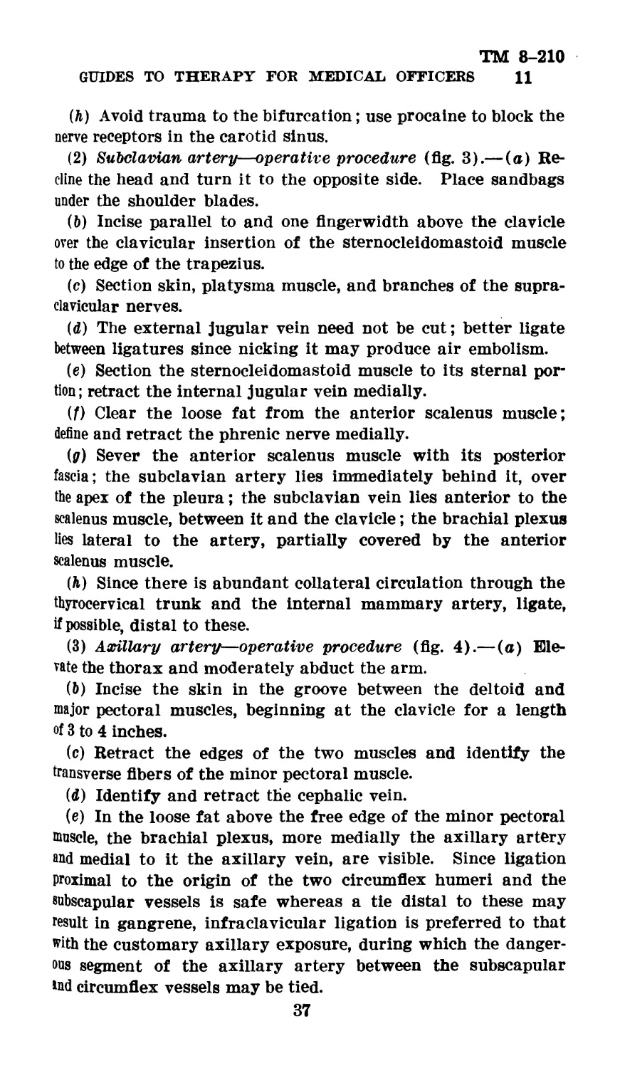
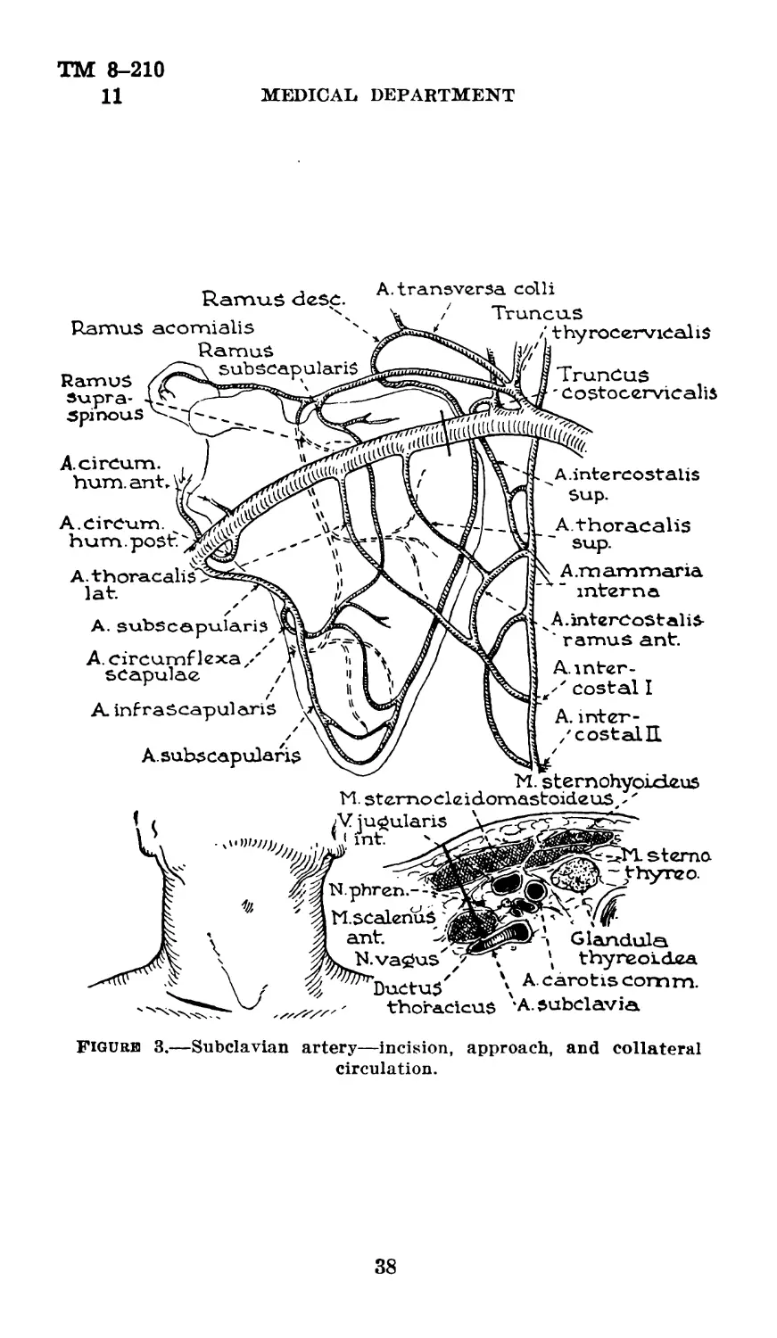
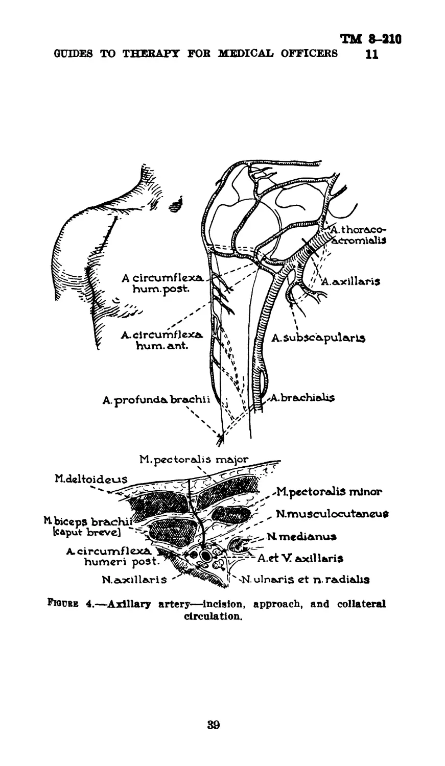
ТМ 8-210
TECHNICAL MANUAL
GUIDES TO THERAPY FOR
MEDICAL OFFICERS
UNITED STATES
GOVERNMENT PRINTING OFFICE
WASHINGTON : 1942
For sale by the Superintendent of Documents, Washington, D. C.
ТМ 8-210
С 1
TECHNICAL MANUAL
—•
GUIDES-ТО THERAPY^FOR MEDICAL OFFICERS
hanges / ' lA^ WAR DEPARTMENT,
No. 1 p ' ’ ' - J \ **b'/ \ " Washington, May 6,1943.
TM 8-210, Marchas Allows:
17. Bu:
♦ Jjfc « « « ♦
(2) Proper prophylactic measures ♦ ♦ ♦ should be taken. Satisfactory
end results, that is, minimal scar formation, largely depend on the avoidance
of pyogenic infection; the moist surfaces of burns provide ideal conditions for
bacterial growth, and it is therefore of paramount importance to employ, if
possible, strict aseptic technic in operating on and dressing burns.
(3) To relieve pain.
c. Treatment.—(1) General.—(a) Proper steps for the prevention or treat-
ment of shock (par. 15) should be instituted. In the presence of extensive burns,
quantities of plasma up to 12 units may be required in the first 24 hours. If
available, concentrated normal human serum albumin in appropriate amounts
may be used likewise. Transfusion of fresh whole blood is often needed to
combat the rapidly developing severe anemia which follows extensive burns;
when anemia exists, whole blood transfusion is particularly indicated as a
preliminary to skin grafting. Parenteral fluid replacement other than that
attained by means of plasma or whole blood transfusion should be accom-
plished by means of 5 percent glucose in sterile distilled water. The intra-
venous administration of sodium chloride solution should be reserved for
those bum cases in which mineral depletion is pronounced, as when great
loss of electrolytes occurs as a result of persistent vomiting.
(d) In all cases with moderate to severe burns, prophylactic chemotherapy
should be administered. Sulfadiazine is the drug of choice (sulfanilamide may
be substituted) with an initial dose of 4.0 grams (60 grains). Subsequent
doses of sulfadiazine should be given only under the direction of a medical
officer. It should be kept in mind that although sulfonamide therapy may
serve to prevent infection, great care must be exercised in employing such
therapy in bum cases. The extensive fluid loss and possible kidney damage
so common in bum cases increases the danger of renal complications from
sulfonamide therapy. Maintenance doses of sulfadiazine should be given
in 0.5 gram (7% grains) doses every 4 hours until such time as adequate
kidney function can be demonstrated, under which circumstance the dosage
may be increased to 1 gram (15 grains) every 4 hours.
(c) Prophylaxis against tetanus (par. 49) is indicated in all patients with
second or third degree bums.
(d) A prophylactic dose of gas-bacillus antitoxin (par. 48) may be given
at the discretion of the medical officer.
0574588
523003°—43----1
ТМ 8-210
С II TECHNICAL MANUAL
(е) Pain should be relieved by adequate doses of morphine. Pain result-
ing from an extensive burn can ordinarily be relieved by a dose of % grain
of morphine. In the presence of pronounced anoxia large doses of morphine
are dangerous, and under such circumstances the dose should not exceed
grain.
(2) Emergency treatment of the burned area.—(a) The burned surface
should be covered with a liberal amount of boric acid ointment, or if this is
not available, with vaseline. The burn should then be covered with strips
of a sterile fine mesh gauze (44 mesh gauze bandage is satisfactory). Over
this should be added a smooth thick layer of sterile gauze dressing (large or
small first aid dressings are especially suitable for this purpose). Finally,
a gauze or muslin bandage should be firmly applied over the dressings.
(b) Contamination of burned surfaces by organisms from the nose and
throat is responsible for most of the more serious infections which subse-
quently develop. Therefore, to minimize contamination from this source,
masking should be practiced by the surgeon and assistants. If masks are
not available, mouths should be kept closed.
(c) The prompt administration of plasma, when feasible, constitutes an
important element in the emergency treatment of bums.
(3) Definitive treatment of the burned area.—The burned area should be
treated as follows, standard operating room technic, if possible, being employed,
with the patient, as well as all attendants, fully masked:
(a) Ether, benzene, lard, or other detergents should be used to remove
grease, if present. The burned area and then, separately, the surrounding skin
are to be carefully cleansed, using neutral soap and water; green soap should
not be used. Avoid the use of brushes in the cleansing of the bum.
(b) All blisters and loose shreds of epidermis are carefully removed, and this
material is saved for bacteriologic study, if feasible. Skin that gives evidence
of irreparable damage through its full thickness should be excised (evidence of
irreparable damage to deeper layers of skin may not be apparent for several
days, and excision in such cases should be done as a secondary procedure).
The resulting wound should be handled like any other open surgical wound, pri-
mary grafting of skin being carried out, if conditions permit. For painful
surgical procedures or dressings, general anesthesia, preferably obtained by intra-
venous injection, should be employed.
(c) Bums of surfaces of any portion of the body may be treated with boric
acid ointment. After thorough surgical cleansing, the burned area should
be covered with a generous application of boric acid ointment. Strips of
a fine mesh sterile gauze (44-mesh gauze bandage is satisfactory) should be
applied. Over this should be added a smooth, thick layer of sterile dressing;
this may consist of gauze, absorbent cotton, cotton waste, or cellulose. The
dressings should be held in place by an evenly and firmly applied bandage;
stockinette or some form of elastic bandage is more effective than the ordinary
roller bandage. The dressing should not be disturbed for 10 days unless
complications develop. Firm pressure is especially important in the case of
burns of the hands and face. Immobilization of the part by splinting should
be effected when feasible.
(d) As an alternate method of definitive treatment, bums of all surfaces
except those of the hands, face, genitalia, and those involving the circum-
2
TH 8-210
GUIDES ТО THERAPY FOR MEDICAL OFFICERS С 1
ference of an extremity may be treated with tannic acid and silver nitrate.
This method, however, should not be employed in the instance of bums which
are grossly contaminated or in which 12 or more hours have elapsed since
the receipt of the bum. A freshly prepared 10 percent aqueous solution of
tannic acid is sprayed over the burned area. This is followed immediately by
spraying the area with a mixture of equal parts of 10 percent tannic acid and
10 percent silver nitrate solutions. This mixture should then be sprayed on the
burn every half hour until a satisfactory eschar has been formed. Care should
be taken to avoid spraying normal skin about the wound. While drying,, the
burned area mtfy be kept exposed to the air in a heated cradle. After the eschar
is dry, it may be covered by a dry sterile dressing. In the absence of infection,
the eschar should be allowed to separate spontaneously. If infection develops,
the eschar must be removed from the entire infected area, and the latter should
then be treated like any other infected wound, with the employment of appropriate
systemic and local therapy (par. 51).
(e) Physiological saline solution has been found useful in the treatment of
burns involving the face, hands, and especially fingers, the flexures, and the
perineum. It may also be used for the treatment of surface infections following
the removal of eschars produced by tannic acid or other agents. Saline may
be employed in the form of packs or baths.
[A G. 062.11 (3-23-43).] (С 1, May 6, 1943.)
24. Care of feet.—a. Foot hygiene,
♦ ♦♦♦♦♦♦
(4) Excessive moisture plays ♦ ♦ ♦ wear oiled shoes. The Army foot
powder is of value. It is composed of salicylic acid 2 percent, desiccated alum
1 percent, starch 10 percent, talcum powder 78 percent, boric acid 6 percent,
and zinc stearate 3 percent. The powder is rubbed on the feet and used as
a dusting powder for the feet and socks.
♦ ♦ ♦ ♦ « « ' «
b. Trichophytosis.—(1)-Proper cleansing and the control of moisture greatly
reduce the incidence of so-called “athlete’s foot.” Prevention of infection in
company bathhouses may be attempted by requiring a foot bath of a few minutes
in a solution of calcium hypochloride or sodium hypochloride giving 50 to 100
parts per millim of available chlorine. The Army foot powder is an efficient
preventive agent if rubbed on the feet and used as a dusting powder for the
feet and socks once or twice daily.
[A. G. 062.11 (3-23-43).] (C 1, May 6, 1943.)
3
ТМ 8-210
GUIDES ТО THERAPY FOR MEDICAL OFFICERS С 1
Section IV is rescinded and the following substituted therefor:
Section IV
DIAGNOSIS AND TREATMENT OF VENEREAL DISEASES
Paragraph
General_____________________________________________________________________ 36
Gonorrhea------------------------------------------------------------------- 37
Syphilis____________________________________________________'------------ 38
Chancroidal infection------------------------------------------------------- 39
Lymphogranuloma venereum-------------------------------------------------- 40
Granuloma inguinale--------------------------------------------------------- 41
36. General.—a. This section on the diagnosis and treatment of venereal
diseases is based on the opinions and recommendations of the Subcommittee on
Venereal Diseases of the Committee on Medicine, Division of Medical Sciences,
National Research Council. Since substantial changes from previous treatment
methods are here recommended, this section should be read carefully by all
medical officers who are called upon to treat venereal diseases. It is published
as a general guide for medical officers and is to be used with due consideration
of factors which may be presented by each individual case. It is not intended
that the recommendations contained herein will be used to the exclusion or
neglect of other indicated therapeutic or nursing procedures.
b. In the zone of the interior and in the communications zone venereal disease
cases will ordinarily be hospitalized for treatment during the infectious stages.
In order to maintain effective strength of organization during combat, considera-
tion may be given to the treatment of venereal disease cases on a duty status
with their organization in the combat zone.
37. Gonorrhea.—a. Diagnosis in male.—(1) A diagnosis of gonorrhea must
not be made in the absence of laboratory confirmation. Treatment for gonorrhea
should be started at once if the patient has an acute purulent urethral discharge,
but material should be obtained before treatment is begun for subsequent
laboratory study (smear and/or culture).
(2) Acute gonorrhea.—The detection of gram-negative intracellular diplo-
cocci in smears of the urethral exudate, or smears of the centrifuged sediment
of the first glass of urine, or fluid from conjunctival sac or joints, establishes
the diagnosis of gonococcal Infection. w In competent hands cultures yield a higher
percentage of positive results and are of particular value in cases in which no
gonococci are found in the smear.
(3) Chronic gonorrhea (posterior urethritis, prostatitis, seminal vesiculitis,
epididymitis, and arthritis.)—The detection of gram-negative intracellular diplo-
cocci in smears of the exudate obtained by digital stripping of the prostate,
Cowper’s glands, and the urethra, or in smears of the centrifuged sediment of
urine passed after stripping the prostate, Cowper’s glands, and the urethra, or
the demonstration of gonococci in cultures of material so obtained, establishes
the diagnosis of gonococcal infection.
b. Diagnosis in female.—(1) A diagnosis of gonorrhea must not be made in
the absence of laboratory confirmation. (Treatment for gonorrhea should be
started at once in women who have evidence of this disease, even though lab-
oratory studies are negative or not available. If laboratory facilities are not
available, material for subsequent laboratory studies should be obtained* before
treatment is begun.)
5
ТМ 8—210
С 1
TECHNICAL MANUAL
(2) The detection of gram-negative intracellular diplococcl In smears of ma-
terial obtained from any of the following: the urethra, Skene’s glands, or the
cervix (or from Bartholin’s glands or the rectum, when clinical symptoms exist) ;
or positive cultures of such material establishes the diagnosis of gonococcal
infection. (Caution: The normal genital bacterial flora and the flora of non-
specific infections may contain organisms that in smear closely resemble gono-
cocci. Therefore, cultural methods should be utilized when possible.)
o. Gonorrheal ophthalmia.—The diagnosis is made on the basis of an acute
purulent conjunctivitis, pus containing gonococci, and rapid involvement of the
external coats of the eye. (Sulfonamide therapy should be instituted immedi-
ately. Prompt ophthalmological consultation is imperative.)
d. Serologic tents -for syphilis.—All patients with gonorrhea should be given a
serologic test for syphilis on admission, a test before being discharged to duty,
and if possible, a final test 2 months after discharge.
e. Treatment of gonorrhea in males and females.—(1) Local treatment of any
kind (injections, irrigations, massages, instrumentations) is contra-indicated in
uncomplicated acute gonorrhea.
(2) Treatment should consist of not more than two courses of chemotherapy.
A careful inquiry should be made as to previous sulfonamide therapy, recent
self-administered chemotherapy for gonorrhea, and for previous drug reactions.
(See par. 42.)
(a) Sulfa thiazole and sulfadiazine are each highly efficient, and in the doses
recommended cause toxic manifestations very infrequently. Either of these com-
pounds is recommended. Sulfapyridine, although nearly as efficient as sulfathia-
zole or sulfadiazine, is more toxic and should be used only if the other compounds
are not available. Sulfanilamide is far less effective than the* other sulfa com-
pounds in gonorrhea and is not recommended.
(b) The recommended dosage for sulfathiazole and sulfadiazine is 1 gram (15
grains) four times a day for 5 days. The recommended dosage for sulfapyridine
is 1 gram (15 grains) three times a day for 5 days.
(c) A second course of the drug, in the same dosage, should be given if there
is evidence of persistence or recurrence of the disease. There should, however,
be a lapse of 5 days between the two courses of medication.
(d) Patients in whom the gonococcus is present after the second course, or who
have not made a satisfactory clinical response, generally should be transferred to
a general hospital.
(e) In males, local treatment to the urethra generally should be administered
only in a general hospital.
(f) In the female, hot vaginal douches (under no more than 2 feet of water
pressure) afford comfort and promote cleanliness. Acute pelvic inflammatory
disease is an indication for bed rest, ice bags to the abdomen, and analgesics. If
enemas are necessary, preliminary bathing of the perineum is indicated before
inserting the rectal tube in order to avoid inducing gonococcal proctitis.
f. Treatment of sulfonamide-resistant gonorrhea in males and females (general
hospitals).—(1) It is recommended that carefully selected patients with sulfona-
mide-resistant infections be given 10 hours of sustained fever therapy, where such
therapy is available, preceded by chemotherapy consisting of sulf a thiazole, 1 gram
every в hours for eight doses. This program should be preceded by 1 week of free-
dom from sulfonamide therapy. Patients who are not subjected to this form of
treatment, and those who are not cured by this means, should be placed upon local
treatment
6
ТМ 8-210
GUIDES ТО THERAPY FOR MEDICAL OFFICERS С 1
(2) Local treatment in male (sulfonamide-resistant cases only).—(a) When
the infection is confined to the anterior urethra, an anterior urethral injection,
once daily, of not more than 6 cc of a 5 percent solution of mild protein silver
or 0.6 percent of strong protein silver is advised. (Retain for 5 minutes.) It
should be kept in mind that prolonged use of chemicals tends to perpetuate urethral
discharge. Discharge caused by overtreatment of this type may be recognized by
the presence of many epithelial cells.
(b) All urethral injections are to be administered by a medical officer or a
trained attendant; not by the patient.
(c) Stop all local treatment if the patient develops acute symptoms of posterior
urethral infection, such as urgency, painful or marked frequency of urination, or
perineal or rectal pain; and confine treatment to hot Sitz baths. When acute
symptoms have subsided, resume anterior urethral injections and continue them
until prostatic stroking is begun.
(d) Extremely gentle prostatic stroking should be tried when the second glass
of urine has been clear, and the first glass nearly so, for 2 weeks. If gentle
massage is painful or causes a recrudescence of other symptoms, it should not
be repeated for 1 week, or until the symptoms have subsided. If it is not painful
and if no recrudescence of symptoms occurs, the gland should be gently stripped at
3- and 4-day intervals, and smears of the prostatic secretion examined every 2
weeks. (When prostatic massage is instituted too soon or applied too vigorously
it often induces complications and retards cure.)
(e) Infections of Cowper’s glands should be searched for in resistant cases.
If these glands are palpable, they should be gently kneaded. This can be ac-
complished at the time prostatic massage is practiced, by placing the thumb
against the perineum and gently massaging first one and then the other gland
with the index finger.
(/) No instruments of any type should be passed into the urethra while
gonococci are present.
(3) Local treatment in female.—(a) See paragraph 37e (2) (f).
(b) The persistence of infection in Skene’s glands, Bartholin’s glands, or the
endocervical glands in spite of the use of measures recommended above con-
stitutes a special problem beyond the province of this directive. Such patients
require skilled gynecological treatment.
g. Determination of cure in male (uncomplicated cases).—(1) Cure is deter-
mined by the'inability to demonstrate gonococci in any of the urogenital fluids
including prostatic secretions, by smears or cultures.
(2) Patients whose symptoms have disappeared as the result of sulfonamide
medication may have the first test of cure on the third day after disappearance
of symptoms. Thus, for example, patients clinically well on the third day of
chemotherapy, whose urethral cultures or smears are negative for gonococci,
may have on the sixth day prostatic massage and may then be discharged *to
duty if smears of the urogenital fluids are normal. When feasible, these
patients should return to the hospital for two subsequent examinations at
weekly intervals and should be discharged as cured after three negative
examinations.
h. Determination of cure in male (sulfonamide-resistant cases).—(1) Cure is
determined by the inability to demonstrate the gonococcus in any of the urogenital
fluids by repeated smears or cultures. Material for these studies should be
obtained in the manner described in paragraph 37a (3). The prostatic secretion
•7
ТМ 8-210
С 1
TECHNICAL MANUAL
should be included in the study of all types of infection. Three successive
negative studies at weekly intervals constitute practical evidence of cure. Tests
for cure may be carried out after the patient has returned to duty.
(2) Patients whose symptoms disappear as the result of fever therapy may
have tests of cure stated on the second day following the treatment.
i. Determination of cure in the female (uncomplicated cases).—(1) Patients
whose symptoms have disappeared as the result of chemotherapy may have tests
of cure begun on the third day after disappearance of symptoms.
(2 ) Cure is determined by—
(a) Absence of tender masses or points of tenderness in the pelvis.
(b) Inability to demonstrate the gonococcus by smears or cultures in material
obtained from the urethra, Skene’s glands, Bartholin’s glands, or the cervix.
Such tests should be repeated every 2 weeks for 3 months, and, if all are found
to be negative, the patient should be discharged from observation. These
tests should be carried out on an ambulatory basis.
(c) To obtain material for smears and cultures, massage the urethra, Bartho-
lin’s glands, and Skene’s glands, obtaining secretion with small cotton-wrapped
applicator or a platinum loop. Pass bivalve vaginal speculum without lubricant,
expose cervix, clean vagina and cervical canal, squeeze cervix between ends of
speculum blades, and obtain expressed fluid on cotton applicators or platinum
loops for smear and culture.
j. Determination of cure in female (sulfonamide-resistant cases).—(1) Patients
whose symptoms have disappeared as. the result of prolonged artificial fever
may have tests of cure begun on the second day after treatment. The tests of
cure are the same as those recommended for female patients in uncomplicated
gonorrhea.
(2) Patients under local treatment should have smears and culture done at
least every 2 weeks. If these tests remain consistently negative for 3 months,
and if there are no demonstrable complications, the patient should be discharged
from observation.
38. Syphilis.—a. Diagnosis of syphilis.— (1) It is of the utmost importance
that the diagnosis in early syphilis (primary and secondary stages) be established
at the earliest practicable moment and that treatment be instituted as soon as
the diagnosis is made.
(2) All ulcerative genital lesions, extragenital lesions characterized by in-
dolence, Induration, and regional lymphadenopathy, and cases of urethritis
accompanied by indolent enlargement of related lymph glands are to be regarded
as probable cases of syphilitic infection until this possibility has been excluded
by repeated darkfleld examinations and repeated serologic tests. In these cases
routine serologic tests will be done not less often than—
(a) On admission to sick report.
(b) Second week.
(c) About the end of the first month.
(d) About the end of the second month.
(3) Routine serologic tests will also be made in all cases of gonorrhea at
least as often as follows:
(a) On admission to sick report.
(b) Before return to a duty status.
(o) About the end of the second month.
8
ТМ 8-210
GUIDES ТО THERAPY FOR MEDICAL OFFICERS С 1
(4) Antisyphilitic treatment will not be started until the diagnosis of syphilis
is definitely established. The demonstration of the Treponema pallidum by dark-
field examination is conclusive as to the necessity for the immediate institution
of treatment A persistent completely positive serology even in the absence of
confirmatory clinical signs or a history of infection is also diagnostic of syphilis,
and usually is an indication for treatment. Positive serologic tests on a single
specimen should never be made the basis of treatment in the absence of unmis-
takable clinical evidence of syphilis. A completely positive precipitation or
complement fixation test (Kahn or Wassermann) confirmed by a positive com-
plement fixation test (Wassermann) on a second specimen indicates syphilis
provided no negative reactions have been obtained. Acute infection, particularly
malaria and possibly vaccination procedures, are believed to give *186 at times
to transient false positive serologic tests. The presence of these complicating
factors should be excluded before making a definite diagnosis of latent syphilis.
In the event of incompletely positive or contradictory serologic reactions the
test should be repeated until the possibility of technical error is excluded.
b. Treatment of syphilis.— (1) General principles of treatment.— (a) No treat-
ment is to be given for suspected early syphilis until the diagnosis is made either
by darkfield examination or confirmed serologic tests. No therapeutic tests are
to be used.
(b) Arsenoxide (mapharsen) will be used as the standard arsenical. Neo-
arsphenamine or other arsenicals cannot be substituted in the treatment schedule
outlined in table I.
(c) If it is necessary to use neoarsphenamine, because of the nonavailability
of arsenoxide, the former drug should be given only at weekly Intervals in courses
of not more than 8 consecutive weekly doses.
(d) Tryparsamide and fever therapy are not to be used outside of a hospital.
(e) Each treatment is to be recorded on the Syphilis Register of the patient
or if for any reason д syphilis register is not available, a written record is to be
kept and transferred to the standard form as soon as possible. Bach entry
will include date, drug, dose, and reaction.
(/) It cannot be too strongly emphasized that regularity of treatment schedule
without long or short time variations or lapses is critically important to both
infection, control, and cure. Every effort must be made to impress this fact
on enlisted men and officers as well as medical personnel.
(p) Emphasis should also be placed on the completion by each patient of the
full schedule of treatment in the time called for regardless of early serologic
reversal.
(2) Treatment of early and latent syphilis.—(a) In view of recent advances
in knowledge regarding the toxicity and therapeutic efficiency of arsenoxide,
the following recommendations regarding the treatment of early and latent
syphilis have been prepared by the Subcommittee on Venereal Diseases of the
National Research Council and approved by The Surgeon General.
(b) Early (primary and secondary) and latent syphilis of any duration should
be treated by an identical treatment system. This treatment may be completed
within 26 weeks. (See treatment schedule, table I.)
(c) Patients with syphilis, early or latent, should, as a rule, be hospitalized
initially to the end that a careful examination may be made and antisyphilitic
treatment started. The period of hospitalization ordinarily need not be pro-
6230030—43---2
ТМ 8-210
С 1
TECHNICAL MANUAL
longed more than 5 to 7 days. Thereafter, treatment should be continued by
unit medical officers; or, in areas where concentration of patients is feasible, in
centralized ambulatory clinics established in hospitals. Whenever possible and
to minimize loss of time from duty, treatment of nonhospitalized patients should
be given after duty hours.
(d) At reception centers, vaccinations and other procedures incident to induc-
tion may be carried out during the initial period of hospitalization for syphilis,
by local arrangements between the commanding officers of the reception center
and the station hospital.
(e) Treatment schedule for early and latent syphilis is shown in table I.
Table I.—Treatment schedule, early and latent syphilis
Week 1 2 3 4 6 6 7 8 9 10 .Arsenoxide (mapharsen) intraven- ously twice weekly, total 20 injec- tions. Bismuth subsalicylate intramuscu- larly once weekly, 5 doses. Omit bismuth—5 weeks.
11 12 13 14 15 16 Omit arsenoxide (mapharsen)—6 weeks. Bismuth subsalicylate—intramuscu- larly once weekly—6 doses.
17 18 19 20 21 22 23 24 25 26 Arsenoxide (mapharsen) as in first course, twice weekly total 20 injections. • Omit bismuth—5 weeks. Bismuth subsalicylate intramuscu- larly once weekly, 5 doses.
Arsenoxide (mapharsen) dosage: Adjusted approximately to body weight;
average dose 60 milligrams, minimum dose 50 milligrams, maximum 70 milligrams.
Bismuth subsalicylate in oil dosage: The standard dose is 0.2 gram of bismuth
subsalicylate intramuscularly (not 0.2 gram of elemental bismuth metal).
1. It is anticipated that individuals now on antisyphilitic therapy in
accordance with other schedules will continue their antisyphilitic
therapy in accordance with the provisions of table I as soon as the
requisite drugs are obtained.
2. In effecting this change of schedule it should be borne in mind that
the purpose of the new plan of treatment is to furnish adequate
10
TH 8-210
GUIDES ТО THERAPY FOR MEDICAL OFFICERS С 1
antisyphilitic therapy in a shortened period of time. The drug of
paramount importance is the arsenical. Therefore, the technique of
change to the new schedule should provide, wherever practicable,
that the amount of arsenical drug administered under a previous
schedule, together with that administered under the schedule of table
I, should equal the total number of doses (1. e., 40) of arsenical drug
advocated In the new treatment schedule of table I, and a minimum
of 16 doses of bismuth subsalicylate. In transferring from one sched-
ule of treatment to the other it will ordinarily suffice to base the
transfer solely on the number of arsenical treatments the individual
has received. For example, if a patient has received 17 doses of
arsenical drug under a previous schedule, he should be started on the
eighteenth dose of mapharsen on the schedule of table I. This method
of transfer should be modified if it would at any time call for a
patient receiving more than 25 consecutive doses of an arsenical drug.
The treatment schedule should always be concluded with an overlap-
ping series of 5 weekly injections of bismuth subsalicylate, as sug-
gested in table I.
3. This plan of treatment is not to be construed to mean, however, that
an individual who has completed the arsenical treatments under a
previous plan of therapy need be given additional arsenical in order
that the total arsenical administered may equal that suggested in
table I.
j. In the case of an individual who has been recently inducted into the
'Army and who had received antisyphilitic treatment prior to entrance,
the principles outlined above may be followed, using the physician’s or
clinic’s record of his previous treatment as a basis for further therapy.
Such records will ordinarily be readily available upon the surgeon’s
written request, accompanied by the individual’s written authoriza-
tion for furnishing the data desired. (This paragraph is included in
view of the contemplated induction of individuals who have early or
latent syphilis without disabling lesions, when facilities for their
housing and treatment have been provided.)
5. Patients with cerebrospinal, cardiovascular, or other types of visceral
syphilis require specialized supervision and prolonged hospital care.
Such cases should be discharged on CDD if discovered soon after in-
duction. Otherwise, they should be transferred to a general hospital
for treatment and disposition.
(3) Technical suggestions.—(a) Discard discolored drugs and solutions and
damaged ampules.
(&) Dissolve arsenoxide in sterile distilled water in the proportion of 10 milli-
grams of drug per 2 cc of water; a dose of 60 milligrams will then be contained
in 12 cc of solution, 50 milligrams in 10 cc, and 70 milligrams in 14 cc.
(c) Shake and aerate arsenoxide; do not shake or aerate the other arsenicals.
(d) Inject arsenoxide rapidly to avoid thrombosis; there is little danger of
speed shock or nitritoid crisis. Other arsenicals should be injected slowly to
avoid speed shock or nitritoid crisis.
(e) Thoroughly shake oily suspensions.
11
ТМ 8-210
С 1 TECHNICAL MANUAL
(f) Attempt aspiration after insertion of needle before making any injection,'
especially intramuscularly.
(g) Inject bismuth intramuscularly into upper outer quadrant of buttock.
Alternate sides.
(Л) Massage firmly after withdrawing needle from buttock and have patient
prolong massage to 3 minutes.
(i) Advise rest, if practicable, after arsenicals.
(/) Warn patient to report his reactions.
(k) Watch mouth and gums for bismuth stomatitis.
(4) Treatment is to be stopped and the patient hospitalised if the following
appear:
(a) An itchy dermatitis of the face or flexures.
(b) Jaundice.
(c) Petechial or other hemorrhagic lesions.
(d) Evidence of cerebral injury, even though slight.
(5) General antireaction therapy.—(a) Epinephrin solution 1:1000, % to 1 cc
subcutaneously for speed shock or nitritoid crisis.
(& ) Glucose 500 cc 5 percent solution intravenously supplemented with thiamine
chloride 5 milligrams, for jaundice.
(c) Vitamin В complex is recommended in suspected liver damage.
(d) In cerebral vascular accidents, measures to be considered are venesection,
and hypertonic saline solution intravenously (500 cc of a 1.5 percent solution).
(e) In blood dyscrasias, transfusions.
(/) Sodium thiosulphate for any type of treatment reaction is considered
valueless. *
(6) Serologic control of treatment.—In patients with early syphilis a serologic
test will be done at the beginning and end of the schedule of treatment outlined
in table I; but treatment may be stopped whether the serologic test for syphilis
(STS) is positive or negative. After the completion of treatment, the STS should
be repeated 3 and 6 months later. If the test is negative after 6 months, the case
may be classified as “Result satisfactory” and the Syphilis Register may be closed.
If the test is positive after 6 months, the patient should be referred to a station
or general hospital. In patients with latent syphilis the STS should be repeated
at the completion of treatment outlined in table I, but the Syphilis Register may be
closed when this treatment is completed, regardless of the result of serologic test.
(7) Spinal fluid examination.—Should be performed in a hospital in patients
with early syphilis at the end of the course of treatment outlined in table I, or as
soon as possible thereafter; but in any event before the Syphilis Register is closed.
In apparent latent syphilis, spinal puncture should be performed in a hospital
before treatment or as soon as possible thereafter, but in any event before the
Syphilis Register is closed.
(8) Control of relapse and infectiousness.—(a) Early syphilis is to be regarded
as infectious until the second injection of arsenoxide has-been given.
(&) Physical inspection of skin (including palms and soles), mucosae, anus, and
genitalia should be performed as often as circumstances permit during treatment
and at each probationary inspection.
(c) The involution of the chancre or secondaries should be watched to detect
treatment-resistant cases.
(d) Patients should be warned to look for and to report mouth, skin, and genital
lesions. Darkfleld examination is of great help in recognizing relapsing lesions
of the skin, mucosa, and genitalia.
12
ТМ 8-210
GUIDES ТО THERAPY FOR MEDICAL OFFICERS С 1
(9) Complications or relapse,—In the event of any complication of treatment
(serious treatment reactions) or any evidence of relapse, clinical or serologic,
the patient should be at once transferred to a station or general hospital.
(10) Cardiovascular, visceral, and neurosyphilis require special treatment in
hospital. For additional details see standard texts.
(11) Treatment of precocious late syphilis (tertidrism).—As soon as possible,
precocious late syphilis (early gummatous and rupial lesions and bone lesions)
should be hospitalized for combined fever and arsenical therapy.
(12) Treatment of congenital syphilis.—On recognition or on appearance of
active lesions, congenital syphilis should be treated on the schedule for early and
latent syphilis.
(13) Separation from the service.—When practicable, the physical status of
every patient with syphilis, whether the disease was acquired before or after
induction into the service, will be completely reevaluated when discharge from
the Army is contemplated.
c. Diagnostic nomenclature for syphilis in Army.
Syphilis, primary.
Syphilis, secondary.
Syphilis, early latent (less than 4 years since infection).
Spinal fluid negative.
Spinal fluid not performed (diagnosis tentative).
Syphilis, late latent (4 or more years since infection).
Spinal fluid negative.
Spinal fluid not performed (diagnosis tentative).
Syphilis, tertiary—otherwise unclassified.
Mucocutaneous.
Osseous.
.Ocular (except optic atrophy).
Visceral (except cardiovascular).
Cardiovascular—other.
Aneurysm (saccular).
Aortic regurgitation (insufficiency).
Aortitis (uncomplicated).
Neurosyphilis—otherwise unclassified.
Asymptomatic.
Acute syphilitic meningitis.
Diffuse meningovascular.
Optic atrophy.
Tabes dorsalis.
Taboparesis.
Psychosis with syphilitic meningoencephalitis (general paresis).
Psychosis with neurosyphilis other than paresis or taboparesis.
’ Syphilis, type undetermined—to include cases in which accurate diagnosis
has not been made.
Syphilis, congenital—include all manifestations.
Arsenical poisoning.
Bismuth poisoning.
Iodine poisoning.
Mercury poisoning.
Spinal puncture for diagnosis or progress.
13
ТМ 8-210
С 1 TECHNICAL MANUAL
Special therapeutic practices:
Fever therapy—
Malaria.
Artificial.
d. Definition of terms and explanations of their use.—(1) Primary.—To in-
clude those cases presenting the primary lesion of syphilis (the chancre), which
have not yet developed secondary manifestation. This diagnosis must be
confirmed by darkfield examination, serologic test of the blood, or both. If blood
serologic test is negative, the diagnosis of primary syphilis is not permissible
without the demonstration of T. pallidum by darkfield.
(2) Secondary.—To include only those cases of early syphilis which show
one or more of the manifestations of systemic dissemination of the spirochete;
for example, generalized enlargement of lymph glands, cutaneous eruption, mucous
patches, condylomata lata, patchy alopecia, laryngitis, bone pains, febrile re-
action, and so forth. The chancre may or may not be present, and if present,
may be in any stage of evolution. In early secondary cases the manifestations
of systemic spirochetal dissemination are increasing, have attained their maxi-
mum, or are waning. This diagnosis must be confirmed by darkfield examination,
serologic test, or both. In early secondary syphilis and in addition to the mani-
festations listed above, ocular or neurologic complications (iritis, neuroretinitis,
acute syphilitic meningitis) should be specially recorded as: “Syphilis, secondary,
manifested by_________”
(3) Latent.—Secondary symptoms have subsided and the active manifestations
of late syphilis have not yet supervened. There are no evidences of syphilis other
than a positive serologic test of the blood. Cases will be classified as: “Latent
(early or late)—spinal fluid negative” or “Latent (early or late)—spinal fluid
not performed (diagnosis tentative).” The date of the Uegative examination
of the spinal fluid will be stated in all cases.
(4) Tertiary.—This classification is to be limited to cases that show active
lesions of late syphilis. The lesion may be a gumma or it may be a diffuse
process and may involve any organ or tissue of the body. The majority of all
patients with tertiary syphilis will fall within six categories:
(a) Mucocutaneous.—Late syphilitic gummatous lesions of skin or mucous
membrane.
(&) Osseous.—Periostitis, osteomyelitis, arthritis, and synovitis.
(c) Ocular.—Iritis, uveitis, keratoiritis, keratitis, and choroiditis, but not
including optic atrophy.
(d) Visceral.—Hepatic, gastric, etc., but not including cardiovascular.
(e) Cardiovascular.—To include all lesions of the heart and great vessels.
Classify as follows:
1. Aneurysm (saccular).—Do not use for a fusiform dilation of the aorta.
Specify artery involved.
3. Aortic regurgitation {aortic insufficiency).—Specify whether with or
without cardiac decompensation.
3. Aortitis, uncomplicated.—To be used only for those patients with symp-
toms and physical or X-ray signs of syphilitic aortic involvement in
the absence of aneurysmal sacculation or aortic regurgitation.
(5) N eurosyphUis.—To include all cases with involvement of the central
nervous system, classified as follows.
14
ТМ 8—210
GUIDES ТО THERAPY FOR MEDICAL OFFICERS С 1
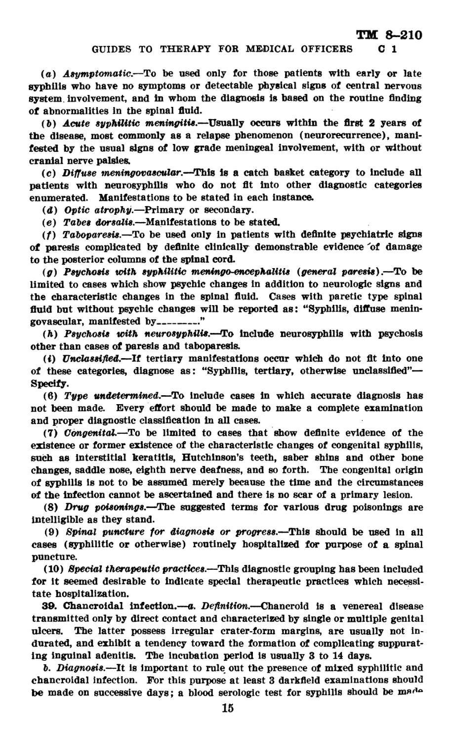
(a) Asymptomatic.—To be used only for those patients with early or late
syphilis who have no symptoms or detectable physical signs of central nervous
system, involvement, and in whom the diagnosis is based on the routine finding
of abnormalities in the spinal fluid.
(b) Acute syphilitic meningitis.—Usually occurs within the first 2 years of
the disease, most commonly as a relapse phenomenon (neurorecurrence), mani-
fested by the usual signs of low grade meningeal involvement, with or without
cranial nerve palsies.
(c) Diffuse meningovascular.—This is a catch basket category to include all
patients with neurosyphilis who do not flt into other diagnostic categories
enumerated. Manifestations to be stated in each instance.
(d) Optic atrophy.—Primary or secondary.
(e) Tabes dorsalis.—Manifestations to be stated.
(f) Taboparesis.—To be used only in patients with definite psychiatric signs
of paresis complicated by deflnite clinically demonstrable evidence 'of damage
to the posterior columns of the spinal cord.
(g) Psychosis with syphilitic meningo-encephalitis (general paresis).—To be
limited to cases which show psychic changes in addition to neurologic signs and
the characteristic changes in the spinal fluid. Cases with paretic type spinal
fluid but without psychic changes will be reported as: “Syphilis, diffuse menin-
govascular, manifested by_______”
(Л) Psychosis with neurosyphilis.—To include neurosyphilis with psychosis
other than cases of paresis and taboparesis.
(i) Unclassified.—If tertiary manifestations occur which do not flt into one
of these categories, diagnose as: “Syphilis, tertiary, otherwise unclassified”—
Specify.
(6) Type undetermined.—To include cases in which accurate diagnosis has
not been made. Every effort should be made to make a complete examination
and proper diagnostic classification in all cases.
(7) Congenital.—To be limited to cases that show definite evidence of the
existence or former existence of the characteristic changes of congenital syphilis,
such as interstitial keratitis, Hutchinson’s teeth, saber shins and other bone
changes, saddle nose, eighth nerve deafness, and so forth. The congenital origin
of syphilis is not to be assumed merely because the time and the circumstances
of the infection cannot be ascertained and there is no scar of a primary lesion.
(8) Drug poisonings.—The suggested terms for various drug poisonings are
intelligible as they stand.
(9) Spinal puncture for diagnosis or progress.—This should be used in all
cases (syphilitic or otherwise) routinely hospitalized for purpose of a spinal
puncture.
(10) Special therapeutic practices.—This diagnostic grouping has been included
for it seemed desirable to indicate special therapeutic practices which necessi-
tate hospitalization.
39. Chancroidal infection.—a. Definition.—Chancroid is a venereal disease
transmitted only by direct contact and characterized by single or multiple genital
ulcers. The latter possess irregular crater-form margins, are usually not in-
durated, and exhibit a tendency toward the formation of complicating suppurat-
ing inguinal adenitis. The incubation period is usually 3 to 14 days.
b. Diagnosis.—It is important to rule out the presence of mixed syphilitic and
chancroidal infection. For this purpose at least 3 darkfield examinations should
be made on successive days; a blood serologic test for syphilis should be
15
ТМ 8-210
С 1
TECHNICAL MANUAL
on admission to the hospital, during the second week, and at monthly intervals
for 2 months following healing of the chancroidal lesions. Laboratory tests for
the diagnosis of chancroid (Ito-Reenstierna skin test or the staining or cultural
isolation of the Ducrey bacillus) are not recommended.
c. Treatment.—(1) Chemotherapy.—(a) Local.—Accessible lesions should be
cleaned with soap and water and dried. They should then be completely covered
with powdered sulfanilamide and a loose, dry dressing applied. This should be
repeated at daily intervals until the lesion heals. Other local medication is not
recommended. In patients with tight phimosis and underlying ulcerative lesions,
the phimotic preputial cavity should be irrigated twice daily with 1-5000 potas-
sium permanganate solution.
(&) Systemic.—Administer sulfathiazole or sulfadiazine 1 gram (15 grains)
4 times a day for 5 days. Sulfanilamide 1 gram (15 grains) 3 times a day for
5 days may be utilized instead of sulfathiazole or sulfadiazine, but is less well
tolerated. Practically all chancroidal infections will respond to the above rou-
tine. In fact, if the lesion does not heal, doubt is cast on the correctness of the
diagnosis of chancroid, and the patient should be restudied from the diagnostic
standpoint, and, if necessary, treated surgically.
(2) Surgical therapy.—(a) Surgical procedure designed to relieve phimosis
or paraphimosis should be resorted to only on the basis of sound clinical
judgment.
(b) Chancroidal bubo.—Most of these will subside with systemic sulfonamide
therapy. If extensive suppuration is present, the bubo may be opened by a small
incision, the pus aspirated, and the cavity packed with sulfanilamide powder.
40. Lymphogranuloma venereum.—a. Definition.—This disease concept in-
cludes the conditions formerly known as lymphogranuloma inguinale, lympho-
pathia venereum, climatic bubo, esthiomene, and inflammatory rectal stricture.
b. Etiology.—A filtrable virus, probably multiple strains.
c. Geographic distribution.—World-wide.
d. Clinical picture.—A systemic disease of the lymphatic system, usually origi-
nating in a trivial and transitory lesion of the penis, vulva, vagina, or rectum,
which frequently escapes the patient’s notice. The invasion of the lymphatic
glands usually occurs from 10 to 30 days after infection, occasionally is delayed
months. Inguinal adenitis is often bilateral and occasionally subsides without
suppuration. During this stage, constitutional symptoms may be observed. Lymph
nodes may fuse to skin, resulting in multiple areas of softening, followed by
numerous fistulae. Extensive scarring accompanies healing. The anorectal syn-
drome usually is found only in the female, and is characterized by rectal pain,
discharge of blood and pus from the anus, a tendency toward extreme chronicity,
and the production of rectal stricture.
e. Differential diagnosis.—Differentiate from malignant tumors, Hodgkin’s dis-
ease, tularemia, tuberculosis, pyogenic infections, chancroidal bubo, and syphilis.
Mixed venereal infections should be ruled out by the darkfield examination of
material from genital lesions for the causative organism of syphilis. Frequent
serologic examinations should be continued for at least 2 months after the dis-
appearance of the lymphatic symptoms.
f. Laboratory tests in lymphogranuloma venereum.—(1) Only one diagnostic
procedure for lymphogranuloma venereum, the intradermal test of Frei, has as yet
come into general use. Other methods used in confirming the diagnosis are
either impractical (animal inoculation, artificial cultivation of the virus, non-
16
TH 8-210
GUIDES TO THERAPY FOR MEDICAL OFFICERS С 1
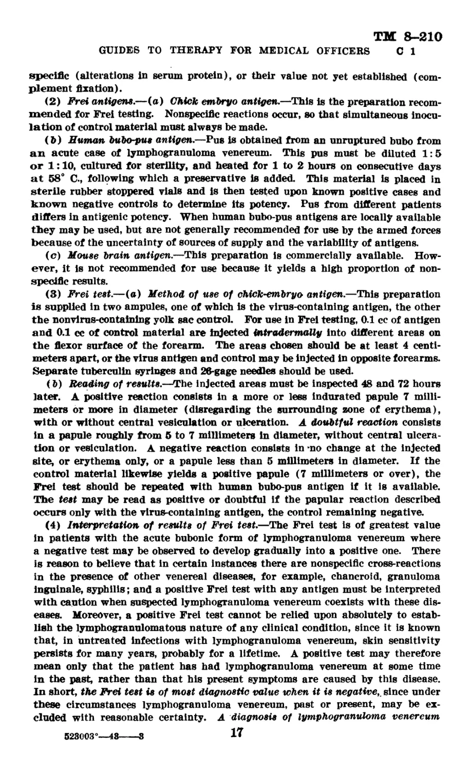
specific (alterations in serum protein), or their value not yet established (com-
plement fixation).
(2) Frei antigens.—(a) Chick embryo antigen.—This is the preparation recom-
mended for Frei testing. Nonspecific reactions occur, so that simultaneous inocu-
lation of control material must always be made.
(b) Human bubo-put antigen.—Pus is obtained from an unruptured bubo from
an acute case of lymphogranuloma venereum. This pus must be diluted 1:5
or 1:10, cultured for sterility, and heated for 1 to 2 hours on consecutive days
at 58° C., following which a preservative is added. This material is placed in
sterile rubber stoppered vials and is then tested upon known positive cases and
known negative controls to determine its potency. Pus from different patients
differs in antigenic potency. When human bubo-pus antigens are locally available
they may be used, but are not generally recommended for use by the armed forces
because of the uncertainty of sources of supply and the variability of antigens.
(c) Mouse brain antigen.—This preparation is commercially available. How-
ever, it is not recommended for use because it yields a high proportion of non-
specific results.
(3) Frei test.—(a) Method of use of chick-embryo antigen.—This preparation
is supplied in two ampules, one of which is the virus-containing antigen, the other
the nonvlrus-containing yolk sac control. For use in Frei testing, 0.1 cc of antigen
and 0.1 cc of control material are injected intradermally into different areas on
the flexor surface of the forearm. The areas chosen should be at least 4 centi-
meters apart, or the virus antigen and control may be injected in opposite forearms.
Separate tuberculin syringes and 20-gage needles should be used.
(b) Reading of results.—The injected areas must be inspected 48 and 72 hours
later. A positive reaction consists in a more or less indurated papule 7 milli-
meters or more in diameter (disregarding the surrounding zone of erythema),
with or without central vesiculation or ulceration. A doubtful reaction consists
in a papule roughly from 5 to 7 millimeters in diameter, without central ulcera-
tion or vesiculation. A negative reaction consists in mo change at the injected
site, or erythema only, or a papule less than 5 millimeters in diameter. If the
control material likewise yields a positive papule (7 millimeters or over), the
Frei test should be repeated with human bubo-pus antigen if it is available.
The test may be read as positive or doubtful if the papular reaction described
occurs only with the virus-containing antigen, the control remaining negative.
(4) Interpretation of results of Frei test.—The Frei test is of greatest value
in patients with the acute bubonic form of lymphogranuloma venereum where
a negative test may be observed to develop gradually into a positive one. There
is reason to believe that in certain instances there are nonspecific cross-reactions
in the presence of other venereal diseases, for example, chancroid, granuloma
inguinale, syphilis; and a positive Frei test with any antigen must be interpreted
with caution when suspected lymphogranuloma venereum coexists with these dis-
eases. Moreover, a positive Frei test cannot be relied upon absolutely to estab-
lish the lymphogranulomatous nature of any clinical condition, since it is known
that, in untreated infections with lymphogranuloma venereum, skin sensitivity
persists for many years, probably for a lifetime. A positive test may therefore
mean only that the patient has had lymphogranuloma venereum at some time
in the past, rather than that his present symptoms are caused by this disease.
In short, the Frei test is of most diagnostic value when it is negative, since under
these circumstances lymphogranuloma venereum, past or present, may be ex-
cluded with reasonable certainty. A diagnosis of lymphogranuloma venereum
523003°—48---8 17
ТМ 8-210
С 1
TECHNICAL MANUAL
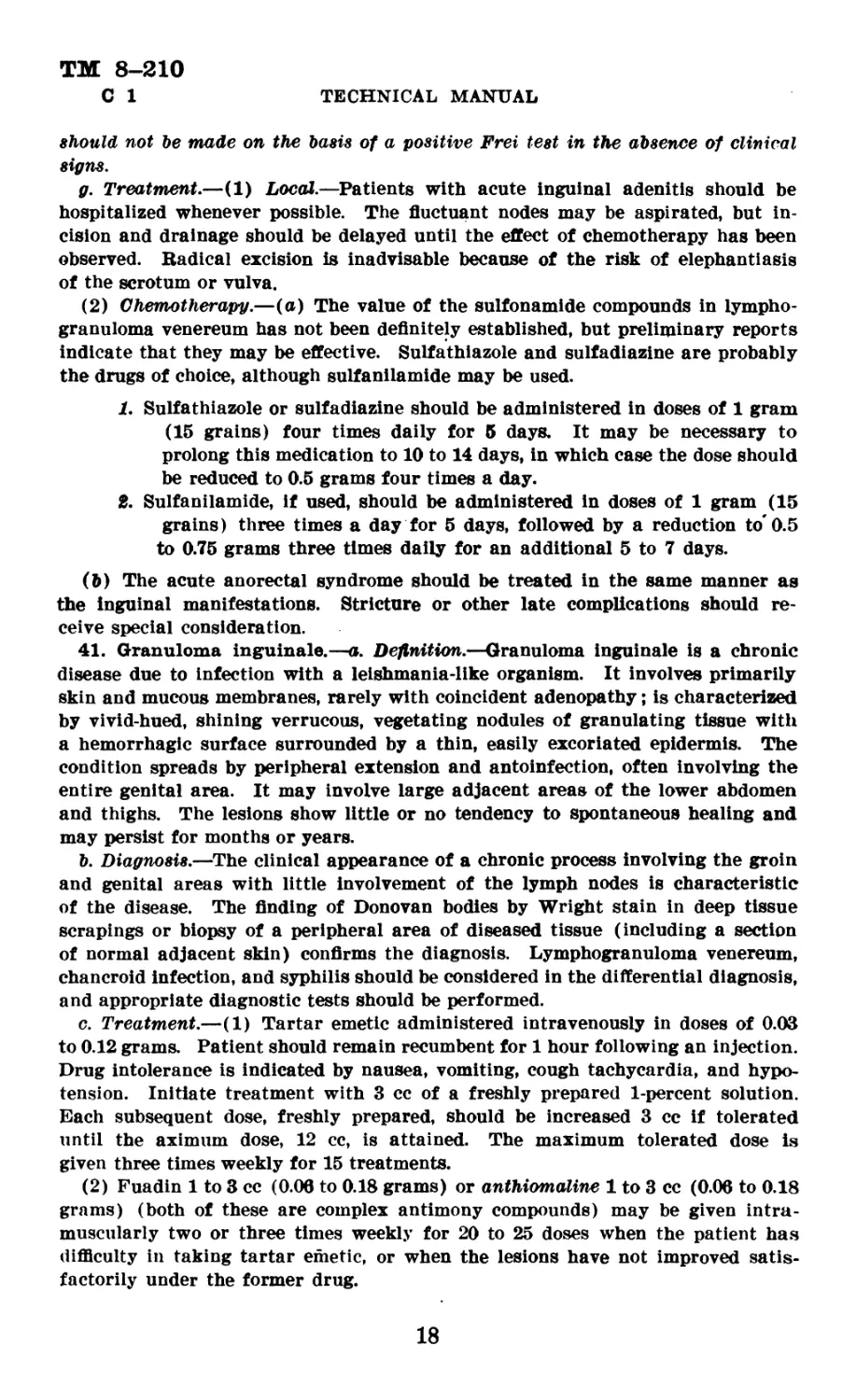
should not be made on the basis of a positive Frei test in the absence of clinical
signs.
g. Treatment.—(1) Local.—Patients with acute inguinal adenitis should be
hospitalized whenever possible. The fluctuant nodes may be aspirated, but in-
cision and drainage should be delayed until the effect of chemotherapy has been
observed. Radical excision is inadvisable because of the risk of elephantiasis
of the scrotum or vulva.
(2) Chemotherapy.—(a) The value of the sulfonamide compounds in lympho-
granuloma venereum has not been definitely established, but preliminary reports
indicate that they may be effective. Sulfathiazole and sulfadiazine are probably
the drugs of choice, although sulfanilamide may be used.
1. Sulfathiazole or sulfadiazine should be administered in doses of 1 gram
(15 grains) four times daily for 5 days. It may be necessary to
prolong this medication to 10 to 14 days, in which case the dose should
be reduced to 0.5 grams four times a day.
2. Sulfanilamide, if used, should be administered in doses of 1 gram (15
grains) three times a day for 5 days, followed by a reduction to* 0.5
to 0.75 grams three times daily for an additional 5 to 7 days.
(b) The acute anorectal syndrome should be treated in the same manner as
the inguinal manifestations. Stricture or other late complications should re-
ceive special consideration.
41. Granuloma inguinale.—a. Definition.—Granuloma inguinale is a chronic
disease due to infection with a leishmania-like organism. It involves primarily
skin and mucous membranes, rarely with coincident adenopathy; is characterized
by vivid-hued, shining verrucous, vegetating nodules of granulating tissue with
a hemorrhagic surface surrounded by a thin, easily excoriated epidermis. The
condition spreads by peripheral extension and antoinfection, often involving the
entire genital area. It may involve large adjacent areas of the lower abdomen
and thighs. The lesions show little or no tendency to spontaneous healing and
may persist for months or years.
b. Diagnosis.—The clinical appearance of a chronic process involving the groin
and genital areas with little involvement of the lymph nodes is characteristic
of the disease. The finding of Donovan bodies by Wright stain in deep tissue
scrapings or biopsy of a peripheral area of diseased tissue (including a section
of normal adjacent skin) confirms the diagnosis. Lymphogranuloma venereum,
chancroid infection, and syphilis should be considered in the differential diagnosis,
and appropriate diagnostic tests should be performed.
c. Treatment.—(1) Tartar emetic administered intravenously in doses of 0.03
to 0.12 grams. Patient should remain recumbent for 1 hour following an injection.
Drug intolerance is indicated by nausea, vomiting, cough tachycardia, and hypo-
tension. Initiate treatment with 3 cc of a freshly prepared 1-percent solution.
Each subsequent dose, freshly prepared, should be increased 3 cc if tolerated
until the aximum dose, 12 cc, is attained. The maximum tolerated dose is
given three times weekly for 15 treatments.
(2) Fuadln 1 to 3 cc (0.06 to 0.18 grams) or anthiomaline 1 to 3 cc (0.06 to 0.18
grams) (both of these are complex antimony compounds) may be given intra-
muscularly two or three times weekly for 20 to 25 doses when the patient has
difficulty in taking tartar emetic, or when the lesions have not improved satis-
factorily under the former drug.
18
ТМ 8-210
GUIDES ТО THERAPY FOR MEDICAL OFFICERS С 1
(3) Courses of tartar emetic, fuadin, or anthiomaline, separated by “rest
periods” of 2 weeks, should be continued for at least 4 months after all lesions
are completely healed, otherwise relapse is almost Certain to occur.
(4) Local treatment of the lesions may be limited to daily dressings; or
surgical excision of the entire area may be necessary. Large дгеав may be
treated with solid carbon dioxide pencils. Deep X-ray therapy in expert hands
has yielded promising results.
[A. G. 062.11 (3-23-43).] (C 1, May 6, 1943.)
42. General.
* * ♦ ♦ ♦ ♦ ♦
c.
♦ . ♦ ♦ ♦ ♦ ♦ ♦
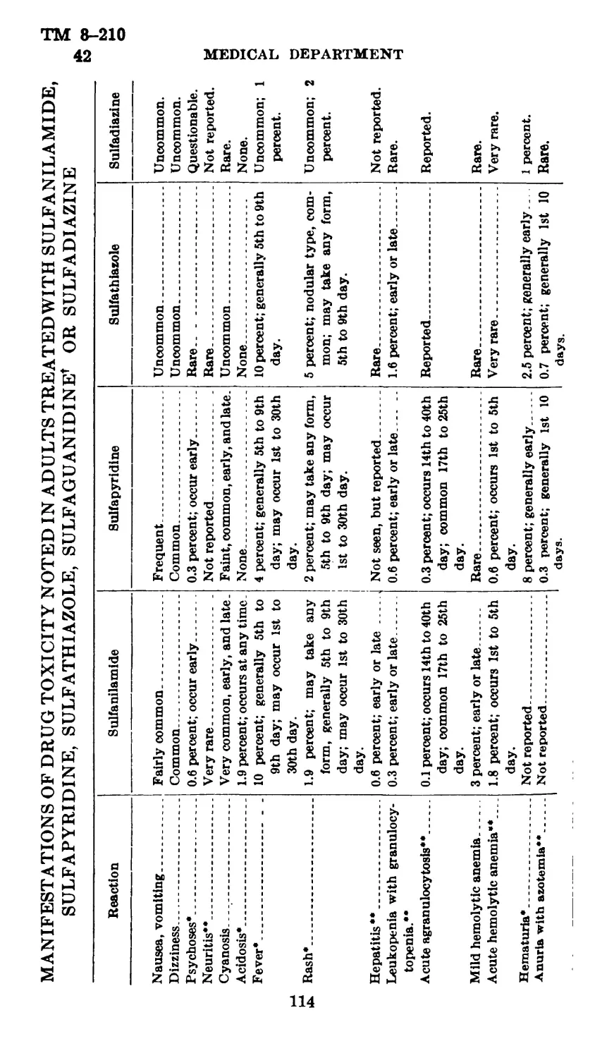
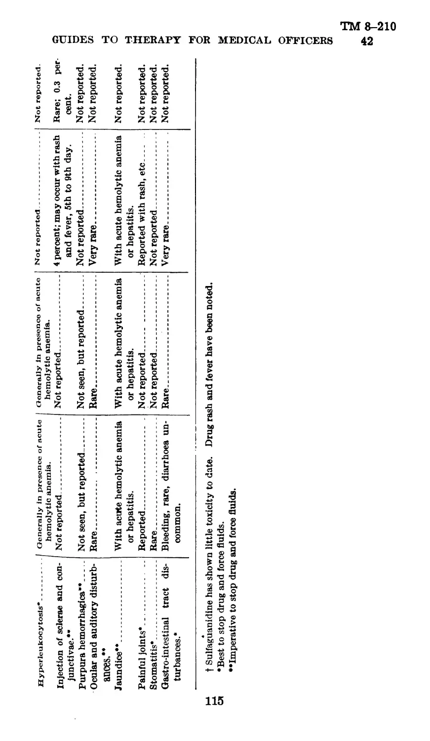
(7) Laboratory procedures used in following patients receiving sulfonamide
compounds.—(a) General.—It should be constantly borne in mind that laboratory
measures should be utilized when feasible as an adjunct in the effort to avoid
complications from the administration of sulfonamide compounds. Careful clini-
cal observation, hemoglobin estimation, and red and white blood counts should
be carried out; nevertheless, in all cases, and in those showing any abnormal
response to the drug or having a history of such an abnormal response, and in
those who have recently taken sulfonamides, or who are suspected of having done
so, action of the drug should be carefully followed by appropriate additional
laboratory procedures. Hospitalization should be promptly effected in case of
complications.
♦ ♦ ♦ ♦ ♦ * ♦
[A. G. 062.11 (3-23-48).] (С 1, May 6,1943.)
51. Wound infections.—a. General.—The problem of * * * certain spe-
cific agents. The following statements refer to the definitive treatment of
wounds and are not to be confused with first aid and emergency measures
applicable to the handling of battle casualties.
b. Prophylaxis.
* ♦ * * * ♦ *
(4) Chemotherapy should be instituted immediately after debridement of
the wound.
(a) Crystalline sulfanilamide should be lightly dusted on the wound.
(b) .Give 2.0 grams (30 grains) sulfadiazine by mouth, and then 1 gram (15
grains) every 6 hours, day and night, for 7 days or until danger of infection
has passed, or there are indications for a change of treatment. The amount
of a sulfonamide already taken by the injured man should be ascertained and the
subsequent dosage reduced accordingly. In most instances the wounded man
.will have received one 4-gram dose of sulfadiazine orally and will have had
crystalline sulfanilamide applied locally to the wound prior to debridement.
Proper consideration to dosage should also be given to patients with urinary
suppression due to hemorrhage, shock, or dehydration. If adequate amounts of
fluid (2,500 to 3,000 cc) cannot be given, the dosage also should be lowered, to
prevent an abnormally high concentration of the drug.
(c) In cases in which sulfadiazine cannot be given by mouth it can be given
entravenously (par. 43).
*******
[A. G. 062.11 (3-23-43).] (С 1, May 6, 1943.)
19
ТМ 8-210
'GUIDES ТО THERAPY FOR MEDICAL OFFICERS С 1
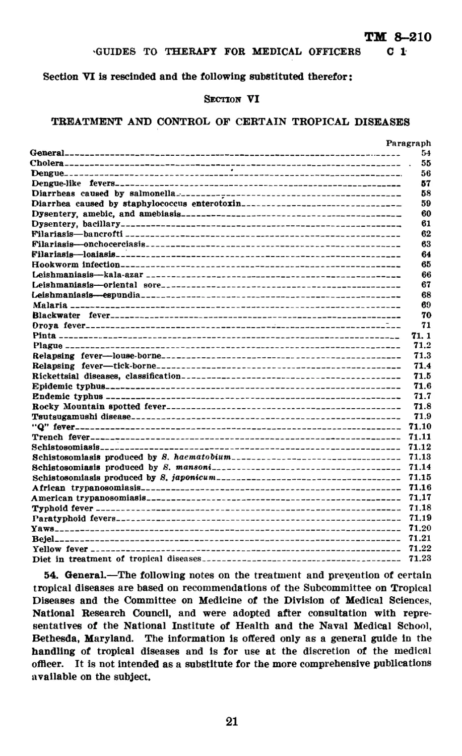
Section VI is rescinded and the following substituted therefor:
Section VI
TREATMENT AND CONTROL OF CERTAIN TROPICAL DISEASES
Paragraph
General---------------------------------------------------------------------- 54
Cholera---------------------------------------------------------------------- . 55
Dengue----------------------------------------------------------------------- 56
Dengue-like fevers------------------------------------------------------------- 57
Diarrheas caused by salmonella.-_______-_____________________________________ 58
Diarrhea caused by staphylococcus enterotoxin__________________________________ 59
Dysentery, amebic, and amebiasis_______________________________________________ 60
Dysentery, bacillary___________________________________________________________ 61
Filariasis—bancrofti----------------------------------------------------------- 62
Filariasis—onchocerciasis------------------------------------------------------ 63
Filariasis—loaiasis—----------------------------------------------------------- 64
Hookworm infection------------------------------------------------------------- 65
Leishmaniasis—kala-azar________________________________________________________ 66
Leishmaniasis—oriental sore____________________________________________________ 67
Leishmaniasis—espundia_________________________________________________________ 68
Malaria________________________________________________________________________ 69
Blackwater fever_______________________________________________________________ 70
Oroya fever---------------------------------------:----------------------l— 71
Pinta------------------------------------------------------------------------ 71. 1
Plague----------------------------------------------------------------------- 71.2
Relapsing fever—louse-borne-------------------------------------------------- 71.3
Relapsing fever—tick-borne___________________________________________________ 71.4
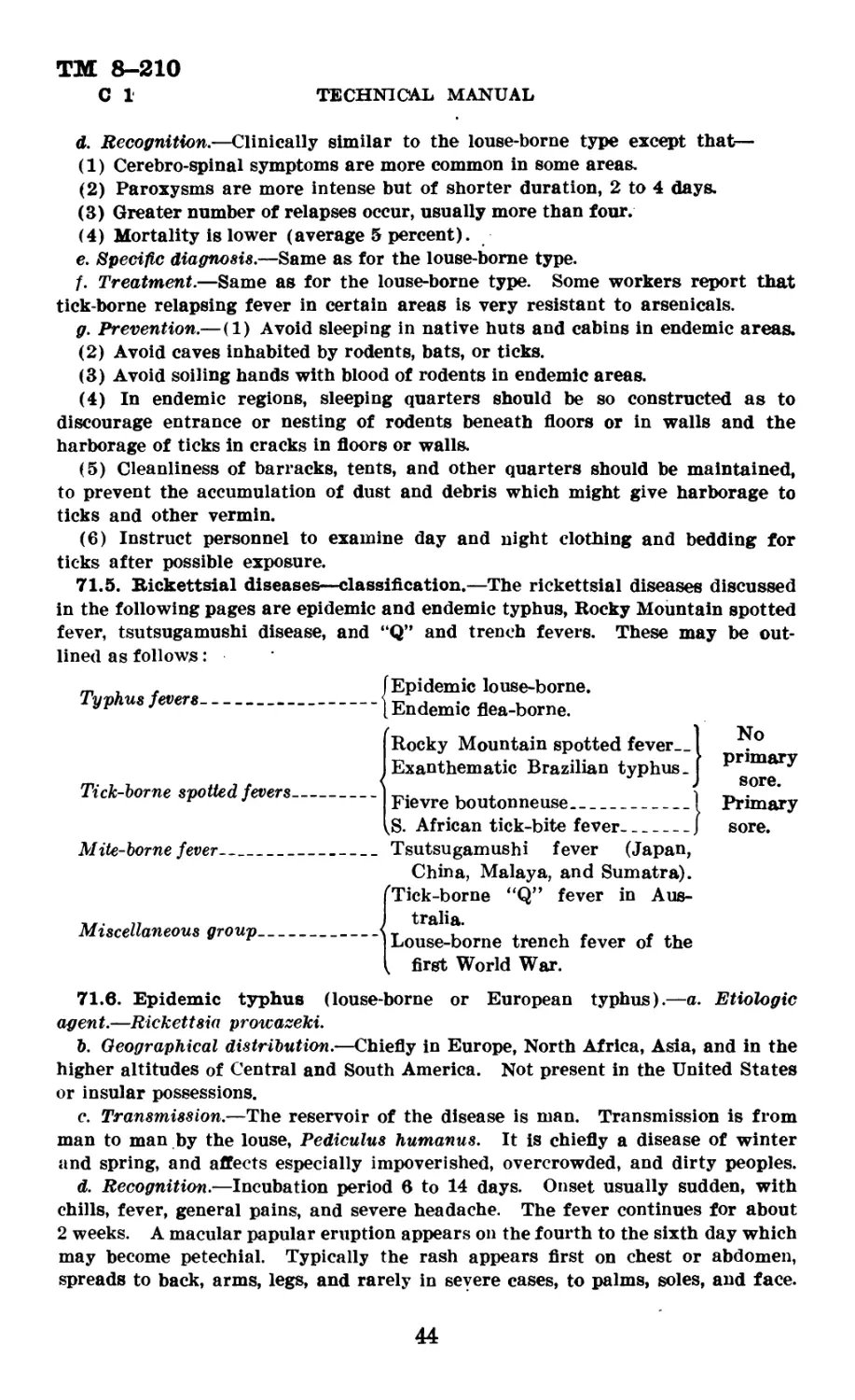
Rickettsial diseases, classification----------------------------------------- 71.5
Epidemic typhus-------------------------------------------------------------- 71.6
Endemic typhus_______________________________________________________________ 71.7
Rocky Mountain spotted fever------------------------------------------------- 71.8
Tsutsugamushi disease________________________________________________________ 71.9
“Q” fever------------------------------------------------------------------- 71.10
Trench fever---------------------------------------------------------------- 71.11
Schistosomiasis_____________________________________________________________ 71.12
Schistosomiasis produced by S. haematobium---------------------------------- 71.13
Schistosomiasis produced by 8. mansoni-------------------------------------- 71.14
Schistosomiasis produced by S. japonicum------------------------------------ 71.15
African trypanosomiasis----------------------------------------------------- 71.16
American trypanosomiasis---------------------------------------------------- 71.17
Typhoid fever--------------------------------------------------------------- 71.18
Paratyphoid fevers---------------------------------------------------------- 71.19
Yaws________________________________________________________________________ 71.20
Bejel_______________________________________________________________________ 71.21
Yellow fever---------------------------------------------------------------- 71.22
Diet in treatment of tropical diseases______________________________________ 71.23
54. General.—The following notes on the treatment and prevention of certain
tropical diseases are based on recommendations of the Subcommittee on Tropical
Diseases and the Committee on Medicine of the Division of Medical Sciences,
National Research Council, and were adopted after consultation with repre-
sentatives of the National Institute of Health and the Naval Medical School,
Bethesda, Maryland. The information is offered only as a general guide in the
handling of tropical diseases and is for use at the discretion of the medical
officer. It is not intended as a substitute for the more comprehensive publications
available on the subject.
21
ТМ 8-210
С 1 TECHNICAL MANUAL
55. Cholera.—a. Etiologic agent.—Vibrio comma.
b. Geographic distribution.—The disease is endemic in Asia. Since ancient
times many pandemics have originated in endemic centers in India, and have
spread over both tropical and temperate regions of the world.
c. Transmission.—Through the ingestion of food or drink contaminated with
feces containing V. comma. Flies are important vectors. Few patients remain
carriers of the vibrios for more than 10 days, but in epidemic areas there is also
a small percentage of healthy contact .carriers who may excrete vibrios for a
month or more.
d. Recognition.—Incubation usually 2 to 3 days. Onset usually sudden, with
profuse, painless “rice water” diarrhea and vomiting, shrinking of face and soft
tissues due to loss of body fluids, muscular cramps, suppression of urin, and
severe prostration. There are mild ambulatory cases and there is a fulminant
type (rare) in which death may occur before purging has begun.
e. Specific diagnosis.—By identification of V. comma in cultures of feces.
Fecal smears stained with carbol fuchsin diluted 1 to 10, showing the comma
forms with “fish in stream” appearance, are highly suggestive.
f. Treatment.—(1) Restoration of body fluids.—This is the most important
therapeutic objective and should be promptly and adequately attacked. Fluids
should be given liberally by mouth unless contra-indicated by vomiting or
nausea. It will usually be necessary to supplement oral administration by
parenteral injections. This may be accomplished by—
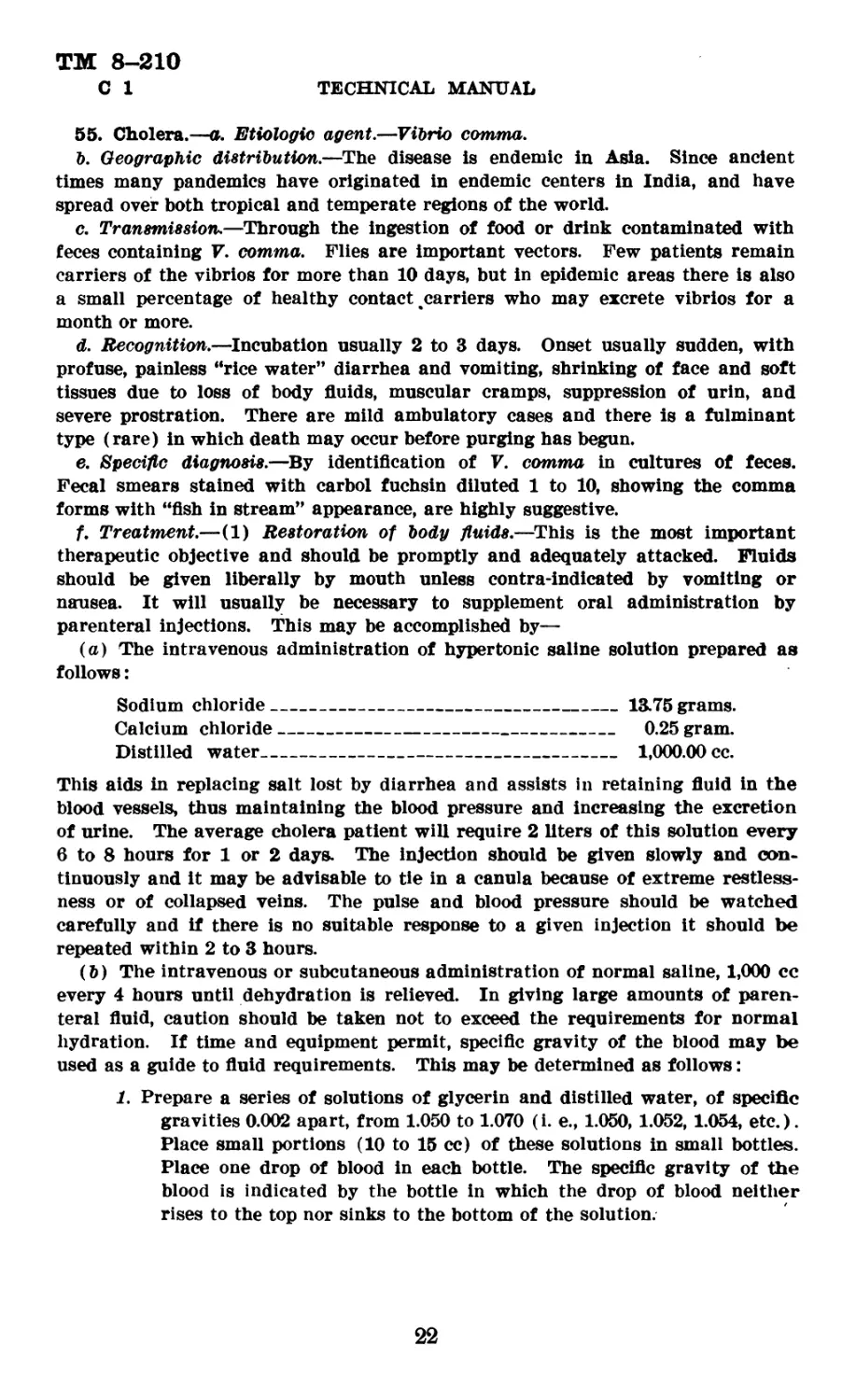
(a) The intravenous administration of hypertonic saline solution prepared as
follows:
Sodium chloride_____________________________________13.75 grams.
Calcium chloride------------------------------------ 0.25 gram.
Distilled water_____________________________________ 1,000.00 cc.
This aids in replacing salt lost by diarrhea and assists in retaining fluid in the
blood vessels, thus maintaining the blood pressure and increasing the excretion
of urine. The average cholera patient will require 2 liters of this solution every
6 to 8 hours for 1 or 2 days. The injection should be given slowly and con-
tinuously and it may be advisable to tie in a canula because of extreme restless-
ness or of collapsed veins. The pulse and blood pressure should be watched
carefully and if there is no suitable response to a given injection it should be
repeated within 2 to 3 hours.
(b) The intravenous or subcutaneous administration of normal saline, 1,000 cc
every 4 hours until dehydration is relieved. In giving large amounts of paren-
teral fluid, caution should be taken not to exceed the requirements for normal
hydration. If time and equipment permit, specific gravity of the blood may be
used as a guide to fluid requirements. This may be determined as follows:
1. Prepare a series of solutions of glycerin and distilled water, of specific
gravities 0.002 apart, from 1.050 to 1.070 (i. e., 1.050, 1.052, 1.054, etc.).
Place small portions (10 to 15 cc) of these solutions in small bottles.
Place one drop of blood in each bottle. The specific gravity of the
blood is indicated by the bottle in which the drop of blood neither
rises to the top nor sinks to the bottom of the solution.
22
ТМ 8-210
GUIDES ТО THERAPY FOR MEDICAL OFFICERS О 1
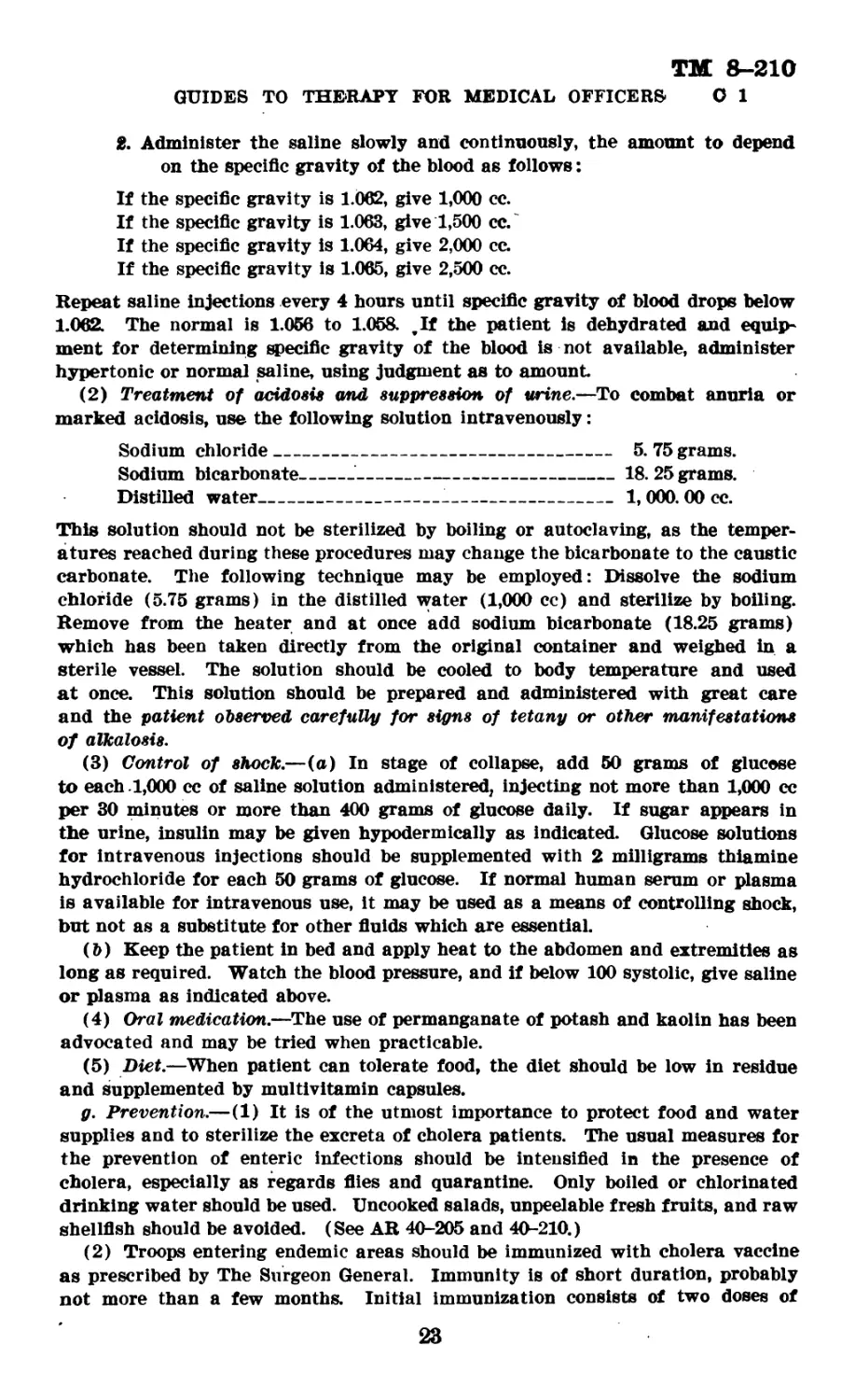
2. Administer the saline slowly and continuously, the amount to depend
on the specific gravity of the blood as follows:
If the specific gravity is 1.062, give 1,000 cc.
If the specific gravity is 1.063, give 1,500 cc/
If the specific gravity is 1.064, give 2,000 cc.
If the specific gravity is 1.065, give 2,500 cc.
Repeat saline injections every 4 hours until specific gravity of blood drops below
1.062. The normal is 1.056 to 1.058. wIf the patient is dehydrated and equip-
ment for determining specific gravity of the blood is not available, administer
hypertonic or normal saline, using judgment as to amount
(2) Treatment of acidosis and suppression of urine.—To combat anuria or
marked acidosis, use the following solution intravenously:
Sodium chloride________________________________________ 5. 75 grams.
Sodium bicarbonate-------------------------------------18. 25 grams.
Distilled water---------------------------------------- 1, 000. 00 cc.
This solution should not be sterilized by boiling or autoclaving, as the temper-
atures reached during these procedures may change the bicarbonate to the caustic
carbonate. The following technique may be employed: Dissolve the sodium
chloride (5.75 grams) in the distilled water (1,000 cc) and sterilize by boiling.
Remove from the heater and at once add sodium bicarbonate (18.25 grams)
which has been taken directly from the original container and weighed in a
sterile vessel. The solution should be cooled to body temperature and used
at once. This solution should be prepared and administered with great care
and the patient observed carefully for signs of tetany or other manifestations
of alkalosis.
(3) Control of shock.—(a) In stage of collapse, add 50 grams of glucose
to each .1,000 cc of saline solution administered; injecting not more than 1,000 cc
per 30 minutes or more than 400 grams of glucose daily. If sugar appears in
the urine, insulin may be given hypodermically as indicated. Glucose solutions
for intravenous injections should be supplemented with 2 milligrams thiamine
hydrochloride for each 50 grams of glucose. If normal human serum or plasma
is available for intravenous use, it may be used as a means of controlling shock,
but not as a substitute for other fluids which are essential.
(b) Keep the patient in bed and apply heat to the abdomen and extremities as
long as required. Watch the blood pressure, and if below 100 systolic, give saline
or plasma as indicated above.
(4) Oral medication.—The use of permanganate of potash and kaolin has been
advocated and may be tried when practicable.
(5) Diet.—When patient can tolerate food, the diet should be low in residue
and supplemented by multivitamin capsules.
g. Prevention.—(1) It is of the utmost importance to protect food and water
supplies and to sterilize the excreta of cholera patients. The usual measures for
the prevention of enteric infections should be intensified in the presence of
cholera, especially as regards flies and quarantine. Only boiled or chlorinated
drinking water should be used. Uncooked salads, unpeelable fresh fruits, and raw
shellfish should be avoided. (See AR 40-205 and 40-210.)
(2) Troops entering endemic areas should be immunized with cholera vaccine
as prescribed by The Surgeon General. Immunity is of short duration, probably
not more than a few months. Initial immunization consists of two doses of
23
ТМ 8-210
С 1
TECHNICAL MANUAL
cholera vaccine given subcutaneously with an interval of 7 to 10 days between
injections. The first dose is 0.5 cc, the second dose 1.0 cc. A stimulating dose
of 1 cc of vaccine should be administered every 4 to 0 months as long as danger
of infection is present. (See sec. VII.)
56. Dengue.—a. Etiologic agent.—The dengue virus.
b. Geographic distribution.—May occur in sudden epidemics of widespread
proportions in almost any part of the tropical or subtropical world, wherever the
vector, the Aedes mosquito, is found. Common in the West Indies, the Pacific
Islands, and in the countries about the China Sea and the Mediterranean.
c. Transmission.—Aedes aegypti is the principal vector; also A. albopictus
(Philippines), and possibly A. taeniorrhynchus (Florida). The patient is infec-
tive to the mosquito from a few hours before onset until the third or fourth day
of the disease. Infected mosquitoes become infective on the eighth to eleventh
day and remain infective for life.
d. Recognition.—After an incubation period lasting from 7 to 9 days there is
sudden onset of fever, “saddleback” in type, ranging from 102° F. to 105° F.
The pulse is slow in relation to the fever. There is intense postorbital aching
with sharp pains on eye movements. Extremely severe pains in joints and
muscles give this disease its name of “break-bone” fever. Leukopenia is present.
Commencing on the third to the fifth day, the symptoms abate. Two or 3 days
later there is a recurrence of fever and pain accompanied by a rash resembling
the eruption of measles or scarlet fever, beginning on hands and feet and spreading
to other parts. This secondary fever lasts from 1 to 2 days. Different epidemics
show great variations in the clinical picture of the disease. The secondary fever
and rash may be lacking. Lymph node enlargement is occasionally a prominent
symptom.
e. Specific diagnosis.—None.
f. Treatment.—Symptomatic.
g. Prevention.—Screening, bed nets, spray killing of mosquitoes, and other
anti-Aedee measures should be energetically prosecuted (see par. 71.11).
57. Dengue-like fevers.—There are several dengue-like febrile diseases in
various parts of the world, and some of these may be of importance to military
personnel.
a. Sandfly fever (Pappataci fever, Phlebotomus fever, 3-day fever).—This dis-
ease is prevalent in the countries bordering the Mediterranean and in China,
India, and Bast Africa. Recently it has been reported in northern Argentina.
It is caused by a virus and is transmitted by sandflies (Phlebotomus). P. pap-
patasii is the principal vector; others are P. minutes and P. pemiciosus. Clin-
ically the disease resembles dengue except for the short duration. (2 to 3 days)
and, commonly, the absence of a recurrent phase and of the rash. There is no
specific diagnosis. The treatment is symptomatic. Prevention is based chiefly
on measures against sandflies. Screening with the ordinary mosquito net does
not bar the small sandflies but when nets are sprayed with pyrethrum insecticides
the sandflies are repelled. Sandflies being poor flyers, breezes from electric fans
may prevent biting; upper floors provide some security.
b. Panama 6-day fever.—In the Canal Zone.
c. Van der Scheer 5-day fever.—In the Dutch Blast Indies.
d. Seven-day fever.—In India. Not to be confused with 7-day fever caused by
L. hebdomadis.
e. Bwamba fever.—In Uganda, Africa.
24
ТМ 8-210
GUIDES ТО THERAPY FOR MEDICAL OFFICERS С 1
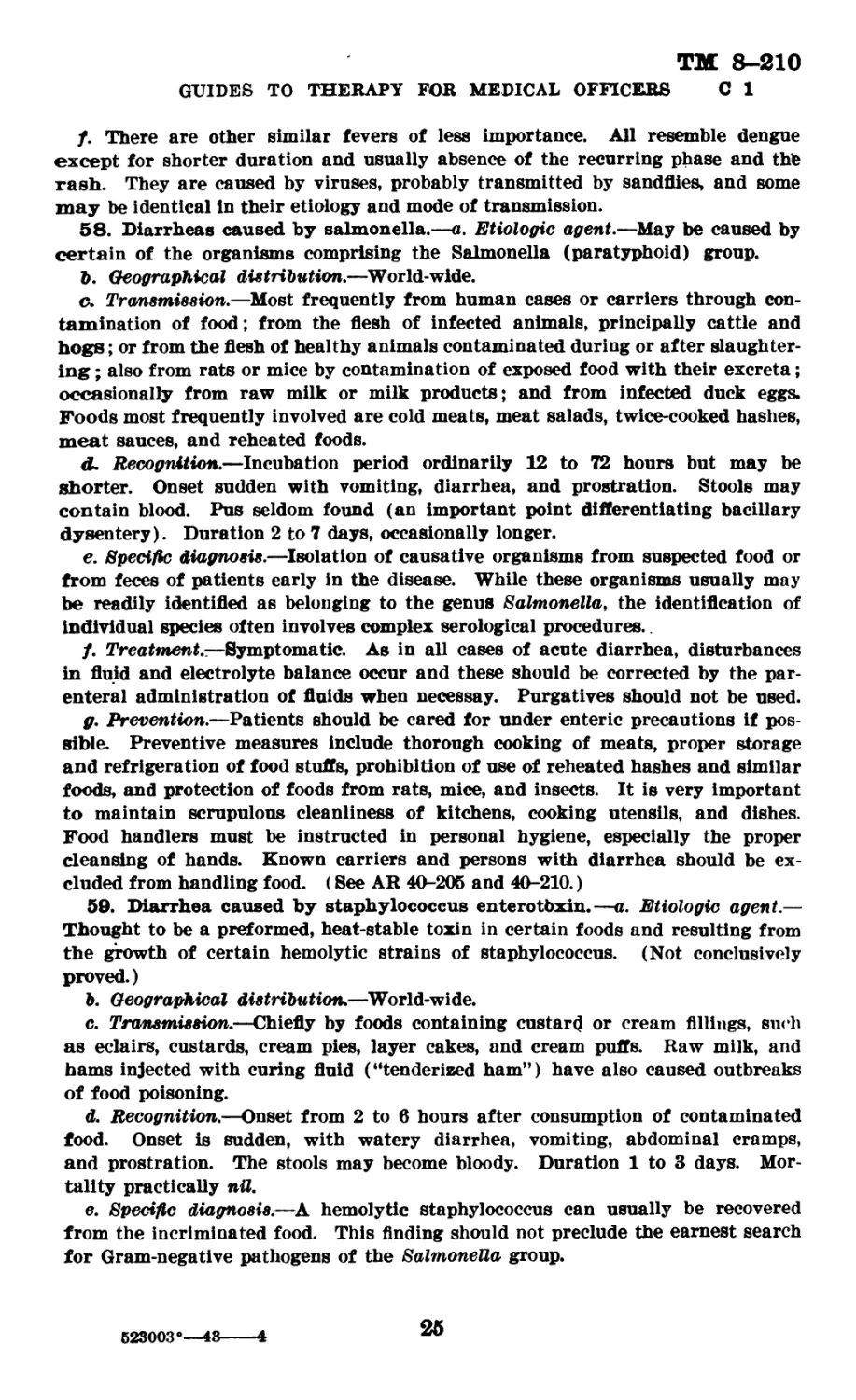
/. There are other similar fevers of less importance. All resemble dengue
except for shorter duration and usually absence of the recurring phase and the
rash. They are caused by viruses, probably transmitted by sandflies, and some
may be identical in their etiology and mode of transmission.
58. Diarrheas caused by salmonella.—a. Etiologic agent.—May be caused by
certain of the organisms comprising the Salmonella (paratyphoid) group.
b. Geographical distribution.—World-wide.
c. Transmission.—Most frequently from human cases or carriers through con-
tamination of food; from the flesh of infected animals, principally cattle and
hogs; or from the flesh of healthy animals contaminated during or after slaughter-
ing ; also from rats or mice by contamination of exposed food with their excreta;
occasionally from raw milk or milk products; and from infected duck eggs.
Foods most frequently involved are cold meats, meat salads, twice-cooked hashes,
meat sauces, and reheated foods.
Recognition.—Incubation period ordinarily 12 to 72 hours but may be
shorter. Onset sudden with vomiting, diarrhea, and prostration. Stools may
contain blood. Pus seldom found (an important point differentiating bacillary
dysentery). Duration 2 to 7 days, occasionally longer.
e. Specific diagnosis.—Isolation of causative organisms from suspected food or
from feces of patients early in the disease. While these organisms usually may
be readily identified as belonging to the genus Salmonella, the identification of
individual species often involves complex serological procedures..
f. Treatment.—Symptomatic. As in all cases of acute diarrhea, disturbances
in fluid and electrolyte balance occur and these should be corrected by the par-
enteral administration of fluids when necessay. Purgatives should not be used.
g. Prevention.—Patients should be cared for under enteric precautions if pos-
sible. Preventive measures include thorough cooking of meats, proper storage
and refrigeration of food stuffs, prohibition of use of reheated hashes and similar
foods, and protection of foods from rats, mice, and insects. It is very important
to maintain scrupulous cleanliness of kitchens, cooking utensils, and dishes.
Food handlers must be instructed in personal hygiene, especially the proper
cleansing of hands. Known carriers and persons with diarrhea should be ex-
cluded from handling food. (See AR 40-206 and 40-210.)
59. Diarrhea caused by staphylococcus enterotbxin.—a. Etiologic agent.—
Thought to be a preformed, heat-stable toxin in certain foods and resulting from
the growth of certain hemolytic strains of staphylococcus. (Not conclusively
proved.)
b. Geographical distribution.—World-wide.
c. Transmission.—Chiefly by foods containing custard or cream fillings, such
as eclairs, custards, cream pies, layer cakes, and cream puffs. Raw milk, and
bams injected with curing fluid (“tenderized ham”) have also caused outbreaks
of food poisoning.
d. Recognition.—Onset from 2 to 6 hours after consumption of contaminated
food. Onset is sudden, with watery diarrhea, vomiting, abdominal cramps,
and prostration. The stools may become bloody. Duration 1 to 3 days. Mor-
tality practically nil.
e. Specific diagnosis.—A hemolytic staphylococcus can usually be recovered
from the incriminated food. This finding should not preclude the earnest search
for Gram-negative pathogens of the Salmonella group.
623003°—43----4
25
ТМ 8-210
С 1
TECHNICAL MANUAL
f. Treatment.—Symptomatic, with complete rest in bed, fluid diet until sub-
sidence of symptoms, parenteral fluids in dehydrated patients. Avoid use of
purgatives.
g. Prevention.—Absolute cleanliness of kitchens and all cooking utensils; thor-
ough cooking of custards and other fillings; and proper refrigeration of com-
monly involved foods before and after cooking, are important. Avoid use of
filled pastries in the Tropics and during warm seasons elsewhere. Use only pas-
teurized milk. Food handlers having furuncles or infected lesions on the hands,
arms, or face should be removed from duty until their lesions are healed. Isola-
tion of patients is unnecessary, since case-to-case transmission does not occur.
(See AR 40-205 and 40-210.)
60. Dysentery, amebic, and amebiasis.—a. Etiologic agent.—Endamoeba
histolytica.
b. Geographical distribution.—The distribution of this disease probably is
world-wide, but infections are much more common in the Tropics and sub-Tropics
than in temperate regions. Even in the latter regions the incidence may be high
in localities where sanitary conditions are poor.
c. Transmission.—Through ingestion of food or drink contaminated with feces
containing cysts (and possibly trophozoites) of the causative organism.
d. Recognition.—The clinical picture varies. Some common types are:
(1) Acute amebic dysentery.—Onset with abdominal discomfort and diarrhea,
gradually increasing in severity until there may be 20 to 40 evacuations daily,
with blood and mucus in the stools. Fever and toxemia generally mild.
(2) Chronic amebic dysentery.—Usually history of acute attack followed by
relapses, with symptoms similar to those of acute attacks but milder.
(3) Amebic colitis without dysentery.—Symptoms diverse in character and
severity. Diarrhea may alternate with constipation. Symptoms may be those of
“irritable colon” and may simulate appendicitis or other surgical conditions.
(4) Amebic abscess of liver.—Usually but not always history of dysentery.
Onset and course of abscess insidious; pain in liver and right scapula; acute
tenderness on percussion over abscess; septic type of fever; leukocytosis.
e. Specific diagnosis.—Depends on demonstrating cysts or motile forms of
E. histolytica in feces, or the motile forms in abscess contents. In the absence
of laboratory facilities a presumptive diagnosis can be made on gross appearance
of dysentery stool (blood and glary mucous, not well mixed, and absence of pus)
and gross appearance of liver abscess contents (chocolate colored fluid). A
therapeutic test, useful when no laboratory facilities are available, is the striking
and sometimes complete symptomatic relief occurring in 24 to 48 hours when
a patient with acute amebic dysentery is given emetine hydrochloride 0.06 gram
(1 grain) subcutaneously on 2 successive days.
f. Treatment.—(1) For acute or chronic amebic dysentery.—(a) General care.—
For acute cases, keep patient in bed on liquid diet, broth at first, later add milk,
eggs, and custards until acute symptoms subside, then soft, low-residue diet.
Supplement diet with four multivitamin capsules daily. Resume full general diet
gradually during convalescence.
(b) Emetine hydrochloride intramuscularly, 0.03 gram (% grain) twice a day
or 0.06 gram (1 grain) once a day for 4 to 6 days. Toxic effects: lowering of
blood pressure, vomiting (controlled by sedative), acute myocardial degeneration,
peripheral neuritis (generally from more prolonged treatment than above). Stop
26
ТМ 8-210
GUIDES ТО THERAPY FOR MEDICAI, OFFICERS С 1
/
emetine as soon as the dysenteric symtoms have subsided. Concurrently with the
administration of emetine give—
(c) Carbarsone, 0.25 gram (4 grains) by mouth three times a day for 7 days.
Toxic symptoms (rare)—abdominal distress, nausea, vomiting, exfoliative der-
matitis (very rare). Follow carbarsone by—
(d) Vioform, 0.25 gram (4 grains) by mouth three times a day for 7 days.
Toxic symptoms—none. Or (instead of vioform) give—
(e) Diodoquin, 0.6 gram (9 grains) by mouth three times a day for 7 days.
Toxic symptoms—none. If neither vioform nor diodoquin is available, give
chiniofon, 1 gram (15 grains) by mouth three times a day for 7 days. Toxic
symptoms—watery diarrhea may occur in ambulatory patients. ,
(f) If treatment is ineffective because of lesions in the lower colon or rectum,
give carbarsone or chiniofon by rectum as follows: Carbarsone 2.0 grams (30
grains) dissolved in 200 cc of 2 percent sodium bicarbonate solution. (Carbarsone
is insoluble in water.) Give as retention enema at night, following a cleansing
enema of 2 percent sodium bicarbonate solution. Retention may be aided by a
mild sedative. Give on five consecutive nights, but if it proves irritating, reduce
to alternate nights. Or chiniofon (instead of carbarsone) by rectum, 4.0 grams
(60 grains) in 200 cc of sterile water, after a cleansing enema of water. Vioform
and diodoquin are too insoluble and too Irritating to administer by rectum.
(2) For amebic hepatitis without liver abscess.—Emetine hydrochloride as
above, but continue for 8 days. If evidence of hepatitis persists after 8 days,
suspect liver abscess, and if indicated, aspirate (see (3) below). If practicable,
check effect of emetine on heart muscle by electrocardiograph before treatment,
and daily from fifth day (changes in Q-R-S complex and inversion of T wave).
Use chiniofon as above, by mouth, for intestinal infection which probably exists.
(Carbarsone may be toxic in some cases of hepatitis.)
(3) For amebic liver abscess.—Emetine hydrochloride as above. After 2 to 4
days of emetine treatment the abscess may be drained by aspiration, repeatedly
if necessary. Open drainage should be avoided. After completion of the emetine
treatment, use chiniofon or vioform as above for the intestinal infection which
probably exists.
(4) For amebic carriers without symptoms.—(a) Carbarsone 0.25 gram (4
grains) by mouth three times a day for 7 days. Toxic symptoms (rare)—see
(1) (c) above.
(&) After completion of carbarsone treatment, give vioform 0.25 gram (4
grains) by mouth three times a day for 7 days. Toxic symptoms—none. Or
diodoquin 0.6 gram (9 grains) by mouth three times a day for 7 days. Toxic
symptoms—none. If neither vioform nor diodoquin is available, give chiniofon
1 gram (15 grains) by mouth three times a day for 7 days. Toxic symptoms—
watery diarrhea may occur in ambulatory patients.
(c) Diet soft, if possible. Avoid excessive activity during treatment. Give
two to four multivitamin capsules daily as supplement.
ff. Prevention.—Drinking water can be made safe by approved methods of sand
filtration, but not by the usual method of chlorination alone (superchlorination
is required for the destruction of cysts). If an adequately treated water supply
is not available, drinking water should be boiled. Troops should be particularly
cautioned not to drink raw water from untreated sources. Raw fruits and vege-
tables should be avoided in areas where human excreta are used for fertilizer, or
where they are washed in contaminated ditch water. Known carriers or persons
27
ТМ 8-210
С 1
TECHNICAL MANUAL
with any intestinal disturbance should be excluded from handling food. Flies and
cockroaches should be excluded from access to food or drink. (See AR 40-205
and 40-210.)
61. Dysentery, bacillary.—a. Etiologic agent,—The dysentery bacilli (genus
Shigella).
b. Geographical distribution.—Bacillary dysentery exists throughout the world ;
especially common in unsanitary localities. Its incidence is highest in tropical
and subtropical regions. Its control is a matter of special importance to armies
in the field.
c. Transmission.—Through the ingestion of food or drink contaminated with
the feces of cases or carriers of dysentery bacilli.
d. Recognition.—Onset sudden, with fever and abdominal cramps soon followed
by passage of loose, yellow or green, watery stools which change to characteristic
mucopurulent bloody stools. Tenesmus is a prominent symptom. Toxemia and
dehydration are marked in severe cases.
e. Specific diagnosis.—Depends on identification of the etiologic agent in stool
cultures.
f. Treatment.—(1) Chemotherapy.—(a) Acute bacillary dysentery.—Sulfagua-
nadine is the drug of choice. The initial dose is 3.5 grams (52*6 grains) followed
by 3.5 grams (52*6 grains) every 4 hours, day and night, until the number of
stools per day is reduced to five or less. Then shift to a maintenance dose of 3.5
grams (52% grains) every 8 hours, day and night, and continue until the stools
have been normal for 96 hours. If improvement is not noted in 7 days, discontinue
the drug.
(&) Chronic bacillary dysentery.—Administer 3.5 grams (52% grains) of sul-
faguanadine every 8 hours, day and night. The duration of chemotherapy should
not exceed 2 weeks.
(c) If sulfa guanadine is not available, use sulf a thiazole or sulfadiazine, initial
dose 4 grams (60 grains), then 1 gram (15 grains) every 4 hours. If improvement
is not noted in 7 days, discontinue the drug. These drugs are more toxic than
sulfaguanadine. Sufficient fluids must be administered to insure a daily urinary
output of at least 1,500 cc.
(2) Supportive treatment.—(a) Complete rest in bed.
(ft) For mild and moderately severe cases without dehydration or severe
toxemia, the fluid intake should be 3,000 cc or more every 24 hours.
(c) For acute fulminant cases, give intravenous glucose, 50 grams in physiologi-
cal salt solution 1,000 cc, sufficient to maintain a daily urinary output of 1,000 cc
or more. Glucose solutions. for intravenous injection should be supplemented
with 2 milligrams of thiaminehydrochloride for each 50 grams of glucose.
(d) Diet should be fluid, or free from residue.
(e) Purgation is not recommended. High enemas and colonic irrigations are
also not recommended.
(3) Serum therapy.—Monovalent Shiga antitoxin may be of some value in ful-
minating and markedly toxic cases of Shiga dysentery but it should never
preclude the use of sulfaguanadine as described above. Patients should be tested
for sensitivity before administering serum. The dose of antitoxin is from 40
to 80 cc given intravenously, twice daily, until temperature falls to normal.
The antitoxin should be diluted in 500 cc of normal saline and given very slowly.
g. Prevention.—(1) General sanitary measures.—Bacillary dysentery is pre-
vented by the same general sanitary measures employed for preventing other
28
ТМ 8-210
GUIDES ТО THERAPY FOR MEDICAL OFFICERS 0 1
enteric infections. These include meticulous control of milk, water, and food
supplies, proper treatment and disposal of sewage and garbage, and protection of
food from flies, and from infectious food handlers. The usual precautions
should be taken as regards isolation of patients and sterilisation of their feces,
clothes, dishes, and other fomites (See AR 40-205 and 40-210).
(2) Vaccination.—This has been widely practiced but it is not recommended,
as its value has never been satisfactorily demonstrated.
62. Filariasis—bancrofti.—a. Etiologic agent.—Wuchereria bancrofti. There
is also a sheathed microfilaria of nocturnal periodicity known as Microfilaria
malaya which is said to be a distinct species.
b. Geographical distribution.—This infection is indigenous in practically all
tropical regions of the world. In the Western Hemisphere it is endemic in Central
America, South America (Colombia, Venezuela, French, Dutch, and British Guiana,
and northern Brazil), and the West Indies. In the United States it has been
endemic near Charleston, South Carolina.
c. Transmission.—The adult worms live in the lymphatic system of infected
humans, releasing larvae (microfilariae) into the lymph and blood streams. A
wide variety of night-biting mosquitoes, including some Anopheles, serve as inter-
mediate hosts. The more important ones are Culex quinquefasciatus in many
parts of the world, Culex pipiens in China, Mansonioides species in India, and
Aedes variegatus in the Pacific Islands. In mosquitoes, the microfilariae, during
a period of about 10 days, develop into infective larvae which migrate to the
mouth parts, escape when the mosquito bites, penetrate the skin, and start a new
infection.
d. Recognition.—In many cases there are no apparent clinical manifestations.
When symptoms develop they are due not directly to the filariae but to the blockage
of the lymphatic vessels by inflammation and by fibrotic changes around the para-
sites. After an indefinite, prolonged incubation period, symptoms may be
produced by the invasion of lymphatic vessels, most often those of the spermatic
cord and groin. The initial acute attack is characterized by lymphangitis, accom-
panied by usual local signs and fever, and may persist for several days. These
attacks recur, and chronic elephantoid manifestations due to blockage of lymph
channels tend to develop in the lower extremities and more frequently the scrotum
and groin. Hydrocele, lymphatic varicocele, epididymitis, and chyluria also occur.
Chyluria may be a prominent symptom after involvement of lymph channels by the
parasite. The urine is cloudy, due to the presence of lipoid substances which are
soluble in ether. On standing, a clot forms in the urine and a surface pellicle is
usually present. The urine also often contains blood, due to rupture of a small
vessel into the lymphatic varix which is responsible for chyluria. In these cir-
cumstances the night specimens of urine may contain microfilariae.
e. Specific diagnosis.—Depends on finding the microfilariae in blood films.
Thick smears may be taken between 9 PM and midnight and stained with
Mayer’s haemalum or with Giemsa’s or Wright’s stain. Fresh coverslip prepara-
tions sometimes reveal the motile microfilariae.
f. Treatment.—There is no specific treatment. For acute lymphangitis usually
caused by hemolytic streptococci, sulfadiazine by mouth is recommended. The
initial dose is 4 grams (60 grains) ; subsequent doses 1 gram (15 grains) every
6 hours day and night until the infection is under control. Surgical measures
may be indicated for elephantiasis.
g. Prevention.—The prevention of filariasis depends entirely on avoidance of
the bites of infective mosquitoes. This depends upon such individual pre-
29
ТМ 8-210
С 1
TECHNICAL MANUAL
ventive measures as the proper use of bed nets, repellents, and protective cloth-
ing; and upon adequate screening (18-mesh), spray-killing of adult mosquitoes,
and eliminating or treating the breeding places of mosquito vectors.
63. Filariasis—onchocerciasis.—a. Etiologic agent.—Onchocerca volvulus.
b. Geographical distribution.—Africa, especially in Congo River basin; western
slope of Guatemala at altitudes from 2,000 to 6,000 feet; southern Mexico, in
States of Chiapas, Oaxaca, Guerrero, and Yucatan.
c. Transmission.—The adult filariae (O. volvulus) live in subcutaneous nodules,
releasing microfilariae which migrate into adjacent lymphatics and other tissues.
The embryos are rarely found in smears from peripheral blood. Various species
of black gnats (Slmulium damnosum, S. metalUcum, and others) serve as inter-
mediate hosts, after ingesting microfilaria while feeding on an infected person.
In the gnat the microfilariae develop in 6 days into infective larvae which
migrate to the mouth parts of the insect, from which they escape while the
gnat is feeding. Man is infected by introduction of the parasite into the
wound made by the insect. The parasite then develops to the adult stage in
from 2 months to a year and is found in the subcutaneous tumors.
d. Recognition.— (1) Formation of tender, freely movable and easily enucle-
ated subcutaneous nodules (diameter 6 to 30 millimeter) in scalp, trunk, axilla,
popliteal space, and vicinity of the elbow.
(2) In about 5 percent of cases, ocular complications follow migration of
microfilariae into the eye, producing keratitis, Iritis, and conjunctivitis. Photo-
phobia, zerosis, impairment of vision, sometimes complete blindness, may follow.
(3) Generalized lichenoid dermatitis may occur, with intense itching, par-
ticularly at night.
e. Specific diagnosis.—(1) Identification of microfilariae in fluid aspirated
from center of nodules, or in tissue of an excised nodule, or in a biopsy specimen
of adjacent tissues.
(2) Demonstration of the threadlike adult worm in excised nodules.
f. Treatment.—Enucleation of nodules.
g. Prevention.—The transmitting gnats (Simulium) breed in streams, the
larvae and pupae being attached to rocks or plants beneath the surface. No
method of control of breeding has been devised. The adults bite only during
daylight and may be deterred by head nets, protective clothing, and chemical
repellents applied to the skin. Smoke smudges and pyrethrum sprays may
repel the gnats from the vicinity of personnel in encampments and buildings.
64. Filariasis—loaiasis.—a. Etiologic agent.—Loa loa.
b. Geographical distribution.—Tropical West Africa, especially Congo River
basin.
c. Transmission.—Adult worms living in subcutaneous tissue liberate micro-
filariae which enter blood stream. Transmitted to man by blood-sucking mango
flies (Chrysops), which bite only in daylight, preferably in shade.
d. Recognition.—Usually no symptoms except when adult worms migrate
under the ocular conjunctiva or in the skin. This may be accompanied by local
pain, swelling, and inflammation. Subcutaneous migratory swellings (Calabar
swellings) up to size of hen’s egg, may appear in the skin. Their relation to the
infection is not completely understood.
e. Specific diagnosis.—Demonstration of the microfilariae in the blood as for
W. bancrofti, except that periodicity of Loa loa is diurnal. Sometimes the adult
worm may be identified after extraction.
/. Treatment.—Surgical removal of the worm. There is no specific drug.
30
ТМ 8-210
GUIDES ТО THERAPY FOR MEDICAL OFFICERS С 1
y. Prevention,—Protection from bite of Chrysops, by protective clothing, nets,
and repellents.
65. Hookworm infection.—a. Etiologic agent.—Necator americanus is almost
exclusively the hookworm of man in the Western Hemisphere. Aneylostoma duo-
denote and Necator are both prevalent in the Eastern Hemisphere. The dog
bookworms Aneylostoma braziliense and A. caninum infect the skin of man in
their larval stage, causing the disease known as “creeping eruption.” (This
infection is acquired usually by lying on a sandy place, such as a beach, which
has been contaminated by dog excreta.)
b. Geographical distribution.—In the United States hookworm infection, occurs
in the South from Virginia and Kentucky to eastern Texas. The* rural population
of some counties in southern South Carolina, Georgia, Alabama, and Mississippi,
and in northern Florida, still show an incidence of 50 percent or more. Hookworm
infection is prevalent in the Tropics wherever the climate is moist and sanitary
disposal of feces is not practiced.
a Transmission.—The hookworms affecting man live in the small intestine and
suck blood from the mucosa. Eggs are passed in the feces and hatch in soiL
Infective larvae develop in a few days, penetrate the skin of man, and reach the
intestine via lungs, trachea, and esophagus.
d. Recognition.—In well nourished adults it requires 50 or more worms to
produce clinical symptoms such as lassitude, fati‘gue, weakness, and pallor.
“Ground itch” is often an early symptom. There may be secondary anemia, slight
or moderate eosinophilia, and edema.
e. Specific diagnosis.—Identification of the characteristic ova in the feces. The
ova of the two common species cannot be differentiated morphologically.
ft Treatment.—(1) General.—Give a light meal, preferably free from fats, the
night before instituting treatment No preliminary purge is necessary unless
constipation exists. Give drug in the morning on an empty stomach. Avoid food
for 2 hours after treatment, and alcohol for 24 hours before and after treat-
ment. A saline purge should be given the following day if bowels have not
moved since treatment.
(2) Drugs.—(a) In the absence of Ascaris infection, use tetrachlorethylene:
adult dose 3 cc in hard gelatin capsules, followed in 2 hours by a saline purge.
Drug sometimes causes dizziness and drowsiness, but no case of fatal toxicity
has been reported.
(b) In the presence of Ascaris infection, use hexylresorcinol followed by
tetrachlorethylene.
Hexyl resorcinol Crystoids (caprocol) : adult dose 1 gram (15 grains) on empty
stomach in morning. Avoid all food for 4 hours after treatment (very impor-
tamt). Toxic symptoms—none, except burning of mouth if crystoid is chewed.
This treatment will usually remove all ascaris worms and about 50 percent of
hookworms. It should be followed after 3 days by treatment with tetrachlor-
ethylene to remove the remainder of the hookworms.
(c) Iron should be administered to all hookworm cases showing anemia. An
effective preparation is ferrous sulphate (desiccated) in capsules; adult dose
0.35 gram (5 grains) three times a day after meals.
(3) Diet.—Should be rich in iron and vitamins.
(4) Post-treatment stool examination.—Should be made 1 week after treat-
ment. If eggs are found, treatment should be repeated until cure is obtained.
31
ТМ 8-210
С 1
TECHNICAL MANUAL
(5) Treatment of “creeping eruption.”—Use one of the following methods.
Repeated applications may be necessary to obtain cure.
(a) Saturate cotton with ethyl acetate; apply to area just beyond advancing
edge of skin lesion, and cover with adhesive tape for 24 hours.
(&) Freeze lightly with ethyl chloride spray or dry ice on area 1 inch in
width just beyond advancing edge of skin lesion.
g. Prevention.—Provide for sanitary disposal of excreta and protect the skin,
especially the feet, against contact with soil contaminated with hookworm
larvae.
66. Leishmaniasis—kala-azar (visceral leishmaniasis).—a. Etiologic agent.—
Leishmania donovani.
b. Geographical distribution.—The disease is widespread, and occurs in Medi-
terranean countries, south Russia, India, China, Manchuria, Abyssinia, Sudan,
northern and eastern Brazil, Chaco Region of Argentina, Paraguay-Brazil border
(Matto Grosso), and northern Bolivia (Yungas).
c. Transmission.—The available evidence suggests that the disease is trans-
mitted by the bites of infected sandflies of the genus Phlebotomus and possibly
through the ingestion of food or drink contaminated with L. donovani, or by
contact. • Dogs and other lower animals are susceptible to infection.
d. Recognition.—The principal symptoms are:
(1) An irregular, recurring fever over a period of weeks, sometimes having
a double daily rise.
(2) Progressive enlargement of spleen, later of the liver.
(3) Anemia, leukopenia, and emaciation.
(4) Dysentery or diarrhea.
e. Specific diagnosis.—By demonstration of L. donovani either in smears or
cultures of the peripheral blood or by material obtained by spleen, liver, or bone
marrow puncture.
f. Treatment—(1) Pentavalent antimony compounds.—A number of effective
pentavalent antimony compounds have been developed, including neostam (sti-
bamine glucoside), solustibosan (Bayer 561), and neostibosan (Bayer 693).
The last named is least toxic and most effective, and its use is recommended. It
is given intravenously, 15 doses on alternate days, the first dose 0.2 gram and
subsequent doses 0.3 gram. Use this drug as a 10 percent solution freshly
prepared from sterile distilled water. The dosage and method of administration
of Neostam is the same as for Neostibosan. (The trivalent compounds of anti-
mony, such as fouadin (neoantimosan), are relatively ineffective in visceral
leishmaniasis.)
(2) Potassium antimony tartrate (USP).—A 2 percent freshly prepared solu-
tion intravenously on alternate days. Initial dose 2.0 cc (0.04 gram), and each
succeeding dose increased by 1.0 cc until 5.0 cc are being given. Continue until
a total of 40 or more doses have been given. If toxic symptoms develop, reduce
dose. Toxic symptoms of antimony drugs: coughing immediately after adminis-
tration (not important) ; nausea, vomiting, dizziness, collapse.
(3) In the Egyptian Sudan and some adjacent areas leishmaniasis is very resist-
ant to treatment with antimony preparations. British investigators have recently
reported that antimony resistant cases show a good response to intravenous
injections of stilbamidine isethionate (4:4 diamidine stilbene isethionate). The
drug must be used in a freshly prepared solution in 10 cc of sterile distilled water
32
ТМ 8-210
GUIDES ТО THERAPY FOR MEDICAL OFFICERS С I1
without heating. The water must be neutral or very slightly acid (pH 6.8-7.0).
Old solutions may cause severe late toxic effects on the liver, kidneys, or pancreas,
even after completion of a course of treatment. The dose is 1.0 milligram
(0.001 gram) per kilogram of body weight (maximum adult dose 0.15 gram)
every other day for 15 injections. Repeat this course of treatment only if cure
is not obtained and after interval of 1 month. Some cases show fall of blood
pressure and syncope after first or second injection. This may be prevented or
relieved by injection of a small dose of epinephrin. The administration of cal*
cium and glucose during treatment may protect the liver from damage.
Norn.—Stilbamadine isethionate is not available for general distribution.
(4) Diet.—The diet may contain whatever foods the patient can tolerate. It
probably is wise to supplement it with vitamin В complex and, if it contains less
than 6 ounces of citrus fruit juice, ascorbic acid should be given in doses of 50
milligrams per day.
g. Prevention.—In endemic areas antisandfly measures should be used, such
as elimination of breeding places and the use of fine mesh bed nets, insect repel-
lents, and insecticide sprays. Avoid unnecessary contact with native dogs. Segre-
gate and treat effectively all cases of the malady.
67. Leishmaniasis—oriental sore (Old World cutaneous leishmaniasis).—
a. Etiologic agent.—Leishmania tropica.
b. Geographical distribution.—Apparently this disease does not occur com-
monly in localities where kala-azar is present. It occurs in Mediterranean coun-
tries, central Asia, India, China (Hunan), southern Russia, and many parts of
Africa.
c. Transmission.—As in the case of kala-azar, sandflies of the genus Phleboto-
mus are suspected as vectors. As the disease can be transferred by inoculation,
the possibility of contact infect must also be considered.
d. Recognition.—The disease begins as a small itching papule on exposed sites.
The lesion gradually extends and ulcerates. Lesions may be multiple, but there
is no general dissemination.
e. Specific diagnosis.—The diagnosis depends on the demonstration of L. tropica
in stained films or cultures of material aspirated from the indurated zone sur-
rounding the ulcer.
f. Treatment.—Local treatment may be tried first, consisting of topical appli-
cations of dry ice or injection of 2 cc of 1 percent solution of berberine sulfate
into the edges of the sores. И these measures are not effective, intravenous
injections of neostibosan may be tried, using the same dosage as for visceral
leishmaniasis.
g. Prevention.—Take the necessary precautions to avoid the transfer of infec-
tious materials by insects or contact.
68. Leishmaniasis—espundia (American mucocutaneous leishmaniasis).—
a. Etiologic agent.—Leishmania brasiliensis.
b. Geographical distribution.—Mexico, Central America, South America (Argen-
tina, Bolivia, Brazil, Colombia, Ecuador, the Guianas, Paraguay, Peru, and Venez-
uela). The disease is said to be common in forest regions among collectors of
chicle gum and rubber. Has been encountered in American soldiers in Panama,
causing chronic ulcerations of the skin.
c. Transmission.—The exact method is unknown, but it is suspected that sand-
flies of the genus Phlebotomus may act as vectors, and also that contact infections
may occur.
523003*—43---- 4 88
TM 3—210
с 1
TECHNICAL MANUAL
d. Recognition.—Commences as small papule, on arms or often on margins of
ears; extends and may suppurate. Later, ulcers appear around margins of nose
and mouth; these may extend and cause extensive destruction of the tissue in naso-
oral region. There is no general dissemination. If untreated, death may occur
from sepsis and exhaustion.
e. Specific diagnosis.—The diagnosis depends on demonstration of L. brasiMensit
in cultures or smears of material obtained by puncture of the edge of the initial
ulcer or in material from nodules or ulcerations in the mucous membranes.
f. Treatment.—The treatment of the initial lesion is the same as that outlined for
oriental sore above. During the stage of mucous membrane involvement, use the
treatment recommended for kala-azar.
g. Prevention.—Use methods similar to those advocated above for the control of
kala-azar and oriental sore.
69. Malaria.—a. Etiologic agents.—Plasmodium vivax, causing benign tertian
malaria; P. falciparum, causing malignant tertian (aestivo-autumnal) malaria;
P. malariae, causing quartan malaria; and P. ovale, causing a mild type of tertian
malaria.
b. Geographic distribution.—The distribution of malaria is almost world-wide
but there is considerable variation in the geographic distribution of the different
species of malaria parasites. P. vivax, which is most widely distributed, occurs
between 45° north and 45° south latitude and is the most common species found
in the Temperate Zones. P. falciparum is confined usually to the Tropics and
sub-Tropics, and in many tropical regions it is the most common species. It rarely
occurs where the average summer temperature is lower than 70° F. or the winter
temperature cooler than 48° F. P. malariae and P. ovale are relatively uncommon
and their distribution is irregular.
c. Transmission.—From man to man through the agency of various species of
anopheline mosquitoes, the relative importance of which varies in different regions.
d. Recognition.—To be suspected not only in patients with periodic chills and
fevers but also in any obscure illness, febrile or nonfebrile, in endemic regions.
The symptoms of malaria may vary in different cases from mild headache or
diarrhea to severe chills and fever, and to delirium or coma. Exposure, surgical
operations, and shock may precipitate a relapse in latent infections.
e. Specific diagnosis.—(1) By demonstration of the plasmodia in blood. In
each suspected case, examine the blood as soon as possible, preferably by thick
smear. Thin smears should also be taken for use when species diagnosis cannot
be made in thick smears. If malaria parasites are not found, take smears on
successive days, because the symptoms in persons suffering from a first attack may
be initiated by a low density of parasites, and because in falciparum infections
there may be very few parasites in the circulating blood during the second 24 hours
of each asexual cycle. Suspect as malaria infection all febrile illnesses occurring
in endemic areas; suspect a recurrence or relapse In all who give a history of
previous malaria.
(2) To obtain thick smears the surface at the end third of a clean and grease-
free slide is placed in contact with the crest of a drop of welling blood and gently
rotated thereon without touching the skin, until the blood is smeared over a
circular area about the size of a dime. Allow the smear to air-dry in a horizontal
position protected from insects and dust. Dry thick films may be stained by the
Giemsa method or with Field’s stain, if available. They should not be called
negative until carefully examined for 5 minutes.
34
ТМ 8-210
GUIDES ТО THERAPY FOR MEDICAL OFFICERS С 1
(3) To obtain thin smears stain with Wright’s or Giemsa’s method. Should
not be called negative until carefully examined for 15 minutes.
(4) In P. falciparum infections estimate the proportion of infected erythrocytes
if possible. If 5 percent or more erythrocytes are infected, treat as you would a
comatose patient (see below). In P. falciparum infections the demonstration
of parasites in the blood is difficult at times, even in the presence of a severe
infection with cerebral symptoms, including coma. When in endemic areas,
suspect as P. falciparum malaria every case of febrile illness in which coma or
medical shock occurs. Headache, fever, and prostration are frequently the only
prodromal symptoms of cerebral malaria. This form may simulate acute alco-
holism ; or the patient may be maniacal, requiring morphine. During the stage
of onset the temperature is often little elevated and in the presence of coma it
may be normal or. subnormal. If facilities for immediate examination of blood
smears are not available for confirmation of the diagnosis in such emergency
cases, immediate malaria therapy should be initiated.
(5) Trauma and surgical procedures frequently precipitate serious recurrence
of malaria. Under these circumstances in endemic regions, malaria should be
suspected if fever occurs. All such cases occurring under circumstances In which
blood examination cannot be made should receive immediate malaria treatment.
f. Treatment.—(1) Medication.—(a) Malaria—uncomplicated (patient able
to retain oral medication).
1. Combined QAP treatment (method of choice).—(a) Quinine sulfate,
0.64 gram (10 grains) three times daily after meals for 2 or 3 days,
or until pyrexia is controlled. Then give
(b) Atabrine, 0.1 gram (iy2 grains) three times daily after meals
for 5 days. Then after 2 days without antimalarial medication,
give
(c) Plasmochin, 0.01 gram (%o grain) three times daily after meals
for 5 days, except for the debilitated patient, who should receive
only two doses daily. (Discontinue if toxic symptoms occur.
Never give atabrine and plasmochin concurrently.)
2. Atabrine-piasmochin treatment.—Atabrine, as above, for 7 days. Then
after 2 days without antimalarial medication, give plasmochin, 0.01
gram three times daily for 5 days, as above. (May be used for simple
vivaa infections and in other infections when no quinine is available.)
9. Quinine-plasmochin treatment.—Quinine sulfate, as above, for 7 days,
during the last 5 of which accompany each dose of quinine with
plasmochine, 0.01 gram three times daily. (Method when no atabrine
is available.)
Note.—Quinine sulfate tablets are more quickly effective if dissolved before taken. Ten
grains of quinine sulfate may be dissolved in 1 ounce of water which has been acidified by
the addition of 10 minims of dilute sulfuric or hydrochloric acid (USP), or 2 grams (30
grains) citric acid.
(b) Malaria with coma.—Quinine dihydrochloride, 0.64 gram (10 grains) in
at least 200 cc sterile normal saline given intravenously very slowly every 8 hours
until patient can retain oral medication; then treat as an uncomplicated case.
(See (a) above.)
(c) Malaria with vomiting (unable to retain oral medication).
1. Quinine dihydrochloride, 1.0 gram (15 grains) in 10 cc sterile normal
saline injected carefully into the gluteal muscles of one buttock; site
35
ТМ 8-210
С 1 TECHNICAL MANUAL
of injection then massaged thoroughly for 2 minutes. Care must be
taken to avoid the sciatic nerve. Dose may be repeated if necessary in
8 hours in other buttock. As soon as patient -can retain oral medica-
tion, treat as an uncomplicated case. (See (a) above.)
2. Atabrine dihydrochloride, 0.2 gram (3 grains) in 5 cc sterile normal
saline may be substituted for intramuscular injection if no quinine
dihydrochloride is available.
(2) Control of vomiting.—Do not allow solid food just before a febrile
paroxysm is expected. If there is nausea or vomiting, sips of alkaline water
may be helpful. If vomiting occurs soon after a dose of quinine, the dose
should be repeated at once. There is a good chance that it will be retained
without further difficulty.
(a) If the vomiting is frequent and troublesome, the intravenous adminis-
tration of glucose is of particular value inasmuch as many patients who are
vomiting develop varying degrees of acidosis, as evidenced by the presence of
acetone bodies in the urine. Glucose may be given by the drip method (prefer-
able) , 500 cc of 5 percent glucose in isotonic saline being infused over a period
of time in the usual way, or 50 cc of 50 percent glucose in saline may be injected
slowly from the syringe. In either case the glucose may be repeated several
hours later if it seems desirable. Glucose solution for Intravenous injection should
be supplemented with 2 milligrams of thiamine hydrochloride for each 50 grams
of glucose.
(b) Even though vomiting has not occurred, certain malaria patients receiving
oral therapy may fail to absorb the drugs. For this reason, patients who fail
to respond within 36 hours to orally administered specific drugs should receive
intravenous or intramuscular therapy (see above) until such time as a thera-
peutic response is obtained. This is usually evidenced by rapid improvement,
patient becoming rational and regaining the ability to ingest and absorb food
and drugs.
(3) General care.—Eeep patient in bed. Discomfort from high temperature
may be relieved by cold sponges and wet packs and the administration of
acetylsalicylic acid. An elevation of temperature above 104° F. in the paroxysms
of viva# and quartan malaria is uncomfortable but not dangerous. On the other
hand, this degree of fever is cause for concern in falciparum infections and is an
indication for a temporary increase in the dose of the antimalarial drug em-
ployed. Iron and arsenic have long been regarded as having value in the
treatment of anemia which may result from an attack of malaria.
(4) General discussion of therapy.—While treatment should be initiated as
soon as a diagnosis is effected, it is desirable to time one of the daily doses of
quinine so that it is administered about 1 hour before the period when a paroxysm
is anticipated.
(a) In general, one may expect that the doses recommended will cause all
trophozoites of P. vivaa and P. malariae to disappear from the peripheral blood
after the third day of administration. In the case of P. falciparum larger
doses may be required to produce analogous results. Considerable difference has
been noted in the response of different strains of the latter parasite to anti-
malarial drugs. If larger amounts of quinine are required, a fourth daily dose
may be administered.
(b) Quinine and atabrine exercise a parasiticidal action on the trophozoites
<>f all species of the human malaria parasites, and on the gametocytes of some,
36
ТМ 8-210
GUIDES ТО THERAPY FOR MEDICAL OFFICERS С 1
but are not lethal to the gametocytes of P. falciparum. Plasmochin exerts
some slight action on the trophozoites of P. malariae and P. vivax, but its most
striking action is upon the gametocytes of P. falciparum, which it devitalizes
and renders noninfectious. When the latter drug is administered, as described
above, the relapse rate appears to be substantially lowered. The simultaneous
or concurrent administration of atabrine and plasmochin appears to aggravate the
toxicity of each and must be avoided.
(c) In patients experiencing definite quotidian (daily) paroxysms, one should
not expect the early doses of these drugs to prevent the occurrence of the next
anticipated paroxysm, nor even the paroxysm expected the following day. How-
ever, if the drugs are adequately absorbed, suppression of the paroxysms on the
third and subsequent days may be expected. If the paroxysms of falciparum
malaria tend to be prolonged and the fever ascends above 104° F., additional doses
of quinine (0.6 grams; 10 grains) may be administered every 3 hours until the
temperature descends.
g. Prevention (see AR 40-2*05 and 40-210).—Malaria preventive measures
should be based so far as possible on the specific epidemiology of malaria in the
area to be controlled. Military necessity may sharply limit the preliminary
malaria survey but as much information as possible should be obtained about
the amount and type of malaria and about the specific local vectors and their
habits. Control measures may be directed against the mosquito or the plasmodium
and in any situation all practical devices should be employed. It is important
to have specific plans and spot maps wherever possible.
(1) In fixed installations.—There are seven general measures available to
protect troops in fixed installations, as follows:
(a) The correct siting of camps. When feasible, camps should be on elevated
ground and at least a mile from important breeding places of the local vector
and from native villages.
(&) The spray-killing and repelling of adult mosquitoes by using approved
pyrethrum insecticidal sprays not only in the military buildings but, if possible,
in surrounding civilian dwellings. This measure is often immedately effectve
in controlling malaria.
(c) The intensive use of proper screening (16- or 18-mesh, with aperatures not
larger than 0.0475 inch diameter) and bed nets (a stiff bobbinet having holes
not over 0.0500 inch diameter). To keep out sandflies the apertures of the net
should not exceed 0.0334 inch diameter.
(d) The routine application of such antilarval measures as oiling, Paris green-
ing, draining, filling, emptying, channeling, and shading. It must be noted that
antilarval measures are never immediately effective in controlling malaria.
(e) Systematic education and training of all ranks in order to develop and
maintain effective malaria discipline.
(f) Use of approved repellent lotions (indalone or 612) and protective clothing,
such as head nets, gloves, and long trousers and sleeves.
(g) Use of quinine and atabrine prophylaxis. This is usually least important
at base camps where mosquito control has been established.
(2) In field operations.—In field operations malaria control must depend
chiefly on prophylactic drugs, repellent lotions, protective clothing, and sleeping
nets, none of which is entirely satisfactory. In some field situations the use
of pyrethrum insecticide sprays may be very helpful.
87
ТМ 8-210
С 1
TECHNICAL MANUAL
h. Clinical prophylaxis (suppressive treatment).—(1) Atabrine,—Give 0.1 gram
(1% grains) twice daily after meals on 2 days a week, allowing a 2- or 3-day
interval between days of medication. Continue only while need for emergency
suppressive treatment exists. (See note below.) Then watch for indications of
malaria and, if required, give curative treatment as in f above.
(2) Quinine.—It no atabrine is available, give quinine sulphate 0.64 gram (10
grains) after the evening meal each day while need for emergency suppressive
treatment exists. (See note below.) Then discontinue, observe for indications
of malaria, and if required, give curative treatment as in f above.
Note.—There is no drug which in safe doses will prevent mosquito-borne infection
with malaria. However, quinine and atabrine, in small doses, are useful in suppressing
the appearance of clinical symptoms after infection. They are almost equally effective.
Such suppressive treatment will enable malaria infected troops to maneuver and fight
actively in spite of an infection which otherwise would incapacitate them. When these
troops stop taking suppressive treatment many of them may develop clinical malaria and
require therapeutic treatment. It may be wise to stagger the terminal point of prophy-
lactic medication so that hospital facilities are not overtaxed when a large force returns
from a hyperendemic area.
70. Blackwater fever.—a. Etiologic agent.—Not definitely known but generally
believed to be associated with repeated attacks of /aWpartzm malaria in hyper-
endemic areas. Predisposing factors seem to include chilling, trauma, alcoholism,
fatigue, and perhaps the irregular and Inadequate use of quinine.
b. Geographical distribution.—Occurs in most areas where malaria is hyper-
endemic.
c. Recognition.—Onset sudden, with persistent fever. The three cardinal
symptoms are hemoglobinuria, fever, and icterus. Temperature may reach 102° F.
to 103° F., usually accompanied by chill. Other symptoms are repeated vomiting
of bile-stained fluid, abdominal pain, Jaundice or subicteric sclerae, enlarged
tender liver and spleen, severe prostration, progressive severe aiiemia, elevated
plasma bilirubin, with an increased indirect Van den Bergh reaction which may
later become direct. There is usually some degree of urinary suppression.
Color of urine may vary from light red to black (hemoglobinuria). Asexual
forms of P. falciparum usually absent from peripheral blood within 2 to 3 hours
of onset. Crescents and forms of tertian and quartan may be present. There
may be urinary suppression due to precipitation of acid hematin in renal tubules.
This may occur early and suddenly, and is the commonest cause of death, if
not corrected promptly. Cardiac failure is a common cause of death in posthemo-
lytic period. Other complications: grave anemia, hyperpyrexia, uncontrollable
vomiting, hiccough, sudden drop in temperature, prostration, and coma.
d. Specific diagnosis.—None. P. falciparum may be found in blood smears
taken either before or after, but rarely during an attack.
e. Treatment.— (1) Do not give quinine or atabrine until convalescence from
the attack of blackwater fever is established.
(2) Absolute rest. Keep patient warm. Whenever possible, treat the patient
at the place where he was taken ill until definite improvement occurs.
(3) Give a minimum of 2,000 cc of fluids per day and up to 6,000 cc if possible.
Volume of urine should be 1,200 to 1,500 cc per 24 hours. If unable to void,
catheterize every 4 hours in order to determine urine output and reaction to
litmus.
88
ТМ 8—210
GUIDES ТО THERAPY FOR MEDICAL OFFICERS С 1
(4) Immediate transfusion of whole blood if the patient is not anuric. This
may be repeated daily as indicated by persistence of severe hemoglobinuria or
severe anemia and asthenia.
(5) During period of vomiting, if urine is acid or if anuria exists, give
1,000 cc of sodium bicarbonate solution intravenously as described under cholera
treatment. Repeat after 12 hours if urine remains acid. When vomiting is
controlled, if urine is acid give sodium bicarbonate 0.6 gram (10 grains) by
mouth every 1 to 2 hours until urine is alkaline to litmus, and thereafter in
sufficient amounts to maintain slightly alkaline reaction.
(6) If plasmodia are found in blood smear after convalescence, give atabrine
0.1 gram the first day, two 0.1-gram doses the second day, and three 0.1-gram
doses the third day. Continue this dosage for 5 additional days. Watch for
recurrence of hemoglobinuria, atabrine has occasionally precipitated an
attack.
f. Prevention.—(1) Adequate treatment of malaria cases, especially when due
to P. falciparum.
(2) Change of residence to temperature climate if feasible. Blackwater fever
often recurs if patient remains in a malarious region.
(3) Recognition of preblackwater state is important. Characteristics: toxic
appearance, slight jaundice, enlarged tender liver, dark-colored urine due to exces-
sive urobilin. Supplement antimalarial therapy with alkalis to maintain the urine
alkaline.
71. Oroya fever.—a. Etiologic agent.—Bartonella bacUliformis. The chronic
stage of oroya fever is verruga peruana. The acute stage as a rule and sometimes
both stages together are referred to as bartonellosis.
b. Geographic distribution.—Endemic areas are confined to hot, narrow valleys
of Peru, Ecuador, and Colombia, and possibly northern Chile and western Bolivia,
at altitudes from 1,500 to 9,000 feet.
c. Transmission.—The causative agent probably is transmitted by sandflies of the
genus Phlebotomus, biting principally at night from January to April. Native
dogs with bartonellosis may be an animal reservoir of infection.
d. Recognition.—(1) Early, acute stage (oroya fever).—Incubation period is
about 3 weeks. Insidious prodromal period is usually characterized by malaise
and low fever. Course of disease is then severe, with high, irregular, remittent
fever. Marked anemia develops rapidly, with intense pain over the long bones.
Death occurs in 10 to 40 percent of cases during the second or third week of the
disease.
(2) Late, chronic stage (verruga peruana).—Generally follows attack of oroya
fever by 30 to 60 days, or more, but may occur without an oroya fever stage. Ini-
tially there is fever. Severe rheumatic pains frequently occur. Miliary (lenticu-
late or nodular, often granulomatous) eruption develops on the face, trunk, and
extensor surface of extremities. Lesions may develop on all mucous surfaces
and the lesions may bleed freely or become gangrenous. Mortality is nil.
e. Specific diagnosis.—(1) Oroya fever.—(a) Identification of organism in
stained blood films.
(b) Blood culture.
(2) Verruga peruana.—(a) Usually history of preliminary attack of oroya
fever.
(b) Characteristic lesions.
f. Treatment.—There is no specific treatment. Multivitamin capsules and iron
should be administered during convalescence.
39
ТМ 8-210
С 1
TECHNICAL MANUAL
д. Prevention,—In endemic areas avoid unnecessary exposure to bites of sand-
flies, especially at dusk and dawn. Sleep under fine mesh bed net. Spray tents
and quarters, and especially the bed net, with pyrethrum insecticide at dusk.
Apply approved mosquito repellent skin lotions, as these are also effective against
sandflies. In some endemic areas it may be feasible to plan travel so as to camp
at night at an altitude above 9,000 feet or below 1,500 feet (outside of the
phlebotomus zone).
71.1. Pinta (carate).—a. Etiologic agent,—Various fungi have been suspected
but since 1938 a spirochete, Treponema cardteum, morphologically Identical with
T. pallidum, has been accepted by some authorities as the causative agent. It
may be found in tissue juice from skin lesions and lymph nodes.
b. Geographical distribution.—-Mexico, West Indies, Central America, and
South America.
c. Transmission.—The disease occurs in damp, lowdying tropical regions. It
affects the dark races chiefly and is rare in whites. It occurs most often in young
adults from 15 to 25. There is no proof of congenital transmission, and it is
apparently not contagious by person-to-person contact. Transmission may be
by the bite of a blood-sucking insect (perhaps by a species of Simulium in some
cases).
d. Recognition,—The initial lesion appears at the site of the port of entry
of infection as a minute papule which appears 7 to 10 days after inoculation.
Distribution of skin lesions is localized at outset; later the lesions may be
generalized. Eruption is often symmetrical, at times strikingly so. In extremely
rare cases it is unilateral. Location of the eruption is usually on face and ex-
tremities ; often over bony prominences, such as knuckles and malleoli. It may
affect any part of skin except the scalp. Palms, soles, and genitals are rarely
affected. Small patches may be present on mucous membranes. (Dolor of skin
lesions is usually a shade of blue (slaty or leaden). Bluish color may be either
diffuse or stippled. When blue pigmentation eventually disappears there is left
a partial depigmentation which may go on to complete depigmentation, simulat-
ing vitiligo. Course of disease is extremely chronic.
e. Specific diagnosis,—Demonstration of the causative agent in material from
typical lesions. According to Leon у Blanco, positive Wassermann reactions
were not obtained prior to the development of secondary lesions, and only in the
advanced cases with marked pigmentation did the positive tests approach 100
percent.
f. Treatment.—As for yaws. Optimum amount and duration of treatment is
not yet established.
g. Prevention.—Mass treatment of infected individuals. Avoidance of con-
tact with infective lesions. Precautions to avoid the bites of flies and gnats.
71.2. Plague.—a. Etiologic agent.—Pasteurella pestis.
b. Geographical distribution.—Endemic in India, Indo-China, the East Indies,
New Caledonia, Central China, Manchuria, and Mongolia. Less Important foci of
endemic infection exist in Africa (Morocco, Egypt, Uganda, Angola, Kenya, Mada-
gascar, and the Belgian Congo), in South America (Brazil, Ecuador, Peru, Bo-
livia, and Argentina), and in the Azores. Sporadic cases have occurred in almost
all the major seaports of the world, including those of the United States.
c. Transmission.— (1) Plague occurs as an epizootic disease among domestic
rats, and is transmitted from rat to rat and from rat to man by certain fleas
(Xenopsylla cheopis and others). Infection of man may occur by regurgitation
of P. pestis into the wound when the flea bites, or by inoculation of the infective
40
ТМ 8-210
GUIDES ТО THERAPY FOR MEDICAL OFFICERS С 1
feces of the flea. Plague in humans exists largely because of the domestic habits
of rats and its occurrence as an endemic or epidemic disease is largely condi-
tioned by the density of the rat population.
(2) In addition to flea-bome transmission, infection may result from handling
infected rodents or infectious tissues.
(3) It must also be remembered that human cases of plague may disseminate
the disease directly by droplet infection or by expectotation or by discharge from
buboes. .
d. Recognition.—Incubation period is usually 3 to 7 days, occasionally longer.
Case fatality rate high. There are three chief clinical types: (1) Bubonic (most
common).—Characterized by enlargement of lymph nodes (buboes) draining the
area of the flea bite. There is usually a rapid and severe course, with high fever
and great prostration.
(2) Septicemic (less common).—May be generalized lymph node enlargement,
subcutaneous hemorrhages, and pustules. Course is rapid and overwhelming,
generally fatal.
(3) Pneumonic.—Clinical picture is that of a virulent septic pnuemonia;
fulminating course; thin, mucoid, bloody sputum; almost invariably fatal. Plague
pneumonia may develop in bubonic cases, or from handling infected rodents or
by droplet infection from another case.
e. Specific diagnosis.—(1) Smear of aspirated bubo contents or sputum stained
with methylene blue shows short bacilli (Pasteurella pestis) with bipolar stain-
ing and sometimes with swollen, vacuolated, involution forms.
(2) Culture from bubo, blood, or sputum on nutrient agar or in broth shows
characteristic Gram-negative organisms.
(3) Inoculation of bubo juice, blood, or sputum into mice, rats, or guinea pigs,
Intraperitoneally, or by skin scarification, produces death in 24 to 72 hours. At
autopsy of animal, characteristic lesions are found and P. pestis can be recovered.
Warning: All infectious material must be handled with the greatest care.
All animals inoculated for plague diagnosis must be free of fleas and other
ectoparasites prior to inoculation and thereafter kept in insect-proof cages in a
separate room from other animals. All persons handling smears, cultures, cages,
or inoculated animals must wear gowns, rubber gloves, and masks, and must
observe the strictest aseptic technique.
f. Treatment.—(1) General.—Morphine as indicated for restlessness and
delirium. Force fluids by mouth or parenterally for toxemia.
(2) Chemotherapy.—Sulfathiazole by mouth as follows: Initial dose 4.0 grams
(60 grains) ; subsequent doses 1.5 grams (22^ grains) every 4 hours day and
night until temperature has been normal for 5 to 7 days. It should be borne in
mind that the toxic effects of these large doses may mask clinical improvement
in the disease. In fulminating cases intravenous sodium sulfa thiazole (5 percent
solution in sterile distilled water) may be tried as follows: Initial dose, 0.06 gram
(1 grain) per kilo of body weight given slowly. Subsequent doses 0.08 gram per
kilo every 6 hours. Change to oral dosage as soon as possible. The use of
sulfadiazine in man has not been reported to date (December 1942), but in mice
it is more effective and less toxic than sulf a thiazole.
(3) Surgical.—Hot, wet applications to the bubo may hasten localization of
the infection. Incision should be delayed until localization is complete in order
to avoid blood stream infection.
41
ТМ 8-210
С 1
TECHNICAL MANUAL
д. Prevention.—(1) Isolation of patient.—This is imperative. Keep patient
in separate screened room and allow only attendants to enter. Burn all waste
articles contaminated by discharges. In pneumonic or suspected pneumonic cases,
attendants must wear hoods with goggles or plastic eye openings, coveralls or
complete gown with trousers, and rubber gloves. Sterilize all bedding, linens,
and utensils in contact with patient, by boiling or autoclaving. Wash walls and
floor of room and all furniture with 5 percent cresol compound after discharge
of case and allow room to air for 48 hours. Handle bodies of persons dying of
plague with strict antiseptic precautions.
(2) Sanitary measures.—(a) Ratproof buildings and ships.
(b) Carry out rat extermination program by trapping and poisoning with red
squill, or by fumigation with methyl bromide or cyanide gas. Cyanide fumigation
should be performed only by thoroughly trained personnel.
(c) Keep under constant surveillance the incidence of plague in the rat popula-
tion of port and other cities of the endemic areas. “Plague in rats usually precedes
plague in humans.”
(d) Constant vigilance should be exercised in and around camps to prevent
rodent harborages and access of rats and other wild rodents to food supplies,
special attention being given to the thorough collection and proper disposal of
garbage.
(e) If a bubonic case occurs, determine source of infection. If source is in a
city or town, trap and examine rats from focus of infection outward until no
more plague-infected rats are found. Then trap and poison from periphery of
infected area to center and follow at once with ratproofing and destruction of
rat harborages.
(f) If a pneumonic case occurs, quarantine all contacts under guard, and
take temperatures every 12 hours for 7 days. Also quarantine under military
or police guard the area where the infection was acquired and where contacts
may have occurred, prohibit entrance and exit of inhabitants, and make house-
to-house inspection, taking temperatures twice daily until 7 days after last case
is discovered, isolating all persons developing fever regardless of cause. Guards
should be cautioned to avoid close contact with quarantined persons. Inspecting
personnel should wear gowns, coveralls, caps, masks, and rubber gloves. (See
AR 40-205 and 40-210.)
(3) Immunization.—All military personnel under serious threat of exposure to
epidemics of human plague should be immunized with plague vaccine. (See
AR 40-210 and sec. VII.) The initial vaccination consists of 2 subcutaneous
injections of plague vaccine with an interval of 7 to 10 days between injections.
The first dose is 0.5 cc and the second dose 1.0 cc of vaccine. Additional 1.0-cc
doses of plague vaccine may be administered whenever, in the opinion of the
surgeon, additional stimulation of immunity is necessary.
h. Sylvatic plague.—(1) General.—This is flea-borne plague, caused by P.
pestis. It occurs as an epizootic disease among wild rodents (ground squirrels,
marmots, rabbits, and others) which do not ordinarily live in close association
with man. This type of plague may be the source of sporadic human infections
and may be introduced into the domestic rat population of cities, following
which epidemics of rat plague and human plague may occur.
(2) Control measures.— (a) Avoid unnecessary contact with wild rodents.
(6) In endemic rural areas control wild rodent population by shooting, poison-
ing, gassing, and filling of burrows.
42
ТМ 8-210
GUIDES ТО THERAPY FOR MEDICAL OFFICERS С 1
71.3. Relapsing fever—louse-borne.—a. Etiologic agent.—Variously desig-
nated spirochaetes as, for example, Borrelia recurrentis or Spirochaeta ober-
meieri in European relapsing fevers.
b. Geographical distribution.—This disease has a cosmopolitan distribution and
is likely to occur in epidemic forjn wherever lousiness prevails. Epidemics in
the last 25 years have occurred in eastern Europe, northern Asia, India, China,
western Asia, North Africa, and Equatorial Africa. Lesser outbreaks have been
reported from Peru, Central America, French Indo-China, and Japan. Chief foci
since 1934: Africa and Russia.
c. Transmission.—From man to man by Pediculous humanus (head and body
louse) and possibly by Phthirus pubis (crab louse). Crushing of louse by scratch-
ing liberates the microorganisms which enter the body through abraded skin.
d. Recognition.—Onset is sudden with headache, generalized pains, chills, and
fever of 104° F. to 105° F. The temperature usually remains high 4 to 6 days
and then subsides abruptly by crisis. Vomiting, splenomegaly, leucocytosis, and
albuminuria may occur during the acute febrile stage. An afebrile period of
4 to 8 days is followed by a relapse with similar but milder symptoms. Several
relapses may occur unless treatment is instituted. Mortality varies from 2 to
50 percent.
e. Specific diagnosis.—Demonstration of Borrelia in the blood during febrile
periods by darkfield examination, by Giemsa-stained thin or thick films, or by
mouse inoculation with the blood of the patient, in which case the organism
appears in the blood of the mouse 24 to 48 hours later.
f. Treatment.—Inject 0.6 gram of neoarsphenamine intravenously. One dose
usually cures, but two or three doses may be necessary. This therapy is most
effective at beginning of the initial attack or of a relapse; it is dangerous at
the time of crisis or just after it, since the patient may collapse. Occasional cases
are resistant to this drug. Good nursing during the fever is important. Maphar-
sen has been used in a few cases with good results and there is reason to believe
that it may replace neoarsphenamine. It should be used in doses of 0.04 to 0.06
gram intravenously.
g. Prevention.—Apply measures for eradication of lice (see louse-borne typhus).
No method of immunization has been developed.
71.4. Relapsing fever—tick-borne.—a. Etiologic agent.—Spirochaetes, vari-
ously designated as Borrelia, Spirilla, Spirochaeta, Spironema, or Treponema,
with such so-called geographical strains as, for instance, Borrelia duttoni in the
relapsing fever of tropical Africa.
b. Geographical distribution.—Patchy distribution; does not tend to spread;
generally remains localized in areas corresponding to distribution of the tick
vector. It is a “place” or “house” disease. Foci in Africa, Asia, Europe, Canada,
United States (western and southwestern States), Central America, and South
America.
c. Transmission.—From lower animal reservoir hosts to man by soft ticks of
the genus Omithodorus. The reservoir hosts include many kinds of rodents,
monkeys, possibly bats, and other mammals. The ticks are found in native huts
and in the burrows and nests of wild rodents. In the United States they may
be found in the cabins and cottages of the western recreational areas and in
the caves of the Colorado River Valley. In human habitations they have the
feeding habits of bedbugs. Attachment to host for feeding is usually brief (12
to 15 minutes) but may continue for 1 hour.
43
ТМ 8-210
С 1
TECHNICAL MANUAL
d. Recognition.—Clinically similar to the louse-borne type except that—
(1) Cerebro-spinal symptoms are more common in some areas.
(2) Paroxysms are more intense but of shorter duration, 2 to 4 days.
(3) Greater number of relapses occur, usually more than four.
(4) Mortality is lower (average 5 percent).
e. Specific diagnosis.—Same as for the louse-borne type.
f. Treatment.—Same as for the louse-borne type. Some workers report that
tick-borne relapsing fever in certain areas is very resistant to arsenicals.
g. Prevention.—(1) Avoid sleeping in native huts and cabins in endemic areas.
(2) Avoid caves inhabited by rodents, bats, or ticks.
(3) Avoid soiling hands with blood of rodents in endemic areas.
(4) In endemic regions, sleeping quarters should be so constructed as to
discourage entrance or nesting of rodents beneath floors or in walls and the
harborage of ticks in cracks in floors or walls.
(5) Cleanliness of barracks, tents, and other quarters should be maintained,
to prevent the accumulation of dust and debris which might give harborage to
ticks and other vermin.
(6) Instruct personnel to examine day and night clothing and bedding for
ticks after possible exposure.
71.5. Rickettsial diseases—-classification.—The rickettsial diseases discussed
in the following pages are epidemic and endemic typhus, Rocky Mountain spotted
fever, tsutsugamushi disease, and “Q” and trench fevers. These may be out-
lined as follows:
Typhus fevers---------------
Epidemic louse-borne.
Endemic flea-borne.
Tick-borne spotted fevers_______
Mite-borne fever-----------------
Miscellaneous group_____________
Rocky Mountain spotted fever—
I Exanthematic Brazilian typhus.
Fievre boutonneuse____________
,8. African tick-bite fever___
Tsutsugamushi fever (Japan,
China, Malaya, and Sumatra).
Tick-borne “Q” fever in Aus-
tralia.
| Louse-borne trench fever of the
No
primary
sore.
Primary
sore.
first World War.
71.6. Epidemic typhus (louse-borne or European typhus).—a. Etiologic
agent.—Rickettsia prowazeki.
b. Geographical distribution.—Chiefly in Europe, North Africa, Asia, and in the
higher altitudes of Central and South America. Not present in the United States
or insular possessions.
c. Transmission.—The reservoir of the disease is man. Transmission is from
man to man by the louse, Pediculus humanus. It is chiefly a disease of winter
and spring, and affects especially impoverished, overcrowded, and dirty peoples.
d. Recognition.—Incubation period 6 to 14 days. Onset usually sudden, with
chills, fever, general pains, and severe headache. The fever continues for about
2 weeks. A macular papular eruption appears on the fourth to the sixth day which
may become petechial. Typically the rash appears first on chest or abdomen,
spreads to back, arms, legs, and rarely in severe cases, to palms, soles, and face.
44
ТМ В-210
GUIDES ТО THERAPY FOR MEDICAL OFFICERS С 1
Mental dullness is common, especially during the second week. Case fatality
rate 20 to 6b percent. One attack confers immunity which is not always permanent.
e. Specific diagnosis.—Agglutination of B. proteus 0X19 (Weil-Felix agglutina-
tion) occurs during the febrile period of the disease, increases in titer during early
convalescence, and disappears in late convalescence. The reaction is of diagnostic
importance when a rising titer occurs. Single tests are of little diagnostic value,
and a differentiation between typhus and Rocky Mountain spotted fever cannot be
made by this test alone. It is Important to use to “O” or nonmotile variant B.
proteus. Of great importance is specific rickettsial complement fixation. This
test becomes positive about the eighth day and increases in titer, remaining positive
for years after the attack. By employing specific antigens, epidemic and endemic
typhus may be distinguished. The complement fixation test is performed routinely
at the Army Medical School, Washington, D. C. Guinea pigs inoculated with
infective blood develop a characteristic temperature curve after a period of incuba-
tion. One attack confers an immunity to a second inoculation.
' f. Treatment.—Symptomatic. Absolute rest in bed, with good nursing care.
Maintenance of fluids by mouth, rectum, or hypodermoclysis. Relief of constipa-
tion by means of enemas or mild laxatives. Tepid sponges for lowering fever.
Relief of headache by aspirin or codeine; morphine may be necessary. No
specific treatment of proven value has been developed. A hyperimmune rabbit
serum is under study, but is not yet generally available. The sulfonamide drugs
have been tried without benefit.
g. Prevention (see AR 40-205, 40-210, and 615-250).—(1) Delousing clothing.—
(a) Methyl bromide.—Recent studies indicate that the best delousing of clothing
and equipment can be obtained by the use of QM issue methyl bromide, either in
portable and demountable forced air circulation gas chambers made of plywood,
or in special QM issue bags made impervious by impregnation with ethyl cellu-
lose. Clothing and equipment are not injured by this method. The fumigant,
methyl bromide, is slightly more toxic than carbon tetrachloride, but when used
in the prescribed manner it can be safely handled and gas masks are not required
by the personnel involved. The gas has great powers of penetration and when
the proper dosage is used it will, in 30 minutes, destroy all insects or insect eggs,
even if they are on clothing in the center of a filled barracks bag. Clothing fumi-
gated in this way can be safely reissued to the owners within 1 hour, or by the
time the concurrent body delousing and bathing procedures have been finished.
(ft) Steam.—Very efficient. Suitable apparatus may easily be improvised.
Causes shrinkage of woolen goods and wrinkling of clothing; damages leather,
felt, and webbing. Pressure sterilizers are employed to best advantage in perma-
nent delousing stations; emergency stations may employ a method utilizing flow-
ing steam, such as the Serbian barrel.
(c) Dry heat.—135° F. for 5 minutes. Does not injure leather, metal, or web-
bing, and does not wrinkle clothing. Difficult to secure penetration of dry heat
into clothing.
(d) Hot water.—Is not suitable as it causes shrinkage of woolen goods and
damages felt and webbing.
(e) Insecticidal powder.—The approved QM issue insecticidal powder may be
dusted into the seams of clothing every 7 to 10 days, not only as a delousing
measure but as a prophylactic against infestation.
(2) Delousing persons.—(a) The delousing of the body surface may be accom-
plished by clipping or shaving of the hair and the application of vinegar or 15
45
ТМ 8-210
С 1
TECHNICAL MANUAL
percent acetic acid to loosen eggs. Then shampoo vigorously with hot soapy
water containing 25 percent kerosene and remove any retained nits or eggs by
combing with a fine comb.
(b) When available, the following insecticidal preparation may be used:
Py ret hr ins_________________________________________________ 2.5
IN 930_______________________________________________________ 20
Castor oil_____________________________________________________ 20
Isopropyl alcohol--------------------------------------------- 810
Water, q. s. ad----------------------------------------------1,000
Apply to hairy parts 15 minutes before bathing.
(c) Another satisfactory method of destroying head and crab lice is to apply
8 cc of one of the following insecticide solutions 24 to 48 hours prior to delousing
of clothing and bathing:
Lethane 384 special-------------------------------------------500
Mineral oil____________________________________________________500
or
Lauryl thiocyanate---------------------------------------------250
Mineral oil____________________________________________________750
(d) In the absence of these special preparations, dust the hairy parts of the
body liberally with QM issue insecticidal powder and rub in. Follow in 24 to
48 hours by soapy bath and combing out of retained nits.
(3) Control patients.—All patients and contacts should be carefully
deloused. This includes destruction of lice and eggs in the clothing as well as
on the body. Contacts should be revaccinated with typhus vaccine unless so
vaccinated within 2 months. The clothing of typhus patients should be steam
sterilized to destroy all infective materials.
(4) Vaccination.—A vaccine prepared with killed rickettsiae has been pro-
vided. At present this vaccine is administered by giving three injections of
1 cc each, subcutaneously, at approximately weekly intervals. A stimulating
dose of 1 cc should be administered every 4 to 6 months as long as serious danger
of infection exists. Additional injections may be given whenever, in the
opinion of the surgeon, additional stimulation of immunity is deemed advisable.
For further details see section VII.
71.7. Endemic typhus (murine or flea-borne typhus).—a. Etiologic agent.—
Rickettsia mooseri.
b. Geographical distribution.—Endemic typhus has been found in all regions
where the epidemic disease occurs. However, endemic typhus may occur alone,
as it does in the southern part of the United States.
c. Transmission.—The reservoir,of the disease is in rats and the disease is
transmitted to man by rat fleas, particularly Xenopsylla cheopsis. The disease
apparently is transmissible from man to man by the body louse. Epidemics
of murine typhus may thus occur in a lousy population although the disease is
not ordinarily associated with lousiness or poverty. Endemic typhus occurs
chiefly in late summer and fall in persons having contact with rats^ or living
or working on premises harboring rats, especially grocery stores, food or grain
warehouses, or restaurants.
d. Recognition.—Incubation period 6 to 14 days. Clinically similar to epidemic
typhus, except that the course is milder and the rash may be less profuse and
46
ТМ 8-210
GUIDES ТО THERAPY FOR MEDICAL OFFICERS С 1
of shorter duration. Case fatality rate about 5 percent. The chief differential
diagnosis is from Rocky Mountain spotted fever.
e. Specific diagnosis.—Weil-Felix agglutination 0X19 same as for epidemic
typhus. Specific rickettsial complement fixation tests are of great assistance
in differentiating endemic from epidemic typhus, as indicated above. Guinea
pigs inoculated with infected blood develop a characteristic temperature curve
after a period of incubation. In the guinea pig this disease is usually charac-
terized by scrotal swelling and the appearance of “Neill-Mooser bodies” in scrap-
ings from the Inflamed tunica vaginalis. “Neill-Mooser bodies” are enlarged
serosal cells filled with intracytoplasmic rickettsiae. Rocky Mountain spotted
fever is differentiated as follows:
Endemic typhus Rocky Mountain spotted fever
History Fever Rash Pulse— Leucocytes Weil-Felix (0X19) Complement fixation Exposure to rat fleas Two weeks First on body; spreads to extremi- ties. Palms and soles usually free Neck and face rarely involved— Usually fades before defervescence. 100 or less Normal limits Second week Positive with typhus antigen Exposure to ticks. Three weeks. First on extremities; spreads to body. Palms and soles usually affected. Neck and face commonly in- volved. Usually persists until deferves- cence. 100 to 130. 10,000 to 18,000. Second or third week. Positive with spotted fever antigen.
/. Treatment.—Same as for epidemic typhus.
g. Prevention.—(1) Elimination of rats by ratprooflng, trapping, and poisoning.
Special care should be directed to proper protection and disposal of garbage to
prevent access by rats. (See AR 49-205 and 40-210.)
(2) The isolation of patients is not necessary in the absence of body lice.
(3) Vaccination.—The incidence of the disease is too low to warrant general
vaccination. This procedure is not considered in places where the rat population
can be controlled.
71.8. Воску Mountain spotted fever (tick fever, exanthematous tick fever,
tick typhus, Brazilian spotted fever (Sao Paulo typhus)).—Immunologically
related diseases: Boutonneuse fever (Mediterranean littoral), Kenya typhus
(East Africa), Tobia fever (Colombia), and South African tickbite fever.
a. Etiologic agent.—Rickettsia rickettsii (Dermacentroxenus rickettsi).
b. Geographical distribution.—Reported from nearly all States of the Union.
Also present in British Columbia and Alberta and in Brazil and Colombia. About
1,000 cases are recognized annually in the United States.
c. Transmission.—Reservoir hosts probably some species of rodents or other
animals. Wood ticks (Dermacentor andersoni) in the western States and dog
ticks (Dermacentor variabilis) in the eastern States transmit the disease to man
during the process of feeding. Other species of ticks are the vectors in other
countries. Seasonal occurrence in northwestern States principally during spring
and early summer; in the eastern and southern States throughout the summer.
Rarely cases occur as late as December.
47
ТМ 8-210
С 1
TECHNICAL MANUAL
d. Recognition.—There is usually a history of exposure to tick bite. Incuba-
tion period 2 to 14 days, usually about 1 week. General features like those of
epidemic typhus, but usually more severe. Duration of fever usually about 3
weeks. Defervescence usually by rapid lysis. Meningismus is not uncommon.
Delirium, convulsions, and coma may develop in severe cases. There is usually a
leucocytosis of 10,000 to 18,000. Eruption similar to typhus but more intense
and usually becomes purpuric. Appears first about wrists and ankles, rapidly
spreads over body, but less intense on trunk. Usually involves palms and soles.
Persists until defervescence. Rash is followed by brownish stain and often by
desquamation. Case fatality rate about 25 percent.
e. Specific diagnosis.—Weil-Felix agglutination OX19 is similar to that de-
scribed for epidemic typhus. A specific rickettsial complement fixation reaction
may be obtained beginning usually about the tenth day and remaining positive
for years after convalescence. By means of this test Rocky Mountain spotted
fever can be distinguished from either epidemic or endemic typhus.
f. Treatment.—Same as for typhus. A hyperimmune rabbit serum has recently
been developed. It is only of value when administered during the early stages of
the disease.
g. Prevention.—Adult ticks are active from February through August. There
is some variation with the climatic conditions and the section of the country.
The disease is transmitted solely through tick contact. In order that the tick
bite may transmit the disease, the tick must remain attached for several hours.
(1) Tick control.— (a) Avoid tick infested area if possible. Common sites for
ticks: tall grass, especially along animal runs, bushes, along the edge of wooded
areas, and along streams.
(&) Remove ticks from clothing and body at least once a day, best done immedi-
ately after coming from areas where ticks may be present and as a routine measure
on retiring. Ticks are likely to attach to the skin on the hairy portions of the
body or where their progress is stopped by constricting clothing, such as the belt.
Ticks should be removed gently to avoid breaking off the mouth parts. This can
be done with tweezers or with cotton saturated with iodine. The tick should
not be crushed with the bare fingers. Paint the site of the tick bite with tincture
of iodine. Wash hands thoroughly after handling ticks, especialy after removing
engorged ticks from animals. Dogs should be deticked at frequent intervals.
(2) Vaccination.—Vaccine is prepared at the Rocky Mountain Laboratory,
U. S. Public Health Service, Hamilton, Montana. It may be procured from that
laboratory or from the National Institute of Health, Bethesda, Maryland, when
authorized by The Surgeon General. Limited amounts may also be obtained from
State health officers. The vaccine may be given in two or three doses of 1 or 2 cc
each, 1 week apart. Immunity is not absolute, but is apparently sufficient to
protect against death should spotted fever be acquired within a year subsequent
to vaccination. The vaccine is probably of no value after an infecting tick contact.
It is valueless in treatment. As a relatively small number of cases occur each
year, mass vaccination is not recommended in the Army.
71.9. Tsutsugamushi disease (Japanese river fever).—a. Etiologic agent.—
Rickettsia orientalis (R. tsutsugamushi, R. nipponica).
b. Geographical distribution.—Japan, Formosa, Indo-Chlna, Malaya, and prob-
ably India, Korea, Sumatra, the Philippine Islands, and Australia (Queensland).
c. Transmission.—The disease occurs in Japan in summer and fall months in
low-lying sections that are subject to river floods. Field mice are reservoir
hosts. The infection is transmitted to man in Japan by the larval form of the
48
ТМ 8-210
GUIDES ТО THERAPY FOR MEDICAL OFFICERS С 1
mite, Trombicula akamushi, and by other species of mites elsewhere. The in-
fection is transmitted to the next generation of mites through the egg.
d. Recognition.—Incubation period is about 1 week. The fever lasts from 2
to 3 weeks. A macular papular rash appears on the second to seventh day,
first on trunk and arms, and rapidly becomes generalized. There is usually a
local ulcer at the site of the infecting bite and the regional lymph nodes become
enlarged and tender. Later there may be a general lymph node involvement.
Leucopenia is usually present.
e. Specific diagnosis.—Agglutinins for B. proteus OXK occur but not for 0X19.
Case fatality rate 4Q to 55 percent in-Japan, 4 percent in Sumatra. Immunity
is confered by an attack, although second attacks have been noted.
f. Treatment.—Same as for typhus.
g. Prevention.—Avoid unnecessary exposure in infected areas during summer
and fall. Bathing in hot water with soap immediately after exposure and
change of clothing may prevent infection. Insect repellents such as dimethyl-
t hall ate, QM issue insecticide powder, or equal parts of flowers of sulfur and tal-
cum powder, may be of value. No vaccine against the disease has been developed.
71.10. “Q” fever.—a. Etiologic agent.—Rickettsia bumeti (R. diaporica).
b. Geographical distribution.—Recognized as a natural infection in humans
in Australia. Cases in Washington, D. C., were probably laboratory Infections.
The disease may be occurring unrecognized in other regions. Specific antibodies
in human blood have been demonstrated in Idaho, Montana, Wyoming, Nebraska,
Nevada, Arizona, and Washington State. In Montana the tick Dermacentor
andersoni has been found naturally infected.
c. Transmission.—Australian cases associated with handling of livestock. The
bandicoot, a marsupial, is apparently a reservoir host. Transmission to a man
by a tick HemaphysaUs humerosa has been suggested. Two types of the disease
may exist, one transmitted by a tick, the other through the respiratory tract, pos-
sibly by dust containing the dried excreta of infected animals or anthropods.
d. Recognition.—Incubation period in the Australian cases is thought to be
10 days to 1 month. Onset acute, with fever, prostration, and headache, accom-
panied by chills and sweats. Duration of acute stage from a few days to 3 weeks.
White blood cells are within normal limits. There is no rash. In most of the
laboratory cases in Washington, vague chest pains, unproductive cough, and rales
were present. X-ray examination of the chest showed a soft infiltrative lesion
which began centrally and spread outward. Clinically these cases could not be
differentiated from the atypical pneumonias reported frequently during the past
few years. Convalescence may be prolonged. The fatality rate is low.
e. Specific diagnosis.—Agglutinins are not produced for any of the strains of
Proteus X so far employed. In the second week complement-fixing antibodies
against R. bumeti appear in the blood serum. The organism can be isolated in
guinea pigs by inoculation of blood taken during the febrile period, and protective
antibodies can be demonstrated in recovered cases.
f. Treatment.—Symptomatic. None of the sulfonamide compounds has been
found to be of any benefit.
g. Prevention.—Further knowledge is required before control measures can be
evolved.
71.11. Trench fever (Wolhynian fever, 5-day fever).—a. Probable etiologic
agent.—Rickettsia quintana.
49
ТМ 8-210
С 1
TECHNICAL MANUAL
Ь. Geographical distribution.—Trench fever appeared during the first World
War. It became epidemic in nearly all the armies of Europe and later spread to
Mesopotamia. Only a few cases have been recognized since the last war.
c. Transmission.—As demonstrated by human experiments, the infection may
be transmitted by the body louse. Recovered cases of trench fever may remain
infective to lice for months. The virus in the dried louse excreta remains virulent
for at least 4 months. It also retains its virulence in the dried urine of patients.
The disease has never been transmitted to laboratory animals. The infecting
agent is present in the blood of human cases, as has been shown by injecting the
patient’s blood into voluteers, but rickettsia have not been demonstrated in humans.
d. Recognition.—Incubation period 1 to 3 weeks. Onset sudden, with prostra-
tion, and severe pain in the muscles and bones, particularly the tibia, radius, and
ulna. Frontal headache and ocular pains may be severe. Congestion of the con-
junctivae is common. The spleen is generally enlarged. In the majority of
cases a rash develops on the first or second day of fever, consisting of small
macules mainly on the trunk. It does not become petechial. Sweats are common.
The leucocyte count is usually between 10,000 and 12,000. The initial bout of
fever lasts about 5 days. Relapses occur at Intervals of 5 to 6 days or even
longer. Three to five relapses may occur. Prostration is pronounced and con-
valescence prolonged. The disease is not fatal. Immunity is apparently not
established by one attack..
e. Specific diagnosis.—None. The reaction of the serum to the Weil-Felix ag-
glutination test is not known.
f. Treatment.—Entirely symptomatic.
. g. Prevention.—Delousing (see epidemic typhus). The virus is present in the
urine and saliva of patients. Therefore, attendants should be protected against
contact infection, body discharges should be disinfected, and clothing and bedding
should be disinfected with steam or by disinfectant solution, such as cresol, 2 to
3 percent. (See AR 40-205 and 40-210.)
71.12. Schistosomiasis.—There are three schistosomes (blood flukes) which
cause serious diseases in man.
a. Schistosoma haemtobium, producing mainly genito-urinary symptoms.
b. Schistosoma mansoni, producing mainly intestinal symptoms.
c. Schistosoma japonicum, producing mainly hepatic symptoms.
71.13. Schistosomiasis produced by & haematobium (Bilharziasis, endemic
haematuria).—a. Geographical distribution.—Especially prevalent in the Nile
Valley; also in other areas of Africa and in parts of Asia and Europe bordering
on the Mediterranean.
b. Transmission.—The adult worms live and lay their eggs in the veins around
the bladder. The eggs escape in the urine, rarely in the feces. When deposited
in fresh water, the egg hatches and the embryo (miracidium) enters a snail
(genus Bulinus, Physopsis, and others). The snails may be found in fresh water
with slightly alkaline reaction, usually containing aquatic vegetation, and occa-
sionally in brackish water. They may inhabit slow-moving streams, irrigation
ditches, limestone sinks, reservoirs, and small pools after flooding of streams.
Larvae (cercariae) emerge from the infected snails and enter the water, infectings
man or animals by penetration of the skin. Thus the disease may be contracted
while bathing, wading, working, or washing clothes in infected water, or by
drinking such water and possibly through the use of a polluted public water
supply.
50
ТМ 8-210
GUIDES ТО THERAPY FOR MEDICAL OFFICERS 0 1
o. Recognition.—(1) Early.—-Papular dermatitis at site of penetration of the
cercariae.
(2) Four to eight weeks after infection.—Fever, giant urticaria, eosinophilia,
and hematuria.
(3) Late.—Vesical ulcers, urinary calculus, and papillomata, urinary fistulae,
splenomegaly, and cirrhosis of liver.
d. Specific diagnosis.—Presence of characteristic terminal-spined ova in urine,
rarely in feces.
e. Treatment.—(1) Fuadin (neoantimosan) intramuscularly; 1.5 cc, 3.5 cc, and
5.0 cc on successive days, then 5.0 cc on alternate days to a total of 10 doses.
Toxic symptoms: vomiting, joint pains (rare). If toxic symptoms appear, reduce
dosage. И eggs containing living embryos are found after completion of treat-
ment, repeat treatment after a rest of 1 week. (See (2) below.)
(2) If a satisfactory response Is not attained with these courses of fuadin, use
potassium antimony tartrate (USP), 2 percent freshly prepared solution intra-
venously on alternate days. Initial dose 2.5 cc (0.05 gram); increase each sub-
sequent dose by 1.25 cc until 7.5 cc are being given; continue until a total of 12
to 15 doses have been given. Administer 2 to 3 hours after a light meal and
have patient lie down for 1 hour after administration. If toxic symptoms appear,
reduce dosage.
(a) Toone symptoms.—Coughing immediately after administration (not im-
portant) ; nausea, vomiting, dizziness, and collapse.
(b) Contraindications to antimony preparations.—Nephrities, jaundice, or
severe liver disease.
(3) Diet.—The diet should liberally supply all the essential nutrient substances.
If evidence of hepatic disease exists, the diet should be high in carbohydrates and
low in fats. Milk, eggs, and cheese are permissible. The supplementation of the
diet with vitamin A and vitamin В complex is advised.
(4) Criterion of cure.—Cessation of passage of eggs containing live embryos.
If cure is not obtained, repeat treatment after 1 month.
f. Prevention.—(1) Avoid entering or using unpurifled fresh water In endemic
areas. The apparent absence of snails from such water does not guarantee that
the water does not contain cercariae.
(2) In case of accidental or unavoidable entrance into water containing cer-
cariae, immediate and complete bathing with soap and pure water may prevent
infection.
(3) If it is necessary to provide bathing facilities in a natural body of fresh
water in an endemic area, a preliminary survey of the surrounding population
should be made to exclude the presence of schistosomiasis. Steep banks and wave
action diminish the probability of the presence of snails. Copper sulfate in a dilu-
tion of 1/200,000 will kill cercariae and snails.
71.14. Schistosomiasis produced by S. mansoni (intestinal schistosomiasis).—
a. Geographical distribution.—In the Nile Valley and in many other parts of
Africa, coexisting with S. haematobium; also in South America (Brazil, Dutch
Guiana, and Venezuela) and West Indies (Puerto Rico, Vieques, the Lesser An-
tilles, including St. Lucia, Antigua, St. Kitts, Nevis, Montserrat, Martinique,
Guadeloupe, and St. Martin).
b. Transmission.—The adult worms live and lay their eggs in the veins around
the colon and rectum. The eggs are discharged in the feces, rarely in the urine;
some are carried to the liver. Otherwise the life cycle closely resembles that of
51
ТМ 8-210
С li TECHNICAL. MANUAL
8. haematobium except that the snail hosts belong to the genera Planorbis or
Australorbis.
c. Recognition,—Same as in 8. haematobium infections except that the colon
and rectum, instead of the bladder, are the principal organs involved, and the
late clinical findings are bloody stools, rectal polyps and fistulae, and prolapse of
the rectum. Hepatic cirrhosis and splenomegaly are more pronounced than in
& haematobium infections.
d. Specific diagnosis.—Presence of characteristic lateraLspined ova in feces,
rarely in urine.
e. Treatment and prevention.—Essentially the same as for 8. haematobium
infection.
71.15 . Schistosomiasis produced by & japomcum (oriental schistosomiasis,
Katayama disease).
a. Geographical distribution.—Occurs only in the Far East. Common in China,
particularly in the Yangtse Valley, also along China Coast from Shanghai to
Canton, and in Yunnan Province. Less common in Japan, Formosa, and the
Philippines (Leyte and Samar Islands) ; possibly in Celebes and Shan States
of Burma.
b. Transmission.—The adult worms live and lay their eggs in the veins of the
small intestines. The eggs are discharged in the feces; many are carried to
the liver. Otherwise the life cycle closely resembles that of 8. haematobium
except that the snail hosts belong to the genera Oncomelania and Katayama.
c. Recognition.—Same as in 8. mansoni infections except that rectal and anal
lesions are less common, while hepatic cirrhosis and splenomegaly are very com-
mon and are the essential late symptoms.
d. Specific diagnosis.—Presence of characteristic spineless ova (rudimentary
lateral spines sometimes discernible) in feces, not in urine.
e. Treatment and prevention.Essentially the same as & haematobium.
71.16 . African trypanosomiasis (sleeping sickness).—a. Etiologic ageht.—
Trypanosoma gambiense and Trypanosoma rhodesiense.
b. Geographical distribution.—Endemic throughout most of tropical Africa, the
boundaries roughly extending from Senegal east to Bahr el Ghazal Province
of the Southern Sudan, thence south through the lake country of East Africa
into Portuguese East Africa and thence west to Angola.
c. Transmission.—The trypanosomes are blood parasites of man and some
wild domestic animals which may serve as reservoir hosts, transmitted by various
species of tsetse flies of the genus Glossina, especially G. palpalis, G. tachinoides,
G. morsitans, and G. swynnertoni. Under favorable conditions the fly becomes
infective in from 18 to 34 days after ingestion of infected blood. Man is infected
by the introduction of trypanosomes by the bite of the infected tsetse fly. These
flies bite only in daylight. Under epidemic conditions simple mechanical trans-
mission by other biting flies and insects may possibly occur.
d. Recognition.— (1) Bite of an infected fly often produces more local reaction
than that of an uninfected fly. After an incubation period usually varying from
10 days to 3 weeks, irregular remittent fever, rapid pulse, deep hyperesthesia
and asthenia appear, often accompanied by headache and neuralgic pains. De-
lirium and acute fever may occur early in T. rhodesiense infections. In white
individuals a characteristic erythematous skin rash on the trunk or thighs is
common. The individual lesions are round or oval patches, pinkish in color
with a clear center. Pruritus and dryness of the skin are common. The liver
52
ТМ 8-210
GUIDES ТО THERAPY FOR MEDICAL OFFICERS С 1
and spleen may be enlarged and the Wassermann reaction may be positive.
Enlarged lymph glands, especially those of the posterior triangle of- the neck,
are an important diagnostic sign. Glands are discrete, at first soft and elastic,
later shrinking and becoming fibrotic. Enlargement persists usually from the
second to the sixth month of the disease.
(2) The cerebral stage (sleeping sickness) may be ushered in by tremor of
tongue and fingers, headache, delusions, hysteria, mania, and the other signs
of meningo-encephalitis and meningo-myelitis. Somnolence is the characteristic
cerebral symptom. Late cases show marked wasting from starvation.
e. Specific diagnosis.—Early diagnosis depends upon demonstration of trypan-
osomes in lymph nodes, blood, or spinal fluid. Aspiration of fluid from lymph
node puncture with hypodermic needle and syringe is most likely to reveal
trypanosomes. Fresh unstained coverslip preparations reveal organisms by their
active movement even if they are few in number. Check with smear stained
with Wright’s stain. Blood smears are less useful but may show trypanosomes
in fresh coverslip preparations, thin stained smears, or better in thick blood
drop preparations as for malaria. Centrifuged citrated blood may reveal light
infections. Spinal fluid should be examined in all early cases for increased cell
count and globulin, and should be centrifuged for trypanosomes as evidence of
cerebral involvement.
f. Treatment.—All cases should be treated in a well-screened hospital.
(1) Early cases.—(a) Tryparsamide, dissolved in 10 cc of distilled water
(salt solution must not be used) : 15 weekly intravenous injections. Individ-
ual dose 0.04 to 0.05 gram per kilo of body weight. Initial adult dose 1.0 to
1.5 grams, subsequent doses 2.0 to 3.0 grams. Vigorous complete treatment is
essential to avoid production of arsenic-fast strain. Toxic effect: Danger of
tryparsamide is optic atrophy. If photophobia, lacrimation, ocular pain, or
dimness of vision occur, reduce dose and lengthen interval to 10 days.
(b) Naphuride, Winthrop (Bayer 205, antrypol) intravenously in 10 cc of
distilled water every 4 days for four to six doses. Initial adult dose 0.3 to 0.5
gram, subsequent doses 1.0 gram. Toxic effect: Naphuride is a kidney irritant.
Urine should be free from albumin before treatment and must be examined after
each treatment and the drug discontinued if increasing albuminuria occurs.
(c) Lumbar puncture should be performed before and after completion of
therapy in every case, and every case should be kept under observation for at
least 2 years. Centrifuged citrated blood should be examined at completion of
therapy, monthly for the next 3 months, and then every 6 months for at least
2 years.
(2) Late cases.—Tryparsamide is the only drug of value in the treatment of
cases with involvement of the central nervous system. Dosage same as for
early cases. The drug should not be given intrathecally. Initial dose should be
half the standard dose because of dangers analogous to those in arsenical treat-
ment of cerebro-spinal syphilis. The spinal fluid affords a guide to the efficacy
of the treatment. First course of treatment should be 20 weekly injections. Re-
peat course of treatment after interval of 1 to 3 months. The blood should be
examined periodically as previously indicated. The spinal fluid should be ex-
amined before and after the second course of treatment and again 6 months and 1
year later. Late cases should be followed for at least 3 years.
g. Prevention.— (1) Control and examination of fixed personnel, .especially
natives, in vicinity of station. Infected personnel should be held and adequately
53
ТМ 8-210
С 1 TECHNICAL MANUAL
treated before being permitted to enter another area where tsetse flies are
present.
(2) Protective clothing.—The tsetse fly frequently bites on the legs; high
shoes with leggings or mosquito boots should be worn. Shorts should not be
worn.
(3) Camp siting.—Camps should be located on high ground away from native
villages, streams, and forests. Buildings should be screened. Brush along
streams near stations should be cleared for a distance of 50 yards from the
water’s edge. Brush about camps should be cleared for a distance of 100 yards.
Motor vehicles should be inspected for presence of tsetse flies. Fishing and
hunting are dangerous in endemic areas.
71.17. American trypanosomiasis (Chagas’ disease).—a. Etiologic agent.—
Trypanosoma cruzi.
b. Geographical distribution.—Brazil, Argentina, Uruguay, Venezuela, Peru,
Panama, San Salvador, Guatemala, and Mexico. Present in certain wild rodents
in California and in reduviid bugs in California, Arizona, New Mexico, and
Texas.
c. Transmission.—From man to man, and from animal reservoir (armadillo,
opossum, and certain rodents) to man by contamination of bite of cone-nosed
reduviid bug, especially Triatoma megista and T. infestans by feces of the
infected insect. These bugs habitually defecate while feeding and thus con-
taminate the bite with their feces in which may be present the infective trypano-
some.
d. Recognition.—A relatively uncommon disease occurring particularly in chil-
dren and characterized by invasion and destruction of the endothelial and other
cells, notably cardiac and striated muscle, by the Leishmania form of the para-
site. The acute stage is characterized by fever, facial edema, adenitis, and
anemia. Symptoms of the chronic stage depend upon the localization of the
parasite especially in the heart, central nervous system, thyroid, or suprarenal
glands. Pathogenicity for adults doubtful
e. Specific diagnosis.—Diagnosis is based upon demonstration of the trypano-
some form T. cruzi in blood or the Leishmania form in the tissues, or demonstra-
tion of the trypanosome in animals (guinea pigs) inoculated with patient’s
blood.
f. Treatment.—Symptomatic.
g. Prevention.—Avoidance of adobe and thatched huts of natives, where
Triatoma commonly hides.
71.18. Typhoid fever.—a. Etiologic agent.—Bacterium typhosum (Eberthella
typhosa).
b. Geographical distribution.—World-wide. (The indigenous races of India,
Africa, and elsewhere are not immune.)
c. Transmission.—Typhoid fever is spread principally by food and water con-
taminated by urine or feces of patients and carriers. Flies, fingers, or fomites
may convey infective material.
d. Recognition.—The incubation period varies from 8 to 14 days, usually 10.
The recognition of typhoid fever in the Tropics, where malaria and the dysen-
teries are complicating factors, and particularly its recognition in individuals
who have been vaccinated, is not always easy. Clinically there may be the
typical constant or remittent fever with slow pulse, enlarged spleen, “rose” spots,
tympanites, muttering delirium, and leucopenia. Constipation rather than
54
ТМ 8—210
GUIDES ТО THERAPY FOR MEDICAL OFFICERS С 1
diarrhea is the rule in tropical typhoid. The rash is often absent or difficult to
differentiate from the many minor infectious and other skin lesions always
present in hot climates. In patients with malaria the onset of typhoid may
be abrupt with a rigor. The temperature usually becomes continuous but
may deviate from that typical of typhoid.
e. Complications.—The usual complications of intestinal perforation and hem-
orrhage must be kept in mind. Perforation calls for immediate surgical inter-
vention. Localized purulent foci of infection may develop in gall bladder, bone
marrow, periosteum, or cartilage, and these also require surgical treatment.
Bed sores, phlebitis, myocarditis, and pneumonia may be complications. In
tropical areas the attack is generally more severe and more prolonged than
in temperate zones. Death may occur as early as the sixth day.
/. Specific diagnosis.—The only certain test, in previously vaccinated individ-
uals, is the isolation of B. typhosum in cultures. Blood culture is usually posi-
tive during the first week or 10 days, sometimes longer, and sometimes during a
relapse. After 2 weeks of fever, positive cultures are more likely to be obtained
from feces and, less often, from urine. It must be remembered that a typhoid
carrier in the Tropics may be suffering from malaria fever although his stool or
urine gives a positive culture for typhoid. Blood serum agglutination test
(Widal reaction) usually becomes positive, 1 to 160 or more, during the second
week and usually remains positive through convalescence or longer. In previ-
ously vaccinated persons positive tests are not a certain indication of typhoid
fever unless a rising titer can be demonstrated.
g. Treatment.—(1) Specific.—There is no specific chemotherapy or serum
treatment. In a few cases good results have been reported with sulfaguanadine
3.0 grams (45 grains) three times a day. But caution is indicated because of
the danger that the usual leucopenia of typhoid fever may develop under sulfa
drug therapy into an agranulocytosis. There is not yet sufficient evidence as to
the efficacy of chemotherapy.
(2) General.—Complete rest in bed until convalescence is well established.
Patients should be moved as little as possible. Careful nursing is most im-
portant. Mouth must be kept clean. Purgatives should be avoided, using enemas
instead. Hot stupes to abdomen and use of rectal tube may relieve tympanites.
Alcohol rubs three or four times a day with tepid sponge baths should be given,
especially when the temperature is above 104° F.
(3) Diet.—Food should be given frequently in small amounts, totaling 3,000 to
4,000 calories per day, consisting of liquids and soft foods with little residue.
Milk, cream, butter, eggs, soft cereals, gelatin, and pureed cooked vegetables and
fruits, especially orange juice, can form the basis of the diet. They should be
fortified with carbohydrates, particularly lactose, with satisfactory provision for
vitamins of the vitamin В complex. This vitamin provision should include at least
3 milligrams of thiamin, 4.5 milligrams of riboflavin, and 30 milligrams of nico-
tinic acid amide. Three of the standard multivitamin capsules daily will supply
these amounts. For the balance of the vitamins of the vitamin В complex, adminis-
tration daily of 3 cc of crude liver extract is suggested. Fluid intake should be
3,000 cc or more per day.
h. Prevention.—(1) Water and milk supplies must be carefully supervised, as
prescribed in AR 40-205. Only pasteurized or sterilized milk should be used. All
milk products must be protected after preparation from contamination by flies
or carriers.
55
ТМ 8—210
С 1
TECHNICAL MANUAL
(2) Other foods must be protected from flies and carriers during and after
preparation. Raw vegetables and unpeelable fruits should not be served in the
Tropics or sub-Tropics where human excreta has possibly been used for fertilizer
or where these foodstuffs have been freshened with ditch water. Oysters, unless
their source is known to be safe, must not be served raw. Refrigeration at 40°
F. or lower should be provided for all food in which bacteria can multiply.
(3) Fly breeding should be energetically combatted at all times. All kitchens
and mess halls should be properly screened, with screen doors opening outward,
and all crevices closed. Fly traps, flypaper, sprays, and flyswatters should be
used in kitchens and mess halls by specially assigned personnel. All foods should
be covered whenever feasible.
(4) Sanitary disposal of excreta must be carried out strictly both in fixed
installations and in the field. (See AR 40-206.)
(5) Permanent food handlers should be examined in accordance with AR 40-206
before employment, and at regular intervals thereafter. All food handlers must be
carefully instructed and trained in personal cleanliness, especially the proper
cleansing of hands after defecation.
(6) Cases must be treated under careful enteric isolation precautions with
sterilization of excreta and bed linen, protection from flies, and personal hygiene
of attendants. Convalescents must not be discharged until stool and urine cultures
are negative. They should be recalled for periodic cultures in order to detect
possible carrier state. (See AR 40-205 and 40-210.)
(7) Sources of infection should be determined by epidemiological investigation
wherever possible, with particular emphasis on the discovery of carriers.
(8) Vaccination with triple typhoid vaccine must be carried out according
to AR 40-210. (See sec. VII.)
71.19. Paratyphoid fevers.—a. Etiologic agent.—The paratyphoid organisms
are Bad. paratyphosum A (Salmonella paratyphi), Bad. paratyphosum В (Sal-
monella schottmuelleri), and other organisms of the genus Salmonella.
b. Geographical distribution.—World-wide.
c. Transmission.—Same as for typhoid fever.
d. Recognition.—Same as for typhoid fever The paratyphoid fevers are usually
milder than typhoid, with fewer complications and a shorter course. Bacteremia
frequently does not occur. There are no clinical differentiating features between
the fevers caused by different varieties of paratyphoid organisms. It must be
borne in mind that the Salmonella organisms can also cause severe bacterial food
poisoning and diarrhea, a clinical picture entirely different from paratyphoid
fever and also more prevalent in both civil and military life. (See par. 58.)
e. Specific diagnosis.—Same as for typhoid fever.
f. Treatment.—Same as for typhoid fever.
g. Prevention.—Same as for typhoid fever.
71.20. Yaws.—a. Etiologic agent.—Treponema pertenue.
b. Geographical distribution.—Common in the Tropics, especially in Africa,
Polynesia, the Philippines, and some parts of the Western Hemisphere. Prevalent
in the West Indies (especially in Jamaica, Haiti, Trinidad, Antigua, and other
islands of the Leeward group), and in coastal and valley settlements of Colombia.
c. Transmission.—By direct contact with lesions of the patient and by nonbiting
flies which convey infective material from the open lesions of the yaws patients.
d. Recognition.—(1) An initial granulomatous lesion (mother yaw) usually
appears on exposed part of body.
56
ТМ 8—210
GUIDES ТО THERAPY FOR MEDICAL OFFICERS С 1»
(2) Generalized or secondary lesions appear in 2 to 8 weeks. These resemble
raspberries (frambesiform) or large warts, but smaller papules, or scaly, ring-
worm-like lesions may appear.
(3) Later, disabling lesions consisting of hyperkeratoses with fissuring or
ulceration of the plantar epithelium may develop. Gummatous lesions like those
of syphilis may occur late in the course of the disease (gangosa—partial destruc-
tion of the nose).
e. Specific diagnosis.—The initial and secondary lesions generally contain
Treponema pertenue, identified by darkfield examination. The blood Wasser-
mann and Kahn reactions usually become positive 1 or 2 weeks after appearance
of the initial lesion.
f. Treatment,—(1) Yaws responds to the same therapy commonly used for
early syphilis, but good results are usually obtained with much less treatment
than in syphilis.
(2) The preferred arsenical is mapharsen. Adult dose for males is 0.06 gram,
and for females 0.04 gram. Neoarsphenamine is also effective, 0.75 gram for adult
males and 0.6 gram for adult females. The preferred bismuth preparation is
bismuth subsalicylate in oil, dose 0.2 gram. The dose for children should be
reduced according to age and weight.
(3) The following standard course of treatment for yaws is recommended:
four weekly injections of mapharsen or neoarsphenamine and bismuth sub-
salicylate given on the same day. This is to be followed without a rest period
by four weekly injections of mapharsen or neoarsphenamine alone, which in turn
is to be followed by eight weekly injections of bismuth subsalicylate alone. Take
serologic test, if possible, with eighth and sixteenth treatment.
(4) Follow the patient by clinical examinations and serologic tests at monthly
intervals for 3 months, and then at intervals of 3 months for 1 year. If a clinical
relapse occurs or if the serologic test remains positive for 6 months after treat-
ment has been started, repeat course of treatment outlined above.
g. Prevention,—Avoid contact with infective open lesions and take precautions
to prevent insect transmission. If troops are stationed in close proximity to
heavily infected native populations, special care should.be given to prevent the
contamination of open sores and wounds by infective material. While uncommon,
cases have been reported in the white race.
71.21. Bejel.—a. Etiologic agent,—Treponema pallidum,
b. Geographical distribution.—Syria, Iraq, and Arabia.
c. Transmission.—Direct contact; possibly also by flies and other insect vectors,
and by indirect contact through unsanitary eating and drinking habits. The
exact portal and mechanism of entry has not been determined. Usually acquired
in childhood, seldom congenital; nonvenereal and rural.
d. Recognition.—Typical primary lesions such as are observed in yaws and
venereal syphilis have not been seen in bejel. Early lesions mucocutaneous;
late lesions—skin ulcers, periostitis of long bones, symmetrical depigmentation
of skin, hyperkeratoses, juxta-articular nodules, erosions of pharynx and nasal
bones; cardiovascular and nervous systems apparently not affected.
e. Specific diagnosis.—Darkfield examination of exudate from mucocutaneous
lesions in early and relapsed cases positive for T. pallidum. Blood Wassermann
and Kahn reactions become positive within a few weeks of the onset, and usually
remain so whether late lesions appear, or the disease continues latent.
57
ТМ 8-210
С 1! TECHNICAL MANUAL
f. Treatment.—Same as for yaws.
g. Prevention.—Same as for yaws.
71.22. Yellow fever.—a. Etiologic agent.—The virus of yellow fever.
b. Geographical distribution.—Yellow fever has previously occurred repeatedly
in severe epidemic form in most of the countries of the Western Hemisphere and
in Africa. Within recent years it has been shown to be endemic in extensive
jungle areas in tropical South America (Bolivia, Brazil, Colombia, Ecuador,
Paraguay, Peru, Venezuela, possibly in the Guianas, Eastern Panama, and else-
where), and in Africa. The transfer of virus from such jungle areas to rural or
urban regions where Aedes aegypti breed and the inhabitants are susceptible has
resulted in epidemics of yellow fever.
c. Transmission.—Within recent years it has been shown that there are im-
portant differences between the transmission of endemic (jungle) and that of
epidemic (urban) yellow fever.
(1) Jungle yellow fever.—Endemic yellow fever is transmitted by other mos-
quitoes as well as Aedes aegypti in tropical or subtropical forest environment.
In man there may be scattered cases or extensive epidemics. The disease is
maintained chiefly by susceptible forest animals. In Brazil Aedes scapularis*
A. fluviatilis, A. leucocelaenus, and Haemogogus capricomii have been incrim-
inated as effective vectors, and the last two have been found naturally infected.
Other species of Aedes and other genera native to North America, Europe, Africa,
and the Orient have also been proved to be efficient vectors in the laboratory.
(2) Urban yellow fever.—This is the Ji-transmitted classical disease of
cities, large towns, and occasionally of rural areas. Where susceptible individ-
uals and Aedes aegypti are numerous the urban disease may appear in epidemic
form after the disease is introduced from areas of jungle or urban infection.
Epidemics may also result from the introduction of nonimmune troops into places
in which the endemicity of urban yellow fever is barely perceptible. Aedes aegypti
are found in the Tropics, sub-Tropics, and temporarily elsewhere, if newly intro-
duced, during a warm season.
d. Recognition.—Incubation period 3 to 6 days; onset sudden; high fever with
short remission third to fourth day followed by recrudescence; pulse rate at first
rapid but begins to fall with a rising temperature; albuminuria is usually marked
on third day; black vomit; skin and mucous membrane hemorrhages; jaundice
of increasing intensity. Mortality: 10 to 30 percent.
e. Specific diagnosis.—A provisional diagnosis may be made on the basis of
symptoms. For a final decision, 30 cc of blood should be drawn early in the
disease and again 3 weeks after onset. The sterile blood specimens should be sent
to a laboratory equipped to make yellow fever protection tests. If the first fails
to protect mice against yellow fever and the second protects, the disease was
yellow fever.
f. Treatment.— (1) Specific.—None.
(2) General.—(a) Isolate the patient in a screened room.
(b) In severe cases during the acute state, give only citrus fruit juices and
alkaline water (e. g., 3,000 cc tap water with 15 grams bicarbonate of soda
daily). In mild and moderate cases a light diet high in carbohydrates and low
in fats is recommended.
(c) Give saline laxative at onset and thereafter enemas as needed.
58
ТМ 8-210
GUIDES ТО THERAPY FOR MEDICAL OFFICERS С 1
(d) If vomiting prevents feeding by mouth, give glucose 50 grams in 1,000 cc
of physiological salt solution intravenously three times daily, accompanied by
thiaminehydrochloride 2 milligrams.
(e) When the patient is able to eat he should receive a diet high in carbo-
hydrates and relatively low in fats. Milk, eggs, and cheese are desirable. This
diet should be supplemented daily with 4 to 6 multivitamin capsules. This regime
should be continued until patient is convalescent. The patient’s activities should
be resumed gradually and a long period of convalescence allowed.
g. Prevention.—(1) Immunization.—Vaccination confers an immunity of sev-
eral years’ duration. All military personnel traveling to, or through, or stationed
in areas in which yellow fever is endemic should be vaccinated against the disease.
(See sec. VII.)
(2 ) Precautions against the transfer of yellow fever from endemic to non-
endemic areas.—Special care must be exercised to prevent the introduction of
yellow fever into nonendemic areas through the transfer of infected mosquitoes
and human beings. Airplane transportation should be safeguarded by the vac-
cination of flying personnel, the elimination of mosquitoes in airplanes by suitable
spray-killing, and the examination and observation of unvaccinated passengers
for signs of infection. See current AAF Regulations for details.
(3 ) General measures to be used in face of epidemic.—(a) Isolate patient in
room protected with 18-mesh screen for 4 days from onset of symptoms (period
of communicability).
(b ) Destroy all mosquitoes in the quarters in which infection may have oc-
curred, using an approved pyrethrum spray.
(c ) Isolate persons who have probably been exposed, in quarters protected
with 18-mesh screen for 10 days (maximum period of incubation), and inspect
daily for onset of suspicious symptoms.
(d ) Vaccinate immediately against yellow fever all persons in the A. aegypti
infected area who have not already received vaccine. This is a very important
control measure.
(e ) Arrange for daily inspection of entire military and civilian population in
exposed area in order to discover any early cases of yellow fever, remembering
that jaundice is seldom an early symptom but that headache and fever are the
commonest manifestations at onset.
(f ) Perform autopsy or viscerotomy on all cases dying of fever of 10 days’
duration or less, both in military and surrounding civilian population, and examine
tissue from liver for histological evidence of yellow fever.
(g ) Institute vigorous, organized campaign for the complete suppression of
A. aegypti breeding. Aedes aegypti is a domestic mosquito. It breeds mostly
in clear water, in artificial containers such as rain barrels, roof gutters, tin cans,
tire casings, flower vases, etc. The eggs are resistant to drying. The adults are
found most frequently in human habitations. Its flight range is limited to a
few hundred yards. Screen wire of United States standard 18-mesh is required
to exclude it.
(Л) To suppress aegypti breeding, the entire camp or community should be
divided into districts so that every room of each building and all surrounding-
premises can be systematically inspected once a week by a trained district in-
spector. Inspectors should carry flashlight. Fuel oil should be applied to all
containers found to harbor aegypti larvae, if they are not immediately eliminated
59
ТМ 8-210
С 1
TECHNICAL MANUAL
or made permanently mosquitoproof. This procedure makes it necessary to
scrub the container and remove the adhering eggs before refilling. Thorough
check inspections by reliable supervisor are essential. Such measures are highly
effective in quickly rendering the community noninfectible. (See AR 40-205
and 40-210.)
71.23. Diet in treatment of tropical diseases.—In tropical diseases the same
dietary principles apply as in all diseases. If the nature of the patient’s con-
dition prevents his consuming a wisely constructed diet, deficiencies should be
made good with specific supplementary substances administered in such a manner
that they will be absorbed.
a. Acute infectious diseases interfere with the intake of food and increase
metabolic activities. The requirement for nutritional elements may thereby be
increased. This applies to calories, proteins, minerals, and vitamins. Various
members of the vitamin В complex are involved in metabolism. They especially
enter into the metabolism of carbohydrates. Deficiency disease due to an in-
adequate supply of factors of the vitamin В complex may be precipitated by
fever, vomiting, diarrhea, or the administration of glucose. Therefore, when
parenteral injections of glucose are administered, it is desirable to guard against
vitamin deficiencies. Each 50 grams of glucose should be accompanied by 2
milligrams thiamine hydrochloride, 50 milligrams ascorbic acid, and 15 milligrams
nicotinic acid amide, given separately by mouth or parenterally or in the glucose
solution.
6. In chronic disease, when a well balanced diet cannot be administered,
supplementary vitamins should be given orally or parenterally. The following
daily doses are considered minimal: 2 milligrams thiamine hydrochloride; 3
milligrams riboflavin; 20 milligrams nicotinic acid amide; 75 milligrams ascorbic
acid; 5,000 international units vitamin A, and 400 international units vitamin D.
c. The medical officer should watch for early clinical signs of manifest nutri-
tional disease, especially glossitis or other evidence of early pellagra, the ocular
symptoms and cheilosis of riboflavin deficiency, and the peripheral neuritis of
thiamine deficiency.
(1) When pellagra can be recognized, give 50 milligrams nicotinic acid amide
orally each hour for 10 hours. Repeat the following day until the active symp-
toms disappear. For clinical riboflavin deficiency, give 3 milligrams riboflavin
three times daily by mouth. For the peripheral neuritis of thiamine deficiency,
give at least 10 milligrams thiamine hydrochloride orally each day for 1 week.
(2) Scurvy, if detected, should be treated by the daily oral administration of
200 milligrams ascorbic acid and the free use of fresh vegetables and citrus
fruits.
(3) Vitamin К deficiency, manifested by hemorrhagic phenomena in obstructive
jaundice, in severe liver damage of any kind, and in purpura neonatorum, or
resulting from severe diarrhea or lack of intestinal absorption and poor secretion
of bile salts may be treated with vitamin К (2 milligrams Intramuscularly daily)
combined with bile salts.
(4) Protein deficiency is encountered in nephrosis, chronic infectious diseases,
poor intestinal absorption, severe diarrhea, and in diets poor in protein. If
manifested clinically by edema, or acute loss of blood protein, it is best treated
by the intravenous administration of human serum or plasma.
60
ТМ 8—210
GUIDES ТО THERAPY FOR MEDICAL OFFICERS С 1
(5) Vitamin A deficiency is manifested by night blindness. Treatment consists
of the daily administration by mouth of 20,000 units of vitamin A for 4 weeks.
[A. G. 062.11 (3-23-43).] (С 1, May 6,1943.)
ВТ ORDER QT THE SECRETARY OF WAB:
G. C. MARSHALL,
Chief of Staff.
Official:
J. A. ULIO,
Major General,
The Adjutant General.
G] „ . c,r ,~NMC|1T pFiMTiwa cmicii iM
ТМ 8-210
С 2
TECHNICAL MANUAL
GUIDES TO THERAPY FOR MEDICAL OFFICERS
Changes) WAR DEPARTMENT,
No. 2 J Washington 25, D. C., 31 August 1943.
TM 8-210,20 March 1942, is changed as follows:
30. Heat exaustion, heat stroke, and heat cramps.—
*******
c. (Superseded.) Heat stroke demands prompt treatment. The
single most important objective is the lowering of the body temperature
as rapidly as possible. All necessary and available measures for the
accomplishment of this objective should begin in the field as soon as the
the condition is recognized. They should be continued without inter-
ruption during transportation of the patient to and after his arrival
at the hospital until the rectal temperature has been lowered to (but
not below) 102° F. In the treatment of the patient, the entire body
should be sprayed or sprinkled with water, rubbed briskly to increase
circulation to the skin, and fanned continually to increase the speed of
evaporation and the consequent loss of heat. Ice or ice water should
not be employed in any form in this connection. When the tempera-
ture has been reduced from the critical levels, the patient must be
closely observed for several days to guard against relapses.
* * * * * ♦ *
[A. G. 300.7 (2 Aug 43).] (С 1, 31 Aug 43.)
ВТ ORDER OF THE SECRETARY OF WAR:
G. C. MARSHALL,
Chief of Staff.
Official:
J. A. ULIO,
Major General,
The Adjutant General.
—43
II. S. GOVERNMENT PRINTING OFFICE:
ТМ 8-210
FOREWORD
The Medical Department of the United States Army has
availed itself of the advisory and consultative facilities of the
National Research Council of the National Academy of Sciences
in the conduct of professional care of the Army during the
present national emergency. The material on which the text
of this manual is based was furnished largely by the various
committees and subcommittees of the Division of Medical
Sciences, National Research Council, with the assistance of
other consultants.
In 1863 President Lincoln established the National Academy
of Sciences in order to have the advice of the leading scientists
of the country in problems of national concern. In 1916 the
Academy offered its services for national defense and at the
request of President Wilson it created the National Research
Council as its active agent. In 1918 President Wilson by ex-
ecutive order requested the National Academy of Sciences to
perpetuate the National Research Council.
Early in 1940 The Surgeons General of the Army and Navy
requested that the Division of Medical Sciences of the National
Research Council establish committees which should act in an
advisory capacity to the two medical corps. The material com-
piled by many of these committees is incorporated in this
manual and was edited by Dr. R. N. Nye of the Committee on
Information. Final compilation was conducted by the pertinent
divisions of the Office of The Surgeon General, United States
Army.
In section II, Surgical Emergencies, the following subcom-
mittees of the Committee on Surgery and consultants were
contributors for these various subjects: “Flesh Wounds” and
“Burns,” the Subcommittee on Surgical Infections, Drs. Frank L.
Meleney, Chairman, W. M. Firor, Sumner Koch, J. S. Lock-
wood, Perrin H. Long, Champ Lyons, and Roy H. McClure, and
also Dr. W. F. Rienhoff, Jr.; “Wounds of the Head and of the
Nervous System,” the following members of the Subcommittee
on Neurosurgery, Drs. Howard C. Naffziger, Chairman, Charles
Bagley, Jr., Gilbert Horrax, Cobb Pilcher, Tracy J. Putnam,
and Byron P. Stookey, and also Dr. Foster Kennedy, Chairman
n
TMfi-210
FOREWORD
of the Subcommittee on Neurology; “Wounds and Injuries of the
Eye,” the Subcommittee on Ophthalmology, Drs. Harry S.
Gradle, Chairman, W. L. Benedict, S. R. Gifford, P. C. Kronfeld,
and L. T. Post; “Wounds and Injuries of Ear, Nose, and
Throat,” the Subcommittee on Otolaryngology, Drs. Harris P.
Mosher, Chairman, G. M. Coates, R. A. Fenton, W. E. Grove,
F. T. Hill, C. J. Imperatori, V. H. Kazanjian, S. J. Kopetzky,
H. I. Lillie, J. J. Shea, B. R. Shurley, and W. P. Wherry;
“Maxillofacial Wounds,” the Subcommittee on Plastic and Max-
illofacial Surgery, Drs. Robert H. Ivy, Chairman, J. Staige
Davis, J. D. Eby, P. C. Lowery, and Ferris Smith; “Wounds of
the Chest,” the Subcommittee on Thoracic Surgery, Drs. Evarts
A. Graham, Chairman, Isaac A. Bigger, E. D. Churchill, and Leo
Eloesser; “Abdominal Wounds,” Dr. Alton Ochsner of the Com-
mittee on Surgery; “Wounds of the Genito-urinary System,” the
Subcommittee on Urology, Drs. Herman L. Kretschmer, Chair-
man, William Braasch, H. G. Hamer, Frank Hinman, O. S.
Lowsley, and A. J. Scholl; “Wounds of the Large Blood Vessels,”
the Subcommittee on Vascular Injuries, Drs. John Homans,
Chairman, A. W. Allen, Geza de Takats, D. C. Elkin, and
W. G. Maddock; “Sprains, Strains, and Contusions,” Dr. Au-
gustus Thorndike, Jr., of Boston; “Fractures,” the Subcommittee
on Orthopedic Surgery, Drs. G. E. Bennett, Chairman, LeRoy C.
Abbot, Harold Conn, William Darrach, R. H. Kennedy, John A.
Key, F. C. Kidner, Guy Leadbetter, P. B. Magnuson, M. N.
Smith-Petersen, and P. D. Wilson; “Relief of Pain” and “Re-
spiratory Emergencies,” the Subcommittee on Anesthesia, Drs.
Ralph M. Waters, Chairman, L. S. Booth, John S. Lundy, E. A.
Rovenstine, and R. M. Tovell, and also Dr. Charles Carrol Lund
of Boston; “Secondary or Wound Shock,” Dr. Alfred Blalock,
Chairman of the Subcommittee on Shock.
Section III, Medical Emergencies, was prepared by the Sub-
committee on Diagnosis and Therapeutics of the Committee on
Medicine, Drs. Warfield T. Longcope, Chairman, Hugh Morgan,
M. C. Pincoffs, and W. B. Porter, Dr. О. H. Perry Pepper,
Chairman of the Committee on Medicine, was at that time
Chairman of the Subcommittee on Diagnosis and Therapeutics,
and the section was prepared under his direction. The para-
graph on “Acute Psychoses and Related Conditions” was pre-
pared by Dr. Edward A. Strecker of the Subcommittee on
Psychiatry.
in
ТМ 8-210
FOREWORD
Section IV, Diagnosis and Treatment of Venereal Diseases,
was prepared by the Subcommittee on Venereal Diseases, Drs.
J. E. Moore, Chairman, E. P. Alyea, C. W. Clarke, O. F. Cox,
Jr., J. F. Mahoney, U. S. Public Health Service, Nels Nelson,
and J H. Stokes.
Section V, Chemotherapy and Serotherapy in Certain In-
fectious Diseases, was prepared by the Committee on Chemo-
therapeutic and Other Agents, Drs. Perrin H. Long, Chairman.
Francis Blake, J. S. Lockwood, J. F. Mahoney, U. S. Public
Health Service, and E. K. Marshall, Jr.
Section VI, Treatment and Control of Certain Tropical Dis-
eases, was prepared by the Subcommittee on Tropical Diseases,
Drs. Henry E. Meleney, Chairman, Mark Boyd, E. H. Hume,
Thomas T. Mackie, W. A. Sawyer, and R. B. Watson.
Section VII, Rickettsial Diseases, was prepared by Dr. Rolla
E. Dyer, U. S. Public Health Service, of the Subcommittee on
Infectious Diseases.
rv
ТМ 8-210
1
TECHNICAL MANUAL
No. 8-210
WAR DEPARTMENT,
Washington, March 20, 1942.
GUIDES TO THERAPY FOR MEDICAL
OFFICERS
Paragraphs
Section I. General_______________________________________ 1
II. Surgical emergencies_______________________ 2-17
III. Medical emergencies------------------------18-35
IV. Diagnosis and treatment of venereal diseases. 36-41
V. Chemotherapy and serotherapy in certain
infectious diseases----------------------------42-53
VI. Treatment and control of certain tropical
diseases_________________________________________54-71
VII. Rickettsial diseases________________________72-77
Page
Index_________________________________________________ 177
Section I
GENERAL
Purpose and scope
Paragraph
____ 1
1. Purpose and scope.—The purpose of this manual is to
provide the medical officer with a handy text containing guides
to therapy under emergency conditions or in diseases with
which he is relatively unfamiliar. Much of the text is based
on recommendations made, at the request of The Surgeon
General, by various committees of the Division of Medical
Sciences, National Research Council; in fact three of the sec-
tions are somewhat modified versions of circular letters already
released by the Office of The Surgeon General. The accepted
1
ТМ 8-210
1-3
MEDICAL DEPARTMENT
treatment of the diseases commonly encountered in civilian
life is not discussed, nor are such topics as the control of
communicable diseases and sanitation, since they are adequately
covered, respectively, by Reprint No. 1697, “Public Health
Reports,” and FM 8-40, to which the medical officer is referred.
Furthermore, it should be emphasized that these guides to
therapy are not rules or regulations that must be followed but
merely suggestions to be used at the discretion of the medical
officer.
Section II
SURGICAL EMERGENCIES
Paragraph
General_____________________________________________________________ 2
Flesh wounds________________________________________________________ 3
Wounds of head and nervous system___________________________________ 4
Wounds and injuries of eye_________________________________________ о
Wounds and injuries of ear, nose, and throat________________________ 6
Maxillofacial wounds________________________________________________ 7
Wounds of chest----------------------------------------------------- 8
Abdominal wounds___________________________________________________ 9
Wounds of genito-urinary system----------------------------------- 1U
Wounds of large blood vessels-------------------------------------- 11
Sprains, strains, and contusions----------------------------------- 12
Fractures__________________________________________________________ 13
Relief of pain_____________________________________________________ 14
Secondary or wound shock___________________________________________ 15
Respiratory emergencies___________________________________________ 16
Burns______________________________________________________________ 17
2. General.—The following paragraphs in this section contain
guides to the treatment of wounds of various parts of the body,
to the care of other types of injury, and to certain types of
general therapy applied to diseased conditions that are usually
associated with wounds and injuries. These guides cover
first-aid and emergency treatment that can and should be
administered at battalion-aid, collecting, and clearing stations
and surgical hospitals. Nontraumatic surgical emergencies are
not considered. It is obvious that no definite rules can be
suggested for a given case; however, these general guides should
be helpful to the medical officer.
3. Flesh wounds.—a. General.— (1) The term “flesh wounds”
includes wounds of the skin, subcutaneous tissue, muscles and
tendons, nerves (par. 4), and blood vessels (par. 11). It does not
2
ТМ 8-210
GUIDES ТО THERAPY FOR MEDICAL OFFICERS 3
concern wounds of body cavities, such as the pleural cavities and
the abdomen, or fractures of bones, which are dealt with
elsewhere (pars. 8, 9, and 13).
(2) Flesh wounds may be incisions, punctures, lacerations, or
contusions. They may be caused by a sharp object, such as
a knife or bayonet, by a blunt object, such as a shell frag-
ment, or by a torsion of the tissues from a heavy blow. They
may be extensive and superficial, or the wound entrance may
be small, with extensive damage to the deep tissues. If blood
vessels are punctured or torn by an object entering through a
small opening, there may be extensive extravasation of blood
through the tissues.
(3) Various kinds of bacteria that were on the skin surface
may be carried in when the wound is made, as well as organ-
isms originally on the object causing the wound. Bits of
clothing highly contaminated with soil or intestinal organisms
are often present and bacteria may get in after the original
injury, if the wound is not properly protected by dressings.
b. Principles of treatment.—(1) Hemorrhage must be con-
trolled.
(2) Contaminating bacteria must be removed as completely
as possible.
(3) Injured tissue must be d£brided, since it harbors organ-
isms, is a favorable medium for their growth, and is slow to
heal.
(4) Further injury and further contamination with bacteria
must be avoided.
c. Treatment.—(1) Relieve pain (par. 14).
(2) Except for minor wounds, anesthetize the patient to
remove properly the damaged tissue.
(3) Control hemorrhage. A tourniquet should be applied
above the wound if hemorrhage cannot be adequately con-
trolled by the application of a firm dressing; it should be
released every hour for a few minutes, provided the bleeding
can be controlled by digital pressure during release; if this
is impossible the ruptured vessel should be litigated as soon as
possible (par. 11).
(4) If not previously applied, protect the wound from further
contamination with a sterile gauze compress.
(5) Administer the necessary prophylactic measures against
pyogenic, tetanus, and gas-bacillus infections (pars. 48, 49, 50,
and 51).
3
ТМ 8-210
3-4 MEDICAL DEPARTMENT
(6) If necessary, treat for shock (par. 15).
(7) As soon as possible, wash the skin thoroughly with neu-
tral soap and water, remove grease or dirt with benzene, and
rub the skin with sponges wet with 70 percent alcohol until the
sponges come away clean.
(8) D£bride the wound under strict aseptic precautions, the
injured skin margins being trimmed back and all the damaged
tissue on the wound surface, including any tissue densely infil-
trated with blood, being removed, as well as all blood clots and
foreign bodies, so that nothing but grossly normal tissue is left
on the wound surface; a clean set of instruments should be used
for the dissection of the deep portions of the wound. (All the
d£brided tissue and foreign bodies should be sent, whenever
practicable, to the bacteriologic laboratory for study, so that
the surgeon may be informed concerning the contaminating
bacteria as soon as possible.)
(9) Clamp all bleeding vessels, and tie them with fine catgut
or silk sutures.
(10) After debridement, not before, forcefully wash out the
wound with large amounts of sterile physiologic saline solution,
to remove all the loose particles of tissue and blood.
(11) Dust the wound with sulfanilamide powder (par. 51).
(12) Pack the wound lightly with sterile gauze and cover with
an encircling dressing of sterile sheetwadding sufficiently wide
to prevent exposure of the wound during transit; in injuries of
the extremities, the tips of the fingers or toes should be left
exposed for inspection as to circulatory adequacy.
(13) When necessary, use plaster or other splints, if available,
to immobilize the part; this applies particularly when muscles
and tendons have been cut. A pressure dressing should be
applied where indicated.
(14) Under combat conditions, no attempt should be made
to close any part of a wound or to suture cut nerves or tendons.
If time is not a factor, the ends of cut nerves and tendons
should be approximated wherever indicated, thereby improving
the chances of a favorable outcome, (par. 4).
4. Wounds of head and nervous system.—a. General.— (1)
Time is of the utmost importance in the treatment of cases with
severe injuries of the head or of the nervous system. Certain
types of emergency treatment can and should be given, but these
procedures should be done as thoroughly and as quickly as
4
ТМ 8-210
GUIDES ТО THERAPY FOR MEDICAL OFFICERS 4
possible so that the patient can be transferred rapidly, with
little or no disturbance during transportation, to a hospital
where proper care is available.
(2) The three primary purposes of emergency treatment are
to—
(a) Combat conditions, such as hemorrhage and shock, that
constitute an immediate danger to life.
(b) Prevent infection.
(c) Transport the patient with all possible speed to a hospital
where complete care can be given and where he can be kept
under close observation.
(3) Secondary considerations are to—
(a) Relieve pain.
(b) Attempt to prevent infection by the administration of
specific prophylactic agents.
(c) Prevent additional injury by immobilization, by the plac-
ing of an inlying catheter, if indicated, and by protection against
decubitus ulcers.
(d) Perform primary closure of superficial wounds, if time
and facilities permit.
b. Treatment.—(1) General.—(a) Keep the patient warm by
whatever means available.
(b) Lay the unconscious patient on his side in such a position
that the breathing is free; no patient should be fed by mouth
until it has been shown, by having him suck a wet swab, that
his swallowing mechanism is unimpaired.
(c) Relieve pain (par. 14). Morphine or opium in any form
should never be given in cases of head injury; it may be given
freely in cases with spinal injury below the cervical region.
(d) For restlessness, up to 0.3 gram (5 grains) of pheno-
barbital sodium parenterally may be given, but for delirium, 30
cc (1 ounce) of paraldehyde or 2 grams (30 grains) of chloral
hydrate by rectum may be necessary; convulsions, which are
infrequent shortly after injury, can be treated with phenobarbi-
tal sodium or a few whiffs of chloroform.
(e) Remove all field dressings, since they are apt to be con-
taminated and are often applied too tightly.
(f) Superficial wounds may be cleansed with soap and water,
but wounds with any possible connection with the meningeal
spaces should be undisturbed except for fresh sterile dressings.
5
ТМ 8-210
4 MEDICAL DEPARTMENT
(<7) The treatment of shock (par. 15), the administration
of sulfanilamide and its derivatives and of specific prophylactic
agents for tetanus and gas-bacillus infection (par. 51), and
similar general measures should follow the course employed for
other wounds, with the exception of sedative and analgesic drugs
(see above).
(h) Make a brief notation on the patient’s record of gross
neurologic findings (state of consciousness, pulse rate, size of
pupils, paralysis, and other neurological signs), the character of
the wound, and the amount and time of administration of any
medication.
(2) Superficial wounds.—This category includes wounds of
the head or back in which there is no evidence of fracture or of
perforation of the meningeal spaces.
(a) If such wounds are seen early and are relatively clean,
and if time and facilities permit, they should be thoroughly
cleansed and debrided, and closed, without drainage, with inter-
rupted silk sutures.
(b) If closure is impracticable, the hair should be clipped (or
better, shaved), the wound cleansed, with the removal of foreign
bodies, dirt, and hair, and a vaseline-gauze dressing applied.
(c) Control hemorrhage by a tight bandage.
(d) If these measures cannot be carried out under novocain
anesthesia, the wound should be inspected, cleaned superficially,
a clean vaseline-gauze dressing applied, and the patient evacu-
ated as rapidly as possible to a hospital where complete care
can be given.
(e) It should always be remembered that every scalp wound,
no matter how trifling, is a potential penetrating wound of the
skull and that many penetrating wounds are found among
those of the wounded who are walking.
(3) Severe head wounds.— (a) Patients with wounds in which
the skull bones are extensively damaged or the dura penetrated
should be evacuated to a hospital with all possible speed.
(b) The most important first-aid measure is to avoid all
disturbance and handling of the wound until the operator is
prepared to follow his cleansing measures with immediate and
adequate debridement; this is usually possible only in com-
pletely equipped hospitals.
(c) Patients with this type of wound should be subjected to
a minimum of manipulation and should be handled as gently
6
ТМ 8-210
GUIDES ТО THERAPY FOR MEDICAL OFFICERS 4
as possible; unless the patient is in shock, slight elevation of
the head is advantageous.
(4) Severe spinal wounds.—(a) Of primary importance in
this type of wound is the handling of the patient The slightest
displacement of fractured vertebral fragments by manipulation
may result in irreparable damage to the spinal cord. Litter
bearers and orderlies should be cautioned, and movement of the
patient should be limited to an irreducible minimum. Potential
damage to the cord by fractured fragments or foreign bodies
may exist where no paralysis or anesthesia is present at first.
For this reason, even apparently minor injuries of the back
should be handled with great care until absence of fracture or
cord injury is established.
(&) The patient should be placed on the litter in such a
way that moderate extension of the spine is obtained; this may
be accomplished with the patient prone or recumbent, but In
the latter position a rolled blanket or pillows must be placed
under the site of injury. If an obvious kyphos exists, no pres-
sure should be made over it. Lifting of the patient to and from
the litter should be rigorously avoided; the patient should be
rolled on. A support should be placed under the forehead, and
sandbags should be placed on each side of the head and neck to
prevent movement, particularly if the cervical spine is involved.
Do not turn the patient’s head sideways.
(c) Except for the most superficial wounds without evidence
of cord injury, no debridement and suture should be attempted;
such cases should be redressed and immediately evacuated to
a properly equipped hospital.
(d) Superficial wounds should be cleansed with soap and
water, and alcohol.
(e) On no account should the spinal cord be touched, if it is
exposed.
(f) If packing is necessary to control hemorrhage, pressure
must not be applied to the cord itself; even severe hemorrhage
is less dangerous.
(ff) If cutaneous anesthesia is present, the skin must be
kept dry and all bony prominences should be protected from
pressure, so far as possible, by rubber rings or gauze “dough-
nuts,” to prevent decubitus ulcers.
(Л) If paralysis of the bladder is present, the bladder should
wer be emptied by manual pressure above the symphysis; a
7
ТМ 8-210
4-5 MEDICAL DEPARTMENT
catheter should be inserted, and tidal drainage should be insti-
tuted as soon as possible. The time of placing the catheter
should be noted on the patient’s record.
(i) In the presence of cord injury, early operation may pre-
vent a fatality or permanent paraplegia; hence, all possible
speed should be employed in evacuating such cases to the rear.
(5) Wounds of peripheral nerves.— (a) Wounds of the ex-
tremities that are likely to have divided peripheral nerves are
essentially of two varieties: through-and-through wounds from
shell fragments or bullets, and widely open, lacerated wounds.
(&) The former should have merely the usual cleansing and
a fresh sterile dressing.
(c) The latter should be irrigated freely with sterile physio-
logic saline solution to get rid of gross particles of dirt and
foreign substances (occasionally dirty tags of skin may be cut
off). The wound should then be covered with a vaseline-gauze
dressing, and the patient should be evacuated as rapidly as
possible to a hospital.
(d) In rare cases, a partly or completely divided nerve may
lie exposed in the wound. If so, and if time permits, suturing
should be carried out at once with mattress stitches of the
finest silk available. The stitches should include only the nerve
sheath. One must always be sure not to unite a divided nerve
with a divided tendon.
(e) Mobilization of nerves and extensive dissection to permit
end-to-end suture is never to be done as part of primary
wound debridement; these operations are a part of the final
treatment after patients have been sent to hospitals.
(f) The foregoing policies may be altered by the coexistence
of extensive fractures or injuries of large arteries; in such
cases, the nerve injury may be regarded as of secondary
urgency.
(g) Since most large arteries are closely associated with one
or more major nerve trunks, in clamping them great care must
be taken to avoid injury of the accompanying nerve or nerves.
(h) In the presence of peripheral nerve injury, the involved
extremity should be placed in such a position that minimum re-
traction of the divided nerve ends takes place, and the extremity
should then be immobilized.
5. Wounds and injuries of eye.—a. Lacerations of eyelids
and surrounding soft tissues.— (1) Gently cleanse with irri
8
ТМ 8-210
GUIDES ТО THERAPY FOR MEDICAL OFFICERS 5
gations of any sterile nonirritating solutions (physiologic saline
solution or 4 percent boric acid).
(2) Approximate the edges of the wound and hold them in
place with a firm bandage.
(3) Do not sew the edges of the wound together.
(4) Proper prophylactic measures to avoid tetanus should be
instituted in wounds about the eye, though they may not be
extensive in character (par. 49).
b. Foreign bodies on surface of eyebalk—(1) Anesthetize the
eye with a 1 percent solution of butyn or a 0.5 percent solution
of pontocaine.
(2) Wipe the foreign body off gently with a cotton-wound
applicator, moistened with a mild antiseptic solution (4 percent
boric acid).
(3) If this is impossible, try to remove the foreign body with
a sterilized eye spud, if available.
(4) Instill a mild antiseptic solution (4 percent boric acid).
(5) Do not try to remove a foreign body without proper
light and magnification.
(6) Remember that a foreign body on the cornea is often fol-
lowed by a serious sight-destroying ulcer if neglected or if re-
moved under nonsterile conditions.
c. Perforating wounds of eyeball.—(1) Anesthetize the eye, as
above.
(2) Remove any superficial foreign bodies, as above.
(3) Flush the eye with a mild antiseptic solution (4 percent
boric acid).
(4) Close the eyelids and apply a fairly firm dressing, held
in place by adhesive tape.
(5) Evacuate as rapidly as possible, so that the patient may
receive proper ophthalmologic care promptly.
(6) Do not close the wound with sutures.
(7) Do not excise any tissue protruding from the wound.
(8) Do not cover the wound with a conjunctival flap, unless
specifically equipped.
d. Contusions and nonperforating injuries of eyeball.—(1)
Manipulate as little as possible.
(2) Try to determine whether there is a perforation.
(3) Evacuate the patient to proper ophthalmologic care as
promptly as possible.
9
ТМ 8-210
5-6 MEDICAL DEPARTMENT
(4) If such injuries are made light of or neglected, they
frequently lead to complete loss of sight.
e. Gas injuries of eyes.—(1) Flush out the eyes with large
amounts of a clean, nonirritating solution (physiologic saline
solution or 4 percent boric acid) as soon as possible.
(2) Fill the eye with any nonirritating sterile grease (vase-
line) .
6. Wounds and injuries of ear, nose, and throat.—a. Ear.—
(1) Contusion of auricle.—Blows on the auricle often cause a
hematoma between the skin and attached soft tissues and the
cartilage. If the hematoma becomes infected there is a progres-
sive destruction of the cartilage, which results in a shriveled ear.
(a) Evacuate the hematoma early by syringe or incision.
(&) Apply a pressure bandage. There should be an ample
bed of gauze behind the auricle to help preserve its form.
(2) Cuts and lacerations of auricle.— (a) Clean according to
surgical principles (see par. 3).
(&) Suture lightly to allow drainage, taking care that the
suture does not pass through the cartilage.
(c) Embed the auricle in gauze, and apply a bandage.
(3) Insects in external auditory canal.—(a) Fill the canal
with oil to drown the insect, or plug the canal externally with
cotton saturated with a few drops of ether or chloroform.
(&) Syringe.
(4) Furunculosis of canal.—Furunculosis of the canal may
appear suddenly and, in the beginning may suggest mastoiditis.
The patient complains that the ear “feels full,” and there is pain
in and about the ear, with swelling behind and in front of the
auricle; however, the swelling behind the ear is not markedly
tender as it is in mastoiditis. Movement of the auricle elicits
pain. The canal is reddened, tender, and symmetrically nar-
rowed until the infection localizes and the boil comes to a head.
Boils often come in crops.
(a) Do not incise the canal.
(&) Irrigate the canal with hydrogen peroxide and pack
lightly with gauze smeared with 10 percent ichthyol ointment.
(c) If feasible, apply heat.
(5) Foreign bodies in external auditory canal.— (a) Soft,
foreign bodies that are not impacted are removed by syringing,
small hooks, or appropriate forceps.
10
ТМ 8-210
GUIDES ТО THERAPY FOR MEDICAL OFFICERS 6
(b) Impacted foreign bodies, like pieces of wood, stone, or
metal, should always be removed by exposing them by an in-
cision through the posterior wall of the canaL The incision is
made opposite the canal in the auriculomastoid groove and car-
ried forward through the canal in front of the foreign body.
(6) Rupture of tympanic membrane.—(a) Clean the auricle
only.
(b) Do not syringe the canaL
(<?) Plug the canal superficially with sterile cotton or gauze.
(7) Bleeding from external auditory canal.—Bleeding from
the external auditory canal not caused by a laceration of the
soft structures of the canal or injury by a foreign body is due
to a fracture of the temporal bone and occurs in severe head
injuries (par. 4).
(a) Clean the auricle only.
(b) Do not syringe the canal.
(c) Plug the canal superficially with sterile cotton or gauze.
(d) Evacuate the patient to a hospital.
(e) If the facial nerve is injured by the fracture of the
skull, no immediate specific treatment is indicated.
(8) Massive wounds of mastoid process.—Deep cleaning of an
extensive mastoid wound should be left to an otologist, since
in this procedure both the facial nerve and the lateral sinus
are involved.
(a) Clean the wound superficially, according to surgical prin-
ciples (par. 3); strictly avoid the use of cleansing solutions.
(b) Leave the wound open, pack with sterile gauze, and apply
a bandage.
(c) Evacuate the patient to a hospital.
(9) Sudden deafness.—(a) Sudden deafness, unilateral or
bilateral, partial or complete, may be due to cerumen sealing one
or both canals. If so, remove the cerumen by syringing the
canal with warm physiologic saline solution. If the cerumen
is hard and tightly impacted, soften it before syringing by
filling the canal with hydrogen peroxide, allowing this to remain
in the canal a few minutes.
(b) Sudden deafness, unilateral or bilateral, partial or com-
plete, after exposure to excessive noise is due to injury of the
internal ear. Hemorrhage into the closed systems of the in-
ternal ear is the usual cause of the injury. The deafness is
11
ТМ 8-210
6 MEDICAL DEPARTMENT
accompanied by nystagmus, tinnitus, and vertigo. In such a
case, evacuate the patient to a hospital for care by an otologist.
&. Nose.—(1) Fracture.—(a) Simple fractures of the nose
with the characteristic lateral deformity and some depression
of the bridge are easily reduced when treated early.
(6) Introduce d flat spatula-like instrument into the nose on
the side of the lateral deformity, and make upward pressure
on the under surface of the nasal bone, raising it until the
overlapping of the nasal bone and the edge of the frontal
process of the superior maxilla is reduced. When this happens
the nasal bones can be easily forced back into the midline, and
will almost always stay in place without the support of a nasal
packing.
(2) Severe -flattening.—No immediate treatment is indicated;
evacuate the patient to a hospital, for treatment by a maxillo-
facial surgeon.
(3) Severe compound fractures.— (a) Control hemorrhage.
(&) Apply a protective dressing.
(c) Evacuate the patient to a hospital.
(4) Penetrating wounds of accessory sinuses.—(a) Cleanse
the wound according to surgical principles (par. 3), avoiding
the use of cleansing solution so far as possible, especially in
penetrating wounds of the frontal sinus.
(6) Do not syringe the frontal sinus cavity.
(o) Pack each sinus to stop bleeding.
(d) Leave the wound open, and apply a sterile-gauze dress-
ing and a bandage.
(e) Evacuate the patient to a hospital.
(5) Fracture of cribriform plate.—After severe trauma to the
skull, with or without an external wound, an intermittent or
continuous watery drip from one or both sides of the nose indi-
cates fracture of the cribriform plate.
(a) Do not syringe the nose.
(&) Protect the nasal cavity by an external sterile pad,
changing the pad frequently, if possible.
(c) Evacuate the patient to a hospital.
c. Throat.— (1) Fracture of hyoid bone.—(a) If there is much
dyspnoea, perform a tracheotomy.
(6) If there is much difficulty in swallowing, feed the patient
through a nasal or pharyngeal tube.
(c) Evacuate the patient to a hospital.
12
ТМ 8-210
GUIDES ТО THERAPY TOR MEDICAL OFFICERS 6-7
(2) Fracture of larynx,—(a) If there Is much continuous
dyspnoea, perform a tracheotomy.
(b) Evacuate the patient to a hospital.
(3) Open and destructive wounds of larynx,—(a) Treat the
wound according to surgical procedure (par. 3).
(b) If there is increasing dyspnoea or marked dyspnoea,
perform a tracheotomy.
(c) Evacuate the patient to a hospital.
(4) Window tracheotomy.—Time permitting, a window tra-
cheotomy, without the insertion of a tracheotomy tube, greatly
simplifies the aftercare of any patient on whom tracheotomy
is performed; it is especially useful in destructive wounds of
the larynx or trachea. If the stumps of the isthmus of the
thyroid gland are properly ligated, so that there is no bleeding
into the trachea, the surgeon is spared the first-night worries
and most of the subsequent worries that accompany the cus-
tomary tracheotomy, with the insertion of a tube. The steps
of the window tracheotomy are as follows:
(a) Expose and mobilize the isthmus of the thyroid for at
least 1 inch.
(b) Clamp the isthmus on both sides of the midline, and cut
it in the midline between the clamps.
(c) Suture each stump of the isthmus with interrupted
sutures, making sure that all bleeding is controlled; if the
latter is not accomplished, the whole purpose of the operation
is defeated.
(d) Turn the stumps of the isthmus outward, and suture them
to the pretracheal muscles and to the skin; this should leave the
front of the trachea widely exposed and dry.
(e) Make a crucial incision in the trachea, and remove the
four triangular flaps of the tracheal wall with a knife or punch,
leaving a tracheal opening at least 1*4 inches in diameter.
(f) The opening remains sufficient for about a week; by then
the pretracheal muscles have returned to the midline, and the
Insertion of a tracheotomy tube is required if the tracheal open-
ing is still necessary.
7. Maxillofacial wounds.—a. General.—(1) A correlated
plan of treatment, if carried out from the time the wound is
incurred until definitive treatment is available, will greatly
shorten the period of disability of patients with jaw injuries,
and a larger number will be restored to approximately normal
13
ТМ 8-210
7 MEDICAL DEPARTMENT
function and appearance than if haphazard methods are fol-
lowed. Certain things should be done and others should not be
done; hence, attention to these points will save many lives and
facilitate later treatment by specialists.
(2) Every officer and soldier of the Medical Department in
the combat zone is supplied with equipment useful in rendering
first-aid treatment for jaw injuries. The first-aid packet adapts
itself admirably for jaw fractures and can be used for a great
many types of gunshot wounds of the face and head. The com-
press, sewn to the central portion, can be made to serve as a
hammock or sling to support the injured structures. By tearing
the attached bandage lengthwise, the dressing becomes an ideal
4-tailed bandage, which may be securely and satisfactorily
applied by an officer or enlisted man. The compress itself can
be separated from the bandage and used as an extra packing,
dressing, or support over any region. The safety pins assist in
making the dressings secure. With the aid of common rubber
bands or elastics and the safety pins, emergency fixation can be
applied with bandages or adhesive tape.
b. First-aid treatment.—The points demanding special atten-
tion in first-aid treatment may be formulated as follows:
(1) Arrest of hemorrhage.
(2) Provision of adequate respiratory airway.
(3) Temporary approximate reduction and fixation of bone
fragments.
(4) Provision of safe transportation to the hospital.
(5) Relief of pain, treatment of shock, and other general
emergency measures are applied as elsewhere (pars. 14, 15,
and 51).
c. Arrest of hemorrhage.—(1) Moderate hemorrhage from a
wound about the jaw can usually be checked by pressure from
a gauze pack inserted in the wound and held in place by a
4-tailed bandage; the latter also gives some temporary support
to a fractured mandible.
(2) Care must be exercised in the application of the pack
and the bandage so as not to increase any respiratory difficulty
occasioned by the nature of the wound itself.
(3) Hemorrhage that cannot be checked in this way demands
a search for the bleeding vessel and application of a clamp to it,
followed by ligation, if ligature material is available; other-
wise the clamp should be left on during transportation to the
hospital.
14
ТМ 8-210
GUIDES ТО THERAPY FOR MEDICAL OFFICERS 7
d. Provision of adequate respiratory airway.—(1) Loss of
bone and muscle attachment frequently results in loss of control
of the tongue, with danger to adequate respiration, particularly in
an unconscious patient This is best controlled by the use of a
long suture through the tip of the tongue, which, in the absence
of a needle, may be transfixed with a safety pin; the suture, to
which may be tied a piece of gauze or bandage, is used to draw
the tongue forward, thus clearing the air passage.
(2) In cases of obstruction due to swelling of the soft tissues,
sufficient airway can be provided by the insertion of a rubber
tube through the nose or the mouth to the nasopharynx
(par. 16).
(3) Tracheotomy should be considered only as a last resort
e. Temporary approximate reduction and fixation of bone
fragments.—(1) If a dental surgeon is available, he should be
assigned the problem of temporary fixation; he is provided with
an emergency maxillofacial kit, which contains instruments and
materials for emergency dental operations and for application
of temporary fixation of fractures of the jaws.
(2) Early treatment should be such as to assure every chance
for the restoration of original occlusion of the teeth, or the
restoration of the function of mastication, even in those cases
with considerable loss of bone; It is particularly important that
collapse of the bone segments be avoided.
(3) The wound should be cleansed (par. 3), and all tooth
fragments, foreign matter, and detached particles of bone should
be removed, since these are elements that invite infection.
(4) Bone particles that still possess periosteal attachment
should never be removed, since this small living attachment may
make all the difference between ney; bone formation with
restored function, and collapsed fragments with the attendant
complications; even comminuted viable bone should be saved.
(5) Reduction and fixation can only be controlled by skill-
ful manipulation of the segments and the application of simple
measures by means of elastic traction and special bandages.
(6) Wiring of teeth of the same jaw across the line of frac-
ture may be used in some cases to maintain fragments during
evacuation, but fixation of the lower to the upper teeth should
never be used prior to unattended travel.
(7) Fixation should be accomplished as soon as possible,
since it helps to reduce pain and shock, assists in the control
15
ТМ 8-210
7
MEDICAL DEPARTMENT
of the tissues essential for the maintenance of a clear air pas-
sage, and is necessary to avoid recurrent hemorrhage and to
facilitate recovery; some of these fixation measures may be
applied by the dental officer at an advanced station, but must
usually be deferred until the wounded man reaches a hospital.
(8) Fractures of the superior maxilla frequently displace
the loose structures downward and backward and definitely
interfere with respiration.
Figure 1.—Emergency splint of wooden tongue depressors, bandage,
and adhesive tape for holding jaw forward.
(9) With bilateral comminuted fractures of the posterior
part of the mandible, the anterior part of the jaw may drop
backward and likewise cause serious interference with respira-
16
ТМ 8-210
GUIDES ТО THERAPY FOR MEDICAL OFFICERS 7-8 •
tion, in which case the front of the jaw may be held forward
by a simple emergency splint (fig. 1).
(a) Material.
Wooden tongue depressors, four.
Adhesive tape.
Bandage, 2-inch.
Ligature wire (supplied with emergency maxillofacial
kit).
(b) Construction.—Two tongue depressors are placed end to
end and are held by two others overlapping them in the middle,
all being bound with adhesive tape.
(c) Application.
1. This is secured with a bandage vertically in the frontal
region, the lower end extending in front of the chin.
2. A wire is passed around the lower front teeth or
around the chin segment of the mandible, and the
ends of the wire fastened to the lower end of the
tongue depressor piece, either directly or with a
rubber band.
3. The spring of the tongue depressor piece or the elastic
effectively keeps the anterior segment of the mandible
forward.
In cases of backward displacement of the upper jaw, forward
traction can be obtained by attachment of the upper front
teeth to this apparatus.
f. Provisions of safe transportation to hospital.—Transporta-
tion or evacuation from advanced stations places a certain
responsibility on the medical department units, for casualties
must be prepared for safe travel by ambulance or hospital train
to a hospital.
(1) Nourishment, sedation, means for the prevention of fur-
ther shock, comfort, and safety are essentials that medical
attention must secure for the greatest number.
(2) Ambulant or semiambulant patients with oral or pharyn-
| qeal wounds should sit up; if they must be recumbent, they
\ should be placed face down if there is any danger of obstruction
J of the air passages.
8. Wounds of chest.—a. General.— (1) The first-aid manage-
ment of thoracic injuries is concerned with the arrest of
hemorrhage from the chest wall and the physical correction
г of disturbances of cardiorespiratory physiology. The arrest of
17
ТМ 8-210
8 MEDICAL DEPARTMENT
hemorrhage Is carried out according to established principles
of general surgery and requires little comment; the manage-
ment of disturbances of the function of the heart and lungs
requires a basic understanding of the dynamics of the chest.
(2) The risks of prolonged transportation and of early oper-
ation should be carefully considered and weighed against each
other. It should be remembered that many wounded with
apparently desperate injuries recover under conservative sup-
portive treatment, and that early open thoracotomy in the
hands of an inexperienced surgeon, with inadequate assistance
(untrained anesthetist) or inadequate instruments, may be ex-
tremely hazardous and difficult. Early intervention, by an open
pneumothorax or by pressure pneumothorax, is urgently indi-
cated by manifestly progressive uncontrollable bleeding. Early
control of infection, while desirable, is less imperative and
should not impel an inadequately equipped medical officer to
operate. Evacuation difficulties may indicate operation, but
when easy transportation is at hand, operation should be put
off until an adequately equipped hospital is reached.
(3) An opening in the chest wall results in a sucking wound
that requires immediate airtight closure. Air accumulating
under pressure in the pleural cavity, often forcing its way into
the tissues as mediastinal or subcutaneous emphysema, must
be afforded a vent. Extensive fractures of the ribs or sternum
or defects of three ribs or more following wound d6bridement
result in paradoxical motion of the mobile chest wall and demand
stabilization by secure strapping or external fixation. Progres-
sive hemorrhage into the pleural cavity from a lacerated lung is
handled by aspiration and artificial pneumothorax. Progressive
hemorrhage into the pericardium from a wound of the heart
results in cardiac tamponade, which may be relieved by needle
aspiration until the necessity of cardiorrhaphy is demonstrated
and facilities are available for carrying out the procedure.
(4) Patients with severe mechanical disturbances of respira-
tion suffer from asphyxia, and oxygen therapy should be insti-
tuted at the earliest moment. Morphine may make respiratory
movements more effective by relieving pain, but if carried to the
point of respiratory depression is deleterious.
(5) Definitive surgical treatment of intrathoracic injuries is
best carried out with intratracheal anesthesia to provide dif-
ferential pressure. Anesthetic agents that may be combined
18
ТМ 8-210
GUIDES ТО THERAPY FOR MEDICAL OFFICERS 8
with a high percentage of oxygen (ether) is preferable to agents
that may be attended by anoxia (nitrous oxide).
(6) A proper debridement of a thoracic wound may include
removal of shell fragments from the lung, resection of devital-
ized or bleeding pulmonary tissue, hemostatic suture of the
lacerated lung, airtight closure of divided bronchi, removal of
foreign material and devitalized tissue from the pleural cavity
and chest wall, and reexpansion of the normal lung tissues,
followed by airtight closure of the chest wall.
(7) There are two important considerations that determine
the employment of drainage of the pleural cavity: the preven-
tion or correction of infection, and the prevention or correction
of mechanical embarrassment of respiration.
(a) Open drainage, which allows free entrance of air into
the pleural cavity at the time of operation or subsequently, is
employed only for encapsulated collections of pus, with firm
adhesions between the rest of the lung and the chest wall.
(&) Closed drainage prevents the entrance of air into the
chest but allows the escape of fluid and air. It is established
by an intercostal tube (usually a catheter introduced through a
trocar).
(c) Constant closed drainage is provided by attaching a flap
valve (condom or finger cot) to the end of the catheter, or
connecting the catheter to a longer tube, the end of which is
immersed in water. A trap for accumulation of exudate is
conveniently placed between the chest and the water-seal valve;
the suction of the chest will draw in the water unless this trap
is provided or unless the water valve is placed at least 3 feet
below the point where the tube enters the chest.
(d) Intermittent closed drainage is accomplished by keeping
a clamp on the catheter except when an aspirating syringe
is being used.
(e) Constant closed drainage with suction or closed drainage
with intermittent irrigation and suction may be applicable in
specialized situations, but for practical purposes can only be
carried out in well-equipped hospitals.
(8) Irrigation of drainage tubes is employed only to maintain
their patency, and for this purpose warm saline solution should
be used. Irrigation of the pleural cavity with chemical anti-
septics is strongly condemned except in certain cases of chronic
19
ТМ 8-210
8 MEDICAL DEPARTMENT
empyema; chemical antiseptics are never to be employed when
a fistula exists between the lung and the pleural space.
(9) Drainage tubes must be anchored securely to the external
chest wall to avoid their retraction into the pleural cavity.
When a tube is draining an area of established infection it can-
not safely be removed until the cavity it drains has become
obliterated.
(10) Certain general methods, such as the relief of pain (also
see above), the treatment of shock, and the administration of
chemotherapeutic and prophylactic agents are adequately dealt
with elsewhere (pars. 14, 15, and 51).
b. Treatment.—(1) Tangential or nonperforating wounds
without hemoptysis, effusion, or shock.—(a) If the wound is
from a bullet, apply a first-aid dressing.
(b) If the wound is from a shell fragment, also d£bride.
(2) Tangential or nonperforating wounds with hemoptysis.—
(a) If the wound is from a bullet, apply a first-aid dressing
and a circular bandage, with or without debridement.
(b) If the wound is from a shell fragment, also d£bride.
(c) If hemoptysis is severe or persistent, also perform an
artificial pneumothorax.
(3) Tangential or nonperforating wounds with simple, non-
compounded rib fractures (single or multiple).—(a) If the
wound is from a bullet, apply a first-aid dressing and a circular
bandage, eventually replacing with a circular elastic bandage
(not too tight).
(b) If the wound is from a shell fragment, also d£bride.
(4) Compression injuries with traumatic asphyxia or cyanosis
involving head, neck, and shoulder girdle.—These are due to
sudden compression of the thoracic viscera, with sudden expul-
sion of the blood from the great veins and right auricle into the
valveless jugular and subclavian veins. Sudden compression of
the thorax is usually the result of cave-ins, explosions, and auto-
mobile accidents. The head, neck, and shoulders may be deep
purple and ecchymotic, the eyes may protrude, the tongue and
lips may be dusky and swollen. There are usually multiple
ecchymoses and petechiae, and there may be coma from intra-
cranial hemorrhage, or choked optic disks with blindness. Re
covery from coma may be protracted. If the patient survives
the first day, he usually recovers spontaneously.
(a) Provide rest.
20
ТМ 8-210
GUIDES ТО THERAPY FOR MEDICAL OFFICERS 8
(b) For stertorous breathing, give 0.3 gram (5 grains) of
caffeine sodium benzoate by mouth, or 0.2 gram (3 grains) sub-
cutaneously.
(c) For asphyxia, give oxygen inhalations.
(d) Examine for ruptured abdominal viscera.
(5) Extensive mobilization of chest wall due to rib fractures.—
Very extensive unilateral rib fractures may produce serious
respiratory embarrassment because of the paradoxical move-
ment of the mobilized wall, plus shifting of the mediastinum.
In extensive bilateral fractures, adequate expansion of the lung
may be impossible.
(a) Provide rest.
(&) Give oxygen inhalations.
(o) Stabilize and elevate the chest wall by two pairs of peri-
chrondrial wire sutures or of towel clips applied to the 3d and
5th rib cartilages and attached to an overhead frame or plaster
jacket.
(6) Massive atelectasis.—Massive collapse may involve the
Injured or, more rarely, the contralateral lung, following blunt
or perforating chest injuries, with or without rib fracture. It
produces dyspnea, a fall in blood pressure, and, sometimes,
cyanosis. Dullness is associated with distant or absent breath
sounds (often over the whole hemithorax, whereas in the pres-
ence of fluid these signs are found over only the dependent parts
of the chest). The hemithorax is immobile, the diaphragm is
elevated, and there is displacement of the heart (apex beat)
and mediastinum toward the affected side. It is due to causes
which prevent circulation of air in all or part of one lung, such
as splinting of the chest wall and diaphragm from pain, aspira-
tion of blood, mucus, or a foreign body, and compression of one
of the main bronchi, but which leave the circulation of blood
through the lung intact. The blood absorbs the air, and since
no more air can enter, the lung assumes the characteristics of a
solid organ.
(o) Place the patient on the uninjured side.
(&) Encourage coughing and deep breathing.
(c) Give morphine, if at all, only in small amounts to control
pain.
(d) If the above maneuvers are not successful, bronchoscopy
and aspiration may be indicated, if feasible.
21
ТМ 8-210
8 MEDICAL DEPARTMENT
(7) Penetrating wounds without serious hemorrhage or
shock.—(a) If the wound is from a bullet, apply a first-aid
dressing.
(b) If the wound is from a shell fragment, also ddbride.
(c) If time and conditions permit, the patient may be X-rayed,
followed by a thoracotomy, with removal of the foreign body.
(8) Penetrating wounds with shock and hematothorax due
to injuries of the intercostal vessels.—This condition is to be
expected if signs of hematothorax are present without hemopty-
sis (see above).
(a) Apply a first-aid dressing or, if the wound is wide and
open, a ligature or pericostal suture.
(b) Place the patient on the injured side.
(c) Eventually ddbrlde and ligate the ruptured vessels, with
rib resection, if necessary, or pericostal suture.
(d) Aspirate the blood with a trocar-catheter; if treatment
for shock is necessary and blood or plasma is not available for
injection, the aspirated blood, in the absence of gross contamina-
tion, may be reinjected after citrating (one-tenth volume of
sterile 2.5 percent aqueous solution of sodium citrate) and
filtering through sterile gauze.
(e) Close the chest without drainage, approximating the soft
parts loosely with interrupted sutures.
(9) Penetrating wounds with shock and hematothorax due
to injuries of internal mammary artery.—This is a dangerous
injury, and exsanguination may take place rapidly (in 2 or 3
hours or less). It is to be suspected if the course of missile
traverses the site of the artery (y2 inch lateral to the sternal
border), if bright-red blood escapes from the wound, and if signs
of hematothorax are present without hemoptysis (see above).
(a) Both ends of the artery should be ligated as soon as
circumstances permit.
(b) Aspirate the blood; it may be reinjected after citrating
and filtering (see above).
(c) Close the chest wall with well-spaced interrupted sutures.
(d) Place the patient on the injured side.
(10) Penetrating wounds with shock and hematothorax from
laceration of lung.—This is to be suspected if signs of hem-
atothorax are present and the patient expectorates blood. The
latter sign is not infallible, for bloody expectoration may be
absent if the periphery of the lung is torn; on the other hand,
22
ТМ 8-210
GUIDES ТО THERAPY FOR MEDICAL OFFICERS 8
blood may be expectorated from a contusion of the lung without
laceration.
I (a) Apply a first-aid dressing.
I (b) Place the patient on the injured side.
(c) X-ray.
(d) If the wound is from a bullet, and the hematothorax is
small, provide bed rest and morphine.
(e) If the wound is from a bullet, and the hematothorax? is
large (to the 6th rib or above), aspirate the blood, reinjecting,
if necessary, after citrating and filtering (see above), and then
perform a pneumothorax.
। (f) If the wound is from a shell fragment, and the hem-
I atothorax is small, d^bride and treat as with bullet wound.
(ff) If the wound is from a shell fragment, and the hemato-
i thorax is large, d£bride, remove the blood, and perform a
thoracotomy, with extraction of the fragment, debridement of
intrathoracic tissues (with suture of the lung to the chest wall
and closure of the chest over the packing), or partial resection
of the lung (with closure of the chest wall leaving a pneumo-
thorax, or catheter drainage) ; reinject the blood, if necessary
(see above).
(Л) Keep the patient on the injured side, if possible.
(11) Penetrating wounds with shock and hematothorax from
heart wounds with communication between the pericardial and
pleural cavities.—This is to be expected if there is massive
intrapleural hemorrhage with churning sounds over the heart;
patients rarely survive.
(a) Immediately aspirate the blood; it may be reinjected,
if necessary (see above).
(b) Repeat if there is a recurrence of hematothorax.
(c) If the wound is from a bullet, perform a cardiorrhaphy
through a transpleural exposure.
(d) If the wound is from a shell fragment, also d6bride, with
removal of the foreign body.
(12) Penetrating wounds involving heart, with ta/mponade.—
This is characterized by circulatory collapse, distended external
hgular veins, and an immobile cardiac shadow.
(a) Aspirate the blood from the pericardium by the costoxi-
Phoid route, if possible.
(&) Repeat if there is a recurrence.
23
ТМ 8-210
8 MEDICAL DEPARTMENT
(c) If it again recurs, perform a cardiorrhaphy through an
extrapleural exposure.
(13) Penetrating wounds with loss of substance of chest
wall.—(a) Immediately close the wound by suture, aided by
adhesive tape or packing, if necessary.
(&) Keep the patient on the injured side.
(c) X-ray.
'(d) D£bride, with removal of foreign bodies.
(e) Suture the lung to the chest wall and close over a gauze
pack, or resect the lung and institute closed catheter drainage.
(14) Penetrating wounds with pressure pneumothorax —
Pressure pneumothorax is a very dangerous complication of
penetrating injuries. It may be recognized by labored respira-
tion, increasingly intense dyspnea, together with loud tympa-
nitic hyperresonance at the top of the chest when the patient
is seated, or at the anterior portion when he is recumbent,
absence of breath sounds on the affected side and deviation
of the trachea, apex beat, and mediastinum toward the un-
affected side. It is likely to be confused with the hyper reson-
ance of a relaxed lung overlying an effusion; however, sharp-
ened breath sounds or bronchial breathing is heard when hyper-
resonance is due to a relaxed lung, while with a pneumothorax
the breath sounds are diminished or absent.
(a) Puncture the pleural cavity In the 2d intercostal space
anteriorly, at least 1 inch from the sternal border, with a No.
16 gage, 2-inch needle, which should remain only so long as
air escapes under pressure.
(b) Apply a first-aid dressing.
(c) Place the patient on the injured side.
(d) If the wound is from a bullet, and air continues to
escape, replace the needle with a trocar-catheter in the same
location, with a rubber drainage tube submerged under sterile
water in a container placed on the floor beside the bed.
(e) If the wound is from a shell fragment, X-ray, d£bride,
and perform a thoracotomy, with extraction of the foreign body
and suture of the tear or perforation in the lung. If the perfo-
ration is small, close the wound air-tight; if associated with
extensive laceration, insert a tube with underwater drainage,
or suture the lung to the chest wall and close over packing.
(15) Penetrating wounds with mediastinal emphysema.—
This infrequent complication may occur with wounds of the
24
ТМ 8-210
GUIDES ТО THERAPY FOR MEDICAL OFFICERS 8
trachea or larger bronchi near the pulmonary hilum. It is
recognized by labored, rattling respiration, dyspnea, and en-
gorgement of the veins of the neck and upper extremities.
Loud, coarse rales may be heard near the midline, and sub-
cutaneous emphysema in the neck, indicated by crackling when
pressure is made with a stethoscope above the sternal notch.
Mediastinal emphysema may be accompanied by pressure pneu-
mothorax, which should be treated in accordance with directions
m the preceding section.
(a) Make a suprasternal incision, if warranted by the
dyspnea.
(6) Withhold fluids or food by mouth.
(c) D6bride, making a low collar incision, with severance
of the sternocleidal attachment, if necessary for adequate
exposure.
(d) If the trachea is perforated and repair is not feasible,
insert a vaseline- or plain-gauze pack down to the trachea.
(e) Feed with a small stomach tube, and parenterally if
necessary.
(f) Observe for symptoms of mediastinitis (chills and fever,
and pain or discomfort on swallowing).
(16) Penetrating wounds with subcutaneous (“surgical”)
emphysema.—Subcutaneous emphysema is disagreeable but
rarely dangerous or fatal; it almost always recedes spontane-
ously. When complicated by pressure pneumothorax, medias-
tinal emphysema or hematothorax, treatment should be directed
to these complications (see above).
(a) Apply a circular bandage or manual compression at the
site of injury.
(6) If the wound is from a bullet and uncomplicated, even-
tually apply compression with a 1-pound or 2-pound sandbag
and administer oxygen; in cases in which the subcutaneous em-
physema is persistently progressive, d£bride to the site of per-
foration (rib fracture), and suture the skin and soft parts
over a vaseline-gauze or iodoform-gauze pack.
(c) If the wound is from a shell fragment, d6bride and per-
form a thoracotomy, with removal of the foreign body. If the
wound is small and contamination slight, suture the perfo-
rated lung and close the wound tight; if the wound is extensive
or grossly contaminated, suture the lung to the chest wall and
close the chest wall over vaseline or iodoform gauze, or insert
an under-water drainage tube.
25
ТМ 8-210
8 MEDICAL DEPARTMENT
(17) Perforating wounds with sites of entrance and exit.—
These are treated like other penetrating wounds.
(18) Wounds and injuries of trachea.—Isolated bullet and
shell-fragment wounds of the trachea are exceedingly rare;
incised (stab) wounds and suicidal wounds in insane men are
occasionally encountered.
(a) Control the hemorrhage and apply a first-aid dressing.
(b) If there is danger of asphyxia, enlarge the opening
through the skin and soft parts; if necessary, a tracheotomy
may be performed, providing a cannula is available.
(c) D6bride, and aspirate blood from the lungs with a
catheter inserted into the tracheal opening.
(d) If the trachea is not contused, edematous or swollen,
suture it loosely, but not the skin or soft parts.
(e) If suture is impracticable, insert a tracheal cannula.
(f) Allow for free escape of air from the site of perforation.
(19) Pleuroabdominal (transdiaphragmatic) wounds.—(a)
Close the sucking pneumothorax by suture.
(&) If the wound is from a bullet, proceed with laparotomy
(par. 9).
(c) If the wound is from a shell fragment, d£bride.
(d) Suture the diaphragm and crush the phrenic nerve if
the chest is wide open and the diaphragmatic injury is extensive.
(e) Control the pulmonary hemorrhage by suture or resec-
tion, and close the chest (using the diaphragm if necessary),
tightly if the wound is clean, or over a gauze pack for the skin
and soft parts if there is a possibility of infection.
(f) Treat intra-abdominal injuries either via thoracotomy and
transdiaphragmatic laparotomy or preferably via a new lap-
arotomy opening.
(g) For right-sided injuries with laceration of the liver,
pack the subdiaphragmatic space and drain through a posterior
or lateral incision, with rib resection if necessary, but not
transdiaphragmatically and through the open chest. Close the
chest tightly or, if obviously contaminated, drain with an under-
water tube. Repair the diaphragm, thus keeping the chest and
abdomen separate.
(h) Left-sided injuries with injury to stomach or spleen need
not be drained. After repairing injuries to abdominal viscera
close the abdomen. Drain the chest with a dependent catheter
and under-water tube, if grossly contaminated. Repair the
26
ТМ 8-210
GUIDES ТО THERAPY FOR MEDICAL OFFICERS 8-9
laphragm and keep abdomen and pleural cavity separate, as in
Ight-sided injuries.
(20) Rupture of diaphragm.—This may accompany crushing
ijuries of the chest or abdomen, or compression (automobile)
[juries. It may be suspected if there is complaint of much
ft-sided epigastric pain and distress, and if there is a hyper-
sonant percussion note at the base of the left lung, with ab-
nt or diminished breath sounds. X-ray films taken in the
«ct position, or better an anteroposterior projection In the
ght recumbent position, will give the diagnosis. The hernia
mally involves the posterior half of the left diaphragm; the
omaeh, a large part of the colon, the spleen, and much of the
nail intestine may He in the left chest in old hernias. Right-
ded hernia is rare, the right diaphragm being protected by
liver. However, either the right or left diaphragm may be
from its parietal insertion.
(e) Provide rest.
(&) Withhold food by mouth if there is distension, distress,
t vomiting.
(c) Repair the rupture with interrupted sutures of chromic
ttgut, either through a laparotomy or thoracotomy opening;
be former is usually preferable in early cases without dense
dhesions. If repair is done transpleurally, crush the phrenic
erve.
(21) Wounds and injuries of esophagus.—These are rare, and
Seated by extensive mediastinal and cervical emphysema,
•hey are differentiated from tracheal injuries by the escape of
under pressure only when the patient swallows.
(®) Withhold fluids and food by mouth.
(h) X-ray, giving a small amount of barium.
(c) Perform a posterior medlastinotomy, with repair, if pos-
*ble; drain.
(d) If repair Is impossible, insert a small stomach tube,
brough an esophagoscope if necessary.
(e) If the tube cannot be passed, perform a gastrostomy.
Abdominal wounds.—a. General.— (1) The time consumed
n handling a patient with a complicated abdominal injury must
* considered, and it is not permissible, especially when the situ-
tton appears hopeless, to operate on such cases to the exclusion
a greater number of casualties of a less complicated nature.
27
ТМ 8-210
9 MEDICAL DEPARTMENT
(2) Death following gunshot and other wounds of the ab
domen is the result of hemorrhage, shock, or infection, alone
or in combination; whereas nothing but the prompt institution
of adequate therapy will combat shock and hemorrhage, it is
possible that infection can be greatly minimized or even pre
vented.
(3) Under ideal conditions most individuals with definite oi
even suspected penetrating wounds of the abdomen should be
given the benefit of exploratory laparotomy unless the injurj
is so extensive as to make surgical intervention or even explora
tion obviously futile; on the other hand, there are certain cases
notably those having simple, penetrating injuries of the liver
that are probably best treated by conservative or nonoperative
methods.
(4) Although it has been contended that penetrating woundt
of the abdomen caused by small-sized missiles do not require
exploratory laparotomy, operation is usually advisable, since
serious hemorrhage due to injury of important blood vessels, ш
well as the presence of gaping, lacerated wounds of the intes
tinal wall is always a possibility.
(5) Abdominal pain is particularly likely to be absent in per
forations of the large bowel, because formed feces escape lesi
readily into the peritoneal cavity and, hence, peritoneal infec
tion does not occur early.
b. Examination.— (1) In addition to the usual methods oi
auscultation and palpation of the abdomen, one should perforn
a rectal examination.
(2) A concentric area of contusion about the wound of en
trance usually indicates that the bullet took a straight course
and that the viscera underlying it are probably injured.
(3) An area of contusion to the right of the edge of th(
wound suggests that the missile passed from right to left, anc
vice versa.
(4) The urine should be carefully examined for the present
of blood, to exclude or to verify a lesion of the genito-uriuary
tract (see par. 10).
(5) X-ray study is important, since it shows not only foreign
bodies, but also free gas in the peritoneal cavity; the absent
of gas in no way precludes the possibility of perforation on th(
intestinal tract.
28
ТМ 8-210
GUIDES ТО THERAPY FOR MEDICAL OFFICERS 9
(6) In wounds involving the upper abdomen the period during
which operation can be safely delayed is longer than that in
wounds of the lower abdomen. Hemorrhage from penetrating
wounds of the liver or spleen tends to be self-limiting and per-
foration of the hollow viscera are less frequent than in wounds
of rhe lower abdomen.
c. Treatment.—(1) Preoperative.—(a) Patients should be
rapidly moved and carefully handled, and body heat should be
preserved.
(&) General measures, such as the relief of pain, the treatment
of shock, and the administration of prophylactic chemothera-
peutic agents should be carried out as described elsewhere (pars.
14.15, and 51).
(c) Catheterization should be performed if the patient is un-
able to void.
(2) Operative—(a) The anesthetic of choice is one which gives
relaxation without increasing the symptoms of shock. Spinal
analgesia produces excellent relaxation, but should not be used in
patients with extremly low blood pressures. If spinal anes-
thesia is employed, whole blood, plasma transfusion, 5 percent
dextrose in physiologic saline solution, or lactated Ringer’s
solution should be injected intravenously during the operation
to prevent a fall in blood pressure due to relaxation of the
arterial bed.
(ft) The type of abdominal incision varies greatly and is
determined by such factors as the site of wound of entry, the
location of the wound of exit, and the position .of the bullet or
shell fragment; rectus abdominus muscle-splitting incisions or
paramedian incisions of ample length generally facilitate opera-
tive procedures to the greatest degree.
(c) On opening the abdomen, all bleeding points should be
controlled. In cases in which there is considerable retroperi-
toneal hemorrhage, the prognosis is bad because the amount of
blood that can be lost in this area and because of the difficulty
in securing hemostasis. Bleeding is particularly likely to be
found in the mesentery or omentum, but can occur from the
intestine, from the stomach, and from the liver and spleen.
(d) All organs through which the missile might have passed
should be carefully examined as speedily as is consistent with
thorough observation. The usual order of examination is small
intestine, large intestine, stomach and, finally, the kidneys,
439700°—42----2
29
ТМ 8-210
9
MEDICAL DEPARTMENT
ureters, bladder, liver, gall bladder, pancreas, and bile ducts.
(e) The simplest type of closure of intestinal wounds should
be employed; resection should not be resorted to unless abso-
lutely necessary; enterostomy is usually not indicated, because
ileus can be effectively prevented postoperatively by the intro-
duction of the double intestinal tube (Wangensteen drainage).
(f) Hemorrhage from injuries of the liver can usually be
controlled by the use of hot packs or by the application of pieces
of lacerated muscle, which tend to produce hemostasis. If
pieces of liver have been broken off, all fragments should be
removed from the peritoneal cavity.
(ff) Simple penetrating wounds of the spleen are of slight
consequence and require little attention; extensive injuries,
however, necessitate splenectomy, and are likely to be associated
with thoracic injuries.
(Л) Injuries of the pancreas are of little practical signifi-
cance ; if they do occur they are usually associated with injuries
of the large blood vessels and result in fatal hemorrhage before
they can be operated on.
(i) Injuries of the mesentery are of importance since they
frequently involve its blood vessels and, hence, jeopardize the
vascularity of the bowel, either because of injury to the vessels
themselves or because of mechanical compression of uninjured
vessels by an intramesenteric hematoma. Injuries occurring
at the junction of the mesentery with the intestine, especially
when associated with a dissecting hematoma, are particularly
likely to result .in the overlooking of an intestinal perforation.
After hemostasis has been secured, it is advisable to close a rent
in the mesentery by grasping the edges with forceps and tying
around the tissue within the grasp of the forceps rather than
by introducing a suture on a needle, because the latter procedure
is likely to result in perforation of an obscured vessel with
further hemorrhage. Injuries of the great omentum should be
corrected by ligation of the bleeding points.
(/) Injuries of the major vessels of the abdominal cavity are
best treated by ligation. Whereas ligation of the aorta or the
inferior vena cava is likely to be followed by circulatory in-
competence, this may sometimes be prevented by supplementary
alcohol injection of the lumbar sympathetic ganglia, which pro-
duces vasodilatation of the collateral channels and maintains
the viability of the parts distal to the point of ligation.
30
ТМ 8-210
GUIDES ТО THERAPY FOR MEDICAL OFFICERS 9-10
(fc) Peritoneal drains should not be inserted; on the other
hand, one rubber tissue drain should be placed just superficial
to the peritoneum and another just beneath the rectus fascia,
being brought out each end of the wound.
(I) The abdomen should be closed with through-and-through
sutures of cotton or silk thread or stainless steel or silver wire.
(3) Postoperative.—(a) Heat should be applied to the ab-
domen and to the extremities to combat ileus and vascular
stagnation.
(b) If shock is imminent, the patient should be placed in the
head-down position; otherwise, the patient should be placed in
the head-up position to allow gravitation of the peritoneal fluid
into the cul-de-sac of Douglas.
(c) Morphine should be given in large amounts in order to
relieve pain, to secure rest, and to improve intestinal tone;
generally 0.016 gram (% grain) of morphine sulfate should
be given hypodermically every 3 or 4 hours, unless the
respiratory rate becomes less than 12 per minute.
(d) An indwelling duodenal catheter or a double tube with
continuous suction (Wangensteen drainage) should be used.
(e) Enemas and flushes should not be used.
(?) When there is peritonitis and a likelihood of ileus, the
inhalation of high, concentrations (95 percent or more) of oxygen
ie of paramount value, but can be accomplished only with
special apparatus, such as a B. L. B. mask.
(g) Fluids must be given parenterally, and 5 percent glucose
in physiologic saline solution or lactated Ringer’s solution may
be administered in amounts up to 3,000 cc every 24 hours.
(Л) The possibility of chloride or plasma-protein depletion
should be kept in mind.
(i) In cases of peritonitis in which sulfanilamide or one of
the other sulfonamides has not been given or has been given
in inadequate amounts, 260 cc of an 0.8 percent aqueous solu-
tion of sulfanilamide should be given by hypodennoclysis every
6 hours until a blood concentration of 5 to 7 milligrams per
100 cc is attained; thereafter smaller doses may be used, but
the drug should be continued, if possible, by mouth, until
clinical improvement occurs.
10. Wounds of genito-urinary system.—a. General.—The re-
lief of pain, the treatment of shock, and the administration of
81
ТМ 8-210
10 MEDICAL DEPARTMENT
chemotherapeutic and prophylactic • agents should follow the
recommendations given elsewhere (pars. 14, 15, and 51).
b. Kidney.—(1) A urine specimen should be obtained from
all patients who have suffered injury or gunshot wounds; if
the patient is unable to void, urethral catheterization—unless
there is urethral injury—should be carried out. Hematuria is
a sign of injury to some part of the urinary tract, and the
location of such injury should be determined.
(2) Excretory or retrograde urograms should be obtained,
if possible, in all cases of suspected renal injury, unless severe
shock or anuria is present.
(3) Except in the presence of massive hemorrhage, surgical
intervention should be delayed until the period of shock is
over.
(4) Surgical intervention, when indicated, should be per-
formed soon enough to employ conservative methods of renal
repair.
(5) Gunshot wounds of the kidney almost always require
nephrectomy, and are almost always accompanied by other
severe visceral injuries, which should be investigated.
(6) . In all abdominal wounds associated with hematuria, the
condition of the kidneys should be investigated at the time of
(Operation.
(7) Patients who have suffered a renal injury should be
kept in bed for at least 10 days.
c. Ureter.—(1) Wounds of the ureter, although rare, demand
early operation.
(2) As an emergency measure, provide extraperltoneal drain-
age for the extravasated urine.
(3) Pass a ureteral catheter prior to operation in all cases
of suspected ureteral injury.
(4) Partial tears of the ureter may be repaired, but with a
completely severed ureter, nephrectomy is usually indicated.
(5) If the repair of a completely severed ureter is attempted,
urinary drainage above the site of the lesion should always
be provided.
d. Bladder.—(1) Suprapubic tenderness Is a cardinal symp-
tom of bladder injury, and its presence should always be
sought, particularly in cases of pelvic fracture.
(2) The presence of bladder rupture may be verified by
cystography and cystoscopy, if the necessary facilities are
available.
32
ТМ 8-210
GUIDES ТО THERAPY FOR MEDICAL OFFICERS 10-11
(3) Do not catheterize a patient with suspected bladder
injury unless facilities for operation are available.
(4) If the bladder is ruptured, immediate operation is
imperative, since the mortality rises rapidly with delay.
(5) Provide adequate prevesical drainage for cases of ex-
traperitoneal rupture.
(6) For intraperitoneal rupture, suture of the opening and
suprapubic cystostomy, with drainage are indicated; peritoneal
drainage should be avoided.
(7) A cystostomy is indicated in cases with massive intra-
vesicular hemorrhage in which the clots canont be evacuated
with an instrument passed through the urethra.
(8) In cases of acute retention due to nerve lesions, a catheter
should be inserted, and tidal drainage should be established
as soon as possible (par. 4).
e. Urethra.—(1) Injuries to the urethra are divided into
two classes—those above and below the urogenital diaphragm—
and should be diagnosed accordingly.
(2) Do not attempt to pass a urethral catheter unless facili-
ties for operation are present.
(3) A cystostomy is necessary for drainage if the injury is
above the urogenital diaphragm.
(4) Urinary extravasation demands immediate incision and
drainage, with adequate diversion of the urinary stream by
cystostomy or external urethrotomy.
(5) Scrotal and penile injuries should be treated according
to the principles of general surgery; adequate urinary drainage
should always be provided.
11. Wounds of large blood vessels.—a. Immediate control
of hemorrhage from a large vessel is called for when bright-
red blood is pouring from a wound. This may be accomplished
by—
(1) A tourniquet. Rubber bandage is preferred, with rubber
tubing next; any material such as rope or cloth may be used
in emergency.
(a) If sacrifice of limb is probable, it should be applied close
to the wound.
(b) If early repair is expected, it should be applied at the
root of the limb at a distance from the wound.
(c) If long transport must follow, the tourniquet should be
released every hour for a few minutes, provided the bleeding
33
ТМ 8-210
11 MEDICAL DEPARTMENT
can be controlled by digital pressure during the period of re-
lease; if this is impossible, the ruptured vessel should be
ligated as soon as possible, to lessen the possibility of
amputation.
(d) A notation should always be made on the record accom-
panying the patient that a tourniquet has been applied.
(2) Direct control of the artery by digital pressure, at the
root of the limb as a rule, but conceivably within the wound
itself.
b. Suspect injury to a large blood vessel—
(1) When there is an unusual amount of swelling, discolora-
tion, or pulsation in the region of the wound or injury.
(2) When there is pallor, cyanosis, coldness, and a sense of
numbness in the limb below the site of injury.
c. Confirm suspicion of injury to a large blood vessel—
(1) By comparing its arterial pulsations with those of the
corresponding artery in the other limb.
(2) By making pressure on the main blood vessel proximal
to the injury to discover whether any sign of vital color remains
in the tips of the extremity (evidence of collateral circulation).
d. Interim treatment in the presence of a serious blood-vessel
injury is as follows:
(1) Heat the individual’s body and the root of the injured
limb; protect the limb from heat loss by woolen coverings, if
possible, but never heat the injured limb itself.
(2) Treat shock according to recommendations given else-
where (see par. 15).
(3) Inject 0.03 gram (% grain) papaverine intravenously to
overcome spasm of the collateral blood supply.
(4) Perform paravertebral novocain block, if feasible, for
the same purpose.
e. Ligation of the injured blood vessel involves the following
procedure:
(1) With an open wound, ligate and divide the vessel by
direct exposure in the wound itself, the wound being enlarged
if necessary, especially in connection with debridement; use
an Esmarch bandage to control the bleeding.
(2) When approach to the artery through the wound appears
too difficult, expose the artery believed to have been injured so
far proximal to the point of injury as to secure a clean, dry
field (figs. 2 to 11, inch). For temporary arrest of the hemor-
34
ТМ 8-210
GUIDES ТО THERAPY FOR MEDICAL OFFICERS Ц
rhage, pass a small rubber tubing or tape around the vessel.
For permanent ligation, divide the artery between two ligatures,
and for additional security transfix both stumps with a suture.
(3) The size of the artery and the state of its wall determine
the nature of the proper suture material, which varies from
medium and heavy silk to tape (for the iliac artery or aorta).
(4) To aid the establishment of a collateral circulation, divide
the arteries, rather than ligate in continuity, and divide the
companion veins; this causes peripheral vasodilatation. In
addition, the sympathetic nerve supply to the limb may be
blocked with protaine, or, if convenient and safe, actually
divided.
(5) If an artery is found to be thrombosed, resect the throm-
bosed segment and the companion vein.
(6) Approach the various great vessels at the points indicated
in figures 2 to 11, inclusive, in which the optimum sites for
ligation are indicated.
f. Incision, approach and collateral circulation in wounds of
certain important arteries.— (1) Common carotid artery—op-
erative procedure (fig. 2).— (a) Place the patient with the head
reclining and the neck turned but not overextended toward the
opposite side.
(b) Incise transversely at the level of the cricoid cartilage,
centering over the palpable carotid pulse. Divide the skin,
subcutaneous fat, platysma, and fascia in the same direction.
(c) Expose the anterior border of sternocleidomastoid mus-
cle; retract this muscle laterally—the superior belly of the omo-
hyoid forms the inner border of the triangle in which the
neurovascular bundle lies.
(d) Within the vascular sheath the descending branch of the
hypoglossal nerve lies anteriorly, the internal jugular vein next,
and the common carotid artery slightly behind the medial to
the vein.
(e) When tying the large vessels, do not pick up the vagus
nerve which lies closely behind them; the sympathetic chain
lies outside of the vascular sheath.
(f) The common facial vein, entering the internal jugular at
the carotid bifurcation, may have to be tied for better exposure.
(g) When the common carotid artery is tied, collateral circu-
lation is established through the vertebral and the opposite
internal carotid arteries, and retrograde through the external
carotid.
35
ТМ 8-210
11
MEDICAL DEPARTMENT
M.stern ohyoideus
M. sternothyreoideus
Vju^ularis ant
FLomohyoideuS
M. stemocleidoy
mast oi deus
A. carotid int.
A.occipitalis
A.darotiS ext.
Ramus asc.-
A.baSi laris
A.vertebralis
'A.maxi 11 ar is ext.
A. lingualiS
A. thyreoidea sup
' A thyreoidea. inf.
—A.darotis comm.
A.anonyma
A.subclavia -
A.cervicalis
trans.
A.dcrvicaliS
asc.
A.cervicaliS
prof.
Truncus /
thyreodervicaliS
Figurbj 2.—Common carotid artery—incision, approach, and collateral
circulation.
A.eommunicanS post
V j a£• int? Z”4 P( Glandula
N. vasjuS / | thyreoidea
FiscalenuS A. Carotid
ant. comm.
Uamus deSC.-'
- - A.mammaria int.
36
ТМ 8-210
GUIDES ТО THERAPY FOR MEDICAL OFFICERS Ц
(Л) Avoid trauma to the bifurcation; use procaine to block the
nerve receptors in the carotid sinus.
(2) Subclavian artery—operative procedure (fig. 3).— (a) Re-
cline the head and turn it to the opposite side. Place sandbags
under the shoulder blades.
(b) Incise parallel to and one fingerwidth above the clavicle
over the clavicular insertion of the sternocleidomastoid muscle
to the edge of the trapezius.
(c) Section skin, platysma muscle, and branches of the supra-
clavicular nerves.
(d) The external jugular vein need not be cut; better ligate
between ligatures since nicking it may produce air embolism.
(e) Section the sternocleidomastoid muscle to its sternal por-
tion; retract the internal jugular vein medially.
(f) Clear the loose fat from the anterior scalenus muscle;
define and retract the phrenic nerve medially.
(ff) Sever the anterior scalenus muscle with its posterior
fascia; the subclavian artery lies immediately behind it, over
the apex of the pleura; the subclavian vein lies anterior to the
scalenus muscle, between it and the clavicle; the brachial plexus
lies lateral to the artery, partially covered by the anterior
scalenus muscle.
(Л) Since there is abundant collateral circulation through the
thyrocervical trunk and the internal mammary artery, ligate,
if possible, distal to these.
(3) Axillary artery—operative procedure (fig. 4).— (a) Ele-
vate the thorax and moderately abduct the arm.
(b) Incise the skin in the groove between the deltoid and
major pectoral muscles, beginning at the clavicle for a length
of 3 to 4 inches.
(c) Retract the edges of the two muscles and identify the
transverse fibers of the minor pectoral muscle.
(4) Identify and retract the cephalic vein.
(e) In the loose fat above the free edge of the minor pectoral
muscle, the brachial plexus, more medially the axillary artery
and medial to it the axillary vein, are visible. Since ligation
proximal to the origin of the two circumflex humeri and the
subscapular vessels is safe whereas a tie distal to these may
result in gangrene, infraclavicular ligation is preferred to that
with the customary axillary exposure, during which the danger-
ous segment of the axillary artery between the subscapular
md circumflex vessels may be tied.
37
ТМ 8-210
11
MEDICAL DEPARTMENT
A.transversa colli
Tru.nc.us
/ thyrocervicaliS
A.circum.
hum. posh
A.thoracalis
lat.
Acircum.
hum. ant. У>
RamuS deSC.
Ramus acomialis
RamuS
subscapulariS
RamuS
Supra-
spinous
A.subscapularis
A. subscapularis
A. circumf lexa/
scapulae
A infraScapulariS
TrunCus
Costocerviealis
^A.intercostalis
Sup.
A.thoracalis
sup.
A.mammana
interna
A. intercostal is
’ ramus ant.
Aanter-
S costal I
A. inter-
/costalll
V
bl. sternohyoideus
M. stemocleidomastoideuS,-'
jVju^ularis *
Figure 3.—Subclavian artery—incision, approach, and collateral
circulation.
sterna
thyreo.
N.phren.
M.SCalenuS
ant. , -» Glandule.
N.va^us^' ч \ thyreoLdea
Duetu< ' A carotis comm,
thoracicus '‘A.subclavia
38
ТМ 8-210
GUIDES ТО THERAPY FOR MEDICAL OFFICERS Ц
M.pectoralis major
M. biceps brachir^
[caput breve] ">-
A. circumf 1 exA.
humeri post.
N. axillaris
M.deltoideuxs
Xmedianua
A.et V axillaris
.MpectoraliS minor
N.mu sculocutaneu*
Figure 4.—Axillary artery—incision, approach, and collateral
circulation.
4" -N. ulnaris ct n. radialis
39
ТМ 8-210
11
MEDICAL DEPARTMENT
(4) Brachial artery—operative procedure (fig. 5).— (a) Ab-
duct the arm in maximal supination.
(b) Incise the skin in the middle of upper arm over the bicipi-
tal groove.
(c) Enter the fascia at the medial edge of the biceps and
retract this muscle.
(d) Isolate the thin, cutaneous antebrachii medialis nerve and
the much heavier median nerve.
(e) Retract the median nerve medially; the brachial vessels
are in its close proximity. High division into radial and
ulnar arteries may occur. No danger accompanies ligation
below the profunda brachii and superior ulnar collaterals.
(5) Cubital artery—operative procedure (fig. 6).—(a) Abduct
and supinate the arm.
(b) Bisect the lacertus flbrosus through an incision running
from the bicipital groove to the edge of the biceps tendon.
(c) Ligate and cut the median cubital vein.
(d) The median nerve is medial, lateral to it the vein, between
and behind it the artery. If the incision is too low the pronator
teres muscle is in the way. Collateral supply is abundant
unless the recurrent vessels are destroyed below the bifurca-
tion of the cubital artery.
(6) Iliac artery—operative procedure (fig. 7).— (a) Place the
patient flat on his back with a sandbag under the pelvis.
(b) Incise the skin from the anterior iliac spine to the pubic
tubercle, three fingerwidths above and parallel to Poupart’s
ligament.
(c) Tie and cut the superficial epigastric vessels and sever in
the same direction the aponeurosis of the external oblique,
cutting across the internal oblique and transversalis muscles
and the transversalis fascia; do not open the peritoneum.
(d) With the two index fingers bluntly dissect off the perito-
neum from the psoas muscle and retract it together with the
ureter, medially and cephalad; on the right the vena cava
divides just behind the common iliac artery; the right common
iliac vein first lies lateral to the artery, then passes behind it
to its medial side; the left common iliac vein lies altogether
medial to the artery.
(e) Collateral circulation following ligation of common iliac
artery goes through the internal mammary, superior and in-
ferior epigastrics, superior hemorrhoidal, lumbar, and middle
sacral arteries; it is insufficient in sudden occlusions but satis-
40
ТМ 8-210
GUIDES ТО THERAPY FOR MEDICAL OFFICERS Ц
A.rad.
A col later-
alisuln inf.
A.prot.
brachii
ant.
Arecur
radially'’
A.profunda
brachii “
- -A.brachialis
A. collaterals
' ulnaris sup.
.A. etV.
brachialis
\ M. triceps brachii
\ [caput long.)
A. Col lateralis
uln. Sup.
M. triceps brachii
[caput med]
N. ulnariS
A.recurrens
‘ ulnaris ant.
A.recurrens
ulnaris post.
3'A.ulnariS
Fiouri 8.—Brachial artery—incision, approach, and collateral
circulation.
M. biceps'?
brachii
N. muSculocutaneuS
ZV brachial is
N. medianuS
^-N.cutaneuS anti-
brachii med.
•V basilica
41
ГМ 8-210
11 MEDICAL DEPARTMENT
A.profunda
brachii ant.
A.brachialis --
Tendo m. bicipitis
braehii ’ Lacertus fibrosis
N. cutaneus I
antibrachh lat. »
Vcephalica
A.Collateral is
i*ulnaris inf.
A.braehiaJis
A.profunda, brachii
/'H
z ii
A.collateralis
ulnari$ inf.
A.recurrenS
raclialis
A.collateralis___
ulnaris Sup.
। A. recurrens
\ ulnaris ant.
Arecurrcns
ulnaris post
M.brachio- / M. pronator
radialiS f , „ *, teres
V brachial is \ N.medianus
H. brachial is
I 'A.ulnari$
A.interossea
communis
A.radialis
Figure 6.—Cubital artery—incision, approach, and collateral
circulation.
42
TM 3-210
GUIDES TO THERAPY FOR MEDICAL OFFICERS Ц
A. ci rcumf lexa
ilium prof.
4 Aglutaea Sup.
W A. iliaca
A.eirCumflexa
fem. I at.
A. iholumbalis
A. epi^astrica int
\ A. iliaca comm.
•A. obtur-
A .glut,
inf.
\ A. pudenda int
A. circum. fem. med.
A. hypo-
” gastrica.
A femorali
FI. oblig. ext.abd.
M. obliq. int. abd.
trans- abd.
-Peritonaeum
"Ureter
-Aa. iliaca comm.
У cava inf
Vertebra lumb.V
4 FI. psoas major
Figubi 7.—Iliac artery—incision, approach, and collateral circulation.
43
ТМ 8-210
11 MEDICAL DEPARTMENT
factory in slowly developing ones or in aneurysms. Collateral
circulation after ligation of the external iliac artery is much
better. The two important vessels are the circumflex iliac
profunda and the deep epigastric as through them the hypo-
gastric artery can feed the femoral, hence the external iliac
artery should be tied above the origin of these two vessels.
(7) Femoral artery—operative procedure (fig. 8).—(a) Place
the patient flat on his back with a sandbag or kidney rest to
hyperextend the thigh.
(&) Incise the skin from the middle of Poupart’s ligament in a
longitudinal direction for 4 inches (or alternatively, parallel
to and two fingerwidths below Poupart’s ligament).
(c) Identify Poupart’s ligament and clear the fascia lata of
fat and the lymph nodes just below it.
(d) Incise the fascia lata longitudinally; the femoral vein is
most medial, next lies the artery, and most lateral the nerve;
the major saphenous vein leads to the femoral artery.
(e) Collateral circulation is established through the inferior
gluteal and medial circumflex to the popliteal artery; ligation
below the profunda opens another important channel to the
popliteal artery through a perforating branch. Tie, if possible,
below the profunda.
(8) Popliteal artery—operative procedure (fig. 9).— (a) Place
the patient on his abdomen with a sandbag under the knee.
(&) Make a generous longitudinal incision through the middle
of the popliteal space, incising the fascia; tie or retract the
small saphenous vein.
(c) In the loose fat, closer to the lateral wall of the space
(biceps), the tibial nerve is encountered, isolated with a nerve
tape and laterally retracted; below and medial is the vein,
deepest and most medial the artery.
(d) The collateral supply is poor at this level; the channels
operating on closure of the femoral enter the popliteal artery
above the fossa where there are no large masses of muscle and,
hence, small collaterals. If ligation is unavoidable, the tie
should be placed above the interior collaterals.
(e) Simultaneous vein ligation and sympathetic block are im-
portant. Arterial suture is preferable to ligation; this caution
is unnecessary in aneurysms of more than 6 weeks’ duration.
(9) Anterior tibial artery—operative procedure (fig. 10).—
(a) Place the knee in full extension with the patient on his back.
44
ТМ 8-210
GUIDES ТО THERAPY FOR MEDICAL OFFICERS Ц
fem.
A. ci ream.
fem. med.'
A. cireum.
fem. lat.'
A. femoralis
N. femoralis
M. iliopsoas 4 M.pectineuS
A. profunda f emoris
Figure 8.—Femoral artery—incision, approach, and collateral
circulation.
A.iliaca exh
A. femorahS
Acireum.
fem. lat.
A. glut. inf.
A. pro. fem.
[rami perfc]
V saphena magna
V. femoralis
45
TM 8-210
11
MEDICAL DEPARTMENT
A.$<mu sup
medialiS
A.poplitea—
Tendo m. semitendinosi
M.Sartorius I FI.Semimem-
branosus
V saph.
parva
N. tibialis
A.$?enu inf.
medialiS
A.senu
later.
"late
. A-rc
'tibi<
ant.
A.tib
' ant.
‘A.tibic
post.
rea / M. biceps femoris
Figure 9.—Popliteal artery—incision, approach, and collat
circulation.
46
ТМ 8-210
GUIDES ТО THERAPY FOR MEDICAL OFFICERS Ц
A recurrent
tibialis ant.
N.' pcronaeus
FI. tibialis
ant.
Mm. exten sores
longi disJitorum
et hallucis
—Ji. tibialis
post.
‘A.et V.
tibialis ant.
A. peronaea
A. eenuinf.
lateralis"
A.malleo-
laris ant.
A.$}enu sup.
lateralis
*
.A.fcenu
inf. med.
A. malleo-
-Laris ant.
med.
A. tibialis
post.
A. tibialis
ant.
Figure 10.—Anterior tibial artery—incision, approach, and collateral
circulation.
47
ТМ 8-210
11
MEDICAL DEPARTMENT
(&) Incise the skin in the middle of the lower leg, a thumb-
width lateral to the lateral margin of the tibia, for a distance
of 3 to 4 inches.
(c) Split the muscle fascia and carefully identify muscle space
between the anterior tibial and the long extensor digitorum
muscles.
(d) Retract these apart and expose, within the cylinder of
fat, the vein, artery, and nerve, which approach the tibia from
above downward. There is abundant collateral circulation from
the posterior tibial and peroneal arteries. Ligation of both
anterior and posterior tibial arteries is only safe if the peroneal
is intact.
(10) Posterior tibial artery—operative procedure (fig. 11).—
(a) With the patient on his back, rotate the thigh externally and
place a sandbag under the bent knee.
(&) Incise the skin for a length of about 3 inches in the middle
of the lower leg, У2 inch medial from the internal margin of
the tibia.
(c) Isolate and retract the saphenous vein and nerve in the
subcutaneous fat.
(d) Sever the insertion of the soleus muscle from the tibial
shaft and retract the belly of this muscle.
(e) Cut the deep fascia longitudinally; toward the tibia, the
fibers of the flexor digitorum longus and the posterior tibial
muscle become visible; the neurovascular bundle lies against
the latter muscle. The tibial nerve should be carefully iso-
lated ; the peroneal vessels are found deeper in the wound. The
main collateral of the posterior tibial artery is outside of the
anterior tibial artery, the peroneal branch. When both pos-
terior tibial and peroneal arteries are injured, ischemia of the
limb may become quite pronounced.
g. Repair of the artery should only be attempted when special
suture material and assistance are available, and when the
procedure holds promise of producing a useful limb.
h. A pulsating hematoma should not be subjected to emer-
gency treatment, but should be left for deliberate treatment,
later.
i. Arterial spasm may be associated with injury to, and
especially thrombosis of, the companion vein. It may be due
to a blow or a nearby flesh wound, and should be suspected
48
ТМ 8-210
GUIDES ТО THERAPY FOR MEDICAL OFFICERS Ц
A. recur, tib. ant
Aperonaea ant
Ramu$ Comm.
V$aph.
M Hex.di$
long.
M. tib. post
N.SaphenuS A.peronaea post.
post.
A. etv. peronaea^
N .Tibialis
IT. Soleus
A.malleolaris
post lat. ''
A.malleolar
post med
^A.Cal. lat--
A.Cal.med.
CaleaneuS
A. tibialis post
Tibia —
Fibula - -
A.peronaea - -
Figure 11.—Posterior tibial artery—incision, approach, and collateral
circulation.
49
ТМ 8-210
11—12 MEDICAL DEPARTMENT
when the limb is pulseless and cold in the absence of direct
evidence of hemorrhage. It should be treated by the applica-
tion of warmth to the body, the intravenous injection of 0.03
gram (У2 grain) papaverine, sympathetic block, or a combina-
tion of these measures.
/. Arteriovenous fistula is unlikely to be detected in conditions
of emergency, but bruit, thrill, or local arterial pulsation may
rarely appear early; it does not call for emergency treatment.
12. Sprains, strains, and contusions.—a. General.— (1) The
use of the words “sprain” and “strain” is very much confused,
but a sprain is usually considered to involve ligamentous
tissue, and a strain, muscle or tendon tissue; the term “contu-
sion” means a bruise, with consequent rupture of the capillaries
in the involved tissues.
(2) All surgical treatment is aimed at shortening to a mini-
mum the convalescent period—-of course within the limits of
safety—but this phase of treatment is apt to be overlooked in
sprains, strains, and contusions.
(3) In acute sprains, strains, and contusions, the treatment
should include (within the first hour, if possible) cold and
compression to minimize the hemorrhage and the hematoma, and
to shorten the period of hematoma absorption and healing;
such treatment not only reduces the period of absorption but
also decreases to a minimum the amount of fibrous scar laid
down in the tissues.
(4) A ligament, muscle, or tendon is more often partially
rather than completely torn across; only complete tears require
operative surgical interference, and all those, particularly in
sprains, do not require operation if the treatment is thorough.
b. Treatment.—(1) Make an accurate diagnosis of the liga-
ments, tendons, and muscles involved.
(2) Immediately apply a compression bandage by placing a
properly cut piece of sponge rubber (% inch thick) or a loose
roll of cotton wadding over the region of the injured tissues
and holding it in place with an elastic bandage, the whole area
being kept cool with ice, if possible; never use strapping of
adhesive tape.
(3) Rest the injured part for 24 to 48 hours; never permit
immediate weight bearing in a sprain or strain involving the
lower extremities.
50
ТМ 8-210
GUIDES ТО THERAPY FOR MEDICAL OFFICERS 12-13
(4) During the convalescent treatment, promote recovery and
the stimulation of lymphatic absorption of the waste products
of the hematoma by the application of heat and by massage, still
using the compression bandage as a support between treatments;
graduated physical activities may be permitted as healing takes
place.
(5) In ligament sprains when tenderness has completely
subsided and function has been completely restored, the injured
part may be considered healed; then permit participation in
active exercise, but only with adequate protection—by adhesive
tape support if exercise involves or may involve a strain on
the injured tissues.
(6) If a considerable amount of scar tissue Is formed in an
area of injured tendon or muscle tissue, it will always be a
weak point, liable to recurrent injury; when undue strain is
expected, protect the previously injured muscles and tendons
by a supportive elastic bandage.
13. Fractures.—a. General.— (1) Death following fractures
occurs early as a result of shock or hemorrhage or late as a
result of infection. The dangers of all three are increased by
transportation without proper traction splints; hence, the
dictum: Splint where they lie.
(2) Immediately following a fracture, especially one produced
by gunshot wounds, there is a period of paralysis of the muscles
of the extremity, which is soon followed by one of spasm, when
the muscles contract, thus driving the bony fragments into the
soft tissues and thereby causing pain and shock; with early
splinting, before the period of spasm has begun, this is avoided.
b. Treatment.—(1) The relief of pain, the treatment of shock,
and the administration of chemotherapeutic and prophylactic
agents should follow the recommendations given elsewhere
(pars. 14, 15, and 51).
(2) If the fracture is compound, the clothing is cut away
from the wound, hemorrhage Is controlled (par. 11), and an
occlusive dressing, a first-aid-packet dressing or a shell-wound
dressing, depending on the size of the wound, is applied; make
no attempt to cleanse or d£bride the wound.
(3) The extremity Is splinted as follows:
(a) For fractures of the femur, fractures about the knee
joint, or of either or both bones of the leg above the ankle, a
hinged half-ring splint is used.
51
ТМ 8-210
13-14 MEDICAL DEPARTMENT
(b) For fractures of the upper extremities, including those
about the shoulder and elbow, the Murray-Jones traction splint
is used.
(c) For fractures of the wrist and hand, a padded-board
coaptation splint is used.
(d) For fractures of the ankle and foot, a wire-ladder splint
is used.
(e) For fractures of the spine, moderate extension is indi-
cated ; this can be accomplished with the patient prone or recum-
bent, but in the latter position a rolled blanket of pillows must
be placed under the site of injury (par. 4).
(/) For fractures of the neck, improvised support, with mod-
erate extension, is provided.
(g) For fractures of the pelvis, a swathe about the pelvis,
with the thighs and legs fixed together by bandages, is applied.
(Л) Detailed information in regard to the construction and
application of the splints is contained in FM 8-50.
(4) A fracture or wound in which a joint is involved should
be treated like a compound fracture, no attempt being made to
cleanse the wound.
(5) The patient is immediately transported to a hospital,
care being taken that he is properly covered with blankets, to
maintain body heat.
14. Relief of pain.—a. General.— (1) Relief of pain follow-
ing injury is of the utmost importance, not only because it
alleviates suffering but also because pain is one of the main
contributory factors in surgical shock.
(2) Pain may be relieved in two ways, neither of which
should be omitted without cause:
(a) First, proper first-aid treatment should be given, If pos-
sible, to the injured part; thus, the application of a traction
splint to a fractured extremity, of a tight swathe to certain
types of chest injury or of a firm supporting bandage to a flesh
wound frequently results in cdmplete or partial relief of pain.
(b) Secondly, sedatives of the proper sort and in proper
amounts should be given whenever indicated, particularly before
painful first-aid manipulations or before transportation.
(3) Sedation is accompanied by depression of the heat-regu-
latory mechanism, hence extra precautions should be taken to
protect the sedated patient against the effects of cold.
52
ТМ 8-210
GUIDES ТО THERAPY FOR MEDICAL OFFICERS 14-15
b. Methods of sedation.— (1) For severe injuries and severe
pain, morphine is the drug of choice and should, if possible, be
given hypodermically.
(a) Inject 0.016 gram (% grain) morphine sulfate and repeat
according to the amount and duration of the relief experienced.
(b) Pain is an antidote for morphine, and a rugged man with
an excruciatingly painful injury may require three injections
at 15-minute or 30-mlnute intervals before relief is experienced;
however, if the respiratory rate is below 14 per minute, the
dose should not be repeated.
(c) Morphine should never be given to unconscious patients or
those with a head injury or with actual or potential respiratory
embarrassment.
(2) The barbiturates are particularly useful for moderate
degrees of pain associated with fear, emotional distress, or
nervousness.
(a) Give 0.15 to 0.30 gram (2% to 5 grains) barbital or
phenobarbital by mouth, previously dissolving the drug in a
warm fluid, if possible; or, inject 0.15 to 0.30 gram (2% to 5
grains) barbital sodium or phenobarbital sodium intramuscu-
larly.
(b) The dose may be repeated in 3 hours, if necessary.
(c) Often the combined use of morphine and a barbiturate is
more effective than either one alone; however, full doses should
not be repeated as often as when given alone.
(3) For minor degrees of pain, acetylsalicylic acid (aspirin)
in 0.6-gram (10-grain) doses every 2 hours, is recommended.
15. Secondary or wound shock.—a. Mechanism.— (1) Pri-
mary shock refers to a condition of collapse that may follow
quickly after the receipt of an injury; it is usually to be ex-
plained on a neurogenic (reflex or psychic) basis. Measures
that may be of aid in treatment include the recumbent position,
the giving of stimulants, the application of heat, the relief of
pain, and the use of vasoconstrictor drugs. An early favorable
response is usually obtained, unless it is complicated by blood
loss or other shock-producing factors. This type of shock is
usually of short duration, although it may progress into the
secondary type.
(2) (a) Secondary or wound shock is usually slower in
onset than primary shock and is most apt to develop an hour
or longer following injury. The predominant feature is a
53
ТМ 8-210
15 MEDICAL DEPARTMENT
marked decrease in circulation, generally produced by the loss
of an effective blood volume, which, in war, usually results
from hemorrhage. The output of the heart declines before the
blood pressure, and hence a marked decline in the blood pres-
sure is more than a danger signal; It Indicates a marked
impairment of the blood supply to the tissues, with resulting
anoxia, which, in turn, produces a general increase of capillary
permeability, with loss of plasma from the vessels.
(&) The reduction of the blood volume in secondary shock
may be due to a loss of whole blood or blood plasma or both.
Dehydration, fatigue, pain, fear, exposure to cold, and other
factors may contribute to its origin. During the early stages
of the development of shock due to the loss of whole blood,
dilution of the red blood corpuscles usually results, unless the
subject is markedly dehydrated. During the terminal stages
of prolonged shock from any cause, even hemorrhage, a general
increase in capillary permeability with plasma loss and hemo-
concentration may result. Hemoconcentration is observed
throughout the early stages of shock, and largely results from
a generalized plasma loss into the tissues. A diminution in
the blood volume caused by plasma loss is more dangerous than
the same reduction due to hemorrhage. The combination of
the loss of whole blood and plasma may be such that there is
no alteration in the concentration of the red blood corpuscles.
(c) There is no infallible, easily performed laboratory pro-
cedure which acquaints one with the condition of the circula-
tion in the incipient stage of shock.
Ъ. Clinical picture.— (1) There is no difficulty in the recog-
nition of the advanced stages of shock. The findings include
pallor, weakness, cold extremities, rapid pulse rate, low blood
pressure, sweating, and frequently, vomiting. Unfortunately,
treatment is often of no avail when the advanced stages have
been reached.
(2) The clinical recognition of the early stages is not so
easy. Pallor and tachycardia, which are the usual findings in
early shock, may be caused by other disturbances. A history
of injury in a patient with cold, pale skin and a pulse rate
greater than 100, particularly if these abnormalities persist for
a number of minutes, usually means incipient shock. Treat-
ment should be instituted immediately, even though the systolic
blood pressure is not below 100. It should be remembered that
54
тм а-21о
GUIDES ТО THERAPY FOR MEDICAL OFFICERS 15
the arterial pressure is being maintained by vasoconstriction
and is likely to decline rapidly with the slightest development
of shock. The reduced blood volume, vasoconstriction, and
lowered blood pressure cause diminution in tissue circulation,
and if this state is continued too long, the oxygen lack causes
irreparable damage. Signs of progression of shock include a
further increase in pulse rate, a decrease in the pulse pressure,
an increase in pallor, and sweating.
ft Trootmeaf.—(1) General.—The prevention of fully de-
veloped failure of the peripheral circulation is the most im-
portant phase of treatment. The patient in profound shock
may be unable to recover even if his blood volume is fully
restored with blood itself. The clinician should recognize the
early signs and should immediately carry out the therapy that
is indicated. It cannot be emphasized too strongly that success
usually depends on the promptness with which the proper treat-
ment is instituted. It is generally agreed that the best single
form of therapy consists in augmenting the reduced blood
volume by the introduction of blood or blood substitutes. An
estimate should be made of the factors that are contributing
to the shock, such as dehydration, continued bleeding, cold,
pain, and fear, and various corrective measures should be per-
formed. The treatment that can be given by various medical
units is dependent on the facilities that are available and on
military operations or enemy action at the time; however, one
should always approach the ideal as nearly as possible.
(2) Arrest of hemorrhage.—(a) This is by far the most
important step. The available means for arresting hemorrhage
in the very advanced positions in active engagements are usually
limited to digital pressure on the artery between the wound
and the heart, the application of a tight dressing, the insertion
of a sterile gauze pack, and the use of a tourniquet (par. 11).
A tourniquet should usually be employed only when other
means for controlling hemorrhage are inadequate. There is
no objection to placing a tourniquet on an extremity that is
injured beyond repair. With civilian casualties, where it is
known that the constriction can soon be released, the tourniquet
should find a more extensive field of usefulness than in field
operations. A fractured limb should be splinted during trans-
port This lessens the additional loss of blood and plasma at
the site of injury and reduces the severity of the pain.
55
ТМ 8-210
15 MEDICAL DEPARTMENT
(b) When the casualty has been transported to a place where
adequate facilities are available, it should be noted whether or
not the bleeding has stopped. If It continues, isolation of the
blood vessel and its ligation and division may be necessary
(par. 11). If a tourniquet is in place and an amputation is
to be performed, the tourniquet should not be removed until
the time of or after the amputation. Preliminary removal of
the tourniquet is followed by the passage of a considerable
part of the blood volume into the dilated peripheral blood
vessels and by the further loss of whole blood and plasma
from the Injured vessels. If the tourniquet is removed and
an attempt to save the part js to be made, facilities for blood
transfusion should be available.
(3) Relief of pain.—There is convincing evidence that mor-
phine in doses of 0.016 to 0.032 gram (% to % grain) is of aid
in the prevention of shock. Larger amounts or doses at too
frequent intervals may result in depression of the respiratory
center. It should be given for the pain and restlessness asso-
ciated with injuries, except all intracranial ones, before the
patient is moved from the scene, and is preferably injected
subcutaneously or intramuscularly, although it may be given by
mouth. Sodium phenobarbital in doses of from 0.18 to 0.30
gram (3 to 5 grains) parenterally should be given for the
restlessness associated with intracranial injuries.
(4) Body temperature.—(a) Every effort should be made to
prevent and to correct excessive chilling. Stretcher bearers
should be familiar with the correct use of waterproof sheets
and blankets. When blankets are not available, protective
clothing should be placed between the canvas of the stretcher
and the patient. More protection is needed under the body
than over it. Wet clothing should be removed and dry gar-
ments should be substituted as early as possible.
(b) When facilities are available, the chilled patient should
be warmed by artificial means. This may be accomplished by
hot-water bottles, by warm bricks or stones wrapped in blankets,
or by more elaborate means, such as a heat cradle or an electric
blanket. Unless contraindications exist, warm fluids may be
given by mouth or by rectum.
(c) It should be realized that excessive heat may cause as
severe ill effects as excessive cold. Peripheral vasodilation due
to overheating may cause a decrease of the blood flow to the
more vital structures.
56
ТМ 8-210
GUIDES ТО THERAPY FOR MEDICAL OFFICERS 15
(5) Restoration, of blood volume.—It Is generally agreed that
the intravenous introduction of whole blood and blood substi-
tutes is the most valuable single method for preventing and
combating shock. It has been emphasized that the blood volume
should be restored before general capillary damage takes place.
The quantity of blood that is given should be governed by the
needs of the patient. If one pint is not effective in restoring
an effective blood volume, it does not follow necessarily that
three or four pints will not result in permanent benefit.
(a) Whole blood is of value in the treatment of shock,
regardless of whether the decrease in the blood volume is due
to hemorrhage or to the loss of plasma. There are many fea-
tures about the use of whole blood that frequently makes its
employment impracticable under field conditions. These in-
clude difficulties in preservation, in transportation, and in mak-
ing compatibility tests. There is, however, no valid objection
to the use of fresh or preserved (for not more than 8 days)
blood, if these obstacles can be overcome.
(b) Liquid blood plasma and serum present advantages over
whole blood: compatibility tests are not necessary and the
keeping qualities are superior. Most cases of shock are associ-
ated with hemoconcentration; hence a given quantity of plasma
or serum is more effective in treatment than an equal quantity
of whole blood. Furthermore, blood plasma and serum are
approximately as effective in the treatment of hemorrhage as is
whole blood. One objection to the use of these fluids in ad-
vanced stations is the fact that ideally they should be stored
at icebox temperature.
(c) Dried blood plasma and serum (fig. 12) present the
advantage, if properly prepared, of prolonged preservability.
Denaturation is slow, and the multiplication of bacteria is re-
tarded. Exposure to moisture must be avoided. Compatibility
tests are not necessary. One has only to dissolve the powder in
distilled water and to introduce the solution intravenously.
(d) Isotonic salt and glucose solutions are useful in the
treatment of shock when plasma or serum are not available.
Restoration of the blood volume, however, is only temporary,
since the fluid rapidly leaves the blood stream. Furthermore,
the intravenous introduction of large quantities of solutions of
crystalloids in the presence of capillary damage may result in
the loss of a large amount of protein from the blood vessels.
57
ТМ 8-210
15
MEDICAL DEPARTMENT
OBSERVATION
“ TUBE
INTRAVENOUS
NEEDLE
DIAGRAM
Figure 12.—Standard Army and Navy package of dried human plasma.
Instructions for use.—1. Open metal cans with attached keys.
2. Remove plasma and water bottles. Cleanse stoppers with alcohol.
3. Remove cellophane from double-ended needle and remove glass tube
from one end of needle.
4. With water bottle in upright position insert uncovered end of
double-ended needle through stopper into the water bottle.
5. Remove cellophane and glass tube covering airway needle and to-
58
ТМ 8-210
GUIDES ТО THERAPY FOR MEDICAL OFFICERS 15
sert needle of airway assembly through rubber stopper into the water
bottle.
6. Elevate free end of airway assembly to prevent water from wetting
cotton filter in airway. Caution: If cotton in airway filter becomes wet—
remove it.
7. Remove glass tube from other end of double-ended needle, invert
water bottle and insert needle through stopper into plasma bottle.
(See diagram A.)
8. Allow water to be drawn in plasma bottle.
9. After water is added, double-ended needle is removed from plasma
bottle.
10. Shake plasma bottle until plasma is completely dissolved.
11. Apply metal clamp to the 2" piece of rubber tubing on the intra-
venous set and close it.
12. Remove coverings from short needle attached to intravenous set
and insert through stopper of plasma bottle.
13. Withdraw needle of airway assembly from water bottle and insert
through stopper into plasma bottle.
14. Invert plasma bottle and suspend it for administration. (See
diagram B.)
15. Fix glass end of the airway assembly with the suspension tape
above the inverted plasma bottle.
16. Remove cellophane from observation tube and intravenous needle.
17. Attach intravenous needle to tube and remove glass tube from
needle.
18. Loosen metal clamp and allow plasma to fill rubber tubing. When
tube is filled, tighten metal clamp.
19. Insert needle in vein and regulate flow with screw clamp. If
patient is to receive additional plasma, restore second bottle as outlined.
Close regulating clamp as soon as first bottle is empty, but before air
enters tube. Pull out needles from first bottle and insert in second
bottle. Elevate end of airway and fix it in place with the suspension
tape. Caution: If vacuum in plasma bottle is lost, apply pressure in
water bottle by forcing air into airway tube. If this method fails, re-
move stoppers and pour water into plasma bottle. Replace stopper on
plasma bottle and administer immediately.
It is in the correction of conditions that may lead to shock,
such as dehydration, that these solutions find their greatest
usefulness.
(e) Hypertonic solutions of crystalloids are rarely indicated
in the prevention and treatment of shock.
(/) Gum acacia in saline solution may be effective, but it is
much more dangerous than plasma or serum and probably should
not be used.
(6) Correction of dehydration.—Dehydration is one of the
most frequently encountered contributing agents in the devel-
59
ТМ 8-210
15
MEDICAL DEPARTMENT
opment of shock. Since the loss of water is accompanied by the
loss of salts, fluids that are given by mouth or otherwise should
contain salt (drinks should contain half a teaspoonful of salt to
the pint). Unfortunately, many injured patients are unable to
retain fluids when given by mouth, and other routes are there-
fore frequently necessary. If the condition of the circulation
is such that fluid can be absorbed from the subcutaneous tissues
or rectum, these routes are preferable to intravenous injection.
If intravenous therapy is necessary, it is well to alternate the
giving of isotonic saline solution with that of whole blood or
plasma. As stated previously, the intravenous injection of
large quantities of solutions of crystalloids may result in dam-
age. Excess salt solution may cause edema, and 5 percent
glucose solution should be alternated with or mixed with saline,
particularly if edema is present.
(7) Oxygen inhalations.—Tissue anoxia occurs in shock and
is somewhat relieved by the inhalation of a high concentration
of oxygen (95 percent or higher). As a matter of fact, the
benefit is not striking, the apparatus is cumbersome, and
usually one’s efforts had better be expended in some other form
of therapy.
(8) Vasoconstrictor drugs.—Vasoconstriction is usually pres-
ent in secondary shock except in the terminal stages, and the
production of additional vasoconstriction may result in harm
rather than benefit. An increase in the blood volume and in the
effective flow of blood to the tissues, rather than solely an
increase in the blood pressure, are to be desired, and these
changes can be accomplished most satisfactorily by the introduc-
tion of blood or blood substitutes. In primary or neurogenic
shock, vasoconstrictor drugs may be given, as previously
indicated.
(9) Adrenocortical extract.—Potent extracts have been avail-
able only a short while, and evidence as to their effectiveness
is somewhat controversial.
(10) Anesthesia.— (a) Whenever possible, local anesthesia
should be used for patients in shock. If an inhalation agent is
used, it should be combined with an adequate quantity of
oxygen, since anoxia is already present. For this reason, oxy-
gen and ether is preferable to oxygen and nitrous oxide, since
the latter mixture generally results in cyanosis.
(&) General anesthesia should be avoided or postponed,
60
ТМ 8-210
GUIDES ТО THERAPY FOR MEDICAL OFFICERS 15-16
whenever possible, until the shock has been treated and the
patient’s condition nas improved. Disastrous consequences fre-
quently result from a failure to appreciate the danger of general
anesthesia in a critically wounded patient.
16. Respiratory emergencies.—a. General.—A respiratory
emergency is any marked interference with the transport of
oxygen to or carbon dioxide from the central nervous system.
b. Mechanism.—It may result from one or more of the fol-
lowing :
(1) Decrease of respiratory exchange.
(2) Obstruction of the upper respiratory passages due to
tongue position, edema, or spasm from irritants.
(3) Blocked alveolar absorbing surface caused by blood,
water, vomitus, fibrosis, or pneumonitis.
(4) Marked decrease in the rate of flow or quantity of cir-
culating blood.
(5) Interference with the normal negative pressure of the
pleural cavity (wounds of the chest wall and diaphragm—
pneumothorax).
c. Causes.—The etiologic agents are shock, hemorrhage of
the upper respiratory tract, drug overdose, gas poisoning, im-
mersion, vomitus, edema, blood or pus collections pressing on the
respiratory tract or rupturing into it, brain injury, paralysis,
and wounds of the thoracic wall, viscera, face, or neck.
d. Signs and symptoms.—These are as follows:
(1) Inadequate, labored, noisy, rapid, or dyspneic breathing,
or apnea.
(2) Pulse abnormalities.
(3) Pale, ashen-gray, or cyanotic color of the skin.
(4) Mental disturbances (overconfidence, delirium, or uncon-
sciousness) .
(5) Muscular disturbances (tremors or convulsions).
e. Treatment.—Attempt to relieve respiratory obstruction and
to restore the proper oxygen and carbon dioxide transport as
follows:
(1) The patient should be placed laterally with the head
down, or prone if fluids or foreign bodies are suspected. The
mandible and head should be adjusted so that they are in proper
relation to the cranium and thorax, respectively; pull the tongue
forward.
(2) If a foreign body is present in the pharynx, larynx, or
439700°—42----3
61
ТМ 8-210
16-17 MEDICAL DEPARTMENT
trachea, an attempt should be made to remove It. Try a sharp
blow on the back, with the body suspended head down, or
wiping, or suction, if available. Endotracheal Suction with
direct vision through a laryngoscope may be tried, if necessary,
and if the apparatus is at hand.
(3) It may be necessary to establish an artificial airway. A
rubber tube passed down to the larynx or into the trachea
through the mouth or nose, either blindly or with an anesthe-
tist’s laryngoscope, is often life-saving. In the presence of hem-
orrhage, a pack should be placed firmly around such a tube. Re-
member that a tracheotomy can be performed with a pocketknife.
(4) In cases due to the inhalation of an irritant gas, with
acute laryngospasm and edema, the following emergency treat-
ment should be given:
(a) Spray into the upper respiratory tract during attempts at
inspiration a 5 percent cocaine solution, nebulized to as fine a
cloud as possible.
Caution: It should be remembered that cocaine is extremely
toxic to some individuals. A solution of this strength should be
used only as an emergency measure, and then only in such
amounts as the emergency warrants.
(b) Administer the richest oxygen atmosphere obtainable.
(5) If inadequate ventilation is due to drug depression, head
injury, open pneumothorax, shock, hemorrhage, or partial ob*
struction that cannot be removed, oxygen enrichment of the in-
spired alt is beneficial. AH&ay* me ft if aoaiMMtk
(6) (a) If exchange is inadequate and oxygen is not avail*
able, respiration may be enhanced by—
1. Mouth-to-mouth, mouth-to-nooe, or mouth-to-artifitial-
airway breathing,
2. The Schaefer maneuver (see par. 29).
(b) If respiration is absent, attempt to reestablish it by these
methods instantly; the airway must be patent (see above).
17. Bums.—a. General —Burns, as discussed in this para-
graph, include all cases with damage of the skin and underly-
ing tissues due to heat, chemicals, or electricity.
b. Fimdamentals of treatment.— (1) The prevention and coo*
trol of shock is the primary consideration in the management
of every burn.
(2) Proper prophylactic measures against pyogenic infectioOr
tetanus, and gas gangrene should be taken.
82
ТМ 8-210
GUIDES ТО THERAPY FOR MEDICAL OFFICERS 17
(3) Satisfactory end results, that Is, minimal ecar formation,
largely depend on the avoidance of pyogenic infection; the moist
surfaces of burns provide ideal conditions for bacterial growth,
and it is therefore of paramount importance to employ, if pos-
sible, strict aseptic technic in operating on and dressing bums.
c. Treatment—(1) Proper steps for the prevention or treat-
ment of shock (par. 15) should be instituted.
(2) In all cases with moderate to severe burns, prophylactic
chemotherapy should be administered. Sulfadiazine is the drug
of choice (sulfanilamide may be substituted), with an initial
dose of 2.0 grams (30 grains) and subsequent doses of 0.5
ram (7% grains) every 4 hours, the small doses being neces-
sitated by the possibility of impaired kidney function. If normal
kidney function can be clearly demonstrated, larger doses of
sulfadiazine can be given (1.0 gram every 4 hours).
(3) Prophylaxis against tetanus (par. 49) is indicated in all
patients with second or third degree bums.
(4) A prophylactic dose of gas-bacillus antitoxin (par. 48)
may be given at the discretion of the medical officer.
(5) The burned area should be treated as follows, standard
operating room technic, if possible, being employed, with the
patient, as well as all attendants, fully masked:
(a) The burned area and then, separately, the surrounding
Ain are carefully cleansed with neutral soap and water; ether
с* ьемепе is used to remove grease, if present
(h) All blisters and loose shreds of epidermis are carefully
amoved, and this material is saved fbr bacteriologic study, if
feasible. Skin that gives evidence of irreparable damage
through its full thickness should be excised (evidence of irrepa-
rable damage to deeper layers of skin may not be apparent for
several days, and excision in such cases should be done as a
eetoodaty procedure)* The resulting wound should be handled
Uke any other open surgical wound, primary grafting of skin
being carried out, if conditions permit. For painful surgical
procedures or dressings, general anesthesia, preferably obtained
by Intravenous injection, should be employed.
(c) Burns of all surfaces except the hands, face, and genitalia
ate to be treated with tannic acid and silver nitrate* A freshly
prepared 10 percent aqueous solution of tannic acid is sprayed
orer the burned area. This is followed immediately by spraying
the area with a mixture of equal parts of 10 percent tannic acid
and io percent silver nitrate solutions. This mixture should
83
ТМ 8-210
17
MEDICAL DEPARTMENT
then be sprayed on the burn every half hour for a total of four
applications. If a satisfactory eschar has not been formed,
spraying of the mixture should be continued until the objective
is attained. Care should be taken to avoid normal skin about
the wound. While drying, the burned area may be kept exposed
to the air in a heated cradle. After the eschar is dry, it may be
covered by a dry sterile dressing.
(d) In the absence of infection, the eschar should be allowed
to separate spontaneously. If infection develops, the eschar
must be removed from the entire infected area, and the latter
should then be treated like any other infected wound, with the
employment of appropriate systemic and local therapy (par.
51).
(e) Tanning has been found to be undesirable in burns of the
hands, face, and genitalia. After thorough surgical cleansing,
burns of these areas should be covered with a generous applica-
tion of an aqueous emulsion containing 5 percent of sulfadiazine.
If this is not available, boric acid ointment may be substituted.
The burned area should then be covered with a fine mesh gauze
(44-mesh gauze bandage is satisfactory). A pressure bandage
should be applied if the hands are involved in the burn. The
dressing should not be disturbed for 10 days unless complications
develop.
(f) Physiological saline solution has been found useful in
the treatment of burns involving the face, hands, and especially
fingers, the flexures, and the perineum. It may also be used for
the removal of tannic acid or other coagula previously applied
and for the treatment of sepsis. Saline may be employed in the
form of packs or baths.
(g) If for any reason as long as 2 hours must elapse before
tanning of a burn can be started, first-aid treatment should be
instituted. For burns of all surfaces except the hands, face, and
genitalia, this should consist of liberal application to the burned
surfaces of a water soluble jelly containing 10 percent of tannic
acid and 5 percent of sulfadiazine. If this is not available, boric
acid ointment may be substituted. The area is then covered
with a sterile dressing. First-aid treatment of burns of the
hands, face, and genitalia should consist of a liberal application
of a water soluble emulsion containing 5 percent of sulfadiazine.
Boric acid ointment may be substituted if necessary. The burn
should then be covered with a fine mesh gauze. A pressure
bandage should be applied to burns of the hands.
64
ТМ 8-210
GUIDES ТО THERAPY FOR MEDICAL OFFICERS 18-19
Sec tion III
MEDICAL EMERGENCIES
Paragraph
General____________________________________________________________________ 18
Acute poisonings____________________________________________________________ 19
Bites and stings------------------------------------------------------------ 20
Poison ivy (Rhus radicans) and poison oak (Rhus diversiloba)
dermatitis__________________________________________________________________ 21
Allergic and anaphylactic reactions----------------------------------------- 22
Food intoxications and poisonings------------------------------------------- 23
Care of feet________________________________________________________________ 24
Seasickness_________________________________________________________________ 25
Altitude sickness___________________________________________________________ 26
Compressed-air illness______________________________________________________ 27
Electric shock______________________________________________________________ 28
Immersion___________________________________________________________________ 29
Heat exhaustion, heat stroke, and heat cramps------------------------------- 30
Exhaustion__________________________________________________________________ 31
Starvation__________________________________________________________________ 32
Frostbite___________________________________________________________________ 33
Sunburn_____________________________________________________________________ 34
Acute psychoses and related conditions-------------------------------------- 35
18. General.—As with the preceding section on surgical
emergencies, the following paragraphs cover suggestions for the
treatment of certain medical conditions that demand more or
less immediate action.
19. Acute poisonings.—a. Acids.— (1) Neutralize with an
alkali, such as magnesia, chalk, white wall plaster, sodium
bicarbonate (cooking soda), or lime water.
(2) Give a demulcent, such as milk, olive oil, or egg white.
(3) Keep the patient warm.
(4) Give 0.016 gram (% grain) morphine sulfate for pain.
(5) Do not pass a stomach tube or give an emetic, because
of the danger of perforation.
b. Alcohol.—(1) Acute alcoholism may be confused with a
number of other conditions, notably spontaneous or insulin-
induced hypoglycemia, cerebral concussion, and acute poisonings
due to drugs, particularly cocaine (with acute cocaine poisoning,
treatment for acute alcoholism frequently results fatally).
(2) If the diagnosis of acute alcoholism is established, no
specific medical treatment is required in most cases.
(®) Put the man to bed, cover him warmly enough to prevent
chilling, and allow him to sleep until he recovers.
(b) The routine use of gastric lavage is not advisable.
65
ТМ 8-210
19 MEDICAL DEPARTMENT
(c) The patient should be watched until it is certain that
the intoxication is not serious.
(</) On his awakening, give 1.0 gram (15 grains) sodium
bicarbonate1 and a saline1 laxative by mouth, if they can be
retained; give 0.3 grain (5 grains) acetylsalicylic acid (aspirin)
by mouth for relief of general distress; if vomiting persists, give
nothing but crushed ice by mouth and administer 0.0006 gram
(1/100 grain) atropine sulfate hypodermically, repeating once
or twice at 3-hour intervals, if necessary.
(3) If the man is noisy, violent, or combative, and if he is
robust—
(a) Administer 0.006 gram (1/10 grain) apomorphine hydro-
chloride hypodermically; this will nearly always quiet the
patient and will usually induce vomiting (apomorphine should
never be given to a drowsy or unconscious patient).
(b) If vomiting does not occur, a stomach tube should be
passed, and the stomach washed out with several quarts of
warm water or of a 5 percent solution of sodium bicarbonate.
(c) Barbiturates and other hypnotics should be avoided as
far as possible, and should never be given until it is certain
that a serious degree of intoxication is not present; paralde-
hyde is the safest, and may be given in doses up to 8 cc
(2 drams), best given orally in grapefruit juice with crushed
ice or rectally mixed with olive oil.
(tf) In rare cases in which apomorphine is ineffective or
inadvisable and hypodermic administration of a sedative is
deemed necessary, inject 0.2 to 0.3 gram (3 to 5 grains) sodium
luminal or sodium amytal; these drugs are too dangerous for
general use, since their depressing effect is added to that of
the alcohol, and since they occasionally excite rather than quiet
the patient.
(4) If a very large amount of alcohol has been imbibed during
a short interval, a serious degree of intoxication may develop,
which can prove fatal from respiratory and circulatory failure,
unless energetically treated; such a condition is indicated by
drowsiness increasing rapidly to coma; dilated pupils; slow,
stertorous breathing; cyanosis; rapid, feeble pulse; loss of
reflexes; incontinence; and rarely» convulsions^
(a) Insert a stomach tube, taking extreme care that it does
nor enter the trachea, and wash out the stomach thoroughly
with warm water or a 5 percent solution of sodium bicarbonate;
ТМ 8-210
GUIDES ТО THERAPY FOR MEDICAL OFFICERS 19
introduce a teaspoonful of sodium bicarbonate and 80 grams (1
ounce) magnesium sulfate, dissolved in a glassful of water,
before withdrawing the tube.
(&) Take great care that the patient does not get chined, by
keeping him well covered with blankets and by using hot-water
bottles, if necessary.
(c) The patient should be carefully watched to prevent the
aspiration of vomitus and the attendant danger of pneumonia.
(d) Inject intravenously a solution containing 25 grams of
glucose dissolved in from 50 to 200 cc of water, together with
15 units of ordinary insulin (do not give insulin without glu-
cose) ; this accelerates the rate of oxidation of alcohol in the
body.
(e) Make the patient breathe a mixture of 10 percent carbon
dioxide and 90 percent oxygen to stimulate respiration and
hence to increase the rate of excretion of alcohol; this is par-
ticularly important if the respirations are depressed.
(f) If intoxication is profound, if the respirations are de-
pressed, and if the reflexes are abolished, give metrazol intra-
venously, not exceeding a dose of 0.1 to 0.3 gram (V& to 5
grains). Give this slowly, taking a minute to complete the
injection; watch the patient carefully and stop the injection
if the reflexes return or if twitching of the facial muscles
appears; it is not necessary to restore consciousness.
(0) If the condition is somewhat less serious, give 0.1 gram
(1^ grains) of metrazol hypodermically and repeat once or
twice, if necessary; instead of metrazol, 0.3 to 0.5 gram (5 to
7% grains) caffeine sodium benzoate or 0.25 to 0.50 gram (4 to
grains) nikethamide (coramine) may be given subcutane-
ously or intravenously.
(k) These last two measures are to be reserved for patients
«’Ao are seriously ill and are not to be used merely to restore
^Mciousnese,
(0 In certain cases, generalized convulsions occur; if mild,
they can be controlled by 0.1 to 0.2 gram (IV2 to 3 grains)
Phenobarbital by mouth, or if severe by 0.5 gram (7% grains)
sodium amytal intravenously.
c. Alkalies.—(1) Neutralize with a weak acid, such as lemon
juice or vinegar.
(2) Keep the patient warm.
(3) Do not pass a stomach tube or give an emetic, because
the danger of perforation.
67
ТМ 8-210
19 MEDICAL DEPARTMENT
d. Barbiturates.—The following treatment, subject to the
needs of the case and limitations of the surroundings, may be
carried out:
(1) Pass a stomach tube and wash out the stomach.
(2) Purge with 30 grams (1 ounce) sodium dibasic phosphate.
(3) Inject 0.003 to 0.006 gram (У20 to Ую grain) picrotoxin
intravenously, repeating as needed. Strychnine hypodermically
0.002 gram (%0 grain) to 0.006 gram (Ho grain) may be used.
(4) Keep the patient warm.
(5) Give continuous oxygen therapy, in combination with
carbon dioxide, to stimulate the respiratory center, if necessary.
(6) Aspirate mucus from the trachea, performing endo-
tracheal intubation, if necessary.
(7) Inject 25 to 50 cc of a 50 percent solution of glucose
intravenously, if necessary, to promote diuresis and dehydra-
tion of the brain and lungs.
(8) Place an indwelling catheter, if necessary.
(0) Perform a lumbar puncture, if necessary, to relieve
increased intracranial pressure.
e. Carbon monoxide.—(1) Treat at once and continuously.
(2) Supply fresh air and keep the patient quiet.
(3) Apply artificial respiration (par. 29), if necessary.
(4) Provide inhalation of 10 percent carbon dioxide and 90
percent oxygen, if possible.
(5) Relieve cerebral edema by the intravenous injection of
25 to 50 cc of a 50 percent solution of glucose.
f. Cocaine.— (1) Acute cocaine poisoning may follow exten-
sive cocainization or the use of a small amount of the drug
in a patient who has an idiosyncrasy to it.
(2) In any operation involving extensive cocainization, give
0.5 to 1.0 gram (7^ to 15 grains) barbital sodium or the
equivalent amount of a similar barbiturate by mouth a half
hour before.
(3) If signs of overdosage (mental excitement or delirium,
with motor restlessness, incoordination, and muscle cramping
or convulsions, followed by circulatory and respiratory depres-
sion or collapse) appear:
(a) Inject immediately 0.2 to 0.5 gram (3 to 7% grains)
sodium amytal or sodium luminal intravenously.
(b) Repeat, if necessary, using extreme caution.
68
ТМ 8-210
GUIDES ТО THERAPY FOR MEDICAL OFFICERS 19
(c) If respiratory depression Is severe, provide inhalation
of 10 percent carbon dioxide and 90 percent oxygen, if possible.
(d) If necessary, apply artificial respiration (par. 29).
g. Cyanides (sodium cyanide, prussic acid).— (1) Cyanides
act very rapidly, hence use great haste.
(2) Inject intravenously 0.3 gram (5 grains) sodium nitrite
dissolved in 10 cc water, taking 3 or 4 minutes for injection.
(3) Inject intravenously 25 grams (6 drams) sodium thio-
sulfate dissolved in 50 cc water, taking 10 minutes for the
injection.
(4) Inject 1.0 cc of 1:1000 solution of epinephrine
(adrenalin), if the blood pressure falls appreciably.
(5) Pass a stomach tube and wash out the stomach with
a 1.5 percent solution of hydrogen peroxide.
(6) Apply artificial respiration (par. 29), if necessary.
(7) Repeat the injections of sodium nitrite and sodium
thiosulfate, if necessary.
(8) If sodium nitrite and sodium thiosulfate are not avail-
able, inject intravenously 50 cc of a 1 percent solution of
methylene blue.
h. Fluorides (chiefly sodium fluoride, in rat poisons and
insecticides; also in the aluminum industry).—Ingestion of
fluorides is immediately followed by symptoms of acute gastro-
intestinal irritation—vomiting (often hematemesis), abdominal
Pains, and diarrhea. Subsequently, attacks of convulsions,
spasms, or paresis occur, between which there is generalized
weakness. If large amounts have been ingested, death usually
tabes place in 6 to 10 hours. If a diagnosis of acute poisoning
is made—
(1) Pass a stomach tube immediately and wash out the
stomach with lime water or a 3 percent solution of calcium
chloride.
(2) Then give 5 drams (75 grains) calcium gluconate orally
or inject 20 cc of a 10 percent solution of calcium gluconate
intravenously.
(3) If symptoms of respiratory paralysis develop, inject
^piratory stimulants or apply artificial respiration (par. 29).
Iodine.—(1) Give a starchy preparation, such as a heaping
toblegpoonful of cornstarch or flour stirred into a pint of boiling
water and the mixture allowed to cool, at once.
(2) Remove the mixture with a stomach tube.
69
ТМ 8-210
19 MEDICAL DEPARTMENT
(3) Repeat these two procedures until the solution is no
longer colored.
(4) Give 500 to 1,500 cc physiologic saline solution intra-
venously.
(5) Give morphine sulfate in adequate amounts to relieve
pain.
/. Marijuana.—(1) The problem of poisoning from marijuana
(Cannabis sativa) is rarely concerned with the treatment of
acute cases, but rather with the relief of addicted individuals
who take the substance either by smoking the leaves or, rarely,
by oral consumption of an infusion or alcoholic extract of
cannabis.
(2) The mode of action of cannabis is poorly understood and
very variable; it usually acts as a descending depressant to
the central nervous system, producing in some respects a state
resembling alcoholism, with, however, a much greater tendency
toward disorientation and dissociation of personality and with
a great lengthening of the sense of time.
(3) Death from the taking of even large quantities is rare.
(4) No specific remedy is recommended in the treatment of
acute poisoning, other than a thorough gastric lavage, followed
by a laxative.
k. Mercury salts (chiefly mercury bichloride).— (1) Give
large quantities of egg-white mixed with milk or water.
(2) Then pass a stomach tube and thoroughly wash out the
stomach.
(3) Inject 500 to 1,000 cc physiologic saline solution intra-
venously at once.
(4) Give a transfusion of blood or plasma if there is evidence
of shock.
(5) If signs of shock develop and a transfusion cannot be
performed, give an intravenous injection of 500 cc of a 10 per-
cent solution of glucose and a subcutaneous injection of phys-
iologic saline solution simultaneously, as a temporary expedient.
(6) Give subsequent saline and glucose injections and trans-
fusions in quantities and at intervals so regulated as to ensure
in the body the presence of a large, but not excessive, supply of
fluid of nearly normal composition.
(7) Give no fluids by mouth so long as there is nausea and
vomiting.
(8) Diarrhea may be relieved by small cleansing enemas
given once a day.
70
TM 8-21Q
GUIDES ТО THERAPY FOR MEDICAL OFFICERS 19
I. Opium (morphine).—(1) Pass a stomach tube and wash
out the stomach with a 0.05 percent solution of potassium per-
manganate.
(2) Inject intravenously 0.5 gram (7% grains) caffeine sodium
)enzoate or theophylline.
(3) If stupor has not occurred, make every effort to keep the
patient awake and moving.
(4) Inhalation of 10 percent carbon dioxide and 90 percent
)xygen may stimulate respiration.
(5) Apply artificial respiration (see par. 29), if necessary.
(6) Give a saline purge—30 grams (1 ounce) magnesium
sulfate.
(7) If shock develops, treat as in shock due to other causes
(par. 15c (5) ).
m. Phenol (carbolic acid).—(1) If taken by mouth, pass a
stomach tube and wash out the stomach with olive or other
edible oils, or if oils are not available use ordinary tap water.
(2) If stomach tube is not available, inject 0.006 gram (%o
grain) apomorphine hydrochloride.
(3) Inject intravenously 1,000 cc of 5 percent glucose in
physiologic saline solution.
(4) Provide inhalation of 10 percent carbon dioxide and 90
percent oxygen or apply artificial respiration (par. 29), if
necessary.
(5) If there is a surface burn, wash off the skin with 95 per-
cent alcohol.
n. Phosphorus (in rat paste and matches).— (1) Pass a stom-
ach tube and wash out the stomach with 500 cc of a 0.5 percent
solution of copper sulfate.
(2) Repeat at 15 minute intervals.
(3) Give a saline purge—30 grams (1 ounce) magnesium
sulfate.
(4) The ingestion of organic oils should be avoided.
(5) Give 0.016 gram (% grain) morphine sulfate for pain.
(6) Eventually give a demulcent preparation, such as boiled
starch solution, eggs, or milk, by mouth.
o. Silver salts (chiefly silver nitrate).—(1) If swallowed, give
orally 30 grams (1 ounce) sodium chloride dissolved in a glass-
fal of water; pass a stomach tube and wash out the stomach.
(2) If in the eye, irrigate freely with physiologic saline solu-
tion.
П
ТМ 8-210
19-20 MEDICAL DEPARTMENT
p. Strychnine.—(1) Inject intravenously immediately 0.5 gram
(7% grains) sodium amytal dissolved in 10 cc sterile water;
inject slowly.
(2) Repeat, as required, until the patient is in a quiet sleep.
(3) If no amytal is available, substitute another barbital
derivative, such as sodium phenobarbital.
(4) Pass a stomach tube and wash out the stomach with a
0.1 percent solution of potassium permanganate.
q. Sulfonamides—See paragraphs 52 and 53.
20. Bites and stings.—a. Scabies.—Apply 5 percent flowers
of sulfur in lanolin.
b. Chigger (red mite) bites.—(1) For prevention, dust the
underclothing and socks with flowers of sulfur or pyrethrum
powder (the latter should not be used for persons who are
allergic to pyrethrum).
(2) Treat bites with phenolated (1 percent phenol) camphor
solution in petrolatum to lessen itching and prevent pyogenic
infection.
c. Tick bites.—(1) In regions infested with ticks, especially
those carrying the rickettsia of Rocky Mountain spotted fever,
all clothing should be removed and the body and clothing care-
fully inspected for ticks at least once a day and preferably every
8 hours.
(2) Attached ticks should be removed without leaving the
head in the skin, if necessary by applying a few drops of chloro-
form or ether.
(3) The ticks should not be crushed with the fingers, since
both blood and feces may transmit skin-invading rickettsia.
d. Spider bites.—(1) Tarantulas of the Western Hemisphere
are said to cause no serious toxic symptoms by their bite, but
a lymphangitis and septicemia from secondary infection may
occur.
(a) Treat the local lesion with wet dressings and, if necessary,
surgical incision.
(b) Give appropriate chemotherapy (par. 43), if septicemia
develops.
(2) Bites of the black widow spider (Latrodectus mactam)
are quite poisonous.
(a) If the patient is seen within a few minutes and if a suc-
tion pump is available, apply a tourniquet above the site, paint
the bite with tincture of iodine, incise, and attempt to suck
out the venom.
72
ТМ 8-210
GUIDES ТО THERAPY FOR MEDICAL OFFICERS 20
(b) If absorption of the poison has occurred—
1. Inject intravenously 20 cc of a 10 percent solution of
calcium gluconate or intramuscularly the specific
antivenom, or both.
2. Give frequent hot baths.
3. Provide adequate sedation.
e. Leeches.—(1) Bites should be painted with tincture of
iodine.
(2) After marching through leech-infested water, boots and
socks should be removed and examined for leeches; if necessary,
the leeches may be made to drop off by applying a strong saline
solution.
f. Bee, wasp, hornet, and ant stings.—(1) In nonhypersensi-
tive patients press out the sting and, if convenient, apply a
compress of ammonia water or baking soda.
(2) If there are allergic manifestations, inject 0.5 or 1.0 cc
of a 1:1000 solution of epinephrine (adrenalin).
ff. Snake bites.—(1) Immediately attempt to prevent the
venom from entering the circulation by applying a tourniquet
above the site of the bite (if possible, apply additional tourni-
quets at higher levels) ; the tourniquet should block venous and
lymphatic flow but not completely obliterate arterial flow, and
should be released for 5 or 10 seconds every 10 or 15 minutes.
(2) As soon as possible, make two or three incisions, 1% to
2 inches long, through the skin, subcutaneous tissues, and
muscle fascia, parallel to the long axis of the extremity, and
through fang punctures; if possible, institute suction over these
incisions.
(3) Keep the patient quiet.
(4) Give the usual treatment for shock. Give plenty of hot
fluids, but no alcohol.
(5) If sedatives are needed, use barbiturates, but no
Morphine.
(6) Give cardiac stimulants, if necessary.
(7) Inject 0.006 gram (%o grain) strychnine sulfate intra-
muscularly for respiratory embarrassment, if necessary.
(8) The intravenous injection of 0.5 gram (7% grains)
calcium chloride may lessen hemolysis.
Caution: If used, this should be injected slowly and care
should be employed that none is injected outside the vein.
73
ТМ 8-210
20 MEDICAL DEPARTMENT
(9) In all seriously poisoned cases, the patients should be
treated for shock and hospitalized as soon as possible.
(10) The intravenous injection of 500 to 1,000 cc of a 5 or
10 percent solution of glucose is helpful if hemolysis occurs, but
repeated blood transfusions are required if the hemolysis is
marked.
(11) Specific antivenoms are available for the treatment of
bites of certain snakes; if the proper antivenom is available!
the directions accompanying the package should be followed,
taking the usual precautions against sensitivity to horse serum
(par. 22).
(12) Even if antivenom is administered, blood transfusions
are usually necessary.
(13) Precautions should be taken against tetanus infection
(par. 49).
h. Jellyfish, Portuguese man-of-war, and nettle stings.—(1)
Dry the skin promptly, and apply pressure to relieve pain.
(2) Scrub the skin with soap and water to remove the
remnants of tentacles, and apply an alcohol dressing.
(3) When dermatitis develops, accompanied by itching, apply
a calamine lotion containing 1 percent phenol.
(4) For bullae, use wet, boric acid compresses.
(5) For dry, itching lesions, apply carbolized vaseline.
(6) If constitutional symptoms (muscle cramps, respiratory
embarrassment, and collapse) develop, appropriate symptomatic
therapy should be given.
i. Animal bites (rabies).—(1) Since rabies may be con-
tracted from the bite of any rabid animal, the animal, usually
a dog, unless known to be rabid, should be incarcerated, if pos-
sible, and watched for at least 10 or 15 days to determine
whether the symptoms and signs of rabies develop.
(2) Immediately open the wound freely, cleanse it, and then
thoroughly cauterize all parts of the exposed tissues with pure
carbolic acid, after which 95 percent alcohol should be applied
(nitric acid is as efficacious as carbolic acid, but is more
painful).
(3) If the animal is known to be or suspected of being
rabid or if the bite is on the head or face, start Pasteur treat-
ment immediately, and continue the inoculations until it is
proved whether or not the animal has rabies.
74
ТМ 8-210
GUIDES ТО THERAPY FOR MEDICAL OFFICERS 20-21
(4) If the bite is on the extremities or body, treatment may
be postponed until the animal is proved to be rabid.
(5) If doubt arises as to the diagnosis, Pasteur treatment
should be started promptly.
21. Poison ivy {Rhus radicans) and poison oak (Rhus
diversiloba) dermatitis.—a. Treatment,— (1) Bathe the ex-
posed parts with a strong soap solution, followed by alcohol;
after the dermatitis has developed, the use of soap and water
should be minimized.
(2) In the erythematous stage, use a calamine lotion com
taining 1 percent phenol or menthol.
(3) In the exudative stage, apply dressings soaked with 4
percent boric acid, 10 percent aluminum acetate, or 0.02 percent
potassium permanganate solution, using cold rather than warm
applications.
(4) In the exfoliative stage, apply boric acid ointment or
carbolated vaseline.
(5) At no time should strong medications be used.
b. Prevention.—The following protective procedure is recom-
mended by the National Institute of Health1 for the prevention
of poison ivy dermatitis:
(1) An alkaline vanishing cream2 containing a nonirritant
oxidizing agent, such as sodium perborate or potassium
periodate.
(2) It should be well rubbed into the skin of the arms and
face of persons before exposure to poison ivy. This procedure
leaves a deposit of the powdered oxidant on the skin.
(3) The protective cream should be allowed to remain on
until the noon hour when it should be removed by washing
with soap and water; this will emulsify the vanishing cream
in the pores of the skin and wash away from within outward
whatever toxin may be in the pores or on the skin.
(4) The cream should be reapplied again after the lunch
hour and again washed off in the evening when work is over.
(5) This vanishing cream should be freshly prepared at least
once in 2 weeks to avoid deterioration.
1 Schwartz, Warren, Goldman—Public Health Reports, Vol. 65, No. 30,
Page 1327, July 26, 1940.
2 Stearic acid (triple pressed) 200 gm, potassium hydroxide (sticks)
14 gm, water 800 cc, alcohol (90 percent) 40 cc. To 50 gm of this
cream, 5 gm of sodium perborate is added.
75
ТМ 8-210
22 MEDICAL DEPARTMENT
22. Allergic and anaphylactic reactions.—a. Allergic
asthma.—(1) This type of asthma should be differentiated from
that due to cardiac disease, edema of the larynx, massive lesions
in the mediastinum, infection of lungs, and gassing.
(2) Treat as follows:
(a) Give 0.3 to 0.5 cc of a 1:1000 solution of epinephrine
(adrenalin).
(b) This may be repeated at 20-minute intervals.
(c) For a prolonged effect, inject 1.0 cc of a 1:500 solution
of epinephrine (adrenalin) in oil intramuscularly.
b. Edema of larynx (angioneurotic edema).— (1) Inject 0.5 cc
of a 1:1000 solution of epinephrine (adrenalin).
(2) Repeat two or three times at 15-minute intervals, if
necessary.
(3) If tracheotomy is indicated, do not delay until deep
cyanosis occurs or until the patient is exhausted.
c. Serum reactions.—Several types of reaction may follow the
parenteral injection of foreign serum.3
(1) Collapse or syncope may occur after any hypodermic
medication.
(a) Treat as for syncope in general.
(b) If the blood pressure becomes dangerously low, inject
0.5 cc of a 1:1000 solution of epinephrine (adrenalin).
(2) Atopic or anaphylactic reactions follow the injection of
serum into susceptible persons. Itching, hives, asthma, and
vomiting may be followed promptly by collapse or even death.
These phenomena occur immediately in a naturally sensitive
person or after 1 to 5 hours in those sensitized by the previous
injection of serum.
(a) Previous to the injection of a/ny foreign serum, the
following precautions should always be observed:
1. Question the patient concerning a personal or family
history of asthma, particularly so-called horse
asthma, hay fever, or other hypersensitivity; serum
should not be given to a patient with a positive
history of asthma, unless in the opinion of the medi-
8 The term “serum” means prophylactic or therapeutic serum obtained
from the blood of animals, and not vaccines, pollen extracts, and other
materials, which are at times loosely and improperly called serums. In
general, instructions included with the commercial packages of thera-
peutic and prophylactic serums should be followed.
76
ТМ 8-210
GUIDES ТО THERAPY FOR MEDICAL OFFICERS 22
cal officer it is vitally necessary and then only with
greatest caution (par. 44).
8. Perform intradermal and ophthalmic tests for sensi-
tivity (par. 44) ; serum should never be given if
the ophthalmic or both reactions are positive un-
less the patient can be desensitized, and with the
greatest caution if the skin reaction is positive.
3. Always have on hand a sterile syringe filled with a
1:1000 solution of epinephrine (adrenalin).
(b) When a reaction does occur—
J. If the injection was given subcutaneously or intra-
muscularly, apply a tourniquet, if possible, above the
site of injection, leaving it in place for 20 or 30 min-
utes and releasing it for a few seconds every few
minutes.
2. Inject immediately 1.0 cc of a 1:1000 solution of
epinephrine (adrenalin) intramuscularly; if a tourni-
quet has been applied, injection should be . made
proximal to it.
3. Repeat this dose every 5 to 15 minutes, as needed.
4- Apply general measures, such as bedrest and warmth,
for the treatment of collapse.
(3) Serum sickness cannot be prevented; it begins 5 to 7
days after the injection and lasts for 3 or 4 days, being charac-
terized by urticaria, fever, lymph node enlargement, arth-
ralgia, alone or in combination. Inject, as needed, 0.5 cc of a
1:1000 solution of epinephrine (adrenalin) for hives, and give
0.3 to 1.0 gram (5 to 15 grains) acetylsalicylic acid (aspirin)
for joint pains and fever.
d. Transfusion reactions.—If substernal oppression, anxiety,
back pain, or flushing of face develop—
(1) Stop the transfusion.
(2) Inject immediately 0.5 to 1.0 cc of a 1:1000 solution of
epinephrine (adrenalin) subcutaneously or intramuscularly.
(3) Administer sodium bicarbonate orally in sufficient amounts
(5 grams (75 grains) every 4 hours) to render the urine
alkaline, and continue to keep the urine alkaline for 48 hours.
(4) Certain allergic manifestations may require additional
injections of epinephrine (adrenalin).
(5) Anuria, oliguria, or hematuria is an indication for im-
mediate hospitalization.
77
ТМ 8-210
23 MEDICAL DEPARTMENT
23. Food intoxications and poisonings.—a. Food intoxica-
tions.—Acute simple diarrhea is usually due to irritating sub-
stances present in spoiled or contaminated (with certain paraty-
phoid bacilli or staphylococci) food, to poor kitchen sanitation,
•or to improper care of eating utensils. (The more serious types
of diarrhea, dysentery, typhoid and paratyphoid fever, and
•cholera are due to specific infections.) Rigid supervision of
the storage, preparation, and distribution of food, and strict
adherence to regulations relative to the care of mess kits greatly
reduce the incidents of diarrheal disorders. It is imperative that
regulations relative to these matters be adhered to if epidemics
are to be avoided in encampments and in the field. If acute
simple diarrhea occurs—
(1) Within 12 hours, if there is no nausea, give 30 cc (1
ounce) castor oil.
(2) If heat to the abdomen does not control pain give 8 cc
(2 drams) paregoric or 0.06 gram (1 grain) codein orally; in
severe cases, morphine may be required.
(3) Limit food to a liquid or bland diet, even for several days
after recovery.
(4) When the stools are watery and contain but little fecal
material, give 4 cc (1 dram) paregoric in warm water with 1
gram (15 grains) bismuth subcarbonate every hour until the
diarrhea is controlled.
(5) If shock is marked, the usual treatment should be given
(par. 15c(5)).
(6) Unless improvement promptly occurs, hospitalization is
indicated.
(7) Protect others from possible infection from vomitus or
feces.
b. Food poisonings.—(1) Botulism.—The appearance, usually
within 48 hours after ingestion of questionable food, of central
nervous system symptoms, such as dizziness, disturbances of
vision, speech and swallowing, weakness and incoordination,
without fever, is suggestive of botulism, rarely of mussel poison-
ing. The possibility of encephalitis or poliomyelitis should be
considered in differential diagnosis. If a diagnosis of botulism
is made—
(a) Inject immediately 50,000 units bivalent antitoxin, diluted
with 1,000 cc of a 5 percent solution of glucose, intravenously,
taking the usual precautions in administering horse serum (pars.
22 and 44).
78
TM 8-21Q
GUIDES ТО THERAPY FOR MEDICAL OFFICERS 23-24
(b) In the very early stages, gastric and colonic lavage may
be employed.
(c) Hospitalize at once, since these patients bear moving
badly after the first 12 hours.
(d) Avoid food and drink by mouth, since there is danger of
strangulation from pharyngeal paralysis.
(e) Asymptomatic partakers of the suspected food should be
given 2,500 units bivalent antitoxin intramuscularly.
(2) Mussel poisoning (shellfish paralysis).—This is due to
the ingestion of any shellfish, usually mussels, that contains
quantities of plankton organisms. This occurs oftenest on the
Pacific coast during the months from June to September, inclu-
sive, when plankton are present in large numbers in the sea
water, as is evidenced by phosphorescence. Prevention is possi-
ble by the avoidance of the eating of any mussels or clams
during these months. Cooking does not destroy the poison.
Death, if it occurs, comes within the first 12 hours, usually from
paralysis of the respiratory center. The ataxia and other symp-
toms usually disappear in nonfatal cases. If the diagnosis is
made—
(a) Increase elimination.
(b) Apply artificial respiration (par. 29), if necessary.
(3) Mushroom poisoning.—The symptoms may be immediate
or delayed (48 hours). Vomiting, abdominal cramps, severe
diarrhea, fixed contracted pupils, and mental confusion occur,
and in certain cases, jaundice, anemia and hemoglobinuria. In
cases with delayed poisoning, usually due to Amanita phalloides,
treatment, other than supportive, is of no avail. If the diagnosis
of acute poisoning is made—
(a) Pass a stomach tube immediately and thoroughly wash
out the stomach.
(b) Give repeated enemas.
(c) Give 30 grams (1 ounce) magnesium sulfate by mouth.
(d) Inject 0.006 gram (Hoo grain) atropine sulfate subcu-
taneously, and repeat if physiologic effects (dilation of the
pupils and dryness of the mouth) are not obtained.
24. Care of feet.—a. Foot hygiene.— (1) Healthy function of
feet can be maintained only when rigid hygienic precautions are
continually enforced by frequent, periodic inspection.
(2) Bad foot hygiene results from failure to keep the feet
clean and from trauma to the feet.
79
ТМ 8-210
24 MEDICAL DEPARTMENT
(3) Proper cleanliness necessitates thorough washing with
soap and water, particularly between the toes, and scrubbing
of the nails, followed by complete rinsing. The nails should be
trimmed transversely and in such a manner that the lateral
edges are never cut back toward the cuticular attachment.
(4) Excessive moisture plays an important role in macera-
tion of the skin and proliferation of surface bacteria and fungi.
Socks should be changed as often as is practicable, especially
during forced marches. Persons whose feet perspire excessively
should change socks and shoes at midday whenever possibly
and should never wear oiled shoes. The following dusting
powder is of value: the Army foot powder composed of salicylic
acid 3 percent, boric acid 5 percent, starch 10 percent, talcum
powder 82 percent.
(5) Trauma to the feet originates from poorly fitting shoes or
from the application of force in excess of that to which the feet
have been accustomed, and usually follows forced marches.
Soldiers’ shoes should be fitted to avoid points of constriction
or pinching; the shoe pressure should be uniformly distributed.
They should be laced snugly and carefully inspected with the
wearer standing erect and bearing a weight equivalent to that
of a soldier’s pack. New shoes should never be worn on march.
Socks should be fitted to the size of the foot. In general, because
of their greater absorbability, light wool issue socks are
preferable.
(6) The gradual accommodation of the feet to increasing
weight bearing, together with cleanliness and attention to foot-
wear, minimizes the incidence of foot injuries. The following
are suggestions relative to the care of the feet after long
marches:
(a) Wash the feet in cold water; after drying, use a dusting
powder.
(b) If the skin is tender, bathe the feet in warm salt water
or alum water; if the feet are swollen and perspiring, soak them
in a 2 percent solution of formalin; if they are very tender soak
them in a 1 percent solution of formalin or a 10 percent solution
of picric acid.
(c) Open blisters with a sterile needle, and circle them with
adhesive tape to lessen pressure.
(d) Dry and stretch the shoes.
(e) Wash and dry the socks.
80
ТМ 8-210
GUIDES ТО THERAPY FOR MEDICAL OFFICERS 24-25
(7) The importance of a foot bath with soap and water
:leansing, vigorous massage (20 minutes), dry socks, and a
‘hange of shoes in the prophylaxis of foot disability resulting
rom exposure to cold and moisture cannot be overemphasized.
b. Trichophytosis.—(1) Proper cleansing and the control of
noisture greatly reduce the incidence of so-called “athlete’s
bot.” Prevention of infection in company bathhouses may be
ittempted by requiring a foot bath of a few minutes in a solu-
ion of calcium hypochloride or sodium hypochloride giving
Ю-100 parts per mi Him of available chlorine. An effective rem-
*dy is the use of the Army foot powder consisting of salicylic
icid 3 percent, boric acid 5 percent, starch 10 percent, talcum
jowder 82 percent. The powder is rubbed on the feet and used
is a dusting powder for the feet and socks.
(2) Those with simple scaling between the toes and occasional
blisters are rarely disabled except during forced marches or
when the feet have remained wet for prolonged periods; their
feet usually respond promptly to rubs with foot powder two or
three times daily.
(3) When inflammation is acute, wet packs of a 4 percent
solution of boric acid or aluminum acetate are useful.
(4) In long-standing infections associated with hyperkera-
tosis, preliminary treatment (four to seven nightly applications)
with either 5 percent salicylic ointment or Whitfield’s ointment,
followed by the Army foot powder is effective.
(5) If the foregoing measures fail, the feet may be painted
daily for 4 days with half-strength (ЗУ2 percent) tincture of
iodine, then once weekly for 4 weeks; the iodine solution should
be applied to the entire foot, ankle, and the free edges of the
Qails, and promptly removed with 60 percent alcohol.
(6) Fumigation of infected shoes may be accomplished by
inserting in each of them a piece of blotting paper containing a
teaspoonful of formalin and wrapping them tightly in paper
for 24 hours. They should be aired thoroughly before using.
(7) When the lesions become secondarily infected, with re-
sulting cellulitis and lymphangitis, the constant application of
dressings saturated with hot solutions of 4 percent boric acid
or physiologic saline solution are indicated, together with ap-
propriate specific chemotherapeutic agents (par. 43).
25. Seasickness.—a. The early symptoms and signs of sea-
sickness are a sense of tightness of the throat, salivation,
anorexia, and frequent swallowing. If the stimuli of motion
81
ТМ 8-210
25-26
MEDICAL DEPARTMENT
are prolonged, the patient becomes pale and nauseated, and
clammy skin, vertigo, incoordination of the muscles, and mental
lethargy become prominent symptoms. In very severe cases
cyanosis, thirst, subnormal temperature, tachycardia, and a
drop in blood pressure are the outstanding features. If vomit-
ing continues over a long period of time, the urine becomes
scanty and highly colored, acetone may be present, and the
resulting dehydration may produce a polycythemia with red
cell counts of 7,000,000 to 8,000,000. The disorder is never
fatal in an otherwise normal person.
b. The treatment of seasickness is notoriously unsatisfactory.
(1) Prophylactic measures of various types have been recom-
mended ;
(a) Plugging the ears may prevent seasickness.
(b) In obese persons and in those with relaxed abdominal
muscles, the wearing of an abdominal support has proved use-
ful in the prevention of plane sickness, and in the prevention
of true seasickness in very susceptible individuals.
(c) Moderate physical activity in the open air and mental
diversion should be advised, as well as rhythmic breathing, with
inspiration timed with the rise of the boat and expiration with
its fall.
(d) A small amount of food and liquid should be taken at
frequent intervals, and alcoholic liquors should be limited to
very moderate quantities. Bromides, phenobarbital, and chloral
hydrate may be given by mouth in small doses.
(2) If the patient is subjected to seasickness for a prolonged
period, severe dehydration may require the injection of fluids
intravenously, preferably 10 percent glucose in physiologic
saline solution.
26. Altitude sickness.—a. Altitude sickness, or mountain
sickness, results from a diminished partial pressure of oxygen
in the environmental atmosphere. An altitude of 25,000 feet
is the utmost limit to which ascent may be safely made with-
out the- use of oxygen. The usual symptoms—headache, vertigo,
physical and mental inertia, dyspnea, palpitation, nausea,
vomiting, impaired hearing, and psychic disorder—occur with
a fair degree of consistency in the unacclimatized person sub-
jected to altitude greater than 12,000 feet; but the greater
the altitude and the more rapid the ascent, the more severe
and varied the symptoms.
82
ТМ 8-210
GUIDES ТО THERAPY FOR MEDICAL OFFICERS 26-28
b. Oxygen inhalation should be begun at altitudes above
10,000 feet, since individual susceptibility varies, and there are
variations in the same person from day to day; it promptly
relieves the symptoms.
c. Rest is imperative for the relief of the fatigue and mental
listurbances.
27. Compressed-air illness.—a. Compressed-air illness (cais-
gon disease, the “bends,” or divers’ palsy) results in persons
tfho, after breathing air under a pressure of considerably
greater than 1 atmosphere, are subjected to unduly rapid
reduction of air pressure. The symptoms, pain in the back,
paraplegia, incoordination, and incontinence of urine, are due
to the release in the tissues of bubbles of atmospheric nitrogen
that have been dissolved in the body fluids under high atmos-
pheric pressure; these bubbles, when sufficiently profuse,
obstruct the circulation.
b. Treatment consists in recompression and subsequent slow
decompression ; however, there are at present no truly stand-
ardized methods for accomplishing this.
c. A recommended procedure is to place the patient immedi-
ately after the onset of symptoms in a steel chamber or lock,
where the pressure is raised, by admitting pure oxygen, until
all symptoms are relieved, 1 additional atmosphere of pressure
then being created to effect complete restoration of blood flow
to the tissues; this pressure is maintained for a minimum of
30 minutes, and then the pressure is gradually reduced until
decompression to normal atmospheric pressure is reached.
28. Electric shock.—a. Many factors govern the effects of
electric currents on the body. Currents of low voltage arrest
the heart without affecting the respiration; alternating currents
of low tension induce ventricular fibrillation; high-tension cur-
rents cause inhibition of respiration. Death may be due to
prolonged muscular tetany resulting in asphyxia, to ventricular
fibrillation, to respiratory failure, or to the delayed effects of
burns.
b. After the victim is freed from the current, immediately
apply artificial respiration (par. 29), continuing until natural
breathing is restored or until rigor mortis sets in; the ad-
ministration of stimulating drugs and the injection of epine-
phrine (adrenalin) are not recommended.
83
ТМ 8-210
29-30 MEDICAL DEPARTMENT
29. Immersion.—a. In cases of immersion, time is the essen-
tial factor; every minute lost after cessation of breathing
decreases the chance of recovery.
b. Start artificial respiration immediately, the preferable
procedure being the prone-pressure or Schaefer method, which
is as follows:
(1) Lay the patient face down, one arm extended directly
overhead, the other bent at the elbow with the face turned to
one side and resting on the hand or forearm, so that the nose
and mouth are free for breathing.
(2) Kneel, straddling the patient’s hips, with the knees just
below the patient’s hip bones or the.opening of pants’ pocket;
place the palms of the hands on the small of the patient’s back,
with the fingers over the patient’s ribs, the little fingers just
touching the lowest ribs, the thumbs alongside the fingers and
the tips of the fingers just out of sight.
(3) While slowly counting “one,” “two,” and with the arms
held straight, swing forward slowly, so that the weight of the
body is gradually, but not violently, brought to bear on the
patient; this should take from 2 to 3 seconds.
(4) While counting “three,” swing backward to remove the
pressure.
(5) While counting “four,” “five,” rest.
(6) Repeat these operations deliberately, swinging forward
and backward twelve to fifteen times a minute, thus making a
complete respiratory cycle in 4 or 5 seconds.
(7) As soon as artificial respiration has been started, and
while it is being continued, an assistant should loosen all tight
clothing about the patient’s neck, chest, and waist, and wrap
the patient with a blanket.
(8) Continue artificial respiration without interruption until
natural breathing is restored or until rigor mortis has set in;
do not stop merely because the patient appears to be dead.
(9) If natural breathing stops after being restored, use this
method of resuscitation again.
30. Heat exhaustion, heat stroke, and heat cramps.—a.
Exposure to a high environmental temperature, particularly
when associated with high humidity, may result in a variety
of indefinite subjective symptoms. On the other hand, three
definite patterns may appear: heat exhaustion, characterized
by muscular weakness, vertigo, profuse sweating, accelerated
84
ТМ 8-210
GUIDES ТО THERAPY FOR MEDICAL OFFICERS 30-31
pulse, and lowered blood pressure; heat stroke, or sun stroke,
characterized by headache, vertigo, nausea, visual disturbances,
and loss of consciousness, the temperature being elevated in
severe cases to 109° F. or more and the pulse being full
and rapid, the face flushed, and the skin dry and hot; and heat
cramps, characterized by painful spasms of the voluntary
muscles, particularly those of the extremities and abdominal
wall.
b. Heat exhaustion usually requires only rest in a cool, shaded
room; if the collapse is prolonged, 1,000 cc of physiologic saline
solution should be injected intravenously, the head should be
lowered by elevating the foot of the bed, and warmth should be
applied to the extremities; the intramuscular injection of 0.5 cc
1:1,000 epinephrine (adrenalin) or of 0.5 gram (7% grains)
caffeine sodium benzoate may be necessary.
c. Heat stroke demands prompt treatment. An ice-water tub
bath is the most effective measure to reduce body temperature,
and the patient should be kept in the bath until the rectal tem-
perature is reduced to 102° F.; if there is* a recrudescence
after removal to a cool room, the patient should again be placed
in the bath.
d. Heat cramps may be prevented by supplying men working in
high temperatures with salinized water (a 0.1 percent solution
of sodium chloride, cooled to about 50° F.) ; the cramps
are promptly relieved by the intravenous injection of 1,500 cc
physiologic saline solution, this quantity being repeated every 4
hours so long as the muscular cramps persist.
31. Exhaustion.—a. (1) Physical exhaustion implies an exag-
geration of the physiologic processes of fatigue; it involves loss
of water through increased respiration, combustion, and sweat-
ing, salt loss from excessive sweating, and a depletion of the
glycogen, fat, and protein stores.
(2) Therapy consists of replacement.
(a) Restore the fluid and salt, particularly under conditions
of elevated temperature and humidity, by the administration
of a 1 percent solution of sodium chloride by mouth or physi-
ologic saline solution intravenously.
(b) Give frequent feedings of moderate amounts of food, at
first simple sugars and subsequently a balanced diet (daily re-
quirements for violent exercise are ordinarily between 5,000
and 8,000 calories).
85
ТМ 8-210
31-33 MEDICAL DEPARTMENT
(c) Rest, aided by oral or parenteral sedation (sodium amytal,
phenobarbital, or bromides), is frequently an urgent necessity.
b. Nervous exhaustion may be associated with loss of fluid,
salt, and deposit substances, but fundamentally requires seda-
tion ; when it is associated with pain due to trauma, morphine
may be indicated in preference to sedatives.
c. For heat exhaustion see paragraph 30.
32. Starvation.—The treatment of starvation is that of re-
placement, orally or parenterally, under resting conditions, ol
certain fundamental substances, particularly carbohydrates, salt,
and water.
a. In acute starvation, the patient should be given warm
drinks, fruit juices, honey, gelatine, cooked cereal, and glucose
in moderate amounts and sodium chloride in sufficient amount
to restore the salt content of the body fluids.
b. If dehydration exists (as evidenced by a dry tongue and
oliguria) and swallowing is difficult, slowly administer 1,000 to
2,000 cc of 10 percent glucose in physiological saline solution
parenterally, repeating at 6-hour intervals, if necessary.
c. If the starvation has been of long duration, employ frequent
feedings of warm liquids for 1 or 2 days and administer acces-
sory food substances, particularly vitamins Bi and C, orally or
parenterally, to prevent the acute appearance of such disorders
as scurvy, pellagra, and beriberi.
d. If starvation is complicated by hemorrhage, give plasma
or whole blood transfusions.
33. Frostbite.—a. Frostbite results from prolonged peripheral
vasoconstriction induced by exposure to cold. The local injury
varies from a simple erythema to secondary thrombosis, ItvM
cyanosis, or gangrene.
b. Prophylactic measures include the wearing of warm, dry
clothes, reasonable physical activity, and the avoidance of alco-
hol and smoking during and immediately before exposure.
c. If frostbite occurs—
(1) Put the patient in a warm room and give a warm drink
preferably coffee, which may be advantageously combined wit!
spirituous liquors.
(2) The frozen limb should be gently pinched and massaged
starting at its junction with the trunk and working distaUy"
rub with a coarse, dry towel, taking care not to injure the
86
ТМ 8-210
GUIDES ТО ТНЙВАРГ FOR MEDICAL OFFICERS 33-35
(3) Whefa a joint is passed, it should be subjected to slight
passive movement Without force, followed by active movement.
(4) Frost blisters should not be disturbed.
(5) Nupercaine one percent ointment may be applied to
relieve local discomfort.
34. Sunburn.—a. Sunburn is an inflammation of the skin
from the actinic energy in sunlight. The intensity of the reac-
tion is in ratio to the length of exposure and the pigment status
)f the individual, red blondes being the most vulnerable. The
inflammation varies from a simple erythema or redness to an
acute dermatitis with vesicles and bullae.
k Sunburn may be prevented by application to the skin of a
heavy-bodied vegetable oil such as olive oil or a metallic powder
such as zinc stearate or zinc oxide.
c. If simple erythema develops, apply mentholated (0.2 per-
cent) cold cream.
d. If erythema with slight vesiculation is present, a lotion,
such as calamine lotion, with or without 1 percent phenol, is
preferable.
e. If there is acute dermatitis with the formation of bullae,
the patient may require hospitalization. For 24 to 48 hours,
Шу detergent packs soaked in a 5 percent solution of aluminum
acetate or, if the skin Is unbrokeu, a saturated solution of mag-
nesium sulfate. Then ийе a drying lotion, such as calamihe
lotion. When the skin begins to crack and pool, apply a salvo,
US ЙПС oatfde Ointment.
35. Acute psychoses and related conditions^—a.
(1) Neuropeychiatric disabilities are vital medico-military prob-
lems because of their incidence and because their presence en-
gagers the morale and discipline of troop».
(2) The objective treatment i® twofold: to return to duty as
капу men as possible and to minimize the consequence of dis-
ability.
b. Водповй.—(1) The diagnosis of the underlying condition
is rarely obvious. The widest variety of symptoms and signs
°ecurs, but allowing for many combinations, the outstanding
symptomatic segments are—
(ft) Physical reactions, ranging from convulsions, coma, lesser
degrees of disturbances of consciousness* paralyses, deafness,
blindness, and severe headache, to tics* limited anaesthesias* and
other milder symptoms.
87
ТМ 8-210
35 MEDICAL DEPARTMENT
(Ь) Emotional reactions, including depression, often suicidal,
elation, resentment, irritability, fear, suspicion, excitement,
anger, rage, and homicidal tendencies, with various admixtures.
(c) Military misbehavior, embracing suspected fifth-column
activities or spying, arousing and spreading discontent, inciting
insubordination, striking an officer, drunkeness, neglect of duty,
cowardice, sulkiness, quarrelsomeness, and petty stealing.
(2) Examination should be extensive enough to answer the
following questions:
(a) Is there definite evidence of underlying structural lesion
or disease?
(b) If so, is the seat of the disease outside the nervous sys-
tem (uremic convulsions, sinus headache, or other organic dis-
ease) ?
(c) Is it psychogenic in origin?
(rf) If psychogenic, is it psychosis or psychoneurosis?
(e) If psychoneurosis, is it the usual war neurosis (conver-
sion hysteria) or a less frequent neurosis (neurasthenia or an
anxiety or obsessive-compulsive neurosis) ?
(/) Can the disabling symptoms be speedily removed?
(g) Is it malingering?
(3) Elaborate histories cannot be expected, but reliable sources
of information are the sick soldier’s officers, his noncommis-
sioned officers, and other soldiers who are his intimates.
(4) A brief neurologic examination, including pupillary re-
sponses, tendon reflexes, abdominal and cremasteric reflexes,
Romberg, posture, and gait, often furnishes valuable diagnostic
leads.
(5) The mental examination Is largely dependent on careful
observation—general appearance, state of body, clothing, facial
expression, attitude, motor activity (purposeful or aimless, re-
lated or unrelated to the environment), catalepsy, stupor, man-
nerism, negativism, suggestibility, echopraxia, and other fune
tional manifestations.
(6) The soldier should be discriminatingly questioned in ordei
to confirm impressions of observational data and to determin<
the presence of decided mood alternatives, overactivity or under
activity of thought and speech, illusions, hallucinations, obses
sions, ideas of reference, delusions, lack of orientation, an<
disturbances of memory.
88
ТМ 8-210
GUIDES ТО THERAPY FOR MEDICAL OFFICERS 35
(7) An estimate of the emotional state and of consciousness
is a necessary condition of diagnosis and treatment. In manic
depressive states the emotional display is likely to be fairly
dear-cut, depressed, often with self-blame and suicidal trends,
or exhiliarated with quick shifts to other emotional reactions,
including rage leading to dangerous violence; in schizophrenia,
the surface emotional expressions tend to be inadequate to the
verbally expressed thinking or even at odds with it.
(8) In the recoverable war neuroses, particularly those occur-
ring in combat, there is often an initial befogged state, perhaps
actively precipitated by concussion, fatigue, or food deprivation;
even more favorable are acute psychoses with delirium depend-
ent upon physical exhaustion.
(9) Malingering is a deliberately planned attempt to evade
military duty or to secure a discharge by feining illness; while
not easy to detect, yet usually the simulation is overdone or in-
complete with the absence of fundamental signs and symptoms.
(10) Military misbehavior is frequently incipient evidence
of psychosis, psychoneurosis, or even organic neurologic disease;
if it has a psychogenic basis, successful treatment depends on
uncovering the underlying condition and dealing with it.
(11) The common war neurosis is conversion hysteria, in
which there is an underlying unconscious conflict—the demands
of the instinct of self-preservation, strongly activated by moving
emotions, fear, horror, and revulsion versus the ideals of sol-
dierly duty, patriotism, honor, training, and discipline reactions.
Under the added burden of conscious worry, stress, deprivation,
fatigue, or physical shock-like concussion, the conflict may be
converted into protective symptoms.
(12) Conversion hysteria is more likely to occur in the intel-
lectually average soldier than in the superior soldier and officer,
and its symptoms appear much more abruptly than in the other
neuroses, and are more apt, especially in combat, to be total
blindness, paralyses, and hemianesthesias, rather than blurring
of vision, weakness, and paresthesias.
c. Treatment,—(1) Proper psychiatric treatment should in-
clude—
(«) Establishment of rapport.
(b) An attempt to remove some of the symptoms by persua-
sion and suggestion, given verbally and sometimes with the aid
of some instrumental device like a tuning fork.
89
ТМ 8-210
35-36
MEDICAL DEPARTMENT
(c) Desensitization and simple explanatory therapy.
(d) The production of positively and negatively conditioned
psychic reflexes—positive, concerning the advantages of return*
ing to the front or to the camp; negative, painting evacuation
in gloomy colors.
(2) A few general rules are as follows:
(a) Give each patient on admission a hot drink.
(b) Each patient should have three full meals a day, unless
otherwise ordered.
(c) Do not discuss symptoms with the patient.
(d) Be firm and optimistic in all dealings with the patient
(e) No one should be permitted to see the patient unless
assigned for duty.
(3) In the milder states of excitement, 2 to 8 cc (Уз to 2
drams) paraldehyde or 0.2 to 0.5 gram (3 to 7*£ grains) sodium
amytal may be given by mouth.
(4) For depressed states, particularly when associated with
slow thinking, give 0.005 to 0.020 gram (И2 to Ц grain) amphe-
tamine (benzedrine) sulfate each morning by mouth.
(5) In more active excited states, inject 1 to 2 cc (15 to 30
minims) paraldehyde or 0.3 to 0.6 gram (5 to 10 grains) sodium
amytal diluted with distilled water, intravenously.
(6) Violence, particularly suicidal or homicidal tendencies,
must be intme^iatcly controlled, by mechanical restraint if neces-
sary.
(7) It should always be borne in mind that, in the majority
of cases, early treatment increases the chance of cure.
Section IV
DIAGNOSIS AND TREATMENT OF VENEREAL DISEASBS
Paragraph
General____________________________________________________ 86
Gonorrhea_________________________________________________— 31
Syphilis________________________________________________ 38
Chancroidal infection-------------------------------------- 39
Lymphogranuloma venereum___________________________________ 40
Granuloma inguinale---------------------------------------- 41
36. General.—This section on the diagnosis and treatment
of venereal diseases is based on the opinions and recommenda-
tions of the Committee on Chemotherapeutic and Other Agents
90
ТМ 8-210
GUIDES ТО THERAPY FOR MEDICAL OFFICERS 36-37
ahd its Subcommittee on Venereal Diseases, Division of Medical
Sciences, National Research Council, aS published in Circular
Letter Number 18, Office of The Surgeon General, March 10r
1941. Because of the rapid development of chemotherapeutic
agents, it is likely that some of these recommendations will
jiaave to be modified from time to time. Certain measures other
than chemotherapeutic are described where appropriate. Com-
monly accepted therapeutic or nursing procedure should not be
excluded nor neglected.
37» Gonorrhea.—a. Diagnosis in mala—(1) A diagnosis of
gonorrhea must not be made in the absence of laboratory con-
firmation, although hi emergencies, where laboratory facilities
are not available within 24 hours, patients with acute purulent
urethral discharge should be started on treatment for gonorrhea.
(2) Acute gonorrhea (anterior urethritis) Is diagnosed on the
basis of a purulent Urethral discharge with gram-stained smear
positive for gram-negative intracellular diplococci.
(3) Chronic gonorrhea (posterior urethritis, prostatitis,
seminal vesiculitis, or arthritis) is diagnosed on the basis of a
positive gram-stained smear of urethral discharge or prostatic
wcietion or by positive culture of appropriate body fickle.
(4) Gonorrheal ophthalmia (an acute purulent conjunctivitis,
tepidly progressing to involve other external coats of the eye)
is diagnosed by positive gram-stained smears of the secretion.
b. Diagavtis oft (1| Definite diaguertl ef early gon-
orrhea in women is more difficult than in men.
(2) Diagnosis Should be based on—
। (a)
1. Symptoms (dysuria, vaginal discharge, Vulvar pruritus,
pelvic ihttammhtien, or attete arthritis).
S. Exposure to known cases of gonorrhea.
(bj) Clinical examination, especially abdominal, pelvic, and
rectal (particularly important .in chronic gonorrhea).
(o) Gram-stained smears of secretions from the urethra, Bar-
tholin’s glands, cervix, and rectum.
Caution: The normal genital flora may contain organisms
that closely resemble gonococci.
'Ctfttute, which should always be performed « smear
findings are not corroborated by Ле history and physical exam-
ination.
Bl
ТМ 8-210
37 MEDICAL DEPARTMENT
c. Treatment of acute gonorrhea in male or female.—(1
Local treatment (male).— (a) Irrigations, instillations, an
other local treatment should not be used in any form excel
in sulfonamide-resistant patients or in those with complication
(posterior urethritis, prostatitis, and seminal vesiculitis) ; an
then only by specially trained personnel in a dispensary C
hospital.
(&) In acute cases, local treatment may do more harm tha
good. ।
(2) Local treatment (female).—(a) In the acute stage, el
ternal cleanliness only; no douches.
(b) In acute pelvic inflammatory disease—
1. Bed rest.
2. Ice bags to abdomen.
3. Keep bowels open; if enemas are necessary, clea
perineum carefully before inserting a rectal tube, t
avoid infecting the rectum.
4. Hot, cleansing douches.
(3) Chemotherapy (male and female).— (a) Even unde
ambulatory conditions, acute gonorrhea may be cured by appri
priate measures in a large proportion of cases; therefore, wh^
considered desirable and local conditions permit, acute gonorrhe
may be treated on an ambulatory basis. Hospitalization i
necessary for the treatment of complications.
(b) Sulfathiazole and sulfapyridine are better than sulfaniU
mide.
(c) In the dose advised below, neither sulfathiazole nor su
fapyridine is likely to cause serious toxic manifestations. (S€
pars. 52 and 53.)
(d) If sulfathiazole fails, use sulfapyridine (see below) ; 1
neither is available use sulfanilamide.
(e) Other medication by mouth (copaiba, sandalwood oil, c
alkalies) is unnecessary and should not be used.
(f) The scheme of treatment4 should follow the outline belo'W
1. Plan A.
4 Before treatment, a careful inquiry should be made as to previoi
sulfonamide therapy, being especially careful to exclude the possibill
of self-administered chemotherapy.
92
ТМ 8-210
GUIDES ТО THERAPY FOR MEDICAL OFFICERS 37
Day or days (incl.) Drug Daily dosage Single dose Time of administration
Gm Gr Gm Gr
!t Sulfathiazole.. 3.0 45 0.5 7Ц Every 3 h., 6 AM to 9 PM
i to 4th Sulfathiazole.. 2.0 30 0.5 7И Every 5 h., 6 AM to 9 PM
lb Examine patient; if discharge persists, stop sulfathiazole and
switch to plan B; if symptoms plan A. have disappeared, continue
th to 9th Sulfathiazole.. 1 2.0 30 1 0.5 7И Every 5 h., 6 AM to 9 PM
Kb 5th to 14th.... If patient remains symptom-free, sto None | | p all treatment.
(th Proceed with determination of cure. 1 1
2. Plan B.—To be used in sulfathiazole-resistant patients
on 5th day.
Day or days (incl.) Drug Daily dosage Single dose Time of administration
kh Sulfapyridine _ Sulfapyridine _ Examine patie evacuate pati disappeared, Sulfapyridine.. If patient rema Nonp Gm Gr 3.0 45 2.0 30 mt; if dis ient to hos continue p | 2.0 30 ins symptc Gm Gr 0.5 7И 0.5 7И charge pel pital for s] lan B. | 0.5 7И' •m-free, stc 1 Every 3 h., 6 AM to 9 PM Every 5 h., 6 AM to 9 PM sists, stop sulfapyridine and pecial care; if symptoms have | Every 5 h., 6 AM to 9 PM >p all treatment.
Khto9th... 1th
Oth to 15th fth... «th to 20th Oth
Prnnnod with datarminfttinn nf ппгй
1
•&. Determination of cure (male).—On the 14th to 20th day
after the start of treatment, and if all symptoms have disap-
peared—
(1) Before patient voids, massage the prostate with sufficient
ti&or to obtain secretion.
(2) Examine a gram-stained smear of the secretion for pus
ind gram-negative intracellular diplococci.
(3) If no diplococci are found, repeat this procedure weekly
tor 8 weeks; if negative for 8 weeks, discharge the patient as
supporting evidence of culture is desirable (see below).
(4) If, on any of these occasions, the prostatic secretion con-
tains dinlococci by smear or gonococci by culture, repeat plan A
(c(3)(f)j above), using sulfapyridine instead of sulfathiazole
439700°—42----4 93
ТМ 8-210
37 MEDICAL DEPARTMENT
(dosage identical) ; and in addition gently massage the prostj
twice weekly.
(5) Provocative tests, such as the passage of instruments if
the urethra, indulgence in alcohol, and sexual excitement, |
not recommended as criteria of cure.
e. Determination of cure (female).—(1) Do a pelvic examil
tion to determine the presence of masses or discharge.
(2) Examine gram-stained smears from the urethra ol
every 2 weeks for 3 months.
(3) Confirm negative or positive urethral smears by culti
from the urethra and cervix at monthly intervals on last
of the menstrual period. To obtain material for smear a
culture—
(a) Massage the urethra and Skene’s glands.
(b) Use a bivalve speculum to expose the cervix (do not I
a lubricant on the speculum).
(c) Cleanse the external os with dry cotton.
(d) Squeeze the cervix gently with blades of the specull
removing any material obtained with the blades of the specuN
(e) Remove the material from the speculum with a platinl
loop or cotton-wound applicator for smear; use a cotton-woU
applicator for culture.
(4) If no diplococci are found by smear or gonococci by 4
ture during 3 or 4 months of post-treatment observation, d
charge the patient as cured.
(5) If, on any of these occasions, diplococci are found
smear or gonococci by culture, repeat plan A, using sulfapyridl
instead of sulfathiazole.
f. Cultures in determination of cure.—Absolute proof of cl
in gonorrhea demands a minimum of three consecutive nq
ti\e cultures of prostatic secretion in men (at 2-week interval
or of secretions from the cervix and urethra in women (at l
oftener than monthly intervals). If cultures are not availal
determination of cure must depend on gram-stained smears.
g. Local treatment in sulfonamide-resistant cases or thl
with complications arising during treatment (posterior uretl
tis, prostatitis, etc.).—When local treatment is necessary»
should be given at dispensaries or hospitals by specially trail
personnel.
(1) For sulfonamide-resistant cases of anterior urethritij
(a) Daily, until discharge ceases, gently irrigate the anted
94
ТМ 8-210
GUIDES ТО THERAPY FOR MEDICAL OFFICERS 37
irethra with 1,000 cc of a warm 0.02 percent solution of potas-
ium permanganate by the gravity method, with the reservoir
ot more than 3 feet above the meatus; do not distend the
rethra.
(ft) Daily, until discharge ceases, inject not more than 8 cc
fa 5 or 10 percent solution of mild silver proteinate (not more
lan 1 week old) into the anterior urethra; retain for 3 minutes
F having the patient compress the meatus with the thumb and
nger.
(c) All irrigations and instillations should be administered
F a medical officer or trained attendant, not by the patient.
(2) For the active stage of posterior urethritis occurring as
complication of treatment—
(a) Stop all local treatment until the acute symptoms have
pbsided.
(6) Give hot hip baths for painful urination.
(c) Resume daily irrigations of the anterior urethra with a
ДО2 percent solution of potassium permanganate when acute
Fmptoms have subsided.
(d) After a week during which the first glass of urine has
een nearly clear and the second glass entirely so, gently stroke
he prostate before the patient voids; if this causes a persistent
kethral discharge or results in symptoms of active posterior
Section, do not repeat until the patient has been free of symp-
Imds and the first glass of urine has been nearly clear and the
^cond glass entirely so for 1 week.
| (e) If the first massage does not cause any activation of the
fens or symptoms, repeat massage of the prostate twice a week,
Pd increase the pressure gradually until it is fairly firm; con-
hue the prostatic massage until the prostatic secretions be-
ome negative for gonococci or diplococci.
(f) Make microscopic studies of the prostatic secretion once
*eek for at least 8 weeks; if all are negative, discharge the
atient as cured. (Cultures of prostatic secretions should be
•ade if facilities are available.)
(3) All cases of posterior urethritis, epididymitis, acute pros-
ktitis, acute seminal vesiculitis, and acute arthritis arising as
^plications of treatment require hospital care; if one of these
^Plications is the original manifestation that brings the
atient under medical observation, it can frequently be con-
tolled by a day or two of ambulatory chemotherapy.
95
ТМ 8-210
37-38
MEDICAL DEPARTMENT
(4) Chronic prostatitis, symptomless and manifested only b]
pus in the prostatic secretion should, in general, be allowed b
go untreated; prolonged courses of prostatic massage are value
less.
Л. Serologic follow-up of patients with gonorrhea.—So far al
possible, all patients, male and female, acquiring acute gonoa
rhea should have a serologic test for syphilis done on admission
and at least one follow-up test to rule out symptomless infectioi
with the latter disease; if only one such test is done, it shook
be performed 3 or 4 months after the onset of gonorrhea.
38. Syphilis.—a. General procedures of treatment.—(1) M
treatment is to be given for suspected early syphilis until t^|
diagnosis is made either by. darkfield examination of by coa
firmed serologic tests; no therapeutic tests are to be used.
(2) Begin with an arsenical in early and latent syphilis (one
half full dose initially, followed by full doses) ; begin with ЬШ
muth in late syphilis.
(3) The preferred arsenical outside a hospital is arsenoxidi
(mapharsen) ; when mapharsen is not available, neoarsphena
mine may be used; the preferred bismuth preparation is bis
muth subsalicylate; in hospitals under competent directiol
arsphenamine or other arsenicals may be employed.
(4) Tryparsamide and fever therapy are not to be used out
side a hospital.
(5) The range of dosage of arsenoxide (mapharsen) is О.в
to 0.06 gram per intravenous injection, of neoarsphenamine, 0.41
to 0.90 gram, of arsphenamine, 0.3 to 0.6 gram; the average doe
of bismuth subsalicylate is 0.2 gram.
(6) A course of an arsenical is eight weekly intra venoq
injections; a course of bismuth is ten weekly intramuscular
injections; a normal rest period, when allowed, is 8 weeks.
(7) Treatment is to be of the continuous alternating type, tha
is, without rest periods between courses and with arsenics
courses alternating with bismuth courses—not alternate injee
tions or simultaneous injections of the two drugs except as ind|
cated for overlap.
(8) Each treatment is to be recorded on the syphilitic regista
of the patient, or if for any reason a syphilitic register is nq|
available, a written record is to be kept and transferred to th
standard form as soon as possible.
96
ТМ 8-210
GUIDES ТО THERAPY FOR MEDICAL OFFICERS 38
(9) Each entry should include date, serial number of injec-
k<on, drug, dose, and reaction.
[ (10) It cannot be too strongly emphasized that clock-like
regularity of the treatment schedule, without long or short time
Variations or lapses, is critically important to both infection
bntrol and cure, being most important in the first 12 months;
very effort must be made to impress this fact on enlisted men
nd officers, as well as on the medical personnel, on all occasions.
(11) Emphasis should also be placed on the completion by each
latient of the full schedule of treatment in the time called for
iy the type of infection presented, discarding serologic tests as
[aides, and so-called “abortive” procedures in seronegative pri-
nary syphilis or “treatment to noninfectiousness.”
(12) The infectiousness of syphilis is not to be predicated on
I blood test result, but on the total time, course, laboratory tests,
physical inspection, and treatment summation of the case.
b. Seroloffic controls in treatment.— (1) Early syphilis (first
I years of infection) is to have a blood serologic test—
(a) At the start of treatment.
(b) At the start of the second course of treatment, if negative
It the beginning.
(c) At the 16th week (the average positive case should be-
tome seronegative).
(d) At the 24th week (a positive reaction indicates need for
ipinal fluid examination).
(e) At the beginning and end of each bismuth course there-
Lfter.
(f) Once in 3 months during the 2 years of post-treatment
probation.
(#) Before discharge from the service at any time.
(2) Early syphilis is to have a spinal fluid test—
(a) Between the 24th and 52d weeks in all cases.
(b) If tlie fluid is abnormal, at least once every 6 months.
(c) If weak or strongly positive blood tests appear after
Negative ones.
(3) Early latency (first 4 years of the infection) is to be
Considered early syphilis in serologic controls both as to blood
tad as to spinal fluid.
J (4) Late latency (after 4th year or infection of uncertain
Juration) is to have a blood test once in 3 months whether on
97
ТМ 8-210
38 MEDICAL DEPARTMENT
treatment or probationary observation (30 percent irreversible^
are tp be expected). '
(5) Latent syphilis is to have a spinal fluid test—
(a) As soon after treatment is begun as practicable.
(b) If, after a series of negative tests, the blood becomei
positive. '
(o) Before discharge, if practicable, unless previously nega
tive.
c. Control or relapse and infectiousness.—(1) Early syphilii
is to be regarded as infectious until the second series of injed
tlons has been given, and should be rated potentially infection
if treatment for any reason is irregular or inadequate (subl
schedule).
(2) Physical inspection of the skin (including the palms an4
soles), mucous membranes, anus, and genitalia should be pen
formed as often as circumstances permit during treatment аЫ
at each probationary inspection.
(3) Patients should be warned to look for and report moutl^
skin, and genital lesions.
(4) The involution of the chancre or secondary lesions should
be watched, to detect treatment-resistant cases.
(5) Darkfield examination is of great help in recognizing
skin, mucosal and genital relapse lesions; the blood test may be
negative.
(6) Relapsing or treatment-resistant infectious types of caseg
require special consideration with a view to effective therapjl
and quarantine control.
d. Treatment interpretation of spinal fluid findings.—(1]
Spinal fluid reports.—These should include—
(a) Quantitative complement-fixation test.
(b) Total protein or standard globulin test.
(c) Differential cell count.
(d) Colloidal gold (gold-sol) test (the fluid should be blood
less).
(2) Minimal abnormality.—Slight increase in protein 'fpandj
+“ to +) or leukocyte count over 5. .Continue with starfla^
treatment, recheck at or before probation.
(3) Grade I.—Blood test positive or negative; spinal
Wassermann test negative; leukocyte count 10 to 25; protein
colloidal gold test low second zone (syphilitic). The patient wi
recover under continuance of standard treatment; recheck in
months.
98
ТМ 8-210
GUIDES ТО THERAPY FOR MEDICAL OFFICERS 38
(4) Grade II.—Blood test positive or negative, usually posi-
tive; spinal fluid Wassermann test positive in low dilutions,
negative in high dilutions; leukocyte count 5 to 100 or more;
protein ++; colloidal gold test pronounced second zone (syphi-
(litic). The patient will recover under prolongation and intensi-
fication of standard intravenous and intramuscular treatment.
(5) Grade III.—Blood test strongly positive; spinal fluid Was-
sermann test strongly positive in all dilutions; leukocyte count
7 to 100 or more; protein + + -{-; colloidal gold test first zone
(paretic). The patient will not recover without tryparsamide
or fever therapy or both; he should be sent to a hospital as soon
|Я8 practicable.
e. Reaction prevention.—(1) Examination.—A physical exam-
ination and a urine examination should precede the institution
of treatment.
• (2) Inspection.—A minimal medical inspection should be made
before each treatment; the neck and arms should be exposed.
Inspect—
(a) Face, for edema of eyelids and dermatitis.
(b) Mouth, for bleeding gums and petechiae.
(o) Scleras, for jaundice.
(d) Elbow flexures and wrists, for dermatitis and petechiae.
(e) For signs of intercurrent infection or fever. (Any posi-
tive finding demands closer examination.)
(3) Questions.—Sick after last treatment? Bowels loose?
trine dark? Skin itch? Any rash? Gums or nose bleed?
Stool black? Pain in arm or hip? (These questions may, if
practicable, be posted in sight of the patient and pointed to by
the person giving the treatment.)
(4) Technical suggestions.—(a) Discard discolored drugs and
solutions and damaged ampules.
(h) Shake and aerate arsenoxide; do not shake or aerate the
other arsenicals.
(c) Inject arsenoxide rapidly to avoid thrombosis (no danger
of speed shock or nitritoid crisis) ; other arsenicals should be
injected slowly to avoid speed shock or nitritoid crisis.
(d) Thoroughly shake oily suspensions of bismuth.
! (e) Aspirate with syringe after insertion of the needle before
Ejecting anything, especially intramuscularly.
I (f) Inject bismuth intramuscularly into the inner angle, upper
outer quadrant of buttock; alternate.
99
ТМ 8-210
38 MEDICAL DEPARTMENT
(g) Massage long and firmly after withdrawing the needl
from the buttock.
(h) Have the patient prolong massage to 3 minutes.
(i) Rest, if practicable, after arsenicals; exercise after intra
muscular injections.
(/) Warn the patient to report his reactions.
(fc) Test the urine once in 2 to 4 weeks; do not stop or modify
treatment for slight albuminuria or cylindruria.
(I) Watch the mouth and gums; prescribe a mouth wash d
hydrogen peroxide or other oxidant and an astringent wash;
give indicated dental attention.
f. Reaction therapy.—(1) For speed shock or nitritold crisiq
inject subcutaneously 0.5 to 1.0 cc of a 1:1000 solution ol
epinephrine (adrenalin).
(2) For jaundice or dermatitis, inject intravenously 500 cl
of a 5 percent solution of glucose.
(3) For threatened cerebral accidents, venesect (400 cc).
(4) Liver extract intramuscularly is occasionally helpful 11
suspected liver damage.
(5) For cerebral vascular accidents, Inject intravenously 501
cc of a hypertonic (1.5 percent) saline solution.
(6) For blood dyscrasias give transfusions.
(7) The value of sodium thiosulphate for any type of treat-
ment reaction is questionable.
(8) Treatment should be stopped and the patient hospitalized
if the following appear:
(a) An itchy dermatitis of the face and flexures.
(b) Jaundice.
(c) Petechiae or other hemorrhagic lesions.
(d) Evidence of cerebral injury, even though slight.
(9) If treatment is stopped because of reaction, medical cod
sultation as to further procedure should be had as promptly ai
possible, especially in patients with early syphilis.
g. Cardiovascular, visceral, and resistant neurosyphilis'
Cardiovascular, visceral, and symptomatic and asymptomatic
neurosyphilis with a Grade III spinal fluid require special treat
ment in a hospital. (For additional details see standard texts.]
h. Precocious late syphilis (tertiarism).—Patients with pre
cocious late syphilis (early gummatous and rupial lesions ami
bone lesions following lapse after inadequate heavy-metal cover
100
ТМ 8-210
GUIDES ТО THERAPY FOR MEDICAL OFFICERS 38
ige of arsenical treatment) should be hospitalized for combined
fever and arsenical therapy as soon as possible.
i. Congenital syphilis.—On the recognition or appearance of
ictive lesions, congenital syphilis should be treated on the sched-
ule of early syphilis.
j. Diagnostic nomenclature for syphilis and its complications
й the Army.— (1) The following diagnostic terms should be
ised on all records:
Primary.
Secondary, early.
Secondary, relapsing.
Latent.
Tertiary—
Mucocutaneous.
Osseous.
Ocular.
Visceral.
Other (specify).
Cardiovascular—
Aneurysm (saccular).
Aortic regurgitation.
Aortitis, uncomplicated.
Neurosyphilis—
Asymptomatic.
Acute syphilitic meningitis.
Diffuse meningovascular.
Tabes dorsalis.
Taboparesis.
General paresis.
Gumma.
With psychosis (other than paresis).
Congenital.
Type undetermined.
Poisoning from arsphenamine (includes all arsphena
mines and mapharsen) ; specify nature, as—
Jaundice.
Dermatitis.
Blood dyscrasia.
Hemorrhagic encephalitis.
Other (specify).
101
ТМ 8-210
38 MEDICAL DEPARTMENT
Poisoning from tryparsamide; specify nature, as—
Amblyopia.
Dermatitis.
Other (specify).
Poisoning from bismuth (specify manifestation).
Poisoning from mercury (specify manifestation).
Poisoning from iodides (specify manifestation).
Fever therapy for syphilis—
Malaria inoculates.
Mechanical fever.
Other (specify).
No disease, spinal puncture.
(2) The terms are used as follows:
(a) Primary.—To include those cases presenting the prima
lesion of syphilis (chancre) which have not yet develop
secondary manifestations. This diagnosis must be confirmed 1
darkfield examination, serologic test of the blood, or both,
the blood seroligic test is negative, the diagnosis of primal
syphilis is not permissible without the demonstration of Trep
пета pallidum by darkfield.
(b) Secondary, early.—To include only those cases of ear:
syphilis which show one or more of the manifestations of sy
temic dissemination of the spirochete; for example, generalize
enlargement of the lymph nodes, cutaneous eruption, mucot
patches, condylomas, patchy alopecia, laryngitis, bone pains, <1
febrile reaction. The chancre may or may not be present am
if present, may be in any stage of evolution. In early seconder
cases the manifestations of systemic spirochetal disseminate
are increasing, have attained their maximum, or are waning
This diagnosis must be confirmed by darkfleld examination
serologic test, or both. Ocular or neurologic complication
(iritis, neuro retinitis, and acute syphilitic meningitis) ma
occur, and should be specially recorded as: “Syphilis, seconder)
early. Manifested by______”
(c) Secondary, relapsing.—In relapsing secondary syphilis, I
second systemic dissemination of the spirochete has taken plac<
usually because of premature cessation of arsenical therapj
The commonest time for relapse is within the first 6 months
and the majority of all early relapsing cases occur within tbj
second year. In addition to relapsing lesions of the skin a»
mucous membranes that may be observed, others may also occur
102
ТМ 8-210
GUIDES ТО THERAPY FOR MEDICAL OFFICERS 38
particularly in the eyes and in the nervous system. These cases
should be reported as: “Syphilis, secondary, relapsing. Mani-
fested by-----”
(d) Latent.—Secondary symptoms have subsided, and the
active manifestations of late syphilis have not yet supervened.
There are no evidences of syphilis other than a positive blood
serologic test. Cases should not be classified as latent unless
(symptomatic neurosyphilis has been excluded by a negative
(lamination of the spinal fluid. The date of the negative ex-
imination of the spinal fluid should be stated in all such cases,
is follows: “Spinal fluid negative (date).” Cases which show no
svidence of syphilis other than a positive, blood serologic test
ind in which involvment of the central nervous system has not
ret been excluded by examination of the spinal fluid are to be
«ported as: “Syphilis, type undetermined, spinal fluid not
aamined. Manifested by_________”
(e) Tertiary.— (1) This classification is to be limited to cases
hat show active lesions of late syphilis other than involvment
if the central nervous system and the cardiovascular system,
foe lesion may be a gumma or it may be a diffuse process,
md may involve any organ or tissue of the body. If the central
lervous system or the cardiovascular system is involved, how-
!ver, these cases should not be reported as tertiary syphilis but,
«cause of their gravity and frequency and the necessity for
specialized treatment should be reported as neurosyphilis or as
cardiovascular syphilis, as the case may be.
(2) The majority of patients with tertiary syphilis, other
han cardiovascular syphilis or neurosyphilis, fall within four
ategories: mucocutaneous (late syphilitic gummatous lesions of
he skin or mucous membranes) ; osseous (periostitis, osteo-
myelitis, arthritis, and synovitis) ; ocular (iritis, uveitis, kera-
oiritis, keratitis, and choroiditis, but not including optic atro-
hy); visceral (hepatic and gastric). Such cases should be
pecially diagnosed as follows: “Syphilis, tertiary, mucocutane-
ns;” “syphilis, tertiary, osseous;” “syphilis, tertiary, ocular;”
syphilis, tertiary, visceral.” If tertiary manifestations occur
foich do not flt into one of these categories, diagnose as:
Syphilis, tertiary, other (specify).”
(f) Cardiovascular.—To include all lesions of the heart and
feat vessels.
103
ТМ 8-210
38
MEDICAL DEPARTMENT
(g) Aneurysm (saccular).—Do not use for a fusiform dilata-
tion of the aorta. Specify artery involved.
(A) Aortic regurgitation.—Specify whether with or without
cardiac decompensation.
(t) Aortitis, uncomplicated.—To be used only for those
patients with symptoms and physical or X-ray signs of syphilitic
aortic involvement in the absence of aneurysmal sacculation or
aortic regurgitation.
(j) Neurosyphilis.—To include all cases with involvement of
the central nervous system.
(k) Asymptomatic.—To be used only for those patients with
early or late syphilis who have no symptoms or detectable physi-
cal signs of central nervous system involvement, and in whom
the diagnosis is based on the routine finding of abnormalitid
in the spinal fluid.
(I) Acute syphilitic meningitis.—Usually occurs within the
first 2 years of the disease, most commonly as a relapse phe-
nomenon (neurorecurrence), manifested by the usual signs of
low grade meningeal involvement, with or without cranial nerve
palsies.
(m) Diffuse meningovascular.—This is a catch basket cate-
gory to include all cases of neurosyphilis that do not fit into
other diagnostic categories enumerated; the manifestations
should be stated.
(n) Tabes dorsalis.—The manifestations should be stated.
(o) Taboparesis.—To be used only in patients with definite
psychiatric signs of paresis complicated by definite clinically:
demonstrable evidence of damage to the posterior columns of the
spinal cord.
(p) General paresis.—To be limited to cases which sho*
psychic changes in addition to neurologic signs and the charac-
teristic changes in the spinal fluid. Patients with a paretic-
type spinal fluid but without psychic changes should be reported
as: “Syphilis, diffuse meningovascular. Manifested by---
or as some other type of neurosyphilis.
(q) Gumma.—This should only include gummas of the brainj
and spinal cord. Gummas of other organs or tissues to be
reported as: “Syphilis, tertiary. Manifested by____”
(r) With psychosis.—To include neurosyphilis with psychosil
other than cases of paresis and taboparesis. I
104
ТМ 8-210
GUIDES ТО THERAPY FOR MEDICAL OFFICERS 38
(e) Congenital.—To be limited to cases that show definite
evidence of the existence or former existence of the character-
istic changes of congenital syphilis, such as interstitial keratitis,
Hutchinson’s teeth, saber shins and other bone changes, saddle
nose, 8th nerve deafness, etc. (The congenital origin of syphilis
is not to be assumed merely because the time and circumstances
Df the infection cannot be ascertained and because there is no
ecar of a primary lesion.)
(0 Type undetermined.—To include cases in which accurate
diagnosis has not been made. (Every effort should be made
ко make a complete examination and proper diagnostic classifi-
cation in all cases.)
(w) Poisonings.—The suggested term for various drug poison-
ings are intelligible as they stand.
(v) Fever therapy.—It is particularly desirable to diagnose
those patients treated with fever, and to specify the type of
fever employed.
(to) No disease, spinal puncture.—This should be used in all
eases, syphilitic or otherwise, routinely hospitalized for the
purpose of spinal puncture except where pertinent diagnoses are
In order. In syphilitic cases record the result of the spinal
fluid test as positive or negative.
(3) The manifestations of the disease should be given in each
case. Lesions should be located and briefly described. Results
of laboratory procedures should be stated.
(4) The diagnosis of syphilis should not appear in the record
of any person unless the presence of the disease has been estab-
lished. For that reason cases not definitely so diagnosed should
not be recorded at any time as: “Under observation for syphilis.”
(5) When syphilitic patients under treatment or during post-
treatment observation are admitted to or retained in a hospital
for follow-up examination (including spinal puncture), and
when no new developments are found that justify a change or
addition to the original diagnosis as to syphilis, the diagnosis
for that hospital admission or portion thereof due to observation
for syphilis shall be recorded as syphilis, old, of the type in
existence at the time of admission or retention in the hospital,
unless all tests for syphilis are negative and the patient can be
I Pronounced cured, in which latter case the diagnosis shall be
recorded as: “Under observation (syphilis) ; no disease.”
105
ТМ 8-2Ю
39 MEDICAL DEPARTMENT
39. Chancroidal infection.—a. Definition.—Chancroid is a
venereal disease transmitted only by direct contact and char-
acterized by single or multiple genital ulcers possessing irregu-
lar crateriform margins, usually nonindurated bases, and a
tendency toward the formation of a complicating suppurating
inguinal adenitis; the incubation period is usually 3 to 14 days.
b. Diagnosis.—It is important to rule out the presence of
mixed syphilitic and chancroidal infection. For this purpose all
local medication should be withheld until three negative dark-
field examinations, carried out on successive days, have been
obtained from accessible lesions. During this period, saline
dressings alone should be used. A blood serologic test for
syphilis must be secured, and the Frei test for lymphogranuloma
venereum should be carried out, if possible. Blood tests for
syphilis should be made at monthly intervals for 6 months fol-
/owing heating of the chancroidal lesions. Laboratory tests for
the absolute diagnosis of chancroid (Ito-Reenstierna skin test
or the staining or cultural isolation of the Ducrey bacillus) are
not recommended.
c. Treatment.— (1) Chemotherapy—(a) Local.
1. Accessible lesions should be cleansed with soap and
water, and dried.
2. They should then be completely covered with powdered
sulfanilamide, and a loose, dry dressing applied; this
should be repeated at daily intervals until the lesion
heals.
3. In patients with tight phimosis and underlying ulcera-
tive lesions, the phimotic preputial cavity should be
irrigated twice daily with a 0.02 percent solution of
potassium permanganate.
(b) Systemic.
1. Administer 3.0 grams (45 grains) of sulfanilamide
daily for 5 days, utilizing divided doses at 4-hour
intervals.
2. For the 9 succeeding days administer 2.0 grams (30
grains) sulfanilamide daily, also in divided doses.
3. At the end of 14 days, discontinue all chemotherapy.
Practically all chancroidal infections will respond to the
above routine. In fact, if the lesion does not heal, doubt is cast
on the correctness of the diagnosis, and the patient should be
106
ТМ 8-210
GUIDES ТО THERAPY FOR MEDICAL OFFICERS 39-40
restudied from the diagnostic standpoint, and, if necessary,
treated surgically.
(2) Surgical therapy,—(a) Surgical procedure designed to
relieve phimosis or paraphimosis should be resorted to only on
the basis of sound clinical judgment.
(b) Most chancroidal buboes subside with systemic sulfanila-
mide therapy; if extensive suppuration is already present or
occurs, the bubo may be opened by a small incision, the pus
aspirated, and the cavity packed with sulfanilamide powder.
40. Lymphogranuloma venereum.—a. Definition.—This dis-
ease concept includes the conditions formerly known as lym-
phogranuloma inguinale, lymphopathia venereum, climatic bubo,
eethiomene, and inflammatory rectal stricture.
b. Etiology.—It is caused by a filterable virus, which probably
has multiple strains.
c. Geographic distribution.—The disease is world-wide, but
most frequent in the Tropics.
d. Clinical picture.—It is a systemic disease of the lymphatic
vessels and structures, usually originating in a trivial and transi-
tory lesion of the penis, vulva, vagina, or rectum, which fre-
quently escapes the patient’s notice. The invasion of the lymph
nodes usually occurs from 10 to 30 days after infection, and
occasionally is delayed for months. The inguinal adenitis is
often bilateral, and occasionally subsides without suppuration.
During this stage, constitutional symptoms may be observed.
Lymph nodes may fuse to the skin, resulting in multiple areas
of softening, followed by numerous fistulas. Extensive scarring
accompanies healing. The anorectal syndrome is usually found
In women, and is characterized by rectal pain, discharge of blood
and pus from the anus, a tendency toward extreme chronicity,
and the production of rectal stricture.
e. Diagnosis.—The disease should be differentiated from
malignant tumors, Hodgkin’s disease, tularemia, tuberculosis,
pyogenic infections, chancroidal bubo, and syphilis.
f. Specific diagnosis (Frei test).—(1) In an infected case,
intracutaneous injection of 0.1 cc Frei antigen gives rise to an
inflamatory papule at least *4 inch in diameter, often with pe'
ripheral erythema and sometimes a central vesicle; the papule
appears within 48 hours, and persists for several days.
(2) In infected persons, the Frei test may give positive results
tor years.
107
ТМ 8-210
40 MEDICAL DEPARTMENT
(3) In suspected cases, negative tests should be repeated;
after the development of the bubo, positive reactions are obtained!
in over 95 percent of cases.
(4) Mixed venereal infections should be ruled out by the dark-:
field examination of material from genital lesions for the
causative organism of syphilis; frequent serologic examinations
should be continued for at least 6 months after the disappearance
of the lymphatic symptoms.
g. Treatment.—(1) Local.—(a) Patients with acute Inguinal
adenitis should be hospitalized whenever possible; the fluctuant
nodes may be aspirated, but incision and drainage should be*
delayed until the effect of chemotherapy has been observed.
(b) Patients with the acute anorectal syndrome should be
treated in the same manner.
(c) Patients with rectal stricture are problems for hospital
management.
(2) Chemotherapy.—(a) The value of the sulfonamide com-,
pounds in the treatment of lymphogranuloma venereum has not
yet been established, but there are some clinical indications that
they may be useful. If employed, the following schedules may-
be followed:
1. Sulfanilamide.—Sulfanilamide therapy should consist
of the administration of 1.25 grams (20 grains) of
the drug four times daily for the first 4 or 5 days,
followed by a reduction to 0.50 to 0.75 gram (7% to
12 grains) four times daily for an additional 7 days;
if inflammatory symptoms (not residual scarring)
persist, repeat the chemotherapeutic course after a
rest period of 10 to 14 days, utilizing the same or one
of the allied chemotherapeutic agents.
2. Sulfapyridine.—Sulfapyridine should be administered
in dosage of 3 grams (45 grains) for the first day
and 2 grams (30 grains) for the following 10 to 12
days (in divided doses at 4-hour intervals); the
course should be repeated following a rest period of
10 to 14 days if clinical evidence indicates active
extension of the lymphatic process.
(b) The acute anorectal syndrome may be treated in the same
manner; stricture or other late complications should receive
special consideration.
108
тма-2Ю
GUIDES ТО THERAPY FOR MEDICAL OFFICERS 40-41
(3) Biologic treatment.—(a) Frei antigen given intrave-
nously in 0.2 to 0.3 cc amounts every second day may be of value.
(b) The duration of treatment depends on the patient’s re-
sponse.
41. Granuloma inguinale.—a. Definition.—Granuloma in-
guinale is a chronic disease due to infection with a leishmania-
like organism. It primarily involves the skin and mucous mem-
branes, rarely with coincident adenopathy, and is characterized
by vivid-hued, shining, verrucous, vegetating nodules of granu-
lating tissue with a hemorrhagic surface surrounded by a thin,
easily excoriated epidermis. The condition spreads by periph-
eral extension and autoinfection, often involving the entire
genital area. It may involve large adjacent areas of the lower
abdomen and thighs. The lesions show little or no tendency
to spontaneous healing and may persist for months or years.
b. Diagnosis.— (1) The clinical appearance of a chronic process
involving the groin and genital areas with little involvement
of the lymph nodes is characteristic of the disease.
(2) The finding of Donovan bodies in Wright-stained smears
of deep-tissue scrapings or in biopsy specimens of a peripheral
area of disease tissue (including a section of normal adjacent
skin) confirms the diagnosis.
(3) Lymphogranuloma venereum, chancroid, and syphilis
should be considered in the differential diagnosis, and the appro-
priate test for each condition should be performed.
c. Treatment.—(1) Tartar emetic should be administered
। intravenously in doses of 0.03 to 0.12 gram (beginning with 3 cc
and increasing to 12 cc, if tolerated, of a 1 percent solution
three times a week), the maximum tolerated dose to be given
for fifteen doses.
(2) One to 3 cc (0.06 to 0.18 gram) of Fuadin or 1 to 3 cc
f0.06 to 0.18 gram) of Anthiomaline (both of these are complex
antimony compounds) may be given intramuscularly two or
three times weekly for twenty or twenty-five doses when the
patient has difficulty in taking tartar emetic, or when the lesions
have not improved satisfactorily under the former drug.
(3) Tartar emetic, Fuadin, or Anthiomaline should be con-
tinued at weekly intervals for at least 4 month after all lesions
are completely healed, otherwise relapse is almost certain to
occur.
109
ТМ 8-210
41-42
MEDICAL DEPARTMENT
(4) Local treatment of the lesions is often limited to daily
dressings; however, surgical excision of the entire area may be
necessary. Large areas may be treated with pencils of carbon
dioxide snow.
Section V
CHEMOTHERAPY AND SEROTHERAPY IN CERTAIN
INFECTIOUS DISEASES
Paragraph
General___________________________________________________________ 42
Hemolytic streptococcus infections________________________________ 43
Scarlet fever_____________________________________________________ 44
Meningococcal meningitis------------------------------------------ 45
Other forms of purulent meningitis______________________________ 46
Pneumonia_________________________________________________________ 47
Gas-bacillus infection-------------------------------------------- 48
Tetanus----------------------------------------------------------- 49
Staphylococcal infections_________________________________________ 50
Wound infections-------------------------------------------------- 51
Peritonitis (secondary to appendicitis or bowel perforation)______ 52
Urinary tract infections------------------------------------------ 53
42. General.—a. This section, covering the chemotherapy and
serotherapy of certain infections diseases, is based on the recom-
mendations of the Committee on Chemotherapeutic and Other
Agents, Division of Medical Sciences, National Research Council,
as published in Circular Letter Number 81, Office of The Surgeon
General, December 5, 1940, and recent revisions. Because of the
rapid development of chemotherapeutic agents, some of these
recommendations have already been changed, and others are
likely to be modified from time to time. Certain other measures
are described briefly where it seems appropriate. However, it
is impracticable, except in a broad way, to delineate the therapy
of individuals suffering from various infections. The following
paragraphs, therefore, serve as a general guide for medical
officers and are to be used at their discretion, with due consider-
ation of all other factors that may be presented by each individ-
ual case. Other indicated therapeutic or nursing procedures
should not be excluded or neglected.
b. The Medical Department will continue to supply thera-
peutic sera for patients who are sensitive to chemotherapeutic
agents (for toxic effects see c(6) below), and for those who,
in the opinion of the medical officer, may be advantageously
treated with such sera.
110
ТМ 8-210
GUIDES ТО THERAPY FOR MEDICAL OFFICERS 42
c. The following general information applies to the adminis-
ration and precautions in the employment of compounds of the
ulfonaniide group, and instructions for the preparation of
)lutions for parenteral administration:
(1) Sensitivity.—In view of occasional sensitivity to the sul-
fonamide compounds, these drugs should not be administered
until after careful interrogation concerning previous medication
with sulfonamides and any ill effects which may have been
experienced by the patient. In case sensitivity to these drugs is
suspected, a small test dose (0.1 to 0.3 grams) should be given
and the patient observed for 12 hours before intensive therapy
is started. Following this the patient should be carefully ob-
served and the drug immediately stopped on the first appearance
of any toxic manifestation.
(2) Previous medication.—In occasional instances it will be
found that upon admission a patient has been taking one or more
of the sulfonamide drugs, either by self-administration or during
the ambulatory phase of his disease upon orders of his attending
physician. All patients should be carefully questioned regarding
previous medication because large initial dosage may result in an
excessive amount being given. Where clandestine self-adminis-
tration is suspected, or where previous medication in an un-
known quantity has been given, a preliminary determination
of the blood concentration should be made, and the Initial dosage
determined accordingly.
(3) Indiscriminate medication with sulfonamides.—There is
a tendency to administer drugs of the sulfonamide group as a
cure-all in minor illnesses, and in illnesses where the indications
for their use are not clear. It must be realized that these drugs
are not without certain hazards of administration and their use
should be carefully controlled.
(4) Supplementary drugs.—The routine use of bicarbonate of
soda is unnecessary in sulfonamide therapy.
(5) Preparation of parenteral solutions.—(a) Intravenous
solutions, except in the case of sulfanilamide, are made up only
with the sodium salts of sulfonamides. There is no sodium sail
о/ sulfanilamide and the drug is rarely given intravenously.
The sodium salts are prepared in 5 percent solutions. To make
a 5 percent solution of sodium salts measure 5 grams (75 grains)
of the salt, transfer it from the bottle with a sterile spatula and
weigh it out in a sterile container, then add to 100 cc of sterile
111
ТМ 8-210
42 MEDICAL DEPARTMENT
freshly distilled water. These solutions cannot be boiled or
autoclaved. They are alkaline, the alkalinity being sufficient
to kill all bacteria except spores. Because of alkalinity, these
solutions must be given intravenously slowly (15 minutes), care
being taken not to get the material outside the vein because if
this occurs it may cause a slough. For this reason, adminis-
tration by the subcutaneous or the intrathecal route should be
avoided. These solutions should never be added to transfusion,
blood, saline, glucose, or intravenous fluids.
(b) To prepare a 1 percent solution of sulfanilamide, 1 gram
of crystalline sulfanilamide should be measured, using sterile
precautions as in (a) above, and transferred to 100 cc of freshly
prepared sterile normal saline solution which has been brought
to a boil. This solution is practically always given by the sub-
cutaneous route, rarely by the intravenous route.
(6) General comment on toxic effects.—(a) It is to be recom-
mended, when possible, that laboratory control of sulfonamide
chemotherapy be carried out, but inability to do blood and urine
examinations should never be considered a contraindication to
the use of these drugs. However, when facilities are available,
the laboratory procedures outlined in (7) below should be car-
ried out in patients who are receiving sulfonamide drugs. Pa-
tients receiving sulfonamide compounds should be seen at least
once a day and should be questioned as to the presence of head-
ache or malaise. These are frequently important early symp-
toms of toxic reaction. Patients should be Inspected at each
visit for the presence of jaundiced sclerae (acute hemolytic
anemia or hepatitis), pale mucous membranes (acute hemolytic
anemia), or rash. The temperature should be carefully recorded.
With recurrent fever after normal temperature in the course of
sulfonamide therapy, the drug should be discontinued immiedi-
ately or if recently discontinued, should not be resumed unless
it has been demonstrated that the fever is due to a recurrence
of the infection. Whenever therapy with the sulfonamide drugs
is stopped because of a drug reaction, fluids should be forced to
5,000 cc per day in order to wash out the drutf.
(b) Agranulocytosis is extremely rare before the 14th day of
therapy. It is imperative that total and differential white blood
cell counts be made in patients stiU under treatment after the
10th day, every 2 days from the 14th to the 40th day. If the
polymorphonuclear leukocytes fall to 50 percent or less in
112
ТМ 8-210
GUIDES ТО THERAPY FOR MEDICAL OFFICERS 42
adult patients, stop the drug immediately. Granulocytopenia
without agranulocytosis may occur.
(o) Any patient who has had a toxic reaction to one of the
sulfonamide group of drugs may have a second and more severe
toxic reaction if one of these drugs is prescribed again. In such
individuals a small test dose of the drug (0.1 to 0.3 grams)
should be given and the patient observed for 12 hours before
intensive therapy is started following which the patient must be
carefully observed and the drug immediately stopped on the first
appearance of any toxic manifestation.
113
MANIFESTATIONS OF DRUG TOXICITY NOTED IN ADULTS TREATED WITH SULFANILAMIDE, H SULFAPYRIDINE, SULFATHIAZOLE, SULFAGUANIDINEf OR SULFADIAZINE . S fe <p
Reaction Sulfanilamide Sulfapyridine Sulfathiazole to Sulfadiazine g
Nausea, vomiting Fairly common Frequent Uncommon Uncommon. Uncommon. Questionable. Not reported. Rare. W XT О None. ьч Uncommon; 1 percent. F и Uncommon; 2 percent. > Я H к Not reported. И Rare. § Reported. Rare. Very rare. 1 percent. Rare.
Dizziness . Common Common Uncommon
Psychoses* 0.6 percent; occur early . 0.3 percent; occur early Rare.. .
Neuritis** Very rare . Not reported.. . Rare
Cyanosis Acidosis* Fever* iu Rash* Hepatitis** Leukopenia with granulocy- topenia.** Acute agranulocytosis** Mild hemolytic anemia Acute hemolytic anemia** Hematuria* Anuria with azotemia** Very common, early, and late. 1.9 percent; occurs at any time. 10 percent; generally 5th to 9th day; may occur 1st to 30th day. 1.9 percent; may take any form, generally 5th to 9th day; may occur 1st to 30th day. 0.6 percent; early or late 0.3 percent; early or late 0.1 percent; occurs 14th to 40th day; common 17th to 25th day. 3 percent; early or late 1.8 percent; occurs 1st to 5th day. Not reported Not reported Faint, common, early, and late. None 4 percent; generally 5th to 9th day; may occur 1st to 30th day. 2 percent; may take any form, 5th to 9th day; may occur 1 1st to 30th day. Not seen, but reported 0.6 percent; early or late .. 0.3 percent; occurs 14th to 40th day; common 17th to 25th day. Rare 0.6 percent; occurs 1st to 5th day. 8 percent; generally early 0.3 percent; generally 1st 10 days. Uncommon None 10 percent; generally 5th to 9th day. 5 percent; nodular type, com- mon; may take any form, 5th to 9th day. Rare 1.6 percent; early or late Reported Rare Very rare 2.5 percent; generally early.. 0.7 percent; generally 1st 10 days.
Hyperleukocy tosls* Injection of sclerae and con- junctivae.*’ Purpura hemorrhagica’* Ocular and auditory disturb- ances.** Jaundice** Painful joints* .. Stomatitis* Gastro-intestinal tract dis- turbances.* Generally in presence of acute hemolytic anemia. Not reported Not seen, but reported Rare With acute hemolytic anemia or hepatitis. Reported _ Rare Bleeding, rare, diarrhoea un- common. Generally in presence of acute hemolytic anemia. Not reported Not seen, but reported Rare With acute hemolytic anemia or hepatitis. Not reported Not reported Rare Not reported 4 percent; may occur with rash and fever, 5th to 9th day. Not reported Very rare With acute hemolytic anemia or hepatitis. Reported with rash, etc .. Not reported Very rare . Not reported. Rare; 0.3 per- cent. Not reported. Not reported. Not reported. Not reported. Not reported. Not reported. § W cc О W w я
f Sulfaguanidine has shown little toxicity to date. Drug rash and fever have been noted. СЛ *Best to stop drug and force fluids. *’Imperative to stop drug and force fluids. g g о >
о
ТМ 8-210
42 MEDICAL DEPARTMENT
(7) Laboratory procedures used in following patients receiv-
ing sulfonamide compounds.—(a) General.—It should be con-
stantly borne in mind that laboratory measures should be utilized
as an adjunct in the effort to avoid complications from the
administration of sulfonamide compounds. However, in the
treatment of gonorrheal urethritis in accordance with S. G. 0.
Circular Letter No. 18, March 10, 1041, the sulfonamide dosage
is so small that blood sulfonamide determination and measure-
ment of daily urine output will not ordinarily be required. Care-
ful clinical observation, hemoglobin estimation, and red and
white blood counts must be carried out; nevertheless, in all
cases, and in those showing any abnormal response to the drug
or having a history of such an abnormal response, and in those
who have recently taken sulfonamides, or who are suspected of
having done so, action of the drug should be carefully followed
by appropriate additional laboratory procedures. Hospitaliza-
tion should be promptly effected in case of complications.
(b) Determination of concentration of sulfonamide compound
in blood, body fluids, and urine.—In patients who are receiving
one of the sulfonamide compounds, determination of the con-
centration of the particular drug in the blood should be made on
the morning of the day following the expiration of a full 24-hour
period of sulfonamide therapy and should be repeated subse-
quently when in the opinion of the ward surgeon such a deter-
mination will aid in the clinical conduct of therapy. The method
of Bratton and Marshall will be employed for the determination
of sulfonamide compounds in the blood, body fluids, and urine.
1. Reagents.—The reagents can be obtained through medi-
cal supply channels.
(a) A solution of trichloroacetic acid containing 15
grams dissolved in water and diluted to 100 cc.
(b) A 0.1 percent solution of sodium nitrite.
Should be prepared fresh each day.
(c) An aqueous solution of N-(l-naphthyl) ethyl-
enediamine dihydrochloride containing 100 milli-
grams per 100 cc. This solution should be kept in
a dark-colored bottle. If kept in ice box when
not in use it will keep for one week.
(d) A solution of saponin containing 0.5 gram per
liter.
116
ТМ 8-210
GUIDES ТО THERAPY FOR MEDICAL OFFICERS 42
(e) 4 N hydrochloric acid.
(f) A solution of ammonium sulfamate, containing
0.5 gram per 100 cc.
(g) A stock solution of sulfanilamide, sulfapyri-
dine, sulfathiazole, sulfaguanidine, or sulfadiazine
in water containing 200 milligrams per liter. The
chemically pure, dry, finely powdered drug should
be used, not tablets. This solution can be kept
for several months in the ice box. The most
convenient standards to prepare from the stock
solution are 1, 0.5, and 0.2 milligrams percent.
To prepare these 5, 2.5, and 1 cc of the stock
solution plus 18 cc of the 15 percent solution of
trichloroacetic acid are diluted to 100 cc. The
appropriate standard should be used depending
on which drug is being determined in blood.
2. Procedures for blood.—Sample and reagent volumes can
be proportionately reduced to give the minimal amount
of filtrate necessary for an accurate color compari-
son. Two cc of oxalated blood are measured into a
flask and diluted with 30 cc of saponin solution, and
after 1 or 2 minutes precipitated with 8 cc of the
solution of trichloroacetic acid. The free drug is
determined in the filtrate as follows: 1 cc of the
sodium nitrite solution is added to 10 cc of the
filtrate. After 3 minutes standing, 1 cc of the sul-
famate solution is added, and after 2 minutes stand-
ing 1 cc of the solution of N-(l-naphthyl) ethyl-
enediamine dihydrochloride is added. The unknown
is compared with an appropriate standard which has
been treated as above. It is convenient to set the 1
milligram percent standard at mm, the 0.5 at 15
mm, and the 0.2 at 20 mms. This comparison can
be made immediately and no change in color is ob-
served for 1 hour or more if solutions are kept in the
dark. To determine the total drug, 10 cc of the
filtrate are treated with 0.5 cc of 4 N hydrochloric
acid, heated in a boiling water bath for 1 hour, cooled,
and the volume adjusted to 10 cc. The subsequent
procedure is as stated above for determining free
drug. The use of a green filter (Corning, Sextant 63,
117
ТМ 8-210
42
MEDICAL DEPARTMENT
No. 401, 2 mm thick) facilitates this comparisa
especially with weak colors. Calculation of results
as follows:
Reading of standard
Reading of unknown
X mg percent in standard
X 20—mg percent in blood
(a) For sulfanilamide and sulfaguanidine no
corrections are necessary.
(b) For sulfapyridine, no corrections for free or
total with less than 5 milligrams percent
in blood. When over 5 milligrams percent,
multiply both free and total values by 1.L
(c) For sulfathiazole, multiply free by 1.1 For
conjugated, subtract uncorrected value of
free from uncorrected value of total and
multiply difference by 1.3.
(d) When a photoelectric colorimeter is avail-
able, dilutions of blood of 1:50 or 1:100
can be used. The blood is diluted with
water (saponin is unnecessary), allowed
to stand a few minutes, and precipitated
with trichloroacetic acid solution, with a
volume which is one-fifth that of the final
mixture. This allows the use of 0.1 or 0.2
cc samples of blood which are measured
with washout pipettes. Determinations on
urine or other body fluids are easily made
after appropriate dilution. The reagent
blank on distilled water is quite low, but
increases with time if the solution is left
in the light. For this reason, solutions to
be read in the photoelectric colorimeter
should be protected from light unless the
reading is made immediately. Some re-
action occurs between the trichloroacetic
acid and the N-(l-naphthyl) ethylenedia-;
mine, since solutions acidified with hydro-
chloric acid do not show an increased color
on exposure to light. A calibration curve
is made for sulfanilamide and other sul-
118
ТМ 8-210
GUIDES ТО THERAPY FOR MEDICAL OFFICERS 42
fonamide drugs can be read from this curve
by using appropriate factors. No stand-
ards are necessary after the calibration
curve has been determined.
If sulfathiazole or sulfapyridine is being used in the treat-
ment of a patient, or if the patient has decreased kidney function
(PSP of 40 percent or below) the determination of sulfonamides
I should be carried out in a manner which will permit an estima-
tion of the concentrations of both the “free” and “total” drug
i in the bloods.
| (o) Red blood cell and hemoglobin determinations.—Each
I patient ((b) above) to whom a sulfonamide compound is to be
administered should have his hemoglobin determined before
sulfonamide therapy is started and once a week thereafter as
long as sulfonamide therapy is continued. If in the opinion of
the ward surgeon more frequent determinations of the patient’s
; hemoglobin are advisable, these should be done. Red blood
counts should be ordered at the discretion of the ward surgeon.
(d) White blood cell counts.—The total white blood cell and
differential count of the patient ((b) above) should be deter-
mined before therapy with sulfonamide compounds is started.
Subsequent cell counts should be done on the 3d, 6th, and 9th
days of therapy. If sulfonamide therapy is continued beyond
10 days, these should be repeated every other day thereafter
until therapy is discontinued. If, in the opinion of the ward
surgeon, sulfonamide therapy is producing a leukopenia of 4,000
' W. В. C. or below and this leukopenia is not due to the infection
from which the patient is suffering, a second total white blood
cell and differential count should be done immediately in order
to check the supposedly low count.
(e) Urine examinations.—The urine output should be meas-
ured and recorded daily upon the chart of all patients receiving
sulfapyridine, sulfathiazole, or sulfadiazine compounds ((b)
above). These patients should have their urine examined every
2 days for the presence of red blood cells and albumin. If any
degree of macroscopic hematuria develops in the course of sul-
fonamide therapy the drug should always be stopped. The ap-
pearance of microscopic hematuria does not necessarily contra-
indicate a continuance of chemotherapy. When present the urine
output should be carefully followed and daily urine examination
performed. If the urine output falls noticeably the drug should
119
ТМ 8-210
42 MEDICAL DEPARTMENT
be stopped. The finding of sulfonamide drug crystals in the
urine of patients receiving these drugs does not in itself con
stitute an indication for stopping the drug.
(f) Bacterial studies.—An attempt should be made in everj
patient in whom sulfonamide therapy is contemplated to establish
the etiological diagnosis of the infection by bacteriological
methods. However, this does not mean that sulfonamide treat-
ment should be delayed until the bacteriological diagnosis is
established. If the clinical diagnosis is that of an infection
known to respond to sulfonamide therapy, then the appropriate
drug should be used at once. All bacteriological culture media
used in the studies of patients who are receiving sulfonamide
compounds should contain 2.0 milligrams percent of para amino-
benzoic acid. This chemical neutralizes the bacteriostatic effect
of the sulfonamides in culture media. If para aminobenzoic acid
is not available a similar concentration of procaine hydrochloride
may be used.
d. Serum should never be given to patients who have a his-
tory of asthma, or those who have a history of sensitivity to
animal dander or serum. Whenever serum is administered, have
a syringe containing 1 cc of epinephrin 1:1,000 solution at the
bedside of the patient in order that this drug may be injected
rapidly if an anaphylactic reaction develops. Two tests for
serum sensitivity are available and it is desirable to use both.
The first consists of the intracutaneous injection of 0.1 cc of a
solution of the serum diluted 1 to 10 in sterile physiological
saline. The test is positive if at the end of 20 minutes the wheal
of the injection has enlarged to the size of a nickel and has an
area of redness about it. The second test is the conjunctival
test. This test consists of instilling one drop of a 1 to 10 dilu-
tion of the serum in sterile physiological saline inside the lower
eyelid of the patient. If redness and swelling of that eye appear
within 20 minutes, the test is positive. A positive reaction can
be counteracted easily by instilling one or two drops of a 1 to
1,000 solution of epinephrin inside the lower eyelid, a procedure
which should be followed in all positive reactors in order to
prevent rare instances of corneal ulceration. These tests are
interpreted as follows:
(1) If the eye and skin tests are negative, then antitoxin may
be given by any route.
(2) A definite wheal-like reaction with surrounding erythema
indicates skin sensitivity.
120
ТМ 8-210
GUIDES ТО THERAPY FOR MEDICAL OFFICERS 42-43
(3) A positive ophthalmic reaction with or without a positive
intradermal test indicates a probable systemic reaction. Serum
should never be given if the ophthalmic and skin tests are posi-
tive, and only with the greatest of care if either the ophthalmic
or skin test is positive. This warning also applies to serum
treatment of meningococcal meningitis, pneumococcal pneumonia,
and gas-bacillus infections.
43. Hemolytic streptococcus infections.—a. Mild or moder-
ately severe hemolytic streptococcus infections, such as ery-
sipelas, mild cellulitis (including wound infections), and tonsili-
tis.—(1) Proper surgical treatment should be given, if neces-
sary ; this includes the opening of all wounds, for the full length
of the infected portion. All localized spontaneous or metastatic
foci of infection should be drained, but not until at least 2 hours
after the starting of chemotherapy.
(2) Immobilization, elevation, and hot, wet dressings favor
the localization and resolution of wound infections.
(3) Sulfadiazine is recommended as the drug of choice, given
as follows:
(a) Initial dose (oral).—4.0 grams (60 grains), due allow-
ance being made for previous administration.
(b) Subsequent doses.—1.0 gram (15 grains) every 4 hours
day and night until 5 days of normal temperature have elapsed.
If the blood concentration of sulfadiazine exceeds 10 milligrams
percent this dose may be decreased accordingly. The subse-
quent dosage may be modified after 48 hours of normal tempera-
ture at the discretion of the medical officer.
b. Otitis media.—This condition is generally caused by hemo-
lytic streptococci, but may be caused by pneumococci or other
organisms and present a special case. Bacteriological cultures
should be taken. The treatment should be started as outlined
in a above and continued according to the organism found on
culture.
(1) Treatment should be given as outlined above; sulfadiazine
should be continued in doses of 0.5 gram (7^ grains) every 6
hours for at least 10 days after a clinical cure has been effected.
(2) A blood concentration of 5 to 10 milligrams percent (free
drug)6 of sulfadiazine is desirable in mild and moderately severe
hemolytic streptococcal infection.
’References to blood concentration throughout this section refer to
“free” drug.
121
ТМ 8-210
43-44 MEDICAL DEPARTMENT
c. Severe hemolytic streptococcus infections, such as meningitis,
septicemia, severe cellulitis, acute osteomyelitis, and acute mas-
toiditis.—(1) Proper surgical and supportive measures should
be provided (see above).
(2) Orally administered sulfadiazine is recommended as the
drug of choice.
(a) Initial dose (oral).—4.0 grams (60 grains).
(&) Subsequent doses.—These should be 1 gram (15 grains)
every 4 hours day and night until temperature has been normal
for 7 days.
(3) In acute streptococcal mastoiditis or osteomyelitis sul-
fadiazine should be continued in small doses of 0.5 gram (7^
grains) four times a day for at least 10 days after a clinical
cure has been effected.
(4) Sodium sulfadiazine, intravenously, may be used in severe
hemolytic streptococcal infections. (See par. 42c(5).) If it
is used, the initial dose is 0.10 gram per kilo of body weight.
Following the initial intravenous dose of sodium sulfadiazine it
is generally sufficient to continue therapy with 1.0 gram of
sulfadiazine by mouth every 4 hours day and night until the
temperature has been normal for 7 days. If, following the initial
intravenous dose of sodium sulfadiazine, it is desired to con-
tinue therapy by the intravenous rather than the oral route,
subsequent doses of sodium sulfadiazine based on 0.05 gram per
kilo of body weight, administered at 12-hour intervals by the
intravenous route, should be used. It is always advisable to
begin oral therapy with sulfadiazine as soon as possible.
(5) A blood concentration of sulfadiazine of 15 milligrams
percent (free drug) should be maintained during the febrile
phase of serious hemolytic streptococcal infections.
44. Scarlet fever.—a. Active immunization with toxin.—(1)
This is not generally recommended for the following reasons:
(a) A high percentage of adults are immune.
(b) Five or more injections of toxin are usually required to
produce immunity.
(2) Active immunization should be used in Dick positive nurses
and orderlies assigned to the care of scarlet fever patients.
b. Simple toxic scarlet fever (exanthematous stage).—(1)
Antitoxin (globulin concentrated) is recommended for all mod-
erately severe to extremely severe cases in patients who do not
have a history of asthma and who are not hypersensitive to the
122
ТМ 8-210
GUIDES ТО THERAPY FOR MEDICAL OFFICERS 44-45
serum (pars. 22 and 42) ; it should be given in one dose as soon
as the diagnosis is made, intramuscularly in moderately severe
cases and intravenously in severe to extremely severe cases ac-
cording to the following schedule of dosage:
(e) Moderately severe, 18,000 units.
(b) Severe, 27,000-36,000 units.
(c) Very severe, 45,000 units.
(2) Sulfadiazine exerts no therapeutic effect on the toxic
stage; it should be used for prophylaxis of septic complications,
giving 0.5 gram (7% grains) every 6 hours for the period of
quarantine.
c. Toxic and septic scarlet fever (exanthematous stage).—(1)
Antitoxin (globulin concentrated) is recommended. Cases with
early septic lesions (purulent rhinopharyngitis, sinusitis, otitis
media, mastoiditis, marked lymphadenitis, etc.), in general, are
actually or potentially more toxic than simple toxic (uncom-
plicated) cases. The dose should be larger, according to the
following schedule:
(a) Moderately severe, 27,000 units.
(b) Severe, 45,000 units.
(c) Very severe, 63,000 units.
(2) Sulfadiazine should also be given because of its chemo-
therapeutic effect on septic lesions; the schedule of dosage is as
follows:
(a) Initial dose (oral).—4.0 grams (60 grains).
(b) Subsequent doses.—1.0 gram (15 grains) every 4 hours
flay and night until temperature has been normal for 5 days;
then 0.5 gram (7% grains) every 6 hours for the period of
quarantine.
d. Late septic scarlatinal complications (post-exanthematous
rtaffe).—(1) Antitoxin is of no value.
(2) Sulfadiazine is recommended, with the dosage and dura-
tion of treatment as in the preceding section.
45. Meningococcal meningitis.—a. Serotherapy.—It is gen-
erally recommended that antimeningococcus serum should not be
used in the treatment of meningococcal meningitis. However, in
individual cases, serum may be used if, in the opinion of the
medical officer, it is indicated and if the patient is not hyper-
sensitive to the serum (par. 42).
b. Chemotherapy.—(1) Sulfadiazine is recommended as the
drug of choice. It should not be injected intrathecally. (If not
123
ТМ 8-210
45-47 MEDICAL DEPARTMENT
available, give sulfanilamide in the same doses by mouth.)
Sulfadiazine is given as follows:
(a) Initial dose (oral).—4.0 grams (60 grains).
(b) Subsequent doses.—1.0 gram (15 grains) every 4 hours
day and night until the temperature has been normal for 7 days
(2) Sodium sulfadiazine is given as follows:
(a) Initial dose (intravenously only).—0.10 gram per kilo ol
body weight.
(b) Subsequent doses.—0.05 gram per kilo of body weight at
12-hour intervals.
(3) A blood concentration of sulfadiazine of 15 milligrams
percent should be maintained during the febrile period of the
disease.
(4) It is always advisable to begin oral therapy with sul-
fadiazine as soon as possible.
c. Lumbar puncture.—This is indicated for diagnosis, and sub-
sequently only to relieve manifestations of increased intra-
cranial pressure.
46. Other forms of purulent meningitis (pneumococcic,
staphylococcic, and cases of unknown origin).—If the etiology
of a purulent meningitis is not promptly established, chemo-
therapy should, nevertheless, be instituted at once; sulfadiazine
is the drug of choice, given as follows:
a. Orally.—(1) Initial dose.—4.0 grams (60 grains).
(2) Subsequent doses.—1.0 gram every 4 hours day and night
until the temperature has been normal for 7 days. A blood
concentration of at least 10 to 15 milligrams per 100 cc is desir-
able, and where these determinations can be obtained, enough
drug should be given to establish and maintain approximately
this concentration until there is an apparent cure of the disease.
b. Intravenously (if oral treatment is impossible).— (1) Initial
dose.—0.10 gram of sodium sulfadiazine per kilogram of body
weight.
(2) Subsequent doses.—Should be based on 0.05 gram per
kilogram of body weight and should be given at 12-hour inter-
vals ; oral therapy with sulfadiazine should be started as soon
as it is practicable.
47. Pneumonia.—a. General.— (1) The specific treatment of
pneumonia is based on etiologic classification and diagnosis,
which should be carried out promptly in all cases whenever
laboratory diagnostic facilities are available.
124
ТМ 8-210
GUIDES ТО THERAPY FOR MEDICAL OFFICERS 47
(2) Pneumonia is classified in two main groups:
(a) Primary—occurring independently of any major predis-
posing cause.
(&) Secondary—occurring as a complication of a major pre-
disposing cause, for example, influenza, measles, of pertussis.
(3) Primary pneumonia is principally caused by the pneu-
mococcus; other bacteria, for example, beta-hemolytic strepto-
coccus, or Friedlander’s bacillus, may occasionally be responsible.
(4) Secondary pneumonia complicating influenza, measles, and
other diseases are of variable etiology, often mixed, sometimes
indeterminate. The higher types of pneumococci, beta-hemolytic
streptococci, and the influenza-bacillus are the most frequent
bacteria concerned; straphylococci, gram-negative micrococci,
and alpha-hemolytic strepococci may be found.
b. Primary pneumonia.—(1) Pneumococcal pneumonia (usually
lobar).—(a) Chemotherapy is recommended as the method of
choice in all cases for the following reasons:
1. It is highly effective, except in the aged with chronic
disease.
2. It is effective against all pneumococcal types.
3. It may be given at once without waiting for type deter-
mination.
4. Its administration is technically simple and relatively
inexpensive.
5. It is also applicable to most bacterial pneumonias othei
than pneumococcal.
(b) Sulfadiazine is recommended as the drug of choice for all
cases for the following reasons:
1. It is greatly superior to sulfanilamide in scope and
effectiveness of therapeutic action.
2. It is at least equivalent to sulfathiazole and to sul-
fapyridine in scope and effectiveness of therapeutic
action.
3. It causes much less nausea, vomiting, and mental dis-
turbance than does sulfathiazole or sulfapyridine.
4- Other untoward reactions are less frequent and severe
than they are with sulf a thiazole or sulfapyridine.
5. The problem of excessive acetylation is not encountered
to the same degree as it is with sulfathiazole or sul-
fapyridine. (If sulfadiazine is not available use
sulfathiazole in the same doses by mouth.)
439700°—42---6
125
ТМ 8-210
47 MEDICAL DEPARTMENT
(c) Method of treatment in chemotherapy.
1. Sulfadiazine should be started at once in all cases as
soon as the clinical diagnosis is made, an attempt at
etiologic diagnosis being carried out at the same time.
2. The initial dose (oral) should be 4.0 grams (60 grains),
and the subsequent doses, 1.0 gram (15 grains) every
4 hours day and night until 72 hours of normal tem-
perature have elapsed.
3. In very severe cases an initial dose of 0.10 gram per
kilo of body weight of a 5 percent solution of sodium
sulfadiazine may be given intravenously.
4. In patients unable to take sulfadiazine by mouth, intra-
venous treatment with a 5 percent solution of sodium
sulfadiazine is also recommended; the initial dose
should be 0.10 gram per kilo of body weight of the
drug, and the subsequent doses 0.05 gram per kilo
of body weight repeated every 12 hours; change to
oral dosage as soon as possible.
5. Force fluids to 3,000-4,000 cc per day.
(d) Serotherapy, that is, the injection of homologous-type
antipneumococcus serum, preferably rabbit serum, is recom-
mended in addition to sulfadiazine therapy in early cases (less
than 72 hours after onset) only when the patients have failed
to show satisfactory response to 48 hours of chemotherapy (this
happens very rarely) and in late cases (more than 72 hours
after onset) that clinically appear unusually severe and are
presumptively or known to be bacteremic or that have failed to
respond to chemotherapy alone.
(e) Method of treatment in serotherapy:
1. If the patient is not hypersensitive to serum (par. 42),
it is recommended that an initial dose of 200,000
units of antipneumococcus rabbit serum of homolo-
gous type be given intravenously in early cases that
have not responded to chemotherapy.
2. An initial dose of 300,000 units should be given in late,
severe, presumptively bacteremic cases.
3. If these prove insufficient, subsequent doses of 100,000
units each should be given every 8 hours.
4. Force fluids to 3,500 cc per day.
(2) Hemolytic streptococcus pneumonia, Friedlander's bacillus
pneumonia.— (a) Sulfadiazine is recommended in all cases; the
126
ТМ 8-210
GUIDES ТО THERAPY FOR MEDICAL OFFICERS 47
schedule of dosage is as follows:
1. Initial dose (oral).—4.0 grams (60 grains).
2. Subsequent doses.—Begin with 1.0 gram (15 grains)
every 4 hours; if response is not satisfactory increase
to 1.5 grams (22% grains) every 4 hours until im-
provement is definite, then reduce dose to 1.0 gram
(15 grains) every 4 hours and continue until the
temperature has been normal for 5 days.
(b) Sodium sulfadiazine, if indicated, should be administered
intravenously as in pneumococcal pneumonia (see above).
c. Staphylococcal pneumonia.—(1) Sulfathiazole is recom-
mended in all cases.
(a) Initial dose (oral).—4.0 grams (60 grains).
(b) Subsequent doses (oral).—Begin with 1.0 gram (15
grains) every 4 hours; if response is not satisfactory increase
to 1.5-2.0 grams (22.5-30 grains) every 4 hours until improve-
ment is definite, then reduce dose to 1.0 gram (15 grains) every
4 hours and continue until temperature has been normal for 5
days.
(2) Sodium sulf a thiazole is given by the intravenous route as
a 5 percent solution. It is advisable to change to oral dosage as
soon as possible.
(a) Initial dose.—4.0 grams (60 grains).
(6) Subsequent doses.—2.0 grams (30 grains) every 6 hours.
d. Secondary pneumonia.— (1) Prophylaxis.—The possible
value of chemotherapy with sulfonamide compounds in the pre-
vention of bacterial pneumonias complicating influenza, measles,
and other diseases is not known at present; specific prophylactic
recommedations are therefore not justified.
(2) Chemotherapy.—Sulfadiazine is recommended in all cases
in which pneumococci, hemolytic streptococci, or Friedlander’s
bacilli are found and believed to be of etiologic significance.
Sulfathiazole is recommended in cases in which staphylococci
predominate. The method of treatment, dosage, and precautions
are the same as in primary pneumonia (see above).
(3) Comment.—Chemotherapy with sulfonamide derivatives
is of no demonstrable value in many bronchopneumonias of in-
determinate (virus ?) etiology or in pulmonary infections due to
alpha-hemolytic streptococci and Haemophilus influenzae; results
in secondary pneumonias, which are often mixed infections, are
therefore variable, often disappointing, and difficult to evaluate,
127
ТМ 8-210
47-48
MEDICAL DEPARTMENT
even when bacteria known to be susceptible to the sulfonamide
compounds are present in the sputum.
48. Gas-bacillus infection.—a. General.—The problem oi
gas-bacillus infection is largely one of prevention, and all woundf
in which such infection is a possibility should receive propel
surgical treatment (par. 3) at the earliest possible moment.
b. Prophylaxis.—Sulfanilamide is recommended as the drug oi
choice, the initial dose being 6.0 grams (90 grains) oral and
subsequent doses 1.0 gram (15 grains) every 4 hours day and
night. This should be continued for 7 days or until definitive
treatment is available. This period of therapy almost always
eliminates the possibility of gas-bacillus infection. Crystalline
sulfanilamide should be used locally. It should be distributed
evenly over the surface of the wound, approximately .01 gram
(iy2 grains) being used per square inch but not over 10 grama
(150 grains) for any one person.
c. Treatment.—(1) General.—The primary wound should be
opened, and all infected tissue should be removed (in occasional
cases, this may be so extensive as to warrant amputation).
(2) Chemotherapy.— (a) Sulfathiazole is recommended at
present as the drug of choice.
(b) The dosage is as follows:
1. Initial dose (oral).—6.0 grams (90 grains).
2. Subsequent doses (oral).—1.0 gram (15 grains) every
4 hours day and night until the temperature has been
normal for 48 hours; then 0.5 gram (7% grains)
every 4 hours day and night until convalescence is
completely established.
(3) Serum therapy.—Polyvalent gas gangrene antitoxin should
be used when in the opinion of the medical officer it is indicated.
It should be administered in adequate dosage according to the
directions inclosed in each individual package.
(4) Local chemotherapy.— (a) If all grossly infected tissue
cannot be surgically removed, sulfathiazole powder should heap*
plied locally, being used in the same way as sulfanilamide powder
(b above).
(b) If all grossly infected tissue appears to have been re-
moved, a paste of zinc peroxide may be applied. This is made
by mixing a medicinal grade of zinc peroxide with an approxi-
mately equal amount of sterile distilled water or physiologic
saline solution, to form a smooth, creamy suspension, which
128
ТМ 8-210
GUIDES ТО THERAPY FOR MEDICAL OFFICERS 48-49
flows readily to all parts of the wound. The wound is then
covered with a thick layer of cotton, wet with water or saline
solution, over which is placed a layer of rubber, cellophane, or
raseline gauze, to prevent evaporation. A fresh dressing should
be applied every 1 or 2 days, washing out the exudate and old
tine peroxide with sterile physiologic saline solution, and these
should be continued until the infection has been controlled.
(5) Adequate -fluids (3,000-4,000 cc daily) should be provided.
49. Tetanus.—a. General.—As in gas-bacillus infection, the
problem of tetanus is largely one of prevention, and all wounds
Should receive proper surgical treatment as soon as possible.
Ъ. Prophylaxis.—(1) The proper prophylactic procedure de-
tends on whether it is definitely known that the patient has
een immunized with a full course of injections of tetanus
oxoid, the “initial vaccination,” with or without “subsequent
raccinations,” as specified in Circular Letter Number 34, Office
if The Surgeon General, April 16, 1941.
(2) In the presence of known and adequate immunization, an
anergency “stimulating” dose (1 cc) of tetanus toxoid should
be given subcutaneously to the following:
(a) All persons who incur wounds or severe burns on the
battlefield.
(b) Patients undergoing secondary operations or manipula-
|tions of old wounds, if toxoid has not been given within the
preceding 3 months.
(c) Those who incur punctured or lacerated nonbattle wounds,
powder burns, or minor injuries that might be complicated by
the introduction of Clostridium tctani into the tissues.
(3) In the absence of previous immunization or if immuniza-
tion was incomplete or questionably complete, a passively immu-
nizing dose (1,500 units) of tetanus antitoxin should be injected
subcutaneously, due care being taken that the patient is not
sensitive to the serum (par. 42) ; in addition, all such patients
should be vaccinated with tetanus toxoid, receiving the specified
course for “initial vaccination” (see above), that is, three sub-
cutaneous injections of 1.0 cc of tetanus toxoid at 3-week
intervals.
c. Treatment.— (1) At the appearance of the earliest signs of
tetanus, immediate therapy is indicated; all cases must be
treated vigorously.
129
ТМ 8-210
49 MEDICAL DEPARTMENT
(2) The patient should be secluded, if possible, in a quiet,
darkened room.
(3) Spasms must be controlled by heavy doses of either
paraldehyde or chloral hydrate. Patients with tetanus vary
widely in their response to these drugs and each case must be
treated individually. From 8 to 40 cc of paraldehyde can be
given by rectum, and this dose can be repeated every 4 hours.
Chloral hydrate can be given in doses of from 1 to 3 grams,
either by mouth or rectum; this dose can be repeated every 4
hours or more often if necessary to control spasms. In severe
cases, 0.03 to 0.06 gram (% to 1 grain) avertin per kilogram
of body weight should be given by rectum. With all these
sedatives, care must be exercised to avoid marked respiratorjj
depression. This heavy sedation is continued during the coursq
of the severe phase of tetanus. If respirations cease, it И
imperative to use artificial respiration (par. 29).
(4) The primary wound should be treated as follows: the
area around the injury is infiltrated with from 5,000 to 10,000
units of tetanus antitoxin, due precautions being taken against
serum sensitivity (par. 42). An hour later the wound is incised
and left open; foreign bodies must be removed and adequate
debridement carried out, when indicated.
(5) Tetanus antitoxin must be administered early and in
adequate amounts. If the patient is sensitive to serum (par.
42), desensitization should be carried out, when feasible. A
syringe containing a 1:1000 solution of epinephrine (adrenalin)
should always be at hand. The schedule of dosage for antitoxin
is as follows: 60,000 units is given intravenously and 40,000
units intraspinally (in severe cases the intravenous dose is re*
peated in 24 hours) ; on the second day and every day thereafter
until definite improvement occurs, approximately 5,000 units is
given intravenously. The intravenous and intraspinal injection!
must be given very slowly. If anaphylactic symptoms develop
during treatment, immediate withdrawal of the needle is impera-
tive and frequent doses (0.5 to 1.0 cc) of a 1: 1000 solution of
epinephrine (adrenalin) should be given.
(6) Tracheotomy should be performed if laryngeal spasm is
causing suffocation.
(7) A liberal fluid diet (2,000 to 4,000 calories) should be
given, and the patient must be kept in fluid balance.
(8) Constant nursing care should be provided, if possible.
130
ТМ 8-210
GUIDES ТО THERAPY FOR MEDICAL OFFICERS 50
50. Staphylococcal inf ections.—a. Localized inf ections.— (1)
Apply hot, wet dressings until definite fluctuation develops.
(2) Lesions that do not evacuate themselves spontaneously
after becoming fluctuant should be promptly incised and drained.
(3) Chemotherapy is not recommended for minor staphylococ-
cal infections.
(4) Patients with facial infections may develop serious com-,
plications and should be handled with conservatism; chemo-
therapy may be indicated (see below).
b. Large boils, carbuncles, and generalized infections.— (1)
Apply hot, wet dressings, and incise or excise (carbuncles) as
goon as fluctuation develops; the incision of uninfected tissue
should be avoided.
(2) Sulfathiazole is recommended as the drug of choice in
^erious staphylococcal infections and should be given to any
patient in whom one or more of the following factors is present:
(a) A white cell count of 12,000 or above.
(b) A temperature of 101° F. or above.
(c) A chill.
(d) A palpably thrombosed vein.
(e) A pulmonary infarct.
(f) A positive blood culture.
(3) The initial oral dose of sulfathiazole should be 4 grams
(60 grains), followed by 1.0 gram (15 grains) every 4 hours
until the temperature has been normal for 48 hours, then 1 gram
(15 grains) every 4 hours for 14 days or longer.
(4) Patients unable to take sulfa thiazole by mouth should
receive sodium sulfathiazole intravenously.
(5) Sulfathiazole powder may also be applied locally, being
used in the same way as sulfanilamide powder (par. 48).
(6) The fluid intake should total at least 3,000 cc daily,
and the urinary output should be carefully recorded, to safe-
guard against renal shutdown.
(7) Employ surgical treatment for any areas of localized
suppuration that may develop. Metastatic suppurative foci
usually indicate the existence of a septic thrombophlebitis; such
foci should be immediately drained, and the thrombosed vein
ligated, if possible.
(8) Persistent illness after presumably adequate drainage
indicates the necessity for a thorough search for unrecognized
foci of infection.
131
ТМ 8-210
50-51 MEDICAL DEPARTMENT
c. Staphylococcal bacteremia.— (1) Give sulfathiazole. .
(a) Initial dose (oral).—4.0 grams (60 grains).
(b) Subsequent doses (oral).—1.5 grams (22y2 grains) every
4 hours until the temperature has been normal for 48 hours;
then reduce the dose to 1.0 gram (15 grains) every 4 hours and
continue at this level for 14 days. The likelihood of relapse is
very great unless prolonged, continuous chemotherapy is em-
ployed.
(2) Make every effort, through careful clinical and X-ray
examinations, to identify foci of localized infection; these should
be treated by surgical drainage if they are accessible. Sul-
fathiazole may control invasive manifestations of infection but
will not, by itself, bring about a cure of areas of localized infec-
tion ; reinvasion from .such areas is likely, if they are not
drained.
d. Chronic staphylococcal suppuration (such as chronic oste-
omyelitis).— (1) Maintain drainage by packing with vaseline
gauze to the depths of the wound until sequestration of necrotic
bone and tissue has occurred and the wound is covered with
clean granulations.
(2) The oral administration of sulfathiazole may be of some
value.
(3) The local application of powdered sulfathiazole is of value.
51. Wound infections.—a. General.—The problem of wound
infection is largely one of prophylaxis, which includes surgical
and supportive care and the administration of certain specific
agents. When infection has developed, definite treatment is
indicated.
b. Prophylaxis.—(1) Proper surgical and supportive care
should be given (sec. II).
(2) Polyvalent gas-bacillus antitoxin should be given, if
indicated (par. 48).
(3) Tetanus toxoid or antitoxin, or both, should be given if
indicated (par. 49).
(4) Chemotherapy should be immediately instituted.
(a) Give 2.0 grams (30 grains) sulfadiazine by mouth, and
then 1 gram (15 grains) every 4 hours, day and night, for 7
days or until definitive treatment is indicated. The amount of
a sulfonamide already taken by the injured man should be ascer-
tained and the subsequent dosage reduced accordingly; proper
consideration to dosage should also be given to patients with
132
ТМ 8-210
GUIDES ТО THERAPY FOR MEDICAL OFFICERS 51-53
urinary suppression due to hemorrhage, shock, or dehydration.
If adequate amounts of fluid (2,500 to 3,000 cc) cannot be given,
the dosage also should be lowered, to prevent an abnormally
high concentration of the drug.
(&) In cases in which sulfadiazine cannot be given by mouth
it can be given intraveneously (par. 43).
(c) Sulfanilamide powder should be lightly dusted on the
W’ound.
c. Treatment.—If infection develops, definitive treatment
according to the recommendations outlined in other paragraphs
in this section should be given. Infections due to certain mis-
cellaneous anaerobic organisms (nonhemolytic streptococci,
nonsporulating bacilli, fusiform bacilli, and spirochetes),
although not so potentially dangerous as tetanus and gas-bacillus
infection, demand special local chemotherapy (par. 48).
52. Peritonitis (secondary to appendicitis or bowel perfo-
ration).—In addition to the surgical procedures indicated in
dealing with peritonitis secondary to appendicitis or bowel per-
foration, the following is recommended regarding chemotherapy:
a. Local treatment.—Sulfanilamide crystals, in an amount be-
tween 4.0 and 8.0 grams (60-120 grains) should be applied to
the peritoneum in the immediate area exposed at operation, and
to the layers of the abdominal wound, during its closure.
b. Systemic treatment.—Parenteral sulfanilamide, 150 cc of
1 percent solution (1.5 grams), should be given every 6 hours
by hypodennoclysis, starting before or immediately after opera-
tion. Continue this dosage for 2 days. If the patient shows
satisfactory progress at that time the dose may be reduced to
120 cc (1.2 grams) every 6 hours. As soon as the patient is
allowed oral feedings the sulfanilamide may be given by mouth
in a dose of 1.0 gram (15 grains) every 4 hours. It is usually
unnecessary to continue treatment beyond the 6th post-operative
day. If no sulfanilamide is implanted locally, the initial dose
of the solution by hypodermoclysis should be 400 cc. (See
par. 42c(5) (6).)
53. Urinary tract infections.—a. General considerations.—It
has been definitely shown that obstruction due either to renal
or bladder calculi or any other type of pathological obstruction
of the urinary tract greatly militates against the successful use
of sulfonamide and other compounds in the treatment of
urinary tract infections. Hence, when such pathological condi-
tions are present, good results in the treatment of urinary-
133
ТМ 8-210
53 MEDICAL DEPARTMENT
tract infections are usually not obtained and every effort should
be made to eradicate the obstruction. It is advisable in the
therapy of urinary tract infections to adjust the fluid intake oi
the patient so that his urine output is in the neighborhood of
1,000 cc per day. If signs of renal impairment (lowered PSP)
are present, doses of the drug should be decreased and the con-
centration of the drug in the blood determined daily in order
that concentrations of more than 10 milligrams percent of the
drug in the blood be avoided. Bacteriological cultures designed
to determine the sterility of the urine should always be em-
ployed, and it is desirable to include, in the bacteriological
procedures a culture made by placing 5 cc of urine on a
blood agar slant containing 2 milligrams percent of para amino-
benzoic acid, this to be incubated for 5 days before being dis-
carded as sterile.
b. (1) Infections due to E. coli, A. Aerogenes, Shigella dispar
and other gram negative organisms belonging to the so-called
typhoid-paratyphoid group.
(2) Specific treatment.—Sulfadiazine is the drug of choice.
Dosage, oral: 1.0 gram (15 grains) every 4 hours day and night
until definite clinical Improvement has been noted. Then decrease
dose to 1.0 gram (15 grains) 4 times a day until the urine
is clear and 2 negative cultures have been obtained in medium
containing para aminobenzoic acid. Then stop drug, wait one
week, and reculture the urine. If sulfadiazine is not available,
sulfathiazole is the second drug of choice, sulfanilamide the
third. The dosage is the same for the latter two drugs.
c. (1) Inf ections due to staphylococcus aureus.
(2) Specific treatment.—Sulf a thiazole is the drug of choice.
Dosage, oral: 1.0 gram (15 grains) every 4 hours day and
night until definite clinical improvement has occurred. Then,
decrease dose to 1.0 gram (15 grains) 4 times a day and main-
tain this until 2 negative urine cultures have been obtained in
medium containing para aminobenzoic acid. Then stop the drug
and reculture the urine one week later.
d. (1) Infections due to Pseudomonas aeruginosa (B. pyo-
cyaneus).
(2) Specific treatment.—Sulfathiazole is the drug of choice.
Dosage, oral: same as for staphylococcal urinary tract infec-
tions.
e. Infections due to B. proteus.—Sulfathiazole is the drug of
134
ТМ 8-210
GUIDES ТО THERAPY FOR MEDICAL OFFICERS 53-54
choice. Dosage, oral: same as for staphylococcal urinary tract
infections.
f. (1) Infections due to entercoccal organisms (Streptococ-
cus fecalis).— (These infections do not respond to sulfonamide
drugs, and hence, such drugs are not used.)
(2) Specific treatment.—Ammonium mandelate is the drug of
choice. Dosage, oral: 3 grams (45 grains) 4 times a day for
a period of 5 to 7 days. Sufficient drug should be administered
to keep the pH of the urine at 5.0 or below. If violent nausea
or vomiting appears the drug should be stopped.
g. Miscellaneous urinary tract infections.—If organisms other
than those already described are the etiological agent of the
urinary tract infection, either sulfadiazine or sulfathiazole
should be used in doses advised above.
Section VI
TREATMENT AND CONTROL OF CERTAIN TROPICAL
DISEASES
Paragraph
General_________________________________________________________________ 54
Amebic dysentery and amebiasis--------------------------------------- 55
Bacillary dysentery_____________________________________________________ 56
Cholera_________________________________________________________________ 57
Filariasis (Bancroft’s type)------------------------------------------ 58
Hookworm infection------------------------------------------------------ 59
Leishmaniasis___________________________________________________________ 60
Malaria___________________________________________________________.____ 61
Blackwater fever_________________________-_____________________________ 62
Onchocerciasis_________________________________________________________ 63
Oroya fever____________________________________________________________ 64
Pinta (carate)______________________________________________________-- 65
Plague____________________________.___________________________________ 66
Relapsing fever_________________________________________________________ 67
Schistosomiasis (mansoni),____________________________________________ 68
laws____________________________________________________________________ 69
Yellow fever____________________________________________________________ 70
Diet in the treatment of tropical diseases--------------------------- 71
54. General.—The following notes on the treatment and pre-
vention of certain tropical diseases among military personnel are
based on recommendations prepared at the request of The Sur-
geon General by the Subcommittee on Tropical Diseases, and
approved by the Committee on Chemotherapy and the Committee
ou Medicine of the Division of Medical Sciences, National Re-
search Council, as published in Circular Letter Number 56,
135
ТМ 8-210
54-55 MEDICAL DEPARTMENT
Office of the Surgeon General, June 9, 1941. The informatioi
is offered only as a general guide in the handling of tropica
diseases and is for use at the discretion of the medical officer
It is not intended as a substitute for the more comprehensivt
publications available on the subject.
55. Amebic dysentery and amebiasis.—a. Etiologic agent.—
Endamoeba histolytica.
b. Geographic distribution.—The distribution of this disease
is probably world-wide, but infections are much more common
in the tropics and subtropics than in temperate regions. Ever
in the latter regions the incidence may be high in localities
where sanitary conditions are poor.
c. Transmission.—The disease is transmitted through the in-
gestion of food or drink contaminated with feces of carriers
or cases containing the cysts, or possibly the trophozoites of
E. histolytica.
d. Si)ecific diagnosis.—The diagnosis of E. histolytica infec-
tion depends on the demonstration of the parasite in the feces
or tissues of the infected individual. The complement-fixation
test is a valuable aid to diagnosis but should be supported where
possible by microscopic demonstration of the parasite. Cultiva-
tion methods are useful in diagnosis.
e. Treatment.— (1) Ameba carriers without dysentery.—Give
either carbarsone or chiniofon as follows:
(a) Carbarsone: 0.25 gram (3% grains) by mouth twice a
day for 10 days; give a soft diet, if possible, and restrict the
activities of the patient. Toxic symptoms (rare) consist of
abdominal distress, nausea, vomiting, or exfoliative dermatitis.
(b) Chiniofon: 1 gram (15 grains) by mouth three times a
day for 10 days. Give a soft diet and bed rest, if possible.
Watery diarrhea is evidence of toxicity.
(c) Beginning 7 days after completion of treatment examine
the stools for cysts on 3 consecutive days. Make similar exam-
inations once a week for 4 weeks thereafter and reexamine 3
months subsequently on 3 consecutive days. Repeat the treat-
ment if cysts are still present
(2) Acute or chronic amebic dysentery.—(a) Inject 0.03 gram
(% grain) emetine hydrochloride intramuscularly twice a day
or 0.06 gram (1 grain) once a day for 4 to 6 days. The toxic
effects include vomiting (controllable by sedatives), acute myo-
cardial degeneration, and peripheral neuritis (from prolonged
136
ТМ 8-210
GUIDES ТО THERAPY FOR MEDICAL OFFICERS 55
treatment). The injections of emetine should be stopped as
soon as the dysenteric symtoms have subsided.
(b) Simultaneously with the beginning of the use of emetine,
give carbarsone or chiniofon by mouth, as outlined above, but
if the treatment is ineffective because of lesions in lower colon
or rectum, give carbarsone or chiniofon by rectum as follows:
1. Dissolve 2.0 grams (30 grains) carbarsone in 200 cc
of a 2 percent solution of sodium bicarbonate (car-
barsone is insoluble in water) and give this solution
as a retention enema following a cleansing enema of
2 percent solution of sodium bicarbonate (retention
may be aided by a mild sedative) ; repeat on the
4 following nights, but if it proves irritating give on
alternate nights.
2. Dissolve 3.0 grams (45 grains) chiniofon in 300 cc of
sterile water and administer the solution similarly
to carbarsone, using a cleansing enema of water.
(c) For acute cases, keep the patient in bed on a liquid and
soft diet (broth at first, later add milk, eggs, custard, and
other bland foods) until the acute symptoms subside, then give
a soft, low-residue diet. For chronic cases, keep patient in bed
on a soft, low-residue diet, and resume a full general diet
gradually during convalescence.
(3) Amebic hepatitis, without liver abscess.—(a) Give eme-
tine hydrochloride, as above, but continue for 8 days.
(b) If evidence of hepatitis persists after 8 days, suspect
liver abscess and consider aspiration (see below).
(c) If practicable, check the effect of emetine on the heart
muscle by electrocardiograms before treatment and daily from
5th day on (look for changes in the QRS complex and inversion
of T-waves.
(d) Use chiniofon, as above, by mouth for intestinal infec-
tion, which probably exists (carbarsone may be toxic in some
oases.of hepatitis).
(4) Amebic liver abscess.— (a) Use emetine hydrochloride, as
above.
(b) After 2 to 4 days of emetine treatment drain the abscess,
preferably by aspiration; repeat, if necessary, or drain continu-
ously through the aspiration wound (open drainage should be
avoided if possible).
137
ТМ 8-210
55-56
MEDICAL DEPARTMENT
(c) Use chiniofon, as above, for intestinal infection, whicl
probably exists.
f. Prevention.—The prevention of E. histolytica infection de
pends on measures that will prevent the ingestion of food oi
drink contaminated with this parasite.
56. Bacillary dysentery.—a. Etiologic agents.—The dysen-
tery bacilli (genus Shigella).
b. Geographic distribution.—Dysentery exists throughout the
world, especially in unsanitary localities. Its incidence is high-
est in tropical and subtropical regions.
c. Transmission.—The disease is transmitted through the inges-
tion of food or drink contaminated with the feces of cases oi
carriers of dysentery bacilli.
d. Specific diagnosis.—Typical cases present characteristic
symptoms, including fever, cramps, diarrhea, and tenesmus, and
the feces contain pus, mucus, and blood. Specific diagnosis de-
pends on the identification of the etiologic agent in stool
cultures.
e. Treatment.—(1) Chemotherapy.—Sulfaguanidine is the
drug of choice.
(a) In patients ill with acute bacillary dysentery, initial dose
is 3.5 grams (52y2 grains) ; maintenance dose 3.5 grams (52^
grains) every 4 hours day and night, until the number of stools
per day is reduced to 5 or less. Then shift to a maintenance
dose of 3.5 grams (52^ grains) every 8 hours, day and night,
and continue until the stools have been normal for 96 hours.
(b) In patients ill with chronic bacillary dysentery, 3.5 grams
(52y2 grains) of sulfaguanidine every 8 hours, day and night
The duration of treatment should not exceed two weeks.
(2) Supportive treatment.—(a) Complete rest in bed.
(b) For mild and moderately severe cases without dehydra-
tion or severe toxemia the fluid intake should be 3,000 cc or
more every 24 hours.
(c) For acute fulminant cases give intravenous glucose, 50
grams in physiological salt solution 1,000 cc sufficient to main-
tain a daily urinary output of 1,000 cc or more. Give also
thiamin hydrochloride 2 milligrams, ascorbic acid 50 milligrams.
(3) Serum therapy.—Shiga antiserum or polyvalent antiserum
advised only for fulminating and markedly toxic cases; 40-80 cc
depending on potency, administered intravenously twice daily
until temperature falls to normal. Test the patient fbr sensi-
138
ТМ 8-210
GUIDES ТО THERAPY FOR MEDICAL OFFICERS 56-57
tization before administering serum (par, 42). The use of
antiserum should not preclude the adequate employment of
sulfaguanidine ((1) above).
(4) Diet.—Fluid, or free from residue.
f. Prevention.—Use sanitary measures similar to those em-
ployed for the prevention of other enteric infections.
57. Cholera.—a. Etiologic agent.—Vibrio comma.
b. Geographic distribution.—The disease is endemic in Asia,
but since ancient times, many pandemics have originated in
endemic centers in India and have spread over the world.
c. Transmission.—The disease is transmitted through the in-
gestion of food or drink contaminated with feces containing
V. comma.
d. Specific diagnosis.—By the identification of V. comma in
cultures of feces.
e. Treatment.— (1) Keep the patient in bed and apply heat
to the abdomen and extremities as long as required. Watch
the blood pressure; if it drops below 100 systolic, give saline or
serum (see below).
(2) When the patient can tolerate food, the diet should be low
in residue and supplemented with an adequate vitamin intake.
(3) Oral medication includes the following:
(a) Give two 0.13 gram (2 grains) potassium permanganate or
calcium permanganate pills, enteric coated, every 30 minutes,
until the stools turn green.
(b) Administer a mixture containing 1 part of kaolin in 4
parts of water by mouth, giving a pint every hour if possible
during the acute stage.
(4) Fluids should be restored as follows:
(a) Inject intravenously a hypertonic saline solution contain-
ing 13.75 grams of sodium chloride and 0.25 gram calcium
chloride per liter of distilled water; it should be administered
slowly and continuously, if possible, gaging the amount ’on an
estimate of the specific gravity of the blood (normal, 1.056 to
1.058) as follows6:
6 To determine the specific gravity of the blood, prepare a series of
solutions of glycerin and distilled water of specific gravities 0.002 apart,
from 1.050 to 1.070 (i. e., 1.050, 1.052, 1.054. etc.). Place small por-
tions (10 to 15 cc) of these solutions in small bottles. Drop one drop
of blood into each bottle. The specific gravity of the blood is indicated
by the bottle in which the drop of blood neither rises to the top nor
sinks to the bottom of the solution.
139
ТМ 8-210
57-58 MEDICAL DEPARTMENT
If the specific gravity is—
1.062, give 1,000 cc.
1.063, give 1,500 cc.
1.064, give 2,000 cc.
1.065, give 2,500 cc.
Repeat the saline injections every 4 hours until specific gravity
of blood drops below 1.062. If the patient is dehydrated and
equipment for determining the specific gravity of the blood is
not available, administer hypertonic saline intravenously, using
judgment as to amount.
(6) If hypertonic saline solution cannot be prepared, inject
slowly 1,000 cc physiologic saline solution intravenously every
4 hours until dehydration is relieved.
(5) To control shock in the stage of collapse, add 50 grams
(1% ounces) glucose to each 1,000 cc of saline solution admin-
istered. Also give 2 milligrams thiamin hydrochloride, 50 milli-
grams ascorbic acid, and 15 milligrams nicotinic acid amide.
If normal human serum or plasma is available for intravenous
use, it should be used as a means of controlling shock, but not
as a substitute for other fluids that are essential.
(6) To combat anuria or marked acidosis, inject intrave-
nously a solution containing 5.75 grams sodium chloride and 18.25
grams sodium bicarbonate per liter of distilled water. This
solution should not be sterilized by boiling or autoclaving as the
temperature reached during these procedures will change
the bicarbonate to the caustic carbonate; hence dissolve the
sodium chloride in the distilled water and sterilize by boiling,
then remove the heat and at once add the sodium bicarbonate,
which has been taken directly from the original container with a
sterile spatula and weighed in a sterile vessel, and finally cool
the solution to body temperature and use at once. This solu-
tion should be prepared and administered with great care and
the patient observed carefully for signs of tetany or other mani-
festations of alkalosis.
f. Prevention.—Use sanitary measures similar to those em-
ployed for the prevention of other enteric infections. Immunize
troops entering endemic areas with a suitable vaccine, and re-
vaccinate annually prior to the cholera season.
58. Filariasis (Bancroft’s type).—a. Etiologic agent.—
Wuchereria bancrofti.
b. Geographic distribution.—The disease is indigenous in
140
ТМ 8-210
GUIDES ТО THERAPY FOR MEDICAL OFFICERS 58
practically all tropical regions of the world. In the Western
Hemisphere it is endemic in Central America, South America
(Colombia, Venezuela, French, Dutch and British Guiana, and
northern Brazil), and the West Indies. In the United States,
it is endemic near Charleston, South Carolina.
c. Transmission.—Adult worms (W. bancrofti) live in the
lymphatic system of infected human beings, releasing larvae
(microfilariae) into the lymph and blood streams. A wide
variety of night-biting mosquitoes serve as intermediate hosts.
Culex quinquefasciatus is a common vector. A period of about
10 days is required for development of microfilariae in mos-
quitoes, during which time the worms migrate to the mos-
quito’s mouth parts. Man is infected by the introduction of
worms into the wound made by the mosquitoes’ bites.
d. Specific diagnosis.—After an indefinite, prolonged incuba-
tion period symptoms are produced by invasion of the lymphatic
vessels, most often those of the spermatic cord and of the
groin. The initial acute attack is characterized by lymphangitis,
accompanied by the usual local signs and fever, and may per-
sist for several days. These attacks recur, and chronic ele-
phantoid manifestations due to blockage of the lymph channels
tend to develop, in the lower extremities and more frequently
the scrotum and groin. Hydrocele, lymphatic varicocele, epidi-
dymitis, and chyluria also occur. Chyluria may be a promi-
nent symptom early in the course of the disease or after evident
involvement of lymph channels by the parasite. The urine is
cloudy due to the presence of lipoid substances which are soluble
in ether. On standing, a clot forms in the urine and a surface
pellicle is usually present. The urine also often contains blood
due to rupture of a small vessel into the lymphatic varix which
is responsible for chyluria. In these circumstances the night
specimens of urine often contain microfilariae. The specific
diagnosis is made by identification of the microfilariae in blood
films. Nocturnal periodicity of the appearance of microfilariae
in the peripheral blood stream is characteristic, with incre-
ment in the evening and decrement in the early morning hours.
e. Treatment.—(1) No specific treatment is known.
(2) If acute lymphangitis, which is usually caused by hemo-
lytic streptococci, develops, sulfadiazine is recommended as the
drug of choice (par. 43).
(3) Surgical measures may be indicated for elephantiasis.
141
ТМ 8-210
58-59 MEDICAL DEPARTMENT
f. Prevention.—Bites of mosquitoes that may serve as imme-
diate hosts should be eliminated, as far as possible, by—
(1) Measures directed toward the prevention of mosquito
biting, such as the screening (18 mesh) of residences or the
temporary use of bed nets or repellents, if screening is not
possible.
(2) The elimination of mosquito breeding places or the use
of mosquito larvicides.
59, Hookworm infection.—a. Etiologic agent.—Я e cat or мпег-
ioanus is almost exclusively the hookworm of man in the West-
ern Hemisphere. Aneylostoma duodenale and N. americanus
are both prevalent in the Eastern Hemisphere. The dog hook-
worms, A. braziliense and A. caninum, infect the skin of man
in their larval stage, causing the disease known as “creeping
eruption.” This infection is acquired usually by lying on sandy
soil and beaches that have been contaminated by dog excreta.
b. Geographic distribution.—In the United States hookworm
infection occurs in the South from Virginia and Kentucky to
eastern Texas. The rural population of some counties in South
Carolina, Georgia, Alabama, and Mississippi and in northern
Florida still show an incidence of 50 percent or over. Hook-
worm is prevalent in the tropics wherever the climate is moist.
c. Transmission.—The hookworms affecting man live in the
small intestine and suck blood from the mucosa. Eggs are
passed in the feces and hatch in the soil. Infective larvae de-
velop in a few days, penetrate the skin of man, and reach the
intestine via the lungs, trachea, and esophagus.
d. Specific diagnosis.—A specific diagnosis is made by Identify-
ing the worms or ova in the feces. In well-nourished adults
fifty or more worms are likely to produce clinical symptoms.
The principal symptoms are lassitude, fatigue, weakness, pallor,
secondary anemia, slight or moderate eosinophilia, and edema.
e. Treatment.—(1) Give a light meal the night before, pref-
erably free from fat; no pre-treatment purge is necessary unless
constipation exists. Give one of the two following drugs in the
morning on an empty stomach:
(a) In the absence of Ascaris infection, give 3 cc tetrachlor-
ethylene in hard gelatin capsules or in 60 cc (2 ounces)
skimmed milk (fatal toxicity has never been reported, but dizzi-
ness and drowsiness have occasionally occurred).
(b) In the presence of Ascaris infection, give 1 gram (15
142
ТМ 8-210
GUIDES ТО THERAPY FOR MEDICAL OFFICERS 59-60
grains) hexylresorcinol crystoids (Caprokol) (no toxic symp-
toms have been reported) ; this will usually remove all ascaris
worms and about 50 percent of the hookworms. It should be
followed after 3 days by treatment with tetrachlorethylene to
remove the remainder of the hookworms.
(2) Avoid food for 4 hours after treatment, and alcohol for
24 hours before and after treatment. A saline purge should be
given the following day, if bowels have not moved since
treatment.
(3) Iron should be administered to all hookworm cases show-
ing anemia; give 0.35 gram (5 grains) ferrous sulfate (exsic-
cated) in capsules by mouth three times a day after meals.
(4) The diet should be rich in iron and vitamins.
(5) Stool examination should be made 1 week after treat-
ment; if eggs are found, treatment should be repeated until
cure is obtained.
(6) For the treatment of “creeping eruption” repeated use of
one of the following methods may be necessary to obtain cure:
(a) Apply a piece of cotton saturated with ethyl acetate to
the area just beyond the advancing edge of the skin lesion,
cover it with adhesive tape, and allow it to remain for 24
hours.
(b) With ethyl chloride spray or carbon dioxide snow (dry
ice) freeze for 1 minute an area 1 inch in diameter just
beyond the advancing edge of the skin lesion.
f. Prevention.—Provide for the sanitary disposal of excreta,
and protect the skin against contact with materials contam-
inated with hookworm larvae.
60. Leishmaniasis.—a. Visceral leishmaniasis kala-azar).—
(1) Etiologic agent.—Leishmania donovani.
(2) Geographic distribution.—The disease is widespread, and
occurs in certain Mediterranean countries, South Russia, India,
China, Manchuria, Abyssinia, Sudan, northern and eastern Bra-
zil, the Chaco region of Argentina, the Paraguay-Brazil border
(Matto Grosso), and northern Bolivia (Yungas).
(3) Transmission.—The methods of transmission in nature
are not yet known. The available evidence suggests that the
disease may be transferred by the bites of infected sand flies
of the genus Phlebotomus, or other biting flies, including the
genus Stomoxys, and possibly through the ingestion of food
143
ТМ 8-210
60 MEDICAL DEPARTMENT
or drink contaminated with L. donovani or by contact. Dogs
and other lower animals are susceptible to infection.
(4) Specific diagnosis.—The diagnosis depends on the demon-
stration of L. donovani either in smears or cultures of the
peripheral blood, tissue juice from lymph nodes, or liver pulp.
Inoculation of hamsters may be of assistance. The symptoms
include irregular recurring fever with progressive enlargement
of the spleen, irregularly undulating fever with a double peak
in each 24 hours, anemia, leukopenia, emaciation, and dysentery
or diarrhea.
(5) Treatment.—(a) Administer one of the two following
drugs:
1. Inject a freshly prepared 2 percent solution of stiba-
mine glucoside (Neostam) intravenously on alternate
days, giving an initial dose of 2.5 cc and increasing
each succeeding dose by 2.5 cc to a maximum dose
of 10 cc; continue treatment until twelve to fifteen
doses have been given. If toxic symptoms develop,
reduce the dose.
2. Inject a freshly prepared 2 percent solution of potas-
sium antimony tartrate, С. P., intravenously on alter-
nate days, giving an initial dose of 2 cc and increas-
ing each succeeding dose by 1 cc to a maximum dose
of 5 cc; continue until a total of forty or more doses
have been given. If toxic symptoms develop, reduce
the dose.
3. The toxic symptoms following the use of antimony com-
pounds include nausea, vomiting, dizziness, and col-
lapse. Coughing immediately after administration is
not an alarming symptom.
(b) The diet may contain whatever foods the patient can
tolerate; it probably is wise to supplement it with polyvitamin
capsules.7
7 Polyvitamin capsule—Item No. 1K61500—
Thiamin hydrochloride________________________________ 1. 0 mgm.
Riboflavin------------------------------------------- 1. 5 mgm.
Nicotinic acid amide------------------------------- 10. 0 mgm.
Ascorbic acid_______________________________________ 37. 5 mgm.
Vitamin A________________________________________ 2, 500. 0 I. U.
Vitamin D________________________________________ 200. 0 I. U.
This is one half the daily requirement for the average adult.
144
ТМ 8-210
GUIDES ТО THERAPY FOR MEDICAL OFFICERS 60
(6) Criteria of cure.—Cessation of symptoms and the absence
of parasites in fluids obtained by hepatic or splenic puncture
(relapse may occur).
b. Old World cutaneous leishmaniasis (Oriental sore).— (1)
Etiologic agent.—Leishmania tropica.
(2) Geographic distribution.—Apparently this disease does not
occur in localities where kala-azar is present. It occurs in
certain Mediterranean countries, Central Asia, Abyssinia, Sudan,
Nigeria, and French Congo.
(3) Transmission.—As in the case of kala-azar, sand flies of
the genus Phlebotomus are suspected as vectors. As the dis-
ease can be transferred by inoculation, the possibility of con-
tact infection must also be considered.
(4) Specific diagnosis.—The diagnosis depends on the dem-
onstration of L. tropica in stained films or cultures of material
aspirated from the indurated zone surrounding the ulcer.
(5) Treatment.—If the lesions are not numerous, inject into
the edges of the sores 2 cc of a 1 percent solution of berberine
sulfate; only one to three such treatments may be required,
if the lesions are numerous, the treatment outlined above for
ala-azar may be used.
(6) Prevention.—Take the necessary precautions to avoid the
transfer of infectious materials by insects or contact.
c. American mucocutaneous leishmaniasis (espundia).— (1)
Etiologic agent.—Leishmania brasiliensis.
(2) Geographic distribution.—The disease occurs in Mexico,
Central America, and South America (Argentina, Bolivia, Bra-
sil, Colombia, Ecuador, the Guianas, Paraguay, Peru, and
Venezuela).
(3) Transmission.—The exact method is unknown, but it is
suspected that sand flies of the genus Phlebotomus may act
as vectors, and also that contact infections may occur. The
disease is said to be common in forest regions among collectors
of chicle-gum and rubber.
(4) Specific diagnosis.—The diagnosis depends on the demon-
stration of L. brasiliensis in cultures or smears of material
obtained by puncture of the edge of the initial ulcer or in
material from nodules in or ulcerations on the mucous mem-
branes.
(5) Treatment.—The treatment of the initial lesions is the
same as that outlined for Oriental sore (see above). During
145
ТМ 8-210
60-61
MEDICAL DEPARTMENT
the stage of mucous membrane involvement, use the treatment
recommended for kala-azar.
(6) Prevention.—Use methods similar to those advocated
above for the control of kala-azar and Oriental sore.
61. Malaria.—a. Etiologic agents.—Plasmodium vivax, P.
falciparum, P. malariae, and P. ovale.
b. Geographic distribution.—The distribution of malaria is
world-wide, but that of the different species of parasites varies.
P. vivax, which is most widely distributed, occurs between 45°
N and 45° S latitude, and is the commonest species found in
the temperate zones. P. falciparum usually is confined to the
tropics and subtropics, and in many regions is the commonest
species; it rarely occurs where the average summer tempera-
ture is lower than 70° F., or the winter temperature cooler than
48° F. P. malariae and P. ovale are relatively uncommon and
their distribution is irregular.
c. Transmission.—Malaria is transmitted from man to man
through the agency of certain species of anopheline mosquitoes,
the relative importance of which varies in different regions.
d. Specific diagnosis.—(1) The diagnosis depends on the dem-
onstration of plasmodia in blood smears stained by the Wright
or Giemsa method. In each suspected case of malaria, blood
smears, preferably both thin and thick, should be examined as
soon as possible. If no malaria parasites are found, take addi-
tional smears on successive days.
(2) In P. falciparum infections estimate the proportion of
infected erythrocytes, if possible; the demonstration of these
parasites is difficult at times, even in the presence of a severe
infection with cerebral symptoms, including coma.
(3) When in endemic areas suspect as P. falciparum malaria
every case of febrile illness in which coma or medical shock
occurs. Headache, fever, and prostration are frequent, and
often the only prodromal symptoms of cerebral malaria. This
form may simulate acute alcoholism, or the patient may be
maniacal, requiring morphine. During the stage of onset the
temperature is often little elevated, and in the presence of coma
it may be normal or subnormal. If facilities for immediate
examination of blood smears are not available for confirmation
of the diagnosis in such emergency cases, immediate therapy
should be initiated without awaiting the results of blood smears.
146
ТМ 8-210
GUIDES ТО THERAPY FOR MEDICAL OFFICERS 61
(4) Trauma, surgery, and childbirth in persons with previ-
ous clinically latent infections frequently precipitate serious
recurrences of malaria. Under these circumstances in endemic
regions, malaria should be suspected if fever occurs. All such
cases occurring under circumstances in which blood examination
cannot be made should receive immediate treatment.
(5) Choice between the methods described below must be
based on the particular circumstances and the condition of the
patient.
e. Treatment.— (1) Keep the patient in bed.
(2) For field forces, use one of the following methods :
(a) Oral therapy.
1. Give 0.6 to 1.0 gram (10 to 15 grains) quinine sul-
fate in capsules or as friable tablets, three times
daily before meals for 7 days; 0.02 gram (% grain)
plasmochin may be given once daily after food while
the patient is taking quinine, providing it is admin-
istered under the supervision of a medical officer.
2. Give 0.1 gram (I1/* grains) atabrine three times daily
after meals for 5 to 7 days; 0.02 gram (% grain)
plasmochin may be given once daily after food for 3
days after completing the course of atabrine, provid-
ing it is administered under the supervision of a
medical officer.
(b) Emergency intramuscular therapy (if oral administra-
tion is impracticable).—Inject 1.5 grams (22% grains) quinine
hydrochloride or dihydrochloride or 0.2 gram (3 grains) atabrine
dihydrochloride intramuscularly, equally distributed between
both buttocks, being sure to avoid regions with large vessels
and nerves; the site of injection must be thoroughly massaged.
The dose may be repeated after 12 hours.
(3) For hospital patients, use one of the following methods:
(a) Oral therapy.—The Sinton treatment may be used for
hospitalized patients. The following mixtures cover one course
of treatment for a patient weighing 150 pounds.
Mixture A
Sodium bicarbonate 92.0 grams (1,380 grains)
Sodium citrate 60. 0 grams (900 grains)
Calcium carbonate 4.6 grams (69 grains)
Water, to make 690.0 cc (23 ounces)
147
ТМ 8-210
61 MEDICAL DEPARTMENT
Mixture Q
Quinine sulfate 14.0 grams (210 grains)
Citric acid 42.0 grams (630 grains)
Magnesium sulfate 84. 0 grams (1, 260 grains)
Water, to make 630. 0 cc (21 ounces)
The amount of quinine is calculated at the rate of 0.65 gram
(10 grains) daily for each 50 pounds of body weight. Conse-
quently the total varies from 14.0 to 18.2 grams (210 to 280
grains) for patients weighing 150 to 200 pounds. Give a pre-
liminary purge of 0.2 gram (3 grains) calomel, followed by 30
grams (1 ounce) magnesium sulfate dissolved in warm water.
After the purge has acted, give 30 cc (1 ounce) doses of mix-
ture A at hourly intervals. One-half hour after the third dose
of mixture A, give 30 cc (1 ounce) of mixture Q. Give a 30 cc
(1 ounce) dose of mixture A, followed in half an hour by a
30 cc (1 ounce) dose of mixture Q three times daily for 7
days. In addition give 0.02 gram (% grain) plasmochin once
daily after food for 7 days.
(b) Parenteral therapy,—Even though vomiting has not
occurred, certain cases receiving oral therapy may fail to absorb
the drugs. Examination of the urine by the Binz or Andre
method will give evidence as to whether or not absorption is
taking place. All such patients, comatose patients, those who
cannot swallow or retain medication by mouth, and those with
P. falciparum infection in whom 5 percent or more of the eryth-
rocytes are infected should be treated intravenously or intra-
muscularly by one of the following methods until clinical
response is obtained or the drugs can be taken by mouth or
both:
1. Inject slowly (15 to 30 minutes) 0.3 to 0.5 gram
(5 to 7Vz grains) quinine hydrochloride or dihydro-
chloride in 150 to 250 cc sterile physiologic saline
solution, containing 0.5 to 1.0 cc of a 1:1000 solu-
tion of epinephrine hydrochloride, intravenously every
8 hours. Intravenous fluids must be limited in the
stage of coma or collapse, unless the patient is
markedly dehydrated. Injections should not be con-
tinued after the patient can take medication by
mouth.
2. Inject slowly 0.2 gram (3 grains) atabrine dihydro-
148
ТМ 8-210
GUIDES ТО THERAPY FOR MEDICAL OFFICERS 61
chloride in 200 cc sterile physiologic saline solu-
tion intravenously; do not give more than 0.4 gram
(6 grains) in 24 hours. Injections should not be con-
tinued after the patient can take medication by
mouth.
3. Inject 1.5 grams (22.5 grains) quinine hydrochloride
or dihydrochloride in 5 cc sterile physiologic saline
solution intramuscularly, equally distributed between
both buttocks, being sure to avoid regions with large
vessels and nerves; this dose may be repeated after
12 hours. Injections should not be continued after
the patient can take medication by mouth.
4. As soon as oral medication is possible, give quinine
or atabrine by mouth (see above).
f. Prevention.— (1) The prevention of malaria may be accom-
plished either by the elimination of the vectors, by the protec-
tion of man from the bites of infected mosquitoes, or by the
destruction of the plasmodia in man in order to prevent infec-
tion of the mosquito.
(2) The elimination of mosquito vectors is an important fac-
tor in the sanitation of permanent posts and camps, but
obviously has a limited application in temporary camps occupied
by troops on field service. Troops in the field are also unable
to avoid areas inhabited by civilian carriers of malaria, who
serve as reservoirs for the infection of the local anopheline
vectors. Under such circumstances the prevention of malaria
must depend mainly on procedures that protect the troops from
the bites of infected mosquitoes.
(3) Whenever practicable, camp sites should be on elevated
ground at least a mile from swampy or marshy areas and an
equal distance from local population groups. The troops should
be provided with bed nets made of 20-mesh bobbinet, and
medical officers should instruct the personnel as to the use of
nets to prevent the exposure of sleepers.
(4) The use of quinine or atabrine for prophylaxis is not
recommended as a routine procedure, since the available infor-
mation indicates that these drugs do not prevent infection.
They are, however, of definite military value in that they do
prevent the appearance of the clinical symptoms of malaria
so long as they are taken, and thus afford a means for keeping
troops fit during periods of emergency in the field. When ad-
149
ТМ 8-210
61-62
MEDICAL DEPARTMENT
ministered to troops in special situations in unsanitated endemic
areas, either of the drugs may be used under the personal
supervision of a responsible officer, as follows:
(a) Give 0.2 gram (3 grains) atabrine twice a week (every
3 or 4 days).
(b) Give 0.3 gram (5 grains) quinine sulfate daily.
62. Blackwater fever.—a. Predisposing causes.—Estivo-
autumnal (malignant tertian) malaria and a previous attack
of black water fever.
b. Precipitating factors.—Chilling, trauma, alcoholism, fatigue,
and quinine.
c. Diagnosis.—The three cardinal symptoms are hemoglob-
inuria, fever, and icterus. At onset the temperature may reach
102 to 103° F. and the patient usually has a chill. Other
symptoms are bile-stained vomiting, which may be persistent,
abdominal pain, jaundice, or subicteric sclerae, enlarged tender
liver and spleen, severe prostration, progressively severe anemia,
and elevated plasma bilirubin, with an increased indirect van
den Bergh reaction, which may become direct later. There is
usually some degree of urinary suppression. The urine varies
from light red to black (hemoglobinuria). Asexual forms of
Plasmodium falciparum are usually absent from the peripheral
blood within 2 or 3 hours of onset; crescents and forms of
tertian and quartan varieties may be present.
d. Dangerous complications.—These include urinary suppres-
sion due to precipitation of acid hematin in the renal tubules,
syncope, cardiac failure, hyperpyrexia, uncontrollable vomiting,
hiccough, grave anemia, sudden drop in temperature with pros-
tration, and coma.
e. Treatment.—(1) Do not give quinine or atabrine until con-
valescence from the attack of blackwater fever is established.
(2) Provide absolute rest in bed, and keep the patient warm.
(3) Give a minimum of 2,000 cc of fluids per day, much more
if possible.
(4) If the urine is acid or anuria exists during the period
of vomiting, inject 1,000 cc of physiologic saline solution or
of 5 percent glucose solution intravenously; this can be re-
peated as indicated.
(5) When vomiting is controlled, give 0.6 gram (10 grains)
sodium bicarbonate by mouth every 1 to 2 hours until the
urine is alkaline to litmus; give it thereafter only if the
urine becomes acid.
150
ТМ 8-210
GUIDES ТО THERAPY FOR MEDICAL OFFICERS 62-63
(6) If the patient is unable to void, catheterize every 4
hours to determine the urinary output and its reaction to litmus.
(7) For severe anemia, give transfusions and repeat daily,
if needed.
(8) If plasmodia are present in the blood after convalescence
is established, give 0.1 gram (iy2 grains) atabrine three times
daily for 5 days; watch for the recurrence of hemoglobinuria,
since atabrine has occasionally precipitated an attack.
(9) For marked acidosis or anuria (par. 57e(6)).
f. Prevention.— (1) Treat every case of estivo-autumnal
malaria to complete cure.
(2) Since recurrence of blackwater fever is common, espe-
cially in the tropics, send the patient to temperate zone, if
possible.
63. Onchocerciasis.—a. Etiologic agent.—Onchocerca volvulus
(0. caecutiens).
6. Geographic distribution.—The disease occurs in Africa and
the Western Hemisphere (the western slope of Guatemala at
altitudes of 2,000 to 6,000 feet and southern Mexico in the
states of Chiapas, Oaxaca, Guerrero, and Yucatan).
c. Transmission.—The adult worms locate principally in sub-
cutaneous tumors in the occipital and temporofrontal regions,
releasing microfilariae into the adjacent lymphatics and other
tissues. The embryos are found rarely in the peripheral blood.
Various species of black gnats (Simulium) serve as intermedi-
ate hosts. A minimum period of 6 days is required for larval
development in the insect, during which time the worms mi-
grate to the gnat’s mouth parts. Man is infected by the intro-
duction of the parasite into the wound made by an infected
insect.
d. Symptoms.— (1) Tender subcutaneous nodules, 4 to 1 inch
in diameter, are found in the scalp; they are freely movable
and easily enucleated.
(2) In about 5 percent of the cases, ocular complications fol-
low migration of microfilariae into the eye, producing keratitis,
Iritis, and conjunctivitis. Photophobia, xerosis, and impairment
of vision follow; sometimes to the extent of complete blindness.
(3) Generalized lichenoid dermatitis with intense itching,
particularly at night, may occur.
e. Diagnosis.—A diagnosis is made by the identification of
adult worms removed from tumors or of microfilariae from
151
ТМ 8-210
63-64 MEDICAL DEPARTMENT
adjacent tissues (biopsy or aspiration).
f. Treatment.—Treatment consists of the early enucleation
of nodules and careful observation for the development of new
ones; puncture and aspiration with a hypodermic needle and
examination for microfilariae should be performed as soon as
a nodule is suspected.
g. Prevention.— (1) The transmitting gnats breed in running
streams, the larvae and pupae being attached to rocks or
plants beneath the surface; no suitable method of control has
been discovered.
(2) Wear flyproof clothing and a veil.
(3) Apply an insect repellent to exposed parts several times
a day.
(4) Employ a smudge of smoke to keep gnats away from an
encampment or out of buildings.
64. Oroya fever.—a. Etiologic agent.—Bartonella bacilli-
formis.
b. Geographic distribution.—Endemic areas are confined to
the hot, narrow valleys of Chile, Peru, Bolivia, Ecuador, and
Colombia north of 16° latitude, at altitudes of 3,000 to 10,000
feet.
c. Transmission.—The causative agent is probably trans-
mitted by sand flies of the genus Phlebotomus, principally at
night, from January to April. Native dogs with verruga
peruana may be an animal reservoir of infection.
d. Symptoms.— (1) In the early, acute stage (Oroya fever)—
the incubation period is about three weeks—there is an insidi-
ous prodromal period with malaise and low fever, followed by
severe illness with a high, irregular fever; severe anemia de-
velops rapidly, and there is severe pain over the long bones.
Death occurs in 10 to 40 percent of the cases in the second
or third week.
(2) The delayed, chronic stage (verruga peruana) follows
the attack of Oroya fever by 30 to 60 days. Initially there are se-
vere rheumatic pains, generally with fever. A miliary den-
ticulate or nodular, often granulomatous) eruption appears on
the face, trunk, and extensor surface of the extremities, which
closely resembles yaws. The eruption may develop on all mu-
cous surfaces and produce severe hemorrhages. The nodules
may become gangrenous or bleed profusely. There is no
mortality.
152
ТМ 8-210
GUIDES ТО THERAPY FOR MEDICAL OFFICERS 64-65
e. Diagnosis.— (1) The diagnosis of Oroya fever is estab-
lished by obtaining positive blood cultures (Battistini’s meth-
od) or by identification of the organism in stained blood films.
(2) Verruga peruana is diagnosed by a characteristic history
of onset and by the presence of typical lesions.
f. Treatment.—There is no specific treatment. Vitamin sup-
plements should be administered, and iron should be used dur-
ing convalescence.
g. Prevention.—Endemic areas should be avoided especially
at night; sand flies of the genus Phlebotomus can penetrate
18-mesh screen.
65. Pinta (carate).—a. Etiologic agent.—Various fungi have
been suspected, but since 193S a spirochete, Treponema cara-
teum, morphologically identical with T. pallidum, has been ac-
cepted by some authorities as the causative agent. It may be
found in tissue juice from skin lesions and lymph nodes.
b. Geographic distribution.—The disease occurs in Mexico,
the West Indies, Central America, and South America.
c. Transmission.—It is prevalent in damp, low-lying tropical
regions, affects the dark races chiefly, and is rare in Whites,
occurring most often in young adults, from 15 to 25. There
is no proof of hereditary transmission, and it is apparently not
contagious by patient-to-patient contact. It is probably trans-
mitted by the bite of some blood-sucking insect or other animal
parasite (a species of Simulium in some cases).
d. Diagnosis.—The distribution of the skin lesions is localized
at the onset, but may be generalized later. The eruption is
often symmetrical, at times strikingly so; in extremely rare
cases it is unilateral. It is usually located on the face and
extremities, often over bony prominences, such as knuckles and
malleoli, but it may affect any part of skin except the scalp.
The palms, soles, and genitals are rarely affected. Small
patches may be present on mucous membranes. The color
of the skin lesions is usually a shade of blue (slaty or leaden),
which may be either diffuse or stippled. When the blue pig-
mentation eventually disappears, partial depigmentation is left,
which may go on to complete loss of pigment simulating vitiligo.
The course of the disease is extremely chronic.
e. Specific diagnosis.—Demonstration of the causative agent
in material from typical lesions establishes the diagnosis.
Positive Wassermann reactions are obtained only subsequent
153
ТМ 8-210
65-66 MEDICAL DEPARTMENT
to the development of secondary lesions (about 60 percent), and
it is only in the advanced cases with marked pigmentation that
the positive reactions approach 100 percent.
f. Treatment.—As for yaws (par. 69), the optimum amount
and duration of treatment have not been established.
g. Prevention.—Contact with infective lesions should be
avoided, and precautions to prevent insect transmission should
be taken.
66. Plague.—a. Etiologic agent.—Pasteurella pestis.
Ъ. Geographic distribution.—In man or animals, the disease
occurs in the United States (Washington, Oregon, California,
Idaho, Nevada, Montana, Wyoming, Utah, Arizona, New Mexico,
and Territory of Hawaii), South America (Ecuador, Peru,
Brazil, Bolivia, and Argentina), the Azores, Africa (all parts),
southern Russia, Asia (all parts), and the East Indies.
c. Transmission.—(1) Bubonic and septicemic plague are
transmitted by rodent fleas. The former is characterized by
enlargement of the lymph nodes (buboes) draining the areas
of fieabite, high fever, and great prostration, and the latter by
a rapid, overwhelming infection, in which the lymph nodes may
not be enlarged.
(2) Pneumonic plague may develop in bubonic cases, from
handling infected rodents or by droplet infection from another
pneumonic case. It is characterized by a fulminating course,
with fever, prostration, and thin, mucoid bloody sputum.
(3) Rats, rabbits, ground squirrels, marmots, and other wild
rodents serve as reservoir hosts.
d. Diagnosis.—(1) A smear of aspirated contents8 of a bubo
or sputum stained with methylene blue shows short bacilli
(Pasteurella pestis) with bipolar staining and swollen, vacuo-
lated involution forms.
(2) A culture from a bubo, the blood or sputum on nutrient
agar or in broth shows characteristic gram negative organisms.
(3) Inoculation of mice, rats, or guinea pigs intraperitoneally
or by skin scarification produces death in 24 to 72 hours, with
8 All infectious material must be handled with the greatest care. Ah
animals inoculated for plague diagnosis must be free of fleas and othei
ectoparasites prior to Inoculation, and thereafter kept in insect-proof
cages in a separate room from other animals. All persons handling
smears, cultures, cages, or inoculated animals must wear gowns, rubbei
gloves, and masks, and must observe the strictest aseptic technic.
154
ТМ 8-210
GUIDES ТО THERAPY FOR MEDICAL OFFICERS 66
characteristic lesions and organisms at autopsy.
e. Treatment.— (1) Morphine and sponging should be pre-
scribed as indicated for restlessness and delirium; force fluids
by mouth or parenterally for toxemia.
(2) Two types of therapy may be tried :
(a) Antiplague horse serum may be effective in bubonic cases
if given early. Give 100 to 250 cc or more, depending on the
dose recommended by the manufacturer, intravenously and re-
peat every 8 to 12 hours until the general symptoms abate, due
tare being taken that the patient is not sensitive to the serum
(see par. 44). Serum is manufactured by the Haffkine Insti-
tute, Bombay, and by the National Hygienic Laboratories of
Argentina, Brazil, Chile, and Peru.
(b) Sulfathiazole has been found effective in mice. Its use
in human cases has not yet been reported. If used, give by
mouth as follows: initial dose, 4.0 grams (60 grains) ; sub-
lequent doses, 1.5 grams (22y2 grains) every 4 hours day and
night until the temperature has been normal for 7 days. In
fulminating cases sodium sulfathiazole may be tried intraven-
ously as follows: initial dose, 0.06 gram (1 grain) per kilo
is a 5 percent solution in sterile, distilled water, given slowly;
subsequent doses, 0.03 gram (У> grain) per kilo every 6 hours;
change to oral dosage as soon as possible.
(3) Hot, wet applications to the bubo may hasten localization
tf the infection. Incision should be avoided or delayed until
localization is complete, to avoid blood stream infection.
f. Isolation of patient.—This is imperative.
(1) Place the patient in a separate screened room and allow
►nly attendants to enter.
(2) Burn all waste articles contaminated by discharges.
(3) In pneumonic, or suspected pneumonic cases, attendants
aust wear hoods with goggles or celluloid eye openings, cover-
ills, or complete gowns with trousers, and rubber gloves.
(4) Sterilize all equipment in contact with the patient by
toiling or autoclaving.
(5) Wash the walls and floor of the room and all furniture
flth 5 percent solution of cresylic acid after discharge of the
use; allow the room to air for 48 hours.
(6) Every precaution should be used in the handling of per-
ons dying of plague in order to prevent spread of the infection.
g. Prevention.— (1) Rat-proof buildings and ships.
155
ТМ 8-210
66-67 MEDICAL DEPARTMENT
(2) Destroy rats by trapping, by poisoning with red squill,
or by fumigation with cyanide gas. Cyanide fumigation should
be performed only by thoroughly trained personnel.
(3) In ports and other cities in endemic areas, examine rats
and their ectoparasites for plague.
(4) Avoid contact with wild rodents and rabbits.
(5) In endemic rural areas about camps, control wild rodents
by hunting, poisoning, and gassing, and filling of burrows.®
Constant vigilance must be exercised in and around camps to
prevent harborages and access of rats and other wild rodents
to food supplies, special attention being given to the thorough
collection and proper disposal of garbage.
(6) If a bubonic case occurs, determine the source of the in-
fection. If the source is in a city or town, trap and examine
rats from the focus of the infection outward until no more
plague infection is found. Then trap and poison from the
periphery to the center, and follow at once with rat-proofing
and destruction of rat harborages.
(7) If a pneumonic case occurs, quarantine all contacts under
guard, and take temperatures every 12 hours for 7 days. Also
quarantine under military or police guard the area where the
infection was acquired and where contacts may have occurred,
prohibit entrance and exit of inhabitants, and make house-to-
house inspection, taking temperatures twice daily until 7 days
after last case is discovered, and isolating all persons develop-
ing fever, regardless of cause. Guards should be cautioned to
avoid close contact with quarantined persons. The inspecting
personnel should wear gowns, coveralls, caps, masks, and rubber
gloves.
(8) Strictly isolate the plague patients, and carefully protect
the attending personnel (see above).
(9) Perform antiplague vaccination of military personnel in
localities where human plague is occurring or is likely to occur.
Vaccination confers partial protection for about 2 years.
67. Relapsing fever.—a. Etiologic agents.—Borrelia recur*
rentis (louse-borne) ; and B. duttoni and others (tick-borne).
b. Geographic distribution.— (1) Louse-borne relapsing fevei
occurs in central and eastern Europe, northern Africa, India,
Malaya, China, Siberia, Japan, and Mexico.
9 Consult U. S. Public Health Service for procedures.
156
ТМ 8-210
GUIDES ТО THERAPY FOR MEDICAL OFFICERS 67
(2) Tick-borne relapsing fever occurs in Africa, Asia, Europe,
North America (Canada, United States, and Mexico), Central
America, and South America (Columbia and Venezuela).
c. Transmission.— (1) Louse-borne relapsing fever is trans-
mitted from man to man by Pcdiculus human us (head and
clothes louse) and possibly by Phthirus pubis (crab louse).
(2) The tick-borne variety is transferred from lower animal
reservoir hosts to man by soft ticks of the genus Ornithodorus.
The reservoir hosts include many kinds of rodents, monkeys,
and possibly bats and other mammals. The ticks breed in
native huts, in cabins, and in cottages in the recreational areas
of the western United States, in caves in the Colorado River
Valley, and in the burrows and nests of wild rodents. They
Invade human habitations, where they have the feeding habits
of bedbugs. Attachment to the host for feeding is usually brief
(12 to 15 minutes), but may continue for 1 hour.
d. Diagnosis.—Specific diagnosis depends on the demonstra-
tion of the parasites in the blood during the typical fever.
e. Treatment.—Inject 0.6 gram (10 grains) neoarsphenamine
intravenously. One dose usually cures, but two or three doses
may be necessary. This therapy is most effective at beginning
of an attack or of a relapse; it is dangerous at the time of
crisis or just after it, since the patient may collapse.
f. Prevention.— (1) To prevent louse-borne relapsing fever,
apply measures that will prevent the spread of lice from
infected to normal persons.
(2) With the tick-borne variety—
(a) Avoid occupation of native huts, cabins, or houses in
endemic areas.
(&) Avoid entrance into caves inhabited by rodents, bats, or
ticks.
(c) Avoid soiling the hands with blood of rodents in endemic
areas.
(d) Construct sleeping quarters for military personnel so
as to discourage entrance or nesting of rodents beneath the
floors or in the walls, or the hiding of ticks in cracks in the
floors or walls.
(c) Maintain cleanliness of barracks, tents, mattresses, beds,
and other furniture and avoid the accumulation of dust or
debris.
(7) If relapsing fever occurs in military personnel, and ap-
439700°—42---6
157
ТМ 8-210
67-68 MEDICAL DEPARTMENT
parently is acquired by tick-bite in barracks, make a careful
search for ticks and, after thorough cleaning, spray the walls,
floors, beds, mattresses, and other furniture with liquid
insecticide; sterilize the bedding by heat.
(fj) Instruct personnel to examine day and night clothing
and bedding for ticks after possible exposure.
68. Schistosomiasis (mansoni).—a. Etiologic agent.—Schis-
tosma mansoni.
b. Geographic distribution.—The disease occurs in Africa,
South America (Brazil, Dutch Guiana, and Venezuela), Central
America (possibly Panama and the Canal Zone), and the West
Indies (Puerto Rico, Vieques, and the Lesser Antilles, including
St. Lucia, Antigua, St. Kitts, Nevis, Montserrat, Martinique,
Guadeloupe, and St. Martin).
c. Transmission.—The adult worms live in the veins of the
colon and rectum, and the eggs are discharged in the feces.
When deposited in fresh water the egg hatches and the embryo
(miracidium) enters a snail of the genus Planorbis or Austra-
lorbis. The snails may be found in fresh water with slightly
alkaline reaction, usually containing aquatic vegetation, and
occasionally in brackish water. They may inhabit slow-moving
streams, irrigation ditches, limestone sinks, reservoirs, and
small pools after the flooding of streams. The larvae (cerca-
riae), which emerge from the infected snails, enter the water
and may infect man or animals by penetration of the skin.
Thus the disease may be contracted while bathing, wading,
working, or washing clothes in infected water, and possibly
through the use of a polluted public water supply.
d. Diagnosis.—(1) Early diagnosis is suggested by a papular
dermatitis at the site of penetration of the cercariae.
(2) After 6 to 8 weeks of infection, fever, giant urticaria,
bloody stools, and eosinophilia are usually present, and the
identification of the lateral-spined ova in stools establishes the
diagnosis; proctoscopic examination may be of assistance.
(3) In late cases, rectal polyps, anal fistulas, prolapse of the
rectum blood, and ova in the stools, splenomegaly, and cirrhosis
of liver may be present.
e. Treatment.— (1) Inject fuadin (Neoantimosan), a 6.3 per-
cent solution of a trivalent organic antimony compound, intra-
muscularly, giving 1.5 cc, 3.5 cc, and 5.0 cc on successive
days, then 5.0 cc on alternate days to a total of ten doses.
158
ТМ 8-210
GUIDES ТО THERAPY FOR MEDICAL OFFICERS 68-89
The toxic symptoms include vomiting and joint pains (rare).
(2) If a satisfactory response is not attained with three
courses of fuadin, use a freshly prepared 2 percent solution of
potassium antimony tartrate, С. P., intravenously on alternate
days, giving an initial dose of 2.5 cc and increasing each sub-
sequent dose by 1.25 cc up to 7.5 cc; continue until a total
of twelve to fifteen doses have been given. The injections
should be made 2 or 3 hours after a light meal, and the patient
should lie down for 1 hour after administration. The solution
should be injected slowly into the lumen of the vein, to avoid
thrombosis; the drug causes necrosis if introduced into the
subcutaneous tissues. Toxic symptoms include nausea, vomit-
ing, dizziness, and collapse. Coughing immediately after ad-
ministration is not an alarming symptom.
(3) Antimony preparations are contraindicated in patients
with nephritis, jaundice, or severe liver disease.
(4) The diet should liberally supply all the essential nutrient
substances. If evidence of hepatic disease exists, it should
be high in carbohydrates and low in meat protein; milk, egg,
and cheese are permissible. Its supplementation with adequate
vitamins10 is advised.
(5) The criterion of cure is the cessation of the passage of
eggs containing living embryos. If cure is not obtained, repeat
the treatment after 1 to 2 months.
f. Prevention.— (1) Prohibit entering or using unpurified
fresh water in endemic areas. The apparent absence of snails
from such water does not guarantee that the water does not
contain cercariae.
(2) In case of accidental or necessary entrance into water
suspected of containing cercariae, immediate and complete bath-
ing with soap and pure water may prevent infection.
(3) If it is necessary to provide bathing facilities in a
natural body of fresh water in an endemic area, a preliminary
survey of the surrounding population should be made to ex-
clude the presence of schistosomiasis. Steep banks and wave
action diminish the probability of the presence of snails. In
an artificial pool fed from a possibly infected source, copper
sulfate in a dilution of 1:200,000, will kill cercariae and snails.
69. Yaws.—a. Etiologic agent.—Treponema pertenue.
10 See note 7 (par. 60a(5) (b)).
159
ТМ 8-210
69 MEDICAL DEPARTMENT
b. Geographic distribution.—The disease is common in the
tropics, especially in Africa, Polynesia, the Philippines, and
some parts of the Western Hemisphere. It is prevalent in the
West Indies (especially in Jamaica, Haiti, Trinidad, Antigua,
and other islands of the Leeward group), and in the coastal
and valley settlements of Colombia.
c. Transmission.—The disease is transmitted by direct con-
tact with human lesions and by nonbiting flies, which convey
infective material.
d. Diagnosis.— (1) The initial granulomatous lesion (mother
yaw) usually appears on an exposed part of the body.
(2) Generalized or secondary lesions appear in 2 to 8 weeks.
These resemble raspberries (frambesiform) or large warts, but
smaller papules or scaly, ringworm-like lesions may appear.
(3) Later, disabling lesions, consisting of hyperkeratoses
with fissuring or ulceration of the plantar epithelium, may
develop; gummatous lesions, like those of syphilis, may occur
late in the course of the disease (gangosa—partial destruction
of the nose).
e. Specific diagnosis.—Diagnosis is established by the micro-
scopic demonstration of the causative agent in a dark-field
preparation of material from typical lesions; the Wassermann
and Kahn tests usually become positive 1 or 2 weeks after
the appearance of the initial lesion.
f. Treatment.— (1) Yaws responds to the same therapy com-
monly used for early syphilis, but good results are usually
obtained with much less treatment than in syphilis.
(2) The preferred arsenical is mapharsen. The adult dose
for men is 0.06 gram (1 grain), and for women 0.04 gram (%
grain) ; neoarsphenamine is also effective, 0.75 gram (12 grains)
for men and 0.6 gram (10 grains) for women. The preferred
bismuth preparation is bismuth subsalicylate in oil, in doses
of 0.2 gram (3 grains). The dose for children should be re-
duced according to age and weight.
(3) The following standard course of treatment for yaws
is recommended: four weekly injections of napharsen or neo-
arsphenamine and bismuth subsalicylate given on the same
day, followed, without a rest period, by four weekly injections
of mapharsen or neoarsphenamine alone, which in turn are
followed by eight weekly injections of bismuth subsalicylate
160
ТМ 8-210
GUIDES ТО THERAPY FOR MEDICAL OFFICERS 69-70
alone. Blood for a serologic test should be taken, if possible,
at the time of the eighth and the sixteenth treatments.
(4) The patient should be followed by clinical examinations
and serologic tests at monthly intervals for 3 months, and then
at intervals of 3 months for 1 year. If a clinical relapse
occurs or if the serologic test remains positive for 6 months
after treatment has been started, the course of treatment out-
lined above should be repeated.
g. Prevention.—Avoid contact with infective lesions and take
precautions to prevent insect transmission.
70. Yellow fever.—a. Etiologic agent.—The virus of yellow
fever.
b. Geographic distribution.—Yellow fever has previously oc-
curred repeatedly in severe epidemic form in most of the
countries of the Western Hemisphere and in Africa. Within
recent years it has been shown to be endemic in extensive jungle
areas in tropical South America (Bolivia, Brazil, Colombia,
Ecuador, Paraguay, Peru, Venezuela, and possibly the Guianas
and elsewhere), and in Africa. The transfer of virus from
such jungle areas to rural or urban regions where Aedes
aegypti breed and where the inhabitants are susceptible to
yellow fever results in epidemics.
c. Transmission.—Within recent years it has been shown that
there are important differences between the mechanisms of
transmission of endemic and epidemic yellow fever:
(1) The endemic disease, which for convenience has been
designated “jungle yellow fever,” probably is maintained in
lower animal or arthropod reservoirs, and it probably is trans-
mitted normally by insect vectors other than A. aegypti. In
Brazil, for example, A. scapularis, A. fluviatilis, A. leucocelac-
nus, and Haemogogus capricornii have been incriminated as
effective vectors, and the last two have been found naturally
infected. Other species of Aedes and other genera native to
North America, Europe, Africa, and the Orient have also been
proved to be efficient vectors in the laboratory.
(2) The epidemic disease, also known as “urban yellow
fever” or “rural yellow fever” occurs when the virus is intro-
duced into communities of susceptible individuals where the
vector A. aegypti is available in sufficient numbers. Any area
where this mosquito exists is potentially liable to have epi-
demics of the disease. The breeding range of A. aegypti is
161
ТМ 8-210
70 MEDICAL DEPARTMENT
between 40° N and 40° S latitude throughout the Old World
and New World.
d. Diagnosis.—The symptoms are typical, and the virus of
yellow fever may be identified by specific protection tests in
animals.
e. Treatment.— (1) There is no specific treatment.
(2) General therapy is as follows:
(a) Place the patient in bed in a room screened with 18
mesh screen, and use a bed net.
(b) Give a saline laxative at onset, and thereafter enemas
as needed.
(c) During the acute stage of severe cases give only citrus
fruit juices and alkaline water (3,000 cc tap water containing
15 grams (% ounce) sodium bicarbonate daily) ; in mild and
moderate cases a light diet high in carbohydrates and low in
fats is recommended.
(d) For the fever, apply an ice cap or cold compresses to
the head, and sponge the body with cool water.
(e) If vomiting prevents feeding by mouth, give 1,000 cc
of 5 percent glucose in physiologic saline solution intravenously
three times daily, also give 2 milligrams thiamin hydrochloride,
50 milligrams ascorbic acid, and 15 milligrams nicotinic acid
amide, which may be given either parenterally or in the glucose
solution. If hemorrhagic manifestations of the disease an
prominent, administration of vitamin К combined with bile salts
may be helpful.
(f) To relieve vomiting, cracked ice and 0.016 gram (Ц
grain) cocaine hydrochloride may be given by mouth and O.03S
gram (У2 grain) codeine sulfate by hypodermic injection.
(g) When the patient is able to eat, he should receive a diet
high in carbohydrates and relatively low in protein; milk, eggs
and cheese are permissible. This diet should be supplemented
daily with adequate vitamin intake. This regime should Ы
continued until patient is convalescent. If anemia persists dur
ing convalescence, liver therapy may be indicated.
(h) The patient’s activities should be resumed gradually, and
a long period of convalescence should be allowed.
f. Prevention.— (1) All military personnel stationed in tropica:
regions of the Western Hemisphere or in other areas where yel
low fever is endemic or is likely to be introduced should b<
vaccinated against the disease, and all personnel ordered to sucl
162
ТМ 8 210
GUIDES ТО THERAPY FOR MEDICAL OFFICERS 70
regions for duty should be vaccinated prior to departure. Vac-
cination usually confers immunity of several years’ duration.
The recommendations, as given in Circular Letter No. 9, Office
of The Surgeon General, February 12, 1941, are as follows:
(a) Inject subcutaneously 0.5 cc diluted yellow fever vac-
cine.11
(&) The vaccine should be given only to persons who are in
good health and who are free from acute disease.
(c) Slight febrile reactions in 5 to 7 days occur in 5 percent
of the cases.
((f) Yellow fever vaccine is to be diluted and injected only
by medical officers.
(e) The simultaneous administration of yellow fever virus
and vaccine (smallpox) virus should be avoided; when both
are to be used, the former should be given first and the latter
at least 5 days later. Yellow fever vaccine may be given
simultaneously with triple typhoid vaccine or tetanus toxoid.
(2) Special care must be exercised to prevent the introduc-
tion of yellow fever into nonendemic areas, through the transfer
of infected mosquitoes and human beings. Special care should
be exercised to safeguard airplane transportation by the vac-
cination of flying personnel, the elimination of mosquitoes in
planes, and the examination and observation of unvaccinated
passengers for signs of infection. (See United States Public
Health Service, Foreign Quarantine Division, Unnumbered Cir-
cular, dated June 9, 1937, “Measures to be Observed to Prevent
Introduction of Yellow Fever into the United States by Air-
craft,” and Circular No. 71, January 27, 1941, “Quarantine
Inspection and Treatment of Aircraft of the Military Forces of
the United States.”)
(3) In the face of an epidemic, the following general measures
should be observed:
n The yellow fever vaccine now available is prepared in the labora-
tories of the Rockefeller Institute for Medical Research, New York
City. It is supplied in ampules containing the desiccated remnants
equivalent to 1 cc or 5 cc of living attenuated virus, which must always
be kept at a temperature not above 4° C. (40° F.), since exposure for
3 or 4 hours at room temperature renders the virus inactive. Before
use, this virus must be suspended in the sterile physiologic saline solu-
tion supplied in the outfit (full instructions are inclosed), thus making
a 1 : 10 dilution. Diluted vaccine that remains unused after 3 hours
should be discarded.
163
ТМ 8-210
70-71
MEDICAL DEPARTMENT
(a) Isolate the patient in room protected with 18-mesh screen
for 4 days from the onset of symptoms (period of communica-
bility).
(b) Fumigate quarters in which the infection may have
occurred.
(c) Isolate persons who have probably been exposed in a
room protected with 18-mesh screen for 10 days (period of
incubation), and inspect daily for the onset of symptoms.
(d) Vaccinate immediately against yellow fever all persons
in the A. aegypti infected area who have not already received
vaccine.
(e) Arrange for daily inspection of the entire military and
civilian population in the exposed area to discover any early
cases of yellow fever.
(f) Perform an autopsy or fiscerotomy on all cases dying of
fever of 10 days’ duration or less, both in the military and
surrounding civilian population, and examine tissue from liver
for histological evidence of yellow fever.
(g) Institute a vigorous organized campaign for control of
the breeding of A. aegypti. It is a domestic mosquito and breeds
mostly in clear water in artificial containers, such as rain bar-
rels, roof gutters, tin cans, tire casings, and flower vases. The
eggs are resistant to drying. The adults are found most fre-
quently in human habitations. Its flight range is limited to a
few hundred yards. Screening of 18 mesh is required to
exclude it.
71. Diet in treatment of tropical diseases.—In tropical dis-
eases the same dietary principles apply as in all diseases. If
the nature of the patient’s condition prevents his consuming a
wisely constructed diet, deficiencies should be made good with
specific supplementary substances administered in such a man-
ner that they will be absorbed.
a. Acute infectious diseases interfere with the intake of food
and increase metabolic activities. The requirement for nutri-
tional elements may thereby be increased. This applies to
calories, proteins, minerals, and vitamins. Various members
of the vitamin В complex are involved in metabolism. They
especially enter into the metabolism of carbohydrates. Defi-
ciency disease due to an inadequate supply of factors of the
vitamin В complex may be precipitated by fever, vomiting, diar*
rhea, or the administration of glucose. Therefore, when par-
184
ТМ 8-210
GUIDES ТО THERAPY FOR MEDICAL OFFICERS 71
enteral injections of glucose are administered, it is desirable
to guard against vitamin deficiencies. Each 50 grams of glucose
should be accompanied by 2 milligrams thiamin hydrochloride,
50 milligrams ascorbic acid, and 15 milligrams nicotinic acid
amide, given separately by mouth or parenterally or in the
glucose solution.
b. In chronic disease, when a well balanced diet cannot be
administered, supplementary vitamins should be given orally or
parenterally. The following daily doses are considered mini-
mal: 2 milligrams thiamin hydrochloride; 3 milligrams ribo-
flavin; 20 milligrams nicotinic acid amide; 75 milligrams as-
corbic acid; 5,000 international units vitamin A, and 400 inter-
national units vitamin D.u
c. The medical officer should watch for early clinical signs of
manifest nutritional disease, especially glossitis or other evi-
dence of early pellagra, the ocular symptoms and cheilosis of
riboflavin deficiency and the peripheral neuritis of thiamin
deficiency.
(1) When pellagra can be recognized, give 50 milligrams nico-
tinic acid amide orally each hour for 10 hours. Repeat the
following day until the active symptoms disappear. For clini-
cal riboflavin deficiency, give 3 milligrams riboflavin three times
daily by mouth. For the peripheral neuritis of thiamin de-
ficiency, give at least 10 milligrams thiamin hydrochloride
orally each day for 1 week.
(2) Scurvy, if detected, should be treated by the daily oral
administration of 200 milligrams ascorbic acid and the free
use of fresh vegetables and citrus fruits.
(3) Vitamin К deficiency, manifested by hemorrhagic phe-
nomena in obstructive jaundice, in severe liver damage of any
kind, and in purpura neonatorum, or resulting from severe
diarrhea or lack of intestinal absorption and poor secretion of
bile salts may be treated with vitamin К combined with bile
salts.
(4) Protein deficiency is encountered in nephrosis, chronic
infectious diseases, poor intestinal absorption, severe diarrhea,
and in diets poor in protein. If manifested clinically by edema,
or acute loss of blood protein, it is best treated by the intra-
venous administration of human serum or plasma (par. 15).
“See note 7 (par. 60a(5) (b)).
165
ТМ 8-210
72-73 MEDICAL DEPARTMENT
Section VII
RICKETTSIAL DISEASES
Paragraph
General_____________________________________________________ 72
Typhus fever------------------------------------------------ 73
Rocky Mountain spotted fever------------------------------ 74
Tsutsugamushi----------------------------------------------- 75
Trench fever________________________________________________ 76
“Q’’ fever__________________________________________________ 77
72. General.—This section has been prepared as an aid in
the diagnosis and control of certain of the rickettsial diseases.
It is to be noted that present knowledge offers no specific therapy
for this group, and that the control of these diseases depends
entirely on their prevention.
73. Typhus fever.—a. Etiologic agents.—There are two epi-
demiological types of typhus fever determined by two arthropod
vectors: epidemic or louse-borne and endemic or rat-flea-borne.
(1) Epidemic typhus (synonyms—exanthematous typhus,
classical, European or Old World typhus, fleckfieber, ship, jail,
or camp fever). This type is caused by rickettsia prowaseki.
(2) Endemic typhus (synonyms—murine, American or flea
typhus, Brill’s disease). This type is caused by rickettsia
mooseri.
b. Geographic distribution.— (1) Epidemic typhus is found
chiefly in Europe, North Africa, Asia, and in the higher alti-
tudes of Central and South America. It is not present in the
United States or insular possessions.
(2) Endemic typhus is found in Southern United States,
including southern California and seaports of the Atlantic coast
and insular possessions of the United States, Mexico, and prob-
ably coastal South America. It is widespread over Europe,
Asia, and Africa, especially in countries bordering the Mediter-
ranean, and in Malaysia, China, and South Africa. About
4,000 cases are reported annually in the United States.
c. Transmission.— (1) Epidemic typhus.—The reservoir of the
disease is man, and it is transmitted from man to man by the
body louse, Pediculus vestimenti. Epidemic typhus is chiefly
a disease of winter and spring, and affects impoverished, over-
crowded, and dirty peoples. Once established, it spreads rapidly
from person to person through the agency of the body louse.
The large epidemics of history have invariably followed, or
been associated with war, famine, or civil revolution.
166
ТМ 8-210
GUIDES ТО THERAPY FOR MEDICAL OFFIC ERS 73
(2) Endemic typhus.—The reservoir of the disease is the
rat and the disease is transmitted to man by the rat-flea, par-
ticularly Xenopsylla cheopis. Endemic typhus is chiefly pres-
ent in late summer and fall and cases are associated with
contact with rats or on premises maintaining rat harbors, for
example, grocery stores, food warehouses, restaurants. The
disease does not spread from person to person in absence of
body lice and is not necessarily associated with lousiness or
poverty.
d. Specific diagnosis.—(1) Epidemic typhus.—The incubation
period is from 6 to 14 days. The onset is variable, usually
sudden with chills, fevers, general pains, and headache. The
fever continues for about 2 weeks. A macular eruption ap-
pears on the fifth or sixth day and rapidly becomes petechial.
Typically, the rash appears on the chest or abdomen first,
spreads to back, arms, legs, and in severe cases to palms, soles,
and face. Mental symptoms are common in severe cases, espe-
cially during the second week. One attack confers immunity
which is not always permanent. Agglutination of B. proteus
OX-19 (Weil-Felix) is positive in the second week, usually in
titers above 1:320. Complement fixation with typhus ricket-
tsiae is positive in the second week.13 The case fatality rate is
20 to 60 percent.
(2) Endemic typhus.—The incubation period is from 6 to 14
days. It is clinically and immunologically identical with epi-
demic typhus, except the rash may be more sparse and fleeting
and the clinical course milder than epidemic typhus. The Weil-
Felix is positive in the second week as in epidemic typhus.
The case fatality rate is about 3 percent. The chief differential
diagnosis is from Rocky Mountain spotted fever. Spotted fever
is usually a more severe infection than endemic typhus. The
rash appears earlier, usually on the third or fourth day, but
occasionally may be delayed. Typically, the rash is first seen on
wrists and ankles, later spreading to the body. The macules
show more tendency to coalesce in spotted fever. The febrile
period is about 3 weeks. The Weil-Felix is also positive in
spotted fever, but spotted fever is negative for complement
fixation with typhus antigen.
13 Complement fixation in rickettsial diseases is new. It is performed
only by the U. S. Public Health Service at present, either at the Na-
tional Institute of Health, Bethesda, Md., or at the Rocky Mountain
Laboratory, Hamilton, Mont.
167
ТМ 8-210
73 MEDICAL DEPARTMENT
e. Treatment.—(1) Epidemic typhus.—No specific treatment
of proven value has been developed. A hyperimmune serum is
under study but is not yet generally available. The new sul-
fonamide drugs have been tried without benefit. The treat-
ment is entirely symptomatic. Absolute rest in bed with good
nursing care should be provided. Fluids should be maintained
preferably by mouth, rectum, or hypodermoclysis. Constipation
may be relieved by enemas or mild laxatives. Tepid sponges
should be employed for the control of fever. Headache may
be relieved by aspirin or codeine—morphine may prove
necessary.
(2) Endemic typhus.—The treatment is the same as for
epidemic typhus.
f. Prevention.— (1) Epidemic typhus.—Prevention depends on
the protection of susceptible individuals against infective lice
or their immunization with effective vaccines.
(a) Protection against lice.™
1. Dclousing of clothing.—Infected articles of clothing
can be deloused by heat or storage.
(a) Dry heat, moist heat, or steam heat may be
employed. Exposure to dry heat should be
for a period of five minutes at a temperature
of 135° F. It does not injure leather, metal,
or webbing, and does not wrinkle clothing.
However, it is difficult to secure adequate pen-
etration of the heat into the clothing. Moist
heat (hot water) may be employed but has
the disadvantage of causing shrinkage of
woolen goods and damage to felt and webbing.
Steam heat is efficient; improvised apparatus
can be easily assembled. It causes little
shrinkage of woolen goods, but produces wrin-
kling of clothing and damages leather, felt,
and webbing. Permanent delousing stations
can best use pressure sterilizers. For emer-
gency stations some form of flowing steam
such as that provided by the Serbian barrel is
the most practicable.
14 See “Military Preventive Medicine,” Dunham, 3d Edition, Chap-
ter XXII.
168
ТМ 8-210
GUIDES ТО THERAPY FOR MEDICAL OFFICERS 73-74
(b) Lice die if starved. Safe storage time is 30
days in cold weather and 3 weeks in warm
weather.
2. Delousing of troops.—This may be accomplished by
the use of chemicals and bathing. Acetic acid (vin-
egar) 10 percent solution, kerosene, or gasoline (pure
or 50 percent suspension in water) will loosen the
eggs attached to hairs and facilitate their subse-
quent removal by bathing, brushing, and combing.
Clipping of the hair is a valuable adjunct to this
treatment. Thorough bathing with soap and water
is an indispensable part of the delousing process.
(ft) Immunization with vaccine.—Vaccines are now available
but their value in the control of typhus epidemics is still under
study.
(c) Care of cases and contacts.—Isolation in vermin-free
rooms should be provided. All lice and louse eggs on the
clothing and on the patient should be destroyed. Exposed sus-
ceptibles should be deloused and quarantined for 14 days after
the lice have been removed. The isolation of contacts is not
necessary in the absence of body lice.
(2) Endemic typhus.—The prevention of endemic typhus is
based on the elimination of rats by rat-proofing, trapping, and
poisoning. Special care should be directed to the proper pro-
tection and disposal of garbage to prevent access by rats. The
use of vaccine in endemic typhus is debatable as the incidence
of the disease is too low to warrant general vaccination. This
procedure is not to be considered in places where the rat
population can be controlled.
74. Rocky Mountain spotted fever.—Synonyms (tick fever,
exanthematous tick fever, tick typhus, exanthematous typhus of
Suo Paulo).
a. Etiologic agent.—Rickettsia rickettsii.—Synonym (Denna-
centroxenus rickettsii). Closely related diseases: Boutonneuse
fever (Mediterranean littoral), Kenya typhus (East Africa),
Tobia fever (Colombia), and possibly South African tick fever.
b. Geographic distribution.—The disease has been reported
from all states of the Union except Maine, Vermont, New Hamp-
shire, Connecticut, Rhode Island, Michigan, and Wisconsin. It
is also present in British Columbia and Alberta and in Brazil.
About 1,000 cases are recognized annually in the United States.
169
ТМ 8-210
74
MEDICAL DEPARTMENT
c. Transmission.—The disease is presumably present in na-
ture in some species of rodents or other animals. Wood ticks
and dog ticks (Dermacentor andersoni and Dermacentor vari-
abilis) transmit the disease to man during the process of
feeding. Cases of spotted fever occur principally during the
spring and early summer in the northwestern states. In the
eastern and southern states the spotted fever season may ex-
tend throughout the summer. Rarely, cases occur as late as
December.
d. Diagnosis.—(1) The incubation period is from 2 to 14 days,
usually about 1 week. The general features are like those
of typhus, but are more severe than usually encountered in
the endemic form of typhus. The onset is sudden, with chills,
fever, generalized aching, soreness of muscles and joints, head-
ache, and prostration. Chills may be repeated for the first few
days. Other symptoms may persist throughout the course.
Fever is usually of about 3 weeks’ duration and is the con-
tinuous type with morning remissions of 1° F. to 3° F. Defer-
vescence usually takes place by rapid lysis. Some evidence of
meningismus is not uncommon. Delirium, convulsions, and
coma may develop in the severe cases.
(2) A macular or maculo-papular eruption appears usually
on the third or fourth day of fever. In rare cases the erup-
tion may be delayed a few days. The macules are rose red
in color and become petechial as the disease progresses.
Typically, the rash first appears on the ankles, feet, wrists,
and hands. It spreads rapidly to the legs, arms, back, chest,
abdomen, neck, and face. The rash is often seen on the soft
palate and the posterior pharyngeal wall. In moderately se-
vere and severe infections the skin of the whole body may
be involved within 48 hours of the first appearance of the
rash. In some cases fresh macules may appear each day for
the first few days. As the disease progresses, the macules
become first petechial and bright in color, later becoming dark
red or purplish. Coalescence may occur. This may be fol-
lowed by necrosis of the skin, especially on the ankles or the
elbows. The rash in spotted fever almost always persists
until defervescence. In many cases the rash persists as brown-
ish spots for weeks or even months after recovery.
(3) The case fatality rate is practically the same for the
various sections of the United States. It varies from 12 per
170
ТМ 8-210
GUIDES ТО THERAPY FOR MEDICAL OFFICERS 74
cent in the age group 1-14 years to 41.8 percent in the age
group 40 years and above. Deaths usually occur before the
14th or 15th day of illness.
e. Differential diagnosis™ (between endemic typhus and
spotted fever, based on typical cases).
Typhus
Spotted fever
Fever____________________
Rash_____________________
Pulse________________
Leucocytes__________
Weil-Felix___________
OX-19______________
OX-2______________
OX-K______________
Complement fixation__
2 weeks_____________________
First on body; spreads to
extremities.
Palms and soles usually free.
Neck and face rarely in-
volved.
Usually fades before defer-
vescence.
100 or less_________________
Normal limits_______________
Second week_________________
Positive 1:320 up_________
Some cases________________
Negative__________________
Positive with typhus anti-
gen.
3 weeks.
First on extremities;
spreads to body.
Palms and soles usually
affected.
Neck and face commonly
involved.
Usually persists until defer-
vescence.
100-130.
10,000 to 18,000.
Second or third week.
Positive 1:320 up.
Some cases.
Negative.
Negative with typhus anti-
gen.
A Treatment.—Treatment is essentially the same as in typhus
(par. 73e). Good nursing care, avoidance of exertion—mental
or physical—maintenance of lluids by mouth preferably,
by hypodermoclysis if necessary, and relief of headache by
aspirin or codeine. There is no specific treatment of proven
value. Convalescent serum, transfusions, immuno-transfusion,
intravenous glucose, and various chemotherapeutic substances
such as metaphen, sulfanilamide, and sulfapyridine have all
been tried without evidence of benefit to the patient. A hyper-
immune rabbit serum is under clinical trial at the present time.
The evidence against the newer chemicals is such as to warrant
definite recommendations against their use. Many physicians
who have had extensive experience in the treament of spotted
15 Absolute differentiation depends on isolation of strain in guinea pigs
and immunological comparison with knowm strains. This should be
attempted only by individuals with special training and having avail-
able adequate equipment.
1П
ТМ 8-210
74 MEDICAL DEPARTMENT
fever consider any form of intravenous therapy as likewise
contraindicated.
g. Prevention.—The season when adult ticks are active ex-
tends from late February through early August. There is
some variation in this with the climatic conditions and the
section of the country. Ticks disappear in the northwestern
states about one month or six weeks earlier than in the eastern
states. Some years unusually warm weather early in the year
may bring the ticks out of hiding a week or so earlier than
usual. The disease is transmitted solely through tick contact
Preventive measures are directed toward eliminating or
reducing chances of such contact.
(1) Avoidance of tick-infested areas as much as possible.
Common sites for ticks are tall grass, especially along animal
runs, bushes, along the edge of wooded areas, and along streams.
The danger of tick contact can be reduced by clearing out
brush and tall grass and the destruction of rodents around
camp sites.
(2) Ticks should be removed from clothing and body at least
once a day, best done immediately after coming from areas
where ticks may be present or as a routine measure on retiring.
Ticks are apt to attach to the skin on the hairy portions of
the body or where their progress is stopped by constricting
clothing, as the belt.
(3) Ticks are best removed with tweezers. The hands should
be washed thoroughly after handling ticks. This is especially
necessary after removing engaged ticks from dogs or other
animals. The site of attachment of a tick should be painted
with iodine after the tick has been removed.
(4) The use of vaccine. The spotted fever vaccine is pre-
pared at the Rocky Mountain Laboratory, Hamilton, Mont. It
may be procured from that laboratory, from the National In-
stitute of Health, Bethesda, Md., and from State health officers.
The vaccine may be given in two or three doses of one or two
cc each, one week apart. The evidence indicates that follow-
ing the first series of injections, one or two doses should be
given at the beginning of subsequent tick seasons to insure
maintenance of immunity. The immunity established is not
absolute, but is apparently sufficient to protect against death
should spotted fever be acquired within a year subsequent to
vaccination. The vaccine is probably of no value after an
172
ТМ 8-210
GUIDES ТО THERAPY FOR MEDICAL OFFICERS 74-76
infecting tick contact. It is valueless in treatment. As a rel-
atively small number of cases occur each year, general
vaccination is not recommended.
75. Tsutsugamushi.—a. Etiologic agent.—Rickettsia nip-
ponica (also it has been named R. tsutsugamushi, R. orientalis,
and R. akamushi).
b. Geographic distribution.—Japan, Federated Malay States,
and probably Sumatra and the Philippine Islands.
c. Transmission.—A reservoir of the disease in small rodents
which presumably exists in nature. The disease occurs in sum-
mer and fall months in low-lying sections that are subject to
river floods. The infection is transmitted to man by the larval
form of the mite, Trombicula akamushi.
d. Diagnosis.—The incubation period is about one week. The
general symptoms are similar to those of typhus and spotted
fever. The febrile course lasts from 2 to 3 weeks, ending by
rapid lysis. The rash is macular or maculo-papular appearing
on the second to seventh day after onset, the common day
being the fifth. This exanthem first appears usually on trunk
and arms, and rapidly becomes generalized. The most striking
diagnostic sign is the development of a local ulcer at the site
of the infecting mite bite. The regional lymph nodes become
enlarged and tender and later there may be a more general
lymph-glandular involvement. Leucopenia is usually present.
Agglutinins develop for B. proteus OX-K but not for OX-19 and
OX-2. The inoculation of the virus into the anterior chamber
of the eye of rabbits gives rise to circum-corneal injection and
iritis. The rickettsial bodies can be found in Descemet’s mem-
brane. The case fatality rate varies from 40 percent to 55
percent. An immunity is conferred by an attack, although
second attacks have been noted.
e. Treatment.—The treatment is symptomatic as in typhus
and spotted fever (pars. 73e and 74f).
f. Prevention.—Avoidance of known infected areas during late
summer and fall. Frequent examination of body for mites
should such areas be visited.
76. Trench fever.—Synonyms (Wolhynian fever, Five-day
fever).
a. Etiologic agent.—Rickettsia quintana.
b. Geographic distribution.—Trench fever appeared during
the First World War. At that time the disease became epidemic
173
ТМ 8-210
76 MEDICAL DEPARTMENT
In nearly all the armies of Europe and later spread to Meso*
potamia. Only a few cases have been recognized since the last
war but there is some evidence that the disease has retained
an endemic foothold in Russia and Poland.
c. Transmission.—The etiologic agent is transmitted by the
body louse. Recovered cases of Trench fever may remain in-
fective to lice for months. After a louse has fed upon an
infected person, a period of 7 to 10 days passes before this louse
can transmit the infection. R. quintana in the dried louse
excreta remains virulent for at least four months. It also
retains its virulence in the dried urine of patients.
d. Diagnosis.—The incubation period is from 2 to 3 weeks.
The onset is sudden, with prostration, severe pain in the muscles
and bones, particularly the tibia, the radius, and ulna. Frontal
headache may be severe. Congestion of the conjunctivae is of
common occurrence. The spleen is enlarged. In 70 to 80
percent of the cases a rash consisting of small macules develops,
usually on the first or second day of fever. It is stated that
this rash does not become petechial as in typhus and spotted
fever. The rash is most pronounced over the lower thorax,
abdomen, and back. Sweats are common. The leucocyte count
is usually between 10,000 and 12,000. The initial bout of fever
lasts about five days. Relapses occur at intervals of 5 or 6
days or even longer. Three to five relapses may occur. Pros-
tration is pronounced and convalescence prolonged. The disease
is not fatal but is important on account of lengthened disability.
Apparently immunity is not established by one attack. The
production of agglutinins for B. proteus X (Weil-Felix reaction)
has not been adequately tested.
e. Treatment.—The treatment is entirely symptomatic, as in
other rickettsial infections.
f. Prevention.—Delousing measures (par. 73f). Since the
rickettsia of trench fever is present in the urine and saliva of
patients, precautions should be taken to guard attendants
against contraction of infection by contact. Care should be
exercised in disposing of body discharges. Clothing and bed-
ding should be disinfected with steam at 160 degrees F. or above,
or by 2 to 3 percent cresylic acid disinfectant.
174
ТМ 8-210
GUIDES ТО THERAPY FOR MEDICAL OFFICERS 77
77. “Q” fever.1*—a. Etiologic agent.—Rickettsia burneti (R.
diaporica).
b. Geographic distribution.—Little is known of the distribu-
tion of the disease. It has been recognized in human beings
in Australia, Montana, and Washington, D. C. There is some
question as to the origin of the Washington cases as they oc-
curred in a laboratory where R. burneti was being studied, and
the possibility of laboratory infection could not be ruled out.
Since this disease has been found in widely separated places
and man is apparently highly susceptible, it would seem that
cases may be passing unrecognized in other sections.
o. Transmission.—In Australia human cases have been asso-
ciated with the handling of stock animals both on the farm and
in the abattoir. An animal reservoir apparently exists in
nature and in the bandicoot (a marsupial). Transmission to
man by tick, Hemaphysalis humerosa, is suggested. In Montana
R. bumeti has been recovered from ticks found in nature
{Dermacentor andersoni) and one human case has been re-
ported from that section. There is some indication that two
types of the disease may exist, one possibly transmitted to man
by the bite of a tick and the second contracted through respira-
tory channels possibly by dust containing the dried excreta
of infected animals or arthropods.
d. Diagnosis.—(1) From the Australian reports the incuba-
tion period is not too well established. Ten days to 1 month is
suggested. The onset is acute with chills, prostration, headache,
and fever. The fever, accompanied by chills and sweats, lasts
from a few days to 2 or 3 weeks. The white cells are within
normal limits. There is no rash and agglutinins are not pro-
duced for any of the strains of proteus X so far examined.
These two points set the disease somewhat apart from the
other known rickettsioses.
(2) One outbreak has been described among laboratory
workers in Washington, D. C. These cases were characterized
by a rather sharp onset with chills, headache, and fever. The
fever lasted an average of 8 or 9 days. The white cells were
19 “Q” fever is essentially a research problem at the moment. The
close resemblance of the clinical picture to that produced by the “virus”
pneumonias remains to be explained and the lack of knowledge of the
prevalence, distribution, and method of transmission of the disease
demands further study.
175
ТМ 8-210
77
MEDICAL DEPARTMENT
within normal limits. Insomnia was a common symptom.
Vague chest pains and an unproductive cough were present in
a majority of the cases. X-ray examination of the chest in
each case showed a soft infiltrative lesion. Serial plates usually
showed that these lung lesions began centrally and spread out-
ward. In several of these cases the symptoms were not sug-
gestive of lung involvement, but usually a dull note and a few
rales could be elicited by the third or fourth day of the illness.
Clinically these cases could not be differentiated from the so-
called “virus” or “atypical” pneumonias reported frequently
in the medical literature of the past few years.
(3) In the second week agglutinins for R. burneti appear in
the sera of cases. Complement-fixing antibodies for an antigen
prepared with the specific rickettsiae also are present. Protec-
tive antibodies can be demonstrated in the sera from recovered
cases. The causative agent can be isolated in guinea pigs by
the intraperitoneal inoculation of blood taken during the febrile
period. The identification of strains thus isolated is made by
complement fixation and cross immunity tests carried out with
known strains. Convalescence from “Q” fever may be pro-
longed. The fatality rate is apparently low.
e. Treatment.—The treatment is symptomatic. None of the
sulfonamide compounds has been found to be of any benefit.
f. Prevention.—So little is known of the transmission of this
disease that nothing of practical value can be suggested for
prevention.
176
INDEX
Paragraph Page
Abdomen, wounds of________________________________ 9 27
Alcoholism, treatment of_________________________ 19 65
Alkalies, poisoning______________________________ 19 65
Amebiais_________________________________________ 55 136
Amebic dysentery:
Diagnosis____________________________________ 55 136
Treatment____________________________________ 55 136
Anesthesia:
In abdominal wounds___________________________ 9 27
In shock_____________________________________ 15 53
Asthma, allergic_________________________________ 22 76
Atelectasis_______________________________________ 8 17
Auricle:
Hematoma of___________________________________ 6 10
Lacerations of________________________________ 6 10
Bacillary dysentery______________________________ 56 138
Bacteremia, staphylococcal_______________________ 50 131
Barbiturates, poisoning__________________________ 19 65
Bites:
Chigger______________________________________ 20 72
Leech________________________________________ 20 72
Scabies______________________________________ 20 72
Snake________________________________________ 20 72
Spider--------------------------------------- 20 72
Tick_________________________________________ 20 72
Blackwater fever_________________________________ 62 150
Bladder, paralysis in cord injuries_______________ 4 4
Blood:
Volume, in shock_____________________________ 15 53
Volume, restoration of_______________________ 15 53
Burns, treatment of______________________________ 17 63
Canal, auditory:
Foreign bodies________________________________ 6 10
Furunculosis__________________________________ 6 10
Hemorrhage____________________________________ 6 10
Insects in____________________________________ 6 10
177
INDEX
Paragraph Page
Carbon monoxide poisoning__________________ 19 65
Chancroid:
Diagnosis_________________________________ 39 106
Treatment_________________________________ 39 106
Chest, wounds of_______________________________ 8 17
Chigger_______________________________________ 20 72
Cholera_______________________________________ 57 139
Chyluria, in filariasis_______________________ 58 140
Cocaine poisoning_____________________________ 19 65
Compressed air illness________________________ 27 83
Contusions____________________________________ 12 50
Cyanides, poisoning___________________________ 19 65
Deafness_______________________________________ 6 10
Debridement:
Flesh wounds_______________________________ 3 2
Thoracic wounds____________________________ 8 17
Dehydration, correction of____________________ 15 53
Dermatitis:
Poison ivy________________________________ 21 75
Poison oak________________________________ 21 75
Treatment of______________________________ 21 75
Diagnostic nomenclature in syphilis___________ 38 96
Diet, in tropical diseases____________________ 71 164
Drainage, pleural cavity_______________________ 8 17
Drugs, vasoconstrictor, in shock______________ 15 53
Dysentery:
Amebic____________________________________ 55 136
Bacillary_________________________________ 56 138
Ear, wounds of_________________________________ 6 10
Edema, laryngeal______________________________ 22 76
Emergencies:
Medical________________________________ 18-35 65
Respiratory_______________________________ 16 61
Surgical________________________________ 2-17 2
Emetine hydrochloride_________________________ 55 136
Emphysema:
Mediastinal-------------------------------- 8 17
Subcutaneous_______________________________ 8 17
Esophagus, wounds of--------------------------- 8 17
178
INDEX
Exhaustion: Paragraph Page
Heat________________________________________ 30 84
Nervous_____________________________________ 31 85
Physical____________________________________ 31 85
Eye:
Wounds of___________________________________ 5 8
Wounds of, gas______________________________ 5 8
Eyeball:
Nonperforating injuries of______________________ 5 8
Perforating injuries of_________________ 5 8
Feet:
Care of____________________________________ 24 79
Trichophytosis_____________________________ 24 79
Filariasis (Bancroft’s type)___________________ 58 140
Flesh, wounds of________________________________ 3 2
Fluorides, poisoning___________________________ 19 65
Food, poisoning________________________________ 23 78
Foreign bodies:
Auditory ranul______________________________ 6 10
Cornea______________________________________ 5 8
Eyeball_____________________________________ 5 8
Fractures:
i Compound, treatment of_______________________ 13 51
I Jaw___________________________________________ 7 13
Rib_________________________________________ 8 17
Frei test____________________________________ 40 107
Frostbite______________________________________ 33 86
Furunculosis, external auditory canal______ 6 10
! Gas-bacillus infections:
Chemotherapy_______________________________ 48 128
Prophylaxis________________________________ 48 128
Serum therapy______________________________ 48 128
Treatment__________________________________ 48 128
Genito-urinary tract, wounds of________________ 10 31
' Gonorrhea:
Determination of cure______________________ 37 91
Diagnosis__________________________________ 37 91
Treatment__________________________________ 37 91
Granuloma inguinale:
Diagnosis__________________________________ 41 109
Treatment__________________________________ 41 109
179
INDEX
Paragraph Page
Head, wounds of___________________________ 4 4
Heart, wounds of__________________________ 8 17
Heat:
Cramps____________________________________ 30 84
Exhaustion________________________________ 30 84
Stroke____________________________________ 30 84
Hematoma, auricle______________________________ 6 10
Hematothorax___________________________________ 8 17
Hemorrhage:
Arrest of_________________________________ 15 53
Control of________________________________ 11 33
External auditory canal____________________ 6 10
Flesh wounds_______________________________ 3 2
Hepatitis, in amebic dysentery________________ 55 136
Hookworm infection____________________________ 59 142
Hygiene, foot_________________________________ 24 79
Immersion_____________________________________ 29 84
Infectious diseases, chemotherapy in_______ 42-53 110
Intoxications, food___________________________ 23 78
Intravenous therapy, sulfonamides_____________ 42 110
Iodine, poisoning_____________________________ 19 65
Jaw:
First-aid dressings________________________ 7 13
First-aid splint___________________________ 7 13
Wounds of__________________________________ 7 13
Larynx, wounds of______________________________ 6 10
Leeches, bites________________________________ 20 72
Leishmaniasis_________________________________ 60 143
Ligation of great vessels_____________________ 11 33
Lymphogranuloma venereum:
Diagnosis_________________________________ 40 107
Treatment_________________________________ 40 107
Malaria:
Diagnosis_________________________________ 61 146
Treatment________________________________ 61 146
Marijuana, poisoning__________________________ 19 65
Mastoiditis, acute____________________________ 43 121
180
INDEX
Paragraph Page
Maxillofacial wounds_____________________________ 7 13
Meningitis:
Chemotherapy of._____________________________ 45 123
Meningococcal________________________________ 45 123
Pneumococcic_________________________________ 46 124
Purulent, other than meningococcal------- 46 124
Staphylococcic_______________________________ 46 124
Mercury salts, poisoning________________________ 19 65
Nerves, peripheral, wounds of-------------------- 4 4
Nervous system, wounds of________________________ 4 4
Nose, wounds of_________________________________ 6
Onchocerciasis_______________________________ 63 151
Opium poisoning_____________________________________________ 65
Oroya fever______________________________- - - 64 152
Osteomyelitis, acute----------------------------------------- 121
Otitis media_____________________________________ ^3 12^
Pain, relief of____________________________________________ *^2
Paraldehyde, in head injuries--------------------- 4 4
Pellagra_____________________________________________________ ^4
Peritonitis, chemotherapy of--------------------- 52 133
Phenobarbital, in head injuries------------------- 4 4
Phenol, poisoning------------------------------- ^9
Phosphorus, poisoning---------------------------- 19 ^5
Pinta (carate)___________________________________ $5 1
V——______________________________________________ 66 154
Pleural cavity, drainage of---------------------- ®
Pneumonia:
Chemotherapy------------------------------ ^24
Primary___________________________________
Secondary--------------------------------- J
Staphylococcal---------------------------- ^7
Pneumothorax_________________________________ *
Poisonings:
Acids_____________________________________ °5
Acute________________________________________ 19
Alcohol_______________________________________ 19 *5
Alkalies_____________________________________ I9
Barbiturates--------------------------------- I9
181
INDEX
Poisonings—Continued. Paragraph Page
Carbon monoxide___________________________ 19 65
Cocaine___________________________________ 19 65
Cyanides__________________________________ 19 65
Fluorides_________________________________ 19 65
Food:
Botulism_____________________________ 23 78
Mushroom_____________________________ 23 78
Mussel_______________________________ 23 78
Iodine____________________________________ 19 65
Marijuana_________________________________ 19 65
Mercury salts_____________________________ 19 65
Opium_____________________________________ 19 65
Phenol____________________________________ 19 65
Phosphorus________________________________ 19 65
Silver salts______________________________ 19 65
Strychnine________________________________ 19 65
Sulfonamides______________.____________ 19 65
Psychoses, acute______________________________ 35 87
“Q” fever_____________________________________ 77 175
Reaction:
Allergic__________________________________ 22 76
Asthma_______________________________ 22 76
Anaphylactic______________________________ 22 76
Drug______________________________________ 38 96
Serum_____________________________________ 22 76
Transfusion_______________________________ 22 76
Relapsing fever_______________________________ 67 156
Respiration, artificial____________________ 16,29 61,84
Respiratory, emergencies of___________________ 16 61
Rickettsial diseases_______________________ 72-77 166
Rocky Mountain spotted fever__________________ 74 169
Scabies_______________________________________ 20 72
Scarlet fever_________________________________ 44 122
Schistosomiasis (mansoni)_____________________ 68 158
Seasickness___________________________________ 25 81
Sedation______________________________________ 14 52
Septicemia, meningococcal_____________________ 43 121
182
INDEX
Serum therapy: Paragraph Page
Bacillary dysentery_________________________ 56 138
Precautions in administration--------------- 42 110
Test for serum sensitivity__________________ 42 110
Shock:
Anesthesia________________________________ 15 53
Blood volume in_____________________________ 15 53
Dehydration in______________________________ 15 53
Electric____________________________________ 28 83
In cholera__________________________________ 57 139
Primary_____________________________________ 15 53
Secondary___________________________________ 15 53
Treatment___________________________________ 15 53
Vasoconstrictor drugs in____________________ 15 53
Wound_______________________________________ 15 53
Sickness, altitude---------------------------- 26 82
Silver salts, poisoning_________________________ 19 65
Snake bites, treatment of_______________________ 20 72
Spider, bites___________________________________ 20 72
Spinal fluid, findings in syphilis-------------- 38 96
Splint, jaw______________________________________ 7 13
Sprains, treatment of___________________________ 12 50
Starvation______________________________________ 32 86
Stings:
Ant_________________________________________ 20 72
Bee_________________________________________ 20 72
Hornet______________________________________ 20 72
Jellyfish___________________________________ 20 72
Nettle._____________________________________ 20 72
Portuguese man-of-war_______________________ 20 72
Wasp________________________________________ 20 72
Strains_________________________________________ 12 50
Streptococcus infections, hemolytic_____________ 43 121
Strychnine, poisoning___________________________ 19 65
Sulfonamide compounds:
Indiscriminate medication with------------ 42 110
Laboratory procedures_______________________ 42 110
Preparation of parenteral solutions----- 42 110
Sensitivity_________________________________ 42 110
Supplementary drugs_________________________ 42 110
Toxic effects_______________________________ 42 110
183
INDEX
Paragraph Page
Sunburn____________________________________ 34 87
Syphilis:
Diagnostic nomenclature__________________ 38 96
Serologic control________________________ 38 96
Spinal fluid findings*------------------- 38 96
Treatment________________________________ 38 96
Tetanus:
Prophylaxis______________________________ 49 129
Treatment________________________________ 49 129
Throat, wounds of_____________________________ 6 10
Tick bites___________________________________ 20 72
Trachea, wounds of---------------------------- 8 17
Tracheotomy___________________________________ 6 10
Transportation, spinal injuries.-------------- 4 4
Trench fever_________________________________ 76 173
Trichophytosis_______________________________ 24 79
Tropical diseases, treatment and control--54-71 135
Tsutsugamushi________________________________ 75 173
Tympanum, rupture of-------------------------- 6 10
Typhus fever:
Endemic__________________________________ 73 166
Epidemic_________________________________ 73 166
Urinary tract infections, chemotherapy of— 53 133
Venereal diseases:
Chanchroid:
Diagnosis__________________________ 39* 106
Treatment___________________________ 39 106
Gonorrhea:
Determination of cure____________________ 37 91
Diagnosis___________________________ 37 91
Treatment________________________. 37 91
Granuloma inguinale:
Diagnosis___________________________ 41 109
Treatment___________________________ 41 109
Lymphogranuloma venereum:
Diagnosis________________________________ 40 107
Treatment___________________________ 40 107
184
INDEX
Vessels, blood: Paragraph Page
Ligation of_______________________________ 11 33
Wounds of_________________________________ 11 33
Wounds:
Abdominal__________________________________ 9 27
Chest______________________________________ 8 17
Esophagus__________________________________ 8 17
Eye________________________________________ 5 8
Flesh______________________________________ 3 2
Genito-urinary tract______________________ 10 31
Head and nervous system____________________ 4 4
Larynx_____________________________________ 6 10
Maxillofacial______________________________ 7 13
Nerves, peripheral_________________________ 4 4
Suture of______________________________ 4 4
Shock in__________________________________ 15 53
Spinal_____________________________________ 4 4
Trachea____________________________________ 8 17
Vessels, blood____________________________ 11 33
Wounds, infected:
Chemotherapy______________________________ 51 132
Prophylaxis_______________________________ 51 132
Treatment_________________________________ 51 132
Yaws__________________________________________ 69 159
Yellow fever__________________________________ 70 161
[A. G. 062.11 (11-24-41).]
By order of the Secretary of War:
G. C. MARSHALL,
Chief of Staff.
Official:
J. A. ELIO,
Major General,
The Adjutant General.
Distribution:
X.
(For explanation of symbols, see FM 21-6.)
185