/


Теги: military medicine field manual
Год: 1973

Текст
FM 8-36
FIELD MANUAL
THE
AIDMAN’S
MEDICAL
GUIDE
HEADQUARTERS, DEPARTMENT OF THE ARMY
MARCH 1973
FM 8-3Ь
Field Manual
No. 8-36
HEADQUARTERS
DEPARTMENT OF THE ARMY
Washington* D.C., 15 March 1973
THE AIDMAN'S MEDICAL GUIDE
1*лгякгяр*1 Ряре
Chapter 1. INTRODUCTION 1 J--1-10 1
2, TACTICS FOR THE AIDMAN 2 1 2-9 7
3. LIFESAVING MEASURES . 3-1-3-12 11
4, WOUNDS: CLASSIFICATION, ST A-
R1LIZATI0N, AND EVACUA-
TION . . 4 1 4-0 25
5. SPECIFIC WOUNDS AND BURNS 5 1 5-12 37
6. CHEMICAL, BIOLOGICAL, AND NU-
CLEAR CASUALTIES 6-1— 49
7. COMMON EMERGENCIES 7-1 — 7-6 55
8. SEASONAL HAZARDS—HOT AND
COLD INJURIES . ... 8- 1- 8-8 61
9. DRUG ABUSE AND EMOTIONAL
PROBLEMS
Section I. Drug Abuse .... _ ... 9-1 -9-4 65
II. Combat Exhaustion 9-5—0-9 67
Chapter 10. NONTRAUMATIC DISEASE 10-1- ID 7 69
JI. DISEASES OF THE SKIN _ 11-1 -11-14 73
12. DISEASES OF THE MUSCULO-
SKELETAL SYSTEM 12-1—12-8 85
13. DISEASES OF THE RESPIRATORY
SYSTEM 13-1—13-9 93
14. DISEASES OF THE CIRCULATORY
SYSTEM 14-1-14 10 103
15. DISEASES OF THE DIGESTIVE
SYSTEM . . 15-1 15-8 199
16. DISEASES OF THE GENITOURI-
NARY SYSTEM 16-1—16- 11 117
17. DISEASES OF THE NERVOUS SYS-
ТЕМ 17-1 -17- R 123
18. COMMON DRUGS AND THEIR
USAGE . .. 18-1 1R-2 127
Appendix _ - . 129
INDEX . _ _ . _ . _ 131
For Mie by the Superinbenden; of Docxir^putn, |Г я GoTf>rnm₽nl Crint-inft Office
Wnfthlngion, O f, KMuZ
Stock Number 0ПЯ-О2П-МВ79 7
CHAPTER 1
INTRODUCTION
1—1. Purpose and Scope
This manual is intended primarily for you, the medical aidman
in the Held. It tells you what to do with the supplies and equipment
that you can carry and can use without hot or running water or
electrical power It also tells you how to protect yourself and
your patients. The first eight chapters of this manual deal pri-
marily with trauma: injuries and wounds. The last nine chapters
of the manual deal essentially with medical diseases. Your com-
ments to improve this manual will be welcomed. Send them direct
to Commandant, ATTN: MEDEW-ZNT, US Army Medical Field
Service School, Brooke Army Medical Center, Fort Sam Houston,
Texas 78234.
1-2. Definitions
Self aid, first aid, and buddy aid are emergency medical procedures
carried out by anyone, whether trained or untrained in medicine.
Emergency medical care is early care given by trained medical
personnel. Definitive medical treatment is that specialized care of
the sick and wounded given by highly trained medical personnel,
ordinarily the doctor. The steps taken by individuals in these
different treatments may be the same, with only the equipment
and application differing.
1-3. Your Resources
In the field, you can give emergency medical treatment but you do
so with limited resources. Your physical resources are limited by
two things: the tactical situation and how much you can carry.
You are trained to improvise in some situations, and to request
assistance in others.
1-4- Your Main Job
In addition to lifesaving and first aid measures, disposition of
patients is your job. When a soldier is wounded, or when you are
faced with a medical problem, ask yourself, “Should I evacuate
this man or treat him here?” Often, the tactical situation and
i
the nature of the man's illness or injuries require you to treat
him. This manual tells you how to treat him.
1-5- Dealing With Your Fellow Troopers
The personal relationship between you and the troops you support
is very important. If you command the confidence and respect of
the troops, you can do a far better job of treating them. At first,
you earn their confidence and respect by how well you conduct
yourself in everyday dealings with them, not by treating patients
in combat. The aidman who is accepted by his troops is known to
them as "Doc." Such a nickname implies you have the respect and
trust of the men you serve. To get this you have to be more than a
skilled medic. You have to be always willing to help a trooper any
way you can.
1—6. What a Good Aidman Does
a. Most of your time is spent, not in combat and treating
patients, but in waiting. While waiting, you care for your equip-
ment and replenish your supplies, but equally important you talk
with the troops. You are the ever-present advisor on their minor
medical problems and the minor medical problems of their families
at home. Often a soldier concerned about the medical problem of
someone in his family comes to you for information. Yon are not
expected to have the answer to every question. Yet if you are
attentive, sympathetic, and honest with the soldier, you will be
remembered kindly.
b. You must do your share of the hard work. You cannot afford
to be known as a "goof-off,” You are expected to defend yonr-
self and your patients when necessary. You are not supposed
to carry a radio or parts of crew-served weapons, but do not hesi-
tate to help a fellow soldier carry a heavy load when you are not
in contact with the enemy.
c. Besides doing your share of the work, you will always look
out for the welfare of your troops. Before the unit goes on a mis-
sion, check out each man. If you find a soldier with a medical
problem, go to the platoon sergeant and tell him the man’s con-
dition, capabilities, and limitations. During the mission, observe
each man. If you get to know the men well, you can tell quicker
when one is getting sick. You can anticipate many medical prob-
lems. For example, if you know the troops are on short water
rations in hot weather, you might anticipate a case of heat ex-
haustion. Some water from your canteen may prevent it. (You
may carry an extra canteen of water to help eliminate this
situation.)
d. At the end of the mission, check each soldier again to see
if any are sick or hurt. Some will get minor wounds but not com-
2
infection to them. Take every opportunity to encourage preventive
measures, such as foot inspections, especially after a long, hot
march.
a. During rest periods and between missions, you should make
sure all minor medical problems are settled. You may want to go
with a trooper on sick call and learn from the medical officer the
best way to continue treating him. If medication is prescribed,
you should be certain it is taken correctly. All the time, you should
support your troops when medical problems are involved. If you
do, they will respect and support you.
j—7. Preoperolionol Briefing*
Commanders usually include medical personnel in briefings before
a mission. If you are not included, find out all you can after
briefings. The more you know about the mission and its likely
medical hazards, the better you can do your work. When altered
for a mission, go to the platoon sergeant or -the platoon leader
and ask about it. Find out how far the men are going, how many
are going, how long they will be away, and how much enemy action
is expected. Then you can decide what supplies to take.
1-fl, Tool* and Equipment
On a combat mission you carry* only the supplies you need and
know how to use, not what is nice to have, You are responsible for
your aid bag, water, weapon, and ammunition. The weapon may
be one of a type that is organic to the unit you support or it may
be the one the tactical commander thinks best for you.
1—9. Your Aid Bag
The surgical instrument and supply set, individual, is a general
use aid bag issued by the medical depot with a standard packing
list of supplies. This standard aid bag is a starting point for
you. It is intended for use as a general-mission bag, not a special-
mission or all-mission bag. You are responsible for packing and
maintaining your aid bag. The aid bag and some items carried
by an average aidman appear in figure 1-1. (See also chap. 18.)
a. What you will need to carry in the aid bag depends upon
the nature of the mission. For example, if the mission is to be a
walk to and a look around a village, lasting about 3 hours and
taking 15 men, with no enemy action expected, you would take a
light bag of supplies. If the mission is to go several miles away,
taking 40 men and setting up a night ambush, with enemy action
expected, you would take a different bag of supplies. If the
company is going on an extended mission, you would take still
another aid bag.
э
l>. As yuur knowledge and experience increase, you will change
items in your aid bag. Some items, like field dressings, bandages,
and aspirins, should always be included while others, like anti-
biotics, should nut be taken to the field without permission of the
medical commander. Contents of aid bags also vary with the area
of operations, local |Hjliey, and supplies available.
1-10. Steps in Solving Medical Problems
a. Get a history and do a rapid physical examination of a
patient. For example, without asking needless questions, find out
DRESSINGS, BANDAGES, VASELINE GAUZE, INSTRUMENT SET
FIELD MEDICAL CARDS, SALT TABLETS, ASPIRIN, ANTI-
MALARIAL TABLETS, ANTIHISTAMINE, WATER PURIFICATION
TABLETS, BACITRACIN DINTMENT, TETRACAINE OINTMENT
GELUSlL, cough ldzenges.
Pigttre i I. Surgical iHSlntmtul and supply set, iudivtdunl {aid bag), with
typical contents.
4
whether the wound was caused by a bullet, a mortar round, a booby
trap, or a fall from a vehicle. If it is a perforating wound, see
if it has caused more than two h<>!^. Determine the number of
wounds. Find out if there is severe hemorrhage, internal bleeding,
or a broken bone(s). Quickly assess the vital signs (pulse, blood
pressure, respiration) to determine whether the patient*» life is
in danger.
b. Make a judgment or a tentative diagnosis. For example,
if the wound is serious, will the patient die soon without definitive
medical treatment? If the wound is not serious, can he continue
his mission with some treatment? What is the tactical situation?
How much time do you have? How much help can you get?
c. Take some positive action.
(1) Get yourself and the patient in the safest position con-
sistent with his injuries and the tactical situation.
(2) Clear the airway and give artificial respiration if neces-
sary. Control hemorrhage as quickly as possible. Treat fur shock
if necessary,
(3) Ask for assistance. Move the patient to a safer place
and request evacuation if indicated.
(4) Reassure the patient. Positive action will reassure him
more than anything you can say to him.
d. For guidance in handling a medical problem beyond your
capability, you may be able to go through communications. Most of
the time the operator can connect you with a medical officer or
other medical personnel who can tell you how to handle the prob-
lem. They can also dispatch personnel and equipment to help you.
5
CHAPTER 2
TACTICS FOR THE AIDMAN
2—1. Staying Alive and Weil
When you go into combat, staying alive and well is mostly a matter
of training, not luck. If you become so engrossed in any activity
that you forget the lessons of basic combat training, it could be
fatal. Other valuable points on tactics are found in FM 8-22,
The Combat Medic.
2-2. Who is Your Boss?
You may wonder who your boss is, or whose orders you follow.
The commander of the element you are supporting has operational
control over you. lie will tell you what you are to do to medically
support his element and when and how he wants it done. The medi-
cal platoon/section/leader handles your administrative matters as
well as technical supervision of your work. If you are concerned
about your pay, leave, promotion orders or assignment orders, yon
should go through your command channels for assistance or
information.
2—3. Where You Work
Where you are located in the platoon will depend upon the desires
of the element commander. lie must know where you are at all
times. The most likely location for you is at or near the command
post. That is normally where the communications are placed. You
should be close to the communications but not so close that you
become a target for snipers. If you have to leave this position,
you must inform the commander. You should he where you are
readily accessible to expected patients and where your men know
they can find you. The commander may direct you to other posi-
tions depending on the situation. During night operations, espe-
cially during total darkness or stake outs, remain at a fixed
location and move only on orders. You can easily be mistaken for
infiltrating enemy if you wander around during darkness.
2—4. Working Under Fire
There are several things you should do if your element comes
under attack. Hit the ground quickly. Look for a signal from the
7
tactical commander. Move to a aafer position us soon as there is a
break in the firing from the enemy. Look again for a signal from
the tactical commander. If there is no signal from the commander,
remain low and in as safe a position as possible. Get your aid bag
in position. If someone is hit and calls for я medic, do not run
out to him immediately. Ask the commander for a signal to move.
If you cannot see the commander, be sure to tell someone to cover
you while you move out. The “stay alive” rule is: be sure you
are covered before you move out to render aid to a patient. Also
remember that a single round going off usually indicates a sniper
is doing the shooting. Do not run to the assistance of a man hit
by sniper fire. You cannot always see the sniper. Usually he can
see you and will shoot you as soon as you move into his line of
sight. So, wait until the sniper is located and disposed of, or
wait for a signal from the commander before moving out. Do not
run immediately to assist a booby trap casualty. Allow the booby
trap experts to escort you to him. Booby traps are often placed in
clusters. Without expert help you too can get hurt by one. There
are many rules of combat. You should learn as much as you can
from the experts. If not, you may learn these rules the hard way.
2-5. Resupply on Missions
While you are out on a mission you can get medical resupply
through medical evacuation channels. If you need specific items of
equipment, they can be delivered by any available means. As a
rule, it is best to request medical resupply at the same time that
you request medical evacuation. The medical evacuation vehicles
are manned by medical personnel who are knowledgeable and
have quicker access to medical supplies and equipment than other
j>ersonnel.
2-6. Evacuation Plan
a. You should hecome familiar with the evacuation plan before
stalling on a mission. The evacuation plan is dictated by the
tactical situation. Normally, a general evacuation plan Is an-
nounced by the tactical command after consultation with the
surgeon. In his planning and briefing for each mission, the tactical
commander will describe the plan for the particular mission. Only
the tactical commander or element commander is fully aware of
tho tactical situation. Therefore, only he can state what the evacua-
tion plan is at any given time. If the element commander denies
your request fur evacuation, accept his decision. Besides knowing
the tactical situation, he is responsible for everybody, not just
the patients.
b. You never order an evacuation. Instead, you request it
through the tactical commander. When you decide an evacuation is
8
needed, contact the command post and describe the patient'a condi-
tion. After discussion of the situation, the commander will usually
make the final decision about evacuation. Safe arrival and de-
parture of the evacuation vehicle is his responsibility. He decides
if it would be tactically sound to allow a vehicle into his area of
operations then. If he denies your request, you have to do the
best you can for the patient commensurate with the tactical
situation.
2—7. Requesting Evacuation
a. You must prepare for the disposition (evacuation) of the
patient after you have initiated lifesaving emergency treatment
You should concentrate on stabilizing his condition, as time and
the tactical situation allow, before the evacuation vehicle arrives.
b. Determine evacuation categories of precedence and make
your request. Categories of precedence for evacuation may change
with the tactical situation. They dictate who is treated, when he
is treated and by whom, and how, when, and where he is to be
evacuated. In addition to the tactical situation, you must consider
(in requesting evacuation) the nature of the wound or illness, the
type of transportation available, and the medical treatment facility
available. A critically injured patient should be evacuated as
rapidly as possible to a clearing station or hospital for example.
On the other hand, a patient with a foreign object in his ear is not
urgent and probably can be treated at an aid station.
2—8. Categories of Precedence for Evacuation
Although your primary concern is with the patient's welfare, you
have a responsibility to other troopers in the company. You should
not endanger them by requesting needless evacuation. Yet, you
must not let the patient die because of your failure to request
proper evacuation. You should be guided by the nature of the
wound or illness in determining which category to assign in the
request for evacuation. The established evacuation categories of
precedence are urgent, priority, and routine.
a. The urgent category is reserved for those patients who
must be evacuated within 2 hours to save life or limb. This means
that patient will be evacuated immediately with a maximum time
limitation of 2 hours.
b. Priority patients are those who must be evacuated within
4 hours. Priority also includes any patients whose condition is
expected to deteriorate to urgent. This does not mean that it will
be 4 hours before the patient is picked up. Rather, he will be
evacuated as soon as possible within the limitation of available
aircraft resources.
c. The routine category is reserved for patients whose con-
9
dition is not expected to deteriorate for several hours, normally
more than 4 hours. Patients at held locations who require a
medical consultation or have any minor injury or illness requiring
treatment beyond the capability of the Held medical personnel
patients will be picked up as soon as all urgent and priority pa-
tients are safely evacuated.
d. It is sometimes necessary to clear patients from an area
of operation because of the tactical situation. For example, a
soldier on a small patrol sprains his ankle. Although the injury
itself may not require evacuation, continuing presence of the in-
jured individual may reduce the effectiveness of the patrol. In such
a circumstance, evacuation may be requested using the categories
above. This will be followed by a statement that the tactical situa-
tion dictates evacuation. This determination must be made by the
tactical commander.
2-9. Evacuation Vehicles
a. Air ambulance or "medevac” helicopters are generally the
most desirable type of evacuation, but they are not always avail-
able. Patients may outnumber the helicopters available. The
enemy may have air superiority or enemy fire may prevent heli-
copters from landing or taking off. The weather may be too severe
for helicopter operations. The flight may be too far, or incoming
helicopters may reveal troop locations to the enemy.
b. When medevac helicopters are not immediately available,
you may consider other types of evacuation. A helicopter gunship
or troop carrier may be able to get in to the patient when other
aircraft cannot. Troop carrier or gunship pilots often volunteer
to carry out urgent patients. You should realize that a troop
carrier is not equipped to carry patients and has no medically
trained personnel aboard. In the gunship or troop carrier the pa-
tient must share floor space with ammunition boxes and weapons
and the ride may be rough. You must decide whether it is wise
to hold the patient until better transportation is available or to
subject him to quick but rough evacuation by gunship or troop
carrier.
c. Ground ambulances and other wheeled vehicles may be avail-
able. However, the patient's condition may be worsened by trans-
porting him on such a vehicle. You must decide whether it is
better to hold and treat the patient or evacuate him by the trans-
portation available.
io
CHAPTER 3
LIFESAVING MEASURES
3-1. Danger of Acute Hemorrhage
Acute hemorrhage is a rapid loss of blood from the blood vessels.
In the event of an acute severe hemorrhage (loss of at least two
pints of blood), an emergency is present. If the bleeding is not
stopped, the patient will die.
3-2. Blood
Blood is a mixture of water, salts, protein, red and white blood
cells, platelets, food, waste, hormones, enzymes, antibodies, and
other substances. The three most important elements of blood lost
in acute hemorrhage are water, salt, and red blood cells. Water
is the fluid that Alls the blood vessels so the heart can function
properly. Water also keeps other elements in suspension so they
can be carried throughout the body. Salt maintains the proper
chemical balance of body fluids; it must be contained in fluids
used to replace lost blood. Red cells carry oxygen to the whole
body including brain, heart, and other vital organs.
3-3. Vascular System
Blood is contained in a system of tubes or vessels called arteries,
capillaries, and veins which together form the vascular system.
The heart pumpe the blood through the system. If a blood vessel
is opened, bleeding results.
a. Arterial Bleeding. Blood leaves the heart through the arteries
under pressure. If an artery is opened, blood will come out force-
fully in spurts. With each beat of the heart there will be a
corresponding spurt of blood. The larger the artery, the more
rapid the blood loss.
b. Venous Bleeding. Blood flowing through veins is under less
pressure than in arteries. However, a break in a vein will allow
blood to flow out of it. The rate of blood loss depends upon the
size of the opened vein.
3-4. Control of Hemorrhage
Control of hemorrhage is primarily mechanical. The mechanics of
control consist mainly of closing off the open blood vessels. This
may be done in several ways. The method most feasible in one
instance may not be best in another instance.
a. Dived Pressure. This is the best and usually the most
practical, method for the company aidman to use. In this method,
blood vessels are compressed against bone and flesh, usually by a
pressure dressing applied directly over the wound. Almost any
bleeding can be controlled this way. A special type of direct pres-
sure is to apply a clamp directly to the bleeding vessel to close it
off. Caution must be exercised that only the bleeding vessel is
clamped.
b. Pressure Points. In this method, the artery is compressed
at a point proximal to the wound, stopping the flow of blood.
This method is not recommended if pressure must be maintained
for a long period of time, but may be useful temporarily until a
pressure dressing can be applied.
c. Toiiritiqivet. A tourniquet will totally stop the flow of blood
in the arm or leg beyond the tourniquet. Consequently, although
it will stop the bleeding by compressing all the vessels, it is
potentially dangerous because it deprives the uninjured tissues
of blood. As a general rule, if a tourniquet is necessary, place it as
close as possible to the wound between the heart and the wound
to stop the bleeding. Some arteries, however, pass between two
bones (as in the forearm) and cannot be compressed by a
tourniquet. This would necessitate placing the tourniquet on the
upper arm to stop the bleeding, Patients who have tourniquets
applied should be clearly identified with a “T” on their forehead.
Once applied, a tourniquet should never be loosened or removed,
except under the supervision of a medical officer.
d. Elevation. If bleeding from a wound is only venous or
capillary, elevation of the wound above the heart may slow the
flow of blood. However, elevation is of no value in control of
arterial bleeding, and may aggravate fractures.
e. Combination of Methods. A combination of measures is
usually most effective. One combination is to use pressure points
until a pressure dressing can be applied.
3-5. Clotting
Blood clots are formed by a chemical reaction that occurs when
blood platelets escape from blood vessels. Slowing the flow of
blood from a wound improves conditions for formation of a clot.
A gauze dressing placed over a wound slows the escape of blood
and gives it something on which to form a clot. This is another
reason why a combination of gauze dressing and pressure is the
best method of controlling external bleeding in combat wounds.
12
3—6. Internal Bleeding
Internal bleeding often results from penetrating or perforating
wounds of the body, especially the abdomen and chest. Shock in
patients with such wounds is good evidence of internal bleeding.
In the field you can do little to control internal bleeding. The
patient must be kept still to allow maximum blood flow to vital
organs and prevent further internal damage. He should be evacu-
ated as soon as possible. Do not give anything by mouth.
3—7. Anoxia
Anoxia, or lack of oxygen, is the most critical medical emergency.
Vital organs, particularly the brain, cannot withstand anoxia—
that is, cannot be deprived of oxygen—for more than 5 minutes
without being damaged permanently. Oxygen deprivation can
occur in one or more of the following conditions.
a. The atmosphere can be deficient in oxygen or contain poisons
that prevent the body from using oxygen it takes in. Examples of
these poisons are toxic chemical agents (toxic gases), carbon
monoxide, smoke, and hot gases.
b. The respiratory system may fail or be prevented from tak-
ing in enough oxygen. Respiratory failure can be Caused by—
(1) Blockage of the air passages by foreign matter such
as water (drowning), mud, blood, vomitus, or wound tissue or
by swelling caused by burns or other wounds.
(2) Injury to the part of the brain that controls respiration.
(3) Collapse of the lungs because of chest wounds or filling
of the chest cavity with blood.
(4) Depression of the respiratory center of the brain by
morphine or other drugs.
(6) Severe, extensive lung disease such as pneumonia.
c. The cardiovascular system may fail to circulate red blood
cells. This can be caused by failure of the heart or large blood
vessels due to trauma or disease and by insufficient volume in
the vascular system due to loss of blood, water, or salt.
3-8. Artiflclai Respiralion in lhe Acutely Injured Patient
If a patient stops breathing you must assist him immediately. The
situation will die tat a the method to be used. Regardless of the
situation, however, immediate steps must be taken to clear the air-
way. If spontaneous breathing does not result, positive pressure
artificial respiration must be begun (para 3-9). This is the only
acceptable method of artificial respiration. It can he given mouth
to mouth, mouth to nose, mouth to oral airway tubing, mouth
to emergency surgical airway, or protective mask to protective
mask by a connecting tube. Mechanical devices for supplying posi-
ts
tive pressure are available at aid stations. Methods using negative
pressure, such as the modified Sylvester method, are of no value.
a. Wound of the Face or Neck.
(1) Clear the airway of blood clots and wound tissue.
(2) Place the patient in the best position for drainage.
(3) If the patient is not breathing, and if mouth-to-mouth
or mouth-to-nose respiration is not possible, perform an emergency
surgical airway and begin positive pressure respiration through
this airway.
(4) Get assistance in controlling hemorrhage. Such a casualty
may have two life-threatening problems: bleeding and breathing.
Alone, you may be unable to save his life.
b. Wounding With Drowning. A soldier wounded while crossing
a stream, a swamp, or a paddy often will sink under the water or
mud. If you do not have time to recover him and move him to
dry ground, you should do the following things—
(1) Raise his head above the water.
(2) Clear the airway of mud or debris with your fingers.
(3) Using mouth-to-mouth respiration, give him one or two
quick puffs of air.
(4) Quickly remove some of his gear if it is too heavy to
support.
(5) Give him a few more quick puffs of air mouth-to-mouth.
(6) Call for assistance.
(7) Give him a few more puffs of air mouth-to-mouth while
moving him from the line of fire and toward dry land.
(8) If he is bleeding, request assistance in controlling the
bleeding while continuing mouth-to-mouth respiration until his
breathing is restored.
c. Blockage of Air Passage by Vomitus. This is a frequent cause
of death. Vomiting can be expected in a patient semiconscious
from heat exhaustion, or in a painfully wounded patient who has
been given morphine, or in a man who has received a blow on
the head or abdomen. Vomiting is common in a man who is
unconscious, semiconscious, or stuporous while under the influence
of alcohol or drugs. Aspiration (breathing in) of vomitus will
block the airway. A person’s airway can be blocked when he
chokes on large pieces of food. Blockage of the airway requires
the following immediate actions.
(1) Clear the airway of the blocking material
(2) Give the man a few quick puffs of air mouth-to-mouth.
If the blocking material cannot be removed and continues to block
the airway, an artificial opening must be made in tbe trachea
(para 3-10).
(3) After the opening has been made, the patient should
14
begin to breathe. If he does not breathe, you should perform
mouth-to-artificial airway respiration. Continue artificial respira-
tion until he is breathing. If there is no carotid pulse, external
cardiac massage plus artificial respiration should be performed as
described in paragraph 3-11.
d. Failure of Respiration Due to Injury f o Nervous System or
Overdose of Drugs. At once begin mouth-to-mouth artificial
respiration and continue it until the patient can breathe or
mechanical respiration is begun.
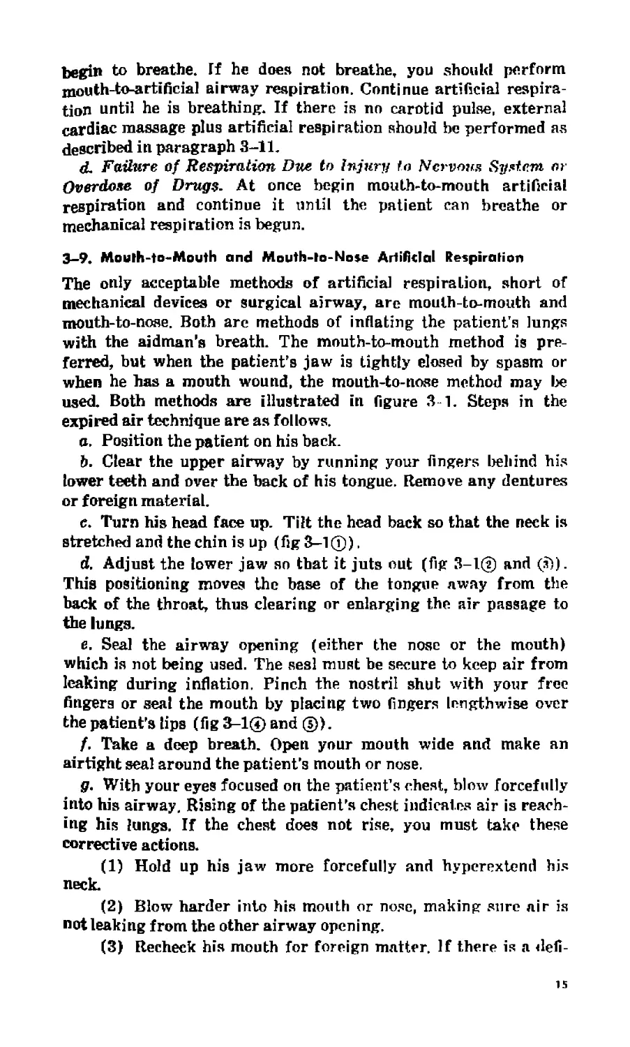
3-9. Mouth-to-Mouth and Mouth-to-Nose Artificial Respiration
The only acceptable methods of artificial respiration, short of
mechanical devices or surgical airway, are mouth-to-mouth and
mouth-to-nose. Both are methods of inflating the patient's lungs
with the aidman’s breath. The mouth-to-mouth method is pre-
ferred, but when the patient’s jaw is tightly closed by spasm or
when he has a mouth wound, the mouth-to-nose method may lx*
used. Both methods are illustrated in figure 3 1. Steps in the
expired air technique are as follows.
a. Position the patient on his back.
Ь. Clear the upper airway by running your fingers behind his
lower teeth and over the back of his tongue. Remove any dentures
or foreign material.
e. Turn his head face up. Tilt the head back so that the neck is
stretched and the chin is up (fig 3-1®).
d. Adjust the lower jaw so that it juts out (fig 3-1® and (£)).
This positioning moves the base of the tongue away from the
back of the throat, thus clearing or enlarging the air passage to
the lungs.
e. Seal the airway opening (either the nose or the mouth)
which is not being used. The sesl must be secure to keep air from
leaking during inflation. Pinch the nostril shut with your free
fingers or seal the mouth by placing two fingers lengthwise over
the patient’s lips (fig 3-1® and ©).
f. Take a deep breath. Open your mouth wide and make an
airtight seal around the patient’s mouth or nose.
g. With your eyes focused on the patient’s chest, blow forcefully
into his airway. Rising of the patient's chest indicates air is reach-
ing his lungs. If the chest does not rise, you must take these
corrective actions.
(1) Hold up his jaw more forcefully and hyperextend his
neck.
(2) Blow harder into his mouth or nose, making sure air is
not leaking from the other airway opening.
(3) Recheck his mouth for foreign matter. If there is a defi-
15
t'KJ и re з J, Steps in »nuuth-tw-jnwHt/i and mvuU-to-noee artificial respiration.
u
nite obstruction of the airway, an emergency surgical opening
must be made,
(4) Remove your mouth, listen for the return of air from
the patient's lungs. If the exhalation is noisy, elevate his jaw
further.
(5) This procedure should be repeated 12 times a minute.
h. If these steps fail to permit inflation of the lungs, an emer-
gency surgical airway must be made.
3—10- Emergency Surgical Airway
Again, most airway obstructions are relieved by nonsurgical meas-
ures. Clearing the upper air passages with the fingers, positioning
the head, neck, and body, adjusting the lower jaw, or a sharp blow
on the patient's back may be all that is needed to dislodge an
obstruction. Persistant obstruction of the airway, however, re-
quires an immediate surgical airway for relief. Diagnosis is estab-
lished when the patient's lungs cannot be inflated by mouth-to-
mouth (or mouth-to-nose) respiration.
a. A patient with persistent airway obstruction will be hard to
restrain, if conscious, so you will ne<id someone to help you hold
him.
b. Quickly get the sharpest cutting instrument you can find.
c. Have your assistant immobilize the patient while you locate
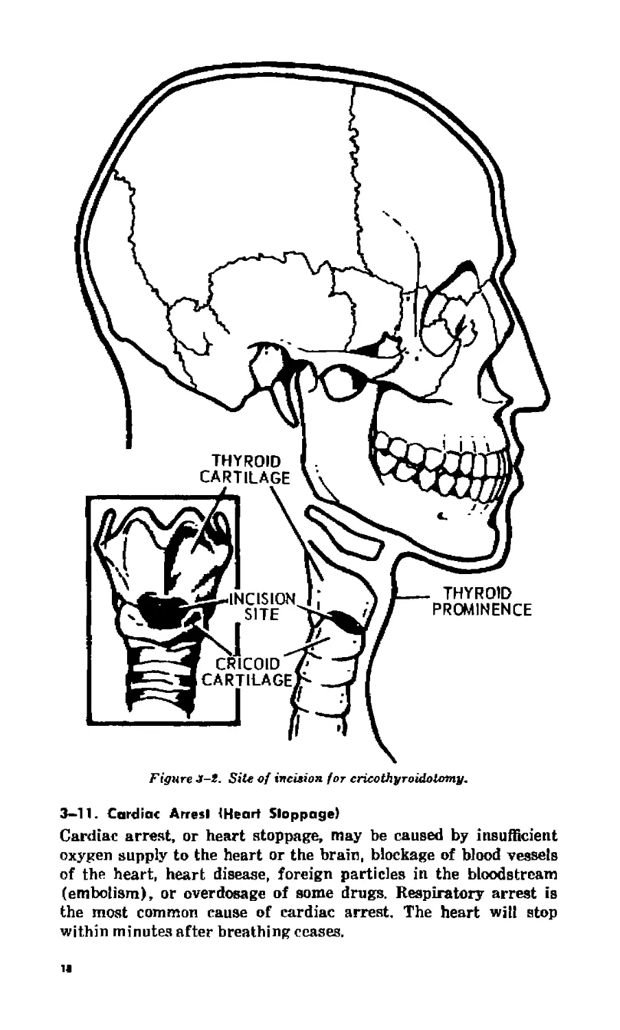
the area over the cricothyroid membrane to make an incision.
d. The cricothyroid membrane is the best place to make an
emergency surgical airway. It is just beneath the skin in the mid-
dle of the front of the neck, It is between the thyroid cartilage
(“Adam's apple”) just above it and the less prominent (in males)
cricoid cartilage below it. See figure 3-2 for location of incision
site.
e. While immobilizing the skin and trachea with one hand, make
an incision horizontally over the cricothyroid membrane through
the skin. Then make a second incision into the larynx through the
membrane until a finger-sized opening is obtained.
f- At this point, the patient should make a gasping inhalation
through the opening which you have made. Enlarge the opening
enough with your fingers to allow complete filling of the lungs.
Let the patient breathe through the opening until he is partly
stabilized while you assist by stretching the opening.
g. Insert a cannula or a tubelike item into the opening. Secure
the cannula in the trachea, as in figure 3-3, to prevent it from
being aspirated or dislodged. Any tubelike item may be used,
including the barrel of a ball-point pen.
k. Place the patient in a position most comfortable to him.
i. If the patient does not breathe on his own, apply positive
pressure respiration to the airway.
17
Figure J-t. Site of incision for cricothyroidoiomy.
3—11. Cardiac Arrest {Heart Stoppage)
Cardiac arrest, or heart stoppage, may be caused by insufficient
oxygen supply to the heart or the brain, blockage of blood vessels
of the heart, heart disease, foreign particles in the bloodstream
(embolism), or overdosage of some drugs. Respiratory arrest is
the most common cause of cardiac arrest. The heart will stop
within minutes after breathing ceases.
11
Figure .T-5. Cannineprtsrf and secured in trachea.
a. Siffns and Symptoms,
(1) Absence of a carotid pulse.
(2) Cessation of breathing.
(3) Dilated pupils of the eyes.
(4) Unconsciousness.
(5) Limp body and flaccid skin.
(6) Cyanosis.
6, Actions to Take Immediately.
(1) Roll the victim onto his back.
(2) Check his airway and remove any obstruction.
(3) Hyperextend the neck and lift the lower jaw for mouth-
to-mouth artificial respiration.
(4) Give him five quick pufts of air by mouth-to-mouth.
(5) Place the heel of your hand on the lower half of the
star num and press down until the sternum is depressed about 2
inches, as in figure 3-4©. Repeat thia compression about 15 times,
about 1 per second.
(6) Return to mouth-to-mouth artificial respiration and give
the victim two respirations,
(7) Repeat this 15-2 cycle until help arrives or you are
certain the patient is dead.
(8) If help is available, one person should give the cardiac
compressions and the other should give mouth-to-mouth artificial
r?
respiration аз in figure 3-4@. The ratio with two operators should
be about five compressions to one artificial respiration. The com-
pressions should not be interrupted, even fur the respirations.
When respiration is being applied, the compressions must be
stopped momentarily.
(9) The cardiac compressions should equal about 60 per
minute, the respirations about 12 per minute.
Figure 3 -4. Cardiac
compression applied by one operator and by two
operators.
20
3-12. Shock
Shock is a complex subject, but basically it means that the body
tissues are not getting enough blood. The most common cause is
hemorrhage where blood escapes from the vascular system and
consequently does not get to the tissues.
a. Diagnosis. There are four broad areas of symptoms in shock.
The first involves feeling the pulse, which is usually abnormally
rapid. There is also a drop in hlood pressure which is detectable
by a weakened pulse. The second area is increased respiratory
rate—the body's response to the lack of oxygen in the tissues.
The third area is the skin which is usually cool and clammy and
pale due to decreased blood flow. The fourth is changed mental
state. In early shock, the patient frequently is agitated and rest-
less. As the shock worsens and the brain is deprived of blood,
drowsiness and unconsciousness result In addition to these con-
siderations, certain wounds are commonly associated with shock.
When these wounds are present, treatment for shock should be
begun even before the clinical signs and symptoms appear. These
wounds include:
(1) Any wound which penetrates the belly, chest, neck, or
pelvis. Internal bleeding is a liksly possibility.
(2) Any wound of the arms or legs which has damaged a
portion of tissue at least as big as a fist. Many bullet wounds of the
thigh are in this category.
(3) Any wound which includes a fracture of a large bone.
Blood loss of at least 1 quart frequently accompanies a fractured
femur, for example.
(4) Any wound which results in blood loss of 1 quart or
more. The blood may be visible on the ground, for example. If the
blood has completely soaked a standard field dressing, this indi-
cates loss of nearly a quart into the dressing. Treatment for shock
is indicated. (A useful experiment you might try is to pour water
into a dry field dressing to see how much water it takes to saturate
it.)
b. Treatment. First, stop the blseding and insure that the pa-
tient is breathing adequately. Position the patient on his back
with his head down to enhance the flow of blood to the brain.
Immediately begin intravenous (I.V.) fluid therapy, preferably
through at least two veins. Administer fluids cautiously in the
presence of possible intracranial injury. Make aure the patient is
comfortable and reassure him. This can help prevent worsening of
the shock.
e. Available Intravenous Solutions. Figure 3-5 shows several
intravenous solutions and an intravenous injection set. One way
21
BM6EITS
PLASM ЛИАТЕ
"tMrjo M*
SERUM
ALBUMlHi
BLOOD VOLUME ЕХРЛЖИЙ
IN PLASTIC ВАС._____
Figure 3-5. Blood volume expanders and infuAitm get.
of carrying bottles—in canteen covers—appears in figure 3-6.
Solutions available to you include the following:
(1) Ringer’s lactate solution (lactated Ringer’s injection) is
the most commonly used volume expander for treating hemor-
rhagic shock when blood is not available. It is a sterile solution of
calcium chloride, potassium chloride, sodium chloride, and sodium
lactate in water for injection. Its composition is closer to that of
the extracellular fluid than is that of any other solution employed
as a fluid and electrolyte replenisher. It expands the extracellular
fluid volume which includes the blood volume. Ringer’s solution
is normally supplied in 1ДЮ0 cc. bottles but it can be procured in
500 cc. plastic containers.
(2) Normal saline (sodium chloride solution) is the second
most commonly used intravenous fluid replacement. It can be used
interchangeably with Ringer's lactate solution and is also an
expander of extracellular fluid volnme.
(3) Plasm an ate is derived from human plasma which has
been heat-treated to kill the hepatitis virus, and diluted to 6 per-
cent strength in a solution similar to saline. Plasmanate is rich in
albumin and tends to remain in the blood vessels; thus, it is a
22
Figure S-f>. Blond volume erpander carried in cnitlrm raver.
plasma expander. It is an excellent replacement fluid for shock.
It is supplied in 600 cc. bottles,
(4) Scrum albumin is a concentrated protein in a small vol-
ume of water. It is useful in treatment of shock primarily if given
with saline or Ringer's lactate. Used alone, it attracts water from
within the cells and tissue spaces into the bloodstream. This may
be dangerous, especially if the shock is due to dehydration, as in
severe diarrhea. It is usually supplied in 100 cc. vials.
d. Use of Fluids in Hemorrhagic Shock. Blood is the best volume
expander. It should be used in preference to anything else to treat
shock due to hemorrhage. To avoid a reaction, only the proper
type of blood should be given. You may not have either the blood
or the facilities for typing it. As rapidly as possible, get the patient
to where these facilities and blood are available. Meantime, start
rapid replacement (two intravenous injections) with saline or
Ringer’s lactate. If plasmanate is available, use it instead. In an
emergency, when evacuation will be delayed several hours, ora]
saline solution can be of great benefit if the patient does not have
an abdominal wound and if he is fully conscious. Mix the salt and
soda packet from your aid bag in the coolest potable water you
can get, Encourage but do not force the patient to drink it. If he
vomits, go slower, and keep trying to have him drink it. Usually
he can keep down most of it. И you are isolated overnight with a
patient who has a 50 percent burn, 7 to 10 liters (or quarts) of
oral saline may keep him alive.
23
CHAPTER 4
WOUNDS: CLASSIFICATION, STABILIZATION, AND
EVACUATION
4—1. Classification of Wounds
For treatment and recording purposes, wounds are classified by
cause, type, or appearance.
a. Classification by Cause.
(1) Bullet wounds. These wounds differ according to the type
of weapon that fires the bullet. Damage to underlying tissue is
affected by the size of the bullet and the velocity of the bullet as it
strikes the patient.
(2) Fragmentation wounds. These are wounds made by frag-
ments of exploding grenades, mortars, mines, booby traps, rockets,
bombs, and artillery rounds. The explosion throws bits of metals
in all directions, often causing multiple wounds of varying sizes.
(3) Wounds due to falls. A fall while a soldier is taking
eover, especially with a pack on his back, can cause twisting, tear-
ing, or wrenching wounds. A fall from a moving vehicle may re-
sult in broken bones and bruises.
(4) Bums. Burns can be caused by many sources. The ones
encountered most frequently are napalm weapons, flame throwers,
gasoline, white phosphorus grenades, or marking rounds. Burns
are discussed in detail in paragraph 5-10.
b. Classification by Appearance.
(1) Bullets and shell fragments make penetrating wounds,
perforating wounds, or both. A penetrating wound is one in which
the bullet or fragment enters but does not leave the body. Knife
or bayonet wounds also are included in this category. A perforat-
ing wound is one in which the bullet or fragment goes all the way
through the body and makes at least two wounds, one of entrance
and one or more of exit. The exit wound is often larger than the
wound of entrance and may be located in an area of the body
distant from the entrance wound. Therefore, every patient with a
bullet wound must be examined thoroughly to see if he has more
than one wound.
(2) A laceration is a cut or a tear. Unless they involve major
blood vessels or impair breathing, lacerations are not a special
25
lifesaving problem for the aidman. Since they can be large and
appear nasty, they may make the patient apprehensive. Usually
there is more fright than pain with a laceration. The main prob-
lem with a lacerated wound incurred in combat is that it becomes
infected easily. To prevent infection and to promote growth of
new tissue, the wound must be debrided. The process of debriding,
or debridement, is the surgical removal of all dirt, contamination,
snd dead tissue. This procedure must be done at a treatment facil-
ity under sterile conditions. Aftsr debridement such wounds are
often left unsntured for a few days. The procedure, called “de-
layed primary closure/’ or “DPC,” prevents infection and permits
better healing. All combat wounds, regardless of size, are con-
sidered contaminated and should receive delayed primary closure.
(3) A closed wound is one with internal damage to bones or
tissue without a connecting wound in tbe outsr skin. Sprains,
strains, dislocations, and certain fractures are closed wounds.
4-2. Relief of Pein
Some pain occurs after most wounds. Pain may be mild or severe,
depending upon the petient and the wound. The patient’s state of
mind at the time of wounding will have some effect on the degree
of pain. Fear and apprehension, for example, make it worse. To
some patients the fear of pain is more real than the pain itself.
You must decide whether or not the relief of pain is in the best
interest of the patient. In many cases, pain is a helpful symptom to
medical personnel. Pain is nature's alarm system; silencing it may
be detrimental to the patient
a. You can give him some relief in these ways.
(1) Positioning. The best position is the one which the pa-
tient finds most comfortable. Positioning the injured part to re-
lieve stress can do much to relieve pain.
(2) .Reassurance. Talk to him reassuringly, Make him feci
that he is in good hands and more help is on its way. The best type
of reassurance is for you not to panic and to act as if everything
is under control.
(3) Medication. Administer an analgesic such as aspirin or
APC. If oral medications and fluid are not contraindicated, aspirin
is an outstanding drug and will relieve all but the most severe
pain.
b, If the pain is extremely severe, you may have to give mor-
phine if it is not contraindicated (para 4-3d).
4—3. Use of Morphine
a. Morphine is the best pain relieving medicine you have, but it
has several dangerous toxic effects. It is a powerful depressor of
the central nervous system, greatly reducing respiration and pain
н
sensation. Also it causes vomiting, dry mouth, constipation, and
retention of urine. It must not be given by anyone who is not fully
aware of its dangers. Never let morphine out of your possession.
It may be stolen for personal use or sale on the black market.
b. Morphine is supplied to you in 16 mg. (one-fourth grain)
syrettes. The number of syrettes you carry is determined by your
medical commander on the hasis of the tactical situation, avail-
ability of evacuation, supply, and your ability to administer it
intelligently. You must know the indications and contraindications
for its use. If not you may do more harm than good. (Contraindi-
cation is any condition which makes a particular treatment un-
desirable or improper.)
c. Morphine is indicated for severe pain especially when the
evacuation lag time is more than 20 minutes. In a tactical situation
where a psychotic patient must be temporarily silenced or sedated,
and no other tranquilizers are available, one syrette of morphine
la often effective in controlling such a patient. This is an emer-
gency measure only. There are better, nonaddicting drugs for
psychosis than morphine.
d. Morphine is contraindicated when its toxic effect will com-
pound an injury to a dangerous degree. Do not give morphine to:
patients who are to be quickly evacuated, who have chest injuries,
depressed respiration, or injuries of the head. Never give mor-
phine to an unconscious patient. Do not give morphine before
eurgery. If there is a probability that the patient may soon be
operated on, he should not get morphine. Both morphine and surgi-
cal anesthesia depress respiration. If the patient is in shock, you
should not give him morphine because it will not be absorbed due
to poor circulation. (Medical officers sometimes administer mor-
phine intravenously while the patient is in shock. Never should
you try to give morphine intravenously. If it is given too fast it
will be fatal.) A dose of morphine should not be repeated within
2 hours, or if there is any reason to believe the first dose has not
been absorbed.
4-4. Treatment of Open Wounds
Control of hemorrhage, relief of pain, and prevention of infection
are the main considerations in treating wounds iu the field.
a. Acute loss of blood may lead to shock, and shock may lead to
death. So, you should do all you can to prevent loss of blood. The
preferred method of controlling bleeding is with a pressure dress-
ing securely applied. Lost vascular fluid (blood) or body fluid
(tissue fluid) should be replaced. Use oral or intravenous fluids as
prescribed in paragraph 3—12,
b. Some wounds are more painful than others. In some trau-
matic amputations there may be relatively little initial pain, while
27
Ui siiiuller wounds the pain may be severe. Second degree burns
and massive tissue wounds involving many nerves are initially
painful. Nearly all wounds cause some pain. Things you can do to
relieve pain are described in paragraphs 4-2 and 4—3.
c. Any combat wound must be considered contaminated. The
best way to prevent more contamination is to cover the wound with
a sterile dressing. Combat wounds are “dirty” wounds. All contain
bacteria. In the held, there is no way for you to cleanse a wound
of bacteria. Pouring antiseptics into a wound will not kill all the
bacteria and may be harmful. Pouring antiseptics on the skin
around a wound does little to keep out bacteria and should be
avoided. When possible, and when evacuation is impossible or
delayed for longer than several hours, gentle cleansing of the skin
around the wound with soap and water may be helpful.
4—5. Factors Affecting Infection
Infection of a wound involves the number and type of pathogenic
organisms entering the wound, condition of tissue in the wound,
and the body’s defense.
a. If the number of organisms is extremely large, they may
overwhelm the body’s defense by sheer numbers. This is likely to
happen in wounds caused by booby traps with fllth and contamina-
tion about them. Punji slake wounds are another example,
b. Some organisms are more toxic than others. For example,
the organisms that cause gas gangrene and tetanus are deadlier
than some organisms that form pus.
c. A cleanly cut wound is not as apt to become infected as a torn,
jagged wound. In the first type of wound, blood tends to flush out
organisms and they have few places to hide and become imbedded.
The second type of wound gives organisms devitalized tissue to
hide in and has much less flushing action by bleeding. A puncture
wound is most likely to become infected with tetanus and gas
gangrene because of lack of oxygen. Penetrating and perforating
wounds are usually heavily contaminated by foreign material car-
ried into deep parts of the body. Penetrating abdominal wounds
often permit contaminated intestinal contents to leu<k into the
cavity.
4-6, Treatment of Closed Wounds
a. Sprain. A sprain is the twisting, tearing, and stretching of
ligaments around a joint. Ligaments are strong, slightly elastic,
fibrous bands of tissue that hold bones in position. A ligament can
be over-stretched and some of its tissue cells injured, or it can
be torn loose from its attachment to the bone. An injured ligament
heals slowly and sometimes never entirely returns to normal.
Biagnosis is made by the presence of a tender, painful joint with
28
3wellJng. h'ractuie ai&o ишь1 Le cuiisjuufcd a possibility with these
findings.
(1) A sprain is treated so as to temporarily replace the func-
tion of the ligaments by supporting the joint while allowing some
movement. You carry elastic rolled bandages for this purpose. A
figure-of-eight bandage around the joint should allow the patient
to complete his immediate mission. The bandage should be adjusted
as swelling occurs. Have a medical officer evaluate the patient after
mission.
(2) Analgesics may be given for pain.
(3) Routine evacuation may be indicated.
b. Strain. A strain is an overstretching of a muscle or the mus-
cle’s tendon. In combat, some muscles will ba forced to function
long after they are tired. This results in acute muscle fatigue or
muscle strain. Diagnosis generally involves finding tender, painful
muscles. Swelling is uncommon.
(1) There is little you can do to treat a strain in the field.
The patient needs rest with just enough exercise to keep the
muscle from getting too stiff. You cannot provide this type of
treatment in the field.
(2) Analgesics may be given for pain.
(3) Heat and massage are also very helpful.
(4) If the strain is severe, routine evacuation is indicated.
c. Dislocation. A dislocation is the displacement of one of the
bones forming a joint. A joint is the articulation of two or more
bones. When one end of a bone forming a joint is forced out of its
articulation, it is dislocated. The dislocation may be incomplete
and temporary. In other words, it may jump out of and back into
normal position, resulting in a condition much like a sprain. If the
bone dislocates from its articulation and remains out of place, it
la a complete dislocation. Damage to surrounding hlood vessels and
nerves may result.
(1) You should not try to reduce a complete dislocation in
the field,
(2) Analgesics should be given for pain.
(3) Immobilization of the joint in the position of least pain
may be helpful. Usually that is the position in which you find it.
(4) Routine evacuation is indicated unless damage to blood
vessels or nerves is suspected because of paralysis, numbness, or
absent pulse. In that case, priority or even urgent evacuation may
be necessary.
d. Fractures. For treatment of fractures, see paragraph 4-7.
4—7. Fraclures
Fractures, or broken bones, are the result of a strong blow or
stress against the body causing one or more bones to crack or
»
break completely. Fractures are either closed (no break in the
skin) or open (skin broken). Open fractures are generally more
serious, because of the danger of infection.
a. Diagnosis. The patient with a hroken bone is almost always
in pain at the fracture site. He will give a history of trauma or
stress and often will state that he felt the bone snap or give way.
He typically has great difficulty in moving the part of the body
beyond the fracture. As you examine the patient» you will find
swelling and tenderness at the fracture. The broken limb may be
obviously deformed. Ultimately, X-rays will be needed to establish
the diagnosis and extent of the fracture.
Ь. Treatment. As with any wounded patient, the first thing to
do is save his life. Make sure he has a clear airway and can
hreathe. Stop externa] bleeding. Almost every fracture is accom-
panied by significant internal bleeding. A fractured femur, for
example, may involve loss of as much as 1,500 cc. of hlood into
the thigh. Plainly, then, a patient with a fracture of a major bone
is in danger of developing hemorrhagic shock. Therefore, intra-
venous solutions should be started as soon as possible on any pa-
tient with a fracture of a major bone. Place a dry sterile dressing
over the wound if it is an open fracture. Administer analgesics for
pain. The patient must be evacuated, but the category depends
upon the seriousness of the fracture.
c. Splinting. Do not attempt to reduce or set a broken bone. In
general, splint the fractured limb as you find it, checking the pulse
beyond the fracture before and after splinting. If the pulse dis-
appears after the splint is applied, it is on too tight and must be
loosened. Also a record of nerve function distal to the fracture
should be made. If the fractured limb is bent so that it pinches off
the blood vessels, you may straighten it carefully as long as no
force is needed, Never try to force an arm or a leg to lie straight.
Splinting is extremely valuable because it prevents further dam-
age to surrounding tissues by the broken bones. Also, splinting
helps to reduce bleeding and pain.
d. Sp/tnfs. Splints and splinting in the field will pose some prob-
lems. You do not carry splint sets, such as the Army leg splint set
You may carry the wire fabric splint. Some aidmen carry two
wire ladder splints wrapped around the outside of the aid bag as
in figure 4-1. To support missions where fractures might occur,
you may carry a few pneumatic splints, The ones used most in
the field are improvised and anatomical splints (fig 4r-2).
(1) An improvised splint is made of any rigid material that
is readily available. Parts of the patient’s gear are often the handi-
est material you can use. Rolled or folded, the patient’s poncho
makes a good splint. So does his rifle when rolled in a jacket. (Be
n
FiffWre i-1- IVtre Ladder splints wrapped, arattnd an aid bap.
sure the rifle is cleared.) Poles or branches from trees also can be
used to make splints.
(2) How much time you can spend on improvising a splint
will depend upon the tactical situation. There may be instances
where you have no time to improvise a splint. In that esse, for a
fracture of the forearm, quickly place the arm inside the jacket
and tuck the jacket as tightly as possible. A fracture of the upper
arm could also be treated this way or with a sling around the
neck to the wrist. For a fracture of the leg, qtiickly tie the broken
leg to the uninjured leg. This is an example of an anatomical
splint, where one part of the body is used to help immobilize
another part.
(3) The wire fabric splint is useful in supporting a massive
tissue wound, It can be fashioned to help support a broken ankle,
wrist, or small bone.
(4) The wire ladder splint can be used for a fractured arm
or leg or to support a massive tissue wound. You should control
the bleeding before applying a splint. If not, pul on the splint so
it can be removed easily and quickly.
(5) A pneumatic splint (fig 4-3) is inflatable and made of
transparent plastic. You blow air into it by mouth to get the
necessary rigidity. Do not use any other means for inflation (such
as a tank of compressed air). The splint requires no padding and
it can be inflated or deflated as desired. The splint should not be
31
FRACTURE (MORE RIGID MATERIAL AND MORE
33
inflated and left uii the patient niuiv than 3U minutes at a Lime.
To do so will interfere with peripheral circulation. Reduction of
peripheral circulation for a long time causes tissue anoxia, which
in turn results in damaged or necrotic tissue. Tissue damage is
proportional to the duration of diminished peripheral circulation
and the degree of tissue anoxia. Therefore, if the patient must
wear a pneumatic splint for an extended time, partially deflate it
every 20 to 30 minutes for a few moments to reestablish peripheral
circulation if it appears that the blood supply to the extremity has
been impaired. Do not use these splints unless you have Lime to
check the patient every few minutes.
(6) Army leg splint sets are stocked at aid stations, clearing
stations, dispensaries, hospitals, and medical depots. If time and
the tactical situation permit, you may ask the evacuation vehicle
operator to bring you an Army leg splint if ita application is indi-
cated. This splint is especially valuable in protecting the nerves
and blood vessels.
4—8. Dressings
A dressing is a pad that is applied directly over a wound. A pre-
pared dressing is usually made of gauze but it can be made of any
figure J-3. Inflated pneumatic splint applied on a patient’s arm.
33
Figure 4-4. Popular tizes of field dressings.
34
ja to control hemorrhage and protect a wound against lurtncr ron-
t^inin&tion. Almost all external bleeding can be controlled with a
correctly applied field dressing.
a. Sues. The most popular sizes of field dressings, shown in
figure 4—4, are described below.
(1) Dressing, first aid, 4 bp 7 inches. This small field dressing
is the one you probably will use most. You should carry a plentiful
supply of these. Many aidmen carry two aid bags, one filled with
dressings and one containing other items. Be sure each soldier
carries at least one small field dressing.
(2) Dressing, first aid, field, 7^ by 8 inches. This is usually
called the medium field dressing. The average aidman carries two
of these. They are used often to reinforce the small field dressing.
(3) Dressing, first aid, field, 11 by 11 inches. This is the large
field dressing. You usually carry one of this size. Most aidmen
prefer to carry more small dressings and use two or three small
ones instead of one large dressing. You can contour two or three
small dressings better than a large one. Large dressings are best
for extensive burns.
(4) Dressing, first aid, field, individual troop, 100 by 120 mm.
This is a two-piece dressing designed to allow one gauze pad to
slide along the affixed bandage. One purpose of this adjustable
dressing is to allow application of the dressing over a perforated
wound of an extremity to cover the wounds of entrance and exit
with tiie same dressing. This dressing is smaller and more ver-
satile than other field dressings.
b. Application. A field dressing has strips of gauze bandage
attached to it The gauze strips or tails are used to secure the
dressing and to apply pressure. First, put a small dressing over
the wound and tie the bandage tails firmly over the dressing to
apply pressure. If the first dressing does not control bleeding,
apply a second one over it. Again, tie the bandage tails firmly.
Several small dressings are more effective than one large dressing
for controlling hemorrhage.
4—9. Bandage»
A bandage is a piece of material used to cover a dressing, apply
additional pressure, or immobilize a part of the body. Bandages
may be made of gauze, muslin, or elastic cotton (fig 4-5). They
may be rolled or folded. Most aidmen prefer to carry a few elastic
rolled bandages about 3 inches wide. Elastic bandages are used to
reinforce dressings in the control of hemorrhage and to support
ankles and knees. Rolled gauze bandages are not often used in the
field. Triangular muslin bandages are sometimes used for support
but are used most as tourniquets. Folded triangular bandages
(cravats) are useful in applying improvised splints.
35
BANDAGE, MUSLIN, 37 BY 37 BY 52 INCHES, t '
BANDAGE, TRIANGULAR (MADE FROM BANDAGE, MUSLIN)..
BANDAGE, COTTON, ELASTIC
Figure f-5. Bartdag es far field use.
34
CHAPTER 5
SPECIFIC WOUNDS AND BURNS
5-1. C!a»»e> of Head Injuries
Head injuries are of two main classes, opan wounds and dosed
wounds. They are further classified as scalp wounds, skull frac-
tures, intracranial wounds, and wounds of the face.
5-2. Scalp Wounds
a. Laceration of the scalp may result in a gaping wound with
profuse Needing. The wound is gaping because of the tension of
tiie layers of the scalp. The profuse bleeding is due to the rich blood
supply. Firm pressure dressings will control bleeding of the scalp.
b. Contusions resulting from blows to the head may form lumps
in tiie scalp. These are usually collections of hlood caused by broken
blood vessels within and under the scalp. Contusions require no
specific treatment by the aidman in the field, but all head wounds
should receive careful examination and constant observation. A
patient may have no complaint other than a slight headache im-
mediately following the injury, even though serioua intracranial
damage may exist.
5—3. Skull Fractures
Cranial wounds are skull fractures. A skull fracture may be a
simple tine break or crack in the skull bone, or it may be a de-
pressed fracture with pieces of the skull penetrating the brain.
A simple skull fracture in itself is not serious as the bone will heal
fairly rapidly. The danger is that the blow which caused the
fracture also ruptured blood vessels under the skull, causing blood
to collect and increase pressure on the underlying brain tissue.
a. Generally, you will be unable to determine whether a
skull fracture is present or not. Sometimes the fracture can be
felt through the scalp, but most skull fractures will not be proven
until X-rays are taken. Consequently, you should suspect skull
fracture in any patient who received a severe blow to the head,
even if the scalp was not lacerated. If you see clear fluid coming
from an ear, the nose, or a head injury, or if you see brain matter
in any head wound, you can be sure a skull fracture is present In
37
addition, pupils of unequal size and vomiting are signs oi brain
injury even if the skull is not fractured.
b. Do not give medication to a patient with a head injury.
The medication may mask the symptoms of a more serious injury.
Observe the patient carefully, paying particular attention to his
vital signs and state of consciousness.
c. Routine evacuation is indicated for simple head injuries
if there is no firm evidence of skull fracture and the vital signs
and state of consciousness are stable.
d. If you can feel a fracture, or if you see the clear cerebro-
spinal fluid coming from the patient's nose, ears, or wound, or
if you see brain matter in the wound, or if the vital signs or
level of consciousness deteriorate, evacuate the patient by the
proper category of precedence. In this case, that would be probably
priority or urgent. If an external wound is present, apply a
loose-fitting dressing. Again, give no medication.
e. The most important thing you can do in the treatment of
head wounds is record the injury. Record the time of the wound-
ing and all signs and symptoms. Make particular note of vital
signs, size of pupils, and state of consciousness both when you
first began treatment and at the time of evacuation. Also record
the time of your observation,
5—4. Intracranial Wounds
Intracranial wounds are serious because they involve the brain and
other tissue inside the ekull. There are two general types of intra-
cranial wounds, open and closed. In the open type, the brain is
exposed to the outside and there is a laceration of the scalp as
well as a skull fracture. In the closed type, there is no opening
from the brain to the outside. Either type will pose problems for
you. For a severe open head wound, you should apply a dry,
sterile dressing and call for immediate evacuation using the urgent
category of precedence. The closed head wound poses special
problems. You have no immediate way to determine the degree of
injury. Therefore, you should do the following things:
a. Observe the patient closely.
b. Record the time of the injury,
c. Check the patient every few minutes for headache, changes
in size of pupil of the eyes or in their reaction to light, impair-
ment of vision, dizziness, slurring of speech, changes in pulse
rate, vomiting, or changes in rate of respiration. Be sure to record
these symptoms and the time of their onset. They indicate that
brain injury is developing, usually from slow bleeding inside
the skull. Always record at least one observation of pupil size
and pulse rate in case of a head injury.
м
d. Request a priority category evacuation for the patient if any
of these symptoms appear,
e. Advise the patient's commander not to plan on using the
patient for critical or sensitive duties while he is being obeerved.
/. Give no medication during the period of observation. Ob-
servation should last about 24 hours. Occasionally the bleeding
inside the skull can be very slow, with the symptoms taking sev-
eral days to develop. The patient's commander should be altered
to thia possibility.
5—5. Woundj of lhe Face
Facial wounds require prompt, positive action because of bleeding
and possible airway obstruction. Airway obstruction is a more im-
mediate threat to life and harder to handle than bleeding. Blood
clots and pieces of bone, flesh, or other foreign material may block
the airway. Blood which is swallowed may cause vomiting and the
voznitus may be aspirated, further complicating the problem. At-
tempts to control bleeding may interfere with breathing. The pa-
tient may be trying frantically to get air. Do these things im-
mediately.
a. Position the patient so that he will not aspirate fluids if
he is bleeding from the mouth or vomiting,
b. Clear the airway of blood clots and foreign matter. Wrap
a piece of gauze bandage around your fingers when you dislodge
blood, vomit us, or mucus from the airway. Gauze makes it
easier for you to grasp things.
c. Prepare to perform an emergency surgical airway. Due to
aspiration of foreign matter, the patient will be hard to manage
and he will remain in danger of aspirating more foreign matter
until bleeding is controlled. It may become necessary to perform
an emergency surgical airway (para 3-10) to relieve airway ob-
struction before full attention can be turned to control of bleeding
from the facial wound.
d. Call for evacuation early. Facial wounds become progres-
sively worse. Airway difficulties get worse with swelling of injured
tissue. Bleeding is hard to control, injured tissue becomes more
Painful, and it is almost impossible to prevent infection. Collect
all pieces of dentures, if any, and evacuHe them with the patient.
They can be valuable aids in treatment and reconstruction,
5—6. Wouads oF the Neck
Wounds of the neck are treated essentially the same as facial
wounds. Airway obstruction and hemorrhage are the main threats
life. Hemorrhage from large blood vessels must be controlled
Quickly. Direct pressure with a pressure dressing must be applied
39
over the bleeding point, alongside but nut over the trachea. lake
these actions or precautions immediately.
(lauiiun. Beware of a possible fracture!
a. Position the patient quickly to prevent more blood from
entering the airway,
b. If large blood vessels are severed, apply direct pressure
quickly.
c. Call for assistance; the patient will be difficult to handle.
d. Clear the airway as rapidly as possible. Consider a surgical
emergency airway early.
e. Due to aspiration of large amounts of blood, the airway may
be blocked. After clearing it, start artificial respiration if spon-
taneous respiration does not occur.
/. Call for evacuation early and request delivery of necessary
resuscitative equipment.
g. In severe hemorrliage, start blood volume expanders
promptly.
A, Handle the patient very gently if you suspect he has a
fractured neck. Immobilize the neck as much as possible.
t. Do not give morphine.
j. Do not give anything by mouth, as the esophagus may be
injured.
5—7. Chest Wounds
Chest wounds respresent an appreciable proportion of combat
wounds. Most fire is directed toward the chest. Penetrating and
perforating wounds of the chest may damage the lungs, trachea,
bronchi, esophagus, diaphragm, or large blood vessels. Most chest
wounds interfere with breathing.
a. The normal chest cavity is an airtight enclosure with one
opening to the air, the trachea. If another opening into the chest
cavity is made, such as a bullet wound through the chest wall,
the lung on that side of the chest can no longer remain expanded
and is said to “collapse?' With each breath, air is sucked into
the chest cavity, permitting the lung to deflate further, This is
called a “sucking chest wound.” The more the lung collapses the
less well the patient can breathe. Therefore, the sucking chest
wound must be sealed shut as soon as possible by any means
available,
(1) The best way is to place several thicknesses of petrolatum-
impregnated gauze over the wound and reinforce it with a field
dressing, as in figure 5-1.
(2) A field dressing (first aid dressing) may be placed
over the wound. Then, the dressing should be covered with air-
tight material to produce a quicker airtight seal over the wound.
(3) A piece of airtight material such as cellophane, plastics,
40
PETROLATUM GAUZE OVER SUCKING
CHEST WOUND
Figure 5—1. Treatment <»/ a sucking chest wound.
41
or poncho placed directly over the wound is effective as long as
it is held firmly in place. These materials tend to slip and leak
as blood seeps under them.
(4) All sucking wounds must be sealed. The test of suc-
cessful sealing is in the patient’s ability to breathe easier and
the cessation of bubbling or hissing at the wound during respira-
tion.
b. Flooding of the chest cavity by internal blseding can also
collapse the lungs. If all wound openings are closed and the pa-
tient still has difficulty, you can assume he hss internal bleeding
or massive lung damage. If the patient begins to present a shock
picture, internal bleeding should be suspected. If he begins to
hemorrhage from the mouth, he is critical. Then, the following
measures are used.
(1) Place the patient in the best breathing position. If
possible, the wounded side should be placed down to increase
breathing of the unwounded side.
(2) Start intravenous infusions of Kinger's lactate solution
or other blood volume expanders.
(3) Keep the patient as still as possible.
(4) Urgent evacuation is indicated.
5—0. Abdominal Wounds
A wound of the abdomen can be misleading. What appears to be a
small, insignificant wound on the outside may be a massive,
bleeding wound on the inside (fig 5-2). All penetrating and
perforating abdominal wounds require exploratory surgery to
stop bleeding. Abdominal wounds may include damage to the
stomach, pancreas, intestines, spleen, liver, kidneys, or large
blood vessels. The early cause of death is uncontrolled bleeding.
Infections, especially those of the internal abdominal lining
(peritonitis), caused by injury to the intestines or by the wound-
ing agent itself, may complicate the case later.
a. St pus and Symptoms of internal Abdominal Bleeding.
(1) Any perforating or penetrating abdominal wound.
(2) Pale skin and weak, rapid pulse.
(3) Thirst, restlessness, and apprehension.
(4) Abdominal rigidity (board-like).
b. Actions You Should Take Immediately.
(1) Instruct the patient to remain as still and quiet as
possible.
(2) Call for urgent evacuation.
(3) Start intravenous infusions of Ringer’s lactate solution
or some other blood volume expander.
(4) If the patient’s organs are protruding, do not try to
replace them.
43
Figure 5-2. Abdominal wound.
(5) Cover the wound loosely with a dry sterile dressing
(6) Allow the patient to take no food or drink by mouth.
Give no oral medication,
(7) For a closed abdominal wound (blunt trauma), give
no medieatbn for pain because it might disguise the symptom.1;
needed for diagnosis. If the wound is open and there is no breath-
ing trouble or head injury, morphine may be given by injection
for pain.
5—9. Traumatic Amputations
Most traumatic amputations are caused by exploding antiperson-
nel mines, land mines, and booby traps. Fragments thrown out by
43
any ui ttic-ji. (k.viic.s can amputate an arm or a leg. High vclucjly
bullets can cause a partial amputation. In treating a patient with
a traumatic amputation, you must do these things immediately.
a. Expose the entire limb above the wound by removing or
cutting off the clothing. Inspect to confirm the extent of damage
to remaining tissue.
b. Apply a tourniquet at once. Often, if the entire extremity
is completely torn off, bleeding will be slight. This is due to
the partial retraction of arteries and contraction of muscles act-
ing like a tourniquet. Even if bleeding is slight, apply a tourni-
quet because, in a few minutes, the muscles will relax and bleeding
will start.
c. The best held tourniquet is made with a cravat bandage
and a stick. As in figure 5-3, place the tourniquet about 2 inches
above the end of the stump or incomplete amputation. Tighten the
tourniquet until all bleeding stops. Secure the tourniquet (fig 5-3)
so that it is easy to remove or to tighten.
d. Cover the stump or incomplete amputation with field dress-
ings, The dressings keeps the wound clean and gives you a visual
check on bleeding. If the dressing becomes soaked with blood,
check the wound or inspect and adjust the tourniquet. Do not apply
a roller bandage at this time; it would interfere with the tour-
niquet.
e. If the amputation is incomplete, put a splint on it. The
tissue splinted should be positioned in approximate anatomical
alignment to avoid further damage to splinted perts of the limba.
Tissue below the probable amputation can be saved sometimes
and used later in making the stump. A temporary improvised
splint that is easy to remove is adequate. Apply the splint so that
the tourniquet can be adjusted without much trouble.
f. At first, the pain should be slight to moderate. Severe
pain may develop later, in 30 minutes to 2 hours. Morphine is not
indicated unless the pain is severe.
5-10. Burns
Burns are damage to tissue caused by exposure to excessive heat,
strong chemicals, or electricity. They are classified by cause, de-
gree, and extent. All classifications should be considered in the
treatment and disposition of a burn petient. Burns are compli-
cated by airway blockage, carbon monoxide poisoning, lung dam-
age, shock, and infections. Most people who die immediately in a
fire die from suffocation. Those who die a few hours later usually
die of shock. Those dying 3 to 10 days after the burn usually
die of infection. Other factors complicate burns but you can do
little in the field to prevent them. Your first job is to treat those
life-threatening conditions which follow burns.
44
APPLY TIE TWD
INCHES ABOVE
WOUND.
45
a. Airway difficulty and carbon monoxide poisoning are the
most immediate threats to life. These conditions may have several
causes.
(J) Edema of the tissue of the air passages is due to burns
from inhaled hot gases. Swelling of the lips and mouth indicates
other tissues farther down the airway are probably swollen. This
swelling may block the air passages and suffocate the patient.
An emergency surgical airway is then indicated. The surgical air-
way should be made before the air passages are completely closed.
(2) Inhalation of carbon monoxide is a threat even if the
patient is not burned. Burning material consumes oxygen and
produces carbon monoxide, a poisonous gas. A patient who has
inhaled carhon monoxide will exhibit a sensation of suffocation,
cherry red Hush, and possibly coma. If the patient inhales carbon
monoxide, you may need to perform artificial respiration for pro-
longed periods of time. Also request the evacuation vehicle to
bring oxygen and resuscitating equipment to supplement treat-
ment.
(3) Smoke inhalation irritates the air passages and delicate
membranes of air sacs in the lungs and can result in a form of
pneumonia. When the patient has inhaled such irritation or poison-
ous material, his lungs mrrst be flushed out with fresh air or
oxygen as fast as possible. If the patient is conscious, he can flush
the fumes out himself by deep breathing and coughing. If he is
unconscious or uncooperative, artificial respiration should be given
to force deep breathing. Request the evacuation vehicle to bring
resuscitative equipment for use in supplementing treatment.
Artificial respiration also will be needed if the patient does not
breathe on his own,
b. Shock is another serious threat to life in a burn patient.
When extensive areas of skin are burned, the patient cannot
properly control loss of body water. If 30 to 40 percent of the
body surface receives second or third degree hums, the body will
lose 6 to 7 liters of water in 24 hours. If this tissue fluid is not
replaced, shock is certain to occur. Since tissues begin to lose
fluid as soon as a bum occure, the sooner fluid replacement is
begun, the better. Fluids may be given orally or intravenously.
If large areas of the body are covered by second or third degree
burns, you should start two or more I.V.’s. Use fluids as directed
in paragraph 3-12. Ringer’s lactate solution is the best I.V. and
may even he given orally if tolerated. To prevent burn shock, start
fluid replacement early, both orally and intravenously.
r. Infection is the third cause of burn deaths. It is a late
complication, but the organisms causing the infection may enter
early after the burn. You should take every reasonable precaution
Л6
to prevent this. Apply only dry, sterile dressings over the burn.
If you do not have dry, sterile dressings, leave the burn open. Do
not cough or sneeze over the burn. Do not pass your hands over
the burn any more than is necessary. Do not place a blanket
over the bum patient unless the weather is extremely cold. Do not
apply medication to a burn. It does little good and when it is
removed it will cause the patient much pain. In particular, do not
apply greasy substances such as butter, olive oil, or suntan lotion
as these will predispose to infection and do no good. The only
material presently acceptable for application to the burn surface
besides the dry sterile dressing is Sulfamyalon burn cream.
5—11. Chamltal Burns
Acids, alkalis, and other strong chemical agents damage the skin,
causing chemical burns. A common chemical burn is that caused
by white phosphorus, a fast-burning metal used in marking rounds
and incendiary grenades. When detonated, the white phosphorus is
blown into small pieces. Each particle of phosphorus burns
rapidly when exposed to air. Particles striking a soldier will im-
bed in or stick to his skin and clothing and continue to burn. 11 is
clothing may ignite, causing more burns. The only way to stop
the phosphorus from burning is to exclude the air (oxygen) from
it. Copper sulfate pads are designed to extinguish burning phos-
phorus. Apply water to the copper sulfate pads and place over
the burning phosphorus while wet. If the pad dries out, apply
more water; the pad must be kept wet. If you do not have copper
sulfate pads, mud is a good substitute. You can wet gauze, but it
must be kept wet or it too will ignite, producing more burn. As
soon as the phosphorus is extinguished, remove the particles if
they are not imbedded too deeply; After removing the particles,
cover the burn with a dry, sterile dressing. Never use copper
sulfate pads as a dressing. If you cannot remove the particles,
notify the evacuation vehicle so that additional copper sulfate may
be brought for in-transit treatment. For safety of the evacuation
vehicle, all patients with white phosphorus burns must be moni-
tored carefully, because a burning piece of phosphorus may ignite
the evacuation vehicle. Other chemical burns must be flushed with
water to wash away the chemical, then treated as any other burn.
5—12. Evacuation of Burned Patients
To determine category of precedence (para 2-8) for evacuation,
you need to know how to classify burns according lo their severity.
Severity classifications are minor, moderate, and severe. The
severity of a burn is determined by the extent of the burn and
the degree of the burn. Extent means area. Degree means depth.
Burns about the head, face, neck, or chest are severe and the
47
jKii lcJil \4ii.h biiuh Lui Uh ahuuld bu evacuated 111 the Ui tjciit ur
priority category because of possible airway complications.
a. Minor burns include first degree burns of any extent and
second degree burns of small area. Moderate burns include second
degree bums of less than 10 to 20 percent of the body surface and
small third degree burns. Severe burns are those in which 20 per-
cent or more of the body surface is covered with second or third
degree burns. The “rule of nine” is useful for calculating per-
cent of body surface burned. Roughly, these are the portions of
body surface over each part of the body: head 9 percent, each arm
9 percent, anterior trunk 18 percent, posterior trunk 18 percent,
each leg 18 percent, groin 1 psreent.
b, A first degree burn is one where there is a reddening of
the skin as in sunburn. A second degree burn is one with blistering
of the skin. A third degree burn is one with charring or complete
destruction of tissue. At times, in the absence of charring it may
be difficult to determine if a third degree burn is present. Some
third degree burns have the appearance of leather, or horsehide
as on a baseball. As third degree areas have lost their nerve
supply, they are insensitive to pain.
c. As a general rule, patients with minor burns should be
evacuated in the routine category. Frequently, they need not be
evacuated at all. Moderate burns should be evacuated in the pri-
ority category. Severe burns should be evacuated in the urgent
category.
48
CHAPTER 6
CHEMICAL, BIOLOGICAL, AND NUCLEAR CASUALTIES
6-1. Problems of the Aidman
Casualties produced by chemical, biological, or nuclear operations
could pose many problems for you as aidman. The number of
casualties will be large. Material to work with will be in short
supply. Evacuation will not be available for many casualties. You
will have to be able and ready to advise the local commander on
the combat capabilities of his troops. Yuur determination of men’s
capabilities will be influenced by the tactical situation,
6—2. Casually-Producing Chemical Agents
The chemical agenta that can be used to injure or to kill men are
nerve agents, blister agents, blood agents, and choking agents.
You need to be familiar with the characteristics and modes of
actions of these agents so that you can prepare for the prevention,
tentative diagnosis, and treatment of casualties they produce.
Nerve agents and blister agents are the ones you are most likely
to encounter.
6—3. Nerve Agents
Nerve agenta are lethal (fatal) because they are extremely toxic
organic componds. They can cause death or disability in minutes.
They are essentially odorless and colorless. They range in per-
sistency from those which are highly volatile with low persistency,
such as standard agent GB, to those with low volatility and high
persistency, such as standard agent VX.
a. One of your duties is to indoctrinate the troops in first aid
and 1 ildy aid for nerve agent poisoning. The soldier and his
budfly must be able to recognize the primary signs and symptoms
of nerve agent poisoning. Self-aid and buddy aid can save lives
and reduce morbidity. Treatment cannot be started until a man
recognizes something is wrong; after recognition, he will probably
have to start treatment himself.
b. After local exposure to nerve agent vapor or aerosol, the
pupils of the eyes will be pinpointed. If exposure has occurred
through the skin or by ingestion, the pupils may be normal or
slightly to moderately reduced in the presence of severe systemic
4»
symptoms. Increased production of secretions results in a running
nose and excessive salivation. Tightness in the chest results from
constriction of the airway with increased secretions in the
tracheobronchial tree. Nausea, vomiting, diarrhea, muscular
twitching, drooling, and sweating may occur. These symptoms may
progress to convulsions, coma, and death. Respiratory failure
is the usual cause of death.
c. Immediately after exposure to a nerve agent, the soldier
should hold his breath, put on the protective mask, and clear it.
Then he should decontaminate his skin and give himself atropine if
he has impaired vision, excessive salivation, trouble in breathing,
or muscular twitching. He should check his buddy for evidence of
exposure to nerve agent, then continue his mission.
d. Act immediately as prescribed in c above to protect yourself.
Then, check all personnel for symptoms of exposure to nerve
agent. If you find casualties, give atropine until symptoms are
alleviated; see e below.
e. There are several vital steps in the treatment of nerve agent
poisoning. The protective mask must be put on as soon as a nerve
agent is suspected or signs or symptoms of such poisoning are
recognized. Liquid contamination must be removed immediately
from the skin or the clothing. (A decontamination kit is provided
for this purpose.) Contamination of the eyes is treated by irriga-
tion with copious amounts of water. Atropine must be used on the
appearance of any sign or symptom or nerve agent poisoning
because it blocks the interna] effects of nerve agents. Every soldier
should carry three automatic injectors or syrettes of atropine.
In freezing weather, they should lie carried next to the body to
prevent freezing. When used, they should be injected into a muscle.
The injection may be repeated every 10 to 15 minutes if symptoms
of nerve agent poisoning persist. Under the supervision of a
medical officer, as many as 10 to 20 or more doses over several
hours msy be required to alleviate symptoms in severe exposure to я
nerve agent. Such a patient requires urgent evacuation. The patient
should not be considered adequately atropinized until he is dry and
flushed and has a heart rate of greater than 110 per minute.
Since atropine's effect is short-lived, the casualty must be observed
for recurrence of symptoms. Atropine is not to be used as a
preventive before contemplated exposure to a nerve agent.
f. И there is respiratory embarrassment, the casualty may
require artificial respiration even after injection of atropine.
Airway obstruction must be relieved by proper positioning of the
casualty’s head, removal of secretions or vomit from his mouth,
and establishment of an airway. If possible, artificial respiration
(modified Sylvester or mask-to-mouth) should be given by some
50
one other than the aidman. You should be available to monitor
as many persons as possible as long as the threat of nerve agents
is present.
6 Л. Blister Agents
The blister agents include mustard (HD), nitrogen mustard
lewisite and other arsenicals, mixtures of mustards and arsenicals
and phosgene oxime. This discussion is limited to mustard (HD),
a very persistent and standard blister agent, because it is the
most widely considered blister agent.
Symptoms of exposure to mustard come from ib? effects
on the skin, eyes, mucosal surfaces, and respiratory tract. Ex-
posure of the skin to mustard is followed by a latent period vary-
ing with the weather and the degree of exposure. The casualty
shows no symptoms for one to several hours after exposure. Then
reddening, itching, or burning will occur, followed by blisters.
The blisters are second degree chemical burns. Unless the burned
area becomes infected, the blisters will heal in from one week to a
few weeks. The redness of blister agent exposure should heal in a
few days. Some areas of the l>ody heal faster than others. For
example, an uncovered area like the face heals faster than a
covered area like the buttocks. Prevention of contamination of
these bums should receive as much consideration as for thermal
burns. Infected mustard burns are treated the same as second
degree thermal burns.
b. Severe internal poisoning with mustard may progress to
vomiting, diarrhea, and shock. Treatment before symptoms start
is nonspecific. Treatment after the onset of sjTnptoms is sup-
portive. The first step in first aid for blister agents is to put on the
protective mask for respiratory protection. If the eyes are con-
taminated, they should be flushed out with copious amounts of
water. No other decoritaminant should be used on the eyes. The
akin should be decontaminated with the decontamination kit the
same as for liquid nerve agent. Blisters are treated the same as
second degree thermal burns.
b-5. Blood Agents
The blood agents are systemic poisons of the cyanide group.
Hydrocyanic acid (AC) and cyanogen chloride (UK) are the main
blood agents. They enter the body through the respiratory tract.
The esntral nervous system, especially the respiratory center, is
extremely susceptible to their actions. Symptoms of blood agent
Poisoning include dizziness, headache, trouble with respiration,
cessation of breathing, and eventual death. First aid in-
cudes putting on the protective mask and giving artificial respira-
51
Lion. Amyl nitrite may be administered by crushing two ampules
and inserting them in the face of the mask.
6-6, Agents Used For Special Effects
Chemical agents such as the choking agent phosgene and the tear
agents CS and UN may be used for special effects. For instance,
phosgene could be disseminated in an area to induce choking
among troops. Then a nerve agent could be dispersed among the
choking troops to produce casualties. CS and ON may be used in
riot control and to force men out of bunkers and foxholes. First
aid for these agents consists mainly of putting on the protective
mask and wearing it until tests show the agents are not present.
If particles of the agents become imbedded in a man's clothing
and emit vapors into his face, he should face the wind so that it
will carry vapors away from his face. There are no specific medi-
cations or treatments for exposure to these agents.
6—7. Menial Incapacilanls (Psychochemicalsl
Mental incapecitants, or psychochemicals, are chemical agents
which disturb normal behavior patterns. They may cause apathy,
fear, disorientation, or confusion or so affect mental processes in
other ways that the casualty appears psychotic (insane). The
management of troops exposed to mental incapacitants will create
problems in command and control as well as treatment. You will
have no medications to use in treating these casualties. You may
be expected to help the commander in managing, controlling, or
making the proper disposition of troops which have become
casualties. Men wearing the protective mask while a mental in-
capecitant is being dispersed will not become casualties.
6—8. Biological Operations
Biological operations employ disease-causing organisms to produce
human casualties, damage food-producing animals and plants,
and cause premature rot of material. Your actions in treating and
disposing of casualties with these diseases are the same as for
diseases spread by other means. Your biggest problems are in
helping the commander in making proper disposition and evacua-
tion of large numbers of casualties. There is little you can do in
giving medications for these diseases, hut you can do several things
to reduce the effectiveness of the operation. You can indoctrinate
the troops in the importance of safeguarding food and water and
avoiding contaminated food and water. You can instruct them to
practice personal cleanliness, control rodents and insects, take
prescribed immunizations, and report illnesses and infections
promptly. The health of the troops is the commander’s responsibil-
ity, but he will rely heavily on you for assistance and on the
medical service for support.
52
^-9. Casualties of Nuclein Weapons
a. The aidman s problems in managing or disposing of nuclear
casualties are enumerated in paragraph 6-1. In dealing with
these problems, compromises must be made. The normal guides
for assigning priorities for treatment and evacuation cannot apply
in a mass casualty situation.
b. Detonation of a nuclear weapon will produce casualties of
three main types; thermal, blast, and nuclear radiation. Thermal
casualties will result from the direct effects of the weapon in the
form of flash burns. Other burns—secondary or flame burns—
will occur in the exposed unit as a result of ignited clothing and
material. Flash burns are expected to be first degree, while flame
burns will be characteristically second and third degree. Both
types of burns must be reevaluated later, but for initial screening
and for advising the commander, flash burns are not expected to
produce as many serious casualties as flame burns. The number of
burned casualties may not be as great as other types of casualties,
but they will require more intensive care and put exceptional de-
mands on medical supplies.
c. Blast can be expected to produce more casualties than any
other effects of a nuclear weapon detonation. Missiles flying
through the air will produce many, varied types of casualties.
Glass and other sharp objects will cause lacerations and puncture
wounds. Objects such as bricks will cause fractures and con-
tusions. Persons picked up by the blast and hurled against objects
may suffer varied wounds. Blast injuries may range from minor
wounds to severe wounds with hemorrhage and ahock. Your pri-
mary duty is first to locate troopers with minor wounds and re-
turn them to some kind of duty. The separation of wounded ac-
cording to type and severity is called “triage and sorting.” Initial
triage and sorting must be done by you and the commander
until more medical personnel arrive. Since you cannot treat all
the casualties, you must train as many fellow soldiers as possible
in first aid and buddy aid. The better trained men will be valuable
in giving emergency medical care if mass casualties occur.
d. Nuclear radiation is of two types. One type comes directly
from the fireball. The other type comes from dispersed radioactive
particles after the fireball has dissipated. The total amount of
radiation received by an individual is more important to you than
is the source of radiation, A small total amount of radiation may
have little or no immediate effect A moderate amount of radia-
tion may produce casualties within a day or so. A large amount
of radiation may produce casualties immediately. There is no way
you can tell when symptoms will appear. A rule of thumb is to
53
..........................ил штанин sicKtiess
appear. Nausea, vomiting, and general weakness are the main
symptoms yon will use in the field to evaluate casualties during
the initial screening, triage, and sorting. Radition injury has no
specific treatment and requires long-term medical management
and supportive therapy.
54
CHAPTER 7
COMMON EMERGENCIES
7—1. Foreign Bodies
Foreign bodies are any external objects which may enter the hu-
man body. They include shell fragments, bullets, gravel, splinters
of wood, and insects. The foreign object may be in the skin, ears,
позе, or eyes. The general rule is, if the object can be easily re-
moved, do so; if it is imbedded and difficult to remove, apply a
dressing and evacuate the patient.
a. In the Eyes. Foreign bodies in the eyes are common, espe-
cially around helicopter and other aircraft operations. Troops are
freqnently airlifted into and out of operational areas. Aircraft
propellers, especially helicopter rotors, kick up dust, grass, or
leaves that get into a soldier’s eyes, temporarily incapacitating
him. The actions for you to take immediately are:
(1) Place the patient in a sitting or squatting position with
his head lilted backward against your body to steady him.
(2) Holding the eyelids open with one hand, pour water
from your canteen into his eye (s).
(3) If the objects are flushed out, allow him to continue his
mission but check on him later.
(4) If the objects do not flush out, lead the patient to a
safe area and make a more thorough examination. If the foreign
object will stick to a moistened piece of gauze, remove it. If the
object is imbedded, do not try to remove it. Apply a dressing over
both eyes and have the patient wait in a safe area until evacua-
tion can be arranged. If objects are imbedded in both eyes, apply
a dressing over both eyes and evacuate the patient.
b. In the Ears. Foreign bodies that get into the ears are usually
insects. Soldiers resting on the ground often complain of insects
crawling into their ears. Using a flashlight, you may be able to
attract the insect by directing light into the oar. Another thing
you can try is to pour water into the ear; that will bring most
insects to the surface. At times, in trying to remove an insect,
a patient will poke a finger into his ear and imbed the insect in
®ar wax so that you cannot remove it.
c. When to Evacuate. You should evacuate all patients with
55
foreign bodies in the eyes, ears, nose, mouth, or skin which cannot
be removed easily. You have too few tools to work with and the
consequences cun be too serious to do otherwise.
7-2. Accidental Poisoning
Accidental poisoning in the field is not common. Occasionally, a
soldier will ingest food or drink that makes him violently ill.
Unfriendly people have been known to poison troops. A soldier
may eat food which is spoiled, thinking strange and exotic food is
supposed to taste that way. He can be sickened also by consuming
a bottled soft drink, such as coke, to which the enemy has added
poisou. This is done by removing the cap from the bottle, adding
poison to its contents, recapping the bottle, and selling it to a
thirsty soldier. Another method of poisoning is through the selling
of poisonous alcoholic beverages. A thirsty soldier may be sold a
nonpoisonous drink, or two, of an advertised alcoholic beverage,
then offered a bottle of what is supposed to be the same or a
better beverage. While under the influence of the first one or
more "good drinks,” he consumes the "better beverage.'* It can
contain one of many poisons, the commonest being methyl or wood
alcohol. When an individual complains of stomach pain, nausea,
vomiting, or diarrhea after eating or drinking some question-
able substance, you should suspect poisoning. First aid is as
follows:
a. Dilute and flush out. Encourage the patient to drink plenty
of water. Filling his stomach with water dilutes the poison. In-
struct him to stick his finger behind his tongue to trigger the gag
reflex. If he is unable to do this, you can do it for him to induce
vomiting. This process may be repeated once or twice, depending
on how much foreign material is flushed out of the stomach. Con-
tents of the stomach come out as a clear liquid when it is clean of
foreign material.
b, Get powdered milk if available from C rations and make a
canteenful of milk. Have the patient drink as much milk as he
tan tolerate.
e. Evacuate the patient for further evaluation.
d Use these same procedures when a patient has swallowed
an overdose of medication or drug, but do not flush or cause
vomiting if he has swallowed a corrosive agent (acid or alkali).
7—3. Poisonous Plonts
Poisonous plants are found in many countries, but are most
abundant in areas with heavy foliage. Dense vegetation has plants
to which many persons are allergic, such as poison ivy, poison oak,
and poison sumac. Skin eruptions and itching may occur hours
after exposure. By then the patient may not know which of many
56
plants спи. c condition. Yoa can only treat what you find
and caution other hoopers to cover their bodies as much as pos-
sible. (For specific conditions and treatments, see para 11-9.)
7—4, Snake Bites
Snake bites are unusual, even in snake-infested areas. Generally,
a snake will avoid a man unless it is forced to defend itself. If
you encounter a snake at close range, do not make any sudden
moves. Back away slowly. A snake can strike accurately for a
distance equal to about one-half of its length. Both poisonous and
nonpoisonous snakes will bite if provoked.
a. Symptoms of poisonous snake bites include the following:
(1) Pain at the aite of the bite. In some cases, pain is im-
mediate and severe. In others, pain may be delayed and slight.
(2) Immediate swelling and discoloration.
(3) Early signs and symptoms of shock.
(4) Headache, dizziness, and blurred vision.
(5) Impairment of circulation, respiration, and coordination.
b. If bite is on an extremity, place an improvised venous tourni-
quet or constricting band above the bite and above the swelling. As
swelling advances up the extremity, move the tourniquet above it.
c. Immobilize the bitten area as much as possible. Movement
speeds circulation within the area.
d. If a medical treatment facility is less than one-half hour
away, do not make incisions. If immediate evacuation is not
available, proceed to make incisions parallel to the veins over the
fang marks about Va inch long and inch deep. The incisions
should not cross. Oral or mechanical suction will help in getting
drainage. Remember though, that oral suction by an individual
with cuts or sores in the mouth endangers him to poisoning. Be
sure to cover the wound with a sterile dressing to avoid a secon-
dary infection.
e. If signs and symptoms of shock develop, start an intra-
venous infusion of any available solution, preferably Ringer’s
lactate solution or saline.
Л If there are no serious signs or symptoms, treat the symp-
toms which are present. Hold the patient for observation and
routine evacuation.
7~5- Insect Bites
Insect bites are frequent in the field. They may be merely a
nuisance or they may be serious. In persons who are extremely
allergic to certain insect bites, severe reaction may follow the
bites. If a severe reaction occurs, an oral dose or an injection of
Benadryl may help to minimize the symptoms. If the reaction is
Bo severe that respiratory difficulty and unconsciousness develop,
57
epinephrine must be injected (para 7-6b(3)). Artificial respira-
tion may be needed. Urgent evacuation is essential. Itching of a
less severe insect bite can be relieved by rubbing tetracaine or
another local anesthetic ointment on the skin. Tetracaine ointment
could be a useful item for you to carry for men in stake-outs,
listening posts, or other places where noisy scratching or slapping
at insects would revest their position to the enemy. However, the
use of local anesthetic ointments should not be substituted for the
proper use of insect repellents.
7—6. Allergic ond Anaphylactic Reactions
Some individuals are highly sensitive to certain substances when
eaten, breathed, or injected into the body. These individuals have
an allergic reaction of variable degree when they take in a sub-
stance to which they are allergic. These reactions, affecting the
entire body, may occur anywhere.
a. Urticaria. The common term for urticaria is “hives.” This
type of less severe systemic allergic reaction involves primarily
the skin. Raised areas that itch appear in the skin all over the
body. In some areas these are large and even connected to each
other, eausing generalized swelling of those areas. This is called
“angioneurotic edema” and occurs usually in the face. Although
the patient with urticaria is uncomfortable, the problem is not
usually life-threateniug unless anaphylaxis develops (para ll-13a).
Angioneurotic edema may involve the tongue and, in that way,
it could produce suffocation.
(1) Determination of the substance which provoked the
urticaria may be difficult or impossible. If it is identifiable, the
individual should avoid it in the future.
(2) Treatment involves administration of an antihistamine
such as Benadryl. In more severe urticaria, 50 mg. of Benadryl
should be given intramuscularly (1.М.), The reaction may take
from hours to one or two days to subside, and evacuation may be
necessary.
(3) It is critically important to observe patients with urti-
caria for development of anaphylaxis, which is often fatal.
b. Anaphylactic Reaction. This is the most severe allergic re-
action. It produces intense bronchial edema. Breathing may be
difficult or impossible. In addition, the circulatory system breaks
down due to dilation of the arteries and leaking of the capillaries.
Blood pressure falls. Death may result if proper treatment is not
given immediately. Antibiotics are the group of drugs which most
commonly produce anaphylactic reaction. Yet these reactions can
result from drugs as common as aspirin, food allergies, and insect
bites. Any time you are to administer a drug to a patient, espe-
cially by injection, you should first question him as to allergy
5B
gnd then observe him at least 15 minutes for possible development
of anaphylaxis.
(1) Although urticaria is usually present, the findings to
look for are breathing difficulty (wheezing) and circulatory col-
lapse (pulse, blood pressure, state of consciousness). The wheezing
may be the limit of the reaction, but the patient must be observed
closely until the wheezing disappears. If it worsens, if the patient
is having great difficulty in breathing, if he becomes stuporous or
comatose, or if his blood pressure drops (weak, thready pulse),
immediate therapeutic steps must be taken.
(2) Apply a tourniquet above the injection site or the insect
bite. This will help slow or prevent further allergen from getting
into the circulation.
(3) Keep the patient breathing by maintaining an airway
and using positive pressure artificial respiration if necessary.
An emergency surgical airway probably is not indicated because
the obstruction involves the smaller bronchi. Epinephrine should
relieve the obstruction. An artificial oral airway may be helpful
if the tongue is swollen.
(4) Start an I.V., using saline-type infusion to help maintain
blood pressure and to provide a rapid route for infusion of drugs.
Be prepared to start closed chest cardiac massage if cardiac arrest
occurs.
(5) Urgent evacuation must be obtained, and the patient
should be accompanied by someone trained in artificial respiration.
59
CHAPTER В
SEASONAL HAZARDS—HOT AND COLD INJURIES
Heat Injuries
Heat injuries are grouped as hest cramps, heat exhaustion, and
heat stroke.
g-2. Heat Cramps
Hest cramps are due primarily to loss of salt from the body. Ex-
cessive sweating without replenishing salt will deplete body salts.
A soldier with heat cramps complains of painful cramps in muscles
of his legs and abdomen. Determine the amount of water intake
and the amount of sweating. If his water intake is low and sweat-
Ing is excessive, you can assume he has heat cramps. If you diag-
nose heat cramps, dissolve two crushed salt tableta in one canteen
of water. Let the patient drink as much of this solution as he
wants. In 15 to 30 minutes, he should be well if heat cramps are
the problem.
5-3. Heal Exhaustion
Heat exhaustion is more serious than heat cramps. Caused by an
excessive loss of water and salts from the body, heat exhaustion
is a condition, preceding circulatory failure, in which there is not
enough fluid to fill the vascular system and tissue spaces. The
patient complains of dizziness, headache, weakness, nausea, vomit-
ing, and cramping in muscles of his legs and abdomen. He usually
has hot, moist, and pale skin. If not treated rapidly, heat ex-
haustion may lead to shock (circulatory failure). Once you have
recognized heat exhaustion, treat it as follows.
a. Give fluids orally with salt, but do not induce vomiting.
b. In severe case of heat exhaustion, if the patient is nauseated
°r vomiting, start intravenous infusions of Ringer’s lactate solu-
tion or saline if available as soon as possible.
c. If vomiting and nausea are present, you may give compa-
ane as directed by a medical officer. (You must have the per-
mission and advice of your medical commander to carry com pa-
and to dispense it.)
If the patient is not stabilized, he should be evacuated as a
Al
priority. If stabilised, he may be evacuated as a routine or given
light duty.
e. The best treatment is prevention. As soon as you see a soldier
staggering, weak, or sweating excessively, ask him about his
intake of water and output of urine. Lack of urine output is a
danger signal. You must either notify the immediate commander
(so that a rest period can be arranged) or plan on evacuating the
patient. Give oral fluids and salt as the patient can tolerate them.
(When operating in a hot environment, you should carry salt
tablets and an extra canteen of water.)
8-4. Heal Stroke
Heat stroke is a malfunction of the body’s heat regulating center
in the brain. It is entirely different from heat cramps or heat ex-
haustion. Because of the patient’s exposure to high temperatures,
especially if he has been without a head cover, his central nervous
system becomes incapable of controlling body temperature. The
rectal temperature will be elevated to 106° F. or higher. Other
signs and symptoms include headache, dizziness, loss of conscious-
ness, and dry, hot skin. The patient may die quickly unless you take
positive measures immediately. His body must be cooled rapidly.
You can do this by immersing him in a stream or a canal or by
pouring water over him and fanning him. The object of treatment
is to reduce the body's temperature to about 101° F. as quickly as
possible. The ideal method is to immerse the patient in an ice
bath. Since you cannot do that in the field, you should duplicate it
as closely as you can. All heat stroke patients should be evacuated
as “urgenta.”
8-5. Cold Injuries
The serious cold injuries include trench foot and frostbite.
Prolonged exposure to extreme cold produces severe injuries.
Patients with relatively minor injuries must be reevaluated if pro-
longed environmental exposure to cold is a problem. Patients desig-
nated for routine evacuation should be given a higher category
after prolonged exposure.
B-6. Trench Fool
Trench foot is caused by prolonged exposure to cold and wetness
and by diminished circulation. Trench foot is worsened by keeping
the feet still, or by wearing tight-fitting boots. Symptoms include
uncomfortable numbness of the feet, clumsiness in walking,
tingling and aching, cramping pain, and swelling of the feet. The
treatment is to warm the affected parts in warm water or next to
a warm body. Remove wet or constricting socks and boots. Do
not massage or ruh the affected parts; massaging frozen parts
breaks down tissue and aggravates the injury. Cover blisters and
frozen areas with a dry sterile dressing applied loosely. Trench
foot is prevented by maintaining circulation in the feet This is
done by avoiding prolonged inactivity of the feet and wearing
loose-fitting dry socks and boob. If a soldier must stand in one
place a long time, force him to exercise his feet to stimulate
circulation.
8-7. Frostbite
Frostbite is the freezing of tissue in a localized area. It is caused
by a lack of circulation of blood in the frozen area. Constriction
of vessels by extreme cold prevents circulation of blond in the
involved area. The result is tissue anoxia and death of the tissue.
Symptoms of frostbite include an uncomfortable coldness in the
affected area, followed by numbness. The skin at first is red,
then pale or waxy white. The injured part has no feeling while
it is frozen. In severe frostbite, edema and hemorrhage may
occur when the part is thawed. First, you should remove all wet
or tight clothing from the frostbitten area. Warm the area. The
best method of warming is to place the involved area in a water
bath at 104е F. If this is not available, the involved area can be
warmed by placing it against the skin of some other area of his
body or someone else’s body. The patient should not be allowed
to smoke, because nicotine in tobacco may further constrict blood
vessels. Cover the frostbitten area with a loose, dry dressing. Do
not try to force circulation to return to the frostbitten part by
rubbing it. Treat the man as a litter patient if his feet are involved.
To minimize cold weather injuries, rotation of troops is advisable
during periods of exposure. They should practice the buddy sys-
tem of inspecting one another for early detection of frostbite.
Troops exposed to extreme cold for a very long time become numb
and drowsy, with slowing of reaction time and impaired vision.
You should remain alert to these possibilities and be ready to
advise the commander on the proper use or the evacuation of pa-
tients with cold injuries.
8—8. Immersion
Prolonged immersion of a part of the body in water for hours,
even in semitropical and tropical areas, can cause immersion in-
jury (para 11-11). A form of immersion foot also results from
immersion of the feet in cool or cold water. The colder the waler is,
the more rapidly injury occurs. The treatment is to rewarm the
patient’s feet to normal temperature. Then, if the tissues have been
damaged, as in frostbite, the treatment would be similar to that,
for frostbite.
63
CHAPTER 9
DRUG ABUSE AND EMOTIONAL PROBLEMS
Section I. DRUG ABUSE
9-1. General
The term "drug abuse” means illicit use of drugs, whether legal
or not, to obtain certain nontherapeutic effects. In other words,
the drug abuser takes the drug for reasons other than diagnosis,
treatment, or prevention of disease. This section discusses briefly
medical problems related to drug abuse.
9-2. Categories oF Commonly Abused Drugs
Three categories of drugs are commonly abused. Their classifica-
tion is based upon the effect of the drug on the central nervous
system.
a. Depressants. Intemperate use of alcohol is the most common
abuse of drugs in this category. Barbiturates and narcotics (such
as heroin) also are primarily depressanta. These drugs generally
give the user a relaxed, tranquil seneation. Used in excess they
can produce lethargy, coma, and even death. All are addicting.
b. Stimulants. These are primarily the amphetamine-type
drugs. Their usage generally results in increased alertness and
euphoria. Excessive use may produce psychotic reactions and
death from high blood pressure. These drugs are addicting and
they frequently lead to death if excessive use is continued over a
period of months,
c. Hallucinogens. These drugs are difficult to classify either
chemieally or as to effect on the user. Marijuana, which can also
be classed as a mild depressant, is the most commonly used
hallucinogen, and its effects are generally the least noticeable.
Other hallucinogens are LSD, STP, peyote, and mescaline. These
drugs tend to produce various degrees of hallucination in the
user. The result is a transient psychosis of "trip." Occasionally,
the psychosis persists. Generally, these drugs are not known to be
directly toxic except so far as the user may be led to behave in
a hazardous manner.
65
9-3. Diagnosis and Treahmenl of Acute Drug Intoxication
Intoxication depends upon the type and amount of drug used.
a. Depressants. These drugs generally produce lethargy in the
user. He may be sleepy, unresponsive, uncoordinated, breathing
slowly, and slurring his speech. The narcotics (such as heroin,
morphine, cocaine, meperidine, and codeine) often produce con-
stricted or “pinpoint” pupils; alcohol and barbiturates have no
effect on the pupils. Alcohol produces a characteristic odor on
the breath. Recent needle marks on the body suggest drug in-
jection. Treatment of acute intoxication with depressants is pri-
marily supportive. The usual cause of death among users is
respiratory failure. Consequently, you may need to perform arti-
ficial respiration (para 3-9) until the patient can breathe on his
own. Even if he is breathing when you first see him, be sure he
is kept under observation in case the effects of the drug deepen.
Induction of vomiting may be helpful if the drug was taken
orally in the immediate past Administration of a stimulant such
as coffee may be useful; but more powerful stimulants should
he given only by a medical officer.
b. Stimulants. Physical evidence of amphetamine intoxication
includes hyperactivity, manic or psychotic behavior, agitation,
rapid speech, rapid heart rate, high or low blood pressure, dilated
pupils, confusion, restlessness, panic states, and convulsions and
coma. Treatment is largely symptomatic and may include induc-
tion of vomiting and sedation. However, you should not sedate
the patient. Rather, evacuate him as soon as possible.
e. Hallucinogens. As with the other types of drugs it is diffi-
cult to list diagnostic physical findings which will tell you the
patient is under the influence of a hallucinogen. The physical
findings, in fact, may be virtually nothing. Your only clue may
be the mental state of the patient. Both depressed and stimulant
types of behavior may result. In other words, the patient may
be depressed and unresponsive or panicky and hyperactive. He
may be completely out of touch with reality. Your best help may
come from the patient’s story or from the history given by his
friends. Treatment is generally supportive, although with severe
reactions certain tranquilizers (such as chlorpromazine) may be
used. This treatment should be performed by a medical officer.
9—4, Chronic Drug Abuse
It is not the purpose of this manual to philosophize or to moralize
about drug abuse. Aside from providing emergency care for the
acutely intoxicated drug abuser, there is little you can do to
curb chronic drug abuse. Perhaps the most important responsibil-
ity you have is to educate the troops about the dangers of dru#
abuse, especially in combat situations. You should also be able to
м
gdvise the commander about the capahilit! eg and limitations of
drug users in the field. The Army is developing1 extensive programs
for rehabilitation of chronic drug abusers. Your responsibility is
to refer men who come to you for help to the appropriate facility.
Section II. COMBAT EXHAUSTION
9-5. Definition
Combat exhaustion is a transient emotional reaction or disturb-
ance resulting from the psychological and physical stress of
battle which is severe enough to make a soldier ineffective in
combat The term “combat exhaustion” is used because it suggests
a temporary condition, originating in combat, that may be over-
come rapidly. Combat exhaustion differs from normal battle
reactions. Normal reactions may appear as increased muscular
tension, shaking or tremor, sweating, loss of appetite, and rapid
heart beat. Combat exhaustion is a harmful extension of these
signs and symptoms. As fatigue, hunger, and fear of battle con-
tinue, worry and uneasiness increase. When a soldier cannot cope
with these feelings, he develops combat exhaustion.
9—6, Factors Influencing Combat Exhaustion
Various stresses in combat contribute to combat exhaustion. One
is the constant presence of danger. Another is frustration and
boredom caused by long periods of waiting. Ixjss of sleep is a
factor. So is the rarity of hot meals. Sore feet, minor wounds, and
akin diseases also contribute to combat exhaustion. Inadequate
orientation of newly arrived troops is another factor. Soldiers
may also be upset by propaganda or bad news from home. They
may consider the systems of reward and punishment unfair, or be
disillusioned with the cause for which they are fighting.
9—7. Signs and Symptoms of Combat Exhaustion
a. Increased pulse rate.
b. Increased muscular tension.
c. Stomach cramps, vomiting, and diarrhea.
d. Abnormal respiration.
e. Heightened reaction to noise.
f. Increased alertness causing sleeplessness.
ff. Anticipation of disaster.
Л. Hypochondriasis (feigned illness).
i. Extreme changes in mood, ranging from crying and complete
breakdown to apathy or complete indifference.
Prevention of Combal Exhaustion
It takes the combined efforts of the commander, yourself, and
fellow troopers to prevent combat exhaustion. You have the
Btost important part in this. If you are alert, you can detect early
67
signs of combat exhaustion. That is when you can do the most
good for the potential patient. When a soldier complains of vague
symptoms, looks nervous, and makes rash statements, you should
become suspicious. If these symptoms get worse, you should talk
privately with the soldier's platoon sergeant or immediate super-
visor. Tins working relationship between you and the leaders
csn prevent most cases of combat exhaustion. Leaders should
know about the disturbed individual so that he is not assigned to a
sensitive task.
9-9. Disposition oF Patients
Patients with combat exhaustion should be treated early while
they are in their platoon or element. You should enlist the aid of
the patient's leaders and buddies in initial treatment. The follow-
ing are some of the things you can do,
a. Give additional consideration to the patient's wounds or
infections. Change the dressings and apply medications. Empha-
size reassurance. Show interest in the man and see that he gets
rest.
b. Request his immediate commander to give him some words
of praise and assurance. Assure the patient that he is needed and
appreciated.
c. Do not permit anyone to scold or ridicule the petient. Slap-
ping or otherwise abusing a patient to “bring him out of it’T is
wrong. The patient would not behave in this manner if he could
help himself.
d. Request one of the commanders to reorient the patient as to
the mission and his importance to it.
e. If the patient has problems at home, reassure him that you
will go with him later to the proper authorities to try to resolve
them. Then, do it.
f. Encourage some of his buddies to talk to him and promise
to cover him if they are attacked.
g. Do not evacuate the patient unless it is entirely necessary.
If it is to the advantage of the patient and the element to evacuate
him, du so by resupply vehicle rather than medical evacuation.
The patient should not get the impression that he has a serious
mental illness or is mentally incapacitated. Irreversible damage
can be done to a patient if he is labeled a “psycho." Unless the
patient has a complete breakdown, evacuate him to company head-
quarters for rest and assistance. Evacuate him to a medical
facility only when that is entirely necessary.
AB
CHAPTER 10
NONTRAUMATIC DISEASE
10-1. Introduction
Traumatic injuries, such as those produced by bullets and grenade
fragments, are relatively easy to diagnose. The problem is usually
easy to see. Diseases caused by bacteria or other infectious orga-
nisms are much harder to diagnose. Following certain procedures,
however, will aid you greatly in discovering and treating the prob-
lem. This chapter outlines general principles in approaching non-
traumatie, or medical diseases. The remaining chapters in this
manual deal with specific medical diseases of each system of
organs.
10-2. Examination
a. You can find out much about a patient by using your sight
and touch and by asking questions. The appearance of a soldier
when you first see him tella much about his condition. You can see
if he has trouble breathing. You can see if the color of his lips,
face, or fingernails is abnormal. Looking at him, you can tell if he
is having a chill. A rash on his skin will be evident to you, too.
Touching his skin, you can tell if it is wet or dry, cool or hot. You
can ask him what his complaint is, and whether he has had it
before. Does he have a headache? Diarrhea? Pains? What kind of
medicine has he taken lately?
b. The examination you make differs from the medical officer’s
mainly in degree of sophistication. Your equipment and assistance
are limited. The medical officer in the hospital can request X-rays
and laboratory tests of the patient to aid his examination. After
making the diagnosis, the medieal officer either treats the petient
or evacuates him—which are the same actions you take in the field.
10—3. Routine Tesis
Sometimes, you need to tell a patient exactly what laboratory test
or other diagnostic procedure may be done on him. For instance,
to a patient with a fragment wound, you might explain that X-rays
will probably be ordered by a physician to determine whether or
not a fragment is imbedded in his leg or arm. A simple explanation
69
of tents and examinations such as the following may help to relieve
a patient’s apprehension.
a. Blood is taken for cell counts, studies of blood chemistry,
crossmatching before transfusions, studies for malaria parasites,
and other tests.
b. Urine is collected to detect acidity or alkalinity and the
presence of blood cells, sugar, protein, and certain minerals.
c. Samples of feces are studied for evidence of blood, bacteria,
amoeba, worms, mucus, and pus.
10-4. Instruments For Exanrinalions
There are many special instruments to aid medical personnel in
examining patients. However, you will have few or none of these
when you go into the field. You do not need them. A rapid, thor-
ough examination using your own senses is a]] you need to arrive
at the key decision you must make—whether or not to evacuate the
patient. Instruments used by medical personnel in the rear, such
as thermometers, stethoscopes, otoscopes, ophthalmoscopes, and
sphygmomanometers, often do little except put numbers on a
medic’s findings.
10—5. Principles of Treatment
In treating a patient, you try to do three things: arrest, stabilize,
and return to normal.
a. Arrest means to remove the cause from the patient or remove
the patient from the cause. If a soldier’s clothing is burning, either
put out the fire or tear off the burning clothing. If a soldier is in
a toxic chemical environment, either remove him from the environ-
ment or put the protective mask on him. If a soldier is suffering
from heat stroke or heat exhaustion, remove him from the heat
and begin to stabilize him. If a soldier has an acute disease with
high fever, reduce the fever and continue the treatment,
Л. Stabilize means to get the patient into the best possible con-
dition for evacuation or treatment. This could mean doing any of
a variety of things. It may mean reducing a high fever, starting
intravenous medication, giving oxygen, getting the patient into a
better environment, or putting him in a comfortable position.
c. Return to normal is the ultimate goal of medical treatment.
This is done by removing the cause of the abnormal condition and
helping the body to repair damaged cells, tissues, and organs.
10-6. Fever
Most complaints which bring patients to you will be localized; that
is, they will indicate a problem in some specific area of the body.
Back pain, headache, cough, pain on urination, and diarrhea are
examples of localized complaints. However, one very important
70
symptom or sign is fever, which is not localizing. Knowledge of the
presence of fever is useful to you, because it establishes firmly the
presence of disease. It may be the only way you can determine that
apetient with vague complaints is really sick.
a. Be sure that fever really exists. Do not take the patient’s tem-
perature immediately after he drinks a hot or cold liquid. If he
. claims to develop fever only at night, invite him to visit you when
he has the fever.
b. Fever indicates inflammation (usually infection) somewhere
in the body. The most dangerous possibility is meningitis (para
17—8a) ; you should always check the patient with fever for a stiff
neck. Careful questioning about the major body systems will often
reveal the general area of the inflammation (respiratory, gastro-
intestinal, or genitourinary).
c. Fever itself can be dangerous if it is excessively high (105° F.
or higher). In this case you must reduce the body temperature as
rapidly as possible. Administer oral aspirin (two tablets) and give
the patient alcohol baths, ice water baths, or anything which might
cool him. Encourage him to drink fluids since fever also tends to
dehydrate the body. Intravenous fluids may be necessary in very
sick patients.
d. Lower temperature fevers may be treated with two aspirin
tablets every 4 hours. First establish and record the presence of
fever in the patient. If he is to be evacuated, note clearly on the
records that aspirin has been given and when it was given.
e. Patients with fever must not perform duties. They must rest.
Refer them to a medical officer. If the temperature is 105° F, nr
higher, request urgent evacuation.
10-7, Immunization
Many diseases are easy to prevent but hard to treat. Tetanus and
rabies can be prevented by immunization, hut once their symptoms
appear they are usually fatal. Cholera can usually be prevented
with proper immunization. Immunizations prescribed by the Army
яге effective in preventing many diseases. Every soldier is respon-
sible for receiving all immunizations for his assigned ares which
are available to him and keeping them current. It is your responsi-
bility to advise the soldier and help him in getting the required
immunizations. The immunizations normally given to military
personnel are for smallpox, cholera, yellow fever, tetanus, typhoid,
poliomyelitis, influenza, diphtheria, and plague.
71
CHAPTER 11
DISEASES OF THE SKIN
11-J. The Skin
The skin is a tough, elastic structure covering the entire body. It
has two principal layers, the epidermis or outer layer, and the
dermis or inner layer. The epidermis hss a superficial layer and
inner layer. The superficial or horny layer consists of dead cells
which are constantly being worn off. These are replaced from the
living cells which form the inner layer. The dermis is the thicker
part of the skin. It consists of connective tissue containing blood
vessels, nerve endings, sweat glands, sebaceous glands, and hair
follicles.
11—2. Functions oF the Sian
The skin is the largest organ of the body. It performs the following
functions:
a. Protection. The skin protects underlying structures by acting
as a mechanical barrier. When the skin is broken, microorganisms
may invade the body through the opening.
b. Regulation of Body Temperature. The skin regulates body
temperature by controlling heat loss in two ways.
(1) Blood vessela in the skin change in size. They dilate and
bring warm blood to the surface to increase heat loss. They
constrict to decrease heat loss.
(2) The skin produces sweat which, when it evaporates, cools
the surface of the body.
c. Sensory Perception. The skin acts as an organ of perception.
It contains sensory nerve endings specialized to detect heat, cold,
pressure, touch, and pain.
d. Secretion. Sweat is salt water which cools the body by evapo-
ration on the skin. It is secreted by the sweat glands which open
by ducts onto the surface of the skin. The ducts or openings are
called pores. Sweat glands are distributed in large numbers over
the body. They secrete an average of 1 quart of sweat a day. The
amount of sweat varies considerably, depending upon the atmos-
pheric temperature and humidity and the amount of exercise per-
formed by the individual. Sweating is continuous, but it may be
73
so slow and the sweat may evaporate so quickly that it is imper-
ceptible. Sweat consists chiefly of water (99 percent), with small
quantities of salts and organic waste products. The skin also
secretes a thick, oily substance called sebum, which is produced by
the sebaceous glands. Sebum lubricates the skin and keeps it soft
and pliable.
e. Absorption. Although absorption is not one of its normal
functions, the skin can absorb water and other substances. Seme
drugs may be taken into the body by absorption. Certain nerve
agents, for example, are absorbed rapidly through the skin.
11—3. Terms Used For Abnormal Skin Conditions
a, Rulla. Large blisters filled with serous fluid,
b. Cellulitis. Infection involving all layers of the skin and in-
flammation of the loose cellular tissue that lies under the skin.
c. Dermatitis. Inflammation of the skin.
d. Edema. Excessive collection of watery fluid in tissue.
e. Erythema. Redness of the skin.
/. Folliculitis. Infection of hair follicle (s).
g. Furuncle. A boil.
h. Impetigo. Bacterial infection limited to the epidermis.
j. Induration. Hardness,
}. Lesion, Any localized abnormality.
k. Pustule. Vesicle containing pus.
I. Pruritis. Itching.
m. Rash. A temporary eruption on the skin.
n. Ulceration. Open sores on the skin.
o. Vesicle. Small blister.
11-4. Elements Hostile te the Skin
Since the skin is large and constantly exposed to man's environ-
ment, it is certain to be one of your biggest medical problems.
Weather, insects, disease germs, and trauma are some of the hos-
tile environmental elements which frequently attack the skin. This
chapter discusses primarily infections and allergies of the skin.
Heat and cold injuries are covered in chapter 8, trauma and burns
in chapters 4 and 5.
11-5. Viral Infections of the Skin
a. Verruca Vulgaris. This is the common wart. There is no
effective medication for the removal of warts. They can be re-
moved by cautery, freezing, or surgery. No way is known to pre-
vent warts.
b. Herpes Simplex. This infection is called a fever blister or
cold sore. It usually occurs on the lips, but it may appear on other
parts of the body. It occurs as a small, painful vesicle (blister),
74
either singular or in clusters. No medication n j .
herpes simplex. Treatment is symptomatic (directed UP
lieving the discomfort). No way is known to prevent her™^ *е'
plex, but the patient can keep it from spreading if he dras^nTt
scratch it. Never put cortisone-type cream or ointment on the
lesions. 3e
c. Herpes Zoster. This is the painful viral infection commonly
known as "shingles.” It appears as a large group of vesicles along
a sensory nerve path. The lesions are very painful. They usually
appear on the skin of the abdomen from under the ribs toward the
navel. The area of the skin along the path of the vesicles is red and
tender to the touch. There is no specific medication for shingles
and no method of preventing it. Treatment is directed to relieving
pain and discomfort. Never apply cortisone-type cream or oint-
ment to these lesions, for that will only make them worse.
11—6. Bacterial Infections of lhe Skin
a. Bacteria are single cell forms of life, visible only under a
microscope. Most are harmless to man. Some are even necessary
to life, like those in the intestines which make vitamins К and В12,
Bacteria are always on the skin. It is not possible by any safe
method to kill all of them. Many bacteria live on the surface of
the epidermis, and most are harmless. The largest numbers of
bacteria live in the hairy openings of the skin. Washing with soap
and water removes some bacteria. This may be worthwhile if it
is not done so often or so roughly that it damages the skin barrier.
Pure alcohol kills some bacteria. Most effective is a mixture of
70 percent alcohol and 30 percent water, which is used to sterilize
the skin before injections. Iodine solutions are also used to kill
bacteria, but care must be taken to avoid iodine burns of the skin.
Burns may be prevented by wiping off excess iodine with 70 per-
cent alcohol.
b. Bacteria on the skin need moisture to grow and multiply. In
hot, humid weather, bacterial skin infections are common because
skin bacteria multiply greatly under hot, moist conditions. The
moisture of a cut or scratch also helps the growth of bacteria.
e. The bacteria that cause the most skin infections are staphy-
lococcus aureus, commonly called "staph,” and Beta hemolytic
streptococcus, commonly called "strep.”
11—7. Types of Bacterial InFections oF the Skin
a. Impetigo. Impetigo is a bacterial infection limited to the
epidermis. It is caused by staph or strep, but often by a combina-
tion of both. It begins suddenly, usually on the face, arms, or legs.
One or a dozen lesions may be present. Bacteria can lodge on
clothing or under fingernails and be spread to other parts of the
75
body or Lu ulhur | kt a unis. Impetigo is mildly lender to the touch,
and it itches or burns slightly. The skin surrounding the lesions
appears normal at first, but a ring of redness develops in a day or
two. Impetigo may develop in a few hours as a vesicle, a pustule,
a bulla up to 3 inches in size, a raw glistening spot, or a crack in
the akin. Within a day or so, a soft, soggy, yellow or colorless crust
forms that is fairly easy to remove. An infected fever blister,
infected hangnail, infected insect bite, cut, or burn at the same site
or elsewhere on the body may be the source of bacterial infection.
Often there is no preceding skin infection. Since it is superficial,
impetigo heals without scarring, but it may leave a red or brown
mark which disappears in 2 weeks.
b. Ecthyma. Ecthyma is similar to impetigo except that it goes
into the dermis and heals with a scar. Ecthyma has a tough, hard,
brown or black crust that is difficult to remove. When the crust is
removed, the raw base may bleed. This crust can form again in a
few hours. Squeezing or pressing on an ecthyma causes great pain.
The raw base leaves an ulcer which is very painful to the touch.
Ecthyma may be either a staphylococcic or streptococcic infection.
c. Folliculitis. Folliculitis is an infection of a hair follicle. It is
small and slightly tender and contains pus. Although usually due
to bacteria, it can be caused by fungi or chemical.
d. Furuncle. Furuncle, or boil, is a staphylococcic infection of
a hair follicle and tissue around it. There is redness and pain. At
first, there may be only a red lump, but in a few days a yellow
"head” develop on the surface. When a furuncle opens, pus, blood
and a plug of dead tissue (core) comes out. If the furuncle is small,
it heals without a visible scar.
e. Cellulitis. Cellulitis is a diffuse staphylococcic or streptococcic
infection of cellular or connective tissue spreading widely through
all layers of the skin. It can be a complication of a localized infec-
tion, such as a furuncle, or it can occur alone.
/. Lymphangitis. Lymphangitis is an infection with red streaks
in the skin going up the lymph vessels of the leg or the arm from
ecthyma, cellulitis, or any infected skin lesion. Lymphangitis gen-
erally is caused by a streptococcic infection. Chills and fever often
occur with it. Regional lymph nodes in the armpit or the groin
draining the affected extremity may be swollen and tender.
11—8. Treatment of Bacterial Infections of the Skin
a. The most important procedure in the treatment of localized
skin infections is to keep them clean. Gentle washing or soaking
in warm water containing surgical soap several times a day is
generally ideal. In impetigo and ecthyma the crust must come off.
Soaking in warm water should loosen the crust for removal. Pus
accumulates under the crust and infection spreads under it. The
76
crust keeps the edges of the skin from growing together and heal-
ing, Usually there is bleeding when the crust of ecthyma is re-
moved no matter how gently it is removed. For noncrusting local-
ized skin infections, such as furuncles or folliculitis, cleansing and
warm water soaks are effective. Do not squeeze a furuncle. Squeez-
ing may cause cellulitis to develop. Antibiotic skin creams may be
valuable in treating skin infections.
b. Systemic antibiotics are required treatment for nonlocalized
skin infections and multiple localized infections. Examples of such
infections are more than three lesions of impetigo, more than two
ecthymas, and any lymphangitis, cellulitis, or folliculitis if it is
sore and painful and several lesions are present. Refer these
patients to a medical officer as soon as possible; you should never
prescribe systemic antibiotics.
11-9. Fungal Infection* of the Skin
a. Fungal infections of the skin are common. Fungi are tiny
multicellular plants without roots, stems, leaves, or green pigment
(chlorophyll). They feed on dead or living organic matter. Exam-
ples of fungi are mushrooms, bread molds, and leather mildew.
Although many thousands of fungal species exist, only 20 species
of fungi can live on the human skin and produce disease,
&. Fungi that attack the human skin use the dead outside layer
for food. As they grow, they digest the dead layer and cause the
skin to redden, blister, scale, and itch. They may grow out from
the center to get more food, sometimes causing ring-shaped lesions
commonly called “ringworm.” Patches of blisters and dull, red
scales between the toes, soles of the feet, and on top of the feet,
ankles, groin, face, and scalp are usually caused by one class of
fungi. Yeast (another class of fungi) infects the groin, armpits,
anus, buttocks, and any moist, warm area where skin rubs against
skin.
c. Fungal infections usually begin as small, red, scaling macules
around the feet, ankles, groin, or buttocks. In a few days, they
become papular, usually scaling on the advancing edge, and tiny
vesicles appear. Mild at first, itching becomes worse as inflamma-
tion increases, often waking the patient Itching of the groin is
particularly severe at night. The center of the lesion is less red
than the edges, producing the ringworm. The papules enlarge and
grow together, forming large areas of dermatitis. The entire but-
tocks and legs may be involved. When the onset is acute and very
rapid, groups of tiny vesicles or pustules are seen. The itching is
usually severe. The patient scratches off the tops of the pustules.
Then bacteria invade and cause a secondary infection. Crusts and
rallulitis may result. On hairy areas, fungus may grow into the
hair follicles, causing pustules, small boils, or folliculitis.
77
11—10. Treatmen I oF Fungal Infections
Antifungal cream (fig 11-1) is used to treat all fungal infections
because it is antifungal, antipruritic, anti-inflammatory, and anti-
bacterial. If this cream is not available or the patient cannot
tolerate it, you may use 1 percent tolnaftate (Tinactic) solution.
Rub the cream gently into the infected areas until it disappears.
If you can see the cream after rubbing, you are using too much.
It is especially important to rub it in well in the groin and between
the toes. After the infection has healed the cream should be used
at least 1 week to prevent the infection from recurring.
Figure 11-1. Antifungal medication.
7*
11—11. Skin D»ea»s Caused by Waler Immerston
In hot countries, three types of disabling skin diseases are caused
by prolonged wetness of the skin.
a- Type 1 is confined to the soles of the feet (fig 11-2) and is
called “warm water immersion foot.” This type of warm water
damage to the skin occurs where there are many creeks, streams,
'canals, and swamps to cross with dry ground between them. After
about 3 days, the thick outer layer of the skin on the soles of the
feet becomes white and wrinkled. Some of the creases in the soles
of the feet become very tender on walking. During the next 2 or
3 days, the pain becomes severe on walking and the feet swell
slightly. When the bool is removed, it may be impossible for the
soldier to put it on again because of the pain and swelling. The
pain is greatest on the heels and balls of the feet. The soldier com-
plains that it feels as if he is walking on pieces of rope in the boot.
The only treatment is to put the man at rest with his boots and
socks off, and to see that his skin is dried and stays dry. In a day
.or so the wrinkling, whiteness and sogginess disappear. The pain
leaves, although the soles of the feet remain tender on walking for
a few days. In 3 to 6 days, the thick skin on the soles begins to peel.
b. Type 2 water immersion skin disease (fig. 11-3) is called
‘^paddy foot.” This condition involves the tops of the feet and legs.
It is common where soldiers have to wade through the muddy rice
paddies, swamps, creeks, streams, and canals. In that situation,
the exposure to water is almost constant. Drying is prevented by
men standing in water or mud or having a heavy coating of mud
on their boots. This disease is most prevalent when the tempera-
ture of the water or mud remains about 85° F. or higher.
(1) It involves the tops of the feet, the ankles, and the legs
up to the boot tops. In 2 or 3 days, the skin turns red, a cellulitis
appears, and much swelling occurs. Because of the swelling, there
Is much pain and tenderness, and the skin is stretched and hard.
As a result, it is easily bruised and scaped. Large deeper raw spots
or abrasions of the skin may be caused by the rubbing of the boot
against the soggy skin. The soles of the feet may not be involved,
or they may have some conditions typical of warm water immer-
sion foot.
(2) About one-half of the men will develop Lender, swollen
lymph nodes in the groin. Mild to moderate fever (100° to 102" F.)
may be present.
(3) These patients are treated by getting them to a dry area,
removing their boots and socks, and putting them at rest with the
feet elevated. It is better not to let them siL, but to insist on them
lying down. Within 6 hours the edema becomes soft and pitting,
that is, dents show after finger pressure. Pain, swelling, vesicles.
79
Figure 11-H. Type 1 immersion fool.
lymph node swelling, and fever subside after a few days of rest.
Then the skin begins to peel off. This peeling lasts about a week.
Occasionally, the abrasions and erosions become infected with
bacteria, and some patients develop fungal infections.
(1) This type of immersion foot can be prevented in 9 out of
>o
Figure 11-S. Type £ immersion fool. Notice the redness and celiulitie.
10 soldiers by drying out the wet skin of the feet and legs for
10 hours. Limiting a combat operation in a wet, muddy area to
48 hours, followed by a drying-out period of 24 hours in a dry
place, reduces the number of these patients considerably.
c. Type 3 water immersion skin disease involves the upper por-
tions of the legs and groin. Soldiers often must wade through
water which is waist-deep and, sometimes, neck-deep. Their trou-
sers may stay wet for many hours. The skin of the groin and inner
thighs becomes very red and painful to the lightest touch. The
disease is treated—and prevented—by permitting the skin to dry.
11-12. Pediculosis
Pediculosis is infestation of the skin with lice like those in figure
n. Pubic (Crab) Louse. The pubic louse, or crab, usually lives
in the hair of the pubic region but may be found in hair on other
Parts of the body. It lays its eggs on the shaft of the hair. These
lice may be acquired through sexual intercourse, by wearing crab-
fnfested clothing, or from contact with infested bedding.
(1) The main symptom is itching. Consequently, fingernail
scratch marks are seen on the skin, The tiny, dark lice are visible
and move violently when pulled off the hair or the skin. They ean
be seen and identified better with a magnifying glass.
(2) Treat the infestation with 2 percent diazinon powder
with lindane powder. Thorough application is important. Let the
medication stay on at least 24 hours. Repeat the application after
1 week and again after 2 weeks.
(3) Infestation by lice is prevented by bathing and dusting
with pesticide powder (diazinon or lindane) after exposure. Con-
taminated clothing and bedding also should be dusted thoroughly
with a pesticide.
b. Body Louse. The body louse (pediculus) lives and lays eggs
on the seams of a person's clothing and feeds on his skin. It lives
on human blood and dies in a short time if denied food. The body
HEAD LOUSE BODY LOUSE
Fjjpttre 11-1. Lice.
louse has military importance because it transmits typhus and
relapsing fever.
(1) The adult body louse has six legs. The female louse
attaches its eggs to fibers of clothing, usually along seams. The.
eggs are white, oval-shaped, and about the size of the period at the
end of this sentence.
(2) Treat the infestation with pesticide powder the same as
for the crab louse (a above). In addition, dust the patient’s
clothing thoroughly especially along the seams.
e. Head Louse. The head louse is similar to the body and crab
louse in its habits. It carries no known disease but, because of
scratching, it is the indirect cause of secondary infections. Treat-
ment for head louse infestation is the same as for crab or body lice.
11-13. Allergic Concfitiorts
In allergic conditions, the soldier is sensitive to some foreign sub-
stances which may contact his skin or be introduced into his body
in the food he eats or the air he breathes. Л first contact is neces-
sary to produce the sensitization. After that the soldier reacts
abnormally to contact with the substance. Some substances can
provoke an allergic reaction in anyone contacting them. Others
cause an allergic reaction only in persons with a constitutional or
inherited predisposition to allergy.
a. Urticaria, or hives, is an allergic reaction to substances which
are injected, breathed in, or eaten. Drug allergy is an example of
hives. It appears as rounded or irregularly shaped, transitory ele-
vations of the skin. In severe cases, it may appear as general swell-
ing of the face, hands, and other parts of the body. Urticaria is
usually associated with much itching and may cover the whole
body. Often, its cause is hard to determine, and the reaction may
recur. Treatment consists primarily of identifying and avoiding
the substance causing the reaction. Other treatments include the
use of antihistamine drugs, such as Benadryl, and calamine lotion.
b. Contact dermatitis is an allergic condition due to sensitiza-
tion of the skin by direct contact with a sensitizing substance. The
skin turns red in the contacted area, itches, and small blisters may
appear. The affected area may become scaly or may have the
appearance of я rash. The blisters may become secondarily in-
fected by bacteria. The reaction appears only in skin which comes
in direct contact with the sensitizing substance, sithough the
patient may carry it to other areas of the skin with his hands. The
sensitizing substance may be almost anything, such as poison ivy,
medicines, clothing, or soaps. Treatment includes removal of the
allergen and use of antihistamines, bland lotions and creams, and
cortisone creams.
вэ
11—1 4. Differential Diagnosis
Edema and swelling.............
Itching..................... .. .
Pus............................
Rash...........................
Scaly .........................
Ulceration ....................
Reddening of skin..............
Elevated temperature...........
Blisters ......................
Pain ..........................
Nits or eggs on hair shaft....
Hives ........................
Allergy, immersion diseases.
Fungal infections, allergies, or
insect bites.
Bacteria] infections, primary or
secondary.
Allergy, infections such as mea-
sles or typhus.
Allergies, fungal infections.
Viral and bacterial infections,
paddy foot, chancre of syphilis.
Infections, water immersion, al-
lergies.
Bacterial and viral infections,
severe allergies.
Viral infections, contact derma-
titis.
Infections, allergies, immersion
diseases, first and second de-
gree burns.
Pediculosis (crabs).
Allergies.
Swollen face, eyes, or lips.....Severe allergies, kidney disease.
Crusty surface .................Bacterial and viral infections.
Chills .........................Bacterial infection.
Red streaks.....................Lymphangitis.
Swelling of feet................Immersion diseases, heart fail-
ure, or kidney disease.
Whiteness and wrinkling.........Immersion diseases.
CHAPTER 12
DISEASES OF THE MUSCULOSKELETAL SYSTEM
12-1. Musculoskeletal System
Diseases of the musculoskeletal system are the ones that cripple
the most and kill the least. They cause much discomfort. They can
lead to permanent deformity, but rarely are they fatal.
12-2. The Skeletal System
a. Skeleton. The skeleton, or bony framework (fig 12-1), in the
adult is made up of about 200 bones. Bones are living tissue even
though the spaces between bone cells consist of inorganic deposits
of calcium. Each bone is a separate organ with its system of blood,
lymphatic vessels, and nerves.
(1) The skeleton gives form and stability to the body, pro-
tects many organs, furnishes a system of levers which allow the
body to move, and manufactures blood cells in the red bone
marrow.
(2) The periosteum, a thin membrane, on the outside of each
bone, is essential for the nourishment, growth, and repair of bone.
The hard, dense, outer layer of bone, known as the compact bone,
is thick along the shaft and thin at the ends. It gives bone its great
strength.
(3) The inner spongy bone is made of the same material as
compact bone but it is more porous. It makes the bone lighter
without sacrificing strength. Bone marrow is found mostly in the
shafts of long bones. Within bone marrow, fats are stored and
new red blood cells are produced.
b. Joint. A joint is a structure which holds together separate
bones’ and provides them a working surface which either permits
or inhibits motion.
(1) Most joints of the human body are inside a fibrous joint
capsule. Cartilage is found in the capsule at the tips of the ones
Which meet there. Cartilage acts as я cushion between the bones
*nd helps to reduce friction in the joint. Lining the inside of this
capsule is the synovial membrane (fig 12-2). Fluid secreted by the
membrane aids in cushioning and lubricating the joint. On the
Outside of this capsule are tough connective tissues, known as
SKULL
SHOULDER J CLAVICLE
GIRDLE ) SCAPULA-
HUMERUS
(ARM)
FOREARM RA0'lJS
(ULNA-,
CARPALS
METACARPALS{
PHALANGES{
FEMUR
(THIGH)
PATELLA
(KNEECAP)
EEC T,BIA-
| FIBULA
TARSALS{
PHALANGES {
RIBS
CERVICAL VERTEBRAE
-STERNUM
THORACIC VERTEBRAE
SACRUM
COCCYX
J METATARSALS
ILIUM | Hip
PUBIS ? BONE
ISCHIUM J
Figure lt-I. The human eketeton.
rLUMBAR VERTEBRAE
ligaments, which actively bind the bones together, Most joint
injuries involve either the ligaments, synovial membrane, or
cartilage.
(2) On the outside of the joint is a closed, slippery, fluid filled
sac, called a bursa. Bursae are found between surfaces which glide
over each other. For example, they may lie between the tendons
and the surfaces on which they glide. (Tendons are connective
tissue which join muscle to bones.) Like cartilage and synovial
fluid, normal bursae reduce friction.
86
Figure IS-t. The knee joint and its parts.
12-3. Muscles
Muscles are organs of voluntary or involuntary action which pro-
vide motion by their ability to contract. Muscle tissue is found
throughout the body and makes up 40 to 50 percent of the body's
weight. According to their type of nervous control, muscles are
classified as either voluntary or involuntary in action.
a. Voluntary muscle, or skeletal muscle, is called “voluntary”
because it is under the direct conscious control of the brain. Most
skeletal muscles are attached directly to the skeleton. By contrac-
tion, they move various parts of the body. Besides enabling the
body to move, the skeletal muscles maintain posture, aid in
respiration, and produce most of the body heat.
b. Involuntary, or smooth muscle, is not under the direct con-
scious control of the brain. Smooth muscles act more or less auto-
biatically. They are found in the walls of blood vessels, respiratory
17
passages, fcii&LiujiileAtinai tract, ureters and urinary bladder, and
some glands. They regulate the size of blood vessels, move food
through the gastrointestinal tract, regulate the air passages in the
lungs, and aid in the transport of urine from the kidneys to the
outside.
c. Cardiac (heart) muscle is unique. Physically it resembles
skeletal muscle but its function is similar to that of smooth muscle,
12—4. Useful Terms
a. Atrophy, A wasting away resulting in a reduction in size.
6. Ankylosis Stiff joint; abnormal immobility and consolidation
of a joint.
c. Articulation. A joint; joining together of bones.
d. Deltoid. The triangular muscle over the shoulder joint, com-
monly called the “pin cushion of the Army/’ which raises and
extends the arm.
e. Inter. A prefix meaning situated or occurring between; an
example is intercostal, meaning between the ribs.
/. Intercostal Muscles. Muscles between the ribs which aid in
respiration,
<j. Intervertebral Disk. A layer of cartilage, shaped like a round
plate, which acts as a cushion between adjacent vertebrae.
h. Myo. A prefix denoting relationship to muscle, as in myositis,
inflammation of a voluntary muscle.
i. Osteo, A prefix denoting relationship to a bone or bones, as in
osteomyelitis, inflammation of bone marrow and bone.
j. Sign. A measurable objective occurrence of the body, such as
pulse, temperature, respiration, or blood pressure.
k. Symptom. A subjective complaint or feeling expressed by a
patient and not measurable. Examples of symptoms are pain,
dizziness, nausea, cyanosis, jaundice, and ache.
I. Trauma. A wound or injury.
12-5. Diseases oF Joints
a. Arthritis. Arthritis means inflammation of joints. The in-
flammation may lead to deformity of the joint. Symptoms are pain,
swelling, redness, and stiffness. Other symptoms and signs may
include infections elsewhere in the body in the case of infectious
arthritis and a low grade fever. After repeated episodes of inflam-
mation, a joint may become permanently stiff with limitation in
motion due to scarring of the joint surfaces.
(1) Infectious arthritis is produced by various bacteria in-
cluding the staphylococcus, streptococcus, pneumococcus, gono-
coccus, and meningococcus. Usually the infecting organisms are
carried to the joint in the blood from infection elsewhere in the
body.
«8
(2) Degenerative joint disease, alsu called osteoarthritis or
hypertrophic arthritis, is a chronic arthritis of middle-aged and
elderly people. The cause is unknown. Continued joint injury may
contribute to the disease. Injury causes the joint cartilage to split
and thin out, with a bony spur developing at the articular ends of
the bone. Joints are stiff and painful.
(3) Rheumatoid arthritis, or atrophic arthritis, is chronic
inflammation of the synovial membrane and the joint capsule. The
articular cartilage is destroyed and the bones atrophy at their
articular ends. Swelling and pain occur in joints, marked de-
formity results, and ankylosis and muscular atrophy occur.
b. Rheumatic Fever. Active rheumatic fever may involve only
joints, but often involves the heart or nervous system. Rheumatic
fever is an allergic reaction to streptococcus infection. The joints
become swollen and painful. Usually the joint disease is migra-
tory; that is, one or two joints are affected fur a few days, they get
better, and then arthritis appears in another joint. Fever is also
present, A heart murmur may be present if the heart is involved
or involuntary movements may occur if the nervous system is
involved.
c. Bursitis. Many bursae (para 12-2b(2)) are present in the
body, especially over bony prominences. Repeated trauma or pos-
sibly an infection causes inflammation in bursae. Fluid accumu-
lates in the bursae, with pain and occasionally redness. Diagnosis
is based on tenderness over a bursa. The most common sites for
bursitis are over the elbow, knee, and shoulder. Treatment is with
aspirin. The disposition is routine evacuation.
12-6. Myositis
Myositis is an inflammation of muscle caused by infection, trauma,
or chemical irritation. The most common type of myositis is due to
viral infection, with involvement of the back muscles in particular.
Inflammation produces muscle spasm, which results in pain, stiff-
ness, and pain on motion. Usually there are no signs or symptoms
other than the localized muscle symptoms.
a. A patient with myositis often gives a history of exposure to
inclement weather, a recent upper respiratory infection, or trauma
to a muscle mass. Many times there is no history to suggest the
cause of the disturbance. A frequently encountered example is
referable to the neck. The patient relates usually that he awakes
with a painful, stiff neck. The pain is unilateral and the least move-
ment of the neck brings on a spasm of the involved muscles of the
neck and shoulder and a sudden occurrence of pain- Other muscle
masses such as the muscles of the shoulder, the neck, or the chest
are subject to myositis. Examination will reveal the involved
muscles to be tender and hard due to spasm.
a?
b. Treatment the field is limited to aspirin and heat. Evacua-
tion to a medical officer is indicated when the tactical situation
permits.
12—7. Abnormal Back Conditions
(i. Law Back Pain. Backache is one of the commonest symptoms
referable to the musculoskeletal system. Backache ha many
causes, including the lifting of heavy objects whicb may produce
a sprain or a strain, inflammation of muscle, and diseases of
organs in the chest, abdominal cavity, or pelvis. It is also a com-
mon neurotic complaint If a soldier has a mild backache and it is
not associated with another symptom, such as fever, nausea,
diarrhea, or paralysis, you may treat him in the field for a short
time with aspirin and back-stretching exercises. If the backache
is severe, persistent, or associated with other signs and symptoms,
you should evacuate the patient to a medical officer when the
tactical situation permits.
b. Acute Backache. This backache is usually caused by trauma.
The trauma may result from the lifting of heavy objects causing
a strain of the back muscles, or from a blow or fall, that injures
the back. Acute backache from strain is localized in the lumbar
region and aggravated by motion of the spine. A patient with acute
backache walks very cautiously, stooped forward so that he does
not jar or move his spine. Mild acute backache due to trauma
usually is not hard to manage. Treatment in the field should be
limited to analgesics and as much rest as possible for a few days.
A patient with persistent backache should be evacuated to a medi-
cal officer. Acute backache can result also from meningitis, en-
cephalitis, prostatitis, kidney or bladder infections, and localized
infections about the vertebral column. Meningitis and encephalitis
usually involve severe headaches and a stiff neck. Infections in the
urinary tract have the additional urinary symptoms of frequency,
urgency, and dysuria (painful or difficult urination). Severe direct
tenderness is present over localized back infections. Kidney stones
also produce severe back pain which radiates from the lumbar
region down into the groin and the inner aspects of the thigh on
the involved side.
c. Chronic Backache. This is backache which is present inter-
mittently or constantly for weeks, months, or longer. A chronic
backache may result from any conditions, except meniugitis or
encephalitis, that cause acute backache. Arthritis of the spine and
trauma are notable causes of chronic backache. Trauma may cause
not only a chronic low back strain but a herniated disk or a rup-
tured disk. The backache of a patient with a herniated or ruptured
disk is most often lumber in location, with pain radiating down
the back of one or both legs when the patient strains, coughs, or
to
sneezes. Arthritis may involve any portion of the vertebral col-
umn, Chronic low back pain may be due to poor posture or re-
peated trauma to the back. Signs and symptoms may be the same
as for acute trauma or arthritis. Treatment in the field is limited
to analgesics. You may suggest limitations in duty assignments.
12—8. Differential Diagnosis
Swelling of joints...............Arthritis (all types), rheumatic
fever, sprains.
Pain ............................Arthritis (all types), rheumatic
fever, infections, inflammation.
Joint pain and stiffness,
improves with activity............Rheumatoid arthritis.
Joint pain and stiffness,
worsens with activity..........Osteoarthritis and other types of
arthritis except rheumatoid
arthritis; sprain; fracture;
gout.
Redness .........................Arthritis, bursitis.
Stiffness........................Arthritis.
Elevated temperature ............Rheumatic fever, all infections.
Infections elsewhere in the
body ..........................Meningitis, encephalitis, kidney,
bladder, prostate.
Stiff neck, headache.............Meningitis, encephalitis.
Back pain radiating down leg..Slipped disc, kidney stone.
Deformity .......................Osteoarthritis, rheumatoid ar-
thritis.
Is condition migratory?..........Rheumatoid arthritis, rheumatic
fever.
Pain on motion. .................Arthritis, bursitis, myositis.
Is pain localized?............... Arthritis, bursitis, localized in-
fections, trauma.
Nausea or vomiting *.............Viral myositis.
Diarrhea *.......................Viral myositis.
Urinary symptoms*................Urinary infections including
prostatitis.
Muscle spasms....................Muscle strain.
* Associated with musculoskeletal symptoms or sirtis.
CHAPTER 13
DISEASES OF THE RESPIRATORY SYSTEM
13-1. Exchange oF Gases
The cells of the body require oxygen for life. Oxygen is used by
the cells and then converted to the waste gas, carbon dioxide,
which is removed from the body. Oxygen and carbon dioxide are
continually being exchanged, both between the body and the atmos-
phere and within the body. The respiratory system performs this
essential exchange of gases, taking oxygen from the air and
releasing carbon dioxide into the air.
13-2. Structure and Function of the Respiratory System
The respiratory system con sista of the nose, paranasal air sinuses,
pharynx, trachea, bronchi, lung, and diaphragm. Figure 13-1
depicts the upper respiratory organs.
a. The nose provides a passage for air, warms and moistens the
inspired air, and removes dust. Fine hairs filter out coarse parti-
cles of dust and the lining of the mucous membrane traps fine
particles. The mucous membrane also warms and moistens the air.
b. Paranasal air sinuses are passages lined with mucous mem-
brane which warm and moisten the air and act as resonance
chambers for the voice.
e. The pharynx, or throat, connects the nose and mouth with
the lower air passages and esophagus (fig 13-1). It contains
masses of lymphoid tissue, such as adenoids and tonsils.
d. The larynx, or voice box, connects the pharynx with the
trachea. It forms the "Adam's apple” in the neck. During swallow-
ing, the epiglottis closes the larynx, keeping food or liquid out of
the trachea,
e. The trachea, or windpipe, is a tube which carries air from
the larynx to the bronchi. It is held open by rings of cartilage.
/. The trachea ends as it branches into two bronchi, one bron-
chus going to each lung. These bronchi then branch into many
smaller bronchi and even smaller bronchioles as they carry air to
and from the millions of tiny air sacs (alveoli) in the lungs
(fig 13-2).
gf. The two lungs (fig 13-2) are the essential organs of reSpiГВ-
93
FRONTAL SINUS
SPHENOID SINUS
UPPER
MIDDLE
TURBINATE
LOWER
NASAL BONE
SOFT PALATE
TONGUE
PALATINE BONE
(HARD PALATE)
EUSTACHIAN
TUBE
VERTEBRA
ORO-PHARYNX
MANDIBLE
EPIGLOTTIS
HYOID BONE
THYROID CARTILAGE
ESOPHAGUS
LARYNX
TRACHEA
Figure 13~1. Upper respiratory organs.
Л NASOPHARYNX
tion. The right lung is divided into three lobes; the left lung into
two. The lungs lie in the thoracic cavity, each lobe enclosed by a
membranous sac formed of two layers of serous membrane called
pleura. One layer of the pleura, the pulmonary visceral layer, lies
next to the lungs. The other layer, the parietal pleura, lines the
thoracic cavity. The portions of the two layers which separate the
lungs from the heart are the mediastinal pleurae. Between the
parietal and visceral pleurae is a small amount of fluid which acts
as a lubricant and a vacuum seal. If air or liquid enters the pleural
sac and creates a space between the pleura] surfaces, the affected
lung is compressed and pertially collapses. The lung is a spongy,
94
lightweight organ consisting mostly of „ .. j
alveoli. It is in these air sacs that oxygen is
and carbon dioxide is released from the blood, mto
13—3. Useful Descriptive Terms
a. Antitussive. Relieving or preventing cough.
b. Apnea. Absence of respiration.
c. Atelectasis. A partial or complete collapse or airless state of
the lungs.
d. Breast Bone. The sternum.
e. Bronchitis. Inflammation of the bronchi.
f. Cyanosis. A bluish discoloration of the skin, especially around
fingernails, toenails, lips, and ear lobes, due to lack of oxygen in the
blood.
Figure 13-5. The luxffit avd air ри.чяпдря.
95
у. Етbolus. A dot or other substance such as air or fat which
travels through the circulation, lodges in a vessel, and obstructs
the How of blood.
h. Hemoptysis. The coughing up of blood or bloodstained
sputum.
i. Infarct. Death of tissue because of a lack of blood supply.
Л Malaise, A vague feeling of bodily discomfort.
k. Neuralgia. Pain which extends along the course of one or
more nerves.
I. Pallor. Paleness; absence of skin coloration.
ш. Pleurisy. Pain due to inflammation of the pleura (pleuritis),
те. Pleural Effusion. Presence of excess fluids in the pleural
cavity.
o. Pneumothorax. Air in the pleural cavity.
p. Pid-monary. Pertaining to the lungs.
q. U.R.l. Upper respiratory infection.
13—4. General Signs and Symptoms oF Respiratory Diseases
a. Cough. An adequate history is essentia] for the proper evalua-
tion of a patient with a cough. If the patient does not volunteer
the information, ask him how long the cough has been present,
whether it has been constant or intermittent, when it is worse,
what aggravates it, the color and amount of sputum produced,
whether associated symptoms such as shortness of breath are
present, and whether he has a history of heart or lung disease.
A cough will be eiher nonproductive (dry) or productive.
(1) A dry cough is seen early in the course of acute bron-
chitis, pneumonia, tuberculosis, and carcinoma (cancer) of the
lung, or as the result of aspiration of a foreign body or an irritant
into the airway. As any of these conditions persists, the sputum
usually becomes mucoid (thick and white) purulent (usually yel-
low, green, or brown), Asthma produces large amounts of mucoid
sputum. Sometimes pneumonia and bronchitis produce purulent
sputum soon after the start of the dry cough.
(2) A productive cough is one in which the patient coughs up
sputum. It is important to make sure the patient is talking about
sputum and not saliva, which is always present. Sputum comes
from the trachea or from deeper in the air passages. Sputum
is usually classified as mucoid, purulent, or bloody. Mucoid sputum
is typical of mild bronchial infections and allergies such as asthma.
Purulent sputum occurs with bacterial infections such as in pneu-
monia or severe bronchitis. Bloody sputum may simply be streaked
with small amounts of blood. This is a common result of any acute
inflammation of the air passages, especially when a hacking cough
is present. Sputum which is completely bloody generally has a
96
more serious implication. It is associated with traumatic chest
injury, lung cancer, and tuberculosis.
(3) Management of a cough depends on its cause, type,
severity, duration, associated signs and symptoms, and type of
sputum. A patient with a cough which has been present inter-
mittently or constantly for more than a week should be referred
to a medical officer for evaluation. A dry, harassing cough asso-
ciated not with temperature elevation but with other symptoms
suggestive of a common cold may be relieved by a teaspoonfu] of
terpin hydrate every 4 hours. The same treatment is proper for
a cough producing small amounts of mucoid sputum. A patient
producing a purulent sputum has a bacterial infection somewhere
in his tracheobronchial tree, perhaps an acute bacterial bronchitis
or pneumonia. He should be sent to a medical officer for evaluation.
Any patient with bloody sputum must be seen by a medical officer.
i. Dyspnea (Shortness of Breath). Dyspnea is usually due to
some form of body oxygen shortage. It occurs temporarily after
exercise, for example. It may also be the result of diminished
oxygen content in the surrounding air. This is best illustrated by
the shortness of breath experienced by unacclimatized troops while
working at an altitude higher than they are accustomed to. Any
disease or injury that keeps enough oxygen from getting into the
blood by the lungs can produce dyspnea. Likewise, a disease or
injury that prevents proper circulation of the blood and oxygen to
all parts of the body will cause dyspnea. A condition that impairs
the nervous control of respiration so that breathing is no longer
automatic also will cause dyspnea. Persistent dyspnea in the
absence of strenuous exercise is a serious condition requiring
evaluation by a medical officer. Consequently, if a patient com-
plains of shortness of breath, ask how long the symptom has been
present and whether he has just finished exercise. Emotional upset
can produce dyspnea, but if it is persistent, the patient should be
referred to a medical officer.
c. Chest Pain. Pain in the chest originates in the heart, chest
wall, or abdominal organs. It may also be psychogenic—that is,
produced by the mind in the absence or organic disease. Careful
questioning about the nature of the pain will help you determine
what is causing the chest pain. Pain involving the heart (cardiac
pain) is discussed in paragraph 14-9.
(1) Pleural pain is caused by irritation or inflammation of
the parietal pleura (pleurisy). It is due most often to infections,
trauma, or cancer spreading from the lungs to the pleura. Pleural
pain is usually characteristic. It may be present in either side of
the chest and is well localized. The pain is aggravated by a deep
inspiration, coughing, or sneezing, and is sticking, stabbing, or
♦7
cutting. It disappears if the breath is held in expiration. Often
associated signs and symptoms are cough, sputum, fever, and
dyspnea. A patient with pleuritic pain should be evacuated for
evaluation.
(Й) Pain arising in the outer structures of the chest wall
(skin, muscles, ribs) is usually aggravated by deep inspiration,
but disappears when the breath is held. Among the possible causes
of this pain are a broken rib, intercostal muscle sprain, herpes
zoster, inflamed rib cartilages, and nerve root irritation. If the
patient twists or bends the thorax laterally while holding his
breath, the pain recurs. The pain also often encircles one or both
sides of the thorax, and a localized area of tenderness can be
elicited in the region of the pain. Cough and fever may not be
present. Dyspnea may occur. The pain is managed by local appli-
cation of heat several times daily for Vs hour, by massage, and by
administration of two aspirin every 4 hours. If the patient does not
improve within a week, he should be seen by a medical officer.
(3) Pain referred to the chest may be felt in any area from
the tip of the shoulder to the lower rib margin. It is usually due
to disease of abdominal organs near the diaphragm, such as the
liver, gallbladder, stomach, transverse colon, or spleen. It is dif-
ferentiated from disease in the chest by the presence of abdominal
tenderness and symptoms referrable to the abdomen, such as
nausea, vomiting, distention, constipation, or diarrhea. Refer the
patient to a medical officer as soon as possible. Do not give him
anything by mouth or to relieve the pain.
(4) Psychogenic chest pain is precipitated by anxiety. It may
mimic cardiac pain. The correct diagnosis is based upon your
ability to question the patient carefully about upsetting events,
duration of the pain, and what brought it on. The better you know
the men you serve, the better you can detect psychogenic symp-
toms. One common cause of psychogenic chest pain is news that
a close relative or friend has suffered a heart attack.
(5) For information on cardiac pain, see paragraph 14-9.
13-5. Upper Respiratory Infections
a. Common Cold (Acute Coryza). The common cold is caused
by a virus. It is characterized by a watery, nonpurulent nasal
discharge. Other symptoms include coughing, hoarseness with
laryngitis, blockage of nasal passages, and sore throat. The patient
may also have a mild fever of less than 101° F., general feeling of
discomfort, and easy fatigability. The ordinary cold persists for
several days to a week, gradually improving after 2 to 3 days.
There is no specific treatment for coryza. Symptomatic treatment
that may be used includes aspirin, antihistamines, cough syrup,
increased intake of fluids, and rest.
4
- Ь' and The» conditions are caused by
invasion of the mucous membrane of the throat caUB7a
viruses or bacteria, especially streptococcus Bnth Л^^?^®113 by
characterized by severe sore throat, fever, intense inflam F”
of the throat, malaise, weariness, and often swelling of theT* ^h
nodes and tenderness in the neck. The throat will show^a*^
redness of the pharynx or the tonsils, or both. In streptococcal
infections, white spots of pus may be present on the tonsils Occa-
sionally, an infection spreads from the tonsils to localize in the
tissue around them, causing a peritonsillar abscess. Specific
therapy includes antibiotics for bacterial pharyngitis and tonsil-
litis. Very helpful for sore throat of any cause is warm salt water
gargle. Have the patient mix a teaspoonful of salt into a glass of
warm water and gargle it for 5 to 10 minutes four times a day.
c. Influenza {Flu, Grippe). Influenza is similar to the common
cold except that the symptoms are more severe. The primary cause
of influenza is a virus; however, many secondary infectors may
invade the body weakened by the influenza virus. The patient is
listless, has headache and muscular aches, particularly in the back,
and may feel very ill. His fever is usually moderately high, 10T to
103" F. He may also have a sore throat, watering eyes, nasal dis-
charge, cough, nausea, and vomiting. Л patient with the flu should
be evacuated. Treatment is directed toward relieving the symp-
toms. Specific treatment for any secondary infection should be
started early by the medical officer.
13—6. Lower Respiratory Infections
a. Acute Bronchitis. This is an inflammation of the mucous
membrane lining the airways (bronchi) into the lungs. It may be
caused by viral or bacterial infections or by physical or chemical
agents. The outstanding symptom is cough. Other symptoms are
general symptoms of infection such as fever and malaise. The
cough may be nonproductive or productive of either mucoid or
purulent sputum. Bronchitis usually is a mild disease of short
duration, but it may progress into pneumonia in a debilitated
patient. It is treated with rest, fluids, cough mixtures, and specific
antibiotics prescribed by a medical officer.
b. Bacterial Pneumonia.
(1) The typical symptoms of bacterial pneumonia are chills,
fever, cough, pain in the chest, and greenish-yellow sputum which
may be streaked with blood. The temperature is high (102° to
104" F.). Pulse is fast (120-140). Respiration is fast. In a severe
case, breathing is difficult and cyanosis is present. Chills are often
the first symptom, appearing suddenly in an apparently health}'
individual without previous symptoms. The pain in the chest is
often severe and aggravated by breathing and coughing (pleuritic).
(2) You cannot treat pneumonia adequately in the licit I. The
patient must tie evacuated to a medical treatment site with labora-
tory facilities. Evacuation is usually by routine or priority,
depending on the severity of the case.
c. Virus Pneumonia. This is also called primary atypical pneu-
monia. The symptoms are similar to bacteria] pneumonia, except
that they are often less severe. Therefore, diagnosis is more diffi-
cult without X-rays. The onset of virus pneumonia may be more
gradual than that of bacteria] pneumonia. The temperature is
often lower and more variable than in bacterial pneumonia. The
cough is seldom productive. Chest pains arc described as more of
an ache. Pulse and respiration are slower. An X-ray is often neces-
sary to conclude a diagnosis of virus pneumonia. The patient must
be evacuated to a medical treatment facility with complete labora-
tory and X-ray capabilities.
13—7. Asthma
Asthma is a chronic, recurrent allergic disease usually beginning
in childhood. The patient is sensitive to an agent such as pollen
which, when it enters his body, causes constriction of the smooth
muscles of the bronchial tree, swelling of the mucous membrane
lining, and increased secretions of the glands in the bronchial
walls. This results in partial blockage of the air passages. The
patient must forcibly exert his respiratory muscles to breathe.
Asthmatic attacks usually last about 2 to 4 hours.
a. Characteristic of asthma is the wheezing noise of air being
forced through the patient’s narrowed and wet bronchial tree.
Coughing s।>ells often accompany the attack.
b. Ideal treatment of asthma consists of isolating the substance
producing the reaction and either having the patient avoid the
substance ur desensitizing him against it. Desensi tization is the
procedure of administering the substance (foreign protein) in
progressively larger doses as the body gradually accommodates
to it. Acute asthmatic attacks are treated with antihistamines
such as Tedral, Benadryl, or Pyrabenzamine. This is because large
amounts of histamine are released during au attack and the hista-
mine actually produces the symptoms. In extremely severe attacks,
injections of epinephrine may be required to keep the airway open.
c. The best way to prevent asthma is to avoid the causative
allergen. A soldier may need to be evacuated from a certain area
if the allergen is abundant there.
13—B. Hyperventilation
Hyperventilation means abnormally rapid breathing rate. It is a
common result of anxiety or fear, typical in combat situations.
The patient may not be aware he is breathing too rapidly, and if
too
' tjie ii>purveULJluLluji pruluijgtd, a beiJca ui uuiuiidj and л j^u tell-
ing symptoms occur. The rapid breathing produces an excess of
oxygen and not enough carbon dioxide in the blood. The resulting
shortage of carbon dioxide in the blood produces numbness of the
hands, fingers, and other parts of the body; prickling of the skin;
trembling; racing of the heart; light-headedness; fainting; cramp-
ing of muscles; curling of the fingers and toes; and extreme
anxiety and apprehension. These symptoms frighten the patient
and cause him to breathe even faster. This accelerates the symp-
toms, resulting in a vicious cycle. Hyperventilation is treated by
Blowing the breathing and elevating the concentration of carbon
dioxide in the lungs. The patient must be reassured and firmly
encouraged to slow his breathing. A paper bag, a poncho, or a
field jacket, may be placed over the patient’s nose and mouth to
trap the exhaled carbon dioxide, forcing him to rebreathe it. The
cover should be left in place for about six respirations and removed
for another six. This cycling should continue until he is improved.
13-9. Differential Diagnosis
Dyspnea .......................Asthma, pneumonia, deceased Oa
supply.
Chill .........................Pneumonia, malaria.
Productive cough...............Bronchitis, pneumonia, asthma.
Nonproductive cough............Bronchitis, cold, flu, pneumonia,
foreign body.
Watery sputum..................Bronchitis, cold, flu.
Blood-streaked sputum .........Pneumonia, tuberculosis, cancer.
Chest pain on exertion.........Cardiac, chest wall pain
(chap И).
Chest pain less when holding
breath .....................Pleuritic pain, chest wall pain.
Chest pain with abdominal
symptoms ...................Referred chest pain.
Rapid breathing................Pneumonia, asthma, bronchitis,
hyperventilation.
Cyanosis ......................Pneumonia (severe), asthma
(severe), chemical poisoning.
Sore throat....................Bacterial or viral pharyngitis or
tonsillitis.
Allergy .......................Asthma, hay fever.
Slightly elevated temperature.. Viral infections.
High temperature ..............Bacterial infections.
Fast or weak pulse.............Pneumonia, asthma, shock.
101
CHAPTER 14
DISEASES OF THE CIRCULATORY SYSTEM
14-1. Grculatory System
Diseases of the circulatory (cardiovascular) system—the heart
and the blood vessels—are the leading cause of death in the United
States. They account for more deaths than the next five most com-
mon diseases combined. Every cell in the hnman body must have
oxygen almost continuously to live. The circulatory system delivers
life-sustaining oxygen in its blood to the cells. In addition, it car-
ries the waste gas, carbon dioxide, away from the cells to the
lungs for release into the air. In exchange, the blood picks up
more oxygen to carry to the body cells. The main components of
the circulatory system are the heart, which pumps the blood; the
blood vessels (arteries, veins, and capillaries) which carry the
blood; the blood itself; and the lymphatic system.
14—2. The Heart
The heart is a four-chambered pump consisting mainly of muscle
tissue. It is about the size of two clenched fists (fig 14—1). Two of
the chambers receive blood and two pump blood. The chambers on
the right side are filled with blood containing much carbon dioxide.
The upper chamber (right atrium) receives blood from the body
while the lower chamber (right ventricle) pumps it to the lungs.
The left chambers are filled with blood rich in oxygen. The upper
chamber (left atrium) receives blood from the lungs while the
lower chamber (left ventricle) pumps it to the body. Valves at the
outflow sites and between the chambers allow the blood to flow in
the correct direction only.
14-3. Arteries
Arteries convey blood away from the heart. Very small arteries
are called arterioles. The system of arteries and arterioles is like
a tree with a large trunk giving off branches which repeatedly
divide and subdivide, becoming progressively smaller. When the
heart contracts, it pumps blood into the arteries. The artery from
the right ventricle to the lungs is the pulmonary artery. The artery
from the left ventricle is the aorta. The aorta then branches to
tD3
Figure H~l. The heart and its parts.
supply all organs in the body. Pulse rate is measured by feeling
blood pulse through an artery.
14—4. Veins
Veins return blood to the heart. The vein emptying directly into
the right atrium is the vena cava. The veins emptying into the left
atrium are the pulmonary veins. Very small veins are called
venules. Veins are characterized by thin walls, low pressure, and
valves which keep blood from flowing backward. Blood is moved
through the veins by a combination of pressure from behind,
squeezing of the veins by muscular contraction on them, and valves
which allow the blood to move only toward and not away from the
heart.
14-5. Capillaries
Capillaries, the smallest blood vessels, carry blood from the arte-
rioles to the venules. Their walls are made of a thin layer of tissue.
The tliin wail permits exchange of fluid, oxygen, and carbon
dioxide between the blood and the tissue cells.
14—6. Blood
a. Function. Blood functions primarily as a way of transporting
substances from one part of the body to another. Blood carries
IM
oxygen from the lungs to the ceJ1?t dioxjde
to the lungs, food from the digestive tract to the eelta, and wastes
from the cells to the kidneys. Blood also functions in fighting
infection maintaining the body's temperature, and maintaining
the bodys chemical balance.
6. Components. Blood is made of plasma and cellular elements.
The cells include red blood cells, white blood cells, and platelets^
and comprise about one-half the volume of blood. Plasma, the fluid*
part of blood, forms the other one-half. Plasma is a clear straw-
colored liquid containing many substances in solution. Among
them are water, gases, protein, fat; carbohydrates, inorganic salts,
enzymes, hormones, and waste products.
(1) Red blood cells carry oxygen from the lungs to the tissue
cells. Red blood cells are formed in the bone marrow. In the aver-
age adult, they number about 5,000,000 per cubic millimeter of
blood. Red cells contain a pigment called hemoglobin, a compound
of iron salt and protein, which gives the cell its color. In tha pres-
ence of oxygen, hemoglobin becomes a brighter red. Therefore,
blood in the left atrium just returning from the lungs will be much
brighter red than blood in the veins just returning from the tissue.
(2) The function of white blood cells is to fight infection.
They are able to ingest and destroy bacteria. They are also capable
of ameboid movement and can pass through capillary walls into
surrounding tissues. An area of infection, such as a boil, is charac-
terized by a great increase of white blood cells (leukocytes), which
gather about the site and try to destroy the bacteria. Pus in a boil
is mostly white cells, with bacteria and dissolved tissue. Diseases
involving bacterial infection are generally accompanied by an
increase in circulating white blood cells, as in appendicitis. White
blood cells are formed in the bone marrow and number about
5,000 to 10,000 per cubic millimeter of blood.
(3) The main function of blood platelets is to aid clotting, or
coagulation, of blood. Coagulation is the body’s method of prevent-
ing excessive loss of blood when blood vessels are broken or cut
open. Undisturbed blood circulates in its vascular system without
clotting. When the blood leaves its natural environment, certain
physical and chemical factors are changed, and the platelets break
up to start the clotting process. Platelets are also formed in the
bone marrow. They number about 250,000 per cubic millimeter of
blood.
14—7. Lymphatic System
The lymphatic system consists of the lymph, lymph vessels, lymph
nodes, and associated organs including the spleen.
a. Lymph is the fluid which passes out of the capillaries and
bathes every cell in the body, supplying nutrient substances and
105
carrying away wastes. It returns to the bloodstream in the lymph
vessels.
t». Lymph vessels start as open-ended ducts within the tissue
spaces. As they travel up toward the heart, they unite with other
lymph capillaries to form larger lymph vessels resembling veins.
Lymph fluid drains from spaces between tissues into these vessels
to he returned to the circulatory system. On the way, it passes
through one or more lymph nodes.
c. Lymph nodes are small, oval bodiee of lymphoid tissue which
lie along the course of lymph vessels. Lymph nodes act as filters
for the removal of infective organisms from the lymph stream.
Normally lymph nodes cannot be felt through the skin. However,
infections can cause lymph nodes to become inflamed and enlarged.
Enlarged nodes may sometimes be felt in the groin, armpit, or neck
following infections in those areas. Lymph vessels may become
infected also in the area of local infections and appear as red
streaks in the skin leading away from an infected wound. Other
diseases of the lymphatic system are rare and will not be discussed
here.
14—8. Useful Descriptive Term
a. Anemia, A condition in which the number of red blood ceils
is below normal.
6. Aneurysm. Localized dilation of an artery.
(?. Aorta. The major artery of the body.
d. Arterial Pressure. The force which causes blood to flow in the
arteries away from the heart.
e. Bradycardia. Abnormal slowness of the heart beat.
f. Cardio. A word stem indicating the heart, as in cardiology—
study of the heart.
g. Carotid Arteries, The principal arteries of the neck; they
supply blood to the brain, face, and scalp.
fe. Congenital. Existing at or before birth; usually refers to an
abnormal condition.
f. Constriction. A narrowing or closing of a blood vessel; a
feeling of tightness or pressure, as in the chest.
j. Coronary Arteries. Arteries supplying blood to the tissues of
the heart.
k. Digitalis. A drug which increases the efficiency of contraction
in a failing heart.
I. Edema. An excessive collection of watery fluid in the tissues.
m. Embolism. The occlusion or blocking of an artery by a clot
(embolus) which has traveled through the circulation from
another area of the body.
n. Hem. Hema. Hemo, Hemato. Combining forms (stems) mean-
1M
ing blood. Examples: hematology, study of blood; hemoptysis,
coughing of blood.
o. Hemotoxic. Poisonous to blood.
p. Hemostat. An instrument for constricting a blood vessel to
atop flow of blood,
q. Muscle Tone. Л state of partial tension (contraction) always
found in muscles.
r. Myocardial Infarction. Death of the heart muscle as a result
of coronary occlusion (loss of blood supply).
s. Phlebo. A combining form indicating the vein, as in phlebitis,
inflammation of a vein.
t. 5te?iosfs. Constricting of a channel,
u. Thrombus. A blood clot inside a vessel which may block flow
of blood.
14-9. Heart Attack
Although there are several diseases of the heart which can occur,
most are uncommon and of no concern to you. However, a heart
attack can occur anywhere, any time, without warning, and it may
be fata],
a. Signs and Symptoms. Heart attack is produced when one of
the blood vessels to the heart muscle (coronary arteries) becomes
blocked. The part of the heart which is deprived of blood dies
rapidly. This usually produces severe chest pain which is generally
described as "crushing,” and is present in the middle of the chest.
Because the heart is damaged, the pulse is often weak, rapid, and
irregular. The patient appears severely ill. Sometimes heart
attacks are preceded for months or years by many brief episodes
of similar but milder chest pain occurring with exertion. This
exertional ebest pain, called "angina pectoris,” indicates narrow-
ing of the coronary arteries.
b. Treatment. The patient with a heart attack needs immediate
hospitalization and should receive urgent evacuation. Until evacua-
tion, rest, sedation, and oxygen are the best measures. Administer
morphine (Ц. grain) for the chest pain. Angina pectoris should
be fully evaluated by a medical officer. Indeed, any chest pain
which is not readily identifiable as chest wall pain or pain from
the abdomen should be referred to a medical officer as soon as
possible. This is especially true if the pulse is irregular, rapid,
or weak.
14—10. Diseases of Arteries and Veins
a. High Blood Pressure. High blood pressure is defined as blood
pressure greater than 140/90. There are many causes of high blood
pressure, including diseases of the kidneys and blood vessels and
tumors of the adrenal glands, but in most cases the cause is
107
unknown. Psychologic factory may be present since mure hyper„
tension is seen in "worriers” and "hard drivers” than in other
types of persons. The disease is dangerous because the high
pressure damages various organs, especially the heart, brain, and
kidneys. Treatment includes removal of the underlying cause if
possible, drugs to lower the blood pressure, rest, sedation, and
low salt diet. The individual should be evaluated by a medical
officer.
b. Arteriosclerosis. This is a disease in which there is harden-
ing of the arteries. The wall of the artery thickens due to the
formation of fat deposits and fibrous tissue, thus narrowing the
artery and interfering with the flow of blood. Blood clots may
form, blocking the artery completely. No treatment for the disease
is known. It usually affects older persons and may be a natural
result of aging. It is especially dangerous when the coronary
arteries which supply the heart muscle are affected, because a
heart attack may result.
c. Thrombophlebitis. This is a disease in which a vein becomes
inflamed and a clot forms in it. Symptoms include fever, tender-
ness along the course of the vein, swelling if the vein is in an
extremity, and the presence of a hard, tender cord if a superficial
vein is involved. Treatment is local heat, rest, and elevation if the
extremities are involved. The patient should be referred to a medi-
cal officer. A serious complication of thrombophlebitis is pulmo-
nary embolism, in which a clot breaks loose from the involved
vein, travels through the right heart, and lodges in the pulmonary
artery. This may cause death.
>08
CHAPTER 15
DISEASES OF THE DIGESTIVE SYSTEM
15-1. Anatomy of the Digestive System
a. The mouth, or oral cavity, is the beginning of the digestive
tract (fig 15-1). Here, food is ground into small particles and
mixed with saliva for swallowing. Saliva moistens food and makes
it easier to chew and to swallow. Enzymes in saliva break starches
into sugars, which is the beginning of chemical digestion. About
1,500 cc. of saliva is secreted daily.
b. The main function of teeth is to grind food to make it easier
for enzymes to act upon and to lessen difficulty in swallowing.
Chewing, swallowing, absorption, peristalsis, and defecation make
up the mechanical part of digestion. Diseased or missing teeth may
result in improperly chewed food, causing improper digestion
deeper in the digestive tract. Swallowing large chunks of im-
properly chewed food adds to the work of the rest of the digestive
tract.
c. The tongue is a muscular organ attached to the back of the
fioor of the mouth. The tongue works with the teeth by shifting
and positioning food so that chewing can occur more efficiently.
The tongue then propels the bolus (rounded mass of chewed food)
from the mouth into the pharynx. This is the first stage of
swallowing. Another function of the tongue is its uee in speech
and taste,
d. The pharynx is a muscular canal leading from the nose and
mouth to the esophagus and larynx. Passage of food from the
pharynx into the esophagus is the second stage of swallowing.
During swallowing, the epiglottis doses the larynx so that food
does not enter there but travels into the esophagus.
e. The esophagus is a muscular tube about 10 inches long, lead-
ing from the pharynx to the stomach. It is lined with mucous
membrane and positioned directly behind the trachea. Its func-
tion is to complete the act of swallowing. Food is moved down the
esophagus by waves of muscular contraction called "peristalsis/*
When vomiting occurs, the peristaltic wave is reversed.
/. The stomach is an expanded portion of the alimentary canal.
It is an elongated, pouch-]ike structure lying just below the
109
gland
HEPATIC DUCT
SMALL
INTESTINE
ESOPHAGUS
PYLORUS
LIVER
GALL BLADDER
CYSTIC DUCT
ASCENDING
COLON
COMMON BILE
DUCT
STOMACH
PANCREAS
JEJUNUM
CECUM
SIGMOID
RECTUM
LARGE
INTESTINE
VERMIFORM
APPENDIX 1LEUM
Fipwre 15-1. The digestive sgstem.
110
diaphragm, mostly to the left of the midline. Circular sphincter
muscles act as valves and guard the openings into and out of the
stomach. The stomach has two main functions.
(1) The stomach is a reservoir for food. It expands when
receiving food and contracts as it releases its contents through the
pyloric valve into the duodenum. In addition, the stomach churns
the food and breaks it down further for mixing with the gastric
juices.
(2) Glands in the lining of the stomach produce gastric
juices and hydrochloric acid. Gastric juices contain two enzymes
which break proteins into simpler forms. Mucous membrane lining
the stomach protects the stomach itself from being digested by the
acid and enzymes. Food leaves the stomach as a semi-liquid.
g. The small intestine is a tube about 22 feet long. It extends
from the pyloric valve to the cecum. The pyloric valve is a valve
at the lower end of the stomach connecting to the upper end of the
small intestine. This valve opens to allow the stomach contents to
enter the small intestine. The cecum is the first portion of the large
intestine. The small intestine is divided into three parts: duo-
denum, jejunum, and ileum. The duodenum is lined with small
glands which secrete juices for digestion. The food is completely
digested and absorbed into the bloodstream in the remainder of the
small intestine. Only wastes and water remain to enter the cecum.
h. The large intestine is a tube about 5 feet long. It extends from
its junction with the small intestine to the rectum. At the junction
of the small and large intestines is the cecum, which is a blind sac
located on the lower right side of the abdomen. Attached to the
lower end of the cecum is the appendix, a tail-like structure ah out
3 inches long with no known function. The ascending colon portion
of the large intestine extends along the right side of the abdomen
from the cecum up to the region of the liver. There the large intes-
tine bends and continues horizontally across the upper portion of
the abdomen to the spleen. This portion is known as the transverse
colon. The large intestine bends again and continues down the left
side of the abdomen (descending colon). The lower portion of the
large intestine (sigmoid colon) makes an S curve toward the
center and rear of the abdomen and ends in the rectum. The pri-
mary function of the large intestine is the absorption of water
from undigested food and waste it receives from the small intes-
tine. As thus mass passes through the large intestine, water is
absorbed from it and into the circulatory system. What remains
is waste or fecal matter. Fecal matter is stored in the rectum until
defecation takes place.
i. The liver is a large organ located in the upper right portion
of the abdomen. It is responsible for many chemical reactions
hi
critical lu life. In addition, Lbc liver a ec re tea a digestive juice
culled bile. Bile is stored in the gall bladder under the liver and
enters the duodenum through the bile duct. The pancreas lies just
to the left of the duodenum, under the stomach. Like the liver, the
pancreas also secretes digestive juices which enter the duodenum
through a duct (pancreatic duct). The pancreas also produces
insulin which passes into the blood and controls the sugar in the
body.
15-2. Useful Descriptive Terms
a. Anorexia. Loss of appetite.
b. Diarrhea. Frequent and watery fecal discharges; the opposite
of constipation.
c. Enzyme. A protein or other organic compound which speeds
changes in the digestion of foods.
d. Feces. The excrement discharged from the intestines.
e. Gastric. Pertaining to the stomach.
f. H emat emesis. Vomiting of blood.
g. ingestion. The act of taking in food for digestion.
h. Jaundice. Yellow; yellowness of the akin and eyes.
i. Melena. Black tarry bowel movement.
j. Metabolism. The sum of the physical and chemical changes in
the living cells by which the function of nutrition is effected after
absorption; energy is provided for the vita] activities and new
material for repair.
k. Nausea. A sick feeling in the stomach.
15-3. Gastric Conditions (Abdominal Pain, Nausea, Vomiting]
a. В eartbum. This is a condition in which an excess of hydro-
chloric acid is produced in an effort to digest an abnormal amount
or kind of food. Acid fumes are expelled with gas from the stomach
through the esophagus, producing a burning sensation. Since the
esophagus is close to the heart, the patient complains of heart-
bum. Actually, it has nothing to do with the heart. It is treated
with an antacid such as Gelusil.
b. Indigestion. This term is used when the stomach has difficulty
breaking down and liquefying food. Indigestion may be caused by
food that is not chewed enough and swallowed in large chunks, by
bulky dry food eaten without enough liquid, or by dry food eaten
while the body is dehydrated. Better eating habits and intake of
more liquids may be the only treatment needed for indigestion.
Antacids may be helpful. Indigestion associated with acticity may
be a symptom of cardiac disease.
c. Gastritis. Highly seasoned food, some infections, excessive
alcohol, and certain chemicals and medicines may irritate the
lining of the stomach, resulting in nausea, loss of appetite, burn-
112
big, pain, belching, vujiiiliiig, anti lieniaiciutdid. Antacids may be
helpful, but mainly the patient needs to be advised about his
dietary habits. If the symptoms persist, especially with fever,
infection is likely and the patient should be seen by a medical
officer.
d. Peptic Ulcer. This is an open lesion (sore) in the lining of
the stomach or duodenum. The lesions may be simple ulcers with-
out severe inflammation or pain. They may produce intense pain,
bleed, obstruct the stomach or duodenum, or they may perforate
into the abdominal cavity. Peptic ulcers are related to stomach
acid and to stressful situations such as worry, frustrations, and
inability to adapt to changing situations. In the management of
the peptic ulcer patient, consultations with the chaplain and psy-
chiatrist are often a part of the treatment. Usual symptoms of
peptic uleer are pain and burning, which are more intense before
eating, nausea, vomiting, hematemesis, loss of appetite, and fre-
quent indigestion, Frequent small meals, administration of an
antacid, and management of mental state are the main considera-
tions in treating peptic uleer. If you suspect peptic ulcer, refer the
patient to a medical officer.
e. Pood Poisoning. This is a term applied to a condition result-
ing from ingestion of foods containing certain microorganisms
or toxins produced by them, poisonous shellfish, or foods contami-
nated with poisonous chemicals. Symptoms include vomiting, pain,
and headache. In the field, treat food poisoning with large amounts
of water and induce vomiting. Evacuate the patient as rapidly as
possible.
f. Gastrointestinal Allergy. This is a disease manifested by
nausea, vomiting, abdominal pain, and diarrhea after the patient
has eaten certain foods. For example, some persons are allergic
to eggs, strawberries, or seafood. Occasionally, the allergic re-
action may involve other portions of the body and hives and
wheezing may result. The disease is prevented by avoiding the
offending food. Evacuation may be required for severe reactions.
15—4. Diarrhea
Diarrhea is the primary manifestation of disease of the intestine.
When the intestines are diseased, they do not absorb food and
water properly. As a result, food and water are excreted from the
rectum. These watery bowel movementa may number 20 or more
a day, depending upon the cause and severity of the problem. The
most common cause is infection, usually viral. The treatment for
simple abdominal cramps and diarrhea is rest, fluids (usually
oral), and medication, such as Kaopectate, to add bulk to the stool.
Fluids are most important. Do not let your patient becoma de-
hydrated; that can be fatal. If either the cramping or diarrhea
113
worsens or does not improve in 2 to 3 days, refer the patient to
a medical officer. If a fever accompanies the diarrhea, or if there
is pus or blood in the stool, evacuate the patient. These circum-
stances may indicate a bacterial or parasitic infection of the intes-
tines, which is much more serious than viral enteritis. Typhoid
fever, cholera, and amebiasis are among diseases in this category.
It is important to remember that most eases of diarrhea result
from eating or drinking unclean foods or liquids or using unclean
utensils. If you emphasize to soldiers the importance of using
water purification tablets, eating only approved foods, and using
clean eating utensils, you can do much to reduce the problem of
diarrhea.
15—5. Abdominal Pain
Disease of almost any organ in the abdomen can produce abdomi-
nal pain. Pain in the upper part of the abdomen is common with
simple indigestion or gastritis. Cramping pain in the lower part
of the abdomen is common with viral enteritis and diarrhea. Yet,
abdominal pain may be an important indication of a serious condi-
tion such as appendicitis. For this reason, never treat abdominal
pain with analgesics such as aspirin or morphine unless the cause
of the pain is obvious, as with a bullet wound, for example. Where
the cause of the pain is not dear, observe the patient for several
hours. If the pain lessens and disappears and the patient feels well,
no further treatment is needed. If the pain remains the same or
worsens, refer the patient to a medical officer. A helpful physical
finding is rebound tenderness. This is tenderness evoked when the
wall of the abdomen is slowly and gently compressed inward and
then released suddenly. Sharp pain on the rebound indicates irrita-
tion of the lining of the abdominal cavity (peritoneum) and
requires referral to a medical officer. Do not let the patient eat
or drink anything. Rebound tenderness is present in fully devel-
oped appendicitis and in any severe abdominal infection. If a
laboratory is available, a blood count can be done to help diagnose
appendicitis. Usually the white blood cell count is elevated. How-
ever, a medical officer must make the evaluation tn suspected
appendicitis. If an infected appendix ruptures, the peritoneum
becomes infected and death may result. Peritonitis, as this is
called, is manifested by intense diffuse abdominal pain and board-
like rigidity of the abdominal wall. The patient wii] tend to keep
his knees drawn up to lessen the tension on the abdominal wall.
15—6. Intestinal Parasitic infestation [Wormsl
Worms from various origins may infect the intestinal tract They
are the roundworm, giant roundworm, hookworm, whipworm, pin-
worm, tapeworm, dwarf tapeworm, and beef, pork, and fish worm.
114
All may be ingested by eating contaminated foods or drinking
contaminated water. Hookworms get into the body through the
akin, usually the skin of the feet or lower lega.
a. Signs and symptoms are related to the type of worm infesta-
tion. Most symptoms include abdominal distress. Blood in the stool
anemia, and bowel obstruction are found in some cases. The most
common presenting complaint is that the patient has seen worms
in hisstool.
b. Treatment depends on the identification of the specific worm
involved. For that reason, the patient must be evacuated to an
area where the stool can be examined by trained laboratory per-
sonnel. Drugs are available for treating specific types of worm
infections.
c. Worms are spread through the intestinal discharges of the
infected person. Food, water, and the hands are the most common
vehicles for transmission of worms. Usually an entire family is
infected, If one member of a family is infected with pinworm, the
whole family should be treated. Reinfection or autoinfection is
common, especially with pinworm.
T5-7. Virol Hepatitis
a. Viral hepatitis is inflammation of the liver resulting from a
specific viral infection. The virus may be transmitted by contami-
nated food, water, hands, needles, syringes, blood, or plasma. Signs
and symptoms include jaundice, malaise, fever, nausea, vomiting,
diarrhea, and clay-colored stool. A person may have the disease
and show no signs or symptoms. He may recover from it and still
have the virus in his blood. Hepatitis damages the liver cells so
that the patient’s liver cannot function adequately. Bile compo-
nents, normally excreted into the intestine by the liver, instead
"back up” into the bloodstream in hepatitis, coloring the skin and
eyes yellow (jaundice).
b. There is no specific treatment for hepatitis. When it is diag-
nosed, the patient should be evacuated for rest, plenty of fluids,
and a diet high in proteins and carbohydrates and low in fats.
Preventive measures include using sterile or disposable syringes
and needles. In addition, drinking water must he potable and food
prepared under sanitary conditions. Good personal hygiene and
sanitary discipline must be maintained. No vaccine for viral
hepatitis is known.
15—8. Hemorrhoids I Piles)
Hemorrhoids are dilated veins in the wall of the rectum, or anal
canal. If the veins are located at the junction of the skin and
mucous membrane at the anus, the swelling protrudes from the
anus and the hemorrhoids arc external. If the veins are located in
tis
the wall их the rectum, the swetijng й nut visible externally, hi
any ease, they are often accompanied by pain on defecation, bright
red rectal bleeding, and itching in the anal area. Relief may be
obtained by the use of suppositories to lubricate the rectal walls
and to suften fecal material. A local anesthetic, such as Dibucaine,
may be mixed with a lubricant and applied to the affected area to
relieve pain and to help elimination. Dehydration aggravates the
condition. Hard, impacted fecal matter tears the hemorrhoids and
makes them bleed. Constipation and straining at defecation may
cause or aggravate hemorrhoids; therefore, a stool softener may
be needed. In severe or chronic cases, the only recourse is surgical
removal of the hemorrhoids.
11b
CHAPTER 16
DISEASES Of THE GENITOURINARY SYSTEM
16-1. Genitourinary System
The genitourinary system consists of the urinary organs for the
production and discharge of urine and the genital organs, which
are used in reproduction.
16-2. Urinary System
The urinary system is composed of organs for filtering and excret-
ing wastes from blood. The urinary organs are two kidneys, two
ureters, one urinary bladder, and one urethra, This system helps
to control the water balance of the body. During formation of
urine, wastes are removed from circulating blood for elimination.
a. Kidneys. The kidneys are bean-shaped organs about 4 inches
long, 2 inches wide, and 1 inch thick. They lie on each side of the
spinal column, against the muscles of the back, beneath the dia-
phragm and behind the peritoneum. The renal artery and renal
vein enter each kidney at its central notch, the hilus. The kidneys
filter the blood, remove liquid wastes (urine content), and retain
in the circulation the usable portion to maintain the body's fluid
balance.
(1) Acute pyelonephritis is an acute inflammation of the kid-
neys caused by bacterial infection. Bacteria may reach the kidneys
through the bloodstream or a ureter. Infection is likely to occur
if the free outflow of urine is blocked and urine stagnates.
(2) Symptoms of acute pyelonephritis include the sudden
onset of chills, fever, pain, and tenderness in the upper back just
below the ribs. Laboratory study of the urine may show pus cells
(white blood cells). Pain dnring urination is a common symptom
of kidney infection. Treatment includes a high fluid intake and
specific antibiotics.
b. Ureter. The pelvis of each kidney is drained by the ureter,
a muscular tube extending from the hilus to the urinary bladder.
Some stones formed in the kidneys pass through the ureters to the
bladder. Often, the passage of stones causes pain and lacerates
ureter walls causing blood in the urine, A stone in the ureter may
block the flow of urine and lead to an infection of the urinary tract
117
above the blockage because of stagnation. These symptoms may be
confused with symptoms of other urinary inflammations. The ure-
ters may also be infected by organisms invading either the kidneys
or the bladder.
r. Urinary Bladder. The urinary bladder is a muscular sac
which stores urine. It is located in the lowest part of the abdomen
just behind the pubis, which is under the pubic hair. The bladder’s
size varies with the amount of urine it contains. The average blad-
der holds about 500 cc. When 200 to 300 cc. of urine collects in the
bladder, sensory nerves carry a sensation of fullness to the brain,
causing a desire to urinate. Urination involves relaxation of the
bladder’s sphincters and contraction of its walls which force urine
Out through the urethra.
(1) Cystitis, or inflammation of the bladder, may result from
microorganisms traveling up the urethra into the bladder, travel-
ing down the ureters into the bladder, or being introduced into the
bladder during catherization.
(2) The symptoms of cystitis are a burning pain in the region
of the urinary bladder, burning pain with urination, frequency,
urgency, and sometimes pus or blood in the urine. Cystitis will
usually respond to antibiotics and a high fluid intake. The causa-
tive organism should be determined in the laboratory if possible
so that the proper antibiotic can be chosen.
d. Urethra. The urethra is the tube that carries urine from the
bladder to the outside. It is about 6 to 8 inches long in the male and
about 1СЛ inches long in the female. The male urethra is divided
into three parts: the prostatic, which passss through the prostate
gland; the membranous, between the prostate and penis; and the
anterior, the part in the penis. The only important urethral disease
is urethritis (para 16-3).
16-3. Nonspecific Urethritis
Nonspecific or nongonococcal urethritis is not classed as a specific
disease until the causative organism is identified. It may be traced
to any of a number of causative organisms. Frequently, its cause
is impossible to determine. Nongonococcal urethritis may be asso-
ciated with syphilis, lymphogranuloma venereum, chancroid, pro-
tozoan infection, and certain fungus infections. Although not gen-
erally classed as a venereal disease, nonspecific urethritis may be
venereal in origin.
e. Symptoms of nonspecific urethritis include a discharge or
pain in the urethra, glans, testicles, perineum, and inguinal re-
gions. The common types of urethral discharges are purulent,
mucopurulent, and serous.
b. Smears from about one-fifth of the patients with urethritis
show pus and epithelial cells, but often no organisms can be
us
demonstrated on culture. Nonspecific urethriHa ;o
from gonococcal urethritis by the absence of the fferen*1®^d
tai. on smears of the discharge examined under
and on culture of the discharge. ПЙеГ the
c. Treatment is directed toward the causative organism if it can
be determined. The wrong antibiotic, especially in inadequate
dosage, may mask the causative organism and make a positive
diagnosis very difficult If a specific organism cannot be found a
broad spectrum antibiotic such as tetracycline is often used. Anti-
biotics should be prescribed only by a medical officer after careful
laboratory studies.
16—4. Useful Descriptive Terms
a. Dys. Prefix denoting painful or difficult urination, as in
dysuria.
6. Gonad. Testicle or ovary.
c. Hematuria. Discharge of urine containing blood.
d. Incontinence. Inability to control voiding of urine.
e. Purulent. Containing, consisting of, or forming pus.
f. Pyuria. Pus in the urine.
g. Renal. Referring to the kidney.
h. Scrotum. Pouch containing the testicles.
16-5, Venereal Diseases
Venereal diseases are those diseases transmitted primarily by
sexual intercourse or other close physical contact. Because of the
lack of information and education and because of misinformation
and fear about these diseases, many infected persons fail to seek
adequate medical treatment Education and information aimed at
prevention, early detection, and adequate treatment are mandatory
to control venerea] diseases.
16-6. Gonorrhea
Gonorrhea is an infectious disease involving chiefly the mucous
membranes of the genitourinary tract, rectum, and cervix. It
occasionally spreads through the blood to serous and synovial
membranes to other parts of the body.
a. The cause of gonorrhea is the gonococcus (GC) organism. It
damages the epithelial tissue lining the urethra and produces pns.
If a smear of the pus is made on a slide, stained properly, and
placed under the microscope, the gonococcus is usually seen.
b. Typically, gonococcal disease in the male is dysuria, with or
without pyuria. Frequently, dysuria with itching occurs 1 or 2
days before the pyuria. Patients refer to the pyuria as a “drip.”
The incubation period (interval between infection and onset of
first symptoms) is 2 to 14 days. Some male gonococcal disease is
119
asymptomatic. Untreated gonococcal disease may spread through
the bloodstream. Late effects of untreated gonorrhea in the male
include stricture and sterility.
c. In the female, primary infection of the urethra, cervix, and
rectum is usual. Gonorrhea in the female can be very difficult to
diagnose due to early mild symptoms and inaccessible infected
sites. The infection may go unnoticed in many cases. As the infec-
tion progresses, there is a purulent discharge which may also be
unnoticed. Late effects may be pain and discomfort. If not treated,
the infection may involve the uterus and fallopian tubes. If the
gonococcus destroys the lining of the fallopian tubes, adhesions
of the walls of the tubes may occur which could obstruct them
permanently. This obstruction can result in sterility.
d. the drug of choice for treating gonorrhea in men or women
is penicillin. The recommended dose for males in continents]
United States is 2.4 million units of aqueous procaine penicillin
given intramuscularly. It is recommended that women receive
4.8 million units as initial treatment. All these patients should
have a followup examination to insure adequacy of treatment. For
infections resistant to penicillin and for individuals allergic to
penicillin, tetracycline may be used. In all cases, treatment should
be determined and supervised by a medical officer. Benzathine
penicillin (long-acting Bicillin) causes the gonococci to develop
resistance, and it must not be used in treating gonococcal diseases.
16-7. Syphilis
Syphilis is an acute and chronic venereal disease which may
involve any organ or tissue. Il may exist without symptoms for
years.
o. Syphilis is caused by bacteria called spirochetes. It is usually
transmitted directly from an infected ]>erson, or by transfusions
of infected blood or plasma, or by passage from mother to fetus.
The spirochete is fragile and will nut live outside the human body,
although it may survive in blood for transfusion. It is easily killed
by sunlight, drying, antiseptics, and antibiotics, especially
penicillin.
b. The spirochete usually passes from one person to another
during sexual intercourse. Within 3 to 10 days a lesion may appear
at the site of infection. This lesion, called “primary chancre," heals
in about 7 to 10 days even if untreated. It is during the primary
chancre phase that the individual is most likely to infect others.
The lesion will usually be indented, or saucer shaped, and filled
with a pus-like exudate. If the lesion is touched or probed, it pro-
duces little pain to the infected person. The lesion is said to be
"painless." In fact, it may be small and never noticed by the
patient At this point, the patient should be directed to the labora-
120
tory, where a sample of the lesion will be placed under the micro-
scope. The diagnosia then can be made by microscopic observation
of the spirochete.
c. The lesion will disappear in a short time, with or without
treatment, but the disease remains active in the body. The orga-
nisms enter the lymphatics and blood and go on to lodgs in other
tissue. Those which lodge in the skin and mucous membrane
produce visible lesions and rashes, a condition called secondary
syphilis. The organisms found in lesions of the skin and mucous
memhrane may infect other people. The organisms which lodge in
the other organs such as the heart, brain, or liver destroy tissue.
When these organs become involved, as long as 10 or 20 years later,
the disease is called tertiary syphilis.
d. During the secondary or later stages of syphilis, the disease
can usually be detected by a serologic blood test. The blood test
becomes positive 14 to 90 days after infection. This is called a
"serology," because blood serum is used in most laboratory tests
for syphilis.
e. Penicillin is the drug of choice for the treatment of syphilis.
The usual treatment is two doses of 2.4 million units given a week
apart. If the infected patient is allergic to penicillin, tetracycline
is the next drug of choice. Treatment must be direeted by a medical
officer. A serology should be done 2 months after treatment and
repeated each 6 months until at least two negative results are
obtained. It is the responsibility of the treating physician or medic
to get the patient to return for these repeat serologies. There is no
immunization against syphilis.
16—S. Granuloma Inguinale
Granuloma inguinale is characterized by granular, purulent lesions
of the skin in the region of the groin, often involving the genitalia.
If granuloma inguinale is left untreated, the organisms will spread
over a large area and produce a large, foul-smelling ulceration.
This ulcer tends to bleed freely. The incubation period is 1 week to
12 weeks, but once established, the lesion may spread quickly. Posi-
tive diagnosis is made in the laboratory. The drug of choice for the
treatment of granuloma inguinale is tetracycline. As a rule, the
older and more extensive the lesion, the longer the duration of
therapy.
16—9. Chancroid
Chancroid is a highly infectious venereal ulcer which infects the
genitalia of both men and women. It is spread by sexual contact.
The ulcer is caused by a bacterium called Hemophilus ducreyi.
Positive identification of the causative organism must be made in
the laboratory. The sore, or ulcer, usually appears in 3 to 5 days
151
after exposure and grows rapidly. It has abrupt edges and a rough
floor, and usually is painful and inflamed. Initially, it resembles
a syphilitic ulcer. A syphilitic ulcer is painless, while the chancroid
ulcer is very painful. The syphilitic lesion is usually singular and
limiting in size. The chancroid ulcer may be multiple and enlarg-
ing. After the lesion heals, scar tissue remains. Major portions of
the penis or vulva may be destroyed by chancroid if it is not
treated promptly and adequately. Chancroid-like lesions must be
evaluated by a medical officer so that a specific diagnosis can be
made. Chancroid is treated with a sulfonamide.
16-10. Lymphogranuloma Venereum
Lymphogranuloma venereum usually starts with a small papule,
which may be so small it is not noticed by the patient. This vene-
real disease is caused by a virus. Its symptoms which bring a
patient to the dispensary are usually fever, headache, myalgia, and
malaise. Swollen tender inguinal lymph nodes develop as the dis-
ease progresses. You should always suspect plague first when you
see swollen inguinal nodes. Lymphogranuloma venereum responds
to antibiotics and frequently requires hospitalization for much of
the treatment.
16-11. Differential Diagnosis
Frequency ......................Pyelonephritis, cystitis,
urethritis, prostatitis.
Urgency.........................Pyelonephritis, cystitis,
urethritis, prostatitis.
Burning on urination............Pyelonephritis, cystitis,
urethritis, prostatitis.
Retropubic pain ................Cystitis.
Fever ..........................Pyelonephritis, cystitis,
prostatitis.
Pus-like discharge from
urethra ......................Prostatitis, urethritis
Sore on penis...................Syphilis, lymphogranuloma
venereum, chancroid,
granuloma inguinale.
Chills .........................Pyelonephritis.
Pain in upper back..............Pyelonephritis.
Pain in lower back..............Prostatitis.
Swollen inguinal lymph nodes.. Chancroid, lymphogranuloma
venereum.
123
CHAPTER 17
DISEASES OF THE NERVOUS SYSTEM
17-1. Nervous System
The nervous system may be divided into three main areas accord-
ing to functions. They are the central, peripheral, and involuntary
(autonomic) nervous systems.
17—2. Central Nervous System
a. Brain. The centra] nervous system is composed of the brain
and spina] cord, It receives information from the peripheral and
autonomic nervous systems. It evaluates the information, stores
some of it, and sends appropriate responses. The main parts of the
brain are the cerebrum, cerebellum, and medulla. The cerebrum
receives, stores, interprets information, sends messages, and re-
cords general and special sensations. As the highest level of the
nervous system, this is where thinking and memory take place.
The cerebellum lies below the cerebrum. It coordinates muscular
activity, regulates muscle tone, and serves as the center for reflex
action and equilibrium. The medulla is found at the base of the
brain near the spinal cord. It contains the centers for the control
of blood pressure, heart rate, and rate and depth of respiratory
movements.
6. Spinal Cord. The spinal cord is a cord of nervous tissue about
18 to 20 inches long located in the spinal canal inside the vertebral
column of the back. The cord serves as a connecting cable of nerves
between the brain and the rest of the body. It also contains some
centers for basic reflex actions.
17—3. Peripheral Nervous System
The peripheral nervous system receives and transmits information
between the outlying part of the body and the central nervous sys-
tem. The system has two parts, sensory and motor. The sensory
nerves carry impulses from the surface of the body to the brain.
Impulses from the brain to the muscles travel along the motor
nerves. The sensory and motor nerve fibers of the body make up
the peripheral nervous system.
17—4. Autonomic Nervous System
Autonomic means self-controlling. For example, if the extremity
muscles need more blood for an emergency, one part of the autcL
nomic nervous system speeds the heart to pump more blood. Blood
vessels in the muscles dilate so that more blood can get to them.
Another part, meanwhile, will slow the organs of digestion and
constrict their blood vessels, making this blood available for the
muscles. When the emergency need no longer exists, the reverse
action takes place. The autonomic nervous system also controls
heart rate, breathing rate, intestinal motility, eye dilation, and
many other functions.
17-5. Useful Descriptive Terms
a. Anesthesia. Loss of sensation; local anesthesia is a loss of
sensation limited to a part of the body.
b. Anxiety. A feeling of apprehension, uncertainty, and fear,
often accompanied by restlessness.
c. Ataxia. Loss of coordination.
d. Bilateral. Pertaining to or affecting both sides of the body.
e. Conversion Reaction. The unconscious conversion of an emo-
tion into physical manifestations and belief by the patient that he
is ill.
f. Emotional Instability. Inability to cope with a situation;
given to easy rage, brooding, and widely fluctuating moods.
<j. Encephalitis. Inflammation of the brain.
h. Hemipleyia. Paralysis of one-half of the body (arm and leg
on one side).
i. Hypnotic. Drug which produces sleep.
7. Neuralgia. Pain which extends along the course of one or
more nerves.
к. Neuritis. Inflammation of nerves.
L N europsychiatric. Pertaining to mental or nervous disorders.
7H. Neurotoxic. Poisonous to nerve tissue.
n. Paralysis. Loss of the power of motion.
o. Paraplegia. Paralysis of both legs.
p. Poliomyelitis. Acute viral infection involving the spinal cord.
q. Psychogenic. Originating in the mind.
17—6. Headache
One of the most common complaints of patients is headache. Most
headaches are nonspecific and indicate no serious condition. How-
ever, frequent or very severe headaches may be a danger signal.
Always check the blood pressure when headache is a complaint.
a. Tension Headache. Tension headache is the commonest type
of headache. Tensing the neck and scalp muscles for long periods
of time will tire the muscles and cause a headache. Tension head-
124
ache is often seen in men stationed on lookout or at a listening
post. Continuous stress, like that placed on the “point-man” or
scout, leads to tension headache. Drivers of tanks and trucks also
complain of tension headache due to keeping their eyes fixed on
the road for long periods of time. Continuous noise, or long periods
of extreme quiet, will cause tension headache. Emotional stress
also produces it Tension headaches are relieved by rest, and the
history essentially establishes the diagnosis. Aspirin is very
helpful.
6. Other Headaches. If there is no history of physical or emo-
tional stress and the headache is not relieved by rest or sleep, there
is potential cause for concern. The patient should be seen by a
medical officer, as he may have a serious or even life-threatening
condition.
17-7. Unconsciousness
Unconsciousness means the patient is completely unaware of what
is going on around him and is unable to make purposeful move-
ments. Sleep is the only normal unconsciousness. Painting is a
brief unconsciousness. Coma is prolonged unconsciousness. Stupor
is partial, transient unconsciousness, The commonest causes of
abnormal unconsciousness are cerebral vascular accident (stroke),
head injury, heat stroke, poisoning, alcoholism, hypoxia, and epi-
lepsy. Acute alcoholic intoxication can suppress respiration and
cause death. Often the cause of unconsciousness is not apparent.
Until specific treatment can be started, do these things.
a. Examine the patient carefully. Be certain that his airway
is not obstructed. Look for head injury, signs of bleeding, heat
stroke, and poisoning.
b. Do not move him needlessly. Generally, it is hest to let him
lie in place.
c. Do not give him anything by mouth.
d. Do not give him morphine.
e. Refer the case to a medical officer.
17—8. Disease» of the Central Nervous System
a. Meningitis. This is an inflammation of the meninges, which
are protective membranes surrounding the brain. The inflamma-
tion may be caused by a virus or one of many bacteria. Signs and
symptoms include headache, stiffness of the neck, fever, and some-
times coma. In some cases, the signs and symptoms may progress
extremely rapidly. Meningitis may be a life-threatening disease
which can kill within hours of onset. Therefore, you must be alert
to this possibility in any patient with fever and a stiff neck. Treat-
ment depends on the type of organism found in the spinal fluid.
The patient must be evacuated to a medical treatment facility with
125
adequate laboratory racihtiea as soon as possible. For bacterial
meningitis, large doses of specific antibiotics will be given by the
medical officer after performing a lumbar puncture and examining
the spinal fluid under a microscope to determine the type of
bacteria present
b. Poliomyelitis. Poliomyelitis, or polio, is a viral infection of
the centra] nervous system involving motor nerve cells in the
spina] cord and brain stem. Some or all of these motor nerves may
be damaged or destroyed, resulting in paralysis of the voluntary
muscles. The disease is prevented by administration of the polio
vaccine.
136
CHAPTER 18
COMMON DRUGS AND THEIR USAGE
18—1. Medications Carried By lhe Aidman
The medications you use in the field should be based upon your
knowledge and skill, the amount of weight you can carry on a
mission, and the evacuation lag time. Of these, knowledge and skill
are most important. The mission usually dictates how much and
what kinds of drugs you carry. As an example, the average aidman
on an ordinary 3-day mission may carry the following items:
a. ASA (Aspirin). Aspirin is the best drug you will carry. It is
useful for fever and as an anti-inflammatory drug. It can be given
to patients with painful minor injuries, bruises, and fever. The
adult dose is two tablets every 4 hours. Carry about 50 tablets and
try to ksep them fresh.
b, Antihistamines. These are used as nasal and sinus deconges-
tants in cold and allergies. A number of antihistamines are
available. The one used most is Benadryl. (One tablet every 6
hours is usually given.) Carry about 24 capsules. Since anti-
histamines produce drowsiness, they should be used with extreme
caution in individuals who must remain alert.
c. Anesthetic Ointment. This deadens the itching of insect bites
and other skin rashes. Tetracaine, dibucaine, nupercaine, and
other “caine” ointments are equally effective. Tetracaine is used
most. Two tubes are enough to carry.
d. Antimalaria Tablets. In malaria-infested areas, soldiers gen-
erally take these tablets daily or weekly, depending on the type.
Carry enough to supply the men for the duration of the mission.
e. Cough Medication. A coughing soldier may give away a posi-
tion, so carry some cough lozenges. They are convenient to carry
and easy to administer.
f. Indigestion Medication. Antacid tablets are commonly pack-
aged in waterproof plastic. One or two tablets may be chewed or
dissolved in water to relieve indigestion.
g. Salt Tablets. When operating in hot weather, many aidmen
carry salt tablets. When troops are sweating a lot, losing body
water and salt, they are subject to heat exhaustion. One or two
salt tablets dissolved in a canteen of water will help prevent heat
127
хл piuL ui iiiib aufuiiun uVuty ball huui' may be iivuduj
under strenuous conditions iu a very hot climate.
Л. Tablets for Nausea and Vomiting. Nausea frequently accom-
panies many minor viral infections. It is also a sign of heat exhaus-
tion. For nausea, an antiemetic such as Compazine or Tigan may
be given. Because of undesirable side effects which may occur,
discuss the use of these drugs with a medical officer before taking
them with you.
t. Morphine. Morphine is the best drug for severe pain such as
that produced by most battle injuries. It should never be given to
any patient with a head injury, breathing difficulty, unconscious-
ness, or abdominal pain of uncertain cause. Because it is a nar-
cotic, a medical officer will determine how much morphine you will
carry and the dose to give.
7. Water Purification Tablets. It is the individual soldier’s
responsibility to carry water purification tablets. For those who
fail to do so, you should carry an extra bottle of the tablets,
fc. Anti-diarrheal Drugs. Kaopectate-type liquids are the most
readily available (in powdered form). Carrying one bottle of this
may be very helpful. The dose is three or four capfuls every 2 to
3 hours until the diarrhea subsides.
18—2. Misuse of Antibiotics
The use of certain drugs, notably antibiotics, in the field is con-
traindicated. Antibiotics are the drugs most often misused by aid-
men. Many times, it may do more harm than good to give anti-
biotics in the held. Adverse conditions likely to develop from field
use of antibiotics include the following:
a. Serious drug reactions may occur which you cannot handle,
especially when penicillin is administered.
b. Inadequate dosage may make organisms resistant to an anti-
biotic and seriously hamper further treatment with it.
c. Giving the wrong antibiotic or an inadequate dose may mask
the causative organism and make it difficult, or impossible, for the
laboratory to identify it.
d. Giving the wrong antibiotic or an inadequate dose may also
mask the signs and symptoms of the disease, making diagnosis
difficult for the medical officer.
128
APPENDIX
REFERENCES
FM 8-22 The Combat Medic.
FM 21-11 First Aid for Soldiers.
TM 8-230 Army Medical Department Handbook of Basic
Nursing.
i»
INDEX
ГягяггмрЬ Раде
Abdominal wounds, treatment of . . 5-8 42
Aid bag .. .... ...___________ . l-9,figl-l 3,4
Air ambulance ...... .......................... 2-9a 10
Airway, emergency surgical . .............. . 3-10, fig 3 2 17, 18
Alcohol . .. . _______... . ..........- .- -- 9-2*i 65
Allergy...................... . . .............. 7-6,11-13, 53,83,
15-3C 113
Amphetamines .................................. 9-26, 9-35 65,66
Amputation, treatment of .... .. . ............5-9 43
Amyl nitrite............ . ._ . .. ........ . . 6- 5 51
Anaphylactic shock . . ............ .. .. .. .. . 7-66 58
Anesthesia............ .. .................. . 17-5a 124
Anesthetic ointments .... . .......... -. - . - - 18-le 127
Angina pectoris ...... ..... _. .......... .. .. 14-9 107
Anoxia .... ._ ...________... . ................3-7 13
Antibiotics..... .............. . _ ........ 11-8, 18-2 76,128
Antifungaleream. .... ............. .. . . . . 11-10 78
Antihistamines . _____ ___________ .... .. ... . 18-15 127
Antimalaria tablets ... .. . .......... .. -- -- 18-ld 127
Army leg splint_______ .. ________ - .- - ------ 4~7d(6) 33
Arteries .. . __________ . ___ ___________ .. — 14-3 103
Arthritis ................ ..................... 12-5a 88
Artificial respiration . .. ---------- . - - - 3-8, 3-9, 9-3a 13,15,
66
Aspirin____ .. _______________________ ______ ___ 4-2a, 18-1 a 26,127
Asthma .. ................ ..................... 13-7 100
Atropine _____________ ...________ ...... .. .. 6-3e 50
Backache ... ........ .....................- 12-7 90
Bacterial infections .. . .................... 4-6, 11-6, 11-7, 28,75,
11-8 76
Bandages . ................................... ..19 35
Barbiturates . . . ..................9 -2a 65
Biological casualties.................... . . 6-8 52
Blister agent casualties ................... 6-4 51
Blood:
Agent casualties............................ . . 6-5 51
Clotting .. .... ................... . .... 3-5,14-65(3) 12,105
Composition .. ... ......... ...... 3-2, (4-6b 11,105
Volume expanders ..........................3-12c 21
Boil (see Furuncle)
Brain______ ______________ . ............. - - 17-2гв.17-3 123
Briefings ......................................... 1-7 3
Bronchitis................. ..... . ....... 1Я-6д 99
Bullet wounds . .. -................... .. - - 4-la 25
РвгиЕГврЬ Гаке
Burns . .. ... 4 la(4), 5-10, 25,44,
5-11, 6-9 47,53
Bursitis ......... 12-5c 89
Cannula in trachea Hg 3-3 19
Capillaries ... . 14-5 104
Cardiac arrest 3-11 18
Chancroid .... 16-9 121
Chest pain ..... 13-4c 97
Chest wounds, treatment of 5-7 40
Closed wounds, treatment of 4-6 28
Cold injuries ... 8-5 62
Combat exhaustion 9 -5—9-9 67
< лип bat tactics .. 2-1 7
Common cold ... ... 13-5a 98
Cough L3-4a, 18-le 96,127
Crabs - - ll-L2a 81
Cricothyroidotomy 3-10 17
Cystitis .... . 16-2c 118
Diagnosis, differential 11-14,12-9, 84, 91,
13-9,16-11 101,122
Diarrhea ..... 15-4,18-1Л 113,128
Diseases of:
Arteries . ... . .. 14-10 107
Digestive system 15-3—15-6 112
Heart ... . ... 14-9 107
Joints .. ... 12-5 88
Muscles ... ...... 12-6 89
Nervous system 17-8 125
Respiratory system . . ... 13-4 96
Skin . .... Ц-5—11-И 74
Urinary system 16-2—16-5 117
Veins 14-lOc 108
Dislocation 4-6c 29
Dressings . ... 4-3 33
Drugabuse ...... 9-1, 9-4 65,66
Ecthyma . 11-76 76
Edema . . 7-6a, ll-3d 58, 74
Emergency:
Medical care 1-2 1
Surgical airway 3-10 17
Evacuation:
Categories ...... ..... 2-8, 5-12 9,47
Plan . ... 2 6 8
Request ... 2-7 9
Vehicles 2-9 10
Examination, diagnostic 10-2 69
Exhaustion (see Combat Exhaustion)
Facial wounds, treatment of 5-5 39
Fever 10-6, 12-56 70, 89
Flu .. 13 5c 99
Foreign bodies, removal from :
Ear .. ... 7-16 55
132
,, t'atUKraph Ршс
Eye . -------- ..... ---------------- .... 7-la ББ
г ractures, treatment of .... ..... _... 4.7
Frostbite ------------- ------- ...... --------- 8-7* 63’
Fungaiinfecliona _ . .................... _ 11-9, Н-щ 77 7g
Furuncle ........... . ...................................... 76'
Gastritis -------- . .............. ____________ (5-3c 112
Gonorrhea..................____ .. ... ____ _ 16-6 139
Granuloma inguinale .... ................ . .. 16-8 121
Grenade wounds . ________ ____ .... .... 4-la(2) 25
Headache .. .. ______ .. ... .. ____ ... ______ 17-6 124
Head wounds, treatment of .. ... ____________5-3 37
Heart:
Anatomy_______ ... ______ . .. .. ... 14-2 103
Attack........ ...... ... ____ . ______ 3-11,14-9 18,107
Heartbum .. _________ .. ____________ . _________ 16-3a 112
Helicopter evacuation ____ . ........... .. ._ 2-9 10
Hemoglobin . . ...................... ........14-66(1) 105
Hemorrhoids . _________ . ___ _____ . ____ . 15-8 115
Hepatitis . ___________________ _. ____ 15-7 115
Heroin... ____ . ______ . ___ . _____ ___________ 9-2a, 9—3a 65,66
Heat:
Cramps ... ___________ ________ ____ ... . 8-2 81
Exhaustion.................... .. - ... .. 8-3 61
Stroke __________________ _________________ 8—4 62
Hemorrhage:
Control . ....................... . ---- .. 3-4 tl
Danger __________________ ________________ 3-1,3-6 11,13
Hives_________________________________________ 7-6a, 11-13л 68,83
Hyperventilation - ____ - - _____ - ______ - - 13-8 100
Immersion foot injury____________ _________ 8-8,11-11 63,79
Immunizations__________ . --- ..... . . . 18-7 71
Impetigo ..................... ...--------- . 1 l-7a 75
Indigestion ______ ____ _____ . -----------... 15-3,18-1/ 112,127
Insect bites . .. _____ __________________ .. 7-5 57
Internal bleeding____________ __________ ...— 3-6,3-12a 13,21
Intestines____________________________________ 15—Ip, Л 111
Intracranial wounds, treatment of_________ ___ 5-4 38
Intravenous (l.V.) solutions _________________ 3-12e, 8-3t 21,81
Joints . _. ____________________________ _______ 12-25,12-5 85,89
Kaopectate ___________________________________ 18-1Л 128
Kidneys .. ____________ ________________ . -----16-2a 117
Laboratory tests ---------
Lice.......................
Lindane powder .. ------
LSD ________ _____________
Lungs . ... .. ..........
Lymph ...............
Lymphogranuloma venereum .
10-3 69
11-12 81
. ll-12a(2) 82
_ . .. 9 -2л 65
______-13-2g 93
. ____14-7a 195
------ 18-10 117
Meningitis
------ L7-8a 12Б
113
РагаспфЬ
Mental incapacitants ___________________________ 6-7 52
Mescaline ____ ..... ____________________________ 9-2c 65
Morphine . .. .. _________________________ 4-3, 9-Зл, 26,66,
14-9*, 18-li 107,128
Mouth-to-mouth artificial respiration ... ______ 3-9, fig 3-1 15,16
Mouth-to-пове artificial respiration . ___ ... . . 3-9, fig3-1 15,16
Muscles ............... ... .. ____ _______ ____ 12-3,12-6 87,89
Mustard ____.... .. .................. .. ______ 6-4 51
Myositis .... ______ ____________________________ 12-6 89
Neck "wounds, treatment of . ..... .. ........ 5—6 49
Nerve agent casualties___ . ................... 6-3 39
Normal saline ______ ___ . ................ . .. 3-12e(2) 22
Nose .. ................... ... _________ ... 13-2 93
Nuclear casualties ............. .. .. .. 6-1,6-9 49,53
Open wounds, treatment of ....... _ .. _____ 4-4 27
Oxygen, lack of (see Anoxia)
Paddy font .. .. ______ ________ ____ ..... — 11-116 78
Pain, relief of..... .. ______ . .. . .. --------. 4-2,14-96 26,107
Pediculosis . ... . _________ ___________ __11-12 81
Penicillin . _____________ _____________________ 16-6d, 16-7e 120,121
Peptic ulcer ... _______. ... .......... .... _. 15-3d 113
Pesticide powder ___ ______ ... ______ _________ 11-12a(3) 82
Peyote .... ... .. ________... ____________ 9-2e 65
Physical examination .. __ ..._______ _________ 10—2, 10-4 69, 70
Piles .. ... ... _______________________________ 15-8 115
Plasmanata .. . ... ... . .. ________ ____3-12c(3) 22
Pneumatic splint .. ...... _______ .. ______ 4-7d(5) 31
Pneumonia.. ... ________ .. .... .. ... ______ 13-66,c 99,100
Poisoning:
Accidental ... .... __________ __________ __ .. 7-2 56
Drug ................... ... ________ ____ 9-3 66
Food . ... _________ . _____________ _______ 15-3/ 113
Nerve agent .. _____ _____ _________________ 6-3 49
Poison ivy ................ .... .. _______ 7-3 56
Polio . ... .................. ................. 17-86 126
Priority evacuation . . ........................ 2-86 9
Problem-solving, medical _______________ _______ 1-10 4
Psychochemicals (see Mental IncupacitanLa)
Radiation injury .... _____ .. ________ ____ 6-9d 53
Rheumatic fever . . .. .. ................. 12-56 89
Ringer's lactate solution ... ______________ ... 3-12e(l), fi-106 22, 46
Routine evacuation . .... ____________. _ . ____ 2-8c 9
Rule of nine .. .. ................. .... . 5-12a 48
Salttableta. _______ .... ................. ... 18-1.0 127
Serology . ... _________ . . ___________ ... . . 16-7d 121
Serum albumin .. . ..................... ._ 3-12c(4) 23
Shock:
Diagnosis ..... ... ______..________________ 3-12w 21
Treatment -................ - — -------- 3- 126-d 21
Skeleton, human .... . . ................... 12-2 85
Skin............- _______ ... .. .. .._ tl-1 73
134
Г*гпКги»Ъ Г>«е
Skull injuries ............................. . 6-3,6-4 37,3В
Snakebite, treatment .. .......... ......7-4 57
Spinal cord .. ........ .. .. ._ .............17-2ft 123
Splinto . . ...... .. ............... ... .... 4-7d 3(1
Sprain ....................................... 1 -fi 28
Stomach .. ...................................15-1 f 109
STP _______ . . .............. .. ..... .. 9-2c «5
Strain............. ........................ 4-65 29
Surgical instrument and supply set, individual .1-9 3
Syphilis ..................................... 16-7 120
Systems:
Circulatory................. . . 14-1 103
Digestive ............_. . _ .... .... 15--1 !09
Genitourinary .. .................. .. .. . 16-1 117
Lymphatic ........ .. .................. . 14-7 105
Musculoskeletal ..................... . . 12-1 85
Nervous .. _. ... . .............. .. ... . 17-1 123
Respiratory ... ... . ............. . ... 13-2 93
Tactics .. ................................... 2-1,2-4 7
Tear agent casualties.......................... 6-6 52
Tourniquet.......... .................... 3-4c, 5-9 12,43
Trachea . .. .. .. .. _______ .. ............. 3-1 Orf, fig 3-2, 17,18,
13-2e 93
Treatment principles . ............... .10 -5 70
Trench foot .. ..... .................. .. .. . 3-6 62
Triage.. .............................. .... . 6-9c 53
Unconsciousness ..... . .... - .- .... 17-7 125
Urethritis....................................16-3 U8
Urgent evacuation ........ _......... .. . 2-8л 9
Urinary bladder_____ . .. .. .. .... . 16-2c UR
Urticaria . ................. .... - - - - - - 7-6a 5B
VD _________________ .. ___________ ..... _____ 16-5—16-10 119
Veins ... ....____ ..... __________ -- — - - 14-4 104
Viral infections .. ___ . ----- . ------ - -- 11-5, 15-7 74, 115
Vital signs ._ ... ------- ... .-........... -- 1—10a 4
Water purification tablets _ .................18-1; 12Я
Windpipe (see Trachea}
Wire ladder splint ...... ... ................ - 4-7d, fig 4-1 30,31
Working under fire ... ------ ............ - 2-4 7
Wounds. .. ........... ... -- -- ......... - 4-1, 4-4, 4-6, 25,27,
5-1—5-10, 6-9 28,37,
53
Worms ..._____ - - — -- - - -- 15-6 114
By Order of the Secretary of the Army:
CREIGHTON W. ABRAMS
General, United States Army
Official: Chief of Staff
VERNE L. BOWERS
Major General, United States Army
The Adjutant General
Distribution:
To be distributed in accordance with DA Form 12-11 require-
ments for Medical Service—Theater of Operations.
GOVWNMENl KINI4NG OEHCE . 1*75 0-5*3 «8
w