/


Теги: weapons nuclear weapons
Год: 1996

Текст
AL/0E-TR-1996-0033
HUMAN RESPONSE ТО NUCLEAR AND ADVANCED
TECHNOLOGY WEAPONS EFFECTS
-<ЛО-1>ЛО(П>Г О2ОД)ЧОТ2Л>
Julie L. Coleman, Captain, USAF
OCCUPATIONAL AND ENVIRONMENTAL HEALTH DIRECTORATE
Bioenvironmental Engineering Division
2402 E Drive
Brooks Air Force Base, TX 78235-5114
May 1996
Final Technical Report for the Period January to December 1995
Approved for public release; distribution is unlimited.
AIR FORCE MATERIEL COMMAND
BROOKS AIR FORCE BASE, TEXAS
NOTICES
When Government drawing, specifications, or other data are used for any purpose other than in
connection with a definitely Government-related procurement, the United Sates Government incurs no
responsibility or any obligation whatsoever. The fact that Government may have formulated or in any
way supplied the said drawings, specifications, or other data, is not to be regarded by implication, or
otherwise in any manner construed, as licensing the holder or any other person or corporation; or as
conveying any rights or permission to manufacture, use, or sell any patented invention that may in any
way be related thereto.
The Office of Public Affairs has reviewed this report, and it is releasable to the National
Technical Information Service, where it will be available to the general public, including foreign
nationals.
This report has been reviewed and is approved for publication.
Government agencies and their contractors registered with Defense Technical Information Center
(DTIC) should direct requests for copies to: DTIC, 8725 John J Kingman Road, Ste 0944, Ft. Belvoir,
VA 22060-6218.
Non-Govemment agencies may purchase copies of this report from: National Technical
Information Services (NTIS), 5285 Port Royal Road, Springfield, VA 22161-2103.
----------
JULIE L. COLEMAN, Capt, USAF, BSC
OIC, Health Physics Branch
JAMES D. MONTGOMERY, lit Col, USAF, BSC
Cniei, Bioenvironmental Engineering Division
REPORT DOCUMENTATION PAGE Form Approved OMB No. 0704-0188
Public reporting burden for this collection of information is estimated to average 1 hour per response, including the bme for reviewing instructions searching existing data sources, gathering and maintaining the data needed, and completing and reviewing the collection of information Send comments regarding this burden estimate or any other aspect of this collection of information, including suggestions for reducing this burden to Washington Headquarters Services, Directorate for information Operations and Reports 1215 Jefferson Davis Highway, Suite 1204 Arlington VA 22202-4302, and to the Office of Management and Budget. Paperwork Reduction Project (0704-0188) Washington DC 20503
1 AGENCY USE ONLY (Leave blank) I 2 REPORT DATE I3 REPORT TYPE AND DATES COVERED I MAY 1996 1 FINAL JANUARY - DECEMBER 1995
4 TITLE AND SUBTITLE HUMAN RESPONSE TO NUCLEAR AND ADVANCED TECHNOLOGY WEAPONS EFFECTS 5. FUNDING NUMBERS
6 AUTHOR(S) JULIE L. COLEMAN, CAPTAIN
7. PERFORMING ORGANIZATION NAME(S) AND ADDRESS(ES) ARMSTRONG LABORATORY (AFMC) OCCUPATIONAL AND ENVIRONMENTAL HEALTH DIRECTORATE BIOENVIRONMENTAL ENGINEERING DIVISION 2402 E DRIVE BROOKS AFB TX 78235-5114 8 PERFORMING ORGANIZATION REPORT NUMBER AL/OE-TR-1996-0033
9 SPONSORING/MONITORING AGENCY NAME(S) AND ADDRESS(ES) 10 SPONSORING/MONITORING AGENCY REPORT NUMBER
11 SUPPLEMENTARY NOTES Prepared in cooperation with Office of Aerospace Studies, Kirtland AFB NM.
12a. DISTRIBUTION/AVAILABILITY STATEMENT Approved for public release; distribution unlimited. 12b DISTRIBUTION CODE
13. ABSTRACT (Maximum 200 words) The purpose of this study is to help the system survivability analyst estimate hardness requirements for systems exposed to nuclear weapons and advanced technology weapons (ATWs). The system survivability analyst is often asked to make quick, order-of-magnitude estimates on the hardness requirements for existing or proposed systems based upon human responses to the effects of nuclear weapons and ATWs. While the analyst may have training in mathematics or engineering, few have specific training in human response to ionizing and non-ionizing radiation. As a result, system survivability analysts have had difficulty answering mission survivability questions that are related to human survivability The intent of this report is to identify the general range of human survivability to nuclear weapons and ATWs and to provide simple example calculations and scenarios that can give the reader rough estimates of the effects of these weapons. While high-powered microwave (HPM) and laser weapons are considered in this report, the main emphasis is on nuclear weapons and their ionizing radiation effects.
14 SUBJECT TERMS Nuclear weapons effects, high-powered microwaves, lasers, radiation effects, advanced technology weapons effects 15 NUMBER OF PAGES 80
16 PRICE CODE
17 SECURITY CLASSIFICATION OF REPORT UNCLASSIFIED 18 SECURITY CLASSIFICATION OF THIS PAGE UNCLASSIFIED 19 SECURITY CLASSIFICATION OF ABSTRACT UNCLASSIFIED 20 LIMITATION OF ABSTRACT UNCLASSIFIED
мем 7АЛ.П Л1 9ЯЛ-ААПП Standard Form 298 (Rev 2-89) Prescribed by ANSI StdZ-39-18
NbN /O4U U1 ZBU t)t)UU 298-102 COMPUTER GENERATED
i
TABLE OF CONTENTS
1. INTRODUCTION.............................................................1
2. NUCLEAR WEAPONS EFFECTS..................................................3
BIOLOGICAL EFFECTS OF IONIZING RADIATION.....................................3
Radiation Effects and Dosimetry .............................................3
Biology of Radiosensitive Tissues and Organs.................................6
Acute Radiation Syndrome.....................................................7
Treatment...................................................................11
NUCLEAR WEAPONS ............................................................15
Blast Overpressure..........................................................15
Thermal Radiation...........................................................17
Ionizing Radiation .........................................................19
3. ADVANCED TECHNOLOGY WEAPONS EFFECTS.....................................21
NON-IONIZING REVIEW.........................................................21
HIGH-POWERED MICROWAVES.....................................................21
Microwave Review............................................................23
Biological Effects..........................................................24
Occupational Limits.........................................................27
LASERS........ 29
Laser Review................................................................29
Biological Effects..........................................................30
Occupational Limits.........................................................33
Useful Calculations ........................................................39
Protection Against Laser Weapons ...........................................41
4. SCENARIOS...............................................................43
IONIZING RADIATION .........................................................43
Hard Mobile Launcher . . . 43
Strategic Aircraft .........................................................44
Mobile Ground Communication Systems.........................................44
HIGH-POWERED RADAR..........................................................46
AN/APQ-120 Radar............................................................46
Large Phased-Array Radar (LPAR).............................................47
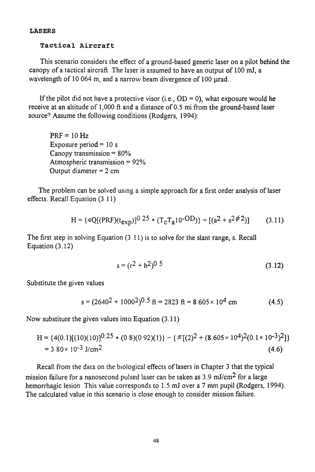
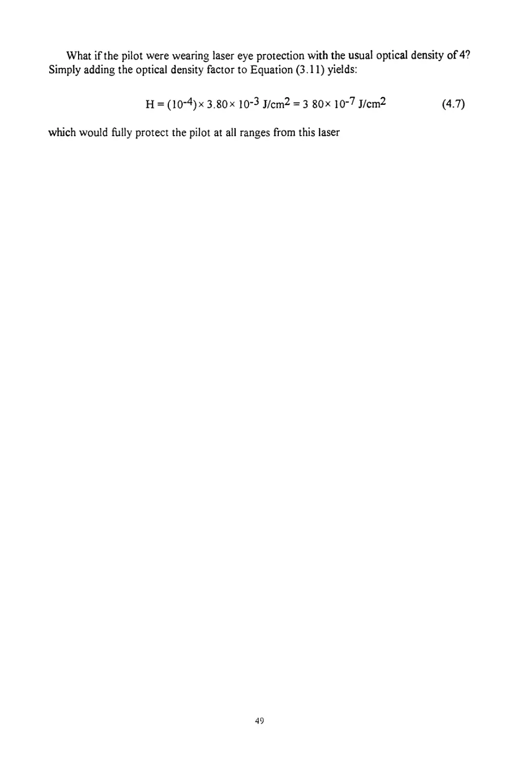
LASERS.................................................................. 48
Tactical A ircraft..........................................................48
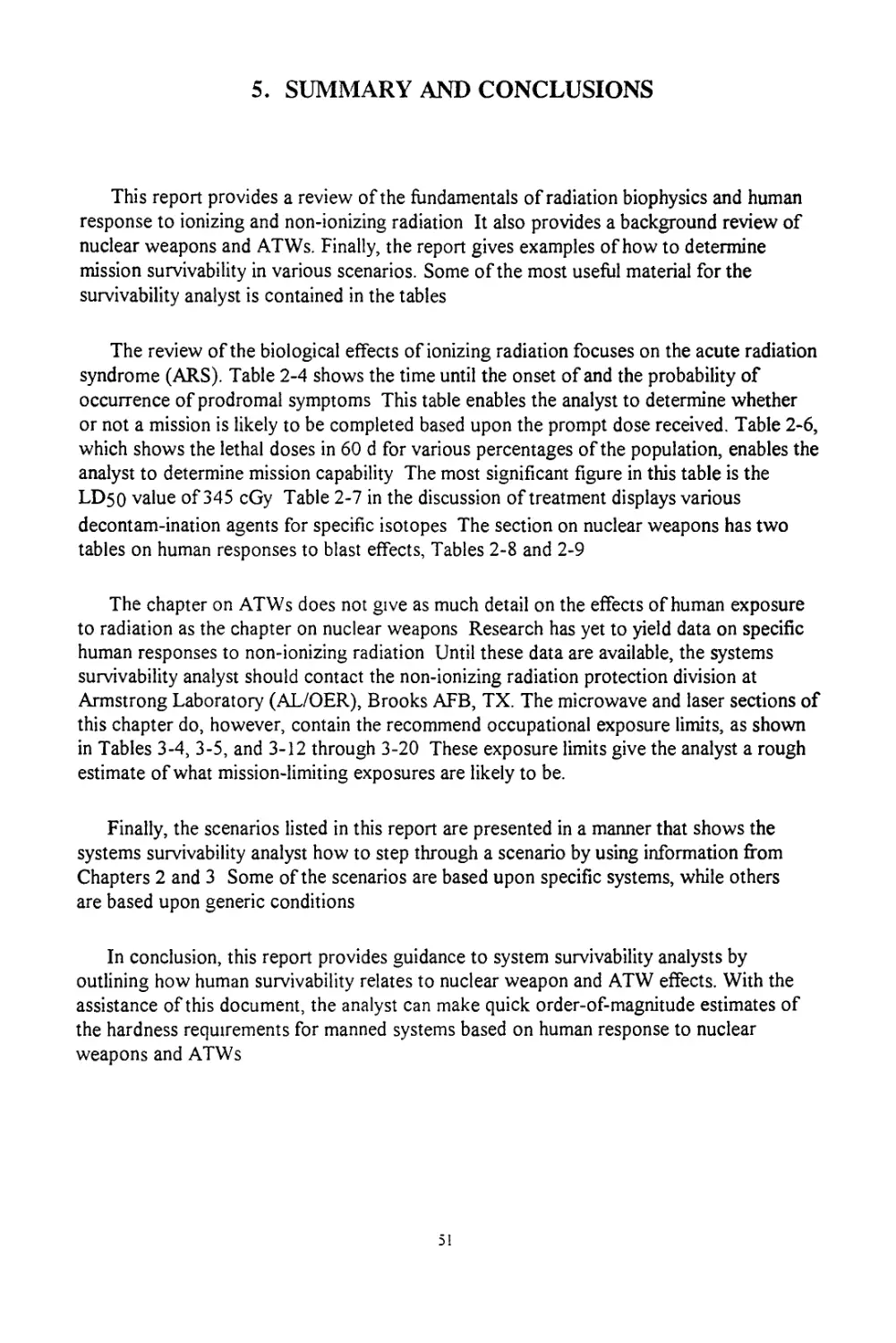
5. SUMMARY AND CONCLUSIONS.................................................51
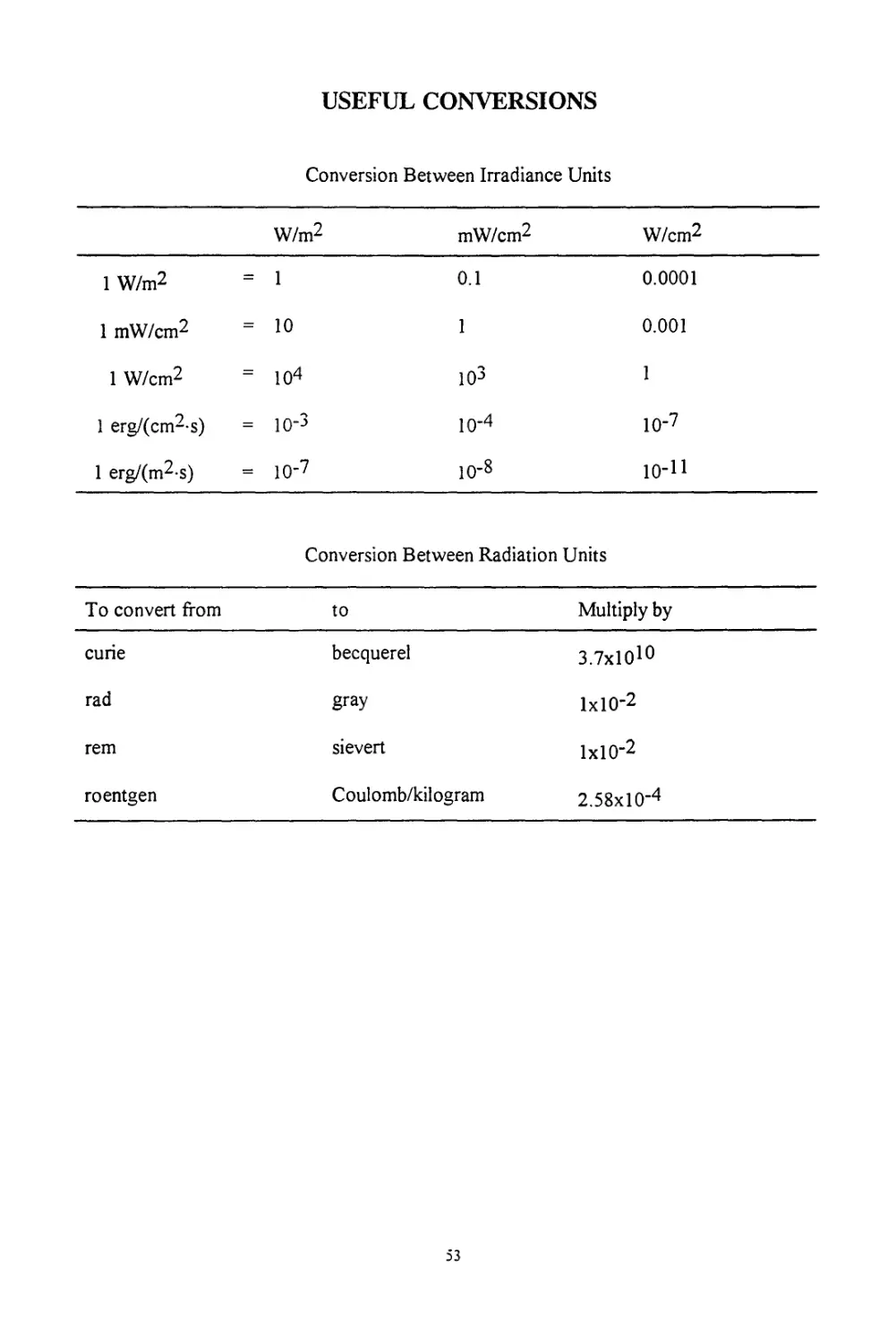
USEFUL CONVERSIONS..........................................................53
ACRONYMS AND ABBREVIATIONS..................................................55
GLOSSARY....................................................................59
REFERENCES..................................................................67
LIST OF TABLES
Table 2-1 Types of Radiation Damage..............................................................4
Table 2-2 Quality Factor Values for Various Radiations............................................5
Table 2-3. Phases of ARS .........................................................................8
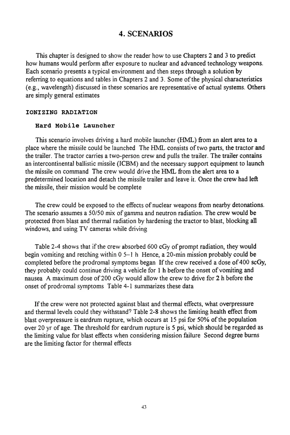
Table 2-4 Times Until Onset and Frequency of Symptoms.............................................9
Table 2-5. Acute Noncancer Effects of Radiation and Approximate Prompt Dose for 10% and 50%
Incidence in People........................................................................12
Table 2-6. Approximate Lethal Doses for 10%, 50%, and 90% of the Population......................13
Table 2-7. Decontamination Methods and Agents for Some Important Radionuclides...................14
Table 2-8. Blast Effects in Man................................................................ .16
Table 2-9 Estimated Long-Duration Blast Levels Required to Produce Lung and Gastrointestinal Tract
Injuries to Prone Man End-On to the Blast at Sea Level.....................................17
Table 3-1 Microwave Frequency Bands .............................................................23
Table 3-2. Electrical Properties of Human Muscle.... .25
Table 3-3. Electrical Properties of Human Fat.... .25
Table 3-4. ANSI Occupational Limits for RFEM Fields m Restricted Areas..........................28
Table 3-5. ANSI Occupational Limits for RFEM Fields in Nonrestncted Areas........................28
Table 3-6 Laser Spectrum Regions................................................................ 29
Table 3-7. Principal Characteristics of Common Lasers............................................31
Table 3-8. Thresholds for Retinal Damage.........................................................32
Table 3-9 Exposure Levels Required for MVL and VH ...................................32
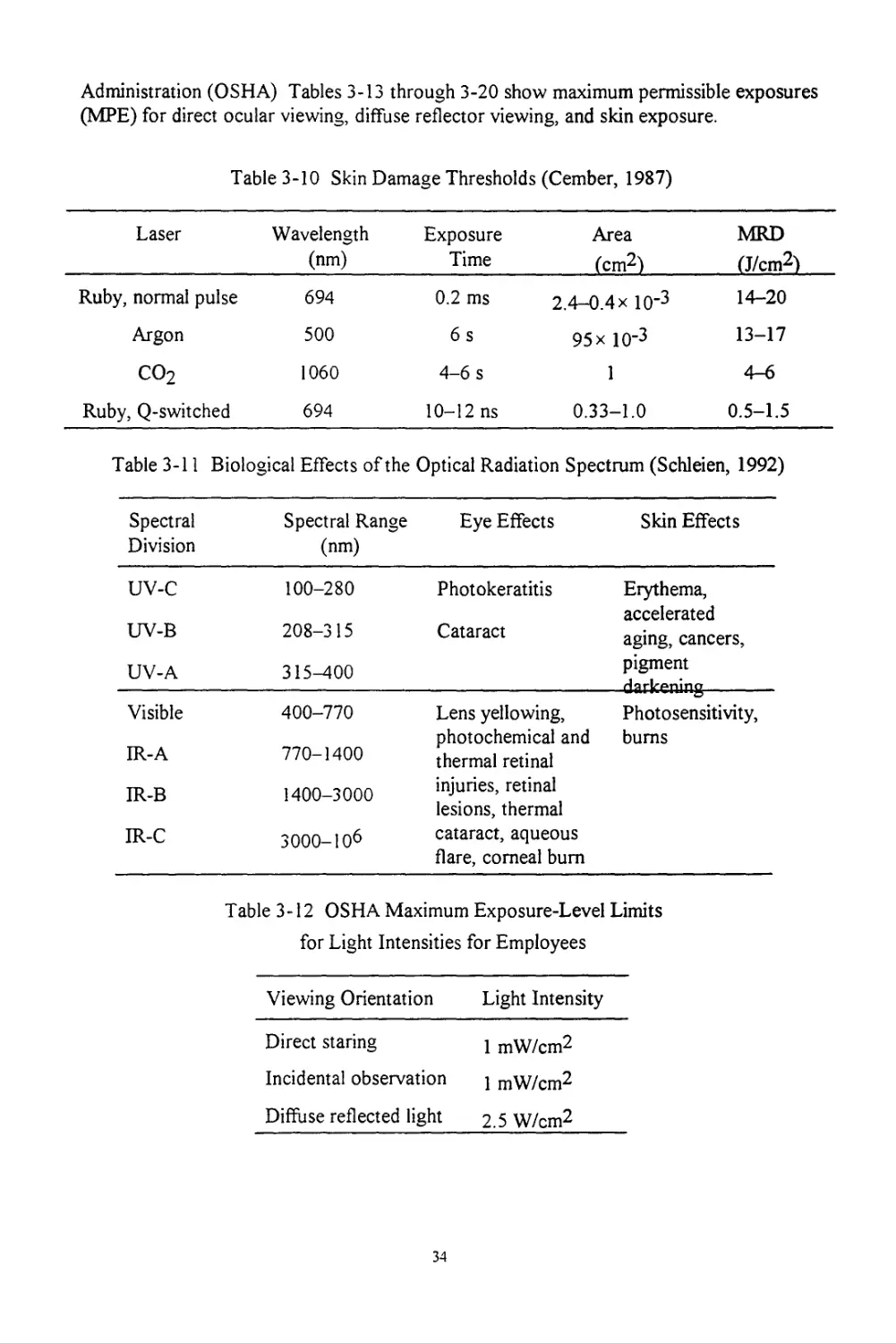
Table 3-10 Skin Damage Thresholds................................................................34
Table 3-11 Biological Effects of the Optical Radiation Spectrum..................................34
Table 3-12 OSHA Maximum Exposure-Level Limits for Light Intensities for Employees................34
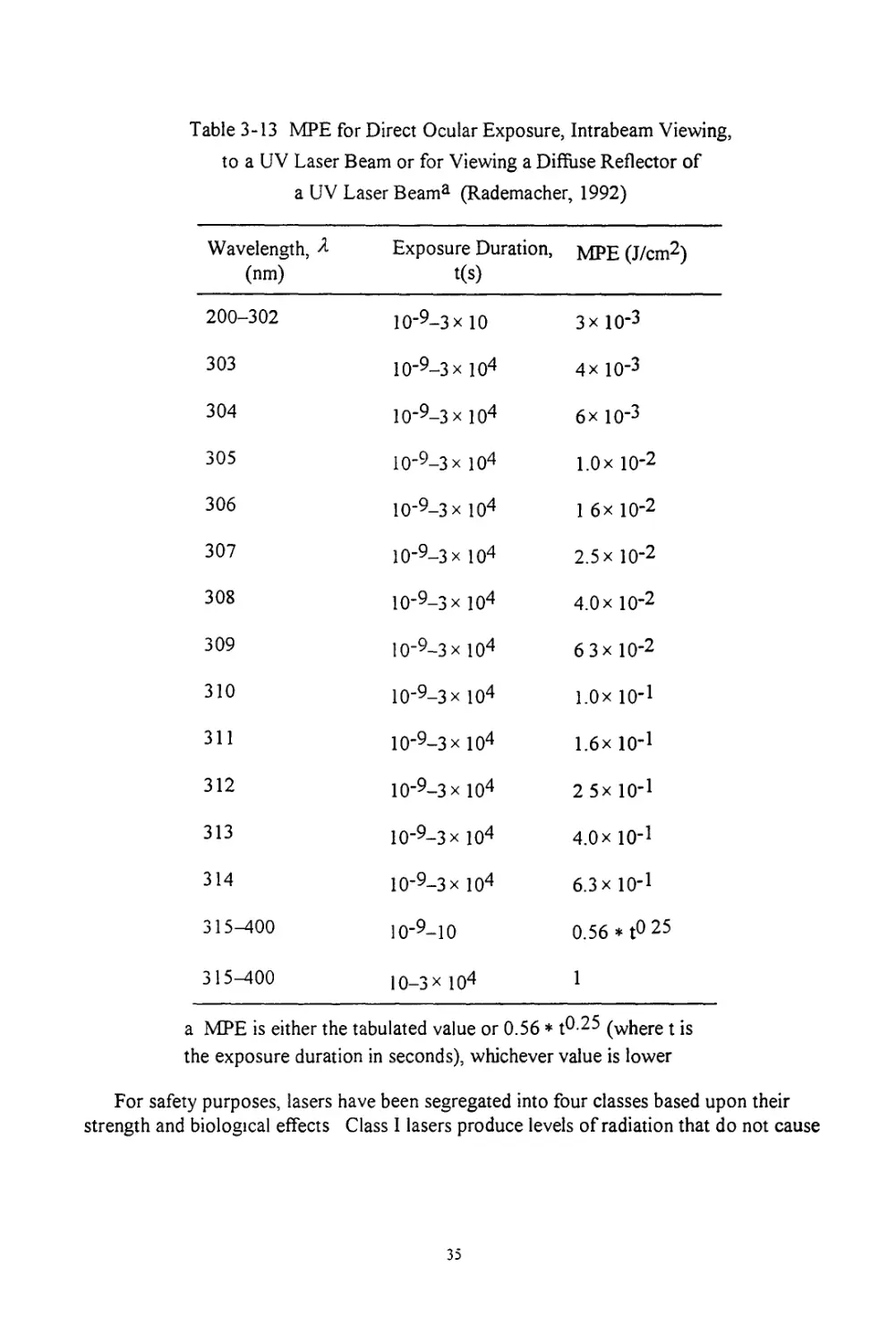
Table 3-13 MPE for Direct Ocular Exposure, Intrabeam Viewing, to a UV Laser Beam or for Viewing a
Diffuse Reflector of a UV Laser Beam ......................................................35
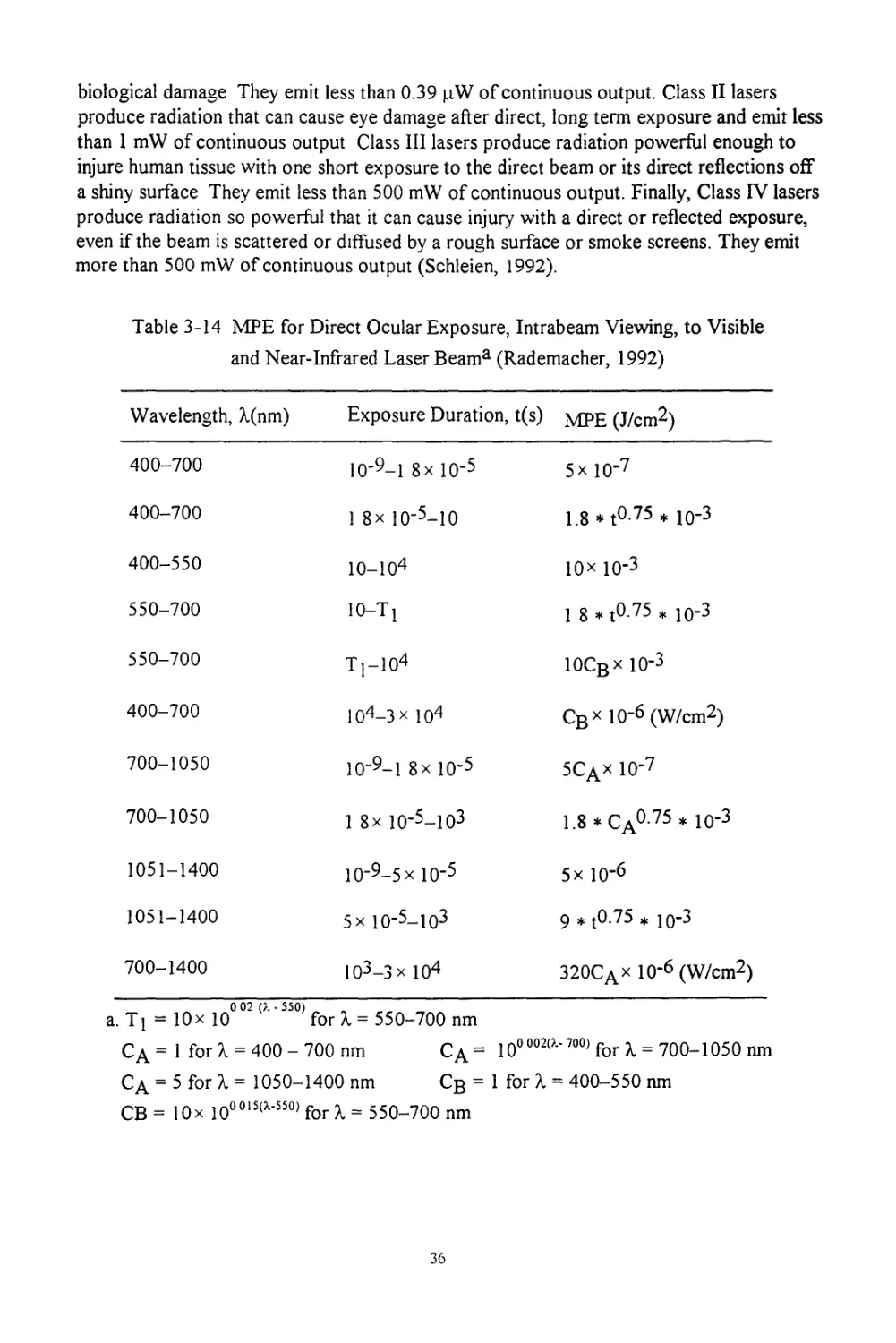
Table 3-14 MPE for Direct Ocular Exposure, Intrabeam Viewing, to Visible and Near-Infrared Laser
Beam.......................................................................................36
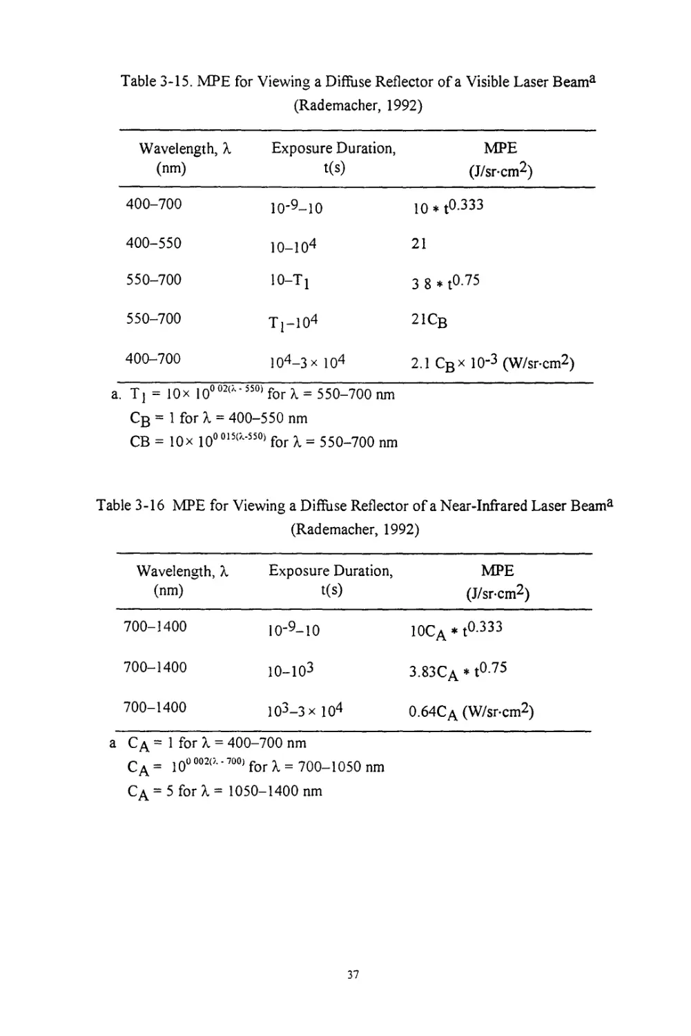
Table 3-15. MPE for Viewing a Diffuse Reflector of a Visible Laser Beam..........................37
Table 3-16 MPE for Viewing a Diffuse Reflector of a Near-Infrared Laser Beam.....................37
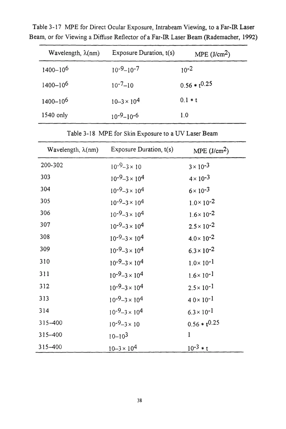
Table 3-17. MPE for Direct Ocular Exposure, Intrabeam Viewing, to a Far-IR Laser Beam, or for Viewing
a Diffuse Reflector of a Far-IR Laser Beam.................................................38
Table 3-18 MPE for Skin Exposure to a UV Laser Beam..............................................38
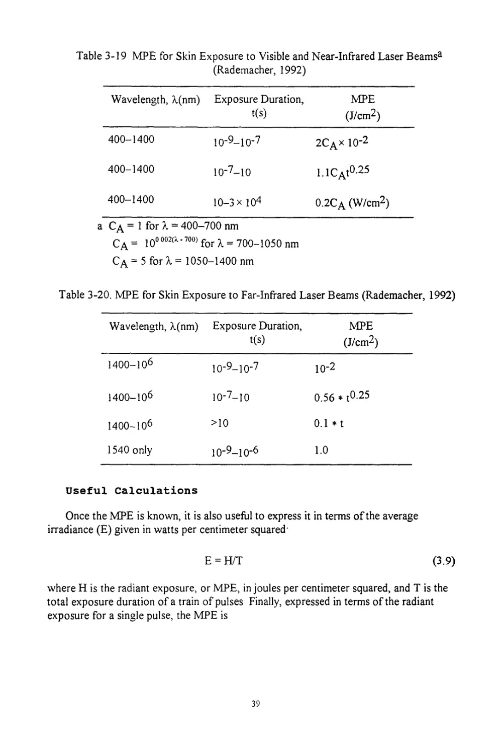
Table 3-19 MPE for Skin Exposure to Visible and Near-Infrared Laser Beams........................39
Table 3-20. MPE for Skin Exposure to Far-Infrared Laser Beams....................................39
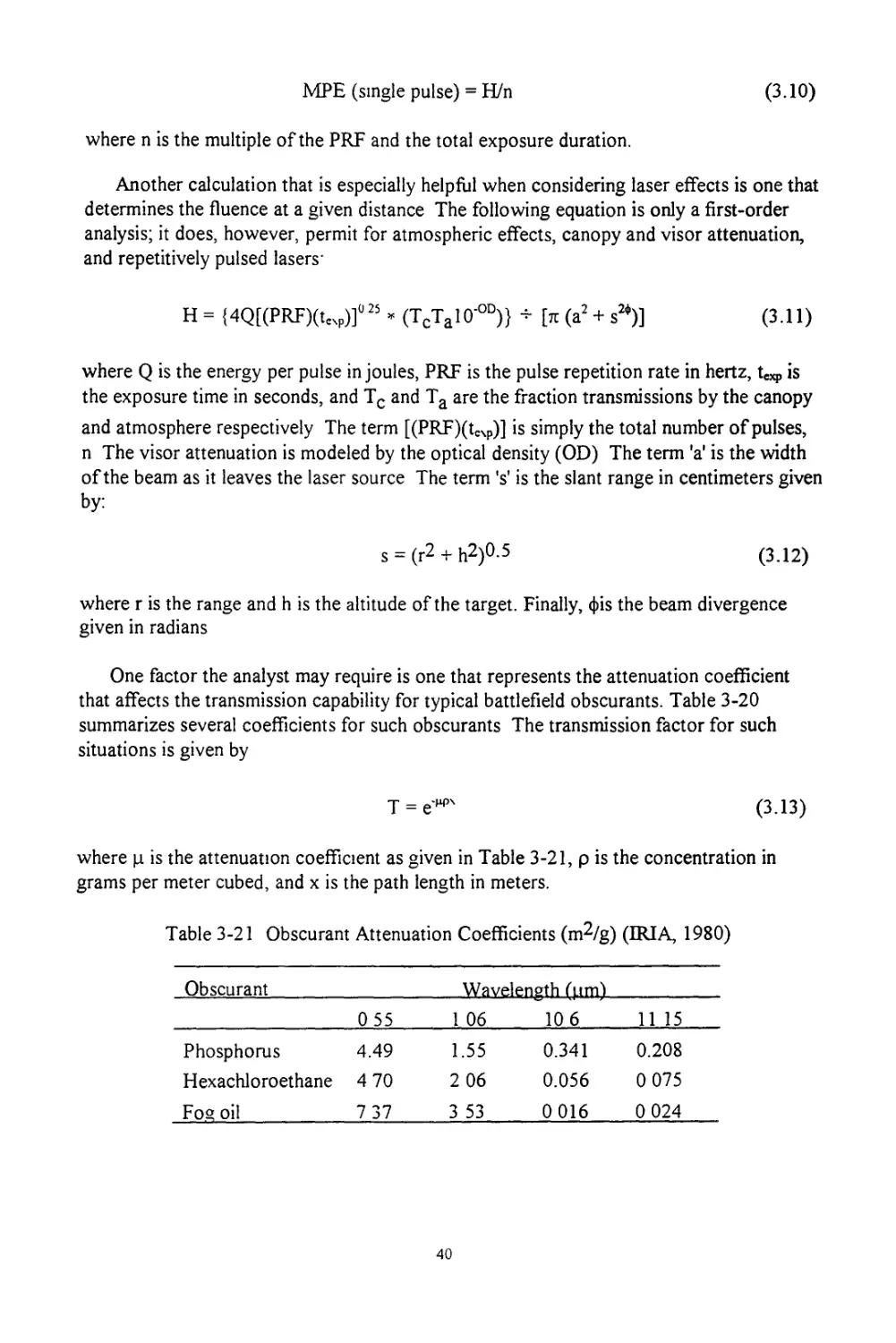
Table 3-21 Obscurant Attenuation Coefficients....................................................40
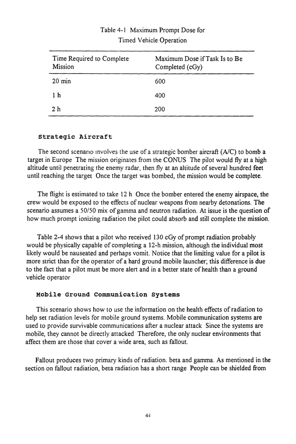
Table 4-1. Maximum Prompt Dose for Timed Vehicle Operation.......................................44
Conversion Between Irradiance Units .............................................................53
Conversion Between Radiation Units ..............................................................53
iv
LIST OF FIGURES
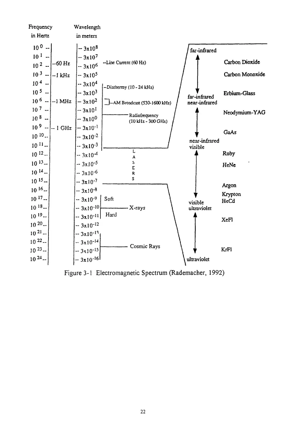
Figure 3-1 Electromagnetic Spectrum................................22
Figure 3-2 Ultraviolet Radiation Spectrum..........................23
1. INTRODUCTION
The purpose of this study is to help the system survivability analyst estimate hardness
requirements for systems exposed to nuclear weapons and advanced technology weapons
(ATWs) The system survivability analyst is often asked to make quick, order-of-
magnitude estimates on the hardness requirements for existing or proposed systems based
upon human responses to the effects of nuclear weapons and ATWs. While the analyst
may have training in mathematics or engineering, few have specific training in human
response to ionizing and non-ionizing radiation. As a result, system survivability analysts
have had difficulty answering mission survivability questions that are related to human
survivability The intent of this report is to identify the general range of human
survivability to nuclear weapons and ATWs and to provide simple example calculations
and scenarios that can give the reader rough estimates of the effects of these weapons.
While high-powered microwave (HPM) and laser weapons are considered in this report,
the main emphasis is on nuclear weapons and their ionizing radiation effects.
Chapter 2 deals with nuclear weapon effects, principally the effects of ionizing
radiation. The first section of the chapter reviews the basics of radiation biophysics as they
apply to ionizing radiation Subsections on radiation effects and dosimetry and on the
biology of radiosensitive tissues and organs provide background on the biological effects
of radiation A subsection on acute radiation syndrome (ARS) provides the data on human
response to different levels of radiation exposure that are critical for the analyst in
determining mission survivability A subsection on treatment gives recommendations for
handling external and internal contamination and external exposure. The last section of
Chapter 2 has subsections on blast overpressure, the effects of thermal radiation, and the
effects of ionizing radiation (prompt and fallout).
Chapter 3 begins with a very brief introduction to non-ionizing radiation to help the
reader understand the other sections in the chapter. The following section on HPMs is
divided into three main subsections a brief discussion of the physical characteristics of
microwaves; a discussion of biological effects, which includes equations for calculating the
depth of the microwave thermal effect, and some data on occupational limits. The last
section of Chapter 3 is on lasers This section mirrors the organization of the section on
HPMs, but contains additional subsections on useful calculations and on different filtering
methods for protection against laser weapons
Chapter 4 is designed to give the reader a better understanding of how to determine
human effects for several hypothetical scenarios involving ionizing radiation, high-
powered radar, and lasers. The AF systems addressed in these scenarios include strategic
and tactical aircraft, ground systems, and mobile launchers. The chapter shows the analyst
how to work through a problem by using information from Chapters 2 and 3. The
discussion includes several simple calculations.
Chapter 5 is the conclusion of the report The chapter is followed by some useful
conversions displayed in tabular form, a list of definitions for all the acronyms and
abbreviations, an extensive glossary of technical and medical terms, and a list of
references.
2. NUCLEAR WEAPONS EFFECTS
This chapter reviews the basic characteristics of nuclear weapons effects. The effects
considered here are blast overpressure, thermal radiation, and ionizing radiation. The blast
overpressure and thermal radiation sections describe briefly human response to such
effects. In order to better understand the effects from ionizing radiation or prompt
radiation and fallout, this chapter begins with an in-depth review of the biological effects
of ionizing radiation
BIOLOGICAL EFFECTS OF IONIZING RADIATION
The biological effects of ionizing radiation have been well researched during the last
several decades This section provides the most current quantification of biological effects
that are accepted by the health physics community. The first subsection provides a very
basic background on qualitative ionizing radiation effects and basic dosimetry. The second
subsection lists several of the most radiosensitive tissues and organs, explains why such
tissues and organs are radiosensitive, and gives dose effects for each. The third subsection
explains the acute radiation syndrome in detail, while the fourth subsection gives a brief
discussion of exposure treatment
Radiation Effects and Dosimetry
The biological effects of radiation depend upon the type of radiation, the absorbed
dose, and how large a part and what part of the body is exposed. Biological effects are
classified as either somatic or genetic. Somatic effects are those that occur in the exposed
individual, whereas genetic effects are those that occur in the exposed individual's
descendants Since genetic effects are not mission-critical, only somatic effects will be
discussed in this report
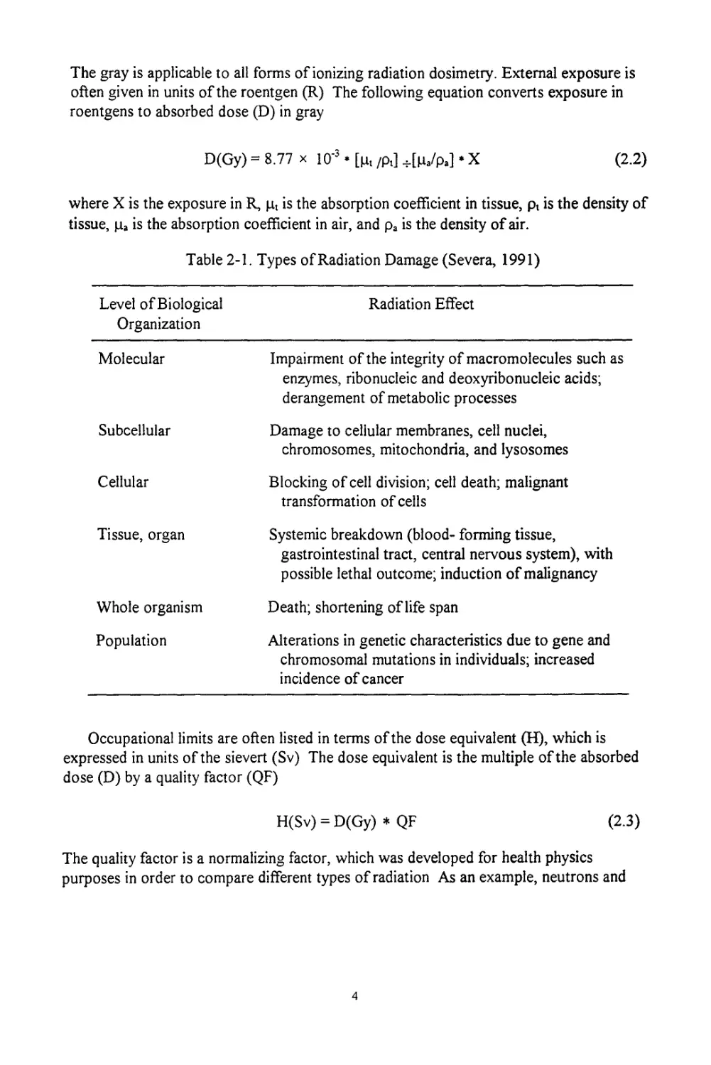
Table 2-1 shows radiation effects for the different levels of biological organization.
Although most of these levels will be mentioned in this report, the main emphasis is given
to radiation effects on the whole organism because these are the effects that have the most
impact upon a mission
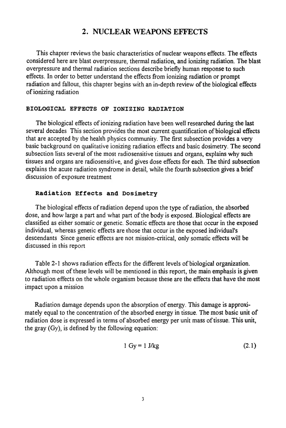
Radiation damage depends upon the absorption of energy. This damage is approxi-
mately equal to the concentration of the absorbed energy in tissue. The most basic unit of
radiation dose is expressed in terms of absorbed energy per unit mass of tissue. This unit,
the gray (Gy), is defined by the following equation:
1 Gy= 1 J/kg
(2.1)
The gray is applicable to all forms of ionizing radiation dosimetry. External exposure is
often given in units of the roentgen (R) The following equation converts exposure in
roentgens to absorbed dose (D) in gray
D(Gy) = 8.77 x Ю’3 * [Ml /Pt] -[Ma/Ра] ‘ X
(2.2)
where X is the exposure in R, pt is the absorption coefficient in tissue, pt is the density of
tissue, pa is the absorption coefficient in air, and pa is the density of air.
Table 2-1. Types of Radiation Damage (Severa, 1991)
Level of Biological Organization Radiation Effect
Molecular Impairment of the integrity of macromolecules such as enzymes, ribonucleic and deoxyribonucleic acids; derangement of metabolic processes
Subcellular Damage to cellular membranes, cell nuclei, chromosomes, mitochondria, and lysosomes
Cellular Blocking of cell division; cell death; malignant transformation of cells
Tissue, organ Systemic breakdown (blood- forming tissue, gastrointestinal tract, central nervous system), with possible lethal outcome; induction of malignancy
Whole organism Death; shortening of life span
Population Alterations in genetic characteristics due to gene and chromosomal mutations in individuals; increased incidence of cancer
Occupational limits are often listed in terms of the dose equivalent (H), which is
expressed in units of the sievert (Sv) The dose equivalent is the multiple of the absorbed
dose (D) by a quality factor (QF)
H(Sv) = D(Gy) * QF
(2-3)
The quality factor is a normalizing factor, which was developed for health physics
purposes in order to compare different types of radiation As an example, neutrons and
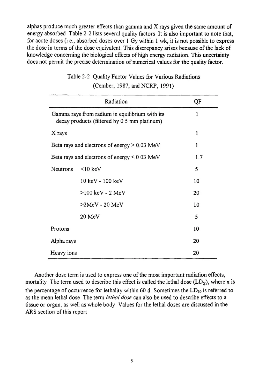
alphas produce much greater effects than gamma and X rays given the same amount of
energy absorbed Table 2-2 lists several quality factors It is also important to note that,
for acute doses (i e., absorbed doses over 1 Gy within 1 wk, it is not possible to express
the dose in terms of the dose equivalent. This discrepancy arises because of the lack of
knowledge concerning the biological effects of high energy radiation. This uncertainty
does not permit the precise determination of numerical values for the quality factor.
Table 2-2 Quality Factor Values for Various Radiations
(Cember, 1987, andNCRP, 1991)
Radiation QF
Gamma rays from radium in equilibrium with its decay products (filtered by 0 5 mm platinum) 1
X rays 1
Beta rays and electrons of energy > 0.03 MeV 1
Beta rays and electrons of energy < 0 03 MeV 1.7
Neutrons <10 keV 5
10 keV - 100 keV 10
>100 keV - 2 MeV 20
>2MeV - 20 MeV 10
20 MeV 5
Protons 10
Alpha rays 20
Heavy ions 20
Another dose term is used to express one of the most important radiation effects,
mortality The term used to describe this effect is called the lethal dose (LDX), where x is
the percentage of occurrence for lethality within 60 d. Sometimes the LD50 is referred to
as the mean lethal dose The term lethal dose can also be used to describe effects to a
tissue or organ, as well as whole body Values for the lethal doses are discussed in the
ARS section of this report
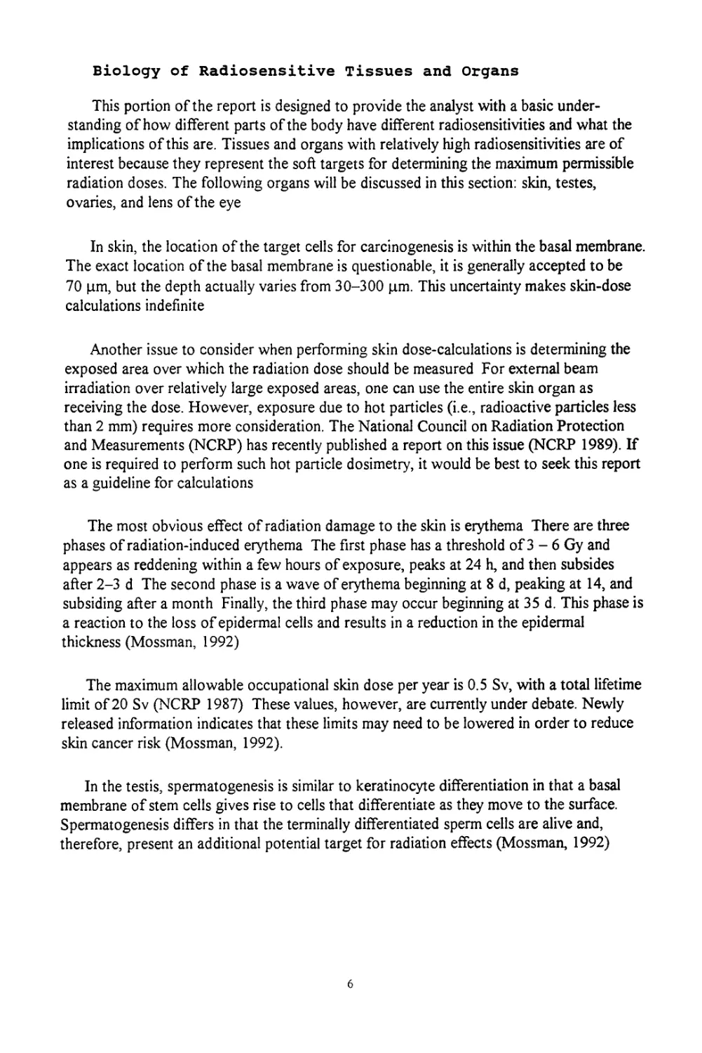
Biology of Radiosensitive Tissues and Organs
This portion of the report is designed to provide the analyst with a basic under-
standing of how different parts of the body have different radiosensitivities and what the
implications of this are. Tissues and organs with relatively high radiosensitivities are of
interest because they represent the soft targets for determining the maximum permissible
radiation doses. The following organs will be discussed in this section: skin, testes,
ovaries, and lens of the eye
In skin, the location of the target cells for carcinogenesis is within the basal membrane.
The exact location of the basal membrane is questionable, it is generally accepted to be
70 цт, but the depth actually varies from 30-300 pirn. This uncertainty makes skin-dose
calculations indefinite
Another issue to consider when performing skin dose-calculations is determining the
exposed area over which the radiation dose should be measured For external beam
irradiation over relatively large exposed areas, one can use the entire skin organ as
receiving the dose. However, exposure due to hot particles (i.e., radioactive particles less
than 2 mm) requires more consideration. The National Council on Radiation Protection
and Measurements (NCRP) has recently published a report on this issue (NCRP 1989). If
one is required to perform such hot particle dosimetry, it would be best to seek this report
as a guideline for calculations
The most obvious effect of radiation damage to the skin is erythema There are three
phases of radiation-induced erythema The first phase has a threshold of 3 - 6 Gy and
appears as reddening within a few hours of exposure, peaks at 24 h, and then subsides
after 2-3 d The second phase is a wave of erythema beginning at 8 d, peaking at 14, and
subsiding after a month Finally, the third phase may occur beginning at 35 d. This phase is
a reaction to the loss of epidermal cells and results in a reduction in the epidermal
thickness (Mossman, 1992)
The maximum allowable occupational skin dose per year is 0.5 Sv, with a total lifetime
limit of 20 Sv (NCRP 1987) These values, however, are currently under debate. Newly
released information indicates that these limits may need to be lowered in order to reduce
skin cancer risk (Mossman, 1992).
In the testis, spermatogenesis is similar to keratinocyte differentiation in that a basal
membrane of stem cells gives rise to cells that differentiate as they move to the surface.
Spermatogenesis differs in that the terminally differentiated sperm cells are alive and,
therefore, present an additional potential target for radiation effects (Mossman, 1992)
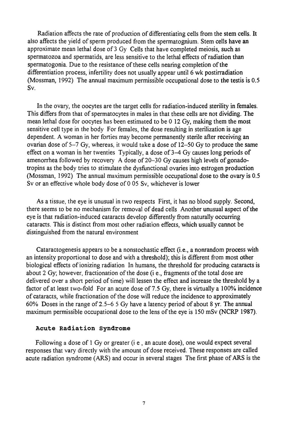
Radiation affects the rate of production of differentiating cells from the stem cells. It
also affects the yield of sperm produced from the spermatognium. Stem cells have an
approximate mean lethal dose of 3 Gy Cells that have completed meiosis, such as
spermatozoa and spermatids, are less sensitive to the lethal effects of radiation than
spermatogonia. Due to the resistance of these cells nearing completion of the
differentiation process, infertility does not usually appear until 6 wk postirradiation
(Mossman, 1992) The annual maximum permissible occupational dose to the testis is 0.5
Sv.
In the ovary, the oocytes are the target cells for radiation-induced sterility in females.
This differs from that of spermatocytes in males in that these cells are not dividing. The
mean lethal dose for oocytes has been estimated to be 0 12 Gy, making them the most
sensitive cell type in the body For females, the dose resulting in sterilization is age
dependent. A woman in her forties may become permanently sterile after receiving an
ovarian dose of 5-7 Gy, whereas, it would take a dose of 12-50 Gy to produce the same
effect on a woman in her twenties Typically, a dose of 3-4 Gy causes long periods of
amenorrhea followed by recovery A dose of 20-30 Gy causes high levels of gonado-
tropins as the body tries to stimulate the dysfunctional ovaries into estrogen production
(Mossman, 1992) The annual maximum permissible occupational dose to the ovary is 0.5
Sv or an effective whole body dose of 0 05 Sv, whichever is lower
As a tissue, the eye is unusual in two respects First, it has no blood supply. Second,
there seems to be no mechanism for removal of dead cells Another unusual aspect of the
eye is that radiation-induced cataracts develop differently from naturally occurring
cataracts. This is distinct from most other radiation effects, which usually cannot be
distinguished from the natural environment
Cataractogenesis appears to be a nonstochastic effect (i.e., a nonrandom process with
an intensity proportional to dose and with a threshold); this is different from most other
biological effects of ionizing radiation In humans, the threshold for producing cataracts is
about 2 Gy; however, fractionation of the dose (i e., fragments of the total dose are
delivered over a short period of time) will lessen the effect and increase the threshold by a
factor of at least two-fold For an acute dose of 7.5 Gy, there is virtually a 100% incidence
of cataracts, while fractionation of the dose will reduce the incidence to approximately
60% Doses in the range of 2.5-6 5 Gy have a latency period of about 8 yr. The annual
maximum permissible occupational dose to the lens of the eye is 150 mSv (NCRP 1987).
Acute Radiation Syndrome
Following a dose of 1 Gy or greater (i e , an acute dose), one would expect several
responses that vary directly with the amount of dose received. These responses are called
acute radiation syndrome (ARS) and occur in several stages The first phase of ARS is the
prodromal phase that lasts from 1-7 d Many different symptoms are associated with the
prodromal phase The following is a list of such symptoms given in approximate order of
increasing severity anorexia, nausea, vomiting, weakness and fatigue, prostration,
diarrhea, conjunctivitis, erythema, hypersthesia, paraesthesia, shock, oliguria, ataxia,
disorientation, coma, and death The next phase is the latent phase that lasts from 7-21 d.
Next is the critical phase that occurs from the 2nd wk or 3rd wk up to the 7th wk. The
following symptoms are those for the critical phase of ARS: fever, abdominal pain,
abdominal distention, scalp pain, epilation, infection, purpura, hemorrhage, and weight
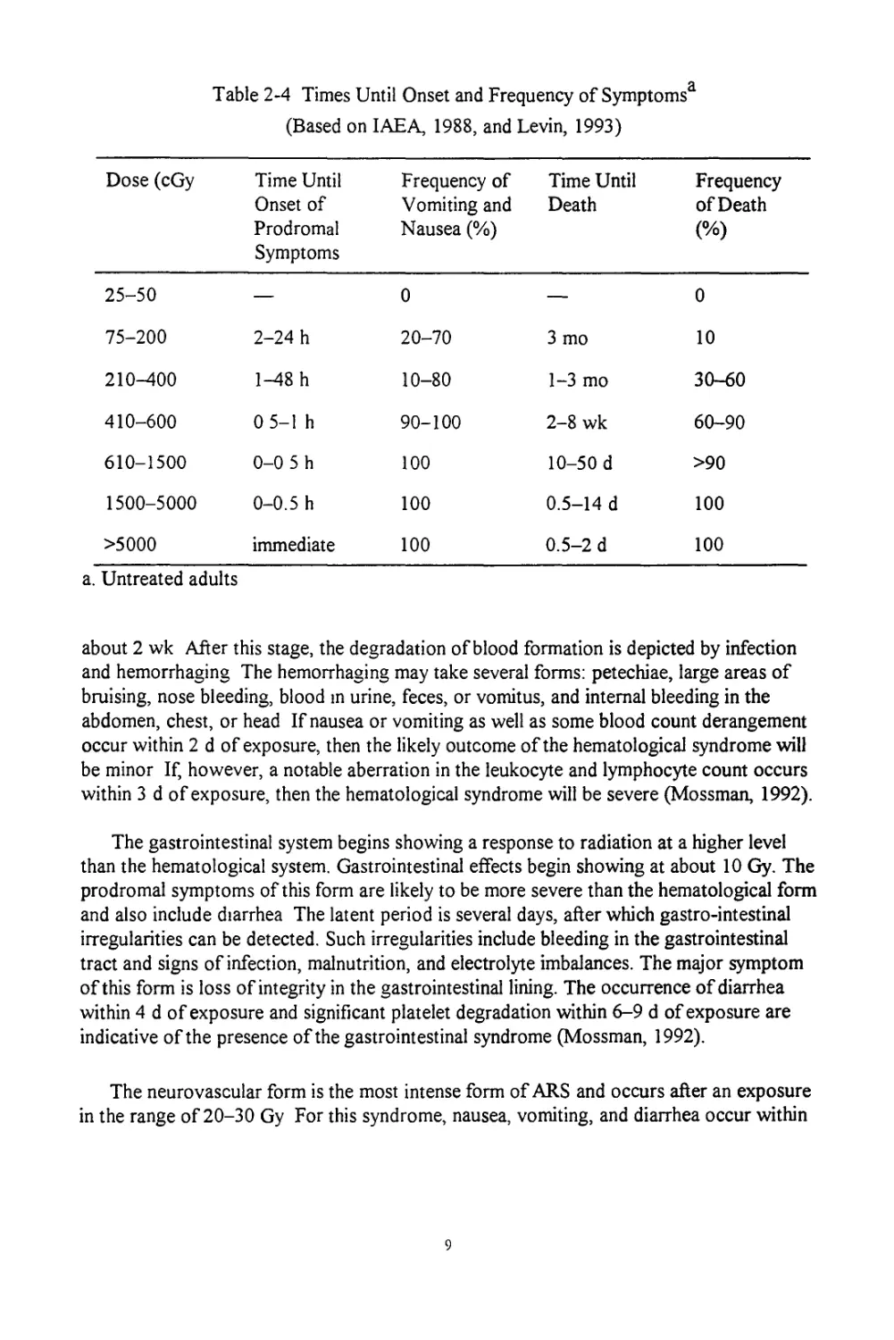
loss. Finally, recovery or death occurs in 8-15 wk Table 2-3 summarizes the phases and
time frames for ARS, while Table 2-4 shows the time until the onset of prodromal
symptoms and death for various doses.
Table 2-3 Phases of ARS
ARS Phase Time Frame Postsexposure
Prodromal 1-7 d
Latent 7-21 d
Critical 2nd-3rd wk up to 7th wk
Death or Recovery 8-15 wk
Besides chronological order, it is also convenient to break ARS into several categories
of response The three main categories in order of increasing severity are the
hematological syndrome, the gastrointestinal syndrome, and the neurovascular syndrome.
A fourth syndrome has recently been discovered, the pulmonary syndrome. Certain effects
are common to all categories' nausea and vomiting, malaise and fatigue, increased
temperature, and blood changes
The hematological category of ARS is the least troublesome of the four categories. In
the dose range of 0-0 25 Gy, virtually no clinical symptoms exist, but a slightly increased
frequency of chromosome aberrations may be detected in lymphocytes. In the range of
0.25-1 Gy, either no symptoms exist or transient nausea occurs. It is also possible at this
stage that lymphopenia occurs and may be accompanied by slight thrombopenia.
Cytogenetic changes in lymphocytes are readily detected Some studies have shown slight
changes in the patient's electroencephalogram In general, the hematological form is
exhibited when the dose is in the LD50 range (3-4 Gy) The symptoms begin with the
prodromal symptoms of nausea and vomiting for approximately 48 h, however, nausea
and vomiting can occur after an exposure of only 1.5 Gy Then a latent period occurs for
Table 2-4 Times Until Onset and Frequency of Symptomsa
(Based on IAEA, 1988, and Levin, 1993)
Dose (cGy Time Until Onset of Prodromal Symptoms Frequency of Vomiting and Nausea (%) Time Until Death Frequency of Death (%)
25-50 — 0 — 0
75-200 2-24 h 20-70 3 mo 10
210-400 1-48 h 10-80 1-3 mo 30-60
410-600 0 5-1 h 90-100 2-8 wk 60-90
610-1500 0-0 5 h 100 10-50 d >90
1500-5000 0-0.5 h 100 0.5-14 d 100
>5000 immediate 100 0.5-2 d 100
a. Untreated adults
about 2 wk After this stage, the degradation of blood formation is depicted by infection
and hemorrhaging The hemorrhaging may take several forms: petechiae, large areas of
bruising, nose bleeding, blood m urine, feces, or vomitus, and internal bleeding in the
abdomen, chest, or head If nausea or vomiting as well as some blood count derangement
occur within 2 d of exposure, then the likely outcome of the hematological syndrome will
be minor If, however, a notable aberration in the leukocyte and lymphocyte count occurs
within 3 d of exposure, then the hematological syndrome will be severe (Mossman, 1992).
The gastrointestinal system begins showing a response to radiation at a higher level
than the hematological system. Gastrointestinal effects begin showing at about 10 Gy. The
prodromal symptoms of this form are likely to be more severe than the hematological form
and also include diarrhea The latent period is several days, after which gastro-intestinal
irregularities can be detected. Such irregularities include bleeding in the gastrointestinal
tract and signs of infection, malnutrition, and electrolyte imbalances. The major symptom
of this form is loss of integrity in the gastrointestinal lining. The occurrence of diarrhea
within 4 d of exposure and significant platelet degradation within 6-9 d of exposure are
indicative of the presence of the gastrointestinal syndrome (Mossman, 1992).
The neurovascular form is the most intense form of ARS and occurs after an exposure
in the range of 20-30 Gy For this syndrome, nausea, vomiting, and diarrhea occur within
minutes. Ataxia, disorientation, shock, and coma occur in minutes to hours. The
prodromal symptoms are accompanied by the state of consciousness ranging from apathy
and lethargy to hyperexcitability, tremors, convulsions, and gait disturbances. Death
occurs within hours up to 1 or 2 d (Mossman, 1992)
The pulmonary form is typically seen in therapeutically irradiated patient. However, it
was also diagnosed in seven Chernobyl accident patients. The threshold dose range for this
category is around 8 to 9 Gy Interstitial edema and respiratory failure may occur within 3
d, or interstitial pneumonitis may occur 14-30 d postirradiation (Mossman, 1992).
Finally, another way to break down the symptoms of ARS is by dose received. The
following discussion describes what occurs for varying doses in increasing order. For
doses in the range of 1-2 Gy, ARS takes a mild degree of the hematopoietic form. Nausea
and vomiting occur m only a fraction of the irradiated persons The symptoms occur in the
first hours after exposure Development of neutropenia and thrombopenia takes 4-6 wk.
This degree of cytopenia usually does not lead to bleeding and infection. The majority of
the patients recover without treatment However, to prevent infection and hemorrhagic
symptoms, careful hematological follow-up and care are required (IAEA, 1988).
Doses in the range of 2-4 Gy yield a moderate degree of the hematological syndrome.
Nausea and vomiting appear after 1-2 h In most victims, the lowest point of neutropenia
and thrombopenia develops in 3-4 wk In all patients, these symptoms are accompanied by
fever and bleeding If the exposed person is given medical treatment, the chances of a full
recovery are about 90% Without medical treatment, mortality can be as high as 50%
(IAEA, 1988)
Doses of 4-6 Gy produce a severe response Nausea and vomiting appear 0.5-1 h after
irradiation Other prodromal symptoms are manifest’ early fever, erythema of skin, and
mucosae The deepest fall in the number of neutrophils and thrombocytes occurs at 2-3 wk
and very low values are reached The latter are maintained for a period of about 2 wk.
Without therapy, the majority of patients will die of infections and the consequences of
bleeding If adequate supportive therapy is instituted, the majority of patients are likely to
recover (IAEA, 1988)
For doses in the range of 6-10 Gy, extremely severe hematopoietic syndrome occurs.
Nausea and vomiting appear very early, less than 30 min after exposure In a substantial
proportion of the victims, diarrhea occurs 1-2 h after the irradiation Maximal cytopenia
appears at 10-14 d Also, at 6-8 d postexposure, acute radiation stomatitis becomes
evident. Sometimes at 8-10 d after exposure, radiation-induced enteritis is seen. Without
medical treatment, 100% of the people exposed to 6-10 Gy probably will die. If therapy,
which will be discussed later, is applied soon after exposure, a fraction of the exposed
population still may survive The lethality results from the coincidence of severe impair-
10
ment of the hematopoietic function and radiation injury to other organs, mostly of the
pharyngeal mucosa, esophagus, and intestines Injury to pulmonary tissue may also play a
significant role (IAEA, 1988)
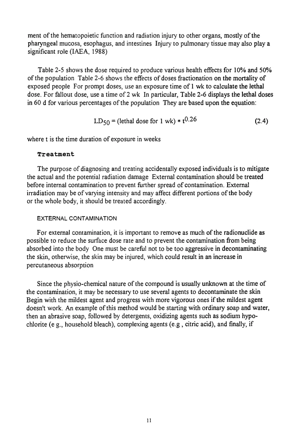
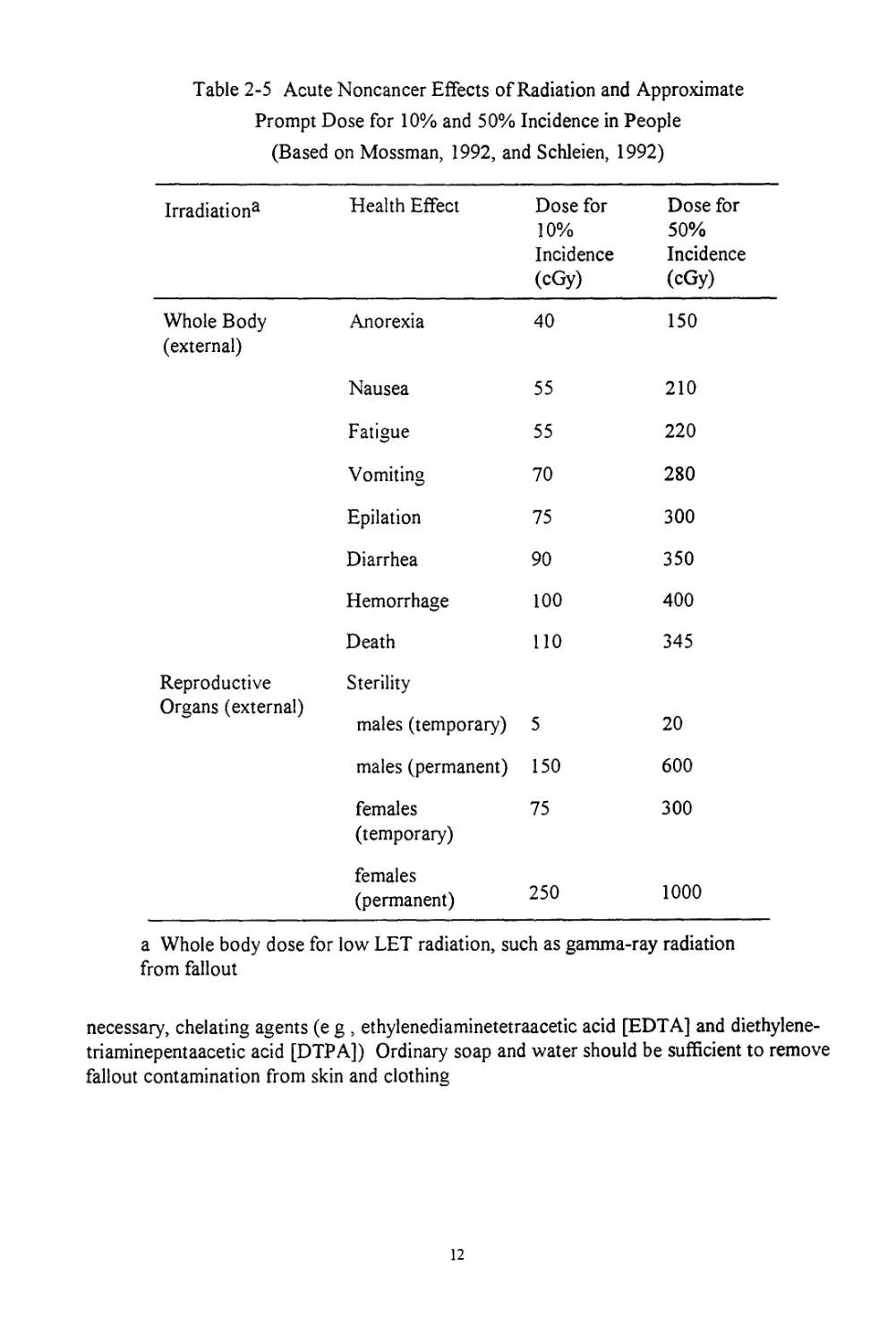
Table 2-5 shows the dose required to produce various health effects for 10% and 50%
of the population Table 2-6 shows the effects of doses fractionation on the mortality of
exposed people For prompt doses, use an exposure time of 1 wk to calculate the lethal
dose. For fallout dose, use a time of 2 wk In particular, Table 2-6 displays the lethal doses
in 60 d for various percentages of the population They are based upon the equation:
LD50 = (lethal dose for 1 wk) * t0-26 (2.4)
where t is the time duration of exposure in weeks
Treatment
The purpose of diagnosing and treating accidentally exposed individuals is to mitigate
the actual and the potential radiation damage External contamination should be treated
before internal contamination to prevent further spread of contamination. External
irradiation may be of varying intensity and may affect different portions of the body
or the whole body, it should be treated accordingly.
EXTERNAL CONTAMINATION
For external contamination, it is important to remove as much of the radionuclide as
possible to reduce the surface dose rate and to prevent the contamination from being
absorbed into the body One must be careful not to be too aggressive in decontaminating
the skin, otherwise, the skin may be injured, which could result in an increase in
percutaneous absorption
Since the physio-chemical nature of the compound is usually unknown at the time of
the contamination, it may be necessary to use several agents to decontaminate the skin
Begin with the mildest agent and progress with more vigorous ones if the mildest agent
doesn't work. An example of this method would be starting with ordinary soap and water,
then an abrasive soap, followed by detergents, oxidizing agents such as sodium hypo-
chlorite (e g., household bleach), complexing agents (e.g, citric acid), and finally, if
Table 2-5 Acute Noncancer Effects of Radiation and Approximate
Prompt Dose for 10% and 50% Incidence in People
(Based on Mossman, 1992, and Schleien, 1992)
Irradiation3 Health Effect Dose for 10% Incidence (cGy) Dose for 50% Incidence (cGy)
Whole Body Anorexia 40 150
(external)
Nausea 55 210
Fatigue 55 220
Vomiting 70 280
Epilation 75 300
Diarrhea 90 350
Hemorrhage 100 400
Death 110 345
Reproductive Sterility
Organs (external)
males (temporary) 5 20
males (permanent) 150 600
females 75 300
(temporary)
females
(permanent) 250 1000
a Whole body dose for low LET radiation, such as gamma-ray radiation
from fallout
necessary, chelating agents (e g , ethylenediaminetetraacetic acid [EDTA] and diethylene-
triaminepentaacetic acid [DTPA]) Ordinary soap and water should be sufficient to remove
fallout contamination from skin and clothing
Table 2-6 Approximate Lethal Doses for 10%, 50%, and
90% of the Population (Based on Schleien, 1992)
Timea (wk) LDW (cGy) ld50 (cGy) ld90 (cGy)
1 110 345 585
2 130 410 700
3 145 460 780
4 160 495 840
8 190 590 1005
24 250 790 1340
a Duration of radiation exposure time
INTERNAL CONTAMINATION
The first step of internal decontamination is to treat any radionuclide-contaminated
wounds Such wounds should be rinsed with water or saline to remove as much of the
compound as possible Once this is done, treat the wound as appropriate. If internal
deposition occurs, whether it be through ingestion or a wound, intervention to decrease
the natural removal time will probably be advantageous to decrease the absorbed dose.
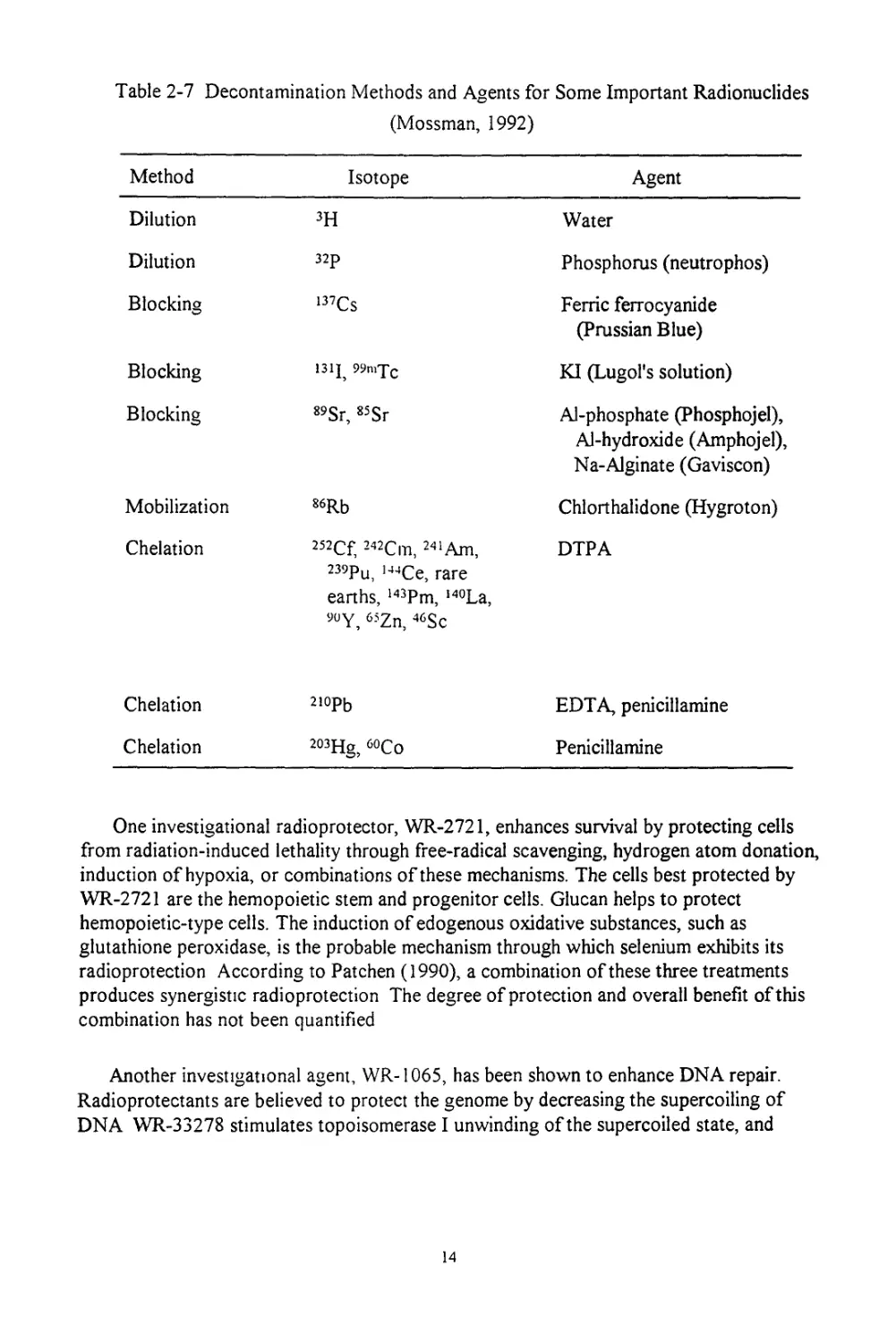
Internally deposited radionuclides may be removed by several methods: dilution,
blocking, mobilization, and chelation. In the dilution method large quantities of the stable
element are administered so that, statistically, the possibility for incorporation and
exposure of the radionuclide is decreased Blocking is a preventative method by which the
agent saturates the metabolic process in a specific tissue with the stable element, thereby
reducing the uptake of the radionuclide Mobilization is a method that increases a natural
turnover process and induces a release of some forms of radionuclides from body tissues.
Chelation is actually a special class of mobilization It is a process by which organic
compounds exchange less firmly bonded ions for other inorganic ions to form a relatively
stable non-ionized ring complex This process converts the nuclides into a more soluble
form that can then be excreted from the body Table 2-7 shows some of the common
agents used for these methods
At this time, many decontamination agents are not available on the open market, but
are available to qualified physicians through the Department of Energy as investigational
drugs, they require written informed patient consent for use At the same time, many
radioprotectors are currently being developed The following section describes a few
radioprotectors that are under development
13
Table 2-7 Decontamination Methods and Agents for Some Important Radionuclides
(Mossman, 1992)
Method Isotope Agent
Dilution 3H Water
Dilution 32p Phosphorus (neutrophos)
Blocking 137Cs Ferric ferrocyanide (Prussian Blue)
Blocking 131J 99n>Tc KI (Lugol's solution)
Blocking 89Sr, 85Sr Al-phosphate (Phosphojel), Al-hydroxide (Amphojel), Na-Alginate (Gaviscon)
Mobilization 86Rb Chlorthalidone (Hygroton)
Chelation 252Cf, 242Cm, 241Am, 239Pu, l44Ce, rare earths, 143Pm, I40La, 9UY, 65Zn, 46Sc DTPA
Chelation 210pb EDTA, penicillamine
Chelation 203Hg, 60Co Penicillamine
One investigational radioprotector, WR-2721, enhances survival by protecting cells
from radiation-induced lethality through free-radical scavenging, hydrogen atom donation,
induction of hypoxia, or combinations of these mechanisms. The cells best protected by
WR-2721 are the hemopoietic stem and progenitor cells. Glucan helps to protect
hemopoietic-type cells. The induction of edogenous oxidative substances, such as
glutathione peroxidase, is the probable mechanism through which selenium exhibits its
radioprotection According to Patchen (1990), a combination of these three treatments
produces synergistic radioprotection The degree of protection and overall benefit of this
combination has not been quantified
Another investigational agent, WR-1065, has been shown to enhance DNA repair.
Radioprotectants are believed to protect the genome by decreasing the supercoiling of
DNA WR-33278 stimulates topoisomerase I unwinding of the supercoiled state, and
14
hence provides the possibility of new mechanisms for radioprotective chemicals to invoke
protection (Holwitt, 1990) Again, as with the previously mentioned investigational
agents, the overall benefit ofWR-1065 has not yet been determined.
Some radioprotectors are designed to target specific organs. WR-77913 is such an
agent This radioprotector has been shown to reduce cataract incidence in irradiated rat
eyes when administered prior to irradiation (Mossman, 1992) Extrapolations of these data
to human benefits have not yet been determined
Another common treatment is to assuage the nausea and vomiting symptoms. The
intragastric administration of the investigational antiemetic and gastrokinetic agent called
zacopride significantly inhibits radiation-induced retching, vomiting, and suppression of
gastric emptying in rhesus monkeys Experimentation shows that it does not cause
behavioral side effects (DuBois, 1988) Currently, thorazine, chlopromazine, and
compzine are used as antiemesis drugs (Ries, 1993) Generally, such antiemetic agents
show better results if administered before the onset of the radiation exposure.
EXTERNAL EXPOSURE
Supportive treatment can considerably increase the life expectancy of a person who
has received a dose in the range of 2 to 5 Gy There are four basic aspects of supportive
treatment’ the maintenance of a clean environment, prophylactic oropharyngeal and
gastrointestinal antibiotic therapy, use of intravenous fluid, nutrients, and blood fractions;
and vigorous therapy of infections
For people who receive high external doses of radiation, say 6 Gy or more, the
chances of dying are essentially 100% unless the patient receives heroic medical treatment.
Heroic treatment is basically supportive treatment supplemented with a bone marrow
transplantation At levels of 8-10 Gy, the immune system is destroyed, so the transplant is
more readily accepted Hence, a person who receives a dose of 8 Gy and undergoes a
bone marrow transplant has a greater chance of survival than one who receives a dose of 7
Gy. The latter individual's bone marrow is destroyed, but, because his immune system is
not, he would likely reject a transplant For persons receiving a dose greater than 10 Gy,
even heroic treatment is futile
NUCLEAR WEAPONS
Blast Overpressure
Blast injuries are divided into two classes, direct (primary) and indirect. The primary
injuries derive from environmental pressure variations, whereas the indirect injuries arise
from the impact of projectiles or as the result of whole-body displacement Blast effects in
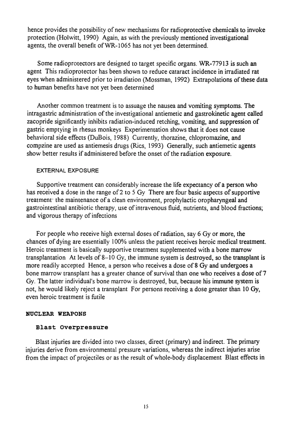
man from fast-rising, long-duration pressure pulses that were seen in the Hiroshima and
Nagasaki exposures are displayed in Table 2-8
There are four main systems of the body that have been studied to determine their
response to blast overpressure They are the respiratory system, solid organs, gastro-
intestinal tract, and the auditory system Damage to each of these systems is described
below
For the respiratory system, the lungs are the target organ; disruption permits air emboli
to enter the bloodstream and cause rapid death Lung hemorrhages range from a few
pinhead-sized petechiae on the surface of the lung to the involvement of entire lobes with
severe hemorrhaging. Severe blasts can rupture the lung itself or cause ribs to fracture and
puncture lung lobes. In the upper air ways, the mucosal lining of the sinus, larynx,
pharynx, and trachea can be bruised from low-level blasts More intense blasts can cause
hemorrhaging beneath the mucosa and reduce the size of the air passage (Levin, June
1993)
Table 2-8 Blast Effects in Man (Glasstone, 1977)
Health Effect Effective Peak Pressure (psi)
Lung Damage:
Threshold 8-15
Severe 20-30
Lethality
Threshold 30-50
50% 50-75
100% 75-115
Eardrum Rupture
Threshold 5
50% 15-20 (for age >20)
30-35 (for age < 20)
In the solid organs category, organs that are close to the lungs or the gastrointestinal
tract are the organs likely to have damage This damage could range from contusions to
rupturing that is due to the overexpansion of gas
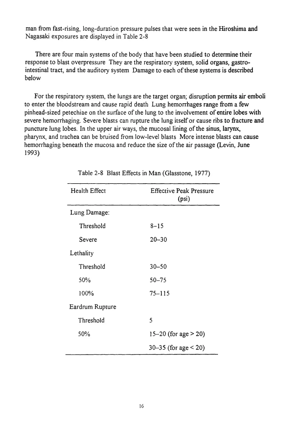
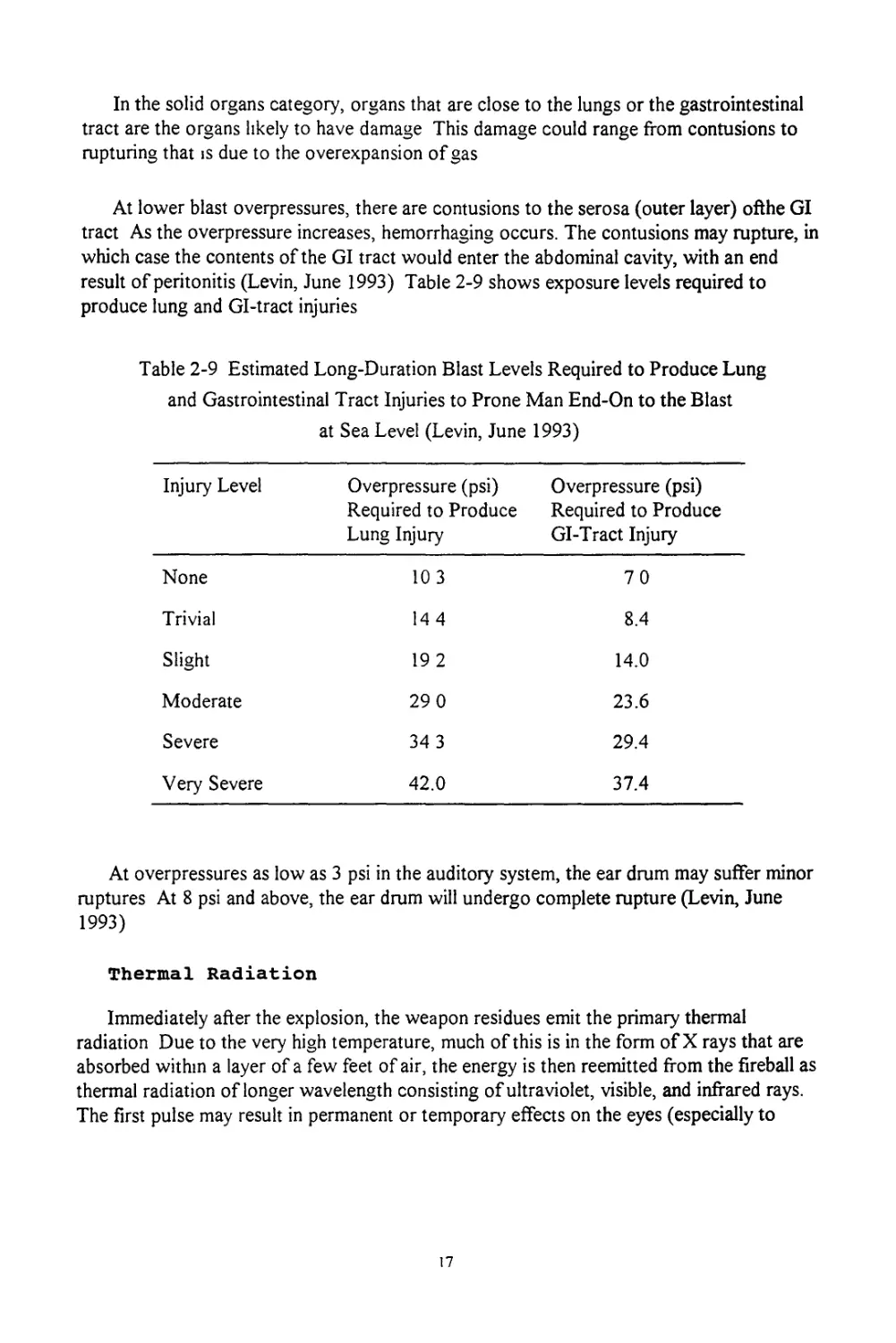
At lower blast overpressures, there are contusions to the serosa (outer layer) ofthe GI
tract As the overpressure increases, hemorrhaging occurs. The contusions may rupture, in
which case the contents of the GI tract would enter the abdominal cavity, with an end
result of peritonitis (Levin, June 1993) Table 2-9 shows exposure levels required to
produce lung and Gl-tract injuries
Table 2-9 Estimated Long-Duration Blast Levels Required to Produce Lung
and Gastrointestinal Tract Injuries to Prone Man End-On to the Blast
at Sea Level (Levin, June 1993)
Injury Level Overpressure (psi) Required to Produce Lung Injury Overpressure (psi) Required to Produce GI-Tract Injury
None 10 3 70
Trivial 14 4 8.4
Slight 19 2 14.0
Moderate 29 0 23.6
Severe 34 3 29.4
Very Severe 42.0 37.4
At overpressures as low as 3 psi in the auditory system, the ear drum may suffer minor
ruptures At 8 psi and above, the ear drum will undergo complete rupture (Levin, June
1993)
Thermal Radiation
Immediately after the explosion, the weapon residues emit the primary thermal
radiation Due to the very high temperature, much of this is in the form of X rays that are
absorbed within a layer of a few feet of air, the energy is then reemitted from the fireball as
thermal radiation of longer wavelength consisting of ultraviolet, visible, and infrared rays.
The first pulse may result in permanent or temporary effects on the eyes (especially to
17
individuals who happen to be looking in the direction of the explosion). The second
radiation pulse may last for several seconds (10 s for 1 MT yield); this pulse causes skin
bums of various degrees up to 12 mi or more and eye effects at even greater distances (for
1 MT yield) This pulse also starts fires
The thermal exposure decreases with distance from the point of detonation as a result
of the inverse square law If an airbrust occurs above dense cloud, smoke, or fog, most
of the thermal radiation will be reflected upward and little will reach the earth's surface.
Conversely, if the airburst occurs below a cloud, the thermal radiation reaching the earth
would be greater than on a clear day. A ground cover of snow can also intensify the
thermal radiation
Most thermal radiation (flash) burns are limited to uncovered areas of the skin that are
facing the center of explosion It is possible, however, for skin to be burned even through
a layer of clothing, in such cases, the radiant exposure must be large enough to char the
fabric Skin that is burned while covered is actually burned by the fabric. A person wearing
dark clothing has a higher risk of receiving a flash burn than one wearing light clothing,
because the dark fabric absorbs more radiant energy (Glasstone, 1977).
Bums are classified according to their severity as either first, second, or third degree
bums A first degree burn involves only the epidermis and causes erythema and edema
without vesiculation A second degree burn involves the epidermis and dermis and usually
forms blisters that may be superficial or deep dermal necrosis, but with epithelial regener-
ation extending from the skin appendages A third degree bum involves the destruction of
the entire skin, deep third degree burns extend into subcutaneous fat, muscle, or bone and
often cause much scarring
In addition to skin burns, thermal radiation may also cause either temporary or
permanent eye damage Temporary eye damage, also known as flashblindness, occurs
when more thermal energy is received than is necessary for image perception, but is not
enough to produce a retinal burn Flashblindness is caused by a bleaching of the light-
sensitive rods and cones in the retina of the eye Flashblindness will typically hide the
entire field of view with a bright afterimage This effect lasts for only a short time (several
seconds to minutes) and recovery is complete. Permanent eye damage (e.g., retinal bums)
arises from the excessive heating of the retinal tissue. Since, the optical process of image
formation within the eye keeps the energy per unit area on the retina constant, regardless
of distance, the probability of permanent eye damage does not decrease as the square of
the distance from detonation. Probability does, however, tremendously increase if a person
looked directly at the fireball (Glasstone, 1977).
Ionizing Radiation
PROMPT RADIATION
Neutrons and gamma rays represent the main ionizing radiation hazards after a nuclear
explosion. Almost all of the neutrons and part of the gamma rays are emitted in the actual
fission process These are referred to as the prompt nuclear radiations because they are
produced simultaneously with the nuclear explosion Generally, anything that emerges
within the first minute is considered to be prompt radiation.
Gamma rays and X rays are electromagnetic radiation that have no mass and no charge
and travel at the speed of light Gamma rays have great ranges in air, whereas X rays have
shorter ranges due to their lower energies Neutrons have appreciable mass, no electric
charge, and relatively great ranges in air, although this characteristic depends upon their
energy. Alpha and beta particles are charged and travel at varying speeds depending upon
their energies, but have relatively short ranges in air
FALLOUT
Fallout is categorized into two parts, early and delayed Early fallout, also called local,
is that which reaches the ground during the first 24 h following a nuclear explosion.
Delayed fallout, also called long range, reaches the ground after the first day. Delayed
fallout consists of very fine, invisible particles that settle in low concentrations over a
substantial portion of the earth's surface The fallout radiation decays rapidly, and normal
operations can be resumed in most areas within two weeks after the nuclear detonations.
The primary radiation hazards from fallout are beta and gamma radiation from the
fission products in the weapon Beta particles from fallout that lands on the skin can
penetrate into the deeper layers of the skin Because of this, the beta radiation is an
external radiation hazard Even moderate clothing will provide substantial protection
against beta radiation, and then the beta radiation becomes primarily an internal radiation
hazard It is important to cover any cuts or abrasions in the skin, and to use respirators or
gas masks to filter the air in a fallout environment
From dose reconstructions of people who lived within a few hundred miles of the
Nevada test site, four significant nuclides accounted for nearly 75% of the whole body
internal dose from the fallout These nuclides, in decreasing order of dose contribution, are
89Sr, 90Sr, 137Cs, and 13iI Inhalation doses are insignificant compared to external doses,
with the exception of 131I dose to the thyroid In almost all cases of interest, the internal
dose is a small fraction of the external dose received from fallout, which can range from
several rem to several hundred rem, depending upon the weather and what kind of shelter
the population is using (Peterson, January 1992) Other nuclides of significant dose
19
considerations are 14C and 3H Minor dose contributions come from 85Kr, 55Fe, and 239Pu
(Moe, 1988)
Since the atomic cloud may reach heights of 30 mi or more, it may take the early
fallout particles as long as 20 to 30 min to reach the ground. As a result, the starting
exposure rates are assumed to begin at 1 h postexplosion to allow most of the early fallout
particles to reach the ground
Calculating the exposure rate can be approximated very easily once the exposure rate
at 1 h postexplosion is known One general rule of thumb for this is called the seven-ten
rule. This rule states that for every sevenfold increase in time, as measured from the time
of explosion, there is an approximate tenfold decrease in exposure rate (NCRP, 1974). For
example, if 1 h past the time of explosion, the exposure rate is 1,000 R/h, then 7 h after
explosion, the rate would be 100 R/h Forty-nine hours postexplosion, the rate would be
10 R/h, 343 h (approximately 2 wk) after explosion, the exposure rate would be 1 R/h.
A more precise approximation is given by
d = d|*f12 (2.5)
where d is the exposure rate at time t, dj is the exposure rate 1 h past the time of
explosion, and t is the time past explosion in hours. This equation is effective for time
greater than 6-10 h after explosion
As mentioned in the section on the biology of radiosensitive tissues and organs in this
chapter, fractionation of the total absorbed dose decreases the negative health effects.
Recall from Table 2-6 that the LD50 for 2 wk of exposure time (the recommended value
for fallout exposure) is 130, 410, and 700 cGy for 10%, 50%, and 90% of the population,
respectively.
20
3. ADVANCED TECHNOLOGY WEAPONS EFFECTS
This section of the report reviews the basic effects of ATWs. The weapons considered
here are high-powered microwaves (HPMs) and lasers A section on non-ionizing
radiation provides a useful review to prepare the reader for the sections on ATWs. The
HPM and laser sections show the human response to the effects of these weapons.
NON-IONIZING REVIEW
Two developments brought attention to the possible public health aspects of non-
ionizing radiation The first was the postwar boom in electronics and communications
based on the microwave portion of the electromagnetic spectrum. This was shortly
followed by the second development, the rapidly increased use of lasers. When discussing
microwaves and lasers, it is important to have an understanding of the electromagnetic
spectrum Figure 3-1 shows a summary of the electromagnetic spectrum and lists some
common lasers
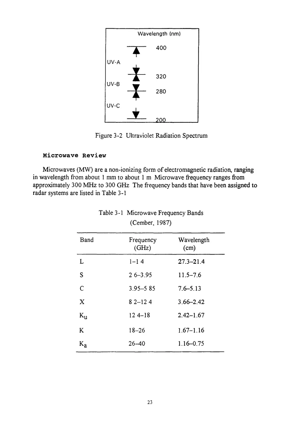
When discussing lasers in particular, it is also useful to understand the breakdown of
the ultraviolet radiation (UVR) spectrum The UVR spectrum may be divided into three
major components that induce significantly different biological effects: a UV-A component
with wavelengths from 400 nm to 320 nm (i e , long wave UVR, near UVR, or black
light); UV-B 320-280 nm (i e , middle UVR or "sunburn" radiation), UV-C 280-200 nm
(i.e , short wave UVR, far UVR, or germicidal radiation) Wavelengths below 200 nm are
of little biological significance since radiation in this region ("vacuum UVR") is absorbed
in very short pathlengths in air The division of the UVR spectrum is shown in Figure 3-2.
HIGH-POWERED MICROWAVES
Research is currently being performed to determine if a cumulative effect can occur
from microwave exposure However, to date, epidemiological studies have not
demonstrated any long-term consequences beyond those present at the time of the
exposure Since the cumulative-effect theory is at an investigational stage, this report will
only consider thermal effects The content of this section includes a basic microwave
review, a discussion of biological effects, and tables of occupational exposure limits.
21
Frequency Wavelength
in Hertz
in meters
10° -
10 1 -
10 2 -
10 3 -
10 4 -
10 5 -
IO6 -
10 7 -
10 8 -
10 9 -
10 10-
10
10 12-
10 13-
10 14 -
10 15-
10 ‘6-
10 17 -
10 18-
10 *9-
10 20..
1021-
1022-
10 23-
10 24-
-60 Hz
-1 kHz
—1 MHz
- 1 GHz
- 3xl08
- 3xl07
- 3xl06
- 3xl05
- 3xl04
— 3xl03
—3xl02
- ЗхЮ1
- 3x10°
- 3x10-’
- 3xl0‘2
- 3x1 O’3
- 3x10-’
-Line Current (60 Hz)
-Diathermy (10-24 kHz)
Zj-AM Broadcast (530-1600 kHz)
• Radiofrequency
(10 kHz-300 GHz)
far-infrared
far-infrared
near-infrared
near-infrared
visible
Carbon Dioxide
Carbon Monoxide
Erbium-Glass
Neodymium-Y AG
GaAs
Ruby
- 3x1 O’5 £
- 3xl0’6 R
-3x1 O’7 ____________-----
- 3x1 O'8
- 3x1 O’9 Soft
- 3x10’1°----------X-rays
„3x10⁻⁴ Hard
- 3xl0’12
- 3xl0’n
- 3xl0’14
-3xl0’15
- 3xl016
Cosmic Rays
HeNe
Argon
Krypton
HeCd
XeFl
KrFl
Figure 3-1 Electromagnetic Spectrum (Rademacher, 1992)
22
Figure 3-2 Ultraviolet Radiation Spectrum
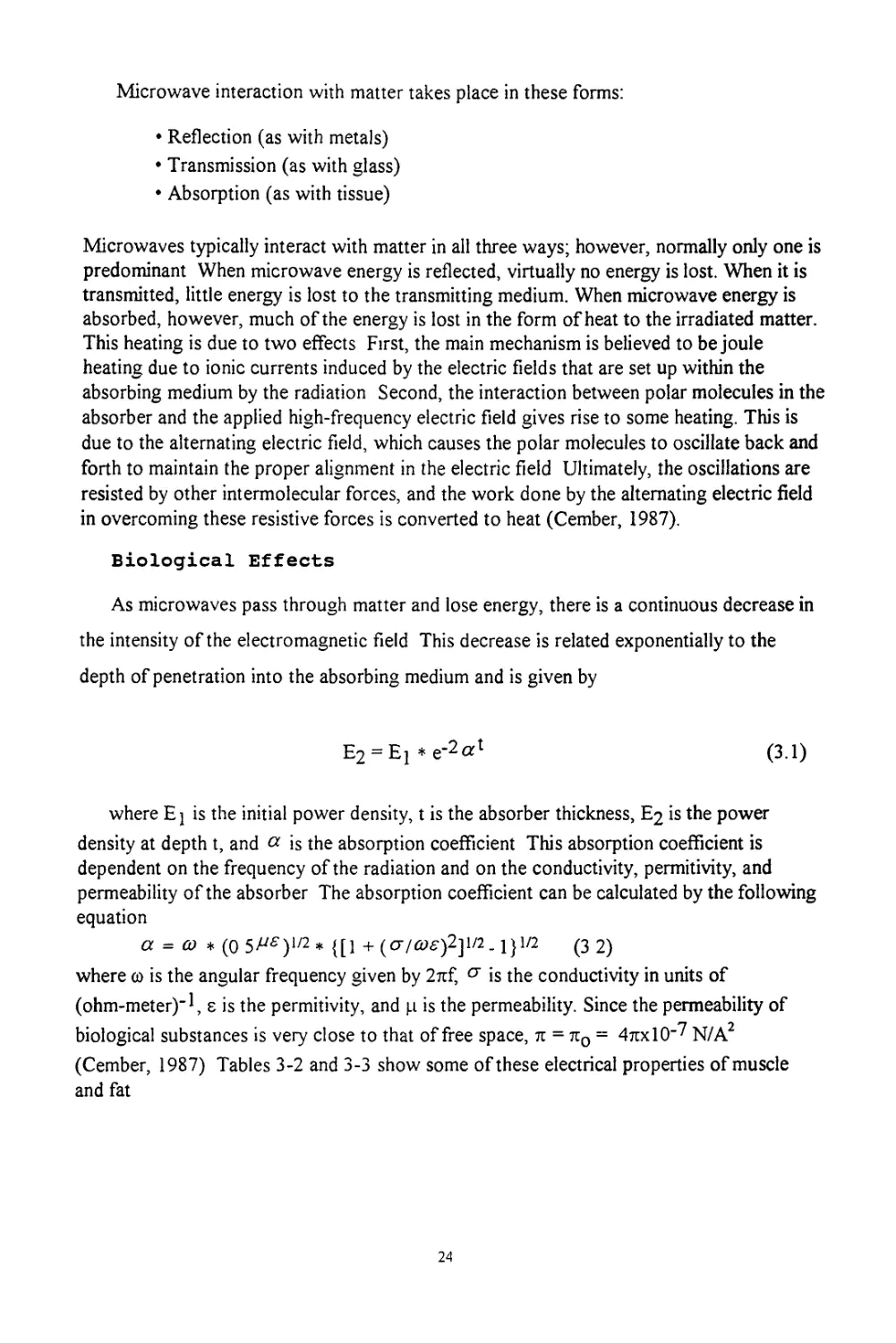
Microwave Review
Microwaves (MW) are a non-ionizing form of electromagnetic radiation, ranging
in wavelength from about 1 mm to about 1 m Microwave frequency ranges from
approximately 300 MHz to 300 GHz The frequency bands that have been assigned to
radar systems are listed in Table 3-1
Table 3-1 Microwave Frequency Bands
(Cember, 1987)
Band Frequency (GHz) Wavelength (cm)
L 1-1 4 27.3-21.4
S 2 6-3.95 11.5-7.6
C 3.95-5 85 7.6-5.13
X 8 2-12 4 3.66-2.42
Ku 12 4-18 2.42-1.67
К 18-26 1.67-1.16
Ka 26-40 1.16-0.75
23
Microwave interaction with matter takes place in these forms:
• Reflection (as with metals)
• Transmission (as with glass)
• Absorption (as with tissue)
Microwaves typically interact with matter in all three ways; however, normally only one is
predominant When microwave energy is reflected, virtually no energy is lost. When it is
transmitted, little energy is lost to the transmitting medium. When microwave energy is
absorbed, however, much of the energy is lost in the form of heat to the irradiated matter.
This heating is due to two effects First, the main mechanism is believed to be joule
heating due to ionic currents induced by the electric fields that are set up within the
absorbing medium by the radiation Second, the interaction between polar molecules in the
absorber and the applied high-frequency electric field gives rise to some heating. This is
due to the alternating electric field, which causes the polar molecules to oscillate back and
forth to maintain the proper alignment in the electric field Ultimately, the oscillations are
resisted by other intermolecular forces, and the work done by the alternating electric field
in overcoming these resistive forces is converted to heat (Cember, 1987).
Biological Effects
As microwaves pass through matter and lose energy, there is a continuous decrease in
the intensity of the electromagnetic field This decrease is related exponentially to the
depth of penetration into the absorbing medium and is given by
E2 = E1*e-2«t (3.1)
where E j is the initial power density, t is the absorber thickness, E2 is the power
density at depth t, and « is the absorption coefficient This absorption coefficient is
dependent on the frequency of the radiation and on the conductivity, permitivity, and
permeability of the absorber The absorption coefficient can be calculated by the following
equation
a = co * (o 5A^)i/2 * {[i + (cr/tyf)2]i/2. ip/2 (3 2)
where co is the angular frequency given by 2я£ a is the conductivity in units of
(ohm-meter)-1, s is the permitivity, and p is the permeability. Since the permeability of
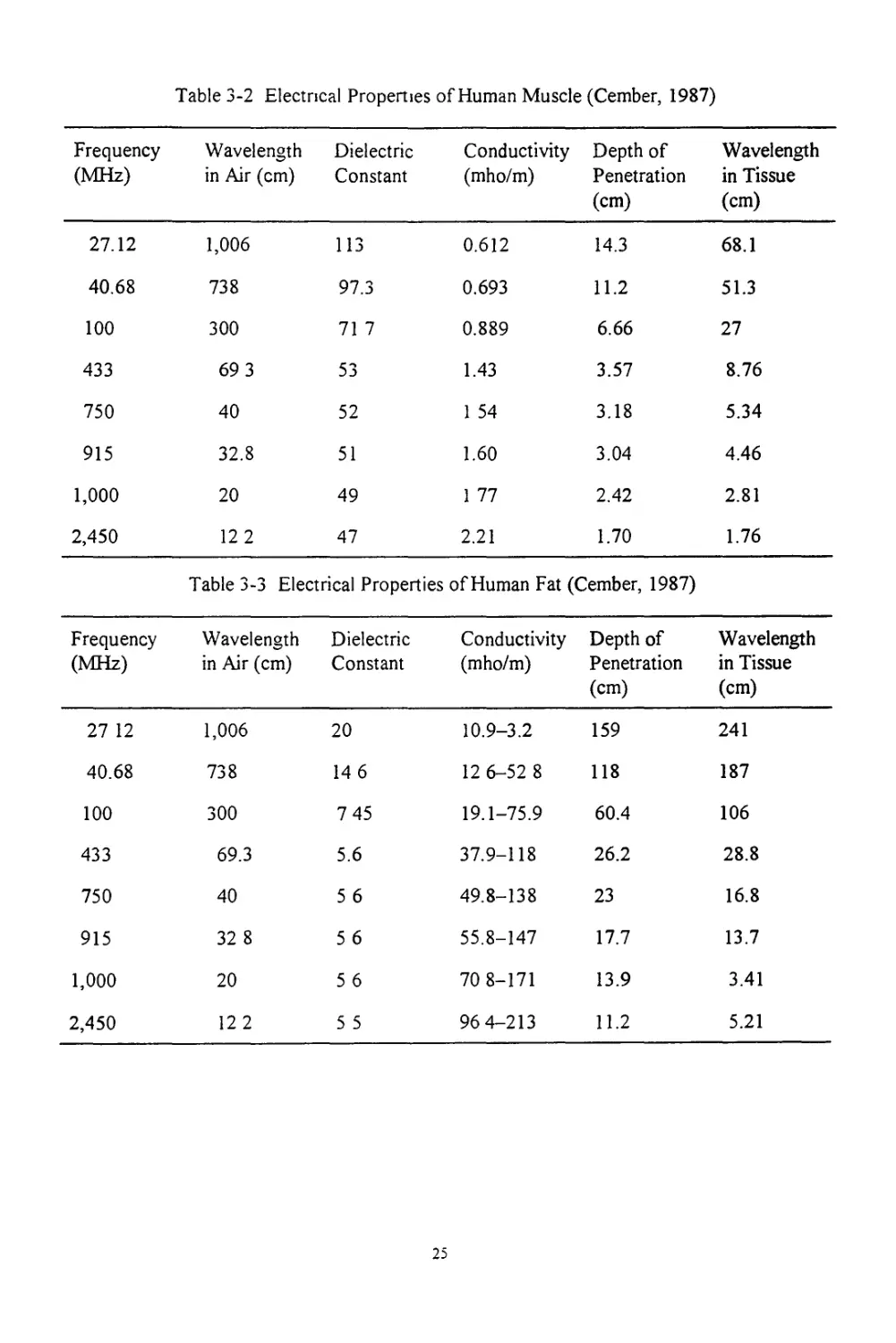
biological substances is very close to that of free space, тг = тг0 = 4тгх10"7 N/A2
(Cember, 1987) Tables 3-2 and 3-3 show some of these electrical properties of muscle
and fat
24
Table 3-2 Electrical Properties of Human Muscle (Cember, 1987)
Frequency (MHz) Wavelength in Air (cm) Dielectric Constant Conductivity (mho/m) Depth of Penetration (cm) Wavelength in Tissue (cm)
27.12 1,006 113 0.612 14.3 68.1
40.68 738 97.3 0.693 11.2 51.3
100 300 71 7 0.889 6.66 27
433 69 3 53 1.43 3.57 8.76
750 40 52 1 54 3.18 5.34
915 32.8 51 1.60 3.04 4.46
1,000 20 49 1 77 2.42 2.81
2,450 12 2 47 2.21 1.70 1.76
Table 3-3 Electrical Properties of Human Fat (Cember, 1987)
Frequency (MHz) Wavelength in Air (cm) Dielectric Constant Conductivity (mho/m) Depth of Penetration (cm) Wavelength in Tissue (cm)
27 12 1,006 20 10.9-3.2 159 241
40.68 738 14 6 12 6-52 8 118 187
100 300 7 45 19.1-75.9 60.4 106
433 69.3 5.6 37.9-118 26.2 28.8
750 40 5 6 49.8-138 23 16.8
915 32 8 5 6 55.8-147 17.7 13.7
1,000 20 5 6 70 8-171 13.9 3.41
2,450 12 2 5 5 96 4-213 11.2 5.21
25
In biological tissue, the rate of heat being produced is inversely proportional to the
square of the penetration depth Therefore, a tissue with a relatively small penetration
depth because of high water content will heat much faster. An example of this tissue type
is muscle. A tissue with a relatively large penetration depth because of lower water
content will heat more slowly An example of this tissue type is fat (Cember, 1987).
Radiation dosimetry involves the measurement of the power and energy that are
absorbed by the internal organs of a biological subject when subjected to external
radiation exposure fields The use of dosimetric instrumentation allows a means for the
quantification of the absorbed dose or specific absorption rate (SAR), which is expressed
in watts per kilogram Use of the absorbed dose rate rather than the external exposure
(power density) is critical in assessing microwave bioeffects SAR data provide key
information that allow the determination of the precise level at which a biological response
occurs in an exposed individual
Some reactions to microwave/radio-frequency exposure may lead to measurable
biological effects that remain within the range of normal (physiological) compensation.
Most biological effects occur by thermal energy conversion (heating). Low level exposure
(<10 mW/cm2) may result in transient functional central nervous system changes
(Michaelson, 1980)
All behavioral effects studies have shown that megawatt irradiation suppresses the
ability to perform a trained task (e g , a rhesus monkey pushing a lever for food) and that a
power-density/dose-rate and duration threshold for achieving the suppression exists
(ranging from 5 to 50 mW/cm2) A projected model from animals shows that the threshold
of behavior disruption of a learned task for a human occurs at a power density of
approximately 110 mW/cm2 (Michaelson, 1980)
A useful value to calculate when considering a microwave source is the effective
radiated power (ERP), which is given by
ERP = P * G (3.3)
where P is the power in milliwatts and G is the gain ratio. The gain ratio is:
G = log’1 [G(dB)/10] (3.4)
where G(dB) is the gain in decibels Once the ERP is known, a simple calculation will
yield the power density (S) in milliwatts per square centimeter. The power density can be
calculated by the following
26
S = ERP - (4tuR2)
(3.5)
where R is the distance from the source to the person given in centimeters. This
calculation assumes far field conditions and a point source.
Finally, the only proven effects from radio-frequency radiation (RFR) are thermal
bums, signs of electric shock or burn, and reasonably prompt cataract formation. The
thermal effect has already been discussed, but it is important to note that at frequencies
over 10 kHz air may become ionized at approximately 1,000 kV/m; this could result in
potentially hazardous electric discharges Also, electric shock can result from currents
induced in conductors within the RFR field Cataracts and vacuoles have been produced in
animal eyes with power densities of 100 mW/crn^ delivered locally for a period of 15 min.
This exposure results in an eye temperature of 41 °C or more (Poitrast, 1986).
Effects on the hematopoietic, immune, endocrine, and nervous systems are under
continued investigation for RFR effects Cardiovascular, blood-brain barrier, and behavior
effects are also under investigation Results to date in these areas are inconclusive.
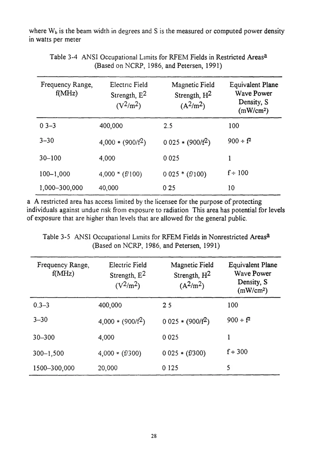
Occupational Limits
The occupational exposure limit at any given time for a radio-frequency electro-
magnetic (RFEM) field is a SAR of 0 4 W/kg For humans, the resonant frequency domain
is taken to be in the frequency of 30-300 MHz, a power density of 1 mW/cm2 averaged
for 6 min results in the occupational limit Tables 3-4 and 3-5 further break down the
RFEM field limits for all frequencies At the given frequencies and power densities
averaged over a period of 6 min, the yield is the maximum limit for SAR = 0.4 W/kg
(NCRP, 1986). Tables 3-4 and 3-5 show the occupational limits for RFEM fields in
restricted and nonrestricted areas respectively
One type of calculation that is useful in determining safe distances from radio-
frequency electromagnetic fields is given by the following-
d = [PG - (4nPd)]05 (3.6)
where d is the distance limit in meters, P is the power of the transmitter in watts, G is the
gain ratio for the antenna, and Pd is the permissible limit in watts per meter. Note that this
equation is simply a variation of Equation (3.5) Equations (3.5) and (3.6) assume a far-
field condition If, however, a far-field condition does not exist, the formula is still correct
if the near-field gain beam widths are used For rotating antennas, use the average power
given by the following equation
P = Wb/360 * S (3.7)
27
where Wb is the beam width in degrees and S is the measured or computed power density
in watts per meter
Table 3-4 ANSI Occupational Limits for RFEM Fields in Restricted Areasa
(Based on NCRP, 1986, and Petersen, 1991)
Frequency Range, f(MHz) Electric Field Strength, E2 (V2/m2) Magnetic Field Strength, H2 (A2/m2) Equivalent Plane Wave Power Density, S (mW/cm2)
0 3-3 400,000 2.5 100
3-30 4,000 * (900/f2) 0 025 * (900/f2) 900 4-P
30-100 4,000 0 025 1
100-1,000 4,000 ’ (f/100) 0 025 * (f/100) f-н 100
1,000-300,000 40,000 0 25 10
a A restricted area has access limited by the licensee for the purpose of protecting
individuals against undue risk from exposure to radiation This area has potential for levels
of exposure that are higher than levels that are allowed for the general public.
Table 3-5 ANSI Occupational Limits for RFEM Fields in Nonrestricted Areasa
(Based on NCRP, 1986, and Petersen, 1991)
Frequency Range, f(MHz) Electric Field Strength, E2 (V2/m2) Magnetic Field Strength, H2 (A2/m2) Equivalent Plane Wave Power Density, S (mW/cm2)
0.3-3 400,000 25 100
3-30 4,000 * (900/f2) 0 025 * (900/f2) 900 - P
30-300 4,000 0 025 1
300-1,500 4,000 * (f/300) 0 025 * (f/300) f-нЗОО
1500-300,000 20,000 0 125 5
28
a A nonrestricted area is neither limited nor controlled by the licensee. This type of area
has no potential for exposure at levels higher than those allowed for the general public.
LASERS
This section of the report includes a basic review of lasers and ultraviolet radiation.
The biological effects of laser radiation on the eye and the skin are discussed, as well as
the occupation limits based on these biological effects. Finally, several laser protection
methods are considered
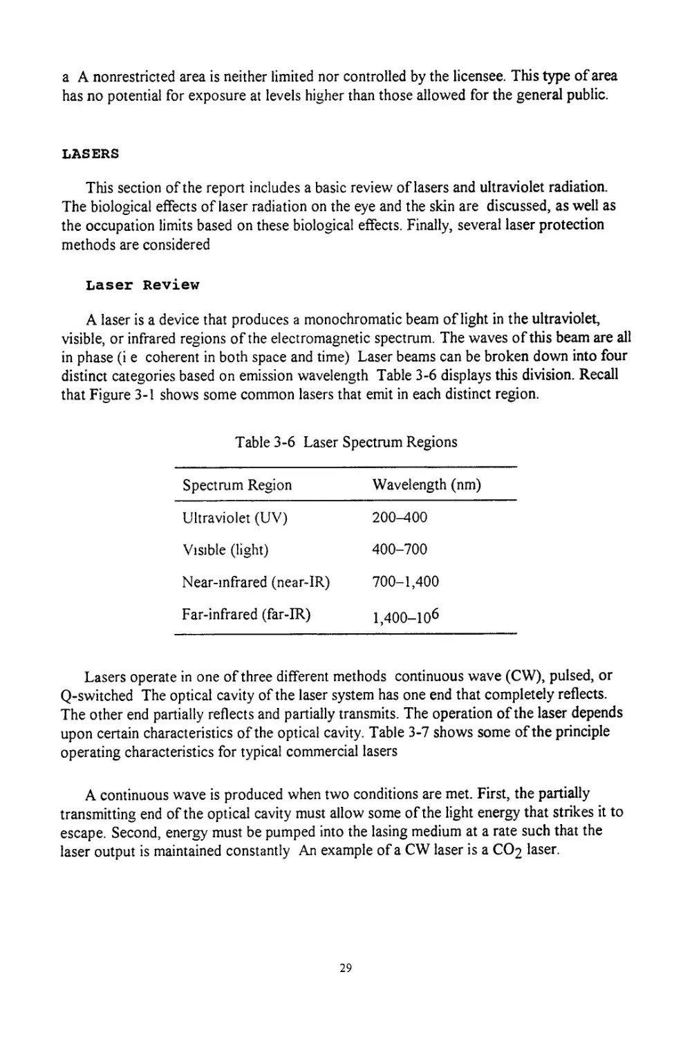
Laser Review
A laser is a device that produces a monochromatic beam of light in the ultraviolet,
visible, or infrared regions of the electromagnetic spectrum. The waves of this beam are all
in phase (i e coherent in both space and time) Laser beams can be broken down into four
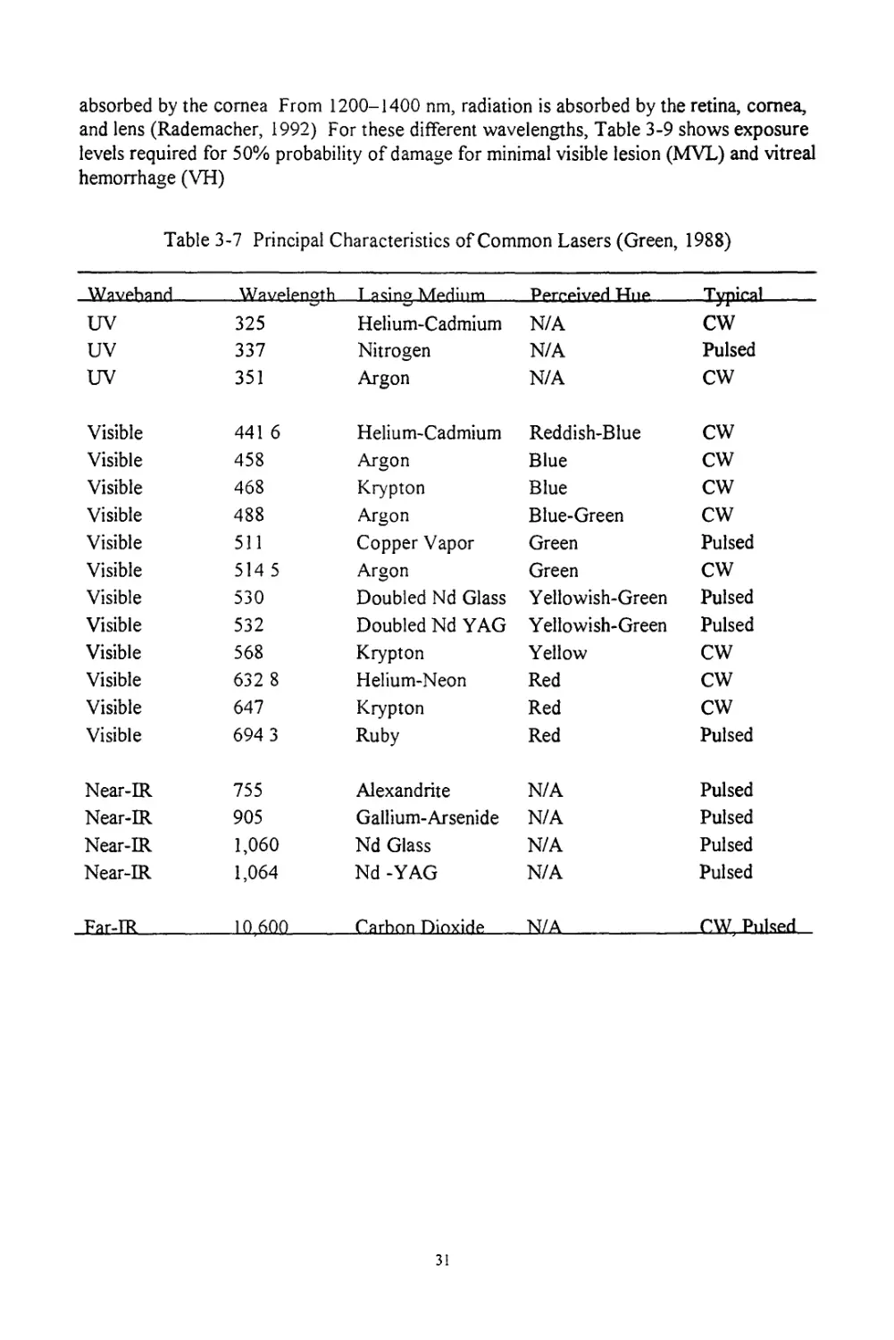
distinct categories based on emission wavelength Table 3-6 displays this division. Recall
that Figure 3-1 shows some common lasers that emit in each distinct region.
Table 3-6 Laser Spectrum Regions
Spectrum Region Wavelength (nm)
Ultraviolet (UV) 200^100
Visible (light) 400-700
Near-infrared (near-IR) 700-1,400
Far-infrared (far-IR) 1,400-Ю6
Lasers operate in one of three different methods continuous wave (CW), pulsed, or
Q-switched The optical cavity of the laser system has one end that completely reflects.
The other end partially reflects and partially transmits. The operation of the laser depends
upon certain characteristics of the optical cavity. Table 3-7 shows some of the principle
operating characteristics for typical commercial lasers
A continuous wave is produced when two conditions are met. First, the partially
transmitting end of the optical cavity must allow some of the light energy that strikes it to
escape. Second, energy must be pumped into the lasing medium at a rate such that the
laser output is maintained constantly An example of a CW laser is a CO2 laser.
29
Pulsed lasers normally deliver output in the form of bursts of light with a duration on
the order of about 0.1 to 10 ms An example of a pulsed laser is the ruby laser.
A specific type of a pulsed laser is the Q-switched laser. A Q-switch is an acousto-
optical or electrooptical device within the optical cavity that is analogous to a shutter.
Hence, it prevents laser emission until it is opened. Typical pulse durations for Q-
switched lasers are on the order of Ins-100 fs.
Biological Effects
Light, both visible and ultraviolet, is biophysically active. Mechanisms of biological
damage from light include both temperature effects and photochemical reactions. The
main mechanism of damage depends on the wavelength of the light and on the type of
tissue exposed For lasers, this main mechanism is believed to be due to temperature
effects The critical organs and tissues for this damage are the eye and the skin.
EYE DAMAGE
The eye is normally the most vulnerable organ for laser radiation damage. More
specifically, different eye tissues suffer different effects depending upon the wavelength of
the laser radiation The structure of the eye that is damaged depends upon the wave-length
of the light and the energy absorption characteristics of the ocular tissues. The retina is
sensitive to wavelengths in the range of 400-1400 nm (visible and near-IR band), while
the lens and cornea respond to UV-A The cornea alone is sensitive to far-IR and all UV.
The basic factor in retinal damage is the rate at which heat energy can be dissipated
from the irradiated tissue A temperature increase of several degrees higher than that
experienced during fevers is believed to be capable of producing permanent damage to the
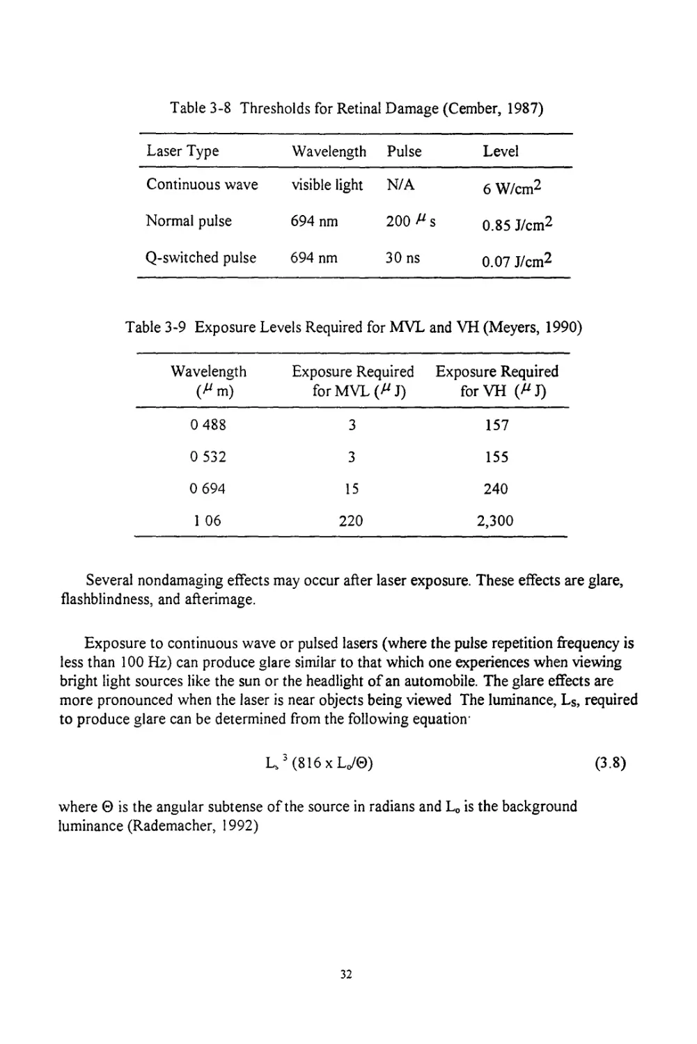
retina Table 3-8 shows the threshold values for damage to the retina from visible light. It
is reasonable to assume there is little or no difference over the entire visible spectrum, so
no values are listed for specific wavelengths (Cember, 1987) On the basis of these
thresholds, typical mission failure for a nanosecond pulsed laser is usually taken as 3.9
mJ/cm^ for a large hemorrhagic lesion This value corresponds to 1.5 mJ over a 7 mm
pupil (Rodgers, 1994)
Pigments and other tissue constituents determine the absorption characteristics of eye
tissue The cornea and lens will absorb most of the radiation in the ultraviolet region with
a wavelength of less than 400 nm Some of the incident radiation in the 315-340 nm
region will be transmitted and absorbed by the retina Radiation in the range of 400-1400
nm is almost completely transmitted through the cornea and lens. The retina and choroid
absorb most of the radiation in this range, with the longer wavelengths penetrating to
deeper tissues Radiations in the far-infrared region, 1400-10^ nm, are almost completely
30
absorbed by the cornea From 1200-1400 nm, radiation is absorbed by the retina, cornea,
and lens (Rademacher, 1992) For these different wavelengths, Table 3-9 shows exposure
levels required for 50% probability of damage for minimal visible lesion (MVL) and vitreal
hemorrhage (VH)
Table 3-7 Principal Characteristics of Common Lasers (Green, 1988)
Waveband T acino Medium Perreived FTne Typical
UV 325 Helium-Cadmium N/A CW
UV 337 Nitrogen N/A Pulsed
UV 351 Argon N/A CW
Visible 441 6 Helium-Cadmium Reddish-Blue CW
Visible 458 Argon Blue CW
Visible 468 Krypton Blue CW
Visible 488 Argon Blue-Green CW
Visible 511 Copper Vapor Green Pulsed
Visible 514 5 Argon Green CW
Visible 530 Doubled Nd Glass Yellowish-Green Pulsed
Visible 532 Doubled Nd YAG Yellowish-Green Pulsed
Visible 568 Krypton Yellow CW
Visible 632 8 Helium-Neon Red CW
Visible 647 Krypton Red CW
Visible 694 3 Ruby Red Pulsed
Near-IR 755 Alexandrite N/A Pulsed
Near-IR 905 Gallium-Arsenide N/A Pulsed
Near-IR 1,060 Nd Glass N/A Pulsed
Near-IR 1,064 Nd -YAG N/A Pulsed
JEarJR 10 600 Carbon Dioxide _NZA CW, Pulsed
Table 3-8 Thresholds for Retinal Damage (Cember, 1987)
Laser Type Wavelength Pulse Level
Continuous wave visible light N/A 6 W/cm2
Normal pulse 694 nm 200 ^s 0.85 J/cm2
Q-switched pulse 694 nm 30 ns 0.07 J/cm2
Table 3-9 Exposure Levels Required for MVL and VH (Meyers, 1990)
Wavelength (Z'm) Exposure Required for MVL (^J) Exposure Required forVH (A J)
0 488 3 157
0 532 3 155
0 694 15 240
1 06 220 2,300
Several nondamaging effects may occur after laser exposure. These effects are glare,
flashblindness, and afterimage.
Exposure to continuous wave or pulsed lasers (where the pulse repetition frequency is
less than 100 Hz) can produce glare similar to that which one experiences when viewing
bright light sources like the sun or the headlight of an automobile. The glare effects are
more pronounced when the laser is near objects being viewed The luminance, Ls, required
to produce glare can be determined from the following equation-
Ls3(816xLo/0)
(3.8)
where 0 is the angular subtense of the source in radians and Lo is the background
luminance (Rademacher, 1992)
32
Experiments conducted by the Naval Aerospace Medical Research Laboratory
(NAMRL) show that delay-of-glare-onset (DGLO) affects both speed and accuracy of
target location performance Visual decrements were noticed even when exposures were
hundreds of times lower than the maximum permissible exposure (MPE) for low-level
argon laser-induced glare The NAMRL recommended that eye protection should be
required to prevent mission disruption at laser intensities that are not harmful to the eye
(Reddix, 1992)
Flashblindness effects can last a few seconds to minutes, depending on the intensity of
the light source, ambient lighting, and the brightness of the object being viewed. The
magnitude of the impairment is largely a function of the size and location of the area of the
eye affected (Rademacher, 1992)
Afterimage is the perception of light, dark, or colored spots after exposure to a bright
light This effect may persist for many days but it is unlikely to cause visual decrement.
SKIN DAMAGE
When the skin receives an acute exposure from a laser, the skin may receive bums that
are much like ordinary thermal or solar burns The exposed skin undergoes coagulation
necrosis of a degree that is directly proportional to the amount of energy absorbed by the
skin The temperature rises only in the local tissue area, since skin has poor thermal
conductivity properties Such a temperature rise leads to the denaturation of tissue
proteins If a great amount of energy is absorbed, the water in the skin tissue is vaporized.
After this, the tissue may be heated to incandescence and may be carbonized.
Injury to skin from laser light depends on two different factors. First, the degree of
damage to the skin from lasers increases as the degree of pigmentation increases. Second,
damage to the skin depends on the wavelength of the laser and the length of the exposure
time Table 3-10 lists the threshold doses to the surface of the forearm of a Caucasian
adult for several different conditions of exposure These thresholds are called minimal
reactive doses (MRD) At the MRD level and below, there will be no damage to the skin.
Occupational Limits
In the United States, laser safety regulations are declared by two agencies: the
Department of Health and Human Services through the Bureau of Radiobiological Health
and the Department of Labor through the Occupational Safety and Health Administration
Table 3-11 displays eye and skin effects for various spectral exposures that these agencies
use to form occupational limits On the basis of the eye effects in Table 3-11, Table 3-12
shows the occupation limits for lasers as regulated by the Occupational Safety and Health
Administration (OSHA) Tables 3-13 through 3-20 show maximum permissible exposures
(MPE) for direct ocular viewing, diffuse reflector viewing, and skin exposure.
Table 3-10 Skin Damage Thresholds (Cember, 1987)
Laser Wavelength (nm) Exposure Time Area (cm2) MRD (J/cm2)
Ruby, normal pulse 694 0.2 ms 2.4-0.4x IO'3 14-20
Argon 500 6 s 95x IO'3 13-17
CO2 1060 4-6 s 1 4-6
Ruby, Q-switched 694 10-12 ns 0.33-1.0 0.5-1.5
Table 3-11 Biological Effects of the Optical Radiation Spectrum (Schleien, 1992)
Spectral Division Spectral Range (nm) Eye Effects Skin Effects
UV-C 100-280 Photokeratitis Erythema, accelerated
UV-B 208-315 Cataract aging, cancers,
UV-A 315-400 pigment dsrbfMiina
Visible 400-770 Lens yellowing, Photosensitivity,
IR-A 770-1400 photochemical and thermal retinal bums
IR-B 1400-3000 injuries, retinal lesions, thermal
IR-C 3000-106 cataract, aqueous flare, corneal bum
Table 3-12 OSHA Maximum Exposure-Level Limits
for Light Intensities for Employees
Viewing Orientation Light Intensity
Direct staring 1 mW/cm2
Incidental observation 1 mW/cm2
Diffuse reflected light 2.5 W/cm2
34
Table 3-13 MPE for Direct Ocular Exposure, Intrabeam Viewing,
to a UV Laser Beam or for Viewing a Diffuse Reflector of
a UV Laser Beama (Rademacher, 1992)
Wavelength, (nm) Exposure Duration, t(s) MPE (J/cm2)
200-302 10-9-3 x 10 3x 10⁻³
303 10-9-3 x Ю4 4x IO'3
304 10-9-3 x Ю4 6x IO’3
305 10-9-3 x Ю4 l.Ox IO'2
306 10-9-3 x Ю4 1 6x IO"2
307 10-9-3 x Ю4 2.5x IO"2
308 10-9-3 x Ю4 4.0x IO’2
309 10-9-3 x Ю4 6 3x IO'2
310 10-9-3 x Ю4 l.Ox 10"1
311 10-9-3 x Ю4 1.6x IO’1
312 10-9-3 x Ю4 2 5x IO’1
313 10-9-3 x Ю4 4.Ox IO’1
314 10-9-3 x Ю4 6.3 x Ю"1
315-400 10-9-10 0.56 * t025
315-400 10-Зх Ю4 1
a MPE is either the tabulated value or 0.56 * t^-25 (where t is
the exposure duration in seconds), whichever value is lower
For safety purposes, lasers have been segregated into four classes based upon their
strength and biological effects Class I lasers produce levels of radiation that do not cause
35
biological damage They emit less than 0.39 pW of continuous output. Class II lasers
produce radiation that can cause eye damage after direct, long term exposure and emit less
than 1 mW of continuous output Class III lasers produce radiation powerful enough to
injure human tissue with one short exposure to the direct beam or its direct reflections off
a shiny surface They emit less than 500 mW of continuous output. Finally, Class IV lasers
produce radiation so powerful that it can cause injury with a direct or reflected exposure,
even if the beam is scattered or diffused by a rough surface or smoke screens. They emit
more than 500 mW of continuous output (Schleien, 1992).
Table 3-14 MPE for Direct Ocular Exposure, Intrabeam Viewing, to Visible
and Near-Infrared Laser Beama (Rademacher, 1992)
Wavelength, X(nm) Exposure Duration, t(s) MPE (J/cm2)
400-700 10'9-l 8x IO’5 5x IO’7
400-700 1 8x 10'5-10 1.8 * t0.75 * Ю-3
400-550 10-104 10х IO'3
550-700 10-T! 1 8 * t0-75 * 10-3
550-700 T]-104 10CBx 10-3
400-700 104-Зх Ю4 CBX 10-6 (w/cm2)
700-1050 10-9-l 8x IO’5 5CAx IO’7
700-1050 1 8x 10⁻⁵-l 03 1.8 * CA°-75 * IO"3
1051-1400 10-9-5x IO’5 5x IO’6
1051-1400 5x Ю-5-1О3 9 * t0.75 ♦ ю-3
700-1400 103-3x Ю4 320CAx IO’6 (W/cm2)
a. T! = 10 x 10° °2 ('" 550) for X = 550-700 nm
CA = 1 for X = 400 - 700 nm CA = 100002lx'700) for X = 700-1050 nm
CA = 5 for X = 1050-1400 nm CB = 1 for X = 400-550 nm
СВ = 10x ю0015^-550) fOr X = 550-700 nm
36
Table 3-15. MPE for Viewing a Diffuse Reflector of a Visible Laser Beama
(Rademacher, 1992)
Wavelength, X (nm) Exposure Duration, t(s) MPE (J/sr-cm2)
400-700 10-9-10 10 * t0-333
400-550 10-104 21
550-700 10-Ti 3 8 * t0-75
550-700 Tj-104 21CB
400-700 104-3x Ю4 2.1 Cgx Ю"3 (W/sr-cm2)
a. Tj = 10x 1O°O2(?'-55O> for X = 550-700 nm
Cg = 1 for X = 400-550 nm
CB = 10 x ioool5(>-55°> for X = 550-700 nm
Table 3-16 MPE for Viewing a Diffuse Reflector of a Near-Infrared Laser Beama
(Rademacher, 1992)
Wavelength, X (nm) Exposure Duration, t(s) MPE (J/sr-cm2)
700-1400 10-9-10 10CA*t°-333
700-1400 10-103 3.83CA * t0-75
700-1400 ю3-3х 104 0.64Ca (W/sr-cm2)
a CA = 1 for X = 400-700 nm
CA = 1OOOO2<Z-7OO) for X = 700-1050 nm
CA = 5forX = 1050-1400 nm
37
Table 3-17 MPE for Direct Ocular Exposure, Intrabeam Viewing, to a Far-IR Laser
Beam, or for Viewing a Diffuse Reflector of a Far-IR Laser Beam (Rademacher, 1992)
Wavelength, X(nm) Exposure Duration, t(s) MPE (J/cm2)
1400-106 Ю-9-1 O’7 IO’2
1400—106 io-7-10 0.56 *t°-25
1400-106 10-3 x Ю4 0.1 * t
1540 only 10-9-Ю-6 1.0
Table 3-18 MPE for Skin Exposure to a UV Laser Beam
Wavelength, X(nm) Exposure Duration, t(s) MPE (J/cm2)
200-302 10-9-3x 10 Зх IO’3
303 10-9-3 x Ю4 4x IO'3
304 10-9-3 x Ю4 6x IO’3
305 10-9-3 x Ю4 l.Ox Ю-2
306 10-9-3 x Ю4 1.6x IO'2
307 10-9-3 x Ю4 2.5x IO’2
308 10-9-3 x Ю4 4.0x IO'2
309 10-9-3 x Ю4 6.3x IO’2
310 10-9-3 x Ю4 l.Ox 10'1
311 10-9-3 x Ю4 1.6x IO'1
312 10-9-3 x Ю4 2.5x Ю"1
313 10-9-3 x Ю4 4 Ox IO’1
314 10-9-3 x Ю4 6.3x IO'1
315-400 10-9-3x 10 0.56 * t°-25
315-400 10-103 I
315-400 10-3 x Ю4 10~3 * t
38
Table 3-19 MPE for Skin Exposure to Visible and Near-Infrared Laser Beamsa
(Rademacher, 1992)
Wavelength, X(nm) Exposure Duration, t(s) MPE (J/cm2)
400-1400 10-9-Ю-7 2Сдх IO'2
400-1400 10-7-10 1.1 CAt0-25
400-1400 10-3x 10« 0.2Сд (W/cm2)
а Сд = 1 for X = 400-700 nm
Сд = io0002a-700) for X = 700-1050 nm
Сд = 5 for X = 1050-1400 nm
Table 3-20. MPE for Skin Exposure to Far-Infrared Laser Beams (Rademacher, 1992)
Wavelength, X(nm) Exposure Duration, t(s) MPE (J/cm2)
1400-106 10-9-Ю-7 IO’2
1400-IO6 10-7-Ю 0.56 * t0-25
1400-106 >10 0.1 *t
1540 only 10-9-Ю-6 1.0
Useful Calculations
Once the MPE is known, it is also useful to express it in terms of the average
irradiance (E) given in watts per centimeter squared-
E = H/T
(3.9)
where H is the radiant exposure, or MPE, in joules per centimeter squared, and T is the
total exposure duration of a train of pulses Finally, expressed in terms of the radiant
exposure for a single pulse, the MPE is
39
MPE (single pulse) = H/n
(З.Ю)
where n is the multiple of the PRF and the total exposure duration.
Another calculation that is especially helpful when considering laser effects is one that
determines the fluence at a given distance The following equation is only a first-order
analysis; it does, however, permit for atmospheric effects, canopy and visor attenuation,
and repetitively pulsed lasers'
H = {4Q[(PRF)(texp)]°25 - (TcTa10’OD)} - [я (a2 + s2*)] (3.11)
where Q is the energy per pulse in joules, PRF is the pulse repetition rate in hertz, texp is
the exposure time in seconds, and Tc and Ta are the fraction transmissions by the canopy
and atmosphere respectively The term [(PRF)(tC4p)] is simply the total number of pulses,
n The visor attenuation is modeled by the optical density (OD) The term ’a’ is the width
of the beam as it leaves the laser source The term 's' is the slant range in centimeters given
by:
s = (r2 + h2)0.5 (3.12)
where r is the range and h is the altitude of the target. Finally, <j>is the beam divergence
given in radians
One factor the analyst may require is one that represents the attenuation coefficient
that affects the transmission capability for typical battlefield obscurants. Table 3-20
summarizes several coefficients for such obscurants The transmission factor for such
situations is given by
T = e*
(3-13)
where p is the attenuation coefficient as given in Table 3-21, p is the concentration in
grams per meter cubed, and x is the path length in meters.
Table 3-21 Obscurant Attenuation Coefficients (m2/g) (IRIA, 1980)
Obscurant Wavelength (nm)
0 55 1 06 10 6 11 15
Phosphorus 4.49 1.55 0.341 0.208
Hexachloroethane 4 70 2 06 0.056 0 075
Fog oil 7 37 3 53 0 016 0 024
40
Protection Against Laser Weapons
Several different methods have been used to protect sensors or eyes from lasers. The
beam may be blocked by an attenuating filter placed in front of the sensor or eye. These
filters must be effective against specific laser wavelengths and at the same time allow
sufficient amounts of other wavelengths to be transmitted for target detection or general
surveillance Another approach may be to block the beam by using indirect viewing
methods through an electrooptical system such as a thermal sight or a low-light television
device. Other methods of laser protection include veiling smoke, rapidly closing and
opening shutters, and the black eye patch (Anderberg, 1993).
When discussing the degree of protection from a filter, the term optical density is
used Optical density is a measure of the attenuation of the light beam due to a
transmitting medium and is given by the following-
OD = log (Io/I) (3.14)
where OD is the optical density, Io is the power of the incident beam and I is the power of
the transmitted beam For laser filters, researchers look for optical densities as high as 18.
When choosing protective filters, the luminous transmission must also be considered. The
optimum protective filter is one that provides maximum attenuation of the laser light while
transmitting the maximum amount of ambient light Luminous transmission is given as the
percentage of transmission of light from a source Eye protection on the battlefield should
typically have an overall transmission of 80% visible light through the filter (Anderberg,
1993).
The most commonly used filter for military applications is the absorption filter, which
can be composed of either glass or plastic Absorbing dyes can also be used in absorption
filters, these dyes can have optical density up to the range of 16 to 20. Dye filters are
simple and inexpensive, however, their optical density can be lowered by aging, oxidation,
or exposure to sunlight (Anderberg, 1993)
Instead of plastic absorption filters, colored glass can be used. Such filters are more
stable; however, in the near infrared region, only a few different wavelength absorption
curves are available
Insulating or dielectric coatings are another filter type that were developed to suppress
reflections from lenses. The modern dielectric interference filter selectively reflects
different wavelengths Since such filters consist of alternating layers of different dielectric
materials, a sharp spectral image can be obtained (i.e a specific wavelength is reflected
while other
41
nearby wavelengths in the visible part of the spectrum are transmitted). The disadvantage
of these filters is that the light reflected changes as the viewing angle changes (Anderberg,
1993)
Finally, a hologram can be made of one plate and it will respond to light just as the
multilayered dielectrics do Holographic filters have the same advantages and
disadvantages as the dielectric filters Another advantage the holographic filter has is that
it can scatter or bend laser light in a controllable way (Anderberg, 1993).
Crews inside tanks and other armored fighting vehicles (AFV) can be protected by
filters and coatings applied to sights and vision blocks If such protection is sufficient to
block any laser beam from entering the vehicle, then the crew does not require individual
eye protection. For those cases in which crews will be exposed, the members can be
equipped with laser protective visors Wrap-around laser protective visors that can be
mounted on the helmet are currently being developed; however, such protection would
not be sufficient against low-energy laser weapons that can use varying wavelengths
(Anderberg, 1993)
In certain scenarios, other protective methods may be more applicable or can be used
in addition to filters Another protective method is to use some form of indirect viewing,
such as a TV system, a thermal imager, or a light intensifier. A disadvantage of using this
method is that there are times when one must be able to view the battlefield with the
naked eye Smoke can be used not only to make target acquisition difficult for the enemy,
but if it is dense enough, it can diffuse and absorb a laser beam Obviously, because smoke
must be able to cover an extensive area densely and maintain that density, it has a great
deal of limitations For infantry or combat soldiers who fight in an area where lasers are a
threat, if no other means are available, a black patch can be worn over one of the eyes.
This method also has obvious disadvantages The soldiers lose their depth perception and
it possible that one eye may be lost (Anderberg, 1993).
42
4. SCENARIOS
This chapter is designed to show the reader how to use Chapters 2 and 3 to predict
how humans would perform after exposure to nuclear and advanced technology weapons.
Each scenario presents a typical environment and then steps through a solution by
referring to equations and tables in Chapters 2 and 3. Some of the physical characteristics
(e.g., wavelength) discussed in these scenarios are representative of actual systems. Others
are simply general estimates
IONIZING RADIATION
Hard Mobile Launcher
This scenario involves driving a hard mobile launcher (HML) from an alert area to a
place where the missile could be launched The HML consists of two parts, the tractor and
the trailer. The tractor carries a two-person crew and pulls the trailer. The trailer contains
an intercontinental ballistic missile (ICBM) and the necessary support equipment to launch
the missile on command The crew would drive the HML from the alert area to a
predetermined location and detach the missile trailer and leave it. Once the crew had left
the missile, their mission would be complete
The crew could be exposed to the effects of nuclear weapons from nearby detonations.
The scenario assumes a 50/50 mix of gamma and neutron radiation. The crew would be
protected from blast and thermal radiation by hardening the tractor to blast, blocking all
windows, and using TV cameras while driving
Table 2-4 shows that if the crew absorbed 600 cGy of prompt radiation, they would
begin vomiting and retching within 0 5-1 h Hence, a 20-min mission probably could be
completed before the prodromal symptoms began If the crew received a dose of400 scGy,
they probably could continue driving a vehicle for 1 h before the onset of vomiting and
nausea A maximum dose of 200 cGy would allow the crew to drive for 2 h before the
onset of prodromal symptoms Table 4-1 summarizes these data
If the crew were not protected against blast and thermal effects, what overpressure
and thermal levels could they withstand9 Table 2-8 shows the limiting health effect from
blast overpressure is eardrum rupture, which occurs at 15 psi for 50% of the population
over 20 yr of age. The threshold for eardrum rupture is 5 psi, which should be regarded as
the limiting value for blast effects when considering mission failure Second degree bums
are the limiting factor for thermal effects
Table 4-1 Maximum Prompt Dose for
Timed Vehicle Operation
Time Required to Complete Mission Maximum Dose if Task Is to Be Completed (cGy)
20 min 600
1 h 400
2h 200
Strategic Aircraft
The second scenario involves the use of a strategic bomber aircraft (A/C) to bomb a
target in Europe The mission originates from the CONUS The pilot would fly at a high
altitude until penetrating the enemy radar, then fly at an altitude of several hundred feet
until reaching the target Once the target was bombed, the mission would be complete.
The flight is estimated to take 12 h Once the bomber entered the enemy airspace, the
crew would be exposed to the effects of nuclear weapons from nearby detonations. The
scenario assumes a 50/50 mix of gamma and neutron radiation. At issue is the question of
how much prompt ionizing radiation the pilot could absorb and still complete the mission.
Table 2-4 shows that a pilot who received 130 cGy of prompt radiation probably
would be physically capable of completing a 12-h mission, although the individual most
likely would be nauseated and perhaps vomit. Notice that the limiting value for a pilot is
more strict than for the operator of a hard ground mobile launcher; this difference is due
to the fact that a pilot must be more alert and in a better state of health than a ground
vehicle operator
Mobile Ground Communication Systems
This scenario shows how to use the information on the health effects of radiation to
help set radiation levels for mobile ground systems. Mobile communication systems are
used to provide survivable communications after a nuclear attack Since the systems are
mobile, they cannot be directly attacked Therefore, the only nuclear environments that
affect them are those that cover a wide area, such as fallout.
Fallout produces two primary kinds of radiation, beta and gamma. As mentioned in the
section on fallout radiation, beta radiation has a short range People can be shielded from
4-4
beta radiation by protective clothing and respirators, and equipment can be shielded by air
filters. The primary hazard from fallout then becomes the gamma radiation. Tables 2-5 and
2-6 give the human effects of exposure to gamma radiation Before we can use the tables,
we must examine the operations concept for the mobile ground system.
For this example, let’s assume that the system has to operate for at least 6 mo after a
general nuclear exchange The system is mobile and can be moved to reduce its fallout
exposure. Further, let’s assume that the shelter housing the communication equipment is
made of thin sheets of metal over plywood and two-by-fours. This shelter provides
essentially no protection against gamma radiation. Finally, let’s assume that two
operations concepts for the system are being considered In the first concept, the mobile
system would have a single crew This crew would be deployed with the system before the
start of the nuclear exchange. In the second concept, a replacement crew would be housed
in a fallout shelter until the crew was needed to operate the system.
In the first operations concept, a single crew would have to operate the mobile system
for 6 mo Fallout levels high enough for people to begin dying would jeopardize the
mission Table 2-5 shows that 10% of exposed people would die at a tissue dose of 110
cGy; therefore, the radiation-exposure level for the system should be set at about 110 cGy.
Table 2-6 shows that there is no need to revise the dose upward to account for the effects
of the fallout dose being delivered over time since most of the fallout radiation (89%) is
received within the first week after the nuclear detonation
In the second operations concept, the mission could still be successful if the members
of the first crew received a high dose and were replaced The question then becomes how
high a dose the crew could receive and still function well enough to turn the system over
to the replacement crew Table 2-5 shows that about half of the crew would die from a
dose of about 345 cGy Also, about half of the crew would suffer from diarrhea and
hemorrhaging at this dose level, and well over half would suffer from nausea, fatigue, and
vomiting. Therefore, a dose of about 350 cGy is probably the maximum dose that the first
crew could receive and still function well enough to turn the system over to the
replacement crew The replacement crew could then receive a dose of about 110 cGy and
continue to operate the system to finish the 6 mo mission. The total dose for the system
would be about 450 cGy
Notice that the discussion above shows how to use the survival of the crew in a fallout
environment to balance the hardness requirements of the equipment in the mobile ground
system This is normally a logical way to estimate the hardness requirements for manned
systems However, this analysis does not show whether the system would be able to
accomplish its mission after a nuclear attack We would have to identify the deployment
areas for the mobile system and estimate the levels of fallout radiation that those areas
probably would receive before we could estimate the fallout levels for the system.
45
HIGH-POWERED RADAR
AN/APQ-120 Radar
The third scenario involves a multimode AN/APQ-120 radar system on an F-4E
aircraft. The system has one transmitter with a continuous wave (CW) output and another
with a pulsed output The pulsed transmitter has two discriminate pulse-width/pulse-
repetition-frequency (PW/PRF) combination modes The system not only is capable of
scanning operator-variable sector sizes, but also can operate in a fixed-beam mode if the
antenna hydraulics are shut off or if the antenna is mechanically pinned (Rademacher,
1989)
The operating parameter of the AN/APQ-120 for CW is 120.02-10.25 GHz with a
power of 200 W During the pulsed mode, the frequency is 8 7-9 4 GHz with a power of
165 kW (maximum peak) and 135 kW (minimum peak) The PRFs are 300 pps and 1060
pps
In 1989 there was an instance of accidental human exposure to the main beam of the
AN/APQ-120 radar Incorrectly assuming the dummy load was engaged, an individual
performed routine testing in the main beam of the radar at a distance of 3 ft from the
antenna. This individual was exposed to the main beam for approximately 3 min with the
CW transmitter and for 1 min with both transmitters. The antenna was in a fixed-beam
mode (Rademacher, 1989)
To calculate the individual's exposure, the power density was measured with the radar
in the CW and pulsed modes with the longest duty cycles This produced a power density
of 220 mW/cm^ and 125 mW/cm^ for each mode respectively. The exposure was
averaged over a 6 min period Given
3 min at 220 mW/cm^ and
1 min at 125 mW/cm^ and 220 mW/cm^, simultaneously
simple averaging over 6 min yields
[(4 min * 220 mW/cm^) + (1 min * 125 mW/cm^)] - 6 min = 167.5 mW/cm^ (4.1)
This estimated exposure exceeds the limit of 10 mW/cm^. The only effect the individual
experienced at the time of overexposure was a warming sensation on the back of his neck
(Rademacher, 1989).
46
Large Phased-Array Radar (LPAR)
In this scenario, a large phased-array radar (LPAR) on an aircraft is assumed to
possess the following specifications
f= 16 GHz
dish diameter = 0 5 m
gain = 35 dB
P = 300 kW
Under these conditions, what power density would a person be exposed to at 100 m
from the source? For reasons of simplification, assume far field conditions and a point
source.
To calculated the effective radiated power (ERP), one must first determine the gain
ratio Using Equation (3 4), the gain ratio is
G=log-1[ G(dB)/10]
G=log-1[35/10] = 3,162
Using Equation (3 3), the effective radiated power is:
ERP = P * G
ERP = 100 kW * 3,162 = 3 162x Ю5 kW
Finally, with the aid of Equation (3.5), the power density (S) is:
S = ERP - [4’R2]
S = 3 16x 105 kW - [4>(100 m)2] = 2.5 kW/m2 = 250 mW/cm2
(3.4)
(4-2)
(3-3)
(4.3)
(3.5)
(4.4)
Table 3-4 shows an ANSI occupational limit for RFEM fields in restricted areas of
10 mW/cm2 for a frequency of 16 GHz It is possible that disruption of a learned task
could occur at this power density
47
LASERS
Tactical Aircraft
This scenario considers the effect of a ground-based generic laser on a pilot behind the
canopy of a tactical aircraft The laser is assumed to have an output of 100 mJ, a
wavelength of 10 064 m, and a narrow beam divergence of 100 grad.
If the pilot did not have a protective visor (i.e., OD = 0), what exposure would he
receive at an altitude of 1,000 ft and a distance of 0.5 mi from the ground-based laser
source9 Assume the following conditions (Rodgers, 1994):
PRF = 10 Hz
Exposure period = 10 s
Canopy transmission = 80%
Atmospheric transmission = 92%
Output diameter = 2 cm
The problem can be solved using a simple approach for a first order analysis of laser
effects. Recall Equation (3 11)
H = {4Q[(PRF)(texp)]° 25 » (TcTa10-OD)) , [(a2 + s2^2)j (З.Ц)
The first step in solving Equation (3 11) is to solve for the slant range, s. Recall
Equation (3.12)
s = (r2 + h2)05 (3.12)
Substitute the given values
s = (26402 + 10002)0.5 ft = 2823 ft = 8 605 x 104 cm (4.5)
Now substitute the given values into Equation (3.11)
H = {4(0.1)[(10)(10)]°-25 * (0 8)(0 92)(1)} ~ {^[(2)2 + (8.605x 10⁴)2(0.1 x IO’2)2]}
= 3 80x IO'3 J/cm2 (4.6)
Recall from the data on the biological effects of lasers in Chapter 3 that the typical
mission failure for a nanosecond pulsed laser can be taken as 3.9 mJ/cm2 for a large
hemorrhagic lesion This value corresponds to 1.5 mJ over a 7 mm pupil (Rodgers, 1994).
The calculated value in this scenario is close enough to consider mission failure.
48
What if the pilot were wearing laser eye protection with the usual optical density of 4?
Simply adding the optical density factor to Equation (3.11) yields:
H = (lO^Jx 3.80X 10⁻³ J/cm2 = 3 80x ]0“2 J/cm2
(4.7)
which would fully protect the pilot at all ranges from this laser
49
5. SUMMARY AND CONCLUSIONS
This report provides a review of the fundamentals of radiation biophysics and human
response to ionizing and non-ionizing radiation It also provides a background review of
nuclear weapons and ATWs. Finally, the report gives examples of how to determine
mission survivability in various scenarios. Some of the most useful material for the
survivability analyst is contained in the tables
The review of the biological effects of ionizing radiation focuses on the acute radiation
syndrome (ARS). Table 2-4 shows the time until the onset of and the probability of
occurrence of prodromal symptoms This table enables the analyst to determine whether
or not a mission is likely to be completed based upon the prompt dose received. Table 2-6,
which shows the lethal doses in 60 d for various percentages of the population, enables the
analyst to determine mission capability The most significant figure in this table is the
LD50 value of 345 cGy Table 2-7 in the discussion of treatment displays various
decontam-ination agents for specific isotopes The section on nuclear weapons has two
tables on human responses to blast effects, Tables 2-8 and 2-9
The chapter on ATWs does not give as much detail on the effects of human exposure
to radiation as the chapter on nuclear weapons Research has yet to yield data on specific
human responses to non-ionizing radiation Until these data are available, the systems
survivability analyst should contact the non-ionizing radiation protection division at
Armstrong Laboratory (AL/OER), Brooks AFB, TX. The microwave and laser sections of
this chapter do, however, contain the recommend occupational exposure limits, as shown
in Tables 3-4, 3-5, and 3-12 through 3-20 These exposure limits give the analyst a rough
estimate of what mission-limiting exposures are likely to be.
Finally, the scenarios listed in this report are presented in a manner that shows the
systems survivability analyst how to step through a scenario by using information from
Chapters 2 and 3 Some of the scenarios are based upon specific systems, while others
are based upon generic conditions
In conclusion, this report provides guidance to system survivability analysts by
outlining how human survivability relates to nuclear weapon and ATW effects. With the
assistance of this document, the analyst can make quick order-of-magnitude estimates of
the hardness requirements for manned systems based on human response to nuclear
weapons and ATWs
USEFUL CONVERSIONS
Conversion Between Irradiance Units
W/m2 mW/cm2 W/cm2
1 W/m2 = 1 0.1 0.0001
1 mW/cm2 = 10 1 0.001
1 W/cm2 = 104 103 1
1 erg/(cm2-s) = 10-3 IO'4 IO’7
1 erg/(m2-s) = 10-7 10’8 10-11
Conversion Between Radiation Units
To convert from to Multiply by
curie becquerel 3.7xl010
rad gray lxlO-2
rem sievert 1x10⁻²
roentgen Coulomb/kilogram 2.58X10’4
53
ACRONYMS AND ABBREVIATIONS
A Ampere
A/C Aircraft
AF Air Force [United States]
AFB Air Force base
AFV Armored fighting vehicle
ARS Acute radiation syndrome
ATW Advanced technology weapon
C Centigrade
cGy Centigray
cm Centimeter
CNS Central nervous system
CONUS Continental United States
CW Continuous wave
d Day
D Absorbed dose
dB Decibel
DGLO Delay-of-glare-onset
DNA Defense Nuclear Agency
Do Mean lethal dose
DTPA Diethylenetriaminepentaacetic acid
E Electric field strength
EDTA Ethylenediaminetetraacetic acid
EM Electromagnetic
EMF Electromagnetic field
ERP Effective radiated power
f Frequency
ft Feet
G Gain
GHz Gigahertz
GI
Gy
h
H
HEL
HML
HPM
Hz
IAEA
ICBM
IR
kg
kHz
Laser
LD50
LEL
Lo
LPAR
Ls
m
MeV
MHz
mi
min
mJ
mm
mo
MPE
MRD
ms
MT
Gastrointestinal
Gray
Hour
Magnetic field strength
High-energy laser
Hard mobile launcher
High-powered microwave
Hertz
International Atomic Energy Agency
Intercontinental ballistic missile
Infrared
Kilogram
Kilohertz
Light amplification by stimulated emission of radiation
Lethal dose for 50% of the population
Low-energy laser
Background luminance
Large phased-array radar
Luminance
Meter
Megaelectronvolt
Megahertz
Mile
Minute
Millijoule
Millimeter
Month
Maximum permissible exposure
Minimal reactive dose
Millisecond
Megaton
56
цш
prad
HS
MVL
mW
MW
MW
MW/RF
NAMRL
NCRP
nm
OD
OSHA
P
pps
PRF
psi
PW
QF
R
RF
RFEM
RFR
s
S
SAR
sr
Sv
USANCA
uv
V
VH
Micrometer
Microrad
Microsecond
Minimum visible lesion
Milliwatt
Megawatt
Mier о wave
Microwave/radio frequency
Naval Aerospace Medical Research Lab
National Council on Radiation Protection and Measurements
Nanometer
Optical density
Occupational Safety and Health Administration
Power
Pulses per second
Pulse repetition frequency
Pounds per square inch
Pulse width
Quality factor
Roentgen
Radio frequency
Radio-frequency electromagnetic field
Radio-frequency radiation
Second
Power density
Specific absorption rate
Steradian
Sievert
United States Army Nuclear and Chemical Agency
Ultraviolet
Volt
Vitreal hemorrhage
57
w
wk
yr
Watt
Week
Years
58
GLOSSARY
absorbed dose (see dose, absorbed)
acute exposure. Irradiation in which the time of exposure does not extend beyond several
hours.
acute radiation dose (see dose, acute radiation)
acute radiation stomatitis. Inflammation of the mucous membrane of the mouth.
acute radiation syndrome (ARS). The complex of symptoms characterizing the disease
known as radiation injury, resulting from excessive exposure of the
whole body (or a large part) to ionizing radiation. The earliest of
these symptoms are nausea, fatigue, vomiting, and diarrhea, which
may be followed by epilation, hemorrhage, inflammation of the mouth
and throat, and general loss of energy. In severe cases, where the
radiation exposure has been relatively large, death may occur within 2
to 4 wk Those who survive 6 wk after the receipt of a single large
dose of radiation may generally be expected to recover.
alpha particle. A positively charged particle ejected spontaneously from the nuclei of
some radioactive elements. It is identical to a helium nucleus that has
a mass number of 4 and an electrostatic charge of +2.
amenorrhea. Absence or cessation of the menses
aqueous flare. During a slit lamp test, which focuses on the aqueous part of the eye, a
flare effect is seen if particles are present in the eye. These particles
can either be debris or they may represent an infection due to ocular
damage
ataxia. Inability to coordinate the muscles in the execution of voluntary movement.
becquerel (Bq). A unit of nuclear disintegration rate. A becquerel is one nuclear
disintegration per second
beta particle. A charged particle emitted from a nucleus during radioactive decay, with a
mass equal to 1/1837 that of a photon. A negatively charged beta
particle is identical to an electron. A positively charged beta particle
is called a positron Large amounts of beta radiation may cause skin
burns, and beta emitters are harmful if they enter the body. Beta
particles are easily stopped by a thin sheet of metal or plastic.
carcinogenesis. The origin or production of cancer.
cataractogenesis. The state of cataract formation.
cataract. Loss of transparency of the lens of the eye.
59
coagulation. Clotting; the process of changing from a liquid to a solid, said especially of
blood
conjuctivitis. Inflammation of the mucous membrane that lines the inner surface of the
eyelids and is continued over the forepart of the eyeball.
contamination, radioactive. The presence of radioactive matter in or on a substance
where it is unwanted
Curie (Ci). A unit of activity, or the degree of radioactivity of a radioactive substance.
One curie equals 3 7x10¹⁰ becquerels.
cutaneous. Relating to the skin
cytogenetic. Genetics relating to the structure and function of the cell, especially the
chromosomes
cytopenia. Reduction of cellular elements in the circulating blood.
denaturation. The process of being made unnatural or changed from the normal in any of
its characteristics, often applied to proteins or nucleic acids that are
heated or otherwise treated to the point where tertiary structural
characteristics are altered
dermal. Dermatic, dermatoid, dermic, relating to the skin.
dose, absorbed. The amount of energy imparted by nuclear (or ionizing) radiation to unit
mass of absorbing material The unit is the rad. In current usage, the
rad unit has been replaced by the SI unit, the gray (Gy, 1 Gy = 100
rad)
dose, acute radiation. Total ionizing radiation dose received at one time and over a
period so short that biological recovery cannot occur.
dose equivalent. The product of the absorbed dose in tissue, quality factor, and all other
necessary modifying factors at the location of interest. The units of
dose equivalent are rem or sievert.
electric field. A fundamental component of electromagnetic (EM) wave, which exists
when there is a voltage difference between to points in space.
electric field strength (E). The magnitude of the electric field expressed in volts per
meter (V/m)
edema. An accumulation of an excessive amount of watery fluid in cells, tissues, or
serous cavities
electroencephalogram (EEG). Shows electric potential of the brain.
emesis. Vomiting
enteritis. Inflammation of the intestine, especially of the small intestine
epithelial. Relating to or consisting of epithelium.
60
epithelium. The purely cellular avascular layer covering all the free surfaces, cutaneous,
mucous, and serous, including the glands and other structures derived
therefrom
erythema. Inflammatory redness of the skin.
externa! exposure. Irradiation comes from sources outside the body.
far-field region. The region far enough from an antenna that the power per unit area
decreases with the square of the range. In the far-field region, the
field has predominantly plane-wave character (i.e., uniform
distributions of electric and magnetic fields in planes transverse to the
direction of propagation).
fluence. The energy density of the EMF when integrated over the duration of the
exposure, usually expressed in units of joules per square centimeters
(J/cm2)
fractionation. The process by which a large radiation dose is received in parts with
delays (e.g. 1 d) in between each exposure.
gamma radiation. High-energy, short wavelength electromagnetic radiation (packet of
energy) emitted from the nucleus Gamma radiation frequently
accompanies alpha and beta emissions and always accompanies
fission Gamma rays are very penetrating and are best stopped or
shielded against by dense materials, such as lead or uranium. Gamma
rays are similar to X rays, but are usually more energetic.
gastrointestinal. Gastroenteric, relating to the stomach and intestines.
gastrointestinal syndrome. Acute radiation syndrome symptoms that are related to the
gastrointestinal tract
gonadotropin. Gonadotropic hormone, a hormone capable of promoting gonadal growth
and function
gray. The International System unit of absorbed dose, equal to the energy imparted by
ionizing radiation to a mass of matter corresponding to one joule per
kilogram Symbolized Gy.
half-life. The time in which half the atoms of a particular radioactive substance
disintegrate to another nuclear form. Measured half-lives vary from
millionths of a second to billions of years Also called physical half-
life.
hardness. A measure of the ability of a system to withstand exposure to one or more
effects of man-made hostile environments
hematological. That which relates to blood and/or blood-forming organs
61
hematological syndrome. Acute radiation syndrome symptoms that are related to the
hematological system
hemopoietic. That which relates to blood cells.
hematochezia. Passage of bloody stools.
heroic treatment. Extensive medical treatment of a patient who suffers from acute
radiation syndrome This may include antibiotics treatment and bone-
marrow transplants
hertz (Hz). The unit for expressing frequency. One hertz is equivalent to one cycle per
second
human resonance range. The frequency region where absorption of RF energy in the
body is enhanced. For sizes ranging from a baby to an adult, peak
absorption varies depending on the individual's size relative to the
wavelength and orientation relative to the polarization of the wave.
The PELs have been established to cover the range in human sizes,
shapes, and positions
hyperesthesia. Abnormal acuteness of sensitivity to touch, pain, or other sensory stimuli.
incandescence. The quality or state of being white, glowing, or luminescent with intense
heat
internal exposure. Irradiation comes from sources inside the body.
interstitial. Relating to spaces within a tissue or organ, but excluding such spaces as
body cavities or potential space.
intragastric. Within the stomach
keratinocyte. A cell of the living epidermis and certain oral epithelium that produces
keratin in the process of differentiating into the dead and fully
keratinized cells of the stratum comeum
lethal dose (LDJ. The lethal whole body dose for x% of the population to die in 60 d.
The value for this without treatment is 3 45 Gy.
leukocyte. White blood cell
lymphocyte. Lymph cell, lympholeukocyte; a white blood cell formed in lymphatic tissue
throughout the body and in normal adults comprising approximately
22 to 28% of the total number of leukocytes in the circulating blood.
lymphopenia. Reduction in the number of lymphocytes in the circulating blood
magnetic field. A fundamental component of EM waves produced by a moving electric
charge
magnetic field strength (H). The strength of the magnetic field expressed in amps per
meter (A/m).
62
malaise. A feeling of general discomfort or uneasiness, as an out-of-sorts feeling.
mean lethal dose. The lethal whole body dose for 50% of the population
meiosis. Meiotic division, the special process of cell division that results in the formation
of gametes, consisting of two nuclear divisions in rapid succession
that result in the formation of four gametocytes, each containing half
the number of chromosomes found in somatic cells.
near-field region. A region generally in close proximity to an antenna or other radiating
structure in which the electric and magnetic fields do not exhibit a
plane-wave relationship, and the power does not decrease with the
square of distance from the source but varies considerably from point
to point. The near-field region is further subdivided into the reactive
near field, which is closest to the radiating structure and contains
most or nearly all of the stored energy, and the radiation near field,
where the radiating field predominates over the reactive field but
lacks substantial plane-wave character and is complicated in
structure For most antennas, the outer boundary of the reactive near-
field region is considered to occur at a distance of one-half
wavelength from the antenna surface
necrosis. The pathologic death of one or more cells, or a portion of tissue or organ,
resulting from irreversible damage.
neutropenia. The presence of abnormally small numbers of neutrophils in the circulating
blood
oliguria. Scanty urination.
ondansetron. Anti-emetic agent; also causes no drowsiness.
oocyte. Ovocyte, the immature ovum.
oropharyngeal. Relating to the oropharynx.
paraesthesia. An abnormal sensation, such as of burning, pricking, tickling, or tingling.
percutaneous. Diadermic, transcutaneous, transdermic, denoting the passage of
substance through unbroken skin, as in absorption by inunction.
permissible exposure limit (PEL). Limits for RF exposure established for the protection
of personnel There are no expectations that any adverse health
effects will occur with exposures that are within the PEL, even under
repeated or long-term exposure conditions.
petechiae. Minute hemorragic spots, of pinpoint to pinhead size.
photokeratitis. Inflammation of the cornea to laser light
photon. A quantum (or packet) of energy emitted in the form of electromagnetic
radiation Gamma rays and X rays are examples of photons.
63
photosensitivity. Circumstance where the skin is sensitive to light
plane wave. An EM wave characterized by mutually orthogonal electric and magnetic
fields that are related by the impedance of free space (377 ohms). For
plane waves, power density (S), electric field strength (E), and
magnetic field strength (H) exhibit the following relationship: S =
£2/3700 = 37 7 * H^, where S is in mW/cm^, E is in V/m, and H is
in A/m
plane-wave-equivalent power density. The magnitude of power density that would
exist for an EM wave in free space having the same electric or
magnetic field strengths
power density (S). The rate of energy transported into a small sphere divided by the
cross-sectional area of that sphere. It is expressed in units of W/m2.
progenitor. A precursor, ancestor, one who begets.
purpura. A condition characterized by hemorrhage into the skin. The color is first red,
gradually darkens to purple, fades to a brownish yellow, and usually
disappears in 2 or 3 wk
quality factor (QF). The modifying factor that is used to derive dose equivalent from
absorbed dose
rad. The special unit of absorbed dose One rad is equal to an absorbed dose of 100
ergs/gram or 0 01 J/kg (0 01 gray)
radiant exposure. Total thermal energy incident per unit area; it is usually expressed in
units of J/cm2
radio frequency (RF). The RF region is defined as extending from 3 kHz to 300 GHz.
radioisotope. An unstable isotope of an element that decays or disintegrates
spontaneously, emitting radiation. Approximately 5000 natural and
artificial radioisotopes have been identified.
radionuclide. A radioisotope
rem. A unit of ionizing radiation, equal to the amount that produces the same damage to
humans as one roetgen of high-voltage x-rays. Derived from roentgen
equivalent man.
re-radiated field. EMF resulting from currents induced in a secondary, predominantly
conducting object by EM waves incident on that object from one or
more primary radiating structures or antennas Re-radiated fields are
sometimes called reflected or scattered fields The scattering object is
sometimes called a re-radiator, or a secondary or parasitic radiator.
retinal lesion. Pathological change in the retina.
64
roentgen (R). A unit of exposure to ionizing radiation It is that amount of gamma or
X rays required to produce ions carrying 1 electrostatic unit of
electrical charge in 1 cnP of dry air under standard conditions.
Named after Wilhelm Roentgen, the German scientist who discovered
X rays in 1895
roentgen equivalent man (rem). The special unit of any of the quantities expressed as
dose equivalent The dose equivalent in rem is equal to the absorbed
dose in rad multiplied by the quality factor (1 rem = 0.01 sievert).
seivert (Sv). The unit of any of the quantities expressed as dose equivalent. The dose
equivalent in sieverts is equal to the absorbed dose in gray multiplied
by the quality factor (1 Sv = 100 rem).
specific absorption. The amount of energy absorbed over an exposure time period. It is
expressed in units of joules per kilogram (J/kg).
specific absorption rate (SAR). The time derivative of the incremental energy (dW)
absorbed by an incremental mass (dm) contained in a volume (dV) of
a given density SAR is expressed in units of W/kg.
spermatid. A cell in a late stage of the development of the spermatozoon; it is a haploid
cell divided from the secondary spermatocyte and evolves by
spermatogenesis into a spermatozoon.
spermatocytes. Parent cell of a spermatid, derived by mitotic division from a
spermatogonium
spermatogenesis. The entire process by which spermatogonial stem cells divide and
differentiate into spermatozoa
spermatogonia. Spermatogonium
spermatogonium. Spermatoblast, spermatogone, the primitive sperm cell derived by
mitotic division from the germ cell, increasing several times in size, it
becomes a primary spermatocyte
spermatozoa. Sperms, sperm cells
stem cell. A self-sustaining cell that relies on its own self-maintenance for its continued
existence All progeny depend on the continued existence of the stem
cell pool
stomatitis. Inflammation of the mucous membrane of the mouth.
subacute syndrome. Less marked in severity or duration than acute radiation syndrome.
supportive treatment. There are four basic aspects of supportive treatment. These are
the maintenance of a clean environment, prophylactic oropharyngeal
and gastrointestinal antibiotic therapy, use of intravenous fluid,
65
nutrients, and blood fractions, and finally, vigorous therapy of
infections
thermal cataract. Cataract formed due to radiation exposure.
thrombopenia. A condition in which there is an abnormally small number of platelets in
the circulating blood
vacuoles. A minute space in any tissue.
vesiculation. Blistering, vesication, the formation of vesicles
whole body exposure. Irradiation of the whole body or most of the body volume. It is
usually an acute exposure.
xerostomia. Dryness of the mouth, having a varied etiology, resulting from diminished or
arrested salivary secretion, or asialism.
66
REFERENCES
Anderberg, Bengt, "Protection and Countermeasures Against Laser Weapons," Military
Technology, May 1993, pp 20-30
Cember, Herman, Introduction to Health Physics, 1987.
DuBois, Andre, Nancy Fiala, Chester A. Boward and Victor Bogo, "Prevention and
Treatment of the Gastric Symptoms of Radiation Sickness," Radiation Research, Vol.
115, pp 595-604, 1988
Glasstone, Samuel and Philip J Dolan, The Effects of Nuclear Weapons, 1977.
Green, Col Robert P., Lt Col R M Cartledge, Maj F. E. Cheney and A. R. Menendez,
Medical Management of Combat Laser Eye Injuries, USAF School of Aerospace
Medicine, Human Systems Division, Brooks AFB, TX, USAFSAM-TR-88-21,
October 1988
Holwitt, Eric A, Erik Koda and С E Swenberg, "Enhancement of Topoisomerase I-
Mediated Unwinding of Supercoiled DNA by the Radioprotector WR-33278,"
Radiation Research, Vol 124, pp 107-109,1990
International Atomic Energy Agency, Safety Series No. 88, Vienna 1988.
IRIA, IRIS Proceedings: "The 18th IRIS Symposium on Infrared Countermeasures, ”
Vol 1, held 25-27 March 1980, printed July 1980.
Levin, Sheldon G , The Effect of Combined Injuries from a Nuclear Detonation on
Soldier Performance, Defense Nuclear Agency, Alexandria, VA, DNA-TR-92-134,
June 1993
Levin, Sheldon G and James W Fulton, Consolidated Human Response Nuclear Effects
Model (CHRNEM), Defense Nuclear Agency, Alexandria, VA, DNA-TR-93-45,
September 1993
Meyers, M andR К Munzer, Survivability and Hardening of Tactical Aircraft in a
Laser Incurred Threat Environment (SHOTLITE)-Follow on Activity (U), Report No.
JTCG/AS-90-CM-001, 3 August 1990 (Secret/NOFORN).
Michaelson, Sol M , "Microwave Biological Effects An Overview," Proceedings of the
IEEE, Vol 68, No 1, January 1980
Moe, H J and E J Vallario, Operational Health Physics Training, ANL-88-26,
Argonne National Laboratory, September 1988
67
Mossman, Kenneth L. and William A Mills, The Biological Basis of Radiation Protection
Practice, 1992
National Council on Radiation Protection and Measurements, Radiological Factors
Affecting Decision-Making m a Nuclear Attack, NCRP Report No. 42, 1972.
National Council on Radiation Protection and Measurements, Biological Effects and
Exposure Criteria For Radiofrequency Electromagnetic Fields, NCRP Report No.
86, 1986.
National Council on Radiation Protection and Measurements, Recommendations on
Limits for Exposure to Ionizing Radiation, NCRP Report No 91, Bethesda, MD,
National Council on Radiation Protection and Measurements, 1987.
National Council on Radiation Protection and Measurements, Limits for Exposure to "Hot
Particles" on the Skin, NCRP Report No 106, Bethesda, MD, National Council on
Radiation Protection and Measurements, 1989
National Council on Radiation Protection and Measurements, Calculations of Absorbed
Dose Distributions, NCRP Report No 108, Bethesda, MD, National Council on
Radiation Protection and Measurements, 1991
Patchen, Myra L , Thomas J MacVittie and Joseph F Weiss, "Combined Modality
Radioprotection The Use of Glucan and Selenium with WR-2721," International
Journal of Radiation Oncology Biology Physics, Vol 18, pp 1069-1075, May 1990.
Petersen, R C , "Radiofrequency/Microwave Protection Guides," Health Physics,
Vol 61, No 1, July 1991
Peterson, К R and C S Shapiro, "Internal Dose Following a Major Nuclear War,"
Health Physics, Vol 62, No 1, p 39, January 1992.
Poitrast, Col Bruce J , Assessing Possible Damage Due to Radio Frequency Radiation,
Occupational and Environmental Health Laboratory, Aerospace Medical Division
(AFSC), Brooks AFB, TX, 86-020C011 IBRA, February 1986
Rademacher, Capt Steven E , Base-level Management of Laser Radiation Protection
Program, Occupation and Environmental Health Directorate, Brooks AFB, TX,
AL-TR-1991-0112, February 1992
Rademacher, 1 Lt Steven E and2LtN. D Montgomery, Investigation of Possible
Overexposure to Radio Frequency Radiation at Carswell AFB TX, August 1989.
68
Reddix, M D., J. A D'Andrea and P D Collyer, Delays in Laser Glare Onset
Differentially Affect Target-Location Performance in a Visual Search Task, Naval
Aerospace Medical Research Laboratory, Naval Air Station, Pensacola, FL, NAMRL-
1367, January 1992
Ries, Robert, REAC/TS
Rodgers, Lt Col Mark E , personal communication, AL/OEOP, Brooks AFB, TX, March
1994.
Schleien, Bernard, The Health Physics and Radiological Health Handbook, 1992.
Severa, Jan and Jaromir Bar, Handbook of Radioactive Contamination and
Decontamination, Studies in Environmental Science No. 47, 1991.
69
STINFO OFFICE
ARMSTRONG LABORATORY/OEPP
2402 E DRIVE
BROOKS AFB TX 78235-5114
OFFICIAL BUSINESS